antioxidant effects of propolis on carp cyprinus carpio exposed to arsenic: biochemical and...
TRANSCRIPT

DISEASES OF AQUATIC ORGANISMSDis Aquat Org
Vol. 108: 241–249, 2014doi: 10.3354/dao02714
Published April 3
INTRODUCTION
Arsenic is the most widespread environmentalcontaminant, arising through natural phenomenasuch as weathering of geochemical sources andfrom anthropogenic activities such as mining, metalalloying and coal burning (Zhang et al. 2012). Theaccumulation of arsenic in organisms in contami-nated water is an important environmental issue,because it may affect all members of a food web,including fish (Shah et al. 2009). In aquatic environ-ments, arsenic exists as As(III) and As(V) (Kavitha etal. 2010). These types of arsenic can accumulate inmany aquatic organisms and may catalyse the oxi-dation of arsenite to arsenate and promote the for-mation of methylarsines through a biomethylationreaction (Hughes 2002). Trivalent arsenic toxicity
may occur either directly by connecting with sulf-phydryl (−SH) groups in proteins while arsenateinterferes with phosphorylation reactions, or indi-rectly by production of reactive oxygen species(ROS). Partial oxidative stress may occur witharsenic toxicity (Ba ner jee et al. 2009).
Oxidative stress has previously been analysedusing a lipid peroxidation assay (Orun et al. 2008,Miranda et al. 2010). Increased levels of malondi-aldehyde (MDA), the last pro duct of lipid peroxida-tion, are used as an indicator of local tissue damage(Duran & Talas 2009, Miranda et al. 2010, Gulhan etal. 2012). Lipid peroxides can change properties ofbiological membranes, resulting in heavy cell dam-age. In order to minimize such damage, cells havedefence systems including both enzymatic (e.g. cata-lase) and nonenzymatic (e.g. flavonoid) processes.
© Inter-Research 2014 · www.int-res.com*Corresponding author: [email protected]
Antioxidant effects of propolis on carp Cyprinus carpio exposed to arsenic:
biochemical and histopathologic findings
Zeliha Selamoglu Talas1,*, Mehmet Fuat Gulhan1, Kenan Erdogan2, Ibrahim Orun2
1Department of Biology, Faculty of Arts and Science, Nigde University, Nigde 51200, Turkey2Department of Biology, Faculty of Arts and Science, Aksaray University, Aksaray 68100, Turkey
ABSTRACT: Propolis, a resinous material produced by worker bees from the leaf buds and exu-dates of plants, is reported to possess various therapeutic properties. The aim of this study was toinvestigate the effects of propolis on biochemical parameters and histopathologic findings in carpCyprinus carpio L. exposed to arsenic. A sublethal concentration of arsenic (0.01 mg l−1) and/or10 mg l−1 propolis were administered to fish for 1 wk. Catalase (CAT) activities and malondialde-hyde (MDA) levels were determined in liver, gill and muscle tissues in control, arsenic only,propolis only and arsenic+propolis treatment groups. Results showed that CAT activity decreasedin the arsenic group compared to the control and propolis groups. CAT activity in the arsenic+propolis group was significantly higher compared to the arsenic group. MDA levels in fishexposed to 0.01 mg l−1 arsenic significantly increased compared to the control group. However,MDA levels in the arsenic+propolis group were significantly lower compared to the arsenic group.Histopathological changes in the liver, gill and muscle tissues of carp were examined by lightmicroscopy: various changes were observed in all tissues of fish in the arsenic group. Propolisshowed important antioxidant effects against arsenic toxicity in all fish tissues.
KEY WORDS: Oxidative stress · Heavy metal · Natural remedy · Histology · Fish
Resale or republication not permitted without written consent of the publisher
This authors' personal copy may not be publicly or systematically copied or distributed, or posted on the Open Web, except with written permission of the copyright holder(s). It may be distributed to interested individuals on request.

Dis Aquat Org 108: 241–249, 2014
Histological changes in animal tissues provide arapid method to detect effects of irritants in varioustissues and organs. Liver, gill and muscle are consid-ered the most important tissues, as toxic agents meta -bolized actively in organs such as the liver have thetendency to accumulate in fish (Roy & Bhattacharya2006), and the gills play a key role as a conduitbetween the organism and its environment. More-over, fish plays an important role in the diet ofhumans and is most often consumed in the form of fillets, i.e. muscle tissue.
External antioxidants and protective compoundsmust be consumed when toxic agents exceed normallevels. Identifying new antioxidants is an active fieldof biochemistry. In recent years, several organic formsof antioxidant molecules have been studied as possi-ble natural therapeutic and preventive agents. Amongthese natural agents, researchers have be come inter-ested in propolis (Talas & Gulhan 2009, Talas et al.2012, Gülçin et al. 2010, Beyraghdar Kashkooli et al.2011, Gulhan et al. 2012). Propolis is a brownishresinous material collected by worker bees from theleaf buds of trees such as birch, poplar, pine, alder,willow and palm. Bees also use material exuded fromwounds in plants (e.g. lipophilic material on leaves,mucilages, gums, resins, lattices) in order to manufac-ture propolis. Once collected, this material is enrichedwith salivary and enzymatic secretions. It is then usedby bees to cover hive walls, fill cracks or gaps and em-balm invading insects that have been killed in thehive (Castaldo & Capasso 2002). Globally, propolis is apopular agent for use in traditional medicine and as afood supplement to benefit human health (Gülçin etal. 2010). Propolis possesses various biological prop-erties such as antibacterial, antifungal, antiviral, anti-inflammatory, an ticancer and immunomodu latory ac-tivities (Banskota et al. 2000). Biological actions ofpropolis are generally attributed to its constituents ofplant origin, mainly phenolics (Burdock 1998). Fla vo -noids are well known to possess antioxidant effectsvia their free-radical-scavenging and metal-chelatingproperties (Rice-Evans 2001). Natural antioxidantsare essential for homeostasis in biological systems, in-cluding both fish and humans. Due to its antioxidantand preservative effects, propolis may both prolongthe physiological functions of some aquatic organismsand contribute to the health benefits of consumers ofaquatic animals (Talas & Gulhan 2009, Talas et al.2012, Gulhan et al. 2012).
In this study, we evaluated histological changesand biochemical parameters to understand the anti -oxidative effects of propolis on carp Cyprinus carpioexposed to arsenic.
MATERIALS AND METHODS
Preparation of propolis extract solution
The chemical composition of propolis is very com-plex. Over 300 components have been identified todate, and its composition is variable, depending uponthe local flora. In our study, propolis was collectedfrom a farm in the village of Kocaavsar in Balikesir,Turkey. Propolis was dissolved to 30% in ethanol,protected from light and moderately shaken for 1 d atroom temperature. The extract was then filteredtwice, dried and stored in sealed bottles at 4°C untiluse (Mani et al. 2006).
Experimental design
Carp were obtained from Azatli Dam Lake inNigde, Turkey. Fish were acclimatized for 15 d in a200 l tank. Airflow in the tank was continuously pro-vided and fish were given artificial dry food oncedaily. Physical and chemical properties of tank waterare provided in Table 1.
After acclimatization, fish were randomly assignedto one of 4 experimental treatment groups, each con-sisting of 28 animals (4 replicate tanks per treatment,each tank containing 7 fish). The average weight ofthe fish was between 500 and 600 g.
Fish in the first group were used as controls andwere given no treatment. Propolis at an antioxidantconcentration of 10 mg l−1 (Talas & Gulhan 2009), wasadministered to the tank water for the second groupfor 1 wk, and the fish were not fed for 12 h before theapplication. Arsenic (As2O3) (98% pure; Aldrich) at0.01 mg l−1 (Schlenk et al. 1997) was added to thethird group of tanks for 1 wk, and they were not fedfor 12 h before. Both 10 mg l−1 propolis and 0.01 mg
242
Parameter (mg l−1) Before After treatment treatment
Dissolved oxygen 7.7 ± 0.2 7.6 ± 0.5Chemical oxygen demand 15.4 ± 0.6 16.8 ± 0.4Suspended solids 34.8 ± 1.2 41.4 ± 1.2Calcium 122.0 ± 1.5 110.1 ± 1.4Sodium 21.7 ± 0.8 17.3 ± 0.7Chloride 15.0 ±1.5 17.0 ±1.6Total nitrogen 5.3 ± 0.2 6.2 ± 0.3Hardness (CaCO3) 170.3 ± 4.1 167.2 ± 1.9Temperature (°C) 18.9 ± 0.7 21 ± 0.3pH 7.6 ± 0.3 7.7 ± 0.2
Table 1. Parameters (mean ± SD) of the water used in theexperiment
Aut
hor c
opy

Talas et al.: Effects of arsenic and propolis on carp
l−1 arsenic (As2O3) were added to the last group oftanks for 1 wk; these fish were also not fed for 12 hbeforehand (Talas et al. 2012). At the end of the treat-ment period, we randomly sampled 2 fish from eachtank for assessment of biochemical parameters; thus8 fish (4 replicates) were sampled per treatment.
The experiments were performed in accordancewith the guidelines for fish research from theNational Institute of Health and approved by the Eth-ical Committee of the Science Institute at Nigde Uni-versity, Nigde, Turkey.
Preparation of tissues for biochemical analyses
After the treatments, fish were anaesthetised withclove oil. Tissues of fish were removed, frozen in liquid nitrogen and stored at −80°C until used. Thetissues were separated into 2 parts for determinationof total protein level, catalase (CAT) activity andlipid peroxidation. Tissues were weighed and thenhomo genized in 100 ml of 2 mM phosphate buffer,pH 7.4, using a PCV Kinematica Status Homoge-nizer. Homo genized samples were sonicated for1.5 min (30 s sonications interrupted by 30 s pauses,on ice). Samples were then centrifuged at 12 000 × g(15 min, 4°C), and the supernatants, if not usedimmediately for the enzyme assays, were keptfrozen at −80°C. Supernatants were used for thedetermination of total protein and CAT activity. Thesecond part of the tissue homogenate was used forlipid peroxidation analysis. Tissues were washed 3times with ice-cold 0.9% NaCl solution and homog-enized in 1.15% KCl. The homogenates wereassayed for malondialdehyde (MDA) levels (Orun etal. 2005).
Total protein assay
Supernatants of tissues were used to determinetotal protein levels. The total protein level was quan-tified by the colorimetric method of Lowry et al.(1951) using BSA as the standard.
Determination of MDA levels and CAT activity
Lipid peroxidation in the tissues of carp wasmeasured according to the concentration of thiobar-bituric acid (TBA) reactive substances. The amountof MDA was used as an index of lipid peroxidation(Yagi 1984). In the TBA test reaction, MDA and TBA
react with the production of pink pigment with amaximum absorption at 532 nm. The reaction wasperformed at pH 2 to 3 at 90°C for 15 min. The sam-ples were mixed with 2 volumes of cold 10% (w/v)tri chloro acetic acid for protein precipitation. Theprecipitate was pelleted by centrifugation, and analiquot of the supernatant was reacted with anequal volume of 0.67% (w/v) TBA in a boiling waterbath for 10 min. After cooling, absorbance was readat 532 nm. The results are expressed as nmol g−1
wet tissue.CAT activities in the tissues were determined by
measuring the composition of hydrogen peroxide at240 nm, according to the method of Aebi (1984), andare expressed as kU g−1 protein, where kU is the first-order rate constant.
Preparation of tissue samples for histological measurement
After each exposure, both the experimental andcontrol fish were sacrificed for histopathological exa -mi nation. Liver, gill and muscle tissues were trans-ferred to 10% formaldehyde solution for 24 h for fix-ation. Following fixation, the tissues were washed for24 h with running tap water in a beaker. The tissueswere dehydrated in graded ethyl alcohol solutions.Tissues were cleared twice in xylene and embeddedin paraffin. Sections (5−10 µm thick) were made andstained with haematoxylin and eosin, and all sectionswere examined under light microscopy. We haphaz-ardly selected 3 different fields of view per slide perfish, and the results from each observation were com-bined for the final results.
Statistical analysis
SPSS 16.0 for Windows was used to analyse thedata. Differences between biochemical parametermeans were determined using Duncan’s multiplerange test in which the significance level was definedas p < 0.05. Data are given as means ± SD (n = 8).
RESULTS
Biochemical assay
Changes in biochemical parameters, viz. CAT ac ti -vities and MDA levels, in liver, gill and muscle tissuesof carp exposed to 0.01 mg l−1 arsenic and 10 mg l−1
243A
utho
r cop
y

Dis Aquat Org 108: 241–249, 2014
propolis are shown in Tables 2 & 3. CATactivities of liver, gill and muscle tissuesof carp in the propolis group did notchange compared to the control group(p > 0.05; Table 2). CAT activities inliver, gill and muscle tissues of the ar-senic group significantly decreased (p <0.05) compared to the control group.We found signi ficant improvements inCAT activities of all tissues in the ar-senic+ propolis group compared to thearsenic group (Table 2).
MDA is an important indicator for oxidative damage, and MDA levels ofliver, gill and muscle tissues in thepropolis group did not change com-pared to the control group (p > 0.05;Table 3). Compared to the controlgroup, significant increases (p < 0.05) inMDA levels of liver, gill and muscle tis-sues were observed in fish exposed toarsenic, but we found that MDA levelsof tissues were lower in the arse -nic+propolis group compared to the arsenic group (p < 0.05; Table 3). Ac-cording to the biochemical data, propo-lis may contribute to the antioxidativedefence system of carp at a concentra-tion of 10 mg l−1. This tolerance can beexplained by the antioxidant effects ofpropolis.
Histology
Liver. Hepatocytes are locatedamong the sinusoids, forming cord-like structures known as hepatic cellcords. Hepatocytes have a roundishpoly gonal cell body containing a clearspherical nucleus typically with 1 nu -cleolus. No histopathological changeswere ob ser ved in livers of the controlfish (Fig. 1a). The histological chan -ges noted in the propolis group,including infiltration of mononuclearcells, are shown in Fig. 1b. In thearsenic group, we ob ser ved infil -tration of mononuclear cells, hydro picdegeneration and con gestion in theliver tissues (Fig. 1c). In the arsenic+propolis group, significant changes inthe liver tissues were observed. Wefound important differences in the
244
Tissue Control Propolis Arsenic Arsenic+propolis
Liver 0.18 ± 0.02c 0.19 ± 0.03c 0.54 ± 0.05a 0.24 ± 0.02b
Gill 1.63 ± 0.13c 1.79 ± 0.21c 2.89 ± 0.23a 2.14 ± 0.18b
Muscle 1.13 ± 0.09c 1.25 ± 0.10c 3.29 ± 0.27a 1.85 ± 0.12b
Table 3. Cyprinus carpio. Changes in mean ± SD (n = 8) malondialdehyde lev-els (nmol g−1) of liver, gill and muscle tissues of carp in control, propolis only,arsenic only and arsenic+propolis groups. Within rows, values with differentsuperscripts are significantly different (Duncan’s multiple range test, p < 0.05)
Tissue Control Propolis Arsenic Arsenic+propolis
Liver 45.53 ± 1.45a 44.11 ± 1.12a 21.49 ± 1.22c 37.83 ± 0.78b
Gill 1.59 ± 0.08a 1.56 ± 0.06a 0.87 ± 0.02c 1.26 ± 0.04b
Muscle 2.06 ± 0.12a 1.96 ± 0.14a 0.80 ± 0.11c 1.46 ± 0.13b
Table 2. Cyprinus carpio. Changes in mean ± SD (n = 8) catalase activities (kUg−1) of liver, gill and muscle tissues of carp in control, propolis only, arseniconly and arsenic+ propolis groups. Within rows, values with different super-scripts are significantly different (Duncan’s multiple range test, p < 0.05)
Fig. 1. Cyprinus carpio. Liver structure of (a) control fish; (b) fish treated onlywith propolis (arrow shows infiltration of mononuclear cells); (c) fish treatedonly with arsenic, showing congestion (A), hydropic degeneration (B) and in-filtration of mononuclear cells (C); and (d) fish treated with arsenic+propolis,showing congestion (A) and hydropic degeneration (B). All images: H&E
stain
Aut
hor c
opy

Talas et al.: Effects of arsenic and propolis on carp
histopathology of the liver between this group andthe group that was treated with only arsenic. Con-gestion and hydropic degeneration were noticed inthe arsenic+propolis group (Fig. 1d).
Gill. Gills comprise primary lamellae, with second-ary lamellae found on the lateral sides of the primarylamellae. The surface of the gill lamellae is coveredwith simple squamous epithelial cells, and capillariesseparated by pillar cells run parallel along the sur-face. The primary lamellae are lined by a thick strat-ified epithelium between the secondary lamellae.This region contains the mucous cells and chloridecells. No histopathological changes were observed inthe gill of the control fish (Fig. 2a). The most commonchange in the propolis group was vacuolization(Fig. 2b). Histopathological results indicated that thegill was the primary target tissue affected by arsenic:vacuolization, necrosis (telangiectasias) adjacent tothe secondary lamellae, cartilage damage, and fusionand shortening of the secondary lamellae were notedin the arsenic group (Fig. 2c). In the arsenic+propolis
group, we observed important differences in thehistopathology of the gill as compared to the arsenicgroup, including shortening and thickening of thesecondary lamellae (Fig. 2d).
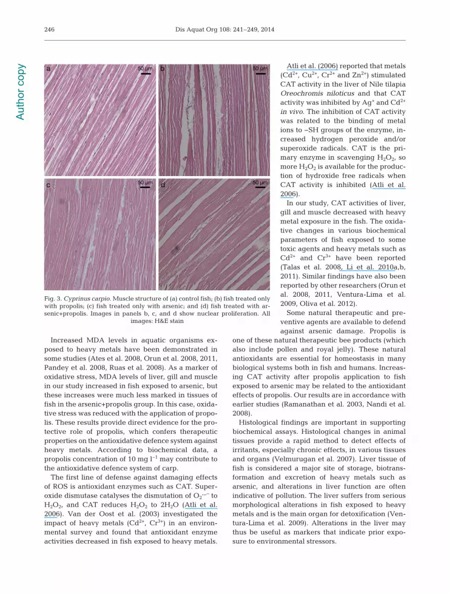
Muscle. No histopathological changes were ob -served in the muscle of control fish (Fig. 3a). Nuclearproliferation was apparent in muscle tissue of thepropolis group (Fig. 3b), as well as in the muscle tis-sues of the arsenic group (Fig. 3c). In the arsenic+propolis group, we observed greater nuclear prolifer-ation compared to the arsenic group (Fig. 3d).
DISCUSSION
Organs of fish were selected on the basis of func-tional criteria that made them preferential targets,i.e. for heavy metal metabolism (liver), heavy metaluptake (gill) and effects of arsenic on the quality offish fillets (muscle). Production of free radicals, whichcan be caused by arsenic, plays an important role in
damage and loss of function in tissuesand organs (Garg et al. 2009, Ventura-Lima et al. 2009, Talas et al. 2012).Lipid peroxidation, which results fromthe oxidative injury of saturated andunsaturated lipids, has been broadlyused as a marker of the induction ofoxidative damage in fish sufferingfrom environmental stress induced byheavy metals (Gioda et al. 2007). Inour study, MDA levels in creased in allof the studied tissues after exposure toarsenic. These findings strongly sug-gest that arsenic caused generalizedoxidative stress in the fish. Thus, theelevated production of ROS may bedue to arsenic toxicity, although othereffects, such as enzymatic inhibition orgenotoxic damage, may also occur.
In recent years, ecotoxicologicalstudies have investigated free radicaldamage and oxidative stress in dif -ferent fish exposed to toxic metalsand organic pollutants (e.g. cadmium,mer cury, copper, arochlor and con-taminated sediments; Orun et al.2008, 2011, Gulhan et al. 2012, Talaset al. 2102). Extensive tissue lipidperoxidation caused increased MDAlevels in liver, gill and muscle tissues(Bernts sen et al. 2003, Orun et al.2008, 2011).
245
Fig. 2. Cyprinus carpio. Gill structure of (a) control fish; (b) fish treated only withpropolis (arrows show vacuolization); (c) fish treated only with arsenic, showingvacuolization (A), necrosis (teleangiectasias) (B), a combination of secondarylamellae and cartilage damage (C) and fusion and shortening of the secondarylamellae (D); and (d) fish treated with arsenic+propolis, showing shortening and
thickening of the secondary lamellae. All images: H&E stain
Aut
hor c
opy
Dis Aquat Org 108: 241–249, 2014
Increased MDA levels in aquatic organisms ex -posed to heavy metals have been demonstrated insome studies (Ates et al. 2008, Orun et al. 2008, 2011,Pandey et al. 2008, Ruas et al. 2008). As a marker ofoxidative stress, MDA levels of liver, gill and musclein our study increased in fish exposed to arsenic, butthese increases were much less marked in tissues offish in the arsenic+propolis group. In this case, oxida-tive stress was reduced with the application of propo-lis. These results provide direct evidence for the pro-tective role of propolis, which confers therapeuticproperties on the antioxidative defence system againstheavy metals. According to biochemical data, apropolis concentration of 10 mg l−1 may contribute tothe anti oxi dative defence system of carp.
The first line of defense against damaging effectsof ROS is antio xidant enzymes such as CAT. Super -oxide dismutase catalyses the dis mutation of O2
…− toH2O2, and CAT re duces H2O2 to 2H2O (Atli et al.2006). Van der Oost et al. (2003) investigated theimpact of heavy metals (Cd2+, Cr3+) in an environ-mental survey and found that antioxidant en zymeactivities decreased in fish ex posed to heavy metals.
Atli et al. (2006) reported that metals(Cd2+, Cu2+, Cr2+ and Zn2+) stimulatedCAT activity in the liver of Nile tilapiaOreochromis niloticus and that CATactivity was inhibited by Ag+ and Cd2+
in vivo. The inhibition of CAT activitywas related to the binding of metalions to −SH groups of the enzyme, in -crea sed hydrogen peroxide and/orsuperoxide radicals. CAT is the pri-mary enzyme in scavenging H2O2, somore H2O2 is available for the produc-tion of hydroxide free radicals whenCAT activity is inhibited (Atli et al.2006).
In our study, CAT activities of liver,gill and muscle decreased with heavymetal exposure in the fish. The oxida-tive changes in various biochemicalparameters of fish exposed to sometoxic agents and heavy metals such asCd2+ and Cr3+ have been reported(Talas et al. 2008, Li et al. 2010a,b,2011). Similar findings have also beenreported by other researchers (Orun etal. 2008, 2011, Ventura-Lima et al.2009, Oliva et al. 2012).
Some natural therapeutic and pre-ventive agents are available to defendagainst arsenic damage. Pro polis is
one of these natural therapeutic bee products (whichalso include pollen and royal jelly). These naturalantioxidants are essential for homeostasis in manybiological systems both in fish and humans. Increas-ing CAT activity after propolis application to fishexposed to arsenic may be related to the antioxidanteffects of propolis. Our results are in accordance withearlier studies (Ramanathan et al. 2003, Nandi et al.2008).
Histological findings are important in supportingbiochemical assays. Histological changes in animaltissues provide a rapid method to detect effects ofirritants, especially chronic effects, in various tissuesand organs (Velmurugan et al. 2007). Liver tissue offish is considered a major site of storage, biotrans -formation and excretion of heavy metals such asarsenic, and alterations in liver function are oftenindicative of pollution. The liver suffers from seriousmorphological alterations in fish exposed to heavymetals and is the main organ for detoxification (Ven-tura-Lima et al. 2009). Alterations in the liver maythus be useful as markers that indicate prior expo-sure to environmental stressors.
246
Fig. 3. Cyprinus carpio. Muscle structure of (a) control fish; (b) fish treated onlywith propolis; (c) fish treated only with arsenic; and (d) fish treated with ar -senic+propolis. Images in panels b, c, and d show nuclear proliferation. All
images: H&E stain
Aut
hor c
opy

Talas et al.: Effects of arsenic and propolis on carp
In our study, the infiltration of mononuclear cells,hydrophic degeneration and congestion were ob -served in the livers of fish exposed to arsenic. Wefound important histopathological differences in thelivers of fish in the arsenic+propolis group comparedto the group that was treated only with arsenic. Ourresults are also in accordance with previous reports(Pedlar et al. 2002, Roy & Bhattacharya 2006). Con-gestion and hydrophic degeneration were observedin liver tissues of the arsenic+propolis group. Theseanalyses show that the damage in liver tissue of thepropolis group was less severe relative to the arsenicgroup.
Necrosis and desquamation of secondary lamellarepithelium, intraepithelial oedema, fusion of adjacentsecondary lamellae, haemorrhage at the primarylamellae, hypertrophy and hyperplasia of epithelialcells have been shown in experimental investigations(Erkmen et al. 2000, Cengiz & Unlu 2003). Erkmen etal. (2000) reported lifting of the epithelial layer fromthe gill lamellae, necrosis and degeneration of thesecondary lamellae, oedema, shortening of the sec-ondary lamellae, and club-shaped lamellae in the gillsof guppies Lebistes reticulatus (= Poecilia reticulata)exposed to cyphenothrin. Shorter gill lamellae, fusion,complete de struction of the lamellae, increased vac-uolation and an irregular appearance of gill lamellaewere ob served in P. reticulata exposed to chlorpyrifos(De Silva & Samayawardhena 2002). Our results thuscorroborate results of previous studies (Pandey et al.2008, Monteiro et al. 2010).
We observed vacuolization, necrosis adjacent tothe secondary lamellae, cartilage damage and fusionand shortening of the secondary lamellae in gills ofcarp exposed to arsenic. In the arsenic+propolisgroup, histopatological changes were evident in thegills, including shortening and thickening of the sec-ondary lamellae, compared to carp exposed to onlyarsenic. These changes in gill tissues were due to thetherapeutic effects of propolis.
Fish plays an important role in the human diet. Fishfillets may be affected by heavy metals, such asarsenic, which can cause structural damage. We ob -served only nuclear proliferations in the muscle tis-sues of both the arsenic group and the arsenic+propolis group. Several studies have investigated theeffects of heavy metals in fish muscle tissues (e.g.Palaniappan & Vijayasundaram 2008, Garg et al.2009). The results of these studies are in line with ourdata. Histopathological studies are useful to evaluatethe potential effects of accumulation of arsenic infish. Our study showed that histopathological chan -ges may originate from the toxic effects of arsenic.
Identifying pathological alterations in organs follow-ing contact with toxicants is useful as a biomarker ofexposure to and effects of pollutants. All of ourhistopathological observations indicated that expo-sure to sublethal concentrations of arsenic causeddestructive effects in the gill, liver and muscle tissuesof Cyprinus carpio.
Experimental studies on the elimination of toxicsubstances such as arsenic in fish are also very im -portant for human health. Certain antioxidant agentscan be used to eliminate and suppress the damagecaused by toxic substances. Our study indicated thatpropolis can repair the deterioration caused byarsenic in fish, as revealed by our analyses of bio-chemical parameters and the histological assay. Thetherapeutic properties of propolis are mostly attri -buted to phenolic components such as flavo noids.Flavonoids have beneficial biological effects, includ-ing anti bacterial, antiviral, anti-inflammatory, anti -allergic, an tioxidant and vasodilatory activities. Propo-lis caused a protective effect against arsenic damagein carp. We suggest that propolis can be used for pro-tection of tissues and organs against degenerativediseases in fish caused by the accumulation of ar -senic. Numerous chemical components of propolishave been repor ted, and this variety is primarilyinfluenced by the botanical and geographical originsof propolis. More than 300 compounds includingvolatile organic compounds, fla vonoid aglycones,phenolic acids and their esters, phenolic aldehydes,alcohols and keto nes, sesquiterpenes, quinones, cou -marins, steroids and amino acids have been reportedamong the components of propolis (Nakamura et al.2010, Beyraghdar Kashkooli et al. 2011). The mostcommon compounds found in propolis include phe-nolics, which play an important protective roleagainst oxidative stress caused by toxic agents suchas xenobiotics and heavy metals. Fla vones, cou mar -ins and many other phenolic compounds also haveantioxidant activities as metal chelators and donorsof hydrogen (Marcucci et al. 2000). All of these com-pounds are responsible for the biological and pharma -cological activities of propolis.
In the future, this work may shed light on investiga-tions of biological activities of new extracts from nat-ural products such as propolis in aquatic organisms.
LITERATURE CITED
Aebi H (1984) Catalase in vitro. Methods Enzymol 105: 121−126
Ates B, Orun I, Talas ZS, Durmaz G, Yilmaz I (2008) Effectsof sodium selenite on some biochemical and hematologi-
247A
utho
r cop
y
Dis Aquat Org 108: 241–249, 2014
cal parameters of rainbow trout (Oncorhynchus mykissWalbaum, 1792) exposed to Pb2+ and Cu2+. Fish PhysiolBiochem 34: 53−59
Atli G, Alptekin O, Tükel S, Canli M (2006) Response ofcatalase activity to Ag2+, Cd2+, Cr2+, Cu2+ and Zn2+ in fivetissues of freshwater fish Oreochromis niloticus. CompBiochem Physiol C Toxicol Pharmacol 143: 218−224
Banerjee P, Bhattacharyya SS, Bhattacharjee N, Pathak S,Boujedaini N, Belon P, Khuda-Bukhsh AR (2009) Ascor-bic acid combats arsenic-induced oxidative stress in miceliver. Ecotoxicol Environ Saf 72: 639−649
Banskota AH, Tezuka Y, Midorikawa K, Matsushige K,Kadota S (2000) Two novel cytotoxic benzofuran deriva-tives from Brazilian propolis. J Nat Prod 63: 1277−1279
Berntssen MH, Aatland A, Handy RD (2003) Chronic dietarymercury exposure causes oxidative stress, brain lesions,and altered behaviour in Atlantic salmon (Salmo salar)parr. Aquat Toxicol 65: 55−72
Beyraghdar Kashkooli O, Ebrahimi Dorcheh E, Mahboobi-Soofiani N, Samie A (2011) Long-term effects of propo-lis on serum biochemical parameters of rainbow trout(Oncorhynchus mykiss). Ecotoxicol Environ Saf 74: 315−318
Burdock GA (1998) Review of the biological properties andtoxicity of bee propolis (propolis). Food Chem Toxicol 36: 347−363
Castaldo S, Capasso F (2002) Propolis, an old remedy usedin modern medicine. Fitoterapia 73: S1−S6
Cengiz EI, Unlu E (2003) Histopathology of gills in mosqui-tofish, Gambusia affinis after long-term exposure to sublethal concentrations of malathion. J Environ Sci HealthB 38: 581−589
De Silva PM, Samayawardhena LA (2002) Low concentra-tions of lorsban in water result in far reaching behavioraland histological effects in early life stages in guppy. Eco-toxicol Environ Saf 53: 248−254
Duran A, Talas ZS (2009) Biochemical changes and sensoryassessment on tissues of carp (Cyprinus carpio, Linnaeus1758) during sale conditions. Fish Physiol Biochem 35: 709−714
Erkmen B, Caliskan M, Yerli SV (2000) Histopathologicaleffects of cyphenothrin on the gills of Lebistes reticula-tus. Vet Hum Toxicol 42: 5−7
Garg S, Gupta RK, Jain KL (2009) Sublethal effects of heavymetals on biochemical composition and their recovery inIndian major carps. J Hazard Mater 163: 1369−1384
Gioda CR, Lissner LA, Pretto A, da Rocha JB and others(2007) Exposure to sublethal concentrations of Zn(II) andCu(II) changes biochemical parameters in Leporinusobtusidens. Chemosphere 69: 170−175
Gülçin I, Bursal E, Sehitoglu MH, Bilsel M, Goren AC (2010)Polyphenol contents and antioxidant activity of lyo phi -lized aqueous extract of propolis from Erzurum, Turkey.Food Chem Toxicol 48: 2227−2238
Gulhan MF, Duran A, Talas ZS, Kakoolaki S, Mansouri SM(2012) Effects of propolis on microbiologic and biochem-ical parameters of rainbow trout (Oncorhynchus mykiss)after exposure to the pesticide. Iran J Fish Sci 11: 490−503
Hughes MF (2002) Arsenic toxicity and potential mecha-nisms of action. Toxicol Lett 133: 1−16
Kavitha C, Malarvizhi A, Kumaran S, Ramesh M (2010)Toxi cological effects of arsenate exposure on hematolo -gical, biochemical and liver transaminases activity in anIndian major carp, Catla catla. Food Chem Toxicol 48: 2848−2854
Li ZH, Zlabek V, Grabic R, Li P, Machova J, Velisek J, Ran-dak T (2010a) Effects of exposure to sublethal propicona-zole on intestine-related biochemical responses in rain-bow trout, Oncorhynchus mykiss. Chem Biol Interact185: 241−246
Li ZH, Li P, Randak T (2010b) Ecotoxocological effects ofshort-term exposure to a human pharmaceutical Vera -pamil in juvenile rainbow trout (Oncorhynchus mykiss).Comp Biochem Physiol C Toxicol Pharmacol 152: 385−391
Li ZH, Zlabek V, Velisek J, Grabic R and others (2011) Acutetoxicity of carbamazepine to juvenile rainbow trout (On -corhynchus mykiss): effects on antioxidant res pon ses,hematological parameters and hepatic EROD. EcotoxicolEnviron Saf 74: 319−327
Lowry OH, Rosebrough NJ, Farr AL, Randall RJ (1951) Pro-tein measurements with the folin phenol reagent. J BiolChem 193: 265−275
Mani F, Damasceno HCR, Novelli ELB, Martins EAM,Sforcin JM (2006) Propolis: effect of different concentra-tions, extracts and intake period on seric biochemicalvariables. J Ethnopharmacol 105: 95−98
Marcucci MC, Ferreres F, Custodio AR, Ferreira MMC,Bankova VS, Garcia-Viguera C, Bretz WA (2000) Evalu-ation of phenolic compounds in Brazilian propolis fromdifferent geographic regions. Z Naturforsch C 55: 76−81
Miranda LE, Capellini VK, Reis GS, Celotto AC, Carlotti CG,Evora PR (2010) Effects of partial liver ischemia followedby global liver reperfusion on the remote tissue expres-sion of nitric oxide synthase: lungs and kidneys. Trans-plant Proc 42: 1557−1562
Monteiro DA, Rantin FT, Kalinin AL (2010) Inorganic mer-cury exposure: toxicological effects, oxidative stress bio-markers and bioaccumulation in the tropical freshwaterfish matrinxa, Brycon amazonicus (Spix and Agassiz,1829). Ecotoxicology 19: 105−123
Nakamura R, Nakamura R, Watanabe K, Oka K, Ohta S,Mishima S, Teshima R (2010) Effects of propolis from dif-ferent areas on mast cell degranulation and identificationof the effective components in propolis. Int Immunophar-macol 10: 1107−1112
Nandi D, Patra RC, Ranjan R, Swarup D (2008) Role of co-administration of antioxidants in prevention of oxidativeinjury following sub-chronic exposure to arsenic in rats.Vet Arh 78: 113−121
Oliva M, Vicente JJ, Gravato C, Guilhermino L, Galindo-Riano MD (2012) Oxidative stress biomarkers in Senegalsole, Solea senegalensis, to assess the impact of heavymetal pollution in a Huelva estuary (SW Spain): seasonaland spatial variation. Ecotoxicol Environ Saf 75: 151−162
Orun I, Ates B, Selamoglu Z, Yazlak H, Ozturk E, Yilmaz I(2005) Effects of various sodium selenite concentrationson some biochemical and hematological parameters ofrainbow trout (Oncorhynchus mykiss). Fresenius EnvironBull 14: 18−22
Orun I, Talas ZS, Ozdemir I, Alkan A, Erdogan K (2008)Antioxidative role of selenium on some tissues of (Cd2+,Cr3+)-induced rainbow trout. Ecotoxicol Environ Saf 71: 71−75
Orun I, Talas ZS, Alkan A (2011) Modulating effect of sele-nium on gills of fish exposed to heavy metals. FreseniusEnviron Bull 20: 104−108
Palaniappan PR, Vijayasundaram V (2008) Fourier transforminfrared study of protein secondary structural changes inthe muscle of Labeo rohita due to arsenic intoxication.Food Chem Toxicol 46: 3534−3539
248A
utho
r cop
y

Talas et al.: Effects of arsenic and propolis on carp 249
Pandey S, Parvez S, Ansari RA, Ali M and others (2008)Effects of exposure to multiple trace metals on biochem-ical, histological and ultrastructural features of gills ofa freshwater fish, Channa punctata Bloch. Chem BiolInteract 174: 183−192
Pedlar RM, Ptashynski MD, Evans R, Klaverkamp JF (2002)Toxicological effects of dietary arsenic exposure in lakewhitefish (Coregonus clupeaformis). Aquat Toxicol 57: 167−189
Ramanathan K, Shila S, Kumaran S, Panneerselvam C(2003) Protective role of ascorbic acid and a-tocopherolon arsenic induced microsomal dysfunction. Hum ExpToxicol 22: 129−136
Rice-Evans C (2001) Flavonoid antioxidants. Curr MedChem 8: 797−807
Roy S, Bhattacharya S (2006) Arsenic-induced histopatho -logy and synthesis of stress proteins in liver of Channapunctatus. Ecotoxicol Environ Saf 65: 218−229
Ruas CB, Carvalho C, Arauj HS, Espindola EL, FernandesMN (2008) Oxidative stress biomarkers of exposure inthe blood of three cichlid species from a polluted river.Ecotoxicol Environ Saf 71: 86−93
Schlenk D, Colley WC, El-Alfy A, Kirby R, Griffin BR (1997)Effect of arsenite, arsenate, and the herbicide mono -sodium methyl arsonate (MSMA) on hepatic metalloth-ionein expression and lipid peroxidation in channel catfish. Comp Biochem Physiol C Pharmacol ToxicolEndocrinol 118: 177−183
Shah AQ, Kazi TG, Arain MB, Jamali MK and others (2009)Accumulation of arsenic in different fresh water fish spe-cies — potential contribution to high arsenic intakes.Food Chem 112: 520−524
Talas ZS, Gulhan MF (2009) Effects of various propolis con-centrations on biochemical and hematological para -meters of rainbow trout (Oncorhynchus mykiss). Eco -toxicol Environ Saf 72: 1994−1998
Talas ZS, Orun I, Ozdemir I, Erdogan K, Alkan A, Yilmaz I(2008) Antioxidative role of selenium against the toxiceffect of heavy metals (Cd2+, Cr3+) on liver of rainbowtrout (Oncorhynchus mykiss Walbaum 1792). Fish Phys-iol Biochem 34: 217−222
Talas ZS, Dundar SP, Gulhan MF, Orun I, Kakoolaki S (2012)Effects of propolis on some blood parameters andenzymes in carp exposed to arsenic. Iran J Fish Sci 11: 405−414
van der Oost R, Beyer J, Vermeulen NPE (2003) Fish bioac-cumulation and biomarkers in environmental riskassessment: a review. Environ Toxicol Pharmacol 13: 57−149
Velmurugan B, Selvanayagam M, Cengiz EI, Unlu E (2007)Histopathology of lambda-cyhalothrin on tissues (gill,kidney, liver and intestine) of Cirrhinus mrigala. EnvironToxicol Pharmacol 24: 286−291
Ventura-Lima J, Castro MR, Acosta D, Fattorini D and others(2009) Effects of arsenic (As) exposure on the antioxidantstatus of gills of the zebrafish Danio rerio (Cyprinidae).Comp Biochem Physiol C Toxicol Pharmacol 149: 538−543
Yagi K (1984) Assay of lipid peroxidation in blood plasma orserum. Methods Enzymol 105: 328−331
Zhang W, Huang L, Wang WX (2012) Biotransformation anddetoxification of inorganic arsenic in a marine juvenilefish Terapon jarbua after waterborne and dietborneexposure. J Hazard Mater 221/222: 162–169
Editorial responsibility: Thomas Braunbeck, Heidelberg, Germany
Submitted: November 18, 2013; Accepted: December 23, 2013Proofs received from author(s): March 12, 2014
Aut
hor c
opy