antigenic heterogeneity of lipid a of haemophilus influenzae
TRANSCRIPT

Vol. 50, No. 1INFECTION AND IMMUNITY, Oct. 1985, p. 9-140019-9567/85/100009-06$02.00/0Copyright X) 1985, Amnerican Society for Microbiology
Antigenic Heterogeneity of Lipid A of Haemophilus influenzaeMICHAEL A. APICELLA,1* KATHLEEN C. DUDAS,1 ANTHONY CAMPAGNARI,1 PETER RICE,2
JOSEPH M. MYLOTTE,1 AND TIMOTHY F. MURPHY'Department of Medicine, Division of Infectious Diseases, State University ofNew York at Buffalo, School of Medicine,
Buffalo, New York 142151; and Department of Research and Education, Section of Infectious Diseases, Boston UniversitySchool of Medicine, Boston, Massachusetts 021302
Received 20 March 1985/Accepted 24 June 1985
The chemical structure and biologic function of the lipid A portion of lipopolysaccharide are not identicalamong gram-negative bacteria. This study indicates that antigenically heterogeneous lipid A exists amongstrains of Haemophilus influenzae. An immunoglobulin G3 murine monoclonal antibody, 3D2, producedagainst a nontypable H. influenzae strain 3524 has specificity for a site on the lipid A portion of the H. influenzaelipopolysaccharide. With the Western blot and immunodot assay, 3D2 recognized this lipid A determinant on14 of 24 (58%) of strains of nontypable H. influenzae and in 51 of 95 (54%) strains ofH. influenzae type b. Thislipid A epitope has a high degree of specificity for H. influenzae, since it is not present on the lipid A of 39gram-negative strains from 14 non-Haemophilus species. In addition, studies of 36 strains of six Haemophilusspecies other than H. inflenzae and 8 strains of 4 species of ActnobaciUus did not contain the 3D2 epitope.Enzyme-linked immunosorbent assay analysis with a kinetic assay and enzyme-linked immunosorbent assayinhibition confirmed the antigenic heterogeneity of H. influenzae lipid A. Thin-layer chromatographydemonstrated that the 3D2 epitope is associated with a chloroform-soluble lipid moiety in the lipid A.Fluorescent antibody analysis of H. influenzae indicated that the epitope is on the cell surface. The monoclonalgntibody was not bactericidal for strain 3524, and it did not inhibit the bactericidal action of normal humanserum against the same strain. These studies demonstrate that the lipid As of H. influenzae are antigenicailyheterogeneous.
Since 1963, the concept that the lipid A component of thelipopolysaccharide (LPS) of gram-negative bacteria repre-sents a heterogeneous group of structures has gained accept-ance (21). Chang and Nowotny have shown that the lipid Apbrtions of the LPS structure are not identical in structure orfunction (4). Other investigators demonstrated that fraction-ation of a single lipid A preparation resulted in the identifi-cation of eight fractions which had a range of antigenexpression (3, 16, 17). Five of these fractions could reactwith anti-lipid A antiserum, whereas the three with thegreatest number of esterfied fatty acids failed to react. Thusit would appear that antigenic variability among lipid A'sshould be anticipated as more extensive studies specificallywith monoclonal antibodies are developed.We have had the opportunity to study a number of
nontypable Haemophilus influenzae LPS preparations with amonioclonal antibody developed to a nontypable H. influ-enzae strain, 3524. This monoclonal antibody, designated3D2, has been shown to react with a determinant present onthe lipid A portion of the LPS. The investigations presentedin this paper indicate that the determinant recognized by thismonoclonal antibody is not present on the lipid A of all H.influenzae strains. Studies of other Haemophilus species anda diverse group of gram-negative and gram-positive bacteriaindicate that this antigenic determinant is highly specific forH. influenzae.
MATERIALS AND METHODSBacteria. Prototype strains of nontypable H. influenzae
representative of the eight sodium dodecyl sulfate-polyacrylamide gel electrophoresis (SDS-PAGE) typesdescribed by Murphy et al. (19) were obtained from our owncollection. The identity of each isolated strain had been
* Corresponding author.
confirmed previously as H. influenzae by colonial morphol-ogy and growth requirement for X and V factors.Counterimmunoelectrophoresis (20) had been used todetermine serotype with antiserum and reference strainsobtained from the Centers for Disease Control, Atlanta, Ga.Nontypable H. influenzae 3524 was isolated from the sputumof a patient with chronic bronchitis at the Erie CountyMedical Center, Buffalo, N.Y. It has been demonstrated to bepiliated (2) and has been shown to have a type 2 outermembrane protein pattern on SDS-PAGE (19). Copies ofHaemophilus paraphrophilus, ATCC 29240, 29241, and29242, H. segnis ATCC 10977, H. aphrophilus ATCC 13252and 19415, and NCTC 5906, 5907, and 5908, H.parainfluenzae ATCC 7901 and 9276, H. aegypticus ATCC11116, H. parahaemolyticus VATCC 10014, nontypable H.influenzae ATCC 19418, H. paraphrohaemolyticus ATCC29237, Actinobacillus actinomycetemcomitans ATCC 29522,29523, and 29524 and NCTC 9707 and 9710, A. equili ATCC19392, A. seminis ATCC 15768, and A. suis ATCC 15557 wereprovided by J. Zambon, State University of New York atBuffalo, School of Dentistry. Isolates of Escherichia coli,Klebsiella pneumoniae, Klebsiella species, Pseudomonasaeruginosa, Pseudomonas species, Enterobacter cloacae,Proteus vulgaris, Proteus mirabilis, Morganella morganiiSerratia marcescens, H. parainfluenzae, H. parahaemolyt-icus, H. haemolyticus, Streptococcus pneumoniae, Neisseriameningitidis, N. gonorrhoeae, Streptococcus viridans, andStaphylococcus species were provided by the clinicalmicrobiology laboratory at the Erie County Medical Center.Strains were stored in Mueller-Hinton medium containing10% glycerol at -70°C until studied and were reconstituted onappropriate media for sample preparation.Development of hybridomas. (i) Immunization of spleen cell
donor mice. BALB/c mice were immunized intraperitoneallywith 0.1 ml of 109 nontypable H. influenzae 3524 cells on
9

10 APICELLA ET AL.
days 0 and 28. On day 32 after the initial immunization,selected animals were sacrificed with chloroform, theirspleens were removed, and splenic lymphocytes were har-vested by perfusion of splenic pulp with minimum essentialmedium (MEM).
(ii) Fusion of donor spleen cells to the NS-1 nonsecretingclone of the P3x63Ag8 BALB/c plasmacytoma. To achievefusion of the donor spleen cells to the NS-1 (nonsecretingvariant of the immunoglobulin Gl [IgGl] BALB/cplasmacytoma P3x63Ag8) plasmacytoma cells (obtainedfromn the Salk Institute for Biology under National CancerInstitute contract NO1-CB-23886), 35% polyethylene glycolwas used in a modification of the procedure of Kennett (13).Briefly, 107 spleen cells were combined with 106 NS-1 cells inMEM with serum. The cells were centrifuged at 170 x g for10 min at 25°C. All of the supernatant was removed, and thepellet was tapped to loosen. A 0.2-ml sample of 35% poly-ethylene glycol 1000 (Sigma Chemical Co., St. Louis, Mo.)in MEM without serum was added, the mixture was stirredlightly and left at 25°C, and timing was started for 8 min. Thecells were pelleted at 500 x g for 3 min. At the end of theoriginal 8 min, 5 ml of MEM with serum was added andgently pipetted once to resuspend the pellet. The mixturewas centrifuged at 250 x g for 5 min at room temperature.All of the supernatant was removed. A 5-ml sample ofcomplete growth medium (MEM with high glucose and 20%bovine fetal serum) was added to resuspend the pellet. Themixture was transferred to a 25 ml Erlenmeyer flask contain-ing the appropriate amount of complete MEM to obtain 3 xi05 plasmacytoma cells per ml. The cells were stirred lightlyand distributed in 0.05-ml samples into microtiter wells. At24 h after the polyethylene glycol fusion, 0.05 ml of hypo-xanthine-aminopterin-thymidine medium was added to eachwell. The microtiter plates were placed in a tissue cultureincubator at 85% humidity under 5% carbon dioxide. Freshhypoxanthine-aminopterin-thymidine medium was added atday 7, and plates were checked for macroscopic plaquesafter day 10. The supernatant from all wells was tested forthe presence of antibody by using an enzyme-linked im-munosorbent assay (ELISA) system in which 10 jig of strain3524 outer membranes per ml was coated to the microtiterplates. The peroxidase-conjugated rabbit anti-murine IgG orprotein A was used to detect the presence of antibodybinding to the outer membranes. Wells which containedhybridomas producing antibody to outer membranes wereretested against microtiter plates coated with strain 3524LPS to identify those clones producing antibody to LPS.These clones were propagated by subsequent transfer tolarger tissue culture wells. Large quantities of antibody wereproduced in tissue culture and by the intraperitoneal injec-tion of 105 cells from the LPS antibody-producing clone into2,6,10,14-tetramethylpentadecane (pristane)-primedBALB/c mice. The resulting ascitic fluid was harvested in 3to 4 weeks, and the ascitic fluid was tested for specificity ofthe respective antibody. The IgG3 antibody was removedfrom the ascitic fluid by passage over a protein A-Sepharosecolumn. Similar peritoneal tumors were established by usingNS-1 cells. The resultant fluid was used as a control.The monoclonal antibody produced in tissue culture fluid
was purified by affinity chromatography over a murine IgGaffinity column. This antibody was used to confirm theresults obtained with the ascitic fluid-derived antibody.
Preparation of LPS, lipid A and outer membranes. H.influenzae LPS was prepared by a modification of thephenol-water method of Perry et al. (22). Lipid A wasprepared by heating 20 mg of the LPS preparation in 1.0 M
acetic acid in a boiling water bath for 1 h. The lipid Aprecipitate was removed by centrifugation at 15,000 x g for30 min. The precipitate was lyophilized after it was washedtwice by suspension in distilled water followed by centrifu-gation. The acetic acid and wash supernatants were pooledand lyophilized. This contained the saccharide portion of theLPS. The lipid A fraction was further fractionated by themethod of Chang and Nowotny (4). This involves sequentialextractions with ethyl acetate and chloroform. Three frac-tions are recovered, an ethyl acetate-insoluble fraction, achloroform-soluble fraction, and a chloroform-insolublefraction. Outer membranes were prepared by the method ofMurphy et al. (19).
Thin-layer chromatography. Thin-layer chromatographywas performed by a modification of the method of Banerjiand Alving with a chloroform-acetone (85:15) solvent systemwith silica gel 60 plates (EM Laboratories, Elmsford, N.Y.)(3). Lipid spots were identified on the dried chromatogramsby using iodine vapors and confirmed with bromothymolblue spray, phosphate-containing residues were identifiedwith molybdenum blue reagent, carbohydrates were identi-fied with orcinol (0.25% orcinol in 75% sulfuric acid), andamino acid-containing compounds were identified with nin-hydrin spray (0.3% in butanol-acetic acid) (26).Western blot analysis. Analysis of the specificity of the 3D2
monoclonal antibody was performed by Western blot meth-odology (14) with a Trans-blot apparatus (Bio-Rad Labora-tories, Richmond, Calif). Briefly, samples were subjected toSDS-PAGE by the Laemmli method (15). At the end of thiselectrophoresis, the gel was removed and placed in theTrans-blot apparatus with a 10- by 20-cm sheet of nitrocel-lulose over 1.5 h at 50 V in 25 mM Tris hydrochloride (pH8.3)-192 mM glycine-20% methanol buffer. After transfer,the nitrocellulose sheet was placed in 3% gelatin and thentreated with 3D2 monoclonal antibody at room temperatureovernight, washed with buffer A (0.9% [wt/vol] NaCl, 10 mMTris hydrochloride [pH 7.4]) and then treated with proteinA-peroxidase conjugate (Zymed, South San Francisco,Calif.). The sheet was then washed with buffer A and treatedwith HRP reagent (Bio-Rad) for 45 min, and the resultingbands were observed on a light box.Immunodot assay. A modified dot assay utilizing nitrocel-
lulose paper was described previously (9). In this assay, thesample to be screened for the presence of antigen is solubil-ized in SDS-PAGE sample buffer at 1 mg/ml, and 10 ,LI isplaced in a 1.5-cm square on the paper. The sample isallowed to dry, and the sheet is then treated in a fashionidentical to that described above for the Western blottechnique.
Kinetic ELISA. The quantitative, single-tube, kinetic-dependent ELISA (herein referred to as the kinetic ELISA)uses enzyme rate kinetics to quantitate bound enzyme andassay ligand (either antigen or antibody). This assay wasperformed by the method of Tsang and co-workers (27).Individual Ultra-UV disposable polystyrene cuvettes (FisherScientific Co., Pittsburgh, Pa.) were coated with LPS fromnontypable H. influenzae strains in concentrations of 0.1, 1,and 5 ,ug/ml. The LPSs were suspended in sensitizationbuffer (0.05 M Tris, 2.0 mM EDTA, 0.3 M KCl [pH 8.0]) bysonication. Cuvettes were sensitized for 1 h a 37°C andovernight at 4°C. Protein A-purified monoclonal antibody3D2 was washed with phosphate-buffered saline-0.3%Tween 20 and reacted at concentrations of 0.1, 1, and 10jig/ml with the LPS-coated cuvettes for 1 h at 37°C. Thecuvettes were washed with phosphate-buffer saline-Tween20, and protein A-peroxidase (1:3,000) was reacted with the
INFECT. IMMUN.
LIPID A OF H. INFLUENZAE 11
cuvettes for 1 h at 370C. The cuvettes were washed withphosphate-buffered saline-Tween 20. After the removal ofthe wash fluid, the cuvettes were placed in a Varian 634spectrophotometer with the analog output connectedthrough an A-D converter to an Apple IIe computer whichhad been programmed to calculate the rate of change ofabsorbance at 460 nm per minute. One milliliter of freshlyprepared O-dianisidine dihydrochloride (Sigma) in 0.05 Msodium acetate (pH 5.0) was added to the cuvette. Thechange in absorbance at 460 nm was measured over the first3 min of each reaction, and the rate of change of absorbanceat 460 nm per minute for each antiserum and LPS concen-tration was calculated.ELISA inhibition studies. ELISA inhibition studies were
performed by the method of Apicella and Gagliardi (1).Fluorescent antibody analysis. Indirect fluorescent anti-
body studies were performed to determine whether 3D2recognized a surface-exposed epitope. Briefly, H. influenzaeorganisms were fixed to glass slides in acetone and exposedto affinity-purified 3D2 antibody derived from the tissueculture supernatant. The fluorescent conjugate used was anFc-specific fluorescein conjugate obtained from Cappel Lab-oratories, Cochranville, Pa. Studies were performed with anA-O macro-star microscope with epifluorescence.
Bactericidal assay. Studies to determine whether 3D2 hadbactericidal activity against H. influenzae strains were per-formed by the method of Rice and Kasper (24). Studies todetermine whether 3D2 was capable of inhibiting the bacte-ricidal response of normal human serum were also per-formed (25).
Other immunological studies. Immunodiffusion studieswere performed on 10-fold-concentrated tissue culture fluidto determine the immunoglobulin class of the monoclonalantibody. For these studies, antisera specific for murineIgM, IgGl, IgG2a, IgG2b, and IgG3 were obtained fromBionetics, Kensington, Md.
RESULTSUtilizing splenocytes from a BALB/c mouse immunized
with nontypable H. influenzae 3524, a series of hybridomaswere developed by fusion of NS-1 plasmacytoma cells.Among the clones producing antibody to the H. influenzaestrain, one designated 3D2 was found by ELISA screening tobe producing antibody to LPS isolated from this strain. Thismonoclonal antibody was determined by immunodiffusion tobe of the IgG3 variety.
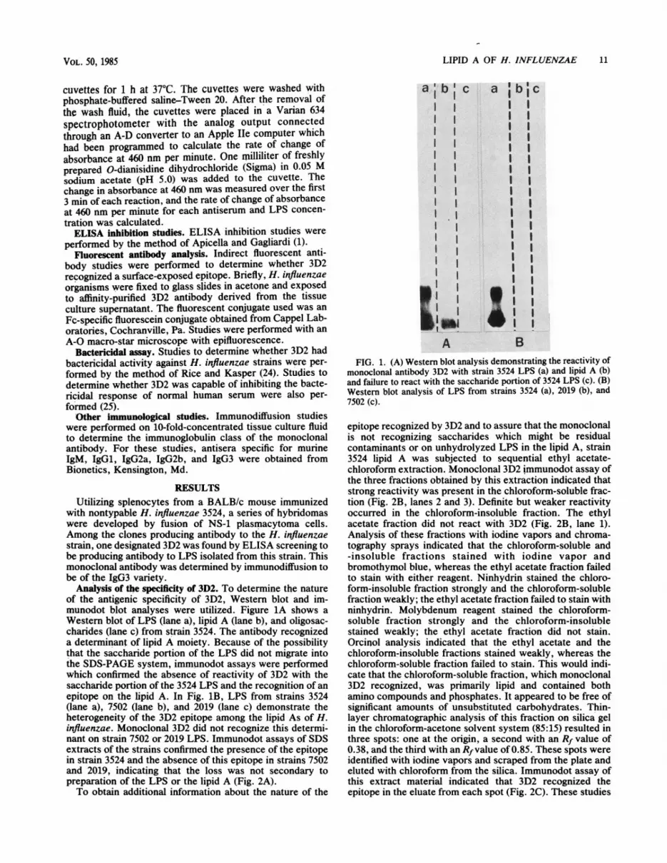
Analysis of the specificity of 3D2. To determine the natureof the antigenic specificity of 3D2, Western blot and im-munodot blot analyses were utilized. Figure 1A shows aWestern blot of LPS (lane a), lipid A (lane b), and oligosac-charides (lane c) from strain 3524. The antibody recognizeda determinant of lipid A moiety. Because of the possibilitythat the saccharide portion of the LPS did not migrate intothe SDS-PAGE system, immunodot assays were performedwhich confirmed the absence of reactivity of 3D2 with thesaccharide portion of the 3524 LPS and the recognition of anepitope on the lipid A. In Fig. 1B, LPS from strains 3524(lane a), 7502 (lane b), and 2019 (lane c) demonstrate theheterogeneity of the 3D2 epitope among the lipid As of H.influenzae. Monoclonal 3D2 did not recognize this determi-nant on strain 7502 or 2019 LPS. Immunodot assays of SDSextracts of the strains confirmed the presence of the epitopein strain 3524 and the absence of this epitope in strains 7502and 2019, indicating that the loss was not secondary topreparation of the LPS or the lipid A (Fig. 2A).To obtain additional information about the nature of the
a i b I c
I1 II II II II II II II II II I1I II II Il
A
a b'cI II II II II II I
II
I
lII
iiB
iIIIIIIIIIIIII
FIG. 1. (A) Western blot analysis demonstrating the reactivity ofmonoclonal antibody 3D2 with strain 3524 LPS (a) and lipid A (b)and failure to react with the saccharide portion of 3524 LPS (c). (B)Western blot analysis of LPS from strains 3524 (a), 2019 (b), and7502 (c).
epitope recognized by 3D2 and to assure that the monoclonalis not recognizing saccharides which might be residualcontaminants or on unhydrolyzed LPS in the lipid A, strain3524 lipid A was subjected to sequential ethyl acetate-chloroform extraction. Monoclonal 3D2 immunodot assay ofthe three fractions obtained by this extraction indicated thatstrong reactivity was present in the chloroform-soluble frac-tion (Fig. 2B, lanes 2 and 3). Definite but weaker reactivityoccurred in the chloroform-insoluble fraction. The ethylacetate fraction did not react with 3D2 (Fig. 2B, lane 1).Analysis of these fractions with iodine vapors and chroma-tography sprays indicated that the chloroform-soluble and-insoluble fractions stained with iodine vapor andbromothymol blue, whereas the ethyl acetate fraction failedto stain with either reagent. Ninhydrin stained the chloro-form-insoluble fraction strongly and the chloroform-solublefraction weakly; the ethyl acetate fraction failed to stain withninhydrin. Molybdenum reagent stained the chloroform-soluble fraction strongly and the chloroform-insolublestained weakly; the ethyl acetate fraction did not stain,Orcinol analysis indicated that the ethyl acetate and thechloroform-insoluble fractions stained weakly, whereas thechloroform-soluble fraction failed to stain. This would indi-cate that the chloroform-soluble fraction, which monoclonal3D2 recognized, was primarily lipid and contained bothamino compounds and phosphates. It appeared to be free ofsignificant amounts of unsubstituted carbohydrates. Thin-layer chromatographic analysis of this fraction on silica gelin the chloroform-acetone solvent system (85:15) resulted inthree spots: one at the origin, a second with an Rf value of0.38, and the third with an Rfvalue of 0.85. These spots wereidentified with iodine vapors and scraped from the plate andeluted with chloroform from the silica. Immunodot assay ofthis extract material indicated that 3D2 recognized theepitope in the eluate from each spot (Fig. 2C). These studies
VOL. 50, 1985

12 APICELLA ET AL.
I
A
B
Co
2
FIG. 2. Immunodot assay demonstrating reactivity of 3D2 withvarious lipid A extracts. Row A contains a phosphate-bufferedsaline control (lane 1) an SDS extract of strain 7502 (lane 2) and an
SDS extract of strain 3524 (lane 3). Row B contains the ethyl acetateextract (lane 1), the chloroform-soluble extract (lane 2), and thechloroform-ihsoluble portion (lane 3) of 3524 lipid A. Row Ccontains extracts eluted from a thin-layer chromatographic platecontaining the chloroform-soluble extract of the lipid A of strain3524. Lane 1 contains extracts at the origin, lane 2 contains extractsat Rf 0.38, and lane 3 contains extracts at Rf 0.85.
would indicate that the 3D2 epitope is not associated withthe oligosaccharide portion of the LPS and that a lipidmoiety free of saccharides reacts with this monoclonalantibody.
Kinetic ELISA studies and ELISA inhibition. To determinewhether the relationship of the epitope seen by 3D2 was
similar on a variety of solid-phase surfaces in addition to
nitrocellulose and whether antigen in solution was recog-nized by 3D2, kinetic ELISA and ELISA inhibition studies
0.1 1 5
[LPS] ugmml
FIG. 3. Kinetic ELISA analysis of LPS from strains 3524 (0),2019 (U), and 7502 (A). Monoclonal antibody 3D2 concentrationwas 1 ,ug/ml. A4w, absorbancy at 460 nm.
were performed. Kinetic ELISA analysis (Fig. 3) demon-
strates the specificity of 3D2 for the epitope in the LPS from
strain 3524 and the relative absence of this epitope on the
LPS from strains 2019. ELISA inhibition studies were per-
formed with LPS from strains 3524, 3198, 2019, and 7502.
The concentration of strain 3524 or 3198 LPS necessary to
achieve 50% inhibition of the 3D2-3524 ELISA was 100
times less than that necessary for inhibition of sttain 2019
LPS and at least.1,000 times less than that required by strain
7502 LPS (Table 1). In addition to confirming the 3D2
reactivity of LPS from strains 3524 and 3198, these results
also suggest that a portion of the 3D2 epitope may be present
on strain 2019 LPS.Fluorescent antibody and bacterial studies. Fluorescent
antibody studies with 3D2 indicated that it identified a
surface exposed epitope on strain 3524. Bactercidal studiesagainst strain 3524 with affinity-purified 3D2 from tissueculture supernatants with hypogammaglobulinemic serum as
a complement source indicated that concentrations.of anti-body as high as 500 ,ug/ml failed to have any bactericidaleffect against strain 3524. Additionally, pretreated strain3524 with 3D2 did not inhibit the bactericidal effect of normalhuman serum on 3524.
Specificity and prevalence studies. Twenty-four nontypableH. influenzae clinical strains were studied for the 3D2 lipid Adeterminant in the immunodot assay. Fourteen strains (58%)were positive. Fifty-one of 95 H. influenzae type b strainswere positive. One strain each of H. influenzae types a, c, d,and e were positive, and a single type f strain was negative.Twenty-four H. parainfluenzae strains, four strains of H.paraphrophilus, four strains of H. aphrophilus, two strainsof H. parahaemolyticus, one strain of H. aegyptis, and one
strain of H. segnis were negative for the 3D2 lipid A
determinant. A variety of non-Haemophilus species were
tested and were negative for this determinant. These include
10 E. coli strains, 6 P. mirabilis strains, 1 P. vulgaris strain,3 S marscescens strains, 1 M. morganii strain, 1 E. cloacae
strain, 1 K. pneumofliae strain, 1 Klebsiella species, 3 P.
aeruginosa strains, 1 Pseudomonas species, 2 Neisseria
species, 6 N. gonorrhoeae straitis, 1 Serratia species, 8
Actinobacillus species, 5 S. aureus strains, 2 Staphylococ-cus species, 1 diphtheroid, and 2 streptococcal strains which
were obtained from clinical isolates. Among the nontypableH. influenzae strains the 3D2 determinant was distributedrandomly among the various outer membrane protein pat-terns which constituted the Murphy et al. subtyping system.
DISCUSSIONLipid A is a component of the LPS of gram-negative
bacteria. It has a basic structure which is highly conservedamong a wide range of gram-negative species and which has
TABLE 1. 3D2 ELISA inhibitiona
Inhibitor 3D2 Concn of LPS (jag/ml) causing:
LPS Immunodot 50%o inhibition 100% inhibition
3S24 + 0.041 (0.017-0.096)b 2.04 (0.56-7.41)3198 + 0.123 (0.05-0.291) 11.64 (3.61-37.6)2019 - 205 (35.9->1,000) >1,0007502 - >1,000 >1,000
a Microtiter wells were coated with 3524 LPS at a concentration of 5 jag/ml.Monoclonal 3D2 concentration was 1 jag/ml.
b Shown within parentheses are 95% confidence limits based on analysis of
experiments run in triplicate.
1 1*1"
INFECT. IMMUN.

LIPID A OF H. INFLUENZAE 13
been shown to be associated with the toxic activity ascribedto LPS. Lipid A is not a single macromolecular structure,but appears to be a complex array of a variety of lipids (3, 11,12, 16, 18). Variation in the lipid A of the same strain hasbeen demonstrated (16). This has been shown to be based onvariation of the composition of the ester-linked fatty acidsand substitution of the phosphate groups (16). The chemicalstructure of lipid A from enteric bacteria has been elaboratedand has been shown to consist of a (1'-6)-linkeddiglucosamine oligosaccharide which contains two phos-phate groups (8). This backbone structure is substituted by avariety of hydrophilic and hydrophobic substances, includ-ing amide-linked and ester-linked fatty acids. Lipid A alsoappears to have a highly conserved antigenic structureamong a wide range of gram-negative bacteria. Immunolog-ical studies of lipid A from enteric bacteria have demon-strated a high degree of cross-reactivity which extends to thenon-Enterobacteriaceae (6, 16). The fundamental antigeniccomponent which will react with polyclonal antisera to lipidA has been shown to be the diglucosamine backbone substi-tuted with a single amide-linked myristic acid (6). Lipid A asa portion of the intact LPS has been shown to be a poorimmunogen. This can be enhanced by coating lipid A toacid-treated bacterial cells or to Re mutants of Enterobacte-riaceae (7).The lipid A of H. influenzae has not been completely
studied. Studies by Raichvarg and associates have indicatedthat H. influenzae type a lipid A contained phosphorus,glucosamine, myristic, palmitic, and oleic acids and threehydroxymyristic acids. In addition, two other long chainfatty acids were found, but not identified (23). Of interest,the free lipid A was not toxic in Swiss mice at doses up to 50mg/kg. No comments were made about the immunologicalcharacteristics of this lipid A preparation or antigenic differ-ences in H. influenzae type a lipid A. However, it wasconcluded that this lipid A preparation did not exhibit all ofthe classical biological activities attributed to enterobacteriallipid A. Flesher and Insel (5) studied LPS from H. influenzaetypes a through f. Chemical analysis was completed only onthe type b serotype and indicated that it contained 30% totalcarbohydrate and 29.3% lipid. Analysis of the sugars in thecarbohydrate revealed glucose, galactose, glucosamine, andheptose, whereas the lipid component contained lauric,myristic, and palmitic acid. Biologic studies with these LPSpreparations indicated that the preparations were toxic formice and could be used to stimulate murine spleen cells. Thislatter effect could be blocked by polymyxin and was consid-ered secondary to the lipid A component. Immunologicalstudies with immunodiffusion and hemagglutination analysissuggested immunological heterogeneity among H. influenzaestrains and indicated the presence of at least three antigen-ically distinct factors associated with these LPS. The sourceof these antigenic differences was considered to be theoligosaccharide portion of the LPS moiety; however, chem-ical analysis indicated that the carbohydrates were com-posed of identical sugars. It was assumed the differenceswere based on differences in sequence or the fine structure ofthe sugars. Recently, Insana demonstrated electrophoreticheterogeneity of H. influenzae by using microextracts ofLPS on SDS-PAGE gels and silver staining (10); 50 strainswere studied, and 33 could be classified into 11 subtypes.Nontypable strains appeared to demonstrate less LPS elec-trophoretic variation than that seen among type b strains.The basis for this heterogeneity has not been elucidated.
In this study, we have demonstrated that there are at leasttwo antigenically distinct lipid A moieties among typable and
nontypable H. influenzae strains. The epitope responsiblefor this differentiation is a lipid moiety which can be recov-ered in the chloroform-soluble portion of the lipid A. Anal-ysis of non-Haemophilus species indicates that the epitopewas absent among the strains studied. The epitope does notappear to be a bactericidal target but appears to be surfaceexposed based on fluorescent antibody analysis. These re-sults are similar to those obtained by Mattsby-Baltzer andKaliser, who showed the limited ability to demonstratesurface exposure of lipid A determinants on Enterobacteri-aceae with polyclonal antisera (18). ELISA inhibition andkinetic ELISA analysis indicated that 3D2 recognizes thedeterminant on free lipid A and lipid A bound to polystyreneplastic surfaces. The ELISA inhibition studies would alsoindicate that this epitope may be present in low concentra-tions on strains which lack the determinant in Western blotanalysis.These studies demonstrate the antigenic heterogeneity of
the lipid A of H. influenzae. Future efforts will be directed atdetermining the association of this determinant with thevirulence of H. influenzae and in determining the precisenature of the structure responsible for the epitope recog-nized by monoclonal antibody 3D2.
ACKNOWLEDGMENTSThis work has been supported by Public Health Service grant
A119641 from the National Institute of Allergy and InfectiousDiseases and by the Veterans Administration.The expert assistance of Phyllis Rosenberg and Marlene Shero in
the preparation of this manuscript is appreciated.
LITERATURE CITED1. Apicella, M. A., and N. C. Gagliardi. 1979. Antigenic heteroge-
neity of the non-serogroup antigen structure of Neisseria gon-orrhoeae lipopolysaccharides. Infect. Immun. 26:870-874.
2. Apicella, M. A., M. Shero, K. C. Dudas, R. R. Stack, W. Klohs,L. J. LaScolea, T. F. Murphy, and J. M. Mylotte. 1984. Fimbria-tion of Haemophilus species isolated from the respiratory tractof adults. J. Infect. Dis. 150:40-43.
3. Banerji, B., and C. R. Alving. 1979. Lipid A from endotoxin:Antigenic activities of purified fractions in liposomes. J. Immu-nol. 123:2558-2562.
4. Chang, C.-M., and A. Nowotny. 1975. Relation of structure tofunction in bacterial 0-antigens. VII. Endotoxicity of 'Lipid A.'Immunochemistry 12:19-28.
5. Flesher, A. R., and R. A. Insel. 1978. Characterization oflipopolysaccharide of Haemophilus influenzae. J. Infect. Dis.138:719-730.
6. Galanos, C., M. A. Freudenberg, F. Jay, D. Nekar, K. Veleva, H.Brade, and W. Strittmatter. 1984. Immunogenic properties oflipid A. Rev. Infect. Dis. 6:546-552.
7. Galanos, C., 0. Fuderity, and 0. Westphal. 1971. Preparationand properties of antisera against the lipid A component ofBacterial lipopolysaccharides. Eur. J. Biochem. 24:116-122.
8. Hase, S., and E. T. Rietschel. 1976. Isolation and analysis of thelipid A backbone. Lipid A structure of lipopolysaccharide fromvarious bacterial groups. Eur. J. Biochem. 63:101-107.
9. Hawkes, R., E. Niday, and J. Gordon. 1982. A dot-immunobinding assay for monoclonal and other antibodies.Anal. Biochem. 119:142-147.
10. Insana, T. J. 1983. Electrophoretic heterogeneity and interstrainvariation of the lipopolysaccharide of Haemophilius influenzae.J. Infect. Dis. 148:492-499.
11. Kasai, N., and A. Yamano. 1964. Studies of lipids of endotoxinswith special emphasis on its relation to biological activities.Ann. N.Y. Acad. Sci. 133:486-507.
12. Kasai, N., and A. Yamano. 1964. Studies on lipids ofendotoxins. Thin layer chromatography of lipid fractions. Jpn.J. Exp. Med. 34:329-344.
13. Kennett, R. H. 1979. Cell fusion. Methods Enzymol. 58:
VOL. 50, 1985

INFECT. IMMUN.
345-359.14. Knecht, D. A., and R. L. Dimond. 1984. Visualization of
antigenic proteins on Western Blots. Anal. Biochem.136:180-184.
15. Laemmli, U. K. 1970. Cleavage of the structural proteins duringassembly of the head of the bacteriophage T4. Nature (London)227:680-685.
16. Mattsby-Baltzer, I., P. Gemski, and C. R. Alving. 1984. Heter-ogeneity of lipid A: comparison of lipid A types from differentgram-negative bacteria. J. Bacteriol. 159:900-904.
17. Mattsby-Baltzer, I., P. Gemski, and C. R. Alving. 1984. Heter-ogeneity of lipid A. Rev. Infect. Dis. 6:444 448.
18. Mattsby-Baltzer, I., and B. Kaliser. 1979. Lipid A and anti-lipidA. Infect. Immun. 23:758-763.
19. Murphy, T. F., K. C. Dudas, J. M. Mylotte, and M. A. Apicella.1983. A subtyping system for nontypable Haemophilus influ-enzae based on outer-membrane proteins. J. Infect. Dis.147:838-846.
20. Myhre, E. B. 1974. Typing of Haemophilus influenzae bycounterimmunoelectrophoresis. Acta Pathol. Microbiol. Scand.Sect. B 82:164-166.
21. Nowotny, A. 1963. Relation of structure of function in bacterial
O antigens. II. Fractionation of lipids present in Boivin-typeendotoxin of Serratia marcescens. J. Bacteriol. 85:427-435.
22. Perry, M. C., V. Davoust, B. B. Diena, F. E. Ashton, and R.Wallace. 1975. The lipopolysaccharides of Neisseria gonor-rhoeae colony types 1 and 4. Can. J. Biochem. 53:623-629.
23. Raichvarg, D., M. Guenounou, C. Brossard, and J. Agneray.1981. Characteristics of a lipid preparation (lipid A) fromHaemophilus influenzae type a lipopolysaccharide. Infect. Im-mun. 33:49-53.
24. Rice, P. A., and D. L. Kasper. 1977. Characterization ofgonococcal antigens responsible for induction of bactericidalantibody in disseminated infection. The role of gonococcalendotoxins. J. Clin. Invest. 60:1149-1158.
25. Rice, P. A., and D. L. Kasper. 1982. Characterization of serumresistant Neisseria gonorrhoeae and gonococcal outer mem-brane proteins. J. Clin. Invest. 70:157-167.
26. Stahl, E. (ed.). 1%5. Thin layer chromatography: a laboratoryhandbook. Academic Press, Inc., New York.
27. Tsang, V. C. W., B. C. Wilson, and J. M. Peralta. 1983.Quantitative, single tube, kinetic-dependent enzyme-linked im-munosorbent assay (ELISA) (k-ELISA). Methods Enzymol.92:391-403.
14 APICELLA ET AL.