anterior pituitary thyrotropes are multifunctional cells
TRANSCRIPT
E-00194-2004-R1.doc 22/06/04 1
Anterior pituitary thyrotropes are multifunctional cells
Carlos Villalobos, Lucía Núñez and Javier García-Sancho
Instituto de Biología y Genética Molecular (IBGM), Universidad de Valladolid and Consejo
Superior de Investigaciones Científicas (CSIC), Departamento de Fisiología y Bioquímica,
Facultad de Medicina, E-47005 Valladolid, Spain
Running Head: Multifunctional thyrotropes
Address reprint requests and correspondence to: J. García-Sancho, IBGM, Dept. Fisiología,
Facultad de Medicina, E-47005 Valladolid, Spain. Tel: (34) 983-423085. Fax: (34) 983-423588.
E-mail: [email protected]
Articles in PresS. Am J Physiol Endocrinol Metab (June 29, 2004). 10.1152/ajpendo.00194.2004
Copyright © 2004 by the American Physiological Society.
E-00194-2004-R1.doc 22/06/04 2
ABSTRACT
Anterior pituitary (AP) contains some unorthodox multifunctional cells that store and
secrete two different AP hormones (polyhormonal cells) and/or respond to several hypothalamic
releasing hormones (HRHs) (multiresponsive cells). Multifunctional cells may be involved in
paradoxical secretion (secretion of a given AP hormone evoked by a non-corresponding HRH)
and trans-differentiation (phenotypic switch between different mature cell types without cell
division). Here we combine calcium imaging (to assess responses to the four HRHs) and multiple
sequential immunoassay of the six AP hormones to perform a single-cell phenotypic study of
thyrotropes in normal male and female mice. Surprisingly, most of the thyrotropes were
polyhormonal, containing, in addition to TSH, LH (40-42%) and prolactin (19-21%).
Thyrotropes co-storing GH and/or ACTH were found only in females (24% of each type). These
results suggest that co-storage of the different hormones does not happen at random and that
gender favors certain hormone combinations. Our results indicate that thyrotropes are a mosaic of
cell phenotypes rather than a single cell type. The striking promiscuity of TSH storage should
originate considerable mix-up of AP hormone secretions on stimulation of thyrotropes. However,
response to TRH was much weaker in the polyhormonal thyrotropes than in the monohormonal
ones. This would limit the appearance of paradoxical secretion under physiological conditions
and suggests that timing of hormone and HRH receptors expression during the
transdifferentiation process are finely and differentially regulated.
Key Words: Anterior Pituitary, Hypothalamic Releasing Factors, Paradoxical Secretion
E-00194-2004-R1.doc 22/06/04 3
INTRODUCTION
The anterior pituitary (AP) contains five main cell types, somatotropes, mammotropes,
corticotropes, thyrotropes and gonadotropes, which secrete GH, prolactin (PRL), ACTH, TSH
and gonadotropins (FSH and LH), respectively. Thyrotropes are the least abundant cell type
amounting about 5-8% of all the AP cells. However, this cell type plays a pivotal role in control
of metabolism through regulation of the thyroid activity. In the classic view, each AP cell type
stores a single hormone whose secretion is specifically regulated by a particular hypothalamic
releasing hormone (HRH). Thus, thyrotropes are believed to store and release TSH and express
functional TRH receptors involved in hypothalamic control of TSH secretion. However, in the
last decade, this “one cell-one hormone” hypothesis has been challenged repeatedly. It has been
reported, for example, that many pituitary cells may store and release more than one AP
hormone. Mammosomatotropes that store and release GH and PRL (13) have been regarded as an
intermediate stage for conversion of somatotropes into mammotropes, a phenotypic switch
between mature cell types without cell division called transdifferentiation (13,31). Polyhormonal
corticotropes (9), somatogonadotropes (4,7,8) and cells storing and releasing LH and PRL (14)
have also been reported. Polyhormonal thyrotropes containing both TSH and GH
(thyrosomatotropes) cells have been observed after thyroidectomy (18) or protracted primary
hypothyroidism (30). In addition, the existence of a cell pool that co-stores and co-secretes
ACTH and TSH during cold stress has been reported (6). Finally, single-cell RT-PCR has also
revealed that about 30% of pituitary cells display mRNAs for multiple AP hormones (17,25-28)
in rats, mice and monkeys. It is remarkable that most of the cells expressing TSHβ mRNA (11%),
actually co-stored it with other AP hormone mRNAs (9 %) whereas cells expressing only the
E-00194-2004-R1.doc 22/06/04 4
TSHβ mRNA were much less abundant (2%) (26). These data suggest that most thyrotropes are
potentially multifunctional cells, although multiple mRNA containing cells do not necessarily
translate the multiple hormone mRNAs into functional proteins (26). Dual protein hormone
content has been reported in corticothyrotropes (9).
On the other hand, a large fraction of rat AP cells bears multiple HRH receptors
(multiresponsive cells, 19,32,33) and stimulation with different HRHs evokes paradoxical PRL
secretion (33). It has been reported that gonadotropes may express GHRH receptors (5), that
somatotropes may express transiently LHRH receptors and that LHRH may stimulate GH
secretion (8). Regarding thyrotropes, it has been reported that CRH produces TSH secretion in
chicken AP cells in vivo acting directly on type 2 CRH receptors (10). Overall, the above studies
indicate that the AP contains multifunctional (multiresponsive and/or polyhormonal) cells whose
stimulation may give rise to paradoxical secretion. Paradoxical secretory responses are common
in non-normal pituitaries, specially pituitary tumors (3,11,21,23), but they have been also
sporadically reported in normal pituitaries, both in vitro and in vivo, including healthy humans
(2,12,16). The expression of functional HRH receptors by thyrotrope subpopulations remains to
be established.
In a recent study, we have attempted a phenotypic characterization of individual AP cells
by the hormones they store and the HRH receptors they express (24). Multifunctional
(multiresponsive and/or polyhormonal) cells were identified within all the five AP cell
subpopulations, but they were more frequent in thyrothropes than in the other cell types. Here we
report the responses of the different polyhormonal thyrotrope subpopulations to the HRHs. We

E-00194-2004-R1.doc 22/06/04 5
find that most thyrotropes show an striking phenotypic heterogeneity. In fact, most of them are
multifunctional cells storing multiple AP hormones and bearing multiple HRH receptors.
E-00194-2004-R1.doc 22/06/04 6
MATERIALS AND METHODS
Pituitary glands were obtained from male and randomly cycling female mice (Balb/c, 12
week-old). Animal experimentation was conducted in accord with accepted standards of human
animal care and the Valladolid University school of medicine ethical committee. Pituitary glands
were removed and digested with trypsin (1mg/ml) for 30 min at 37ºC as described elsewhere
(24). Cells were plated on poly-L-lysine-coated coverslips and used for the experiments within 2-
4 hours. We have shown before that responses to the HRHs by these cells are better than those
maintained in primary culture for 1-3 days (32,33). Single cell responsiveness to the four HRHs
(10 nM) was assessed from the changes of cytosolic free calcium concentration ([Ca2+]c), which
were measured in fura-2-loaded cells by digital imaging fluorescence microscopy as described
previously (24,32,33; see also Fig. 1).
At the end of the Ca2+ measurements, the hormonal contents of the cells were typed by
multiple sequential primary immunofluorescence (MSPI), combining three different fluorescent
labels and image processing to resolve the five AP hormones (TSH, LH, PRL, GH and ACTH)
within about 2 hours (Fig. 1). Briefly, cells were fixed with 4% paraformaldehyde in phosphate
buffered saline (PBS) for 10 min., permeabilized with 0.3% triton X-100 in the above solution for
3 min. and washed with PBS for 5 min. Then 10% goat serum in PBS was added. After 5 min.,
antibodies against three AP hormones (TSH, FSH, LH) labeled with Oregon Green 488, Cascade
Yellow and Alexa Fluor 350, respectively, were added and the incubation was continued for 30
min. After washing, specific fluorescence images corresponding to each fluorophore were
captured to reveal stained cells. This step enables one to type cells storing either TSH, LH, FSH
E-00194-2004-R1.doc 22/06/04 7
as well as the ones co-storing combinations of these AP hormones. After capturing the first series
of images, cells were washed and incubated again with antibodies against GH, PRL and ACTH,
labeled with Oregon Green 488 (PRL), Cascade Yellow (GH) and Alexa Fluor 350 (ACTH), and
the incubation was continued for 30 min. After washing, three new fluorescence images were
taken with the same fluorescence settings as above. This new series of images revealed cells
stained by the second set of antibodies in addition to those stained with the first set. To reveal the
specific staining by the second set of antibodies, the first series of fluorescence images were
subtracted from the second ones. This procedure enabled us detection of cells storing single or
multiple AP hormones (Fig. 1) and typing of 90-93% of the cells present in the microscope field
(701 male and 558 female cells studied; total number of cells determined by nuclear staining with
Hoeschst 33258). See references 1 and 24 for a more detailed description of the procedure.
Antisera against mouse PRL (#AFP131078Rb), rat ß-TSH (#AFP1274789), rat GH
(#AFP411S), rat ß-FSH (#AFPHSFSH6Rb), rat ß-LH (AFP571292393R) and rat ACTH
(#AFP71111591GP) were generous gifts from Dr. A.F. Parlow. The anti-rat reagents work in
mouse just as well as in rat (NHPP and Dr. Parlow, personal communication). Fluorescent
antibodies were prepared by labeling with either Oregon Green 488, Cascade Yellow or Alexa
Fluor 350 and purified over a protein A-Sepharose column. Fura-2/AM, Oregon Green 488-
isothiocyanate, and the succinimidyl esters of Cascade Yellow and Alexa Fluor 350 were
purchased from Molecular Probes Europe. The HRHs were obtained from Sigma.
E-00194-2004-R1.doc 22/06/04 8
RESULTS AND DISCUSSION
The combination of calcium imaging and multiple immunocytochemistry in the same
cells has enabled us the phenotypic characterization of thyrotropes for the expression of
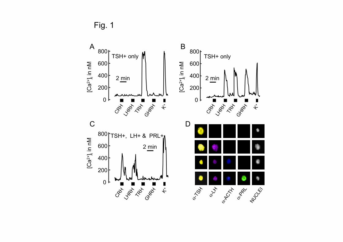
functional HRH receptors and storage of different AP hormones. Fig. 1 summarizes how
characterization was achieved. Calcium imaging revealed that thryrotropes may express, in
addition to TRH, other HRH receptors, as revealed by the increases in [Ca2+]c induced by
sequential perfusion with the four HRHs. Fig. 1 also shows a representative multiple, sequential
immunocytochemistry revealing that thyrotropes may store, in addition to TSH, other AP
hormones. Monohormonal thyrotropes may respond to TRH or to multiple HRHs (Fig.1A and B).
Polyhormonal thyrotropes may show also multiresponsiveness (Fig.1C). When we assayed the
content of different hormones we were surprised to find that most of thyrotropes were actually
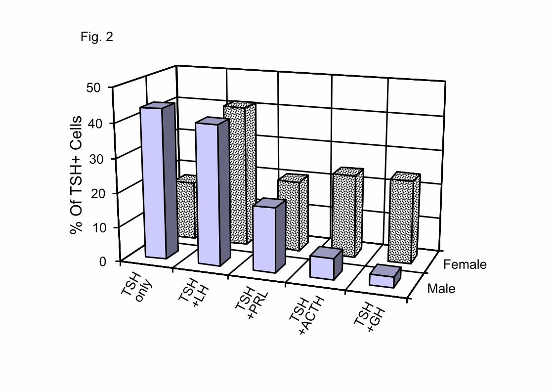
polyhormonal, i.e. they co-stored other hormones together with TSH. Fig. 2 compares the profiles
of hormonal contents in male and female mice. In the male over 50% of the cells were
polyhormonal. Luteinizing hormone (LH) was co-stored with TSH in 41% of the cells. PRL was
co-stored in another 19% of the cells, and ACTH and GH were co-stored with TSH in 6 and 3%
of the cells, respectively. In the females the abundance of polyhormonal cells was even larger
(83%). The dominant cell type was also the gonadothyrotrope (41%), followed by cells co-
storing ACTH or GH (24% each one) and cells co-storing PRL (21%). Some cells stored 3 or
more hormones (31% in the male and 45% in the female). Moreover, we must keep in mind that
monohormonal and polyhormonal thyrotropes were identified after a sequence of stimulatory
events (series of releasing hormones). Because of the possibility that a cell could release all of its
stores of a particular hormone, the actual multihormonal state seen after the multiple releasing
hormone stimulation could be an understimate of that seen in the basal state.
E-00194-2004-R1.doc 22/06/04 9
It has been reported before that somatotropes can transdifferentiate to thyrotropes after
thyroidectomy (18) or protracted hypothyroidism (30). Cells containing both hormones, GH and
TSH, were observed during this process (18,30). We find here that thyrosomatotropes are already
present in the AP of normal mouse, especially in female, but other hormonal combinations, for
example thyrogonadotropes, were much more frequent. This suggests that the appearance of
bihormonal cells is not aimless. Somatotropes are much more frequent than gonadotropes in mice
pituitary (24) and, at random, the combination TSH-GH should be much more likely. On the
other hand, thyrosomatotrope formation should be specifically favored by the pressure of low
plasma thyroid hormone levels in hypothyroidism. As with other polyhormonal cells (24)
polyhormonal thyrotropes were more frequent in female than in male, suggesting that the sexual
cycle may favor transdifferentiation. It is remarkable that sex differences did not affect
gonadotropins (LH) or PRL, which were similarly co-stored with TRH in male and female.
However, co-storage of ACTH and GH by thyrotropes was affected by sex (Fig. 2).
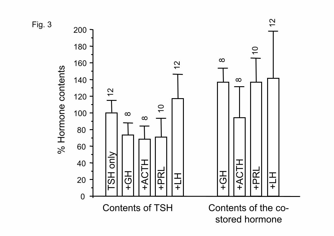
In order to estimate the single cell contents of each stored hormone, the fluorescence
emission with the different antibodies was quantified, standardized, and values obtained in mono-
and polyhormonal cells were compared. Results are summarized in Fig. 3. The contents of TSH
in polyhormonal thyrotropes ranged between 70 and 118% of the values found in monohormonal
thyrotropes. On the other hand, the contents of the different co-stored hormones in TSH-
containing cells was 93-144 % of the contents measured in the corresponding monohormonal
cells. Therefore, hormonal contents of the co-stored hormones were not traces, but amounts
comparable to the ones contained in the corresponding monohormonal cells (Fig. 3). This output
E-00194-2004-R1.doc 22/06/04 10
suggests that considerable mix-up of hormones could happen when thyrotropes are stimulated to
secrete and this could be related to paradoxical secretion.
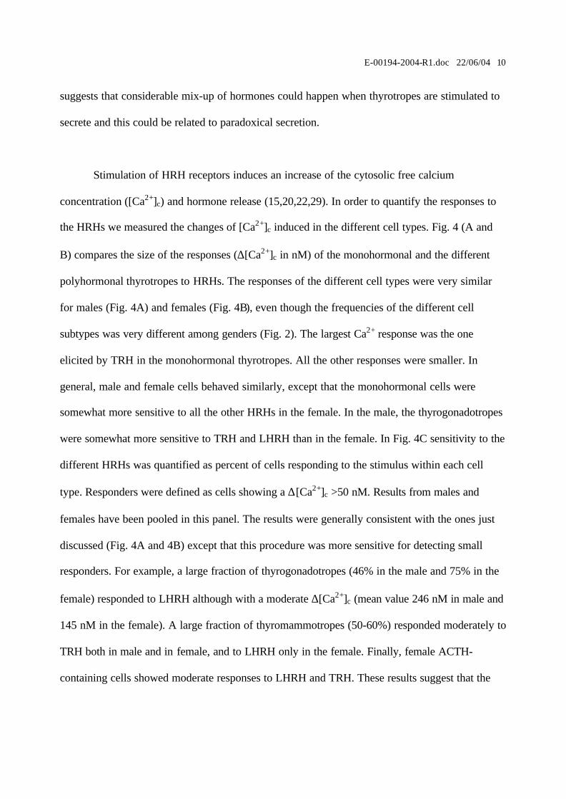
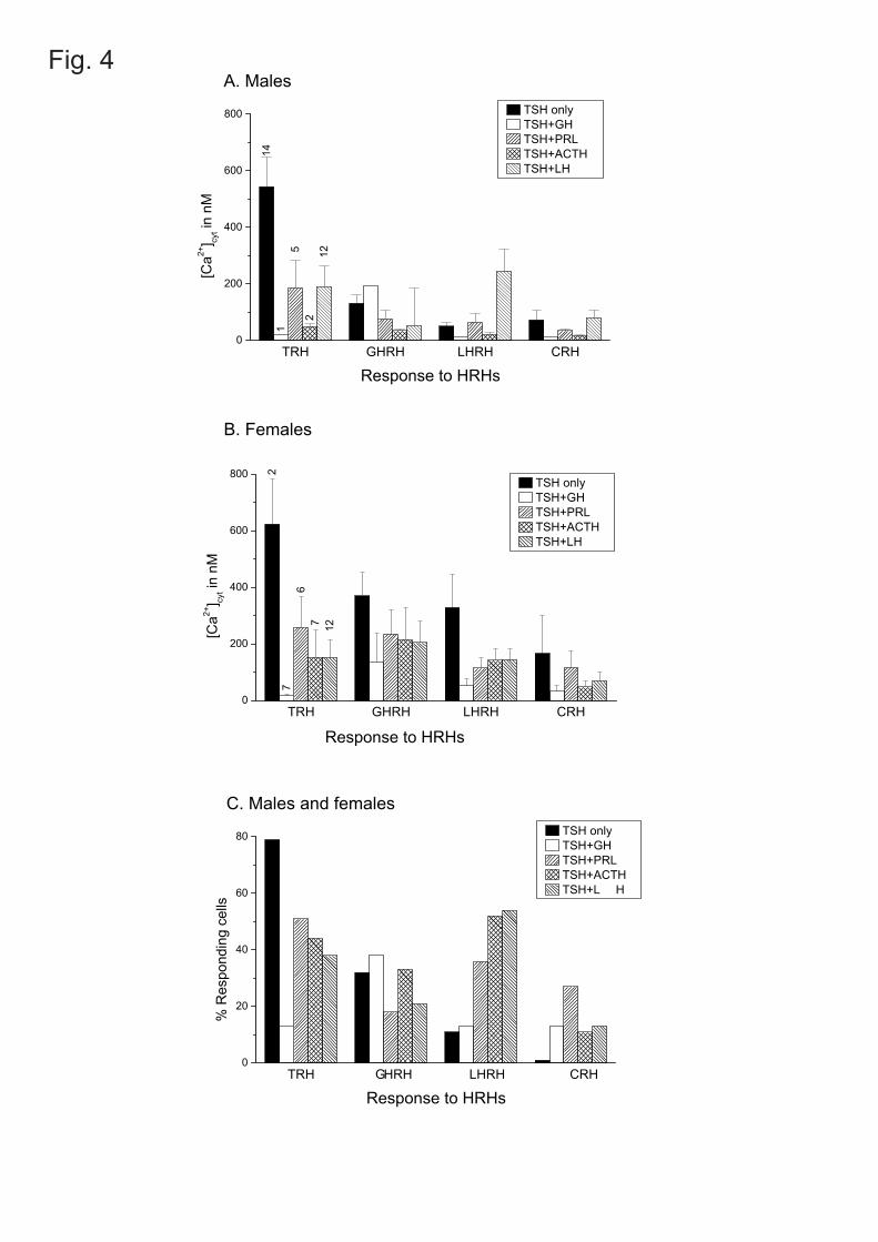
Stimulation of HRH receptors induces an increase of the cytosolic free calcium
concentration ([Ca2+]c) and hormone release (15,20,22,29). In order to quantify the responses to
the HRHs we measured the changes of [Ca2+]c induced in the different cell types. Fig. 4 (A and
B) compares the size of the responses (∆[Ca2+]c in nM) of the monohormonal and the different
polyhormonal thyrotropes to HRHs. The responses of the different cell types were very similar
for males (Fig. 4A) and females (Fig. 4B), even though the frequencies of the different cell
subtypes was very different among genders (Fig. 2). The largest Ca2+ response was the one
elicited by TRH in the monohormonal thyrotropes. All the other responses were smaller. In
general, male and female cells behaved similarly, except that the monohormonal cells were
somewhat more sensitive to all the other HRHs in the female. In the male, the thyrogonadotropes
were somewhat more sensitive to TRH and LHRH than in the female. In Fig. 4C sensitivity to the
different HRHs was quantified as percent of cells responding to the stimulus within each cell
type. Responders were defined as cells showing a ∆[Ca2+]c >50 nM. Results from males and
females have been pooled in this panel. The results were generally consistent with the ones just
discussed (Fig. 4A and 4B) except that this procedure was more sensitive for detecting small
responders. For example, a large fraction of thyrogonadotropes (46% in the male and 75% in the
female) responded to LHRH although with a moderate ∆[Ca2+]c (mean value 246 nM in male and
145 nM in the female). A large fraction of thyromammotropes (50-60%) responded moderately to
TRH both in male and in female, and to LHRH only in the female. Finally, female ACTH-
containing cells showed moderate responses to LHRH and TRH. These results suggest that the
E-00194-2004-R1.doc 22/06/04 11
mix-up of hormone secretion may be, after all, not as large as inferred from the large percentages
of polyhormonal cells, because polyhormonal thyrotropes are poor responders to HRHs including
TRH. Cells with mixed phenotypes could contribute, however, to gonadotropine and/or PRL
secretion, specially in females if changes concentrate, as suggested by Childs (7), at a given
moment of the cycle. Changes of TSH secretion during sex cycle may not be physiologically
relevant as the secretion by the thyroid gland has a long temporal inertia. When blood thyroid
hormone levels decrease the formation of thyrosomatotropes seems favored (18,30). Under these
pathophysiological conditions thyrosomatotropes could contribute to TSH secretion. It would be
very interesting to investigate the functional expression of HRH receptors in these cells.
Multifunctional (polyhormonal and/or multiresponsive) cells have been proposed to be an
intermediate stage in the transdifferentiation process. Thus cells co-storing GH and PRL show up
during transformation of somatotropes into lactotropes (13,31). Transformation of somatotropes
to gonadotropes involves not only dual hormone storage (GH and gonadotropins) but also
multiple HRH receptor expression (GHRH and LHRH) (7). It must be remarked that
thyrogonadotropes and thyromammotropes are normal constituents of male and female pituitary
under physiological conditions. Does that mean that transformations of these cell kinds are
happening under the normal resting conditions? The significance of this finding is unclear at the
moment. On the other hand, the fraction of thyrosomatotropes and thyrocorticotropes is much
larger in the female than in the male (Fig. 2). This observation suggests that transdifferentiation
of GH and ACTH cells towards thyrotropes may be favored at some stage of the sex cycle.
In summary, our results indicate that thyrotropes are not a single cell type but rather a
mosaic of different cell phenotypes whose relative abundance could vary in different physiologic

E-00194-2004-R1.doc 22/06/04 12
and pathophysiogic conditons. However, monohormonal thyrotropes respond much better to
TRH than the polyhormonal ones, thus explaining why paradoxical secretion is so elusive. In any
case, thyrotropes from mice AP seem an excellent model for further studies on
transdifferentiation.

E-00194-2004-R1.doc 22/06/04 13
ACKNOWLEDGEMENTS
We thank the National Hormone and Peptide Program and Dr. A. F. Parlow for providing
antisera against AP hormones and J. Fernández for technical help.
GRANTS
This work was supported by grants from the Spanish Ministerio de Ciencia y Tecnología
(MCyT; BFI2001-2073) and Fondo de Investigaciones Sanitarias (FIS 01/0769 and 03/1231). C.
Villalobos and L. Núñez are supported by the Ramón y Cajal Program of MCyT.
E-00194-2004-R1.doc 22/06/04 14
REFERENCES
1. Alarcón P, and García-Sancho J. Differential calcium responses to the pituitary adenylate
cyclase-activating polypeptide (PACAP) in the five main cell types of rat anterior pituitary
Pflügers Arch – Eur J Physiol 440: 685-691, 2000.
2. Amsterdam JD, Winokur A, Lucki I, Snyder P, Harris RI, Caroff S, and Rickels K.
Growth hormone, prolactin and thyrotropin responses to gonadotropin-releasing hormone in
depressed patients and healthy volunteers. Psychoneuroendocrinology 7: 177-184, 1982.
3. Barlier A, Pellegrini-Bouiller I, Caccavelli L, Gunz G, Morange-Ramos I, Jaquet P, and
Enjalbert A. Abnormal transduction mechanisms in pituitary adenomas. Horm Res 47: 227-
34, 1997.
4. Childs GV, Unabia G, and Wu P. Differential expression of growth hormone messenger
ribonucleic acid by somatotropes and gonadotropes in male and cycling female rats.
Endocrinology 141: 1560-1570, 2000.
5. Childs GV, Unabia G, Miller BT, and Collins TJ. Differential expression of gonadotropin
and prolactin antigens by GHRH target cells from male and female rats. J Endocrinol
162:177-187, 1999.
6. Childs GV, Westlund KN, and Unabia G. Characterization of anterior pituitary target cells
for arginine vasopresine: including cells that store adrenocorticotropin, thyrotropin-β and
both hormones. Endocrinology 125: 554-559, 1989.
7. Childs GV. Development of gonadotropes may involve cyclic transdifferentiation of growth
hormone cells. Arch Physiol Biochem 110: 42-49, 2002.
E-00194-2004-R1.doc 22/06/04 15
8. Childs GV. Growth hormone cells as co-gonadotropes: partners in the regulation of the
reproductive system. Trends Endocrinol Metab 11: 168-175, 2000.
9. Childs GV. Multipotential pituitary cells that contain ACTH and other pituitary hormones.
Trends Endocrinol Metab 2: 112-117, 1991.
10. De Groef B, Goris N, Arckens L, Kuhn ER, and Darras VM. Corticotropin-releasing
hormone (CRH)-induced thyrotropin release is directly mediated through CRH receptor type
2 on thyrotropes. Endocrinology 144: 5537-5544, 2003.
11. De Marinis L, Mancini A, Zuppi P, Anile C, and Maira G. Paradoxical growth hormone
response to thyrotropin-releasing hormone in acromegaly. Clinical correlations and
prognostic value. Acta Endocrinol (Copenh) 122: 443-449, 1999.
12. Fischer UG, Wood SH, Bruhn J, Roseff SJ, Mortola J, Rivier JE, and Yen SSC. Effect of
human corticotropin-releasing hormone on gonadotropin secretion in cycling and
postmenopausal women. Fertility and Sterility 58: 1108-1112, 1992.
13. Frawley LS, and Boockfor FR. Mammosomatotropes: presence and functions in normal
and neoplastic pituitary tissue. Endocr Rev 12: 337-353, 1991.
14. Fukami K, Tasaka K, Mizuki J, Kasahara K, Masumoto N, Miyake A, and Murata Y.
Bihormonal cells secreting both prolactin and gonadotropins in normal rat pituitary cells.
Endocr J 44: 819-26, 1997.
15. Gershengorn MC. Mechanism of thyrotropin releasing hormone stimulation of pituitary
hormone secretion. Annu Rev Physiol 48: 515-526, 1986.
16. Harvey S. Thyrotrophin-releasing hormone: a growth hormone-releasing factor. J Endocrinol
125: 345-358, 1990.
17. Hauspie A, Seuntjens E,Vankelecom H, and Denef C. Stimulation of combinatorial
expression of prolactin and glycoprotein hormone α-subunit genes by gonadotropin-releasing
E-00194-2004-R1.doc 22/06/04 16
hormone and estradiol-17β in single rat pituitary cells during aggregate cell culture.
Endocrinology 144: 388 –399, 2003.
18. Horvath E, Lloyd RV, and Kovacs K. Propylthiouracyl-induced hypothyroidism results in
reversible transdifferentiation of somatotrophs into thyroidectomy cells. A morphologic study
of the rat pituitary including immunoelectron microscopy. Lab Invest 63: 511-520, 1990.
19. Kasahara K, Tasaka K, Masumoto N, Mizuki J, Tahara M, Miyake A, and Tanizawa O.
Characterization of rat pituitary cells by their responses to hypothalamic releasing hormones.
Biochem Biophys Res Commun 1999: 1436-1441, 1994.
20. Kato M, Hoyland J, Sikdar SK, and Mason WT. Imaging of intracellular calcium in rat
anterior pituitary cells in response to growth hormone releasing factor. J Physiol (London)
447: 171-189, 1992.
21. Losa M, Schopohl J, Müller A, and von Werder K. Growth hormone releasing factor
induces prolactin secretion in acromegalic patients but not in normal subjects. Acta
Endocrinol 109: 467-473, 1985.
22. Luini A, Lewis D, Guild S, Corda D, and Axelrod J. Hormone secretagogues increase
cytosolic calcium by increasing cAMP in corticotropin-secreting cells. Proc Natl Acad Sci
USA 82: 8034-8038, 1985.
23. Matsukura S, Kakita T, Hirata Y, Yoshimi H, and Fukase M. Adenylate cyclase of GH
and ACTH producing tumors of human: activation by non-specific hormones and other
bioactive substances. J Clin Endocrinol Metab 44: 392-397, 1977.
24. Núñez L, Villalobos C, Senovilla L, and García-Sancho J. Multifunctional cells of mouse
anterior pituitary reveal a striking sexual dimorphism. J Physiol (London) 549: 835-843,
2003.
E-00194-2004-R1.doc 22/06/04 17
25. Okada Y, Fujii Y, Moore JP Jr, and Winters SJ. Androgen receptors in gonadotrophs in
pituitary cultures from adult male monkeys. Endocrinology 144: 267-273, 2003.
26. Roudbaraki M, Lorsignol A, Langouche L, Callewaert G, Vankelecom H, and Denef C.
Target cells of χ3-melanocyte-stimulating hormone detected through intracellular Ca2+
responses in immature rat pituitary constitute a fraction of all main pituitary cell types, but
mostly express multiple hormone phenotypes at the messenger ribonucleic acid level.
Refractoriness to melanocortin-3 receptor blockade in the lacto-somatotroph lineage.
Endocrinology 140: 4874-4885, 1999.
27. Seuntjens E, Hauspie A, Roudbaraki M, Vankelecom H, and Denef C. Combined
expression of different hormone genes in single cells of normal rat and mouse pituitary. Arch
Physiol Biochem 110: 12-15, 2002.
28. Seuntjens E, Hauspie A, Vankelecom H, and Denef C. Ontogeny of plurihormonal cells in
the anterior pituitary of the mouse, as studied by means of hormone mRNA detection in
single cells. J Neuroendocrinol 14:611-619, 2002.
29. Shangold GA, Murphy SN, and Miller RJ. Gonadotropin-releasing hormone-induced Ca2+
transients in single identified gonadotropes require both intracellular Ca2+ mobilization and
Ca2+ influx. Proc Natl Acad Sci USA 85: 6566-6570, 1988.
30. Vidal S, Horvath E, Kovacs K, Cohen SM, Lloyd RV, and Scheithauer BW.
Transdifferentiation of somatotrophs to thyrotrophs in the pituitary of patients with protracted
primary hypothyroidism. Virchows Arch 436: 43-51, 2000.
31. Vidal S, Horvath E, Kovacs K, Lloyd RV, and Smyth HS. Reversible transdifferentiation:
interconversion of somatotrophs and lactotrophs in pituitary hyperplasia. Mod Pathol 14: 20-
28, 2001.

E-00194-2004-R1.doc 22/06/04 18
32. Villalobos C, Núñez L, and García-Sancho J. Functional glutamate receptors in a
subpopulation of anterior pituitary cells. FASEB J 10:654-660, 1996.
33. Villalobos C, Núñez L, Frawley LS, García-Sancho J, and Sánchez A. Multi-
responsiveness of single anterior pituitary cells to hypothalamic-releasing hormones: a
cellular basis for paradoxical secretion. Proc Natl Acad Sci USA 94: 14132-14137, 1997.
E-00194-2004-R1.doc 22/06/04 19
FIGURE LEGENDS
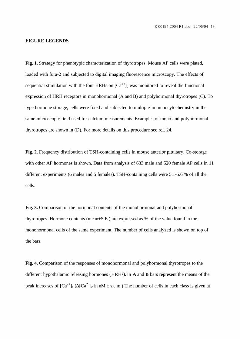
Fig. 1. Strategy for phenotypic characterization of thyrotropes. Mouse AP cells were plated,
loaded with fura-2 and subjected to digital imaging fluorescence microscopy. The effects of
sequential stimulation with the four HRHs on [Ca2+]c was monitored to reveal the functional
expression of HRH receptors in monohormonal (A and B) and polyhormonal thyrotropes (C). To
type hormone storage, cells were fixed and subjected to multiple immunocytochemistry in the
same microscopic field used for calcium measurements. Examples of mono and polyhormonal
thyrotropes are shown in (D). For more details on this procedure see ref. 24.
Fig. 2. Frequency distribution of TSH-containing cells in mouse anterior pituitary. Co-storage
with other AP hormones is shown. Data from analysis of 633 male and 520 female AP cells in 11
different experiments (6 males and 5 females). TSH-containing cells were 5.1-5.6 % of all the
cells.
Fig. 3. Comparison of the hormonal contents of the monohormonal and polyhormonal
thyrotropes. Hormone contents (mean±S.E.) are expressed as % of the value found in the
monohormonal cells of the same experiment. The number of cells analyzed is shown on top of
the bars.
Fig. 4. Comparison of the responses of monohormonal and polyhormonal thyrotropes to the
different hypothalamic releasing hormones (HRHs). In A and B bars represent the means of the
peak increases of [Ca2+]c (∆[Ca2+]c in nM ± s.e.m.) The number of cells in each class is given at

E-00194-2004-R1.doc 22/06/04 20
top of the bars. A: males. B: females. In C data from the same experiments were quantified as %
responding cells. Responding cells are defined as those giving a ∆[Ca2+]c > 50 nM after
stimulation with the HRH. ). Data from male and female have been pooled in this panel.
2 min 2 min
2 min
TSH+ only
TSH+, LH+ & PRL+
TSH+ onlyA
D
B
C
0
200
400
600
800
0
200
400
600
800
0
200
400
600
800
[Ca2
+ ] iin
nM
[Ca2
+ ] iin
nM
[Ca2
+ ] iin
nM
α-TS
H
α-LH
α-AC
THα-
PRL
NUCLEI
CRHLH
RHTR
HGHRH K
+
CRHLH
RHTR
HGHRH K
+
CRHLH
RHTR
HGHRH K
+
Fig. 1
TSH
only
TSH
+LH
TSH
+PRL
TSH
+ACT
H
TSH
+GH
Male
Female0
10
20
30
40
50%
OfT
SH+
Cel
ls
Fig. 2
0
20
40
60
80
100
120
140
160
180
200
% H
orm
one
cont
ents 12
8
10
12
8
TSH
onl
y
+GH
+AC
TH
+PR
L
+LH
12
8
10
8
+LH
+GH
+AC
TH
+PR
L
Contents of TSH Contents of the co-stored hormone
Fig. 3
TRH GHRH LHRH CRH0
20
40
60
80
C. Males and females
% R
espo
ndin
g ce
lls
Response to HRHs
TSH only TSH+GH TSH+PRL TSH+ACTH TSH+L H
27
67 12
TRH GHRH LHRH CRH0
200
400
600
800
B. Females
�[C
a2+] cy
t in n
M
Response to HRHs
TSH only TSH+GH TSH+PRL TSH+ACTH TSH+LH
141
52
12
TRH GHRH LHRH CRH0
200
400
600
800
A. Males
�[C
a2+] cy
t in n
M
Response to HRHs
TSH only TSH+GH TSH+PRL TSH+ACTH TSH+LH
Fig. 4