antegrade selective cerebral perfusion during operations on the thoracic aorta: our experience
TRANSCRIPT
CHAPTER
4
Antegrade selective cerebral perfusion during operations on
the thoracic aorta: factors influencing survival and neurologic
outcome in 413 patients
Marco Di Eusanioa, Marc A. A. M. Schepensa, Wim J. Morshuisa, Roberto Di Bartolomeob,
Angelo Pierangelib, Karl M. Dosschea.
Department of Cardiopulmonary Surgery, St Antonius Hospital, Nieuwegein, The Netherlands a
Department of Cardiac Surgery, Policlinico S’Orsola, University of Bologna, Bologna, Italy b
Journal of Thoracic and Cardiovascular Surgery 2002;124:1080-6.
Chapter 4
- 52 -
Abstract
Objective. We retrospectively analysed hospital mortality and neurologic outcome after surgery on
the thoracic aorta with the aid of antegrade selective cerebral perfusion to determine a predictive
risk model.
Methods. Between October 1995 and May 2001, 413 patients (mean age 63.0±11.5 years)
underwent surgery on the thoracic aorta using antegrade selective cerebral perfusion. Indication for
surgery was acute type A dissection in 116 patients (28.1%), degenerative aneurysm in 227
(55.0%), post-dissection aneurysm in 70 (16.9%). One hundred-twenty-five patients (30.3%) were
operated on urgently; concomitant procedures were performed in 171 patients (41.4%). Mean
cerebral perfusion time was 63.0±38.7 minutes (range, 16-220 minutes). Preoperative and
intraoperative factors were evaluated by univariate and multivariate analysis to identify predictors
of hospital mortality and neurological outcome
Results. The hospital mortality rate was 9.4%. Stepwise logistic regression revealed urgency status
(p=0.000; OR=19.9) and recent history of recent central neurologic event (p=0.004; OR=8.0) to be
independent determinants for hospital mortality. Temporary neurological dysfunction occurred in
20 patients (5.1%). Urgency status (p=0.005; OR=7.5), history of central neurological event
(p=0.003; OR=8.6) and coronary artery bypass grafting (p=0.019; OR=6.0) were independent
determinants of temporary neurological dysfunction. Urgency status (p=0.003; OR=8.6) was the
only independent determinant for permanent neurological dysfunction, and it occurred in 15
patients (3.7%).
Conclusion. Antegrade selective cerebral perfusion is an effective method of brain protection.
Cerebral perfusion times longer than 90 minutes were not associated with an increased risk of
hospital mortality or poorer neurological outcome. Urgency status and recent history of central
neurological event were retained as important risk factors for hospital mortality and neurological
outcome.
Survival and neurologic outcome in 413 patients
- 53 -
Despite gradual improvement of the results in surgery of the aortic arch, brain injury still
remains the most feared complication and frequent cause of death (1-7).
Available techniques of cerebral protection include Deep Hypothermic Circulatory Arrest
(DHCA) alone or in combination with Retrograde Cerebral Perfusion (RCP) and Antegrade
Selective Cerebral Perfusion (ASCP). All three methods have both advantages and disadvantages.
The purpose of our study was to evaluate the results of ASCP with moderate hypothermic
circulatory arrest in patients undergoing surgery of the proximal thoracic aorta, with particular
emphasis on the predictors of hospital mortality and neurological outcome.
Patients and Methods
Patients’ profile. After defining common perioperative variables (see Appendix) a total of 413
medical records of patients who underwent thoracic aorta operations using ASCP at the St
Antonius Hospital (Nieuwegein, The Netherlands) and Sant’ Orsola Hospital (Bologna, Italy)
between October 1995 and May 2001 were retrospectively examined and included in the study.
There were 268 men (64.9%) and 145 women (35.1%) whose age ranged from 21 to 85
years (mean 63.0 ± 11.5 years); 204 (49.4%) were older then 65 years. Of the entire cohort, 288
patients (69.7%) were operated on electively, 125 (30.3%) underwent urgent operation (116 patients
sustained acute dissection and 9 impending aneurysmal rupture). Indication for surgery was acute
type A dissection in 116 patients (28.1%), chronic post dissection aneurysm in 70 (16.9%),
degenerative aneurysm in 227 (55.0%). Associated diseases included Chronic Obstructive
Pulmonary Disease (COPD) in 44 patients (10.7%), chronic renal dysfunction (defined as a
creatinine serum level exceeding 250 µmol/L) in 14 (3.4%). Twenty patients (4.8%) had a recent
history of central neurologic event (transient ischemic attack=9, stroke=11), 60 (14.5%) had
undergone previous aortic/cardiac surgical procedures through a median sternotomy. All electively
operated patients underwent preoperative evaluation of cerebral circulation with Doppler ultrasound
of the extra-cranial vessels, digital subtraction angiography of the extra-cranial and intra-cranial
circulation, carotid compression test with monitoring by electroencephalogram to evaluate
occlusion intolerance or a Trans-Cranial Doppler (TCD) ultrasound study.
Operative technique. Anaesthetic management, methods of brain and myocardial protection were
similar in both Institutions.
Chapter 4
- 54 -
Induction of anaesthesia was obtained with propofol 2 mg/kg, fentanyl 2 µg/kg and pancuronium
0.1 mg/kg. Propofol and fentanyl were used for maintenance of anaesthesia. For all patients, pH
control was carried out using the alpha-stat method.
A median sternotomy was used in 395 (95.6%) patients, a median sternotomy plus anterolateral
thoracotomy in 6 (1.4%). In the remaining 12 (3%) patients, the diseased aorta was exposed through
a left posterolateral thoracotomy. After systemic heparinization, cardiopulmonary bypass was
instituted with a cannula for arterial return in the ascending aorta or in the femoral artery and a
venous single-two stage cannula in the right atrium or a long venous cannula through the left
femoral vein into the right atrium. The left side of the heart was vented through the right superior
pulmonary vein. Myocardial protection was achieved with cold crystalloid cardioplegia and topical
pericardial cooling.
Details of our cannulation technique and method of ASCP with moderate hypothermic circulatory
arrest have been previously described (8-9). Briefly, after the cardiopulmonary bypass was
instituted and the patients were cooled to 22-26°C of nasopharyngeal temperature, systemic
circulation was arrested and the diseased aorta opened. With the patient in Trendelenburg position,
under direct visual control, in general 15 F retrograde coronary sinus perfusion cannulas (Medtronic
DLP; Chase Medical Inc, Houston, TX), connected to the oxygenator with a separate single-roller
pump head, were inserted into the innominate and left common carotid arteries through the aortic
lumen. After the cannulas were properly placed, the balloons at the tip of the cannulas were
manually inflated and held in place by an encircling tape. The left subclavian artery was clamped or
occluded with a Fogarty catheter (Baxter health care, Irvine CA; IFM Clearwater, FL) in order to
avoid the steal phenomenon.
The cerebral perfusion was started at a rate of 10ml/min/kg and adjusted to maintain a right radial
arterial pressure between 40 and 70 mm of Hg. The introduction of the cerebral perfusion catheters
usually took less than 3 minutes.
During open distal anastomosis (10-11), blood perfusion to the lower half of the body from the
femoral artery, when cannulated, was arrested or reduced to 500 ml/min.
Tools of cerebral monitoring included: right radial arterial pressure line in all cases,
electroencephalogram, regional oxygen saturation in the bilateral frontal lobes by means of a near-
infrared spectroscopy (NIRS) , TCD measurement of the blood velocity of the middle cerebral
artery to confirm the proper placement and function of both cannulas when available.
Transesophageal echocardiography was routinely used to assess cardiac contractility, blood flow
conditions, aortic pathology , intracardiac air.
The extent of the aortic replacement and the associated procedures are listed in Table 1 and 2.

Survival and neurologic outcome in 413 patients
- 55 -
Table 1. Overview of the extent of the aortic replacement (N=413)
Extent of replacement N %
Ascending aorta + hemiarch 214 51.8
Ascending aorta + total arch 138 33.4
Total thoracic aorta 18 4.4
Arch + descending aorta 13 3.1
Isolated arch 24 5.8
Others 6 1.5
Table 2. Overview of the concomitant procedures
Procedures N %
Aortic valve replacement 44 10.7
Bentall procedure 103 24.9
Aortic valve sparing procedures 15 3.6
Aortic valve suspension 9 2.2
Homograft 2 0.5
Elephant trunk 78 18.9
CABG 13 3.1
CABG, Coronary Artery Bypass Grafting
En bloc (12) or separated graft techniques were used to reimplant the arch vessels when a complete
aortic arch replacement was performed.
Definitions of neurological complications. Patients were considered to have had permanent
neurological injuries if they exhibit presence of new neurological dysfunction after surgery whether
focal injury (stroke) or global (coma), or were found to have new focal or multiple brain lesions
confirmed by means of brain CT-scan or MRI.
Transient neurological dysfunction (TND), as defined by Ergin and associates (13), indicated the
occurrence of postoperative confusion, agitation, delirium, prolonged obtundation, or transient
parkinsonism with negative brain CT scanning and complete resolution before discharge.

Chapter 4
- 56 -
Statistical analysis. Continuous variables were expressed as the mean ± one standard deviation,
and categorical variables as percentages. All preoperative and intraoperative variables were first
analysed using univariate analysis (unpaired two-tailed t test, Chi-square test or Fisher’s exact test
when appropriate) to determine whether any single factor influenced hospital mortality and
neurologic outcome. A p values of less than 0.05 was taken to indicate statistical significance. The
analysis for permanent neurologic dysfunction and TND were conducted separately. Risk factors
for permanent neurological dysfunction were examined in all patients who survived the operation
long enough to undergo neurological evaluation, and risk factors for TND were assessed in all
operative survivors without permanent neurological dysfunction. Variables that achieved p less than
0.05 in the univariate analysis were examined using multivariate analysis by forward stepwise
logistic regression to evaluate independent risk factors for hospital mortality, permanent
neurological dysfunction and TND.
Statistical analysis was performed using SPSS 7.0 statistical software (SPSS Inc, Chicago, IL).
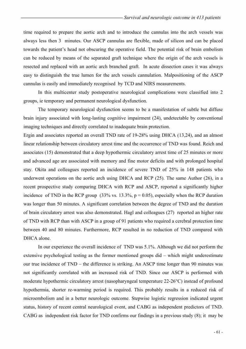
Results Cardiopulmonary bypass data. The mean CPB time was 201±62 minutes (range from 85 to 493
minutes), and the mean myocardial ischemic time was 124±45 minutes (range from 28 to 280
minutes). The mean ASCP time was 63±39 minutes (range from 16 to 220 minutes) (Fig. 1). Two
hundred thirty-five patients (56.9%) had a ASCP time longer than 45 minutes and 90 (21.8%)
longer than 90 minutes.
30 60 90 120 150 180 210ASCP time
20
40
60
80
100
120
140
Patie
nts
Cou
nt
Figure 1. Distribution of patients by antegrade selective cerebral perfusion time.

Survival and neurologic outcome in 413 patients
- 57 -
Hospital mortality. Operative mortality was 0.9% (4/413). Thirty-nine patients died during
hospitalisation, for an overall in-hospital mortality rate of 9.4%. The hospital mortality was 13/288
(4.5%) for elective and 26/125 (20.8%) for urgent surgery (p = 0.000). Causes of death were: multi
organ failure (n=15), septic shock (n=4), neurological damage (n=2), myocardial infarction (n=2),
low cardiac output (n=4), bleeding (n=4), bowel ischemia (n=2), rupture of a distal aneurysm (n=5),
rupture on a proximal anastomotic site (n=1).
On univariate analysis, the following factors had a significant influence on hospital mortality:
urgent status (p = 0.000), acute dissection (p = 0.000), history of recent central neurological event
(p = 0.001), preoperative renal insufficiency (p = 0.034). Multivariate analysis revealed urgent
status (p = 0.000; OR=19.9), history of recent central neurological event (p=0.004; OR=8.0) to be
independent predictors of hospital mortality (Table 3). The extent of aortic replacement and ASCP
duration greater of 90 minutes were not statistically correlated with an increased risk of hospital
mortality.
In 287 patients who underwent elective surgery, univariate analysis indicated age > 65 years (p =
0.003), COPD (p = 0.014), preoperative renal insufficiency (p = 0.046) and history of recent central
neurological event (p = 0.057) as adverse risk factors for hospital mortality. In the same group of
patients, significant predictors of hospital mortality after multivariate analysis were age > 65 years
(p=0.012; OR=6.1) and history of recent central neurological event (p = 0.017; OR= 5.6).
Hospital morbidity. Permanent neurologic dysfunction, which was evaluated in all patients who
survived the operation long enough to undergo an adequate neurological examination, was reported
in 15 of 405 patients (3.7%). Four patients (0.9%) never regained consciousness after surgery; a
brain CT scan showed multiple cerebral infarctions in these cases; in 11 (2.7%) a focal injury was
diagnosed.
In univariate analysis, acute dissection (p=0.007) and urgent status (p=0.003) showed statistically
significant correlation with the occurrence of permanent neurologic dysfunction. On multiple
logistic regression analysis, urgent status (p=0.003; OR=8.6) was found to be independent predictor
of permanent neurologic dysfunction (Table 4).
Transient neurologic dysfunction, which was evaluated only in patients without permanent
neurological damages, occurred in 20 of 390 patients (5.1%).
Acute dissection (p=0.006), urgent status (p = 0.003), history of recent central neurological event (p
= 0.002), and CABG (p = 0.019) were associated with a significantly increased risk of TND on
univariate analysis. Stepwise logistic regression indicated urgent status (p = 0.005; OR = 7.5),
history of recent central neurological event (p = 0.003; OR = 8.6), and CABG (p = 0.013; OR = 6.0)

Chapter 4
- 58 -
as independent predictor of TND (Table 5). ASCP duration greater of 90 minutes was not a
significant risk factor for permanent neurologic dysfunction or TND.
In the group of the elective patients univariate assessment revealed CABG (p = 0.023) and aortic
valve replacement (p = 0.049) to be associated with TND.
Other postoperative complications were bleeding requiring a repeat thoracotomy in 61 patients
(14.8%), postoperative myocardial infarction (serum CPK level > 300 IU/L with a CPK/MB
fraction > 3%) in 14 patients (3.4%). Pulmonary complications requiring a mechanical ventilatory
support longer than 5 days occurred in 57 patients (13.8%); 25 of them underwent urgent surgery
(p < 0.001). Renal failure requiring temporary haemodialysis occurred in 20 patients (4.8%); 11 of
them underwent urgent surgery (p< 0.001).
Table 3. Univariate and Multivariate Analysis for Hospital Mortality (n = 413).
Variable
No.
of
patients
No. of
hospital
deaths (%)
Univariate
analysis
(P value)
Multivariate
analysis
(P value)
OR
Acute dissection 0.000
Yes 116 24 (20.6)
No 297 15 (5)
Urgency 0.000 0.000 19.9
Yes 125 26 (20.8)
No 288 13 (4.5)
History of neurologic event 0.001 0.004 8.0
Yes 20 7 (35)
No 393 32 (8.1)
Pre-op renal insufficiency 0.034
Yes 14 4 (28.5)
No 399 35 (8.7)

Survival and neurologic outcome in 413 patients
- 59 -
Table 4. Univariate and Multivariate Analysis for Permanent neurological
Dysfunction (n = 405).
Variable
No.
of
patients
No.
of
PND (%)
Univariate
analysis
(P value)
Multivariate
analysis
(P Value)
OR
Acute dissection 0.007
Yes 110 9 (8.2)
No 295 6 (2.0)
Urgency 0.003 0.003 8.6
Yes 119 10 (8.4)
No 286 5 (1.7)
PND, Permanent Neurological Dysfunction
Table 5. Univariate and Multivariate Analysis for Temporary Neurological
Dysfunction (n = 390).
Variable
No.
of
patients
No.
of
TND (%)
Univariate
analysis
(P value)
Multivariate
analysis
(P value)
OR
Acute dissection 0.006
Yes 101 11 (10.9)
No 289 9 (3.1)
Urgency 0.003 0.005 7.5
Yes 109 12 (11)
No 281 8 (2.8)
History of neurologic event 0.002 0.003 8.6
Yes 19 5 (26.3)
No 371 15 (4)
Concomitant CABG 0.019 6.0
Yes 12 3(25)
No 378 17(4.5)
TND, Temporary Neurological Dysfunction; CABG, Coronary Artery Bypass Grafting

Chapter 4
- 60 -
Discussion
Although results of aortic arch surgery have improved in the last decades, neurological
injuries, resulting from interruption of cerebral circulation, still remain the most feared
complications and frequent cause of death. Available methods of cerebral protection include
DHCA with or without RCP and ASCP.
DHCA provides a still, bloodless operative field and is technically less complicated. Aortic
arch and arch vessels may be carefully inspected and manipulation can be avoided resulting in a
reduced cerebral embolic risk. However, this technique has the disadvantage of a limited “safe”
time of circulatory arrest (1,14,15) and a prolonged cardiopulmonary bypass time is required in
order to cool down and re-warm the patient which may result in a number of pulmonary, renal,
cardiac, endothelial dysfunction as well as an increased microembolisms production (1,2,16).
Coagulative complications are associated with deeper levels of hypothermia. We believe DHCA to
be an excellent method of brain protection when a circulatory arrest time shorter than 30 minutes is
anticipated.
RCP was introduced in aortic arch surgery to prolong the “safe” time of circulatory arrest
(17). Flush out of embolic material (18), cerebral metabolic support (19,20), catabolite removal and
enhanced cerebral hypothermia maintenance (21) are the supposed neuroprotective mechanisms but
still remain controversial. Moreover, Griepp and colleagues experimentally found that RCP,
especially at high pressure, although successful in removing some emboli, may result in cerebral
injuries (18,22). We had a very limited experience with RCP with results similar to those obtained
with DHCA alone .
ASCP, as described by Kazui and colleagues (11) is our first-choice method of cerebral
protection. It prolongs the “safe” time of circulatory arrest (8,9,23), improves the cerebral cooling,
and can be used with moderate hypothermia. Suggested drawbacks of this technique include a
greater complexity, a cumbersome operative field and the manipulation and cannulation of the arch
vessels especially in presence of cloth, loose atheroma or dissection.
In our series the mean ASCP time was 63 minutes whereas 51.8% of patients had only
hemiarch replacement. Indeed , when ASCP is used, the procedure takes a slightly longer time.
However, this mean ASCP time was largely influenced by more complex and time consuming
operations, such as aortic arch replacement with the separated graft technique, in which ASCP
continued until the concluding anastomosis for the left common carotid artery was performed.
Actually, in the subgroup of patients having hemiarch replacement the mean ASCP time was 40±20
minutes (elective repair: 35±19 min, urgent repair: 47±17 min; p=0.001). In our experience the
Survival and neurologic outcome in 413 patients
- 61 -
time required to prepare the aortic arch and to introduce the cannulas into the arch vessels was
always less then 3 minutes. Our ASCP cannulas are flexible, made of silicon and can be placed
towards the patient’s head not obscuring the operative field. The potential risk of brain embolism
can be reduced by means of the separated graft technique where the origin of the arch vessels is
resected and replaced with an aortic arch branched graft. In acute dissection cases it was always
easy to distinguish the true lumen for the arch vessels cannulation. Malpositioning of the ASCP
cannulas is easily and immediately recognised by TCD and NIRS measurements.
In this multicenter study postoperative neurological complications were classified into 2
groups, ie temporary and permanent neurological dysfunction.
The temporary neurological dysfunction seems to be a manifestation of subtle but diffuse
brain injury associated with long-lasting cognitive impairment (24), undetectable by conventional
imaging techniques and directly correlated to inadequate brain protection.
Ergin and associates reported an overall TND rate of 19-28% using DHCA (13,24), and an almost
linear relationship between circulatory arrest time and the occurrence of TND was found. Reich and
associates (15) demonstrated that a deep hypothermic circulatory arrest time of 25 minutes or more
and advanced age are associated with memory and fine motor deficits and with prolonged hospital
stay. Okita and colleagues reported an incidence of severe TND of 25% in 148 patients who
underwent operations on the aortic arch using DHCA and RCP (25). The same Author (26), in a
recent prospective study comparing DHCA with RCP and ASCP, reported a significantly higher
incidence of TND in the RCP group (33% vs. 13.3%, p = 0.05), especially when the RCP duration
was longer than 50 minutes. A significant correlation between the degree of TND and the duration
of brain circulatory arrest was also demonstrated. Hagl and colleagues (27) reported an higher rate
of TND with RCP than with ASCP in a group of 91 patients who required a cerebral protection time
between 40 and 80 minutes. Furthermore, RCP resulted in no reduction of TND compared with
DHCA alone.
In our experience the overall incidence of TND was 5.1%. Although we did not perform the
extensive psychological testing as the former mentioned groups did – which might underestimate
our true incidence of TND – the difference is striking. An ASCP time longer than 90 minutes was
not significantly correlated with an increased risk of TND. Since our ASCP is performed with
moderate hypothermic circulatory arrest (nasopharyngeal temperature 22-26°C) instead of profound
hypothermia, shorter re-warming period is required. This probably results in a reduced risk of
microembolism and in a better neurologic outcome. Stepwise logistic regression indicated urgent
status, history of recent central neurological event, and CABG as independent predictors of TND.
CABG as independent risk factor for TND confirms our findings in a previous study (8); it may be
Chapter 4
- 62 -
speculated (28) that the presence of coronary artery disease, and therefore the necessity of CABG,
may be a further indication of cerebrovascular disease in these patients, which puts them at higher
risk of cerebral dysfunction postoperatively.
In this series, the overall hospital mortality rate was 9.4% and the permanent neurological
dysfunction rate 3.6%. This compares favourably with other reports. Kazui and colleagues (23)
reported an early mortality rate of 12.7% in a group of 220 consecutive patients undergoing total
arch replacement with the aid of ASCP. In that series the incidence of permanent neurological
dysfunction was 3.3%. Preoperative renal failure, pump time longer than 300 minutes, early series
and shock were independent determinants of hospital mortality while old cerebral infarction and
pump time longer than 300 minutes were independent determinants for permanent neurological
dysfunction. No statistical correlation between ASCP time and hospital mortality or adverse
neurologic outcome was found. In 656 patients undergoing aortic surgery using DHCA, Svensson
and colleagues (1) reported a hospital survival and a stroke rate of 88% and 7%, respectively. An
increased risk of stroke in patients treated with periods of circulatory arrest greater of 40 minutes
and an increased early mortality for circulatory arrest time beyond 65 minutes were observed. Ueda
(29) reported a hospital mortality of 10% and a stroke rate of 4% in 249 patients undergoing aortic
arch surgery using RCP as a method of brain protection. RCP time, pump time and advanced age
were indicated as risk factors for hospital mortality on multivariate analysis.
In the 413 patients analysed in this study, urgent status and history of recent central neurological
event were indicated as independent determinants of hospital mortality on multivariate analysis.
When the elective cases where considered separately, age > 65 years emerged as a further adverse
risk factor for hospital mortality. Urgent status was again statistically correlated with an increased
risk of permanent neurological dysfunction and TND together with history of cerebrovascular
disease and CABG. Patients undergoing urgent surgery had an increased risk of pulmonary and
renal postoperative complications. Looking at these findings, consistent with other reports
(1,23,27,28), we speculate that a more aggressive operative timing with an earlier elective aortic
repair is probably necessary in patients at risk of rupture. Furthermore, the contribution of advanced
age to hospital mortality might also be reduced. However, in patients with history of
cerebrovascular disease, further improvements in cerebral protection techniques are required.
In conclusion, antegrade selective cerebral perfusion is an effective and safe method of brain
protection allowing time-consuming aortic repairs to be performed with encouraging results in
terms of hospital mortality and neurological outcome. Urgency status and recent history of central
neurological event still remain important preoperative risk factors for hospital mortality and
neurological outcome.

Survival and neurologic outcome in 413 patients
- 63 -
References
1. Svensson LG, Crawford ES, Hess HR, et al. Deep hypothermia with circulatory arrest:
determinants of stroke and early mortality in 656 patients. J Thorac Cardiovasc Surg 1993;
106: 19
2. Crawford ES, Svensson LG, Coselli JS, Safi HJ, Hess KR. Surgical treatment of aneurysm
and/or dissection of the ascending aorta, transverse aortic arch, and ascending aorta and
transverse aortic arch. Factors influencing survival in 717 patients. J Thorac Cardiovasc
Surg 1989; 98: 659-674.
3. Safi HJ, Brien HW, Winter JN, Thomas AC, Maulsby RL, Doerr HK, et al. Brain protection
via cerebral retrograde perfusion during aortic arch aneurysm repair. Ann Thorac
Surg1993;56:655-8.
4. Kazui T, Kimura N, Yamada O, Komatsu S. Surgical outcome of aortic arch aneurysms
using selective cerebral perfusion during. Ann Thorac Surg 1994; 57:904-11.
5. Bachet J, Guilmet D, Goudot B, Termignon JL, Teodori G, Dreyfus G, et al. Cold
cerebroplegia. A new technique of cerebral protection during operations on the transverse
aortic arch. J Thorac Cardiovasc Surg 1991; 102:85-94.
6. Dossche KM, Schepens MAAM, Morshuis WJ, Muysoms FE, Langemeijer JJ, Vermeulen
FEE. Antegrade selective cerebral perfusion in operation on the proximal thoracic aorta.
Ann Thorac Surg 1999;67:1904-10.
7. Di Bartolomeo R, Pacini D, Di Eusanio M, Pierangeli A. Antegrade selective cerebral
perfusion during operations on the thoracic aorta: our experience. Ann Thorac Surg 2000;
70:10-6
8. Di Bartolomeo R, Di Eusanio M, Pacini D, Pagliaro M, Savini C, Nocchi A, et al. Antegrade
selective cerebral perfusion during surgery of the thoracic aorta: risk analysis. Eur J
Cardiothorac Surg 2001; 19:765-770.
9. Dossche KM, Schepens MAAM, Morshuis WJ, Waanders FG. Bilateral antegrade selective
cerebral perfusion during surgery on the proximal thoracic aorta. Eur J Cardiothorac Surg
2000; 17 : 462-467.
10. Livesay JJ, Cooley DA, Duncan JM, Ott DA, Walker WE, Reul GJ. Open aortic
anastomosis : improved results in the treatment of aneurysms of the aortic arch. Circulation
1982; 66(Suppl 1):122-7.
11. Kazui T, Inoue N, Yamada O, Komatsu S. Selective cerebral perfusion during operation for
aneurysm of the aortic arch: a reassessment. Ann Thorac Surg 1992; 53:109-14.

Chapter 4
- 64 -
12. Pearce CW, Weichert RF, Del Real RE. Aneurysm of the aortic arch. Simplified technique
for excision and prosthetic replacement. J Thorac Cardiovasc Surg 1969; 58:886-890.
13. Ergin MA, Galla JD, Lansman SL, Quintana C, Bodian C, Griepp RB. Hypothermic
circulatory arrest in operations on the thoracic aorta. Determinants of operative mortality
and neurologic outcome. J Thorac Cardiovasc Surg 1994; 107:788-99.
14. Mc Cullogh JN, Zhang N, Reich DL, Juvonen TS, Klein JJ, Spielvogel D, et al. Cerebral
metabolic suppression during hypothermic circulatory arrest in humans. Ann Thorac Surg
1999; 67:1895-9.
15. Reich DL, Uysal S, Sliwinski M, Ergin MA, Kahn RA, Konstadt SN, et al.
Neuropsychologic outcome after deep hypothermic circulatory arrest in adults. J Thorac
Cardiovasc Surg 1999; 117:156-63.
16. Cooper WA, Duarte IG, Thourani VH, Nakamura M, Wang NP, Brown MB, et al.
Hypothermic circulatory arrest causes multisystem vascular endothelial dysfunction and
apoptosis. Ann Thoac Surg 2000; 69:696-703.
17. Ueda Y, Miki S, Kushura K, Okita Y, Tahata T, Yamanaka K. Surgical treatment of
aneurysm or dissection involving the ascending aorta and aortic arch utilizing circulatory
arrest and retrograde cerebral perfusion. J Cardiovasc Surg 1990; 31: 553-558.
18. Juvonen T, Weisz DJ, Wolfe D, Khang N, Bodian CA, McCullogh JN, et al. Can retrograde
perfusion mitigate cerebral injury following particulate embolization. A study in a chronic
porcine model. Ann Thorac Surg 1998; 57:904-11.
19. Usui A, Hotta T, Hiroura M, Murase M, Maeda M, Koyama T, et al. Retrograde cerebral
perfusion through a superior vena caval cannula protects the brain. Ann Thorac Surg 1992;
53:47-53.
20. Wong C, Bonser RS. Retrograde perfusion and true reverse brain blood flow in humans. Eur
J Cardiothorac Surg 2000 ;17 :597-601.
21. Antilla V, Pokela M, Kiviluoma K, Makiranta M, Hirvonen J, Juvonen T. Is maintained
cranial hypothermia the only factor leading to improved outcome after retrograde cerebral
perfusion? An experimental study with a chronic porcine model. J Thorac Cardiovasc Surg
2000; 119:1021-1029.
22. Griepp RB, Juvonen T, Griepp EB, McCullogh JN, Ergin MA. Is retrograde cerebral
pefusion an effective means of neural support during deep hypothermic circulatory arrest?
Ann Thorac Surg 1997; 64: 913-916.

Survival and neurologic outcome in 413 patients
- 65 -
23. Kazui T, Washijama N, Muhammad BAH, Terada H, Yamashita K, Takinamy M, et al.
Total arch replacement using aortic arch branched grafts with the aid of antegrade selective
cerebral perfusion. Ann Thorac Surg 2000; 70:3-9.
24. Ergin MA, Uysal S, Reich DL, Apaydin A, Lansman SL, McCullogh JN, et al. Temporary
neurological dysfunction after deep hypothermic circulatory arrest: a clinical marker of
long-term functional deficit. Ann Thorac Surg 1999; 67:1887-90.
25. Okita Y, Takamoto S, Ando M, Morata T, Matsukawa R, Kawashima Y. Mortality and
cerebral outcome in patients who underwent aortic arch operation using deep hypothermic
circulatory arrest with retrograde cerebral perfusion: no relation of early death, stroke,
delirium to the duration of circulatory arrest. J Thorac Cardiovasc Surg 1998; 155:129-138.
26. Okita Y, Minatoya K, Tagusari O, Ando M,Nagatsuka K, Kitamura S. Prospective
comparative study of brain protection in total aortic arch replacement: deep hypothermic
circulatory arrest with retrograde cerebral perfusion or selective cerebral perfusion. Ann
Thorac Surg 2001; 72:72-79.
27. Hagl C, Ergin MA, Galla JD, Lansman SL, McCullogh JN, Spielvogel D, et al. Neurologic
outcome after ascending aorta-aortic arch operations: effect of brain protection technique in
high risk patients. J Thorac Cardiovasc Surg 2001;121:1107-21.
28. Ehrlich MP, Ergin MA, McCullogh JN, Lansman SL, Galla JD, Bodian CA, et al. Predictors
of adverse outcome and transient neurological dysfunction after ascending aorta/hemiarch
replacement. Ann Thorac Surg 2000;69:1755-63.
29. Ueda Y, Okita Y, Aomi S, Koyonagi H, Takamoto S. Retrograde cerebral perfusion for
aortic arch surgery: analysis of risk factors. Ann Thorac Surg 1999; 67:1879:82.

Chapter 4
- 66 -
Appendix. Preoperative, Intraoperative and
Postoperative Variables Included in Analysis
Age> 65 years
Gender
Acute dissection
Status (elective/urgent)
Preoperative renal insufficiency (creatinine > 250 µmol/L)
Chronic Obstructive Pulmonary Disease
History of recent central neurological event (≤6 months)
Previous cardiovascular surgery through median sternotomy
Extent of replacement (hemiarch, arch, ascending aorta + arch,
total thoracic aorta, arch + descending aorta, others)
Concomitant aortic valve replacement
Concomitant aortic valve sparing procedures
Concomitant aortic valve suspension
Concomitant Bentall procedure
Concomitant Homograft
Concomitant coronary artery bypass grafting
Elephant trunk
Cardiopulmonary bypass time > 180 minutes
Myocardial ischemic time > 120 minutes
Selective cerebral perfusion time > 90 minutes
Exitus
Post-op respiratory failure
Post-op myocardial infarction
Post-op haemodialysis
Bleeding requiring rethoracotomy
Permanent neurological dysfunction
Temporary neurological dysfunction