annual report - rajiv gandhi centre for biotechnology
TRANSCRIPT
AnnuAl RepoRt2 0 1 5
Rajiv Gandhi Centre for BiotechnologyThiruvananthapuram, Kerala
Phone: +91 471 2341716, 2347975Fax: +91 471 2348096, 2346333
E-mail: [email protected]: www.rgcb.res.in

Contents
DIRECTOR’S REPORT 05
CANCER BIOLOGY
Cancer Research Program - 1 09
Cancer Research Program - 2 17
Cancer Research Program - 3 26
Cancer Research Program - 4 35
Cancer Research Program - 5 50
Cancer Research Program - 6 61
Cancer Research Program - 7 67
Cancer Research Program - 8 71
Cancer Research Program - 9 79
CARDIOVASCULAR DISEASE & DIABETES BIOLOGY
Laboratory - 1 107
Laboratory - 2 121
Laboratory - 3 132
Labouratory - 4 139
CHEMICAL & ENVIRONMENTAL BIOLOGY
Chemical Biology Laboratory - 1 142
Chemical Biology Laboratory - 2 149
Molecular Ecology Laboratory 154
Environmental Biology Laboratory 162
NEUROBIOLOGY
Molecular Neurobiology Laboratory 171
Neuro Stem Cell Biology Laboratory 177
Human Molecular Genetics 184
Neuro Bio-physics Laboratory 191
PLANT DISEASE BIOLOGY & BIOTECHNOLOGY
PDBB Laboratory - 1 196
PDBB Laboratory - 2 206
PDBB Laboratory - 3 212
PDBB Laboratory - 4 217
REPRODUCTIVE BIOLOGY
Molecular Reproduction Laboratory - 1 224
Molecular Reproduction Laboratory - 2 236
TROPICAL DISEASE BIOLOGY
Mycobacteria Research Laboratory 247
Cholera Research Laboratory 256
Leptospira Biology Laboratory 264
Molecular Virology Laboratory 266
Viral Disease Biology Laboratory - 1 271
Viral Disease Biology Laboratory - 2 280
Malaria Biology Laboratory 284
Microbiome Research Laboratory 293
RGCB CORE SERVICE FACILITIES 297
RGCB RESEARCH ENGINEERING SERVICES 312
RGCB ADMINISTRATION AND STAFF LIST 316
RGCB PROGRAMS 324
RGCB SKY GREEN 326
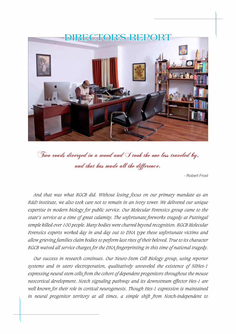
DIRECTOR’S REPORT
Two roads diverged in a wood and I took the one less traveled by, and that has made all the difference.
- Robert Frost
And that was what RGCB did. Without losing focus on our primary mandate as an
R&D institute, we also took care not to remain in an ivory tower. We delivered our unique
expertise in modern biology for public service. Our Molecular Forensics group came to the
state’s service at a time of great calamity. The unfortunate fireworks tragedy at Puttingal
temple killed over 100 people. Many bodies were charred beyond recognition. RGCB Molecular
Forensics experts worked day in and day out to DNA type these unfortunate victims and
allow grieving families claim bodies to perform last rites of their beloved. True to its character
RGCB waived all service charges for the DNA fingerprinting in this time of national tragedy.
Our success in research continues. Our Neuro-Stem Cell Biology group, using reporter
systems and in utero electroporation, qualitatively unraveled the existence of NIHes-1
expressing neural stem cells from the cohort of dependent progenitors throughout the mouse
neocortical development. Notch signaling pathway and its downstream effector Hes-1 are
well known for their role in cortical neurogenesis. Though Hes-1 expression is maintained
in neural progenitor territory at all times, a simple shift from Notch-independent to
-dependent state makes it pleiotropic as the former maintains the neural stem cells in a non-
dividing/slow-dividing state, whereas the latter is very much required for maintenance and
proliferation of radial glial cells. Jackson James et al, thus provide an additional complexity
in neural progenitor heterogeneity regarding differential Hes-1 expression in the germinal
zone during neo-cortical development.
RGCB’s Cancer Research Program provided data on the vital role of DEPTOR in survival
of cervical squamous cell carcinoma (SCC). DEPTOR, an endogenous inhibitor of mTOR
complexes is de-regulated in cancers and was found to be overexpressed in both cervical
SCC cell lines and tissues. It’s silencing in cervical SCC cells induced apoptosis, mainly by
up-regulation of p38 MAPK and by inhibiting PI3K/AKT pathway via a feedback inhibition
from mTORC1-S6K. DEPTOR silencing resulted in reduced expression of the nitric oxide
synthases iNOS and eNOS, as well as increased activation of ERK1/2 and p38 MAP kinases.
In summary, DEPTOR is found to promote survival of cervical SCC cells and its reduction
induced apoptosis via differential effects on PI3K/AKT and p38 MAPK.
Priya Srinivas et al analyzed the effect of Plumbagin (PB) and structurally related
naphthaquinones on BRCA1/2 silenced prostate cancer cells and the ability of PB to target
cancer stem cells. PB has putative role in tumor suppression in BRCA defective cancers
and has the ability to directly target cancer stem cells (CSCs) and holds promise for novel
therapeutic approaches against BRCA mutated cancers as well as CSCs. Ruby John Anto and
colleagues studied the anticancer activity of 3,5-dihydroxy Q1 -4-ethyl-trans-stilbene (DETS)-
a natural stilbene, with a structural similarity to resveratrol (trans- 3,4,5-trihydroxystilbene),
first identified as bioactive bacterial secondary metabolite isolated from Bacillus cereus
associated with a rhabditidentomopathogenic nematode and reports that the compound
showed maximum cytotoxicity toward the human melanoma cell line, [A375, IC50: 24.01
μM] followed by cervical [HeLa-46.17 μM], colon [SW480- 47.28 μM], liver [HepG2- 69.56
μM] and breast [MCF-7- 84.31 μM] cancer cells. DETS, like resveratrol, down-regulates the
expression status of major molecules contributing to melanoma progression, such as BRAF,
β-catenin and Brn-2, all of which converge in MITF-M, the master regulator of melanoma
signaling.
RGCB once again demonstrated its value in leading Translational Cancer Research.
Oral leukoplakia is a potentially malignant lesion of the oral cavity, for which no effective
treatment is available. We investigated the effectiveness of curcumin, a potent inhibitor
of NF-kB/COX-2, molecules perturbed in oral carcinogenesis, to treat leukoplakia. Subjects
with oral leukoplakia were randomized (1:1 ratio) to receive orally, either 3.6 g/day of
curcumin or placebo, for 6 months. The primary endpoint was clinical response obtained by
bi-dimensional measurement of leukoplakia size at recruitment and 6 months. Histologic
response, combined clinical and histologic response, durability and effect of long-term
therapy for an additional six months in partial responders, safety and compliance were the
secondary endpoints. Combined clinical and histologic response assessment indicated a
significantly better response with curcumin. The treatment did not raise any safety concerns.
Treatment of oral leukoplakia with curcumin (3.6 g for six months), thus was well tolerated
and demonstrated significant and durable clinical response for 6 months.
Findings from the Phase – IV trial comparing the 2 versus 3 doses of quadrivalent HPV
vaccine among young Indian girls, support the WHO recommendation of two doses, at
least six months apart, for routine vaccination of young girls. While our non-randomized
comparisons confirm that the immune response following two doses given at 6 months
apart is non-inferior to that of three doses, one dose vaccination was associated with lower
antibody levels. Nevertheless, the single dose antibodies were as avid as the two and three
dose antibodies as reflected by the similar geometric mean avidity indices and also induced
detectable titers of neutralizing antibodies although at a lower level. Our initial results
provide the first evidence that the lower vaccine-induced antibody levels following one
dose of quadrivalent HPV vaccine provide similar protection against vaccine-targeted HPV
infections as the higher antibody levels from two or three doses over a four-year follow-up
period. Our preliminary results following one dose of quadrivalent vaccine are promising
and suggest that future trials of HPV vaccines should include a single dose arm. A single-
dose HPV vaccination offers the best chance to overcome the barriers currently prohibiting
vaccine uptake globally; on condition it provides strong and sustained protection against
HPV infection in the long term.
Our plant biotechnologists have focused on improving our indigenous crop varieties.
Over the last century, the commercial production of black pepper (Piper nigrum) has
been significantly affected by ‘quick wilt’ disease caused by the oomycete pathogen,
Phytophthoracapsici. There are no varieties of black pepper that can completely resist this
disease and progress in understanding the molecular components of this phyto-patho-
system has been severely hampered due to lack of proteome or transcriptome databases
in the plant. Manjula S. et al explored the potential of a transcriptome-assisted label-free
quantitative proteomics approach for elucidation of leaf proteome of black pepper when
challenged with P. capsici. This simple and integrative approach has brought out novel
and comprehensive insights into the complex network of proteome changes involved in
black pepper- Phytophthora interaction, which is of practical interest for developing crop
improvement strategy in this major spice crop. The study also provides convincing evidence
on the effectiveness of a transcriptome-based label-free proteomics approach for elucidating
the host response to biotic stress in non- model crop species like P. nigrum.
3’ UTR regulation plays crucial rule in most of the diseases, and the non-canonical PAP,
Star-PAP has been identified to be a key player in this. Rakesh Laishram and colleagues
demonstrate the role of phosphorylation in regulation of specific target gene/UTRs. Similarly,
defining the phosphorylation sites on Star-PAP mediated by CKIα/PKC or other kinases
could help in understanding the mechanism of how Star-PAP could regulate multiple genes
responsible for distinct functions or diseases. The study further extends to characterize a
novel mechanism where the phosphorylation is linked with the PI4,5P2 signaling in the
3’-end processing complex with kinases such as CKIα. Rakesh Laishram et al also show
the mechanism of specificity of target mRNA selection by the non-canonical PAP, Star-PAP
by defining the role of distinct cis regulatory elements and trans-acting factors involved
in the specificity of the target mRNA selection which explains the reason for non cross
regulation between the two PAPs – a study that has potential implications in alternative
polyadenylation of mRNAs as more than 60% of human genes have multiple polyadenyation
sites at the 3’-UTR.
Construction at our new campus in Aakulam is progressing well. RGCB’s development
and profile is also constantly growing with sustained support and excellent cooperation
from our Governing Council led by Professor K. Vijay Raghavan, Secretary, Department of
Biotechnology and the Scientific Advisory Council led by Professor Nirmal Kumar Ganguly.
We were fortunate in getting Dr. Aravind Duggal, Advisor at the Department of Biotechnology
as our Nodal Officer. I must thank my scientific, technical and administrative colleagues
for all the excellent work that makes this institute stand out in the country.
Professor M. Radhakrishna Pillai
FRCPath, PhD
CAnCeR ReSeA
RCH pRoGRAM
- laboratory 1
Post-Doctoral FellowsAbitha Murali,PhDSwati Kaushik,PhD
ICMR Woman ScientistVinitha Richard,PhD
PhD StudentsAsha LekshmiShankara Narayanan V.Santhik S.L.
Technical SupportIshaque Abdulla Prakash R.
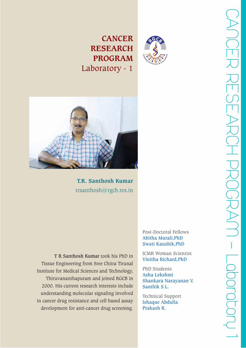
T R Santhosh Kumar took his PhD in
Tissue Engineering from Sree Chitra Tirunal
Institute for Medical Sciences and Technology,
Thiruvananthapuram and joined RGCB in
2000. His current research interests include
understanding molecular signaling involved
in cancer drug resistance and cell based assay
development for anti-cancer drug screening.
T.R. Santhosh Kumar
CANCER RESEARCH PROGRAM
Laboratory - 1
- 10 - annual report 2015
rajiv gandhi centre for biotechnology
Regulated origins of cell-to-cell death response heterogeneity: deterministic influence of the cell cycle and redox status
Asha Lekshmi, Shankara Narayanan V, Prakash R. and T. R. Santhosh Kumar
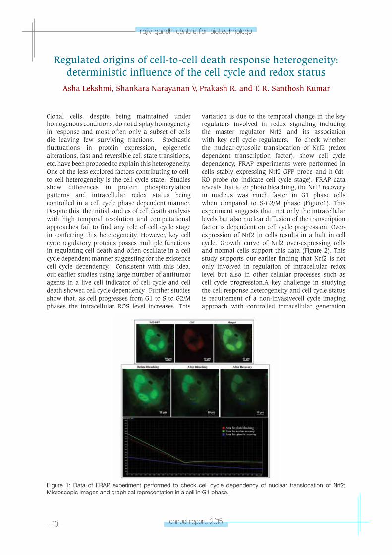
Clonal cells, despite being maintained under homogenous conditions, do not display homogeneity in response and most often only a subset of cells die leaving few surviving fractions. Stochastic fluctuations in protein expression, epigenetic alterations, fast and reversible cell state transitions, etc. have been proposed to explain this heterogeneity. One of the less explored factors contributing to cell-to-cell heterogeneity is the cell cycle state. Studies show differences in protein phosphorylation patterns and intracellular redox status being controlled in a cell cycle phase dependent manner. Despite this, the initial studies of cell death analysis with high temporal resolution and computational approaches fail to find any role of cell cycle stage in conferring this heterogeneity. However, key cell cycle regulatory proteins posses multiple functions in regulating cell death and often oscillate in a cell cycle dependent manner suggesting for the existence cell cycle dependency. Consistent with this idea, our earlier studies using large number of antitumor agents in a live cell indicator of cell cycle and cell death showed cell cycle dependency. Further studies show that, as cell progresses from G1 to S to G2/M phases the intracellular ROS level increases. This
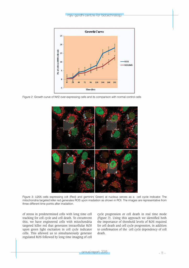
variation is due to the temporal change in the key regulators involved in redox signaling including the master regulator Nrf2 and its association with key cell cycle regulators. To check whether the nuclear-cytosolic translocation of Nrf2 (redox dependent transcription factor), show cell cycle dependency, FRAP experiments were performed in cells stably expressing Nrf2-GFP probe and h-Cdt-KO probe (to indicate cell cycle stage). FRAP data reveals that after photo bleaching, the Nrf2 recovery in nucleus was much faster in G1 phase cells when compared to S-G2/M phase (Figure1). This experiment suggests that, not only the intracellular levels but also nuclear diffusion of the transcription factor is dependent on cell cycle progression. Over-expression of Nrf2 in cells results in a halt in cell cycle. Growth curve of Nrf2 over-expressing cells and normal cells support this data (Figure 2). This study supports our earlier finding that Nrf2 is not only involved in regulation of intracellular redox level but also in other cellular processes such as cell cycle progression.A key challenge in studying the cell response heterogeneity and cell cycle status is requirement of a non-invasivecell cycle imaging approach with controlled intracellular generation
Figure 1: Data of FRAP experiment performed to check cell cycle dependency of nuclear translocation of Nrf2; Microscopic images and graphical representation in a cell in G1 phase.
annual report 2015 - 11 -
rajiv gandhi centre for biotechnology
Figure 2: Growth curve of Nrf2 over-expressing cells and its comparison with normal control cells
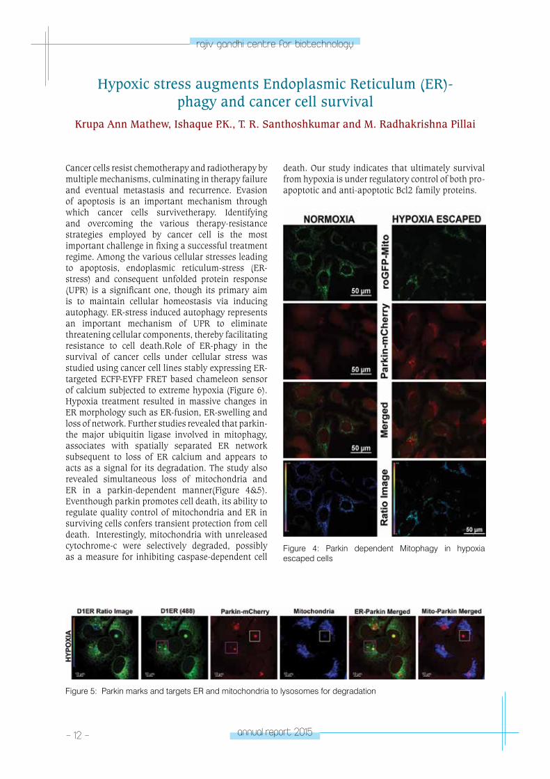
Figure 3: U205 cells expressing cdt (Red) and geminin( Green) at nucleus serves as a cell cycle indicator. The mitochondria targeted killer red generates ROS upon irradaition as shown in ROI. The images are representative from three different time points after irradaition.
of stress in predetermined cells with long time cell tracking for cell cycle and cell death. To circumvent this, we have engineered cells with mitochondria targeted killer red that generates intracellular ROS upon green light excitation in cell cycle indicator cells. This allowed us to simultaneously generate regulated ROS followed by long time imaging of cell
cycle progression or cell death in real time mode (Figure 3). Using this approach we identified both the importance of threshold levels of ROS required for cell death and cell cycle progression, in addition to confirmation of the cell cycle dependency of cell death.
- 12 - annual report 2015
rajiv gandhi centre for biotechnology
Hypoxic stress augments Endoplasmic Reticulum (ER)- phagy and cancer cell survival
Krupa Ann Mathew, Ishaque P.K., T. R. Santhoshkumar and M. Radhakrishna Pillai
Cancer cells resist chemotherapy and radiotherapy by multiple mechanisms, culminating in therapy failure and eventual metastasis and recurrence. Evasion of apoptosis is an important mechanism through which cancer cells survivetherapy. Identifying and overcoming the various therapy-resistance strategies employed by cancer cell is the most important challenge in fixing a successful treatment regime. Among the various cellular stresses leading to apoptosis, endoplasmic reticulum-stress (ER-stress) and consequent unfolded protein response (UPR) is a significant one, though its primary aim is to maintain cellular homeostasis via inducing autophagy. ER-stress induced autophagy represents an important mechanism of UPR to eliminate threatening cellular components, thereby facilitating resistance to cell death.Role of ER-phagy in the survival of cancer cells under cellular stress was studied using cancer cell lines stably expressing ER-targeted ECFP-EYFP FRET based chameleon sensor of calcium subjected to extreme hypoxia (Figure 6). Hypoxia treatment resulted in massive changes in ER morphology such as ER-fusion, ER-swelling and loss of network. Further studies revealed that parkin- the major ubiquitin ligase involved in mitophagy, associates with spatially separated ER network subsequent to loss of ER calcium and appears to acts as a signal for its degradation. The study also revealed simultaneous loss of mitochondria and ER in a parkin-dependent manner(Figure 4&5). Eventhough parkin promotes cell death, its ability to regulate quality control of mitochondria and ER in surviving cells confers transient protection from cell death. Interestingly, mitochondria with unreleased cytochrome-c were selectively degraded, possibly as a measure for inhibiting caspase-dependent cell
death. Our study indicates that ultimately survival from hypoxia is under regulatory control of both pro-apoptotic and anti-apoptotic Bcl2 family proteins.
Figure 4: Parkin dependent Mitophagy in hypoxia escaped cells
Figure 5: Parkin marks and targets ER and mitochondria to lysosomes for degradation
annual report 2015 - 13 -
rajiv gandhi centre for biotechnology
Transcriptomics reveal drug escape mechanisms of cancer cells Santhik SL, Abitha Murali, Mydhily Nair R.B., Rajesh Raju, Reshmi G,
T.R Santhosh Kumar and M. Radhakrishna Pillai
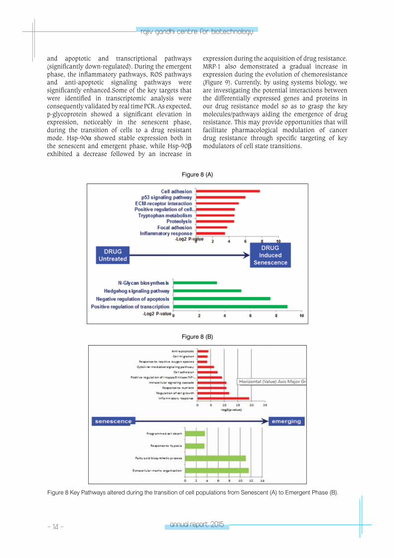
Development of drug resistance is an important factor in the failure of anticancer therapeutics. The ability to evade drugs by cancer cells could be through the enhanced expression of transporters that increases anticancer drugs efflux, alterations in drug metabolism, mutations of drug targets and the activation of survival or inactivation of downstream death signaling pathways. We have previously characterized, phenotypically and functionally distinct cell state transitions during drug escape. These transitions are non- cycling surviving senescent phenotype (SEN), emerging drug resistant cells (EME) with reactivation of cell cycle entry
and actively dividing resistant cells (RES) that significantly differ from the parental population.In order to obtain the global gene expression patterns associated with the progression of drug resistance, we examined differentially expressed genes with respect to SEN, RES and EME colonies by transcriptomics (Figure 7).Some of the key pathways that are altered during the acquisition of drug resistance are represented in Figure 8 (A) and (B). The transcriptomic profile revealed that the key pathways drastically altered during the transition are the cell adhesion pathways, p53 signaling, proteolysis and inflammatory responses (significantly up-regulated)
Figure 6: Hypoxia escaped cells shows low and high ER calcium
Figure 7: Differential expression Matrix depicting the amount of up-regulated and down-regulated transcripts in our drug resistance model system.
- 14 - annual report 2015
rajiv gandhi centre for biotechnology
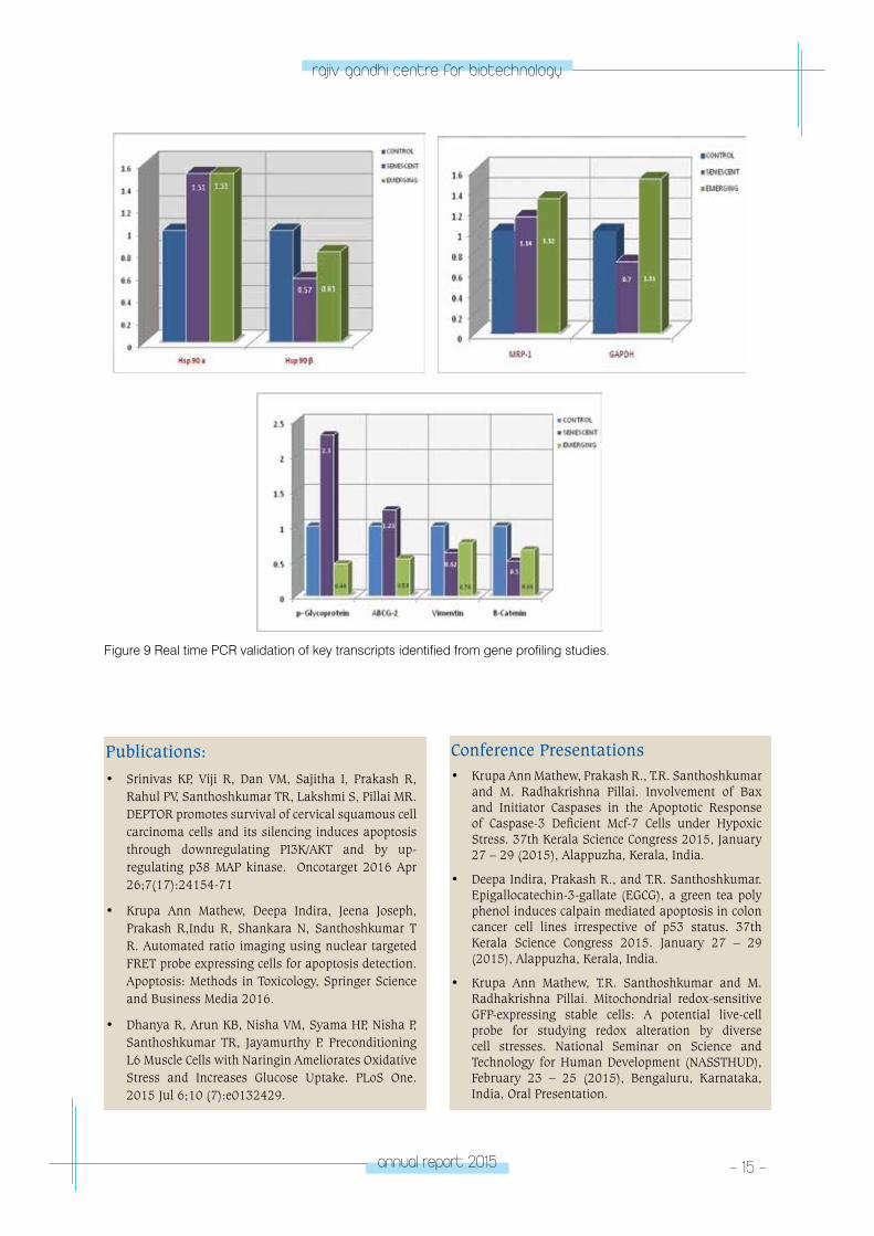
and apoptotic and transcriptional pathways (significantly down-regulated). During the emergent phase, the inflammatory pathways, ROS pathways and anti-apoptotic signaling pathways were significantly enhanced.Some of the key targets that were identified in transcriptomic analysis were consequently validated by real time PCR. As expected, p-glycoprotein showed a significant elevation in expression, noticeably in the senescent phase, during the transition of cells to a drug resistant mode. Hsp-90α showed stable expression both in the senescent and emergent phase, while Hsp-90β exhibited a decrease followed by an increase in
expression during the acquisition of drug resistance. MRP-1 also demonstrated a gradual increase in expression during the evolution of chemoresistance (Figure 9). Currently, by using systems biology, we are investigating the potential interactions between the differentially expressed genes and proteins in our drug resistance model so as to grasp the key molecules/pathways aiding the emergence of drug resistance. This may provide opportunities that will facilitate pharmacological modulation of cancer drug resistance through specific targeting of key modulators of cell state transitions.
Figure 8 (A)
Figure 8 (B)
Figure 8 Key Pathways altered during the transition of cell populations from Senescent (A) to Emergent Phase (B).
annual report 2015 - 15 -
rajiv gandhi centre for biotechnology
Figure 9 Real time PCR validation of key transcripts identified from gene profiling studies.
Publications: • Srinivas KP, Viji R, Dan VM, Sajitha I, Prakash R,
Rahul PV, Santhoshkumar TR, Lakshmi S, Pillai MR. DEPTOR promotes survival of cervical squamous cell carcinoma cells and its silencing induces apoptosis through downregulating PI3K/AKT and by up-regulating p38 MAP kinase. Oncotarget 2016 Apr 26;7(17):24154-71
• Krupa Ann Mathew, Deepa Indira, Jeena Joseph, Prakash R,Indu R, Shankara N, Santhoshkumar T R. Automated ratio imaging using nuclear targeted FRET probe expressing cells for apoptosis detection. Apoptosis: Methods in Toxicology, Springer Science and Business Media 2016.
• Dhanya R, Arun KB, Nisha VM, Syama HP, Nisha P, Santhoshkumar TR, Jayamurthy P. Preconditioning L6 Muscle Cells with Naringin Ameliorates Oxidative Stress and Increases Glucose Uptake. PLoS One. 2015 Jul 6;10 (7):e0132429.
Conference Presentations • Krupa Ann Mathew, Prakash R., T.R. Santhoshkumar
and M. Radhakrishna Pillai. Involvement of Bax and Initiator Caspases in the Apoptotic Response of Caspase-3 Deficient Mcf-7 Cells under Hypoxic Stress. 37th Kerala Science Congress 2015, January 27 – 29 (2015), Alappuzha, Kerala, India.
• Deepa Indira, Prakash R., and T.R. Santhoshkumar. Epigallocatechin-3-gallate (EGCG), a green tea poly phenol induces calpain mediated apoptosis in colon cancer cell lines irrespective of p53 status. 37th Kerala Science Congress 2015. January 27 – 29 (2015), Alappuzha, Kerala, India.
• Krupa Ann Mathew, T.R. Santhoshkumar and M. Radhakrishna Pillai. Mitochondrial redox-sensitive GFP-expressing stable cells: A potential live-cell probe for studying redox alteration by diverse cell stresses. National Seminar on Science and Technology for Human Development (NASSTHUD), February 23 – 25 (2015), Bengaluru, Karnataka, India, Oral Presentation.
- 16 - annual report 2015
rajiv gandhi centre for biotechnology
EXTRAMURAL RESEARCH GRANTS
Title of the project Funding Agency Duration
Design and Development of New Generation FRET probe expressing stable Cancer Cells for anticancer drug screening: From In Vitro HTS Screen to whole animal Imaging
Department of Biotechnology, Government of India
2013-2017
Development of an oral tumor animal model for photodynamic therapy
VINVISH Technologies 2016-2017
• Krupa Ann Mathew, Deepa I., T.R. Santhoshkumar and M. Radhakrishna Pillai. Hypoxic Stress Augments Endoplasmic Reticulum (ER)-phagy and Cancer Cell Survival. The XXXIX All India Cell Biology Conference – Cellular Organization and Dynamics, December 6 – 8 (2015), Thiruvananthapuram, Kerala, India.
• Asha Lekshmi and T.R. Santhoshkumar. Generation of Osteosarcoma cell lines stably expressing fluorescent probe for visualization of autophagic response. 37th Kerala Science Congress. January 27 – 29 (2015), Alappuzha, Kerala, India, Poster Presentation.
• Santhik S.L., M. Radhakrishna Pillai and Santhosh Kumar T.R. Development of florescent based imaging tool to visualize drug escape and cell cycle entry in cancer cells. The XXXIX All India Cell Biology Conference – Cellular Organization and Dynamics, December 6 – 8 (2015), Thiruvananthapuram, Kerala, India.
Best Presentation Awards• Best Oral Presentation Award: Krupa Ann Mathew,
Deepa I., T.R. Santhoshkumar and M. Radhakrishna Pillai. Endoplasmic Reticulum (ER) and Lysosome-Targeted Fluorescent Protein Probe-Expressing Stable Cells Reveal Dynamics of ER-phagy under Cellular Stress. International Conference on Contemporary Research Trends in Diagnostics and Therapeutics. February 15 – 18 (2015), Anna University, Chennai, Tamil Nadu, India.
• Best Oral Presentation Award: Deepa I., Santhik SL and T.R. Santhoshkumar. Bcl-2 targeted to Endoplasmic reticulum protects cells from apoptosis through mitophagy dependent redox regulation. Annual Meeting of Society for Biotechnologists India (SBTI) 2015 & National Conference on Recent Advances in Biomedical Sciences and Biotechnology Oral Presentation
CAnCeR ReSeA
RCH pRoGRAM
- laboratory 2CANCER
RESEARCH PROGRAM
Laboratory - 2
Ruby John Anto
Post-Doctoral FellowVinod V.
PhD StudentsLekshmi R. NathHaritha H. NairMohan Shankar G.Minakshi SaikiaShabna S.
Project FellowAiswarya U.S.
Technical AssistantJannet S.
Ruby John Anto took her PhD in Biochemistry
from Amala Cancer Research Centre, Thrissur and
did post doctoral training at MD Anderson Cancer
Centre, Houston, Texas, before joining RGCB in
2004
- 18 - annual report 2015
rajiv gandhi centre for biotechnology
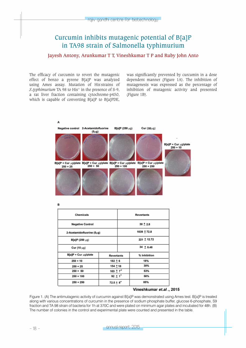
Curcumin inhibits mutagenic potential of B[a]P in TA98 strain of Salmonella typhimurium
Jayesh Antony, Arunkumar T T, Vineshkumar T P and Ruby John Anto
The efficacy of curcumin to revert the mutagenic effect of benzo a pyrene B[a]P was analyzed using Ames assay. Mutation of His-strains of S.typhimurium TA 98 to His+ in the presence of S-9, a rat liver fraction containing cytochrome-p450, which is capable of converting B[a]P to B[a]PDE,
was significantly prevented by curcumin in a dose dependent manner (Figure 1A). The inhibition of mutagenesis was expressed as the percentage of inhibition of mutagenic activity and presented (Figure 1B).
Figure 1. (A) The antimutagenic activity of curcumin against B[a]P was demonstrated using Ames test. B[a]P is treated along with various concentrations of curcumin in the presence of sodium phosphate buffer, glucose 6-phosphate, S9 fraction and TA 98 strain of bacteria for 1h at 370C and were plated on minimum agar plates and incubated for 48h. (B) The number of colonies in the control and experimental plate were counted and presented in the table.
annual report 2015 - 19 -
rajiv gandhi centre for biotechnology
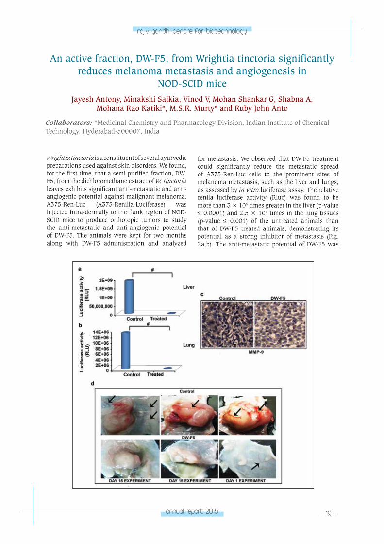
An active fraction, DW-F5, from Wrightia tinctoria significantly reduces melanoma metastasis and angiogenesis in
NOD-SCID miceJayesh Antony, Minakshi Saikia, Vinod V, Mohan Shankar G, Shabna A,
Mohana Rao Katiki*, M.S.R. Murty* and Ruby John Anto
Collaborators: *Medicinal Chemistry and Pharmacology Division, Indian Institute of Chemical Technology, Hyderabad-500007, India
Wrightia tinctoria is a constituent of several ayurvedic preparations used against skin disorders. We found, for the first time, that a semi-purified fraction, DW-F5, from the dichloromethane extract of W. tinctoria leaves exhibits significant anti-metastatic and anti-angiogenic potential against malignant melanoma. A375-Ren-Luc (A375-Renilla-Luciferase) was injected intra-dermally to the flank region of NOD-SCID mice to produce orthotopic tumors to study the anti-metastatic and anti-angiogenic potential of DW-F5. The animals were kept for two months along with DW-F5 administration and analyzed
for metastasis. We observed that DW-F5 treatment could significantly reduce the metastatic spread of A375-Ren-Luc cells to the prominent sites of melanoma metastasis, such as the liver and lungs, as assessed by in vitro luciferase assay. The relative renila luciferase activity (Rluc) was found to be more than 3 × 10⁵ times greater in the liver (p-value ≤ 0.0001) and 2.5 × 10³ times in the lung tissues (p-value ≤ 0.001) of the untreated animals than that of DW-F5 treated animals, demonstrating its potential as a strong inhibitor of metastasis (Fig. 2a,b). The anti-metastatic potential of DW-F5 was
- 20 - annual report 2015
rajiv gandhi centre for biotechnology
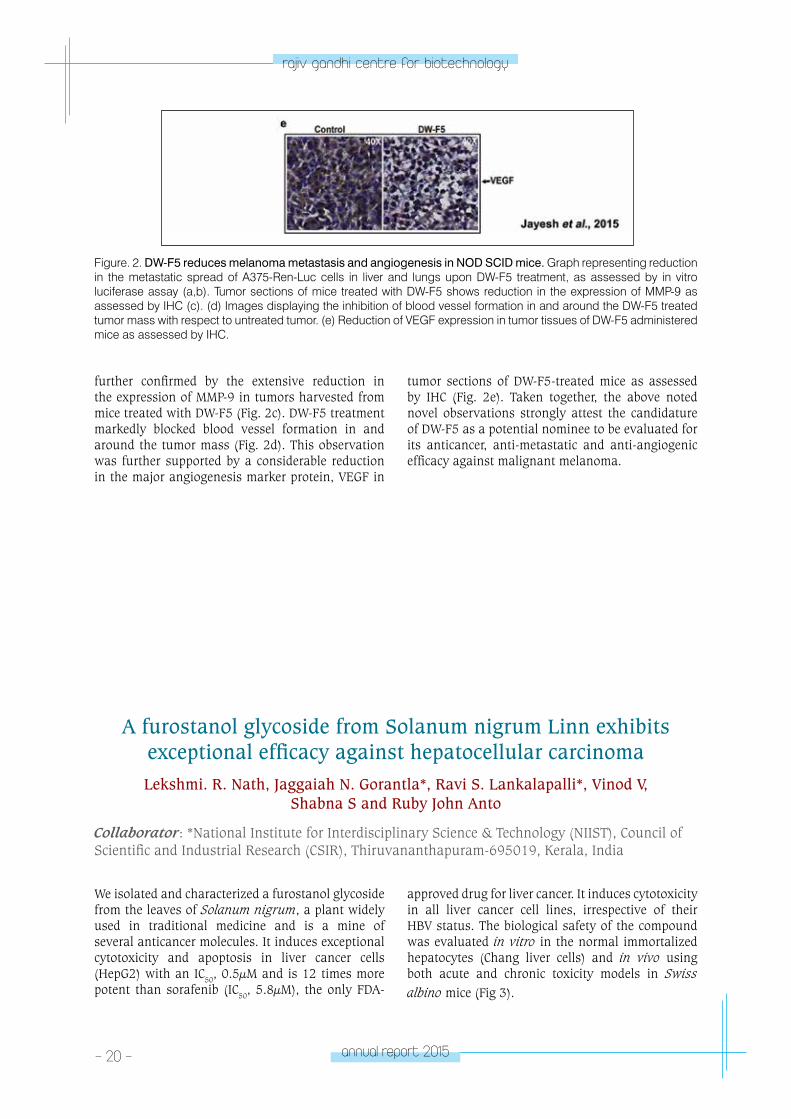
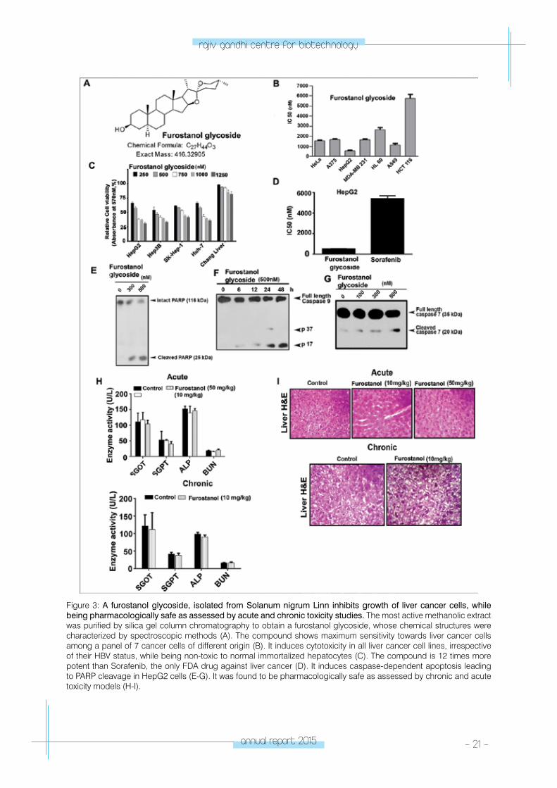
A furostanol glycoside from Solanum nigrum Linn exhibits exceptional efficacy against hepatocellular carcinomaLekshmi. R. Nath, Jaggaiah N. Gorantla*, Ravi S. Lankalapalli*, Vinod V,
Shabna S and Ruby John Anto
Collaborator: *National Institute for Interdisciplinary Science & Technology (NIIST), Council of Scientific and Industrial Research (CSIR), Thiruvananthapuram-695019, Kerala, India
We isolated and characterized a furostanol glycoside from the leaves of Solanum nigrum, a plant widely used in traditional medicine and is a mine of several anticancer molecules. It induces exceptional cytotoxicity and apoptosis in liver cancer cells (HepG2) with an IC50, 0.5µM and is 12 times more potent than sorafenib (IC50, 5.8µM), the only FDA-
approved drug for liver cancer. It induces cytotoxicity in all liver cancer cell lines, irrespective of their HBV status. The biological safety of the compound was evaluated in vitro in the normal immortalized hepatocytes (Chang liver cells) and in vivo using both acute and chronic toxicity models in Swiss albino mice (Fig 3).
Figure. 2. DW-F5 reduces melanoma metastasis and angiogenesis in NOD SCID mice. Graph representing reduction in the metastatic spread of A375-Ren-Luc cells in liver and lungs upon DW-F5 treatment, as assessed by in vitro luciferase assay (a,b). Tumor sections of mice treated with DW-F5 shows reduction in the expression of MMP-9 as assessed by IHC (c). (d) Images displaying the inhibition of blood vessel formation in and around the DW-F5 treated tumor mass with respect to untreated tumor. (e) Reduction of VEGF expression in tumor tissues of DW-F5 administered mice as assessed by IHC.
further confirmed by the extensive reduction in the expression of MMP-9 in tumors harvested from mice treated with DW-F5 (Fig. 2c). DW-F5 treatment markedly blocked blood vessel formation in and around the tumor mass (Fig. 2d). This observation was further supported by a considerable reduction in the major angiogenesis marker protein, VEGF in
tumor sections of DW-F5-treated mice as assessed by IHC (Fig. 2e). Taken together, the above noted novel observations strongly attest the candidature of DW-F5 as a potential nominee to be evaluated for its anticancer, anti-metastatic and anti-angiogenic efficacy against malignant melanoma.
annual report 2015 - 21 -
rajiv gandhi centre for biotechnology
Figure 3: A furostanol glycoside, isolated from Solanum nigrum Linn inhibits growth of liver cancer cells, while being pharmacologically safe as assessed by acute and chronic toxicity studies. The most active methanolic extract was purified by silica gel column chromatography to obtain a furostanol glycoside, whose chemical structures were characterized by spectroscopic methods (A). The compound shows maximum sensitivity towards liver cancer cells among a panel of 7 cancer cells of different origin (B). It induces cytotoxicity in all liver cancer cell lines, irrespective of their HBV status, while being non-toxic to normal immortalized hepatocytes (C). The compound is 12 times more potent than Sorafenib, the only FDA drug against liver cancer (D). It induces caspase-dependent apoptosis leading to PARP cleavage in HepG2 cells (E-G). It was found to be pharmacologically safe as assessed by chronic and acute toxicity models (H-I).
- 22 - annual report 2015
rajiv gandhi centre for biotechnology
3,5-dihydroxy-4-ethyl-trans-stilbene (DETS) isolated from Bacillus cereus act as a potent candidate against malignant
melanoma in vitroLekshmi R Nath, Shabna A, Nishanth Kumar S1, Dileep Kumar BS1, Arya A. Das2,
Bala Nambisan2, Chellapan Mohandas2, Ruby John Anto
Collaborators: 1National Institute for Interdisciplinary Science & Technology, Thiruvananthapuram and 2Central Tuber Crops Research Institute, Sreekariyam, Thiruvananthapuram.
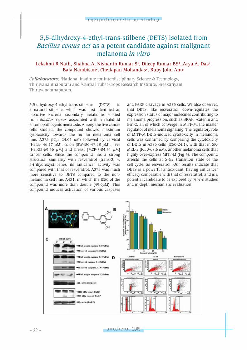
3,5-dihydroxy-4-ethyl-trans-stilbene (DETS) is a natural stilbene, which was first identified as bioactive bacterial secondary metabolite isolated from Bacillus cereus associated with a rhabditid entomopathogenic nematode. Among the five cancer cells studied, the compound showed maximum cytotoxicity towards the human melanoma cell line, A375 (IC50: 24.01 µM) followed by cervical [HeLa- 46.17 µM], colon [SW480-47.28 µM], liver [HepG2-69.56 µM] and breast [MCF-7-84.31 µM] cancer cells. Since the compound has a strong structural similarity with resveratrol (trans-3, 4, 5-trihydroxystilbene), its anticancer activity was compared with that of resveratrol. A375 was much more sensitive to DETS compared to the non-melanoma cell line, A431, in which the IC50 of the compound was more than double (49.6µM). This compound induces activation of various caspases
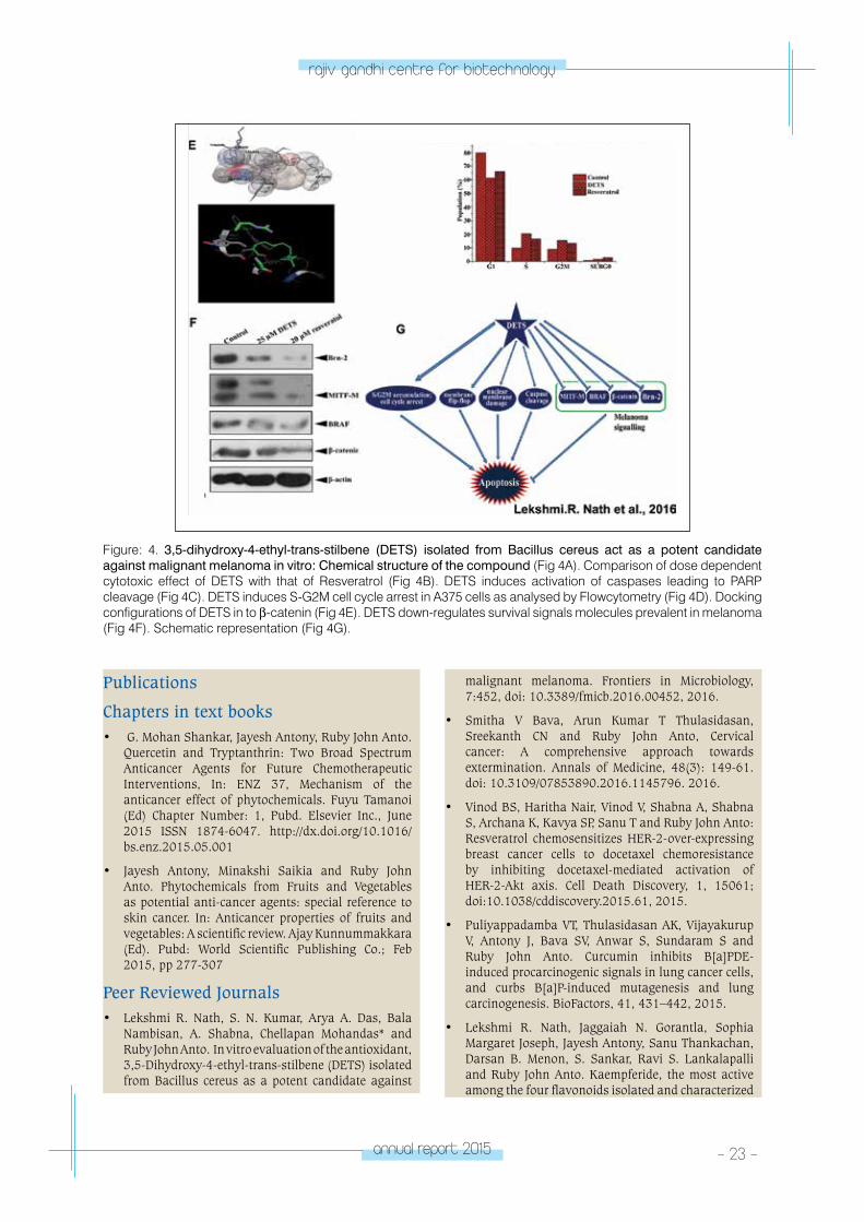
and PARP cleavage in A375 cells. We also observed that DETS, like resveratrol, down-regulates the expression status of major molecules contributing to melanoma progression, such as BRAF, β-catenin and Brn-2, all of which converge in MITF-M, the master regulator of melanoma signaling. The regulatory role of MITF-M DETS-induced cytotoxicity in melanoma cells was confirmed by comparing the cytotoxicity of DETS in A375 cells (IC50-24.1), with that in SK-MEL-2 (IC50-67.6 µM), another melanoma cells that highly over-express MITF-M (Fig 4). The compound arrests the cells at S-G2 transition state of the cell cycle, as resveratrol. Our results indicate that DETS is a powerful antioxidant, having anticancer efficacy comparable with that of resveratrol, and is a potential candidate to be explored by in vivo studies and in-depth mechanistic evaluation.
annual report 2015 - 23 -
rajiv gandhi centre for biotechnology
Figure: 4. 3,5-dihydroxy-4-ethyl-trans-stilbene (DETS) isolated from Bacillus cereus act as a potent candidate against malignant melanoma in vitro: Chemical structure of the compound (Fig 4A). Comparison of dose dependent cytotoxic effect of DETS with that of Resveratrol (Fig 4B). DETS induces activation of caspases leading to PARP cleavage (Fig 4C). DETS induces S-G2M cell cycle arrest in A375 cells as analysed by Flowcytometry (Fig 4D). Docking configurations of DETS in to β-catenin (Fig 4E). DETS down-regulates survival signals molecules prevalent in melanoma (Fig 4F). Schematic representation (Fig 4G).
Publications
Chapters in text books • G. Mohan Shankar, Jayesh Antony, Ruby John Anto.
Quercetin and Tryptanthrin: Two Broad Spectrum Anticancer Agents for Future Chemotherapeutic Interventions, In: ENZ 37, Mechanism of the anticancer effect of phytochemicals. Fuyu Tamanoi (Ed) Chapter Number: 1, Pubd. Elsevier Inc., June 2015 ISSN 1874-6047. http://dx.doi.org/10.1016/bs.enz.2015.05.001
• Jayesh Antony, Minakshi Saikia and Ruby John Anto. Phytochemicals from Fruits and Vegetables as potential anti-cancer agents: special reference to skin cancer. In: Anticancer properties of fruits and vegetables: A scientific review. Ajay Kunnummakkara (Ed). Pubd: World Scientific Publishing Co.; Feb 2015, pp 277-307
Peer Reviewed Journals• Lekshmi R. Nath, S. N. Kumar, Arya A. Das, Bala
Nambisan, A. Shabna, Chellapan Mohandas* and Ruby John Anto. In vitro evaluation of the antioxidant, 3,5-Dihydroxy-4-ethyl-trans-stilbene (DETS) isolated from Bacillus cereus as a potent candidate against
malignant melanoma. Frontiers in Microbiology, 7:452, doi: 10.3389/fmicb.2016.00452, 2016.
• Smitha V Bava, Arun Kumar T Thulasidasan, Sreekanth CN and Ruby John Anto, Cervical cancer: A comprehensive approach towards extermination. Annals of Medicine, 48(3): 149-61. doi: 10.3109/07853890.2016.1145796. 2016.
• Vinod BS, Haritha Nair, Vinod V, Shabna A, Shabna S, Archana K, Kavya SP, Sanu T and Ruby John Anto: Resveratrol chemosensitizes HER-2-over-expressing breast cancer cells to docetaxel chemoresistance by inhibiting docetaxel-mediated activation of HER-2-Akt axis. Cell Death Discovery, 1, 15061; doi:10.1038/cddiscovery.2015.61, 2015.
• Puliyappadamba VT, Thulasidasan AK, Vijayakurup V, Antony J, Bava SV, Anwar S, Sundaram S and Ruby John Anto. Curcumin inhibits B[a]PDE-induced procarcinogenic signals in lung cancer cells, and curbs B[a]P-induced mutagenesis and lung carcinogenesis. BioFactors, 41, 431–442, 2015.
• Lekshmi R. Nath, Jaggaiah N. Gorantla, Sophia Margaret Joseph, Jayesh Antony, Sanu Thankachan, Darsan B. Menon, S. Sankar, Ravi S. Lankalapalli and Ruby John Anto. Kaempferide, the most active among the four flavonoids isolated and characterized
- 24 - annual report 2015
rajiv gandhi centre for biotechnology
from Chromolaena odorata, induces apoptosis in cervical cancer cells while being pharmacologically safe. RSC Adv., 2015, 5, 100912. Impact factor:3.84
• Jayesh Antony, Minakshi Saikia, Vino T. Cheriyan, Nishanth Kumar S and Ruby John Anto. Sesbania: A prospective candidate to be excavated for anticancer drugs. The Natural Products Journal, 5(4): 273-287, DOI: 10.2174/1872211310999151110155644, 2015.
• Jayesh Antony, Minakshi Saikia, Vinod.V, Lekshmi. R. Nath, Mohana Rao Katiki, M.S.R. Murty, Anju Paul, Shabna A, Harsha Chandran, Sophia Margaret Joseph, Nishanth Kumar. S, Elizabeth Jayex Panakkal, Sriramya I.V, Sridivya I.V, Sophia Ran, Sankar S, Easwary Rajan and Ruby John Anto DW-F5: A novel formulation against malignant melanoma from Wrightia tinctoria, Scientific Reports, 5:11107 , DOI: 10.1038/srep11107, 2015
• Jisha J. Pillai, Arun Kumar T. Thulasidasan, Ruby John Anto, Nandan C. Devika, N. Ashwanikumar and G. S. Vinod Kumar. Curcumin entrapped folic acid conjugated PLGA–PEG nanoparticles exhibit enhanced anticancer activity by site specific delivery, RSC Adv., 5, 25518-25524. DOI: 10.1039/C5RA00018A. 2015.
Conference Presentations• Invited Talk: Ruby John Anto, Anticancer potential
of natural products, National Seminar on Modern trends in Chemistry, February 2016, Farook College, Calicut, Kerala.
• Lekshmi.R.Nath, Ravi Shankar L and Ruby John Anto. In vitro and in vivo evaluation of the anticancer potential of SN2, a spirostan-3-ol derivative identified and characterized from Solanum nigrum against hepatocellular carcinoma, Oral Presentation, 28th Kerala Science Congress, 28-30 January 2016, Thennipalam, Malappuram, Kerala.
• Invited Talk: Ruby John Anto, Tryptanthrin: A potent anti-melanoma molecule from Wrightia tinctoria, International Conference on Cancer Research: New Horizons, 19-21st November, 2015, NCCS, Pune.
• Lekshmi.R.Nath and Ruby John Anto Poster Presentation at the 8th Eurobiotechnology congress, Frankfurt, Germany. August 17-20, 2015.
• Arun Kumar T Thulasidasan, Devika Nandan.C, Lekha Nair K, Jisha J Pillai, GS Vinod Kumar, and Ruby John Anto. Nano Encapsulation of curcumin using PLGA improves its therapeutic potential, Oral Presentation at the UGC, DST & DBT sponsored “National Conference on Challenges and Future Prospects of Applied Research in Life Sciences” held on 6th February 2015 at Bharathidasan University, Tiruchirappalli.
• Minakshi Saikia, Jayesh Antony, Vinod V, Sophia Margaret Joseph, Lekshmi R Nath and Ruby John Anto. Evaluation of the anticancer property of Wrightia tinctoria (Roxb.) R.Br. leaves against
malignant melanoma. Poster Presentation, 7th HOPE Meeting with Nobel laureates conducted by Japan Society for Promotion of Science (JSPS), 1-5 March, 2015, Tokyo, Japan.
• Haritha H Nair, Vinod Balachandran, Jayesh Antony and Ruby John Anto. Thymidylate synthase-dependent down-regulation of NF-κB is the pivotal signaling mechanism regulating curcumin-mediated chemosensitization of breast cancer cells to 5-FU. Young Researchers Forum Oral presentation in the 7th Global Summit on Cancer Therapy organized by Omics International held at Dubai from 5-7th October 2015.
• Haritha H.Nair, Vinod Balachandran,Vinod V, Shabna A. Resveratrol chemosensitizes HER-2-over-expressing breast cancer cells to docetaxel chemoresistance by inhibiting docetaxel-mediated activation of HER-2-Akt axis. Poster presentation at Global Cancer Summit 2015 held at J.N Tata Auditorium, Bangalore from November 18 - 20, 2015.
• Minakshi Saikia, Jayesh Antony, Vinod V, Shabna A, Mohana Rao Katiki, MSR Murty and Ruby John Anto. Evaluation of DW-F5, a potent anticancer principle isolated from the leaves of Wrightia tinctoria. Poster presentation. International Conference on Cancer Research: New Horizons, 19-21st November 2015, NCCS, Pune, India.
Ph.D Awarded• Jayesh Antony 2015 (Title: Molecular evaluation
of anticancer properties of active principle/s from Wrightia tinctoria).
• Arun Kumar T Thulasidasan 2016 (Title: Nanoparticle-based drug delivery and drug release for chemotherapy and chemoprevention)
Awards & Honors• Best paper Award: Lekshmi.R.Nath, at 28th Kerala
Science Congress, India, 2016 under Biotechnology category.
• OMICS International Best Poster Award: Lekshmi.R.Nath, at the 8th Eurobiotechnology congress, Frankfurt, Germany. August 17-20, 2015
• Best Oral Presentation Award Arun Kumar T Thulasidasan, Devika Nandan C, Lekha Nair K, J Jisha Pillai, G.S.Vinod Kumar and Ruby John Anto. at National Conference organized by dept. of Biochemistry, on Challenges and Future Prospects of Applied Research in Life Sciences-2015, (6th February 2016) Bharathidasan University, Tiruchirappalli.
• JSPS HOPE Fellowship: Minakshi Saikia, Awarded to participate and present a poster in the 7th HOPE meeting with Nobel Laureates, conducted by the Japan Society for Promotion of Science (JSPS) held at Tokyo (March 1-5, 2015).
annual report 2015 - 25 -
rajiv gandhi centre for biotechnology
EXTRA MURAL GRANTS
No Title of Project Funding Agency From - To
1
Comparison of the chemopreventive efficacy of free curcumin and biodegradable polymer based nano curcumin in Benzo[a] pyrene–induced lung carcinogenesis
Department of Science & Technology, Government of India
2013 -16
2In vivo evaluation of the anticarcinogenic effect and toxicity of the active principle of Wrightia tinctoria
Indian Council of Medical Research
2015 -18
3Mechanistic evaluation and in vivo validation of the anticancer principle isolated from Chro-molaena odorata against cervical cancer (PI)
Kerala State Council for Science, Technology & Environment
2016 -19
4Isolation, identification and characterization of anticancer principle from the medicinal plant Corallocarpus epigaeus (Co-PI)
Kerala State Council for Science, Technology & Environment
2016 -19
5
Investigating the mechanism behind the protec-tive effect of the anticancer compounds isolated from Woodfordia fruticosa (L.) Kurz flowers against hepatocellular carcinoma (Co-PI)
Kerala State Council for Science, Technology & Environment
2016 -19
CAnCeR ReSeA
RCH pRoGRAM
- laboratory 3CANCER
RESEARCH PROGRAM
Laboratory - 3
Suparna Sengupta
Technical OfficerSudha B. Nair
PhD Students:Smreti VasudevanReshma ThamkachyRohith Kumar N. J.S. SreejaDrishya Dharmapal
Suparna Sengupta received her PhD in
Biochemistry from Bose Institute, Kolkata and
subsequently worked as a postdoctoral associate
at University of Kansas, USA and then as a CSIR
Pool-Officer at National Institute of Immunology,
New Delhi, before joining RGCB.
annual report 2015 - 27 -
rajiv gandhi centre for biotechnology
Role of p53 on Diaminothiazole induced cell death in colon cancer
Reshma Thamkachy and Suparna Sengupta
Collaborator: K.N.Rajasekharan, University of Kerala
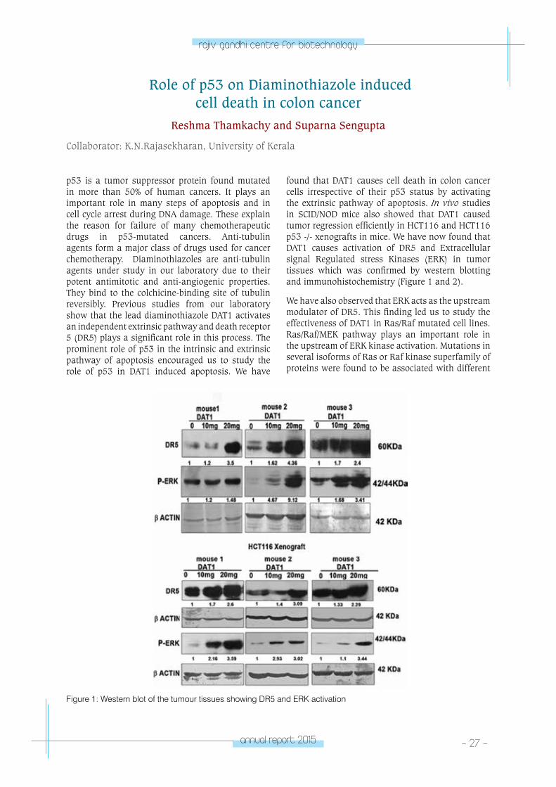
p53 is a tumor suppressor protein found mutated in more than 50% of human cancers. It plays an important role in many steps of apoptosis and in cell cycle arrest during DNA damage. These explain the reason for failure of many chemotherapeutic drugs in p53-mutated cancers. Anti-tubulin agents form a major class of drugs used for cancer chemotherapy. Diaminothiazoles are anti-tubulin agents under study in our laboratory due to their potent antimitotic and anti-angiogenic properties. They bind to the colchicine-binding site of tubulin reversibly. Previous studies from our laboratory show that the lead diaminothiazole DAT1 activates an independent extrinsic pathway and death receptor 5 (DR5) plays a significant role in this process. The prominent role of p53 in the intrinsic and extrinsic pathway of apoptosis encouraged us to study the role of p53 in DAT1 induced apoptosis. We have
found that DAT1 causes cell death in colon cancer cells irrespective of their p53 status by activating the extrinsic pathway of apoptosis. In vivo studies in SCID/NOD mice also showed that DAT1 caused tumor regression efficiently in HCT116 and HCT116 p53 -/- xenografts in mice. We have now found that DAT1 causes activation of DR5 and Extracellular signal Regulated stress Kinases (ERK) in tumor tissues which was confirmed by western blotting and immunohistochemistry (Figure 1 and 2).
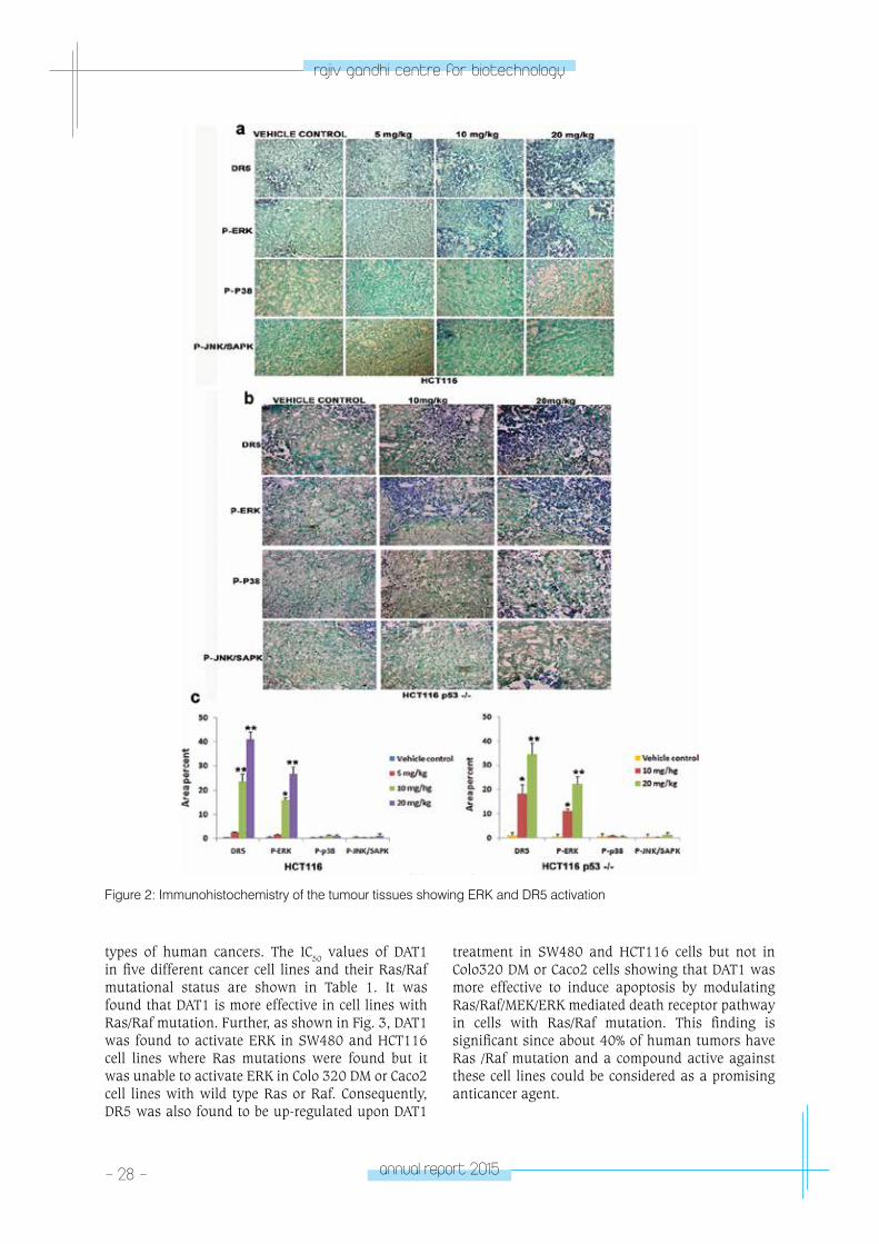
We have also observed that ERK acts as the upstream modulator of DR5. This finding led us to study the effectiveness of DAT1 in Ras/Raf mutated cell lines. Ras/Raf/MEK pathway plays an important role in the upstream of ERK kinase activation. Mutations in several isoforms of Ras or Raf kinase superfamily of proteins were found to be associated with different
Figure 1: Western blot of the tumour tissues showing DR5 and ERK activation
- 28 - annual report 2015
rajiv gandhi centre for biotechnology
Figure 2: Immunohistochemistry of the tumour tissues showing ERK and DR5 activation
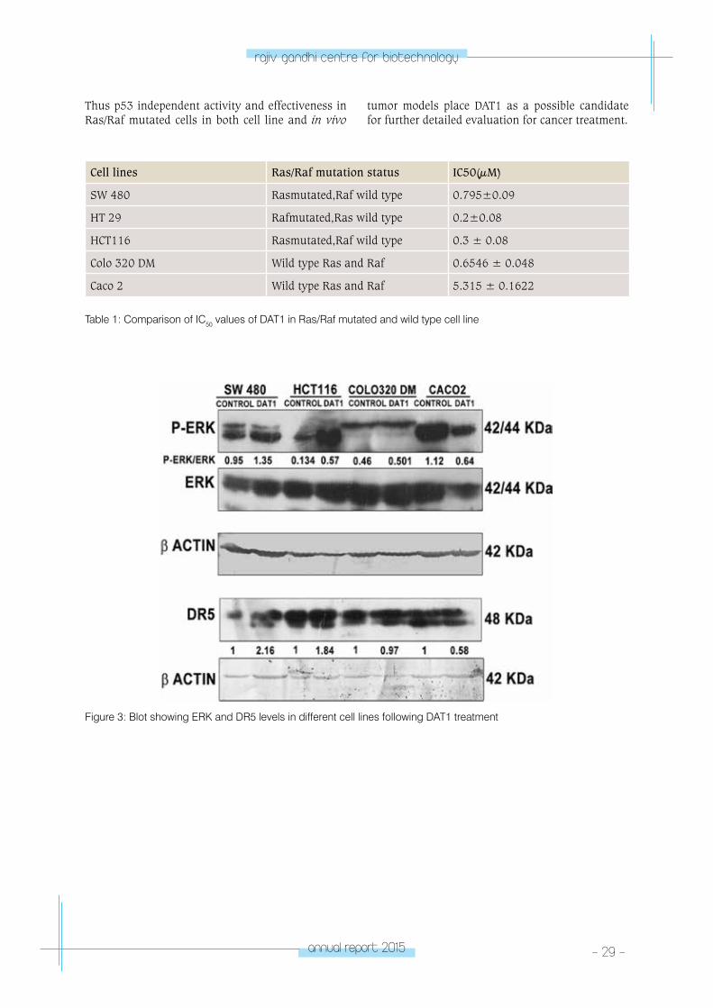
types of human cancers. The IC50 values of DAT1 in five different cancer cell lines and their Ras/Raf mutational status are shown in Table 1. It was found that DAT1 is more effective in cell lines with Ras/Raf mutation. Further, as shown in Fig. 3, DAT1 was found to activate ERK in SW480 and HCT116 cell lines where Ras mutations were found but it was unable to activate ERK in Colo 320 DM or Caco2 cell lines with wild type Ras or Raf. Consequently, DR5 was also found to be up-regulated upon DAT1
treatment in SW480 and HCT116 cells but not in Colo320 DM or Caco2 cells showing that DAT1 was more effective to induce apoptosis by modulating Ras/Raf/MEK/ERK mediated death receptor pathway in cells with Ras/Raf mutation. This finding is significant since about 40% of human tumors have Ras /Raf mutation and a compound active against these cell lines could be considered as a promising anticancer agent.
annual report 2015 - 29 -
rajiv gandhi centre for biotechnology
Cell lines Ras/Raf mutation status IC50(µM)
SW 480 Rasmutated,Raf wild type 0.795±0.09
HT 29 Rafmutated,Ras wild type 0.2±0.08
HCT116 Rasmutated,Raf wild type 0.3 ± 0.08
Colo 320 DM Wild type Ras and Raf 0.6546 ± 0.048
Caco 2 Wild type Ras and Raf 5.315 ± 0.1622
Table 1: Comparison of IC50 values of DAT1 in Ras/Raf mutated and wild type cell line
Figure 3: Blot showing ERK and DR5 levels in different cell lines following DAT1 treatment
Thus p53 independent activity and effectiveness in Ras/Raf mutated cells in both cell line and in vivo
tumor models place DAT1 as a possible candidate for further detailed evaluation for cancer treatment.
- 30 - annual report 2015
rajiv gandhi centre for biotechnology
Analysis of Fodrin Association with Gamma Tubulin Complex in Mammalian Brain
Rohith Kumar N, J.S. Sreeja and Suparna Sengupta
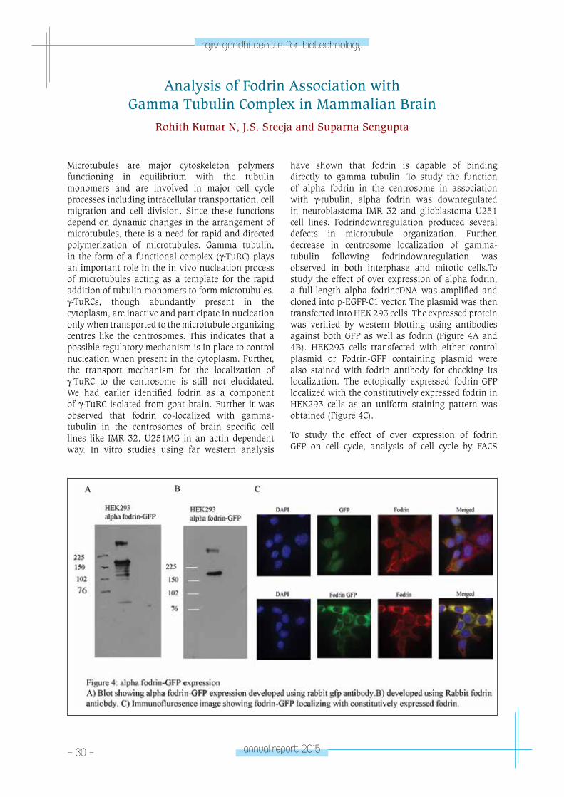
Microtubules are major cytoskeleton polymers functioning in equilibrium with the tubulin monomers and are involved in major cell cycle processes including intracellular transportation, cell migration and cell division. Since these functions depend on dynamic changes in the arrangement of microtubules, there is a need for rapid and directed polymerization of microtubules. Gamma tubulin, in the form of a functional complex (γ-TuRC) plays an important role in the in vivo nucleation process of microtubules acting as a template for the rapid addition of tubulin monomers to form microtubules. γ-TuRCs, though abundantly present in the cytoplasm, are inactive and participate in nucleation only when transported to the microtubule organizing centres like the centrosomes. This indicates that a possible regulatory mechanism is in place to control nucleation when present in the cytoplasm. Further, the transport mechanism for the localization of γ-TuRC to the centrosome is still not elucidated.We had earlier identified fodrin as a component of γ-TuRC isolated from goat brain. Further it was observed that fodrin co-localized with gamma-tubulin in the centrosomes of brain specific cell lines like IMR 32, U251MG in an actin dependent way. In vitro studies using far western analysis
have shown that fodrin is capable of binding directly to gamma tubulin. To study the function of alpha fodrin in the centrosome in association with γ-tubulin, alpha fodrin was downregulated in neuroblastoma IMR 32 and glioblastoma U251 cell lines. Fodrindownregulation produced several defects in microtubule organization. Further, decrease in centrosome localization of gamma-tubulin following fodrindownregulation was observed in both interphase and mitotic cells.To study the effect of over expression of alpha fodrin, a full-length alpha fodrincDNA was amplified and cloned into p-EGFP-C1 vector. The plasmid was then transfected into HEK 293 cells. The expressed protein was verified by western blotting using antibodies against both GFP as well as fodrin (Figure 4A and 4B). HEK293 cells transfected with either control plasmid or Fodrin-GFP containing plasmid were also stained with fodrin antibody for checking its localization. The ectopically expressed fodrin-GFP localized with the constitutively expressed fodrin in HEK293 cells as an uniform staining pattern was obtained (Figure 4C).
To study the effect of over expression of fodrin GFP on cell cycle, analysis of cell cycle by FACS
annual report 2015 - 31 -
rajiv gandhi centre for biotechnology
was performed on HEK293 cells or IMR32 cells transfected with either the control plasmid or Fodrin-GFP containing plasmid. It was observed that following 48hrs, there was a decrease in G1 phase and a corresponding shift towards S and G2/M phase in fodrin over-expressed cells when compared to control as shown in the table below. It can be noted that in IMR 32 or U251 MGfodrin down-regulated cells, a reduction in the number of cells in G2/M phase was observed. Thus, both down-regulation as well as over expression of alpha fodrinleads to
changes in the cell cycle indicating that alpha fodrin is involved in cell cycle progression.
Interestingly over expression of fodrin also caused changes in microtubule organization with aberrations in mitotic spindle. Following 48 hrs of transfection with fodrin GFP in IMR32 cells, abnormal mitotic cells were observed. This indicates that a regulated level of fodrin is required for proper cell cycle phases.
G1phase S phase G2/M phase
HEK 293 EGFP 73.5% 15.65% 10.75%
HEK 293 Fodrin GFP 60.2% 20.95% 18.55%
Establishing the role of fodrin in gamma tubulin mediated functions
J.S. Sreeja, Rohith Kumar N and Suparna Sengupta
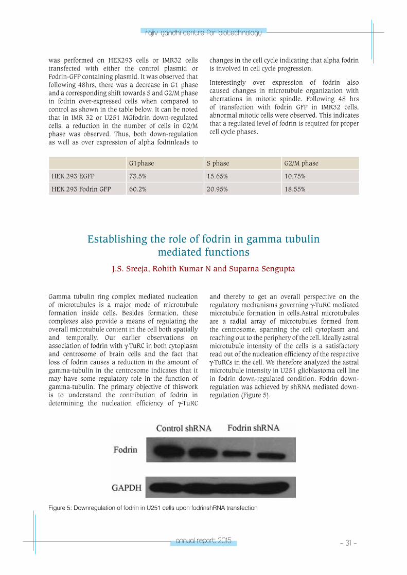
Gamma tubulin ring complex mediated nucleation of microtubules is a major mode of microtubule formation inside cells. Besides formation, these complexes also provide a means of regulating the overall microtubule content in the cell both spatially and temporally. Our earlier observations on association of fodrin with γ-TuRC in both cytoplasm and centrosome of brain cells and the fact that loss of fodrin causes a reduction in the amount of gamma-tubulin in the centrosome indicates that it may have some regulatory role in the function of gamma-tubulin. The primary objective of thiswork is to understand the contribution of fodrin in determining the nucleation efficiency of γ-TuRC
and thereby to get an overall perspective on the regulatory mechanisms governing γ-TuRC mediated microtubule formation in cells.Astral microtubules are a radial array of microtubules formed from the centrosome, spanning the cell cytoplasm and reaching out to the periphery of the cell. Ideally astral microtubule intensity of the cells is a satisfactory read out of the nucleation efficiency of the respective γ-TuRCs in the cell. We therefore analyzed the astral microtubule intensity in U251 glioblastoma cell line in fodrin down-regulated condition. Fodrin down-regulation was achieved by shRNA mediated down-regulation (Figure 5).
Figure 5: Downregulation of fodrin in U251 cells upon fodrinshRNA transfection
- 32 - annual report 2015
rajiv gandhi centre for biotechnology
Aster intensity was determined in the area adjacent to the centrosome. The mean aster intensity in mitotic cells reduced significantly in fodrin down-regulated U251 cells compared to scrambled shRNA treated control cells. The above results therefore indicate a possible regulatory/controlling role of fodrin over γ-TuRC mediated microtubule nucleation. To study the role of fodrin in vivo, experiments
are underway to establish stable neuroblastoma and glioblastoma cell lines expressing GFP tagged tubulin and EB1 (microtubule end binding protein). In vitro experiments to understand the role of purified fodrin on γ-TuRC mediated microtubule nucleation, spectrophotometry based procedures are also underway.
Inhibition of Breast Cancer Cells and Stem Cell like Spheroids by 6-Shogaol
Anasuya Ray, Smreti Vasudevan and Suparna Sengupta
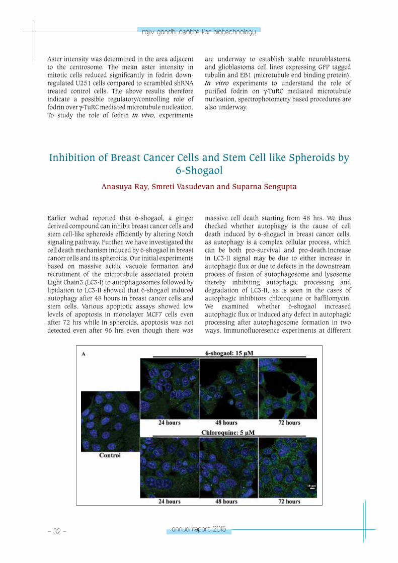
Earlier wehad reported that 6-shogaol, a ginger derived compound can inhibit breast cancer cells and stem cell-like spheroids efficiently by altering Notch signaling pathway. Further, we have investigated the cell death mechanism induced by 6-shogaol in breast cancer cells and its spheroids. Our initial experiments based on massive acidic vacuole formation and recruitment of the microtubule associated protein Light Chain3 (LC3-I) to autophagosomes followed by lipidation to LC3-II showed that 6-shogaol induced autophagy after 48 hours in breast cancer cells and stem cells. Various apoptotic assays showed low levels of apoptosis in monolayer MCF7 cells even after 72 hrs while in spheroids, apoptosis was not detected even after 96 hrs even though there was
massive cell death starting from 48 hrs. We thus checked whether autophagy is the cause of cell death induced by 6-shogaol in breast cancer cells, as autophagy is a complex cellular process, which can be both pro-survival and pro-death.Increase in LC3-II signal may be due to either increase in autophagic flux or due to defects in the downstream process of fusion of autophagosome and lysosome thereby inhibiting autophagic processing and degradation of LC3-II, as is seen in the cases of autophagic inhibitors chloroquine or baffilomycin. We examined whether 6-shogaol increased autophagic flux or induced any defect in autophagic processing after autophagosome formation in two ways. Immunofluoresence experiments at different

annual report 2015 - 33 -
rajiv gandhi centre for biotechnology
Figure 6:6-shogaol induces autophagic flux and cell death in breast cancer cells
time points detected increase in LC3 punctaeupto 48 hours followed by decrease at 72 hours in case of 6-shogaol treatment. In contrast, continuous increase in LC3 accumulation with time was observed in case of chloroquine (Figure 6A), indicating that unlike chloroquine treatment, LC3 got processed with time for 6-shogaol treatment. Further, we used a low concentration of chloroquine in combination with 5 and 15 µM of 6-shogaol and checked the effect on LC3-II accumulation by western blotting. Since chloroquine is an autophagy inhibitor,
an increase in LC3 content in the combination compared to 6-shogaol alone would indicate that autophagic flux generated by 6-shogaol is inhibited by chloroquine. We indeed found a significant increase in LC3-II expression in the combination of 5 and 15 µM of 6-shogaol with chloroquine (3.3 and 3.43 fold increase respectively) when compared with the treatment of 6-shogaol alone (1.8 and 2.1 fold) (Figure 6B). Further, to check the role of autophagy in the cell death induced by 6-shogaol, we used the autophagy inhibitor chloroquine, in a
A scheme showing the action of 6-shogaol in breast cancer cells and spheroids is given below.
- 34 - annual report 2015
rajiv gandhi centre for biotechnology
low concentration that limited the cell death induced by chloroquine itself. 5µM chloroquine alone or in combination with 6-shogaol (1-9 µM) was added to MCF-7 cells and kept for 48 hours prior to analysis of the cell viability by MTT assay. Combination of chloroquine and 6-shogaol decreased the cell death percentage drastically from that of the cells treated with 6-shogaol alone (Figure 6C). The decrease in cell death percentage correlated with the increase in LC3 in combinations, indicating that cell death induced
by 6-shogaol is dependent on the completion of autophagy. LC-3 cleavage upon 6-shogaol treatment was also observed in MCF-7 spheroids (Figure 6D).
A scheme showing the action of 6-shogaol in breast cancer cells and spheroids is given below. Thus the results indicate that autophagy is a prominent mode of cell death triggered by 6-shogaol, although a modest amount of apoptosis was found in the cells after prolonged 6-shogaol treatment.
Publications• Reshma Thamkachy, Rohith Kumar, K. N.
Rajasekharan and Suparna Sengupta (2016) ERK mediated upregulation of death receptor 5 overcomes the lack of p53 functionality in the diaminothiazole DAT1 induced apoptosis in colon cancer models: efficiency of DAT1 in Ras-Raf mutated cells. Molecular Cancer 15:22 DOI 10.1186/s12943-016-0505-7
• Anasuya Ray, Smreti Vasudevan, Suparna Sengupta (2015) 6-Shogaol Inhibits Breast Cancer Cells and Stem Cell-Like Spheroids by Modulation of Notch Signaling Pathway and Induction of Autophagic Cell Death. PLoS One. 10(9): e0137614. doi: 10.1371/journal.pone.0137614
• Smreti Vasudevan, Sannu Ann Thomas, Krishnankutty C. Sivakumar, Reena J. Komalam, Keerthi V. Sreerekha, Kallikat N. Rajasekharan and Suparna Sengupta* (2015): Diaminothiazoles Evade Multidrug Resistance in Cancer Cells and Xenograft Tumour Models and Develop Transient Specific Resistance: Understanding the Basis of Broad-
Spectrum vs Specific Resistance. Carcinogenesis36, 883-93 Doi: 10.1093/ carcin/bgv072
Conference Presentations:• Rohith Kumar N, Shashikala Sasidharan, Suparna
Sengupta. “Alpha fodrin affects microtubule organization and localization of gamma tubulin at centrosome” at the 6th EMBO meeting, Birmingham, UK, 5th-8th September 2015.
• Reshma Thamkachy, Nisha Elizabeth Thomas, Smreti Vasudevan, K.N.Rajasekharan and Suparna Sengupta “Reversible and p53 independent action on the Ras-Raf-ERK-DR5 axis makes diaminothiazoles highly promising for cancer therapy” at the - 6th International Translational Cancer Research Conference onPrevention and Treatment of Cancer: Hypes and Hopes, Ahmedabad, India, February 4-7, 2016
Ph.D. Awarded: • Smreti Vasudevan: Study of drug resistance
mechanisms in cancer against antimitotic agents using paclitaxel and diaminothiazoles”
CAnCeR ReSeA
RCH pRoGRAM
- laboratory 4CANCER
RESEARCH PROGRAM
Laboratory - 4
S. Asha Nair
PhD StudentsDhanya K.Chithra J.S.Tapas Pradhan
Research FellowManu Prasad M.
Research AssistantMaharrish C.
Laboratory AssistantPrameela Kumari T.
Asha Nair took her PhD from the University
of Kerala, working at the Regional Cancer
Center, Thiruvananthapuram. She trained as a
postdoctoral fellow at Harvard Medical School
and MD Anderson Cancer Center Houston, USA
before joining RGCB in 2006.
- 36 - annual report 2015
rajiv gandhi centre for biotechnology
Molecular Mechanism of Drug Resistance in Colorectal CancerManu Prasad M., Tapas Pradhan, Saneesh Babu. P.S., K. Chandramohan*,
and S. Asha Nair
*Collaborator: Regional Cancer Centre, Thiruvananthapuram
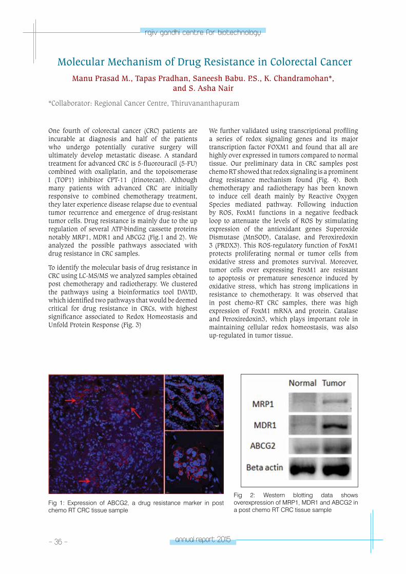
One fourth of colorectal cancer (CRC) patients are incurable at diagnosis and half of the patients who undergo potentially curative surgery will ultimately develop metastatic disease. A standard treatment for advanced CRC is 5-fluorouracil (5-FU) combined with oxaliplatin, and the topoisomerase I (TOP1) inhibitor CPT-11 (Irinotecan). Although many patients with advanced CRC are initially responsive to combined chemotherapy treatment, they later experience disease relapse due to eventual tumor recurrence and emergence of drug-resistant tumor cells. Drug resistance is mainly due to the up regulation of several ATP-binding cassette proteins notably MRP1, MDR1 and ABCG2 (Fig.1 and 2). We analyzed the possible pathways associated with drug resistance in CRC samples.
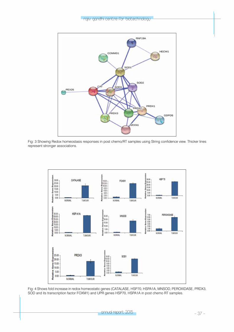
To identify the molecular basis of drug resistance in CRC using LC-MS/MS we analyzed samples obtained post chemotherapy and radiotherapy. We clustered the pathways using a bioinformatics tool DAVID, which identified two pathways that would be deemed critical for drug resistance in CRCs, with highest significance associated to Redox Homeostasis and Unfold Protein Response (Fig. 3)
We further validated using transcriptional profiling a series of redox signaling genes and its major transcription factor FOXM1 and found that all are highly over expressed in tumors compared to normal tissue. Our preliminary data in CRC samples post chemo RT showed that redox signaling is a prominent drug resistance mechanism found (Fig. 4). Both chemotherapy and radiotherapy has been known to induce cell death mainly by Reactive Oxygen Species mediated pathway. Following induction by ROS, FoxM1 functions in a negative feedback loop to attenuate the levels of ROS by stimulating expression of the antioxidant genes Superoxide Dismutase (MnSOD), Catalase, and Peroxiredoxin 3 (PRDX3). This ROS-regulatory function of FoxM1 protects proliferating normal or tumor cells from oxidative stress and promotes survival. Moreover, tumor cells over expressing FoxM1 are resistant to apoptosis or premature senescence induced by oxidative stress, which has strong implications in resistance to chemotherapy. It was observed that in post chemo-RT CRC samples, there was high expression of FoxM1 mRNA and protein. Catalase and Peroxiredoxin3, which plays important role in maintaining cellular redox homeostasis, was also up-regulated in tumor tissue.
Fig 1: Expression of ABCG2, a drug resistance marker in post chemo RT CRC tissue sample
Fig 2: Western blotting data shows overexpression of MRP1, MDR1 and ABCG2 in a post chemo RT CRC tissue sample
annual report 2015 - 37 -
rajiv gandhi centre for biotechnology
Fig: 3 Showing Redox homeostasis responses in post chemo/RT samples using String confidence view. Thicker lines represent stronger associations.
Fig: 4 Shows fold increase in redox homeostatic genes (CATALASE, HSP70, HSPA1A, MNSOD, PEROXIDASE, PRDX3, SOD and its transcription factor FOXM1) and UPR genes HSP70, HSPA1A in post chemo RT samples.
- 38 - annual report 2015
rajiv gandhi centre for biotechnology
Distal Surgical Margin Harbors Cancer Stem Cells that may Influence Colorectal Cancer Outcome.
Tapas Pradhan, Manu Prasad M., K. Chandramohan*, and S. Asha Nair
*Collaborator: Regional Cancer Centre, Thiruvananthapuram
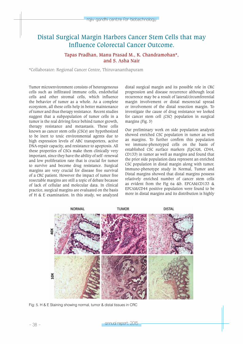
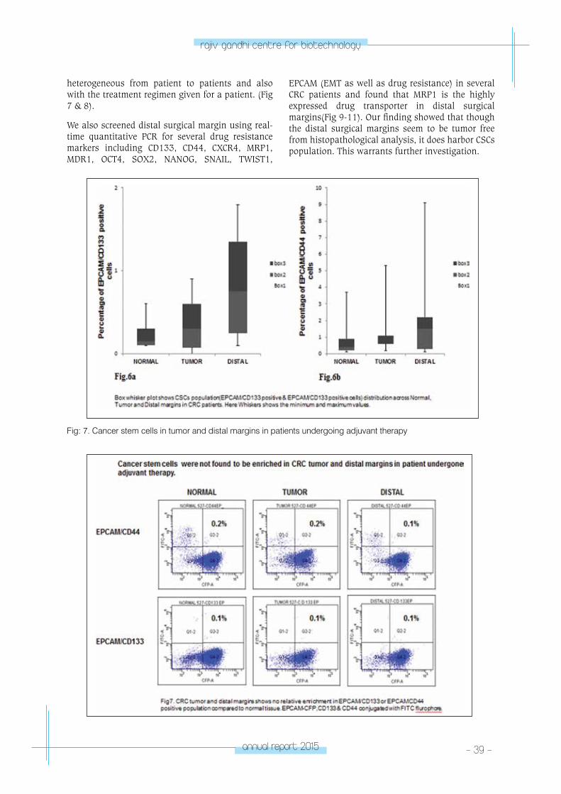
Tumor microenvironment consists of heterogeneous cells such as infiltrated immune cells, endothelial cells and other stromal cells, which influence the behavior of tumor as a whole. As a complete ecosystem, all these cells help in better maintenance of tumor and thus therapy resistance. Recent studies suggest that a subpopulation of tumor cells in a tumor is the real driving force behind tumor growth, therapy resistance and metastasis. These cells known as cancer stem cells (CSCs) are hypothesized to be inert to toxic environmental agents due to high expression levels of ABC transporters, active DNA-repair capacity, and resistance to apoptosis. All these properties of CSCs make them clinically very important, since they have the ability of self -renewal and low proliferation rate that is crucial for tumor to survive and become drug resistance. Surgical margins are very crucial for disease free survival of a CRC patient. However the impact of tumor free resectable margins are still a topic of debate because of lack of cellular and molecular data. In clinical practice, surgical margins are evaluated on the basis of H & E examination. In this study, we analyzed
distal surgical margin and its possible role in CRC progression and disease recurrence although local recurrence may be a result of lateral/circumferential margin involvement or distal mesorectal spread or involvement of the distal resection margin. To investigate the cause of drug resistance we looked for cancer stem cell (CSC) population in surgical margins (Fig. 5)
Our preliminary work on side population analysis showed enriched CSC population in tumor as well as margins. To further confirm this population we immune-phenotyped cells on the basis of established CSC surface markers (EpCAM, CD44, CD133) in tumor as well as margins and found that the prior side population data represent an enriched CSC population in distal margin along with tumor. Immuno-phenotype study in Normal, Tumor and Distal margins showed that distal margins possess relatively enriched number of cancer stem cells as evident from the Fig 6a &b. EPCAM/CD133 & EPCAM/CD44 positive population were found to be more in distal margins and its distribution is highly
Fig: 5. H & E Staining showing normal, tumor & distal tissues in CRC
annual report 2015 - 39 -
rajiv gandhi centre for biotechnology
Fig: 7. Cancer stem cells in tumor and distal margins in patients undergoing adjuvant therapy
heterogeneous from patient to patients and also with the treatment regimen given for a patient. (Fig 7 & 8).
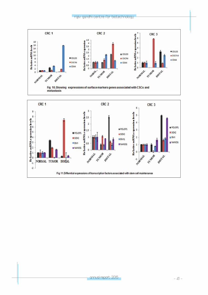
We also screened distal surgical margin using real-time quantitative PCR for several drug resistance markers including CD133, CD44, CXCR4, MRP1, MDR1, OCT4, SOX2, NANOG, SNAIL, TWIST1,
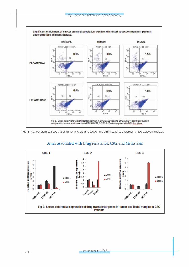
EPCAM (EMT as well as drug resistance) in several CRC patients and found that MRP1 is the highly expressed drug transporter in distal surgical margins(Fig 9-11). Our finding showed that though the distal surgical margins seem to be tumor free from histopathological analysis, it does harbor CSCs population. This warrants further investigation.
- 40 - annual report 2015
rajiv gandhi centre for biotechnology
Fig: 8. Cancer stem cell population tumor and distal resection margin in patients undergoing Neo-adjuvant therapy.
Genes associated with Drug resistance, CSCs and Metastasis
annual report 2015 - 41 -
rajiv gandhi centre for biotechnology
- 42 - annual report 2015
rajiv gandhi centre for biotechnology
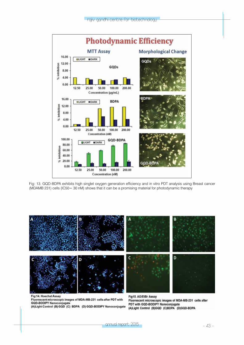
Graphene quantum dot – BODIPY nanoconjugates for PDT applications
Manu Prasad M., Saneesh Babu P.S., D. Ramaiah*, and S. Asha Nair
Collaborator: *Photochemistry and Photonics Division, National Institute for Interdisciplinary Science and Technology (NIIST), Thiruvananthapuram and CSIR-North East Institute of Science & Technology, Jorhat 785006, Assam.
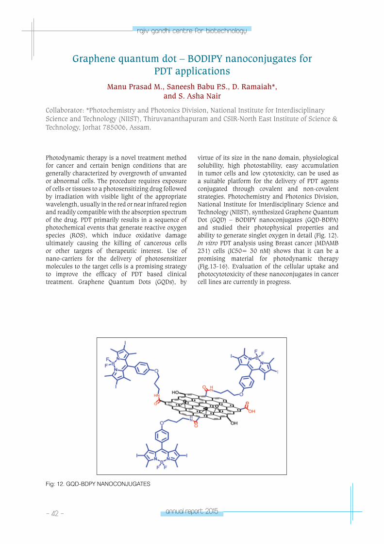
Photodynamic therapy is a novel treatment method for cancer and certain benign conditions that are generally characterized by overgrowth of unwanted or abnormal cells. The procedure requires exposure of cells or tissues to a photosensitizing drug followed by irradiation with visible light of the appropriate wavelength, usually in the red or near infrared region and readily compatible with the absorption spectrum of the drug. PDT primarily results in a sequence of photochemical events that generate reactive oxygen species (ROS), which induce oxidative damage ultimately causing the killing of cancerous cells or other targets of therapeutic interest. Use of nano-carriers for the delivery of photosensitizer molecules to the target cells is a promising strategy to improve the efficacy of PDT based clinical treatment. Graphene Quantum Dots (GQDs), by
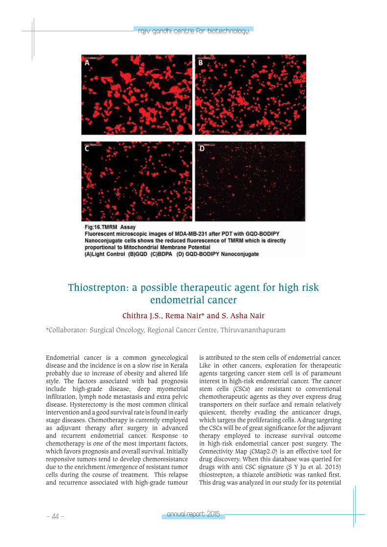
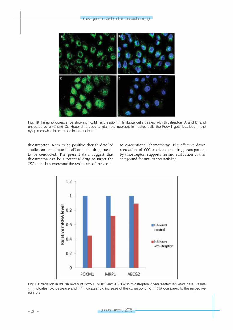
virtue of its size in the nano domain, physiological solubility, high photostability, easy accumulation in tumor cells and low cytotoxicity, can be used as a suitable platform for the delivery of PDT agents conjugated through covalent and non-covalent strategies. Photochemistry and Photonics Division, National Institute for Interdisciplinary Science and Technology (NIIST), synthesized Graphene Quantum Dot (GQD) – BODIPY nanoconjugates (GQD-BDPA) and studied their photophysical properties and ability to generate singlet oxygen in detail (Fig. 12). In vitro PDT analysis using Breast cancer (MDAMB 231) cells (IC50= 30 nM) shows that it can be a promising material for photodynamic therapy (Fig.13-16). Evaluation of the cellular uptake and photocytotoxicity of these nanoconjugates in cancer cell lines are currently in progress.
Fig: 12. GQD-BDPY NANOCONJUGATES
annual report 2015 - 43 -
rajiv gandhi centre for biotechnology
Fig: 13. GQD-BDPA exhibits high singlet oxygen generation efficiency and in vitro PDT analysis using Breast cancer (MDAMB 231) cells (IC50= 30 nM) shows that it can be a promising material for photodynamic therapy
- 44 - annual report 2015
rajiv gandhi centre for biotechnology
Thiostrepton: a possible therapeutic agent for high risk endometrial cancer
Chithra J.S., Rema Nair* and S. Asha Nair
*Collaborator: Surgical Oncology, Regional Cancer Centre, Thiruvananthapuram
Endometrial cancer is a common gynecological disease and the incidence is on a slow rise in Kerala probably due to increase of obesity and altered life style. The factors associated with bad prognosis include high-grade disease, deep myometrial infiltration, lymph node metastasis and extra pelvic disease. Hysterectomy is the most common clinical intervention and a good survival rate is found in early stage diseases. Chemotherapy is currently employed as adjuvant therapy after surgery in advanced and recurrent endometrial cancer. Response to chemotherapy is one of the most important factors, which favors prognosis and overall survival. Initially responsive tumors tend to develop chemoresistance due to the enrichment /emergence of resistant tumor cells during the course of treatment. This relapse and recurrence associated with high-grade tumour
is attributed to the stem cells of endometrial cancer. Like in other cancers, exploration for therapeutic agents targeting cancer stem cell is of paramount interest in high-risk endometrial cancer. The cancer stem cells (CSCs) are resistant to conventional chemotherapeutic agents as they over express drug transporters on their surface and remain relatively quiescent, thereby evading the anticancer drugs, which targets the proliferating cells. A drug targeting the CSCs will be of great significance for the adjuvant therapy employed to increase survival outcome in high-risk endometrial cancer post surgery. The Connectivity Map (CMap2.0) is an effective tool for drug discovery. When this database was queried for drugs with anti CSC signature (S Y Ju et al. 2015) thiostrepton, a thiazole antibiotic was ranked first. This drug was analyzed in our study for its potential

annual report 2015 - 45 -
rajiv gandhi centre for biotechnology
inhibitory effect on endometrial cancer stem cells. MTT assays were carried to identify IC50 (Inhibitory Concentration) of thiostrepton on endometrial cancer cell lines Ishikawa, Hec1A and An3Ca and this drug was found to kill tumor cells more efficiently in comparison with cisplatin, the widely used drug in clinic against endometrial cancer. Our results show that CD133 protein, which increases malignant potential of tumor cells, was found to accumulate on prolonged treatment with cisplatin (Fig:17). Treatment with cisplatin may provide a therapeutic pressure to enrich CD133 +ve stem cell phenotype. CD9 and CD13, expressed by the differentiated cell pool were reduced on treatment with cisplatin suggesting that differentiated cells (non CSC) are targeted by this drug. The expression of other stem cell markers remained unchanged on cisplatin treatment. The increasing resistance to cispaltin in endometrial cancer may be due to the inefficiency of this conventional drug to target the cancer stem cells. Thiostrepton on contrary was found to decrease the expression of CD133 and other stemness-associated genes like NANOG and MTA1, thus reduce the cancer stem cell pool (Fig:18).
Treatment with thiostrepton also reduced the mRNA level of drug resistant proteins like ABCG2, MDR1 and ABCC1, which was analyzed by Real time PCR. Thiostrepton was also found to reduce the FoxM1 protein level and induce a cytoplasmic localization of the protein that was studied by immunofluorescence (Fig:19). FoxM1 (Forkhead Box M1) is a transcription factor associated with cell proliferation and need to be localized in the nucleus for its transcriptional related activity. It is a key molecule for acquisition of EMT and CSC phenotype and is demonstrated to be involved in the proliferation of CSCs (Wang. Z et.al, 2011). Altogether, FoxM1 contributes to development of chemoresistance and in cancer progression. Thiostrepton may act by inhibiting FoxM1, thereby inhibiting the transcription of drug resistance genes as well as stem cell associated genes. Previous data on glioma showed that FoxM1 knockdown inhibit CD133 expression. Thiostrepton inhibits the FoxM1 mediated transcription, which eventually may reduce curb CSC pool expansion. The exact mechanism of action of thiostrepton and its molecular targets affecting the CSCs are yet to be determined. The synergic effect of cisplatin and
Fig: 17. Western blot analysis of CD133 and MTA1 in Ishikawa cells following cisplatin treatment for various time periods. CD133 accumulates with time and drug treatment.
Fig: 18. Western blot analysis of CD133 and MTA1 in Ishikawa cells following thiostrepton treatment for various time periods. CD133 and MTA1 levels are decreased upon thiostrepton treatment.
- 46 - annual report 2015
rajiv gandhi centre for biotechnology
thiostrepeton seem to be positive though detailed studies on combinatorial effect of the drugs needs to be conducted. The present data suggest that thiostrepton can be a potential drug to target the CSCs and thus overcome the resistance of these cells
to conventional chemotheray. The effective down regulation of CSC markers and drug transporters by thiostrepton supports further evaluation of this compound for anti cancer activity.
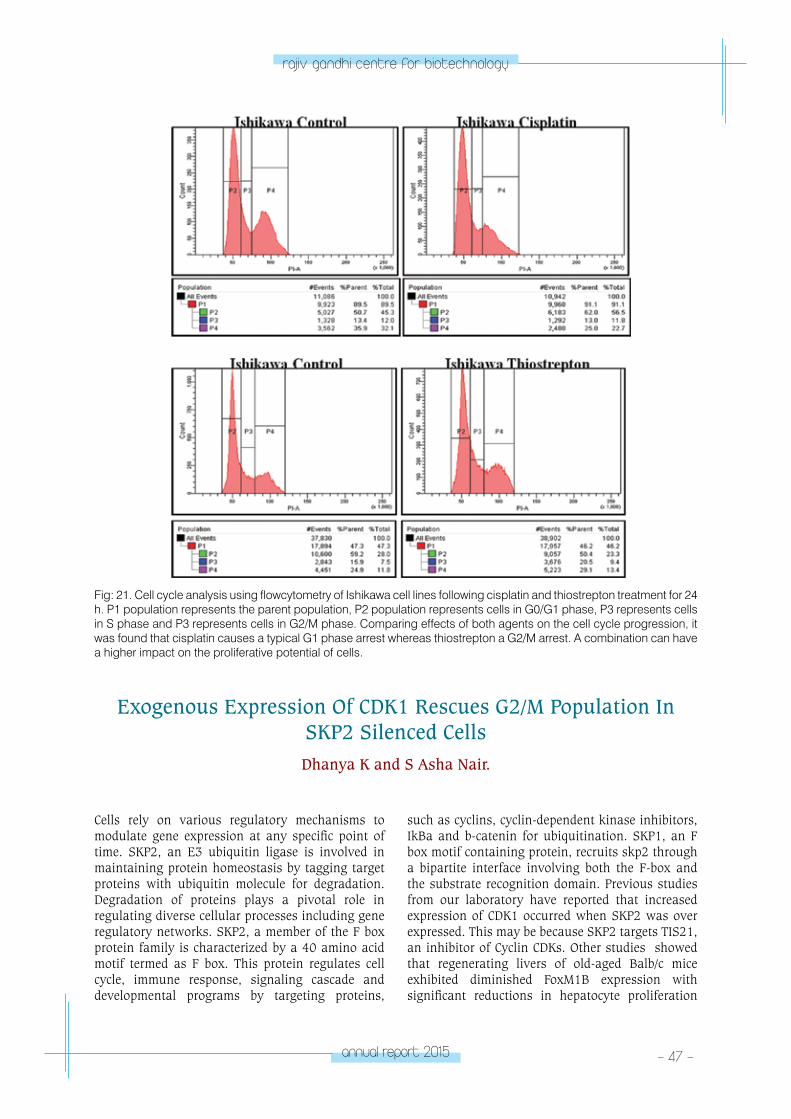
Fig: 19. Immunofluorescence showing FoxM1 expression in Ishikawa cells treated with thiostrepton (A and B) and untreated cells (C and D). Hoechst is used to stain the nucleus. In treated cells the FoxM1 gets localized in the cytoplasm while in untreated in the nucleus.
Fig: 20: Variation in mRNA levels of FoxM1, MRP1 and ABCG2 in thiostrepton (5μm) treated Ishikawa cells. Values <1 indicates fold decrease and >1 indicates fold increase of the corresponding mRNA compared to the respective controls
annual report 2015 - 47 -
rajiv gandhi centre for biotechnology
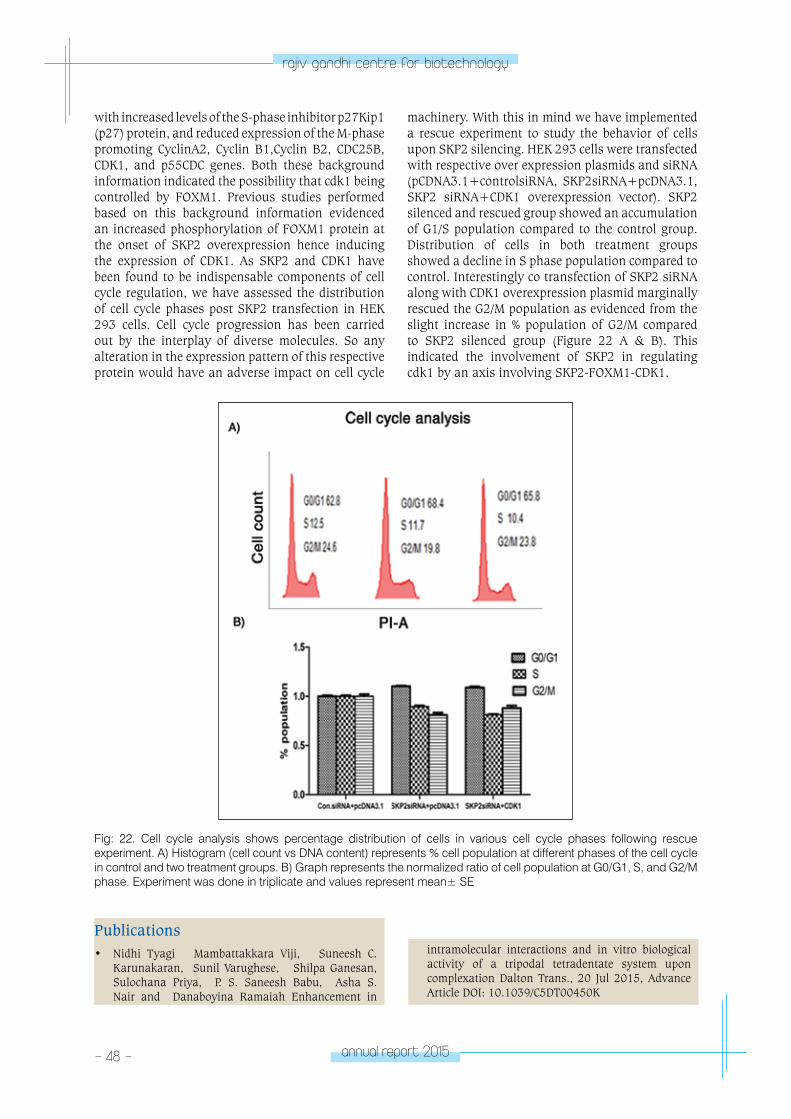
Cells rely on various regulatory mechanisms to modulate gene expression at any specific point of time. SKP2, an E3 ubiquitin ligase is involved in maintaining protein homeostasis by tagging target proteins with ubiquitin molecule for degradation. Degradation of proteins plays a pivotal role in regulating diverse cellular processes including gene regulatory networks. SKP2, a member of the F box protein family is characterized by a 40 amino acid motif termed as F box. This protein regulates cell cycle, immune response, signaling cascade and developmental programs by targeting proteins,
such as cyclins, cyclin-dependent kinase inhibitors, IkBa and b-catenin for ubiquitination. SKP1, an F box motif containing protein, recruits skp2 through a bipartite interface involving both the F-box and the substrate recognition domain. Previous studies from our laboratory have reported that increased expression of CDK1 occurred when SKP2 was over expressed. This may be because SKP2 targets TIS21, an inhibitor of Cyclin CDKs. Other studies showed that regenerating livers of old-aged Balb/c mice exhibited diminished FoxM1B expression with significant reductions in hepatocyte proliferation
Exogenous Expression Of CDK1 Rescues G2/M Population In SKP2 Silenced CellsDhanya K and S Asha Nair.
Fig: 21. Cell cycle analysis using flowcytometry of Ishikawa cell lines following cisplatin and thiostrepton treatment for 24 h. P1 population represents the parent population, P2 population represents cells in G0/G1 phase, P3 represents cells in S phase and P3 represents cells in G2/M phase. Comparing effects of both agents on the cell cycle progression, it was found that cisplatin causes a typical G1 phase arrest whereas thiostrepton a G2/M arrest. A combination can have a higher impact on the proliferative potential of cells.
- 48 - annual report 2015
rajiv gandhi centre for biotechnology
with increased levels of the S-phase inhibitor p27Kip1 (p27) protein, and reduced expression of the M-phase promoting CyclinA2, Cyclin B1,Cyclin B2, CDC25B, CDK1, and p55CDC genes. Both these background information indicated the possibility that cdk1 being controlled by FOXM1. Previous studies performed based on this background information evidenced an increased phosphorylation of FOXM1 protein at the onset of SKP2 overexpression hence inducing the expression of CDK1. As SKP2 and CDK1 have been found to be indispensable components of cell cycle regulation, we have assessed the distribution of cell cycle phases post SKP2 transfection in HEK 293 cells. Cell cycle progression has been carried out by the interplay of diverse molecules. So any alteration in the expression pattern of this respective protein would have an adverse impact on cell cycle
machinery. With this in mind we have implemented a rescue experiment to study the behavior of cells upon SKP2 silencing. HEK 293 cells were transfected with respective over expression plasmids and siRNA (pCDNA3.1+controlsiRNA, SKP2siRNA+pcDNA3.1, SKP2 siRNA+CDK1 overexpression vector). SKP2 silenced and rescued group showed an accumulation of G1/S population compared to the control group. Distribution of cells in both treatment groups showed a decline in S phase population compared to control. Interestingly co transfection of SKP2 siRNA along with CDK1 overexpression plasmid marginally rescued the G2/M population as evidenced from the slight increase in % population of G2/M compared to SKP2 silenced group (Figure 22 A & B). This indicated the involvement of SKP2 in regulating cdk1 by an axis involving SKP2-FOXM1-CDK1.
Fig: 22. Cell cycle analysis shows percentage distribution of cells in various cell cycle phases following rescue experiment. A) Histogram (cell count vs DNA content) represents % cell population at different phases of the cell cycle in control and two treatment groups. B) Graph represents the normalized ratio of cell population at G0/G1, S, and G2/M phase. Experiment was done in triplicate and values represent mean± SE
Publications• Nidhi Tyagi Mambattakkara Viji, Suneesh C.
Karunakaran, Sunil Varughese, Shilpa Ganesan, Sulochana Priya, P. S. Saneesh Babu, Asha S. Nair and Danaboyina Ramaiah Enhancement in
intramolecular interactions and in vitro biological activity of a tripodal tetradentate system upon complexation Dalton Trans., 20 Jul 2015, Advance Article DOI: 10.1039/C5DT00450K
annual report 2015 - 49 -
rajiv gandhi centre for biotechnology
• Louis WC Chow, Luis Costa, BinTean Teh, Daiang Li, Gu Feng, XinYuanGuan,Asha Nair, Li Zhu, Masahir Sugimoto, Amit Dutt, Masakazu Toi, Sudeep Gupta, Rajendra Badwe, Stefan Knapp, M. Radhakrishna Pillai and Rakesh Kumar. Cancer Genomics and Biology 2015–Meeting Report Genes and Cancer DOI:10.18632/ genesandcancer.92 2016
Conference Presentations• S. Asha Nair Tapas Pradhan, K. Chandramohan,
Manu Prasad, S.Asha Nair and M.Radhakrishna Pillai. Exploring surgical margins in Colorectal Cancer Quest for cellular and molecular prognostic markers at International Conference on Cancer Genomics and Biology, China - November 2015.
• Dhanya. K, Manu Pasad, Maharish C and S. Asha Nair. Thiostrepton, A Foxm1 and Proteasome Inhibitor Degrades Mutant P53 Via Chaperone Mediated Autophagy; International Conference on Translational Medicine and Imaging (ICTMI 2016), March 4-5, 2016, RGCB, Thiruvanthapuram.
Awards • BEST POSTER AWARD: Tapas Pradhan, K. Chandra-
mohan, Manu Prasad, S. Asha Nair and M.Radhakr-ishna Pillai. Distal surgical margins harbor Cancer stem cells and could have high prognosis importance in Colorectal cancer. International CME on Update on treatment of Colorectal cancers which was held in Trivandrum, Kerala on 7th November 2015.
EXTRAMURAL GRANTS
No Project Title Period Funding agency
1Functional significance of fork head box protein, foxm1b in cdk1 turnover - A molecular analysis.
2013-2016Department of Science & Technology, Government of India
2.Molecular mechanism of drug resistance in colorectal cancer: tumor stem like cells as unique targets in residual disease
2013-2016Department of Biotechnology, Government of India
3.Transcriptional and translational profiling of drug resistance genes following therapeutic intervention in colorectal cancer
2013-2016Council for Scientific & Industrial Research
CAnCeR ReSeA
RCH pRoGRAM
- laboratory 5CANCER
RESEARCH PROGRAM
Laboratory - 5
Priya Srinivas
Post Doctoral Fellows: Ratheeshkumar T. PhD
Technical StaffArya NagendranJithin Dev
Project fellowNeetha R.L.
PhD studentsSreelatha K.H.Satheesh Kumar S.Reshma R.S.RevathyArathi RajanGeetu Rose Varghese
Priya Srinivas has a PhD in Biochemistry
working at Regional Cancer Centre,
Thiruvananthapuram, Kerala. She joined RGCB
in the year 2000 and also worked as a Visiting
Scientist for a year from 2009 at the Department
of Biochemistry and Molecular Biology, Mayo
Clinic, Rochester, Minnesota, USA.
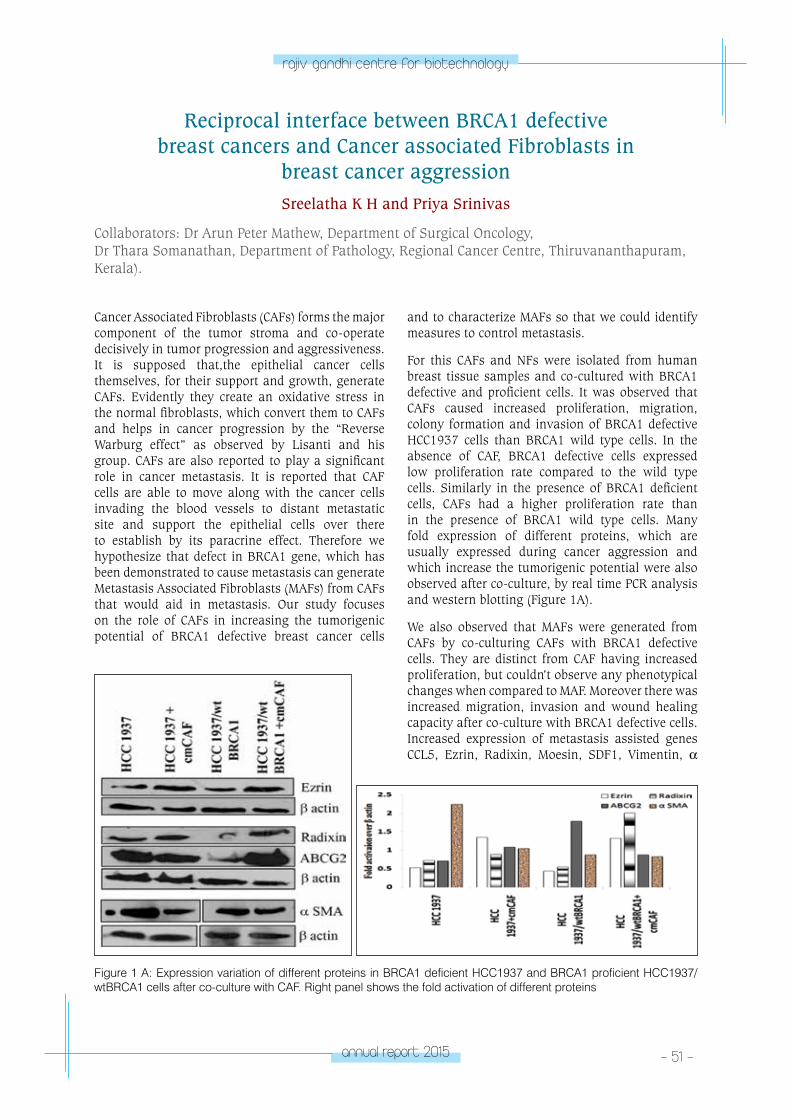
annual report 2015 - 51 -
rajiv gandhi centre for biotechnology
Cancer Associated Fibroblasts (CAFs) forms the major component of the tumor stroma and co-operate decisively in tumor progression and aggressiveness. It is supposed that,the epithelial cancer cells themselves, for their support and growth, generate CAFs. Evidently they create an oxidative stress in the normal fibroblasts, which convert them to CAFs and helps in cancer progression by the “Reverse Warburg effect” as observed by Lisanti and his group. CAFs are also reported to play a significant role in cancer metastasis. It is reported that CAF cells are able to move along with the cancer cells invading the blood vessels to distant metastatic site and support the epithelial cells over there to establish by its paracrine effect. Therefore we hypothesize that defect in BRCA1 gene, which has been demonstrated to cause metastasis can generate Metastasis Associated Fibroblasts (MAFs) from CAFs that would aid in metastasis. Our study focuses on the role of CAFs in increasing the tumorigenic potential of BRCA1 defective breast cancer cells
and to characterize MAFs so that we could identify measures to control metastasis.
For this CAFs and NFs were isolated from human breast tissue samples and co-cultured with BRCA1 defective and proficient cells. It was observed that CAFs caused increased proliferation, migration, colony formation and invasion of BRCA1 defective HCC1937 cells than BRCA1 wild type cells. In the absence of CAF, BRCA1 defective cells expressed low proliferation rate compared to the wild type cells. Similarly in the presence of BRCA1 deficient cells, CAFs had a higher proliferation rate than in the presence of BRCA1 wild type cells. Many fold expression of different proteins, which are usually expressed during cancer aggression and which increase the tumorigenic potential were also observed after co-culture, by real time PCR analysis and western blotting (Figure 1A).
We also observed that MAFs were generated from CAFs by co-culturing CAFs with BRCA1 defective cells. They are distinct from CAF having increased proliferation, but couldn’t observe any phenotypical changes when compared to MAF. Moreover there was increased migration, invasion and wound healing capacity after co-culture with BRCA1 defective cells. Increased expression of metastasis assisted genes CCL5, Ezrin, Radixin, Moesin, SDF1, Vimentin, α
Reciprocal interface between BRCA1 defective breast cancers and Cancer associated Fibroblasts in
breast cancer aggressionSreelatha K H and Priya Srinivas
Collaborators: Dr Arun Peter Mathew, Department of Surgical Oncology, Dr Thara Somanathan, Department of Pathology, Regional Cancer Centre, Thiruvananthapuram, Kerala).
Figure 1 A: Expression variation of different proteins in BRCA1 deficient HCC1937 and BRCA1 proficient HCC1937/wtBRCA1 cells after co-culture with CAF. Right panel shows the fold activation of different proteins
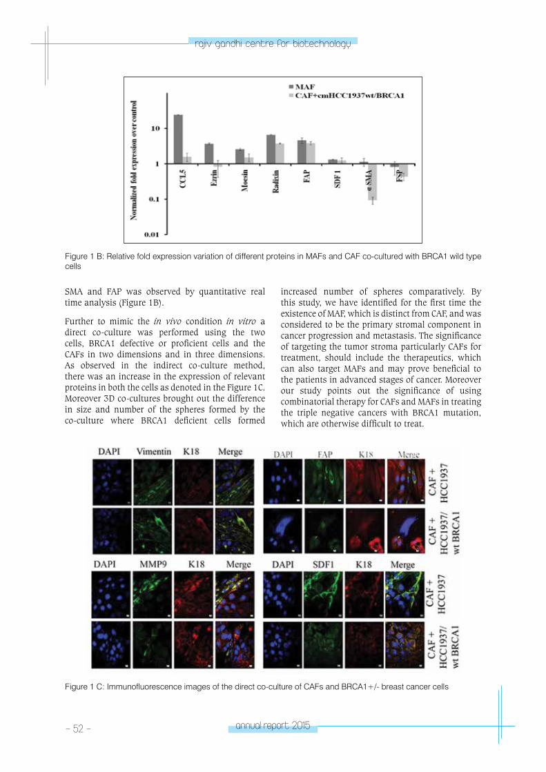
- 52 - annual report 2015
rajiv gandhi centre for biotechnology
SMA and FAP was observed by quantitative real time analysis (Figure 1B).
Further to mimic the in vivo condition in vitro a direct co-culture was performed using the two cells, BRCA1 defective or proficient cells and the CAFs in two dimensions and in three dimensions. As observed in the indirect co-culture method, there was an increase in the expression of relevant proteins in both the cells as denoted in the Figure 1C. Moreover 3D co-cultures brought out the difference in size and number of the spheres formed by the co-culture where BRCA1 deficient cells formed
increased number of spheres comparatively. By this study, we have identified for the first time the existence of MAF, which is distinct from CAF, and was considered to be the primary stromal component in cancer progression and metastasis. The significance of targeting the tumor stroma particularly CAFs for treatment, should include the therapeutics, which can also target MAFs and may prove beneficial to the patients in advanced stages of cancer. Moreover our study points out the significance of using combinatorial therapy for CAFs and MAFs in treating the triple negative cancers with BRCA1 mutation, which are otherwise difficult to treat.
Figure 1 B: Relative fold expression variation of different proteins in MAFs and CAF co-cultured with BRCA1 wild type cells
Figure 1 C: Immunofluorescence images of the direct co-culture of CAFs and BRCA1+/- breast cancer cells
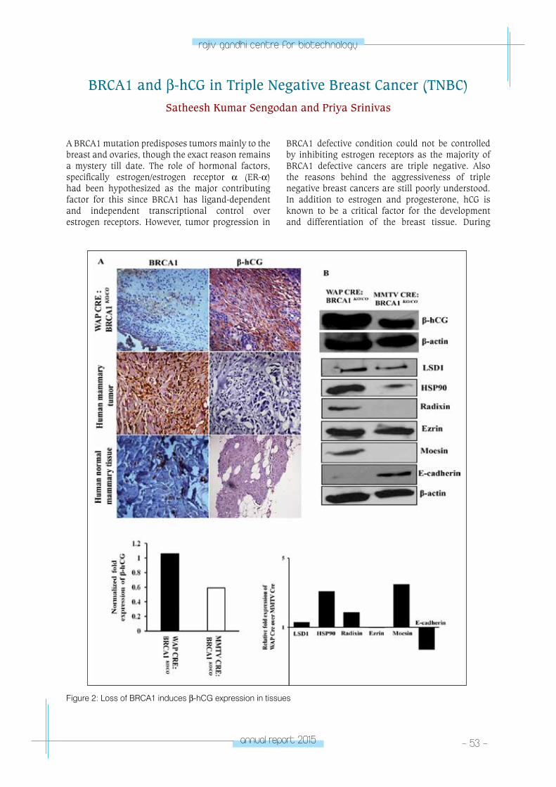
annual report 2015 - 53 -
rajiv gandhi centre for biotechnology
A BRCA1 mutation predisposes tumors mainly to the breast and ovaries, though the exact reason remains a mystery till date. The role of hormonal factors, specifically estrogen/estrogen receptor α (ER-α) had been hypothesized as the major contributing factor for this since BRCA1 has ligand-dependent and independent transcriptional control over estrogen receptors. However, tumor progression in
BRCA1 defective condition could not be controlled by inhibiting estrogen receptors as the majority of BRCA1 defective cancers are triple negative. Also the reasons behind the aggressiveness of triple negative breast cancers are still poorly understood. In addition to estrogen and progesterone, hCG is known to be a critical factor for the development and differentiation of the breast tissue. During
BRCA1 and β-hCG in Triple Negative Breast Cancer (TNBC)Satheesh Kumar Sengodan and Priya Srinivas
Figure 2: Loss of BRCA1 induces β-hCG expression in tissues
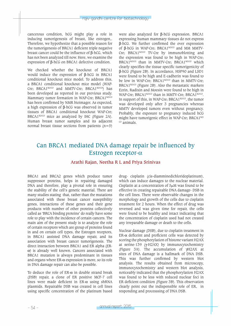
- 54 - annual report 2015
rajiv gandhi centre for biotechnology
cancerous condition, hCG might play a role in inducing tumorigenesis of breast, like estrogen,. Therefore, we hypothesize that a possible reason for the tumorigenesis of BRCA1 deficient triple negative breast cancer could be the influence of β-hCG, which has not been analyzed till now. Here, we examine the expression of β-hCG on BRCA1 defective condition.
We checked whether the knockout of BRCA1 would induce the expression of β-hCG in BRCA1 conditional knockout mice model. To address this, a BRCA1 conditional knockout mice model (WAP-Cre; BRCA1KO/CO and MMTV-Cre; BRCA1KO/CO) has been developed as reported in our previous study. Mammary tumor formation in WAP-Cre; BRCA1KO/CO has been confirmed by NMR bioimager. As expected, a high expression of β-hCG was observed in tumor tissues of BRCA1 conditional knockout WAP-Cre; BRCA1KO/CO mice as analyzed by IHC (Figure 2A). Human breast tumor samples and its adjacent normal breast tissue sections from patients (n=5)
were also analyzed for β-hCG expression. BRCA1 expressing human mammary tissues do not express β-hCG. We further confirmed the over expression of β-hCG in WAP-Cre; BRCA1KO/CO and MM MMTV-Cre; BRCA1KO/CO TV-Cre by immunoblotting and the expression was found to be high in WAP-Cre; BRCA1KO/CO than in MMTV-Cre; BRCA1KO/CO which clearly specifies the tissue specific tumorigenicity of β-hCG (Figure 2B). In accordance, HSP90 and LSD1 were found to be high and E-cadherin was found to be low in WAP-Cre; BRCA1KO/CO than in MMTV-Cre; BRCA1KO/CO (Figure 2B). Also the metastatic markers Ezrin, Radixin and Moesin were found to be high in WAP-Cre; BRCA1KO/CO than in MMTV-Cre; BRCA1KO/CO. In support of this, in WAP-Cre; BRCA1KO/CO, the tumor was developed only after 3 pregnancies whereas MMTV developed tumors even without pregnancy. Probably, the exposure to pregnancy induced hCG might have tumorigenic effect in WAP-Cre; BRCA1KO/
CO animals.
Can BRCA1 mediated DNA damage repair be influenced by Estrogen receptor-α
Arathi Rajan, Neetha R L and Priya Srinivas
BRCA1 and BRCA2 genes which produce tumor suppressor proteins, helps in repairing damaged DNA and therefore, play a pivotal role in ensuring the stability of the cell’s genetic material. There are many studies stating that, rather than the mutations associated with these breast cancer suseptibility genes, interactions of these genes and their gene products with number of other proteins collectively called as ‘BRCA binding proteins’ do really have some role to play with the incidence of certain cancers. The main aim of the present study is to analyze the role of certain receptors which are group of proteins found in and on certain cell types, the Estrogen receptors, in BRCA1 assisted DNA damage repair, and its association with breast cancer tumorigenesis. The direct interaction between BRCA1 and ER alpha (ER-α) is already well known. Cancers associated with BRCA1 mutation is always predominant in tissues and organs where ER-α expression is more, so its role in DNA damage repair can also be possible.
To deduce the role of ER-α in double strand break (DSB) repair, a clone of ER positive MCF-7 cell lines were made deficient in ER-α using shRNA plasmids. Repairable DSB was created in cell lines using specific concentration of the platinum based
drug- cisplatin (cis-diamminedichloridoplatinum), which can induce damages to the nuclear material. Cisplatin at a concentration of 5µM was found to be effective in creating repairable DNA damage- DSB in the cell lines. There were observable changes in the morphology and growth of the cells due to cisplatin treatment for 2 hours. When the effect of drug was reversed and was given time for repair, the cells were found to be healthy and intact indicating that the concentration of cisplatin used had not created any irreparable damage or death to the cells.
Nuclear damage (DSB), due to cisplatin treatment in ER-α deficeint and proficient cells was detected by scoring the phosphorylation of histone variant H2AX at serine-139 (γ-H2AX) by immunocytochemistry (Figure 3A). The accumulation of γH2AX at sites of DNA damage is a hallmark of DNA DSB. This was further confirmed by western blot analysis. The results obtained from microscopy, immunocytochemistry and western blot analysis, noticeably indicated that the phosphorylation H2AX was found to be less with reduced nuclear foci in ER deficient condition (Figure 3B). This observation clearly point out the indispensible role of ER, in responding and processsing of DNA DSB.
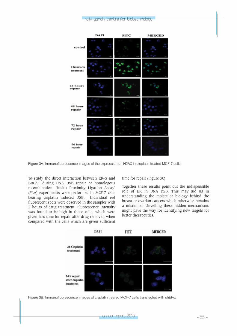
annual report 2015 - 55 -
rajiv gandhi centre for biotechnology
Figure 3A: Immunofluorescence images of the expression of γH2AX in cisplatin treated MCF-7 cells
Figure 3B: Immunofluorescence images of cisplatin treated MCF-7 cells transfected with shERα.
To study the direct interaction between ER-α and BRCA1 during DNA DSB repair or homologous recombination, ‘insitu Proximity Ligation Assay’ (PLA) experiments were performed in MCF-7 cells bearing cisplatin induced DSB. Individual red fluorescent spots were observed in the samples with 2 hours of drug treatment. Fluorescence intensity was found to be high in those cells, which were given less time for repair after drug removal, when compared with the cells which are given sufficient
time for repair (Figure 3C).
Together these results point out the indispensible role of ER in DNA DSB. This may aid us in understanding the molecular biology behind the breast or ovarian cancers which otherwise remains a misnomer. Unveiling these hidden mechanisms might pave the way for identifying new targets for better therapeutics.
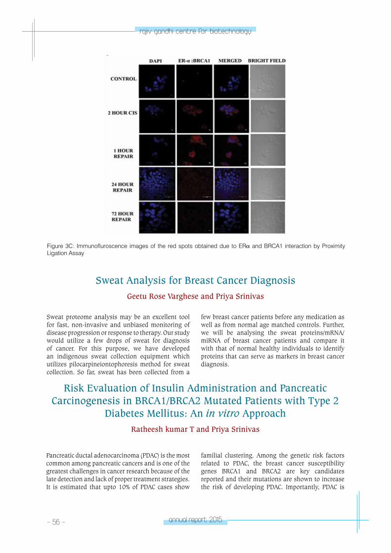
- 56 - annual report 2015
rajiv gandhi centre for biotechnology
Figure 3C: Immunofluroscence images of the red spots obtained due to ERα and BRCA1 interaction by Proximity Ligation Assay
Sweat proteome analysis may be an excellent tool for fast, non-invasive and unbiased monitoring of disease progression or response to therapy. Our study would utilize a few drops of sweat for diagnosis of cancer. For this purpose, we have developed an indigenous sweat collection equipment which utilizes pilocarpineiontophoresis method for sweat collection. So far, sweat has been collected from a
few breast cancer patients before any medication as well as from normal age matched controls. Further, we will be analysing the sweat proteins/mRNA/miRNA of breast cancer patients and compare it with that of normal healthy individuals to identify proteins that can serve as markers in breast cancer diagnosis.
Risk Evaluation of Insulin Administration and Pancreatic Carcinogenesis in BRCA1/BRCA2 Mutated Patients with Type 2
Diabetes Mellitus: An in vitro ApproachRatheesh kumar T and Priya Srinivas
Pancreatic ductal adenocarcinoma (PDAC) is the most common among pancreatic cancers and is one of the greatest challenges in cancer research because of the late detection and lack of proper treatment strategies. It is estimated that upto 10% of PDAC cases show
familial clustering. Among the genetic risk factors related to PDAC, the breast cancer susceptibility genes BRCA1 and BRCA2 are key candidates reported and their mutations are shown to increase the risk of developing PDAC. Importantly, PDAC is
Sweat Analysis for Breast Cancer DiagnosisGeetu Rose Varghese and Priya Srinivas
annual report 2015 - 57 -
rajiv gandhi centre for biotechnology
considered to be the third most common cancer associated BRCA1/2 mutation carriers. In addition to BRCA1/BRCA2 mutations, types 2 diabetes mellitus (T2DM) and glucose lowering therapies also poses an increased risk for pancreatic cancer development. A possible mechanism behind T2DM and PDAC in these patients might be the insulin administration as part of their diabetes treatment and subsequent cross talk between insulin signaling and mitogenic pathways.
It has been reported that insulin, in addition to its affinity for insulin receptor (IR), could also bind to IGF-1R (insulin like growth factor) and the hybrid receptor formed by IR and IGF-1R. Most interestingly, BRCA1 is known to negatively regulate IGF-1R levels at the transcriptional level in breast, uterine, prostate and ovarian cancers thereby activating mitogenic signaling
pathways. This negative regulation of IGF-1R in PDAC is unknown. Even though, there are enough evidences to support the notion that BRCA1 could regulate IGF-1R, till date there are no direct evidences to prove the effect of insulin in BRCA1/BRCA2 mutated diabetic patients for the possible onset of pancreatic carcinogenesis.
We hypothesize that, use of insulin in diabetic cases of BRCA1 or BRCA2 mutation carriers are at increased risk for developing PDAC. We propose that when BRCA1 or BRCA2 is mutated/non functional in insulin sensitive mutated pancreatic cells, the IGF-1R levels might go unchecked leading to up-regulation of metabolic signaling pathways and subsequent carcinogenesis. This will be demonstrated by an in vitro approach using a normal BRCA1/BRCA2 knockout pancreatic ductal cell line grown in the presence/absence of insulin.
β-hCG and BRCA1 in Gestational Trophoblastic DiseasesRevathy, Krishnapriya RS, Sreevidya P S and Priya Srinivas
Collaborators: Dr. Nirmala C, Professor, Department of Obstetrics & Gynecology, SAT Hospital, Medical College, Thiruvananthapuram. Dr. Jayshree V. Vaman, Additional Professor, Department of Obstetrics & Gynecology, T D Medical College, Alappuzha. Dr. Santha Sadasivan, Professor, Department of Pathology, Medical College, Thiruvananthapuram Dr. Aysha P V, Senior Gynecologist, PRS Hospital, Thiruvananthapuram. Dr. Balaraman Nair, Chief Pathologist, DDRC SRL Diagnostics, Thiruvananthapuram. Dr Anil Kumar T V, Sree Chitra Tirunal Institute for Medical Sciences & Technology.
Gestational Trophoblastic Diseases (GTD), includes a group of diseases derived from placental trophoblasts and is characterized by the abnormally elevated levels of β-hCG. The disease is of high incidence in South-East Asia especially India, with a high incidence rate of 5.1/1000 deliveries in Kerala (Sekharan et.al, 2006 & Lybol et.al, 2011). Two of the cohort studies show antagonistic reports about the role of β-hCG exposure to the breast cancer incidence, where in hCG exposure during hydatidiform moles were shown to increase the breast cancer risk (Erlandssonet. al, 2000) and also showing that it decreases the breast cancer risk (Gudipudiet. al, 2008). But till date, no studies have been done to analyze this correlation at the molecular level. Also, Though BRCA1 plays a significant role in DNA repair pathways; its specific role in cancer development especially in breast/ovarian cancers
is still not known completely. Various positive and negative modifiers of BRCA1 have been identified, the best-characterized stimulant being estrogen, thus controlling proliferation and malignancies of hormone regulated tissues like breast, ovary and prostate. In this study, we aim to analyze a possible link for the β-hCG expression in GTD to the breast cancer incidence at the molecular level by trying to look into the expression levels of BRCA1, which is a prime tumor suppressor gene associated with the breast cancers. Also, β-hCG being an important player in the scene of very many cancers, makes it relevant to study its interactome with the major tumor suppressors/ oncogenes/cell cycle regulators. With all the above factors into consideration, GTD was selected the ideal system for our study. Unraveling the etiopathology of GTD, would help in the prediction of malignant potential and recurrence
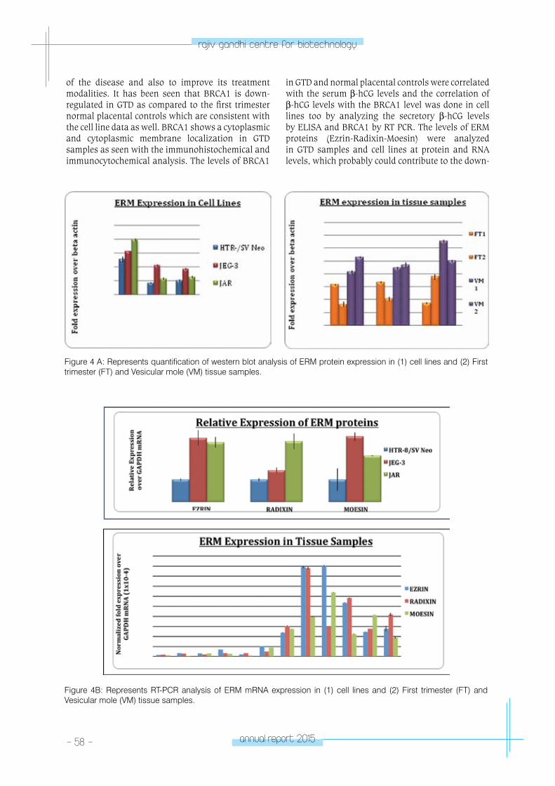
- 58 - annual report 2015
rajiv gandhi centre for biotechnology
of the disease and also to improve its treatment modalities. It has been seen that BRCA1 is down-regulated in GTD as compared to the first trimester normal placental controls which are consistent with the cell line data as well. BRCA1 shows a cytoplasmic and cytoplasmic membrane localization in GTD samples as seen with the immunohistochemical and immunocytochemical analysis. The levels of BRCA1
in GTD and normal placental controls were correlated with the serum β-hCG levels and the correlation of β-hCG levels with the BRCA1 level was done in cell lines too by analyzing the secretory β-hCG levels by ELISA and BRCA1 by RT PCR. The levels of ERM proteins (Ezrin-Radixin-Moesin) were analyzed in GTD samples and cell lines at protein and RNA levels, which probably could contribute to the down-
Figure 4 A: Represents quantification of western blot analysis of ERM protein expression in (1) cell lines and (2) First trimester (FT) and Vesicular mole (VM) tissue samples.
Figure 4B: Represents RT-PCR analysis of ERM mRNA expression in (1) cell lines and (2) First trimester (FT) and Vesicular mole (VM) tissue samples.
annual report 2015 - 59 -
rajiv gandhi centre for biotechnology
regulation of BRCA1 levels. ERM levels showed an over-expression, which might be a possible reason for the over-expression of BRCA1 in the system (Figure 4A & B). The cytoplasmic localization of the BRCA1 protein in placenta and GTD could also be contributing to the increased proliferation rate of the trophoblastic cells so present in both the tissue samples. Also, a positive interaction was shown between BRCA1 and β-hCG as evident by the proximity ligation assay. Further analysis has
to be done to look into the possible reason for their interaction, as it is restricted to the tumor cells. This study aiming to correlate BRCA1 and β-hCG in cancers would improvise the treatment modalities not only for GTD but also for BRCA1 related cancers. Also new strategies can be developed to identify the high-risk group or recurrent GTD cases/BRCA1 related cancers, which would be helpful to the society.
Publications• Veena Somasundaram, Sreelatha K. Hemalatha,
Krishnendu Pal, Sutapa Sinha, Asha S. Nair, Debabrata Mukhopadhyay, Priya Srinivas. Selective mode of action of plumbagin through BRCA1 deficient breast cancer stem cells BMC Cancer, 2016 May 26;16(1):336. doi: 10.1186/s12885-016-2372-4.
• Veena Somasundaram, Revathy Nathan, Sreelatha K.H., Satheesh Kumar S., Priya Srinivas ‘ “Nitric Oxide and Reactive Oxygen Species: Clues to target Oxidative Damage Repair Defective Breast Cancers” accepted for publication. Critical Reviews in Oncology/Hematology. 2016 May;101:184-92.
• Rakesh S., Jerald M.K., Jedy J., Veena S., Sreelatha K.H., Satheesh K.S., Revathy, Anil kumar T. & Priya Srinivas, Increased sensitivity of BRCA defective triple negative breast tumors to plumbagin through induction of DNA Double Strand Breaks (DSB). Scientific Reports. 2016 May 25;6:26631. doi: 10.1038/srep26631.
• Reshma R.S., Sreelatha K.H., Veena Somasundaram, Satheesh Kumar S., Revathy, Rakesh Sathish Nair, Priya Srinivas. Plumbagin, a naphthaquinone derivative induces apoptosis and in BRCA 1/2 defective castrate resistant prostate cancer cells as well as prostate cancer stem-like cells. Pharmacological Research Volume 105, March 2016, Pages 134–145.
• P.M. Aswathy, P.S. Jairani, Joe Verghese, Srinivas Gopala, Priya Srinivas, P.S. Mathuranath. Progranulin mutation analysis: Identification of one novel mutation in exon 12 associated with frontotemporal dementia. Neurobiology of Aging. 2015, Dec 8. doi: 10.1016/j.neurobiolaging. 2015.11.026.
• Rakesh S. Nair; Mini Kuriakose; Veena Somasunda-ram; Vinesh Shenoi; M.R. Prathapachandra Kurup; Priya Srinivas, The Molecular Response of a Novel Vanadium Complex of Nicotinoyl Hydrazone in Cer-vical Cancers - A Possible Interference with HPV On-cogenic Markers. Life Sciences 116 (2014) 90–97. IF. 2.538.
Book Chapter• Satheesh Kumar S., Sreelatha K.H., Revathy Nadhan,
and Priya Srinivas. Role of BRCA1 in Breast Cancer Metastasis. Charpter 1, In the book “Gynecologic Cancers – Basic Sciences, Clinical and Therapeutic Perspectives “, ISBN 978-953-51-4278-2, In Tech, Europe, March 2nd, 2016.
Conference presentations• Revathy Nadhan, Krishnapriya R.S., Nirmala C.,
Jayashree V. Vaman, Santha Sadasivan, Aysha P.V., Sreevidya P.S., Satheesh Kumar S., Sreelatha K.H., Balaraman Nair M. , Anilkumar T.V. and Priya Srinivas. Gestational Trophoblastic Diseases – BRCA1 – Breast Cancer: An Enthralling Trio. Poster presented at Fourth AACR International Conference on Frontiers in Basic Cancer Research, Pennsylvania Convention Centre, Philadelphia, Pennsylvania, USA, 23rd to 26th October 2015.
• Satheesh Kumar S, Sreelatha K.H., Revathy Nadhan and Priya Srinivas. β-hCG: The key culprit in BRCA1 mutated triple negative breast cancer. Poster presented at Fourth AACR International Conference on Frontiers in Basic Cancer Research, Pennsylvania Convention Centre, Philadelphia, Pennsylvania, USA, 23rd to 26th October 2015.
• Sreelatha K.H., Arun Peter Mathew, Thara Somanthan, Jithin Dev, Veena Somasundaram, Reshma R.S., Satheesh Kumar S., Revathy, Priya Srinivas. A study on the decisive role of activated fibroblast, Cancer Associated Fibroblast (CAFs) cells on triple negative breast cancer cells. Paper presented on Global Cancer Summit held at J.N.Tata Auditorium, Indian Institute of Science, Bengaluru from November 18-20th, 2015.
• Revathynadhan, Nirmala C., Jayashree V. Vaman, Krishnapriya R.S., SanthaSadasivan, Aysha P.V., Sreevidya P.S., Satheesh Kumar S., Sreelatha K.H., Balaraman Nair M., Anilkumar T.V., Priya srinivas. An insight into the role of ERM proteins in the etiopathology of trophoblastic tumors. Paper presented on Global Cancer Summit held at J.N.Tata Auditorium, Indian Institute of Science, Bengaluru from November 18-20th, 2015.
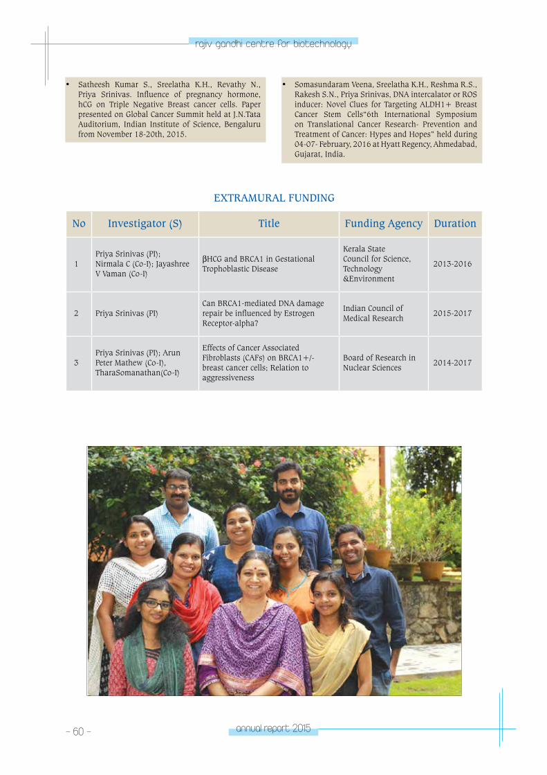
- 60 - annual report 2015
rajiv gandhi centre for biotechnology
• Satheesh Kumar S., Sreelatha K.H., Revathy N., Priya Srinivas. Influence of pregnancy hormone, hCG on Triple Negative Breast cancer cells. Paper presented on Global Cancer Summit held at J.N.Tata Auditorium, Indian Institute of Science, Bengaluru from November 18-20th, 2015.
• Somasundaram Veena, Sreelatha K.H., Reshma R.S., Rakesh S.N., Priya Srinivas, DNA intercalator or ROS inducer: Novel Clues for Targeting ALDH1+ Breast Cancer Stem Cells“6th International Symposium on Translational Cancer Research- Prevention and Treatment of Cancer: Hypes and Hopes” held during 04-07- February, 2016 at Hyatt Regency, Ahmedabad, Gujarat, India.
EXTRAMURAL FUNDING
No Investigator (S) Title Funding Agency Duration
1Priya Srinivas (PI); Nirmala C (Co-I); Jayashree V Vaman (Co-I)
βHCG and BRCA1 in Gestational Trophoblastic Disease
Kerala State Council for Science, Technology &Environment
2013-2016
2 Priya Srinivas (PI)Can BRCA1-mediated DNA damage repair be influenced by Estrogen Receptor-alpha?
Indian Council of Medical Research
2015-2017
3Priya Srinivas (PI); Arun Peter Mathew (Co-I), TharaSomanathan(Co-I)
Effects of Cancer Associated Fibroblasts (CAFs) on BRCA1+/- breast cancer cells; Relation to aggressiveness
Board of Research in Nuclear Sciences
2014-2017
CAnCeR ReSeA
RCH pRoGRAM
- laboratory 6CANCER
RESEARCH PROGRAM
Laboratory - 6
S Sreeja
Ph.D Students:Jazir H. Parvathy M. Lakshmi M.L.Juberiya M.A.Vini R.
Research Fellows Hima S.Viji R.
Technical Personnel Savitha H.
Sreeja received PhD in Biotechnology from
University of Kerala for work on molecular
mechanisms of estrogen action and joined
RGCB in 2001.
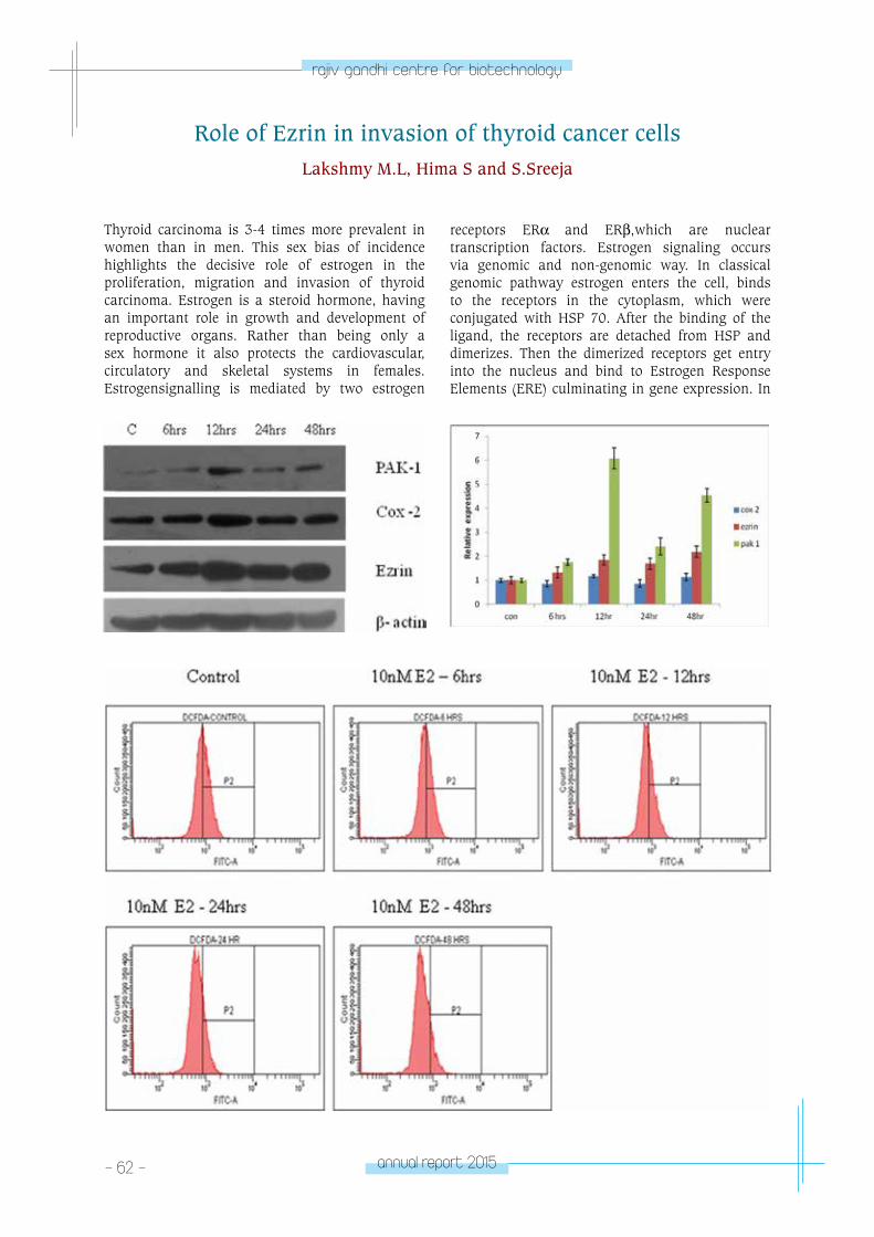
- 62 - annual report 2015
rajiv gandhi centre for biotechnology
Thyroid carcinoma is 3-4 times more prevalent in women than in men. This sex bias of incidence highlights the decisive role of estrogen in the proliferation, migration and invasion of thyroid carcinoma. Estrogen is a steroid hormone, having an important role in growth and development of reproductive organs. Rather than being only a sex hormone it also protects the cardiovascular, circulatory and skeletal systems in females. Estrogensignalling is mediated by two estrogen
receptors ERα and ERβ,which are nuclear transcription factors. Estrogen signaling occurs via genomic and non-genomic way. In classical genomic pathway estrogen enters the cell, binds to the receptors in the cytoplasm, which were conjugated with HSP 70. After the binding of the ligand, the receptors are detached from HSP and dimerizes. Then the dimerized receptors get entry into the nucleus and bind to Estrogen Response Elements (ERE) culminating in gene expression. In
Role of Ezrin in invasion of thyroid cancer cells Lakshmy M.L, Hima S and S.Sreeja
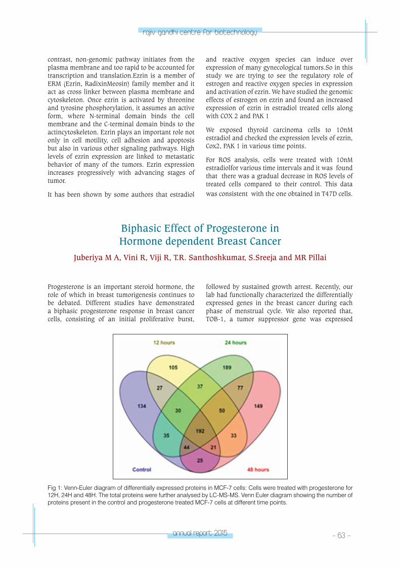
annual report 2015 - 63 -
rajiv gandhi centre for biotechnology
Biphasic Effect of Progesterone in Hormone dependent Breast Cancer
Juberiya M A, Vini R, Viji R, T.R. Santhoshkumar, S.Sreeja and MR Pillai
Progesterone is an important steroid hormone, the role of which in breast tumorigenesis continues to be debated. Different studies have demonstrated a biphasic progesterone response in breast cancer cells, consisting of an initial proliferative burst,
followed by sustained growth arrest. Recently, our lab had functionally characterized the differentially expressed genes in the breast cancer during each phase of menstrual cycle. We also reported that, TOB-1, a tumor suppressor gene was expressed
contrast, non-genomic pathway initiates from the plasma membrane and too rapid to be accounted for transcription and translation.Ezrin is a member of ERM (Ezrin, RadixinMeosin) family member and it act as cross linker between plasma membrane and cytoskeleton. Once ezrin is activated by threonine and tyrosine phosphorylation, it assumes an active form, where N-terminal domain binds the cell membrane and the C-terminal domain binds to the actincytoskeleton. Ezrin plays an important role not only in cell motility, cell adhesion and apoptosis but also in various other signaling pathways. High levels of ezrin expression are linked to metastatic behavior of many of the tumors. Ezrin expression increases progressively with advancing stages of tumor.
It has been shown by some authors that estradiol
and reactive oxygen species can induce over expression of many gynecological tumors.So in this study we are trying to see the regulatory role of estrogen and reactive oxygen species in expression and activation of ezrin. We have studied the genomic effects of estrogen on ezrin and found an increased expression of ezrin in estradiol treated cells along with COX 2 and PAK 1
We exposed thyroid carcinoma cells to 10nM estradiol and checked the expression levels of ezrin, Cox2, PAK 1 in various time points.
For ROS analysis, cells were treated with 10nM estradiolfor various time intervals and it was found that there was a gradual decrease in ROS levels of treated cells compared to their control. This data was consistent with the one obtained in T47D cells.
Fig 1: Venn-Euler diagram of differentially expressed proteins in MCF-7 cells: Cells were treated with progesterone for 12H, 24H and 48H. The total proteins were further analysed by LC-MS-MS. Venn Euler diagram showing the number of proteins present in the control and progesterone treated MCF-7 cells at different time points.
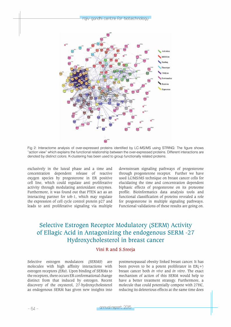
- 64 - annual report 2015
rajiv gandhi centre for biotechnology
Fig 2: Interactome analysis of over-expressed proteins identified by LC-MS/MS using STRING: The figure shows “action view” which explains the functional relationship between the over-expressed proteins. Different interactions are denoted by distinct colors. K-clustering has been used to group functionally related proteins.
Selective Estrogen Receptor Modulatory (SERM) Activity of Ellagic Acid in Antagonizing the endogenous SERM -27
Hydroxycholesterol in breast cancerVini R and S.Sreeja
exclusively in the luteal phase and a time and concentration dependent release of reactive oxygen species by progesterone in ER positive cell line, which could regulate anti proliferative activity through modulating antioxidant enzymes. Furthermore, it was found out that PTEN act as an interacting partner for tob-1, which may regulate the expression of cell cycle control protein p27 and leads to anti proliferative signaling via multiple
downstream signaling pathways of progesterone through progesterone receptor. Further we have used LC/MS/MS technique on breast cancer cells for elucidating the time and concentration dependent biphasic effects of progesterone on its proteome profile. Bioinformatics data analysis tools and functional classification of proteins revealed a role for progesterone in multiple signaling pathways. Functional validations of these results are going on.
Selective estrogen modulators (SERMS) are molecules with high affinity interactions with estrogen receptors (ERs). Upon binding of SERMs to the receptors, there occurs ER conformational change distinct from that induced by estrogen. Recent discovery of the oxysterol, 27-hydroxycholesterol as endogenous SERM has given new insights into
postmenopausal obesity linked breast cancer. It has been proven to be a potent proliferator in ER(+) breast cancer both in vivo and in vitro. The exact mechanism of action of this SERM would help to have a better treatment strategy. Furthermore, a molecule that could potentially compete with 27HC, reducing its deleterious effects at the same time does
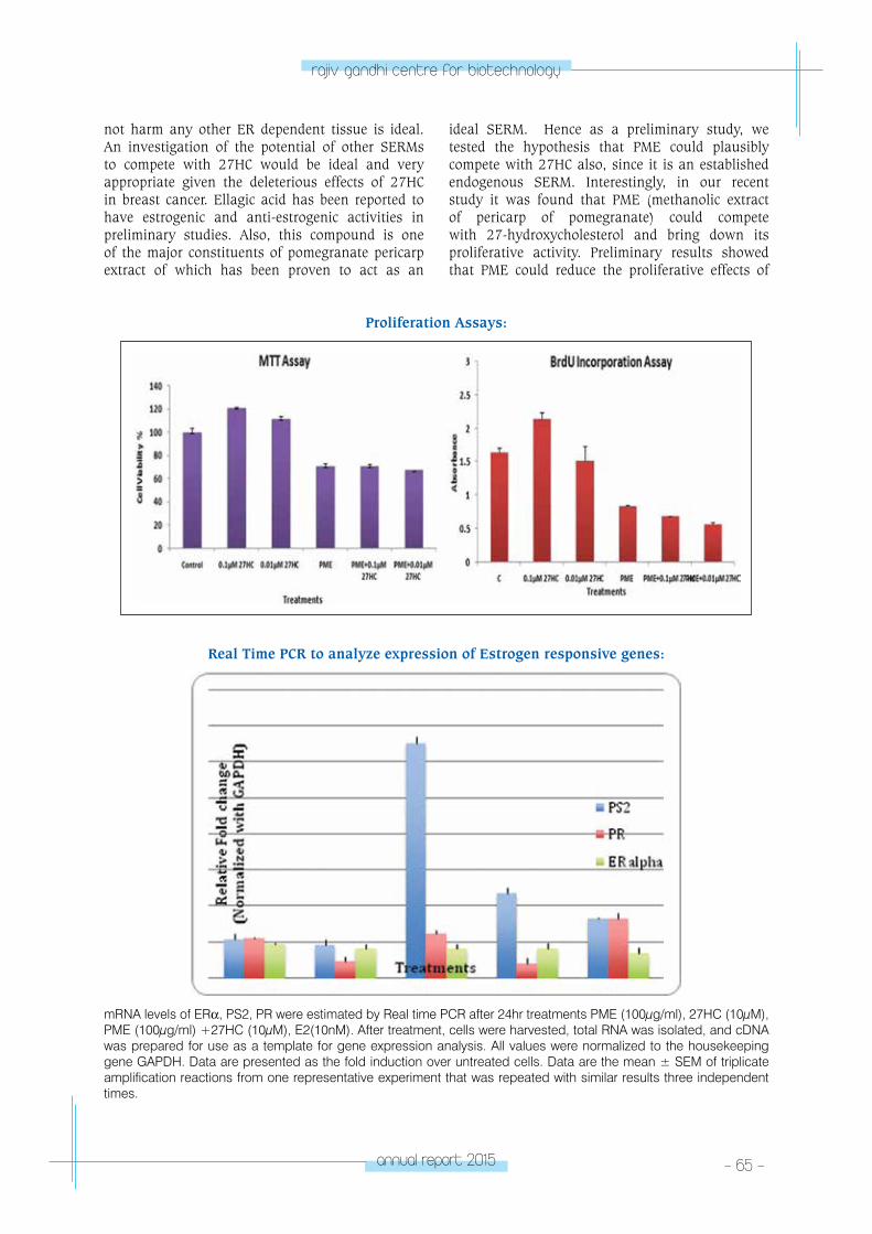
annual report 2015 - 65 -
rajiv gandhi centre for biotechnology
not harm any other ER dependent tissue is ideal. An investigation of the potential of other SERMs to compete with 27HC would be ideal and very appropriate given the deleterious effects of 27HC in breast cancer. Ellagic acid has been reported to have estrogenic and anti-estrogenic activities in preliminary studies. Also, this compound is one of the major constituents of pomegranate pericarp extract of which has been proven to act as an
ideal SERM. Hence as a preliminary study, we tested the hypothesis that PME could plausibly compete with 27HC also, since it is an established endogenous SERM. Interestingly, in our recent study it was found that PME (methanolic extract of pericarp of pomegranate) could compete with 27-hydroxycholesterol and bring down its proliferative activity. Preliminary results showed that PME could reduce the proliferative effects of
Proliferation Assays:
mRNA levels of ERα, PS2, PR were estimated by Real time PCR after 24hr treatments PME (100μg/ml), 27HC (10μM), PME (100μg/ml) +27HC (10μM), E2(10nM). After treatment, cells were harvested, total RNA was isolated, and cDNA was prepared for use as a template for gene expression analysis. All values were normalized to the housekeeping gene GAPDH. Data are presented as the fold induction over untreated cells. Data are the mean ± SEM of triplicate amplification reactions from one representative experiment that was repeated with similar results three independent times.
Real Time PCR to analyze expression of Estrogen responsive genes:

- 66 - annual report 2015
rajiv gandhi centre for biotechnology
Publications • Juberiya M. A, Hima S, Indhu H, Sreeja S, Jem P,
S.Sreejaand M. R.Pillai ,Progesterone regulates the proliferation of breast cancer cells - invitro evidence”, Drug design, Development and Therapy, Vol:9,5987—5999,http://dx.doi.org/10.2147/DDDT. .S89390 , (2015)
• HimaSithul and S.Sreeja “Regulatory role of Estrogen induced Reactive Oxygen Species in the modulatory function of UCP 2 in Papillary Thyroid Cancer cells”, International Union of Biochemistry and Molecular Biology IUBMB Life DOI 10.1002/iub.1440 (2015)
• Vini R, Juberiya M A andS.Sreeja, Evidence of pomegranate methanolic extract (PME) for antagonizing the endogenous SERM, 27- Hydroxycholesterol, International Union of Biochemistry and Molecular Biology, DOI: 10.1002/iub.1465,(2015)
• HimaSithul and S.Sreeja Modulatory effect of Estrogen in Tumor Micro Environment of Papillary Thyroid Carcinoma, International Union of Biochemistry and Molecular Biology IUBMB Life DOI 10.1002/iub.1462,(2015)
EXTRA MURAL FUNDING
Investigators Title Funding Agency Duration
S.Sreeja
An in-vitro investigation on the role of Estrogen and Reactive Oxygen Species in the invasion of Thyroid Cancer cells with emphasis on TGF – beta signaling
Kerala State Council for Science, Technology and Environment
2013-2016
27HC in ER positive cell line MCF-7. Also, it was found that PME could bring down the levels of 27HC upregulated genes that include PS2 and PR. Differential transcript levels of co-regulators like SRC-1 and NCOR-1 was also observed.
Cell viability was determined in ER positive MCF-7 cells after treating the cells with 27HC(0.1 µM,0.01 µM),PME (100 lg/ml),PME(100 lg/ml) 127HC(0.1 µM), PME(100 lg/ml) 127HC(0.01 µM) by using a)MTT assay and b) BrdU assay. Both the assays indicate that PME reduced 27HC induced proliferation.
The competitive activity of the extract against 27HC-induced events and cell proliferation in MCF-7 model highlights the potential specificity of the extract to attenuate postmenopausal obesity-linked breast cancer risk wherein the estrogen levels drop and 27HC seems to be relatively high. Hence, both the SERM property and its potential to reduce the cholesterol levels might together reduce the breast cancer risk. A detailed study of the constituents mainly ellagic acid, its possible potential in competing with 27HC and the molecular mechanism of action needs to be investigated .
CAnCeR ReSeA
RCH pRoGRAM
- laboratory 7CANCER
RESEARCH PROGRAM
Laboratory - 7
K.B.Harikumar
Ph.D StudentsSabira MohammedManendra Babu L. Yadu Vijayan
Senior Research FellowShirley James
Junior Research FellowAparna J.S.
Harikumar obtained his PhD from the Mahatma
Gandhi University while working at Amala
Cancer Research Centre and then trained
as a Post Doctoral Fellow at M D Anderson
Cancer Centre, Houston, USA and Virginia
Commonwealth University, Richmond, USA.
Harikumar is a recipient of the Department of
Biotechnology’s Ramalingaswami Re-entry
Fellowship.
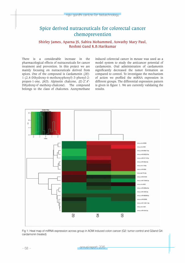
- 68 - annual report 2015
rajiv gandhi centre for biotechnology
Spice derived nutraceuticals for colorectal cancer chemoprevention
Shirley James, Aparna JS, Sabira Mohammed, Aswathy Mary Paul, Reshmi Gand K.B.Harikumar
There is a considerable increase in the pharmacological effects of nutraceuticals for cancer treatment and prevention. In this project we are mainly focusing on nutraceuticals derived from spices. One of the compound is Cardamonin (2E)-1-(2,4-Dihydroxy-6-methoxyphenyl)-3-phenyl-2-propen-1-one, (8CI); Alpinetin chalcone, (E)-2′,4′-Dihydroxy-6′-methoxy-chalcone). The compound belongs to the class of chalcones. Azoxymethane
induced colorectal cancer in mouse was used as a model system to study the anticancer potential of cardamonin. Oral administration of cardamonin significantly decreased the tumor formation as compared to control. To investigate the mechanism of action we profiled the miRNA expression in different groups. The differential expression pattern is given in figure 1. We are currently validating the results.
Fig 1: Heat map of miRNA expression across group in AOM induced colon cancer (G2: tumor control and G3and G4: cardamonin treated)
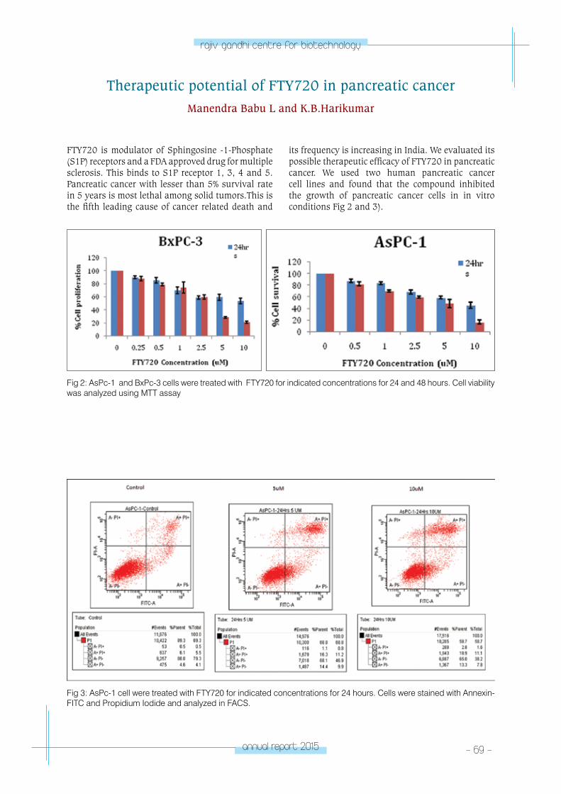
annual report 2015 - 69 -
rajiv gandhi centre for biotechnology
Therapeutic potential of FTY720 in pancreatic cancerManendra Babu L and K.B.Harikumar
FTY720 is modulator of Sphingosine -1-Phosphate (S1P) receptors and a FDA approved drug for multiple sclerosis. This binds to S1P receptor 1, 3, 4 and 5. Pancreatic cancer with lesser than 5% survival rate in 5 years is most lethal among solid tumors.This is the fifth leading cause of cancer related death and
its frequency is increasing in India. We evaluated its possible therapeutic efficacy of FTY720 in pancreatic cancer. We used two human pancreatic cancer cell lines and found that the compound inhibited the growth of pancreatic cancer cells in in vitro conditions Fig 2 and 3).
Fig 2: AsPc-1 and BxPc-3 cells were treated with FTY720 for indicated concentrations for 24 and 48 hours. Cell viability was analyzed using MTT assay
Fig 3: AsPc-1 cell were treated with FTY720 for indicated concentrations for 24 hours. Cells were stained with Annexin-FITC and Propidium Iodide and analyzed in FACS.
- 70 - annual report 2015
rajiv gandhi centre for biotechnology
Training• Harikumar K.B: Selected for International Brain
Research Organization (IBRO)’s North American Laboratory activity program at UT. M. D. Anderson Cancer Center, Houston, TX in October 2015 (3 weeks)
RESEARCH GRANTS EXTRA-MURAL FUNDING
No Title Funding Agency Duration
1Spice derived phytochemicals Sesamin and Cardamonin- for colorectal cancer chemoprevention
Department of Biotechnology-Ramalingaswami fellowship
2012-2017
2Novel aspects of sphingosine 1-phosphate (S1P) in innate immune responses and host defense mechanism
Department of Science and Technology, Government of India
2013-2016
3Sphingosine 1-phosphate signaling in pancreatic cancer
Department of Biotechnology, Government of India
2015-2017
CAnCeR ReSeA
RCH pRoGRAM
- laboratory 8CANCER
RESEARCH PROGRAM
Laboratory - 8
Debasree Dutta
Ph.D studentsSyed Khaja MohieddinAditi MajumderSujata Prakash Gaikwad
Research FellowsSunu JosephVaishna V.
Debasree has a Masters degree in Biochemistry
from the University of Calcutta followed by a
MTech and PhD in Biotechnology from Jadavpur
University. She trained as a Post Doctoral fellow
from 2007 to 2011 at the University of Kansas
Medical Center in USA and joined RGCB in 2011.
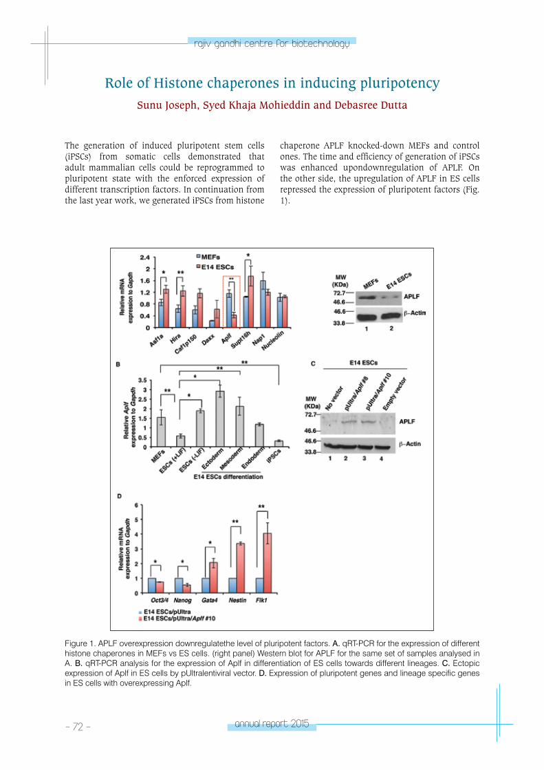
- 72 - annual report 2015
rajiv gandhi centre for biotechnology
Role of Histone chaperones in inducing pluripotencySunu Joseph, Syed Khaja Mohieddin and Debasree Dutta
The generation of induced pluripotent stem cells (iPSCs) from somatic cells demonstrated that adult mammalian cells could be reprogrammed to pluripotent state with the enforced expression of different transcription factors. In continuation from the last year work, we generated iPSCs from histone
chaperone APLF knocked-down MEFs and control ones. The time and efficiency of generation of iPSCs was enhanced upondownregulation of APLF. On the other side, the upregulation of APLF in ES cells repressed the expression of pluripotent factors (Fig. 1).
Figure 1. APLF overexpression downregulatethe level of pluripotent factors. A. qRT-PCR for the expression of different histone chaperones in MEFs vs ES cells. (right panel) Western blot for APLF for the same set of samples analysed in A. B. qRT-PCR analysis for the expression of Aplf in differentiation of ES cells towards different lineages. C. Ectopic expression of Aplf in ES cells by pUltralentiviral vector. D. Expression of pluripotent genes and lineage specific genes in ES cells with overexpressing Aplf.
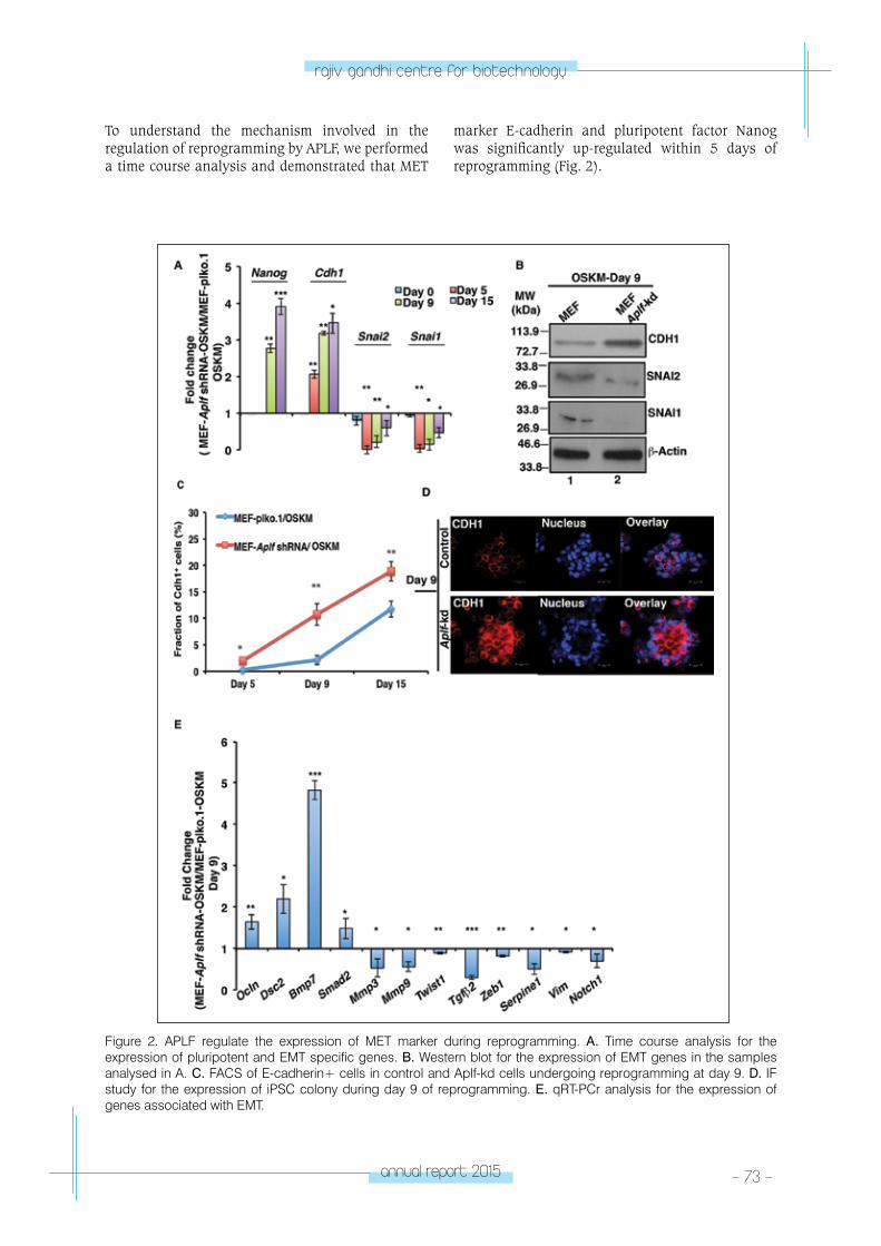
annual report 2015 - 73 -
rajiv gandhi centre for biotechnology
Figure 2. APLF regulate the expression of MET marker during reprogramming. A. Time course analysis for the expression of pluripotent and EMT specific genes. B. Western blot for the expression of EMT genes in the samples analysed in A. C. FACS of E-cadherin+ cells in control and Aplf-kd cells undergoing reprogramming at day 9. D. IF study for the expression of iPSC colony during day 9 of reprogramming. E. qRT-PCr analysis for the expression of genes associated with EMT.
To understand the mechanism involved in the regulation of reprogramming by APLF, we performed a time course analysis and demonstrated that MET
marker E-cadherin and pluripotent factor Nanog was significantly up-regulated within 5 days of reprogramming (Fig. 2).
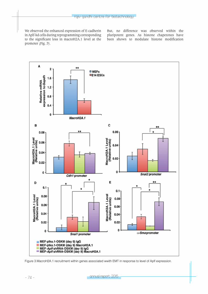
- 74 - annual report 2015
rajiv gandhi centre for biotechnology
We observed the enhanced expression of E-cadherin in Aplf-kd cells during reprogramming corresponding to the significant loss in macroH2A.1 level at the promoter (Fig. 3).
But, no difference was observed within the pluripotent genes. As histone chaperones have been shown to modulate histone modification
Figure 3.MacroH2A.1 recruitment within genes associated wwith EMT in response to level of Aplf expression.
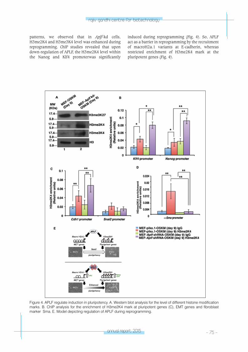
annual report 2015 - 75 -
rajiv gandhi centre for biotechnology
patterns, we observed that in Aplf-kd cells, H3me2K4 and H3me3K4 level was enhanced during reprogramming. ChIP studies revealed that upon down-regulation of APLF, the H3me2K4 level within the Nanog and Klf4 promoterwas significantly
induced during reprogramming (Fig. 4). So, APLF act as a barrier in reprogramming by the recruitment of macroH2a.1 variants at E-cadherin, whereas restricted enrichment of H3me2K4 mark at the pluripotent genes (Fig. 4).
Figure 4. APLF regulate induction in pluripotency. A. Western blot analysis for the level of different histone modification marks. B. ChIP analysis for the enrichment of H3me2K4 mark at pluripotent genes (C), EMT genes and fibroblast marker γSma. E. Model depicting regulation of APLF during reprogramming.
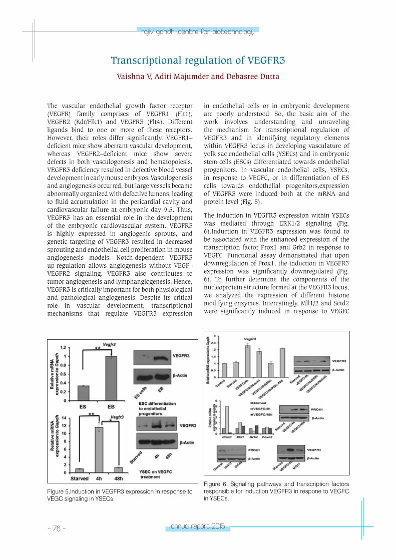
- 76 - annual report 2015
rajiv gandhi centre for biotechnology
Transcriptional regulation of VEGFR3Vaishna V, Aditi Majumder and Debasree Dutta
The vascular endothelial growth factor receptor (VEGFR) family comprises of VEGFR1 (Flt1), VEGFR2 (Kdr/Flk1) and VEGFR3 (Flt4). Different ligands bind to one or more of these receptors. However, their roles differ significantly. VEGFR1–deficient mice show aberrant vascular development, whereas VEGFR2–deficient mice show severe defects in both vasculogenesis and hematopoiesis.VEGFR3 deficiency resulted in defective blood vessel development in early mouse embryos. Vasculogenesis and angiogenesis occurred, but large vessels became abnormally organized with defective lumens, leading to fluid accumulation in the pericardial cavity and cardiovascular failure at embryonic day 9.5. Thus, VEGFR3 has an essential role in the development of the embryonic cardiovascular system. VEGFR3 is highly expressed in angiogenic sprouts, and genetic targeting of VEGFR3 resulted in decreased sprouting and endothelial cell proliferation in mouse angiogenesis models. Notch-dependent VEGFR3 up-regulation allows angiogenesis without VEGF–VEGFR2 signaling. VEGFR3 also contributes to tumor angiogenesis and lymphangiogenesis. Hence, VEGFR3 is critically important for both physiological and pathological angiogenesis. Despite its critical role in vascular development, transcriptional mechanisms that regulate VEGFR3 expression
in endothelial cells or in embryonic development are poorly understood. So, the basic aim of the work involves understanding and unraveling the mechanism for transcriptional regulation of VEGFR3 and in identifying regulatory elements within VEGFR3 locus in developing vasculature of yolk sac endothelial cells (YSECs) and in embryonic stem cells (ESCs) differentiated towards endothelial progenitors. In vascular endothelial cells, YSECs, in response to VEGFC, or in differentiation of ES cells towards endothelial progenitors,expression of VEGFR3 were induced both at the mRNA and protein level (Fig. 5).
The induction in VEGFR3 expression within YSECs was mediated through ERK1/2 signaling (Fig. 6).Induction in VEGFR3 expression was found to be associated with the enhanced expression of the transcription factor Prox1 and Grb2 in response to VEGFC. Functional assay demonstrated that upon downregulation of Prox1, the induction in VEGFR3 expression was significantly downregulated (Fig. 6). To further determine the components of the nucleoprotein structure formed at the VEGFR3 locus, we analyzed the expression of different histone modifying enzymes. Interestingly, Mll1/2 and Setd2 were significantly induced in response to VEGFC
Figure 5.Induction in VEGFR3 expression in response to VEGC signaling in YSECs.
Figure 6. Signaling pathways and transcription factors responsible for induction VEGFR3 in respone to VEGFC in YSECs.
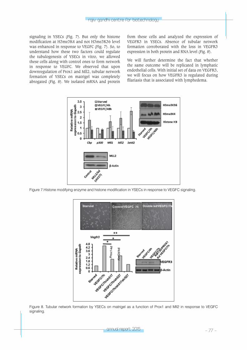
annual report 2015 - 77 -
rajiv gandhi centre for biotechnology
Figure 7.Histone modifying enzyme and histone modification in YSECs in response to VEGFC signaling.
Figure 8. Tubular network formation by YSECs on matrigel as a function of Prox1 and Mll2 in response to VEGFC signaling.
signaling in YSECs (Fig. 7). But only the histone modification at H3me3K4 and not H3me3K36 level was enhanced in response to VEGFC (Fig. 7). So, to understand how these two factors could regulate the tubulogenesis of YSECs in vitro, we allowed these cells along with control ones to form network in response to VEGFC. We observed that upon downregulation of Prox1 and Mll2, tubular network formation of YSECs on matrigel was completely abrogated (Fig. 8). We isolated mRNA and protein
from these cells and analyzed the expression of VEGFR3 in YSECs. Absence of tubular network formation corroborated with the loss in VEGFR3 expression in both protein and RNA level (Fig. 8).
We will further determine the fact that whether the same outcome will be replicated in lymphatic endothelial cells. With initial set of data on VEGFR3, we will focus on how VEGFR3 is regulated during filariasis that is associated with lymphedema.
- 78 - annual report 2015
rajiv gandhi centre for biotechnology
Publications • Kochurani KJ, Suganya AA, Nair MG, Louis JM,
Majumder A, Kumar SK, Abraham P, Dutta D, Maliekal TT. Live detection and purification of cells based on the expression of a histone chaperone, HIRA, using a binding peptide. Scientific Reports. 2015 Nov 24;5:17218
• Majumder A, Syed KM, Joseph S, Scambler PJ, Dutta D. (2015). Histone chaperone HIRA in Regulation of Transcription Factor RUNX1. J BiolChem 290:13053-63
Conference/poster presentations• Sunu Joseph, Syed KhajaMohieddin, DebasreeDutta.
Histone chaperone Aplf regulates reprogramming. Till & McCulloch meeting 2015, organized by Stem Cell Network, Canada. SJ received full travel grant to attend and present the work.
RESEARCH GRANTS
No Title Funding Agency Duration
1Transcriptional regulation of VEGFR 3
Council for Scientific &IndustrailResearch,India. 2013-2016
2Hemogenic endothelium- regulation and reprograming
Department of Science & Technology, Government of India
2013-2016
3Histone chaperones in regulation of reprogramming
Department of Biotechnology, Government of India 2012-2015
CAnCeR ReSeA
RCH pRoGRAM
- laboratory 9CANCER
RESEARCH PROGRAM
Laboratory - 9
M. Radhakrishna Pillai
Program ScientistsReshmi G.Ani V. Das
Post Doctoral FellowsSrinivas K.P.Rajesh Raju
PhD StudentsDeivendran S. Hezlin MarzookSajitha I.S.Priya R. PrabhuDeepthi PrasadRahul Sanawar
Project PersonnelAswathy Mary PaulVivek Anand A.Meena Vinay KumarBijesh GeorgeVishnu V.M.Shuba SankaranViji S.Jinu AustinJayakrishnan K.S.
Technical PersonnelRintu T. VargheseKannan T.R.Edwin S.
Radhakrishna Pillai joined RGCB in 2005
moving from the Regional Cancer Centre at
Thiruvananthapuram where he was Professor
of Molecular Medicine. Dr. Pillai is a Fellow of
the Royal College of Pathologists, London, the
National Academy of Medical Sciences, India,
the National Academy of Sciences, India and the
Indian Academy of Sciences.
- 80 - annual report 2015
rajiv gandhi centre for biotechnology
International Agency for Research on Cancer (IARC), in 2009, initiated a Phase-IV HPV vaccine trial with the objective of generating scientific evidence on the efficacy of two-dose HPV vaccination regimen as compared to the current standard three-dose regimen, generating information on the feasibility, safety and acceptability of the two-dose HPV vaccination regimens with an ultimate aim to provide decision makers in India and other developing countries with operational, efficacy and safety data to empower them to make an appropriate, evidence-based HPV vaccination policy and to invest in its wide-scale implementation and disseminate the information on the lessons learned to inform India and other developing countries about the feasibility, effectiveness, safety and acceptability of the vaccination approaches. 17,729 unmarried girls aged 10-18 years were recruited through Tata Memorial Hospital - Mumbai, NargisDutt Memorial Cancer Hospital - Barshi, Jehangir Clinical Development Centre - Pune, Christian Fellowship Community Health Centre - Ambilikai, Gujarat Cancer Research Institute - Ahmedabad, All India Institute of Medical Sciences - New Delhi, MNJ Institute of Oncology and Regional Cancer Centre - Hyderabad and Cancer Foundation of India (CFI) –Kolkata representing different geographical regions of India. The quadrivalent vaccine (Gardasil) containing HPV 16, 18, 6 and 11virus like particles (VLPs) provided free of cost by MERCK™ was administered to the participant girls of the study and has progressed very well with RGCB as the central tissue repository and a hub of all experimental analysis. Status of immune response against the vaccine targeted HPV types viz HPV 16, 18,6 and 11 till 48 months after vaccination and early results on the analysis of cervical cells for incident infections has been published in the January 2016 issue of Lancet Oncology. Our data supports the existing knowledge that prophylactic vaccine containing recombinant virus-like particles (VLP) assembled from the L1 capsid proteins of HPV 6, 11, 16 and 18 (quadrivalent vaccine) can effectively prevent persistent infection with vaccine-
included HPV types. Early results from various studies suggest that apart from the antibodies raised against the HPV types targeted by the vaccine (HPV 16, 18, 6 and 11) the quadrivalent HPV vaccine is capable of stimulating cross reactive antibodies and thusprovide limited additional cross-protection against phylogenetically related non-targeted HPV types. We compared four cohorts of girls whoreceived 3 doses on days 1, 60 and 180 or later (3-dose); 2 doses on days 1 and 180 or later (2-dose); 2 doses on days 1 and 60 by default (2 doses/D); and 1 dose by default (1 dose/D). The primary outcomes considered were cross-protective immunogenicity demonstrated by the L1-binding antibody-levels, neutralizing antibody-levels and antibody avidity for the non-vaccine-targeted HPV-types related to HPV-16 (species 9: HPV-31/33/35/45/52/58) and HPV-18 (species 7: HPV-45). Initial results indicate that the 2-dose induced high non-inferior peak mean immune responses compared to the 3-dose at month 7 after first dose for the non-vaccine-targeted HPV-31/33/35/52/58/45. These responses in both dose groups dropped to or below seropositivity levels by the 48th month after first dose, except for HPV-31. The mean immune responses in the 2-doses/D groups at month 18 after first dose were non-inferior, whereas the responses of the 1-dose/D group were inferior to the 3-dose group for HPV-types of species 7 and 9. Avidity indices above 50% were observed at month 18 after first dose in the different dose groups for the HPV-types of species 7 and 9 except in 1 dose/D for HPV-33, and 2-dose, 2 doses/D and 1 dose/D for HPV-35. The proportion of samples with detectable concentrations of neutralization antibodies for the 3-dose, 2-dose, 2 dose/D and 1 dose/D vaccination cohorts at month 18 after first dose were 50%, 39%, 28% and 7% for HPV-31, 12%, 12%, 5% and 5% for HPV-33, and 8%, 5%, 3% and 7% for HPV-45, respectively. The study is still ongoing as these observations need to be correlated with virological and disease end-points, as the immune correlates of protection are still unknown.
Randomized trial of 2 vs 3 doses of Human Papilloma Virus (HPV) vaccine in India
Priya R Prabhu, Kannan T R, Rintu T Varghese, Subha Sankaran, Jayakrishnan K S, Jinu Austin, Edwin S, *R. Sankaranarayanan,
*Masimmo Tomassino, *Tarik Gheit **Michael Pawlita and M. Radhakrishna Pillai.
Collaborators: *International Agency for Research on Cancer (IARC), Lyon, France and **German Cancer Research Centre (DKFZ), Heidelberg, Germany.
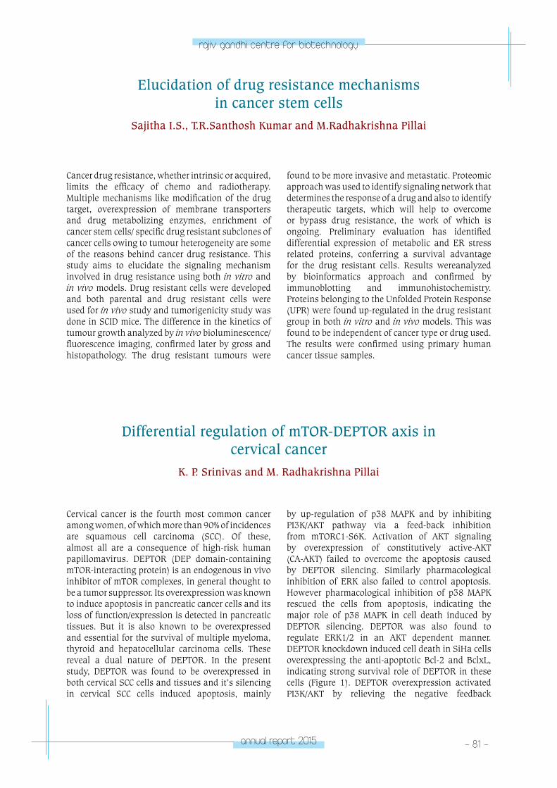
annual report 2015 - 81 -
rajiv gandhi centre for biotechnology
Elucidation of drug resistance mechanisms in cancer stem cells
Sajitha I.S., T.R.Santhosh Kumar and M.Radhakrishna Pillai
Cancer drug resistance, whether intrinsic or acquired, limits the efficacy of chemo and radiotherapy. Multiple mechanisms like modification of the drug target, overexpression of membrane transporters and drug metabolizing enzymes, enrichment of cancer stem cells/ specific drug resistant subclones of cancer cells owing to tumour heterogeneity are some of the reasons behind cancer drug resistance. This study aims to elucidate the signaling mechanism involved in drug resistance using both in vitro and in vivo models. Drug resistant cells were developed and both parental and drug resistant cells were used for in vivo study and tumorigenicity study was done in SCID mice. The difference in the kinetics of tumour growth analyzed by in vivo bioluminescence/ fluorescence imaging, confirmed later by gross and histopathology. The drug resistant tumours were
found to be more invasive and metastatic. Proteomic approach was used to identify signaling network that determines the response of a drug and also to identify therapeutic targets, which will help to overcome or bypass drug resistance, the work of which is ongoing. Preliminary evaluation has identified differential expression of metabolic and ER stress related proteins, conferring a survival advantage for the drug resistant cells. Results wereanalyzed by bioinformatics approach and confirmed by immunoblotting and immunohistochemistry. Proteins belonging to the Unfolded Protein Response (UPR) were found up-regulated in the drug resistant group in both in vitro and in vivo models. This was found to be independent of cancer type or drug used. The results were confirmed using primary human cancer tissue samples.
Cervical cancer is the fourth most common cancer among women, of which more than 90% of incidences are squamous cell carcinoma (SCC). Of these, almost all are a consequence of high-risk human papillomavirus. DEPTOR (DEP domain-containing mTOR-interacting protein) is an endogenous in vivo inhibitor of mTOR complexes, in general thought to be a tumor suppressor. Its overexpression was known to induce apoptosis in pancreatic cancer cells and its loss of function/expression is detected in pancreatic tissues. But it is also known to be overexpressed and essential for the survival of multiple myeloma, thyroid and hepatocellular carcinoma cells. These reveal a dual nature of DEPTOR. In the present study, DEPTOR was found to be overexpressed in both cervical SCC cells and tissues and it’s silencing in cervical SCC cells induced apoptosis, mainly
by up-regulation of p38 MAPK and by inhibiting PI3K/AKT pathway via a feed-back inhibition from mTORC1-S6K. Activation of AKT signaling by overexpression of constitutively active-AKT (CA-AKT) failed to overcome the apoptosis caused by DEPTOR silencing. Similarly pharmacological inhibition of ERK also failed to control apoptosis. However pharmacological inhibition of p38 MAPK rescued the cells from apoptosis, indicating the major role of p38 MAPK in cell death induced by DEPTOR silencing. DEPTOR was also found to regulate ERK1/2 in an AKT dependent manner. DEPTOR knockdown induced cell death in SiHa cells overexpressing the anti-apoptotic Bcl-2 and BclxL, indicating strong survival role of DEPTOR in these cells (Figure 1). DEPTOR overexpression activated PI3K/AKT by relieving the negative feedback
Differential regulation of mTOR-DEPTOR axis in cervical cancer
K. P. Srinivas and M. Radhakrishna Pillai
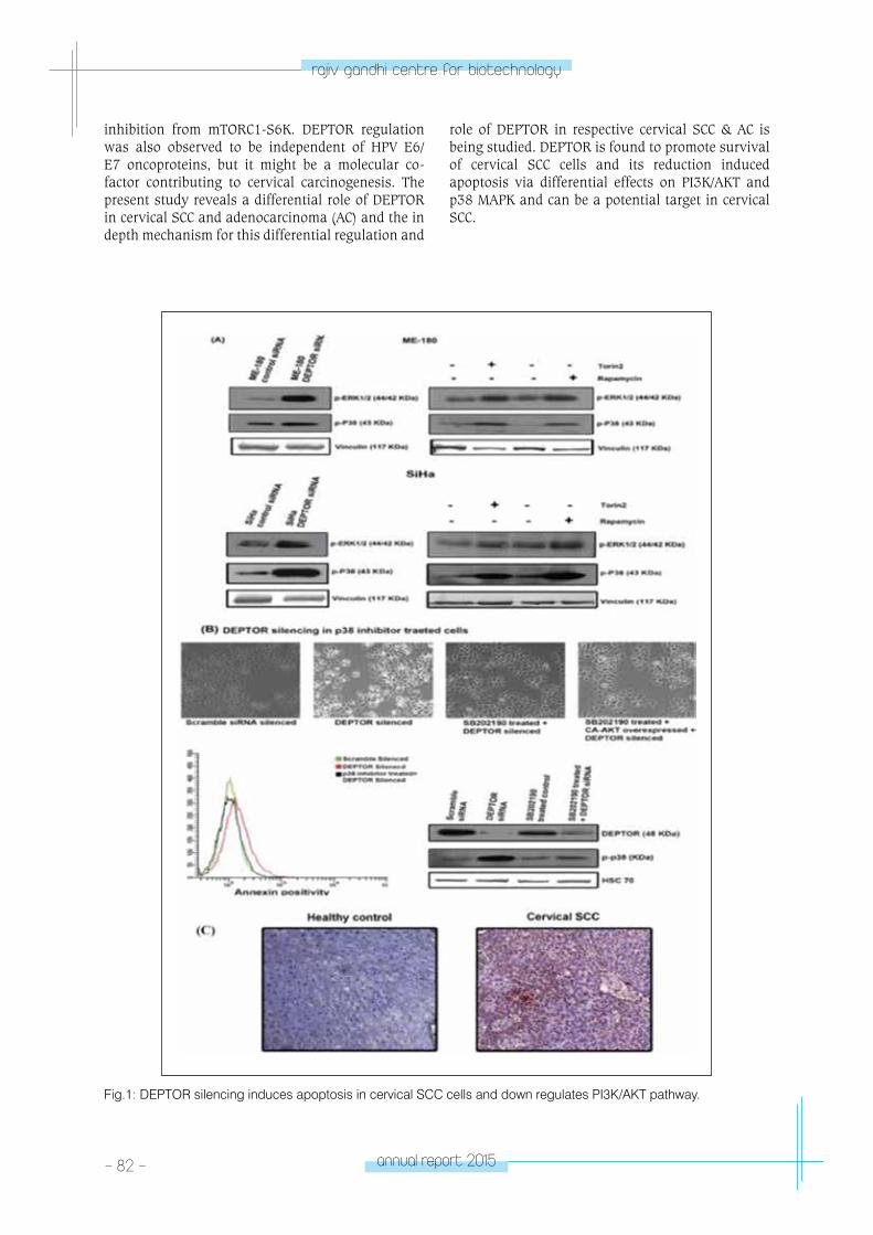
- 82 - annual report 2015
rajiv gandhi centre for biotechnology
Fig.1: DEPTOR silencing induces apoptosis in cervical SCC cells and down regulates PI3K/AKT pathway.
inhibition from mTORC1-S6K. DEPTOR regulation was also observed to be independent of HPV E6/E7 oncoproteins, but it might be a molecular co-factor contributing to cervical carcinogenesis. The present study reveals a differential role of DEPTOR in cervical SCC and adenocarcinoma (AC) and the in depth mechanism for this differential regulation and
role of DEPTOR in respective cervical SCC & AC is being studied. DEPTOR is found to promote survival of cervical SCC cells and its reduction induced apoptosis via differential effects on PI3K/AKT and p38 MAPK and can be a potential target in cervical SCC.
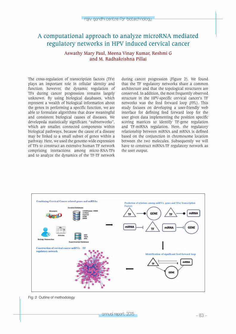
annual report 2015 - 83 -
rajiv gandhi centre for biotechnology
The cross-regulation of transcription factors (TFs) plays an important role in cellular identity and function; however, the dynamic regulation of TFs during cancer progression remains largely unknown. By using biological databases, which represent a wealth of biological information about the genes in performing a specific function, we are able to formulate algorithms that draw meaningful and consistent biological causes of diseases. We developeda statistically significant “subnetworks”, which are smaller, connected components within biological pathways, because the cause of a disease may be linked to a small subset of genes within a pathway. Here, we used the genome-wide expression of TFs to construct an extensive human TF network comprising interactions among micro-RNA-TFs and to analyze the dynamics of the TF-TF network
during cancer progression (Figure 2). We found that the TF regulatory networks share a common architecture and that the topological structures are conserved. In addition, the most frequently observed structure in the HPV-specific cervical cancer’s TF networks was the feed forward loop (FFL). This study focuses on developing a user-friendly web interface for defining feed forward loop for the user given data implementing the position specific scoring matrices to identify TF-gene regulation and TF-miRNA regulation. Here, the regulatory relationship between miRNA and mRNA is defined based on the conjunction in chromosome location between the two molecules. Subsequently we will have to construct miRNA-TF regulatory network as the user output.
A computational approach to analyze microRNA mediated regulatory networks in HPV induced cervical cancer
Aswathy Mary Paul, Meena Vinay Kumar, Reshmi G and M. Radhakrishna Pillai
Fig: 2 Outline of methodology
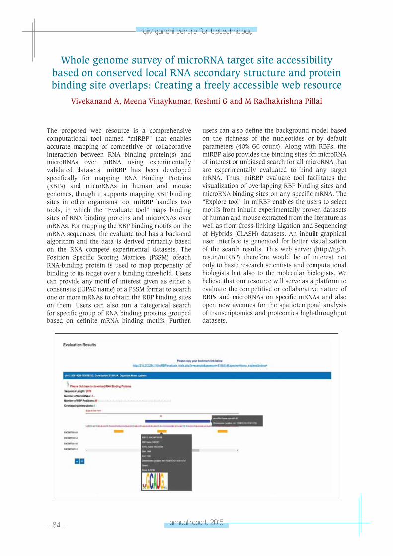
- 84 - annual report 2015
rajiv gandhi centre for biotechnology
The proposed web resource is a comprehensive computational tool named “miRBP” that enables accurate mapping of competitive or collaborative interaction between RNA binding protein(s) and microRNAs over mRNA using experimentally validated datasets. miRBP has been developed specifically for mapping RNA Binding Proteins (RBPs) and microRNAs in human and mouse genomes, though it supports mapping RBP binding sites in other organisms too. miRBP handles two tools, in which the “Evaluate tool” maps binding sites of RNA binding proteins and microRNAs over mRNAs. For mapping the RBP binding motifs on the mRNA sequences, the evaluate tool has a back-end algorithm and the data is derived primarily based on the RNA compete experimental datasets. The Position Specific Scoring Matrices (PSSM) ofeach RNA-binding protein is used to map propensity of binding to its target over a binding threshold. Users can provide any motif of interest given as either a consensus (IUPAC name) or a PSSM format to search one or more mRNAs to obtain the RBP binding sites on them. Users can also run a categorical search for specific group of RNA binding proteins grouped based on definite mRNA binding motifs. Further,
users can also define the background model based on the richness of the nucleotides or by default parameters (40% GC count). Along with RBPs, the miRBP also provides the binding sites for microRNA of interest or unbiased search for all microRNA that are experimentally evaluated to bind any target mRNA. Thus, miRBP evaluate tool facilitates the visualization of overlapping RBP binding sites and microRNA binding sites on any specific mRNA. The “Explore tool” in miRBP enables the users to select motifs from inbuilt experimentally proven datasets of human and mouse extracted from the literature as well as from Cross-linking Ligation and Sequencing of Hybrids (CLASH) datasets. An inbuilt graphical user interface is generated for better visualization of the search results. This web server (http://rgcb.res.in/miRBP) therefore would be of interest not only to basic research scientists and computational biologists but also to the molecular biologists. We believe that our resource will serve as a platform to evaluate the competitive or collaborative nature of RBPs and microRNAs on specific mRNAs and also open new avenues for the spatiotemporal analysis of transcriptomics and proteomics high-throughput datasets.
Whole genome survey of microRNA target site accessibility based on conserved local RNA secondary structure and protein binding site overlaps: Creating a freely accessible web resource
Vivekanand A, Meena Vinaykumar, Reshmi G and M Radhakrishna Pillai
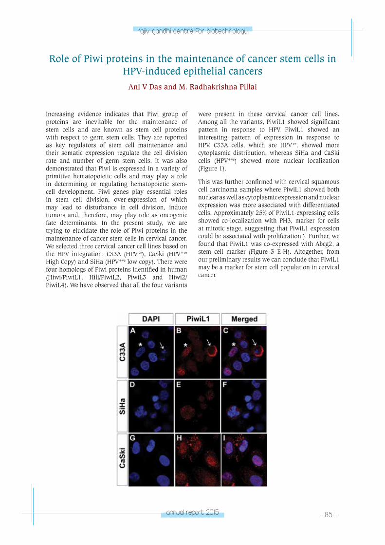
annual report 2015 - 85 -
rajiv gandhi centre for biotechnology
Role of Piwi proteins in the maintenance of cancer stem cells in HPV-induced epithelial cancers
Ani V Das and M. Radhakrishna Pillai
Increasing evidence indicates that Piwi group of proteins are inevitable for the maintenance of stem cells and are known as stem cell proteins with respect to germ stem cells. They are reported as key regulators of stem cell maintenance and their somatic expression regulate the cell division rate and number of germ stem cells. It was also demonstrated that Piwi is expressed in a variety of primitive hematopoietic cells and may play a role in determining or regulating hematopoietic stem-cell development. Piwi genes play essential roles in stem cell division, over-expression of which may lead to disturbance in cell division, induce tumors and, therefore, may play role as oncogenic fate determinants. In the present study, we are trying to elucidate the role of Piwi proteins in the maintenance of cancer stem cells in cervical cancer. We selected three cervical cancer cell lines based on the HPV integration: C33A (HPV-ve), CaSki (HPV+ve High Copy) and SiHa (HPV+ve low copy). There were four homologs of Piwi proteins identified in human (Hiwi/PiwiL1, Hili/PiwiL2, PiwiL3 and Hiwi2/PiwiL4). We have observed that all the four variants
were present in these cervical cancer cell lines. Among all the variants, PiwiL1 showed significant pattern in response to HPV. PiwiL1 showed an interesting pattern of expression in response to HPV. C33A cells, which are HPV-ve, showed more cytoplasmic distribution, whereas SiHa and CaSki cells (HPV+ve) showed more nuclear localization (Figure 1).
This was further confirmed with cervical squamous cell carcinoma samples where PiwiL1 showed both nuclear as well as cytoplasmic expression and nuclear expression was more associated with differentiated cells. Approximately 25% of PiwiL1-expressing cells showed co-localization with PH3, marker for cells at mitotic stage, suggesting that PiwiL1 expression could be associated with proliferation.). Further, we found that PiwiL1 was co-expressed with Abcg2, a stem cell marker (Figure 3 E-H). Altogether, from our preliminary results we can conclude that PiwiL1 may be a marker for stem cell population in cervical cancer.
- 86 - annual report 2015
rajiv gandhi centre for biotechnology
Publications• Ani V Das and M. Radhakrishna Pillai. Implications
of miR cluster 143/145 as universal anti-oncomiRs and their dysregulation during tumorigenesis.Cancer Cell International (2015) 15:92.
• Rajesh Raju, SachinGadakh, PriyankaGopal, Bijesh George, JayshreeAdvani, SowmyaSoman, T S Keshava Prasad and ReshmiGirijadevi. Differential ligand-signaling network of CCL19/CCL21-CCR7 system. Database (Oxford). 2015; bav106.
• “Immunogenicity and HPV infection following one, two and three doses of quadrivalent vaccine: Early results from a multi-center cohort study in India”, R. Sankaranarayanan, Prabhu PR, Pawlita M, Gheit T, Bhatla N, Muwonge R, Nene BM, Esmy PO, Joshi S, Reddy Poli UR, Jivarajani P, Verma Y, Zomawia E, Siddiqi M, Shastri SS, Jayant K, Malvi SG, Lucas E, Michel A, Butt J, JankiBabu, Subha S, Kannan TR, Varghese R, Divate U, Thomas S, Joshi G, Willhauck-Fleckenstein M, Waterboer T, Muller M, Sehr P, Hingmire S, AKriplani A, Mishra G, Pimple S, Jadhav R, Sauvaget S, Tommasino M, Pillai MR, for the Indian HPV vaccine study group, Lancet Oncology, Volume 17, No. 1, p67–77, January 2016
• KP Srinivas, VijiRemadevi, Vipin Mohan Dan, IS Sajitha, R Prakash, PV Rahul, TR Santhosh Kumar, S Lakshmi and M Radhakrishna Pillai (2016). DEPTOR promotes survival of cervical squamous cell carcinoma cells and its silencing induces apoptosis through down-regulating PI3K/AKT and by up-regulating p38MAPK. Oncotarget. 7(17): 24154-24171.
Book Chapter• Prabhu PR and Pillai M R, “Human Papillomaviruses
and Squamous Cell Carcinomas of Head and Neck Region, Contemporary Oral Oncology: Biology, Epidemiology, Etiology and Prevention” © Springer International Publishing, Switzerland 2016; DOI 10.1007/978-3-319-14911-0_3
Honors & Awards • Best poster award at Asia Oceania Research
Organization on Genital Infections and neoplasia (AOGIN 2015): Ani V Das, Deepti Prasad, Srinivas
K.P., VijiRemadevi and M. Radhakrishna Pillai. Role of Piwi proteins in cervical cancers and their association with HPV. Christian Medical College, Bhagayam, Vellore, India
• Best poster award at the 35th Annual Convention of Indian Association of Cancer Research (IACR), organized by Indian Association of Cancer Research (IACR) and Asian Clinical Oncology Society (ACOS):Priya R Prabhu was awarded the IACR- ACOS Best Poster Award for the poster titled“ Cross-reactive antibodies against non-targeted HPV subtypes upon quadrivalent HPV vaccine administration”, New Delhi, from 8th -10th April, 2016.
Conferences/ Workshops• RECOMB-15, 19th Annual International Conference
on Research in Computational Molecular Biology:Vivekanand A, Reshmi G and Pillai MR. “miRBP: An architecture to find competitive interaction between microRNA and RNA binding proteins”, Warsaw, Poland, April 12-15, 2015.
• 30th International Papilloma Virus Conference: Platform Presentation - R. Sankaranarayanan, Priya R Prabhu, Michael Pawlita, NeerjaBhatla and M. Radhakrishna Pillai, “Comparison of immunogenicity following single, two or three doses of Human Papilloma Virus vaccination in India”. Lisbon, Portugal from 17th to 21st September, 2015
• Asia Oceania Research Organization on Genital Infections and neoplasia (AOGIN 2015): Ani V Das, Deepti Prasad, Srinivas K.P., VijiRemadevi and M. Radhakrishna Pillai. Role of Piwi proteins in cervical cancers and their association with HPV. Christian Medical College, Vellore, India.
International Travel Grants• Vivekanand A awarded International Travel
Fellowship from Department of Biotechnology, India to attend a conference, RECOMB-15 heldat Warsaw, Poland, April 12-15, 2015.
• Priya R Prabhu was awarded Department of Biotechnology (DBT), Government of India, International Travel Fellowship to attend and deliver an oral presentation at HPV2015- 30th (International Papilloma Virus Conference) in Lisbon, Portugal from 17th to 21st September, 2015
RESEARCH GRANTS
No Title of the project Investigators Funding Agency Duration
1
Whole genome survey of microRNA target site accessibility based on conserved local RNA secondary structure and protein binding site overlaps: Creating a freely accessible web resource.
Reshmi G and M.R. Pillai.
Department of Biotechnology, Government of India.
2012-2015
annual report 2015 - 87 -
rajiv gandhi centre for biotechnology
2
Integrative Computational Analysis to Drive Discovery of MicroRNA-mediated Regulatory Networks in HPV Induced Cervical Cancer,
Reshmi G and M.R. Pillai.
Department of Biotechnology, Government of India.
2013-2016
3Developing Models to Forecast Outbreak of Dengue Virus in Kerala: Creating a freely accessible web resource
Reshmi G and M.R. Pillai.
Kerala State Council for Science, Technology & Environment, Government of Kerala.
2014-2017
4Systems Biology Assessment of Influenza A/pH1N1 vaccination in an Indian Cohort.
M. R. PillaiDepartment of Biotechnology, Government of India.
2014-2016
5Identifying Systems-level Cellular Networks Involved in Neurotropic Flavi Virus- Host Interaction.
Reshmi G and M.R. Pillai.
Department of Biotechnology, Government of India.
2015-2018
6
Development of the Cell-specific temporally regulated transcriptomic and proteomic map of cancer signalling pathway.
Rajesh RajuDepartment of Biotechnology- Fast Track Young Scientist Project
2015-2018
7Role of human papillomavirus infection and other co-factors in the aetiology of the head and neck cancer in India.
M. R. PillaiIndian Council of Medical Research, Government of India
2015-2018
8
Accurate and satisfactory analysis of all high risk HPV types and some low risks including HPV 6 and 11 antibody titers for the 2-versus 3 dose HPV vaccination clinical trial in India - Follow-up Study.
M. R. PillaiInternational Agency for Research on Cancer, France
2016-2021
9National Program for Drug Discovery and Developmental Therapeutics (NFDDDT)
M. R. PillaiDepartment of Science and Technology, Government of India
2016-2019
RGCB-George Washington university Joint program
in Cancer Research
RGCB-George Washington University Joint Program
in Cancer Research
Rakesh Kumar, Ph D
Upon recommendation of the RGCB Governing
Counil, Professor Rakesh Kumar of the
Department of Biochemistry, George Washington
University, USA was invited to RGCB as Visiting
Distinguished Professor of Biotechnology to co-
direct a joint research program with
Professor M. Radhakrishna Pillai.
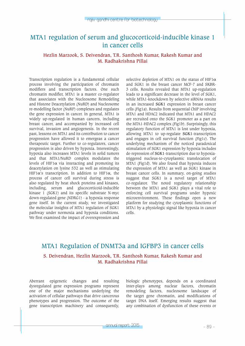
annual report 2015 - 89 -
rajiv gandhi centre for biotechnology
Transcription regulation is a fundamental cellular process involving the participation of chromatin modifiers and transcription factors. One such chromatin modifier, MTA1 is a master co-regulator that associates with the Nucleosome Remodeling and Histone Deacetylation (NuRD) and Nucleosome re-modelling factor (NuRF) complexes and regulates the gene expression in cancer. In general, MTA1 is widely up-regulated in human cancers, including breast cancer, and accompanied by increased cell survival, invasion and angiogenesis. In the recent past, lessons on MTA1 and its contribution to cancer progression have allowed it to emergeas a cancer therapeutic target. Further to co-regulators, cancer progression is also driven by hypoxia. Interestingly, hypoxia also increases MTA1 levels in solid tumors and that MTA1/NuRD complex modulates the levels of HIF1α via interacting and promoting its deacetylation on lysine 532 as well as stimulating HIF1α’s transcription. In addition to HIF1α, the process of cancer cell survival during stress is also regulated by heat shock proteins and kinases, including, serum and glucocorticoid-inducible kinase 1 (SGK1) and its specific substrate N-myc down-regulated gene (NDRG1) - a hypoxia response gene itself. In the current study, we investigated the molecular insights of MTA1 regulation of SGK1 pathway under normoxia and hypoxia conditions. We first examined the impact of overexpression and
selective depletion of MTA1 on the status of HIF1α and SGK1 in the breast cancer MCF-7 and SKBR-3 cells. Results revealed that MTA1 up-regulation leads to a significant decrease in the level of SGK1, while MTA1-knockdown by selective siRNAs results in an increased SGK1 expression in breast cancer cells (Fig1a). Results from sequential ChIP involving MTA1 and HDAC2 indicated that MTA1 and HDAC2 are recruited onto the SGK1 promoter as a part on the MTA1-HDAC2 complex (Fig1b). Surprisingly, this regulatory function of MTA1 is lost under hypoxia, allowing MTA1 to up-regulate SGK1-transcription and engages in cell survival function (Fig1c). The underlying mechanism of the noticed paradoxical stimulation of SGK1 expression by hypoxia includes de-repression of SGK1-transcription due to hypoxia-triggered nucleus-to-cytoplasmic translocation of MTA1 (Fig1d). We also found that hypoxia induces the expression of MTA1 as well as SGK1 kinase in breast cancer cells. In summary, on-going studies suggest that SGK1 is a novel target of MTA1 co-regulator. The noted regulatory relationship between the MTA1 and SGK1 plays a vital role to enforcing cell survival programs under hypoxic microenvironment. These findings open a new platform for studying the cytoplasmic functions of MTA1 by a physiologic signal like hypoxia in cancer cells.
MTA1 regulation of serum and glucocorticoid-inducible kinase 1 in cancer cells
Hezlin Marzook, S. Deivendran, T.R. Santhosh Kumar, Rakesh Kumar and M. Radhakrishna Pillai
MTA1 Regulation of DNMT3a and IGFBP3 in cancer cellsS. Deivendran, Hezlin Marzook, T.R. Santhosh Kumar, Rakesh Kumar and
M. Radhakrishna Pillai
Aberrant epigenetic changes and resulting dysregulated gene expression programs represent one of the major mechanisms underlying the activation of cellular pathways that drive cancerous phenotypes and progression. The outcome of the gene transcription machinery and consequently,
biologic phenotypes, depends on a coordinated inter-plays among nuclear factors, chromatin remodeling factors, nucleosome landscape of the target gene chromatin, and modifications of target DNA itself. Emerging results suggest that any combination of dysfunction of these events or
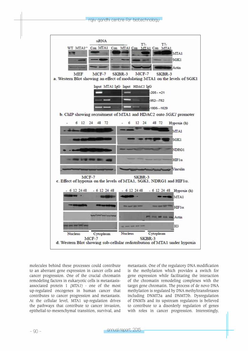
- 90 - annual report 2015
rajiv gandhi centre for biotechnology
molecules behind these processes could contribute to an aberrant gene expression in cancer cells and cancer progression. One of the crucial chromatin remodeling factors in eukaryotic cells is metastasis-associated protein 1 (MTA1) - one of the most up-regulated oncogenes in human cancer that contributes to cancer progression and metastasis. At the cellular level, MTA1 up-regulation drives the pathways that contribute to cancer invasion, epithelial-to-mesenchymal transition, survival, and
metastasis. One of the regulatory DNA modification is the methylation which provides a switch for gene expression while facilitating the interaction of the chromatin remodeling complexes with the target gene chromatin. The process of de novo DNA methylation is regulated by DNA methyltransferases including DNMT3a and DNMT3b. Dysregulation of DNMTs and its upstream regulators is believed to contribute to a disorderly regulation of genes with roles in cancer progression. Interestingly,
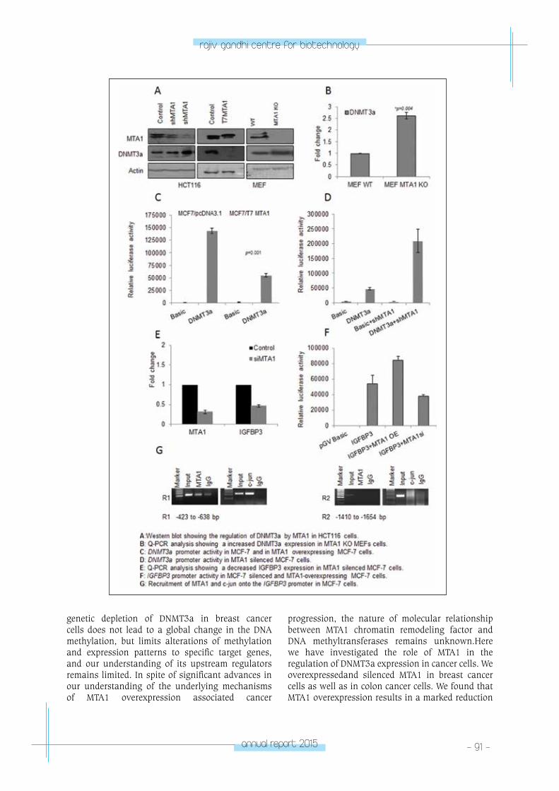
annual report 2015 - 91 -
rajiv gandhi centre for biotechnology
genetic depletion of DNMT3a in breast cancer cells does not lead to a global change in the DNA methylation, but limits alterations of methylation and expression patterns to specific target genes, and our understanding of its upstream regulators remains limited. In spite of significant advances in our understanding of the underlying mechanisms of MTA1 overexpression associated cancer
progression, the nature of molecular relationship between MTA1 chromatin remodeling factor and DNA methyltransferases remains unknown.Here we have investigated the role of MTA1 in the regulation of DNMT3a expression in cancer cells. We overexpressedand silenced MTA1 in breast cancer cells as well as in colon cancer cells. We found that MTA1 overexpression results in a marked reduction
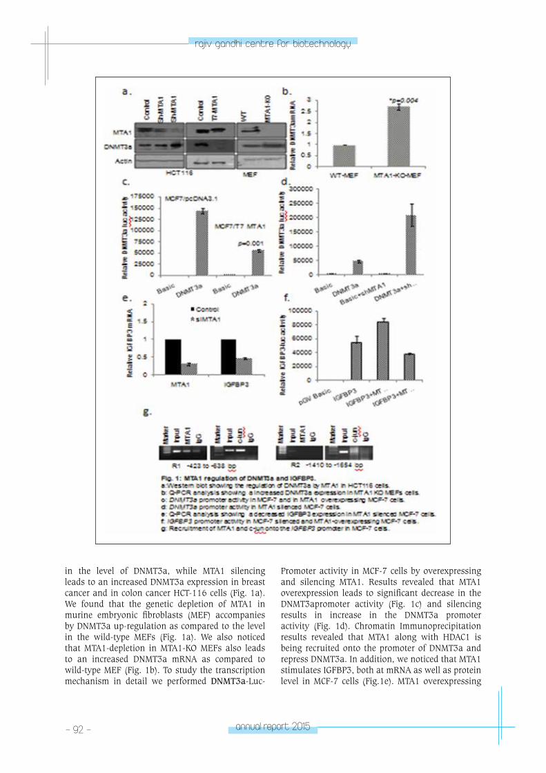
- 92 - annual report 2015
rajiv gandhi centre for biotechnology
in the level of DNMT3a, while MTA1 silencing leads to an increased DNMT3a expression in breast cancer and in colon cancer HCT-116 cells (Fig. 1a). We found that the genetic depletion of MTA1 in murine embryonic fibroblasts (MEF) accompanies by DNMT3a up-regulation as compared to the level in the wild-type MEFs (Fig. 1a). We also noticed that MTA1-depletion in MTA1-KO MEFs also leads to an increased DNMT3a mRNA as compared to wild-type MEF (Fig. 1b). To study the transcription mechanism in detail we performed DNMT3a-Luc-
Promoter activity in MCF-7 cells by overexpressing and silencing MTA1. Results revealed that MTA1 overexpression leads to significant decrease in the DNMT3apromoter activity (Fig. 1c) and silencing results in increase in the DNMT3a promoter activity (Fig. 1d). Chromatin Immunoprecipitation results revealed that MTA1 along with HDAC1 is being recruited onto the promoter of DNMT3a and repress DNMT3a. In addition, we noticed that MTA1 stimulates IGFBP3, both at mRNA as well as protein level in MCF-7 cells (Fig.1e). MTA1 overexpressing
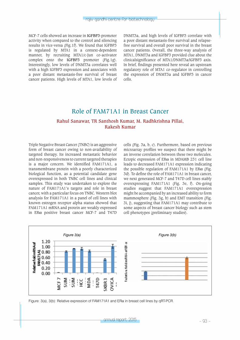
annual report 2015 - 93 -
rajiv gandhi centre for biotechnology
MCF-7 cells showed an increase in IGFBP3-promoter activity when compared to the control and silencing results in vice-versa (Fig.1f). We found that IGFBP3 is regulated by MTA1 in a context-dependent manner, by recruiting MTA1/c-Jun co-activator complex onto the IGFBP3 promoter (Fig.1g). Interestingly, low levels of DNMT3a correlates well with a high IGFBP3 expression and associates with a poor distant metastasis-free survival of breast cancer patients. High levels of MTA1, low levels of
DNMT3a, and high levels of IGFBP3 correlate with a poor distant metastasis-free survival and relapse-free survival and overall poor survival in the breast cancer patients. Overall, the three-way analysis of MTA1, DNMT3a and IGFBP3 provided clue about the clinicalsignificance of MTA1/DNMT3a/IGFBP3 axis. In brief, findings presented here reveal an upstream regulatory role of MTA1 co-regulator in controlling the expression of DNMT3a and IGFBP3 in cancer cells.
Role of FAM71A1 in Breast CancerRahul Sanawar, TR Santhosh Kumar, M. Radhkrishna Pillai,
Rakesh Kumar
Triple Negative Breast Cancer (TNBC) is an aggressive form of breast cancer owing to non-availability of targeted therapy. Its increased metastatic behavior and non-responsiveness to current targeted therapies is a major concern. We identified FAM171A1, a transmembrane protein with a poorly characterized biological function, as a potential candidate gene overexpressed in both TNBC cell lines and clinical samples. This study was undertaken to explore the nature of FAM171A1’s targets and role in breast cancer, with a particular focus on TNBC. Western blot analysis for FAM171A1 in a panel of cell lines with known estrogen receptor alpha status showed that FAM171A1 mRNA and protein are weakly expressed in ERα positive breast cancer MCF-7 and T47D
cells (Fig. 3a, b, c). Furthermore, based on previous microarray profiles we suspect that there might be an inverse correlation between these two molecules. Ectopic expression of ERα in MDAMB 231 cell line leads to decreased FAM171A1 expression indicating the possible regulation of FAM171A1 by ERα (Fig. 3d). To define the role of FAM171A1 in breast cancer, we next generated MCF-7 and T47D cell lines stably overexpressing FAM171A1 (Fig. 3e, f). On-going studies suggest that FAM171A1 overexpression might be accompanied by an increased ability to form mammosphere (Fig. 3g, h) and EMT transition (Fig. 3i, j), suggesting that FAM171A1 may contribute to some aspects of breast cancer biology such as stem cell phenotypes (preliminary studies).
Figure 3(a) Figure 3(b)
Figure: 3(a), 3(b): Relative expression of FAM171A1 and ERα in breast cell lines by qRT-PCR.
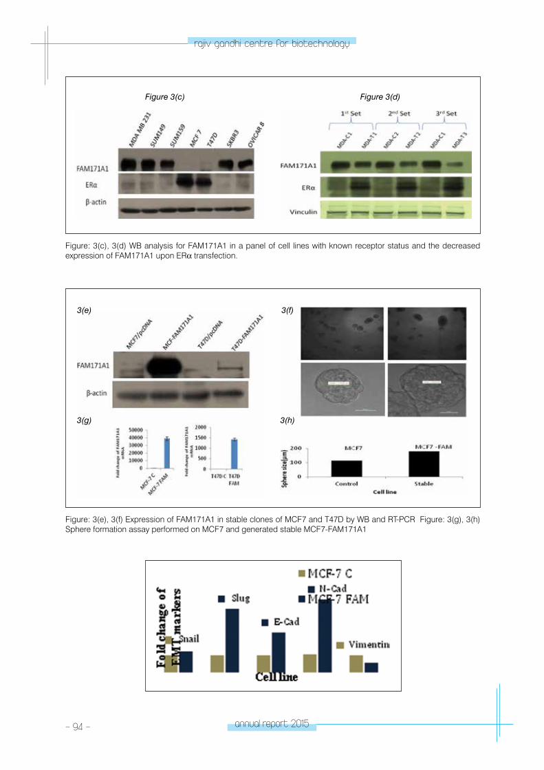
- 94 - annual report 2015
rajiv gandhi centre for biotechnology
Figure 3(c) Figure 3(d)
Figure: 3(c), 3(d) WB analysis for FAM171A1 in a panel of cell lines with known receptor status and the decreased expression of FAM171A1 upon ERα transfection.
Figure: 3(e), 3(f) Expression of FAM171A1 in stable clones of MCF7 and T47D by WB and RT-PCR Figure: 3(g), 3(h) Sphere formation assay performed on MCF7 and generated stable MCF7-FAM171A1
3(e)
3(g)
3(f)
3(h)
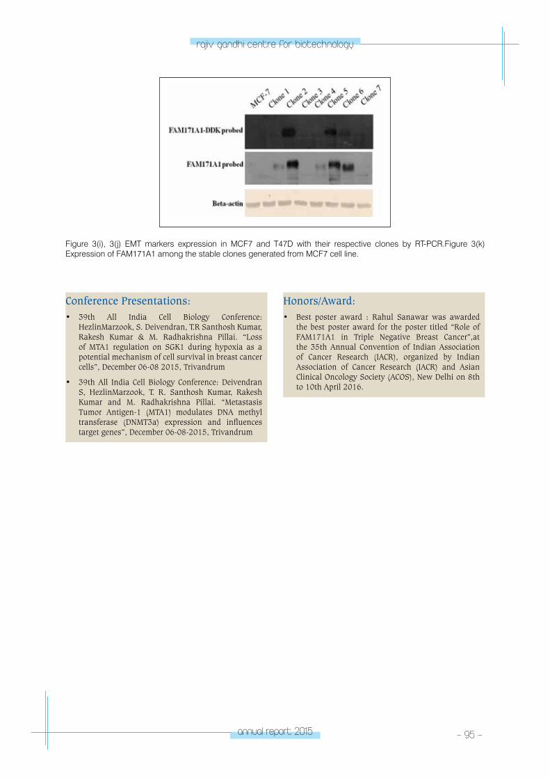
annual report 2015 - 95 -
rajiv gandhi centre for biotechnology
Figure 3(i), 3(j) EMT markers expression in MCF7 and T47D with their respective clones by RT-PCR.Figure 3(k) Expression of FAM171A1 among the stable clones generated from MCF7 cell line.
Conference Presentations:• 39th All India Cell Biology Conference:
HezlinMarzook, S. Deivendran, T.R Santhosh Kumar, Rakesh Kumar & M. Radhakrishna Pillai. “Loss of MTA1 regulation on SGK1 during hypoxia as a potential mechanism of cell survival in breast cancer cells”, December 06-08 2015, Trivandrum
• 39th All India Cell Biology Conference: Deivendran S, HezlinMarzook, T. R. Santhosh Kumar, Rakesh Kumar and M. Radhakrishna Pillai. “Metastasis Tumor Antigen-1 (MTA1) modulates DNA methyl transferase (DNMT3a) expression and influences target genes”, December 06-08-2015, Trivandrum
Honors/Award:• Best poster award : Rahul Sanawar was awarded
the best poster award for the poster titled “Role of FAM171A1 in Triple Negative Breast Cancer”,at the 35th Annual Convention of Indian Association of Cancer Research (IACR), organized by Indian Association of Cancer Research (IACR) and Asian Clinical Oncology Society (ACOS), New Delhi on 8th to 10th April 2016.
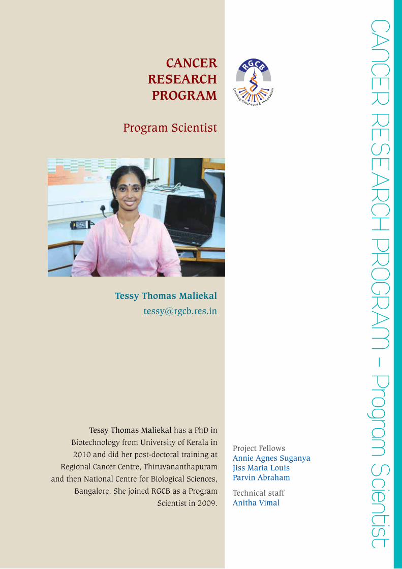
CAnCeR ReSeA
RCH pRoGRAM
- program Scientist
CANCER RESEARCH PROGRAM
Program Scientist
Tessy Thomas Maliekal
Project FellowsAnnie Agnes SuganyaJiss Maria LouisParvin Abraham
Technical staffAnitha Vimal
Tessy Thomas Maliekal has a PhD in
Biotechnology from University of Kerala in
2010 and did her post-doctoral training at
Regional Cancer Centre, Thiruvananthapuram
and then National Centre for Biological Sciences,
Bangalore. She joined RGCB as a Program
Scientist in 2009.
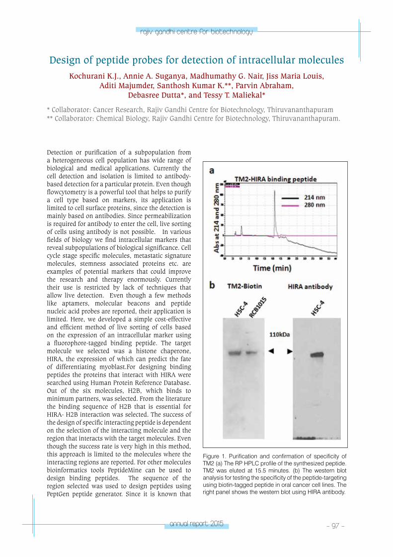
annual report 2015 - 97 -
rajiv gandhi centre for biotechnology
Detection or purification of a subpopulation from a heterogeneous cell population has wide range of biological and medical applications. Currently the cell detection and isolation is limited to antibody-based detection for a particular protein. Even though flowcytometry is a powerful tool that helps to purify a cell type based on markers, its application is limited to cell surface proteins, since the detection is mainly based on antibodies. Since permeabilization is required for antibody to enter the cell, live sorting of cells using antibody is not possible. In various fields of biology we find intracellular markers that reveal subpopulations of biological significance. Cell cycle stage specific molecules, metastatic signature molecules, stemness associated proteins etc. are examples of potential markers that could improve the research and therapy enormously. Currently their use is restricted by lack of techniques that allow live detection. Even though a few methods like aptamers, molecular beacons and peptide nucleic acid probes are reported, their application is limited. Here, we developed a simple cost-effective and efficient method of live sorting of cells based on the expression of an intracellular marker using a fluorophore-tagged binding peptide. The target molecule we selected was a histone chaperone, HIRA, the expression of which can predict the fate of differentiating myoblast.For designing binding peptides the proteins that interact with HIRA were searched using Human Protein Reference Database. Out of the six molecules, H2B, which binds to minimum partners, was selected. From the literature the binding sequence of H2B that is essential for HIRA- H2B interaction was selected. The success of the design of specific interacting peptide is dependent on the selection of the interacting molecule and the region that interacts with the target molecules. Even though the success rate is very high in this method, this approach is limited to the molecules where the interacting regions are reported. For other molecules bioinformatics tools PeptideMine can be used to design binding peptides. The sequence of the region selected was used to design peptides using PeptGen peptide generator. Since it is known that
Design of peptide probes for detection of intracellular moleculesKochurani K.J., Annie A. Suganya, Madhumathy G. Nair, Jiss Maria Louis,
Aditi Majumder, Santhosh Kumar K.**, Parvin Abraham, Debasree Dutta*, and Tessy T. Maliekal*
* Collaborator: Cancer Research, Rajiv Gandhi Centre for Biotechnology, Thiruvananthapuram ** Collaborator: Chemical Biology, Rajiv Gandhi Centre for Biotechnology, Thiruvananthapuram.
Figure 1. Purification and confirmation of specificity of TM2 (a) The RP HPLC profile of the synthesized peptide. TM2 was eluted at 15.5 minutes. (b) The western blot analysis for testing the specificity of the peptide-targeting using biotin-tagged peptide in oral cancer cell lines. The right panel shows the western blot using HIRA antibody.
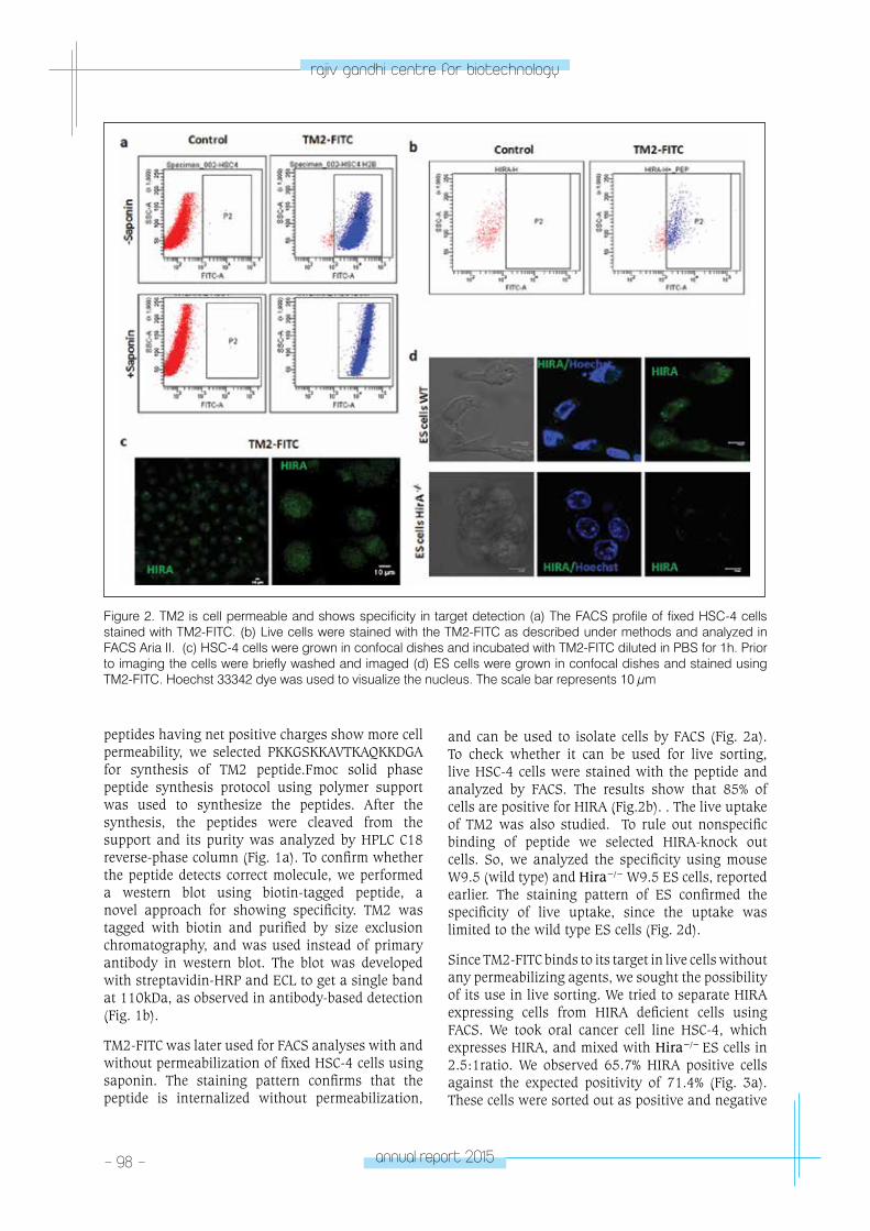
- 98 - annual report 2015
rajiv gandhi centre for biotechnology
peptides having net positive charges show more cell permeability, we selected PKKGSKKAVTKAQKKDGA for synthesis of TM2 peptide.Fmoc solid phase peptide synthesis protocol using polymer support was used to synthesize the peptides. After the synthesis, the peptides were cleaved from the support and its purity was analyzed by HPLC C18 reverse-phase column (Fig. 1a). To confirm whether the peptide detects correct molecule, we performed a western blot using biotin-tagged peptide, a novel approach for showing specificity. TM2 was tagged with biotin and purified by size exclusion chromatography, and was used instead of primary antibody in western blot. The blot was developed with streptavidin-HRP and ECL to get a single band at 110kDa, as observed in antibody-based detection (Fig. 1b).
TM2-FITC was later used for FACS analyses with and without permeabilization of fixed HSC-4 cells using saponin. The staining pattern confirms that the peptide is internalized without permeabilization,
and can be used to isolate cells by FACS (Fig. 2a). To check whether it can be used for live sorting, live HSC-4 cells were stained with the peptide and analyzed by FACS. The results show that 85% of cells are positive for HIRA (Fig.2b). . The live uptake of TM2 was also studied. To rule out nonspecific binding of peptide we selected HIRA-knock out cells. So, we analyzed the specificity using mouse W9.5 (wild type) and Hira−/− W9.5 ES cells, reported earlier. The staining pattern of ES confirmed the specificity of live uptake, since the uptake was limited to the wild type ES cells (Fig. 2d).
Since TM2-FITC binds to its target in live cells without any permeabilizing agents, we sought the possibility of its use in live sorting. We tried to separate HIRA expressing cells from HIRA deficient cells using FACS. We took oral cancer cell line HSC-4, which expresses HIRA, and mixed with Hira−/− ES cells in 2.5:1ratio. We observed 65.7% HIRA positive cells against the expected positivity of 71.4% (Fig. 3a). These cells were sorted out as positive and negative
Figure 2. TM2 is cell permeable and shows specificity in target detection (a) The FACS profile of fixed HSC-4 cells stained with TM2-FITC. (b) Live cells were stained with the TM2-FITC as described under methods and analyzed in FACS Aria II. (c) HSC-4 cells were grown in confocal dishes and incubated with TM2-FITC diluted in PBS for 1h. Prior to imaging the cells were briefly washed and imaged (d) ES cells were grown in confocal dishes and stained using TM2-FITC. Hoechst 33342 dye was used to visualize the nucleus. The scale bar represents 10 μm

annual report 2015 - 99 -
rajiv gandhi centre for biotechnology
populations with gates P2 and P3 and were allowed to grow for 5 days in 8-well chamber slide. The DIC images confirm that the positive population was HSC-4 keratinocytes and the negative population was ES cells (Fig. 3b). Again wild type ES cells and Hira−/− ES cells were mixed in 1:1 ratio and sorted as mentioned before. The observed ratio of positivity was 49.6% against expected ratio of 50% (Fig. 3a). The ES cells were viable and formed colonies in 5 days as shown in Fig. 3b. These cells grown in slide chambers were stained with antibodies for HIRA and an epithelial marker, CD44. The results
showed that the P2 population representing HSC-4 cells were all positive for HIRA and CD44, while the P3 population showed negativity for both molecules (Fig 3c). Likewise, in the separation of wild type and Hira−/− ES cells the fractionation was successful as the colonies of P2 population showed positivity for HIRA, and negative population did not show any staining. The P3 population in the mixture of ES cells and HSC-4 cells, however, showed <1% of contamination from CD44+ HIRA+ cells. The cross contamination in P3 population for the mixture of null and wild type ES cells was <4%.
Figure 3. TM2-FITC live sorting (a) The FACS profile of a mixture of live HSC-4 cells and ES cells stained with TM2-FITC. The two populations in P2 and P3 gate were sorted. (b) Sorted cells were grown in 8-well chamber slide for 5 days, and DIC images were taken (c) the 5-day old sorted cells were stained with indicated antibodies after fixation with 4% PFA. The scale bar represents 10 μm
- 100 - annual report 2015
rajiv gandhi centre for biotechnology
Characterization and evaluation of signaling pathways regulating self-renewal of oral cancer stem cells
Jiss Maria Louis, Annie Agnes Suganya, Madhumathy G. Nair, Kochurani K.J., Balagopal P.G.*, Paul Sebastian*, Keshava K. Datta**, Harsha Gowda**, Ani Das§, Abdul Balagopal§§, Rajagopal R.***, Akhilesh Pandey**, Tessy Thomas Maliekal.
* Collaborator: Surgical Oncology, Regional Cancer Centre, Thiruvananthapuram. ** Institute of Bioinformatics, Bangalore . *** Veterinary Surgeon, Rajiv Gandhi Centre for Biotechnology, Thiruvananthapuram. § Cancer Research Program, Rajiv Gandhi Centre for Biotechnology, Thiruvananthapuram. §§ Neuro Stem Cell Biology, Rajiv Gandhi Centre for Biotechnology, Thiruvananthapuram.
Chemotherapy is one of the standard therapeutic approaches for cancer. While chemotherapy is often capable of inducing cell death in tumors and reducing the tumor bulk, in many cases there is a chance for recurrence. This is a major problem in oral cancer treatment. The most important reason for recurrence is the regulation of chemoresistance and self-renewal ability by microenvironment. Cancer stem cell, a population of cancer cells with self-renewing property is the key reason for tumor relapse and recurrence. Signaling pathways and their intermediates regulating self-renewal are important in oral cancer recurrence. So for introducing better treatment strategies it is necessary to study how self-renewal is regulated by these factors. Understanding the signaling networks of CSCs that sustain the “CSC-state” will provide insights to the potential targets that could be used to develop chemotherapeutic drugs. Since majority of the signaling events are mediated through phosphorylation, a SILAC-based phosphoproteome analysis of sphere cultured cells (with enriched CSCs) in comparison to monolayer cells were performed to understand the signaling events that sustain the CSC characteristics. Based on the results obtained several pathways were selected for further analysis.
ALDH1A1 is a marker for different forms of cancer including oral cancer. It regulates self-renewal through modulating retinoic acid pathway. ALDH1A1-mRFP reporter construct that reports the self-renewing cells were made in the lab previously. Using ALDH1A1-mRFP transfected oral cancer cells, mRFP+ and mRFP- cells were sorted by FACS. 1x10⁴
mRFP+ cells were able to form tumor in NOD/SCID mice after 4 weeks of injection, while mRFP- cells were not able to generate tumor (Fig 4a and b).
5-Fluorouracil is a chemotherapeutic drug used for oral cancer treatment. ALDH1A1-mRFP cells were treated with 5-FU, and after 3 days of treatment trypan blue staining was performed. When compared with untreated cells, where 0.15x10⁶ viable cells were present, only 2000 cells survived in 5-FU treated cells. These results show that few viable cells, which are resistant to 5-FU remain after treatment, which can cause recurrence. When ALDH1A1-mRFP cells were treated with 5-FU along with growth factors that can activate signaling pathways (WNT, EGF and TGF-β), viable cell count was increased when compared tothe control (Fig 4c). This result shows that these growth factors regulate recurrence.To study whether signaling pathways provide self-renewal ability during 5-FU treatment, ALDH1A1-mRFP cells were treated with 5-FU along with growth factors and it was observed, that there was a significant increase in mRFP expression in the case of EGF and TGF-β treatment when compared with control (5-FU alone treated) (Fig 4d). Immunofluorescence analysis of mRFP expression with or without the growth factors in a co-culture system comprising cancer stem cells, cancer cells and fibroblast cells confirmed that EGF and TGF-β up-regulates self-renewal ability (Fig 4d). Collectively our results prove that EGF and TGF-β present in the tumor microenvironment can impart self-renewal ability to cells surviving chemotherapy, which results in recurrence.
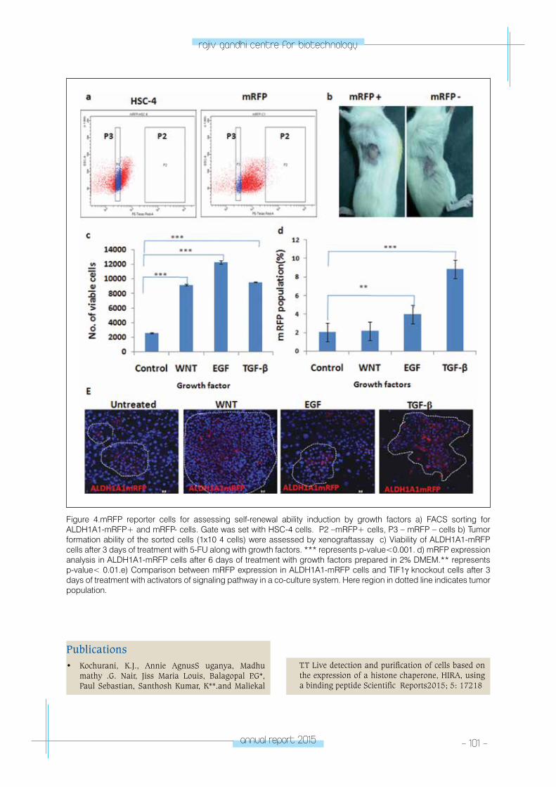
annual report 2015 - 101 -
rajiv gandhi centre for biotechnology
Figure 4.mRFP reporter cells for assessing self-renewal ability induction by growth factors a) FACS sorting for ALDH1A1-mRFP+ and mRFP- cells. Gate was set with HSC-4 cells. P2 –mRFP+ cells, P3 – mRFP – cells b) Tumor formation ability of the sorted cells (1x10 4 cells) were assessed by xenograftassay c) Viability of ALDH1A1-mRFP cells after 3 days of treatment with 5-FU along with growth factors. *** represents p-value<0.001. d) mRFP expression analysis in ALDH1A1-mRFP cells after 6 days of treatment with growth factors prepared in 2% DMEM.** represents p-value< 0.01.e) Comparison between mRFP expression in ALDH1A1-mRFP cells and TIF1γ knockout cells after 3 days of treatment with activators of signaling pathway in a co-culture system. Here region in dotted line indicates tumor population.
Publications• Kochurani, K.J., Annie AgnusS uganya, Madhu
mathy .G. Nair, Jiss Maria Louis, Balagopal P.G*, Paul Sebastian, Santhosh Kumar, K**.and Maliekal
T.T Live detection and purification of cells based on the expression of a histone chaperone, HIRA, using a binding peptide Scientific Reports2015; 5: 17218
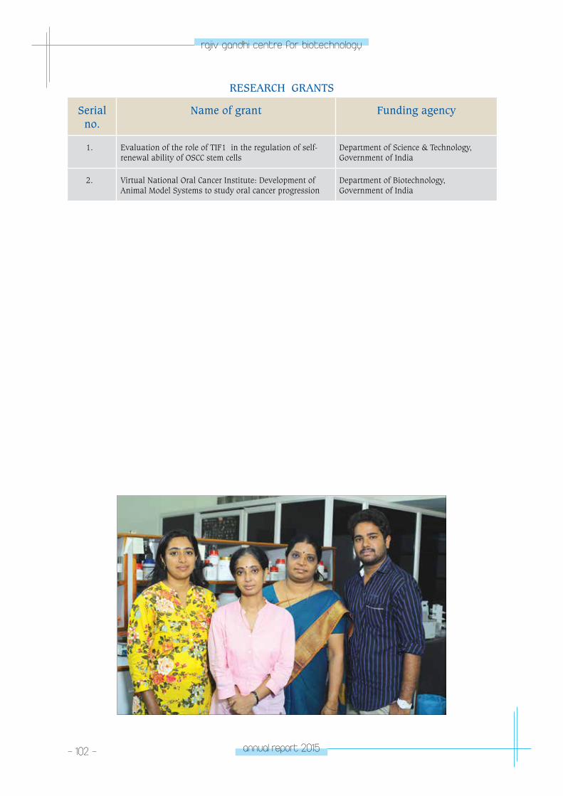
- 102 - annual report 2015
rajiv gandhi centre for biotechnology
RESEARCH GRANTS
Serial no.
Name of grant Funding agency
1. Evaluation of the role of TIF1β in the regulation of self-renewal ability of OSCC stem cells
Department of Science & Technology, Government of India
2. Virtual National Oral Cancer Institute: Development of Animal Model Systems to study oral cancer progression
Department of Biotechnology, Government of India
CAnCeR ReSeA
RCH pRoGRAM
- program Scientist
CANCER RESEARCH PROGRAM
Faculty Fellow
Radhika Nair
Research FellowReshma Murali
Radhika completed her PhD from the National
Institute of Immunology, New Delhi and then
trained as a Post Doctoral Fellow at the MRC-
Hutchison Cancer Cell Unit, Cambridge, UK
and Garvan Institute of Medical Research/ The
Kinghorn Cancer Centre, Sydney, Australia.
Radhika is a recipient of the Department of
Science and Technology’s (SERB) Ramanujan Re-
entry Fellowship.
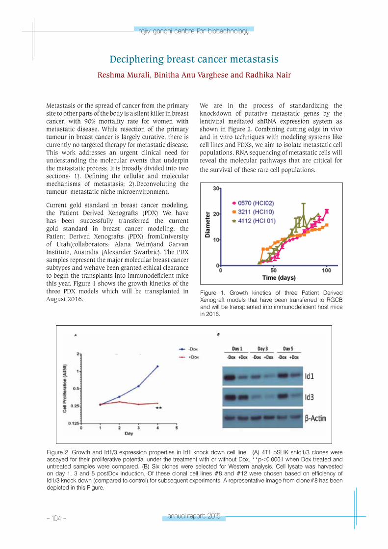
- 104 - annual report 2015
rajiv gandhi centre for biotechnology
Deciphering breast cancer metastasisReshma Murali, Binitha Anu Varghese and Radhika Nair
Metastasis or the spread of cancer from the primary site to other parts of the body is a silent killer in breast cancer, with 90% mortality rate for women with metastatic disease. While resection of the primary tumour in breast cancer is largely curative, there is currently no targeted therapy for metastatic disease. This work addresses an urgent clinical need for understanding the molecular events that underpin the metastatic process. It is broadly divided into two sections- 1). Defining the cellular and molecular mechanisms of metastasis; 2).Deconvoluting the tumour- metastatic niche microenvironment.
Current gold standard in breast cancer modeling, the Patient Derived Xenografts (PDX) We have has been successfully transferred the current gold standard in breast cancer modeling, the Patient Derived Xenografts (PDX) fromUniversity of Utah(collaborators: Alana Welm)and Garvan Institute, Australia (Alexander Swarbric). The PDX samples represent the major molecular breast cancer subtypes and wehave been granted ethical clearance to begin the transplants into immunodeficient mice this year. Figure 1 shows the growth kinetics of the three PDX models which will be transplanted in August 2016.
Figure 1. Growth kinetics of three Patient Derived Xenograft models that have been transferred to RGCB and will be transplanted into immunodeficient host mice in 2016.
Figure 2. Growth and Id1/3 expression properties in Id1 knock down cell line. (A) 4T1 pSLIK shId1/3 clones were assayed for their proliferative potential under the treatment with or without Dox. **p<0.0001 when Dox treated and untreated samples were compared. (B) Six clones were selected for Western analysis. Cell lysate was harvested on day 1, 3 and 5 postDox induction. Of these clonal cell lines #8 and #12 were chosen based on efficiency of Id1/3 knock down (compared to control) for subsequent experiments. A representative image from clone#8 has been depicted in this Figure.
We are in the process of standardizing the knockdown of putative metastatic genes by the lentiviral mediated shRNA expression system as shown in Figure 2. Combining cutting edge in vivo and in vitro techniques with modeling systems like cell lines and PDXs, we aim to isolate metastatic cell populations. RNA sequencing of metastatic cells will reveal the molecular pathways that are critical for the survival of these rare cell populations.
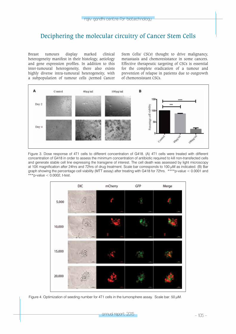
annual report 2015 - 105 -
rajiv gandhi centre for biotechnology
Breast tumours display marked clinical heterogeneity manifest in their histology, aetiology and gene expression profiles. In addition to this inter-tumoural heterogeneity, there also exists highly diverse intra-tumoural heterogeneity, with a subpopulation of tumour cells (termed Cancer
Deciphering the molecular circuitry of Cancer Stem Cells
Stem Cells/ CSCs) thought to drive malignancy, metastasis and chemoresistance in some cancers. Effective therapeutic targeting of CSCs is essential for the complete eradication of a tumour and prevention of relapse in patients due to outgrowth of chemoresistant CSCs.
Figure 3. Dose response of 4T1 cells to different concentration of G418. (A) 4T1 cells were treated with different concentration of G418 in order to assess the minimum concentration of antibiotic required to kill non-transfected cells and generate stable cell line expressing the transgene of interest. The cell death was assessed by light microscopy at 10X magnification after 24hrs and 72hrs of drug treatment. Scale bar corresponds to 100 μM as indicated. (B) Bar graph showing the percentage cell viability (MTT assay) after treating with G418 for 72hrs. ****p-value < 0.0001 and ***p-value < 0.0002, t-test.
Figure 4. Optimization of seeding number for 4T1 cells in the tumorsphere assay. Scale bar: 50 μM.
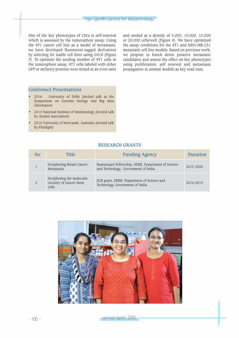
- 106 - annual report 2015
rajiv gandhi centre for biotechnology
Conference Presentations• 2016 University of Delhi (Invited talk at the
Symposium on Genome biology and Big data informatics)
• 2015 National Institute of Immunology (Invited talk by Alumni association)
• 2015 University of Newcastle, Australia (Invited talk by Fluidigm)
RESEARCH GRANTS
No Title Funding Agency Duration
1Deciphering Breast Cancer Metastasis
Ramanujan Fellowship, SERB, Department of Science and Technology, Government of India.
2015-2020
2Deciphering the molecular circuitry of Cancer Stem Cells
ECR grant, SERB, Department of Science and Technology, Government of India.
2016-2019
One of the key phenotypes of CSCs is self-renewal which is assessed by the tumorsphere assay. Using the 4T1 cancer cell line as a model of metastasis, we have developed fluorescent-tagged derivatives by selecting for stable cell lines using G418 (Figure 3). To optimize the seeding number of 4T1 cells in the tumorsphere assay, 4T1 cells labeled with either GFP or mCherry proteins were mixed at an even ratio
and seeded at a density of 5,000, 10,000, 15,000 or 20,000 cells/well (Figure 4). We have optimized the assay conditions for the 4T1 and MDA-MB-231 metastatic cell line models. Based on previous work, we propose to knock down putative metastatic candidates and assess the effect on key phenotypes using proliferation, self renewal and metastasis propagation in animal models as key read outs.
CARDIoV
ASCulAR DISeASe A
nD DIABeteS BIoloGY pRoGRAM
laboratory 1
CARDIOVASCULAR DISEASE AND DIABETES
BIOLOGY PROGRAMLaboratory - 1
Chandrasekharan C Kartha
Program ScientistsSurya Ramachandran, PhDSumi S. PhD
PhD StudentsVikas Panchal, MPharmJaya Mary Thomas, MPharm
Project FellowsVinitha A. MSc (SRF)
Animal TechnicianSreehari V.G.
C C Kartha is a MD in Pathology from All India
Institute of Medical Sciences, New Delhi. He
moved to RGCB in 2009 from Sree Chitra Tirunal
Institute for Medical Sciences & Technology,
Trivandrum where he was Senior Professor
& Head, Division of Cellular & Molecular
Cardiology. Professor Kartha is a fellow of the
Royal College of Physicians (London), National
Academy of Medical Sciences (India) and Indian
Academy of Sciences.
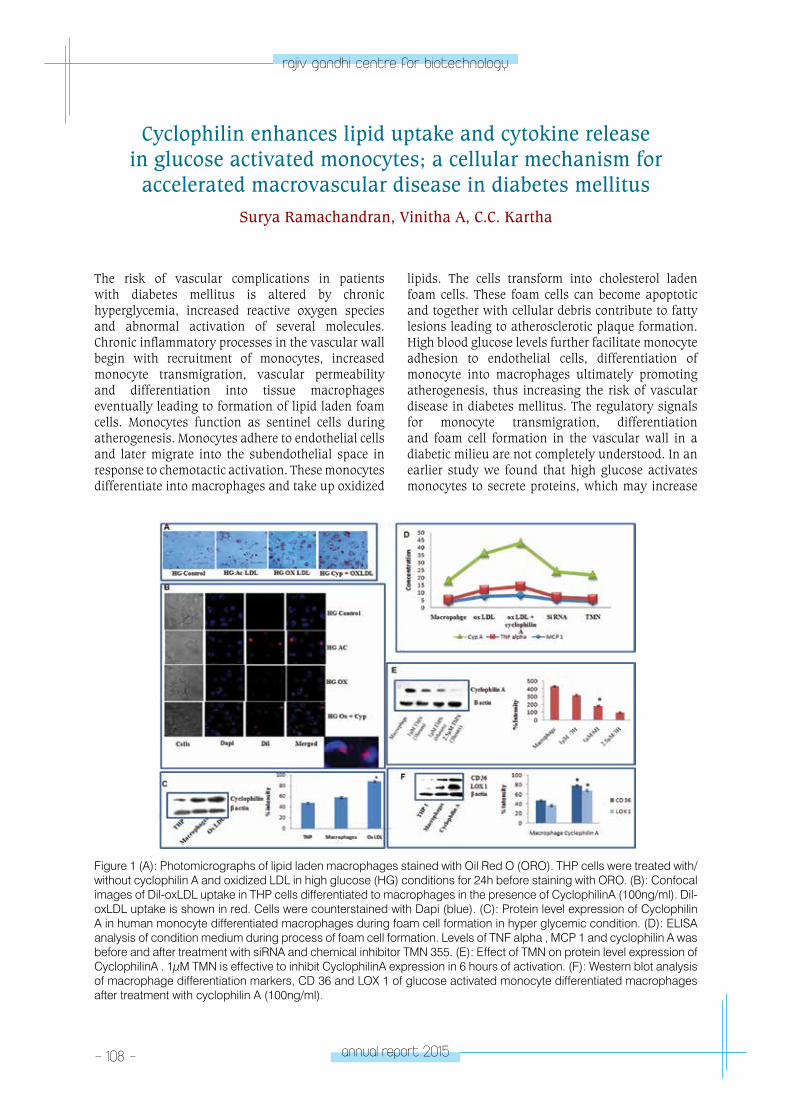
- 108 - annual report 2015
rajiv gandhi centre for biotechnology
The risk of vascular complications in patients with diabetes mellitus is altered by chronic hyperglycemia, increased reactive oxygen species and abnormal activation of several molecules. Chronic inflammatory processes in the vascular wall begin with recruitment of monocytes, increased monocyte transmigration, vascular permeability and differentiation into tissue macrophages eventually leading to formation of lipid laden foam cells. Monocytes function as sentinel cells during atherogenesis. Monocytes adhere to endothelial cells and later migrate into the subendothelial space in response to chemotactic activation. These monocytes differentiate into macrophages and take up oxidized
lipids. The cells transform into cholesterol laden foam cells. These foam cells can become apoptotic and together with cellular debris contribute to fatty lesions leading to atherosclerotic plaque formation. High blood glucose levels further facilitate monocyte adhesion to endothelial cells, differentiation of monocyte into macrophages ultimately promoting atherogenesis, thus increasing the risk of vascular disease in diabetes mellitus. The regulatory signals for monocyte transmigration, differentiation and foam cell formation in the vascular wall in a diabetic milieu are not completely understood. In an earlier study we found that high glucose activates monocytes to secrete proteins, which may increase
Cyclophilin enhances lipid uptake and cytokine release in glucose activated monocytes; a cellular mechanism for
accelerated macrovascular disease in diabetes mellitusSurya Ramachandran, Vinitha A, C.C. Kartha
Figure 1 (A): Photomicrographs of lipid laden macrophages stained with Oil Red O (ORO). THP cells were treated with/without cyclophilin A and oxidized LDL in high glucose (HG) conditions for 24h before staining with ORO. (B): Confocal images of Dil-oxLDL uptake in THP cells differentiated to macrophages in the presence of CyclophilinA (100ng/ml). Dil-oxLDL uptake is shown in red. Cells were counterstained with Dapi (blue). (C): Protein level expression of Cyclophilin A in human monocyte differentiated macrophages during foam cell formation in hyper glycemic condition. (D): ELISA analysis of condition medium during process of foam cell formation. Levels of TNF alpha , MCP 1 and cyclophilin A was before and after treatment with siRNA and chemical inhibitor TMN 355. (E): Effect of TMN on protein level expression of CyclophilinA . 1μM TMN is effective to inhibit CyclophilinA expression in 6 hours of activation. (F): Western blot analysis of macrophage differentiation markers, CD 36 and LOX 1 of glucose activated monocyte differentiated macrophages after treatment with cyclophilin A (100ng/ml).
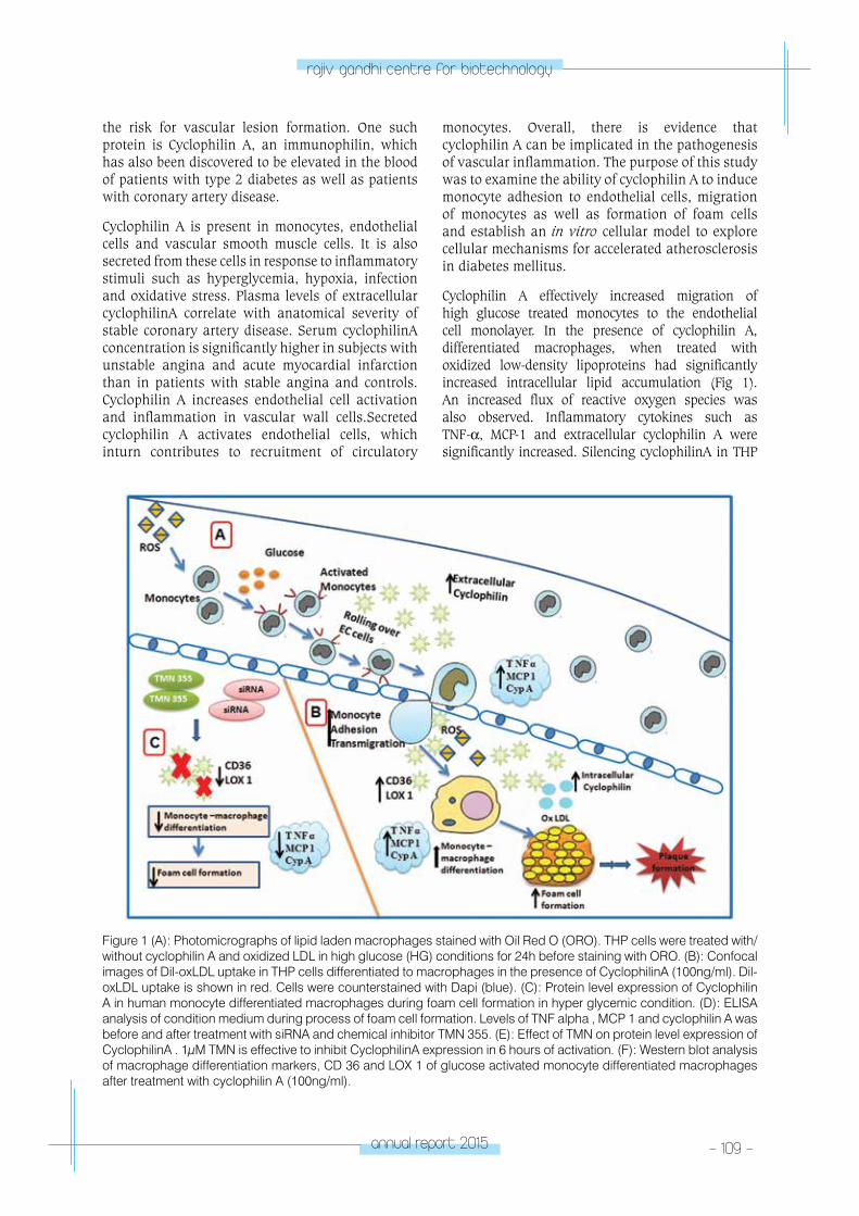
annual report 2015 - 109 -
rajiv gandhi centre for biotechnology
the risk for vascular lesion formation. One such protein is Cyclophilin A, an immunophilin, which has also been discovered to be elevated in the blood of patients with type 2 diabetes as well as patients with coronary artery disease.
Cyclophilin A is present in monocytes, endothelial cells and vascular smooth muscle cells. It is also secreted from these cells in response to inflammatory stimuli such as hyperglycemia, hypoxia, infection and oxidative stress. Plasma levels of extracellular cyclophilinA correlate with anatomical severity of stable coronary artery disease. Serum cyclophilinA concentration is significantly higher in subjects with unstable angina and acute myocardial infarction than in patients with stable angina and controls. Cyclophilin A increases endothelial cell activation and inflammation in vascular wall cells.Secreted cyclophilin A activates endothelial cells, which inturn contributes to recruitment of circulatory
monocytes. Overall, there is evidence that cyclophilin A can be implicated in the pathogenesis of vascular inflammation. The purpose of this study was to examine the ability of cyclophilin A to induce monocyte adhesion to endothelial cells, migration of monocytes as well as formation of foam cells and establish an in vitro cellular model to explore cellular mechanisms for accelerated atherosclerosis in diabetes mellitus.
Cyclophilin A effectively increased migration of high glucose treated monocytes to the endothelial cell monolayer. In the presence of cyclophilin A, differentiated macrophages, when treated with oxidized low-density lipoproteins had significantly increased intracellular lipid accumulation (Fig 1). An increased flux of reactive oxygen species was also observed. Inflammatory cytokines such as TNF-α, MCP-1 and extracellular cyclophilin A were significantly increased. Silencing cyclophilinA in THP
Figure 1 (A): Photomicrographs of lipid laden macrophages stained with Oil Red O (ORO). THP cells were treated with/without cyclophilin A and oxidized LDL in high glucose (HG) conditions for 24h before staining with ORO. (B): Confocal images of Dil-oxLDL uptake in THP cells differentiated to macrophages in the presence of CyclophilinA (100ng/ml). Dil-oxLDL uptake is shown in red. Cells were counterstained with Dapi (blue). (C): Protein level expression of Cyclophilin A in human monocyte differentiated macrophages during foam cell formation in hyper glycemic condition. (D): ELISA analysis of condition medium during process of foam cell formation. Levels of TNF alpha , MCP 1 and cyclophilin A was before and after treatment with siRNA and chemical inhibitor TMN 355. (E): Effect of TMN on protein level expression of CyclophilinA . 1μM TMN is effective to inhibit CyclophilinA expression in 6 hours of activation. (F): Western blot analysis of macrophage differentiation markers, CD 36 and LOX 1 of glucose activated monocyte differentiated macrophages after treatment with cyclophilin A (100ng/ml).
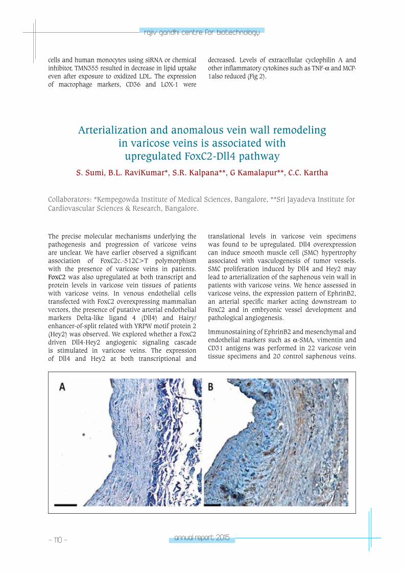
- 110 - annual report 2015
rajiv gandhi centre for biotechnology
cells and human monocytes using siRNA or chemical inhibitor, TMN355 resulted in decrease in lipid uptake even after exposure to oxidized LDL. The expression of macrophage markers, CD36 and LOX-1 were
decreased. Levels of extracellular cyclophilin A and other inflammatory cytokines such as TNF-α and MCP-1also reduced (Fig 2).
Arterialization and anomalous vein wall remodeling in varicose veins is associated with
upregulated FoxC2-Dll4 pathwayS. Sumi, B.L. RaviKumar*, S.R. Kalpana**, G Kamalapur**, C.C. Kartha
Collaborators: *Kempegowda Institute of Medical Sciences, Bangalore, **Sri Jayadeva Institute for Cardiovascular Sciences & Research, Bangalore.
The precise molecular mechanisms underlying the pathogenesis and progression of varicose veins are unclear. We have earlier observed a significant association of FoxC2c.-512C>T polymorphism with the presence of varicose veins in patients. FoxC2 was also upregulated at both transcript and protein levels in varicose vein tissues of patients with varicose veins. In venous endothelial cells transfected with FoxC2 overexpressing mammalian vectors, the presence of putative arterial endothelial markers Delta-like ligand 4 (Dll4) and Hairy/enhancer-of-split related with YRPW motif protein 2 (Hey2) was observed. We explored whether a FoxC2 driven Dll4-Hey2 angiogenic signaling cascade is stimulated in varicose veins. The expression of Dll4 and Hey2 at both transcriptional and
translational levels in varicose vein specimens was found to be upregulated. Dll4 overexpression can induce smooth muscle cell (SMC) hypertrophy associated with vasculogenesis of tumor vessels. SMC proliferation induced by Dll4 and Hey2 may lead to arterialization of the saphenous vein wall in patients with varicose veins. We hence assessed in varicose veins, the expression pattern of EphrinB2, an arterial specific marker acting downstream to FoxC2 and in embryonic vessel development and pathological angiogenesis.
Immunostaining of EphrinB2 and mesenchymal and endothelial markers such as α-SMA, vimentin and CD31 antigens was performed in 22 varicose vein tissue specimens and 20 control saphenous veins.
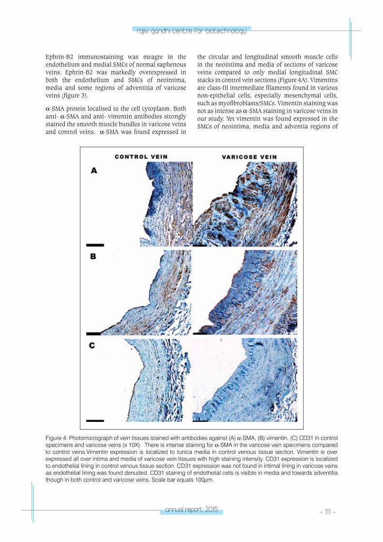
annual report 2015 - 111 -
rajiv gandhi centre for biotechnology
Ephrin-B2 immunostaining was meagre in the endothelium and medial SMCs of normal saphenous veins. Ephrin-B2 was markedly overexpressed in both the endothelium and SMCs of neointima, media and some regions of adventitia of varicose veins (figure 3).
α-SMA protein localised to the cell cytoplasm. Both anti- α-SMA and anti- vimentin antibodies strongly stained the smooth muscle bundles in varicose veins and control veins. α-SMA was found expressed in
the circular and longitudinal smooth muscle cells in the neointima and media of sections of varicose veins compared to only medial longitudinal SMC stacks in control vein sections (Figure 4A). Vimentins are class-III intermediate filaments found in various non-epithelial cells, especially mesenchymal cells, such as myofibroblasts/SMCs. Vimentin staining was not as intense as α-SMA staining in varicose veins in our study. Yet vimentin was found expressed in the SMCs of neointima, media and adventia regions of
Figure 4: Photomicrograph of vein tissues stained with antibodies against (A) α-SMA, (B) vimentin, (C) CD31 in control specimens and varicose veins (x 10X). There is intense staining for α-SMA in the varicose vein specimens compared to control veins.Vimentin expression is localized to tunica media in control venous tissue section. Vimentin is over expressed all over intima and media of varicose vein tissues with high staining intensity. CD31 expression is localized to endothelial lining in control venous tissue section. CD31 expression was not found in intimal lining in varicose veins as endothelial lining was found denuded. CD31 staining of endothelial cells is visible in media and towards adventitia though in both control and varicose veins. Scale bar equals 100μm.
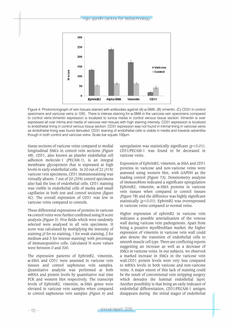
- 112 - annual report 2015
rajiv gandhi centre for biotechnology
Figure 4: Photomicrograph of vein tissues stained with antibodies against (A) α-SMA, (B) vimentin, (C) CD31 in control specimens and varicose veins (x 10X). There is intense staining for α-SMA in the varicose vein specimens compared to control veins.Vimentin expression is localized to tunica media in control venous tissue section. Vimentin is over expressed all over intima and media of varicose vein tissues with high staining intensity. CD31 expression is localized to endothelial lining in control venous tissue section. CD31 expression was not found in intimal lining in varicose veins as endothelial lining was found denuded. CD31 staining of endothelial cells is visible in media and towards adventitia though in both control and varicose veins. Scale bar equals 100μm.
tissue sections of varicose veins compared to medial longitudinal SMCs in control vein sections (Figure 4B). CD31, also known as platelet endothelial cell adhesion molecule-1 (PECAM-1), is an integral membrane glycoprotein that is expressed at high levels in early endothelial cells. In 20 out of 22 (91%) varicose vein specimens, CD31 immunostaining was virtually absent. 7 out of 20 (35%) control specimens also had the loss of endothelial cells. CD31 staining was visible in endothelial cells of media and small capillaries in both test and control sections (figure 4C). The overall expression of CD31 was low in varicose veins compared to controls.
These differential expressions of proteins in varicose vs control veins were further confirmed using H score analysis (Figure 5). Five fields which were randomly selected were analysed in all vein specimens. H score was calculated by multiplying the intensity of staining (0 for no staining, 1 for weak staining, 2 for medium and 3 for intense staining) with percentage of immunopositive cells. Calculated H score values were between 0 and 300.
The expression patterns of EphrinB2, vimentin, α-SMA and CD31 were assessed in varicose vein tissues and control saphenous vein samples. Quantitative analysis was performed at both mRNA and protein levels by quantitative real time PCR and western blot respectively. The transcript levels of EphrinB2, vimentin, α-SMA genes were elevated in varicose vein samples when compared to control saphenous vein samples (Figure 6) and
upregulation was statistically significant (p<0.01).CD31/PECAM-1 was found to be decreased in varicose veins.
Expression of EphrinB2, vimentin, α-SMA and CD31 proteins in varicose and non-varicose veins were assessed using western blot, with GAPDH as the loading control (Figure 7A). Densitometry analysis of immunoblots indicated a significant upregulation EphrinB2, vimentin, α-SMA proteins in varicose vein tissues when compared to control tissues (Figure 7B) and the difference was highly significant statistically (p<0.01). EphrinB2 was overexpressed in varicose veins compared to normal veins.
Higher expression of ephrinB2 in varicose vein indicates a possible arterialization of the venous wall during varicose vein pathogenesis. Apart from being a putative myofibroblast marker, the higher expression of vimentin in varicose vein wall could also denote the transition of endothelial cells to smooth muscle cell type. There are conflicting reports suggesting an increase as well as a decrease of SMCs in varicose veins. In our subjects, we observed a marked increase in SMCs in the varicose vein wall.CD31 protein levels were very less compared to mRNA levels in both varicose and non-varicose veins. A major extent of this lack of staining could be the result of conventional vein stripping surgery which denudes the luminal endothelial layer. Another possibility is that being an early indicator of endothelial differentiation, CD31/PECAM-1 antigen disappears during the initial stages of endothelial
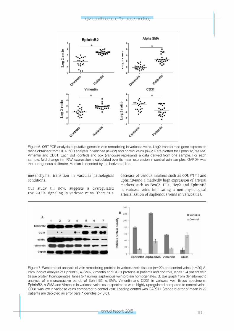
annual report 2015 - 113 -
rajiv gandhi centre for biotechnology
mesenchymal transition in vascular pathological conditions.
Our study till now, suggests a dysregulated FoxC2-Dll4 signaling in varicose veins. There is a
decrease of venous markers such as COUP-TFII and EphrinB4and a markedly high expression of arterial markers such as FoxC2, Dll4, Hey2 and EphrinB2 in varicose veins implicating a non-physiological arterialization of saphenous veins in varicosities.
Figure 6. QRT-PCR analysis of putative genes in vein remodeling in varicose veins. Log2-transformed gene expression ratios obtained from QRT- PCR analysis in varicose (n=22) and control veins (n=20) are plotted for EphrinB2, α-SMA, Vimentin and CD31. Each dot (control) and box (varicose) represents a data derived from one sample. For each sample, fold change in mRNA expression is calculated over its mean expression in control vein samples. GAPDH was the endogenous calibrator. Median is denoted by the horizontal line.
Figure 7. Western blot analysis of vein remodeling proteins in varicose vein tissues (n=22) and control veins (n=20).A. Immunoblot analysis of EphrinB2, α-SMA, Vimentin and CD31 proteins in patients and controls, lanes 1-4 patient vein tissue protein homogenates, lanes 5-7 normal saphenous vein protein homogenates. B. Bar graph from densitometric analysis of immunoreactive bands of EphrinB2, α-SMA, Vimentin and CD31 in varicose vein tissue specimens. EphrinB2, α-SMA and Vimentin in varicose vein tissue specimens were highly upregulated compared to control veins. CD31 was low in varicose veins compared to control vein. Loading control was GAPDH. Standard error of mean in 22 patients are depicted as error bars.* denotes p<0.01.
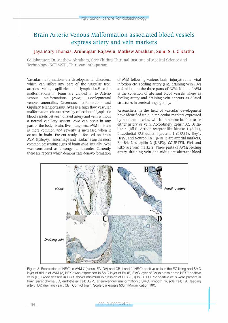
- 114 - annual report 2015
rajiv gandhi centre for biotechnology
Vascular malformations are developmental disorders, which can affect any part of the vascular tree: arteries, veins, capillaries and lymphatics.Vascular malformations in brain are divided in to Arterio Venous Malformations (AVM), Developmental venous anomalies, Cavernous malformations and Capillary telangiectasias. AVM is a high flow vascular malformation, characterized by collection of dysplastic blood vessels between dilated artery and vein without a normal capillary system. AVM can occur in any part of the body: brain, liver, lungs etc. AVM in brain is more common and severity is increased when it occurs in brain. Present study is focused on brain AVM. Epilepsy, hemorrhage and headache are the most common presenting signs of brain AVM. Initially, AVM was considered as a congenital disorder. Currently there are reports which demonstrate denovo formation
of AVM following various brain injury/trauma, viral infection etc. Feeding artery (FA), draining vein (DV) and nidus are the three parts of AVM. Nidus of AVM is the collection of aberrant blood vessels where as feeding artery and draining vein appears as dilated structures in cerebral angiography.
Researchers in the field of vascular development have identified unique molecular markers expressed by endothelial cells, which determine its fate to be either artery or vein. Accordingly EphrinB2, Delta-like 4 (Dll4), Activin-receptor-like kinase 1 (Alk1), Endothelial PAS domain protein 1 (EPAS1), Hey1, Hey2, and Neuropilin 1 (NRP1) are arterial markers; EphB4, Neuropilin 2 (NRP2), COUP-TFII, Flt4 and Rtk5 are vein markers. Three parts of AVM; feeding artery, draining vein and nidus are aberrant blood
Brain Arterio Venous Malformation associated blood vessels express artery and vein markers
Jaya Mary Thomas, Arumugam Rajavelu, Mathew Abraham, Sumi S, C C Kartha
Collaborator: Dr. Mathew Abraham, Sree Chithra Thirunal Institute of Medical Science and Technology (SCTIMST), Thiruvananthapuram.
Figure 8. Expression of HEY2 in AVM 7 (nidus, FA, DV) and CB 1 and 2. HEY2 positive cells in the EC lining and SMC layer of nidus of AVM (A).HEY2 was expressed in SMC layer of FA (B).SMC layer of DV express some HEY2 positive cells (C). Blood vessels in CB 1 shows minimum expression of HEY2 (D).In CB1 HEY2 positive cells were present in brain parenchyma.EC, endothelial cell: AVM, arteriovenous malformation ; SMC, smooth muscle cell; FA, feeding artery; DV, draining vein ; CB, Control brain. Scale bar equals 50μm.Magnification 10X.
Nidus Feeding artery
Draining vein
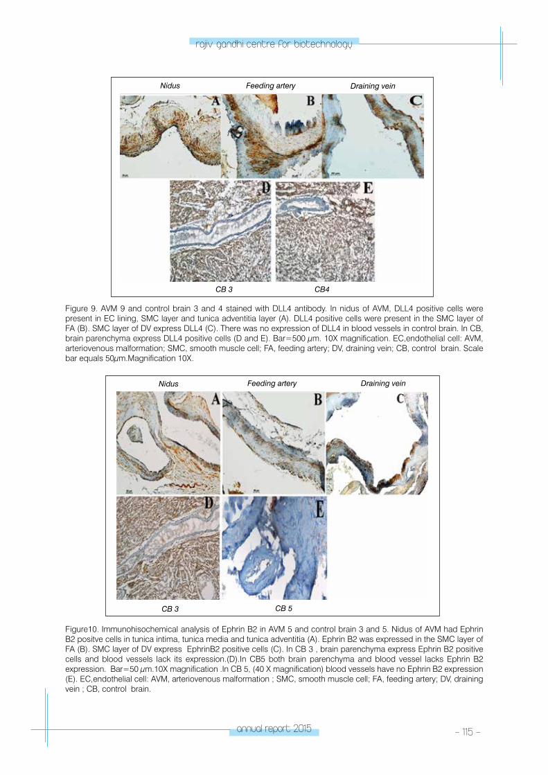
annual report 2015 - 115 -
rajiv gandhi centre for biotechnology
Figure 9. AVM 9 and control brain 3 and 4 stained with DLL4 antibody. In nidus of AVM, DLL4 positive cells were present in EC lining, SMC layer and tunica adventitia layer (A). DLL4 positive cells were present in the SMC layer of FA (B). SMC layer of DV express DLL4 (C). There was no expression of DLL4 in blood vessels in control brain. In CB, brain parenchyma express DLL4 positive cells (D and E). Bar=500 μm. 10X magnification. EC,endothelial cell: AVM, arteriovenous malformation; SMC, smooth muscle cell; FA, feeding artery; DV, draining vein; CB, control brain. Scale bar equals 50μm.Magnification 10X.
CB 3 CB4
Nidus Feeding artery Draining vein
Figure10. Immunohisochemical analysis of Ephrin B2 in AVM 5 and control brain 3 and 5. Nidus of AVM had Ephrin B2 positve cells in tunica intima, tunica media and tunica adventitia (A). Ephrin B2 was expressed in the SMC layer of FA (B). SMC layer of DV express EphrinB2 positive cells (C). In CB 3 , brain parenchyma express Ephrin B2 positive cells and blood vessels lack its expression.(D).In CB5 both brain parenchyma and blood vessel lacks Ephrin B2 expression. Bar=50 μm.10X magnification .In CB 5, (40 X magnification) blood vessels have no Ephrin B2 expression (E). EC,endothelial cell: AVM, arteriovenous malformation ; SMC, smooth muscle cell; FA, feeding artery; DV, draining vein ; CB, control brain.
Nidus Feeding artery
CB 5CB 3
Draining vein
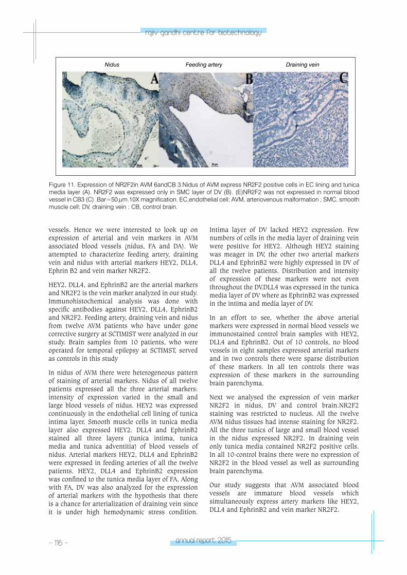
- 116 - annual report 2015
rajiv gandhi centre for biotechnology
Figure 11. Expression of NR2F2in AVM 6andCB 3.Nidus of AVM express NR2F2 positive cells in EC lining and tunica media layer (A). NR2F2 was expressed only in SMC layer of DV (B). (E)NR2F2 was not expressed in normal blood vessel in CB3 (C) .Bar=50 μm.10X magnification. EC,endothelial cell: AVM, arteriovenous malformation ; SMC, smooth muscle cell; DV, draining vein ; CB, control brain.
Nidus Feeding artery Draining vein
vessels. Hence we were interested to look up on expression of arterial and vein markers in AVM associated blood vessels (nidus, FA and DA). We attempted to characterize feeding artery, draining vein and nidus with arterial markers HEY2, DLL4, Ephrin B2 and vein marker NR2F2.
HEY2, DLL4, and EphrinB2 are the arterial markers and NR2F2 is the vein marker analyzed in our study. Immunohistochemical analysis was done with specific antibodies against HEY2, DLL4, EphrinB2 and NR2F2. Feeding artery, draining vein and nidus from twelve AVM patients who have under gone corrective surgery at SCTIMIST were analyzed in our study. Brain samples from 10 patients, who were operated for temporal epilepsy at SCTIMST, served as controls in this study
In nidus of AVM there were heterogeneous pattern of staining of arterial markers. Nidus of all twelve patients expressed all the three arterial markers: intensity of expression varied in the small and large blood vessels of nidus. HEY2 was expressed continuously in the endothelial cell lining of tunica intima layer. Smooth muscle cells in tunica media layer also expressed HEY2. DLL4 and EphrinB2 stained all three layers (tunica intima, tunica media and tunica adventitia) of blood vessels of nidus. Arterial markers HEY2, DLL4 and EphrinB2 were expressed in feeding arteries of all the twelve patients. HEY2, DLL4 and EphrinB2 expression was confined to the tunica media layer of FA. Along with FA, DV was also analyzed for the expression of arterial markers with the hypothesis that there is a chance for arterialization of draining vein since it is under high hemodynamic stress condition.
Intima layer of DV lacked HEY2 expression. Few numbers of cells in the media layer of draining vein were positive for HEY2. Although HEY2 staining was meager in DV, the other two arterial markers DLL4 and EphrinB2 were highly expressed in DV of all the twelve patients. Distribution and intensity of expression of these markers were not even throughout the DV.DLL4 was expressed in the tunica media layer of DV where as EphrinB2 was expressed in the intima and media layer of DV.
In an effort to see, whether the above arterial markers were expressed in normal blood vessels we immunostained control brain samples with HEY2, DLL4 and EphrinB2. Out of 10 controls, no blood vessels in eight samples expressed arterial markers and in two controls there were sparse distribution of these markers. In all ten controls there was expression of these markers in the surrounding brain parenchyma.
Next we analysed the expression of vein marker NR2F2 in nidus, DV and control brain.NR2F2 staining was restricted to nucleus. All the twelve AVM nidus tissues had intense staining for NR2F2.All the three tunics of large and small blood vessel in the nidus expressed NR2F2. In draining vein only tunica media contained NR2F2 positive cells. In all 10-control brains there were no expression of NR2F2 in the blood vessel as well as surrounding brain parenchyma.
Our study suggests that AVM associated blood vessels are immature blood vessels which simultaneously express artery markers like HEY2, DLL4 and EphrinB2 and vein marker NR2F2.
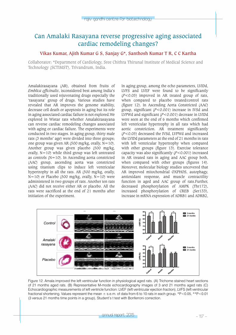
annual report 2015 - 117 -
rajiv gandhi centre for biotechnology
Amalakirasayana (AR), obtained from fruits of Emblica officinalis, isconsidered best among India’s traditionally used rejuvenating drugs especially the ‘rasayana’ group of drugs. Various studies have revealed that AR improves the genome stability, decrease cell death or apoptosis in aging but its role in aging associated cardiac failure is not explored.We explored in Wistar rats whether Amalakirasayana can reverse cardiac remodeling changes associated with aging or cardiac failure. The experiments were conducted in two stages. In aging group, thirty male rats (3 months’ age) were divided into three groups; one group was given AR (500 mg/kg, orally, N=10). Another group was given placebo (500 mg/kg, orally, N=10) while third group was left untreated as controls (N=10). In Ascending aorta constricted (AAC) group, ascending aorta was constricted using titanium clips to induce left ventricular hypertrophy in all the rats. AR (500 mg/kg, orally, N=10) or Placebo (500 mg/kg, orally, N=10) were administered in two groups of rats. Another ten rats (AAC) did not receive either AR or placebo. All the rats were sacrificed at the end of 21 months after initiation of the experiment.
In aging group, among the echo parameters, LVIDd, LVFS and LVEF were found to be significantly (P<0.05) improved in AR treated group of rats, when compared to placebo treated/control rats (figure 12). In Ascending Aorta Constricted (AAC) group, significant (P<0.001) increase in IVSd and LVPWd and significant (P<0.001) decrease in LVIDd were seen at the end of 6 months which confirmed left ventricular hypertrophy in all rats which had aortic constriction. AR treatment significantly (P<0.05) decreased the IVSd, LVPWd and increased the LVIDd parameters at the end of 21 months in rats with left ventricular hypertrophy when compared with other groups (figure 13). Exercise tolerance capacity was also significantly (P<0.001) increased in AR treated rats in aging and AAC group both, when compared with other groups (figures 14). Moreover, molecular biology studies uncovered that AR improved mitochondrial OXPHOS, autophagy, antioxidant response, and muscle contractility function in aged and AAC group of rats.Further, decreased phosphorylation of AMPK (Thr172), increased phosphorylation of CREB (Ser133), increase in mRNA expression of ADRB1 and ADRB2,
Figure 12. Amala improved the left ventricular function in physiological aged rats. (A) Trichome stained heart sections of 21 months aged rats. (B) Representative M-mode echocardiography images of 3 and 21 months aged rats (C) Echocardiographic measurements of left ventricle function: LVEF (left ventricular ejection fraction), LVFS (left ventricular fractional shortening. Values represent the mean ± s.e.m. of data from 6 to 10 rats in each group. *P<0.05, **P<0.01 (3 versus 21 months time points in a group), Student’s t test with Bonferroni correction.
Control
Amalaki rasayana
Placebo
Can Amalaki Rasayana reverse progressive aging associated cardiac remodeling changes?
Vikas Kumar, Ajith Kumar G S, Sanjay G*, Santhosh Kumar T R, C C Kartha
Collaborator: *Department of Cardiology, Sree Chithra Thirunal Institute of Medical Science and Technology (SCTIMST), Trivandrum, India.
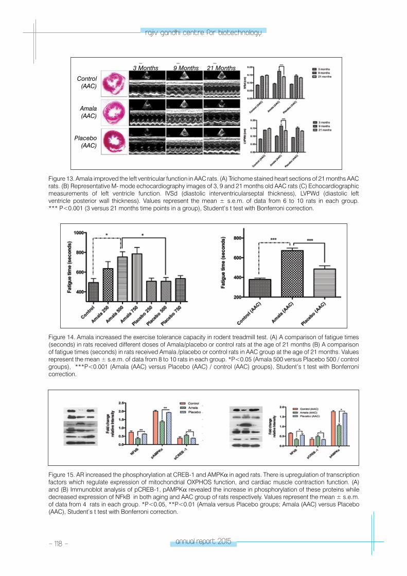
- 118 - annual report 2015
rajiv gandhi centre for biotechnology
Control(AAC)
3 Months 9 Months 21 Months
Amala (AAC)
Placebo (AAC)
Figure 13. Amala improved the left ventricular function in AAC rats. (A) Trichome stained heart sections of 21 months AAC rats. (B) Representative M- mode echocardiography images of 3, 9 and 21 months old AAC rats (C) Echocardiographic measurements of left ventricle function. IVSd (diastolic interventricularseptal thickness), LVPWd (diastolic left ventricle posterior wall thickness). Values represent the mean ± s.e.m. of data from 6 to 10 rats in each group. *** P<0.001 (3 versus 21 months time points in a group), Student’s t test with Bonferroni correction.
Figure 14. Amala increased the exercise tolerance capacity in rodent treadmill test. (A) A comparison of fatigue times (seconds) in rats received different doses of Amala/placebo or control rats at the age of 21 months (B) A comparison of fatigue times (seconds) in rats received Amala /placebo or control rats in AAC group at the age of 21 months. Values represent the mean ± s.e.m. of data from 8 to 10 rats in each group. *P<0.05 (Amala 500 versus Placebo 500 / control groups), ***P<0.001 (Amala (AAC) versus Placebo (AAC) / control (AAC) groups), Student’s t test with Bonferroni correction.
Figure 15. AR increased the phosphorylation at CREB-1 and AMPKα in aged rats. There is upregulation of transcription factors which regulate expression of mitochondrial OXPHOS function, and cardiac muscle contraction function. (A) and (B) Immunoblot analysis of pCREB-1, pAMPKα revealed the increase in phosphorylation of these proteins while decreased expression of NFkB in both aging and AAC group of rats respectively. Values represent the mean ± s.e.m. of data from 4 rats in each group. *P<0.05, **P<0.01 (Amala versus Placebo groups; Amala (AAC) versus Placebo (AAC), Student’s t test with Bonferroni correction.
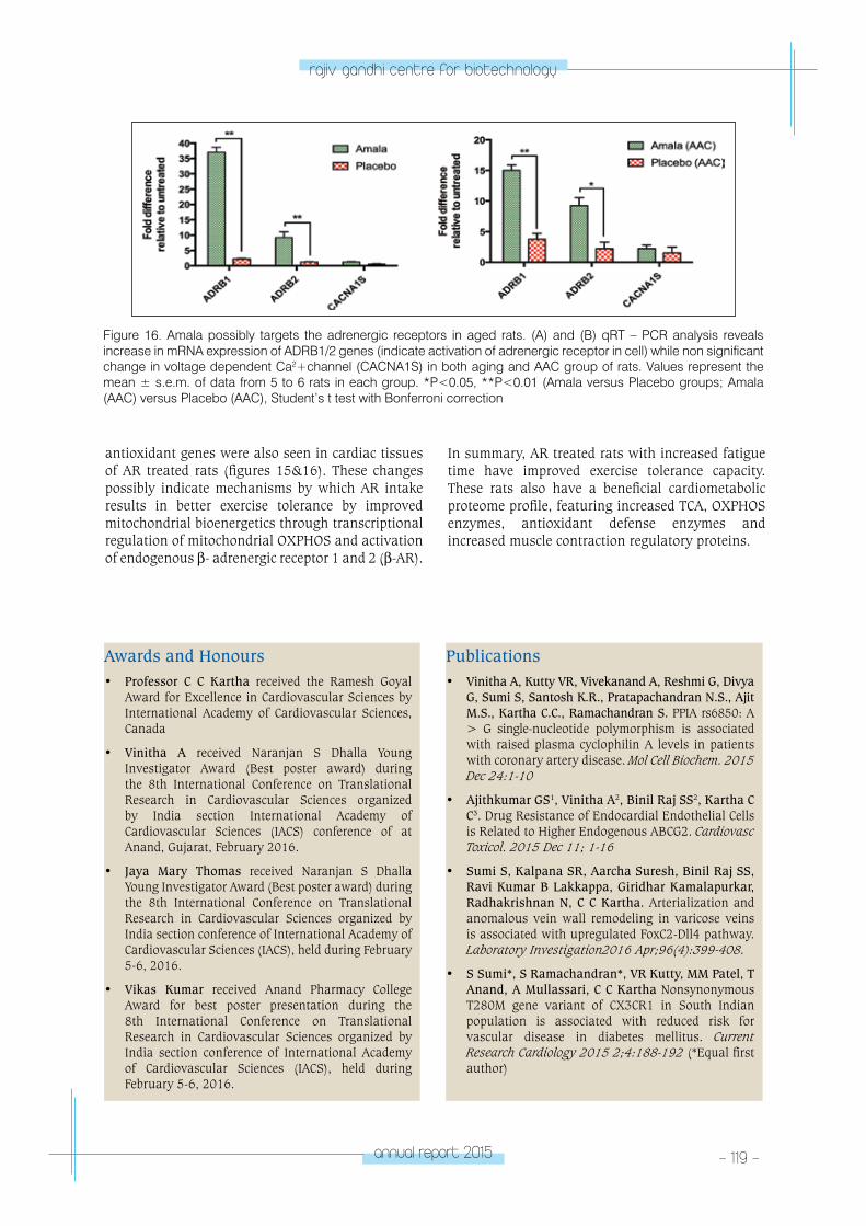
annual report 2015 - 119 -
rajiv gandhi centre for biotechnology
antioxidant genes were also seen in cardiac tissues of AR treated rats (figures 15&16). These changes possibly indicate mechanisms by which AR intake results in better exercise tolerance by improved mitochondrial bioenergetics through transcriptional regulation of mitochondrial OXPHOS and activation of endogenous β- adrenergic receptor 1 and 2 (β-AR).
In summary, AR treated rats with increased fatigue time have improved exercise tolerance capacity. These rats also have a beneficial cardiometabolic proteome profile, featuring increased TCA, OXPHOS enzymes, antioxidant defense enzymes and increased muscle contraction regulatory proteins.
Figure 16. Amala possibly targets the adrenergic receptors in aged rats. (A) and (B) qRT – PCR analysis reveals increase in mRNA expression of ADRB1/2 genes (indicate activation of adrenergic receptor in cell) while non significant change in voltage dependent Ca2+channel (CACNA1S) in both aging and AAC group of rats. Values represent the mean ± s.e.m. of data from 5 to 6 rats in each group. *P<0.05, **P<0.01 (Amala versus Placebo groups; Amala (AAC) versus Placebo (AAC), Student’s t test with Bonferroni correction
Awards and Honours• Professor C C Kartha received the Ramesh Goyal
Award for Excellence in Cardiovascular Sciences by International Academy of Cardiovascular Sciences, Canada
• Vinitha A received Naranjan S Dhalla Young Investigator Award (Best poster award) during the 8th International Conference on Translational Research in Cardiovascular Sciences organized by India section International Academy of Cardiovascular Sciences (IACS) conference of at Anand, Gujarat, February 2016.
• Jaya Mary Thomas received Naranjan S Dhalla Young Investigator Award (Best poster award) during the 8th International Conference on Translational Research in Cardiovascular Sciences organized by India section conference of International Academy of Cardiovascular Sciences (IACS), held during February 5-6, 2016.
• Vikas Kumar received Anand Pharmacy College Award for best poster presentation during the 8th International Conference on Translational Research in Cardiovascular Sciences organized by India section conference of International Academy of Cardiovascular Sciences (IACS), held during February 5-6, 2016.
Publications• Vinitha A, Kutty VR, Vivekanand A, Reshmi G, Divya
G, Sumi S, Santosh K.R., Pratapachandran N.S., Ajit M.S., Kartha C.C., Ramachandran S. PPIA rs6850: A > G single-nucleotide polymorphism is associated with raised plasma cyclophilin A levels in patients with coronary artery disease. Mol Cell Biochem. 2015 Dec 24:1-10
• Ajithkumar GS1, Vinitha A2, Binil Raj SS2, Kartha C C3. Drug Resistance of Endocardial Endothelial Cells is Related to Higher Endogenous ABCG2. Cardiovasc Toxicol. 2015 Dec 11; 1-16
• Sumi S, Kalpana SR, Aarcha Suresh, Binil Raj SS, Ravi Kumar B Lakkappa, Giridhar Kamalapurkar, Radhakrishnan N, C C Kartha. Arterialization and anomalous vein wall remodeling in varicose veins is associated with upregulated FoxC2-Dll4 pathway. Laboratory Investigation2016 Apr;96(4):399-408.
• S Sumi*, S Ramachandran*, VR Kutty, MM Patel, T Anand, A Mullassari, C C Kartha Nonsynonymous T280M gene variant of CX3CR1 in South Indian population is associated with reduced risk for vascular disease in diabetes mellitus. Current Research Cardiology 2015 2;4:188-192 (*Equal first author)
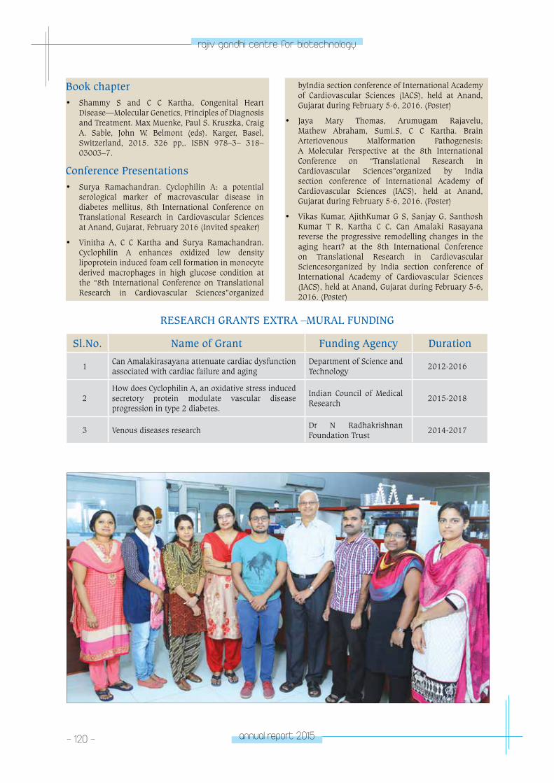
- 120 - annual report 2015
rajiv gandhi centre for biotechnology
Book chapter• Shammy S and C C Kartha, Congenital Heart
Disease—Molecular Genetics, Principles of Diagnosis and Treatment. Max Muenke, Paul S. Kruszka, Craig A. Sable, John W. Belmont (eds). Karger, Basel, Switzerland, 2015. 326 pp,. ISBN 978–3– 318–03003–7.
Conference Presentations• Surya Ramachandran. Cyclophilin A: a potential
serological marker of macrovascular disease in diabetes mellitus, 8th International Conference on Translational Research in Cardiovascular Sciences at Anand, Gujarat, February 2016 (Invited speaker)
• Vinitha A, C C Kartha and Surya Ramachandran. Cyclophilin A enhances oxidized low density lipoprotein induced foam cell formation in monocyte derived macrophages in high glucose condition at the “8th International Conference on Translational Research in Cardiovascular Sciences”organized
byIndia section conference of International Academy of Cardiovascular Sciences (IACS), held at Anand, Gujarat during February 5-6, 2016. (Poster)
• Jaya Mary Thomas, Arumugam Rajavelu, Mathew Abraham, Sumi.S, C C Kartha. Brain Arteriovenous Malformation Pathogenesis: A Molecular Perspective at the 8th International Conference on “Translational Research in Cardiovascular Sciences”organized by India section conference of International Academy of Cardiovascular Sciences (IACS), held at Anand, Gujarat during February 5-6, 2016. (Poster)
• Vikas Kumar, AjithKumar G S, Sanjay G, Santhosh Kumar T R, Kartha C C. Can Amalaki Rasayana reverse the progressive remodelling changes in the aging heart? at the 8th International Conference on Translational Research in Cardiovascular Sciencesorganized by India section conference of International Academy of Cardiovascular Sciences (IACS), held at Anand, Gujarat during February 5-6, 2016. (Poster)
RESEARCH GRANTS EXTRA –MURAL FUNDING
Sl.No. Name of Grant Funding Agency Duration
1Can Amalakirasayana attenuate cardiac dysfunction associated with cardiac failure and aging
Department of Science and Technology
2012-2016
2How does Cyclophilin A, an oxidative stress induced secretory protein modulate vascular disease progression in type 2 diabetes.
Indian Council of Medical Research
2015-2018
3 Venous diseases researchDr N Radhakrishnan Foundation Trust
2014-2017
CARDIoV
ASCulAR DISeASe A
nD DIABeteS BIoloGY pRoGRAM
laboratory 2
CARDIOVASCULAR DISEASE AND DIABETES
BIOLOGY PROGRAMLaboratory - 2
Rakesh. S. Laishram
PhD studentsSudheesh A.P.Nimmy FrancisNimmy MohanJohirul Islam
Project AssistantDivya Kandala
Rakesh Laishram received his PhD working
at the Centre for DNA Fingerprinting
and Diagnostics, Hyderabad and then
trained as a post doctoral fellow at the
Department of Pharmacology, University of
Wisconsin-Madison, USA. Rakesh is a DBT-
Welcome Trust Fellow.
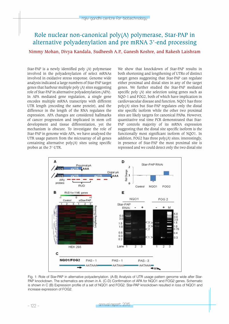
- 122 - annual report 2015
rajiv gandhi centre for biotechnology
Star-PAP is a newly identified poly (A) polymerase involved in the polyadenylation of select mRNAs involved in oxidative stress response. Genome wide analysis indicated a large numbers of Star-PAP target genes that harbour multiple poly (A) sites suggesting role of Star-PAP in alternative polyadenylation (APA). In APA mediated gene regulation, a single gene encodes multiple mRNA transcripts with different UTR length (encoding the same protein), and the difference in the length of the RNA regulates the expression. APA changes are considered hallmarks of cancer progression and implicated in stem cell development and tissue differentiation, yet the mechanism is obscure. To investigate the role of Star-PAP in genome wide APA, we have analysed the UTR usage pattern from the microarray of all genes containing alternative poly(A) sites using specific probes at the 3’-UTR.
Role nuclear non-canonical poly(A) polymerase, Star-PAP in alternative polyadenylation and pre mRNA 3’-end processing
Nimmy Mohan, Divya Kandala, Sudheesh A.P., Ganesh Koshre, and Rakesh Laishram
We show that knockdown of Star-PAP results in both shortening and lengthening of UTRs of distinct target genes suggesting that Star-PAP can regulate either proximal and distal sites in any of the target genes. We further studied the Star-PAP mediated specific poly (A) site selection using genes such as NQO-1 and FOG2, both of which have implication in cardiovascular disease and function. NQO1 has three poly(A) sites but Star-PAP regulates only the distal site specific isoform while the other two proximal sites are likely targets for canonical PAPα. However, quantitative real time PCR demonstrated that Star-PAP controls majority of its mRNA expression suggesting that the distal site specific isoform is the functionally most significant isoform of NQO1. In addition, FOG2 has three poly(A) sites; interestingly, in presence of Star-PAP the most proximal site is repressed and we could detect only the two distal site
Fig. 1: Role of Star-PAP in alternative polyadenylation. (A-B) Analysis of UTR usage pattern genome wide after Star-PAP knockdown. The schematics are shown in A. (C-D) Confirmation of APA for NQO1 and FOG2 genes. Schematic is shown in C (B) Expression profile of a set of NQO1 and FOG2. Star-PAP knockdown resulted in loss of NQO1 and increase expression of FOG2.
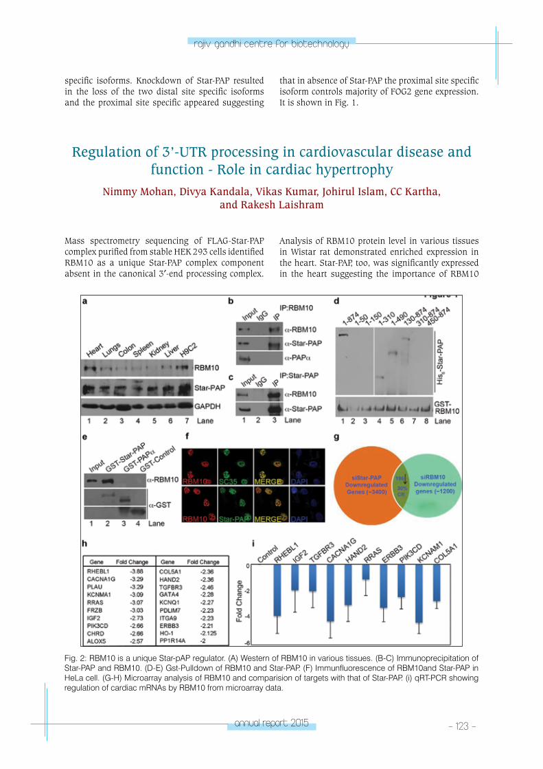
annual report 2015 - 123 -
rajiv gandhi centre for biotechnology
specific isoforms. Knockdown of Star-PAP resulted in the loss of the two distal site specific isoforms and the proximal site specific appeared suggesting
that in absence of Star-PAP the proximal site specific isoform controls majority of FOG2 gene expression. It is shown in Fig. 1.
Regulation of 3’-UTR processing in cardiovascular disease and function - Role in cardiac hypertrophy
Nimmy Mohan, Divya Kandala, Vikas Kumar, Johirul Islam, CC Kartha, and Rakesh Laishram
Mass spectrometry sequencing of FLAG-Star-PAP complex purified from stable HEK 293 cells identified RBM10 as a unique Star-PAP complex component absent in the canonical 3′-end processing complex.
Analysis of RBM10 protein level in various tissues in Wistar rat demonstrated enriched expression in the heart. Star-PAP, too, was significantly expressed in the heart suggesting the importance of RBM10
Fig. 2: RBM10 is a unique Star-pAP regulator. (A) Western of RBM10 in various tissues. (B-C) Immunoprecipitation of Star-PAP and RBM10. (D-E) Gst-Pulldown of RBM10 and Star-PAP. (F) Immunfluorescence of RBM10and Star-PAP in HeLa cell. (G-H) Microarray analysis of RBM10 and comparision of targets with that of Star-PAP. (i) qRT-PCR showing regulation of cardiac mRNAs by RBM10 from microarray data.
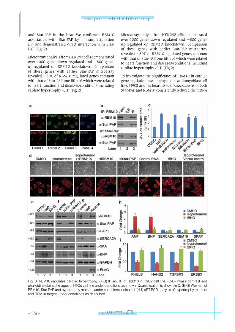
- 124 - annual report 2015
rajiv gandhi centre for biotechnology
Fig. 3: RBM10 regulates cardiac hypertrophy. (A-B) IF and IP of RBM10 in H9C2 cell line. (C-D) Phase contrast and phalloidine stained images of H9Ce cell line under conditions as shown. Quanitification is shown in D. (E-G) Western of RBM10, Star-PAP and hypertrophy markers under conditions indicated. (H-I) qRT-PCR analysis of hypertrophy markers and RBM10 targets under conditions as described.
and Star-PAP in the heart.We confirmed RBM10 association with Star-PAP by immunprecipitation (IP) and demonstrated direct interaction with Star-PAP (Fig. 2).
Microarray analysis from HEK 293 cells demonstrated over 1000 genes down regulated and ~800 genes up-regulated on RBM10 knockdown. Comparison of these genes with earlier Star-PAP microarray revealed ~30% of RBM10 regulated genes common with that of Star-PAP, one fifth of which were related to heart function and diseases/conditions including cardiac hypertrophy (CH) (Fig 2).
Microarray analysis from HEK 293 cells demonstrated over 1000 genes down regulated and ~800 genes up-regulated on RBM10 knockdown. Comparison of these genes with earlier Star-PAP microarray revealed ~30% of RBM10 regulated genes common with that of Star-PAP, one fifth of which were related to heart function and diseases/conditions including cardiac hypertrophy (CH) (Fig 2).
To investigate the significance of RBM10 in cardiac gene regulation, we employed rat cardiomyoblast cell line, H9C2 and rat heart tissue. Knockdowns of both Star-PAP and RBM10 consistently reduced the mRNA
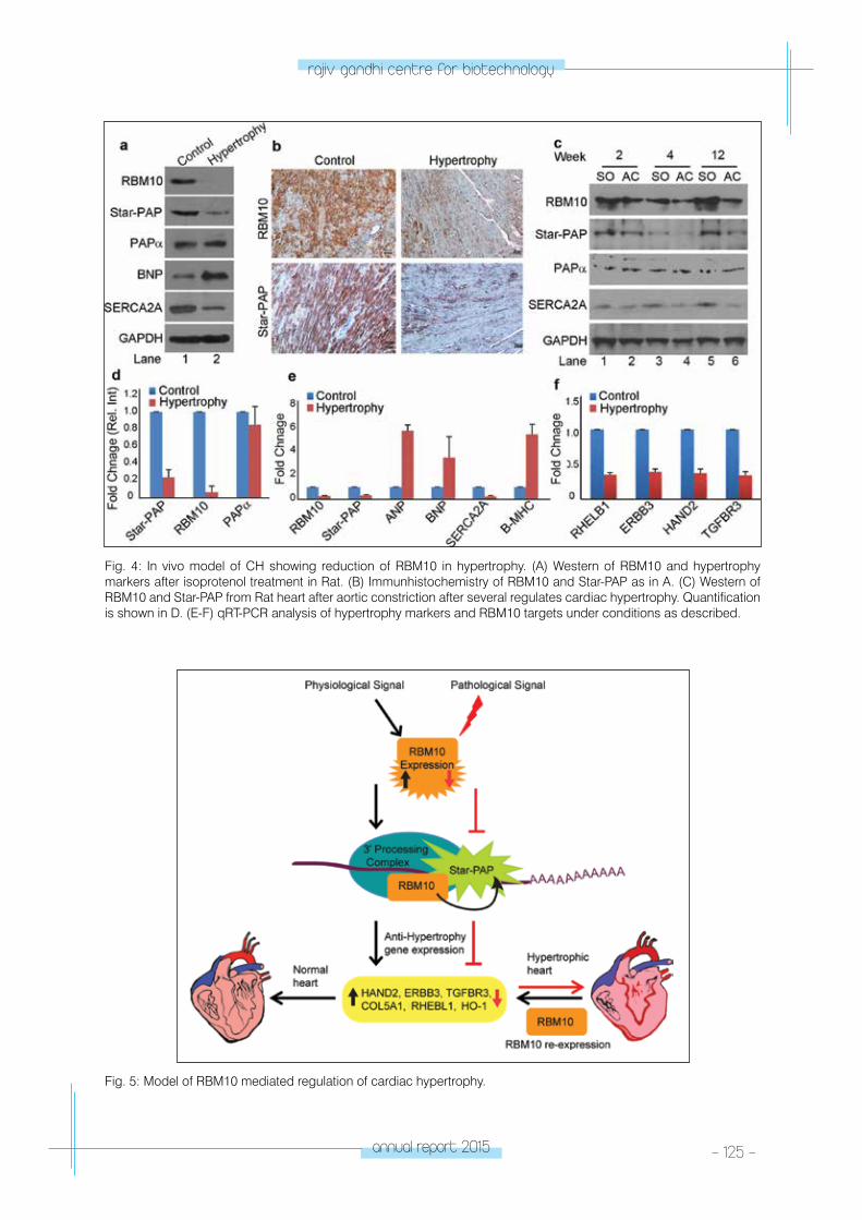
annual report 2015 - 125 -
rajiv gandhi centre for biotechnology
Fig. 4: In vivo model of CH showing reduction of RBM10 in hypertrophy. (A) Western of RBM10 and hypertrophy markers after isoprotenol treatment in Rat. (B) Immunhistochemistry of RBM10 and Star-PAP as in A. (C) Western of RBM10 and Star-PAP from Rat heart after aortic constriction after several regulates cardiac hypertrophy. Quantification is shown in D. (E-F) qRT-PCR analysis of hypertrophy markers and RBM10 targets under conditions as described.
Fig. 5: Model of RBM10 mediated regulation of cardiac hypertrophy.
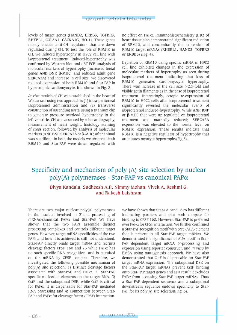
- 126 - annual report 2015
rajiv gandhi centre for biotechnology
levels of target genes (HAND2, ERBB3, TGFBR3, RHEBL1, COL5A1, CACNA1G, HO-1). These genes mostly encode anti-CH regulators that are down regulated during CH. To test the role of RBM10 in CH, we induced hypertrophy in H9C2 cell line with isoproterenol treatment. Induced-hypertrophy was confirmed by Western blot and qRT-PCR analysis of molecular markers of hypertrophy (increased foetal genes ANP, BNP, β-MHC; and reduced adult gene SERCA2A) and increase in cell size. We discovered reduced expression of both RBM10 and Star-PAP in hypertrophic cardiomyocyte. It is shown in Fig. 3.
In vivo models of CH was established in the heart of Wistar rats using two approaches (1) intra-peritoneal isoproterenol administration and (2) transverse constriction of ascending aorta using a titanium clip to generate pressure overload hypertrophy in the left ventricle. CH was assessed by echocardiography, measurement of heart weight, histology staining of cross section, followed by analysis of molecular markers (ANP, BNP, SERCA2A or β-MHC) after animal was sacrificed. In both the models we observed both RBM10 and Star-PAP were down regulated with
no effect on PAPα. Immunohistochemistry (IHC) of heart tissue also demonstrated significant reduction of RBM10, and concomitantly the expression of RBM10 target mRNAs (RHEBL1, HAND2, TGFBR3 or ERBB3) (Fig. 4).
Depletion of RBM10 using specific siRNA in H9C2 cell line exhibited changes in the expression of molecular markers of hypertrophy as seen during isoproterenol treatment indicating that loss of RBM10 generates cardiomyocyte hypertrophy. There was increase in the cell size >2.5-fold and visible actin filaments as in the case of isoproterenol treatment. Interestingly, ectopic re-expression of RBM10 in H9C2 cells after isoproterenol treatment significantly reversed the molecular events of isoproterenol induced-hypertrophy. While ANP, BNP or β-MHC that were up regulated on isoproterenol treatment was markedly reduced; SERCA2A expression was elevated to the normal level on RBM10 expression. These results indicate that RBM10 is a negative regulator of hypertrophy that attenuates myocyte hypertrophy(Fig 5).
Specificity and mechanism of poly (A) site selection by nuclear poly(A) polymerases - Star-PAP vs canonical PAPαDivya Kandala, Sudheesh A.P., Nimmy Mohan, Vivek A, Reshmi G.
and Rakesh Laishram
There are two major nuclear poly(A) polymerases in the nucleus involved in 3’-end processing of mRNAs-canonical PAPα and Star-PAP. We have shown that the two PAPs assemble distinct processing complexes and controls different target genes. However, target mRNA specificities of the two PAPs and how it is achieved is still not understood. Star-PAP directly binds target mRNA and recruits cleavage factors CPSF 160 and 73 while PAPα has no such specific RNA recognition, and is recruited on the mRNA by CPSF complex. Therefore, we investigated the following possible mechanism of poly(A) site selection: 1) Distinct cleavage factors associated with Star-PAP and PAPα. 2) Star-PAP specific nucleotide elements on the target RNA. 3) CstF and the suboptimal DSE, while CstF is critical for PAPα, it is dispensable for Star-PAP mediated RNA processing and 4) Competition between Star-PAP and PAPα for cleavage factor (CPSF) interaction.
We have shown that Star-PAP and PAPα has different interacting partners and that both compete for binding to CPSF 160. However, Star-PAP is preferred over PAPα for CPSF interaction. We further confirmed a Star-PAP recognition motif with core -AUA- element that is present in all Star-PAP target mRNAs. We demonstrated the significance of AUA motif in Star-PAP dependent target mRNA 3’-processing and expression using reporter construct, and in vitro by EMSA using mutagenesis approach. We have also demonstrated that CstF is dispensable for Star-PAP target mRNA expression. The suboptimal DSE on the Star-PAP target mRNAs prevent CstF binding onto Star-PAP target genes and as a result it excludes PAPα from accessing Star-PAP target mRNAs. Thus a Star-PAP dependent sequence and a suboptimal downstream sequence endows specificity to Star-PAP for its poly(A) site selection(Fig. 6).
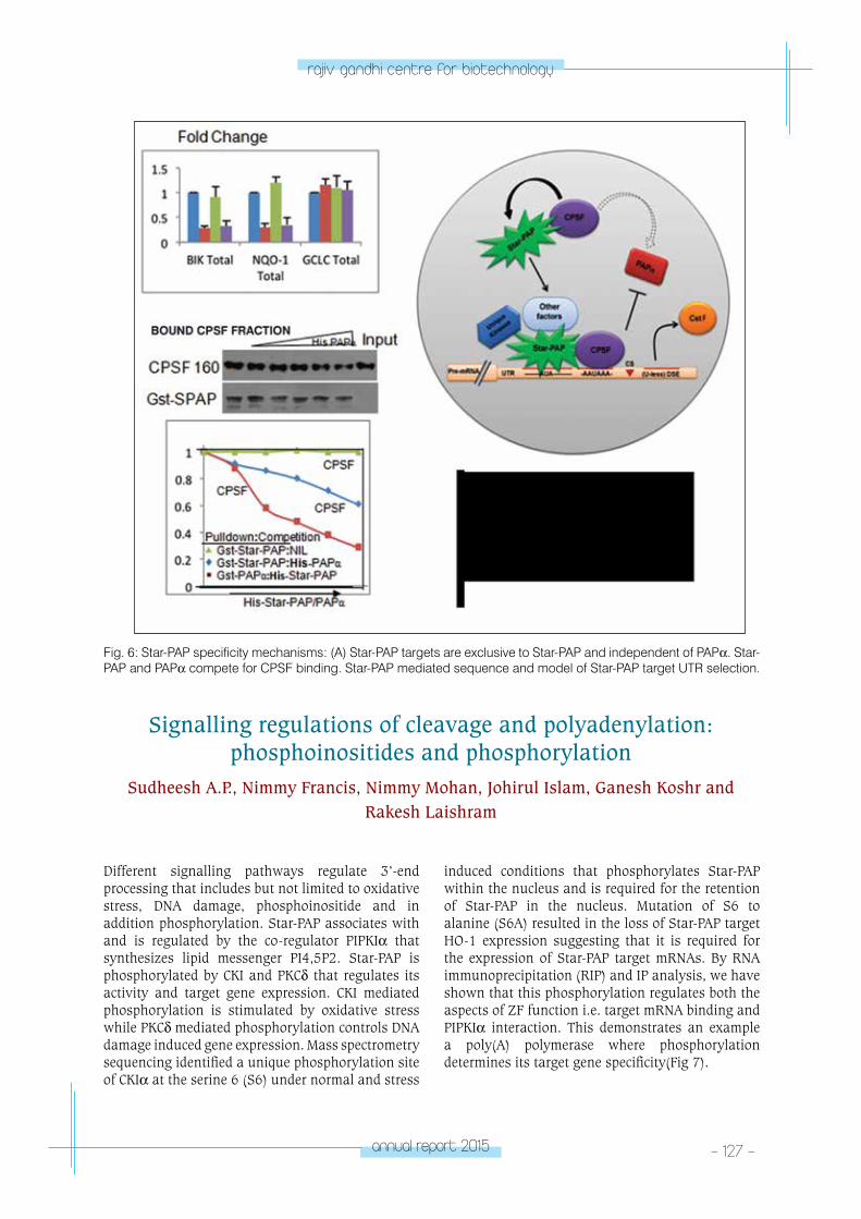
annual report 2015 - 127 -
rajiv gandhi centre for biotechnology
Different signalling pathways regulate 3’-end processing that includes but not limited to oxidative stress, DNA damage, phosphoinositide and in addition phosphorylation. Star-PAP associates with and is regulated by the co-regulator PIPKIα that synthesizes lipid messenger PI4,5P2. Star-PAP is phosphorylated by CKI and PKCδ that regulates its activity and target gene expression. CKI mediated phosphorylation is stimulated by oxidative stress while PKCδ mediated phosphorylation controls DNA damage induced gene expression. Mass spectrometry sequencing identified a unique phosphorylation site of CKIα at the serine 6 (S6) under normal and stress
Signalling regulations of cleavage and polyadenylation: phosphoinositides and phosphorylation
Sudheesh A.P., Nimmy Francis, Nimmy Mohan, Johirul Islam, Ganesh Koshr and Rakesh Laishram
induced conditions that phosphorylates Star-PAP within the nucleus and is required for the retention of Star-PAP in the nucleus. Mutation of S6 to alanine (S6A) resulted in the loss of Star-PAP target HO-1 expression suggesting that it is required for the expression of Star-PAP target mRNAs. By RNA immunoprecipitation (RIP) and IP analysis, we have shown that this phosphorylation regulates both the aspects of ZF function i.e. target mRNA binding and PIPKIα interaction. This demonstrates an example a poly(A) polymerase where phosphorylation determines its target gene specificity(Fig 7).
Fig. 6: Star-PAP specificity mechanisms: (A) Star-PAP targets are exclusive to Star-PAP and independent of PAPα. Star-PAP and PAPα compete for CPSF binding. Star-PAP mediated sequence and model of Star-PAP target UTR selection.
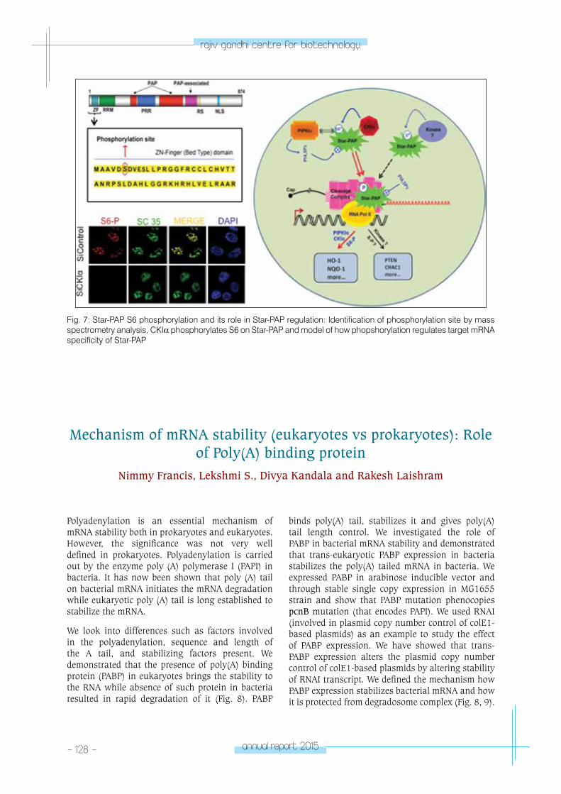
- 128 - annual report 2015
rajiv gandhi centre for biotechnology
Polyadenylation is an essential mechanism of mRNA stability both in prokaryotes and eukaryotes. However, the significance was not very well defined in prokaryotes. Polyadenylation is carried out by the enzyme poly (A) polymerase I (PAPI) in bacteria. It has now been shown that poly (A) tail on bacterial mRNA initiates the mRNA degradation while eukaryotic poly (A) tail is long established to stabilize the mRNA.
We look into differences such as factors involved in the polyadenylation, sequence and length of the A tail, and stabilizing factors present. We demonstrated that the presence of poly(A) binding protein (PABP) in eukaryotes brings the stability to the RNA while absence of such protein in bacteria resulted in rapid degradation of it (Fig. 8). PABP
binds poly(A) tail, stabilizes it and gives poly(A) tail length control. We investigated the role of PABP in bacterial mRNA stability and demonstrated that trans-eukaryotic PABP expression in bacteria stabilizes the poly(A) tailed mRNA in bacteria. We expressed PABP in arabinose inducible vector and through stable single copy expression in MG1655 strain and show that PABP mutation phenocopies pcnB mutation (that encodes PAPI). We used RNAI (involved in plasmid copy number control of colE1-based plasmids) as an example to study the effect of PABP expression. We have showed that trans-PABP expression alters the plasmid copy number control of colE1-based plasmids by altering stability of RNAI transcript. We defined the mechanism how PABP expression stabilizes bacterial mRNA and how it is protected from degradosome complex (Fig. 8, 9).
Fig. 7: Star-PAP S6 phosphorylation and its role in Star-PAP regulation: Identification of phosphorylation site by mass spectrometry analysis, CKIα phosphorylates S6 on Star-PAP and model of how phopshorylation regulates target mRNA specificity of Star-PAP
Mechanism of mRNA stability (eukaryotes vs prokaryotes): Role of Poly(A) binding protein
Nimmy Francis, Lekshmi S., Divya Kandala and Rakesh Laishram
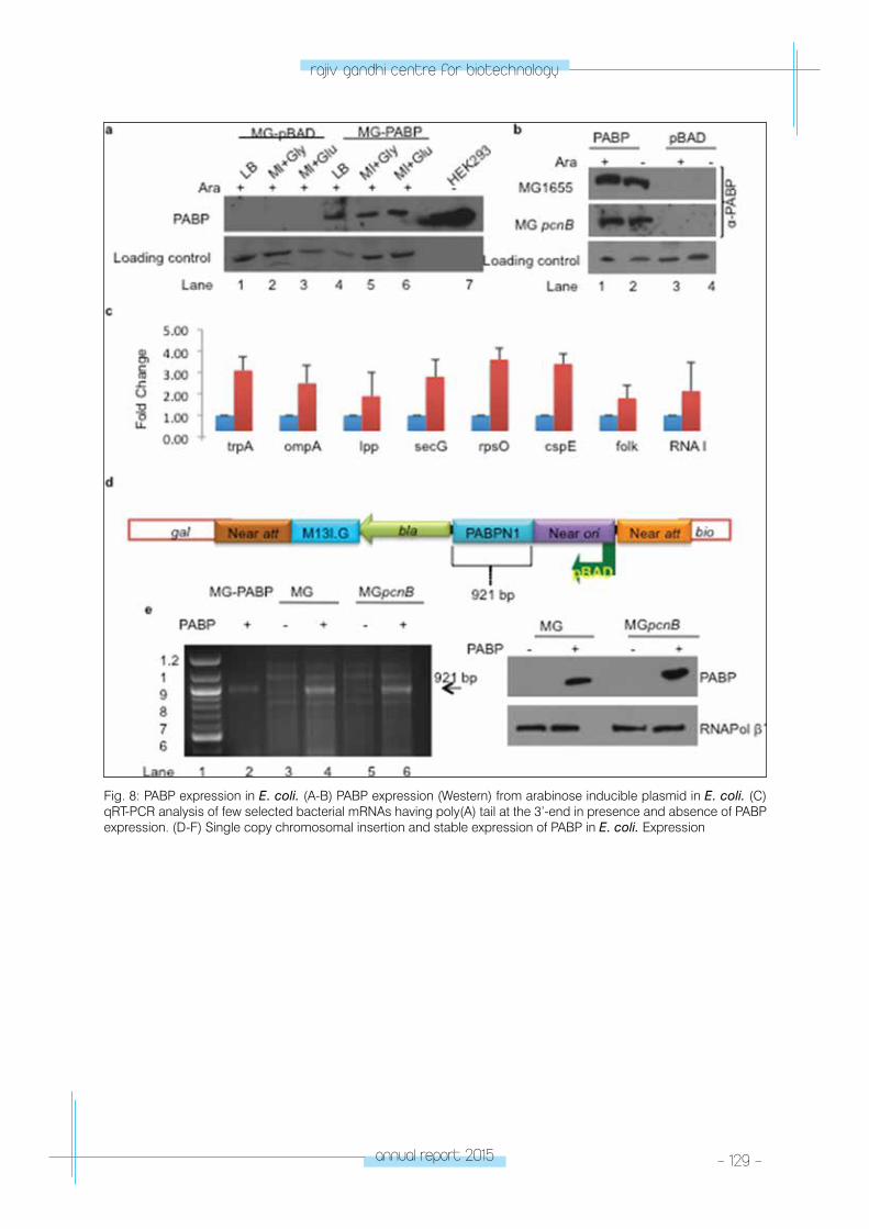
annual report 2015 - 129 -
rajiv gandhi centre for biotechnology
Fig. 8: PABP expression in E. coli. (A-B) PABP expression (Western) from arabinose inducible plasmid in E. coli. (C) qRT-PCR analysis of few selected bacterial mRNAs having poly(A) tail at the 3’-end in presence and absence of PABP expression. (D-F) Single copy chromosomal insertion and stable expression of PABP in E. coli. Expression
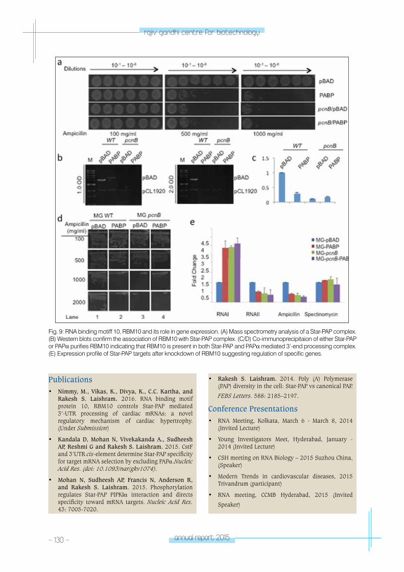
- 130 - annual report 2015
rajiv gandhi centre for biotechnology
Fig. 9: RNA binding motiff 10, RBM10 and its role in gene expression. (A) Mass spectrometry analysis of a Star-PAP complex. (B) Western blots confirm the association of RBM10 with Star-PAP complex. (C/D) Co-immunoprecipitaion of either Star-PAP or PAPα purifies RBM10 indicating that RBM10 is present in both Star-PAP and PAPα mediated 3’-end processing complex. (E) Expression profile of Star-PAP targets after knockdown of RBM10 suggesting regulation of specific genes.
Publications • Nimmy, M., Vikas, K., Divya, K., C.C. Kartha, and
Rakesh S. Laishram. 2016. RNA binding motif protein 10, RBM10 controls Star-PAP mediated 3’-UTR processing of cardiac mRNAs: a novel regulatory mechanism of cardiac hypertrophy. (Under Submission)
• Kandala D, Mohan N, Vivekakanda A., Sudheesh AP, Reshmi G and Rakesh S. Laishram. 2015. CstF and 3’UTR cis-element determine Star-PAP specificity for target mRNA selection by excluding PAPα.Nucleic Acid Res. (doi: 10.1093/nar/gkv1074).
• Mohan N, Sudheesh AP, Francis N, Anderson R, and Rakesh S. Laishram. 2015. Phosphorylation regulates Star-PAP PIPKIα interaction and directs specificity toward mRNA targets. Nucleic Acid Res. 43: 7005-7020.
• Rakesh S. Laishram. 2014. Poly (A) Polymerase (PAP) diversity in the cell: Star-PAP vs canonical PAP.
FEBS Letters. 588: 2185–2197.
Conference Presentations• RNA Meeting, Kolkata, March 6 - March 8, 2014
(Invited Lecture)
• Young Investigators Meet, Hyderabad, January - 2014 (Invited Lecture)
• CSH meeting on RNA Biology – 2015 Suzhou China, (Speaker)
• Modern Trends in cardiovascular diseases, 2015 Trivandrum (participant)
• RNA meeting, CCMB Hyderabad, 2015 (Invited
Speaker)
annual report 2015 - 131 -
rajiv gandhi centre for biotechnology
RESEARCH GRANTS
Sl. No Title Funding Agency Year
1Specificity and mechanism of Star-PAP mediated 3’-end processing in gene regulation
Wellcome Trust-DBT India Alliance
2012-2017
2Regulation of 3’-end processing in oxidative stress response and cardiovascular diseases
Department of Biotechnology (IYBA)
2013-2016
33’-UTR regulation of cardiac genes with roles in pressure overload cardiac hypertrophy
Department of Biotechnology Approved
4Splicing independent function of RNA binding protein motif 10, RBM10 in 3’-end processing and gene expression
Department of Science and Technology
Approved
CARDIoV
ASCulAR DISeASe A
nD DIABeteS BIoloGY pRoGRAM
Diabetes Biology laboratory
CARDIOVASCULAR DISEASE AND DIABETES
BIOLOGY PROGRAMLaboratory - 3
Abdul Jaleel. K. A
Post Doctoral FellowMahesh Krishna, PhD
Ph.D StudentAneesh Kumar A.
Abdul Jaleel obtained his PhD from
JamiaMilliaIslamia, New Delhi while doing
his research fellowship at All India Institute
of Medical Sciences, New Delhi. He trained
as a post-doctoral fellow and then continued
asfaculty at Mayo Clinic, Rochester, MN, USA. He
joined RGCB in Dec 2011.
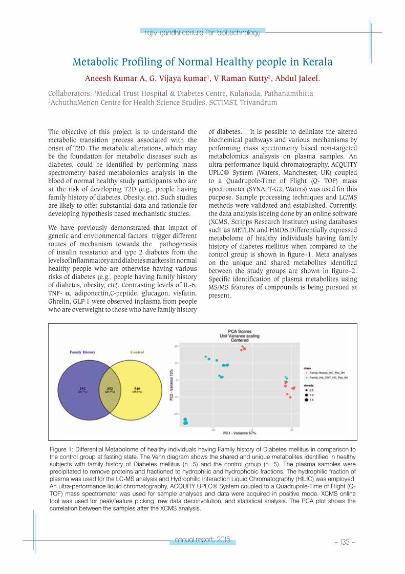
annual report 2015 - 133 -
rajiv gandhi centre for biotechnology
The objective of this project is to understand the metabolic transition process associated with the onset of T2D. The metabolic alterations, which may be the foundation for metabolic diseases such as diabetes, could be identified by performing mass spectrometry based metabolomics analysis in the blood of normal healthy study participants who are at the risk of developing T2D (e.g., people having family history of diabetes, Obesity, etc). Such studies are likely to offer substantial data and rationale for developing hypothesis based mechanistic studies.
We have previously demonstrated that impact of genetic and environmental factors trigger different routes of mechanism towards the pathogenesis of insulin resistance and type 2 diabetes from the levelsof inflammatory and diabetes markers in normal healthy people who are otherwise having various risks of diabetes (e.g., people having family history of diabetes, obesity, etc). Contrasting levels of IL-6, TNF- α, adiponectin,C-peptide, glucagon, visfatin, Ghrelin, GLP-1 were observed inplasma from people who are overweight to those who have family history
of diabetes. It is possible to deliniate the altered biochemical pathways and various mechanisms by performing mass spectrometry based non-targeted metabolomics analsysis on plasma samples. An ultra-performance liquid chromatography, ACQUITY UPLC® System (Waters, Manchester, UK) coupled to a Quadrupole-Time of Flight (Q- TOF) mass spectrometer (SYNAPT-G2, Waters) was used for this purpose. Sample processing techniques and LC/MS methods were validated and established. Currently, the data analysis isbeing done by an online software (XCMS, Scripps Research Institute) using databases such as METLIN and HMDB.Differentially expressed metabolome of healthy individuals having family history of diabetes mellitus when compared to the control group is shown in figure–1. Meta analyses on the unique and shared metabolites identified between the study groups are shown in figure–2. Specific identification of plasma metabolites using MS/MS features of compounds is being pursued at present.
Metabolic Profiling of Normal Healthy people in KeralaAneesh Kumar A, G. Vijaya kumar1, V Raman Kutty2, Abdul Jaleel.
Collaborators: 1Medical Trust Hospital & Diabetes Centre, Kulanada, Pathanamthitta 2AchuthaMenon Centre for Health Science Studies, SCTIMST, Trivandrum
Figure 1: Differential Metabolome of healthy individuals having Family history of Diabetes mellitus in comparison to the control group at fasting state. The Venn diagram shows the shared and unique metabolites identified in healthy subjects with family history of Diabetes mellitus (n=5) and the control group (n=5). The plasma samples were precipitated to remove proteins and fractioned to hydrophilic and hydrophobic fractions. The hydrophilic fraction of plasma was used for the LC-MS analysis and Hydrophilic Interaction Liquid Chromatography (HILIC) was employed. An ultra-performance liquid chromatography, ACQUITY UPLC® System coupled to a Quadrupole-Time of Flight (Q- TOF) mass spectrometer was used for sample analyses and data were acquired in positive mode. XCMS online tool was used for peak/feature picking, raw data deconvolution, and statistical analysis. The PCA plot shows the correlation between the samples after the XCMS analysis.
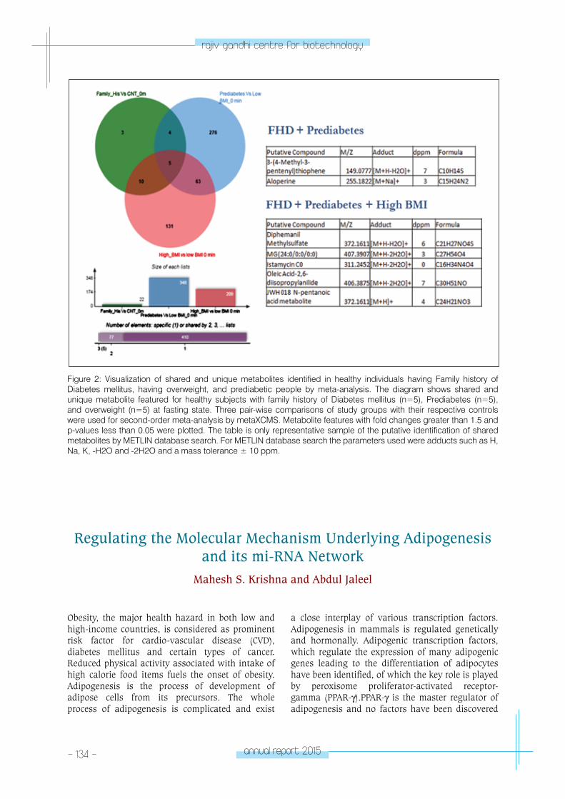
- 134 - annual report 2015
rajiv gandhi centre for biotechnology
Regulating the Molecular Mechanism Underlying Adipogenesis and its mi-RNA Network
Mahesh S. Krishna and Abdul Jaleel
Figure 2: Visualization of shared and unique metabolites identified in healthy individuals having Family history of Diabetes mellitus, having overweight, and prediabetic people by meta-analysis. The diagram shows shared and unique metabolite featured for healthy subjects with family history of Diabetes mellitus (n=5), Prediabetes (n=5), and overweight (n=5) at fasting state. Three pair-wise comparisons of study groups with their respective controls were used for second-order meta-analysis by metaXCMS. Metabolite features with fold changes greater than 1.5 and p-values less than 0.05 were plotted. The table is only representative sample of the putative identification of shared metabolites by METLIN database search. For METLIN database search the parameters used were adducts such as H, Na, K, -H2O and -2H2O and a mass tolerance ± 10 ppm.
Obesity, the major health hazard in both low and high-income countries, is considered as prominent risk factor for cardio-vascular disease (CVD), diabetes mellitus and certain types of cancer. Reduced physical activity associated with intake of high calorie food items fuels the onset of obesity. Adipogenesis is the process of development of adipose cells from its precursors. The whole process of adipogenesis is complicated and exist
a close interplay of various transcription factors. Adipogenesis in mammals is regulated genetically and hormonally. Adipogenic transcription factors, which regulate the expression of many adipogenic genes leading to the differentiation of adipocytes have been identified, of which the key role is played by peroxisome proliferator-activated receptor-gamma (PPAR-γ).PPAR-γ is the master regulator of adipogenesis and no factors have been discovered
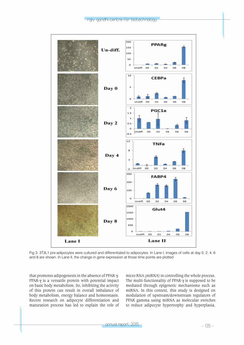
annual report 2015 - 135 -
rajiv gandhi centre for biotechnology
that promotes adipogenesis in the absence of PPAR-γ. PPAR-γ is a versatile protein with potential impact on basic body metabolism. So, inhibiting the activity of this protein can result in overall imbalance of body metabolism, energy balance and homeostasis. Recent research on adipocyte differentiation and maturation process has led to explain the role of
micro RNA (miRNA) in controlling the whole process. The multi-functionality of PPAR-γ is supposed to be mediated through epigenetic mechanisms such as miRNA. In this context, this study is designed on modulation of upstream/downstream regulators of PPAR gamma using miRNA as molecular switches to reduce adipocyte hypertrophy and hyperplasia.
Fig 3: 3T3L1 pre-adipocytes were cultured and differentiated to adipocytes. In Lane I, images of cells at day 0, 2, 4, 6 and 8 are shown. In Lane II, the change in gene expression at those time points are plotted
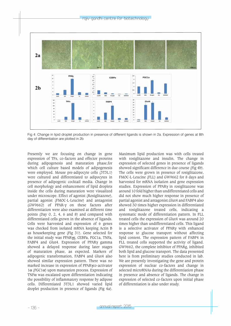
- 136 - annual report 2015
rajiv gandhi centre for biotechnology
Fig 4: Change in lipid droplet production in presence of different ligands is shown in 2a. Expression of genes at 8th day of differentiation are plotted in 2b
Presently we are focusing on change in gene expression of TFs, co-factors and effector proteins during adipogenesis and maturation phase,for which cell culture based models of adipogenesis were employed. Mouse pre-adipocyte cells (3T3L1) were cultured and differentiated to adipocytes in presence of adipogenic cocktail media. Change in cell morphology and enhancement of lipid droplets inside the cells during maturation were visualized under microscope. Effect of agonist (Rosiglitazone), partial agonist (FMOC-L-Leucine) and antagonist (GW9662) of PPAR-γ on these factors after differentiation were also examined at different time points (Day 0, 2, 4, 6 and 8) and compared with differentiated cells grown in the absence of ligands. Cells were harvested and expression of 6 genes was checked from isolated mRNA keeping Actin B as housekeeping gene (Fig 31). Gene selected for the initial study was PPARγg, CEBPa, PGC1a, TNFa, FABP4 and Glut4. Expression of PPARγ gamma showed a delayed response during later stages of maturation phase, as expected. Markers of adipogenic transformation, FABP4 and Glut4 also showed similar expression pattern. There was no marked increase in expression of PPARγco-activator 1α (PGC1α) upon maturation process. Expression of TNFα was escalated upon differentiation indicating the possibility of inflammatory response by adipose cells. Differentiated 3T3L1 showed varied lipid droplet production in presence of ligands (Fig 4a).
Maximum lipid production was with cells treated with rosiglitazone and insulin. The change in expression of selected genes in presence of ligands showed significant difference in due course (Fig 4b). The cells were grown in presence of rosiglitazone, FMOC-L-Leucine (FLL) and GW9662 for 8 days and harvested for mRNA isolation and gene expression studies. Expression of PPARγ in rosiglitazone was around 10 fold higher than undifferentiated cells and did not show much higher response in presence of partial agonist and antagonist.Glut4 and FABP4 also showed 30 times higher expression in differentiated and rosiglitazone treated cells, indicating a systematic mode of differentiation pattern. In FLL treated cells the expression of Glut4 was around 20 times higher than undifferentiated cells. This ligand is a selective activator of PPARγ with enhanced response to glucose transport without affecting lipid content. The expression pattern of FABP4 in FLL treated cells supported the activity of ligand. GW9662, the complete inhibitor of PPARg, inhibited both lipid and glucose transport. The data presented here is from preliminary studies conducted in lab. We are presently investigating the gene and protein expression of nuclear co-factors and change in selected microRNAs during the differentiation phase in presence and absence of ligands. The change in expression of selected co-factors upon initial phase of differentiation is also under study.
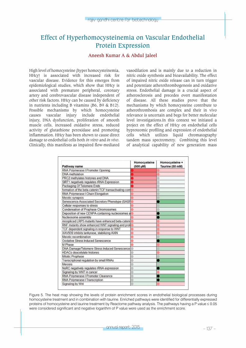
annual report 2015 - 137 -
rajiv gandhi centre for biotechnology
High level of homocysteine (hyper homocysteinemia, HHcy) is associated with increased risk for vascular disease. Evidence for this emerges from epidemiological studies, which show that HHcy is associated with premature peripheral, coronary artery and cerebrovascular disease independent of other risk factors. HHcy can be caused by deficiency in nutrients including B vitamins (B6, B9 & B12). Possible mechanisms by which homocysteine causes vascular injury include endothelial injury, DNA dysfunction, proliferation of smooth muscle cells, increased oxidative stress, reduced activity of glutathione peroxidase and promoting inflammation. HHcy has been shown to cause direct damage to endothelial cells both in vitro and in vivo. Clinically, this manifests as impaired flow-mediated
vasodilation and is mainly due to a reduction in nitric oxide synthesis and bioavailability. The effect of impaired nitric oxide release can in turn trigger and potentiate atherothrombogenesis and oxidative stress. Endothelial damage is a crucial aspect of atherosclerosis and precedes overt manifestation of disease. All these studies prove that the mechanisms by which homocysteine contribute to atherothrombosis are complex and their in vivo relevance is uncertain and begs for better molecular level investigations.In this context we initiated a project on the effect of HHcy on endothelial cells byproteomic profiling and expression of endothelial cells which utilizes liquid chromatography tandem mass spectrometry. Combining this level of analytical capability of new generation mass
Figure 5. The heat map showing the levels of protein enrichment scores in endothelial biological processes during homocysteine treatment and in combination with taurine. Enriched pathways were identified for differentially expressed proteins of homocysteine and taurine treatment by Reactome pathway analysis. The pathways having a P value ≤ 0.05 were considered significant and negative logarithm of P value were used as the enrichment score.
Effect of Hyperhomocysteinemia on Vascular Endothelial Protein Expression
Aneesh Kumar A & Abdul Jaleel
- 138 - annual report 2015
rajiv gandhi centre for biotechnology
spectrometry technology with the study of biological sample from healthy and diseased subjects has the potential to transform our understanding of disease mechanisms. HUVEC were cultured in endothelial cell basal medium-2 (EBM-2). Once reached confluence the cells were incubated for 24 hours in various concentrations of homocysteine (10, 100 and 500 µM), Taurine (10 30 & 50 mM) and combinations of homocysteine and taurine. Proteins were extracted from the cells and performed proteomics analyses by LC/MS/MS. The post MS data were subjected to database search using PLGS for protein profiling and relative quantification. The reference sequence (RefSeq) IDs obtained after the PLGS analysis were converted into gene symbol by online ID conversion tool named Biological
Database Network(bioDBnet). In PLGS expression analysis, proteins having a P value (Probability of up-regulation) greater than 0.95 and less than 0.05 were considered up regulated and down regulated respectively. The enriched pathways were identified for differentially expressed proteins by REACTOME pathway analysis to study the biological significance of the intervention. The pathways having a P value <- 0.05 were considered as significant and negative logarithm of P value (enrichment score) were used for graphical representation by MS Excel. The preliminary data shows (Fig.ure5-1) alteration of several important biological processes during homocysteine treatment and reversal of that in combination with taurine.
Publications:• Saritha VN, George J K, Jaleel A, Surendran A,
Saravanakumar M, Kalavathy MC, Somanathan T, Rema P, Sujathan K. Analysis of differentially expressed proteins in the exfoliated cells of normal and squamous cell carcinoma of the uterine cervix to define candidate markers for cervical cancer. International Journal of Biochemistry and Biotechnology 2016; 5(1):626-36
• Muhamed J, Rajan A, Surendran A, Jaleel A, Anilkumar TV. Comparative profiling of extractable
proteins in extracellular matrices of porcine cholecyst and jejunum intended for preparation of tissue engineering scaffolds. J Biomed Mater Res B ApplBiomater. 2015, doi: 10.1002/jbm.b.33567.
• Gopinath V, Raghunandanan S, Gomez RL, Jose L, Surendran A, Ramachandran A, Pushparajan AR, Mundayoor S, Jaleel A, Kumar RA. Profiling the proteome of Mycobacterium tuberculosis during dormancy and reactivation. Molecular & Cellular Proteomics. 2015;14(8):2160-76
RESEARCH GRANTS
Title Funding Agency Duration
Identification of metabolic alterations in subclinical B12 deficiency by mass spectrometry based metabolomics
Kerala State Council for Science, Technology&Environment (KSCSTE)
2016-19

CARDIoV
ASCulAR DISeASe A
nD DIABeteS BIoloGY pRoGRAM
A
dipocyte Biology laboratoryCARDIOVASCULAR
DISEASE AND DIABETES BIOLOGY PROGRAM
Laboratory - 4
Sona Rajakumari
Scientist C & Ramalingaswami Fellow
Sona Rajakumari received her PhD from the
Graz University of Technology and the Karl
Franzens University Graz, Austria. She did
her post-doctoral training (2010-2015) from
Perelman School of Medicine, University of
Pennsylvania, USA. Sona joined Rajiv Gandhi
Centre for Biotechnology in June 2015. She is
also a recipient of Ramalingaswami Re-entry
Fellowship.
Research FellowsAnu B. Gayathri
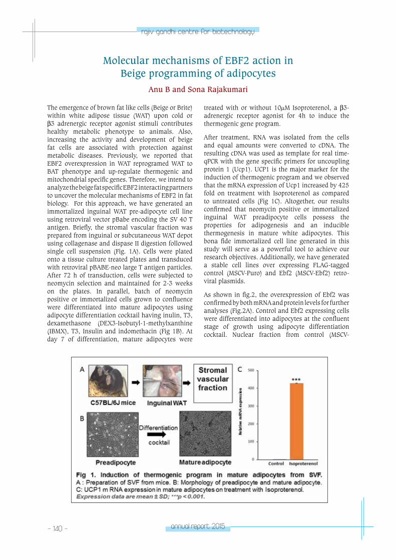
- 140 - annual report 2015
rajiv gandhi centre for biotechnology
Molecular mechanisms of EBF2 action in Beige programming of adipocytes
Anu B and Sona Rajakumari
The emergence of brown fat like cells (Beige or Brite) within white adipose tissue (WAT) upon cold or β3 adrenergic receptor agonist stimuli contributes healthy metabolic phenotype to animals. Also, increasing the activity and development of beige fat cells are associated with protection against metabolic diseases. Previously, we reported that EBF2 overexpression in WAT reprogramed WAT to BAT phenotype and up-regulate thermogenic and mitochondrial specific genes. Therefore, we intend to analyze the beige fat specific EBF2 interacting partners to uncover the molecular mechanisms of EBF2 in fat biology. For this approach, we have generated an immortalized inguinal WAT pre-adipocyte cell line using retroviral vector pBabe encoding the SV 40 T antigen. Briefly, the stromal vascular fraction was prepared from inguinal or subcutaneous WAT depot using collagenase and dispase II digestion followed single cell suspension (Fig. 1A). Cells were plated onto a tissue culture treated plates and transduced with retroviral pBABE-neo large T antigen particles. After 72 h of transduction, cells were subjected to neomycin selection and maintained for 2-3 weeks on the plates. In parallel, batch of neomycin positive or immortalized cells grown to confluence were differentiated into mature adipocytes using adipocyte differentiation cocktail having inulin, T3, dexamethasone (DEX3-Isobutyl-1-methylxanthine (IBMX), T3, Insulin and indomethacin (Fig 1B). At day 7 of differentiation, mature adipocytes were
treated with or without 10µM Isoproterenol, a β3-adrenergic receptor agonist for 4h to induce the thermogenic gene program.
After treatment, RNA was isolated from the cells and equal amounts were converted to cDNA. The resulting cDNA was used as template for real time-qPCR with the gene specific primers for uncoupling protein 1 (Ucp1). UCP1 is the major marker for the induction of thermogenic program and we observed that the mRNA expression of Ucp1 increased by 425 fold on treatment with Isoproterenol as compared to untreated cells (Fig 1C). Altogether, our results confirmed that neomycin positive or immortalized inguinal WAT preadipocyte cells possess the properties for adipogenesis and an inducible thermogenesis in mature white adipocytes. This bona fide immortalized cell line generated in this study will serve as a powerful tool to achieve our research objectives. Additionally, we have generated a stable cell lines over expressing FLAG-tagged control (MSCV-Puro) and Ebf2 (MSCV-Ebf2) retro-viral plasmids.
As shown in fig.2, the overexpression of Ebf2 was confirmed by both mRNA and protein levels for further analyses (Fig.2A). Control and Ebf2 expressing cells were differentiated into adipocytes at the confluent stage of growth using adipocyte differentiation cocktail. Nuclear fraction from control (MSCV-
annual report 2015 - 141 -
rajiv gandhi centre for biotechnology
Puro) and Ebf2 (MSCV-Ebf2) expressing cells were subjected to immunoprecipitation (IP) assay using FLAG M2-agarose beads. The immunoprecipitated EBF2 complex was further validated using western blot analysis (Fig 2B). Finally, the affinity purified
protein complex will be subjected to protein mass-spectrometry analysis. The proteomic results will be analyzed using MaxQuant software to identify EBF2 potential interacting partners and further will be validated by co-immunoprecipitation assays.
RESEARCH GRANTS
No Title Funding Agency Duration
1 Molecular mechanisms of EBF2 action in Beige programming of adipocytes (Ramalingaswami Re-Entry Fellowship)
Department of Biotechnology, Government of India
2015- 2020
2 Early B cell factor 2 orchestrate chromatin remodeling to drive brown fat gene program
Department of Science and Technology, Government of India
2016-2019
3 Molecular reprograming of white adipose tissue to combat metabolic diseases (DBT - IYBA)
Department of Biotechnology, Government of India
2016-2019
CHeMICA
l & enVIRonM
entAl BIoloGY pRoGRA
M
- Chemical Biology laboratory – 1
CHEMICAL & ENVIRONMENTAL
BIOLOGY PROGRAMChemical Biology
Laboratory - 1
K. Santhosh Kumar
Santhosh Kumar took his PhD in Chemistry
from the School of Chemistry Sciences, Mahatma
Gandhi University and did his postdoctoral
training at the Department of Biochemistry,
University of Illinois Urbana-Champaign, USA.
He joined RGCB in 1996.
Research AssistantSmitha Devi
Technical Assistant Aswani Kumar
Ph. D StudentsReshmi V. Asha R.Neethu Ajayakumar Pratibha NarayanAnju Krishnan A.
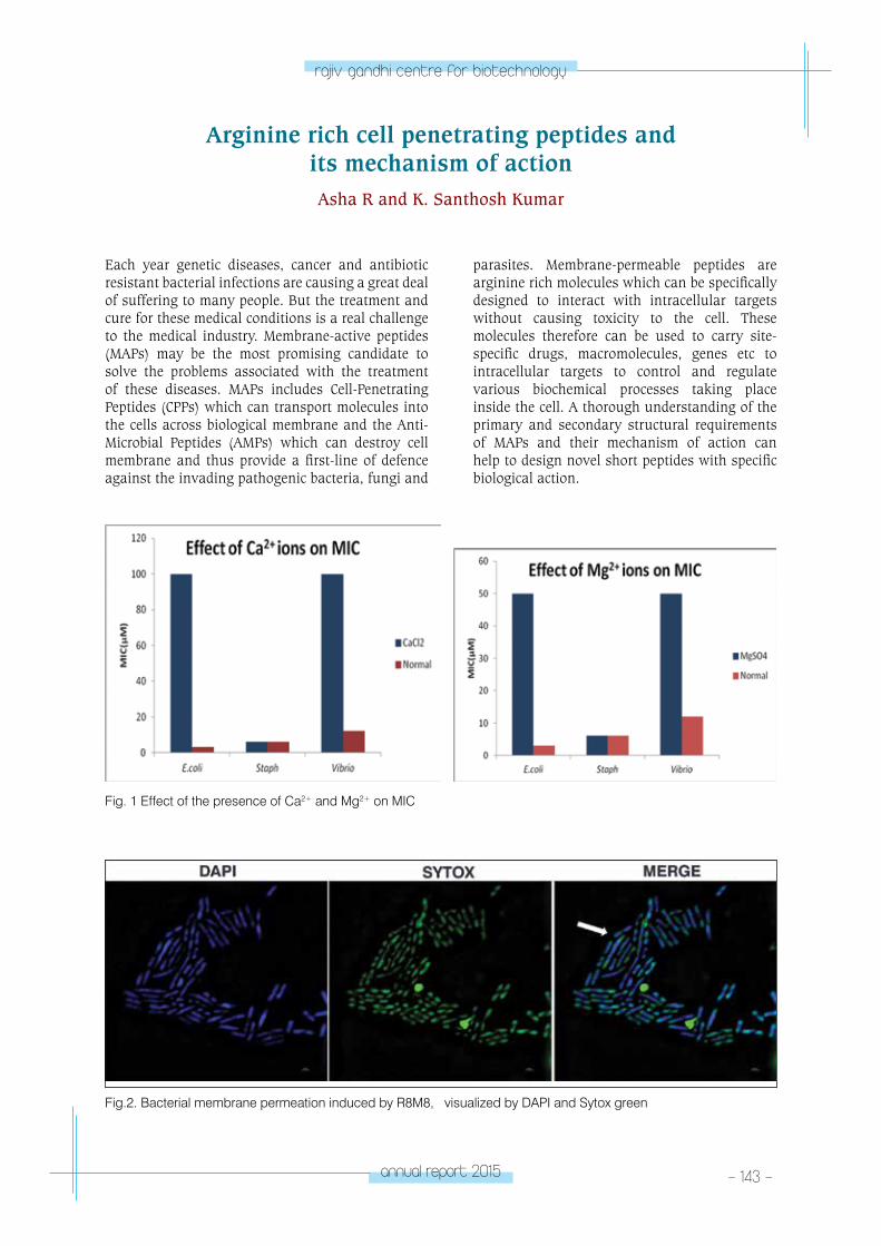
annual report 2015 - 143 -
rajiv gandhi centre for biotechnology
Arginine rich cell penetrating peptides and its mechanism of actionAsha R and K. Santhosh Kumar
Each year genetic diseases, cancer and antibiotic resistant bacterial infections are causing a great deal of suffering to many people. But the treatment and cure for these medical conditions is a real challenge to the medical industry. Membrane-active peptides (MAPs) may be the most promising candidate to solve the problems associated with the treatment of these diseases. MAPs includes Cell-Penetrating Peptides (CPPs) which can transport molecules into the cells across biological membrane and the Anti-Microbial Peptides (AMPs) which can destroy cell membrane and thus provide a first-line of defence against the invading pathogenic bacteria, fungi and
parasites. Membrane-permeable peptides are arginine rich molecules which can be specifically designed to interact with intracellular targets without causing toxicity to the cell. These molecules therefore can be used to carry site-specific drugs, macromolecules, genes etc to intracellular targets to control and regulate various biochemical processes taking place inside the cell. A thorough understanding of the primary and secondary structural requirements of MAPs and their mechanism of action can help to design novel short peptides with specific biological action.
Fig. 1 Effect of the presence of Ca2+ and Mg2+ on MIC
Fig.2. Bacterial membrane permeation induced by R8M8, visualized by DAPI and Sytox green
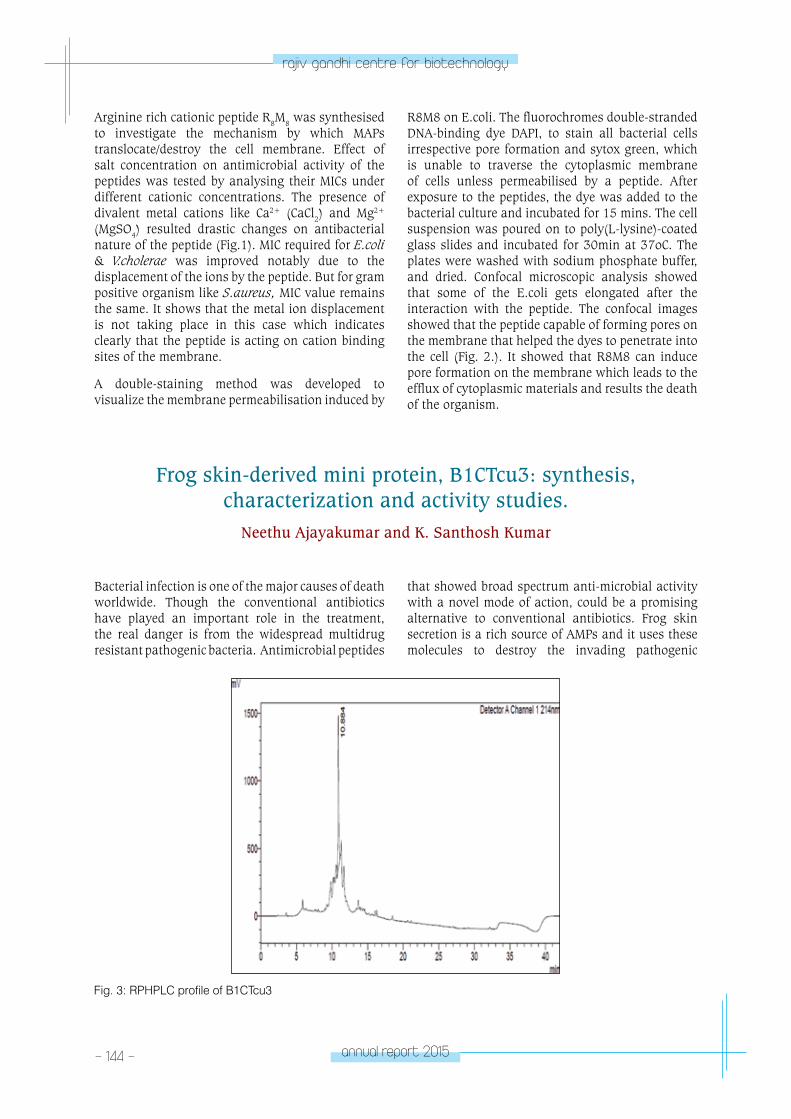
- 144 - annual report 2015
rajiv gandhi centre for biotechnology
Arginine rich cationic peptide R8M8 was synthesised to investigate the mechanism by which MAPs translocate/destroy the cell membrane. Effect of salt concentration on antimicrobial activity of the peptides was tested by analysing their MICs under different cationic concentrations. The presence of divalent metal cations like Ca2+ (CaCl2) and Mg2+ (MgSO4) resulted drastic changes on antibacterial nature of the peptide (Fig.1). MIC required for E.coli & V.cholerae was improved notably due to the displacement of the ions by the peptide. But for gram positive organism like S.aureus, MIC value remains the same. It shows that the metal ion displacement is not taking place in this case which indicates clearly that the peptide is acting on cation binding sites of the membrane.
A double-staining method was developed to visualize the membrane permeabilisation induced by
R8M8 on E.coli. The fluorochromes double-stranded DNA-binding dye DAPI, to stain all bacterial cells irrespective pore formation and sytox green, which is unable to traverse the cytoplasmic membrane of cells unless permeabilised by a peptide. After exposure to the peptides, the dye was added to the bacterial culture and incubated for 15 mins. The cell suspension was poured on to poly(L-lysine)-coated glass slides and incubated for 30min at 37oC. The plates were washed with sodium phosphate buffer, and dried. Confocal microscopic analysis showed that some of the E.coli gets elongated after the interaction with the peptide. The confocal images showed that the peptide capable of forming pores on the membrane that helped the dyes to penetrate into the cell (Fig. 2.). It showed that R8M8 can induce pore formation on the membrane which leads to the efflux of cytoplasmic materials and results the death of the organism.
Frog skin-derived mini protein, B1CTcu3: synthesis, characterization and activity studies.
Neethu Ajayakumar and K. Santhosh Kumar
Bacterial infection is one of the major causes of death worldwide. Though the conventional antibiotics have played an important role in the treatment, the real danger is from the widespread multidrug resistant pathogenic bacteria. Antimicrobial peptides
that showed broad spectrum anti-microbial activity with a novel mode of action, could be a promising alternative to conventional antibiotics. Frog skin secretion is a rich source of AMPs and it uses these molecules to destroy the invading pathogenic
Fig. 3: RPHPLC profile of B1CTcu3
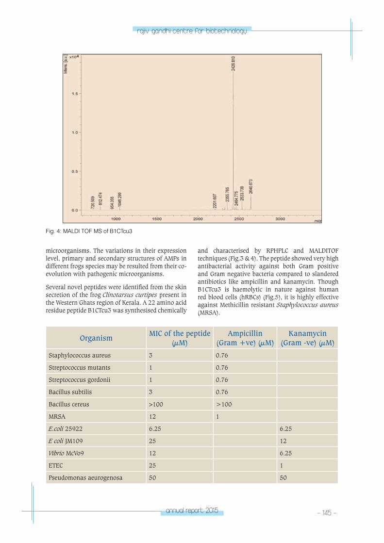
annual report 2015 - 145 -
rajiv gandhi centre for biotechnology
microorganisms. The variations in their expression level, primary and secondary structures of AMPs in different frogs species may be resulted from their co-evolution with pathogenic microorganisms.
Several novel peptides were identified from the skin secretion of the frog Clinotarsus curtipes present in the Western Ghats region of Kerala. A 22 amino acid residue peptide B1CTcu3 was synthesised chemically
and characterised by RPHPLC and MALDITOF techniques (Fig.3 & 4). The peptide showed very high antibacterial activity against both Gram positive and Gram negative bacteria compared to slandered antibiotics like ampicillin and kanamycin. Though B1CTcu3 is haemolytic in nature against human red blood cells (hRBCs) (Fig.5), it is highly effective against Methicillin resistant Staphylococcus aureus (MRSA).
Fig. 4: MALDI TOF MS of B1CTcu3
OrganismMIC of the peptide
(µM)Ampicillin
(Gram +ve) (µM)Kanamycin
(Gram -ve) (µM)
Staphylococcus aureus 3 0.76
Streptococcus mutants 1 0.76
Streptococcus gordonii 1 0.76
Bacillus subtilis 3 0.76
Bacillus cereus >100 >100
MRSA 12 1
E.coli 25922 6.25 6.25
E coli JM109 25 12
Vibrio McVo9 12 6.25
ETEC 25 1
Pseudomonas aeurogenosa 50 50
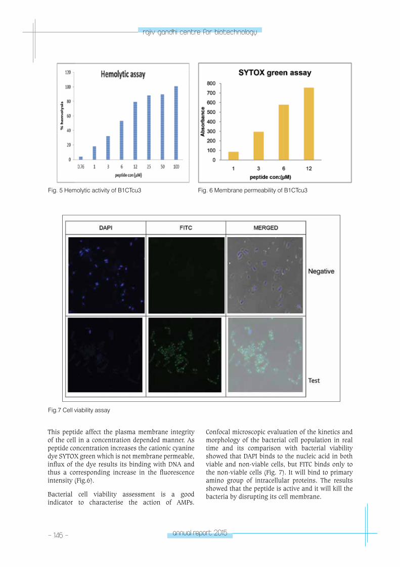
- 146 - annual report 2015
rajiv gandhi centre for biotechnology
This peptide affect the plasma membrane integrity of the cell in a concentration depended manner. As peptide concentration increases the cationic cyanine dye SYTOX green which is not membrane permeable, influx of the dye results its binding with DNA and thus a corresponding increase in the fluorescence intensity (Fig.6).
Bacterial cell viability assessment is a good indicator to characterise the action of AMPs.
Fig. 5 Hemolytic activity of B1CTcu3 Fig. 6 Membrane permeability of B1CTcu3
Confocal microscopic evaluation of the kinetics and morphology of the bacterial cell population in real time and its comparison with bacterial viability showed that DAPI binds to the nucleic acid in both viable and non-viable cells, but FITC binds only to the non-viable cells (Fig. 7). It will bind to primary amino group of intracellular proteins. The results showed that the peptide is active and it will kill the bacteria by disrupting its cell membrane.
Fig.7 Cell viability assay
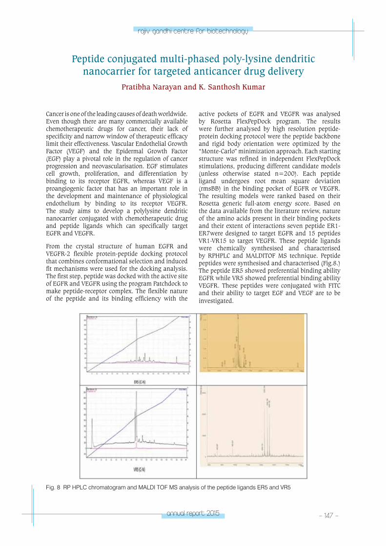
annual report 2015 - 147 -
rajiv gandhi centre for biotechnology
Peptide conjugated multi-phased poly-lysine dendritic nanocarrier for targeted anticancer drug delivery
Pratibha Narayan and K. Santhosh Kumar
Cancer is one of the leading causes of death worldwide. Even though there are many commercially available chemotherapeutic drugs for cancer, their lack of specificity and narrow window of therapeutic efficacy limit their effectiveness. Vascular Endothelial Growth Factor (VEGF) and the Epidermal Growth Factor (EGF) play a pivotal role in the regulation of cancer progression and neovascularisation. EGF stimulates cell growth, proliferation, and differentiation by binding to its receptor EGFR, whereas VEGF is a proangiogenic factor that has an important role in the development and maintenance of physiological endothelium by binding to its receptor VEGFR. The study aims to develop a polylysine dendritic nanocarrier conjugated with chemotherapeutic drug and peptide ligands which can specifically target EGFR and VEGFR.
From the crystal structure of human EGFR and VEGFR-2 flexible protein-peptide docking protocol that combines conformational selection and induced fit mechanisms were used for the docking analysis. The first step, peptide was docked with the active site of EGFR and VEGFR using the program Patchdock to make peptide-receptor complex. The flexible nature of the peptide and its binding efficiency with the
active pockets of EGFR and VEGFR was analysed by Rosetta FlexPepDock program. The results were further analysed by high resolution peptide-protein docking protocol were the peptide backbone and rigid body orientation were optimized by the “Monte-Carlo” minimization approach. Each starting structure was refined in independent FlexPepDock stimulations, producing different candidate models (unless otherwise stated n=200). Each peptide ligand undergoes root mean square deviation (rmsBB) in the binding pocket of EGFR or VEGFR. The resulting models were ranked based on their Rosetta generic full-atom energy score. Based on the data available from the literature review, nature of the amino acids present in their binding pockets and their extent of interactions seven peptide ER1-ER7were designed to target EGFR and 15 peptides VR1-VR15 to target VEGFR. These peptide ligands were chemically synthesised and characterised by RPHPLC and MALDITOF MS technique. Peptide peptides were synthesised and characterised (Fig.8.) The peptide ER5 showed preferential binding ability EGFR while VR5 showed preferential binding ability VEGFR. These peptides were conjugated with FITC and their ability to target EGF and VEGF are to be investigated.
Fig. 8 RP HPLC chromatogram and MALDI TOF MS analysis of the peptide ligands ER5 and VR5
- 148 - annual report 2015
rajiv gandhi centre for biotechnology
Antagonistic CCR5 peptides to inhibit co-receptor mediated cell entry of HIV1.
Anju Krishnan A and K. Santhosh Kumar
The entry of human immunodeficiency virus type 1 (HIV-1) to target cells is a multi-step process involving the interaction of viral envelope proteins with cell surface receptors. Their binding to CD4, followed by the interaction with specific chemokine receptors (CCR5 or CXCR4) trigger the molecular rearrangement of envelope transmembrane subunit that leads to the membrane fusion. Chemokines are small proteins with several functions including immune surveillance and immune cell recruitment. Chemokine receptor antagonists that block the interaction of the HIV-1 envelope proteins with CCR5 or CXCR4 and CD4 can be a potential inhibiter HIV-1. This technique can be utilised as a new therapeutic strategy to develop novel therapeutic and preventive
strategies to combat the AIDS epidemic. But the lack of selectivity in the chemokine system has made it problematic to use natural chemokines in clinical applications due to their potential side effects. Synthetic antagonistic peptides of CCR5 and their analogous may be a good alternative ligands strategy to prevent the cooperative binding mechanis.
MIP-I β is the one of the natural, nonspecific and cross reactive chemokine that binds to CCR5 receptor. Short synthetic peptides and their analogs from the N-terminal region of MIP-1 β are chemically synthesised to analyse their agonistic/antagonistic nature towards CCR5.
Publications• Kochurani, K.J., Annie A. Suganya, Madhumathy G.
Nair, Jiss Maria Louis, Aditi Majumder, Santhosh Kumar K, Parvin Abrahamb, Debasree Dutta, and Tessy T. Maliekal “Live detection and purification of cells based on the expression of a histone chaperone, HIRA, using a binding peptide” Scientific Reports 5, 17218 (2015) doi:10.1038/srep17218
• P. Geetha, M.S. Latha, Saumya S. Pillai, B. Deepa, K. Santhosh Kumar, Mathew Koshy “Green synthesis and characterization of alginate nanoparticles and its role as a biosorbent for Cr(VI) ions” Journal of Molecular Structure 1105 (2016) 54-60
CHeMICA
l & enVIRonM
entAl BIoloGY pRoGRA
M
- Chemical Biology laboratory – 2
CHEMICAL & ENVIRONMENTAL
BIOLOGY PROGRAMChemical Biology
Laboratory – 2
G. S. Vinod Kumar
Vinod Kumar received his PhD in Polymer
Chemistry from School of Chemical Science,
Mahatma Gandhi University and joined RGCB in
2004.
PhD StudentsAmritha VijayanMeenu Vasudevan S.Mrunal Vitthal Wanjale Teena Jacob ChirayilAkhil K. Mohan
Tech Assistant Bindhu S.
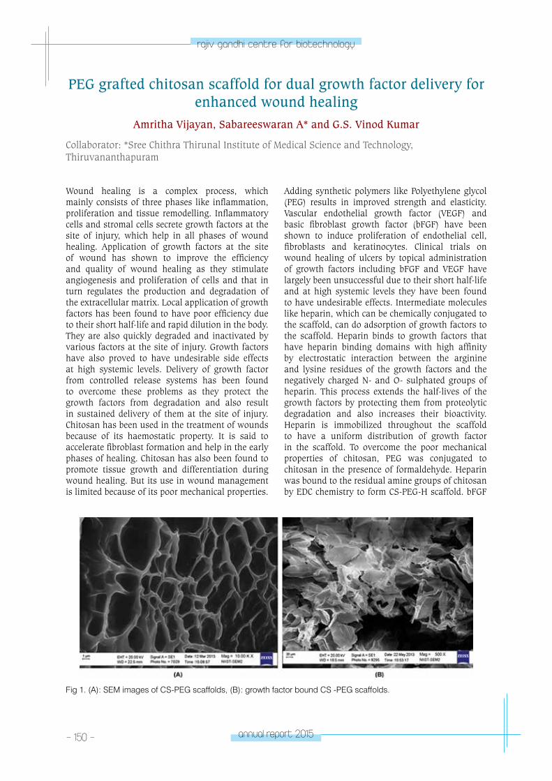
- 150 - annual report 2015
rajiv gandhi centre for biotechnology
PEG grafted chitosan scaffold for dual growth factor delivery for enhanced wound healing
Amritha Vijayan, Sabareeswaran A* and G.S. Vinod Kumar
Collaborator: *Sree Chithra Thirunal Institute of Medical Science and Technology, Thiruvananthapuram
Wound healing is a complex process, which mainly consists of three phases like inflammation, proliferation and tissue remodelling. Inflammatory cells and stromal cells secrete growth factors at the site of injury, which help in all phases of wound healing. Application of growth factors at the site of wound has shown to improve the efficiency and quality of wound healing as they stimulate angiogenesis and proliferation of cells and that in turn regulates the production and degradation of the extracellular matrix. Local application of growth factors has been found to have poor efficiency due to their short half-life and rapid dilution in the body. They are also quickly degraded and inactivated by various factors at the site of injury. Growth factors have also proved to have undesirable side effects at high systemic levels. Delivery of growth factor from controlled release systems has been found to overcome these problems as they protect the growth factors from degradation and also result in sustained delivery of them at the site of injury. Chitosan has been used in the treatment of wounds because of its haemostatic property. It is said to accelerate fibroblast formation and help in the early phases of healing. Chitosan has also been found to promote tissue growth and differentiation during wound healing. But its use in wound management is limited because of its poor mechanical properties.
Adding synthetic polymers like Polyethylene glycol (PEG) results in improved strength and elasticity. Vascular endothelial growth factor (VEGF) and basic fibroblast growth factor (bFGF) have been shown to induce proliferation of endothelial cell, fibroblasts and keratinocytes. Clinical trials on wound healing of ulcers by topical administration of growth factors including bFGF and VEGF have largely been unsuccessful due to their short half-life and at high systemic levels they have been found to have undesirable effects. Intermediate molecules like heparin, which can be chemically conjugated to the scaffold, can do adsorption of growth factors to the scaffold. Heparin binds to growth factors that have heparin binding domains with high affinity by electrostatic interaction between the arginine and lysine residues of the growth factors and the negatively charged N- and O- sulphated groups of heparin. This process extends the half-lives of the growth factors by protecting them from proteolytic degradation and also increases their bioactivity. Heparin is immobilized throughout the scaffold to have a uniform distribution of growth factor in the scaffold. To overcome the poor mechanical properties of chitosan, PEG was conjugated to chitosan in the presence of formaldehyde. Heparin was bound to the residual amine groups of chitosan by EDC chemistry to form CS-PEG-H scaffold. bFGF
Fig 1. (A): SEM images of CS-PEG scaffolds, (B): growth factor bound CS -PEG scaffolds.
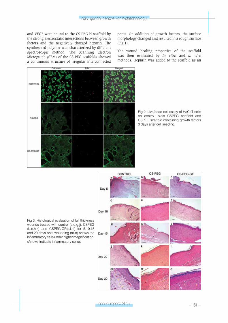
annual report 2015 - 151 -
rajiv gandhi centre for biotechnology
Fig 2. Live/dead cell assay of HaCaT cells on control, plain CSPEG scaffold and CSPEG scaffold containing growth factors 3 days after cell seeding.
Fig 3. Histological evaluation of full thickness wounds treated with control (a,d,g,j), CSPEG (b,e,h,k) and CSPEG-GF(c,f,i,l) for 5,10,15 and 20 days post wounding.(m-o) shows the inflammatory cells under higher magnification. (Arrows indicate inflammatory cells).
and VEGF were bound to the CS-PEG-H scaffold by the strong electrostatic interactions between growth factors and the negatively charged heparin. The synthesized polymer was characterized by different spectroscopic method. The Scanning Electron Micrograph (SEM) of the CS-PEG scaffolds showed a continuous structure of irregular interconnected
pores. On addition of growth factors, the surface morphology changed and resulted in a rough surface (Fig 1).
The wound healing properties of the scaffold was then evaluated by in vitro and in vivo methods. Heparin was added to the scaffold as an
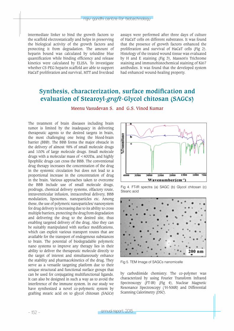
- 152 - annual report 2015
rajiv gandhi centre for biotechnology
intermediate linker to bind the growth factors to the scaffold electrostatically and helps in preserving the biological activity of the growth factors and protecting it from degradation. The amount of heparin bound was calculated by toluidine blue quantification while binding efficiency and release kinetics were calculated by ELISA. To investigate whether CS-PEG heparin scaffold are able to support HaCaT proliferation and survival, MTT and live/dead
assays were performed after three days of culture of HaCaT cells on different substrates. It was found that the presence of growth factors enhanced the proliferation and survival of HaCaT cells (Fig 2). Histology of the treated wound tissue was evaluated by H and E staining (Fig 3), Masson’s Trichrome staining and immunohistochemical staining of Ki67 antibodies. It was found that the developed system had enhanced wound-healing property.
Fig 4. FT-IR spectra (a) SAGC (b) Glycol chitosan (c) Stearic acid
Fig 5. TEM Image of SAGCs nanomicelle
Synthesis, characterization, surface modification and evaluation of Stearoyl-graft-Glycol chitosan (SAGCs)
Meenu Vasudevan S. and G.S. Vinod Kumar
The treatment of brain diseases including brain tumor is limited by the inadequacy in delivering therapeutic agents to the desired targets in brain, the most challenging one being the blood-brain barrier (BBB). The BBB forms the major obstacle in the delivery of almost 98% of small molecule drugs and 100% of large molecule drugs. Small molecule drugs with a molecular mass of <400Da, and highly lipophilic drugs can cross the BBB. The conventional drug therapy increases the concentration of the drug in the systemic circulation but does not lead to a proportional increase in the concentration of drug in the brain. Various approaches taken to overcome the BBB include use of small molecule drugs, prodrugs, chemical delivery systems, olfactory route, intraventricular infusion, intracerebral delivery, BBB modulation, liposomes, nanoparticles etc. Among these, the use of polymeric nanoparticles/ nanosystem for drug delivery is increasing due to its ability to cross multiple barriers, protecting the drug from degradation and delivering the drug to the desired site, thus enabling targeted delivery of the drug. Also they can be suitably manipulated with surface modifications, which can exploit various transport routes that are available for the transport of endogenous substances to brain. The potential of biodegradable polymeric nano systems to improve any therapy lies in their ability to deliver the therapeutic molecule directly to the target of interest and simultaneously enhance the stability and pharmacokinetics of the drug. They serve as a versatile targeting platform due to their unique structural and functional surface groups that can be used for conjugating multifunctional ligands. It can also be designed in such a way as to avoid the interference of the immune system. In our study we have synthesized a novel co-polymeric system by grafting stearic acid on to glycol chitosan (SAGCs)
by carbodiimide chemistry. The co-polymer was characterized by using Fourier Transform Infrared Spectroscopy (FT-IR) (Fig 4), Nuclear Magnetic Resonance Spectroscopy (1H-NMR) and Differential Scanning Calorimetry (DSC).
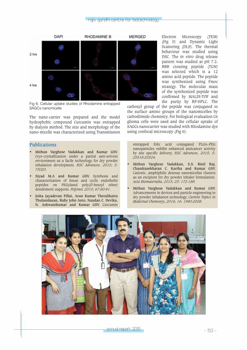
annual report 2015 - 153 -
rajiv gandhi centre for biotechnology
Electron Microscopy (TEM) (Fig 5) and Dynamic Light Scattering (DLS). The thermal behaviour was studied using DSC. The in vitro drug release pattern was studied at pH 7.2. BBB crossing peptide (TGN) was selected which is a 12 amino acid peptide. The peptide was synthesized using Fmoc strategy. The molecular mass of the synthesized peptide was confirmed by MALDI-TOF and the purity by RP-HPLC. The
carboxyl group of the peptide was conjugated to the surface amino groups of the nanomicelles by carbodiimide chemistry. For biological evaluation C6 glioma cells were used and the cellular uptake of SAGCs nanocarrier was studied with Rhodamine dye using confocal microscopy (Fig 6).
Fig 6. Cellular uptake studies of Rhodamine entrapped SAGCs nanomicelle
Publications• Mithun Varghese Vadakkan and Kumar GSV.
Cryo-crystallization under a partial anti-solvent environment as a facile technology for dry powder inhalation development. RSC Advances, 2015; 5: 73020.
• Siyad M.A and Kumar GSV. Synthesis and characterization of linear and cyclic endothelin peptides on PEGylated poly(O-benzyl ether) dendrimeric supports. Polymer, 2015; 67:80-91.
• Jisha Jayadevan Pillai, Arun Kumar Theralikattu Thulasidasan, Ruby John Anto, Nandan C. Devika, N. Ashwanikumar and Kumar GSV. Curcumin
entrapped folic acid conjugated PLGA–PEG nanoparticles exhibit enhanced anticancer activity by site specific delivery. RSC Advances, 2015; 5: 25518-25524.
• Mithun Varghese Vadakkan, S.S. Binil Raj, Chandrasekharan C. Kartha and Kumar GSV. Cationic, amphiphilic dextran nanomicellar clusters as an excipient for dry powder inhaler formulation. Acta Biomaterialia, 2015; 23: 172-188.
• Mithun Varghese Vadakkan and Kumar GSV. Advancements in devices and particle engineering in dry powder inhalation technology. Current Topics in Medicinal Chemistry, 2016; 16: 1990-2008.
The nano-carrier was prepared and the model hydrophobic compound Curcumin was entrapped by dialysis method. The size and morphology of the nano-micelle was characterized using Transmission
CHeMICA
l & enVIRonM
entAl BIoloGY pRoGRA
M
- Molecular ecology laboratory
CHEMICAL & ENVIRONMENTAL
BIOLOGY PROGRAMMolecular Ecology
Laboratory
Sanil George
Sanil George received his PhD in Zoology from
Mahatma Gandhi University and joined RGCB in
1992.
Post -Doctoral FellowShyla S. Gopal, PhD
PhD StudentsVineeth Kumar T.V.Kiran S. KumarAnoop V.S.
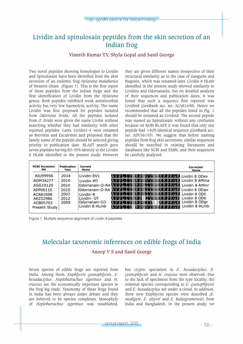
annual report 2015 - 155 -
rajiv gandhi centre for biotechnology
Lividin and spinulosain peptides from the skin secretion of an Indian frog
Vineeth Kumar T.V, Shyla Gopal and Sanil George
Two novel peptides showing homologies to Lividin and Spinulosain have been identified from the skin secretion of an endemic frog Hylarana malabarica of Western Ghats (Figure 1). This is the first report of these peptides from the Indian frogs and the first identification of Lividin from the Hylarana genus. Both peptides exhibited weak antimicrobial activity but very low haemolytic activity. The name Lividin was first proposed for peptides isolated from Odorrana livida. All the peptides isolated from O. livida were given the name Livdin without searching whether they had similarity with other reported peptides. Later, Lividin1-4 were renamed as Brevinin and Esculentin and proposed that the family name of the peptide should be selected giving priority to publication date. BLAST search gave seven peptides having 80–95% identity to the Lividin 8 HLmb identified in the present study. However
they are given different names irrespective of their structural similarity as in the case of Gaegurin and Rugosin, which was renamed later. Lividin 8 HLmb identified in the present study showed similarity to Lividins and Odorranains, but on detailed analysis of their sequences and publication dates, it was found that such a sequence first reported was Lividin8 (GenBank acc. no: ACA81698). Hence we recommended that all the peptides listed in Fig. 1 should be renamed as Lividin8. The second peptide was named as Spinulosain without any confusion because on NCBI BLAST, it was found that only one peptide had >80% identical sequence (GenBank acc. no: ADV36193). We suggest that before naming peptides from frog skin secretions, similar sequences should be searched in existing literatures and databases like NCBI and EMBL and their sequences be carefully analyzed.
Figure 1. Multiple sequence alignment of Lividin 8 peptides
Molecular taxonomic inferences on edible frogs of IndiaAnoop V S and Sanil George
Seven species of edible frogs are reported from India. Among them, Euphlyctis cyanophlyctis, E. hexadactylus, Hoplobatrachus tigerinus and H. crassus are the economically important species in the frog leg trade. Taxonomy of these frogs found in India has been always under debate and they are believed to be species complexes. Monophyly of Hoplobatrachus tigerinus was established,
but cryptic speciation in E. hexadactylus; E. cyanophlyctis and H. crassus were observed. Due to the lack of specimens from the type locality, the nominal species corresponding to E. cyanophlyctis and E. hexadactylus are under a cloud. In addition, three new Euphlyctis species were described (E. mudigere, E. aloysii and E. kalasgramensis) from India and Bangladesh. In the present study, we
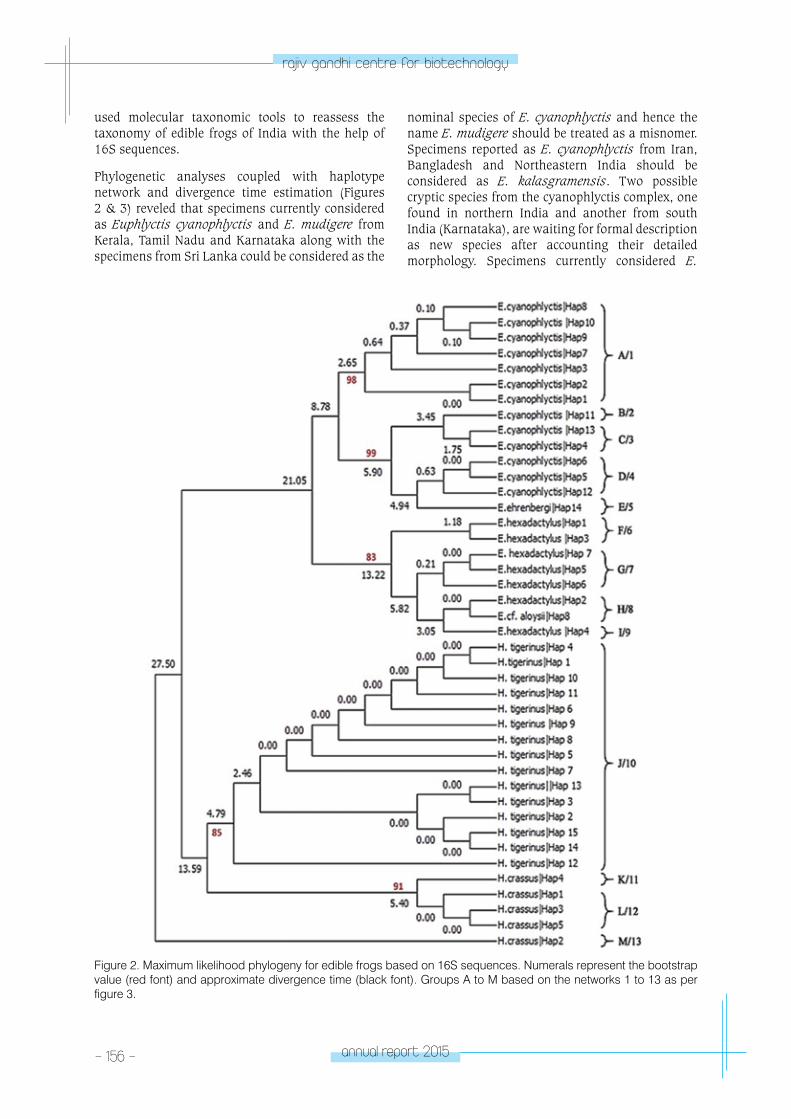
- 156 - annual report 2015
rajiv gandhi centre for biotechnology
nominal species of E. cyanophlyctis and hence the name E. mudigere should be treated as a misnomer. Specimens reported as E. cyanophlyctis from Iran, Bangladesh and Northeastern India should be considered as E. kalasgramensis. Two possible cryptic species from the cyanophlyctis complex, one found in northern India and another from south India (Karnataka), are waiting for formal description as new species after accounting their detailed morphology. Specimens currently considered E.
used molecular taxonomic tools to reassess the taxonomy of edible frogs of India with the help of 16S sequences.
Phylogenetic analyses coupled with haplotype network and divergence time estimation (Figures 2 & 3) reveled that specimens currently considered as Euphlyctis cyanophlyctis and E. mudigere from Kerala, Tamil Nadu and Karnataka along with the specimens from Sri Lanka could be considered as the
Figure 2. Maximum likelihood phylogeny for edible frogs based on 16S sequences. Numerals represent the bootstrap value (red font) and approximate divergence time (black font). Groups A to M based on the networks 1 to 13 as per figure 3.
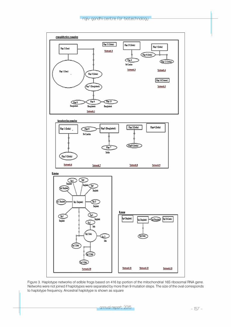
annual report 2015 - 157 -
rajiv gandhi centre for biotechnology
Figure 3. Haplotype networks of edible frogs based on 416 bp portion of the mitochondrial 16S ribosomal RNA gene. Networks were not joined if haplotypes were separated by more than 9 mutation steps. The size of the oval corresponds to haplotype frequency. Ancestral haplotype is shown as square
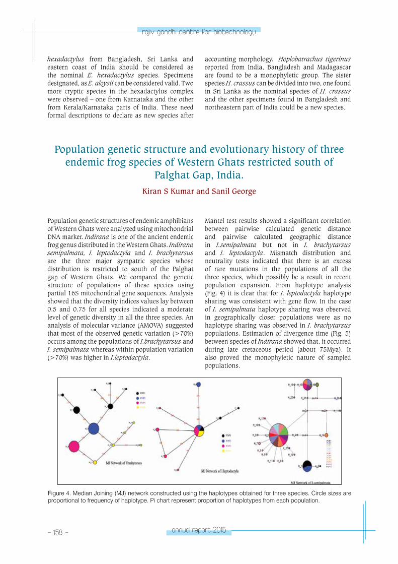
- 158 - annual report 2015
rajiv gandhi centre for biotechnology
Figure 4. Median Joining (MJ) network constructed using the haplotypes obtained for three species. Circle sizes are proportional to frequency of haplotype. Pi chart represent proportion of haplotypes from each population.
Population genetic structure and evolutionary history of three endemic frog species of Western Ghats restricted south of
Palghat Gap, India.Kiran S Kumar and Sanil George
Population genetic structures of endemic amphibians of Western Ghats were analyzed using mitochondrial DNA marker. Indirana is one of the ancient endemic frog genus distributed in the Western Ghats. Indirana semipalmata, I. leptodactyla and I. brachytarsus are the three major sympatric species whose distribution is restricted to south of the Palghat gap of Western Ghats. We compared the genetic structure of populations of these species using partial 16S mitochondrial gene sequences. Analysis showed that the diversity indices values lay between 0.5 and 0.75 for all species indicated a moderate level of genetic diversity in all the three species. An analysis of molecular variance (AMOVA) suggested that most of the observed genetic variation (>70%) occurs among the populations of I.brachytarsus and I. semipalmata whereas within population variation (>70%) was higher in I.leptodactyla.
Mantel test results showed a significant correlation between pairwise calculated genetic distance and pairwise calculated geographic distance in I.semipalmata but not in I. brachytarsus and I. leptodactyla. Mismatch distribution and neutrality tests indicated that there is an excess of rare mutations in the populations of all the three species, which possibly be a result in recent population expansion. From haplotype analysis (Fig. 4) it is clear that for I. leptodactyla haplotype sharing was consistent with gene flow. In the case of I. semipalmata haplotype sharing was observed in geographically closer populations were as no haplotype sharing was observed in I. brachytarsus populations. Estimation of divergence time (Fig. 5) between species of Indirana showed that, it occurred during late cretaceous period (about 75Mya). It also proved the monophyletic nature of sampled populations.
hexadactylus from Bangladesh, Sri Lanka and eastern coast of India should be considered as the nominal E. hexadactylus species. Specimens designated, as E. aloysii can be considered valid. Two more cryptic species in the hexadactylus complex were observed – one from Karnataka and the other from Kerala/Karnataka parts of India. These need formal descriptions to declare as new species after
accounting morphology. Hoplobatrachus tigerinus reported from India, Bangladesh and Madagascar are found to be a monophyletic group. The sister species H. crassus can be divided into two, one found in Sri Lanka as the nominal species of H. crassus and the other specimens found in Bangladesh and northeastern part of India could be a new species.
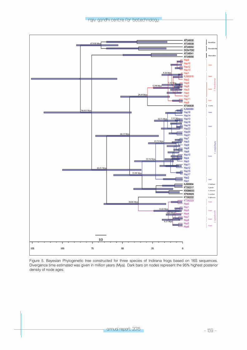
annual report 2015 - 159 -
rajiv gandhi centre for biotechnology
Figure 5. Bayesian Phylogenetic tree constructed for three species of Indirana frogs based on 16S sequences. Divergence time estimated was given in million years (Mya). Dark bars on nodes represent the 95% highest posterior density of node ages.
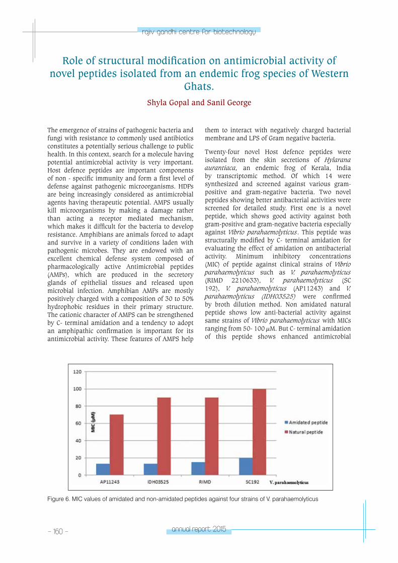
- 160 - annual report 2015
rajiv gandhi centre for biotechnology
Figure 6. MIC values of amidated and non-amidated peptides against four strains of V. parahaemolyticus
Role of structural modification on antimicrobial activity of novel peptides isolated from an endemic frog species of Western
Ghats.Shyla Gopal and Sanil George
The emergence of strains of pathogenic bacteria and fungi with resistance to commonly used antibiotics constitutes a potentially serious challenge to public health. In this context, search for a molecule having potential antimicrobial activity is very important. Host defence peptides are important components of non - specific immunity and form a first level of defense against pathogenic microorganisms. HDPs are being increasingly considered as antimicrobial agents having therapeutic potential. AMPS usually kill microorganisms by making a damage rather than acting a receptor mediated mechanism, which makes it difficult for the bacteria to develop resistance. Amphibians are animals forced to adapt and survive in a variety of conditions laden with pathogenic microbes. They are endowed with an excellent chemical defense system composed of pharmacologically active Antimicrobial peptides (AMPs), which are produced in the secretory glands of epithelial tissues and released upon microbial infection. Amphibian AMPs are mostly positively charged with a composition of 30 to 50% hydrophobic residues in their primary structure. The cationic character of AMPS can be strengthened by C- terminal amidation and a tendency to adopt an amphipathic confirmation is important for its antimicrobial activity. These features of AMPS help
them to interact with negatively charged bacterial membrane and LPS of Gram negative bacteria.
Twenty-four novel Host defence peptides were isolated from the skin secretions of Hylarana aurantiaca, an endemic frog of Kerala, India by transcriptomic method. Of which 14 were synthesized and screened against various gram-positive and gram-negative bacteria. Two novel peptides showing better antibacterial activities were screened for detailed study. First one is a novel peptide, which shows good activity against both gram-positive and gram-negative bacteria especially against Vibrio parahaemolyticus. This peptide was structurally modified by C- terminal amidation for evaluating the effect of amidation on antibacterial activity. Minimum inhibitory concentrations (MIC) of peptide against clinical strains of Vibrio parahaemolyticus such as V. parahaemolyticus (RIMD 2210633), V. parahaemolyticus (SC 192), V. parahaemolyticus (AP11243) and V. parahaemolyticus (IDH03525) were confirmed by broth dilution method. Non amidated natural peptide shows low anti-bacterial activity against same strains of Vibrio parahaemolyticus with MICs ranging from 50- 100 µM. But C- terminal amidation of this peptide shows enhanced antimicrobial
annual report 2015 - 161 -
rajiv gandhi centre for biotechnology
Publication• Jonathan P. A. Gardner, Jamila Patterson, Sanil
George and J. K. Patterson Edward 2016. Combined evidence indicates that Perna indica Kuriakose and Nair 1976 is Perna perna (Linnaeus, 1758) from the Oman region introduced into southern India more than 100 years ago. Biol Invasions DOI 10.1007/s10530-016-1074-9 ; IF 2.586
Genbank submissions KU179079-KU179090 KU198183-KU198192 KU999300- KU999342
Conference Presentations• Kiran S Kumar and Sanil George (2016). Genetic
Structure of Indirana semipalmata, an Endemic Frog Species of Western Ghats having Restricted Distribution. National Seminar on Recent Challenges in Biodiversity Conservation, Department of Zoology, University of Keala. 22-23 March 2016
• Anoop V S and Sanil George (2016). In search of two nominal sympatric frog species of India- A molecular taxonomical approach. National seminar on recent challenges in biodiversity Conservation, Department of Zoology, University of Kerala, 22-23 March 2016.
Workshops attended• Kiran S. Kumar. “Fundamentals and Application
of Biostatistics” jointly organized by the CEPC Laboratory & Technical Division, Kollam and SN College, Kollam During August, 2015 at Cashew Bhavan, Kollam
• Kiran S. Kumar. DST-SERB ‘Second School in Herpetology’ held at Madras Crocodile Bank Trust. Dec 2015
Awards• Anoop V S and Sanil George (2016). National
seminar on recent challenges in biodiversity Conservation, Department of Zoology, University of Kerala, 22-23 March 2016. (BEST PAPER AWARD- oral presentation)
• Vineethkumar TV, Shyla Gopal and Sanil George, 2016. First report of Lividin and Spinulosain Peptides from the Skin Secretion of an Indian frog Acta Biologica Hungarica 67 (1), 121-124 Doi: 10.1556/018.67.2016.1.10, IF 0.589
• Jesmina A.S and George S. 2015. New distribution records for the critically endangered frog Indirana gundia (Dubois, 1986) from Kerala part of Western Ghats, India”. Biodiversity Data Journal 3: e5825. doi: 10.3897/BDJ.3.e5825.PubMed.
activity by reducing the MICs 2-5 times. Amidated peptide shows bactericidal activity below 20µM in all strains of Vibrio parahaemolyticus. Apart from reducing MICs, amidation of peptide does not affect
the hemolytic activity on Human RBC. This property makes this peptide an attractive molecule having high therapeutic index.
CHeMICA
l & enVIRonM
entAl BIoloGY pRoGRA
M
- environmental Biology laboratory
CHEMICAL & ENVIRONMENTAL
BIOLOGY PROGRAMEnvironmental Biology
Laboratory
K. Harikrishnan
Hari Krishnan took his PhD in Aquatic Biology
from the University of Kerala and joined RGCB in
2001.
PhD StudentsArjun. J.K.Aneesh B.
Support Staff Kavitha T.Silu JubyGeetha S.L.
annual report 2015 - 163 -
rajiv gandhi centre for biotechnology
The rise of antibiotic resistance in microorganisms is one of the most significant public health concerns. Despite the threat posed by antibiotic resistance in infectious bacteria, little is known about the diversity, distribution, and origin of resistance genes. The soil microbial community is highly complex and contains a high density of antibiotic-producing bacteria, making it a likely source of diverse antibiotic resistance determinants and act as a genetic reservoir of antimicrobial resistance genes. Resistance genes present in the environment do pose a threat to human health, if they migrate to clinical settings and transfer to pathogens. Little is known about these resistance gene reservoirs in the environment and their contribution to antibiotic resistance in clinical scenario. One major group of antibiotic resistance determinants abundant in soil is β-lactamases, which hydrolyze β-lactam antibiotics penicillin and cephalosporins. These antibiotics are prescribed more frequently to humans and live stocks due to its low toxicity and high efficacy, developing a powerful selection pressure for genes encoding resistance elements in environments proximal to human activity. Coconut husk retting is a major activity in the backwaters
of Kerala, which is a unique reservoir for microbes, which are in close contact with human and animal activities. The study was focused on the detection of beta-lactamase gene available in the sediment
Prevalence of gene conferring beta-lactam resistance in backwater sediment
Arjun J.K. and Hari Krishnan K.
- 164 - annual report 2015
rajiv gandhi centre for biotechnology
Amylases are enzymes, which digest starch into simpler molecules such as maltose and glucose and are of great industrial significance. Amylases have been derived from several fungi, yeast, bacteria and actinomycetes. Alpha and beta amylases are the two major classes of amylases present in
bacteria. α-Amylase (EC 3.2.1.1; 1,4-α-D-glucan glucanohydrolase) randomly cleaves internal α-1,4-glucosidic linkages to produce different sizes of oligosaccharides and β-Amylase (EC 3.2.1.2; 1,4-α-D-glucan malto hydrolase) is an exo-enzyme that hydrolyse (1-4)-α-D-glucosidic linkages in
starch, to remove successive maltose units from the non-reducing ends of the chains. Amylases find potential applications in a number of industrial processes such as in food, fermentation, textiles and paper industries. Enzymatic hydrolysis using amylases have been employed in starch-processing industries replacing chemical hydrolysis as an environmental friendly green technology option. The discovery of novel amylases with enhanced activity is highly warranted to turn industries more environmental friendly.
Soil is a potential source for novel microbial enzymes and in the study we have screened out 300 amylase positive bacterial isolates and the isolate PHB29 exhibiting a high level of amylolytic activity on starch agar plate assay was further characterised. The isolate was identified as Bacillus megaterium by 16S rDNA sequence analysis and submitted the sequence to GenBank with accession number KF056893.
Characterisation of alpha and beta amylases from a soil bacterium
Aneesh B. and Hari Krishnan K.
at the retting zones in the back-water through the gene targeted metagenomic approach. Sediment was collected aseptically from the coconut husk retting area and isolated the community DNA following hot alkaline lysis method. Beta-lactamase gene was amplified from the metagenomic DNA using the designed gene specific oligonucleotide primers. (Fig1). The amplicon was cloned into a TA cloning vector (pGEM-T easy) and a library was generated. The clones in the library were sequenced and the sequence identified was submitted in NCBI GenBank under the accession number JN561602. The homology of the gene was analyzed with NCBI BLASTx and a phylogenetic tree was constructed with MEGA 5.0 software (Fig 2). The phylogeny revealed that the protein sequence belonged to the structural class-A of β-lactamases (TEM BLA), the predominant beta-lactamase found in pathogens.
Beta lactamase genes are prone to mutations due to the selection pressure from indiscriminate use of antibiotics. Studying the occurrence of new mutations in beta lactamase gene in environments is essential for the development of improved antibiotic preparations.
As microbial antibiotic resistance is increasing in clinical settings, it is imperative to extend the studies to environments to understand the ecology of resistance genes in infectious disease and in natural microbial communities. The identification of new resistance genes, particularly those potentially present in non-culturable microbes may facilitate prediction of the emergence of resistance and to develop strategies for prevention and treatment against antibiotic resistant pathogens.
annual report 2015 - 165 -
rajiv gandhi centre for biotechnology
The genes coding for alpha and beta amylases were amplified from PHB29 using designed gene specific primers (Fig: 3). The amplicons were sequenced and the homology search in GenBank revealed that the alpha and beta amylases exhibited 97% similarity with that of Bacillus sp. and Bacillus megaterium respectively. A phylogenetic tree was constructed based on the beta amylase amino acid sequence similarity and found to be clustered with Bacillus megaterium (Fig 4). The genes were sub cloned and expressed in E. coli (Fig 5). The β-amylase gene was further sub cloned in pET 22b (+) and over expressed in E. coli (BL21DE3). The enzyme activity at different temperatures was checked and found the maximum specific activity of 0.445 units/mg of
protein at 45ºC which is comparatively better than other reported bacterial β-amylases (Fig 6).
Starch is considered as a cheap and easily available carbon source and its utilization may be an ideal strategy for cost effective fermentative production of various bio-products such as organic acids, alcohols, biopolymers, etc. E. coli cells cannot utilize starch in natural conditions and this restricts the use of starch as a carbon source in fermentation processes involving E. coli. Through these studies, we have succeeded in developing a starch utilizing recombinant system with two amylase genes. Further validation of this recombinant construct to make use for the value added production of various fermentative products are progressing.
- 166 - annual report 2015
rajiv gandhi centre for biotechnology
Degradation of single-carbon compounds by the bacterial communities from a wetland
Kavitha T. and Hari Krishnan K.
One carbon (C1) compounds are ubiquitous in the environment which are produced and released into the atmosphere by the breakdown of biomass and many of them act as environmental pollutants influencing the global climate processes. Wetlands are considered as the significant natural source and sink for C1 compounds from where a great part of such hydrocarbons are emitted to the surroundings. Methylotrophs present in wetlands play a vital role in the metabolism of C1 compounds and aid in the regulation of global carbon cycle. A wide range of C1 compounds are consumed by methylotrophs in the environment, including methane, methanol, methylated amines, methylated glycines, halomethanes, and methylated sulfur species. Wetland ecosystems contribute up to 15% destruction of total global methane through methanotrophs, a subgroup of methylotrophs, which are the only known biological sink for methane. Since the knowledge on the diversity of methylotrophs and methanotrophs and their degradation potential of C1 compounds are scarce from the Kuttanad wetland ecosystem, the study was initiated. During the study, the metagenomic DNA was isolated from the wetland soil and
subjected to 16S rRNA gene targeted next generation sequencing (Ion-PGM, Life Technologies) to elucidate the bacterial diversity. The methanotroph community in the wetland was represented by the
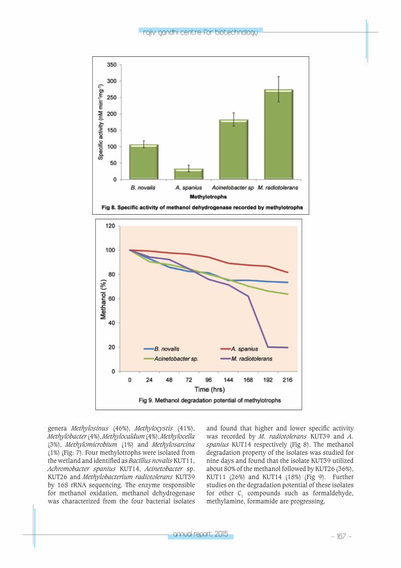
annual report 2015 - 167 -
rajiv gandhi centre for biotechnology
genera Methylosinus (46%), Methylocystis (41%), Methylobacter (4%), Methylocaldum (4%), Methylocella (3%), Methylomicrobium (1%) and Methylosarcina (1%) (Fig: 7). Four methylotrophs were isolated from the wetland and identified as Bacillus novalis KUT11, Achromobacter spanius KUT14, Acinetobacter sp. KUT26 and Methylobacterium radiotolerans KUT39 by 16S rRNA sequencing. The enzyme responsible for methanol oxidation, methanol dehydrogenase was characterized from the four bacterial isolates
and found that higher and lower specific activity was recorded by M. radiotolerans KUT39 and A. spanius KUT14 respectively (Fig 8). The methanol degradation property of the isolates was studied for nine days and found that the isolate KUT39 utilized about 80% of the methanol followed by KUT26 (36%), KUT11 (26%) and KUT14 (18%) (Fig 9). Further studies on the degradation potential of these isolates for other C1 compounds such as formaldehyde, methylamine, formamide are progressing.
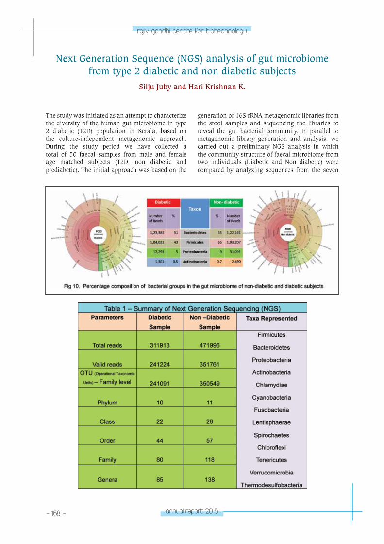
- 168 - annual report 2015
rajiv gandhi centre for biotechnology
The study was initiated as an attempt to characterize the diversity of the human gut microbiome in type 2 diabetic (T2D) population in Kerala, based on the culture-independent metagenomic approach. During the study period we have collected a total of 50 faecal samples from male and female age matched subjects (T2D, non diabetic and prediabetic). The initial approach was based on the
generation of 16S rRNA metagenomic libraries from the stool samples and sequencing the libraries to reveal the gut bacterial community. In parallel to metagenomic library generation and analysis, we carried out a preliminary NGS analysis in which the community structure of faecal microbiome from two individuals (Diabetic and Non diabetic) were compared by analyzing sequences from the seven
Next Generation Sequence (NGS) analysis of gut microbiome from type 2 diabetic and non diabetic subjects
Silju Juby and Hari Krishnan K.
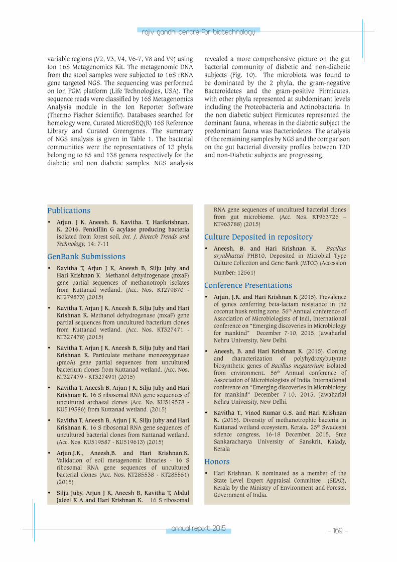
annual report 2015 - 169 -
rajiv gandhi centre for biotechnology
variable regions (V2, V3, V4, V6-7, V8 and V9) using Ion 16S Metagenomics Kit. The metagenomic DNA from the stool samples were subjected to 16S rRNA gene targeted NGS. The sequencing was performed on Ion PGM platform (Life Technologies, USA). The sequence reads were classified by 16S Metagenomics Analysis module in the Ion Reporter Software (Thermo Fischer Scientific). Databases searched for homology were, Curated MicroSEQ(R) 16S Reference Library and Curated Greengenes. The summary of NGS analysis is given in Table 1. The bacterial communities were the representatives of 13 phyla belonging to 85 and 138 genera respectively for the diabetic and non diabetic samples. NGS analysis
revealed a more comprehensive picture on the gut bacterial community of diabetic and non-diabetic subjects (Fig. 10). The microbiota was found to be dominated by the 2 phyla, the gram-negative Bacteroidetes and the gram-positive Firmicutes, with other phyla represented at subdominant levels including the Proteobacteria and Actinobacteria. In the non diabetic subject Firmicutes represented the dominant fauna, whereas in the diabetic subject the predominant fauna was Bacteriodetes. The analysis of the remaining samples by NGS and the comparison on the gut bacterial diversity profiles between T2D and non-Diabetic subjects are progressing.
Publications• Arjun. J K, Aneesh. B, Kavitha. T, Harikrishnan.
K. 2016. Penicillin G acylase producing bacteria isolated from forest soil, Int. J. Biotech Trends and Technology, 14: 7-11
GenBank Submissions• Kavitha T, Arjun J K, Aneesh B, Silju Juby and
Hari Krishnan K. Methanol dehydrogenase (mxaF) gene partial sequences of methanotroph isolates from Kuttanad wetland. (Acc. Nos. KT279870 - KT279873) (2015)
• Kavitha T, Arjun J K, Aneesh B, Silju Juby and Hari Krishnan K. Methanol dehydrogenase (mxaF) gene partial sequences from uncultured bacterium clones from Kuttanad wetland. (Acc. Nos. KT327471 - KT327478) (2015)
• Kavitha T, Arjun J K, Aneesh B, Silju Juby and Hari Krishnan K. Particulate methane monooxygenase (pmoA) gene partial sequences from uncultured bacterium clones from Kuttanad wetland. (Acc. Nos. KT327479 - KT327491) (2015)
• Kavitha T, Aneesh B, Arjun J K, Silju Juby and Hari Krishnan K. 16 S ribosomal RNA gene sequences of uncultured archaeal clones (Acc. No. KU519578 - KU519586) from Kuttanad wetland. (2015)
• Kavitha T, Aneesh B, Arjun J K, Silju Juby and Hari Krishnan K. 16 S ribosomal RNA gene sequences of uncultured bacterial clones from Kuttanad wetland. (Acc. Nos. KU519587 - KU519613) (2015)
• Arjun,J.K., Aneesh,B. and Hari Krishnan,K. Validation of soil metagenomic libraries - 16 S ribosomal RNA gene sequences of uncultured bacterial clones (Acc. Nos. KT285538 - KT285551) (2015)
• Silju Juby, Arjun J K, Aneesh B, Kavitha T, Abdul Jaleel K A and Hari Krishnan K. 16 S ribosomal
RNA gene sequences of uncultured bacterial clones from gut microbiome. (Acc. Nos. KT963726 – KT963788) (2015)
Culture Deposited in repository• Aneesh, B. and Hari Krishnan K. Bacillus
aryabhattai PHB10, Deposited in Microbial Type Culture Collection and Gene Bank (MTCC) (Accession
Number: 12561)
Conference Presentations• Arjun, J.K. and Hari Krishnan K (2015). Prevalence
of genes conferring beta-lactam resistance in the coconut husk retting zone. 56th Annual conference of Association of Microbiologists of Indi, International conference on “Emerging discoveries in Microbiology for mankind” December 7-10, 2015, Jawaharlal Nehru University, New Delhi.
• Aneesh, B. and Hari Krishnan K. (2015). Cloning and characterization of polyhydroxybutyrate biosynthetic genes of Bacillus megaterium isolated from environment. 56th Annual conference of Association of Microbiologists of India, International conference on “Emerging discoveries in Microbiology for mankind” December 7-10, 2015, Jawaharlal Nehru University, New Delhi.
• Kavitha T., Vinod Kumar G.S. and Hari Krishnan K. (2015). Diversity of methanotrophic bacteria in Kuttanad wetland ecosystem, Kerala. 25th Swadeshi science congress, 16-18 December, 2015, Sree Sankaracharya University of Sanskrit, Kalady, Kerala
Honors• Hari Krishnan. K nominated as a member of the
State Level Expert Appraisal Committee (SEAC), Kerala by the Ministry of Environment and Forests, Government of India.
- 170 - annual report 2015
rajiv gandhi centre for biotechnology
EXTRA MURAL GRANTS
Studies on the distribution and diversity of methanotrophic microbial communities, mitigating methane emission in Kuttanad wetland ecosystem, Kerala
Hari Krishnan. K (PI)
G.S. Vinod Kumar (Co-PI)
Department of Environment and Climate Change, Govt. of Kerala
2013 - 16
Metagenomic analysis of gut microbiome from type 2 diabetic subjects in Kerala - a preliminary study
Hari Krishnan. K (PI)
Abdul Jaleel. K.A (Co-PI) KSCSTE, Govt. of Kerala 2014 - 17
neuRoBIoloGY pRoGRAM
- M
olecular neurobiology laboratoryNEUROBIOLOGY
PROGRAMMolecular Neurobiology
Laboratory
R. V. Omkumar
Omkumar received his PhD Biochemistry from
the Indian Institute of Science, Bangalore. He did
postdoctoral reserch at Purdue University and
at California Intitute of Technology, USA before
joining RGCB in 1996.
PhD Students Mathew JohnArchana G.M.Arun Kumar R.C.Ramya R. PrabhuMantosh KumarLakshmi K.Sowmya GunasekharanReena Sarah JacobSethu Lakshmi
- 172 - annual report 2015
rajiv gandhi centre for biotechnology
Spatiotemporal features of calcium signaling pathways are important in determining their physiological consequences. It is known that calcium influx into neurons through different channels differ in their levels and durations. While normal calcium influx into synapses occurs during learning and memory, unregulated calcium influx causes neuronal death which happens in many neurodegenerative diseases. Calcium channels such as N-methyl-D-aspartate receptors (NMDAR) and voltage gated calcium channels (VGCC) as well as calcium stimulated molecules like calcium/calmodulin dependent protein kinase II (CaMKII) are primary players in synaptic calcium signalling.
Interaction of CaMKII with NMDAR subunit GluN2B causes allosteric modulation of catalysis by CaMKII. It also renders CaMKII resistant to dephosphorylation by phosphatases. These biochemical events are involved in the molecular memory mechanisms underlying learning and memory. We have developed mutants of CaMKII in which GluN2B binding does not cause catalytic modulation or inhibition of dephosphorylation. Using structural bioinformatics and mutagenesis we are working on elucidating the mechanism of the GluN2B-mediated modulation of CaMKII. We are also investigating the physiological significance of this regulation. Towards this goal, siRNA-mediated downregulation of CaMKII in neuronal cultures is being standardised. Computational modelling of the biochemical functions of CaMKII supported by experiments is being undertaken in a collaborative project.
Neuronal Calcium Signaling in Health and Disease
Binding interaction between CaMKII and NMDAR is regulated by the phosphorylation status of the GluN2B subunit. We have monitored the phosphorylation status of GluN2B in rats in vivo. Administration of agonists and antagonists show that phosphorylation of GluN2B is enhanced by activation of NMDAR and VGCC in a CaM kinase dependent manner in vivo.
Our studies on calcium signalling in cerebellar granule (CG) cells show that depolarisation of CG cells in culture by KCl treatment gives rise to a proteome profile that differs with that of CG cells in vivo. Our data show that depolarisation induced activation of VGCC is necessary for inducing expression of new proteins. Further experiments to reveal the underlying mechanisms are being carried out as this information would help in understanding regulation of proteins that are differentially expressed in vivo.
Aberrant calcium signalling due to alterations in miRNA mediated regulation of NMDAR expression is associated with neuropsychiatric diseases such as schizophrenia. We have initiated a study to understand the alterations in biochemical mechanisms arising from impaired miRNA regulation that lead to the disease. Using bioinformatics based screening, a set of miRNAs that may target the 3’UTRs of Grin2A and Grin2B genes that code for the GluN2A and GluN2B subunits respectively, of NMDAR have been identified. Genes for the miRNAs and the sequences of the 3’UTRs of Grin2A and Grin2B were cloned into expression vectors for experimentally demonstrating the binding between the miRNAs and the corresponding 3’UTRs.
Novel Techniques for the assay of Ca2+ channels
Impaired functioning of calcium channels leads to several disease conditions. Hence calcium channel inhibitors are widely used as drugs for many diseases. The NMDAR calcium channel has different subtypes that arise from variation in subunit composition. These subtypes show differences in their pharmacological and electrophysiological characteristics as well as in their tissue distribution. To obtain best results in drug therapy, drugs that
target specific subtypes are necessary. The origin of several pathophysiological conditions such as autism spectrum disorders and schizophrenia can be attributed to mutations in any of the components of the NMDAR complex. Drug resistance and the varied responses of different individuals towards the same drug can also arise from genetic variation in NMDAR subunits. Thus, the diversity of NMDAR channels is a major challenge in the discovery of

annual report 2015 - 173 -
rajiv gandhi centre for biotechnology
effective drugs. Further challenge to NMDAR-directed drug discovery arises from the existing calcium channel assays that depend on real time methods which are both technically challenging as well as expensive. All these have resulted in a dearth of effective drugs directed against NMDARs. We have attempted to address this problem by developing end-point detection based assays for calcium channel activities.
We had earlier developed a new assay for calcium channels based on end point measurement that is comparatively simpler as well as inexpensive. The assay involves transfecting HEK-293 cells with a pair of proteins, GFP-CaMKII and a GluN2B sequence motif, that interact upon intracellular calcium release. Their interaction results in a characteristic fluorescence pattern that acts as the
signal for calcium. We now have developed a stable clone of HEK-293 cells, which we termed as Calcium Sensor cell line that constitutively expresses GFP-CaMKII and the GluN2B sequence motif. This further simplifies the assay method by eliminating the transfection step for introducing the calcium sensing proteins. We have started using the cell line to screen for inhibitors of NMDAR subtypes having GluN2A or GluN2B subunit.
In a second method developed by our group, cell death induced by activation of NMDAR transfected in HEK-293 cells is used as a signal for NMDAR activity. This method has been demonstrated for assay of NMDAR containing GluN2B. We are currently trying to adapt this method for the assay of other NMDAR subtypes and other calcium channels.
Bioprospecting for Neuroprotectants
Several plants with therapeutic value have been described in traditional medicine. Such plants can be used as source material in the search for biologically active compounds that can be developed into drugs. In addition, synthetic analogues of phytochemicals are also used to test their effect on biological systems with the aim of finding therapeutically useful activities. We have been focussing on cellular targets such as CaMKII and calcium channels for which assays have been developed in our lab. We have found a plant extract that inhibits NMDAR ectopically expressed in HEK-293 cells. The extract also inhibits glutamate induced excitotoxicity in primary cortical neurons. Administration of the plant extract to rats subjected to excitotoxic treatment ameliorates their impaired performance in Morris water maze test. Thus the extract shows neuroprotection in vitro and in vivo. Biochemical analysis of brain tissues of the animals treated with the extract under excitotoxic conditions is being carried out towards understanding the underlying molecular mechanisms. The extract is also being subjected to subfractionation towards identifying the active compound/s.
Lobeline is an alkaloid that is known to be neuroprotective against MPTP+ induced neurodegeneration in Parkinson’s disease model in rats. We find that lobeline inhibits NMDAR activity and protects primary cortical neurons against glutamate induced excitotoxicity effectively.
Tacrine is a cholinesterase inhibitor and is known to be neuroprotective. Tacrine has also been shown to inhibit NMDA receptor. However it is required at high concentrations, to cause significant inhibition. In addition, tacrine has been shown to have side effects such as hepatotoxicity. Hence it was necessary to improve its inhibitory potency so that dosage could be reduced. Towards this goal, novel tacrine derivatives were designed, synthesized and were biologically evaluated for their potency of inhibiting NMDAR. Several of the derivatives were much more potent than tacrine in inhibiting NMDAR activity. At the concentrations required to significantly inhibit NMDAR, the derivatives did not show hepatotoxic property as seen by their effect on viability of HepG-2 cell line. Cytotoxicity of the compounds were also analysed on HEK-293 cells. Compound(s) with minimal IC50 value for NMDAR inhibition were taken forward further to test their neuroprotective properties in primary neuronal culture systems. Some of the tacrine derivatives tested were able to protect rat primary cortical neurons from glutamate induced excitotoxicity at significantly reduced concentrations compared to tacrine. The structure-activity relationship guided study suggested that many of the novel tacrine derivatives might be useful as neuroprotective agents by antagonizing the activity of NMDAR.
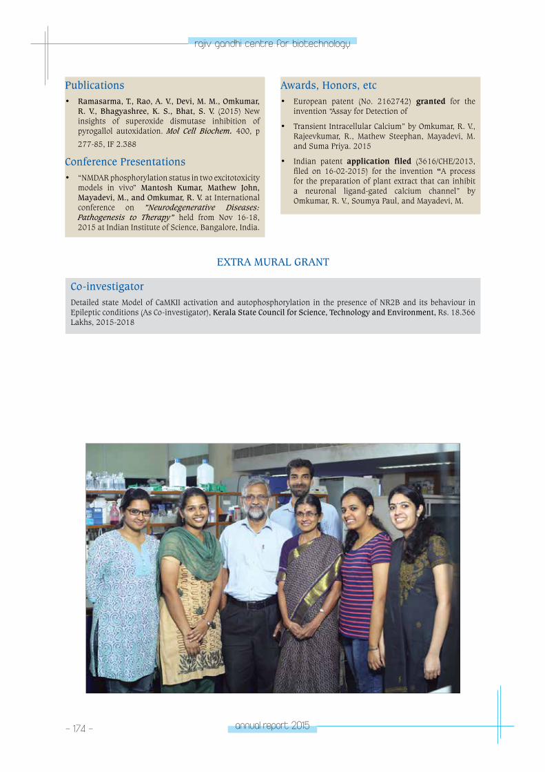
- 174 - annual report 2015
rajiv gandhi centre for biotechnology
Publications• Ramasarma, T., Rao, A. V., Devi, M. M., Omkumar,
R. V., Bhagyashree, K. S., Bhat, S. V. (2015) New insights of superoxide dismutase inhibition of pyrogallol autoxidation. Mol Cell Biochem. 400, p
277-85, IF 2.388
Conference Presentations • “NMDAR phosphorylation status in two excitotoxicity
models in vivo” Mantosh Kumar, Mathew John, Mayadevi, M., and Omkumar, R. V. at International conference on ”Neurodegenerative Diseases: Pathogenesis to Therapy” held from Nov 16-18, 2015 at Indian Institute of Science, Bangalore, India.
Awards, Honors, etc• European patent (No. 2162742) granted for the
invention “Assay for Detection of
• Transient Intracellular Calcium” by Omkumar, R. V., Rajeevkumar, R., Mathew Steephan, Mayadevi, M. and Suma Priya. 2015
• Indian patent application filed (3616/CHE/2013, filed on 16-02-2015) for the invention “A process for the preparation of plant extract that can inhibit a neuronal ligand-gated calcium channel” by Omkumar, R. V., Soumya Paul, and Mayadevi, M.
EXTRA MURAL GRANT
Co-investigatorDetailed state Model of CaMKII activation and autophosphorylation in the presence of NR2B and its behaviour in Epileptic conditions (As Co-investigator), Kerala State Council for Science, Technology and Environment, Rs. 18.366 Lakhs, 2015-2018
neuRoBIoloGY pRoGRAM
- M
olecular neurobiology laboratoryNEUROBIOLOGY
PROGRAMMolecular Neurobiology
Laboratory
Mayadevi. M
Mayadevi received her MSc and PhD in
Chemistry from the University of Kerala and
worked at Case westsrn Reserve University and
Baylor Research Institute, USA, before joining
RGCB in 1993.
- 176 - annual report 2015
rajiv gandhi centre for biotechnology
Calcium signaling stimulates certain kinases and phosphatases in brain resulting in phosphorylation and dephosphorylation of proteins. Although these phosphorylation-dephoshorylation events are transient they bring about changes in proteins effective enough to influence synaptic plasticity. The same protein may undergo phosphorylation at multiple sites. The phosphorylation at different sites of the same protein have different effects on neuronal function. Calcium/calmodulin dependent protein kinase II (CaMKII) is an abundant protein in the neuron which gets autophosphorylated at multiple sites. The autophosphorylation at Thr286 of CaMKII maintains the enzyme in an active state which is otherwise inactive in the basal state of calcium. Thr286 autophosphorylated CaMKII state is one way that contributes to stable memory formation. Protein phosphatase1 (PP1) dephosphorylates phospho-Thr286-CaMKII. This kinase/phosphatase system working as a molecular switch constitutes a molecular memory model. Previous work from the neurobiology division has shown that the binding of GluN2B to the T-site of CaMKII modulates the activity of the enzyme and enables the switch to perform in an energy efficient manner. Further the
binding of GluN2B to CaMKII slows down the rate of dephosphorylation of phospho-Thr286 CaMKII and inhibits the dephosphorylation. GluN2B induces structural modifications on CaMKII that instils some constraints on the accessibility of PP1 to the phospho-Thr286 site. We have identified two residues that may be involved in the structural modifications that CaMKII undergoes upon GluN2B binding. The residues Glu96 and His282 are also involved in ATP binding. Autophosphorylated and non-autophosphorylated forms of E96A and H282A CaMKII mutants bind to GluN2B as seen in pulldown-assays. Although E96A-CaMKII mutant binds to GluN2B, the activity of the mutant is not modulated by GluN2B. The rate of dephosphorylation of WT-CaMKII is reduced when GluN2B is bound to its T-site. However, even in the GluN2B bound state the mutants, E96A and H282A, of CaMKII did not exhibit reduced rate of dephosphorylation. We presume GluN2B induces in CaMKII similar kind of structural changes during modulation of catalysis and dephosphorylation. Glu96 and His282 are two of the key residues bringing about the structural changes in the two events.
Identification of residues involved in GluN2B-induced inhibition of dephosphorylation of CaMKII
Mayadevi M., Lakshmi, K. and Omkumar, R. V.
Patents:Title of Invention: Assay for detection of transient intracellular Ca2+
Inventor’s Names: Omkumar, R. V., Rajeevkumar, R., Mathew Steephan, Mayadevi, M. and Suma Priya, S.
Granting Agency: Europian Patent Office
Status: Patent Granted, No. 2162742
neuRoBIoloGY pRoGRAM
neuro-Stem
Cell Biology laboratoryNEUROBIOLOGY
PROGRAMNeuro-Stem Cell Biology
Laboratory
Jackson James
Jackson James took his PhD in Molecular
Neurobiology from Cochin University of Science
& Technology, India He worked as Postdoctoral
fellow at Lied Transplant center, Dept. of
Ophthalmology, University of Nebraska Medical
Center, Omaha USA before joining RGCB in 2004.
PhD studentsDhanesh S.B.Subashini C.Lalitha S.Riya Ann PaulParvathy S.Meera V.
Technical AssistantBiju S. NairSreeja S.
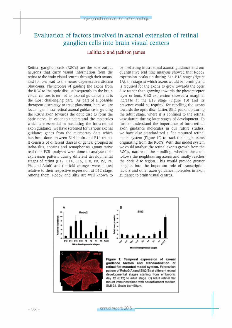
- 178 - annual report 2015
rajiv gandhi centre for biotechnology
Retinal ganglion cells (RGC’s) are the sole output neurons that carry visual information from the retina to the brain visual centres through their axons, and its loss lead to the neuro-degenerative disease Glaucoma. The process of guiding the axons from the RGC to the optic disc, subsequently to the brain visual centres is termed as axonal guidance and is the most challenging part. As part of a possible therapeutic strategy to treat glaucoma, here we are focusing on intra-retinal axonal guidance ie, guiding the RGC’s axon towards the optic disc to form the optic nerve. In order to understand the molecules which are essential in mediating the intra-retinal axon guidance, we have screened for various axonal guidance genes from the microarray data which has been done between E14 brain and E14 retina. It consists of different classes of genes, grouped as Robo-slits, ephrins and semaphorins. Quantitative real-time PCR analyses were done to analyse their expression pattern during different developmental stages of retina (E12, E14, E16, E18, P0, P2, P4, P6, and Adult) and the fold changes were plotted relative to their respective expression at E12 stage. Among them, Robo2 and slit2 are well known to
be mediating intra-retinal axonal guidance and our quantitative real time analysis showed that Robo2 expression peaks up during E14-E18 stage (Figure 1A), the stage at which axons would be forming and is required for the axons to grow towards the optic disc rather than growing towards the photoreceptor layer or lens. Slit2 expression showed a marginal increase at the E18 stage (Figure 1B) and its presence could be required for repelling the axons towards the optic disc. Latter, Slit2 peaks up during the adult stage, where it is confined to the retinal vasculature during later stages of development. To further understand the importance of intra-retinal axon guidance molecules in our future studies, we have also standardized a flat mounted retinal model system (Figure 1C) to track the single axons originating from the RGC’s. With this model system we could analyse the retinal axon’s growth from the RGC’s, nature of the bundling, whether the axon follows the neighbouring axons and finally reaches the optic disc region. This would provide greater insights into the important role of transcription factors and other axon guidance molecules in axon guidance to brain visual centres.
Evaluation of factors involved in axonal extension of retinal ganglion cells into brain visual centers
Lalitha S and Jackson James
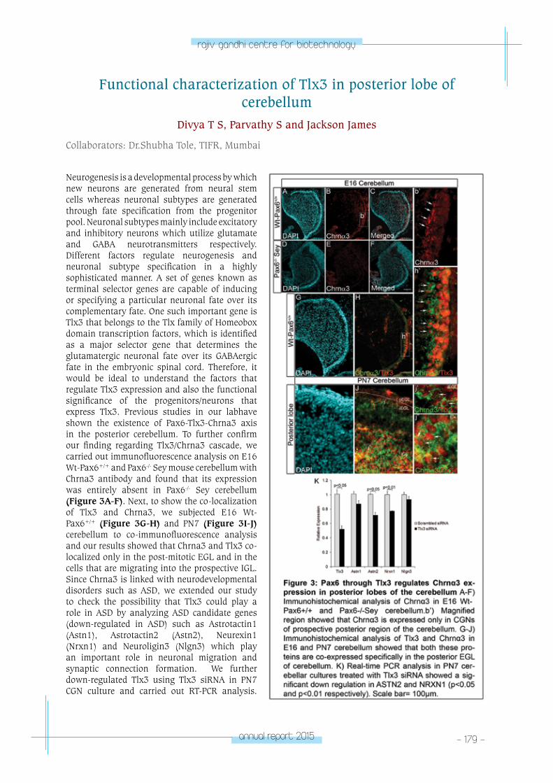
annual report 2015 - 179 -
rajiv gandhi centre for biotechnology
Neurogenesis is a developmental process by which new neurons are generated from neural stem cells whereas neuronal subtypes are generated through fate specification from the progenitor pool. Neuronal subtypes mainly include excitatory and inhibitory neurons which utilize glutamate and GABA neurotransmitters respectively. Different factors regulate neurogenesis and neuronal subtype specification in a highly sophisticated manner. A set of genes known as terminal selector genes are capable of inducing or specifying a particular neuronal fate over its complementary fate. One such important gene is Tlx3 that belongs to the Tlx family of Homeobox domain transcription factors, which is identified as a major selector gene that determines the glutamatergic neuronal fate over its GABAergic fate in the embryonic spinal cord. Therefore, it would be ideal to understand the factors that regulate Tlx3 expression and also the functional significance of the progenitors/neurons that express Tlx3. Previous studies in our labhave shown the existence of Pax6-Tlx3-Chrna3 axis in the posterior cerebellum. To further confirm our finding regarding Tlx3/Chrna3 cascade, we carried out immunofluorescence analysis on E16 Wt-Pax6+/+ and Pax6-/- Sey mouse cerebellum with Chrna3 antibody and found that its expression was entirely absent in Pax6-/- Sey cerebellum (Figure 3A-F). Next, to show the co-localization of Tlx3 and Chrna3, we subjected E16 Wt-Pax6+/+ (Figure 3G-H) and PN7 (Figure 3I-J) cerebellum to co-immunofluorescence analysis and our results showed that Chrna3 and Tlx3 co-localized only in the post-mitotic EGL and in the cells that are migrating into the prospective IGL. Since Chrna3 is linked with neurodevelopmental disorders such as ASD, we extended our study to check the possibility that Tlx3 could play a role in ASD by analyzing ASD candidate genes (down-regulated in ASD) such as Astrotactin1 (Astn1), Astrotactin2 (Astn2), Neurexin1 (Nrxn1) and Neuroligin3 (Nlgn3) which play an important role in neuronal migration and synaptic connection formation. We further down-regulated Tlx3 using Tlx3 siRNA in PN7 CGN culture and carried out RT-PCR analysis.
Functional characterization of Tlx3 in posterior lobe of cerebellum
Divya T S, Parvathy S and Jackson James
Collaborators: Dr.Shubha Tole, TIFR, Mumbai
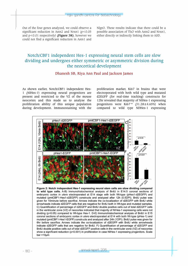
- 180 - annual report 2015
rajiv gandhi centre for biotechnology
Out of the four genes analyzed, we could observe a significant reduction in Astn2 and Nrxn1 (p<0.05 and p<0.01 respectively) (Figure 3K), however we could not find a significant reduction in Astn1 and
Nlgn3. These results indicate that there could be a possible association of Tlx3 with Astn2 and Nrxn1, either directly or indirectly linking them to ASD.
Notch/CBF1 independent Hes-1 expressing neural stem cells are slow dividing and undergoes either symmetric or asymmetric division during
the neocortical development
Dhanesh SB, Riya Ann Paul and Jackson James
As shown earlier, Notch/CBF1 independent Hes-1 (NIHes-1) expressing neural progenitors are present and restricted to the VZ of the mouse neocortex and this made us to analyse the proliferation ability of this unique population during development. Immunostaining with the
proliferation marker, Ki67 in brains that were electroporated with both wild type and mutated d2EGFP (for real-time tracking) constructs for 12hr revealed that majority of NIHes-1 expressing progenitors were Ki67-ve (31.28±4.65%) when compared to wild type NDHes-1 expressing
annual report 2015 - 181 -
rajiv gandhi centre for biotechnology
progenitors (71.68±3.91%). In order to further elucidate the cell cycle kinetics of NIHes-1 expressing neural progenitors compared to actively dividing NDHes-1 expressing progenitors, we carried out BrdU pulsing with NIHes-1 and NDHes-1 reporting plasmids. Our results showed that the percentage of BrdU and d2EGFP (real-time tracking)/EGFP (long-term tracking) double positive progenitors expressing NIHes-1 was high after 1day electroporation with EGFP and 6hr BrdU pulse (73.38±9.50% for NDHes-1 and 32.19±5.50% for NIHes-1, Figure. 2D,E&F) compared to 12h electroporation with d2EGFP and 10min BrdU pulse experiments (5.32±1.17% for NIHes-1 and 14.75±4.55% for NDHes-1, Figure. 2A, B&C). It appears that after acquiring Notch dependency these progenitors represent the classical NDHes-1 expressing progenitors. To understand the cleavage orientation in progenitors with NDHes-1 and NIHes-1 expression state we carried out immunostaining with PH3, a marker for metaphase stage and DAPI for nuclear staining in electroporated E14 embryos with both wild type and mutated d2EGFP plasmids. Our results showed that the NIHes-1 expressing neural stem cells followed both symmetrical as well as asymmetrical mode of division similar to
NDHes-1 expressing progenitors. Therefore, the unique nature harboured by NIHes-1 expressing progenitors such as equal competency for both symmetric and asymmetric division, increase in cell cycle length and differential behaviour even after acquiring Notch dependency emphasis the existence of heterogeneity within the Hes-1 expressing progenitors in the VZ. However, since Notch receptor is essential for canonical Hes-1 activation, the possibility of ligand expression in non-canonically differentially expressed Hes-1 cells to transduce the signaling cascade in neighboring cells could not be excluded. This differential Hes-1 expression along with the lateral inhibition exerted by Notch may maintain the neural progenitors or induce them to proliferate. Among various Notch ligands, Dll1 is prominently expressed in the developing neocortex. In order to characterize various Notch ligand expressions in Notch/CBF1 independent Hes-1 expressing cells, dual fluorescent reporter construct pDll1-tdTomato-Hes-1/mtCBF1-EGFP was generated and further in vivo studies are being carried out to understand the micro environmental changes happening in the niche with respect to Dll1 ligand expression.
Wnt5a conditional mutants display defects in proliferation of GABAergic and glutamatergic progenitors
Subashini C and Jackson James
Collaborator: Rejji Kuruvilla, Johns Hopkins University, Baltimore, USA
Wnt5a signaling has been previously reported to promote survival, proliferation and differentiation of neural progenitors in different regions of the brain in a context dependent manner. Since, we observed a significant reduction in size and number of GABAergic and glutamatergic neurons, we next sought to determine if the reduction was due to reduced proliferation or apoptosis. In order to address this question, we carried out TUNEL assay which did not reveal any observable difference in number of apoptotic cells between Wnt5a cKOs (Nestin-Cre x Wnt5af/f) and wild-type controls at any stage of development (E14.5, PN1 and PN7). Therefore, we next assessed if loss of Wnt5a resulted in defective proliferation of cerebellar progenitors. Immunohistochemical labeling of mid-sagittal sections at E14.5 stage with proliferation marker,
Ki67 (marks the cells in G1, S and M phase) revealed significant reduction (p<0.005) in number of proliferating cells both in VZ and EGL of the Wnt5a cKOs as compared to the wild-type siblings. Since, we continued to observe a reduction in the size of the cerebellum at an early postnatal stage (PN1), we analyzed the rate of proliferation in EGL and PWM. Our results showed a significant reduction (p<0.005) in the number of Ki67+ve proliferating cells at both germinal centers compared to the wild-type counterparts (Figure 4A-G). Consistently, we also observed a significant reduction (p<0.005) in number of phospho histone 3 (PH3) positive mitotic cells in the Wnt5a cKOs (Figure 4H-O). But contrarily, at PN7 we did not observe thinning of Ki67+ve outer EGL and quantitative analysis also did not reveal reduction in Ki67+ve granule neuron progenitors
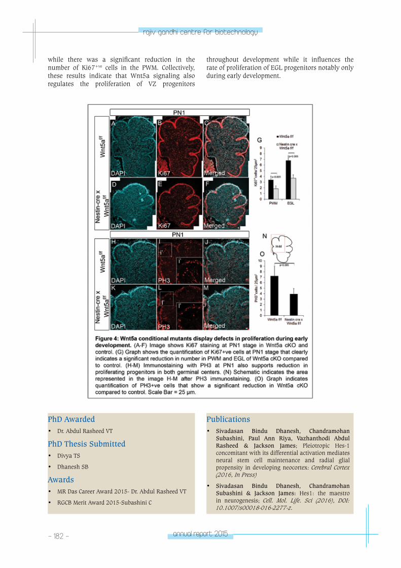
- 182 - annual report 2015
rajiv gandhi centre for biotechnology
while there was a significant reduction in the number of Ki67+ve cells in the PWM. Collectively, these results indicate that Wnt5a signaling also regulates the proliferation of VZ progenitors
throughout development while it influences the rate of proliferation of EGL progenitors notably only during early development.
PhD Awarded• Dr. Abdul Rasheed VT
PhD Thesis Submitted• Divya TS
• Dhanesh SB
Awards• MR Das Career Award 2015- Dr. Abdul Rasheed VT
• RGCB Merit Award 2015-Subashini C
Publications• Sivadasan Bindu Dhanesh, Chandramohan
Subashini, Paul Ann Riya, Vazhanthodi Abdul Rasheed & Jackson James; Pleiotropic Hes-1 concomitant with its differential activation mediates neural stem cell maintenance and radial glial propensity in developing neocortex: Cerebral Cortex (2016, In Press)
• Sivadasan Bindu Dhanesh, Chandramohan Subashini & Jackson James: Hes1: the maestro in neurogenesis; Cell. Mol. Life. Sci (2016), DOI: 10.1007/s00018-016-2277-z.
annual report 2015 - 183 -
rajiv gandhi centre for biotechnology
• Anupama V, George M, Dhanesh SB, Chandran A, James J, Shivakumar K. Molecular mechanisms in H2O2-induced increase in AT1 receptor gene expression in cardiac fibroblasts: a role for endogenously generated Angiotensin II; Journal of Molecular and Cellular Cardiology (2016, In Press)
• Mereena George, Anupama Vijayakumar, Sivadasan Bindu Dhanesh, Jackson James, K. Shivakumar: Molecular basis and functional significance of Angiotensin II-induced increase in Discoidin Domain Receptor 2 gene expression in cardiac fibroblasts; Journal of Molecular and Cellular Cardiology, 90 (2016) 59–69.
RESEARCH GRANTS:
No. Investigator(s) Title Funding Agency Duration
1. Dr. Jackson James (PI) Dr. R V OmkumarDr. Santhosh Kumar S N
Characterization of Notch independent Hes-1 mediated maintenance and fate specification of neural progenitors
Dept. of Biotechnology, Govt. of India
20013-2016 (3Year)
2. Dr. Jackson James (PI) Dr. R V Omkumar
Transcriptional regulation of Tlx3 (Hox11L2) by Notch signaling and its involvement in excitatory Vs. inhibitory fate specification of neural progenitors
Dept. of Science & Technology, Govt. of India
20013-2016 (3Year)
neuRoBIoloGY pRoGRAM
Hum
an Molecular Genetics laboratory
NEUROBIOLOGY PROGRAM
Human Molecular Genetics Laboratory
Moinak Banerjee
Moinak Banerjee received his PhD from ML
Sukhadia University, Udaipur, Subsequently he
did postdoctoral training in AIIMS, New Delhi
and CCMB, Hyderabad, before joining RGCB in
1996.
Ph.D StudentsSwathy B.Ann Mary AlexShafique C.M.Sindoora K.P.
Technical StaffBindu AsokanVeluthai G.
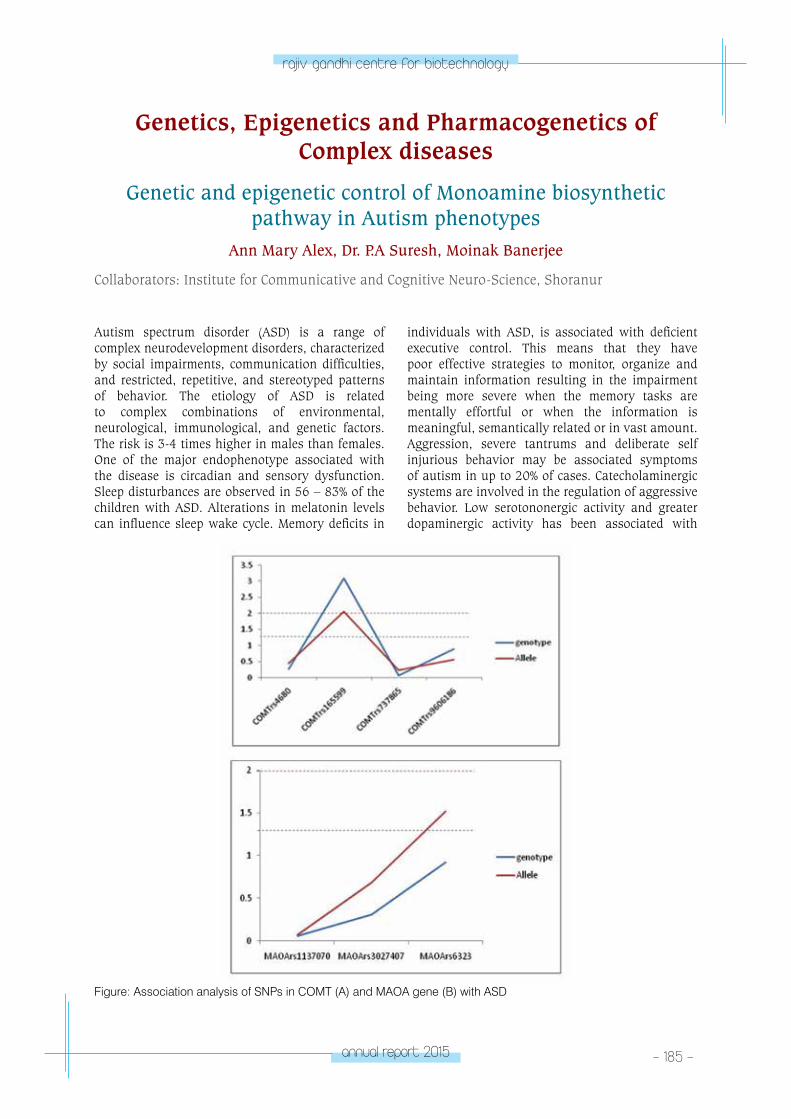
annual report 2015 - 185 -
rajiv gandhi centre for biotechnology
Autism spectrum disorder (ASD) is a range of complex neurodevelopment disorders, characterized by social impairments, communication difficulties, and restricted, repetitive, and stereotyped patterns of behavior. The etiology of ASD is related to complex combinations of environmental, neurological, immunological, and genetic factors. The risk is 3-4 times higher in males than females. One of the major endophenotype associated with the disease is circadian and sensory dysfunction. Sleep disturbances are observed in 56 – 83% of the children with ASD. Alterations in melatonin levels can influence sleep wake cycle. Memory deficits in
individuals with ASD, is associated with deficient executive control. This means that they have poor effective strategies to monitor, organize and maintain information resulting in the impairment being more severe when the memory tasks are mentally effortful or when the information is meaningful, semantically related or in vast amount. Aggression, severe tantrums and deliberate self injurious behavior may be associated symptoms of autism in up to 20% of cases. Catecholaminergic systems are involved in the regulation of aggressive behavior. Low serotononergic activity and greater dopaminergic activity has been associated with
Genetics, Epigenetics and Pharmacogenetics of Complex diseases
Genetic and epigenetic control of Monoamine biosynthetic pathway in Autism phenotypes
Ann Mary Alex, Dr. P.A Suresh, Moinak Banerjee
Collaborators: Institute for Communicative and Cognitive Neuro-Science, Shoranur
Figure: Association analysis of SNPs in COMT (A) and MAOA gene (B) with ASD
- 186 - annual report 2015
rajiv gandhi centre for biotechnology
aggression. These enzymes act in the synthesis pathway of melatonin and serotonin which has multiple physiological activities including the phenotypes exhibited in ASD cases. One of the major routes of deactivation of monoamine neurotransmitters in the brain is metabolism by two key enzymes, monoamine oxidase (MAO) and catechol-Omethyltransferase (COMT). Genotypic and allelic frequencies were analyzed for the SNPs to understand the association with the diseases. The aim of my study is to understand the genes that influence these major underlying phenotypes and how they influence the genetic network, in the development of autism from a population perspective. 7 SNPs were chosen for the study based on the functional relevance and tagging status in 150 cases and 200 controls.
Among the SNPs screened in the COMT gene rs165599 was found to be clinically relevant in
autism. This SNP is in the 3’UTR which indicated its modulation with environmental effects. The A allele of rs165599 is likely to modulate higher expression of the COMT gene indicating higher rate of degradation. Females are likely to be more affected with this allele. Similarly in MAOA gene in the X chromosome 3 SNPs were screened. Among the SNPs screened in MAOA gene rs6323 was associated in autism irrespective of their gender. T allele of rs1137070 was associated with autism in males.
In conclusion, the 2 genes involved in the catecholaminergic systems, COMT and MAOA were found to be associated with autism. Differential gender specific association was clearly visible in the MAOA gene. These genes which have been identified to be involved in controlling many of the endophenotypes of ASD can be a useful marker in diagnosis.
Elucidating the genetics of methylation map in intracranial aneurysm
Sanish Sathyan, Linda Koshy, H V Easwer, S. Premkumar, Jacob P Alapatt, Suresh Nair, R.N. Bhattacharya, Moinak Banerjee
Collaborators: SCTIMST, Thiruvanathapuram. Calicut Medical College, Kozhikode
Intracranial aneurysm (IA) or Brain aneurysm is a cerebrovascular disorder occurs due to the weakening of an artery. The aneurysm wall becomes thin, weak and results in the localised dilation or ballooning of blood vessels. This complication arises due to certain medical problems, genetic conditions and trauma which can damage or injure the artery walls. When a cerebral aneurysm ruptures, a non- traumatic hemorrhage occurs, usually refers a sub arachnoid hemorrhage (SAH). Bleeding results in the mixing of blood with CSF creating an increased pressure on the brain and subsequently may lead to brain damage with paralysis or coma and in some cases even death. Vasospasm is another major complication associated with aneurysm rupture. Irritating blood byproducts causes the walls of an artery to spasm and contract, resulting in a low blood supply to the brain and leads to a secondary stroke. One third of patient who suffers a SAH will survive with good recovery, one third will survive with disability, and one third will die.
Some of the risk factors for the development of aneurysm include smoking, hypertension, and
heavy alcohol consumption (Koshy etal. 2010). Similarly increasing age, female sex, family histories are also known risk factors for aneurysmal SAH (aSAH). The familial occurrence and the association with heritable conditions indicate that genetic factors may play a role in the development of IA. There are no diagnostic tests for specific genetic risk factors to identify patients who are at a high risk of developing intracranial aneurysms (Caranci et al., 2013) and no single gene has been identified to be responsible for IAs formation or rupture (Krischek et al., 2008). Since both genetic and environmental risk factors are involved, identifying the molecular mechanisms underlying the pathogenesis of IAs are considered complex (Tromp et al., 2014). It is likely that the true mechanism underlying aneurysm formation will involve the interplay between a genetic predisposition and environmental risk factors (Sandford et al., 2007).
An earlier study in the lab on south Indian population reported the importance of polymorphism associated with structural genes in IA (Koshy et al., 2006, 2008) and involvement of environmental risk
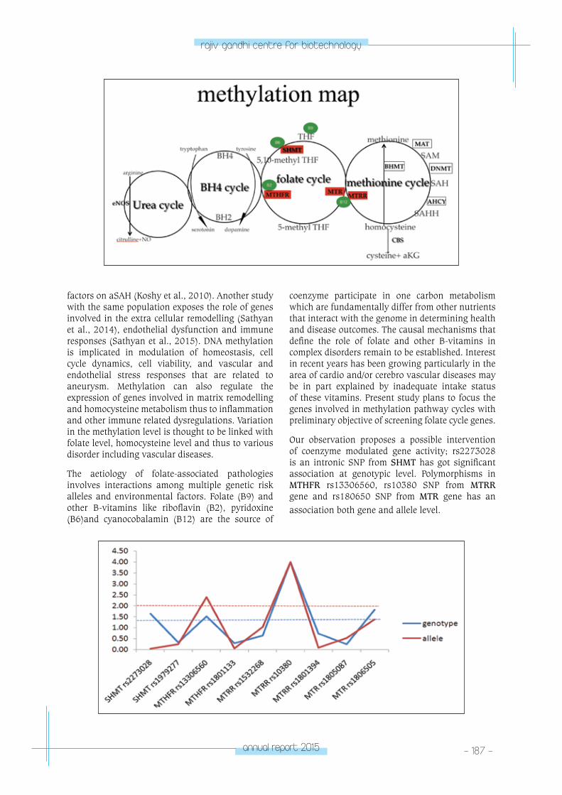
annual report 2015 - 187 -
rajiv gandhi centre for biotechnology
factors on aSAH (Koshy et al., 2010). Another study with the same population exposes the role of genes involved in the extra cellular remodelling (Sathyan et al., 2014), endothelial dysfunction and immune responses (Sathyan et al., 2015). DNA methylation is implicated in modulation of homeostasis, cell cycle dynamics, cell viability, and vascular and endothelial stress responses that are related to aneurysm. Methylation can also regulate the expression of genes involved in matrix remodelling and homocysteine metabolism thus to inflammation and other immune related dysregulations. Variation in the methylation level is thought to be linked with folate level, homocysteine level and thus to various disorder including vascular diseases.
The aetiology of folate-associated pathologies involves interactions among multiple genetic risk alleles and environmental factors. Folate (B9) and other B-vitamins like riboflavin (B2), pyridoxine (B6)and cyanocobalamin (B12) are the source of
coenzyme participate in one carbon metabolism which are fundamentally differ from other nutrients that interact with the genome in determining health and disease outcomes. The causal mechanisms that define the role of folate and other B-vitamins in complex disorders remain to be established. Interest in recent years has been growing particularly in the area of cardio and/or cerebro vascular diseases may be in part explained by inadequate intake status of these vitamins. Present study plans to focus the genes involved in methylation pathway cycles with preliminary objective of screening folate cycle genes.
Our observation proposes a possible intervention of coenzyme modulated gene activity; rs2273028 is an intronic SNP from SHMT has got significant association at genotypic level. Polymorphisms in MTHFR rs13306560, rs10380 SNP from MTRR gene and rs180650 SNP from MTR gene has an association both gene and allele level.
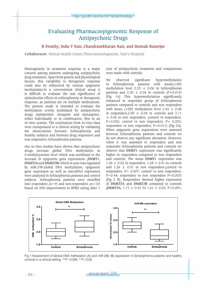
- 188 - annual report 2015
rajiv gandhi centre for biotechnology
Evaluating Pharmacoepigenomic Response of Antipsychotic Drugs
B Swathy, Indu V Nair, Chandrasekharan Nair, and Moinak Banerjee
Collaborator: Mental Health Centre,Thiruvananthapuram, Nair’s Hospital
Heterogeneity in treatment response is a major concern among patients undergoing antipsychotic drug treatment. Apart from genetic and physiological factors, this variability to therapeutic response could also be influenced by various epigenetic mechanisms.In a conventional clinical setup it is difficult to evaluate the real significance of epimolecular effects in schizophrenia or therapeutic response, as patients are on multiple medications. The present study is intended to evaluate the methylation events modulated by antipsychotic drugs (haloperidol, clozapine and olanzapine), either individually or in combination, first in an in-vitro system. The conclusions from in-vitro data were extrapolated to a clinical setting by validating the observations between Schizophrenia and healthy subjects and between drug responsive and non-responsive Schizophrenia patients.
Our in-vitro studies have shown that antipsychotic drugs increase global DNA methylation at 5-methylcytosine level which was attributed to the increase in epigenetic gene expressions (DNMT1, DNMT3A and DNMT3B) which in turn was regulated by miR-29b.Global DNA methylation, epigenetic gene expression as well as microRNA expression were analysed in Schizophrenia patients and control subjects. Schizophrenia patients were stratified into responders (n=9) and non-responders (n=10) based on 50% improvement in BPRS rating after 1
year of antipsychotic treatment and comparisons were made with controls.
We observed significant hypermethylation in Schizophrenia patients with mean(±SD) methylation level 2.23 ± 0.06 in Schizophrenia patients and 2.00 ± 0.04 in controls (P=0.014) [Fig 1A] This hypermethylation significantly enhanced in responder group of Schizophrenia patients compared to controls and non responders with mean (±SD) methylation level 2.44 ± 0.08 in responders,2.00 ± 0.04 in controls and 2.11 ± 0.06 in non responders. (control vs responders, P=0.002; control vs non responders, P= 0.253; responders vs non responders, P=0.011) [Fig 2A].When epigenetic gene expressions were assessed between Schizophrenia patients and controls we do not observe any significant alteration. However, when it was assessed in responders and non responder Schizophrenia patients and controls we observe that DNMT1 expression was significantly higher in responders compared to non responders and controls. The mean DNMT1 expression was 1.36 ± 0.02 in responders, 1.28 ± 0.01 in controls and 1.26 ± 0.01 in non responders.(control vs responders, P= 0.007; control vs non responders, P=0.44; responders vs non responders P=0.003) [Fig 2 B]. Responders showed higher expression of DNMT3A and DNMT3B compared to controls (DNMT3A, 1.71 ± 0.01 Vs 1.61 ± 0.02, P=0.001;
Fig 1:Assessment of Global DNA methylation (A) and miR-29b (B) expression in Schizophrenia patients and healthy controls in a clinical setting. **P<0.005, * P<0.05.
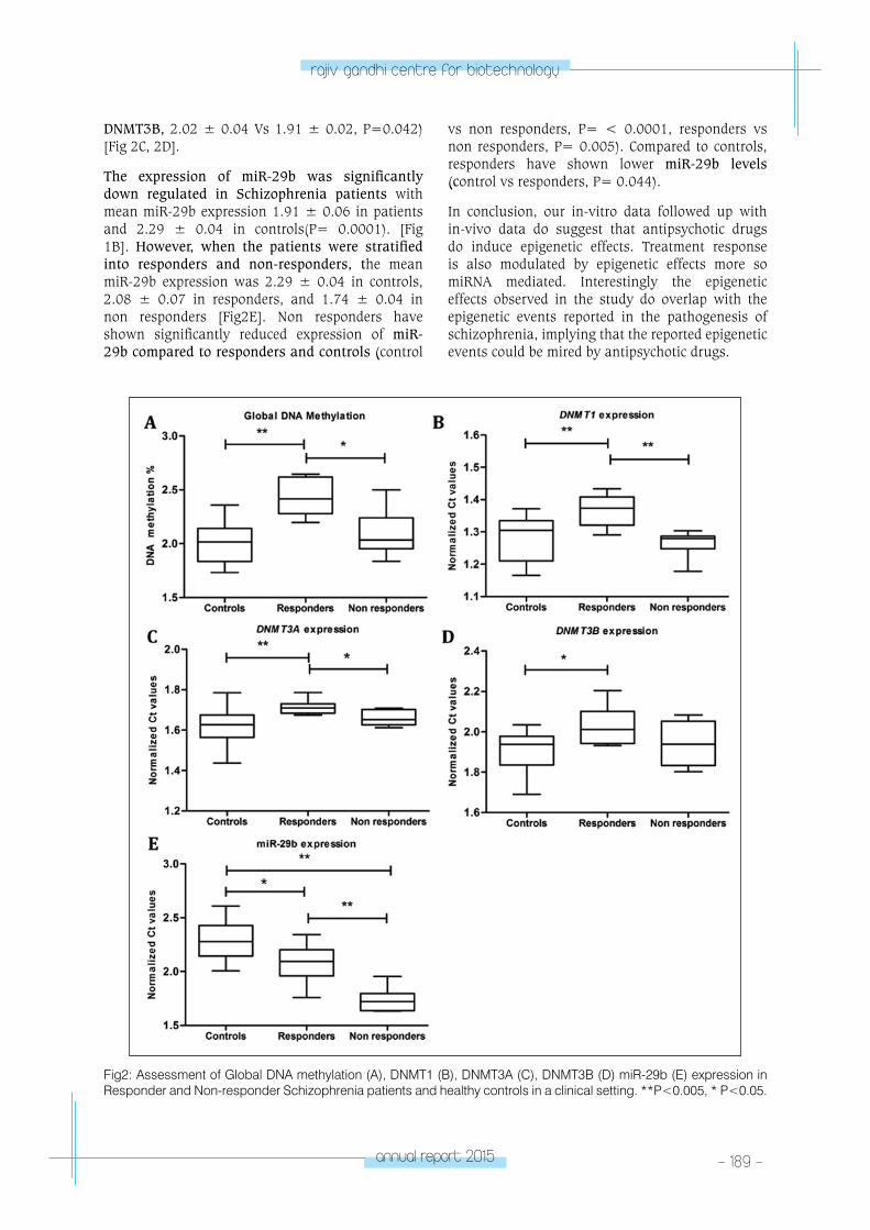
annual report 2015 - 189 -
rajiv gandhi centre for biotechnology
DNMT3B, 2.02 ± 0.04 Vs 1.91 ± 0.02, P=0.042) [Fig 2C, 2D].
The expression of miR-29b was significantly down regulated in Schizophrenia patients with mean miR-29b expression 1.91 ± 0.06 in patients and 2.29 ± 0.04 in controls(P= 0.0001). [Fig 1B]. However, when the patients were stratified into responders and non-responders, the mean miR-29b expression was 2.29 ± 0.04 in controls, 2.08 ± 0.07 in responders, and 1.74 ± 0.04 in non responders [Fig2E]. Non responders have shown significantly reduced expression of miR-29b compared to responders and controls (control
vs non responders, P= < 0.0001, responders vs non responders, P= 0.005). Compared to controls, responders have shown lower miR-29b levels (control vs responders, P= 0.044).
In conclusion, our in-vitro data followed up with in-vivo data do suggest that antipsychotic drugs do induce epigenetic effects. Treatment response is also modulated by epigenetic effects more so miRNA mediated. Interestingly the epigenetic effects observed in the study do overlap with the epigenetic events reported in the pathogenesis of schizophrenia, implying that the reported epigenetic events could be mired by antipsychotic drugs.
Fig2: Assessment of Global DNA methylation (A), DNMT1 (B), DNMT3A (C), DNMT3B (D) miR-29b (E) expression in Responder and Non-responder Schizophrenia patients and healthy controls in a clinical setting. **P<0.005, * P<0.05.
- 190 - annual report 2015
rajiv gandhi centre for biotechnology
Publications• Sanish Sathyan, Linda V Koshy, Lekshmi Srinivas,
Easwer HV, Premkumar S, Suresh Nair, R.N. Bhattacharya, Jacob P Alapatt, Moinak Banerjee. Pathogenesis of intracranial aneurysm is mediated by proinflammatory cytokine TNFA and IFNG and through stochastic regulation of IL10 and TGFB1 by co-morbid factors. J of Neuroinflammation 2015, 12:135.
PhD awards• Mr. Sanish Sathyan, Mrs. Saradalekshmi KR.
Awards• RIKEN summer internship fellowship to attend
RIKEN BSI Summer Program 2015-“Sculpting Neural Circuits and behavior”, June 10-Aug 5,2015, RIKEN Brain Science Institute, Japan.
Conference presentation• Moinak Banerjee. How to dissect a complex disease.
CME talk, 14th Feb 2015, Puspagiri Medical college, Thiruvalla.
• B Swathy and Moinak Banerjee, Evaluating Pharmacoepigenomic Response of Antipsychotic Drugs, June 10-Aug 5, 2015, RIKEN BSI, RIKEN Brain Science Institute, Japan.
• Moinak Banerjee. Changing scenarios in dissecting a disease. Omics and Biomarker Analysis: In disease pathology. June 22-23, 2015. Dept. of Zoology, Kerala University, Trivandrum.
• Moinak Banerjee. The changing realm of drug discovery in the era of personalized medicine. 13th Aug. 2015. Loyola College, Chennai.
• Moinak Banerjee. The changing realm of drug discovery in the era of personalized medicine. 10th Aug. 2015, Academic Staff College, University of Calicut, Calicut.
• Moinak Banerjee. Understanding genetic and epigenetics of Schizophrenia. Mind, Body and Culture. 25-27th Sept. 2015, World Psychiatry Association meeting KOCHI .
• Moinak Banerjee. The changing realm of drug discovery in the era of personalized medicine. International Conference - NEW HORIZONS IN BIOTECHNOLOGY (NHBT-2015). 23-25, Nov 2015, NIIRST, Trivandrum.
• Moinak Banerjee. Changing dimensions in Forensics. National conference on genomics and society-Prospects, challenges and concerns. 17-19th Feb. 2016, Dept. of Biotechnology, University of Kerala, Trivandrum.
• Moinak Banerjee. Trends and complexities in molecular diagnostics. National Seminar on Emerging Trends in the Diagnosis and Management of Cognitive and Communicative Disorders, 4th June 2016, ICCONS, Shoranur.
• B Swathy and Moinak Banerjee, “Antipsychotic drug induced epigenetic modifications can predict drug response variability in Schizophrenia patients, March3-5,2016, ISHG 2016, 41st Indian Society of Human Genetics Annual Meeting & International Conference, Chennai.
• Ann Mary Alex and Moinak Banerjee Polymorphism screening in catecholaminergic genes suggest similar genetic risk loci for Autism and Schizophrenia. March3-5,2016, ISHG 2016, 41st Indian Society of Human Genetics Annual Meeting & International Conference, Chennai.
neuRoBIoloGY pRoGRAM
neuro Bio-physics laboratory
NEUROBIOLOGY PROGRAM
Neuro Bio-Physics Laboratory
Rashmi Mishra
Rashmi Mishra obtained her PhD in
Neuroscience from the National Brain Research
Centre, Manesar , India. She did post doctoral
training at Max Planck Intitute of Molecular Cell
Biology and Genetics, Dresden, Germany, the
Curie Institute, Paris, France and Tufts University
School of Medicine, Boston USA. She currently
holds the Department of Biotechnology’s
Ramalingaswami Fellowship. Project FellowSebastian John (ICMR SRF)
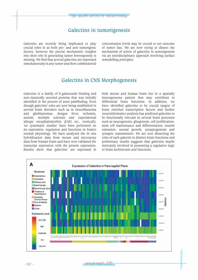
- 192 - annual report 2015
rajiv gandhi centre for biotechnology
Galectins are recently being implicated to play crucial roles in as both pro- and anti tumorigenic factors, however the precise mechanistic insights into their role in generating tumor heterogeneity is missing. We find that several galectins are expressed simultaneously in any tumor and their combinatorial
concentration levels may be crucial to net outcome of tumor fate. We are now trying to dissect the mechanism of action of galectins in tumorigenesis via an interdisciplinary approach involving surface remodelling principles.
Galectins in tumorigenesis
Galectins in CNS Morphogenesis
Galectins is a family of b-galactoside binding and non-classically secreted proteins that was initially identified in the process of axon pathfinding. Even though galectins’ roles are now being established in several brain disorders such as in neuroblastoma and glioblastomas, dengue fever, ischemia, autism, multiple sclerosis and experimental allergic encephalomyelitis (EAE) etc., ironically, ‘no systematic studies’ have been performed on its expression, regulation and functions in brain’s normal physiology. We have analyzed the in situ hybridization data from mouse and microarray data from human brain and have now validated the transcript expression with the protein expression. Results show that galectins’ are expressed in
both mouse and human brain but in a spatially heterogeneous pattern that may contribute to differential brain functions. In addition, we have identified galectins to be crucial targets of brain enriched transcription factors and further neuroinformatics analysis has predicted galectins to be functionally relevant in several brain processes such as neurogenesis, gliogenesis, cell proliferation, stem cell maintenance and differentiation, neurite extension, axonal growth, synaptogenesis and synaptic transmission. We are now dissecting the roles of each galectin in distinct brain functions and preliminary results suggests that galectins maybe intricately involved in presenting a regulative logic to brain architecture and functions.
A
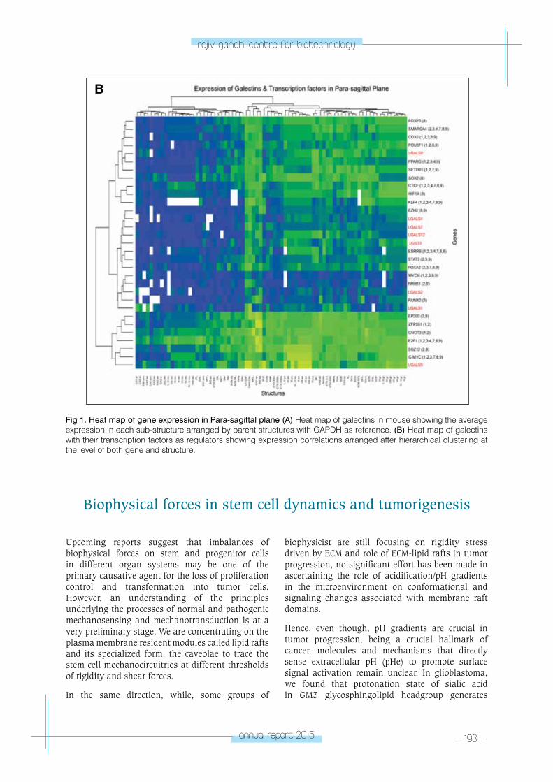
annual report 2015 - 193 -
rajiv gandhi centre for biotechnology
Fig 1. Heat map of gene expression in Para-sagittal plane (A) Heat map of galectins in mouse showing the average expression in each sub-structure arranged by parent structures with GAPDH as reference. (B) Heat map of galectins with their transcription factors as regulators showing expression correlations arranged after hierarchical clustering at the level of both gene and structure.
B
Biophysical forces in stem cell dynamics and tumorigenesis
Upcoming reports suggest that imbalances of biophysical forces on stem and progenitor cells in different organ systems may be one of the primary causative agent for the loss of proliferation control and transformation into tumor cells. However, an understanding of the principles underlying the processes of normal and pathogenic mechanosensing and mechanotransduction is at a very preliminary stage. We are concentrating on the plasma membrane resident modules called lipid rafts and its specialized form, the caveolae to trace the stem cell mechanocircuitries at different thresholds of rigidity and shear forces.
In the same direction, while, some groups of
biophysicist are still focusing on rigidity stress driven by ECM and role of ECM-lipid rafts in tumor progression, no significant effort has been made in ascertaining the role of acidification/pH gradients in the microenvironment on conformational and signaling changes associated with membrane raft domains.
Hence, even though, pH gradients are crucial in tumor progression, being a crucial hallmark of cancer, molecules and mechanisms that directly sense extracellular pH (pHe) to promote surface signal activation remain unclear. In glioblastoma, we found that protonation state of sialic acid in GM3 glycosphingolipid headgroup generates
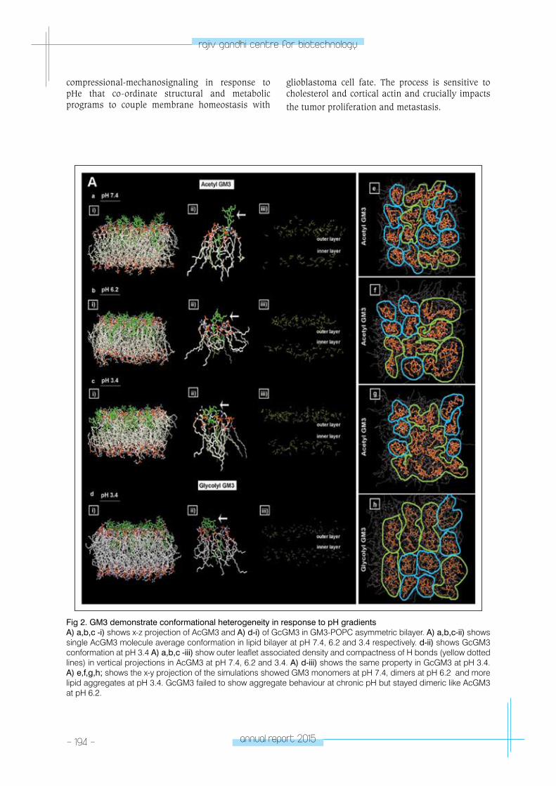
- 194 - annual report 2015
rajiv gandhi centre for biotechnology
Fig 2. GM3 demonstrate conformational heterogeneity in response to pH gradientsA) a,b,c -i) shows x-z projection of AcGM3 and A) d-i) of GcGM3 in GM3-POPC asymmetric bilayer. A) a,b,c-ii) shows single AcGM3 molecule average conformation in lipid bilayer at pH 7.4, 6.2 and 3.4 respectively. d-ii) shows GcGM3 conformation at pH 3.4 A) a,b,c -iii) show outer leaflet associated density and compactness of H bonds (yellow dotted lines) in vertical projections in AcGM3 at pH 7.4, 6.2 and 3.4. A) d-iii) shows the same property in GcGM3 at pH 3.4. A) e,f,g,h; shows the x-y projection of the simulations showed GM3 monomers at pH 7.4, dimers at pH 6.2 and more lipid aggregates at pH 3.4. GcGM3 failed to show aggregate behaviour at chronic pH but stayed dimeric like AcGM3 at pH 6.2.
compressional-mechanosignaling in response to pHe that co-ordinate structural and metabolic programs to couple membrane homeostasis with
glioblastoma cell fate. The process is sensitive to cholesterol and cortical actin and crucially impacts the tumor proliferation and metastasis.
annual report 2015 - 195 -
rajiv gandhi centre for biotechnology
Publications• Rashmi Mishra and Sebastian John. 2016.
Galectin-9: From Cell Signaling to Complex Disease Dynamics. Journal of Biosciences (in press)
RESEARCH GRANTS, EXTRAMURAL FUNDING
No Grant Title Funding Agency Duration
1Rapid Grant for Young InvestigatorHow Neurons Respond to Biomechanical Forces: Implications to Brain Functions and Neurodegeneration
DBT 2013-2016
2
Ramalingaswami FellowshipMechanotransduction through Caveolae: Lipid Rafts in homeostatic control of cell proliferation signaling and tumorigenesis
DBT 2012-2017
3Neuro TaskForce GrantMechanotransduction through Caveolae in Neural Stem Cell Niches: Role in Cell Signaling and Proliferation Control
DBT 2013-2017
plAnt DISeASe BIoloGY & BIoteCHnoloGY
- pDBB laboratory – 1PLANT DISEASE BIOLOGY
& BIOTECHNOLOGY (PDBB)
PDBB Laboratory - 1
V. V. Asha
V V Asha has a Ph.D in Botany from University
of Kerala, Trivandrum, and joined RGCB in 1997.
PhD studentsKrishna Radhika N.Praseeja R.J.Greeshma TomSheena Philip
Technical Associate Gayathri L.T.
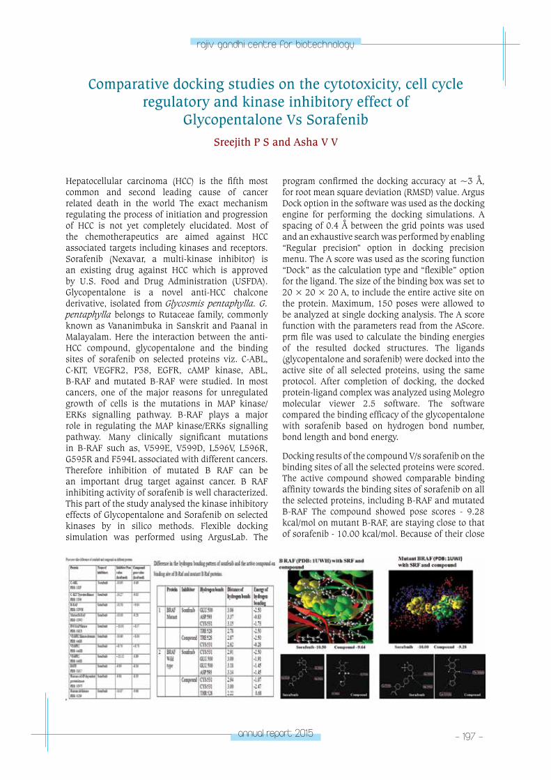
annual report 2015 - 197 -
rajiv gandhi centre for biotechnology
Hepatocellular carcinoma (HCC) is the fifth most common and second leading cause of cancer related death in the world The exact mechanism regulating the process of initiation and progression of HCC is not yet completely elucidated. Most of the chemotherapeutics are aimed against HCC associated targets including kinases and receptors. Sorafenib (Nexavar, a multi-kinase inhibitor) is an existing drug against HCC which is approved by U.S. Food and Drug Administration (USFDA). Glycopentalone is a novel anti-HCC chalcone derivative, isolated from Glycosmis pentaphylla. G. pentaphylla belongs to Rutaceae family, commonly known as Vananimbuka in Sanskrit and Paanal in Malayalam. Here the interaction between the anti-HCC compound, glycopentalone and the binding sites of sorafenib on selected proteins viz. C-ABL, C-KIT, VEGFR2, P38, EGFR, cAMP kinase, ABL, B-RAF and mutated B-RAF were studied. In most cancers, one of the major reasons for unregulated growth of cells is the mutations in MAP kinase/ERKs signalling pathway. B-RAF plays a major role in regulating the MAP kinase/ERKs signalling pathway. Many clinically significant mutations in B-RAF such as, V599E, V599D, L596V, L596R, G595R and F594L associated with different cancers. Therefore inhibition of mutated B RAF can be an important drug target against cancer. B RAF inhibiting activity of sorafenib is well characterized. This part of the study analysed the kinase inhibitory effects of Glycopentalone and Sorafenib on selected kinases by in silico methods. Flexible docking simulation was performed using ArgusLab. The
Comparative docking studies on the cytotoxicity, cell cycle regulatory and kinase inhibitory effect of
Glycopentalone Vs SorafenibSreejith P S and Asha V V
program confirmed the docking accuracy at ~3 Å, for root mean square deviation (RMSD) value. Argus Dock option in the software was used as the docking engine for performing the docking simulations. A spacing of 0.4 Å between the grid points was used and an exhaustive search was performed by enabling “Regular precision” option in docking precision menu. The A score was used as the scoring function “Dock” as the calculation type and “flexible” option for the ligand. The size of the binding box was set to 20 × 20 × 20 A, to include the entire active site on the protein. Maximum, 150 poses were allowed to be analyzed at single docking analysis. The A score function with the parameters read from the AScore.prm file was used to calculate the binding energies of the resulted docked structures. The ligands (glycopentalone and sorafenib) were docked into the active site of all selected proteins, using the same protocol. After completion of docking, the docked protein-ligand complex was analyzed using Molegro molecular viewer 2.5 software. The software compared the binding efficacy of the glycopentalone with sorafenib based on hydrogen bond number, bond length and bond energy.
Docking results of the compound V/s sorafenib on the binding sites of all the selected proteins were scored. The active compound showed comparable binding affinity towards the binding sites of sorafenib on all the selected proteins, including B-RAF and mutated B-RAF The compound showed pose scores - 9.28 kcal/mol on mutant B-RAF, are staying close to that of sorafenib - 10.00 kcal/mol. Because of their close
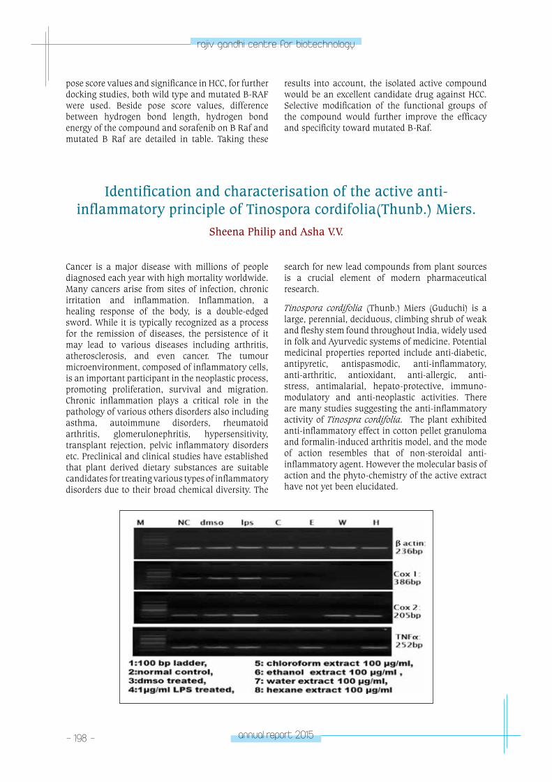
- 198 - annual report 2015
rajiv gandhi centre for biotechnology
pose score values and significance in HCC, for further docking studies, both wild type and mutated B-RAF were used. Beside pose score values, difference between hydrogen bond length, hydrogen bond energy of the compound and sorafenib on B Raf and mutated B Raf are detailed in table. Taking these
results into account, the isolated active compound would be an excellent candidate drug against HCC. Selective modification of the functional groups of the compound would further improve the efficacy and specificity toward mutated B-Raf.
Identification and characterisation of the active anti-inflammatory principle of Tinospora cordifolia(Thunb.) Miers.
Sheena Philip and Asha V.V.
Cancer is a major disease with millions of people diagnosed each year with high mortality worldwide. Many cancers arise from sites of infection, chronic irritation and inflammation. Inflammation, a healing response of the body, is a double-edged sword. While it is typically recognized as a process for the remission of diseases, the persistence of it may lead to various diseases including arthritis, atherosclerosis, and even cancer. The tumour microenvironment, composed of inflammatory cells, is an important participant in the neoplastic process, promoting proliferation, survival and migration. Chronic inflammation plays a critical role in the pathology of various others disorders also including asthma, autoimmune disorders, rheumatoid arthritis, glomerulonephritis, hypersensitivity, transplant rejection, pelvic inflammatory disorders etc. Preclinical and clinical studies have established that plant derived dietary substances are suitable candidates for treating various types of inflammatory disorders due to their broad chemical diversity. The
search for new lead compounds from plant sources is a crucial element of modern pharmaceutical research.
Tinospora cordifolia (Thunb.) Miers (Guduchi) is a large, perennial, deciduous, climbing shrub of weak and fleshy stem found throughout India, widely used in folk and Ayurvedic systems of medicine. Potential medicinal properties reported include anti-diabetic, antipyretic, antispasmodic, anti-inflammatory, anti-arthritic, antioxidant, anti-allergic, anti-stress, antimalarial, hepato-protective, immuno-modulatory and anti-neoplastic activities. There are many studies suggesting the anti-inflammatory activity of Tinospra cordifolia. The plant exhibited anti-inflammatory effect in cotton pellet granuloma and formalin-induced arthritis model, and the mode of action resembles that of non-steroidal anti-inflammatory agent. However the molecular basis of action and the phyto-chemistry of the active extract have not yet been elucidated.
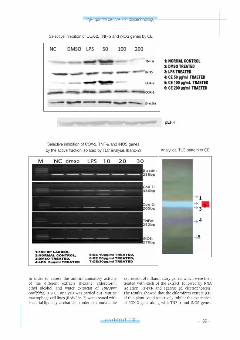
annual report 2015 - 199 -
rajiv gandhi centre for biotechnology
Selective inhibition of COX-2, TNF-α and iNOS genes by CE
pERK
Analytical TLC pattern of CESelective inhibition of COX-2, TNF-α and iNOS genes
by the active fraction isolated by TLC analysis (band-2)
In order to assess the anti-inflammatory activity of the different extracts (hexane, chloroform, ethyl alcohol and water extracts) of Tinospra cordifolia, RT-PCR analysis was carried out. Murine macrophage cell lines (RAW264.7) were treated with bacterial lipopolysaccharide in order to stimulate the
expression of inflammatory genes, which were then treated with each of the extract, followed by RNA isolation, RT-PCR and agarose gel electrophoresis. The results showed that the chloroform extract (CE) of this plant could selectively inhibit the expression of COX-2 gene along with TNF-α and iNOS genes.
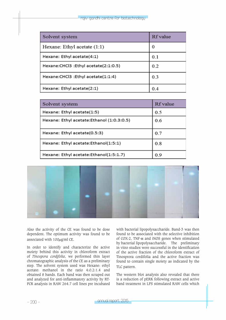
- 200 - annual report 2015
rajiv gandhi centre for biotechnology
Also the activity of the CE was found to be dose dependent. The optimum activity was found to be associated with 100µg/ml CE.
In order to identify and characterize the active moiety behind this activity in chloroform extract of Tinospora cordifolia, we performed thin layer chromatographic analysis of the CE as a preliminary step. The solvent system used was Hexane: ethyl acetate: methanol in the ratio 4:0.2:1.4 and obtained 8 bands. Each band was then scraped out and analyzed for anti-inflammatory activity by RT-PCR analysis in RAW 264.7 cell lines pre incubated
with bacterial lipopolysaccharide. Band-3 was then found to be associated with the selective inhibition of COX-2, TNF-α and iNOS genes when stimulated by bacterial lipopolysaccharide. The preliminary in vitro studies were successful in the identification of the active fraction of the chloroform extract of Tinosprora cordifolia and the active fraction was found to contain single moiety as indicated by the TLC pattern.
The western blot analysis also revealed that there is a reduction of pERK following extract and active band treatment in LPS stimulated RAW cells which
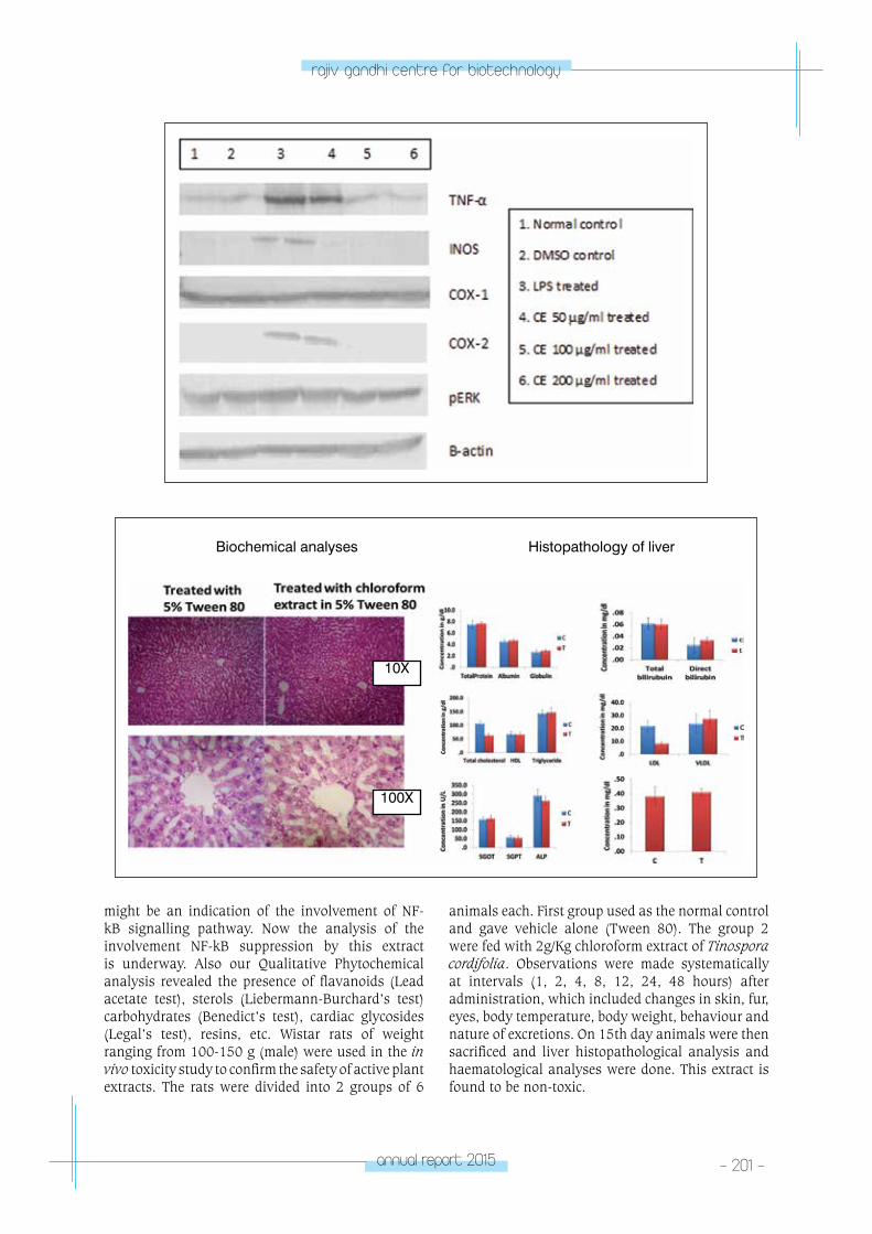
annual report 2015 - 201 -
rajiv gandhi centre for biotechnology
might be an indication of the involvement of NF-kB signalling pathway. Now the analysis of the involvement NF-kB suppression by this extract is underway. Also our Qualitative Phytochemical analysis revealed the presence of flavanoids (Lead acetate test), sterols (Liebermann-Burchard’s test) carbohydrates (Benedict’s test), cardiac glycosides (Legal’s test), resins, etc. Wistar rats of weight ranging from 100-150 g (male) were used in the in vivo toxicity study to confirm the safety of active plant extracts. The rats were divided into 2 groups of 6
animals each. First group used as the normal control and gave vehicle alone (Tween 80). The group 2 were fed with 2g/Kg chloroform extract of Tinospora cordifolia. Observations were made systematically at intervals (1, 2, 4, 8, 12, 24, 48 hours) after administration, which included changes in skin, fur, eyes, body temperature, body weight, behaviour and nature of excretions. On 15th day animals were then sacrificed and liver histopathological analysis and haematological analyses were done. This extract is found to be non-toxic.
Biochemical analyses
10X
100X
Histopathology of liver
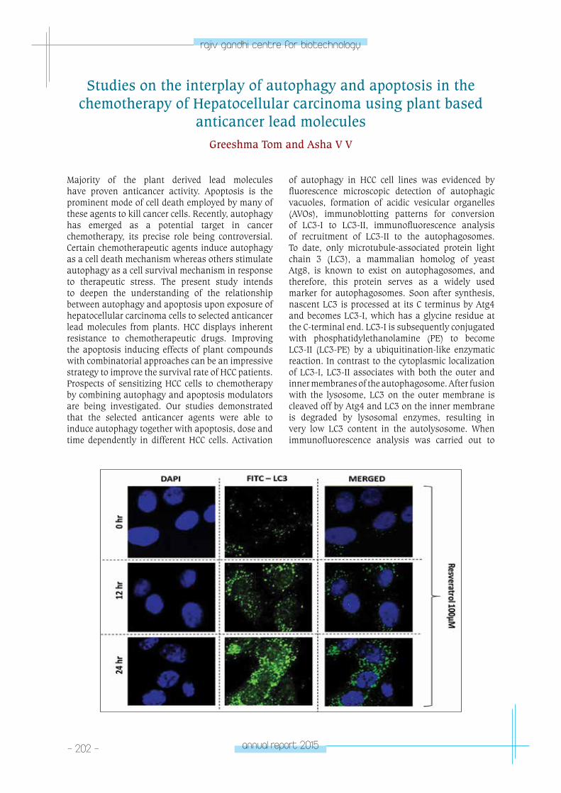
- 202 - annual report 2015
rajiv gandhi centre for biotechnology
Studies on the interplay of autophagy and apoptosis in the chemotherapy of Hepatocellular carcinoma using plant based
anticancer lead moleculesGreeshma Tom and Asha V V
Majority of the plant derived lead molecules have proven anticancer activity. Apoptosis is the prominent mode of cell death employed by many of these agents to kill cancer cells. Recently, autophagy has emerged as a potential target in cancer chemotherapy, its precise role being controversial. Certain chemotherapeutic agents induce autophagy as a cell death mechanism whereas others stimulate autophagy as a cell survival mechanism in response to therapeutic stress. The present study intends to deepen the understanding of the relationship between autophagy and apoptosis upon exposure of hepatocellular carcinoma cells to selected anticancer lead molecules from plants. HCC displays inherent resistance to chemotherapeutic drugs. Improving the apoptosis inducing effects of plant compounds with combinatorial approaches can be an impressive strategy to improve the survival rate of HCC patients. Prospects of sensitizing HCC cells to chemotherapy by combining autophagy and apoptosis modulators are being investigated. Our studies demonstrated that the selected anticancer agents were able to induce autophagy together with apoptosis, dose and time dependently in different HCC cells. Activation
of autophagy in HCC cell lines was evidenced by fluorescence microscopic detection of autophagic vacuoles, formation of acidic vesicular organelles (AVOs), immunoblotting patterns for conversion of LC3-I to LC3-II, immunofluorescence analysis of recruitment of LC3-II to the autophagosomes. To date, only microtubule-associated protein light chain 3 (LC3), a mammalian homolog of yeast Atg8, is known to exist on autophagosomes, and therefore, this protein serves as a widely used marker for autophagosomes. Soon after synthesis, nascent LC3 is processed at its C terminus by Atg4 and becomes LC3-I, which has a glycine residue at the C-terminal end. LC3-I is subsequently conjugated with phosphatidylethanolamine (PE) to become LC3-II (LC3-PE) by a ubiquitination-like enzymatic reaction. In contrast to the cytoplasmic localization of LC3-I, LC3-II associates with both the outer and inner membranes of the autophagosome. After fusion with the lysosome, LC3 on the outer membrane is cleaved off by Atg4 and LC3 on the inner membrane is degraded by lysosomal enzymes, resulting in very low LC3 content in the autolysosome. When immunofluorescence analysis was carried out to
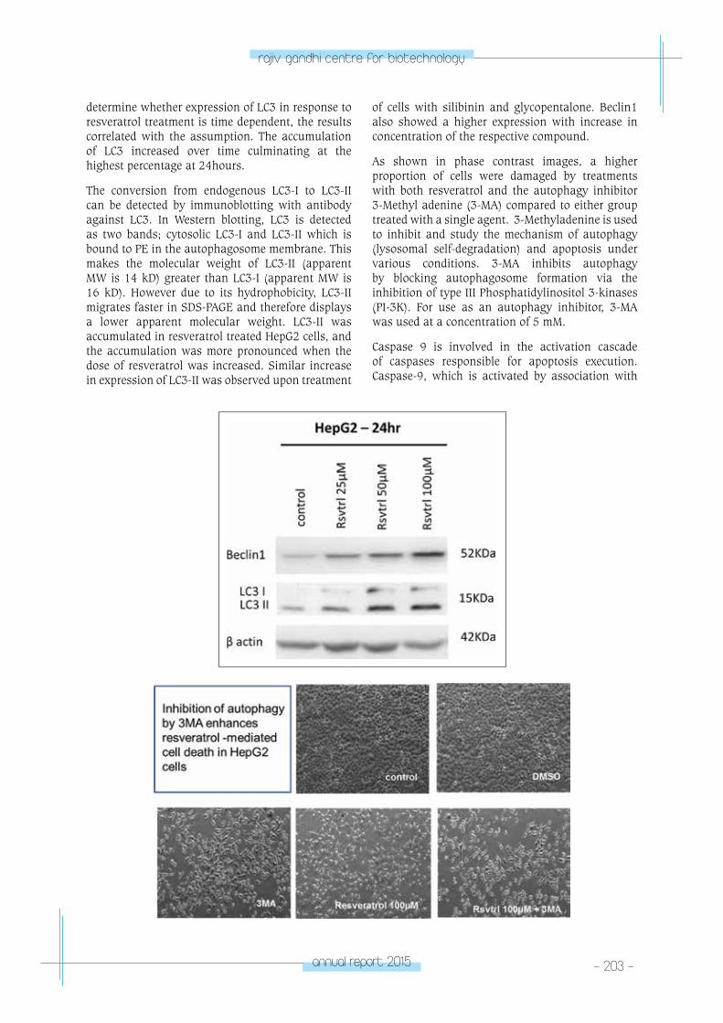
annual report 2015 - 203 -
rajiv gandhi centre for biotechnology
determine whether expression of LC3 in response to resveratrol treatment is time dependent, the results correlated with the assumption. The accumulation of LC3 increased over time culminating at the highest percentage at 24hours.
The conversion from endogenous LC3-I to LC3-II can be detected by immunoblotting with antibody against LC3. In Western blotting, LC3 is detected as two bands; cytosolic LC3-I and LC3-II which is bound to PE in the autophagosome membrane. This makes the molecular weight of LC3-II (apparent MW is 14 kD) greater than LC3-I (apparent MW is 16 kD). However due to its hydrophobicity, LC3-II migrates faster in SDS-PAGE and therefore displays a lower apparent molecular weight. LC3-II was accumulated in resveratrol treated HepG2 cells, and the accumulation was more pronounced when the dose of resveratrol was increased. Similar increase in expression of LC3-II was observed upon treatment
of cells with silibinin and glycopentalone. Beclin1 also showed a higher expression with increase in concentration of the respective compound.
As shown in phase contrast images, a higher proportion of cells were damaged by treatments with both resveratrol and the autophagy inhibitor 3-Methyl adenine (3-MA) compared to either group treated with a single agent. 3-Methyladenine is used to inhibit and study the mechanism of autophagy (lysosomal self-degradation) and apoptosis under various conditions. 3-MA inhibits autophagy by blocking autophagosome formation via the inhibition of type III Phosphatidylinositol 3-kinases (PI-3K). For use as an autophagy inhibitor, 3-MA was used at a concentration of 5 mM.
Caspase 9 is involved in the activation cascade of caspases responsible for apoptosis execution. Caspase-9, which is activated by association with
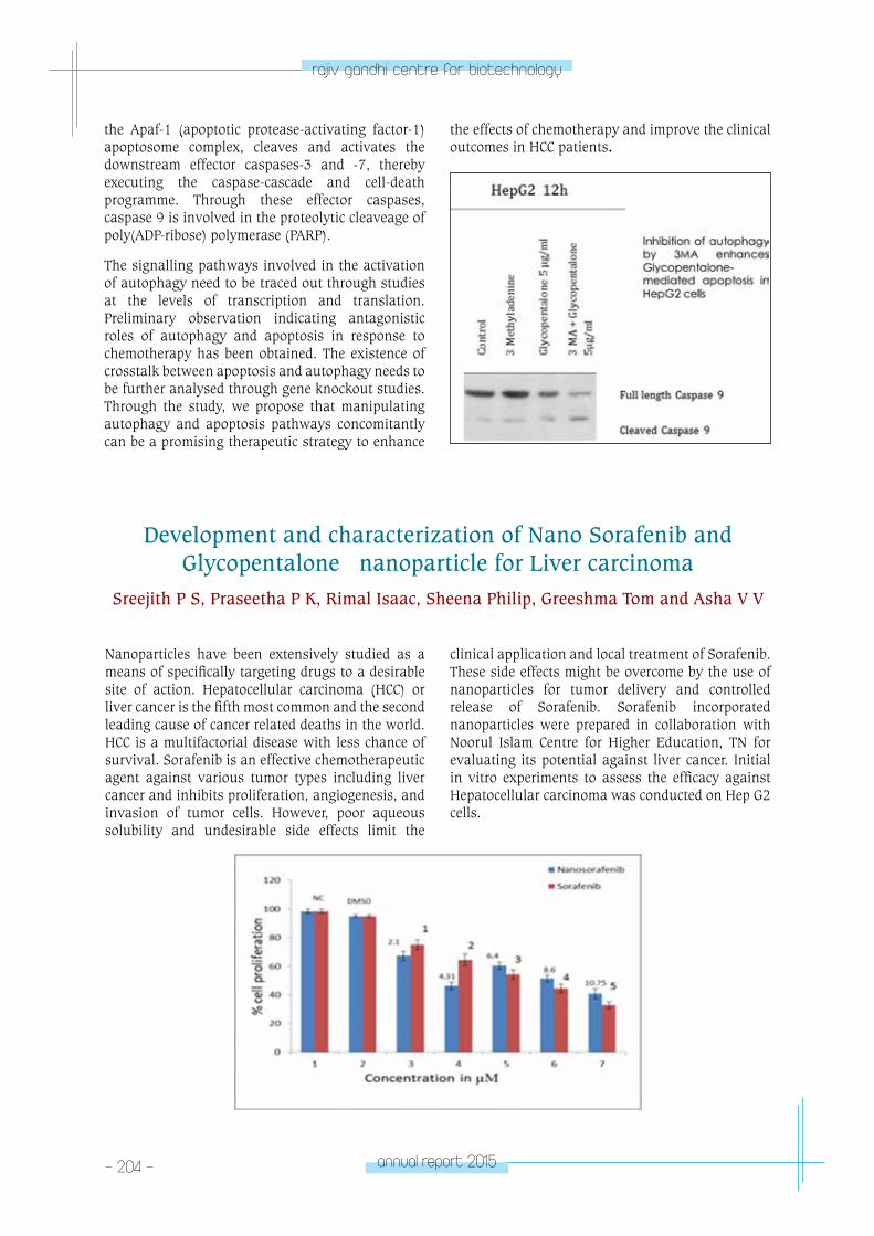
- 204 - annual report 2015
rajiv gandhi centre for biotechnology
the Apaf-1 (apoptotic protease-activating factor-1) apoptosome complex, cleaves and activates the downstream effector caspases-3 and -7, thereby executing the caspase-cascade and cell-death programme. Through these effector caspases, caspase 9 is involved in the proteolytic cleaveage of poly(ADP-ribose) polymerase (PARP).
The signalling pathways involved in the activation of autophagy need to be traced out through studies at the levels of transcription and translation. Preliminary observation indicating antagonistic roles of autophagy and apoptosis in response to chemotherapy has been obtained. The existence of crosstalk between apoptosis and autophagy needs to be further analysed through gene knockout studies. Through the study, we propose that manipulating autophagy and apoptosis pathways concomitantly can be a promising therapeutic strategy to enhance
Development and characterization of Nano Sorafenib and Glycopentalone nanoparticle for Liver carcinoma
Sreejith P S, Praseetha P K, Rimal Isaac, Sheena Philip, Greeshma Tom and Asha V V
Nanoparticles have been extensively studied as a means of specifically targeting drugs to a desirable site of action. Hepatocellular carcinoma (HCC) or liver cancer is the fifth most common and the second leading cause of cancer related deaths in the world. HCC is a multifactorial disease with less chance of survival. Sorafenib is an effective chemotherapeutic agent against various tumor types including liver cancer and inhibits proliferation, angiogenesis, and invasion of tumor cells. However, poor aqueous solubility and undesirable side effects limit the
clinical application and local treatment of Sorafenib. These side effects might be overcome by the use of nanoparticles for tumor delivery and controlled release of Sorafenib. Sorafenib incorporated nanoparticles were prepared in collaboration with Noorul Islam Centre for Higher Education, TN for evaluating its potential against liver cancer. Initial in vitro experiments to assess the efficacy against Hepatocellular carcinoma was conducted on Hep G2 cells.
the effects of chemotherapy and improve the clinical outcomes in HCC patients.
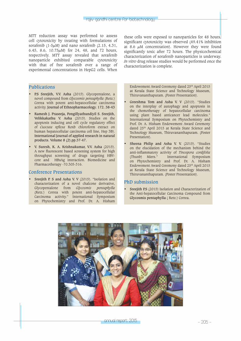
annual report 2015 - 205 -
rajiv gandhi centre for biotechnology
Publications• P.S Sreejith, V.V. Asha (2015). Glycopentalone, a
novel compound from Glycosmis pentaphylla (Retz.) Correa with potent anti-hepatocellular carcinoma activity. Journal of Ethnopharmacology. 172. 38-43
• Ramesh J. Praseeja, Pongillyathundiyil S. Sreejith, Velikkakathu V. Asha (2015). Studies on the apoptosis inducing and cell cycle regulatory effect of Cuscuta reflexa Roxb chloroform extract on human hepatocellular carcinoma cell line, Hep 3B). International Journal of applied research in natural products. Volume 8 (2).pp.37-47.
• V. Suresh, K. A. Krishnakumar, V.V. Asha (2015). A new fluorescent based screening system for high throughput screening of drugs targeting HBV-core and HBsAg interaction. Biomedicine and Pharmacotherapy -70.305-316.
Conference Presentations• Sreejith P. S and Asha V. V (2015). “Isolation and
characterization of a novel chalcone derivative, Glycopentalone from Glycosmis pentaphylla (Retz.) Correa with potent anti-hepatocellular Carcinoma activity.” International Symposium on Phytochemistry and Prof. Dr. A. Hisham
Endowement Award Ceremony dated 25th April 2015 at Kerala State Science and Technology Museum, Thiruvananthapuram. (Poster Presentation).
• Greeshma Tom and Asha V. V (2015). “Studies on the interplay of autophagy and apoptosis in the chemotherapy of hepatocellular carcinoma using plant based anticancer lead molecules.” International Symposium on Phytochemistry and Prof. Dr. A. Hisham Endowement Award Ceremony dated 25th April 2015 at Kerala State Science and Technology Museum, Thiruvananthapuram. (Poster Presentation).
• Sheena Philip and Asha V. V. (2015). “Studies on the elucidation of the mechanism behind the anti-inflammatory activity of Tinospora cordifolia (Thunb) Miers.” International Symposium on Phytochemistry and Prof. Dr. A. Hisham Endowement Award Ceremony dated 25th April 2015 at Kerala State Science and Technology Museum, Thiruvananthapuram. (Poster Presentation).
PhD submission• Sreejith P.S (2015) Isolation and Characterization of
the Anti-hepatocellular Carcinoma Compound from Glycosmis pentaphylla ( Retz.) Correa.
MTT reduction assay was performed to assess cell cytotoxicity by treating with formulations of sorafenib (1-5µM) and nano sorafenib (2.15, 4.31, 6.45, 8.6, 10.75µM) for 24, 48, and 72 hours, respectively. MTT assay revealed that sorafenib nanoparticle exhibited comparable cytotoxicity with that of free sorafenib over a range of experimental concentrations in HepG2 cells. When
these cells were exposed to nanoparticles for 48 hours, significant cytotoxicity was observed (49.41% inhibition at 8.6 µM concentration). However they were found significantly toxic after 72 hours. The physicochemical characterization of sorafenib nanoparticles is underway. In vitro drug release studies would be performed once the characterization is complete.
plAnt DISeASe BIoloGY & BIoteCHnoloGY
- pDBB laboratory – 2PLANT DISEASE BIOLOGY
& BIOTECHNOLOGYPDBB Laboratory - 2
George Thomas
George Thomas received his PhD in Life Sciences
from University of Hyderabad and joined RGCB
in 1997.
Manager (Technical Services)George Varghese
PhD studentsGeethu Elizabath ThomasSmini VargheseLesly Augustine
Project FellowsVinitha M.R.Neethu Mathew
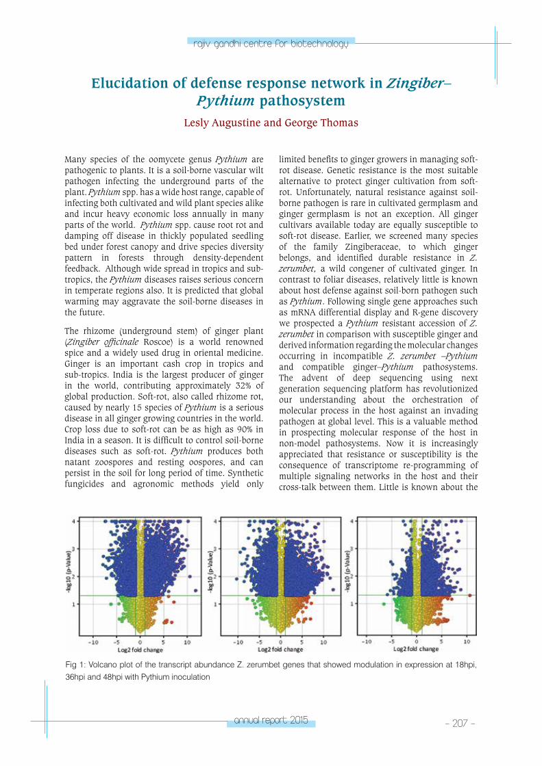
annual report 2015 - 207 -
rajiv gandhi centre for biotechnology
Elucidation of defense response network in Zingiber–Pythium pathosystem
Lesly Augustine and George Thomas
Many species of the oomycete genus Pythium are pathogenic to plants. It is a soil-borne vascular wilt pathogen infecting the underground parts of the plant. Pythium spp. has a wide host range, capable of infecting both cultivated and wild plant species alike and incur heavy economic loss annually in many parts of the world. Pythium spp. cause root rot and damping off disease in thickly populated seedling bed under forest canopy and drive species diversity pattern in forests through density-dependent feedback. Although wide spread in tropics and sub-tropics, the Pythium diseases raises serious concern in temperate regions also. It is predicted that global warming may aggravate the soil-borne diseases in the future.
The rhizome (underground stem) of ginger plant (Zingiber officinale Roscoe) is a world renowned spice and a widely used drug in oriental medicine. Ginger is an important cash crop in tropics and sub-tropics. India is the largest producer of ginger in the world, contributing approximately 32% of global production. Soft-rot, also called rhizome rot, caused by nearly 15 species of Pythium is a serious disease in all ginger growing countries in the world. Crop loss due to soft-rot can be as high as 90% in India in a season. It is difficult to control soil-borne diseases such as soft-rot. Pythium produces both natant zoospores and resting oospores, and can persist in the soil for long period of time. Synthetic fungicides and agronomic methods yield only
limited benefits to ginger growers in managing soft-rot disease. Genetic resistance is the most suitable alternative to protect ginger cultivation from soft-rot. Unfortunately, natural resistance against soil-borne pathogen is rare in cultivated germplasm and ginger germplasm is not an exception. All ginger cultivars available today are equally susceptible to soft-rot disease. Earlier, we screened many species of the family Zingiberaceae, to which ginger belongs, and identified durable resistance in Z. zerumbet, a wild congener of cultivated ginger. In contrast to foliar diseases, relatively little is known about host defense against soil-born pathogen such as Pythium. Following single gene approaches such as mRNA differential display and R-gene discovery we prospected a Pythium resistant accession of Z. zerumbet in comparison with susceptible ginger and derived information regarding the molecular changes occurring in incompatible Z. zerumbet –Pythium and compatible ginger–Pythium pathosystems. The advent of deep sequencing using next generation sequencing platform has revolutionized our understanding about the orchestration of molecular process in the host against an invading pathogen at global level. This is a valuable method in prospecting molecular response of the host in non-model pathosystems. Now it is increasingly appreciated that resistance or susceptibility is the consequence of transcriptome re-programming of multiple signaling networks in the host and their cross-talk between them. Little is known about the
Fig 1: Volcano plot of the transcript abundance Z. zerumbet genes that showed modulation in expression at 18hpi, 36hpi and 48hpi with Pythium inoculation
- 208 - annual report 2015
rajiv gandhi centre for biotechnology
molecular processes that are triggered in a host against Pythium. Efficient application of genomics information in agriculture demands in-depth knowledge about the behaviour of defense pathways in a host in response to a pathogen and also the level of their interaction between. We performed a comparative transcriptome analysis of host in incompatible Z. zerumbet – Pythium and compatible ginger – Pythium interactions following RNA-Seq technology and gathered substantial information regarding the putative pathways that are regulated differently between the compatible and incompatible pathosystems.Well characterized EST databases were developed for ginger and Z. zerumbet and the sequencing read data has been submitted in NCBI Sequence Read Archive (SRA) domain. The RNA-Seq information provide us a great opportunity in developing high throughput expression arrays for studying the temporal expression changes in large number of genes in multiple systems. Keeping this in mind we used the RNA-seq data for designing microarray probes. A total of 87897 Zingiber zerumbet assembled unigene transcripts were used for designing the probes. The probes were designed by setting various parameters such as Tm matching, target of the transcript for probe designing(3’ region), length of the probe (50bp), antisense probe orientation and >2 probes per target sequence. We obtained altogether 58476 probes. This includes the
probes designed from Z. zerumbet transcripts, non-plant negative control sequences, positive control sequences, specific gene sequences. The probes were printed in an 8x60K array format and the slides were custom synthesized. RNA samples isolated from different post-inoculated time periods such as 18hours post inoculation (hpi), 36hpi and 48hpi, and control samples were used to hybridize with expression array. Two biological replicates were used for each treatment for the array hybridization.
The expression signals were quantified and normalized. The genes whose expression showed significant modulation, either up-regulation or down-regulation, were annotated and were subjected to various computational methods to understand the temporal changes of expression pattern in different defense pathways. Expression changes at global level were visualized by volcano plotting (Fig 1). When the three time periods were taken together, 15928 genes showed significant modulation in expression with up regulation in 8499 genes and down regulation in 7429 genes. Genes which showed significant expression signature were further classified based on gene ontology (GO) and grouped into various KEGG pathways (Fig 2). The major biological process identified in the GO categorization included oxidation-reduction process and metabolic processes.
Fig 2: Functional categorization and KEGG pathway classification of differentially modulated genes in Z. zerumbet in response to Pythium inoculation
annual report 2015 - 209 -
rajiv gandhi centre for biotechnology
Molecular genetics behind host response to the soft rot pathogen Pythium myriotylum Drechsler in Zingiber spp.
with contrasting breeding systemSmini Varghese, Geethu Elizabath Thomas, Vinitha M. R and George Thomas
The two hypotheses, the Red queen and the Muller’s ratchet that conceptualize the relationship between breeding behaviour, genetic diversity and adaptive fitness, represent the two sides of the same coin. While the Red queen hypothesize the positive effects of genetic recombination, the Muller’s ratchet address the negative effect of obligate clonal multiplication. The Red queen put forward that the genetic recombination provide adaptive benefits to an organism through the generation of variability, formation of new allele compliment and purging out lethal mutations whereas the Muller’s ratchet postulates that the background selection against lethal mutations push back an obligate clone into original state rendering it unable to enter into evolutionary arms race with pathogen populations. Further, the Red queen postulates that pathogen maintains sex on the host. The sexual reproduction enable the host to continuously generate genetic variability for efficiently compete with pathogens in evolutionary arms race. Accordingly, we can
see high genetic diversity in sexual species and a narrow genetic base in asexual species. However, different eco-evolutionary parameters such as the threshold of genetic diversity required for limiting disease spread, relationship between the host population genetic structure and disease dynamics and the influence of different breeding strategies in the emergence and fixation of resistance traits are not clearly understood. Alongside there are also reports that clonal lineages of certain species are more adaptive than sexual lineages rendering it difficult to explain the survival and the adaptive fitness of genetically narrow asexual solely on the basis of the existing theories of sex. It is difficult to identify absolute system to test the two hypotheses. The pantropic plant family Zingiberaceae represents an ideal system to study the interrelationships between reproductive behaviour, genetic diversity and adaptive fitness. This family consists of species with contrasting breeding behaviour. The cultivated spice crop ginger (Zingiber officinale) is a
Fig 3:Heterozygous profile in seven concatenated gene loci in a representative set of Z. zerumbet individual: CP – clonal individuals; SP - sexual individuals. Exon regions are highlighted in red colour. Asterisks in blue and green colour indicate variable locations in non synonymous and synonymous positions respectively. (CCoAOMT - Caffeoyl- Coenzyme A3-O-Methyl transferase; ACS - ACC Synthase; COMT - Caffeicacid O-methyltransferass; ICS - Isochorismate synthase; EDS1- Enhanced disease susceptibility 1; PR5 - Pathogenesis related protein; AOC- Allene oxide cyclase)
- 210 - annual report 2015
rajiv gandhi centre for biotechnology
known obligate asexual, that propagate exclusively through rhizome. In our earlier studies, we identified sexual reproduction in two wild species Zingiber neesanum and Zingiber nimmonii, and hemiclonal breeding behaviour in another wild species Zingiber zerumbet. Certain populations of Z. zerumbet are sexual while others are predominantly clonal, reproduced through rhizome. Many species of the oomycete genus Pythium is known to cause rhizome rot disease in members of family Zingiberaceae. Supporting the postulates of Muller’s ratchet, the obligate asexual ginger is highly susceptible to Pythium. However, contradicting the Red queen postulates, the sexual populations of Zingiber neesanum and Zingiber nimmonii were highly susceptible. Discordance to Red queen was observed in the hemiclonal Z. zerumbet also with susceptibility in sexual populations and resistance in asexual populations. We were interested to portray the nature and extent of genetic diversity in ginger cultivars and the sexual and asexual populations of Z. zerumbet and to examine the interrelationship between their breeding pattern, molecular genetics parameters and Pythium responsiveness. To address this question we examined the nucleotide diversity of defense related gene loci in both ginger and Z. zerumbet populations and compared them with the nucleotide diversity estimated in house-keeping genes. The alleles retrieved in defense gene loci were heterozygous in Z. zerumbet populations. However, the sexual and clonal populations showed distinct pattern of within population heterozygosity profile in Z. zerumbet. The heterozygosity profiles were fixed in asexual populations and the individuals in such
populations were immune to Pythium, whereas the level of heterozygosity varied considerably between individuals within sexual populations and so was their response to Pythium disease. A significant relationship was obtained between heterozygosity level and resistance to Pythium. Results show that the heterozygosity panels with adaptive fitness against Pythium infection are tend to be retained in Z. zerumbet by clonal expansion, whereas the alignment of desirable heterozygosity panels is either not attained in sexual populations or are being subjected to continuous recombination and segregation, leading to Pythium susceptibility(Fig 3). Our study presents an extension to Red Queen hypothesis wherein hemiclonality provides fitness to Z. zerumbet to perpetuate heterozygosity that gives resistance to Pythium. Presumably, species with hemiclonal ability may have a fair chance to survive ecological undulations.
We evaluated the same set of gene loci in altogether 16 prominent ginger clones.Genetic base of ginger was exceptionally narrow and the cultivars were uniformly susceptible to soft-rot. Concordant with Muller’s ratchet, the asexuality drives ginger into genetical shallowness, which renders it with ineffective host-pathogen coevolution and poor fitness. Overall, the results speak about the general evolutionary trajectories of members in family Zingiberaceae. We can presume that this family as a whole may have had undergone genomic disturbance, either by whole genome duplications or due towide spread gene flow.
Conference Presentations• Smini Varghese, George Thomas. Heterozygosity
fitness-correlations and soft rot resistance in Zingiber zerumbet: Mixed breeding system facilitates adaptive evolution by clonal expansion. Paper presented at 7th International symposium on the family Zingiberaceae “Gingers for Life”, Chiang Mai, Thailand, 17 – 20 August 2015.
• Vinitha M R, Suresh Kumar U, Sabu M and George Thomas. Pattern of nucleotide variations in the standard DNA barcode loci in different genera of Indian Zingiberaceae. Poster presented at the 6th International Barcode of Life Conference, University of Guelph, Canada, 18 – 21 August, 2015.
• Smini Varghese, Geethu Elizabath Thomas, George Thomas. Genome characteristics show that the cultivated ginger (Zingiber officinale Roscoe) is in Muller’s Ratchet and explains its severe disease susceptibility. Poster presented at the 3rd Current Opinion Conference on Plant Genome Evolution, Amsterdam, The Netherlands, 6 – 8 September, 2015.
PhD Produced• Mariet Jose (2015) DNA fingerprinting and
discrimination of the traditional medicinal rice ‘Njavara’ from other traditional rice strains, University of Kerala
annual report 2015 - 211 -
rajiv gandhi centre for biotechnology
EXTRAMURAL FUNDING
Investigator Title Funding Agency Duration
George ThomasCollaborator: Dr. V. G. Jayalekshmy, Kerala Agricultural University
Development of rice varieties for Kerala with pyramided genes for resistance to BLB by marker assisted selection
Department of Biotechnology, Government of India
2013 - 2018
George Thomas
De novo transcriptome sequencing microarray development and elucidation of Pythium responsive defense pathways in Zingiber zerumbet Smith
Council of Scientific and Industrial Research
2015 -2017
plAnt DISeASe BIoloGY & BIoteCHnoloGY
pDBB laboratory - 3
PLANT DISEASE BIOLOGY & BIOTECHNOLOGYPDBB Laboratory - 3
Soniya. E. V
Soniya was awarded PhD from the Department
of Botany, University of Kerala. Following this,
she worked as a Research Associate at Central
Tuber Crops Research Institute, Sreekaryam,
Thiruvananthapuram before she joined Rajiv
Gandhi Centre for Biotechnology. She was also
INSA/DFG Visiting Scientist at Max Planck
Institute of Chemical Ecology, Jena, Germany.
Manager (Technical Services)Manoj P. Kumar, PhD
Project AssistantSinsha Prakashan
Laboratory AssistantsVijayalekshmi S.K.Kalapriya V.S.
PhD StudentsAsha S.Aiswarya G.Mallika V.Divya KattupalliMaimoonath Beevi Y. P.Aswathi U.Sweda Sreekumar
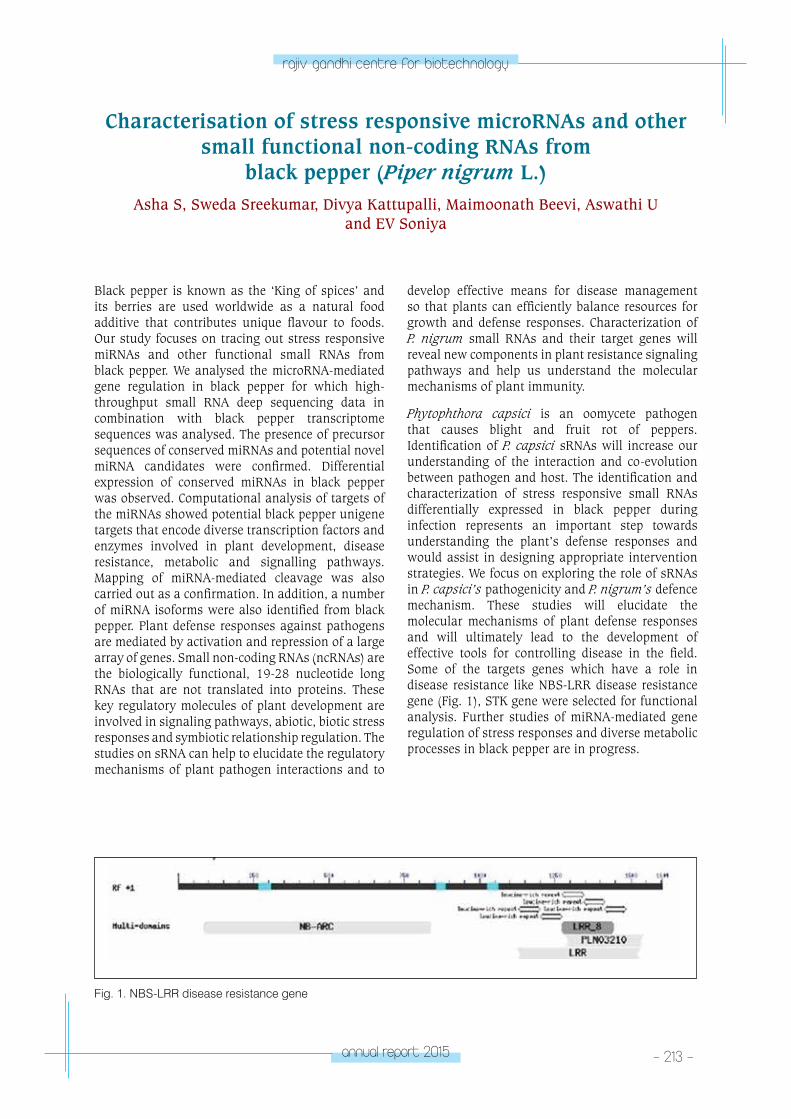
annual report 2015 - 213 -
rajiv gandhi centre for biotechnology
Black pepper is known as the ‘King of spices’ and its berries are used worldwide as a natural food additive that contributes unique flavour to foods. Our study focuses on tracing out stress responsive miRNAs and other functional small RNAs from black pepper. We analysed the microRNA-mediated gene regulation in black pepper for which high-throughput small RNA deep sequencing data in combination with black pepper transcriptome sequences was analysed. The presence of precursor sequences of conserved miRNAs and potential novel miRNA candidates were confirmed. Differential expression of conserved miRNAs in black pepper was observed. Computational analysis of targets of the miRNAs showed potential black pepper unigene targets that encode diverse transcription factors and enzymes involved in plant development, disease resistance, metabolic and signalling pathways. Mapping of miRNA-mediated cleavage was also carried out as a confirmation. In addition, a number of miRNA isoforms were also identified from black pepper. Plant defense responses against pathogens are mediated by activation and repression of a large array of genes. Small non-coding RNAs (ncRNAs) are the biologically functional, 19-28 nucleotide long RNAs that are not translated into proteins. These key regulatory molecules of plant development are involved in signaling pathways, abiotic, biotic stress responses and symbiotic relationship regulation. The studies on sRNA can help to elucidate the regulatory mechanisms of plant pathogen interactions and to
Characterisation of stress responsive microRNAs and other small functional non-coding RNAs from
black pepper (Piper nigrum L.)Asha S, Sweda Sreekumar, Divya Kattupalli, Maimoonath Beevi, Aswathi U
and EV Soniya
develop effective means for disease management so that plants can efficiently balance resources for growth and defense responses. Characterization of P. nigrum small RNAs and their target genes will reveal new components in plant resistance signaling pathways and help us understand the molecular mechanisms of plant immunity.
Phytophthora capsici is an oomycete pathogen that causes blight and fruit rot of peppers. Identification of P. capsici sRNAs will increase our understanding of the interaction and co-evolution between pathogen and host. The identification and characterization of stress responsive small RNAs differentially expressed in black pepper during infection represents an important step towards understanding the plant’s defense responses and would assist in designing appropriate intervention strategies. We focus on exploring the role of sRNAs in P. capsici’s pathogenicity and P. nigrum’s defence mechanism. These studies will elucidate the molecular mechanisms of plant defense responses and will ultimately lead to the development of effective tools for controlling disease in the field. Some of the targets genes which have a role in disease resistance like NBS-LRR disease resistance gene (Fig. 1), STK gene were selected for functional analysis. Further studies of miRNA-mediated gene regulation of stress responses and diverse metabolic processes in black pepper are in progress.
Fig. 1. NBS-LRR disease resistance gene
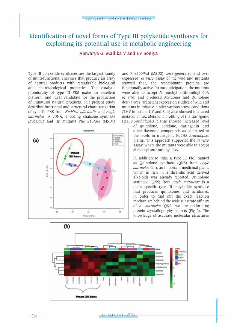
- 214 - annual report 2015
rajiv gandhi centre for biotechnology
Type III polyketide synthases are the largest family of multi-functional enzymes that produce an array of natural products with remarkable biological and pharmacological properties. The catalytic promiscuity of type III PKS make an excellent platform and ideal candidate for the production of unnatural natural products. Our present study describes functional and structural characterization of type III PKS from Emblica officinalis and Aegle marmelos. A cDNA, encoding chalcone synthase (EoCHS1) and its mutants Phe 215/Ser (MSD1)
Identification of novel forms of Type III polyketide synthases for exploiting its potential use in metabolic engineering
Aiswarya G, Mallika V and EV Soniya
and Phe265/Val (MSD2) were generated and over expressed. In vitro assay of the wild and mutants showed that, the recombinant proteins are functionally active. To our anticipation, the mutants were able to accept N- methyl anthraniloyl CoA in vitro and produced Acridones and Quinolone derivatives. Transient expression studies of wild and mutants in tobacco, under various stress conditions (TMV infection, UV and Salt) also showed increased metabolic flux. Metabolic profiling of the transgenic F215S Arabidopsis plants showed increased level
of quinolone, acridone, naringenin and other flavonoid compounds as compared to the levels in transgenic EoCHS Arabidopsis plants. This approach supported the in vitro assay, where the mutants were able to accept N-methyl anthraniloyl CoA.
In addition to this, a type III PKS named as Quinolone synthase (QNS) from Aegle marmelos Corr, an important medicinal plant, which is rich in anthranilic acid derived alkaloids was already reported. Quinolone synthase (QNS) from Aegle marmelos is a plant specific type III polyketide synthase that produces quinolones and acridones. In order to find out the exact reaction mechanism behind the wide substrate affinity of A. marmelos QNS, we are performing protein crystallography aspects (Fig 2). The knowledge of accurate molecular structures
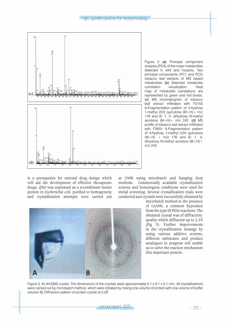
annual report 2015 - 215 -
rajiv gandhi centre for biotechnology
Figure 2 (a) Principal component analysis (PCA) of the major metabolites detected in wild and mutants. Two principal components (PC1 and PC2) tobacco leaf extracts of MS based metabolites (b) Selected metabolite correlation visualization. Heat map of metabolite correlations are represented by green and red boxes.(c) MS chromatogram of tobacco leaf extract infiltrated with F215S A-Fragmentation pattern of 4-hydroxy 1-methyl 2(H) quinolone (M+H)+ m/z 176 and B- 1, 3- dihydroxy N-methyl acridone (M+H)+ m/z 242. (d) MS profile of tobacco leaf extract infiltrated with F265V A-Fragmentation pattern of 4-hydroxy 1-methyl 2(H) quinolone (M+H) + m/z 176 and B- 1, 3- dihydroxy N-methyl acridone (M+H)+ m/z 242.
at 298K using microbatch and hanging drop methods. Commercially available crystallization screens and homologous conditions were used for initial screening. Several crystallization trials were conducted and crystals were successfully obtained by
microbatch method in the presence of CoASH, a common byproduct from the type III PKSs reactions. The obtained crystal was of diffraction-quality which diffracted up to 2.2Å (Fig 3). Further improvements in the crystallization strategy by using various additive screens, different substrates and product analogues in progress will enable us to solve the reaction mechanism this important protein.
is a prerequisite for rational drug design which will aid the development of effective therapeutic drugs. QNS was expressed as a recombinant fusion protein in Escherichia coli, purified to homogeneity and crystallization attempts were carried out
Figure 3. A) AmQNS crystal. The dimensions of the crystals were approximately 0.1 x 0.1 x 0.1 mm. All crystallizations were carried out by microbatch method, which were initiated by mixing one volume of protein with one volume of buffer solution B) Diffraction pattern of protein crystal at 2.2Å
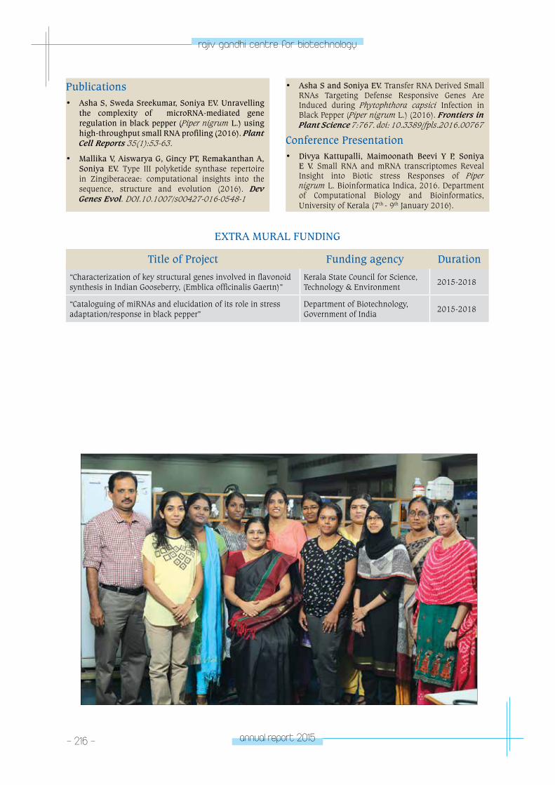
- 216 - annual report 2015
rajiv gandhi centre for biotechnology
Publications• Asha S, Sweda Sreekumar, Soniya EV. Unravelling
the complexity of microRNA-mediated gene regulation in black pepper (Piper nigrum L.) using high-throughput small RNA profiling (2016). Plant Cell Reports 35(1):53-63.
• Mallika V, Aiswarya G, Gincy PT, Remakanthan A, Soniya EV. Type III polyketide synthase repertoire in Zingiberaceae: computational insights into the sequence, structure and evolution (2016). Dev Genes Evol. DOI.10.1007/s00427-016-0548-1
• Asha S and Soniya EV. Transfer RNA Derived Small RNAs Targeting Defense Responsive Genes Are Induced during Phytophthora capsici Infection in Black Pepper (Piper nigrum L.) (2016). Frontiers in Plant Science 7:767. doi: 10.3389/fpls.2016.00767
Conference Presentation• Divya Kattupalli, Maimoonath Beevi Y P, Soniya
E V. Small RNA and mRNA transcriptomes Reveal Insight into Biotic stress Responses of Piper nigrum L. Bioinformatica Indica, 2016. Department of Computational Biology and Bioinformatics, University of Kerala (7th - 9th January 2016).
EXTRA MURAL FUNDING
Title of Project Funding agency Duration
“Characterization of key structural genes involved in flavonoid synthesis in Indian Gooseberry, (Emblica officinalis Gaertn)”
Kerala State Council for Science, Technology & Environment
2015-2018
“Cataloguing of miRNAs and elucidation of its role in stress adaptation/response in black pepper”
Department of Biotechnology, Government of India
2015-2018
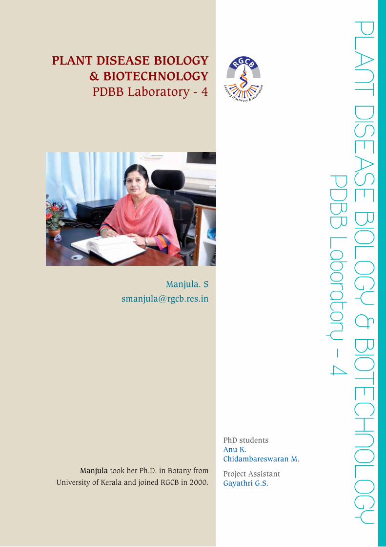
plAnt DISeASe BIoloGY & BIoteCHnoloGY
pDBB laboratory - 4
PLANT DISEASE BIOLOGY & BIOTECHNOLOGYPDBB Laboratory - 4
Manjula. S
Manjula took her Ph.D. in Botany from
University of Kerala and joined RGCB in 2000.
PhD studentsAnu K.Chidambareswaran M.
Project AssistantGayathri G.S.
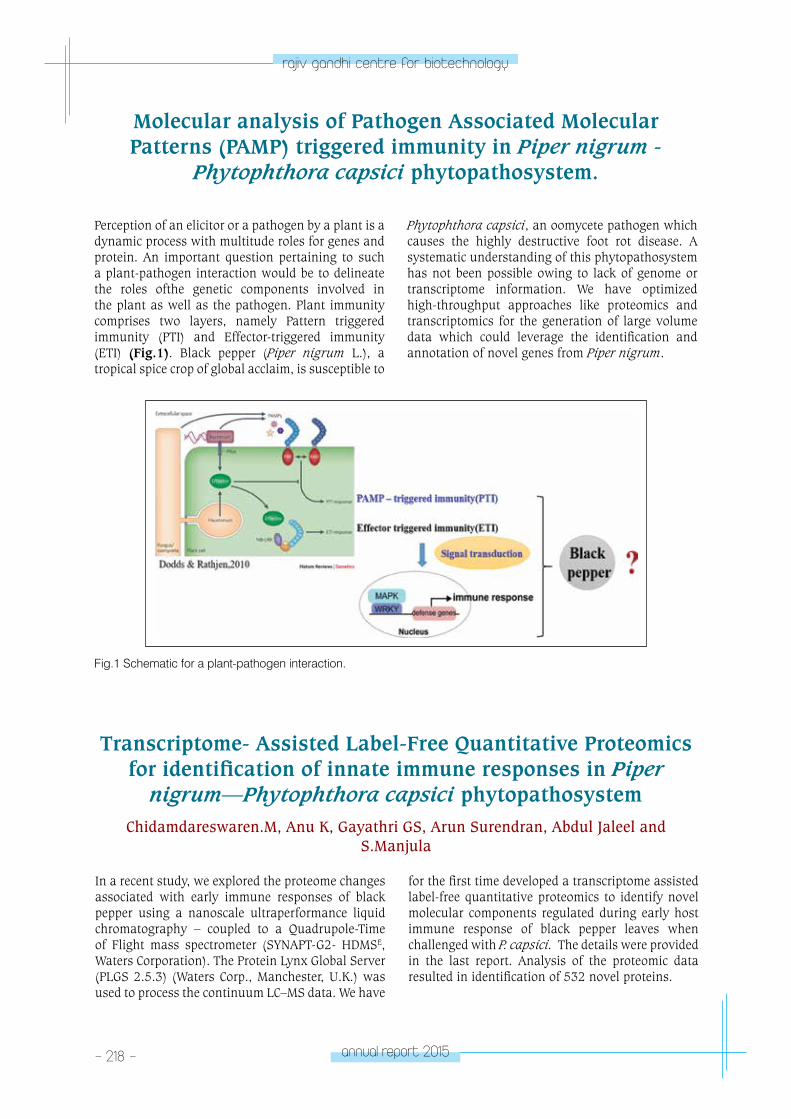
- 218 - annual report 2015
rajiv gandhi centre for biotechnology
Perception of an elicitor or a pathogen by a plant is a dynamic process with multitude roles for genes and protein. An important question pertaining to such a plant-pathogen interaction would be to delineate the roles ofthe genetic components involved in the plant as well as the pathogen. Plant immunity comprises two layers, namely Pattern triggered immunity (PTI) and Effector-triggered immunity (ETI) (Fig.1). Black pepper (Piper nigrum L.), a tropical spice crop of global acclaim, is susceptible to
Molecular analysis of Pathogen Associated Molecular Patterns (PAMP) triggered immunity in Piper nigrum -
Phytophthora capsici phytopathosystem.
Phytophthora capsici, an oomycete pathogen which causes the highly destructive foot rot disease. A systematic understanding of this phytopathosystem has not been possible owing to lack of genome or transcriptome information. We have optimized high-throughput approaches like proteomics and transcriptomics for the generation of large volume data which could leverage the identification and annotation of novel genes from Piper nigrum.
Fig.1 Schematic for a plant-pathogen interaction.
Transcriptome- Assisted Label-Free Quantitative Proteomics for identification of innate immune responses in Piper
nigrum—Phytophthora capsici phytopathosystemChidamdareswaren.M, Anu K, Gayathri GS, Arun Surendran, Abdul Jaleel and
S.Manjula
In a recent study, we explored the proteome changes associated with early immune responses of black pepper using a nanoscale ultraperformance liquid chromatography – coupled to a Quadrupole-Time of Flight mass spectrometer (SYNAPT-G2- HDMSE, Waters Corporation). The Protein Lynx Global Server (PLGS 2.5.3) (Waters Corp., Manchester, U.K.) was used to process the continuum LC–MS data. We have
for the first time developed a transcriptome assisted label-free quantitative proteomics to identify novel molecular components regulated during early host immune response of black pepper leaves when challenged with P. capsici. The details were provided in the last report. Analysis of the proteomic data resulted in identification of 532 novel proteins.
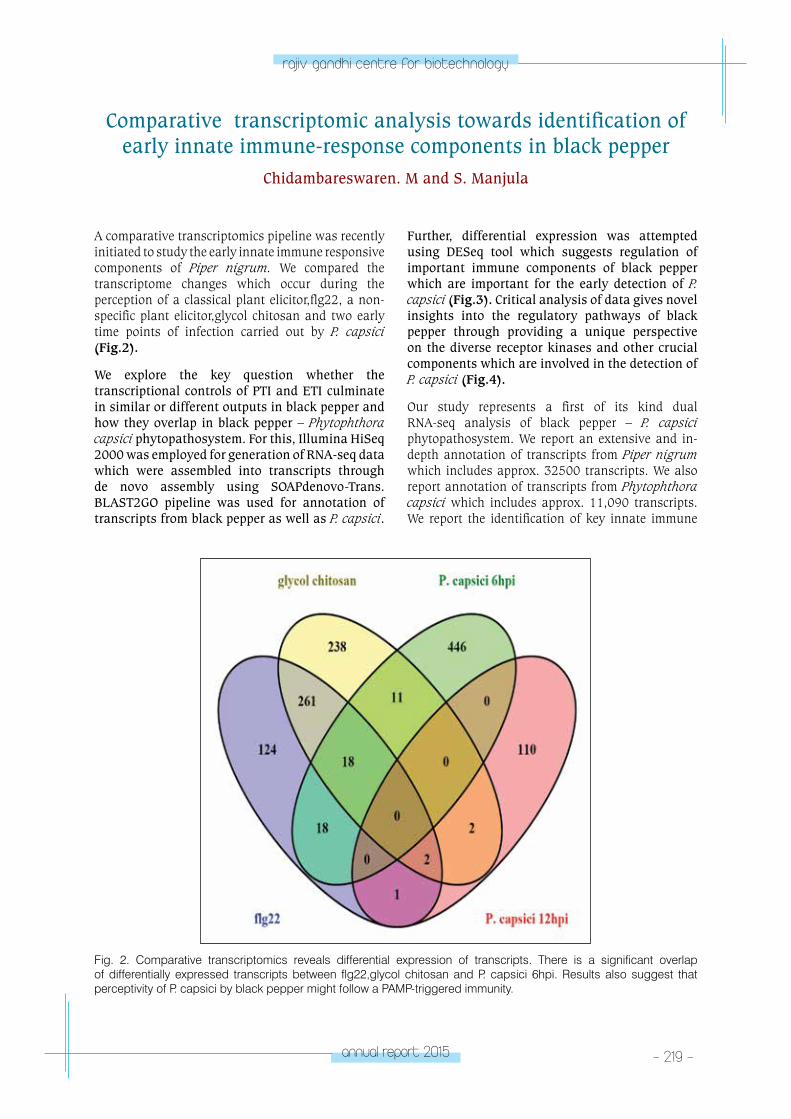
annual report 2015 - 219 -
rajiv gandhi centre for biotechnology
Comparative transcriptomic analysis towards identification of early innate immune-response components in black pepper
Chidambareswaren. M and S. Manjula
A comparative transcriptomics pipeline was recently initiated to study the early innate immune responsive components of Piper nigrum. We compared the transcriptome changes which occur during the perception of a classical plant elicitor,flg22, a non-specific plant elicitor,glycol chitosan and two early time points of infection carried out by P. capsici (Fig.2).
We explore the key question whether the transcriptional controls of PTI and ETI culminate in similar or different outputs in black pepper and how they overlap in black pepper – Phytophthora capsici phytopathosystem. For this, Illumina HiSeq 2000 was employed for generation of RNA-seq data which were assembled into transcripts through de novo assembly using SOAPdenovo-Trans. BLAST2GO pipeline was used for annotation of transcripts from black pepper as well as P. capsici.
Further, differential expression was attempted using DESeq tool which suggests regulation of important immune components of black pepper which are important for the early detection of P. capsici (Fig.3). Critical analysis of data gives novel insights into the regulatory pathways of black pepper through providing a unique perspective on the diverse receptor kinases and other crucial components which are involved in the detection of P. capsici (Fig.4).
Our study represents a first of its kind dual RNA-seq analysis of black pepper – P. capsici phytopathosystem. We report an extensive and in-depth annotation of transcripts from Piper nigrum which includes approx. 32500 transcripts. We also report annotation of transcripts from Phytophthora capsici which includes approx. 11,090 transcripts. We report the identification of key innate immune
Fig. 2. Comparative transcriptomics reveals differential expression of transcripts. There is a significant overlap of differentially expressed transcripts between flg22,glycol chitosan and P. capsici 6hpi. Results also suggest that perceptivity of P. capsici by black pepper might follow a PAMP-triggered immunity.
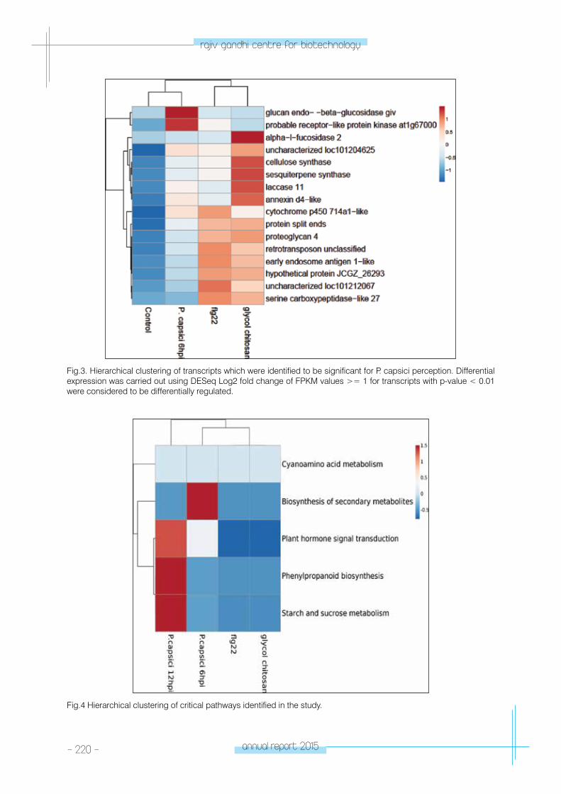
- 220 - annual report 2015
rajiv gandhi centre for biotechnology
Fig.3. Hierarchical clustering of transcripts which were identified to be significant for P. capsici perception. Differential expression was carried out using DESeq Log2 fold change of FPKM values >= 1 for transcripts with p-value < 0.01 were considered to be differentially regulated.
Fig.4 Hierarchical clustering of critical pathways identified in the study.
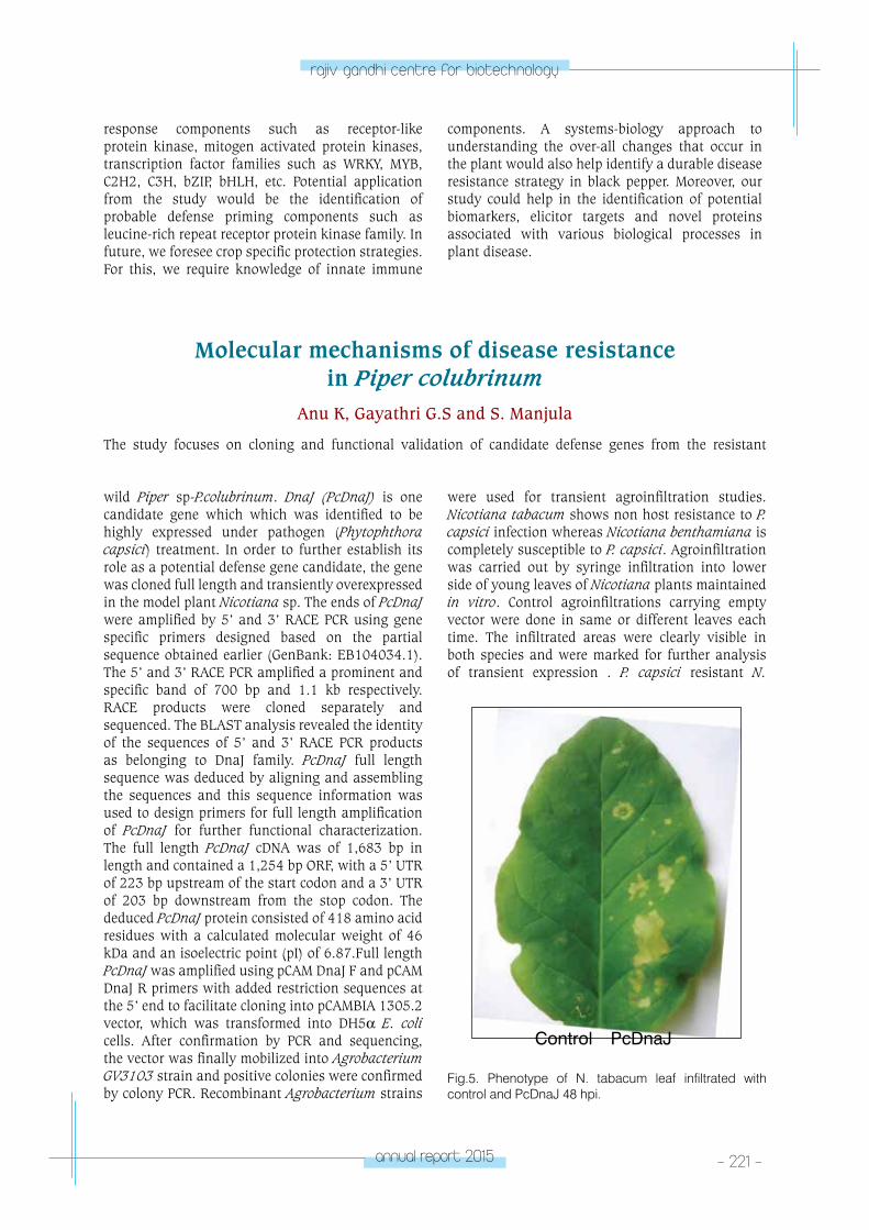
annual report 2015 - 221 -
rajiv gandhi centre for biotechnology
Molecular mechanisms of disease resistance in Piper colubrinum
Anu K, Gayathri G.S and S. Manjula
The study focuses on cloning and functional validation of candidate defense genes from the resistant
wild Piper sp-P.colubrinum. DnaJ (PcDnaJ) is one candidate gene which which was identified to be highly expressed under pathogen (Phytophthora capsici) treatment. In order to further establish its role as a potential defense gene candidate, the gene was cloned full length and transiently overexpressed in the model plant Nicotiana sp. The ends of PcDnaJ were amplified by 5’ and 3’ RACE PCR using gene specific primers designed based on the partial sequence obtained earlier (GenBank: EB104034.1). The 5’ and 3’ RACE PCR amplified a prominent and specific band of 700 bp and 1.1 kb respectively. RACE products were cloned separately and sequenced. The BLAST analysis revealed the identity of the sequences of 5’ and 3’ RACE PCR products as belonging to DnaJ family. PcDnaJ full length sequence was deduced by aligning and assembling the sequences and this sequence information was used to design primers for full length amplification of PcDnaJ for further functional characterization. The full length PcDnaJ cDNA was of 1,683 bp in length and contained a 1,254 bp ORF, with a 5’ UTR of 223 bp upstream of the start codon and a 3’ UTR of 203 bp downstream from the stop codon. The deduced PcDnaJ protein consisted of 418 amino acid residues with a calculated molecular weight of 46 kDa and an isoelectric point (pI) of 6.87.Full length PcDnaJ was amplified using pCAM DnaJ F and pCAM DnaJ R primers with added restriction sequences at the 5’ end to facilitate cloning into pCAMBIA 1305.2 vector, which was transformed into DH5α E. coli cells. After confirmation by PCR and sequencing, the vector was finally mobilized into Agrobacterium GV3103 strain and positive colonies were confirmed by colony PCR. Recombinant Agrobacterium strains
were used for transient agroinfiltration studies. Nicotiana tabacum shows non host resistance to P. capsici infection whereas Nicotiana benthamiana is completely susceptible to P. capsici. Agroinfiltration was carried out by syringe infiltration into lower side of young leaves of Nicotiana plants maintained in vitro. Control agroinfiltrations carrying empty vector were done in same or different leaves each time. The infiltrated areas were clearly visible in both species and were marked for further analysis of transient expression . P. capsici resistant N.
response components such as receptor-like protein kinase, mitogen activated protein kinases, transcription factor families such as WRKY, MYB, C2H2, C3H, bZIP, bHLH, etc. Potential application from the study would be the identification of probable defense priming components such as leucine-rich repeat receptor protein kinase family. In future, we foresee crop specific protection strategies. For this, we require knowledge of innate immune
components. A systems-biology approach to understanding the over-all changes that occur in the plant would also help identify a durable disease resistance strategy in black pepper. Moreover, our study could help in the identification of potential biomarkers, elicitor targets and novel proteins associated with various biological processes in plant disease.
Fig.5. Phenotype of N. tabacum leaf infiltrated with control and PcDnaJ 48 hpi.
Control PcDnaJ
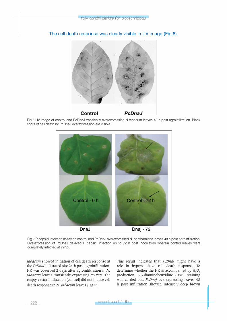
- 222 - annual report 2015
rajiv gandhi centre for biotechnology
The cell death response was clearly visible in UV image (Fig.6).
Fig.6 UV image of control and PcDnaJ transiently overexpressing N.tabacum leaves 48 h post agroinfiltration. Black spots of cell death by PcDnaJ overexpression are visible.
DnaJ Dnaj - 72
Control - 0 h Control - 72 h
Fig.7 P. capsici infection assay on control and PcDnaJ overexpressed N. benthamiana leaves 48 h post agroinfiltration. Overexpression of PcDnaJ delayed P. capsici infection up to 72 h post inoculation wherein control leaves were completely infected at 72hpi.
tabacum showed initiation of cell death response at the PcDnaJ infiltrated site 24 h post agroinfiltration. HR was observed 2 days after agroinfiltration in N. tabacum leaves transiently expressing PcDnaJ. The empty vector infiltration (control) did not induce cell death response in N. tabacum leaves (Fig.5).
This result indicates that PcDnaJ might have a role in hypersensitive cell death response. To determine whether the HR is accompanied by H2O2 production, 3,3-diaminobenzidine (DAB) staining was carried out. PcDnaJ overexpressing leaves 48 h post infiltration showed intensely deep brown
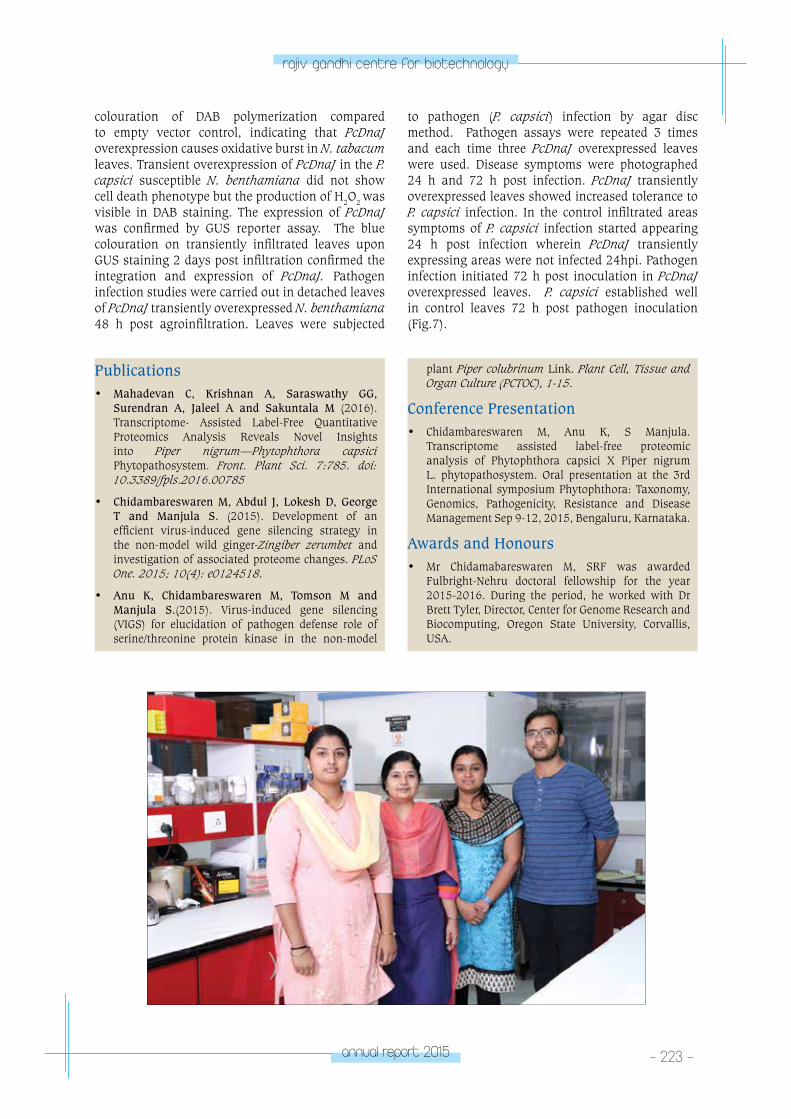
annual report 2015 - 223 -
rajiv gandhi centre for biotechnology
colouration of DAB polymerization compared to empty vector control, indicating that PcDnaJ overexpression causes oxidative burst in N. tabacum leaves. Transient overexpression of PcDnaJ in the P. capsici susceptible N. benthamiana did not show cell death phenotype but the production of H2O2 was visible in DAB staining. The expression of PcDnaJ was confirmed by GUS reporter assay. The blue colouration on transiently infiltrated leaves upon GUS staining 2 days post infiltration confirmed the integration and expression of PcDnaJ. Pathogen infection studies were carried out in detached leaves of PcDnaJ transiently overexpressed N. benthamiana 48 h post agroinfiltration. Leaves were subjected
to pathogen (P. capsici) infection by agar disc method. Pathogen assays were repeated 3 times and each time three PcDnaJ overexpressed leaves were used. Disease symptoms were photographed 24 h and 72 h post infection. PcDnaJ transiently overexpressed leaves showed increased tolerance to P. capsici infection. In the control infiltrated areas symptoms of P. capsici infection started appearing 24 h post infection wherein PcDnaJ transiently expressing areas were not infected 24hpi. Pathogen infection initiated 72 h post inoculation in PcDnaJ overexpressed leaves. P. capsici established well in control leaves 72 h post pathogen inoculation (Fig.7).
Publications• Mahadevan C, Krishnan A, Saraswathy GG,
Surendran A, Jaleel A and Sakuntala M (2016). Transcriptome- Assisted Label-Free Quantitative Proteomics Analysis Reveals Novel Insights into Piper nigrum—Phytophthora capsici Phytopathosystem. Front. Plant Sci. 7:785. doi: 10.3389/fpls.2016.00785
• Chidambareswaren M, Abdul J, Lokesh D, George T and Manjula S. (2015). Development of an efficient virus-induced gene silencing strategy in the non-model wild ginger-Zingiber zerumbet and investigation of associated proteome changes. PLoS One. 2015; 10(4): e0124518.
• Anu K, Chidambareswaren M, Tomson M and Manjula S.(2015). Virus-induced gene silencing (VIGS) for elucidation of pathogen defense role of serine/threonine protein kinase in the non-model
plant Piper colubrinum Link. Plant Cell, Tissue and Organ Culture (PCTOC), 1-15.
Conference Presentation• Chidambareswaren M, Anu K, S Manjula.
Transcriptome assisted label-free proteomic analysis of Phytophthora capsici X Piper nigrum L. phytopathosystem. Oral presentation at the 3rd International symposium Phytophthora: Taxonomy, Genomics, Pathogenicity, Resistance and Disease Management Sep 9-12, 2015, Bengaluru, Karnataka.
Awards and Honours• Mr Chidamabareswaren M, SRF was awarded
Fulbright-Nehru doctoral fellowship for the year 2015-2016. During the period, he worked with Dr Brett Tyler, Director, Center for Genome Research and Biocomputing, Oregon State University, Corvallis, USA.
MoleCulA
R RepRoDuCtIon laboratory - 1MOLECULAR
REPRODUCTIONLaboratory - 1
Pradeep Kumar. G
Pradeep Kumar was awarded PhD in Life
Sciences from Devi Ahilya Unviersity, Indore and
joined the Faculty of Life Sciences of the same
university He worked as a Fellow-in-residence
at the Centre for Biomedical Research at the
Rockefeller University, New York, NY; visiting
faculty in University of Virginia, Charlottesville,
VA and as visiting faculty at University of
Florida, Gainesville, FL. He joined Rajiv Gandhi
Centre for Biotechnology in the year 2004.
Technical Staff Tessy Ann Mary
Project Personnel Anil Kumar T.R.Jeeva S.
PhD students Karthika Radhakrishnan Nomesh Yadu Soumya A. Devi A.N. Mahitha Sahadevan Irfan KhanAswathy J.R.
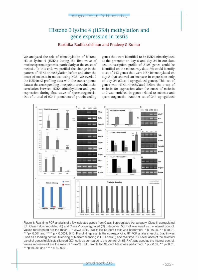
annual report 2015 - 225 -
rajiv gandhi centre for biotechnology
We analyzed the role of trimethylation of histone H3 at lysine 4 (H3K4) during the first wave of murine spermatogenesis, particularly at the onset of meiosis. To this end, we profiled the change in the pattern of H3K4 trimethylation before and after the onset of meiosis in mouse using NGS. We overlaid the H3K4me3 profiling data with the transcriptome data at the corresponding time points to evaluate the correlation between H3K4 trimethylation and gene expression during first wave of spermatogenesis. Out of a total of 6244 promoters of protein coding
genes that were identified to be H3K4 trimethylated at the promoter on day 8 and day 24 in our data set, transcription profile of 3105 genes could be identified on the microarray data. We could identify a set of 143 genes that were H3K4trimethylated on day 8 that showed an increase in expression only on day 24 (Class I upregulated genes). This set of genes was H3K4trimethylated before the onset of meiosis for expression after the onset of meiosis and was enriched in genes related to meiosis and spermatogenesis. Another set of 244 upregulated
Histone 3 lysine 4 (H3K4) methylation and gene expression in testis
Karthika Radhakrishnan and Pradeep G Kumar
Figure 1: Real time PCR analysis of a few selected genes from Class II upregulated (A) category, Class III upregulated (C), Class I downregulated (E) and Class V downregulated (G) categories. 5SrRNA was used as the internal control. Values represented are the mean 2^-∆∆Ct ±SE. Two tailed Student t-test was performed. * p <0.05, ** p<0.01, ***p<0.001 and **** p <0.0001. B, D, F and H represents the corresponding RT PCR analysis results. β-actin was used as a loading control. Silencing of Meisetz silencing in GC1 cells (I) and real time PCR evaluation of the selected panel of genes in Meisetz silenced GC1 cells as compared to the control (J). 5SrRNA was used as the internal control. Values represented are the mean 2^-∆∆Ct ±SE. Two tailed Student t-test was performed. * p <0.05, ** p<0.01, ***p<0.001 and **** p <0.0001.

- 226 - annual report 2015
rajiv gandhi centre for biotechnology
genes (Class II and Class III) showed an increase in H3K4 trimethylation and a corresponding increase in transcription during day 8 to day 24 transition. Functional analysis showed that Class II upregulated category was enriched in genes related to spermatogenesis and spermatid development. Moreover, we could demonstrate that silencing of Meisetz, a known H3K4trimethylating enzyme shown to be important to the process of spermatogenesis, resulted in a greater than 50% reduction of all the genes under study, of which, many are very important to the process of
spermatogenesis. Creb3l1, Ggnb1, Gpx4, Hspa1l, Zfp 35, Nfib, Wnt5a and Zfp37 showed statistically significant reduction by greater than 80% following silencing of Meisetz in GC1 cells. Our data showed that Meisetz dependent H3K4trimethylation at the promoter of many of these spermatid development related genes such as Crem, Gpx4, Zfp35, Zfp37 is crucial for the expression of these genes. Any aberration in the expression of Meisetz would result in improper H3K4trimethylation, which in turn will affect the expression of these genes, possibly resulting in infertility(Fig 1).
MicroRNA-mediated regulation of gene expression in the division and differentiation of spermatogonial cells
in mouse testis.Mahitha Sahadevan and Pradeep G Kumar
MicroRNAs are single stranded endogenous non- coding RNA molecules of ~ 22 nucleotides, which regulate a large number of protein coding genes at post-transcriptional level either by miRNA –directed translational repression or mRNA decay. Recent studies have emphasized the importance of microRNAs (small regulatory RNA) in regulating
spermatogenesis. Previous studies conducted in our lab have reported the occurrence of many changes in microRNA and mRNA profiles spanning the first waves of spermatogenesis using prepubertal (post natal day 8, P8), pubertal (post natal day 16, P16) and adolescent (post natal day 24, P24) mouse testes. Through microarray data differential expression
of 67 miRNAs and 8,226 mRNAs were identified. Using miRWalk predictions, these two datasets were integrated into miRNA-dependent regulatory networks. In network, miR-34b-5p, miR-34c and miR-449a shows progressive increase from P8 through P16 to P24 while remaining miRNAs in the network showed statistically significant changes in their levels either during P8 to P16 transition or P16 to P24 transition. We have identified that the target genes of miR449a are common target for miR34c also. In order to confirm
Figure 2. Real time analysis of miR34c, miR449a and their common target genes during first wave of spermatogenesis.
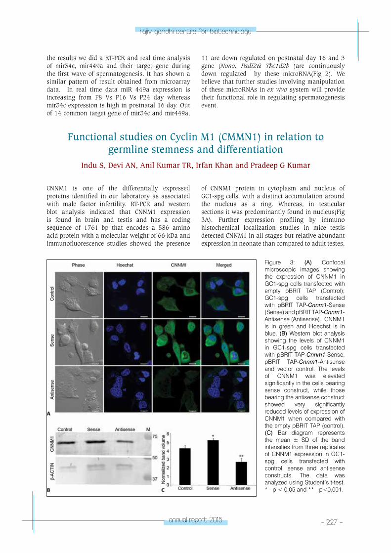
annual report 2015 - 227 -
rajiv gandhi centre for biotechnology
CNNM1 is one of the differentially expressed proteins identified in our laboratory as associated with male factor infertility. RT-PCR and western blot analysis indicated that CNNM1 expression is found in brain and testis and has a coding sequence of 1761 bp that encodes a 586 amino acid protein with a molecular weight of 66 kDa and immunofluorescence studies showed the presence
Functional studies on Cyclin M1 (CMMN1) in relation to germline stemness and differentiation
Indu S, Devi AN, Anil Kumar TR, Irfan Khan and Pradeep G Kumar
of CNNM1 protein in cytoplasm and nucleus of GC1-spg cells, with a distinct accumulation around the nucleus as a ring. Whereas, in testicular sections it was predominantly found in nucleus(Fig 3A). Further expression profiling by immuno histochemical localization studies in mice testis detected CNNM1 in all stages but relative abundant expression in neonate than compared to adult testes,
the results we did a RT-PCR and real time analysis of mir34c, mir449a and their target gene during the first wave of spermatogenesis. It has shown a similar pattern of result obtained from microarray data. In real time data miR 449a expression is increasing from P8 Vs P16 Vs P24 day whereas mir34c expression is high in postnatal 16 day. Out of 14 common target gene of mir34c and mir449a,
11 are down regulated on postnatal day 16 and 3 gene (Nono, Padi2& Tbc1d2b )are continuously down regulated by these microRNA(Fig 2). We believe that further studies involving manipulation of these microRNAs in ex vivo system will provide their functional role in regulating spermatogenesis event.
Figure 3: (A) Confocal microscopic images showing the expression of CNNM1 in GC1-spg cells transfected with empty pBRIT TAP (Control); GC1-spg cells transfected with pBRIT TAP-Cnnm1-Sense (Sense) and pBRIT TAP-Cnnm1-Antisense (Antisense). CNNM1 is in green and Hoechst is in blue. (B) Western blot analysis showing the levels of CNNM1 in GC1-spg cells transfected with pBRIT TAP-Cnnm1-Sense, pBRIT TAP-Cnnm1-Antisense and vector control. The levels of CNNM1 was elevated significantly in the cells bearing sense construct, while those bearing the antisense construct showed very significantly reduced levels of expression of CNNM1 when compared with the empty pBRIT TAP (control). (C) Bar diagram represents the mean ± SD of the band intensities from three replicates of CNNM1 expression in GC1-spg cells transfected with control, sense and antisense constructs. The data was analyzed using Student’s t-test. * - p < 0.05 and ** - p<0.001.
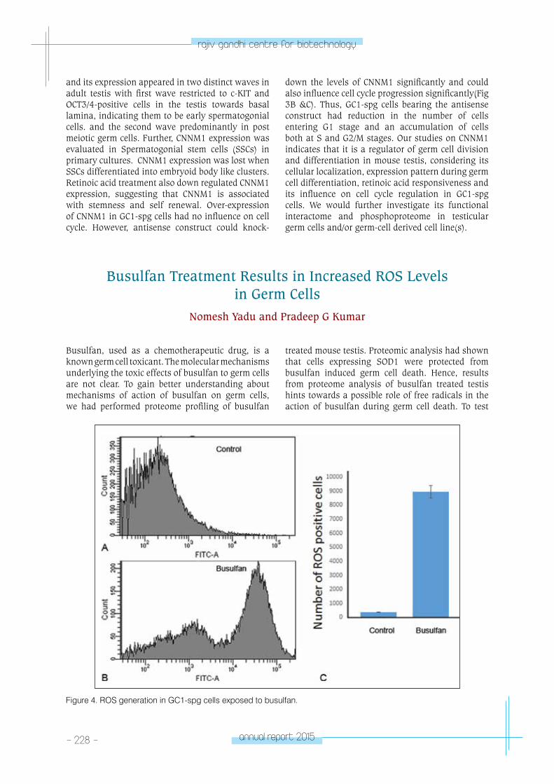
- 228 - annual report 2015
rajiv gandhi centre for biotechnology
and its expression appeared in two distinct waves in adult testis with first wave restricted to c-KIT and OCT3/4-positive cells in the testis towards basal lamina, indicating them to be early spermatogonial cells. and the second wave predominantly in post meiotic germ cells. Further, CNNM1 expression was evaluated in Spermatogonial stem cells (SSCs) in primary cultures. CNNM1 expression was lost when SSCs differentiated into embryoid body like clusters. Retinoic acid treatment also down regulated CNNM1 expression, suggesting that CNNM1 is associated with stemness and self renewal. Over-expression of CNNM1 in GC1-spg cells had no influence on cell cycle. However, antisense construct could knock-
down the levels of CNNM1 significantly and could also influence cell cycle progression significantly(Fig 3B &C). Thus, GC1-spg cells bearing the antisense construct had reduction in the number of cells entering G1 stage and an accumulation of cells both at S and G2/M stages. Our studies on CNNM1 indicates that it is a regulator of germ cell division and differentiation in mouse testis, considering its cellular localization, expression pattern during germ cell differentiation, retinoic acid responsiveness and its influence on cell cycle regulation in GC1-spg cells. We would further investigate its functional interactome and phosphoproteome in testicular germ cells and/or germ-cell derived cell line(s).
Busulfan Treatment Results in Increased ROS Levels in Germ Cells
Nomesh Yadu and Pradeep G Kumar
Busulfan, used as a chemotherapeutic drug, is a known germ cell toxicant. The molecular mechanisms underlying the toxic effects of busulfan to germ cells are not clear. To gain better understanding about mechanisms of action of busulfan on germ cells, we had performed proteome profiling of busulfan
treated mouse testis. Proteomic analysis had shown that cells expressing SOD1 were protected from busulfan induced germ cell death. Hence, results from proteome analysis of busulfan treated testis hints towards a possible role of free radicals in the action of busulfan during germ cell death. To test
Figure 4. ROS generation in GC1-spg cells exposed to busulfan.
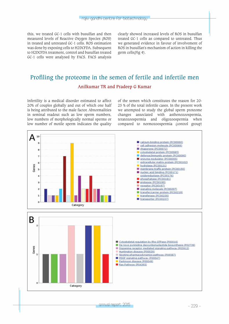
annual report 2015 - 229 -
rajiv gandhi centre for biotechnology
this, we treated GC-1 cells with busulfan and then measured levels of Reactive Oxygen Species (ROS) in treated and untreated GC-1 cells. ROS estimation was done by exposing cells to H2DCFDA. Subsequent to H2DCFDA treatment, control and busulfan treated GC-1 cells were analysed by FACS. FACS analysis
clearly showed increased levels of ROS in busulfan treated GC-1 cells as compared to untreated. Thus we generated evidence in favour of involvement of ROS in busulfan’s mechanism of action in killing the germ cells(Fig 4).
Profiling the proteome in the semen of fertile and infertile menAnilkumar TR and Pradeep G Kumar
Infertility is a medical disorder estimated to affect 20% of couples globally and out of which one half is being attributed to the male factor. Abnormalities in seminal readout such as low sperm numbers, low numbers of morphologically normal sperms or low number of motile sperm indicates the quality
of the semen which constitutes the reason for 20-25 % of the total infertile cases. In the present work we attempted to study the global sperm proteome changes associated with asthenozoospermia, teratozoospermia and oligozoospermia when compared to normozoospermia (control group)
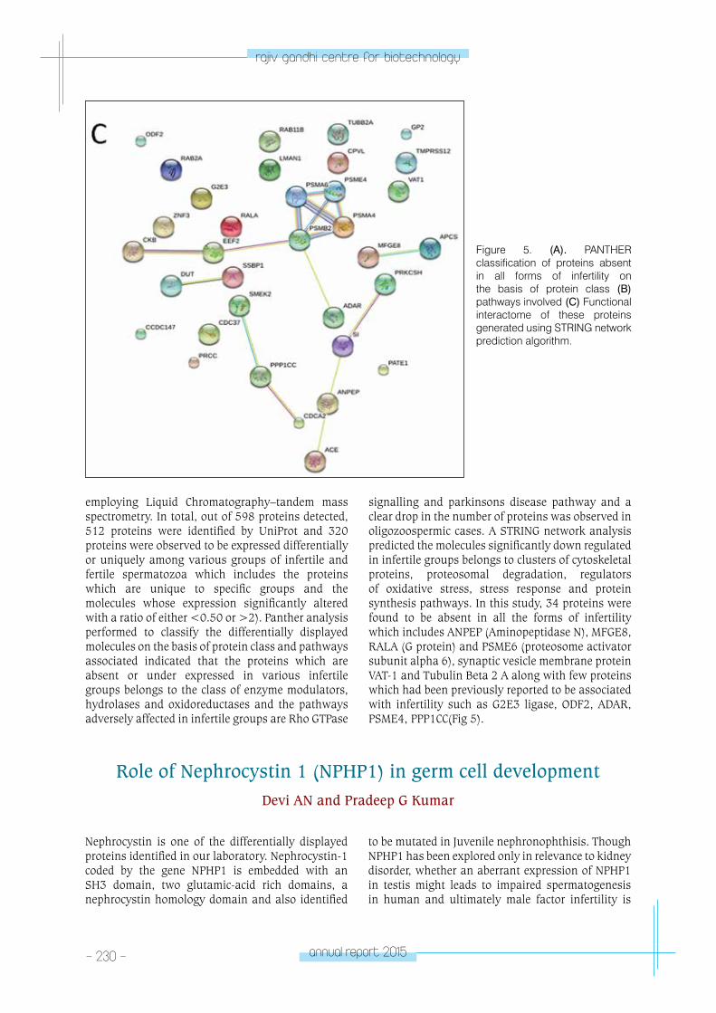
- 230 - annual report 2015
rajiv gandhi centre for biotechnology
employing Liquid Chromatography–tandem mass spectrometry. In total, out of 598 proteins detected, 512 proteins were identified by UniProt and 320 proteins were observed to be expressed differentially or uniquely among various groups of infertile and fertile spermatozoa which includes the proteins which are unique to specific groups and the molecules whose expression significantly altered with a ratio of either <0.50 or >2). Panther analysis performed to classify the differentially displayed molecules on the basis of protein class and pathways associated indicated that the proteins which are absent or under expressed in various infertile groups belongs to the class of enzyme modulators, hydrolases and oxidoreductases and the pathways adversely affected in infertile groups are Rho GTPase
signalling and parkinsons disease pathway and a clear drop in the number of proteins was observed in oligozoospermic cases. A STRING network analysis predicted the molecules significantly down regulated in infertile groups belongs to clusters of cytoskeletal proteins, proteosomal degradation, regulators of oxidative stress, stress response and protein synthesis pathways. In this study, 34 proteins were found to be absent in all the forms of infertility which includes ANPEP (Aminopeptidase N), MFGE8, RALA (G protein) and PSME6 (proteosome activator subunit alpha 6), synaptic vesicle membrane protein VAT-1 and Tubulin Beta 2 A along with few proteins which had been previously reported to be associated with infertility such as G2E3 ligase, ODF2, ADAR, PSME4, PPP1CC(Fig 5).
Figure 5. (A). PANTHER classification of proteins absent in all forms of infertility on the basis of protein class (B) pathways involved (C) Functional interactome of these proteins generated using STRING network prediction algorithm.
Nephrocystin is one of the differentially displayed proteins identified in our laboratory. Nephrocystin-1 coded by the gene NPHP1 is embedded with an SH3 domain, two glutamic-acid rich domains, a nephrocystin homology domain and also identified
to be mutated in Juvenile nephronophthisis. Though NPHP1 has been explored only in relevance to kidney disorder, whether an aberrant expression of NPHP1 in testis might leads to impaired spermatogenesis in human and ultimately male factor infertility is
Role of Nephrocystin 1 (NPHP1) in germ cell developmentDevi AN and Pradeep G Kumar
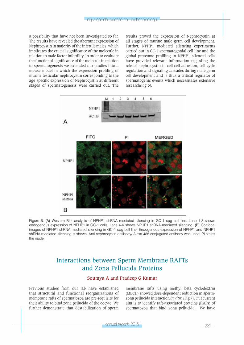
annual report 2015 - 231 -
rajiv gandhi centre for biotechnology
Figure 6. (A) Western Blot analysis of NPHP1 shRNA mediated silencing in GC-1 spg cell line. Lane 1-3 shows endogenous expression of NPHP1 in GC-1 cells. Lane 4-6 shows NPHP1 shRNA mediated silencing. (B) Confocal images of NPHP1 shRNA mediated silencing in GC-1 spg cell line. Endogenous expression of NPHP1 and NPHP1 shRNA mediated silencing is shown. Anti nephrocystin antibody/ Alexa-488 conjugated antibody was used. PI stains the nuclei.
a possibility that have not been investigated so far. The results have revealed the aberrant expression of Nephrocystin in majority of the infertile males, which implicates the crucial significance of the molecule in relation to male factor infertility. In order to evaluate the functional significance of the molecule in relation to spermatogenesis we extended our studies into a mouse model in which the expression profiling of murine testicular nephrocystin corresponding to the age specific expression of Nephrocystin at different stages of spermatogenesis were carried out. The
results proved the expression of Nephrocystin at all stages of murine male germ cell development. Further, NPHP1 mediated silencing experiments carried out in GC-1 spermatogonial cell line and the global proteome profiling in NPHP1 silenced cells have provided relevant information regarding the role of nephrocystin in cell-cell adhesion, cell cycle regulation and signaling cascades during male germ cell development and is thus a critical regulator of spermatogenic events which necessitates extensive research(Fig 6).
Interactions between Sperm Membrane RAFTs and Zona Pellucida Proteins
Soumya A and Pradeep G Kumar
Previous studies from our lab have established that structural and functional reorganizations of membrane rafts of spermatozoa are pre-requisite for their ability to bind zona pellucida of the oocyte. We further demonstrate that destabilization of sperm
membrane rafts using methyl beta cyclodextrin (MBCD) showed dose-dependent reduction in sperm-zona pellucida interaction in vitro (Fig 7). Our current aim is to identify raft-associated proteins (RAPs) of spermatozoa that bind zona pellucida. We have
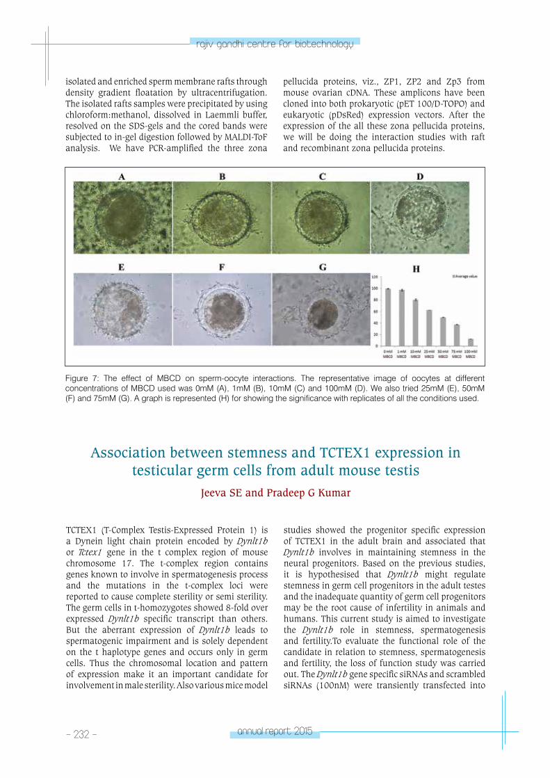
- 232 - annual report 2015
rajiv gandhi centre for biotechnology
Association between stemness and TCTEX1 expression in testicular germ cells from adult mouse testis
Jeeva SE and Pradeep G Kumar
TCTEX1 (T-Complex Testis-Expressed Protein 1) is a Dynein light chain protein encoded by Dynlt1b or Tctex1 gene in the t complex region of mouse chromosome 17. The t-complex region contains genes known to involve in spermatogenesis process and the mutations in the t-complex loci were reported to cause complete sterility or semi sterility. The germ cells in t-homozygotes showed 8-fold over expressed Dynlt1b specific transcript than others. But the aberrant expression of Dynlt1b leads to spermatogenic impairment and is solely dependent on the t haplotype genes and occurs only in germ cells. Thus the chromosomal location and pattern of expression make it an important candidate for involvement in male sterility. Also various mice model
studies showed the progenitor specific expression of TCTEX1 in the adult brain and associated that Dynlt1b involves in maintaining stemness in the neural progenitors. Based on the previous studies, it is hypothesised that Dynlt1b might regulate stemness in germ cell progenitors in the adult testes and the inadequate quantity of germ cell progenitors may be the root cause of infertility in animals and humans. This current study is aimed to investigate the Dynlt1b role in stemness, spermatogenesis and fertility.To evaluate the functional role of the candidate in relation to stemness, spermatogenesis and fertility, the loss of function study was carried out. The Dynlt1b gene specific siRNAs and scrambled siRNAs (100nM) were transiently transfected into
Figure 7: The effect of MBCD on sperm-oocyte interactions. The representative image of oocytes at different concentrations of MBCD used was 0mM (A), 1mM (B), 10mM (C) and 100mM (D). We also tried 25mM (E), 50mM (F) and 75mM (G). A graph is represented (H) for showing the significance with replicates of all the conditions used.
isolated and enriched sperm membrane rafts through density gradient floatation by ultracentrifugation. The isolated rafts samples were precipitated by using chloroform:methanol, dissolved in Laemmli buffer, resolved on the SDS-gels and the cored bands were subjected to in-gel digestion followed by MALDI-ToF analysis. We have PCR-amplified the three zona
pellucida proteins, viz., ZP1, ZP2 and Zp3 from mouse ovarian cDNA. These amplicons have been cloned into both prokaryotic (pET 100/D-TOPO) and eukaryotic (pDsRed) expression vectors. After the expression of the all these zona pellucida proteins, we will be doing the interaction studies with raft and recombinant zona pellucida proteins.
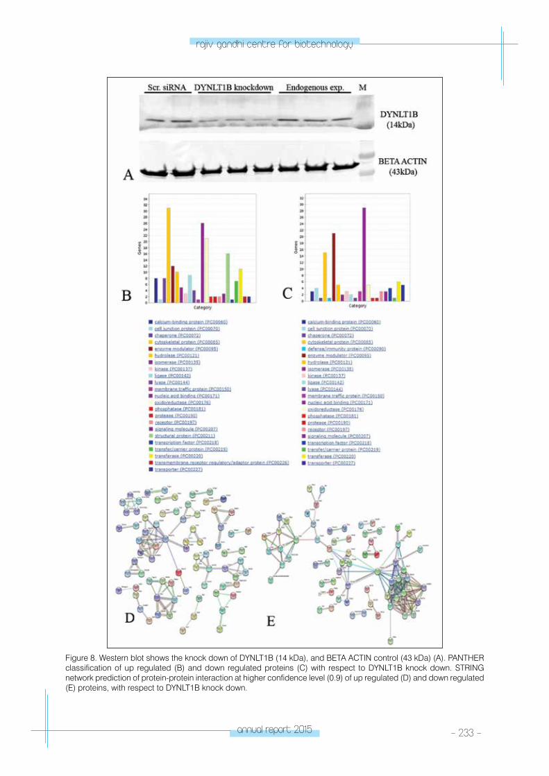
annual report 2015 - 233 -
rajiv gandhi centre for biotechnology
Figure 8. Western blot shows the knock down of DYNLT1B (14 kDa), and BETA ACTIN control (43 kDa) (A). PANTHER classification of up regulated (B) and down regulated proteins (C) with respect to DYNLT1B knock down. STRING network prediction of protein-protein interaction at higher confidence level (0.9) of up regulated (D) and down regulated (E) proteins, with respect to DYNLT1B knock down.
- 234 - annual report 2015
rajiv gandhi centre for biotechnology
GC1 spermatogonial cell line by lipofectamine mediated transfection respectively. After 36 hour post transfection, the cells were harvested and lysed for RNA and protein preparation (Fig. 8A). The crude protein samples subjected LC-MS analysis identified 445 differentially expressed proteins and among them 239 was up regulated and 206 were down regulated with respect to Dynlt1b knockdown as compared to the control. The mass spec identified differentially expressed proteins subjected to GO analysis using UniProt database search and other online tools like PANTHER classification and STRING analysis. PANTHER classification tool categorised the up regulated proteins into twenty three categories according to protein class. The major proportion of the proteins in the up regulated group were categorised as cytoskeletal proteins followed by nucleic acid binding proteins
and oxidoreductases (Fig. 8B). Likewise, the down regulated proteins were also classified into twenty two categories and among them major portion was in nucleic acid binding proteins followed by enzyme modulators and cytoskeletal proteins (Fig. 8C). The STRING network prediction analysis showed no distinct interacting clusters at lower confidence level (0.4), but at the higher confidence (0.7 and 0.9) levels the interacting clusters can be observed (Fig. 8D. and Fig. 8E). Further enrichment by KEGG pathway identified 145 pathways for up regulated proteins and 133 pathways for down regulated proteins with significant p-values. Among them, the major biological pathways identified were metabolic pathway and ribosome associated pathway for up regulated and down regulated proteins respectively. We are currently exploring the connection between DYNLT1 and germline stemness, if any.
Papers published • Indu S, Sekhar SC, Jeeva S, Anilkumar TR, Pillai
SM, Laloraya M and Kumar PG (2015) Aberrant expression of DYNLT1 is associated with human male factor infertility. Mol. Cell Proteom. 14(12):3185-95 (doi:10.1074/mcp.M115.050005).
• Varghese DS, Chandran U, Soumya A, Pillai SM, Jayakrishnan K, Reddi PP and Kumar PG (2016) Aberrant expression of TAR DNA binding protein-43 is associated with spermatogenic disorders in human males. Reprod. Fertil. Dev. 28(6): 713-22 (DOI: 10.1071/RD14090).
• Chandran U, Indu S, Anilkumar TR, Devi AN, Khan I, Srivastava D and Kumar PG (2016) Expression of Cnnm1 and its association with stemness, cell cycle and differentiation in spermatogenic cells in mouse testis. Biol. Reprod (doi: 10.1095/ biolreprod.115.130369)
• Radhakrishnan K, Bhagya KP, Anilumar TR, Devi AN, Sengottaiyan J and Kumar PG (2016) Autoimmune regulator (AIRE) is expressed in spermatogenic cells and it altered the expression of several nucleic acid binding and cytoskeletal proteins in GC1-spg spermatogonial cells. Mol Cell Proteom. doi:10.1074/mcp.M115.052951.
PhDs awarded• Divya Saro Varghese (2015) Functional analysis of
TDP-43 in relation to spermatogenesis
• Sreesha Sree (2015) Role of microRNAs in germ cell differentiation and fertilization.
PhDs theses Submitted• Bhagya KP (2015) Functional evaluation of AIRE in
mammalian testis
• Karthika Radhakrishnan (2016) H3K4 Methylation and Spermatogenesis
Conference Presentations
Invited Lectures• Kumar PG (2015) Stem cells: Past, Present and
Future. At Central University of Rajasthan, Ajmer, Rajasthan, 11 September 2015.
• Kumar PG (2015) Molecular markers for male factor infertility. National Conference on Reproductive Health Challenges: Issues and Remedies (NCRHC-2015), IIS University, Jaipur, 11-13 September, 2015.
• Kumar PG (2015) Panel discussion on Biotechnology. Swasraya Bharat 2015, Swapna Nagari, Kozhikode, 15-21 October, 2015.
• Kumar PG (2016) Differential display proteomics of spermatozoa: our leads in understanding male factor infertility. Dr. TC Anand Kumar Memorial Gold Medal Oration at the 26th Annual Meeting of Indian Society for the Study of Reproduction and Fertility, National Institute of Occupational Health, Ahmedabad, 18-20 February, 2016.
• Kumar PG (2016) Identification of Molecular Markers of Male Factor Infertility. National Institute for Science, Education and Research, Bhuhaneswar, 17-18 May, 2016.
• Kumar PG (2016) Where to go? Mar Ivanios College, Trivandrum, 11 June, 2016.
Honors and Awards• Dr. TC Anand Kumar Memorial Oration and
Gold Medal of the Indian Society for the Study of Reproduction and Fertility (ISSRF), 2016.
annual report 2015 - 235 -
rajiv gandhi centre for biotechnology
RESEARCH GRANTS
No. Investigator(s) Title Funding Agency Duration
1 Pradeep Kumar G Molecular evaluation of interactions between sperm membrane rafts and zona pellucida proteins
Department of Biotechnology, Government of India
2011-2015
2 Pradeep Kumar G Association between stemness and TCTEX1 expression in testicular germ cells from adult mouse testis
Board of Research in Nuclear Sciences
2011-2015
3 Pradeep Kumar G Role of CLP-1 in cell cycle regulation in spermatogenic cells
Department of Science & Technology
2013-2016
4 Pradeep Kumar G Evaluation of cellular aging and genome stability in spermatogonial stem cells
Council for Scientific & Industrial Research
2014-2017
MoleCulA
R RepRoDuCtIon laboratory - 2MOLECULAR
REPRODUCTIONLaboratory – 2
Malini Laloraya
Malini Laloraya received her PhD from Devi
Ahilya Vishwavidyalya, Indore. She was a Fellow
in Residence at a Center for Biomedical Research,
Population Council, Rockefeller University, New
York and was a Visiting Faculty at University
of Virginia, VA and University of Florida,
Gainesville, FL, USA. She joined RGCB in the
year 2004.
Post Doctoral FellowsRenjini A.P. PhDSushma Padmaja
Technical PersonnelG. Sheela
Project PersonnelLekshmy C.R.Selin Joseph
PhD studentsPhilip Litto ThomasPrashanth Narayan Annu Joseph Soumya V. Betcy Susan Johnson Neethu Puthumadathil
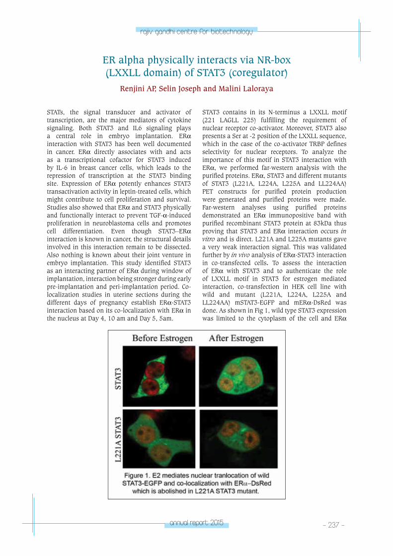
annual report 2015 - 237 -
rajiv gandhi centre for biotechnology
STATs, the signal transducer and activator of transcription, are the major mediators of cytokine signaling. Both STAT3 and IL6 signaling plays a central role in embryo implantation. ERα interaction with STAT3 has been well documented in cancer. ERα directly associates with and acts as a transcriptional cofactor for STAT3 induced by IL-6 in breast cancer cells, which leads to the repression of transcription at the STAT3 binding site. Expression of ERα potently enhances STAT3 transactivation activity in leptin-treated cells, which might contribute to cell proliferation and survival. Studies also showed that ERα and STAT3 physically and functionally interact to prevent TGF-α-induced proliferation in neuroblastoma cells and promotes cell differentiation. Even though STAT3–ERα interaction is known in cancer, the structural details involved in this interaction remain to be dissected. Also nothing is known about their joint venture in embryo implantation. This study identified STAT3 as an interacting partner of ERα during window of implantation, interaction being stronger during early pre-implantation and peri-implantation period. Co-localization studies in uterine sections during the different days of pregnancy establish ERα-STAT3 interaction based on its co-localization with ERα in the nucleus at Day 4, 10 am and Day 5, 5am.
ER alpha physically interacts via NR-box (LXXLL domain) of STAT3 (coregulator)
Renjini AP, Selin Joseph and Malini Laloraya
STAT3 contains in its N-terminus a LXXLL motif (221 LAGLL 225) fulfilling the requirement of nuclear receptor co-activator. Moreover, STAT3 also presents a Ser at -2 position of the LXXLL sequence, which in the case of the co-activator TRBP defines selectivity for nuclear receptors. To analyze the importance of this motif in STAT3 interaction with ERα, we performed far-western analysis with the purified proteins. ERα, STAT3 and different mutants of STAT3 (L221A, L224A, L225A and LL224AA) PET constructs for purified protein production were generated and purified proteins were made. Far-western analyses using purified proteins demonstrated an ERα immunopositive band with purified recombinant STAT3 protein at 83kDa thus proving that STAT3 and ERα interaction occurs in vitro and is direct. L221A and L225A mutants gave a very weak interaction signal. This was validated further by in vivo analysis of ERα-STAT3 interaction in co-transfected cells. To assess the interaction of ERα with STAT3 and to authenticate the role of LXXLL motif in STAT3 for estrogen mediated interaction, co-transfection in HEK cell line with wild and mutant (L221A, L224A, L225A and LL224AA) mSTAT3-EGFP and mERα-DsRed was done. As shown in Fig 1, wild type STAT3 expression was limited to the cytoplasm of the cell and ERα
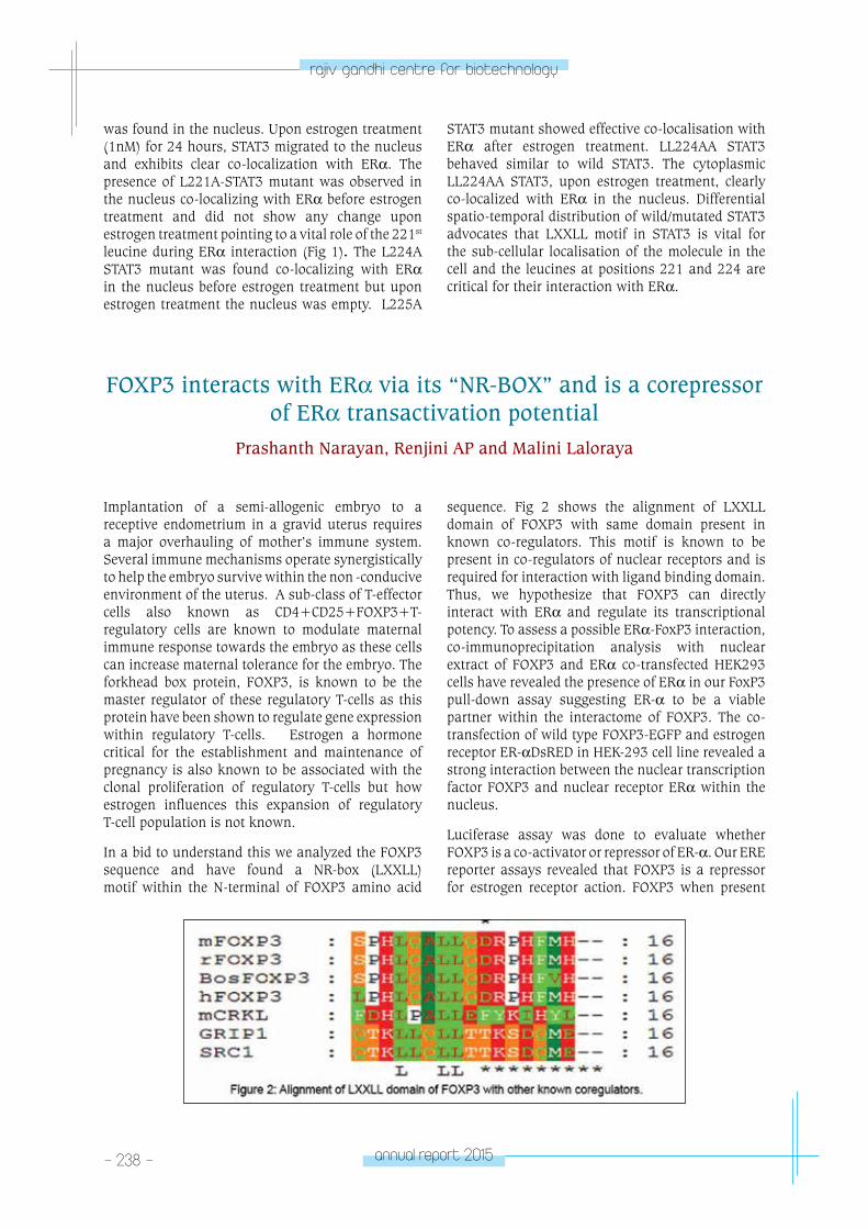
- 238 - annual report 2015
rajiv gandhi centre for biotechnology
was found in the nucleus. Upon estrogen treatment (1nM) for 24 hours, STAT3 migrated to the nucleus and exhibits clear co-localization with ERα. The presence of L221A-STAT3 mutant was observed in the nucleus co-localizing with ERα before estrogen treatment and did not show any change upon estrogen treatment pointing to a vital role of the 221st leucine during ERα interaction (Fig 1). The L224A STAT3 mutant was found co-localizing with ERα in the nucleus before estrogen treatment but upon estrogen treatment the nucleus was empty. L225A
STAT3 mutant showed effective co-localisation with ERα after estrogen treatment. LL224AA STAT3 behaved similar to wild STAT3. The cytoplasmic LL224AA STAT3, upon estrogen treatment, clearly co-localized with ERα in the nucleus. Differential spatio-temporal distribution of wild/mutated STAT3 advocates that LXXLL motif in STAT3 is vital for the sub-cellular localisation of the molecule in the cell and the leucines at positions 221 and 224 are critical for their interaction with ERα.
FOXP3 interacts with ERα via its “NR-BOX” and is a corepressor of ERα transactivation potential
Prashanth Narayan, Renjini AP and Malini Laloraya
Implantation of a semi-allogenic embryo to a receptive endometrium in a gravid uterus requires a major overhauling of mother’s immune system. Several immune mechanisms operate synergistically to help the embryo survive within the non -conducive environment of the uterus. A sub-class of T-effector cells also known as CD4+CD25+FOXP3+T-regulatory cells are known to modulate maternal immune response towards the embryo as these cells can increase maternal tolerance for the embryo. The forkhead box protein, FOXP3, is known to be the master regulator of these regulatory T-cells as this protein have been shown to regulate gene expression within regulatory T-cells. Estrogen a hormone critical for the establishment and maintenance of pregnancy is also known to be associated with the clonal proliferation of regulatory T-cells but how estrogen influences this expansion of regulatory T-cell population is not known.
In a bid to understand this we analyzed the FOXP3 sequence and have found a NR-box (LXXLL) motif within the N-terminal of FOXP3 amino acid
sequence. Fig 2 shows the alignment of LXXLL domain of FOXP3 with same domain present in known co-regulators. This motif is known to be present in co-regulators of nuclear receptors and is required for interaction with ligand binding domain. Thus, we hypothesize that FOXP3 can directly interact with ERα and regulate its transcriptional potency. To assess a possible ERα-FoxP3 interaction, co-immunoprecipitation analysis with nuclear extract of FOXP3 and ERα co-transfected HEK293 cells have revealed the presence of ERα in our FoxP3 pull-down assay suggesting ER-α to be a viable partner within the interactome of FOXP3. The co-transfection of wild type FOXP3-EGFP and estrogen receptor ER-αDsRED in HEK-293 cell line revealed a strong interaction between the nuclear transcription factor FOXP3 and nuclear receptor ERα within the nucleus.
Luciferase assay was done to evaluate whether FOXP3 is a co-activator or repressor of ER-α. Our ERE reporter assays revealed that FOXP3 is a repressor for estrogen receptor action. FOXP3 when present
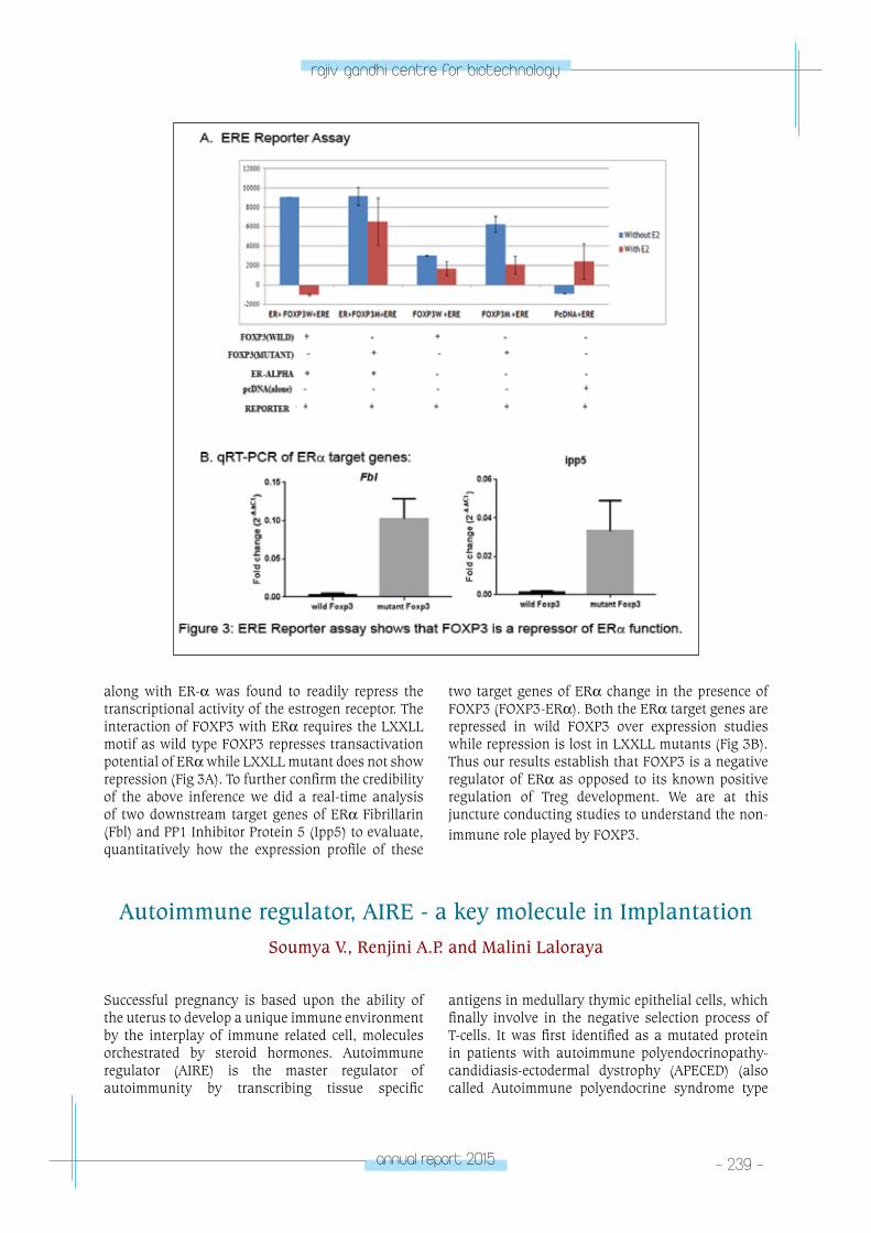
annual report 2015 - 239 -
rajiv gandhi centre for biotechnology
along with ER-α was found to readily repress the transcriptional activity of the estrogen receptor. The interaction of FOXP3 with ERα requires the LXXLL motif as wild type FOXP3 represses transactivation potential of ERα while LXXLL mutant does not show repression (Fig 3A). To further confirm the credibility of the above inference we did a real-time analysis of two downstream target genes of ERα Fibrillarin (Fbl) and PP1 Inhibitor Protein 5 (Ipp5) to evaluate, quantitatively how the expression profile of these
two target genes of ERα change in the presence of FOXP3 (FOXP3-ERα). Both the ERα target genes are repressed in wild FOXP3 over expression studies while repression is lost in LXXLL mutants (Fig 3B). Thus our results establish that FOXP3 is a negative regulator of ERα as opposed to its known positive regulation of Treg development. We are at this juncture conducting studies to understand the non-immune role played by FOXP3.
Autoimmune regulator, AIRE - a key molecule in ImplantationSoumya V., Renjini A.P. and Malini Laloraya
Successful pregnancy is based upon the ability of the uterus to develop a unique immune environment by the interplay of immune related cell, molecules orchestrated by steroid hormones. Autoimmune regulator (AIRE) is the master regulator of autoimmunity by transcribing tissue specific
antigens in medullary thymic epithelial cells, which finally involve in the negative selection process of T-cells. It was first identified as a mutated protein in patients with autoimmune polyendocrinopathy-candidiasis-ectodermal dystrophy (APECED) (also called Autoimmune polyendocrine syndrome type
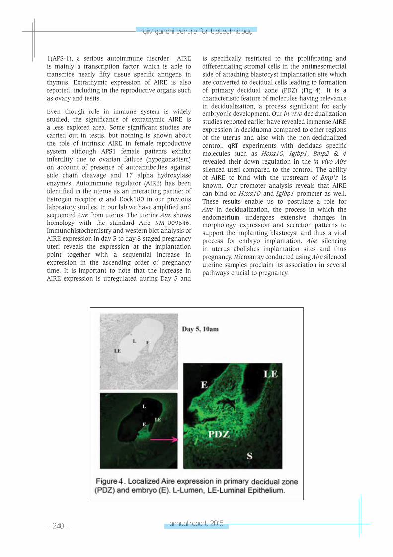
- 240 - annual report 2015
rajiv gandhi centre for biotechnology
1(APS-1), a serious autoimmune disorder. AIRE is mainly a transcription factor, which is able to transcribe nearly fifty tissue specific antigens in thymus. Extrathymic expression of AIRE is also reported, including in the reproductive organs such as ovary and testis.
Even though role in immune system is widely studied, the significance of extrathymic AIRE is a less explored area. Some significant studies are carried out in testis, but nothing is known about the role of intrinsic AIRE in female reproductive system although APS1 female patients exhibit infertility due to ovarian failure (hypogonadism) on account of presence of autoantibodies against side chain cleavage and 17 alpha hydroxylase enzymes. Autoimmune regulator (AIRE) has been identified in the uterus as an interacting partner of Estrogen receptor α and Dock180 in our previous laboratory studies. In our lab we have amplified and sequenced Aire from uterus. The uterine Aire shows homology with the standard Aire NM_009646. Immunohistochemistry and western blot analysis of AIRE expression in day 3 to day 8 staged pregnancy uteri reveals the expression at the implantation point together with a sequential increase in expression in the ascending order of pregnancy time. It is important to note that the increase in AIRE expression is upregulated during Day 5 and
is specifically restricted to the proliferating and differentiating stromal cells in the antimesometrial side of attaching blastocyst implantation site which are converted to decidual cells leading to formation of primary decidual zone (PDZ) (Fig 4). It is a characteristic feature of molecules having relevance in decidualization, a process significant for early embryonic development. Our in vivo decidualization studies reported earlier have revealed immense AIRE expression in deciduoma compared to other regions of the uterus and also with the non-decidualized control. qRT experiments with deciduas specific molecules such as Hoxa10, Igfbp1, Bmp2 & 4 revealed their down regulation in the in vivo Aire silenced uteri compared to the control. The ability of AIRE to bind with the upstream of Bmp’s is known. Our promoter analysis reveals that AIRE can bind on Hoxa10 and Igfbp1 promoter as well. These results enable us to postulate a role for Aire in decidualization, the process in which the endometrium undergoes extensive changes in morphology, expression and secretion patterns to support the implanting blastocyst and thus a vital process for embryo implantation. Aire silencing in uterus abolishes implantation sites and thus pregnancy. Microarray conducted using Aire silenced uterine samples proclaim its association in several pathways crucial to pregnancy.
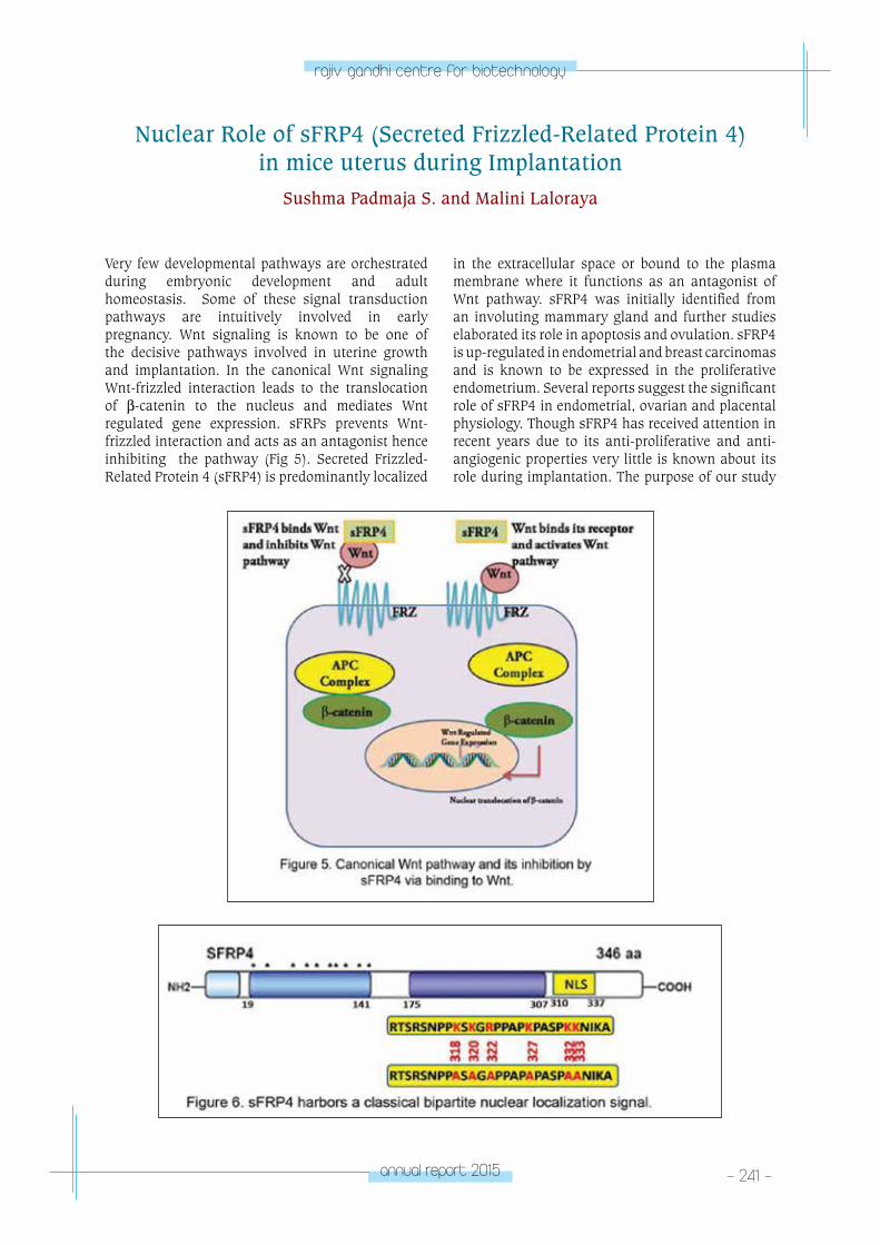
annual report 2015 - 241 -
rajiv gandhi centre for biotechnology
Very few developmental pathways are orchestrated during embryonic development and adult homeostasis. Some of these signal transduction pathways are intuitively involved in early pregnancy. Wnt signaling is known to be one of the decisive pathways involved in uterine growth and implantation. In the canonical Wnt signaling Wnt-frizzled interaction leads to the translocation of β-catenin to the nucleus and mediates Wnt regulated gene expression. sFRPs prevents Wnt-frizzled interaction and acts as an antagonist hence inhibiting the pathway (Fig 5). Secreted Frizzled-Related Protein 4 (sFRP4) is predominantly localized
Nuclear Role of sFRP4 (Secreted Frizzled-Related Protein 4) in mice uterus during Implantation
Sushma Padmaja S. and Malini Laloraya
in the extracellular space or bound to the plasma membrane where it functions as an antagonist of Wnt pathway. sFRP4 was initially identified from an involuting mammary gland and further studies elaborated its role in apoptosis and ovulation. sFRP4 is up-regulated in endometrial and breast carcinomas and is known to be expressed in the proliferative endometrium. Several reports suggest the significant role of sFRP4 in endometrial, ovarian and placental physiology. Though sFRP4 has received attention in recent years due to its anti-proliferative and anti-angiogenic properties very little is known about its role during implantation. The purpose of our study
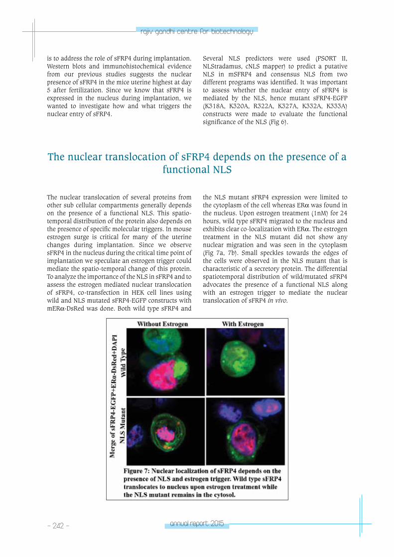
- 242 - annual report 2015
rajiv gandhi centre for biotechnology
is to address the role of sFRP4 during implantation. Western blots and immunohistochemical evidence from our previous studies suggests the nuclear presence of sFRP4 in the mice uterine highest at day 5 after fertilization. Since we know that sFRP4 is expressed in the nucleus during implantation, we wanted to investigate how and what triggers the nuclear entry of sFRP4.
Several NLS predictors were used (PSORT II, NLStradamus, cNLS mapper) to predict a putative NLS in mSFRP4 and consensus NLS from two different programs was identified. It was important to assess whether the nuclear entry of sFRP4 is mediated by the NLS, hence mutant sFRP4-EGFP (K318A, K320A, R322A, K327A, K332A, K333A) constructs were made to evaluate the functional significance of the NLS (Fig 6).
The nuclear translocation of sFRP4 depends on the presence of a functional NLS
The nuclear translocation of several proteins from other sub cellular compartments generally depends on the presence of a functional NLS. This spatio-temporal distribution of the protein also depends on the presence of specific molecular triggers. In mouse estrogen surge is critical for many of the uterine changes during implantation. Since we observe sFRP4 in the nucleus during the critical time point of implantation we speculate an estrogen trigger could mediate the spatio-temporal change of this protein. To analyze the importance of the NLS in sFRP4 and to assess the estrogen mediated nuclear translocation of sFRP4, co-transfection in HEK cell lines using wild and NLS mutated sFRP4-EGFP constructs with mERα-DsRed was done. Both wild type sFRP4 and
the NLS mutant sFRP4 expression were limited to the cytoplasm of the cell whereas ERα was found in the nucleus. Upon estrogen treatment (1nM) for 24 hours, wild type sFRP4 migrated to the nucleus and exhibits clear co-localization with ERα. The estrogen treatment in the NLS mutant did not show any nuclear migration and was seen in the cytoplasm (Fig 7a, 7b). Small speckles towards the edges of the cells were observed in the NLS mutant that is characteristic of a secretory protein. The differential spatiotemporal distribution of wild/mutated sFRP4 advocates the presence of a functional NLS along with an estrogen trigger to mediate the nuclear translocation of sFRP4 in vivo.
annual report 2015 - 243 -
rajiv gandhi centre for biotechnology
In a developing embryo, a small group of cells called inner cell mass (ICM) remain pluripotent and give rise to all cell types of the fetus and some extra embryonic tissues. The ICM cells proliferate for a limited period after which they lose their pluripotency and undergo differentiation. However, in specific culture conditions, ICM cells give rise to embryonic stem (ES) cells, which can be maintained indefinitely without losing their pluripotency. Although not identical to ICM cells, ES cells maintain the ability to undergo differentiation to all cell lineages. Therefore, ES cells are the best model to study the cell biology of pluripotency and differentiation.
Maintenance of pluripotency in ES cells is controlled by a variety of genes. Existence of epigenetic control of pluripotency has also been suggested. Molecular mechanisms that govern cell differentiation are poorly understood. It involves a repertoire of molecules and pathways, which interact with each other to bring about characteristic changes into defined cell types. Increasing evidence indicate that extrinsic signals from the stem cell microenvironment can converge on intrinsic signals to regulate stem cell proliferation and differentiation. The mouse embryonic stem cell line R1/E require Leukaemia Inhibitory Factor (LIF) to maintain pluripotency (+LIF) while without LIF the cells tend to differentiate (-LIF). LIF activates the JAK-STAT3 pathway. JAK-STAT pathway has been shown to be activated by ROS where, H2O2 stimulates the activity of the known STAT kinases, JAK2 and TYK2. Thus our aim is to understand whether Reactive Oxygen Species (ROS), especially H2O2 plays a role in maintaining cellular pluripotency.
Molecular mechanism of cell differentiation and pluripotencyPhilip Litto Thomas and Malini Laloraya
Reactive Oxygen Species (ROS) were historically considered as harmful by product of oxygen metabolism, which would cause drastic effects on cellular functions. At high concentrations these molecules are known to damage DNA, lipids and proteins. Several studies have revealed examples of how changes in the concentrations of reactive oxygen species (ROS) are perceived and transferred into signals that change the transcription of genes (Fig 8). However, different forms of ROS including
superoxide [O2]•− and H2O2 have been implicated
in many cell-signaling pathways involving varied physiological processes like immunity, autophagy, differentiation and self-renewal.
There is mounting evidence on the role of ROS as a second messenger in various cell types. ROS molecules are highly reactive and unstable in nature. Among the different forms of ROS generated within the cells, H2O2 is the most stable and has been found to cause kinase and phosphatase activation/ inactivation by cysteine oxidation. Phosphatases including PTP1b, PTEN and kinases such as MAPK are known to be regulated by H2O2. Reactive oxygen species play a role as transducers of mechanical strain-induced cardiovascular differentiation of embryonic stem cells. Levels of superoxide and that of NADPH oxidase, which generate superoxide has been reported to increase in ES cells undergoing differentiation towards cardiac lineage.
We have earlier reported an increased presence of H2O2 in the inner cell mass of implanting blastocysts, the in vivo equivalent of ES cells. We have also reported elevated H2O2 in mouse embryonic stem
- 244 - annual report 2015
rajiv gandhi centre for biotechnology
cells in pluripotent ES cells compared to cells that underwent differentiation by fluorescent labeling with H2DCF-DA. Increased H2O2
concentration in ES cells were further confirmed by quantitatively determining H2O2 levels using Amplex Red assay. Further validation came from our results of higher expression of SOD2 in pluripotent cells (+LIF) compared to differentiating (-LIF) ones as determined by western blotting. Also, quantitative PCR of Sod1, Sod2 and Sod3 genes show increased presence in pluripotent cells. Immunocytochemistry of SOD1, SOD2 and SOD3 showed differential expression in pluripotent v/s differentiated ES cells. SOD activity assay substantiated the data from qRT-PCR and western blot where pluripotent cells (+LIF) showed increased activity compared to differentiated ES cells (-LIF). On the other hand, catalase which removes H2O2 in the cells is in lower concentration in pluripotent cells compared to differentiated cells. Thus higher H2O2 levels in pluripotent ES cells are a combinatorial effect of elevated H2O2 genration pathway and decreased H2O2 degradation mechanism.
ES cells transfected with siRNA to Sod2 revealed downregulation of Nanog and Rex1 - key pluripotency markers, suggesting a dependence of pluripotency factors like Nanog on Sod2 (Fig 9). Further work is ongoing to dissect the molecular mechanisms by which H2O2 regulates Nanog and Rex1 expression.
Increased insulin synthesis, secretion and beta cell proliferation are known to be augmented by prolactin and growth hormone in both rodents and humans by activating Stat5b gene. Type 1 diabetic mouse (NOD) is characterized by a L327M mutation in Stat5b while Stat5b knockouts have mild glucose intolerance. Although reports exist of possible STAT5b mediated FOXP3 regulation and consequent lowering of Tregs to explain the autoimmune destruction of beta cells in the islets of Langerhans in pancreas in Type 1 diabetes (T1D), there exists a lacuna as to the mechanism by which STAT5B affects pancreatic proliferation and function. The aim of this research is to assess the impact of Stat5b silencing and/or effect of mutant Stat5b in beta cell maintenance and function, if any, using a pancreatic beta (MIN6) cells. Transcriptome profiling of Stat5b silenced versus control siRNA treated MIN6 cells was done on Illumina microarray expression platform. To compare the probe intensities of sample arrays, Box
Mechanism of STAT5B in pancreatic beta cell proliferation/ sustenance and its significance in diabetes.
Annu Joseph, Lekshmy CR and Malini Laloraya
Whisker plot indicating median at the middle of the box, 25th and 75th percentile or 1st and 3rd quartile are shown in (Fig 10a). Whiskers are extensions of the box, snapped to the point within 1.5 times the interquartile. Volcano plot analysis was used to identify differentially expressed genes with a log 2 fold change (FC) >1.1 and p-value <0.05. A total of 1274 genes were filtered as differentially expressed, out of which 769 were down regulated and 505 were up regulated (Fig 10b). A heat map was then generated based on the clustering of differentially expressed significant transcripts (Fig 10c). Green and red indicates down-regulated and up-regulated differentially expressed genes respectively.
Gene Ontology (GO) analysis was performed on the differentially expressed transcripts with a fold change of >1.1. Of the total differentially expressed genes, 31 showed more than 1.5 fold change which are represented in (Fig 11).
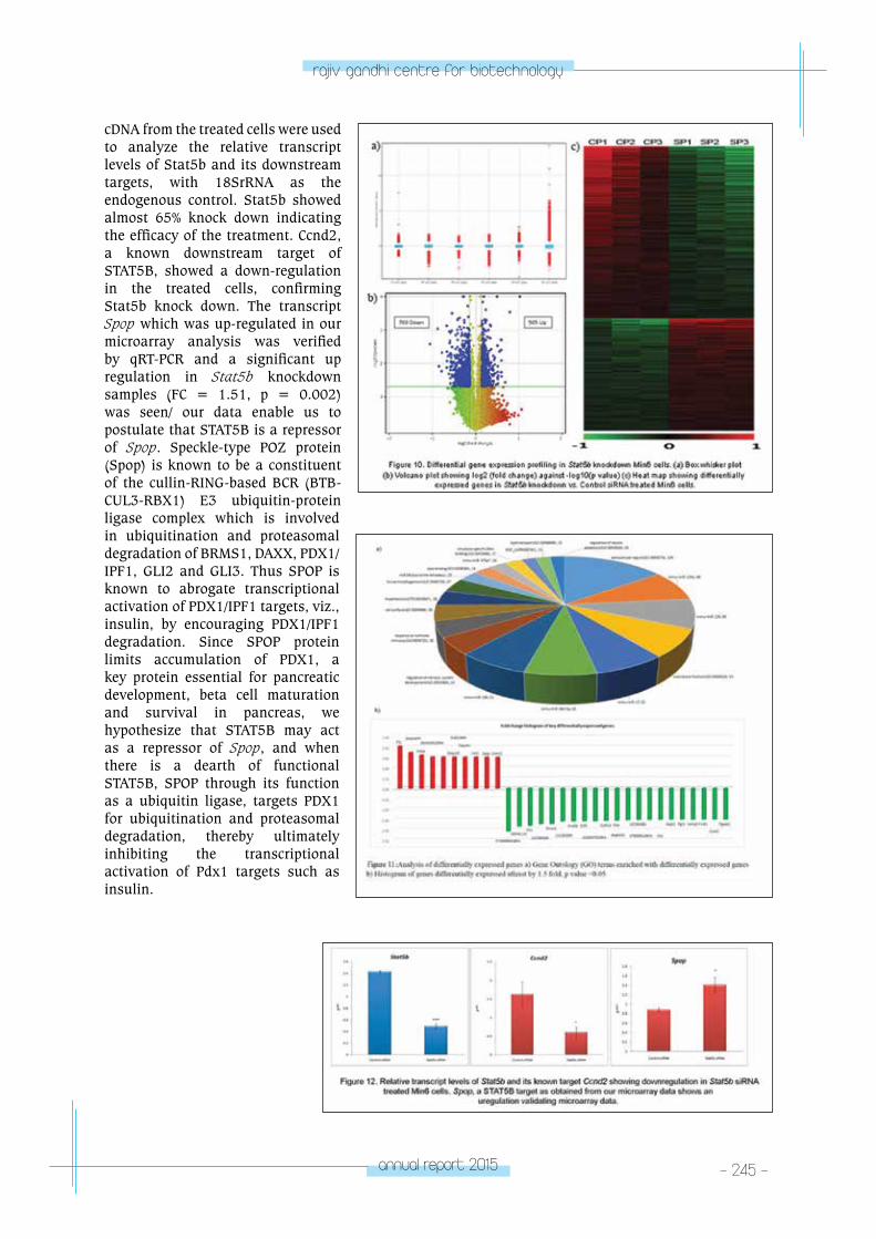
annual report 2015 - 245 -
rajiv gandhi centre for biotechnology
cDNA from the treated cells were used to analyze the relative transcript levels of Stat5b and its downstream targets, with 18SrRNA as the endogenous control. Stat5b showed almost 65% knock down indicating the efficacy of the treatment. Ccnd2, a known downstream target of STAT5B, showed a down-regulation in the treated cells, confirming Stat5b knock down. The transcript Spop which was up-regulated in our microarray analysis was verified by qRT-PCR and a significant up regulation in Stat5b knockdown samples (FC = 1.51, p = 0.002) was seen/ our data enable us to postulate that STAT5B is a repressor of Spop. Speckle-type POZ protein (Spop) is known to be a constituent of the cullin-RING-based BCR (BTB-CUL3-RBX1) E3 ubiquitin-protein ligase complex which is involved in ubiquitination and proteasomal degradation of BRMS1, DAXX, PDX1/IPF1, GLI2 and GLI3. Thus SPOP is known to abrogate transcriptional activation of PDX1/IPF1 targets, viz., insulin, by encouraging PDX1/IPF1 degradation. Since SPOP protein limits accumulation of PDX1, a key protein essential for pancreatic development, beta cell maturation and survival in pancreas, we hypothesize that STAT5B may act as a repressor of Spop, and when there is a dearth of functional STAT5B, SPOP through its function as a ubiquitin ligase, targets PDX1 for ubiquitination and proteasomal degradation, thereby ultimately inhibiting the transcriptional activation of Pdx1 targets such as insulin.
- 246 - annual report 2015
rajiv gandhi centre for biotechnology
List of publications• Padmanabhan RA, Laloraya M. Estrogen-
Initiated Protein Interactomes During Embryo Implantation. Am J Reprod Immunol. 2016 Mar;75(3):256-62. doi: 10.1111/aji.12455. Epub 2015 Dec 14. Review.
• Indu S, Sekhar SC, Sengottaiyan J, Kumar A, Pillai SM, Laloraya M, Kumar PG. Aberrant Expression of Dynein light chain 1 (DYNLT1) is Associated with Human Male Factor Infertility. Mol Cell Proteomics. 2015 Dec; 14(12):3185-95. doi: 10.1074/mcp.M115.050005. Epub 2015 Oct
Invited talks• Talk entitled “Unraveling the complex network
of PCOS pathogenesis using a systems biology approach” at INTERNATIONAL CONFERENCE ON MOLECULAR SIGNALLING: RECENT TRENDS IN BIOSCIENCES, November 20-22, 2015, Department of Zoology, North-Eastern Hill University, Shillong-793022, Meghalaya, India
• Talk entitled “Sleep disorders in PCOS could be an outcome of altered circadian rhythm due to aberrant miRNA status” at 26th Annual Meeting of the ISSRF along with International Conference on Reproductive Health with Emphasis on Occupational, Environmental and Lifestyle Factors & February 18-20, 2016 at National Institute of Occupational Health, Ahmedabad.
• Talk entitled “New Frontiers in Female Reproduction Research” 37th Prof. MM Sebastian memorial lecture, 11th February 2016, Department of Zoology, St Berchmans, College Changanasserry
Oral Presentations (2015-2016)• Soumya V. Oral presentation entitled “Uterine
Autoimmune Regulator Dysfunction causes implantation failure” at UGC Sponsored Two Day National Conference on ‘Myths and Facts about Life Style Diseases–A Practical Perspective’, 3rd & 4th August 2015, St. Philomena’s College, MYSURU.
EXTRA-MURAL FUNDING
Sl. No.
Investigator(s) TitleFunding Agency
Duration
1. Dr. Malini Laloraya(PI)
Mechanism of STAT5B in pancreatic beta cell proliferation/sustenance and its significance in diabetes.
Board of Research in Nuclear Sciences
21-01-15 to 20-03-18
2.Dr. Malini Laloraya (PI)
Deciphering the role of ER alpha in modulating the strength of STAT3 function.
Department of Science and Technology
14-10-15 to 13-10-18
tRopICAl DISeASe BIoloGY
- Mycobacteria Research laboratory
TROPICAL DISEASE BIOLOGY
Mycobacteria Research Laboratory
R. Ajay Kumar
Ajay Kumar received his PhD in Microbiology
from Madurai Kamaraj University, Madurai, and
did post-doctoral training at Indian Institute of
Science, Bangalore, Sri Chitra Thirunal Institute
for Medical Sciences Technology, Trivandrum,
and University of Massachusetts Medical School,
Worcester, USA
PhD StudentsRoshna Lawrence GomezAneesh ChandranRanjit RamachandaranSajith RaghunandananAkhil RajBalaji M.
Research AssociateVipin Gopinath, PhD
Junior Research FellowShyama SasikumarVinodh Joseph
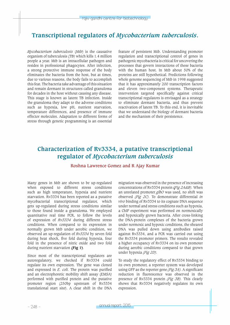
- 248 - annual report 2015
rajiv gandhi centre for biotechnology
Transcriptional regulators of Mycobacterium tuberculosis.
Mycobacterium tuberculosis (Mtb) is the causative organism of tuberculosis (TB) which kills 1.4 million people a year. Mtb is an intracellular pathogen and resides in professional phagocytes. After infection, a strong protective immune response of the body eliminates the bacteria from the host, but at times, due to various reasons, the body fails to accomplish this feat. The bacteria take advantage of this situation and remain dormant in structures called granuloma for decades in the host without causing any disease. This stage is known as latent TB infection. Inside the granuloma they adapt to the adverse conditions such as hypoxia, low pH, nutrient starvation, temperature differences, and presence of immune effector molecules. Adaptation to different forms of stress through genetic programming is an essential
feature of persistent Mtb. Understanding promoter regulation and transcriptional control of genes in pathogenic mycobacteria is critical for uncovering the processes that govern interactions of these bacteria with the human host. In Mtb about 50% of the proteins are still hypothetical. Predictions following whole genome sequencing of Mtb in 1998 suggested that it has approximately 200 transcription factors and eleven two-component systems. Therapeutic intervention targeted specifically against critical transcriptional regulators is envisaged as a strategy to eliminate dormant bacteria, and thus prevent reactivation of latent TB. To this end, it is inevitable that we understand the biology of dormant bacteria and the mechanism of their persistence.
Characterization of Rv3334, a putative transcriptional regulator of Mycobacterium tuberculosis
Roshna Lawrence Gomez and R Ajay Kumar
Many genes in Mtb are shown to be up-regulated when exposed to different stress conditions such as high temperature, hypoxia and nutrient starvation. Rv3334 has been reported as a putative mycobacterial transcriptional regulator, which gets up-regulated during stress conditions similar to those found inside a granuloma. We employed quantitative real time PCR, to follow the levels of expression of Rv3334 during different stress conditions. When compared to its expression in normally grown Mtb under aerobic condition, we observed an up-regulation of Rv3334 by seven fold during heat shock, five fold during hypoxia, four fold in the presence of nitric oxide and two fold during nutrient starvation (Fig 1).
Since most of the transcriptional regulators are autoregulatory, we checked if Rv3334 could regulate its own expression. The gene was cloned and expressed in E. coli. The protein was purified and an electrophoretic mobility shift assay (EMSA) performed with purified protein and the putative promoter region (250bp upstream of Rv3334 translational start site). A clear shift in the DNA
migration was observed in the presence of increasing concentrations of Rv3334 protein (Fig 2A&B). When an unrelated promoter glbO was used, no shift was observed (Fig 2C). To demonstrate differential in vivo binding of Rv3334 to its cognate DNA sequence under normal and stress conditions such as hypoxia, a ChIP experiment was performed on normoxically and hypoxically grown bacteria. After cross-linking the DNA-protein complexes of the bacteria grown under normoxic and hypoxic conditions, the sheared DNA was pulled down using antibodies raised against Rv3334, and a PCR was carried out using the Rv3334 promoter primers. The results revealed a higher occupancy of Rv3334 on its own promoter during aerobic conditions compared to that grown under hypoxia (Fig 2D).
To study the regulatory effect of Rv3334 binding to its own promoter, a reporter system was developed using GFP as the reporter gene (Fig 3A). A significant reduction in fluorescence was observed in the presence of Rv3334 protein (Fig 3B). This clearly shows that Rv3334 negatively regulates its own expression.
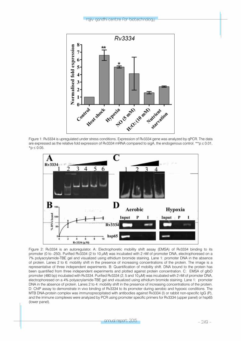
annual report 2015 - 249 -
rajiv gandhi centre for biotechnology
Figure 1: Rv3334 is upregulated under stress conditions. Expression of Rv3334 gene was analyzed by qPCR. The data are expressed as the relative fold expression of Rv3334 mRNA compared to sigA, the endogenous control. **p ≤ 0.01, *p ≤ 0.05.
Figure 2: Rv3334 is an autoregulator. A: Electrophoretic mobility shift assay (EMSA) of Rv3334 binding to its promoter (0 to -250). Purified Rv3334 (2 to 10 μM) was incubated with 2 nM of promoter DNA, electrophoresed on a 7% polyacrylamide-TBE gel and visualized using ethidium bromide staining. Lane 1: promoter DNA in the absence of protein. Lanes 2 to 6: mobility shift in the presence of increasing concentrations of the protein. The image is a representative of three independent experiments. B: Quantification of mobility shift. DNA bound to the protein has been quantified from three independent experiments and plotted against protein concentration. C: EMSA of glbO promoter (480 bp) incubated with Rv3334. Purified Rv3334 (2, 5 and 10 μM) was incubated with 2 nM of promoter DNA, electrophoresed on a 4% polyacrylamide-TBE gel and visualized using ethidium bromide staining. Lane 1: promoter DNA in the absence of protein. Lanes 2 to 4: mobility shift in the presence of increasing concentrations of the protein. D: ChIP assay to demonstrate in vivo binding of Rv3334 to its promoter during aerobic and hypoxic conditions. The MTB DNA-protein complex was immunoprecipitated with antibodies against Rv3334 (I) or rabbit non-specific IgG (P), and the immune complexes were analyzed by PCR using promoter specific primers for Rv3334 (upper panel) or hsp65 (lower panel).
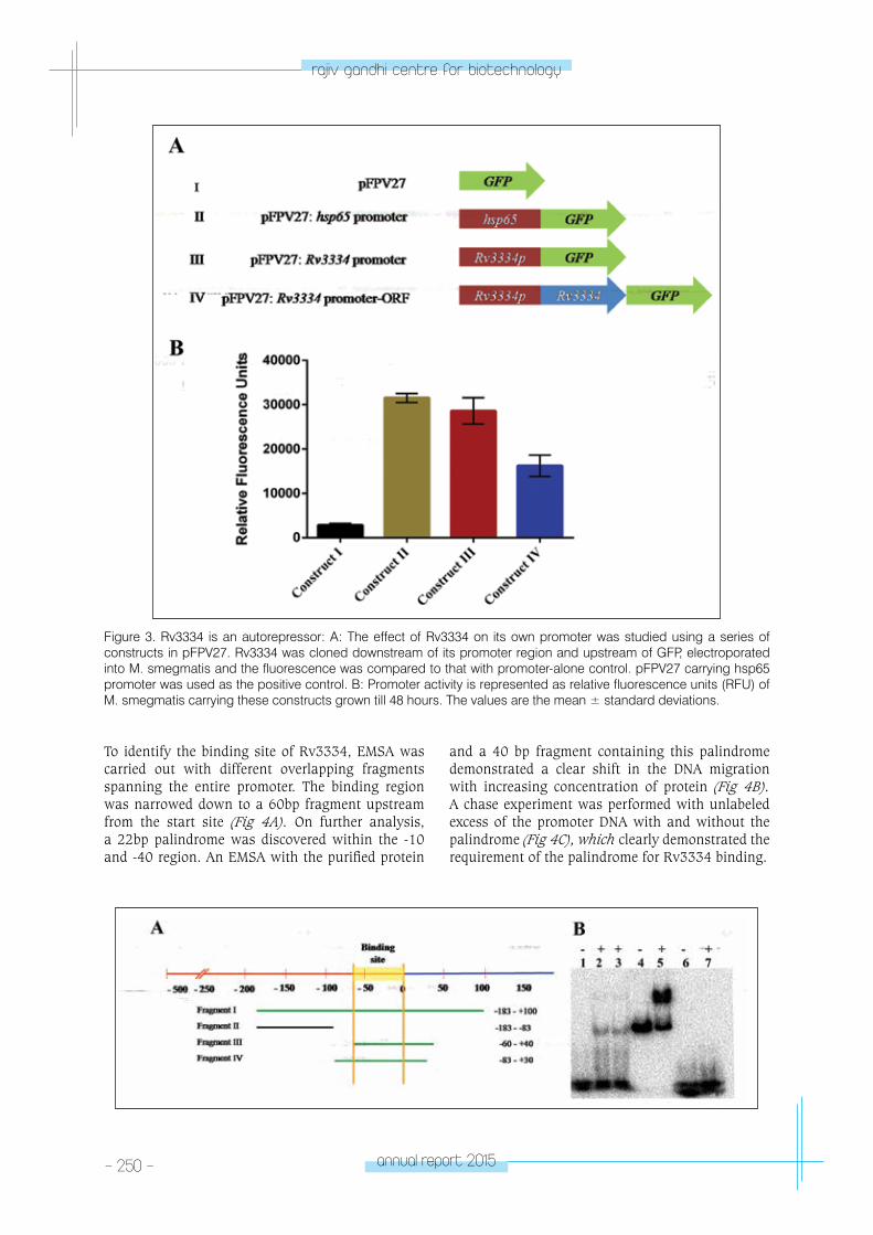
- 250 - annual report 2015
rajiv gandhi centre for biotechnology
Figure 3. Rv3334 is an autorepressor: A: The effect of Rv3334 on its own promoter was studied using a series of constructs in pFPV27. Rv3334 was cloned downstream of its promoter region and upstream of GFP, electroporated into M. smegmatis and the fluorescence was compared to that with promoter-alone control. pFPV27 carrying hsp65 promoter was used as the positive control. B: Promoter activity is represented as relative fluorescence units (RFU) of M. smegmatis carrying these constructs grown till 48 hours. The values are the mean ± standard deviations.
To identify the binding site of Rv3334, EMSA was carried out with different overlapping fragments spanning the entire promoter. The binding region was narrowed down to a 60bp fragment upstream from the start site (Fig 4A). On further analysis, a 22bp palindrome was discovered within the -10 and -40 region. An EMSA with the purified protein
and a 40 bp fragment containing this palindrome demonstrated a clear shift in the DNA migration with increasing concentration of protein (Fig 4B). A chase experiment was performed with unlabeled excess of the promoter DNA with and without the palindrome (Fig 4C), which clearly demonstrated the requirement of the palindrome for Rv3334 binding.
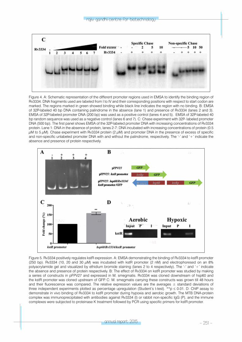
annual report 2015 - 251 -
rajiv gandhi centre for biotechnology
Figure 4. A: Schematic representation of the different promoter regions used in EMSA to identify the binding region of Rv3334. DNA fragments used are labeled from I to IV and their corresponding positions with respect to start codon are marked. The regions marked in green showed binding while black line indicates the region with no binding. B: EMSA of 32P-labeled 40 bp DNA containing palindrome in the absence (lane 1) and presence of Rv3334 (lanes 2 and 3). EMSA of 32P-labeled promoter DNA (200 bp) was used as a positive control (lanes 4 and 5). EMSA of 32P-labeled 40 bp random sequence was used as a negative control (lanes 6 and 7). C: Chase experiment with 32P- labeled promoter DNA (500 bp). The first panel shows EMSA of the 32P-labeled promoter DNA with increasing concentrations of Rv3334 protein. Lane 1: DNA in the absence of protein, lanes 2-7: DNA incubated with increasing concentrations of protein (0.5 μM to 5 μM). Chase experiment with Rv3334 protein (2 μM) and promoter DNA in the presence of excess of specific and non-specific unlabeled promoter DNA with and without the palindrome, respectively. The ‘-’ and ‘+’ indicate the absence and presence of protein respectively.
Figure 5. Rv3334 positively regulates kstR expression. A: EMSA demonstrating the binding of Rv3334 to kstR promoter (250 bp). Rv3334 (10, 20 and 30 μM) was incubated with kstR promoter (2 nM) and electrophoresed on an 8% polyacrylamide gel and visualized by ethidium bromide staining (lanes 2 to 4 respectively). The ‘-’ and ‘+’ indicate the absence and presence of protein respectively. B: The effect of Rv3334 on kstR promoter was studied by making a series of constructs in pFPV27 and expressed in M. smegmatis. Rv3334 was cloned downstream of hsp60 and the kstR promoter was cloned upstream of GFP. C: M. smegmatis carrying these constructs was grown till 48 hours and their fluorescence was compared. The relative expression values are the averages ± standard deviations of three independent experiments plotted as percentage upregulation (Student’s t-test). **p ≤ 0.01. D: ChIP assay to demonstrate in vivo binding of Rv3334 to kstR promoter during hypoxia and aerobic growth. The MTB DNA-protein complex was immunoprecipitated with antibodies against Rv3334 (I) or rabbit non-specific IgG (P), and the immune complexes were subjected to proteinase K treatment followed by PCR using specific primers for kstR promoter.
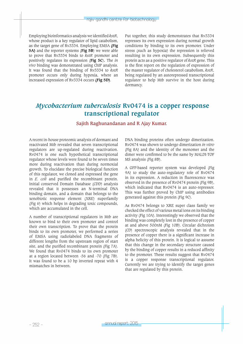
- 252 - annual report 2015
rajiv gandhi centre for biotechnology
Employing bioinformatics analysis we identified kstR, whose product is a key regulator of lipid catabolism, as the target gene of Rv3334. Employing EMSA (Fig 5A) and the reporter systems (Fig 5B) we were able to prove that Rv3334 binds to kstR promoter and positively regulates its expression (Fig 5C). The in vivo binding was demonstrated using ChIP analysis. It was found that the binding of Rv3334 to kstR promoter occurs only during hypoxia, where an increased expression of Rv3334 occurs (Fig 5D).
Put together, this study demonstrates that Rv3334 represses its own expression during normal growth conditions by binding to its own promoter. Under stress (such as hypoxia) the repression is relieved resulting in its own expression. Subsequently this protein acts as a positive regulator of kstR gene. This is the first report on the regulation of expression of the master regulator of cholesterol catabolism, kstR, being regulated by an autorepressed transcriptional regulator to help Mtb survive in the host during dormancy.
Mycobacterium tuberculosis Rv0474 is a copper response transcriptional regulator
Sajith Raghunandanan and R Ajay Kumar.
A recent in-house proteomic analysis of dormant and reactivated Mtb revealed that seven transcriptional regulators are up-regulated during reactivation. Rv0474 is one such hypothetical transcriptional regulator whose levels were found to be seven times more during reactivation than during normoxial growth. To elucidate the precise biological function of this regulator, we cloned and expressed the gene in E. coli and purified the recombinant protein. Initial conserved Domain Database (CDD) analysis revealed that it possesses an N-terminal DNA binding domain, and a domain that belongs to the xenobiotic response element (XRE) superfamily (Fig 6) which helps in degrading toxic compounds, which are accumulated in the cell.
A number of transcriptional regulators in Mtb are known to bind to their own promoter and control their own transcription. To prove that the protein binds to its own promoter, we performed a series of EMSA using radiolabeled DNA fragments of different lengths from the upstream region of start site, and the purified recombinant protein (Fig 7A). We found that Rv0474 binds to its own promoter at a region located between -56 and -70 (Fig 7B). It was found to be a 10 bp inverted repeat with 4 mismatches in between.
DNA binding proteins often undergo dimerization. Rv0474 was shown to undergo dimerization in vitro (Fig 8A) and the identity of the monomer and the dimer were confirmed to be the same by MALDI-TOF MS analysis (Fig 8B).
A GFP-based reporter system was developed (Fig 9A) to study the auto-regulatory role of Rv0474 in its expression. A reduction in fluorescence was observed in the presence of Rv0474 protein (Fig 9B), which indicated that Rv0474 is an auto-repressor. This was further proved by ChIP using antibodies generated against this protein (Fig 9C).
As Rv0474 belongs to XRE super class family we checked the effect of various metal ions on its binding activity (Fig 10A). Interestingly we observed that the binding was completely lost in the presence of copper at and above 500nM (Fig 10B). Circular dichroism (CD) spectroscopic analysis revealed that in the presence of copper there is a significant increase in alpha helicity of this protein. It is logical to assume that this change in the secondary structure caused by the binding of copper results in a reduced affinity to the promoter. These results suggest that Rv0474 is a copper response transcriptional regulator. Currently we are trying to identify the target genes that are regulated by this protein.
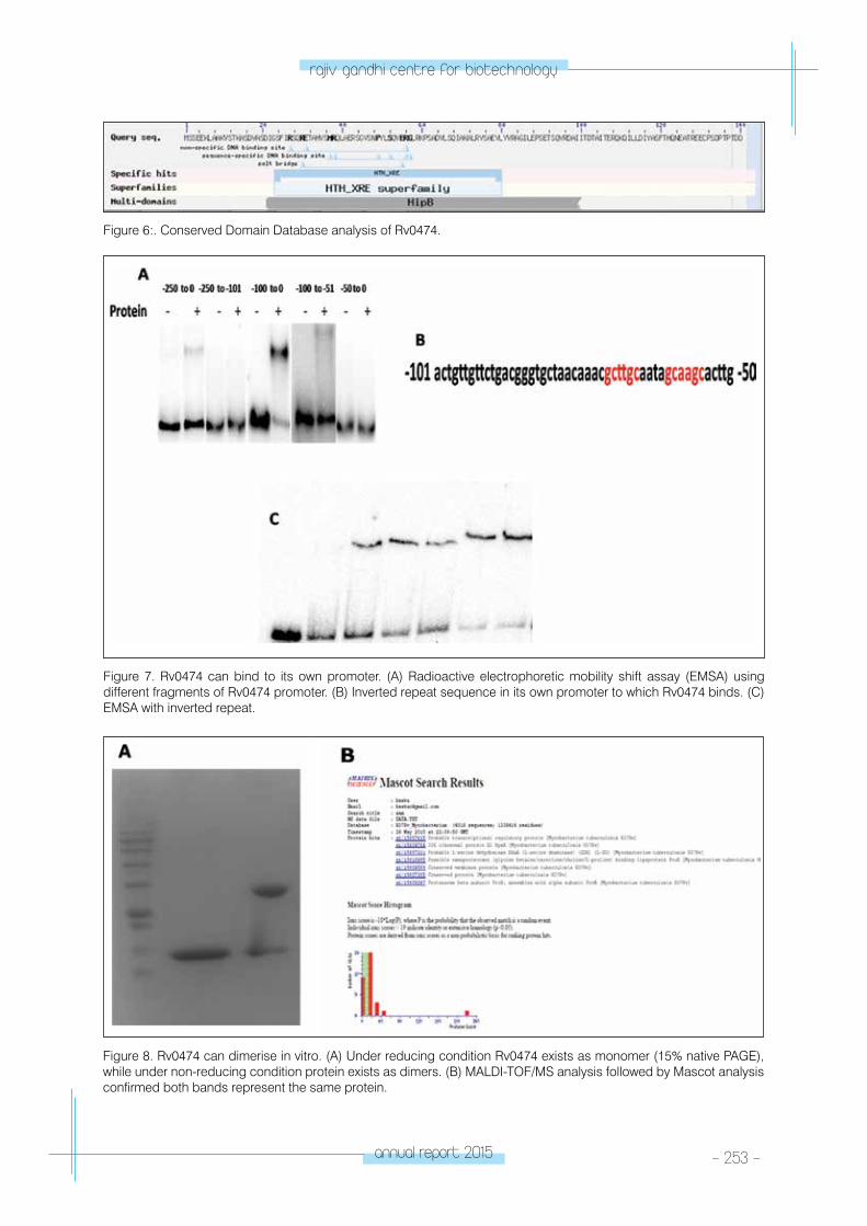
annual report 2015 - 253 -
rajiv gandhi centre for biotechnology
Figure 6:. Conserved Domain Database analysis of Rv0474.
Figure 7. Rv0474 can bind to its own promoter. (A) Radioactive electrophoretic mobility shift assay (EMSA) using different fragments of Rv0474 promoter. (B) Inverted repeat sequence in its own promoter to which Rv0474 binds. (C) EMSA with inverted repeat.
Figure 8. Rv0474 can dimerise in vitro. (A) Under reducing condition Rv0474 exists as monomer (15% native PAGE), while under non-reducing condition protein exists as dimers. (B) MALDI-TOF/MS analysis followed by Mascot analysis confirmed both bands represent the same protein.
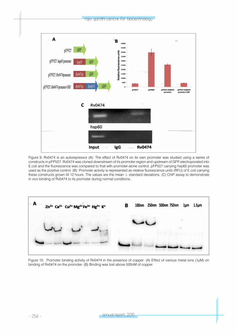
- 254 - annual report 2015
rajiv gandhi centre for biotechnology
Figure 9. Rv0474 is an autorepressor (A): The effect of Rv0474 on its own promoter was studied using a series of constructs in pFPV27. Rv0474 was cloned downstream of its promoter region and upstream of GFP, electroporated into E.coli and the fluorescence was compared to that with promoter-alone control. pFPV27 carrying hsp65 promoter was used as the positive control. (B): Promoter activity is represented as relative fluorescence units (RFU) of E.coli carrying these constructs grown till 12 hours. The values are the mean ± standard deviations. (C) ChIP assay to demonstrate in vivo binding of Rv0474 to its promoter during normal conditions.
Figure 10. Promoter binding activity of Rv0474 in the presence of copper: (A) Effect of various metal ions (1μM) on binding of Rv0474 on the promoter. (B) Binding was lost above 500nM of copper.
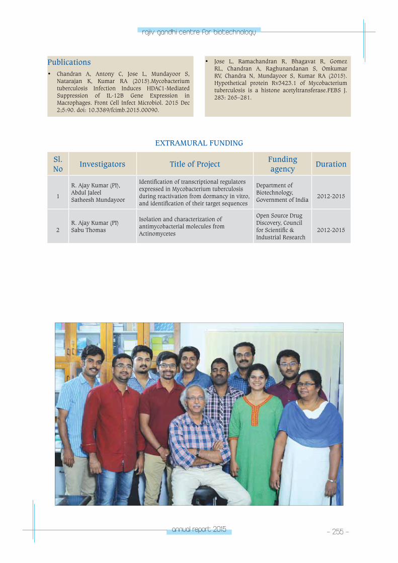
annual report 2015 - 255 -
rajiv gandhi centre for biotechnology
Publications• Chandran A, Antony C, Jose L, Mundayoor S,
Natarajan K, Kumar RA (2015).Mycobacterium tuberculosis Infection Induces HDAC1-Mediated Suppression of IL-12B Gene Expression in Macrophages. Front Cell Infect Microbiol. 2015 Dec 2;5:90. doi: 10.3389/fcimb.2015.00090.
• Jose L, Ramachandran R, Bhagavat R, Gomez RL, Chandran A, Raghunandanan S, Omkumar RV, Chandra N, Mundayoor S, Kumar RA (2015). Hypothetical protein Rv3423.1 of Mycobacterium tuberculosis is a histone acetyltransferase.FEBS J. 283: 265–281.
EXTRAMURAL FUNDING
Sl. No
Investigators Title of ProjectFunding agency
Duration
1
R. Ajay Kumar (PI),Abdul Jaleel Satheesh Mundayoor
Identification of transcriptional regulators expressed in Mycobacterium tuberculosis during reactivation from dormancy in vitro, and identification of their target sequences
Department of Biotechnology, Government of India
2012-2015
2R. Ajay Kumar (PI)Sabu Thomas
Isolation and characterization of antimycobacterial molecules from Actinomycetes
Open Source Drug Discovery, Council for Scientific & Industrial Research
2012-2015
tRopICAl DISeASe BIoloGY
- Cholera and Biofilm Research Laboratory
TROPICAL DISEASE BIOLOGY
Cholera Research Laboratory
Sabu Thomas
Sabu Thomas received his PhD in Aquatic
Biology from the University of Kerala and joined
RGCB in 2001. He was a member in the Second
Indian Arctic Expedition team organized by the
Ministry of Environment, Government of India.
Sabu Thomas is also a member in the Global
Task Force on Cholera Control (GTFCC) of the
World Health Organization.
PhD studentsWilson Peter AbrahamDivya M.P.Akhilandeswarre D.Karthika S.Lekshmi N.Devika Das J.
Research AssistantDeepa Mathew P.
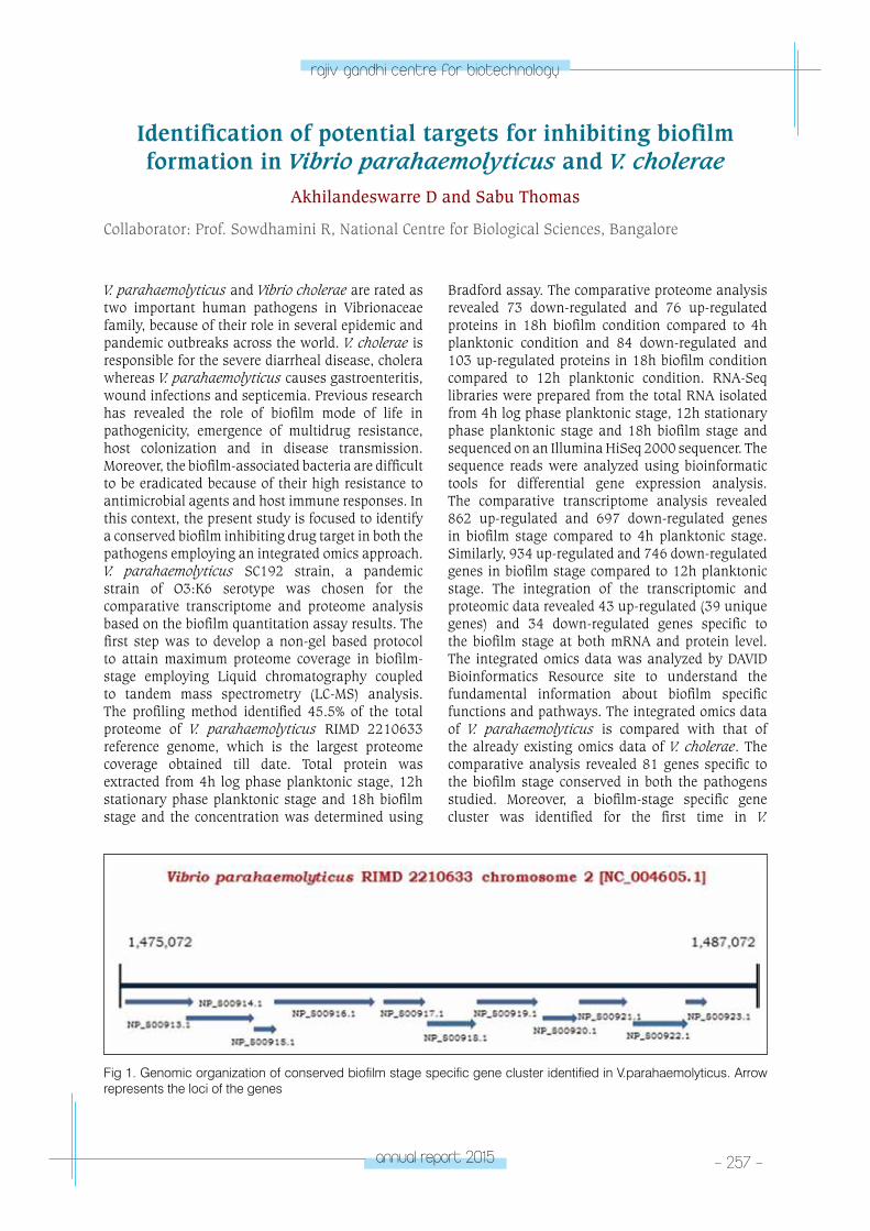
annual report 2015 - 257 -
rajiv gandhi centre for biotechnology
Identification of potential targets for inhibiting biofilm formation in Vibrio parahaemolyticus and V. cholerae
Akhilandeswarre D and Sabu Thomas
Collaborator: Prof. Sowdhamini R, National Centre for Biological Sciences, Bangalore
V. parahaemolyticus and Vibrio cholerae are rated as two important human pathogens in Vibrionaceae family, because of their role in several epidemic and pandemic outbreaks across the world. V. cholerae is responsible for the severe diarrheal disease, cholera whereas V. parahaemolyticus causes gastroenteritis, wound infections and septicemia. Previous research has revealed the role of biofilm mode of life in pathogenicity, emergence of multidrug resistance, host colonization and in disease transmission. Moreover, the biofilm-associated bacteria are difficult to be eradicated because of their high resistance to antimicrobial agents and host immune responses. In this context, the present study is focused to identify a conserved biofilm inhibiting drug target in both the pathogens employing an integrated omics approach. V. parahaemolyticus SC192 strain, a pandemic strain of O3:K6 serotype was chosen for the comparative transcriptome and proteome analysis based on the biofilm quantitation assay results. The first step was to develop a non-gel based protocol to attain maximum proteome coverage in biofilm-stage employing Liquid chromatography coupled to tandem mass spectrometry (LC-MS) analysis. The profiling method identified 45.5% of the total proteome of V. parahaemolyticus RIMD 2210633 reference genome, which is the largest proteome coverage obtained till date. Total protein was extracted from 4h log phase planktonic stage, 12h stationary phase planktonic stage and 18h biofilm stage and the concentration was determined using
Bradford assay. The comparative proteome analysis revealed 73 down-regulated and 76 up-regulated proteins in 18h biofilm condition compared to 4h planktonic condition and 84 down-regulated and 103 up-regulated proteins in 18h biofilm condition compared to 12h planktonic condition. RNA-Seq libraries were prepared from the total RNA isolated from 4h log phase planktonic stage, 12h stationary phase planktonic stage and 18h biofilm stage and sequenced on an Illumina HiSeq 2000 sequencer. The sequence reads were analyzed using bioinformatic tools for differential gene expression analysis. The comparative transcriptome analysis revealed 862 up-regulated and 697 down-regulated genes in biofilm stage compared to 4h planktonic stage. Similarly, 934 up-regulated and 746 down-regulated genes in biofilm stage compared to 12h planktonic stage. The integration of the transcriptomic and proteomic data revealed 43 up-regulated (39 unique genes) and 34 down-regulated genes specific to the biofilm stage at both mRNA and protein level. The integrated omics data was analyzed by DAVID Bioinformatics Resource site to understand the fundamental information about biofilm specific functions and pathways. The integrated omics data of V. parahaemolyticus is compared with that of the already existing omics data of V. cholerae. The comparative analysis revealed 81 genes specific to the biofilm stage conserved in both the pathogens studied. Moreover, a biofilm-stage specific gene cluster was identified for the first time in V.
Fig 1. Genomic organization of conserved biofilm stage specific gene cluster identified in V.parahaemolyticus. Arrow represents the loci of the genes
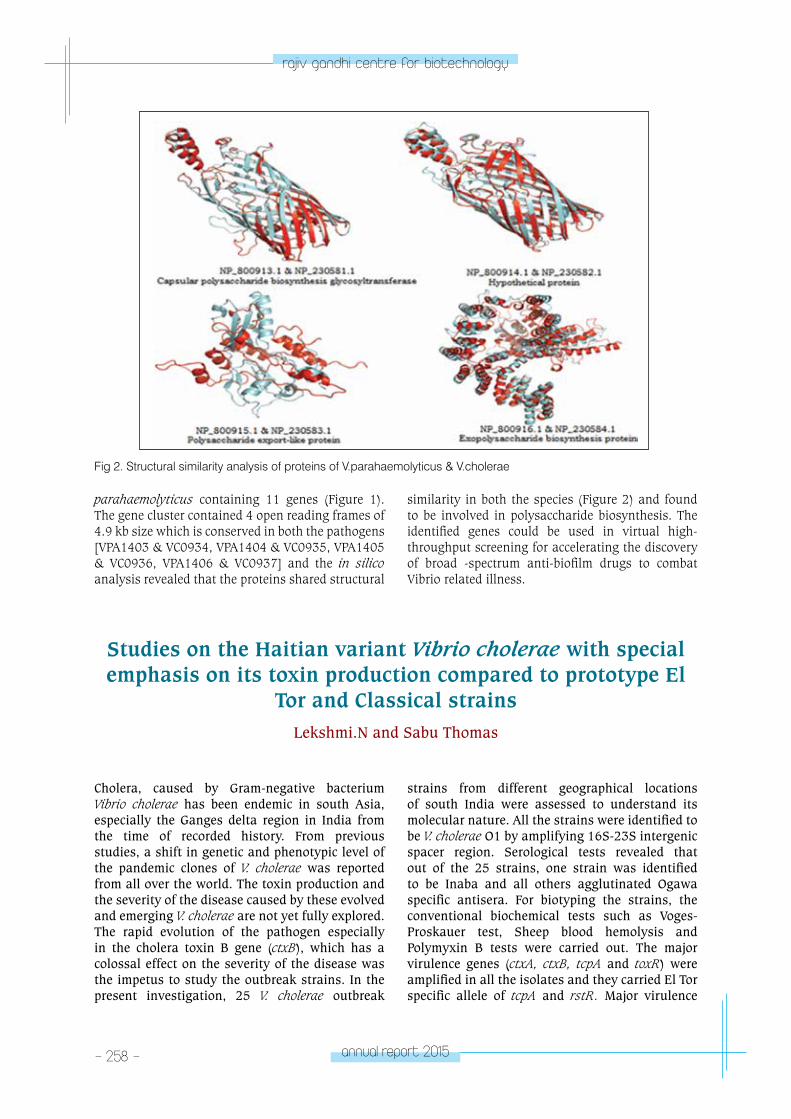
- 258 - annual report 2015
rajiv gandhi centre for biotechnology
parahaemolyticus containing 11 genes (Figure 1). The gene cluster contained 4 open reading frames of 4.9 kb size which is conserved in both the pathogens [VPA1403 & VC0934, VPA1404 & VC0935, VPA1405 & VC0936, VPA1406 & VC0937] and the in silico analysis revealed that the proteins shared structural
similarity in both the species (Figure 2) and found to be involved in polysaccharide biosynthesis. The identified genes could be used in virtual high-throughput screening for accelerating the discovery of broad -spectrum anti-biofilm drugs to combat Vibrio related illness.
Fig 2. Structural similarity analysis of proteins of V.parahaemolyticus & V.cholerae
Studies on the Haitian variant Vibrio cholerae with special emphasis on its toxin production compared to prototype El
Tor and Classical strainsLekshmi.N and Sabu Thomas
Cholera, caused by Gram-negative bacterium Vibrio cholerae has been endemic in south Asia, especially the Ganges delta region in India from the time of recorded history. From previous studies, a shift in genetic and phenotypic level of the pandemic clones of V. cholerae was reported from all over the world. The toxin production and the severity of the disease caused by these evolved and emerging V. cholerae are not yet fully explored. The rapid evolution of the pathogen especially in the cholera toxin B gene (ctxB), which has a colossal effect on the severity of the disease was the impetus to study the outbreak strains. In the present investigation, 25 V. cholerae outbreak
strains from different geographical locations of south India were assessed to understand its molecular nature. All the strains were identified to be V. cholerae O1 by amplifying 16S-23S intergenic spacer region. Serological tests revealed that out of the 25 strains, one strain was identified to be Inaba and all others agglutinated Ogawa specific antisera. For biotyping the strains, the conventional biochemical tests such as Voges-Proskauer test, Sheep blood hemolysis and Polymyxin B tests were carried out. The major virulence genes (ctxA, ctxB, tcpA and toxR) were amplified in all the isolates and they carried El Tor specific allele of tcpA and rstR. Major virulence
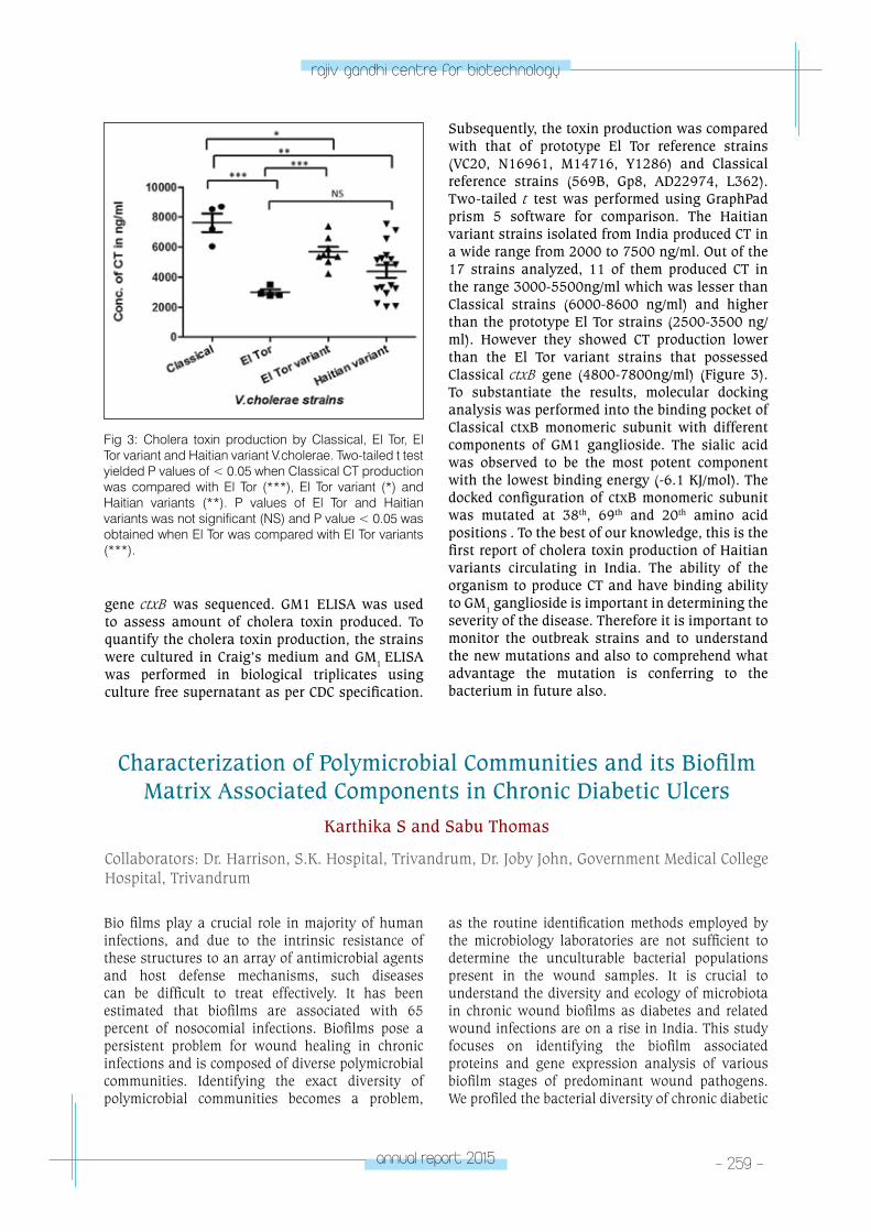
annual report 2015 - 259 -
rajiv gandhi centre for biotechnology
Fig 3: Cholera toxin production by Classical, El Tor, El Tor variant and Haitian variant V.cholerae. Two-tailed t test yielded P values of < 0.05 when Classical CT production was compared with El Tor (***), El Tor variant (*) and Haitian variants (**). P values of El Tor and Haitian variants was not significant (NS) and P value < 0.05 was obtained when El Tor was compared with El Tor variants (***).
Characterization of Polymicrobial Communities and its Biofilm Matrix Associated Components in Chronic Diabetic Ulcers
Karthika S and Sabu Thomas
Collaborators: Dr. Harrison, S.K. Hospital, Trivandrum, Dr. Joby John, Government Medical College Hospital, Trivandrum
gene ctxB was sequenced. GM1 ELISA was used to assess amount of cholera toxin produced. To quantify the cholera toxin production, the strains were cultured in Craig’s medium and GM1 ELISA was performed in biological triplicates using culture free supernatant as per CDC specification.
Subsequently, the toxin production was compared with that of prototype El Tor reference strains (VC20, N16961, M14716, Y1286) and Classical reference strains (569B, Gp8, AD22974, L362). Two-tailed t test was performed using GraphPad prism 5 software for comparison. The Haitian variant strains isolated from India produced CT in a wide range from 2000 to 7500 ng/ml. Out of the 17 strains analyzed, 11 of them produced CT in the range 3000-5500ng/ml which was lesser than Classical strains (6000-8600 ng/ml) and higher than the prototype El Tor strains (2500-3500 ng/ml). However they showed CT production lower than the El Tor variant strains that possessed Classical ctxB gene (4800-7800ng/ml) (Figure 3). To substantiate the results, molecular docking analysis was performed into the binding pocket of Classical ctxB monomeric subunit with different components of GM1 ganglioside. The sialic acid was observed to be the most potent component with the lowest binding energy (-6.1 KJ/mol). The docked configuration of ctxB monomeric subunit was mutated at 38th, 69th and 20th amino acid positions . To the best of our knowledge, this is the first report of cholera toxin production of Haitian variants circulating in India. The ability of the organism to produce CT and have binding ability to GM1 ganglioside is important in determining the severity of the disease. Therefore it is important to monitor the outbreak strains and to understand the new mutations and also to comprehend what advantage the mutation is conferring to the bacterium in future also.
Bio films play a crucial role in majority of human infections, and due to the intrinsic resistance of these structures to an array of antimicrobial agents and host defense mechanisms, such diseases can be difficult to treat effectively. It has been estimated that biofilms are associated with 65 percent of nosocomial infections. Biofilms pose a persistent problem for wound healing in chronic infections and is composed of diverse polymicrobial communities. Identifying the exact diversity of polymicrobial communities becomes a problem,
as the routine identification methods employed by the microbiology laboratories are not sufficient to determine the unculturable bacterial populations present in the wound samples. It is crucial to understand the diversity and ecology of microbiota in chronic wound biofilms as diabetes and related wound infections are on a rise in India. This study focuses on identifying the biofilm associated proteins and gene expression analysis of various biofilm stages of predominant wound pathogens. We profiled the bacterial diversity of chronic diabetic
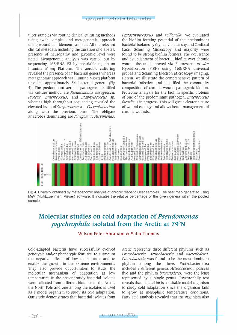
- 260 - annual report 2015
rajiv gandhi centre for biotechnology
ulcer samples via routine clinical culturing methods using swab samples and metagenomic approach using wound debridement samples. All the relevant clinical metadata including the duration of diabetes, presence of neuropathy and glycemic level were noted. Metagenomic analysis was carried out by sequencing 16SrRNA V3 hypervariable region on Illumina Miseq Platform. The aerobic culturing revealed the presence of 17 bacterial genera whereas metagenomic approach via Illumina MiSeq platform unveiled approximately 54 bacterial genera (Fig 4). The predominant aerobic pathogens identified via culture method are Pseudomonas aeruginosa, Proteus, Enterococcus, and Staphylococcus sp. whereas high throughput sequencing revealed the elevated levels of Streptococcus and Corynebacterium along with the previous ones. The obligate anaerobes dominating are Finegoldia, Parvimonas,
Peptostreptococcus and Veillonella. We evaluated the biofilm forming potential of the predominant bacterial isolates by Crystal violet assay and Confocal Laser Scanning Microscopy and majority were found to be strong biofilm formers. The occurrence and establishment of bacterial biofilm over chronic wound tissues is proved via Fluorescent in situ Hybridization (FISH) using 16SrRNA universal probes and Scanning Electron Microscopy imaging. Herein, we illustrate the comprehensive pattern of bacterial infection and identified the community composition of chronic wound pathogenic biofilm. Proteome analysis for the biofilm specific proteins of one of the predominant pathogen, Enterococcus faecalis is in progress. This will give a clearer picture of wound ecology and allows better management of chronic wounds.
Fig 4. Diversity obtained by metagenomic analysis of chronic diabetic ulcer samples. The heat map generated using MeV (MultiExperiment Viewer) software. It indicates the relative percentage of the given genera within the pooled sample
Molecular studies on cold adaptation of Pseudomonas psychrophila isolated from the Arctic at 79°N
Wilson Peter Abraham & Sabu Thomas
Cold-adapted bacteria have successfully evolved genotypic and/or phenotypic features, to surmount the negative effects of low temperature and to enable the growth in the extreme environments. They also provide opportunities to study the molecular mechanism of adaptation at low temperature. In the present study bacterial isolates were collected from different biotopes of the Arctic, the North Pole and one among the isolates is used as a model organism to study its cold adaptation. Our study demonstrates that bacterial isolates from
Arctic represents three different phylums such as Proteobacteria, Actinobacteria and Bacteriodetes. Proteobacteria was found to be the most dominant phylum among the three. Proteobacteriacea includes 8 different genera, Actinobacteria possess five and the phylum bacteriodetes, were the least represented by a single genus. Psychrophily test reveals that isolate166 is a suitable model organism to study cold adaptation since the organism fails to grow at mesophilic temperature conditions. Fatty acid analysis revealed that the organism also
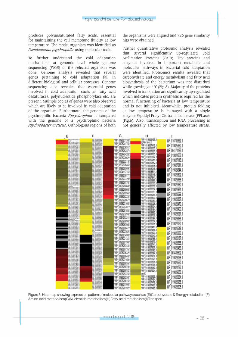
annual report 2015 - 261 -
rajiv gandhi centre for biotechnology
produces polyunsaturated fatty acids, essential for maintaining the cell membrane fluidity at low temperature. The model organism was identified as Pseudomonas psychrophila using molecular tools.
To further understand the cold adaptation mechanisms at genomic level whole genome sequencing (WGS) of the selected organism was done. Genome analysis revealed that several genes pertaining to cold adaptation fall in different biological and cellular processes. Genome sequencing also revealed that essential genes involved in cold adaptation such, as fatty acid desaturases, polynucleotide phosphorylase etc. are present. Multiple copies of genes were also observed which are likely to be involved in cold adaptation of the organism. Furthermore, the genome of the psychrophilic bacteria P.psychrophila is compared with the genome of a psychrophilic bacteria Psychrobacter arcticus. Orthologous regions of both
the organisms were aligned and 726 gene similarity hits were obtained.
Further quantitative proteomic analysis revealed that several significantly up-regulated Cold Acclimation Proteins (CAPs), key proteins and enzymes involved in important metabolic and molecular pathways in bacterial cold adaptation were identified. Proteomics results revealed that carbohydrate and energy metabolism and fatty acid biosynthesis of the bacterium was not disturbed while growing at 4°C (Fig.5). Majority of the proteins involved in translation are significantly up-regulated which indicates protein synthesis is required for the normal functioning of bacteria at low temperature and is not inhibited. Meanwhile, protein folding at low temperature is managed with a single enzyme Peptidyl Prolyl Cis trans Isomerase (PPLase) (Fig.6). Also, transcription and RNA processing is not generally affected by low temperature stress.
Figure 5. Heatmap showing expression pattern of molecular pathways such as (E)Carbohydrate & Energy metabolism(F)Amino acid metabolism(G)Nucleotide metabolism(H)Fatty acid metabolism(I)Transport
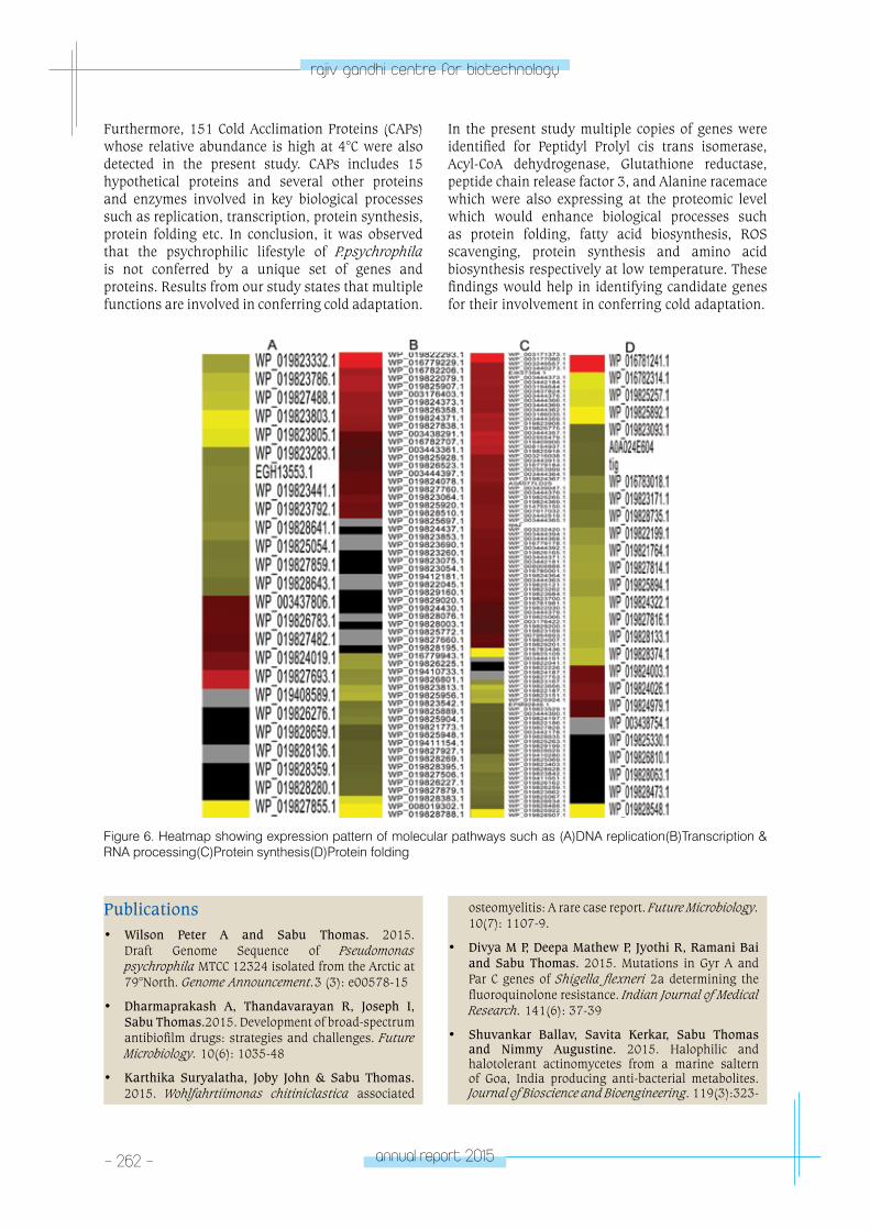
- 262 - annual report 2015
rajiv gandhi centre for biotechnology
Furthermore, 151 Cold Acclimation Proteins (CAPs) whose relative abundance is high at 4°C were also detected in the present study. CAPs includes 15 hypothetical proteins and several other proteins and enzymes involved in key biological processes such as replication, transcription, protein synthesis, protein folding etc. In conclusion, it was observed that the psychrophilic lifestyle of P.psychrophila is not conferred by a unique set of genes and proteins. Results from our study states that multiple functions are involved in conferring cold adaptation.
In the present study multiple copies of genes were identified for Peptidyl Prolyl cis trans isomerase, Acyl-CoA dehydrogenase, Glutathione reductase, peptide chain release factor 3, and Alanine racemace which were also expressing at the proteomic level which would enhance biological processes such as protein folding, fatty acid biosynthesis, ROS scavenging, protein synthesis and amino acid biosynthesis respectively at low temperature. These findings would help in identifying candidate genes for their involvement in conferring cold adaptation.
Figure 6. Heatmap showing expression pattern of molecular pathways such as (A)DNA replication(B)Transcription & RNA processing(C)Protein synthesis(D)Protein folding
Publications• Wilson Peter A and Sabu Thomas. 2015.
Draft Genome Sequence of Pseudomonas psychrophila MTCC 12324 isolated from the Arctic at 79°North. Genome Announcement.3 (3): e00578-15
• Dharmaprakash A, Thandavarayan R, Joseph I, Sabu Thomas.2015. Development of broad-spectrum antibiofilm drugs: strategies and challenges. Future Microbiology. 10(6): 1035-48
• Karthika Suryalatha, Joby John & Sabu Thomas. 2015. Wohlfahrtiimonas chitiniclastica associated
osteomyelitis: A rare case report. Future Microbiology. 10(7): 1107-9.
• Divya M P, Deepa Mathew P, Jyothi R, Ramani Bai and Sabu Thomas. 2015. Mutations in Gyr A and Par C genes of Shigella flexneri 2a determining the fluoroquinolone resistance. Indian Journal of Medical Research. 141(6): 37-39
• Shuvankar Ballav, Savita Kerkar, Sabu Thomas and Nimmy Augustine. 2015. Halophilic and halotolerant actinomycetes from a marine saltern of Goa, India producing anti-bacterial metabolites. Journal of Bioscience and Bioengineering. 119(3):323-

annual report 2015 - 263 -
rajiv gandhi centre for biotechnology
30.
Conference Presentations• Karthika S, Joby John, Sabu Thomas.
Characterization of bacterial population associated with chronic wound infections and its biofilm formation. International Symposium on New Perspectives in Modern Biotechnology, Pondicherry, India, March 2015. (Best Poster Award).
• Lekshmi N and Sabu Thomas, 2015. Molecular Studies on the Haitian variant ctxB gene and cholera toxin production in Vibrio cholerae O1outbreak strains isolated from India. International Conference on Infectious Diseases and Nanomedicine December 15-18, Kathmandu, Nepal.
• Divya M.P and Sabu Thomas, 2015.Serological and molecular analysis of toxigenic Vibrio parahaemolyticus isolated from different environmental sources. EMBO Conference on Aquatic Microbial Ecology, August 23-25, Uppsala, Sweden.
Invited Lectures• Sabu Thomas, 2015. Review of the cholera research
program in RGCB emphasizing the importance of Environmental surveillance of Vibrio cholerae and control of cholera. Secretariat of the Global Task
Force on Cholera Control (GTFCC) Working Group on Laboratory Methods, organized by Dept. of Pandemic and Epidemic Diseases, World Health Organization. November 26-27, THSTI, Faridabad.
• Sabu Thomas, 2015. Science and Inventions: Foundation for Smart Living: Walk With A Scholar Programme, February 21, S.G. College, Kottaracara, Organised by Directorate of Collegiate Education, Govt. of Kerala.
• Sabu Thomas, 2015. Identification of broad-spectrum antibiofilm drug targets: An integrated omic approach. September11, Sree Buddha College of Engineering, Pandalam.
• Bacterial Whole Genome & Metagenome Sequence Submissions
• Wilson P A & Sabu Thomas. Pseudomonas psychrophila strain RGCB 166, whole genome shotgun sequencing project. Bioproject:PRJNA278112, Biosample: SAMN03406680, Acc. No. LBHT00000000
• Divya M.P and Sabu Thomas. Draft Genome Sequence of an Environmental Vibrio parahaemolyticus K23. Bioproject: PRJNA285022, Biosample: SAMN03735013, Acc. No. LQGU00000000
• Karthika S & Sabu Thomas. Metagenome of Diabet-ic Ulcer. SRA Acc. no.: SRX1453631, Bioproject: PRJ-NA304366, Biosample: SAMN04299664
RESEARCH GRANTS
Title Investigators Funding Agency Duration
Isolation and characterization of antimicrobial molecules from Actinomycetes
Dr. R. Ajay Kumar (PI)
Dr. Sabu Thomas (Co-PI)
Council for Scientific and Industrial Research-OSDD
2012-15
Analysis of polymicrobial biofilms in chronic wound infections and development of antibiofilm therapeutic to promote wound healing.
Dr. Sabu Thomas (PI)
Dr. Sanil George (Co-PI)
Department of Biotechnology
2016-19
tRopICAl DISeASe BIoloGY
- parasite Biology laboratoryTROPICAL DISEASE
BIOLOGYLeptospira Biology
Laboratory
Program Scientist
Iype Joseph
annual report 2015 - 265 -
rajiv gandhi centre for biotechnology
Epidemiology of Leptospirosis
Leptospirosis is a zoonosis of significant public health importance in Kerala where a humid tropical climate prevails. It is a disease with epidemic potential. In Kerala, the reported human case-fatality rate ranges from 5.7 to 16.9%. All districts of Kerala are reporting cases throughout the year, with the maximum number of cases reported during the rainy season. Approximately 100 – 230 deaths are reported every year due to this disease. Leptospirosis is a complex bacterial disease with multiple modes of transmission, numerous hosts, multitude of pathogenic serovars (>200), various clinical manifestations, and the need for complex testing to provide laboratory confirmation. It affects both humans and livestock. Most mammalian species (e.g.: Ruminants, Dogs, Cats, Cattle, Rodents, Pigs, Mongoose, etc) are natural hosts to various serovars of leptospires. Humans and animals may become infected through direct contact with contaminated urine or indirectly through exposure to contaminated water or soil. Incidental hosts, like man and livestock, can get clinical illness when infected by pathogenic leptospires. The abundance of the various natural hosts in areas of human habitation and occupation determines the potential for transmission for leptospira. Rainfall plays an important role in promoting transmission in Kerala. Leptospirosis has been identified as a disease requiring priority attention in ten states including Kerala during the 12th Five year plan period by the Working Group on Disease Burden of communicable diseases (Planning Commission of India, currently NITI Aayog). In Trivandrum District, the routine communicable disease monitoring system maintained by the Department of Health Services records all cases reported from various parts of the district. All government and few major private hospitals regularly report leptospirosis cases. In the period, January to August 2015, details of 252 patients were available. Out of these, all recovered patients were requested to participate in a sero-survey. The sero-survey intended to find out the most prevalent sero-groups of Leptospira among them. 33 persons volunteered to join the study. They came from 11 institutions in Trivandrum district. The serum of these 33 persons was used for Microscopic Agglutination Test (MAT) against 5 live cultures of bacteria. The panel included L. Icterohaemorrhagiae - icterohaemorrhagiae – RGA, L. Grippotyphosa - grippotyphosa - Moskva V, L. Canicola - canicola - H. Uterecht IV, L. Javanica - poi
– Poi and L. Tarassovi - tarassovi – Perepelicin. 7 samples (21.2 %) gave positive agglutination reaction to the L. Canicola - canicola - H. Uterecht IV at 1:40 dilution. The results point to the role of the Serogroup Canicola in Trivandrum district in causing human Leptospirosis. The usual carrier for serovar Canicola are the dogs, in whom, it may be controlled with yearly vaccination or selective treatment. Currently, dogs are not considered to be important transmitters in Trivandrum and control measures are directed only to rodents. Further studies in this district are likely to bring more light on future preventive measures. This initial study also points to another aspect, that is, the remaining 26 persons would have been affected by other serogroups not included in the panel. Based on the comments received at the Scientific Advisory Committee (July 2015), study into presence of infection in animal herds was initiated. Animals can become sick due to leptospirosis and sometimes, they can be asymptomatic carriers. The animals may acquire it from urine of other carriers or from the birth canal during delivery. This veterinary study is being done in collaboration with Animal Husbandry Department of Government of Kerala. Serum samples of 20 healthy, captive Elephants were collected. They were subjected to MAT test using the same panel (as above). Three samples (15 %) gave positive agglutination reactions. One sample each reacted to L. Javanica - poi – Poi and L. Canicola - canicola - H. Uterecht IV, while one reacted to two serovars, L. Grippotyphosa - grippotyphosa - Moskva V & L. Canicola - canicola - H. Uterecht IV. All reactions were done at 1:40 dilution. Further studies can help us to know whether these elephants are carriers and thus of risk to mahouts and visitors. The relative role of rodents and elephants in transmission of infection can be studied.
A project on “Serogroup Pattern of Leptospirosis in Government Medical College, Trivandrum, with special reference to serogroup specific clinical profile” has been initiated in collaboration with the Department of Internal Medicine. Sample collection has begun. The systems established at RGCB were transferred to two other institutions. Support was given for establishment of leptospira culture system and MAT to Department of Microbiology, Government Medical College, Thiruvananthapuram and Department of Microbiology, Academy of Medical Sciences (ACME), Pariyaram, Kerala, India.
Publications• Abraham R, Manakkadan A, Mudaliar P, Joseph I,
Sivakumar KC, Nair RR, Sreekumar E. Correlation of phylogenetic clade diversification and in vitro
infectivity differences among Cosmopolitan genotype strains of Chikungunya virus. Infection, genetics and evolution: Journal of Molecular Epidemiology and Evolutionary Genetics In Infectious Diseases 37:174-184, January 2016
tRopICAl DISeASe BIoloGY
- Molecular V
irology laboratoryTROPICAL DISEASE
BIOLOGYMolecular Virology
Laboratory
E. Sreekumar
E. Sreekumar is a post-graduate in Veterinary
Immunology and has a PhD in Biotechnology
from Kerala University. He joined RGCB in
2004. He is a Fulbright-Nehru Professional
and Academic Excellence (FNAPE) Fellow and
worked as a Visiting Faculty (2015-2016) at
the Department of Microbiology & Immunology,
Johns Hopkins Bloomberg School of Public
Health, Baltimore, Maryland, USA.
PhD StudentsRachy AbrahamPrashant MudaliarAnupriya M.G.Sneha SinghSreeja R. Nair
Project FellowNeha Vijay Hulylakar
Technical PersonnelUnnikrishnan V.R.
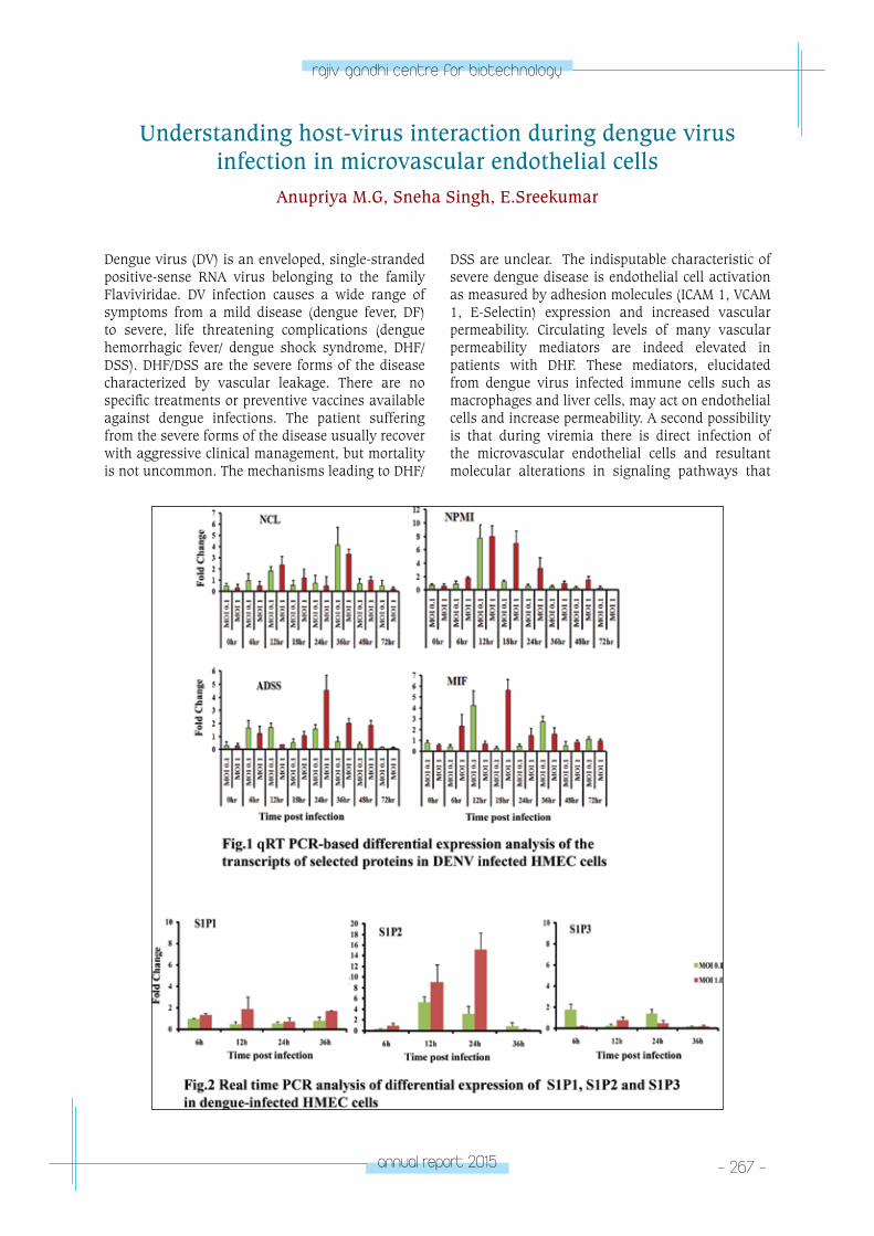
annual report 2015 - 267 -
rajiv gandhi centre for biotechnology
Understanding host-virus interaction during dengue virus infection in microvascular endothelial cells
Anupriya M.G, Sneha Singh, E.Sreekumar
Dengue virus (DV) is an enveloped, single-stranded positive-sense RNA virus belonging to the family Flaviviridae. DV infection causes a wide range of symptoms from a mild disease (dengue fever, DF) to severe, life threatening complications (dengue hemorrhagic fever/ dengue shock syndrome, DHF/ DSS). DHF/DSS are the severe forms of the disease characterized by vascular leakage. There are no specific treatments or preventive vaccines available against dengue infections. The patient suffering from the severe forms of the disease usually recover with aggressive clinical management, but mortality is not uncommon. The mechanisms leading to DHF/
DSS are unclear. The indisputable characteristic of severe dengue disease is endothelial cell activation as measured by adhesion molecules (ICAM 1, VCAM 1, E-Selectin) expression and increased vascular permeability. Circulating levels of many vascular permeability mediators are indeed elevated in patients with DHF. These mediators, elucidated from dengue virus infected immune cells such as macrophages and liver cells, may act on endothelial cells and increase permeability. A second possibility is that during viremia there is direct infection of the microvascular endothelial cells and resultant molecular alterations in signaling pathways that
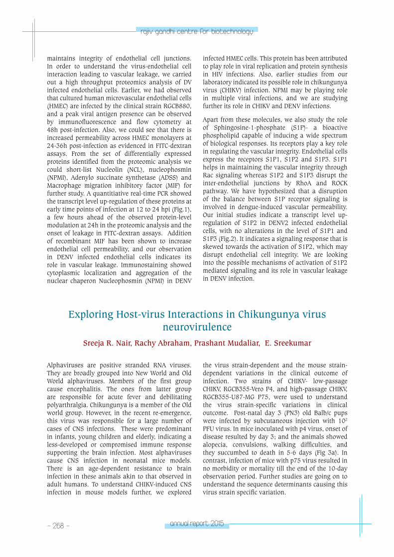
- 268 - annual report 2015
rajiv gandhi centre for biotechnology
maintains integrity of endothelial cell junctions. In order to understand the virus-endothelial cell interaction leading to vascular leakage, we carried out a high throughput proteomics analysis of DV infected endothelial cells. Earlier, we had observed that cultured human microvascular endothelial cells (HMEC) are infected by the clinical strain RGCB880, and a peak viral antigen presence can be observed by immunofluorescence and flow cytometry at 48h post-infection. Also, we could see that there is increased permeability across HMEC monolayers at 24-36h post-infection as evidenced in FITC-dextran assays. From the set of differentially expressed proteins identified from the proteomic analysis we could short-list Nucleolin (NCL), nucleophosmin (NPMI), Adenylo succinate synthetase (ADSS) and Macrophage migration inhibitory factor (MIF) for further study. A quantitiative real-time PCR showed the transcript level up-regulation of these proteins at early time points of infection at 12 to 24 hpi (Fig.1), a few hours ahead of the observed protein-level modulation at 24h in the proteomic analysis and the onset of leakage in FITC-dextran assays. Addition of recombinant MIF has been shown to increase endothelial cell permeability, and our observation in DENV infected endothelial cells indicates its role in vascular leakage. Immunostaining showed cytoplasmic localization and aggregation of the nuclear chaperon Nucleophosmin (NPMI) in DENV
infected HMEC cells. This protein has been attributed to play role in viral replication and protein synthesis in HIV infections. Also, earlier studies from our laboratory indicated its possible role in chikungunya virus (CHIKV) infection. NPMI may be playing role in multiple viral infections, and we are studying further its role in CHIKV and DENV infections.
Apart from these molecules, we also study the role of Sphingosine-1-phosphate (S1P)- a bioactive phospholipid capable of inducing a wide spectrum of biological responses. Its receptors play a key role in regulating the vascular integrity. Endothelial cells express the receptors S1P1, S1P2 and S1P3. S1P1 helps in maintaining the vascular integrity through Rac signaling whereas S1P2 and S1P3 disrupt the inter-endothelial junctions by RhoA and ROCK pathway. We have hypothesized that a disruption of the balance between S1P receptor signaling is involved in dengue-induced vascular permeability. Our initial studies indicate a transcript level up-regulation of S1P2 in DENV2 infected endothelial cells, with no alterations in the level of S1P1 and S1P3 (Fig.2). It indicates a signaling response that is skewed towards the activation of S1P2, which may disrupt endothelial cell integrity. We are looking into the possible mechanisms of activation of S1P2 mediated signaling and its role in vascular leakage in DENV infection.
Exploring Host-virus Interactions in Chikungunya virus neurovirulence
Sreeja R. Nair, Rachy Abraham, Prashant Mudaliar, E. Sreekumar
Alphaviruses are positive stranded RNA viruses. They are broadly grouped into New World and Old World alphaviruses. Members of the first group cause encephalitis. The ones from latter group are responsible for acute fever and debilitating polyarthralgia. Chikungunya is a member of the Old world group. However, in the recent re-emergence, this virus was responsible for a large number of cases of CNS infections. These were predominant in infants, young children and elderly, indicating a less-developed or compromised immune response supporting the brain infection. Most alphaviruses cause CNS infection in neonatal mice models. There is an age-dependent resistance to brain infection in these animals akin to that observed in adult humans. To understand CHIKV-induced CNS infection in mouse models further, we explored
the virus strain-dependent and the mouse strain-dependent variations in the clinical outcome of infection. Two strains of CHIKV- low-passage CHIKV, RGCB355-Vero P4, and high-passage CHIKV, RGCB355-U87-MG P75, were used to understand the virus strain-specific variations in clinical outcome. Post-natal day 3 (PN3) old Balb/c pups were infected by subcutaneous injection with 102
PFU virus. In mice inoculated with p4 virus, onset of disease resulted by day 3; and the animals showed alopecia, convulsions, walking difficulties, and they succumbed to death in 5-6 days (Fig 3a). In contrast, infection of mice with p75 virus resulted in no morbidity or mortality till the end of the 10-day observation period. Further studies are going on to understand the sequence determinants causing this virus strain specific variation.
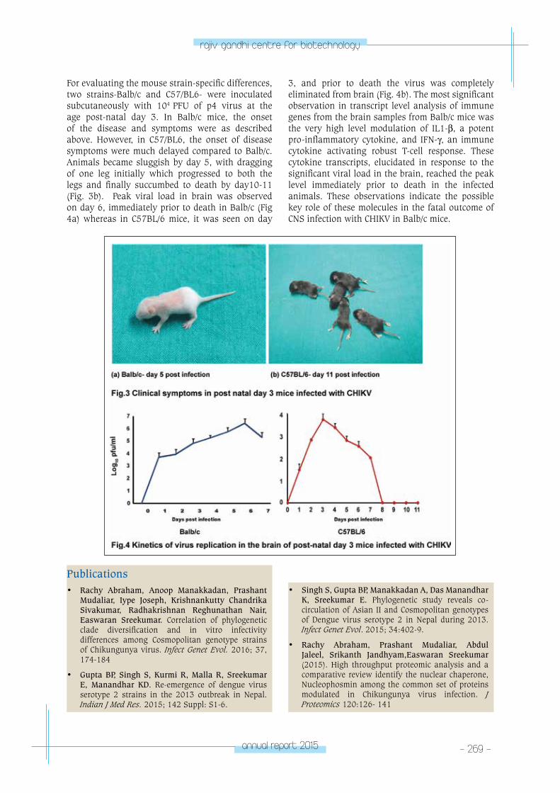
annual report 2015 - 269 -
rajiv gandhi centre for biotechnology
For evaluating the mouse strain-specific differences, two strains-Balb/c and C57/BL6- were inoculated subcutaneously with 104 PFU of p4 virus at the age post-natal day 3. In Balb/c mice, the onset of the disease and symptoms were as described above. However, in C57/BL6, the onset of disease symptoms were much delayed compared to Balb/c. Animals became sluggish by day 5, with dragging of one leg initially which progressed to both the legs and finally succumbed to death by day10-11 (Fig. 3b). Peak viral load in brain was observed on day 6, immediately prior to death in Balb/c (Fig 4a) whereas in C57BL/6 mice, it was seen on day
3, and prior to death the virus was completely eliminated from brain (Fig. 4b). The most significant observation in transcript level analysis of immune genes from the brain samples from Balb/c mice was the very high level modulation of IL1-β, a potent pro-inflammatory cytokine, and IFN-γ, an immune cytokine activating robust T-cell response. These cytokine transcripts, elucidated in response to the significant viral load in the brain, reached the peak level immediately prior to death in the infected animals. These observations indicate the possible key role of these molecules in the fatal outcome of CNS infection with CHIKV in Balb/c mice.
Publications• Rachy Abraham, Anoop Manakkadan, Prashant
Mudaliar, Iype Joseph, Krishnankutty Chandrika Sivakumar, Radhakrishnan Reghunathan Nair, Easwaran Sreekumar. Correlation of phylogenetic clade diversification and in vitro infectivity differences among Cosmopolitan genotype strains of Chikungunya virus. Infect Genet Evol. 2016; 37, 174-184
• Gupta BP, Singh S, Kurmi R, Malla R, Sreekumar E, Manandhar KD. Re-emergence of dengue virus serotype 2 strains in the 2013 outbreak in Nepal. Indian J Med Res. 2015; 142 Suppl: S1-6.
• Singh S, Gupta BP, Manakkadan A, Das Manandhar K, Sreekumar E. Phylogenetic study reveals co-circulation of Asian II and Cosmopolitan genotypes of Dengue virus serotype 2 in Nepal during 2013. Infect Genet Evol. 2015; 34:402-9.
• Rachy Abraham, Prashant Mudaliar, Abdul Jaleel, Srikanth Jandhyam,Easwaran Sreekumar (2015). High throughput proteomic analysis and a comparative review identify the nuclear chaperone, Nucleophosmin among the common set of proteins modulated in Chikungunya virus infection. J Proteomics 120:126- 141
- 270 - annual report 2015
rajiv gandhi centre for biotechnology
Conferences/ seminars/workshops • Sneha Singh, Anupriya MG, E Sreekumar.
Involvement of Angiopoietin/Tie2 chemistry during dengue induced vascular leakage. 4th Molecular Virology Meeting 2015 at RGCB, Kerala, India.
• Sneha Singh, Anupriya MG, E Sreekumar. Characterization of in vitro infectivity phenotype differences of Dengue virus serotype- 2 strains from Kerala belonging to distinct phylogenetic clades, 56th Annual Conference of Association of Microbiologists of India 2015 at JNU, India.
• Sreeja R Nair, Rachy Abraham, E Sreekumar. Determining the adaptation specific mutations in Chikungunya virus passaged in Astrocytic cells. 4th Molecular Virology Meeting, RGCB, TVpm, 16-17th April 2015.
• Vadivel Murugan I., Rachy Abraham., Sreeja R Nair, Anupriya M G, E Sreekumar. Analysis of immune sera against predicted major immunogenic regions of Chikungunya viral proteins E1 and E2 in virus detection. 4th Molecular Virology Meeting, RGCB, TVpm, 16-17th April 2015.
• Rachy Abraham, Sreeja R Nair, Abdul Jaleel and E Sreekumar. Identification of differentially expressed proteins upon Chikungunya virus infection in astrocytic cells. Indian Cell biology Conference, Tvpm, 6-8th December 2015.
• Sreeja R Nair, Rachy Abraham, E. Sreekumar. Interferon-gamma and IL-1beta activation precede death in neonatal mice models of central nervous system (CNS) infection by Chikungunya virus. at 17th International Congress on Infectious Diseases Hyderabad India, 2-5th March 2016.
International fellowships • Sneha Singh was awarded Newton Bhabha PhD
fellowship 2015-16 by DST, Govt. of India and British Council, UK. She would carry out her research in the Department of Biochemistry, Oxford University for 6 months (March-August 2016).
• Dr. Sreekumar received the Fulbright-Nehru Academic and Professional Excellence fellowship from the US-India Educational foundation. He would work as a Visiting Faculty in the laboratory of Prof. Diane Griffin, Department of Molecular Microbiology & Imunology, Johns Hopkins Bloomberg School of Public Health, Baltimore, Maryland, USA for 9 months from September 2015 to May 2016.
Conferences organized• DBT-BBSRC-DFID workshop on Aquaculture from
February 23-27, 2015
• 4th Molecular Virology Meeting, 16th &17th April, 2015
RESEARCH GRANTS
Title Investigator(s) Funding Agency Duration
Characterization of Neurovirulence of Chikungunya virus in cellular and animal models
E.Sreekumar & Jackson James
Department of Biotechnology, Government of India
2012-2015
Antivirals from Medicinal plants of Western Ghats selected based on Traditional Knowledge (TK) / Ethnomedical information: RGCB component of a joint collaborative project with Jawarhalal Nehru Tropical Botanical Gardens and Research Institute)
E. Sreekumar & D. Suja
Department of Biotechnology, Government of India
2015-2018
tRopICAl DISeASe BIoloGY
- Viral Disease Biology laboratory-1
TROPICAL DISEASE BIOLOGY
Viral Disease Biology Laboratory - 1
Joshy Jacob, PhD Senior Adviser to RGCB in Virology &
Immunology (Emory University, Atlanta, USA)
M. Radhakrishna Pillai, FRCPath, PhD.
Professor of Disease Biology
Program ScientistsVijesh Sreedhar, MDReshmi G. PhD.
Research AssociateSara Jones, PhD.
Project OfficerPradip V. Fulmali
Research FellowsBijesh GeorgeAhalya S.Raji PrasadAnjana S.S.
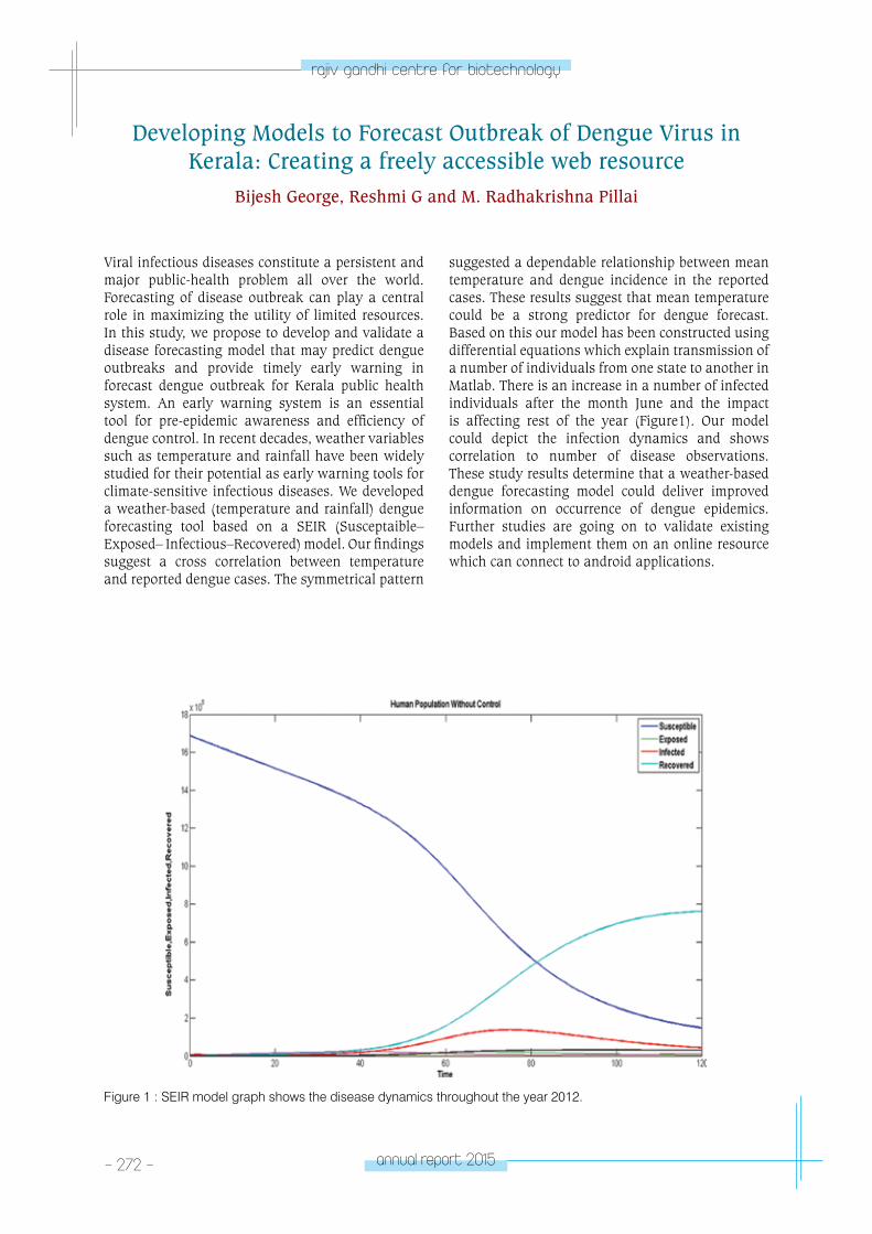
- 272 - annual report 2015
rajiv gandhi centre for biotechnology
Developing Models to Forecast Outbreak of Dengue Virus in Kerala: Creating a freely accessible web resource
Bijesh George, Reshmi G and M. Radhakrishna Pillai
Viral infectious diseases constitute a persistent and major public-health problem all over the world. Forecasting of disease outbreak can play a central role in maximizing the utility of limited resources. In this study, we propose to develop and validate a disease forecasting model that may predict dengue outbreaks and provide timely early warning in forecast dengue outbreak for Kerala public health system. An early warning system is an essential tool for pre-epidemic awareness and efficiency of dengue control. In recent decades, weather variables such as temperature and rainfall have been widely studied for their potential as early warning tools for climate-sensitive infectious diseases. We developed a weather-based (temperature and rainfall) dengue forecasting tool based on a SEIR (Susceptaible–Exposed– Infectious–Recovered) model. Our findings suggest a cross correlation between temperature and reported dengue cases. The symmetrical pattern
suggested a dependable relationship between mean temperature and dengue incidence in the reported cases. These results suggest that mean temperature could be a strong predictor for dengue forecast. Based on this our model has been constructed using differential equations which explain transmission of a number of individuals from one state to another in Matlab. There is an increase in a number of infected individuals after the month June and the impact is affecting rest of the year (Figure1). Our model could depict the infection dynamics and shows correlation to number of disease observations. These study results determine that a weather-based dengue forecasting model could deliver improved information on occurrence of dengue epidemics. Further studies are going on to validate existing models and implement them on an online resource which can connect to android applications.
Figure 1 : SEIR model graph shows the disease dynamics throughout the year 2012.

annual report 2015 - 273 -
rajiv gandhi centre for biotechnology
Infection and pathogenesis caused by neurotropic Flavi viruses is a product of a series of complex interactions between the virus and nervous tissues and further influenced by viral diversity and the host’s susceptibility status and immune response. It is thus necessary to identify such host cellular factors that modulate neurotropic Flavi virus infection and its possible role in the presentation of neurological symptoms stemming from these viruses. We use a systems biology approach to elucidate the pathogenesis of neurotropic flavivirus infection and how viral infection alters host cell signaling at a network level. Stocks of WNV (804994) and JEV (P20778) were prepared in BHK cells and the titre of both the viruses were determined by Cytopathic Effect Assay (CPE) by Quadruplet wells analysis and Plaque assay in Duplicate wells analysis (Fig 1A & 1B). The growth kinetics of both JEV and WNV was studied in two different cells lines Neuronal (IMR-32) & Epithelial (HeLa) of human origin. Cells infected with JEV (P20778) and WNV (804994) at 1 m.o.i and supernatant were collected at different time intervals. The sample was then used for determination of genomic RNA copy by one step real time PCR and infectious virus particle by TCID50. At
0hrs p.i log10, genomic RNA copy of JEV and WNV were 5.23 and 5.51 respectively which increased progressively with time in neuronal cell. Both JEV & WNV efficiently infect neuronal cells and reached the peak timers at the end of 72 hrs p.i. The genomic copy number in JEV and WNV reaches to 10.1 and 10.5 at 72 hrs p.i. (Fig 1C). The pattern of results were in correlation with the infectious virus particle determined by TCID50. The growth kinetic of JEV and WNV in HeLa cells showed different pattern compared to IMR cell. At 0 hrs p.i genomic RNA copy for JEV and WNV were 5.56 & 5.64 respectively. The overall growth of the JEV and WNV in Hela cell was less that the growth observed in IMR cells. It was observed that WNV infects Hela cell more efficiently and grows faster in early hours of replication compared to JEV which was evident from at least 1 log difference in the genomic copies of the WNV and JEV. The maximum genomic copies quantified in JEV infected Hela cells was 7.27 after 60 hrs p.i. Following this a reduction in the genomic copies was observed. In case of WNV copy numbers increased as the infection progresses and maximum of 7.66 copies were observed at 96 hrs p.i (Fig 1D).
Identifying Systems-level Cellular Networks Involved in Neurotropic Flavi Virus-Host Interaction
Ahalya S, Pradip V. Fulmali, Sudipto Saha*, Reshmi G and M. Radhakrishna Pillai
*Collaborator: Bose Institute, Kolkata, India
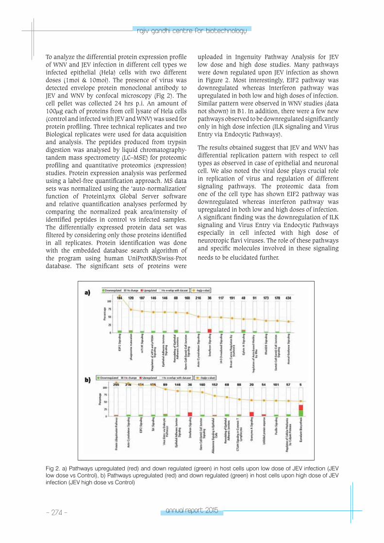
- 274 - annual report 2015
rajiv gandhi centre for biotechnology
To analyze the differential protein expression profile of WNV and JEV infection in different cell types we infected epithelial (Hela) cells with two different doses (1moi & 10moi). The presence of virus was detected envelope protein monoclonal antibody to JEV and WNV by confocal microscopy (Fig 2). The cell pellet was collected 24 hrs p.i. An amount of 100µg each of proteins from cell lysate of Hela cells (control and infected with JEV and WNV) was used for protein profiling. Three technical replicates and two Biological replicates were used for data acquisition and analysis. The peptides produced from trypsin digestion was analysed by liquid chromatography-tandem mass spectrometry (LC–MSE) for proteomic profiling and quantitative proteomics (expression) studies. Protein expression analysis was performed using a label-free quantification approach. MS data sets was normalized using the ‘auto-normalization’ function of ProteinLynx Global Server software and relative quantification analyses performed by comparing the normalized peak area/intensity of identified peptides in control vs infected samples. The differentially expressed protein data set was filtered by considering only those proteins identified in all replicates. Protein identification was done with the embedded database search algorithm of the program using human UniProtKB/Swiss-Prot database. The significant sets of proteins were
uploaded in Ingenuity Pathway Analysis for JEV low dose and high dose studies. Many pathways were down regulated upon JEV infection as shown in Figure 2. Most interestingly, EIF2 pathway was downregulated whereas Interferon pathway was upregulated in both low and high doses of infection. Similar pattern were observed in WNV studies (data not shown) in B1. In addition, there were a few new pathways observed to be downregulated significantly only in high dose infection (ILK signaling and Virus Entry via Endocytic Pathways).
The results obtained suggest that JEV and WNV has differential replication pattern with respect to cell types as observed in case of epithelial and neuronal cell. We also noted the viral dose plays crucial role in replication of virus and regulation of different signaling pathways. The proteomic data from one of the cell type has shown EIF2 pathway was downregulated whereas interferon pathway was upregulated in both low and high doses of infection. A significant finding was the downregulation of ILK signaling and Virus Entry via Endocytic Pathways especially in cell infected with high dose of neurotropic flavi viruses. The role of these pathways and specific molecules involved in these signaling needs to be elucidated further.
Fig 2. a) Pathways upregulated (red) and down regulated (green) in host cells upon low dose of JEV infection (JEV low dose vs Control), b) Pathways upregulated (red) and down regulated (green) in host cells upon high dose of JEV infection (JEV high dose vs Control)
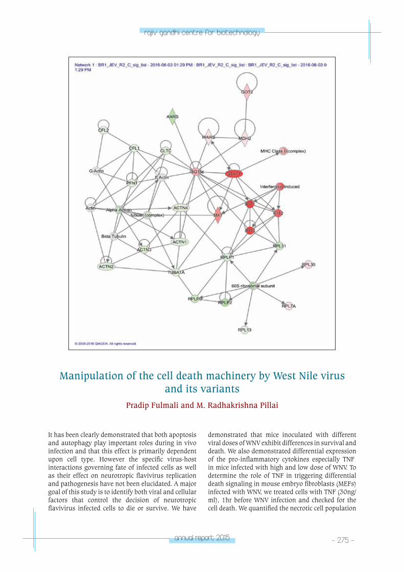
annual report 2015 - 275 -
rajiv gandhi centre for biotechnology
Manipulation of the cell death machinery by West Nile virus and its variants
Pradip Fulmali and M. Radhakrishna Pillai
demonstrated that mice inoculated with different viral doses of WNV exhibit differences in survival and death. We also demonstrated differential expression of the pro-inflammatory cytokines especially TNFβ in mice infected with high and low dose of WNV. To determine the role of TNF in triggering differential death signaling in mouse embryo fibroblasts (MEFs) infected with WNV, we treated cells with TNF (30ng/ml), 1hr before WNV infection and checked for the cell death. We quantified the necrotic cell population
It has been clearly demonstrated that both apoptosis and autophagy play important roles during in vivo infection and that this effect is primarily dependent upon cell type. However the specific virus-host interactions governing fate of infected cells as well as their effect on neurotropic flavivirus replication and pathogenesis have not been elucidated. A major goal of this study is to identify both viral and cellular factors that control the decision of neurotropic flavivirus infected cells to die or survive. We have
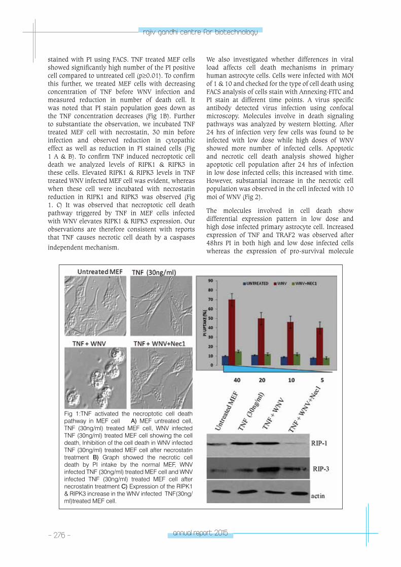
- 276 - annual report 2015
rajiv gandhi centre for biotechnology
stained with PI using FACS. TNF treated MEF cells showed significantly high number of the PI positive cell compared to untreated cell (p≥0.01). To confirm this further, we treated MEF cells with decreasing concentration of TNF before WNV infection and measured reduction in number of death cell. It was noted that PI stain population goes down as the TNF concentration decreases (Fig 1B). Further to substantiate the observation, we incubated TNF treated MEF cell with necrostatin, 30 min before infection and observed reduction in cytopathic effect as well as reduction in PI stained cells (Fig 1 A & B). To confirm TNF induced necroptotic cell death we analyzed levels of RIPK1 & RIPK3 in these cells. Elevated RIPK1 & RIPK3 levels in TNF treated WNV infected MEF cell was evident, whereas when these cell were incubated with necrostatin reduction in RIPK1 and RIPK3 was observed (Fig 1. C) It was observed that necroptotic cell death pathway triggered by TNF in MEF cells infected with WNV elevates RIPK1 & RIPK3 expression. Our observations are therefore consistent with reports that TNF causes necrotic cell death by a caspases
independent mechanism.
We also investigated whether differences in viral load affects cell death mechanisms in primary human astrocyte cells. Cells were infected with MOI of 1 & 10 and checked for the type of cell death using FACS analysis of cells stain with Annexing-FITC and PI stain at different time points. A virus specific antibody detected virus infection using confocal microscopy. Molecules involve in death signaling pathways was analyzed by western blotting. After 24 hrs of infection very few cells was found to be infected with low dose while high doses of WNV showed more number of infected cells. Apoptotic and necrotic cell death analysis showed higher apoptotic cell population after 24 hrs of infection in low dose infected cells; this increased with time. However, substantial increase in the necrotic cell population was observed in the cell infected with 10 moi of WNV (Fig 2).
The molecules involved in cell death show differential expression pattern in low dose and high dose infected primary astrocyte cell. Increased expression of TNF and TRAF2 was observed after 48hrs PI in both high and low dose infected cells whereas the expression of pro-survival molecule
Fig 1:TNF activated the necroptotic cell death pathway in MEF cell A) MEF untreated cell, TNF (30ng/ml) treated MEF cell, WNV infected TNF (30ng/ml) treated MEF cell showing the cell death, Inhibition of the cell death in WNV infected TNF (30ng/ml) treated MEF cell after necrostatin treatment B) Graph showed the necrotic cell death by PI intake by the normal MEF, WNV infected TNF (30ng/ml) treated MEF cell and WNV infected TNF (30ng/ml) treated MEF cell after necrostatin treatment C) Expression of the RIPK1 & RIPK3 increase in the WNV infected TNF(30ng/ml)treated MEF cell.
annual report 2015 - 277 -
rajiv gandhi centre for biotechnology
NFkB was almost inhibited as infection progress in both high dose and low dose infected astrocyte cell. The high expression of FADD and caspases-8 molecules was noted in low dose infected cells while reduced expression of these molecules were observed in high dose infected cell. Cyclo D was also found to be elevated in cell infected with high-dose WNV compared to low-dose WNV. Significantly increase expression of Rip3 kinase along with Rip1 kinase expression in high dose compared to low dose (Fig 3). These results imply that initial virus dose affects WNV replication & cell death induction. In low dose of infection, less number of cells get
infected which results in less abundance of viral genome and protein while high dose results in more viral genome and protein abundance. The high and low abundance of viral genome and proteins leads to expression of certain cell death molecules which in turn decide the fate of cell death. As observed in this case, WNV induced RIP kinases dependent cell death with high dose while caspase dependent cell death was evident in low dose infected cells. It triggers apoptosis in early infection stage in primary cells while it turns toward necrosis in later stage of infection by activating the RIP1-RIP3 necrosome
complex (Fig 4).
Fig 2: Cell death pattern in primary Astrocyte cell infected with different dose of WNV A) Astrocyte cell showing the apoptotic and necrotic cell death after infection with 1 & 10 moi of WNV respectively B) Confocal picture shows astrocyte cell infected with low and high dose of WNV after 24 hr C) Graph shows percent population of apoptotic and necrotic cell.
- 278 - annual report 2015
rajiv gandhi centre for biotechnology
Systems Biology Assessment of Influenza A/pH1N1 vaccination in an Indian cohort
Sara Jones, Richard B Kennedy*, Joshy Jacob and M Radhakrishna Pillai
* Collaborator: Department of Internal Medicine, Mayo Clinic, Rochester, MN USA.
occur in adults who are greater than 50 years of age. The effect of influenza vaccine declines after the age of 70 years due to immunesenescence. The mechanisms involved and how these immune responses are altered with age, have led us to adopt a systems biology approach to understand exactly why the response to vaccination diminishes with
Fig 3: Blots showing the regulation of different signaling molecules involved in induction of cell death in primary astrocyte cell infected with low and high dose of WNV
Fig 4: Schematic representation of induction of death pathways during infection with low and high dose of WNV
Vaccination against influenza continues to be a well-established and promising strategy to protect against influenza and the resulting influenza-related complications. Unfortunately, vaccine use is still sporadic and infrequent in the Indian subcontinent, especially among those most at risk, greater than 90% of influenza-related annual deaths
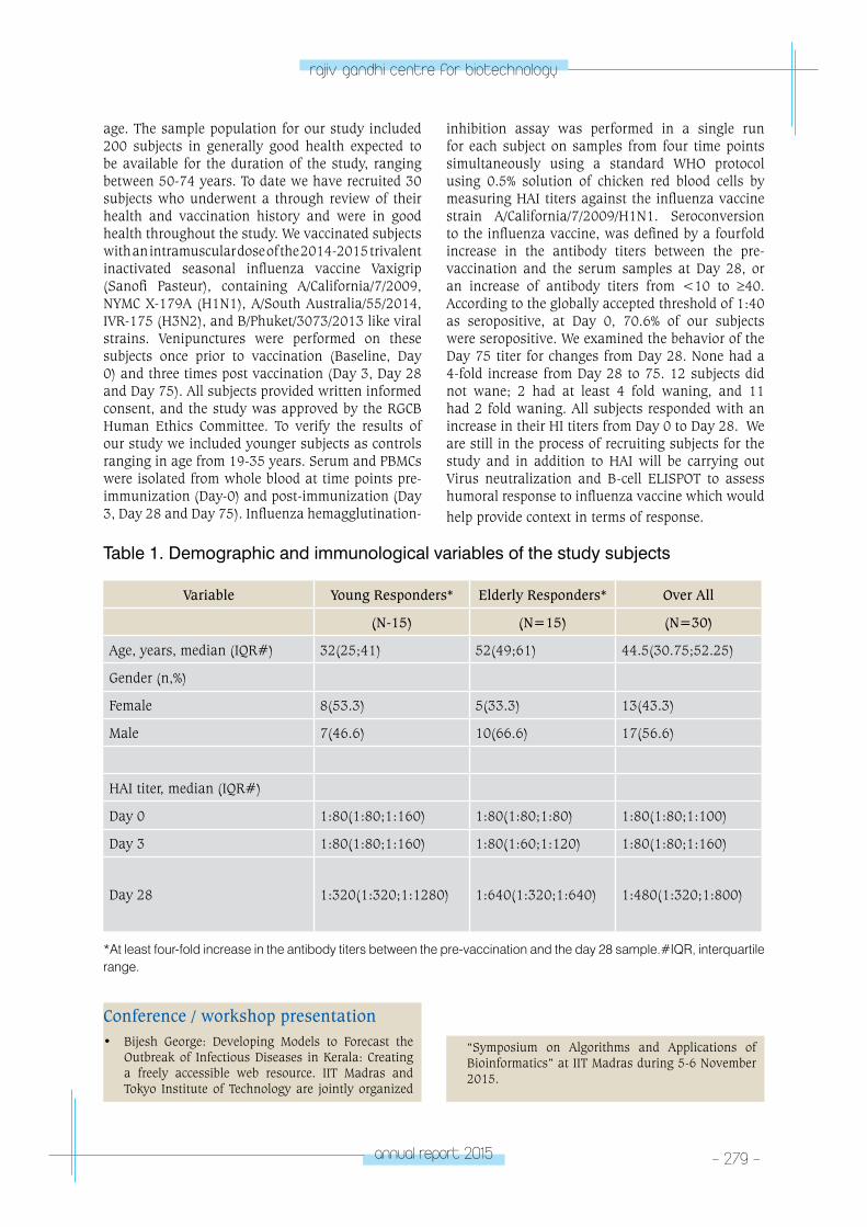
annual report 2015 - 279 -
rajiv gandhi centre for biotechnology
age. The sample population for our study included 200 subjects in generally good health expected to be available for the duration of the study, ranging between 50-74 years. To date we have recruited 30 subjects who underwent a through review of their health and vaccination history and were in good health throughout the study. We vaccinated subjects with an intramuscular dose of the 2014-2015 trivalent inactivated seasonal influenza vaccine Vaxigrip (Sanofi Pasteur), containing A/California/7/2009, NYMC X-179A (H1N1), A/South Australia/55/2014, IVR-175 (H3N2), and B/Phuket/3073/2013 like viral strains. Venipunctures were performed on these subjects once prior to vaccination (Baseline, Day 0) and three times post vaccination (Day 3, Day 28 and Day 75). All subjects provided written informed consent, and the study was approved by the RGCB Human Ethics Committee. To verify the results of our study we included younger subjects as controls ranging in age from 19-35 years. Serum and PBMCs were isolated from whole blood at time points pre-immunization (Day-0) and post-immunization (Day 3, Day 28 and Day 75). Influenza hemagglutination-
inhibition assay was performed in a single run for each subject on samples from four time points simultaneously using a standard WHO protocol using 0.5% solution of chicken red blood cells by measuring HAI titers against the influenza vaccine strain A/California/7/2009/H1N1. Seroconversion to the influenza vaccine, was defined by a fourfold increase in the antibody titers between the pre-vaccination and the serum samples at Day 28, or an increase of antibody titers from <10 to ≥40. According to the globally accepted threshold of 1:40 as seropositive, at Day 0, 70.6% of our subjects were seropositive. We examined the behavior of the Day 75 titer for changes from Day 28. None had a 4-fold increase from Day 28 to 75. 12 subjects did not wane; 2 had at least 4 fold waning, and 11 had 2 fold waning. All subjects responded with an increase in their HI titers from Day 0 to Day 28. We are still in the process of recruiting subjects for the study and in addition to HAI will be carrying out Virus neutralization and B-cell ELISPOT to assess humoral response to influenza vaccine which would help provide context in terms of response.
Table 1. Demographic and immunological variables of the study subjects
Variable Young Responders* Elderly Responders* Over All
(N-15) (N=15) (N=30)
Age, years, median (IQR#) 32(25;41) 52(49;61) 44.5(30.75;52.25)
Gender (n,%)
Female 8(53.3) 5(33.3) 13(43.3)
Male 7(46.6) 10(66.6) 17(56.6)
HAI titer, median (IQR#)
Day 0 1:80(1:80;1:160) 1:80(1:80;1:80) 1:80(1:80;1:100)
Day 3 1:80(1:80;1:160) 1:80(1:60;1:120) 1:80(1:80;1:160)
Day 28 1:320(1:320;1:1280) 1:640(1:320;1:640) 1:480(1:320;1:800)
*At least four-fold increase in the antibody titers between the pre-vaccination and the day 28 sample.#IQR, interquartile range.
Conference / workshop presentation• Bijesh George: Developing Models to Forecast the
Outbreak of Infectious Diseases in Kerala: Creating a freely accessible web resource. IIT Madras and Tokyo Institute of Technology are jointly organized
“Symposium on Algorithms and Applications of Bioinformatics” at IIT Madras during 5-6 November 2015.
tRopICAl DISeASe BIoloGY
- Viral Disease Biology laboratory-2
TROPICAL DISEASE BIOLOGY
Viral Disease Biology Laboratory - 2
John. B. Johnson
John B. Johnson obtained his Ph.D. from the
University of Pune, in pursuant to his research
carried out at the National Centre for Cell
Science, Pune. He trained as a Post Doctoral
Fellow at Wake Forest Health Science, Winston
Salem, North Carolina USA joined RGCB in
2014. He is also a recipient of the Department
of Biotechnology’s Ramalingaswamy Re-entry
Fellowship.
PhD studentsNisha Asok KumarUmerali K.Joydeep NagReshma K.M.
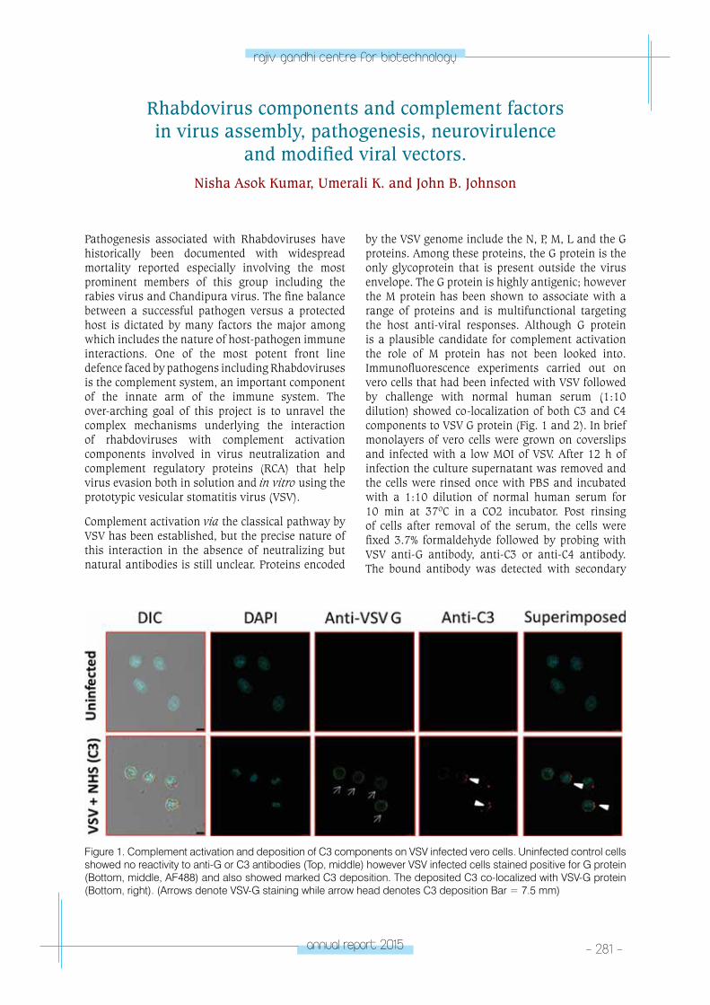
annual report 2015 - 281 -
rajiv gandhi centre for biotechnology
Rhabdovirus components and complement factors in virus assembly, pathogenesis, neurovirulence
and modified viral vectors.Nisha Asok Kumar, Umerali K. and John B. Johnson
Pathogenesis associated with Rhabdoviruses have historically been documented with widespread mortality reported especially involving the most prominent members of this group including the rabies virus and Chandipura virus. The fine balance between a successful pathogen versus a protected host is dictated by many factors the major among which includes the nature of host-pathogen immune interactions. One of the most potent front line defence faced by pathogens including Rhabdoviruses is the complement system, an important component of the innate arm of the immune system. The over-arching goal of this project is to unravel the complex mechanisms underlying the interaction of rhabdoviruses with complement activation components involved in virus neutralization and complement regulatory proteins (RCA) that help virus evasion both in solution and in vitro using the prototypic vesicular stomatitis virus (VSV).
Complement activation via the classical pathway by VSV has been established, but the precise nature of this interaction in the absence of neutralizing but natural antibodies is still unclear. Proteins encoded
by the VSV genome include the N, P, M, L and the G proteins. Among these proteins, the G protein is the only glycoprotein that is present outside the virus envelope. The G protein is highly antigenic; however the M protein has been shown to associate with a range of proteins and is multifunctional targeting the host anti-viral responses. Although G protein is a plausible candidate for complement activation the role of M protein has not been looked into. Immunofluorescence experiments carried out on vero cells that had been infected with VSV followed by challenge with normal human serum (1:10 dilution) showed co-localization of both C3 and C4 components to VSV G protein (Fig. 1 and 2). In brief monolayers of vero cells were grown on coverslips and infected with a low MOI of VSV. After 12 h of infection the culture supernatant was removed and the cells were rinsed once with PBS and incubated with a 1:10 dilution of normal human serum for 10 min at 37OC in a CO2 incubator. Post rinsing of cells after removal of the serum, the cells were fixed 3.7% formaldehyde followed by probing with VSV anti-G antibody, anti-C3 or anti-C4 antibody. The bound antibody was detected with secondary
Figure 1. Complement activation and deposition of C3 components on VSV infected vero cells. Uninfected control cells showed no reactivity to anti-G or C3 antibodies (Top, middle) however VSV infected cells stained positive for G protein (Bottom, middle, AF488) and also showed marked C3 deposition. The deposited C3 co-localized with VSV-G protein (Bottom, right). (Arrows denote VSV-G staining while arrow head denotes C3 deposition Bar = 7.5 mm)
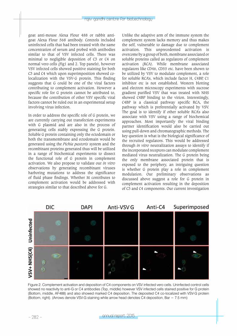
- 282 - annual report 2015
rajiv gandhi centre for biotechnology
goat anti-mouse Alexa Flour 488 or rabbit anti-goat Alexa Flour 568 antibody. Controls included uninfected cells that had been treated with the same concentration of serum and probed with antibodies similar to that of VSV infected cells. There was minimal to negligible deposition of C3 or C4 on normal vero cells (Fig1 and 2. Top panels), however VSV infected cells showed positive staining for both C3 and C4 which upon superimposition showed co-localization with the VSV-G protein. This finding suggests that G could be one of the viral factors contributing to complement activation. However a specific role for G protein cannot be attributed to, because the contribution of other VSV specific viral factors cannot be ruled out in an experimental setup involving virus infection.
In order to address the specific role of G protein, we are currently carrying out transfection experiments with G plasmid and are also in the process of generating cells stably expressing the G protein. Soluble G protein containing only the ectodomain or both the transmembrane and ectodomain would be generated using the Pichia pastoris system and the recombinant proteins generated thus will be utilized in a range of biochemical experiments to dissect the functional role of G protein in complement activation. We also propose to validate our in vitro observations by generating recombinant viruses harboring mutations to address the significance of fluid phase findings. Whether M contributes to complement activation would be addressed with strategies similar to that described above for G.
Unlike the adaptive arm of the immune system the complement system lacks memory and thus makes the self, vulnerable to damage due to complement activation. This unprecedented activation is overcome by a group of both, membrane associated or soluble proteins called as regulators of complement activation (RCA). While membrane associated regulators like CD46, CD55 etc. have been shown to be utilized by VSV to modulate complement, a role for soluble RCA’s, which include factor H, C4BP, C1 inhibitor etc is not established. Western blotting and electron microscopy experiments with sucrose gradient purified VSV that was treated with NHS showed C4BP binding to the virion. Interestingly, C4BP is a classical pathway specific RCA, the pathway which is preferentially activated by VSV. The goal is to identify if other soluble RCA’s also associate with VSV using a range of biochemical approaches. Most importantly the viral binding partner identification would also be carried out using pull-down and chromatographic methods. The key question is what is the biological significance of the recruited regulators. This would be addressed through in vitro neutralization assays to identify if the incorporated receptors can modulate complement mediated virus neutralization. The G protein being the only membrane associated protein that is exposed to the periphery, an intriguing question is whether G protein play a role in complement modulation. Our preliminary observations as discussed above suggest a role for G protein in complement activation resulting in the deposition of C3 and C4 components. Our current investigation
Figure 2. Complement activation and deposition of C4 components on VSV infected vero cells. Uninfected control cells showed no reactivity to anti-G or C4 antibodies (Top, middle) however VSV infected cells stained positive for G protein (Bottom, middle, AF488) and also showed marked C4 deposition. The deposited C4 co-localized with VSV-G protein (Bottom, right). (Arrows denote VSV-G staining while arrow head denotes C4 deposition, Bar = 7.5 mm)
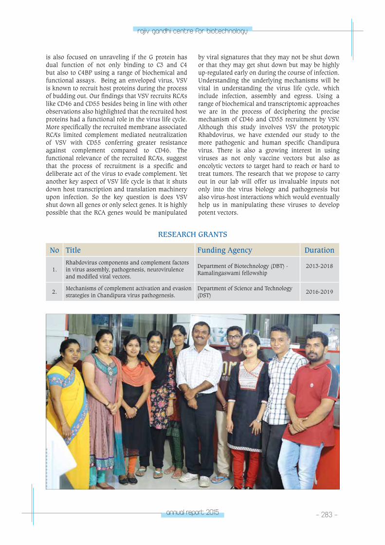
annual report 2015 - 283 -
rajiv gandhi centre for biotechnology
is also focused on unraveling if the G protein has dual function of not only binding to C3 and C4 but also to C4BP using a range of biochemical and functional assays. Being an enveloped virus, VSV is known to recruit host proteins during the process of budding out. Our findings that VSV recruits RCA’s like CD46 and CD55 besides being in line with other observations also highlighted that the recruited host proteins had a functional role in the virus life cycle. More specifically the recruited membrane associated RCA’s limited complement mediated neutralization of VSV with CD55 conferring greater resistance against complement compared to CD46. The functional relevance of the recruited RCA’s, suggest that the process of recruitment is a specific and deliberate act of the virus to evade complement. Yet another key aspect of VSV life cycle is that it shuts down host transcription and translation machinery upon infection. So the key question is does VSV shut down all genes or only select genes. It is highly possible that the RCA genes would be manipulated
by viral signatures that they may not be shut down or that they may get shut down but may be highly up-regulated early on during the course of infection. Understanding the underlying mechanisms will be vital in understanding the virus life cycle, which include infection, assembly and egress. Using a range of biochemical and transcriptomic approaches we are in the process of deciphering the precise mechanism of CD46 and CD55 recruitment by VSV. Although this study involves VSV the prototypic Rhabdovirus, we have extended our study to the more pathogenic and human specific Chandipura virus. There is also a growing interest in using viruses as not only vaccine vectors but also as oncolytic vectors to target hard to reach or hard to treat tumors. The research that we propose to carry out in our lab will offer us invaluable inputs not only into the virus biology and pathogenesis but also virus-host interactions which would eventually help us in manipulating these viruses to develop potent vectors.
RESEARCH GRANTS
No Title Funding Agency Duration
1.Rhabdovirus components and complement factors in virus assembly, pathogenesis, neurovirulence and modified viral vectors.
Department of Biotechnology (DBT) - Ramalingaswami fellowship
2013-2018
2.Mechanisms of complement activation and evasion strategies in Chandipura virus pathogenesis.
Department of Science and Technology (DST)
2016-2019
tRopICAl DISeASe BIoloGY
- parasite Biology laboratoryTROPICAL DISEASE
BIOLOGYMalaria Biology Laboratory
Arumugam Rajavelu
INSPIRE Faculty Fellow
Arumugam Rajavelu obtained his PhD from
Jacobs University of Bremen-Germany in 2011
and trained as a Post-doctoral fellow at Stuttgart
University for two years. He joined RGCB in
December 2013 as Department of Science &
Technology as an INSPIRE Faculty Fellow.
PhD StudentsDevadathan V.S.Jabeena
Junior Research FellowGayathri G.
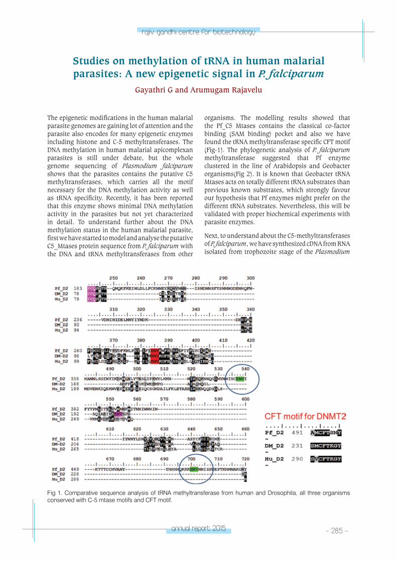
annual report 2015 - 285 -
rajiv gandhi centre for biotechnology
Studies on methylation of tRNA in human malarial parasites: A new epigenetic signal in P. falciparum
Gayathri G and Arumugam Rajavelu
The epigenetic modifications in the human malarial parasite genomes are gaining lot of attention and the parasite also encodes for many epigenetic enzymes including histone and C-5 methyltransferases. The DNA methylation in human malarial apicomplexan parasites is still under debate, but the whole genome sequencing of Plasmodium falciparum shows that the parasites contains the putative C5 methyltransferases, which carries all the motif necessary for the DNA methylation activity as well as tRNA specificity. Recently, it has been reported that this enzyme shows minimal DNA methylation activity in the parasites but not yet characterized in detail. To understand further about the DNA methylation status in the human malarial parasite, first we have started to model and analyse the putative C5_Mtases protein sequence from P. falciparum with the DNA and tRNA methyltransferases from other
organisms. The modelling results showed that the Pf_C5 Mtases contains the classical co-factor binding (SAM binding) pocket and also we have found the tRNA methyltransferase specific CFT motif (Fig-1). The phylogenetic analysis of P. falciparum methyltransferase suggested that Pf enzyme clustered in the line of Arabidopsis and Geobacter organisms(Fig 2). It is known that Geobacter tRNA Mtases acts on totally different tRNA substrates than previous known substrates, which strongly favour our hypothesis that Pf enzymes might prefer on the different tRNA substrates. Nevertheless, this will be validated with proper biochemical experiments with parasite enzymes.
Next, to understand about the C5-methyltransferases of P. falciparum, we have synthesized cDNA from RNA isolated from trophozoite stage of the Plasmodium
Fig 1. Comparative sequence analysis of tRNA methyltransferase from human and Drosophila, all three organisms conserved with C-5 mtase motifs and CFT motif.
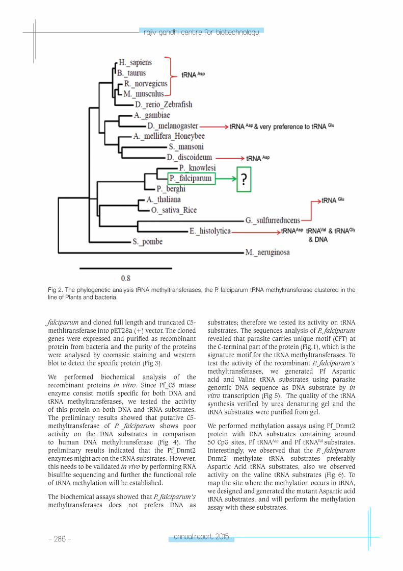
- 286 - annual report 2015
rajiv gandhi centre for biotechnology
Fig 2. The phylogenetic analysis tRNA methyltransferases, the P. falciparum tRNA methyltransferase clustered in the line of Plants and bacteria.
falciparum and cloned full length and truncated C5-methltransferase into pET28a (+) vector. The cloned genes were expressed and purified as recombinant protein from bacteria and the purity of the proteins were analysed by coomasie staining and western blot to detect the specific protein (Fig 3).
We performed biochemical analysis of the recombinant proteins in vitro. Since Pf_C5 mtase enzyme consist motifs specific for both DNA and tRNA methyltransferases, we tested the activity of this protein on both DNA and tRNA substrates. The preliminary results showed that putative C5-methyltransferase of P. falciparum shows poor activity on the DNA substrates in comparison to human DNA methyltransferase (Fig 4). The preliminary results indicated that the Pf_Dnmt2 enzymes might act on the tRNA substrates. However, this needs to be validated in vivo by performing RNA bisulfite sequencing and further the functional role of tRNA methylation will be established.
The biochemical assays showed that P. falciparum’s methyltransferases does not prefers DNA as
substrates; therefore we tested its activity on tRNA substrates. The sequences analysis of P. falciparum revealed that parasite carries unique motif (CFT) at the C-terminal part of the protein (Fig.1), which is the signature motif for the tRNA methyltransferases. To test the activity of the recombinant P. falciparum’s methyltransferases, we generated Pf Aspartic acid and Valine tRNA substrates using parasite genomic DNA sequence as DNA substrate by in vitro transcription (Fig 5). The quality of the tRNA synthesis verified by urea denaturing gel and the tRNA substrates were purified from gel.
We performed methylation assays using Pf_Dnmt2 protein with DNA substrates containing around 50 CpG sites, Pf tRNAAsp and Pf tRNAVal substrates. Interestingly, we observed that the P. falciparum Dnmt2 methylate tRNA substrates preferably Aspartic Acid tRNA substrates, also we observed activity on the valine tRNA substrates (Fig 6). To map the site where the methylation occurs in tRNA, we designed and generated the mutant Aspartic acid tRNA substrates, and will perform the methylation assay with these substrates.
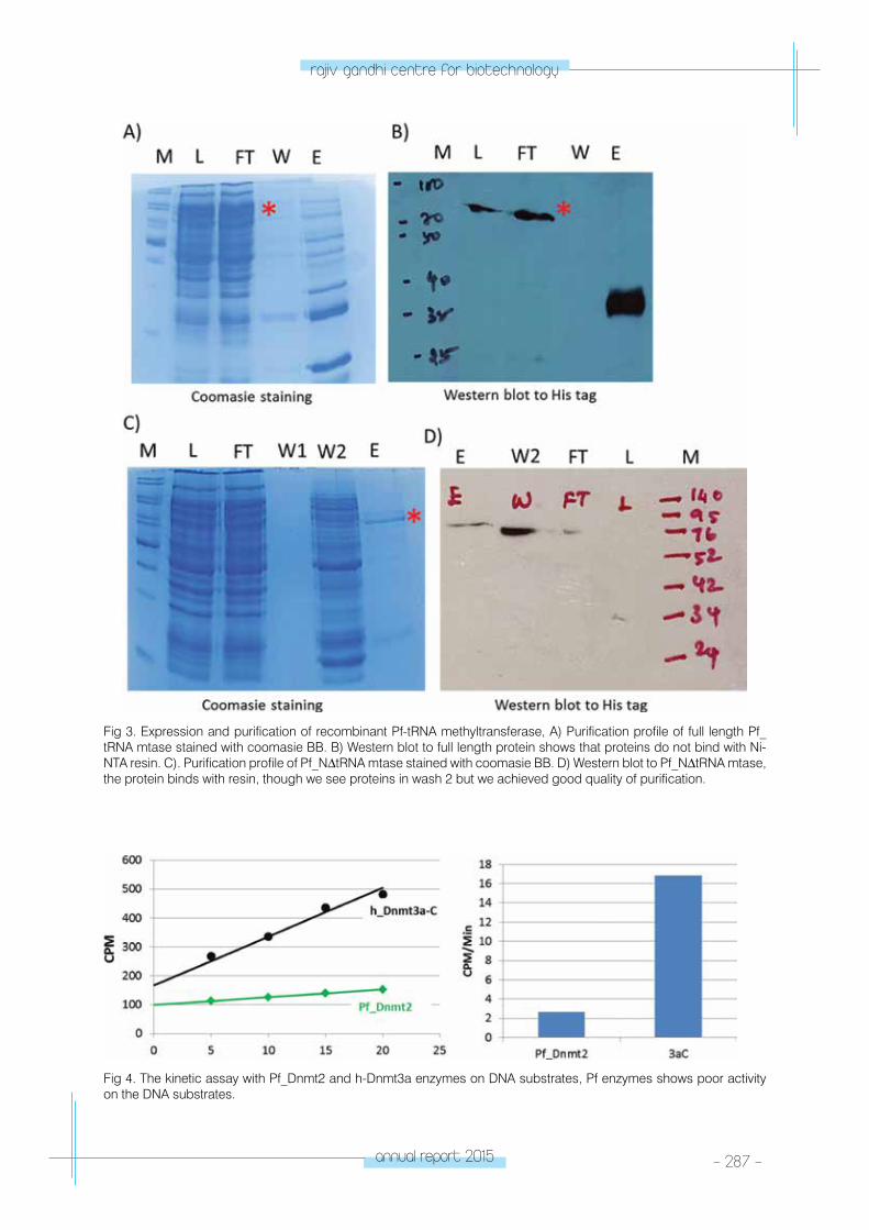
annual report 2015 - 287 -
rajiv gandhi centre for biotechnology
Fig 3. Expression and purification of recombinant Pf-tRNA methyltransferase, A) Purification profile of full length Pf_tRNA mtase stained with coomasie BB. B) Western blot to full length protein shows that proteins do not bind with Ni-NTA resin. C). Purification profile of Pf_NΔtRNA mtase stained with coomasie BB. D) Western blot to Pf_NΔtRNA mtase, the protein binds with resin, though we see proteins in wash 2 but we achieved good quality of purification.
Fig 4. The kinetic assay with Pf_Dnmt2 and h-Dnmt3a enzymes on DNA substrates, Pf enzymes shows poor activity on the DNA substrates.
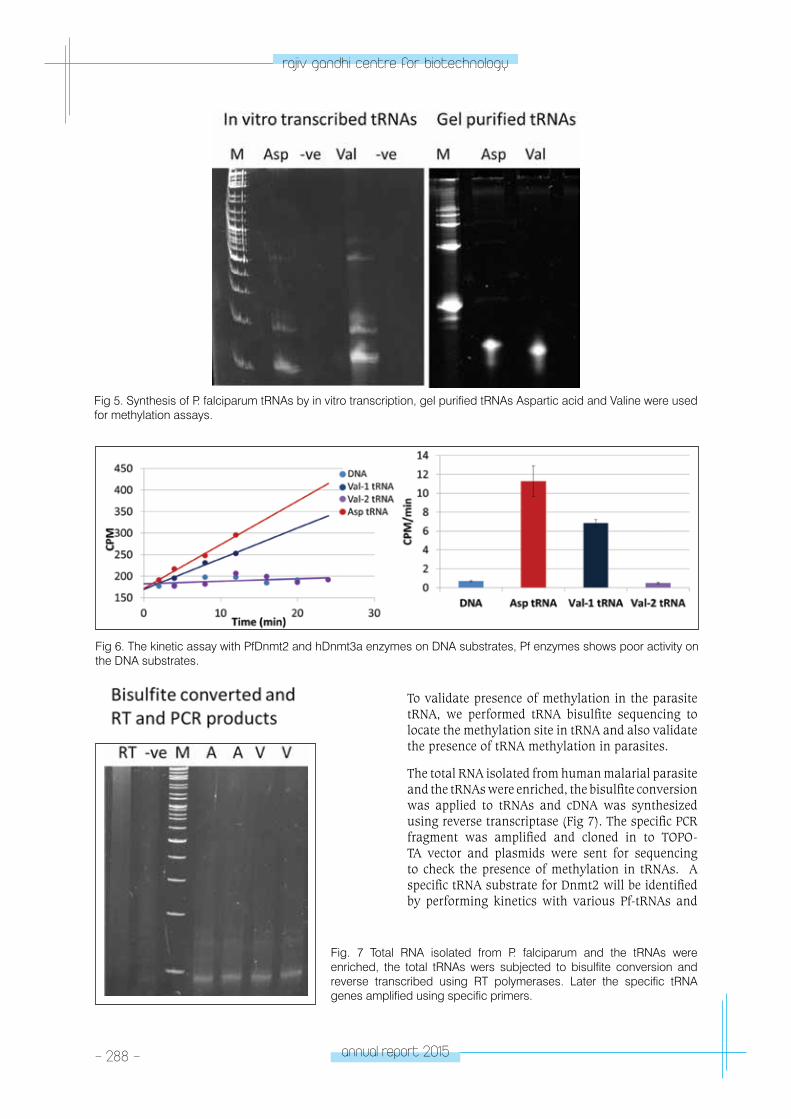
- 288 - annual report 2015
rajiv gandhi centre for biotechnology
Fig 5. Synthesis of P. falciparum tRNAs by in vitro transcription, gel purified tRNAs Aspartic acid and Valine were used for methylation assays.
To validate presence of methylation in the parasite tRNA, we performed tRNA bisulfite sequencing to locate the methylation site in tRNA and also validate the presence of tRNA methylation in parasites.
The total RNA isolated from human malarial parasite and the tRNAs were enriched, the bisulfite conversion was applied to tRNAs and cDNA was synthesized using reverse transcriptase (Fig 7). The specific PCR fragment was amplified and cloned in to TOPO-TA vector and plasmids were sent for sequencing to check the presence of methylation in tRNAs. A specific tRNA substrate for Dnmt2 will be identified by performing kinetics with various Pf-tRNAs and
Fig 6. The kinetic assay with PfDnmt2 and hDnmt3a enzymes on DNA substrates, Pf enzymes shows poor activity on the DNA substrates.
Fig. 7 Total RNA isolated from P. falciparum and the tRNAs were enriched, the total tRNAs wers subjected to bisulfite conversion and reverse transcribed using RT polymerases. Later the specific tRNA genes amplified using specific primers.
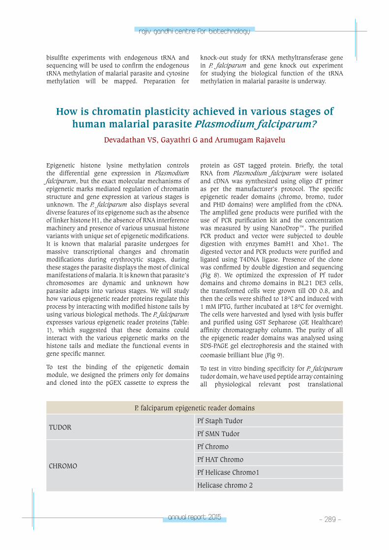
annual report 2015 - 289 -
rajiv gandhi centre for biotechnology
Epigenetic histone lysine methylation controls the differential gene expression in Plasmodium falciparum, but the exact molecular mechanisms of epigenetic marks mediated regulation of chromatin structure and gene expression at various stages is unknown. The P. falciparum also displays several diverse features of its epigenome such as the absence of linker histone H1, the absence of RNA interference machinery and presence of various unusual histone variants with unique set of epigenetic modifications. It is known that malarial parasite undergoes for massive transcriptional changes and chromatin modifications during erythrocytic stages, during these stages the parasite displays the most of clinical manifestations of malaria. It is known that parasite’s chromosomes are dynamic and unknown how parasite adapts into various stages. We will study how various epigenetic reader proteins regulate this process by interacting with modified histone tails by using various biological methods. The P. falciparum expresses various epigenetic reader proteins (Table: 1), which suggested that these domains could interact with the various epigenetic marks on the histone tails and mediate the functional events in gene specific manner.
To test the binding of the epigenetic domain module, we designed the primers only for domains and cloned into the pGEX cassette to express the
protein as GST tagged protein. Briefly, the total RNA from Plasmodium falciparum were isolated and cDNA was synthesized using oligo dT primer as per the manufacturer’s protocol. The specific epigenetic reader domains (chromo, bromo, tudor and PHD domains) were amplified from the cDNA. The amplified gene products were purified with the use of PCR purification kit and the concentration was measured by using NanoDrop™. The purified PCR product and vector were subjected to double digestion with enzymes BamH1 and Xho1. The digested vector and PCR products were purified and ligated using T4DNA ligase. Presence of the clone was confirmed by double digestion and sequencing (Fig 8). We optimized the expression of Pf tudor domains and chromo domains in BL21 DE3 cells, the transformed cells were grown till OD 0.8, and then the cells were shifted to 18ºC and induced with 1 mM IPTG, further incubated at 18ºC for overnight. The cells were harvested and lysed with lysis buffer and purified using GST Sepharose (GE Healthcare) affinity chromatography column. The purity of all the epigenetic reader domains was analysed using SDS-PAGE gel electrophoresis and the stained with coomasie brilliant blue (Fig 9).
To test in vitro binding specificity for P. falciparum tudor domain, we have used peptide array containing all physiological relevant post translational
bisulfite experiments with endogenous tRNA and sequencing will be used to confirm the endogenous tRNA methylation of malarial parasite and cytosine methylation will be mapped. Preparation for
knock-out study for tRNA methyltransferase gene in P. falciparum and gene knock out experiment for studying the biological function of the tRNA methylation in malarial parasite is underway.
How is chromatin plasticity achieved in various stages of human malarial parasite Plasmodium falciparum?
Devadathan VS, Gayathri G and Arumugam Rajavelu
P. falciparum epigenetic reader domains
TUDORPf Staph Tudor
Pf SMN Tudor
CHROMO
Pf Chromo
Pf HAT Chromo
Pf Helicase Chromo1
Helicase chromo 2
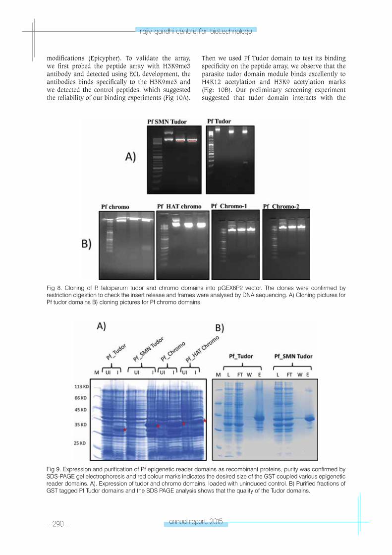
- 290 - annual report 2015
rajiv gandhi centre for biotechnology
modifications (Epicypher). To validate the array, we first probed the peptide array with H3K9me3 antibody and detected using ECL development, the antibodies binds specifically to the H3K9me3 and we detected the control peptides, which suggested the reliability of our binding experiments (Fig 10A).
Then we used Pf Tudor domain to test its binding specificity on the peptide array, we observe that the parasite tudor domain module binds excellently to H4K12 acetylation and H3K9 acetylation marks (Fig: 10B). Our preliminary screening experiment suggested that tudor domain interacts with the
Fig 8. Cloning of P. falciparum tudor and chromo domains into pGEX6P2 vector. The clones were confirmed by restriction digestion to check the insert release and frames were analysed by DNA sequencing. A) Cloning pictures for Pf tudor domains B) cloning pictures for Pf chromo domains.
Fig 9. Expression and purification of Pf epigenetic reader domains as recombinant proteins, purity was confirmed by SDS-PAGE gel electrophoresis and red colour marks indicates the desired size of the GST coupled various epigenetic reader domains. A). Expression of tudor and chromo domains, loaded with uninduced control. B) Purified fractions of GST tagged Pf Tudor domains and the SDS PAGE analysis shows that the quality of the Tudor domains.
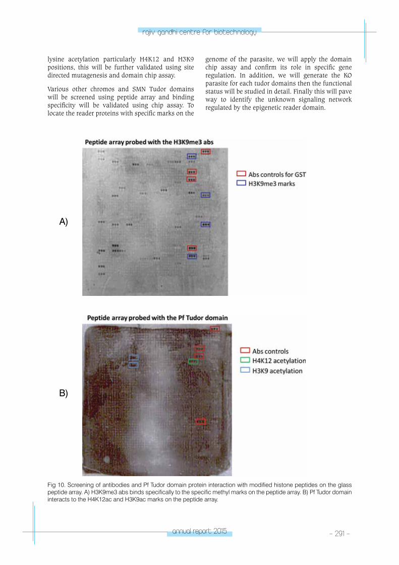
annual report 2015 - 291 -
rajiv gandhi centre for biotechnology
lysine acetylation particularly H4K12 and H3K9 positions, this will be further validated using site directed mutagenesis and domain chip assay.
Various other chromos and SMN Tudor domains will be screened using peptide array and binding specificity will be validated using chip assay. To locate the reader proteins with specific marks on the
genome of the parasite, we will apply the domain chip assay and confirm its role in specific gene regulation. In addition, we will generate the KO parasite for each tudor domains then the functional status will be studied in detail. Finally this will pave way to identify the unknown signaling network regulated by the epigenetic reader domain.
Fig 10. Screening of antibodies and Pf Tudor domain protein interaction with modified histone peptides on the glass peptide array. A) H3K9me3 abs binds specifically to the specific methyl marks on the peptide array. B) Pf Tudor domain interacts to the H4K12ac and H3K9ac marks on the peptide array.
A)
B)
- 292 - annual report 2015
rajiv gandhi centre for biotechnology
RESEARCH GRANTS
No Title Funding Agency Duration
1Functional role of tRNA modifications in Plasmodium falciparum (INSPIRE program)
Department. of Science and Technology,Government of India.
2014-2018
2Role of epigenetic histone methylation marks in chromatin plasticity at various stages of malarial parasite (DBT - IYBA)
Department. of Biotechnology,Government of India.
2015-2018
3Functional studies on the lncRNAs of Plasmodium falciparum and its role in antigenic variation process
Department of Science and Technology,Government of India.
2016-2019
4Molecular determinants of vascular differentiation in the pathogenesis of cerebral arteriovenous malformations
Kerala State Council for Science, Technology & Environment
2016-2019
MICRoBIoM
e ReSeARCH lABoRAtoRY
Microbiome Research Laboratory
Santanu Chattopadhyay, PhDG.N. Ramachandran Fellow ([email protected])
Sourav Sen Gupta, PhDG.N. Ramachandran Fellow ([email protected])
Sanjai D,Senior Manager, Technical Services ([email protected])
Deepak ChauhanPhD Student
Professor M. Radhakrishna Pillai, FRCPath, PhD
Professor of Disease Biology ([email protected])
G. Balakrish Nair, PhD Honorary Senior Consultant

- 294 - annual report 2015
rajiv gandhi centre for biotechnology
RGCB has recently begun studies on the relevance of microbiome in its various disease biology programs. The microbiome comprises all of genetic material within a microbiota and is the ecological community of commensal, symbiotic and pathogenic microorganisms of a particular niche. It is now appreciated that the human body contains as many microbial cells as human cells. However, most of them cannot be cultured using current techniques. By metagenomic analyses using high throughput sequencing and high-end computation, it is now recognized that microbiota will have a tremendous impact of on human health.
Helicobacter pylori infection and modulation of gastrointestinal microbiome in the context of peptic ulcer and gastric cancer
Santanu Chattoppadhyay, Sanjai D., Deepak Chauhan and G.B. Nair
Collaborator: Department of Gastroenterology, Medical College Hospital, Thiruvananthapuram
Infection with the gastric pathogen Helicobacter pylori is associated with gastritis, duodenal and gastric ulcers as well as gastric adenocarcinoma and gastric mucosa-associated lymphoid tissue lymphoma. In India, 80% of the population is infected with H. pylori. However little information exists on the H. pylori strain that is associated with gastro-duodenal disease. Earlier reports have suggested that diet is a major reason for of the high frequency of such cancers in Northeast and in South India. It is also known that diet can alter the composition of microbiome. Furthermore, the alteration of microbiome is associated with cancer. Intriguingly, recent studies pointed out that the H. pylori infection could be associated with alteration of gastric microbiome composition, but the
clinical significance of this alteration has not been established. Importantly, there is no study based on Indian population that shows the genotype of H. pylori, composition of gastro-duodenal microbiome and disease outcome for a particular geographical region. Since India is a large country and known for its population diversity, region specific H. pylori genotype, region specific microbiome and region specific antibiotic resistance may determine the occurrence and cure of gastro-duodenal diseases. In Kerala, 62% of the population is infected with H. pylori. Information on the predominant H. pylori virulence genes like vacA and cagA as well as the in vitro antibiotic sensitivity profile are lacking, in spite of increasing incidence of gastro-duodenal disorders. Also, there is no study for
Fig 1. (A) PCR of ureB gene of H. pylori from biopsy. (B) Isolated H. pylori strain TMC5 on blood agar plate. (C) Biochemical confirmation of H. pylori by urease test. (D) Extracted DNA from biopsy and stool samples for metagenomics analysis.
annual report 2015 - 295 -
rajiv gandhi centre for biotechnology
Kerala that analyses microbiome to understand the total microbial population living within the gastrointestinal tract. We have started culturing H. pylori. Biopsies from patients with gastro-duodenal problems were obtained. PCR using primers specific for ureB gene of H. pylori gave positive result for one sample (TMC5) collected from a patient with gastric cancer (Fig 1A). We were able to isolate H. pylori
strain from the same patient and not from other patients (Fig 1B). The purified strain was confirmed as H. pylori by positive urease (Fig 1C), catalase, oxidase tests as well as by visualizing curved Gram negative bacilli under microscope. Also, for metagenomics analyses, we have isolated DNA from biopsy and stool samples (Fig 1D).
SLE continues to be a multi-factorial disease for which a causative etiology is still undetermined. Recent research has shown that the human associated microbiome especially of the gut have a role in autoimmune disease, but little is yet known about how or why. Our work will broadly focus on whether bacteria in the gut influence the development and progression of lupus in Indian and American patients. To standardize methodology, aliquots of intact frozen fecal samples from patients with lupus were shipped in dry ice from OMRF to RGCB. DNA was extracted at both facilities using the same protocol and initial yields and quality compared between the two labs. Each laboratory then separately processed the DNA for high-throughput sequencing of the bacterial 16S gene for bacterial diversity and composition analysis. Amplified fragments of the V1 to V5 region of the bacterial 16S gene generated using DNA extracted at RGCB as template, was subjected to sequencing on a 454 GS FLX sequencer, and analyzed using the MG-RAST microbiome sequence analysis pipeline. The above sequences were re-analyzed using another popular microbiome analysis software package QIIME. At OMRF the V3 & V4 region of the 16S gene was amplified from DNA extracted at their facility and subjected to high-throughput sequencing on the Illumina sequencing platform and sequences were analyzed using the QIIME-OMRF pipeline. The results are summarized in Fig. 3. 16S sequences generated from RGCB produced varying profiles
Systemic Lupus Erythematosus (SLE) and the microbiome: comparative study of patients from India and the U.S.
Sourav Sengupta, Patrick M. Gaffney*, M. Radhakrishna Pillai and G.B. Nair
*Collaborator: Arthritis & Clinical Immunology Research Program, Oklahoma Medical Research Foundation (OMRF), Oklahoma City, USA
when analyzed by MG-RAST and QIIME. Some important observations were increased estimation of Bacteroidetes and Proteobacteria and decreased abundance estimates of Firmicutes for some samples when using the QIIME method.
When comparing OMRF (Illumina sequences; QIIME analysed) with RGCB sequences (QIIME analyzed), we found that phyla Fusobacteria and TM7 was only exclusively detected in OMRF sequences but at extremely low frequencies which probably contributed significantly to the inter-facility differences in beta-diversity. Sample A5 that was sequenced at a more coverage than other samples using 454, we could detect three additional phyla (Euryarchaeota, Chloroflexi, Nitrospirae) at low abundances when analysed using MG-RAST and not QIIME. Sample A7 also showed the presence of one of these phyla (Chloroflexi) only from the MG-RAST analysis (Fig. 2). In Additiona to the sequencing platform, other factors that probably contributed to inter-facility variations include region of the 16S gene sequenced, amplification primer bias, transport (temperature fluctuations), analysis pipeline and databases used for annotation and sub-sampling (stool micro-environment may harbor different abundances)We will also analyze sequences from OMRF using MG-RAST and determine the best method to use for all future experiments. We next plan to establish clinical partners in India and begin sample collection for microbiome compositional and functional analysis.
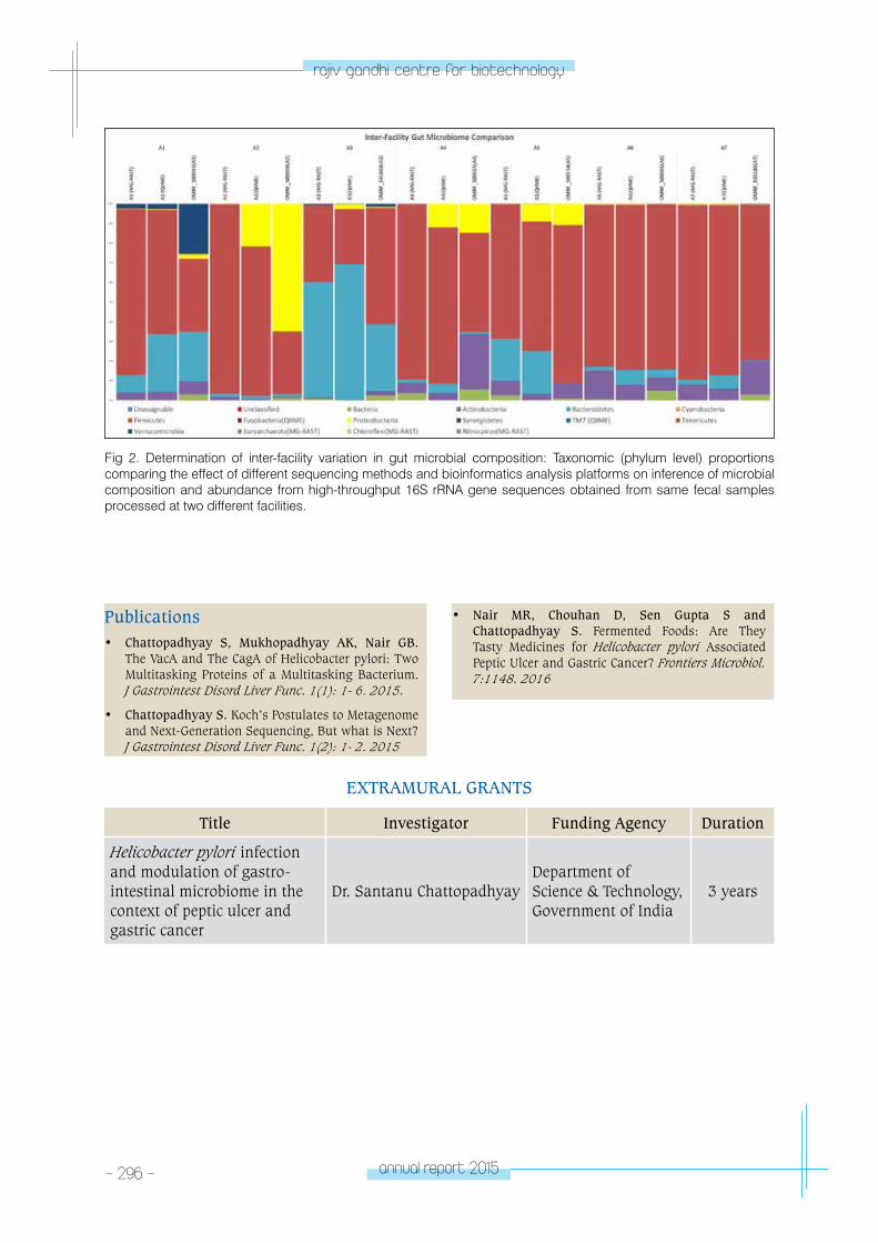
- 296 - annual report 2015
rajiv gandhi centre for biotechnology
Fig 2. Determination of inter-facility variation in gut microbial composition: Taxonomic (phylum level) proportions comparing the effect of different sequencing methods and bioinformatics analysis platforms on inference of microbial composition and abundance from high-throughput 16S rRNA gene sequences obtained from same fecal samples processed at two different facilities.
Publications • Chattopadhyay S, Mukhopadhyay AK, Nair GB.
The VacA and The CagA of Helicobacter pylori: Two Multitasking Proteins of a Multitasking Bacterium. J Gastrointest Disord Liver Func. 1(1): 1- 6. 2015.
• Chattopadhyay S. Koch’s Postulates to Metagenome and Next-Generation Sequencing, But what is Next? J Gastrointest Disord Liver Func. 1(2): 1- 2. 2015
• Nair MR, Chouhan D, Sen Gupta S and Chattopadhyay S. Fermented Foods: Are They Tasty Medicines for Helicobacter pylori Associated Peptic Ulcer and Gastric Cancer? Frontiers Microbiol. 7:1148. 2016
EXTRAMURAL GRANTS
Title Investigator Funding Agency Duration
Helicobacter pylori infection and modulation of gastro-intestinal microbiome in the context of peptic ulcer and gastric cancer
Dr. Santanu Chattopadhyay Department of Science & Technology, Government of India
3 years

RGCBCORE SERVICEFACILITIES
lABoRAtoRY MeDICIne & M
oleCulAR M
eDICIne (lMM
D)
LABORATORY MEDICINE & MOLECULAR MEDICINE
(LMMD)
Radhakrishnan R. Nair
Radhakrishnan Nair received his PhD from the
University of Kerala working at the Regional
Cancer Centre, Thiruvananthapuram. He
subsequently worked in faculty positions at
various Medical Colleges including Manipal
College of Medical Sciences, Oman University
and the Atlantic University, New York. He joined
RGCB in October 2011.
Program ScientistDayakar S. PhD
Manager (Technical)Sanjai D. MSc
Technical personnel on training assignment from Ramanujam Institute for Basic Sciences, Kerala State Council for Science, Technology & EnvironmentHeera Pillai R. Sanugosh K. Mohammed Ashik
Research TechnologistsJayalakshmi S. Vinitha P.T.
Laboratory TechnologistSreeja S.
Project AssistantAkhila Suresh
ReceptionistKarthika V.
House KeepingSumaja V.
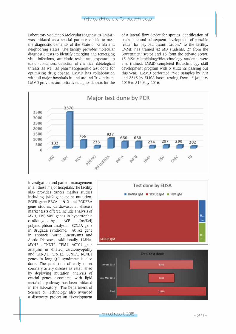
annual report 2015 - 299 -
rajiv gandhi centre for biotechnology
Laboratory Medicine & Molecular Diagnostics (LMMD) was initiated as a special purpose vehicle to meet the diagnostic demands of the State of Kerala and neighboring states. The facility provides molecular diagnostic tests to identify emerging and remerging viral infections, antibiotic resistance, exposure to toxic substances, detection of chemical &biological threats as well as pharmacogenomic test done for optimizing drug dosage. LMMD has collaboration with all major hospitals in and around Trivandrum. LMMD provides authoritative diagnostic tests for the
investigation and patient management in all these major hospitals.The facility also provides cancer marker studies including JAK2 gene point mutation, EGFR gene BRCA 1 & 2 and PGDFRA gene studies. Cardiovascular disease marker tests offered include analysis of MYH, TPT, MBP genes in hypertrophic cardiomyopathy, ACE (Ins/Del) polymorphism analysis, SCN5A gene in Brugada syndrome, ACTA2 gene in Thoracic Aortic Aneurysms and Aortic Diseases. Additionally, LMNA, MYH7 , TNNT2, TPM1, ACTC1 gene analysis in dilated cardiomyopathy and KCNQ1, KCNH2, SCN5A, KCNE1 genes in long Q-T syndrome is also done. The prediction of early onset coronary artery disease as established by deploying mutation analysis of crucial genes associated with lipid metabolic pathway has been initiated in the laboratory. The Department of Science & Technology also awarded a disvovery project on “Development
of a lateral flow device for species identification of snake bite and subsequent development of portable reader for payload quantification.” to the facility. LMMD has trained 42 MD students, 27 from the Government sector and 15 from the private sector. 15 MSc Microbiology/Biotechnology students were also trained. LMMD completed Biotechnology skill development program with 5 students passing out this year. LMMD performed 7965 samples by PCR and 3515 by ELISA based testing From 1st January 2015 to 31st May 2016.
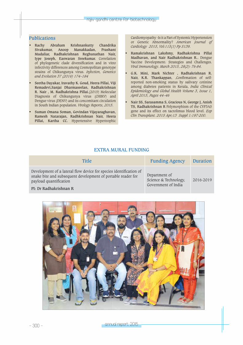
- 300 - annual report 2015
rajiv gandhi centre for biotechnology
EXTRA MURAL FUNDING
Title Funding Agency Duration
Development of a lateral flow device for species identification of snake bite and subsequent development of portable reader for payload quantification
PI: Dr Radhakrishnan R
Department of Science & Technology, Government of India
2016-2019
Publications • Rachy Abraham Krishnankutty Chandrika
Sivakumar, Anoop Manakkadan, Prashant Mudaliar, Radhakrishnan Reghunathan Nair, Iype Joseph, Easwaran Sreekumar. Correlation of phylogenetic clade diversification and in vitro infectivity differences among Cosmopolitan genotype strains of Chikungunya virus. Infection, Genetics and Evolution 37 (2016) 174–184
• Seetha Dayakar, Iravathy K. Goud, Heera Pillai, Viji Remadevi,Sanjai Dharmaseelan, Radhakrishnan R. Nair , M. Radhakrishna Pillai.(2015) Molecular Diagnosis of Chikungunya virus (CHIKV) and Dengue virus (DENV) and its concomitant circulation in South Indian population. Virology Reports, 2015.
• Suman Omana Soman, Govindan Vijayaraghavan, Ramesh Natarajan, Radhkrishnan Nair, Heera Pillai, Kartha CC. Hypertensive Hypertrophic
Cardiomyopathy -Is it a Part of Systemic Hypertension or Genetic Abnormality? American Journal of Cardiology 2015, Vol:115(1) Pp S139.
• Ramakrishnan Lakshmy, Radhakrishna Pillai Madhavan, and Nair Radhakrishnan R.. Dengue Vaccine Development: Strategies and Challenges. Viral Immunology. March 2015, 28(2): 76-84.
• G.K. Mini, Mark Nichter , Radhakrishnan R. Nair, K.R. Thankappan, ,Confirmation of self-reported non-smoking status by salivary cotinine among diabetes patients in Kerala, India Clinical Epidemiology and Global Health Volume 3, Issue 1, April 2015, Pages 44–46
• Nair SS, Sarasamma S, Gracious N, George J, Anish TS, Radhakrishnan R Polymorphism of the CYP3A5 gene and its effect on tacrolimus blood level. Exp Clin Transplant. 2015 Apr;13 Suppl 1:197-200.
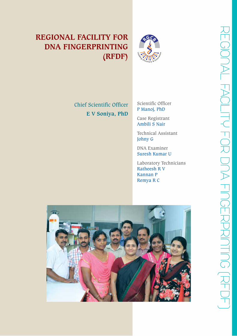
ReGIonAl FACIlItY FoR DnA
FInGeRpRIntInG (RFDF)
REGIONAL FACILITY FOR DNA FINGERPRINTING
(RFDF)
Scientific OfficerP Manoj, PhD
Case RegistrantAmbili S Nair
Technical AssistantJohny G
DNA ExaminerSuresh Kumar U
Laboratory TechniciansRatheesh R VKannan PRemya R C
Chief Scientific Officer
E V Soniya, PhD
- 302 - annual report 2015
rajiv gandhi centre for biotechnology
RFDF offers DNA fingerprinting services to legal bodies, crime investigating and law enforcing agencies. The samples analyzed at RFDF relate to maternity/paternity disputes, crime, rape incidents and cases involving man missing. CO1-based molecular identification and DNA barcoding of fauna especially for species identification in wildlife forensics is yet another service offered by RFDF. Other services offered by this facility include DNA fingerprinting of plants and animals in case-by-case manner using RAPD, AFLP or microsatellite markers and DNA barcoding of animals using CO1 gene and plants using matK and rbcL. The facility also offers hands on training on DNA fingerprinting and DNA barcoding techniques. Details about various DNA fingerprinting/barcoding services and training programmes are provided in our website. In 2015-2016 RFDF analyzed more than 141 samples related to identification, maternity/paternity and relationship disputes forwarded by courts from different districts of Kerala and Kerala Women’s Commission. A major achievement was the facility’s role in a major disaster case: the explosion at Puttingal temple near Kollam, Kerala. The facility analyzed more than 250 samples to identify the body parts recovered from the blast tragedy site. RFDF was proactive in helping the investigation agencies in collecting blood samples of relatives of victims in the tragedy to identify the missing persons. As part of our commitment to the people of Kerala, all the analysis was carried out free of cost. RFDF was also involved in solving many sensational murder cases. RFDF has also received more than 208 samples related to animal poaching forwarded from various forest range offices through court. Animal poaching is
one of the major threats to the animals in wild. It is imperative to punish the offenders to prevent illegal poaching. Samples confiscated by forest officers in Kerala Forest Department are forwarded to our lab for identification of species, so as to enable them to charge the case and punish the offenders. DNA Barcoding helps to identify animals even from minute or cooked samples. But the exact identification of species from the Western Ghats region of Kerala, which is one of the hottest biodiversity hotspots, is often difficult or not possible due to the lack of reference sequences in databases. In collaboration with Zoological Gardens, Department of Museum & Zoos, Thiruvananthapuram we are in the process of developing a DNA Barcode database of captive animals in Thiruvananthapuram Zoo, which will be useful in wildlife forensics to provide evidence to the legal bodies to punish the offenders in poaching cases and thus aiding in the conservation of endangered and endemic animals. We have collected blood/muscle samples from captive animals in Thiruvananthapuram Zoo, India. Samples included many endemic and threatened species present in the Western Ghats and many local as well as migratory birds. DNA was isolated from the samples and COI as well as Cytochrome B genes were amplified and sequenced using universal primers. Preliminary data generated was presented at the 6th International Barcode of Life Conference held at University of Guelph, Canada from August 18, 2015 to August 21, 2015.Five candidates were given training in DNA fingerprinting/barcoding during this period. In addition the facility also provided analysis for more than 1000 samples submitted DNA Barcoding/Fingerprinting/Sequencing analysis from various research institutions, colleges and universities from all over India.
Conference Presentation• Suresh Kumar U, Ratheesh R V, Jacob Alexander
and E V Soniya (2015) Development of DNA Barcode Database of captive animals in Thiruvananthapuram Zoo, Kerala, India. Scientific abstracts from the 6th International Barcode of Life Conference, University of Guelph, Canada. Genome, 58(5): 239. DOI:
10.1139/gen-2015-0087.
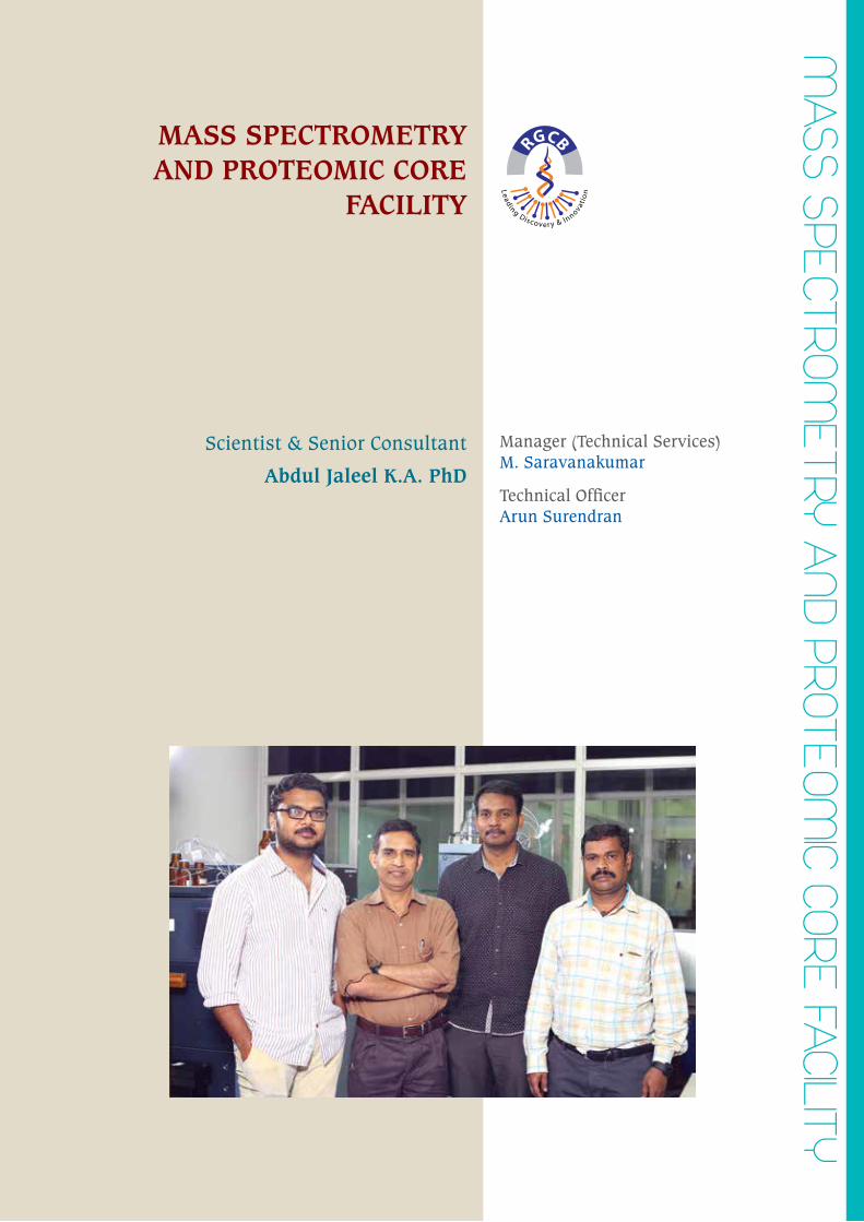
MASS SpeCtRoM
etRY AnD pRoteoM
IC CoRe FACIlItY
MASS SPECTROMETRY AND PROTEOMIC CORE
FACILITY
Manager (Technical Services)M. Saravanakumar
Technical OfficerArun Surendran
Scientist & Senior Consultant
Abdul Jaleel K.A. PhD
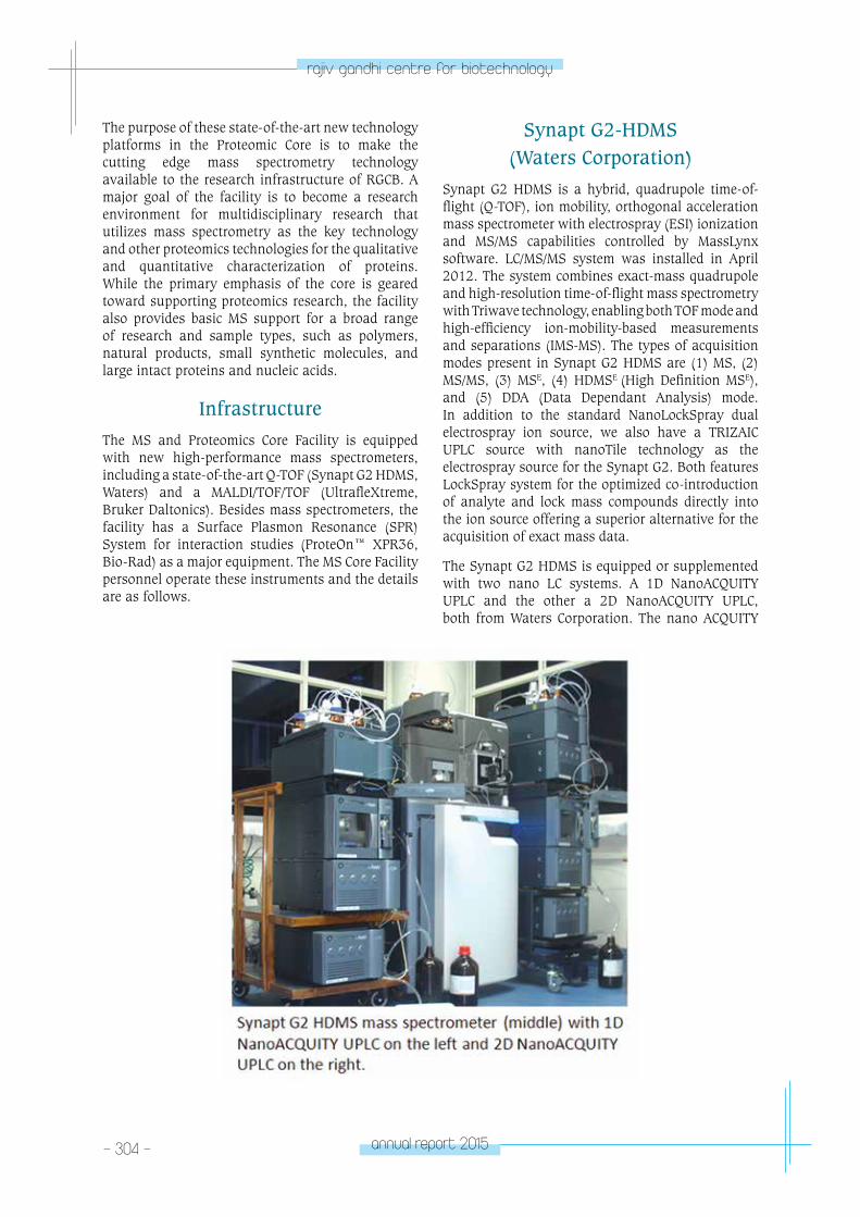
- 304 - annual report 2015
rajiv gandhi centre for biotechnology
The purpose of these state-of-the-art new technology platforms in the Proteomic Core is to make the cutting edge mass spectrometry technology available to the research infrastructure of RGCB. A major goal of the facility is to become a research environment for multidisciplinary research that utilizes mass spectrometry as the key technology and other proteomics technologies for the qualitative and quantitative characterization of proteins. While the primary emphasis of the core is geared toward supporting proteomics research, the facility also provides basic MS support for a broad range of research and sample types, such as polymers, natural products, small synthetic molecules, and large intact proteins and nucleic acids.
InfrastructureThe MS and Proteomics Core Facility is equipped with new high-performance mass spectrometers, including a state-of-the-art Q-TOF (Synapt G2 HDMS, Waters) and a MALDI/TOF/TOF (UltrafleXtreme, Bruker Daltonics). Besides mass spectrometers, the facility has a Surface Plasmon Resonance (SPR) System for interaction studies (ProteOn™ XPR36, Bio-Rad) as a major equipment. The MS Core Facility personnel operate these instruments and the details are as follows.
Synapt G2-HDMS (Waters Corporation)
Synapt G2 HDMS is a hybrid, quadrupole time-of-flight (Q-TOF), ion mobility, orthogonal acceleration mass spectrometer with electrospray (ESI) ionization and MS/MS capabilities controlled by MassLynx software. LC/MS/MS system was installed in April 2012. The system combines exact-mass quadrupole and high-resolution time-of-flight mass spectrometry with Triwave technology, enabling both TOF mode and high-efficiency ion-mobility-based measurements and separations (IMS-MS). The types of acquisition modes present in Synapt G2 HDMS are (1) MS, (2) MS/MS, (3) MSE, (4) HDMSE (High Definition MSE), and (5) DDA (Data Dependant Analysis) mode. In addition to the standard NanoLockSpray dual electrospray ion source, we also have a TRIZAIC UPLC source with nanoTile technology as the electrospray source for the Synapt G2. Both features LockSpray system for the optimized co-introduction of analyte and lock mass compounds directly into the ion source offering a superior alternative for the acquisition of exact mass data.
The Synapt G2 HDMS is equipped or supplemented with two nano LC systems. A 1D NanoACQUITY UPLC and the other a 2D NanoACQUITY UPLC, both from Waters Corporation. The nano ACQUITY
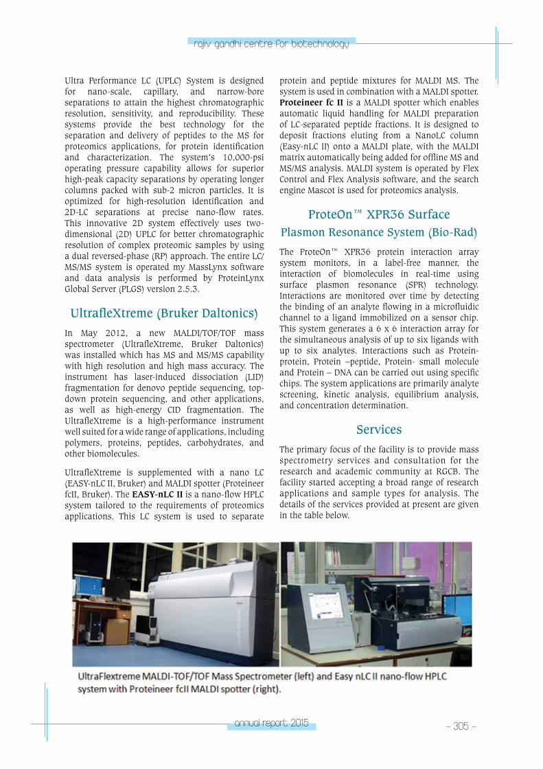
annual report 2015 - 305 -
rajiv gandhi centre for biotechnology
Ultra Performance LC (UPLC) System is designed for nano-scale, capillary, and narrow-bore separations to attain the highest chromatographic resolution, sensitivity, and reproducibility. These systems provide the best technology for the separation and delivery of peptides to the MS for proteomics applications, for protein identification and characterization. The system’s 10,000-psi operating pressure capability allows for superior high-peak capacity separations by operating longer columns packed with sub-2 micron particles. It is optimized for high-resolution identification and 2D-LC separations at precise nano-flow rates. This innovative 2D system effectively uses two-dimensional (2D) UPLC for better chromatographic resolution of complex proteomic samples by using a dual reversed-phase (RP) approach. The entire LC/MS/MS system is operated my MassLynx software and data analysis is performed by ProteinLynx Global Server (PLGS) version 2.5.3.
UltrafleXtreme (Bruker Daltonics)In May 2012, a new MALDI/TOF/TOF mass spectrometer (UltrafleXtreme, Bruker Daltonics) was installed which has MS and MS/MS capability with high resolution and high mass accuracy. The instrument has laser-induced dissociation (LID) fragmentation for denovo peptide sequencing, top-down protein sequencing, and other applications, as well as high-energy CID fragmentation. The UltrafleXtreme is a high-performance instrument well suited for a wide range of applications, including polymers, proteins, peptides, carbohydrates, and other biomolecules.
UltrafleXtreme is supplemented with a nano LC (EASY-nLC II, Bruker) and MALDI spotter (Proteineer fcII, Bruker). The EASY-nLC II is a nano-flow HPLC system tailored to the requirements of proteomics applications. This LC system is used to separate
protein and peptide mixtures for MALDI MS. The system is used in combination with a MALDI spotter. Proteineer fc II is a MALDI spotter which enables automatic liquid handling for MALDI preparation of LC-separated peptide fractions. It is designed to deposit fractions eluting from a NanoLC column (Easy-nLC II) onto a MALDI plate, with the MALDI matrix automatically being added for offline MS and MS/MS analysis. MALDI system is operated by Flex Control and Flex Analysis software, and the search engine Mascot is used for proteomics analysis.
ProteOn™ XPR36 Surface Plasmon Resonance System (Bio-Rad)
The ProteOn™ XPR36 protein interaction array system monitors, in a label-free manner, the interaction of biomolecules in real-time using surface plasmon resonance (SPR) technology. Interactions are monitored over time by detecting the binding of an analyte flowing in a microfluidic channel to a ligand immobilized on a sensor chip. This system generates a 6 x 6 interaction array for the simultaneous analysis of up to six ligands with up to six analytes. Interactions such as Protein- protein, Protein –peptide, Protein- small molecule and Protein – DNA can be carried out using specific chips. The system applications are primarily analyte screening, kinetic analysis, equilibrium analysis, and concentration determination.
ServicesThe primary focus of the facility is to provide mass spectrometry services and consultation for the research and academic community at RGCB. The facility started accepting a broad range of research applications and sample types for analysis. The details of the services provided at present are given in the table below.
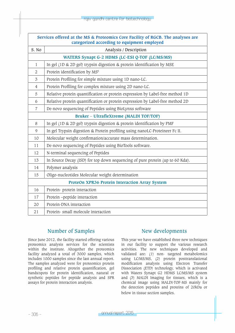
- 306 - annual report 2015
rajiv gandhi centre for biotechnology
Services offered at the MS & Proteomics Core Facility of RGCB. The analyses are categorized according to equipment employed
S. No Analysis / Description
WATERS Synapt G-2 HDMS (LC-ESI Q-TOF (LC/MS/MS)
1 In gel (1D & 2D gel) trypsin digestion & protein identification by MSE
2 Protein identification by MSE
3 Protein Profiling for simple mixture using 1D nano-LC.
4 Protein Profiling for complex mixture using 2D nano-LC.
5 Relative protein quantification or protein expression by Label-free method 1D
6 Relative protein quantification or protein expression by Label-free method 2D
7 De-novo sequencing of Peptides using BioLynxs software
Bruker – UltrafleXtreme (MALDI TOF/TOF)
8 In gel (1D & 2D gel) trypsin digestion & protein identification by PMF
9 In gel Trypsin digestion & Protein profiling using nanoLC-Proteineer Fc II.
10 Molecular weight confirmation/accurate mass determination.
11 De-novo sequencing of Peptides using BioTools software.
12 N-terminal sequencing of Peptides
13 In Source Decay (ISD) for top down sequencing of pure protein (up to 60 Kda).
14 Polymer analysis
15 Oligo-nucleotides Molecular weight determination
ProteOn XPR36 Protein Interaction Array System
16 Protein- protein interaction
17 Protein –peptide interaction
20 Protein-DNA interaction
21 Protein- small molecule interaction
Number of SamplesSince June 2012, the facility started offering various proteomics analysis services for the scientists within the institute. Altogether the proteomics facility analyzed a total of 3000 samples, which includes 1000 samples since the last annual report. The samples analyzed were for proteomics protein profiling and relative protein quantification, gel bands/spots for protein identification, natural or synthetic peptides for peptide analysis and SPR assays for protein interaction analysis.
New developmentsThis year we have established three new techniques in our facility to support the various research activities. The new techniques developed and validated are: (1) non- targeted metabolomics using LC/MS/MS, (2) protein posttranslational modification analysis using Electron Transfer Dissociation (ETD) technology, which is activated with Waters Synapt G2 HDMS LC/MS/MS system and (3) MALDI Imaging for tissues, which is a chemical image using MALDI-TOF-MS mainly for the detection peptides and proteins of 20kDa or below in tissue section samples.
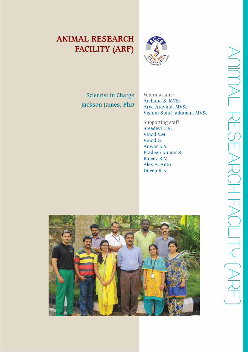
AnIM
Al ReSeA
RCH FACIlItY (ARF)
ANIMAL RESEARCH FACILITY (ARF)
Veterinarians:Archana S. MVScArya Aravind, MVScVishnu Sunil Jaikumar, MVSc
Supporting staff: Sreedevi L.R.Vinod V.M.Vinod G.Anwar K.Y.Pradeep Kumar S.Rajeev R.V.Alex A. AntoDileep R.K.
Scientist in Charge
Jackson James, PhD
- 308 - annual report 2015
rajiv gandhi centre for biotechnology
The Animal Research Facility (ARF) at RGCB has been growing and evolving over the years as well equipped and dedicated facility for researchers and facility whose primary objectives are to provide animals for researchers and facilitate the animal related research. ARF functions in conjunction with an efficient Institutional Animal Ethics Committee (IAEC) and thereby ensures ethical and humane treatment of animals by all means. There are separate breeding rooms for each species of animals such as mice, rats and rabbits. Presently, ARF has conventional rooms for rats, barrier facility having two transgenic rooms for mice (transgenic experimental room and transgenic breeding room), rabbit room, procedure room for conducting minor procedures and euthanasia. ARF maintains valuable colonies of various transgenic, knock outs (KOs) and immune compromised SCIDs in Individually Ventilated Caging System (IVC) with all the necessary adjunct facilities including animal changing station, air handling unit, air conditioning, dehumidifier etc., to maintain optimum conditions for the animals. All procedures involving infectious agents are done in bio-security type II cabinets. ARF also
carries out regular Animal handling certification course for students/staff. Only certified students/staff are permitted to carry out experiments in the ARF. Entry to the facility is restricted by Biometric Access System and fair practice/compliance with the ARF policies is monitored by the surveillance cameras installed at various locations inside ARF. The strains of animals currently maintained in ARF are BALB/c, Swiss Albino, NODSCID, pHesl-d2EGFPTg, C57BL/6J, AG129, SIPR1 , Pax6 Sey, Pax6 cko/cko , Nestin ERT2-Cre , DBA/1J, GU-2 GFP, Wistar rats, SHR rats and New Zealand White rabbits.
ARF also carries out regular screening for common pathogens and aims at providing healthy pathogen free animals to users. We have also initiated procedures to carry out cryopreservation of embryos, microinjection, embryo transfer and generation of transgenic and KOs and aim to become a regional facility for generation of transgenic and KOs. Our facility at RGCB-BIC (KINFRA) is also ready and will cater to the animal requirement of scientists based at RGCB-BIC.
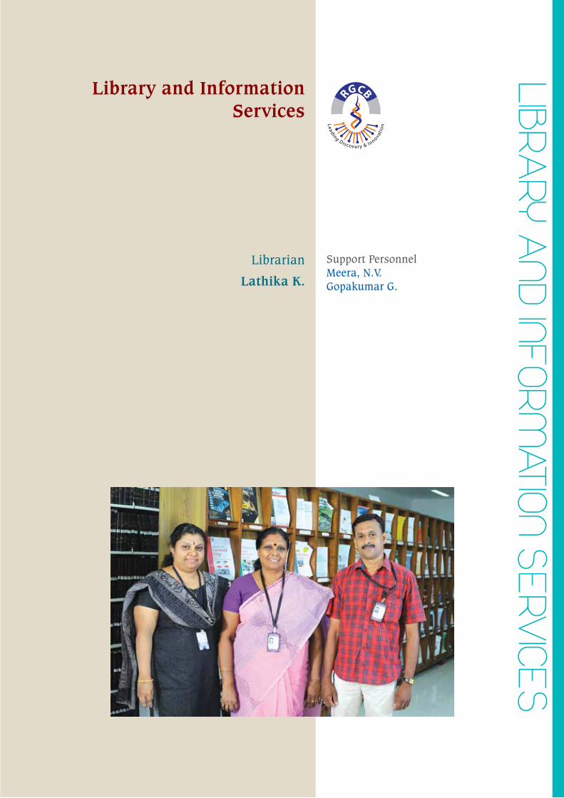
lIBRARY A
nD InFoRMAtIon SeRV
ICeSLibrary and Information
Services
Support PersonnelMeera, N.V.Gopakumar G.
Librarian
Lathika K.
- 310 - annual report 2015
rajiv gandhi centre for biotechnology
RGCB Library maintained its distinctive status among the other research Libraries during the year under review. Playing a key role in the dissemination of information as and when required to the scientific community as well as researches and students. Library could discharge its onerous responsibilities with a sense of contentment, true to the expectation of all who seek its assistance in whatever manner.
RGCB has taken membership in the Biomed Central there by enabling researchers to publish peer – reviewed articles in their open access journals. Developing Library Network facilitated document delivery services and sharing of e-resources on account of RGCB’s membership with DELNET.
The library also provided specialized services like Electronic Document Delivery Service, photocopying, scanning of documents, indexing, selective dissemination of information, current awareness, conference alert, Newspaper clippings, internet, e- alerts, and referral services etc. Every month Library is preparing research publication status of the centre and it is uploaded in the website and in the library notice board.
Information Resource ManagementThe library managed to cater to the documentation and information needs of the institute. By strengthening the information dissemination of electronic journals and other e-resources, effectively utilizing the institute’s LAN.
The total print collection has more than 8000 documents, which includes books, protocols, standards, manuals bound volumes of journals. During the year 2014-15, 95 international books were added. The library has a wide and varied collection of books, journals, back volumes, theses, reports, reprints, CD ROMs, DVDs etc. Library has a separate section on general books for reading. 30 Indian periodicals were subscribed and 12 periodicals were received on gratis, 10 Newspapers, 8 popular magazines are included in our collection.
e- ResourceDepartment of Biotechnology’s e- Library Consortium (DeLCON) this year enhanced the number of e-journals, facilitated access to 1171 selective Journals, e- book series. Consortium Members have the facility to access e- journals of 19 international publishers. These publishers included American Association for Advancement
of Science (AAAS), American Association for Cancer Research (AACR), American Chemical Society (ACS), Annual Reviews, American Society for Biochemistry and Molecular Biology, American Society For Microbiology, Cold Spring Harbor Laboratory Press, Marry Ann Liebert, Nature Publication Group, Oxford University Press (OUP), PNAS, Springer, Society for General Microbiology, Wiley-Blackwell, Elsevier Science (Science Direct), American Society of Plant Biologist, American Association of Immunologist etc.
Journal of Visualized Experiments (JoVE) Biology, publishes peer-reviewed scientific video protocols to accelerate biological, medical, chemical and physical research is added in our collection.
To promote awareness of library services and resources and to enhance the level of usage of the embedded wealth knowledge, promotional programmes were carried out at frequent intervals. This includes regular announcements and broadcasting of messages of new facilities/services initiated and hosted in the intranet. Trial access to various e-resources was also provided for researchers.
Updating of in house databasesUpdating of in house databases of books, periodical, bound volumes of journals, PhD these, reports, conference proceedings etc. was carried out and uplinked to the website. Database of PhD these and peer –reviewed publications of RGCB covering the period 2014-2015 was updated.
Membership with national & international bodies:
Biomed Central – BMC:- RGCB continued to be a member of BMC and this enabled the publication of articles.
DeLCON:- DBT’s electronic library consortia provided access to e- journals, e- books and databases.
DELNET:- Developing Library Network facilitated document delivery services and sharing of e-resources on account of RGCB’s membership with delnet.
ServicesDocument Lending service: Each Scientists member is entitled to borrow 4 books at a time, while the number is restricted to two books in the case of students and other users.
annual report 2015 - 311 -
rajiv gandhi centre for biotechnology
OPAC – Library offers Online Public Access Catalogue (OPAC) which allows user to browsing library collection through the web.
e- Resources and Internet Facility: The Library is well equipped with a good number of computers with internet connectivity through LAN. The Library is having access to a very good number of electronic journals, e- books, journals archives etc. Users were given full access to e- resources.
Scientometrics Services: Continued the Bibliometric/ Scientometric/Impact Factor Analysis of RGCB Publications using Journal Citation report. Necessary assistance was offered for publishing in journals with high Impact Factor.
Resource Sharing activities: The library shared its resources with other special/academic libraries of India. As a member of DBT’s electronic Library Consortia (DeLCON), the Library maintained close contacts with libraries under DBT institutions and North Eastern Region (NER) Institutions.
Electronic Document Delivery Service is yet another service available.
Reprographic services: Facilities provided with photocopiers, printers, scanners, including colour printer. One high end photocopy/printer was added during the year.
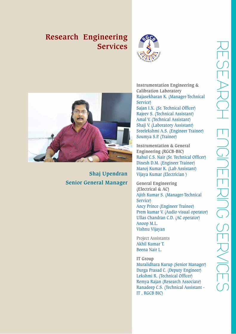
ReSeARCH enGIneeRInG SeRV
ICeSResearch Engineering
Services
Shaj Upendran
Senior General Manager
Instrumentation Engineering & Calibration LaboratoryRajasekharan K. (Manager-Technical Service)Sajan I.X. (Sr. Technical Officer)Rajeev S. (Technical Assistant)Amal V. (Technical Assistant)Shaji V. (Laboratory Assistant)Sreelekshmi A.S. (Engineer Trainee)Soumya S.P. (Trainee)
Instrumentation & General Engineering (RGCB-BIC)Rahul C.S. Nair (Sr. Technical Officer)Dinesh D.M. (Engineer Trainee)Manoj Kumar K. (Lab Assistant)Vijaya Kumar (Electrician )
General Engineering (Electrical & AC)Ajith Kumar S. (Manager-Technical Service)Ancy Prince (Engineer Trainee)Prem kumar V. (Audio visual operator)Ullas Chandran C.D. (AC operator)Anoop M.L.Vishnu Vijayan
Project AssistantsAkhil Kumar T.Beena Nair L.
IT GroupMuralidhara Kurup (Senior Manager)Durga Prasad C. (Deputy Engineer)Lekshmi R. (Technical Officer)Remya Rajan (Research Associate)Ranadeep C.S. (Technical Assistant - IT , RGCB BIC)
annual report 2015 - 313 -
rajiv gandhi centre for biotechnology
Rajiv Gandhi Centre for Biotechnology houses a large number of molecular biology and biotechnological research instruments. The Instrumentation Engineering Division is responsible for the installation, maintenance and repair of sophisticated research instruments in RGCB as well as the maintenance of Central instrumentation facility. The Division also maintains a well equipped engineering workshop with facilities required for the repair and calibration of the sophisticated instruments as its part. Repair up to the PCB level is done here reducing the downtime and repair costs. By attending to many essential repair works of instruments, dependence on expensive maintenance contracts with dealers has been reduced. The instrumentation Group also carries out design, modification, and fabrication of research instruments.
During the year, problems of Liquid Nitrogen Plant, FACS ARIA Flow Cytometer, Confocal Laser Scanninmg Microscope, Spectrophotometers, Ultra Centrifuges, High speed Centrifuges, Table Top Centrifuges, Gel Documentation systems, Transmission Electron Microscope, Upright and Inverted Microscopes, PCR machines, Electronic balances, Speed Vac Concentrator, CO2 Incubators, HPLC, Freeze Dryers, Microplate Washer, DNA sequencer (48 capillary), Confocal Laser Scanning Microscope, Animal Imager, etc. have been repaired successfully.
The Instrumentation Division also maintains the Centralized Instrumentation facilities, Computers, PC based security surveillance system, Biometrics Time Attendance recorders, Conferencing facilities, Communication systems, Liquid Nitrogen Plant, Incinerator, Auditoriums, Convention Centre etc. It also carries out the supervision of 11KV electrical substation, 340 ton AC plant, 630 KVA, 750 KVA & 1010KVA DG sets. All minor electrical works required by the scientific staff also carries out by the in house engineering department.
Research Engineering Services has been successfully completed the installations of new instruments in BIO-INNOVTION CENTRE at Kazhakuttam KINFA campus. This includes the installation of instruments, Electrical works, Air-conditioning works etc.
Centralized Instrumentation Facility
In addition to the basic facilities available in all the research laboratories, we have a centralized core facility equipped with several minor and major
equipments to cater the requirements of our research personnel. The following are the facilities available in the core facility:
Spectroscopy • MALDI TOF/TOF Mass Spectrometer - Bruker• Bench top MALDI TOF Mass Spectrometer
- Shimadzu• Spectrophotometer - Perkin elmer, Thermo,
Labomed • Luminescence Spectrometer - Perkin Elmer• Multimode Plate Reader - Tecan • FTIR Spectrometer - Thermo
Genomics and Proteomics • Next Generation Sequencing Systems (NGS)• Ion Proton Torrent System - Life Technologies• Ion Proton Personal Genomic Machine - Life
Technologies• Gene Chip Scanner - Affymatrix• DNA sequencer (96 capillary) - Applied
Biosystems (ABI)• Genetic Analyzer (Single capillary) - Applied
Biosystems (ABI)• DNA sequencer (48 capillary) - Applied
Biosystems (ABI)• High Definition Mass Spectrometer (HDMS) -
Waters Corporation• Protein sequencer - Shimadzhu• Protein Interaction Analyzer by SPR - Biorad• Real Time PCR - Applied Biosystems, Biorad,
Cepheid.• Automated Peptide Synthesizer - Applied
Biosystems• Amino acid analyzer - Shimadzhu• Nucleic Acid Extraction System - Precision
System Science, Beckman• Fully Automatic Nucleic Acid Extraction System
- BioMerieux
Separation and Purification • Pulse Field Electrophoresis - Biorad• Ultra Centrifuges - Beckman Coulter• High speed Centrifuges - Sorvall, Hittachi, and
Kuboto.• Table Top Centrifuges - Hareaus, Zigma, Jouan,
Eppendorff.• Protien purification system - Biorad• HPLC – Shimadzhu, Waters Corporation• UPLC – Waters Coroporation
- 314 - annual report 2015
rajiv gandhi centre for biotechnology
• nanoLC - Bruker• Gas chromatograph - Shimadzhu• Automated Flash Chromatograph - Biotage
Imaging
• Phospher Imager - Biorad• Multi Imager - Biorad• Gel Documentation systems - Biorad, UVP,
Syngene.• Confocal Laser Scanninmg Microscope - Leica
Microsystems, Nikon• Super Continnum White Laser Confocal Laser
Scanning Microscope - Leica Microsystems• Stuctured Illumination Microscope with
Confocal Laser Scanning System - Nikon• Spinning Disc Confocal Microscope - BD
Biosciences• Transmission Electron Microscope - Jeol• Upright and Inverted Microscopes - Leica,
Zeiss, Nikon, Olympus• Bench top High Throughput Bioimager - BD
Biosciences• Invivo Animal Imager - Xenogen• Ultra Sound Scanner - Phillips
Flow Cytometry • FACS ARIA Flow Cytometer with sorter - BD
Biosciences• FACS ARIA II Flow Cytometer - BD Biosciences• FACS ARIA III Flow Cytometer - BD Biosciences
Others • Fully Automated Liquid Handling System -
Beckman Coulter• Liquid Scintillation Counter - Wallac• Ultra Microtome - Leica• Submicron size Analyser - Beckman Coulter• Robotic Spotter for MALDI TOF/TOF plates• Non-Invasive Blood Pr. Monitor with ECG for
animal facility - Iworks/IITC• Sample preparation System for NGS, Ion
OneTouch - Life Technologies• Eletrophysiology Setup• Liquid Nitrogen Plant - Sterling• Incinerator - Thermax
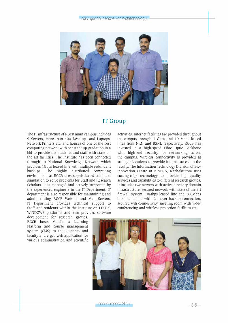
annual report 2015 - 315 -
rajiv gandhi centre for biotechnology
The IT infrastructure of RGCB main campus includes 9 Servers, more than 400 Desktops and Laptops, Network Printers etc. and houses of one of the best computing network with constant up-gradation in a bid to provide the students and staff with state-of-the art facilities. The Institute has been connected through to National Knowledge Network which provides 1Gbps leased line with multiple redundant backups. The highly distributed computing environment at RGCB uses sophisticated computer simulation to solve problems for Staff and Research Scholars. It is managed and actively supported by the experienced engineers in the IT Department. IT department is also responsible for maintaining and administrating RGCB Website and Mail Servers. IT Department provides technical support to Staff and students within the Institute on LINUX, WINDOWS platforms and also provides software development for research groups. RGCB hosts Moodle a Learning Platform and course management system (CMS) to the students and faculty and ergcb web application for various administration and scientific
activities. Internet facilities are provided throughout the campus through 1 Gbps and 10 Mbps leased lines from NKN and BSNL respectively. RGCB has invested in a high-speed Fibre Optic Backbone with high-end security for networking across the campus. Wireless connectivity is provided at strategic locations to provide Internet access to the faculty. The Information Technology Division of Bio-innovation Centre at KINFRA, Kazhakuttom uses cutting-edge technology to provide high-quality services and capabilities to different research groups. It includes two servers with active directory domain infrastructure, secured network with state of the art firewall system, 10Mbps leased line and 100Mbps broadband line with fail over backup connection, secured wifi connectivity, meeting room with video conferencing and wireless projection facilities etc.
IT Group
- 316 - annual report 2015
rajiv gandhi centre for biotechnology
Standing Left to Right: Suma Nair (Project Assistant), Dr. E.V. Soniya (Associate Dean), Dr. R Ajaykumar (Member), Professor Jagadeesh Chandran (Senior Manager, Academic Affairs), Dr. Jackson James (Associate Dean), Dr. E. Sreekumar (Associate Dean), Dr. T.R. Santoshkumar (Associate Dean), Dr. Debasree Dutta (Member)
Sitting Left to Right: Professor Sivaramakrishnan (PhD Program Coordinator), Dr. K. Santoshkumar (Dean), Professor M.R.Pillai (Chairman), Professor C.C. Kartha (Honorary Distinguished Visiting Professor)
RGCB ACADEMIC COMMITTEE
Standing Left to Right : Vishnu S. (Project Assistant), Rahul H. (Project Trainee), Dileep Kumar R. (Technical Assistant), Smitha L. R. (Project Assistant), Asha V. S. (Project Assistant), Preetha J. (Section Officer).
Sitting Left to Right : Dr. R. Ashok (Registrar), Professor M. Radhakrishna Pillai (Director), S. Mohanan Nair (Chief General Manager).
RGCB PROJECT MANAGEMENT DIVISION
RGCB Research and General Administration
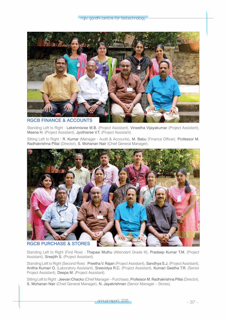
annual report 2015 - 317 -
rajiv gandhi centre for biotechnology
RGCB FINANCE & ACCOUNTS
Standing Left to Right (First Row) : Thapasi Muthu (Attendant Grade III), Pradeep Kumar T.M. (Project Assistant), Sreejith S. (Project Assistant).
Standing Left to Right (Second Row) : Preetha V. Rajan (Project Assistant), Sandhya S.J. (Project Assistant), Anitha Kumari O. (Laboratory Assistant), Sreevidya R.C. (Project Assistant), Kumari Geetha T.R. (Senior Project Assistant), Deepa M. (Project Assistant).
Sitting Left to Right : Jeevan Chacko (Chief Manager – Purchase), Professor M. Radhakrishna Pillai (Director), S. Mohanan Nair (Chief General Manager), N. Jayakrishnan (Senior Manager – Stores).
RGCB PURCHASE & STORES
Standing Left to Right : Lekshmisree M.B. (Project Assistant), Vineetha Vijayakumar (Project Assistant), Meena H. (Project Assistant), Jyothisree V.T. (Project Assistant).
Sitting Left to Right : R. Kumar (Manager - Audit & Accounts), M. Babu (Finance Officer), Professor M. Radhakrishna Pillai (Director), S. Mohanan Nair (Chief General Manager).
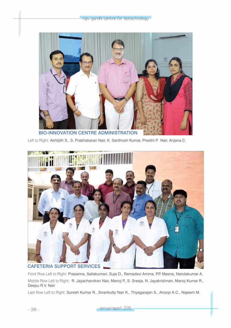
- 318 - annual report 2015
rajiv gandhi centre for biotechnology
Front Row Left to Right: Prasanna, Sailakumari, Suja O., Remadevi Amma, P.P. Meena, Nandakumar A.
Middle Row Left to Right: R. Jayachandran Nair, Manoj P., S. Sreeja, N. Jayakrishnan, Manoj Kumar R., Deepu R.V. Nair
Last Row Left to Right: Suresh Kumar R., Sivankutty Nair K., Thyagarajan S., Anoop K.C., Najeem M.
Left to Right: Akhiljith S., S. Prabhakaran Nair, K. Santhosh Kumar, Preethi P. Nair, Anjana C.
BIO-INNOVATION CENTRE ADMINISTRATION
CAFETERIA SUPPORT SERVICES
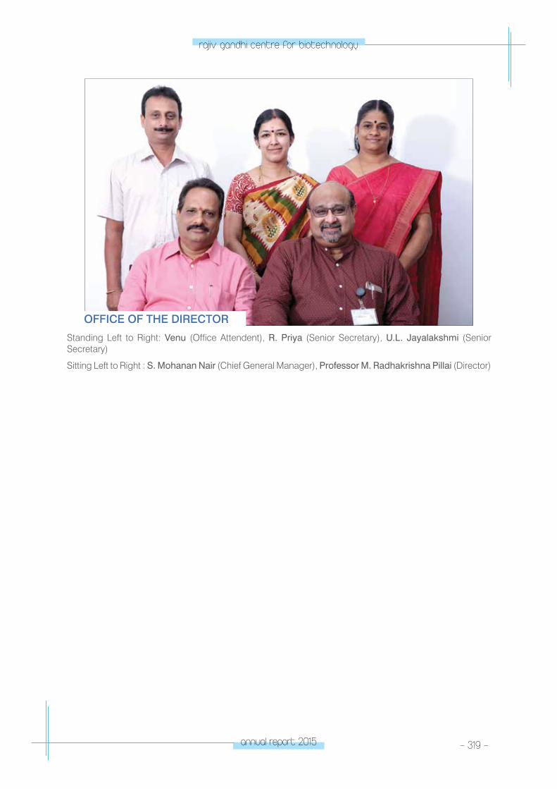
annual report 2015 - 319 -
rajiv gandhi centre for biotechnology
Standing Left to Right: Venu (Office Attendent), R. Priya (Senior Secretary), U.L. Jayalakshmi (Senior Secretary)
Sitting Left to Right : S. Mohanan Nair (Chief General Manager), Professor M. Radhakrishna Pillai (Director)
OFFICE OF THE DIRECTOR
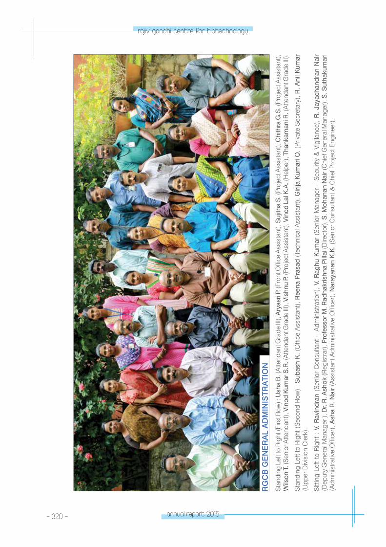
- 320 - annual report 2015
rajiv gandhi centre for biotechnology
Sta
ndin
g Le
ft to
Rig
ht (F
irst R
ow) :
Ush
a B
. (A
ttend
ant G
rade
III),
Ary
asri
P. (F
ront
Offi
ce A
ssis
tant
), S
ujith
a S
. (Pr
ojec
t Ass
ista
nt),
Chi
thra
G.S
. (Pr
ojec
t Ass
ista
nt),
Wils
on T
. (S
enio
r Atte
ndan
t), V
inod
Kum
ar S
.R. (
Atte
ndan
t Gra
de II
I), V
ishn
u P.
(Pro
ject
Ass
ista
nt),
Vin
od L
al K
.A. (
Hel
per)
, Tha
nkam
ani R
. (A
ttend
ant G
rade
III).
Sta
ndin
g Le
ft to
Rig
ht (S
econ
d R
ow) :
Sub
ash
K. (
Offi
ce A
ssis
tant
), R
eena
Pra
sad
(Tec
hnic
al A
ssis
tant
), G
irija
Kum
ari O
. (P
rivat
e S
ecre
tary
), R
. Ani
l Kum
ar
(Upp
er D
ivis
ion
Cle
rk).
Sitt
ing
Left
to R
ight
: V
. R
avin
dran
(S
enio
r C
onsu
ltant
– A
dmin
istra
tion)
, V.
Rag
hu K
umar
(S
enio
r M
anag
er –
Sec
urity
& V
igila
nce)
, R
. Ja
yach
andr
an N
air
(Dep
uty
Gen
eral
Man
ager
), D
r. R
. Ash
ok (R
egis
trar)
, Pro
fess
or M
. Rad
hakr
ishn
a P
illai
(Dire
ctor
), S
. Moh
anan
Nai
r (C
hief
Gen
eral
Man
ager
), S
. Sut
haku
mar
i (A
dmin
istra
tive
Offi
cer)
, Ash
a R
. Nai
r (A
ssis
tant
Adm
inis
trativ
e O
ffice
r), N
aray
anan
K.K
. (S
enio
r Con
sulta
nt &
Chi
ef P
roje
ct E
ngin
eer)
.
RG
CB
GE
NE
RA
L A
DM
INIS
TRA
TIO
N
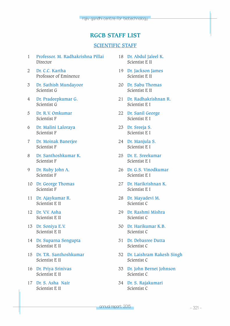
annual report 2015 - 321 -
rajiv gandhi centre for biotechnology
1 Professor. M. Radhakrishna Pillai Director
2 Dr. C.C. Kartha Professor of Eminence
3 Dr. Sathish Mundayoor Scientist G
4 Dr. Pradeepkumar G. Scientist G
5 Dr. R.V. Omkumar Scientist F
6 Dr. Malini Laloraya Scientist F
7 Dr. Moinak Banerjee Scientist F
8 Dr. Santhoshkumar K. Scientist F
9 Dr. Ruby John A. Scientist F
10 Dr. George Thomas Scientist F
11 Dr. Ajaykumar R. Scientist E II
12 Dr. V.V. Asha Scientist E II
13 Dr. Soniya E.V. Scientist E II
14 Dr. Suparna Sengupta Scientist E II
15 Dr. T.R. Santhoshkumar Scientist E II
16 Dr. Priya Srinivas Scientist E II
17 Dr. S. Asha Nair Scientist E II
RGCB STAFF LIST
SCIENTIFIC STAFF
18 Dr. Abdul Jaleel K. Scientist E II
19 Dr. Jackson James Scientist E II
20 Dr. Sabu Thomas Scientist E II
21 Dr. Radhakrishnan R. Scientist E I
22 Dr. Sanil George Scientist E I
23 Dr. Sreeja S. Scientist E I
24 Dr. Manjula S. Scientist E I
25 Dr. E. Sreekumar Scientist E I
26 Dr. G.S. Vinodkumar Scientist E I
27 Dr. Harikrishnan K. Scientist E I
28 Dr. Mayadevi M. Scientist C
29 Dr. Rashmi Mishra Scientist C
30 Dr. Harikumar K.B. Scientist C
31 Dr. Debasree Dutta Scientist C
32 Dr. Laishram Rakesh Singh Scientist C
33 Dr. John Bernet Johnson Scientist C
34 Dr. S. Rajakumari Scientist C
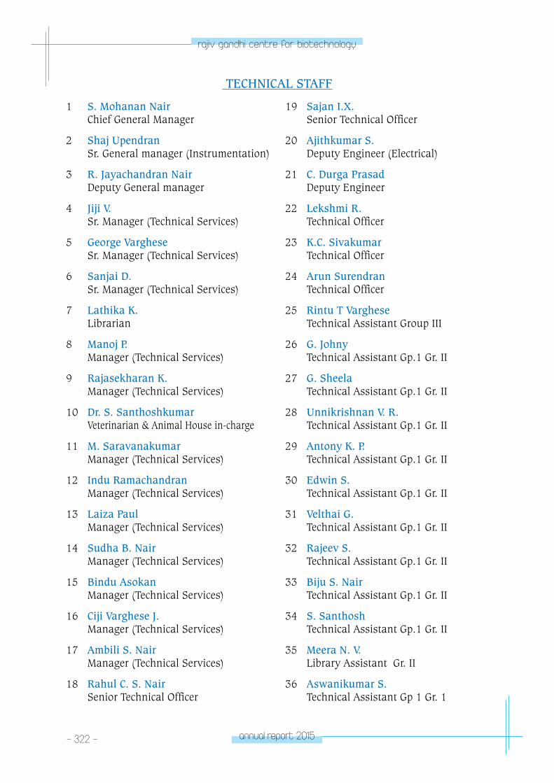
- 322 - annual report 2015
rajiv gandhi centre for biotechnology
1 S. Mohanan Nair Chief General Manager
2 Shaj Upendran Sr. General manager (Instrumentation)
3 R. Jayachandran Nair Deputy General manager
4 Jiji V. Sr. Manager (Technical Services)
5 George Varghese Sr. Manager (Technical Services)
6 Sanjai D. Sr. Manager (Technical Services)
7 Lathika K. Librarian
8 Manoj P. Manager (Technical Services)
9 Rajasekharan K. Manager (Technical Services)
10 Dr. S. Santhoshkumar Veterinarian & Animal House in-charge
11 M. Saravanakumar Manager (Technical Services)
12 Indu Ramachandran Manager (Technical Services)
13 Laiza Paul Manager (Technical Services)
14 Sudha B. Nair Manager (Technical Services)
15 Bindu Asokan Manager (Technical Services)
16 Ciji Varghese J. Manager (Technical Services)
17 Ambili S. Nair Manager (Technical Services)
18 Rahul C. S. Nair Senior Technical Officer
TECHNICAL STAFF
19 Sajan I.X. Senior Technical Officer
20 Ajithkumar S. Deputy Engineer (Electrical)
21 C. Durga Prasad Deputy Engineer
22 Lekshmi R. Technical Officer
23 K.C. Sivakumar Technical Officer
24 Arun Surendran Technical Officer
25 Rintu T Varghese Technical Assistant Group III
26 G. Johny Technical Assistant Gp.1 Gr. II
27 G. Sheela Technical Assistant Gp.1 Gr. II
28 Unnikrishnan V. R. Technical Assistant Gp.1 Gr. II
29 Antony K. P. Technical Assistant Gp.1 Gr. II
30 Edwin S. Technical Assistant Gp.1 Gr. II
31 Velthai G. Technical Assistant Gp.1 Gr. II
32 Rajeev S. Technical Assistant Gp.1 Gr. II
33 Biju S. Nair Technical Assistant Gp.1 Gr. II
34 S. Santhosh Technical Assistant Gp.1 Gr. II
35 Meera N. V. Library Assistant Gr. II
36 Aswanikumar S. Technical Assistant Gp 1 Gr. 1

annual report 2015 - 323 -
rajiv gandhi centre for biotechnology
37 Prakash R. Laboratory Technician
38 Kannan T.R. Laboratory Technician
39 Amal V. Technical Assistant Group I
40 Dileep Kumar R. Technical Assistant Group I
41 Reena Prasad Technical Assistant
42 Gopakumar G. Helper
43 Venugopalan J. Helper
44 K.A. Vinod Lal Helper
45 Jayanandan J. Helper
46 Sumaja V. Helper/Lab Helper
ADMINISTRATION
1 Dr. Ashok R. Registrar Grade II
2 K.M. Nair Chief Controller
3 M.Babu Finance Officer
4 Jeevan Chacko Chief Manager (Purchase)
5 K.K. Jayasree Accounts Officer
6 Suthakumari S. Administrative Officer
7 V. K. Reghukumar Senior manager ( Security & Vigilance)
8 Jayakrishnan N. Senior Manager (Purchase)
9 R. Kumar Manager (Accounts & Audit)
10 Jayalekshmi U. S. Sr. PS to Director
11 Priya R. Sr. PS to Director
12 Asha R. Nair Asst. Administrative Officer
13 O.Girijakumari Private Secretary
14 Preetha J. Section Officer
15 Subash K. Office Assistant
16 Anilkumar R. UDC
17 Harikumar S. Driver Grade IV
18 Vijayakumar S. Driver Grade IV
19 T. Wilson Senior Attendant
20 Chandrika Devi B. Senior Attendant
21 Thapasi Muthu Nadar C. Attendant Grade III
22 Usha B. ` Attendant Grade III
23 Vinodkumar S. R. Attendant Grade III
24 Thankamany R. Attendant Grade III
25 Manukumar V. M. Driver