androgen receptor or estrogen receptor-β blockade alters dhea-, dht-, and e2-induced proliferation...
TRANSCRIPT

The Prostate 71:766 ^777 (2011)
Androgen-InducedPSAExpressionRequiresNotOnlyActivationofARButAlso Endogenous IGF-IorIGF-I/PI3K/Akt Signaling inHumanProstate
Cancer Epithelial Cells
Xunxian Liu,* Renee Y. Choi, Shayma M. Jawad, and Julia T. Arnold
Endocrine Section, Laboratoryof Clinical Investigation,Divisionof Intramural Research,National Center forComplementaryandAlternativeMedicine,National InstitutesofHealth, Bethesda,Maryland
BACKGROUND. Prostate cancer (PrCa) risk is positively associatedwith levels of insulin-likegrowth factor I (IGF-I) and prostate specific antigen (PSA), both androgen receptor (AR)signaling target genes in PrCa cells. Although activated AR is required for androgen-inductionof expression of both genes, effects of the IGF-I signaling pathways on the androgen-inductionof PSA have not been studied.METHODS. Human prostate stromal and epithelial cancer cells were treated alone or incoculture with steroid hormone and/or inhibitors. Gene or protein expressionwas analyzed byreal time RT-PCR or Western blotting of lysates, nuclear extracts, or immunoprecipitatedproducts.RESULTS. In PrCa epithelial cells, endogenous IGF-I, significantly induced by R1881, wasrequired for R1881-induction of PSA. Increased IGF-I correlated with accumulation ofcytoplasmic dephospho b-catenin (CPDP b-catenin), a co-activator of AR signaling. ExogenousIGF-I enhanced R1881-induced PSA and accumulation of CPDP b-catenin in LAPC-4 cells.Functional depletion of IGF-I or IGF-I receptor diminished PSA induction. Induction of IGF-Ireached a plateauwhile PSA consecutively increased. Inhibiting PI3K abolishedR1881-inducedAkt phosphorylation, CPDP and nuclear b-catenin and nuclear association of AR/b-catenin,consequently abrogating R1881-induced expression of IGF-I and/or PSA.CONCLUSIONS. By integrating androgen, IGF-I and b-catenin signaling pathways, thesedata reveal that androgen-induced PSA expression requires activation of AR and endogenousIGF-I or IGF-I/PI3K/Akt signaling, suggesting a positive feedback cycle for increasedproduction of PSA associated with PrCa. Prostate 71: 766–777, 2011. # 2010 Wiley-Liss, Inc.
KEY WORDS: Akt; AR; b-catenin; IGF-I; prostate cancer; PSA; R1881
INTRODUCTION
Androgens and their cognate receptor, androgenreceptor (AR) play an essential role in mediating malesexual differentiation in utero, sperm production atpuberty, prostate development in the adult, and alsopromoting prostate cancer (PrCa) growth in PrCapatients [1]. In PrCa cells, the androgenic responsecross-talks with multifunctional growth factor signal-ing pathways (such as EGF, FGF, IGF, TGF-b, andVEGF) [2]. Among these growth factors, IGF-I mayhave the most inseparable connection with PrCa. IGF-Iis found in both stromal and epithelial prostate tissuecompartments but changes with cancer progression. In
normal prostate, cells IGF-I expression is higher instroma than in epithelium and is primarily a paracrinesignal. But in PrCa tissue, it is more strongly expressedin epithelial malignant cells becoming an autocrinesignal to the epithelial compartment [3].
There are no conflicts of interest for any of the authors.
*Correspondence to: Xunxian Liu, NIH-NCCAM Building 10, Rm2B47, MSC 1547; 9000 Rockville Pike, Bethesda, MD 20892-1547.E-mail: [email protected] 16 April 2010; Accepted 26 September 2010DOI 10.1002/pros.21293Published online 28 October 2010 in Wiley Online Library(wileyonlinelibrary.com).
� 2010 Wiley-Liss, Inc.

Previous studies in this laboratory addressed steroidhormonal-induced prostate stromal paracrine effectson epithelial cells and demonstrated that androgen-induced PSA levels in LAPC-4 cells were consistentlyincreased when they grown in the presence of 6Sprimary prostate stromal cells [4,5]. This suggestedstromal cell paracrine factors may amplify androgenicsignals. One factor likely to be involved in thisamplification is IGF-I. Although activated AR isrequired for androgen-induction of expression of bothIGF-I and PSA, the role of IGF-I in androgen-inductionof PSA has not been elucidated. It is understood thatandrogen interacts with the AR and can induce PSAtranscription via androgen response elements (AREs)in the PSA promoter [6]. Androgens (testosterone anddihydro-testosterone) induced IGF-I in LNCaP cells [7].Two AREs have been identified in the IGF-I upstreampromoter, leading to androgen activation of IGF-Iexpression in PrCa cells [8]. Androgens also stimulateexpression of IGF-I in human primary PrCa-associatedstromal cells (6S cells) but not in normal humanprostate stromal cells [9]. The evidence of androgeninduction of IGF-I and the existence of ARE in the IGF-Ipromoter in PrCa cells suggest that genetic mutationsenhancing androgen inductionof IGF-I expressionmaycontribute to promotion of PrCa, as IGF-I is a pro-cancer factor. IGF-I activates both phosphoinositide3-kinase (PI3K)/Akt and Raf/mitogen-associated/extracellular regulated kinase (MEK)/extracellularregulated kinases (ERK) pathways via IGF-I receptor(IGF-IR), resulting in promotion of growth andreduction of apoptosis in cells, increasing the possibil-ity of cancer promotion [10]. Androgens increase IGF-IR expression in HepG2 and LNCaP cells, providingmore receptors for IGF signaling in the PrCa cells [8].Additionally, elevated levels of IGF-I in blood havebeen associated with increased risk of PrCa [11–14].IGF-I increases AR transactivation in PrCa cells, undervery low/absent androgen levels [15,16] and PrCa cellgrowth [17]. Overexpression of FOXO-1 suppressesandrogen-induced AR transactivation, by binding toligand-AR to the AR promoters and disrupting ofandrogen-induced AR nuclear localization; this sup-pression is attenuated by IGF-I administration viaactivation of PI3K/Akt pathway, leading to phosphor-ylating and inactivating FOXO-1 [18].
We previously reported that IGF-I rescued campto-thecin-induced apoptotic death in normal prostateepithelial (NPE) cells when co-cultured with prostatestromal (6S) cells after treatment with androgen, andthat IGF-I also promoted NPE cell proliferation byactivating PI3K/Akt then inactivating glycogen syn-thesis kinase-3 (GSK3) [19]. Akt reduction of GSK3activity consequently leads to stabilization of b-catenin[20], a co-activator of AR signaling to enhance tran-
scription of IGF-I and prostate specific antigen (PSA)[19,21,22].
IGF has been shown to induce cellular levels of b-catenin in LNCaP prostate cancer cells which have amutation in the AR ligand binding domain [23]. Thepresent study contributes understanding of additionalpathways for regulation of PSA expression using theco-culture of LAPC-4, containing the wild-type AR,andprimaryprostate stromal (6S) cells ormono-cultureof these cells. It demonstrates that while IGF-I alone upto 50 ng/ml cannot induce PSA in LAPC-4 cells,endogenous production of IGF-I by 6S stromal or PrCaepithelial cells or IGF-I/PI3K/Akt signaling is requiredfor R1881-stimulated expression of IGF-I and PSA. PSAvalues from LAPC-4 cells coculturedwith stromal cellswere consistently increased, indicating contributions ofIGF-I from stromal cells. This study recreates auto-crine/paracrine positive feedback loop via genomicand non-genomic pathways of endogenous IGF-Iproduction in both prostate stromal and epithelialcells, upregulation of cytoplasmic b-catenin, stabiliza-tion of the AR, increased PSA expression, and addi-tional IGF-I production.
MATERIALS ANDMETHODS
Cell Culture
The sources of primary prostate stromal 6S cells andLAPC-4 cells and the co-culture and mono-cultureconditions have been described [4,24]. LNCaP cellswere purchased from ATCC (Manassas, VA). LNCaPcells were cultured as LAPC-4 and 6S cells. Cocultureswere prepared in Treatment Media consisting ofMedium 199 (phenol red-free)/F12 phenol red-reduced media (Invitrogen, Carlsbad, CA) (1:1) sup-plemented with penicillin (100U/ml), streptomycin(100mg/ml), glutamine (100mg/ml; Invitrogen), and2% charcoal dextran-treated FBS (CDS, Hyclone Labs).LAPC-4 cellswere seeded at adensity of 10,000 cells perwell ontoMillipore PICM12mm inserts (Billerica,MA)coated with a 1:10 dilution in H2O of MatrigelTM (BDBiosciences, Bedford, MA). Parallel cultures of stromalcells (6S) cells were seeded at 10,000 cells perwell in 24-well plates. LAPC-4 and 6S cellswere grown separatelyfor 2 or 3 days. Cocultures were initiated by placinginserts containing epithelial cells into wells containingstromal cells and adding appropriate hormone orreagent treatments.
Antibodies and Reagents
Antibodies against IGF-I, Insulin like growth factor-Ireceptor (IGF-IR) and normal IgG without sodiumazide have been described [19]. Anti- (a-)AR,a-pAkt308T, a-b-catenin, a-IGF-IRb, a-Lamin A/C,
The Prostate
IGF-IRole inAndrogen Induction of PSA 767

control siRNA and IGF-IRab siRNA were purchasedfrom Santa Cruz (Santa Cruz, CA); a-Akt, a-pERK, a-ERK were from Cell Signaling Technology (Danvers,MA), and a-GAPDH from Advanced ImmunoChem-ical, Inc. (Long Beach, CA). a-Cytoplasmic dephospho-(CPDP) b-catenin was obtained from EMD Chemicals(Gibbstown, NJ) and a-PSA from Abcam (Cambridge,MA). R1881 was bought from PerkinElmer (Wellesley,MA) dissolved in ethanol and 10 nM was used in cellmedia (final ethanol concentrations<0.02%). IGF-I, LY-294002 (LY-1 mg/ml applied to cells), cell permeablePI3K inhibitor, and PD 98059 (PD-1 mg/ml applied tocells), inhibitor of the activation of mitogen-activatedprotein kinase kinase (MAPKK) were obtained fromSigma–Aldrich (St. Louis, MO).
RNAExtraction andReal-TimeQuantitativeRT-PCR
Cells were left untreated, or treated with R1881(10 nM) and/or LY (1 mg/ml) for 2 days. RNA wasextracted and evaluation of gene expression wasperformed as reported [5,9]. For real-time PCR of geneexpression in LNCaP cells, LNCaP cells were platedwith 1,500 cells/well in a 96-well plate and transfectedwith control siRNA or IGF-IRab siRNA for 2 days.Transfection was performed according to siRNAmanufacturer’s protocol. Then, the cells were treatedwithout orwithR1881 for 2 days. Real-timePCRof IGF-I, PSA, and RPLP0 was performed using Power SYBRGreen Cell to CT Kit (Applied Biosystems, Foster City,CA) and RPLP0 values were used for normalization.Each value of real-time PCR fold change was averagedfrom 3 to 6 replicates and each point shown in real-timePCR figures was averaged from the values derivedfrom three separate experiments.
Immunoprecipitation andWestern BlotAnalysis
Cells were treated without or with R1881 and/orother reagents (normal IgG,Anti IGF-I and IGF-IR, IGF-I, LY, or PD) for 3 days and lysed using lysis buffer andprotocols from Cell Signaling Technology. Proteinconcentrations were assessed in the cell lysates usingthe bicinchoninic acid protein (BCA) assay kit (ThermoScientific, Rockford, IL). Western blotting procedureshave been described [25].
Nuclear Fractionation and Immunoprecipitation/Western Blot Analysis toDetect Localization and/or
Association of AR andb-Catenin
The nuclear localization and/or association of ARand b-catenin were studied by immunoprecipitation/Western blot according to previously reported meth-ods [25] with minor modifications. Treated LAPC-4 or6S cells were suspended with Trypsin EDTA (Invitro-
gen) and pelleted. The pellets were lysed using lysisbuffer containing 20mM Tris–HCl (pH 7.8), 0.5%Nonidet P-40, 137mM NaCl, 50 mM EDTA, 1mMNaF, 1mM NaVO3, and protease inhibitors (RocheDiagnostics, Mannheim, Germany) for 10min on ice.Cell lysateswere spun at 2,000 rpm for 5min at 48C. Thepellets were re-suspended in the lysis buffer, passedthrough a 30.5-gauge needle to disrupt nuclei and thencleared at 3,000 rpm for 5min. Protein extracts weredirectly loaded onto NuPAGE 4–12% Bis-Tris Gels forWestern blot to measure AR or b-catenin concentra-tions, or incubated with a-AR overnight at 48C,followed by 1 hr incubation with protein A agarose(Sigma–Aldrich). Control immunoprecipitation sam-ples were performed with normal rabbit IgG (SantaCruz). After three washes with 1ml of the lysis buffer,the pellets were suspended in SDS sample buffer,heated at 958C for 5min, and loaded onto NuPAGE forWestern blot.
ConfocalMicroscopyAnalysis
LAPC-4 cells were plated onto sterile cover slips.Cells were left untreated, or treated with R1881 and/orLY for 2 days. The fixing, blocking and stainingprocedures have been described [26]. Samples werestained with primary antibodies,a-AR (rabbit IgG,1:100) and a-b-catenin (goat IgG, 1:50), and secondaryantibodies, donkey anti-rabbit conjugated rhodamine1:200 and donkey anti-goat conjugated FITC 1:100(Santa Cruz). VECTA-SHIELD HardSet MountedMedium with DAPI (Vector Labs, Burlingame, CA)was employed for mounting cover slips. Cells wereviewed with a confocal laser microscope (Lenses: 63W;LSM510; Carl Zeiss MicroImaging, Inc.) in the Lab ofCellular and Developmental Biology, NIDDK, NIH.
Statistical Analysis
All shown data represent two or more separateexperiments. Quantitative data are presented as meanvalues� SEM, averaged from three or more separateexperiments. The statistical analysis was performedusing the JMP (SAS Institute, Cary, NC) statisticalpackage based on t-test. An adjusted P-value of �0.05was considered significant. Densitometric values forprotein bands were reported in arbitrary units.
RESULTS
Effects of R1881and Primary Stromal Cells onLAPC-4 Expression of PSA, AR, andb-Catenin
Previously, we determined R1881-induced LAPC-4PSA gene and protein expression by �2-fold when inco-culture with 6S cells compared to that from LAPC-4
The Prostate
768 Liuet al.

cells in mono-culture using ELISA and real-time PCR[4,5]. In the current study, we used Western blot tomeasure PSA production in LAPC-4 cell lysates in co-culture versus mono-culture treated without or withR1881. There was no detectable PSA from untreatedLAPC-4 cells grown in co-culture or mono-culturewhereas R1881 induced PSAprotein expression in bothco-cultured andmono-cultured LAPC-4 cells (Fig. 1A).The PSA production from LAPC-4 cells in co-culturewas �2-fold (P< 0.001) over that from cells in mono-culture (Fig. 1B), which is similar to the increase aspreviously determined using ELISA [4,5]. AR wasexpressed in LAPC-4 cells and R1881 treatment greatlystabilized the protein, but no differences of ARexpression were found between cells grown in mono-and co-culture (Fig. 1A). Because b-catenin is a co-activator of AR signaling for androgen-induced PSA[21,22], R1881might also induce b-catenin stabilization.
Indeed, R1881 increased cytoplasmic dephospho-(CPDP) b-catenin but not whole b-catenin (Pan b-catenin) in LAPC-4 cells in co-culture or mono-culture(Fig. 1A). Additionally R1881 induced CPDP b-cateninexpression>2.5-fold in LAPC-4 cells in co-culture overthat in cells in mono-culture (Fig. 1A,C, P< 0.01). Theincreased levels of CPDP b-catenin in co-culturedLAPC4 cells may contribute to the increased PSA asinduced by R1881.
R1881-Induced IGF-IStabilizesb-Catenin and Is aPrerequisite for R1881Induction of PSA
The link between increased levels of CPDP b-cateninand PSA was investigated by analyzing how b-cateninis stabilized by R1881. Androgens induce IGF-Iexpression in 6S cells [9] and exogenous IGF-I canstabilize b-catenin by activating PI3K/Akt followed byinactivation of GSK3, resulting in accumulation ofcytosolic b-catenin [27], which may be a resource ofCPDP b-catenin. This pathway was evaluated usingboth stromal and epithelial PrCa cells grown in mono-culture or co-culture. IGF-I mRNA expression wasmeasured in LAPC-4 cells and 6S cells grown in mono-or co-culture using real-time PCR. R1881 induced IGF-ImRNA expression four- to fivefold (Fig. 2A, P< 0.001)in LAPC-4 cells compared to controls in both mono-culture and co-culture. These results indicate thatandrogen also induces IGF-I expression in PrCaepithelial LAPC-4 cells expressing normal AR. R1881also induced IGF-I mRNA expression in 6S cells inmono-culture by four- to fivefold (Fig. 2B, P< 0.05),consistent with the previous results in 6S cells inducedby androgens [9,19] and in co-culture by two- tothreefold (P< 0.05) (Fig. 2A,B). Therefore, R1881 sig-nificantly inducedmore expression of IGF-I in both celltypes in both culture conditions.
Previously IGF-I was shown to induce and enhanceandrogen-induced PSA in the AR-mutant LNCaP cells[15], but its effect on a normal AR such as in LAPC-4cellswas unknown. LAPC-4 cellswere left untreated ortreated with R1881 and/or IGF-I (1 ng, 10 ng, or 50 ng/ml) for 3 days. Figure 2C shows that IGF-I alone, atconcentrations up to 50 ng/ml, could not induce PSA;however, increasing amounts of IGF-I progressivelyincreasedR1881-inducedPSA.Cells treatedwithR1881plus exogenous IGF-I evidently produced more PSAthan that done by cells treated with R1881 alone. Theinability of IGF-I alone to induce PSA in LAPC-4 cellsmay be due to inability of IGF-I to activate the normalAR in LAPC-4 cells as IGF-I alone could not induce ARstability (Fig. 2C), as had been found in the mutant ARLNCaP cells [15]. Finally, CPDP b-catenin but not ARstability was positively associated with IGF-I concen-trations. This demonstrates that increased IGF-I (either
The Prostate
Fig. 1. PSA protein production in LAPC-4 cells grown in co-cul-turewith6Scellsor inmono-culture.A:LAPC-4cellswere treatedandculturedasindicated.Westernblotsof theLAPC-4lysateswereprobed by indicated antibodies. B: Relative intensity of R1881-induced PSA.The intensity of R1881-induced PSAprotein inmono-cultured LAPC-4 cells was set as one for comparison.C: RelativeintensityofR1881-inducedCPDPb-catenin. Shownintensitiesweresubtracted from their basal levels. The intensity of R1881-inducedCPDPb-cateninprotein inmono-cultured LAPC-4 cellswas set asone. B,C: Data are averaged from three blots including the shownoneinAandpresentedasmeanvalues� SEM. *** versus the other:P< 0.001and**versus theother:P< 0.01.
IGF-IRole inAndrogen Induction of PSA 769

The Prostate
Fig. 2. IGF-I is required for and enhances R1881induction of PSA.A,B: R1881increased IGF-ImRNA expression in LAPC-4 and 6S cells inmono-cultureorco-culture.Theexpressedfoldchanges arerelative to thebasal levels of IGF-ImRNAexpressioninmono-culturewhichweresetasone.Data areaveragedfromthreeseparateexperimentsandpresentedasmeanvalues� SEM.C: IGF-IenhancedR1881inductionofPSA.LAPC-4 cells were treated as indicated (D). IGF-I is required for R1881-induced PSA and CPDP b-catenin. LAPC-4 cells were culturedand treated as indicated.C,D:Westernblots of the lysateswereprobedby the indicated antibodies. Shownresults represent three separateexperiments. E: Kinetics of R1881-induced mRNA expression of IGF-I and PSA. The highest fold changes in each group were set as one.Data are averaged from four separate experiments and presented as mean values� SEM. * and *** versus control treatments: P< 0.05 or0.001;and#or###versus theothersin the samegroup:P< 0.05or0.001.
770 Liuet al.

R1881-induced or exogenous), stabilizes b-cateninwhich, togetherwith theR1881-activatedAR, promotesPSA expression.
To determine the importance of the IGF pathway inPSA production, endogenous IGF-I was blocked bytreating LAPC-4 cells in mono-culture or co-culturewith antibodies against both IGF-I and IGF-IR. PSAandCPDP b-catenin were used as read-outs of endogenousIGF-I as shown in similar studies using exogenous IGF-I [12,27]. PSA was induced by 10 nM R1881 in LAPC-4cells in mono-culture or co-culture for 3 days (Fig. 2D).Normal IgG (3.5 mg/ml), used as a control antibody,slightly reduced R1881-induced PSA induction andCPDP b-catenin expression whereas the R1881-induced PSA and CPDP b-catenin were stronglyinhibited by the combination of anti-IGF-I (2.5 mg/ml)and anti-IGF-IR (1.0 mg/ml), compared to R1881-induced PSA and CPDP b-catenin in cells treatedwithout antibody (Fig. 2D). However, R1881-inducedAR stability was not affected by antibody treatments,suggesting that IGF-I induction byR1881plays a criticalrole in accumulation of CPDP b-catenin, and conse-quently the PSA induction. In anotherwords, b-cateninstabilization is required for R1881-induced PSA induc-tion. Because IGF-IR mediates biological activities ofboth IGF-I and IGF-II [28], anti-IGF-IR antibody mayalso block IGF-II activity. However, IGF-II was notincreased by androgens in either cell type (data notshown).
To elucidate kinetics of androgen induction of IGF-Iand PSA gene expression, LAPC-4 cells were treatedwith R1881 for 0, 1, 2, 4, 8, 24, 48, and 72 hr. At theearly time points of 1, 2, 4, and 8hr, R1881-inducedmRNA levels of IGF-I and PSA were minimal andsimilar to those at time 0 (data not shown). However,R1881-induced mRNAs of IGF-I and PSA were signifi-cant from Days 1 to 3 (Fig. 2E). R1881-induced IGF-ImRNA reached a plateau on Day 2 (P< 0.05) whereasinduction of PSA consecutively increased with signifi-cant increases with each day (P< 0.05 or 0.001). IGF-ImRNA expression therefore transiently precedes andmay be involved in androgen induction of PSAmRNA.
R1881Induces PSAVia IGF-I/PI3K/Akt Pathway
The requirement of IGF-I for R1881-induced PSAand accumulation of CPDP b-catenin was demon-strated (Fig. 2). It has been previously suggested thatIGF-I induces the accumulation of cytosolic b-cateninvia PI3K/Akt; additionally, the IGF-I/Raf/MEK/ERKpathway is required for b-catenin/TCF signaling (b-CTS) [27]. The following studies validate that the IGF-I/PI3K/Akt pathway is involved in R1881-inducedPSA and in accumulation of CPDP b-catenin in LAPC-4cells and 6S cells, and investigate whether Raf/MEK/
ERK pathway was also involved in b-catenin/ARsignaling.
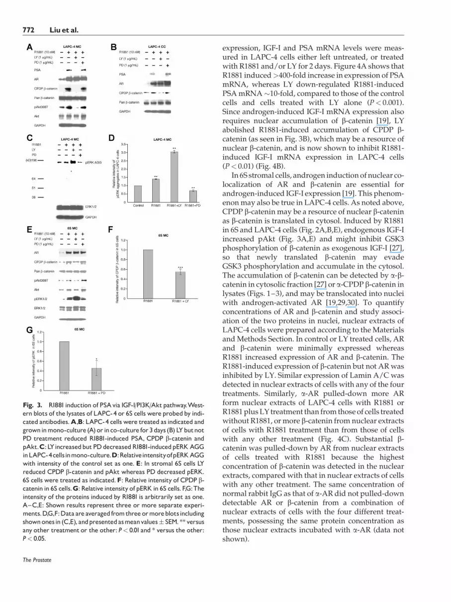
LAPC-4 cells (mono-culture or co-culture; Fig. 3A–D) and 6S cells (mono-culture Fig. 3E–G) were leftuntreated or treated with R1881 or R1881 plus 1 mg/mlLY (inhibitor of PI3K) or 1 mg/ml PD (inhibitor ofMAPKK) for 3 days. In LAPC-4 cells, R1881-inducedexpression of PSA, CPDP b-catenin and pAkt308T(mono-culture) was abolished by LY but not PD(Fig. 3A,B), whereas R1881-induced pERK aggregate(AGG) (mono-culture) increased �40% of the control(P< 0.01) and was down-regulated by PD to �70% ofthe control (P< 0.01) (Fig. 3C,D). Interestingly, R1881-induced pERK aggregate increased >3-fold of thecontrol (P< 0.01) by LY treatment. In the stromal 6Scells (Fig. 3E–G), R1881-induced CPDP b-catenin wasinhibited>40% (P< 0.001) by LY but not PD (Fig. 3E,F).Also, in 6S cells, R1881-induced pAktwas abrogated byLY but not PD, whereas R1881-induced pERK wasreduced to �40% by PD (P< 0.05) (Fig. 3E,G). In bothcell types, the R1881-induced AR stability was notaffected by either LY or PD (Fig. 3A,B,E). Androgen-induced AR stability might contribute to b-cateninstabilization in 6S cells [19] so that LY abrogation ofpAkt did not completely inhibit R1881-induced accu-mulation of CPDP b-catenin (Fig. 3E,F). R1881-inducedpERKwas also dramatically increased by LY treatmentin 6S cells. The increased pERK in cells treated withR1881 plus LYmight be androgen-dependent, as pERKdid not increase in LAPC-4 cells treated with LY alonecompared with that in control cells (data not shown).In both cells types (6S and LAPC-4 cells) with long term(3-day) treatment, increased expression of pERK1/2(Thr202/Tyr204 and Thr185/Tyr187) required R1881-induced IGF-I activation of upstream kinases of ERKand those kinaseswereperhaps also activatedbyGSK3,as LY inhibition of PI3K/Akt activities might result inunchecked GSK3 activities.
These experiments confirmed that both R1881-induced PSA and accumulation of CPDP b-cateninuse PI3K/Akt pathway but not MAPK pathway inthese prostate cells. Also, IGF-I activated Raf/MEK/ERK pathway is required for b-CTS [27] but notb-catenin/AR signaling.
PI3KPathway Is Involved in R1881Induction of IGF-Iand PSAGene Expression,b-CateninNuclear
Localization, and/orAssociation of AR/b-Catenin
The ability of LY to abolish R1881-induced PSAprotein expression is likely partly due to genomiceffects, because LY also abrogated R1881-inducedaccumulation of CPDP b-catenin, a co-activator forAR transactivation in the nuclei of the LAPC-4 cells. Toconfirm LY inhibition of R1881-induced target gene
The Prostate
IGF-IRole inAndrogen Induction of PSA 771
expression, IGF-I and PSA mRNA levels were meas-ured in LAPC-4 cells either left untreated, or treatedwith R1881 and/or LY for 2 days. Figure 4A shows thatR1881 induced>400-fold increase in expression of PSAmRNA, whereas LY down-regulated R1881-inducedPSAmRNA�10-fold, compared to those of the controlcells and cells treated with LY alone (P< 0.001).Since androgen-induced IGF-I mRNA expression alsorequires nuclear accumulation of b-catenin [19], LYabolished R1881-induced accumulation of CPDP b-catenin (as seen in Fig. 3B), which may be a resource ofnuclear b-catenin, and is now shown to inhibit R1881-induced IGF-I mRNA expression in LAPC-4 cells(P< 0.01) (Fig. 4B).
In 6S stromal cells, androgen inductionof nuclear co-localization of AR and b-catenin are essential forandrogen-induced IGF-I expression [19]. This phenom-enonmay also be true in LAPC-4 cells. As noted above,CPDP b-catenin may be a resource of nuclear b-cateninas b-catenin is translated in cytosol. Induced by R1881in 6S and LAPC-4 cells (Fig. 2A,B,E), endogenous IGF-Iincreased pAkt (Fig. 3A,E) and might inhibit GSK3phosphorylation of b-catenin as exogenous IGF-I [27],so that newly translated b-catenin may evadeGSK3 phosphorylation and accumulate in the cytosol.The accumulation of b-catenin can be detected by a-b-catenin in cytosolic fraction [27] or a-CPDP b-catenin inlysates (Figs. 1–3), and may be translocated into nucleiwith androgen-activated AR [19,29,30]. To quantifyconcentrations of AR and b-catenin and study associ-ation of the two proteins in nuclei, nuclear extracts ofLAPC-4 cells were prepared according to theMaterialsandMethods Section. In control or LY treated cells, ARand b-catenin were minimally expressed whereasR1881 increased expression of AR and b-catenin. TheR1881-induced expression of b-catenin but not ARwasinhibited by LY. Similar expression of Lamin A/C wasdetected in nuclear extracts of cells with any of the fourtreatments. Similarly, a-AR pulled-down more ARform nuclear extracts of LAPC-4 cells with R1881 orR1881plusLY treatment than from thoseof cells treatedwithout R1881, ormore b-catenin from nuclear extractsof cells with R1881 treatment than from those of cellswith any other treatment (Fig. 4C). Substantial b-catenin was pulled-down by AR from nuclear extractsof cells treated with R1881 because the highestconcentration of b-catenin was detected in the nuclearextracts, compared with that in nuclear extracts of cellswith any other treatment. The same concentration ofnormal rabbit IgG as that of a-AR did not pulled-downdetectable AR or b-catenin from a combination ofnuclear extracts of cells with the four different treat-ments, possessing the same protein concentration asthose nuclear extracts incubated with a-AR (data notshown).
The Prostate
Fig. 3. R1881induction of PSAvia IGF-I/PI3K/Aktpathway.West-ern blots of the lysates of LAPC-4 or 6S cells were probedby indi-cated antibodies.A,B: LAPC-4 cells were treated as indicated andgrowninmono-culture (A)or inco-culture for3days (B) LYbutnotPD treatment reduced R1881-induced PSA, CPDP b-catenin andpAkt.C: LYincreasedbutPDdecreasedR1881-inducedpERKAGGinLAPC-4cellsinmono-culture.D:RelativeintensityofpERKAGGwith intensity of the control set as one. E: In stromal 6S cells LYreduced CPDP b-catenin and pAkt whereas PD decreased pERK.6S cells were treated as indicated.F: Relative intensity of CPDP b-catenin in 6S cells.G: Relative intensity of pERK in 6S cells.F,G:Theintensity of the proteins induced by R1881 is arbitrarily set as one.A^C,E: Shown results represent three or more separate experi-ments.D,G,F:Data are averaged fromthree ormoreblotsincludingshownonesin (C,E), andpresentedasmeanvalues� SEM.**versusany other treatment or the other: P< 0.01and * versus the other:P< 0.05.
772 Liuet al.

The Prostate
Fig. 4. LY inhibited R1881 induction of expression of PSA and IGF-I mRNAs via diminishing R1881-induced nuclear b-catenin. A: LYstrongly reduced R1881-induced expression of PSA mRNA in LAPC-4 cells. B: LY inhibited R1881-induced expression of IGF-I mRNA inLAPC-4 cells.C:LYdecreasedR1881-inducednuclearb-catenin andassociationofAR/b-catenin inLAPC-4 cells.D:LYreducedR1881-inducednuclearb-cateninobservedusingconfocalmicroscopy.LAPC-4cellswere treatedasindicated.Cellswere fixedandblockedasdescribedin theMaterials andMethodsSection,andstainedby theindicatedantibodiesandDAPI [AR(red),b-catenin (green)andDAPI (blue)].Datarepresentthreeseparateexperiments.E:LYinhibitedR1881-inducedIGF-ImRNAexpressionin6Scells.A,B,E:ThecontrollevelsofmRNAexpressionaresetasone.Data areaveragedfromthreeseparateexperimentsandpresentedasmeanvalues� SEM.***versusanyother treatment:P< 0.001and ** versus anyother treatment:P< 0.01.LYinhibitedR1881-inducednuclear expression ofAR and b-catenin in 6S cells.C,F: Preparation ofnuclearextracts of LAPC-4 and6S cells forWesternblots andimmunoprecipitation/Westernblots is describedin theMaterials andMethodsSection.Westernblotswerestainedby theindicatedantibodies.Datarepresent three separateexperiments.
IGF-IRole inAndrogen Induction of PSA 773

To visualize the dynamics of AR and b-catenin,LAPC-4 cells were treated without or with R1881 and/or LY for 2 days. Localization of AR and b-catenin wasanalyzed using confocal microscopy. In control cellsand cells treatedwithLYalone, stainingofAR (red) andb-catenin (green) represent their basal expression(Fig. 4D) where AR was localized in the cytoplasmand b-catenin was concentrated in plasma membrane.AR and b-catenin occasionally localized in nuclei butthey were separate, as purple staining (AR) andcyanine staining (b-catenin) are observed in themergedimages. R1881 considerably induced expression of ARand b-catenin, as well as their nuclear co-localization,indicated by increased white color visualized in themerged image. LY treatment diminished R1881-induced white color by strongly reducing expressionof b-catenin but not AR, leaving R1881-induced AR innuclei (blue DAPI) seen as purplish red color in themerged image. Although AR was still strongly asso-ciated with nuclei, which is suggested by purplish redcolor (Fig. 4D), LY strongly inhibited R1881-inducedPSA and IGF-I (Fig. 4A,B), because of lacking b-catenin,a co-activator of AR signaling, in nuclei (Fig. 4C,D).These data support that androgen induction of b-catenin but not AR, and induction of androgen signal-ing for PSA and IGF-I transcription require R1881-induced IGF-I/PI3K/Akt signaling axes in LAPC-4cells.
LY also abrogated R1881-induced IGF-I mRNAexpression in 6S cells (Fig. 4E); however, LY did notblock R1881-induced AR expression but reducedR1881-induced CPDP b-catenin expression by >40%and the amount of CPDP b-catenin was still consid-
erable, in 6S cell lysates (Fig. 3E,F). Western blot ofnuclear extracts of 6S cells shows that LY significantlyinhibited R1881-induced nuclear expression of AR andb-catenin; again, lamin A/C was used as a loadingcontrol (Fig. 4F). These data suggest that LY inhibitsR1881-induced AR/b-catenin translocation into nucleiin 6S cells (Fig. 6).
Depletion of IGF-IRDiminishes R1881-InducedGeneExpression of IGF-Iand PSA
The requirement of IGF-I signaling for androgen-induction of IGF-I and PSA gene expression wasadditionally demonstrated using siRNA to depleteIGF-IR. Because transfection efficiency of LAPC-4 cellsis very low, we used LNCaP cells, another PrCaepithelial cell line that induces IGF-I and PSA aftertreated with androgen [7,15]. LNCaP cells were trans-fected with control siRNA or IGF-IRab siRNA for2days, and then treatedwithout orwithR1881 for 24 hr.The cells were lysed for Western blots. The a-IGF-IRbantibody stains two bands. One is IGF-IR precursor,and the other is IGF-IR b. IGF-IR was depleted at leastgreater than 50% by IGF-IRab siRNA, independent oftreatmentwith R1881 (Fig. 5A). In cells transfectedwithcontrol siRNA or IGF-IRab siRNA, R1881 increasedIGF-IR expression (IGF-IR precursor and IGF-IRb)substantially greater than that of cognate controls(Fig. 5A). This observation is consistentwith a previousreport of androgen-induced increase of IGF-IR expres-sion in LNCaP cells [8]. R1881 also increased ARstability greater than 40% regardless of either siRNAadministration, whereas IGF-IRab siRNA alone
The Prostate
Fig. 5. Depletion of IGF-IR diminishes R1881-inducedgene expression of IGF-I and PSA in LNCaPcells.A: IGF-IRab siRNA inhibitedR1881-inducedCPDPb-catenin, pAkt, andpERK1/2.The treatments andantibodies areindicated.Datarepresent two separate experiments.B: IGF-IRab siRNA reduced R1881-inducedmRNAs of IGF-I and PSA.Data are averaged from three separate experiments and presented asmeanvalues� SEM.**versus anyother treatmentin thecognategroup:P< 0.01.
774 Liuet al.

slightly decreased AR expression, compared to that ofcells treated with control siRNA alone (Fig. 5A). R1881induction of CPDP b-catenin, pAkt308T, and pERK1/2expression was substantially reduced by IGF-IRabsiRNA. Therefore, IGF-IRab siRNA decreased R1881-induced IGF signaling but androgen-induced ARexpression remained as the cells expressed >90% ARlevel compared to that of cells treated with controlsiRNA and R1881 (Fig. 5A). In LNCaP cells, R1881induced greater than eight- or sixfold of mRNAexpression of IGF-I or PSA (P< 0.01) whereas IGF-IRab siRNA significantly inhibited >50% of theinduction (Fig. 5B). The reduction can be attributedto IGF-IRab siRNA inhibition of R1881-induced IGFsignaling (Fig. 5A).
The summary figure (Fig. 6) displays the interactionof these pathways in the 6S (stromal) and LAPC-4(cancer-epithelial) cells. Both cell types produceendogenous IGF-I with androgen stimulation. Down-stream of the IGF-IR, PI3K-induced pAKt inactivatesGSK3, resulting in stabilization of b-catenin and anincrease of AR/b-catenin association. This complex istranslocated to the nucleus where genomic transcrip-tion of PSA in epithelial cells and IGF-I in both cell typesis further promoted.
DISCUSSION
The study presented here provides a moleculardissection of IGF-I in cell-cell paracrine and/or auto-crine interactions within prostate stromal and epithe-lial cells in response to androgens. It is well establishedthat stromal–epithelial interactions are important forprostate development and maintenance [31] and thatthese interactions may become disordered in prostatepathology and cancer progression [32]. The datacompared here analyze the paracrine secondary effectsof IGF-I and androgen in stromal cells and theaccumulative effects to the epithelial cells, comparedto epithelial autocrine effects.
The IGF-I pathway is an important mediator ofgrowth and prostate pathology [32,33]. It is linkedwiththe PSA pathway as PSA is an IGF-I binding protein 3(IGFBP3) protease, allowing release of IGF-I intomicroenvironment [33]. PSA is the player and IGF-I isthe responder in this non-genomic pathway. Our studydemonstrates the interconnecting relationship of thesetwoproteins in non-genomic andgenomicpathways.Adiagram representing the highlights of the results isshown in Figure 6 which reviews the pathways of AR,b-catenin and IGF-I interactions in both 6S and LAPC4
The Prostate
Fig. 6. Feedbackcycle of R1881-induced cross-talkbetweenAR signaling and IGF-signaling in prostate stromal 6S cells and LAPC-4 cancerepithelialcells.AndrogensinduceIGF-Iexpressionin6SandLAPC-4cellsandstabilizeb-cateninbyactivatingPI3K/Aktresultinginaccumulationof cytosolic b-catenin.The AR-b-catenin complex is translocated to nucleus for additional production of PSA (LAPC-4) and IGF-I (6S andLAPC-4). Activities insidenuclei suggestgenomic effectswhereas activities outsidenuclei includingparacrine or autocrine or intra-cytosoliccommunications suggest non-genomic effects. ST, stabilization; GT, genetic transcription/translation. Several critical protein components ofbothpathways(IGF-I/PI3K/AktandIGF-I/Raf/MEK/ERK),suchasPI3K,GSK3,Raf,etc.areomittedfor simplification.[Color figurecanbeviewedin theonlineissue,whichis availableatwileyonlinelibrary.com.]
IGF-IRole inAndrogen Induction of PSA 775

cells. IGF-I can induce accumulation of CPDP b-catenin, leading to enhancing R1881-induced PSAexpression. R1881 binds to AR in both PrCa epithelialand 6S cells and induces the stabilization (ST) of thereceptor. AR stabilization also stabilizes b-catenin [34–36] which together localize to the nuclei of 6S cells andLAPC-4 cells and activate IGF-I and PSA (epithelialonly) gene expression. The induced IGF-I is secretedfrom 6S cells and LAPC-4 cells and can then bind toIGF-IR in 6S cells and LAPC-4 cells, thus activatingPI3K/Akt pathway and further stabilizing b-catenin,resulting in more of the AR co-activator and moreexpression of IGF-I and/or PSA. The androgen-induced expression of IGF-I and PSA is associatedwith accumulation of cytoplasmic and nuclear b-catenin, since functional depletion of IGF-I or blockingof PI3K/Akt signaling byLYor IGF-IRab siRNAdown-regulated R1881-induced accumulation of CPDP and/or nuclear b-catenin, and abrogated R1881-inducedexpression of IGF-I and/or PSA. Increasing exogenousIGF-I enhanced ligand-AR signaling via increasing b-catenin, a co-activator of AR signaling, resulting in theincreased PSA expression.
In normal prostate tissues, IGF-I mRNA expressionis predominantly paracrine expressed in the stromalcompartmentwhichhas ninefold IGF-I expression overepithelial cells [37]. A common feature of prostatecancer is the shift from paracrine (stromal) to autocrine(epithelial) expression of growth factors and the shiftto androgen independence [38]. In prostate cancertissues, the IGF-I expression levels are increased inboth stromal and epithelial components [3]. IncreasedIGF-I expression was also reported in reactive stromalcells compared to normal prostate stromal cells [9].IGF-I also protects cells from apoptosis [39] especiallyin the reactive stromal microenvironment [19]. Thisstudy adds to the increasing literature in cell–cellinteractions and by elucidating mechanisms of para-crine/steroid hormone-induced changes in cellulardifferentiation, proliferation, and gene expression[3–5,8,40,41].
CONCLUSION
The analysis of 6S prostate stromal cell and LAPC-4IGF production represents the microenvironment ofprostate cancer with locally increased intra-prostaticIGF levels. The epithelial autocrine production aug-ments the stromal paracrine production to amplify theIGF-PSA pathway expression. This study combinesseveral signaling pathways and also demonstrates thatthe ERK pathway is not involved signaling pathwaysfrom androgen to PSA in LAPC-4 cells. It is the first todemonstrate that androgen induced-PSA requiresandrogen-induced IGF-I in the PrCa cells, suggesting
a cellular and molecular basis of increased productionof human prostate PSA associated with PrCa.
ACKNOWLEDGMENTS
We thank Jennifer Chu for technical assistance. Weare grateful for Dr. Hui Chen and Dr. RanganathMuniyappa for reviewing the manuscript and render-ing valuable suggestions. This research was supportedby the Intramural Research Program of the NationalCenter for Complementary and Alternative Medicine,National Institutes of Health, Bethesda, MD 20892.
REFERENCES
1. Brinkmann AO, Trapman J. Genetic analysis of androgenreceptors in development and disease. Adv Pharmacol 2000;47:317–341.
2. Zhu ML, Kyprianou N. Androgen receptor and growth factorsignaling cross-talk in prostate cancer cells. Endocr Relat Cancer2008;15(4):841–849.
3. CardilloMR,Monti S, Di Silverio F, Gentile V, Sciarra F, ToscanoV. Insulin-like growth factor (IGF)-I, IGF-II and IGF type Ireceptor (IGFR-I) expression in prostatic cancer. Anticancer Res2003;23(5A):3825–3835.
4. Arnold JT, GrayNE, Jacobowitz K, Viswanathan L, Cheung PW,McFann KK, Le H, BlackmanMR. Human prostate stromal cellsstimulate increased PSA production in DHEA-treated prostatecancer epithelial cells. J Steroid BiochemMol Biol 2008;111(3–5):240–246.
5. Gray NE, Liu X, Choi R, Blackman MR, Arnold JT. Endocrine–immune–paracrine interactions in prostate cells as targeted byphytomedicines. Cancer Prev Res (Phila, PA) 2009;2(2):134–142.
6. Cleutjens KB, van Eekelen CC, van der Korput HA, BrinkmannAO, Trapman J. Two androgen response regions cooperate insteroid hormone regulated activity of the prostate-specificantigen promoter. J Biol Chem 1996;271(11):6379–6388.
7. Arnold JT, Le H, McFann KK, Blackman MR. Comparativeeffects of DHEA vs. testosterone, dihydrotestosterone, andestradiol on proliferation and gene expression in human LNCaPprostate cancer cells. Am J Physiol Endocrinol Metab 2005;288(3):E573–E584.
8. Wu Y, Zhao W, Zhao J, Pan J, Wu Q, Zhang Y, Bauman WA,CardozoCP. Identification of androgen response elements in theinsulin-like growth factor I upstream promoter. Endocrinology2007;148(6):2984–2993.
9. Le H, Arnold JT, McFann KK, Blackman MR. DHT andtestosterone, but not DHEA or E2, differentially modulate IGF-I, IGFBP-2, and IGFBP-3 in human prostatic stromal cells. Am JPhysiol Endocrinol Metab 2006;290(5):E952–E960.
10. Shelton JG, Steelman LS, White ER, McCubrey JA. Synergybetween PI3K/Akt and Raf/MEK/ERK pathways in IGF-1Rmediated cell cycle progression and prevention of apoptosis inhematopoietic cells. Cell Cycle 2004;3(3):372–379.
11. Amling CL. Relationship between obesity and prostate cancer.Curr Opin Urol 2005;15(3):167–171.
12. Hsing AW, Gao YT, Chua S Jr, Deng J, Stanczyk FZ. Insulinresistance and prostate cancer risk. J Natl Cancer Inst 2003;95(1):67–71.
13. Chan JM, Stampfer MJ, Giovannucci E, Gann PH, Ma J,Wilkinson P, Hennekens CH, Pollak M. Plasma insulin-like
The Prostate
776 Liuet al.

growth factor-I and prostate cancer risk: A prospective study.Science 1998;279(5350):563–566.
14. Mantzoros CS, Tzonou A, Signorello LB, Stampfer M, Tricho-poulos D, Adami HO. Insulin-like growth factor 1 in relation toprostate cancer and benign prostatic hyperplasia. Br J Cancer1997;76(9):1115–1118.
15. Culig Z, Hobisch A, Cronauer MV, Radmayr C, Trapman J,Hittmair A, Bartsch G, Klocker H. Androgen receptor activationin prostatic tumor cell lines by insulin-like growth factor-I,keratinocyte growth factor, andepidermal growth factor.CancerRes 1994;54(20):5474–5478.
16. Orio F Jr, Terouanne B, Georget V, Lumbroso S, Avances C,Siatka C, Sultan C. Potential action of IGF-1 and EGF onandrogen receptor nuclear transfer and transactivation innormal and cancer human prostate cell lines. Mol CellEndocrinol 2002;198(1–2):105–114.
17. BurfeindP,ChernickyCL,RininslandF, Ilan J.AntisenseRNAtothe type I insulin-like growth factor receptor suppresses tumorgrowth andprevents invasionby rat prostate cancer cells in vivo.Proc Natl Acad Sci USA 1996;93(14):7263–7268.
18. FanW, Yanase T, MorinagaH, Okabe T, NomuraM, DaitokuH,Fukamizu A, Kato S, Takayanagi R, Nawata H. Insulin-likegrowth factor 1/insulin signaling activates androgen signalingthrough direct interactions of Foxo1 with androgen receptor. JBiol Chem 2007;282(10):7329–7338.
19. Liu X, Allen JD, Arnold JT, Blackman MR. Lycopene inhibitsIGF-I signal transduction and growth in normal prostateepithelial cells by decreasing DHT-modulated IGF-I productionin co-cultured reactive stromal cells. Carcinogenesis 2008;29(4):816–823.
20. Fukumoto S,HsiehCM,MaemuraK, LayneMD,Yet SF, LeeKH,Matsui T, Rosenzweig A, TaylorWG, Rubin JS, PerrellaMA, LeeME. Akt participation in the Wnt signaling pathway throughDishevelled. J Biol Chem 2001;276(20):17479–17483.
21. Masiello D, Chen SY, Xu Y, Verhoeven MC, Choi E, HollenbergAN, Balk SP. Recruitment of beta-catenin by wild-type ormutant androgen receptors correlates with ligand-stimulatedgrowth of prostate cancer cells. Mol Endocrinol 2004;18(10):2388–2401.
22. Chesire DR, Ewing CM, Gage WR, Isaacs WB. In vitro evidencefor complex modes of nuclear beta-catenin signaling duringprostate growth and tumorigenesis. Oncogene 2002;21(17):2679–2694.
23. Verras M, Sun Z. Beta-catenin is involved in insulin-like growthfactor 1-mediated transactivation of the androgen receptor. MolEndocrinol 2005;19(2):391–398.
24. Weeraratna AT, Arnold JT, George DJ, DeMarzo A, Isaacs JT.Rational basis for Trk inhibition therapy for prostate cancer.Prostate 2000;45(2):140–148.
25. Liu X, Rubin JS, Kimmel AR. Rapid, Wnt-induced changes inGSK3beta associations that regulate beta-catenin stabilizationare mediated by Galpha proteins. Curr Biol 2005;15(22):1989–1997.
26. Liu X, Arnold JT, Blackman MR. Dehydroepiandrosteroneadministration or Galpha q overexpression induces beta-catenin/T-Cell factor signaling and growth via increasingassociation of estrogen receptor-beta/Dishevelled2 in andro-gen-independent prostate cancer cells. Endocrinology 2010;151(4):1428–1440.
27. Desbois-Mouthon C, Cadoret A, Blivet-Van Eggelpoel MJ,Bertrand F, Cherqui G, Perret C, Capeau J. Insulin and IGF-1stimulate the beta-catenin pathway through two signallingcascades involving GSK-3beta inhibition and Ras activation.Oncogene 2001;20(2):252–259.
28. Rechler MM, Nissley SP. Peptide growth factors and theirreceptors I. New York, New York: Springer-Verlag; 1991.
29. Cunha GR, Cooke PS, Kurita T. Role of stromal-epithelialinteractions in hormonal responses. Arch Histol Cytol 2004;67(5):417–434.
30. Cunha GR. Role of mesenchymal–epithelial interactions innormal and abnormal development of the mammary gland andprostate. Cancer 1994;74 (3 Suppl):1030–1044.
31. Kleinberg DL, Ruan W, Yee D, Kovacs KT, Vidal S. Insulin-likegrowth factor (IGF)-I controls prostate fibromuscular develop-ment: IGF-I inhibition prevents both fibromuscular and glan-dular development in eugonadal mice. Endocrinology 2007;148(3):1080–1088.
32. RuanW, Powell-Braxton L, Kopchick JJ, KleinbergDL. Evidencethat insulin-like growth factor I and growth hormone arerequired for prostate gland development. Endocrinology1999;140(5):1984–1989.
33. Cohen P, Graves HC, Peehl DM, Kamarei M, Giudice LC,Rosenfeld RG. Prostate-specific antigen (PSA) is an insulin-likegrowth factor binding protein-3 protease found in seminalplasma. J Clin Endocrinol Metab 1992;75(4):1046–1053.
34. Pawlowski JE, Ertel JR, Allen MP, Xu M, Butler C, Wilson EM,Wierman ME. Liganded androgen receptor interaction withbeta-catenin: Nuclear co-localization and modulation of tran-scriptional activity in neuronal cells. J Biol Chem 2002;277(23):20702–20710.
35. Mulholland DJ, Cheng H, Reid K, Rennie PS, Nelson CC. Theandrogen receptor can promote beta-catenin nuclear trans-location independently of adenomatous polyposis coli. J BiolChem 2002;277(20):17933–17943.
36. Singh R, Artaza JN, Taylor WE, Braga M, Yuan X, Gonzalez-Cadavid NF, Bhasin S. Testosterone inhibits adipogenic differ-entiation in 3T3-L1 cells: Nuclear translocation of androgenreceptor complex with beta-catenin and T-cell factor 4 maybypass canonical Wnt signaling to down-regulate adipogenictranscription factors. Endocrinology 2006;147(1):141–154.
37. Ohlson N, Bergh A, Stattin P, Wikstrom P. Castration-inducedepithelial cell death in human prostate tissue is related to locallyreduced IGF-1 levels. Prostate 2007;67(1):32–40.
38. Gao J, Arnold JT, Isaacs JT. Conversion from a paracrine to anautocrine mechanism of androgen-stimulated growth duringmalignant transformationof prostatic epithelial cells.CancerRes2001;61(13):5038–5044.
39. Kulik G, Klippel A, Weber MJ. Antiapoptotic signalling by theinsulin-like growth factor I receptor, phosphatidylinositol3-kinase, and Akt. Mol Cell Biol 1997;17(3):1595–1606.
40. Cano P, Godoy A, Escamilla R, Dhir R, Onate SA. Stromal–epithelial cell interactions and androgen receptor-coregulatorrecruitment is altered in the tissuemicroenvironment of prostatecancer. Cancer Res 2007;67(2):511–519.
41. WuQ, Shi J, ChenL,WangCY, Park I, LeeC, Zhang J. Regulationof proliferation and differentiation of prostatic stromal cells byoestradiol through prostatic epithelial cells in a paracrinemanner. Br J Urol Int 2008;101(4):497–502.
The Prostate
IGF-IRole inAndrogen Induction of PSA 777