and long-term - durham e-theses
TRANSCRIPT
Durham E-Theses
The role of posttraumatic stress disorder in explaining
the psychosocial outcome of subarachnoid haemorrhage
patients and their informal carers in both the short- and
long-term
Noble, Adam J.
How to cite:
Noble, Adam J. (2008) The role of posttraumatic stress disorder in explaining the psychosocial outcome of
subarachnoid haemorrhage patients and their informal carers in both the short- and long-term, Durhamtheses, Durham University. Available at Durham E-Theses Online: http://etheses.dur.ac.uk/2553/
Use policy
The full-text may be used and/or reproduced, and given to third parties in any format or medium, without prior permission orcharge, for personal research or study, educational, or not-for-pro�t purposes provided that:
• a full bibliographic reference is made to the original source
• a link is made to the metadata record in Durham E-Theses
• the full-text is not changed in any way
The full-text must not be sold in any format or medium without the formal permission of the copyright holders.
Please consult the full Durham E-Theses policy for further details.

Academic Support O�ce, Durham University, University O�ce, Old Elvet, Durham DH1 3HPe-mail: [email protected] Tel: +44 0191 334 6107
http://etheses.dur.ac.uk
2
The Role of Posttraumatic Stress Disorder in Explaining the
Psychosocial Outcome of Subarachnoid Haemorrhage Patients
and their Informal Carers in both the Short- and Long-Term.
Adam J. Noble
One Volume
Submitted for the degree of Doctor of Philosophy
Durham University, Department of Psychology, 2008
The copyright of this thesis rests with the autho! or the university to which it was submitted. No quotation from it or info~ation ?erived from it rna; be published Without the prior written cons.ent of t?e author or university, and any Information derived from it should be acknowledged.
2 7 FEB 2009
Table of contents
Table of contents............................................................................... ii
List of tables..................................................................................... vii
List of figures.................................................................................... X
Declaration....................................................................................... xiv
Acknowledgements............................................................................ xv
Abstract.......................................................................................... 1
Chapter 1: General introduction......................................................... 3
1.1 Introduction........ .. .. .. . .. . .. . .. . .. . . . . .. . .. . .. . .. . .. . .. . .. . .. . .. . .. . .. .. . . .. . . . . .. . .. 4
1.2 Overview of Part One: SAH patients' health-related quality of life,
psychosocial outcome and post-traumatic stress
disorder.................................................................................. 5
1.3 Overview of Part Two: Family and friends of SAH patients -
psychosocial outcome and post-traumatic stress
disorder................................................................................. 6
1.4 Overview of final chapter ......................................................... . 7
Part One: SAH patients' health-related quality of life, psychosocial
outcome and post-traumatic stress disorder........ . . . . . . . . . . . . . . . . . . . . . . . . . . . . . . . 8
Chapter 2: The impact of a subarachnoid haemorrhage on patients'
health-related quality of life . . . . . .. . . . .. . . .. . . . . . . . .. . . . . .. . . . .. . . . .. . . . . . . . .. . . . .. . . . . . .. . . . . . . . . .. . . 9
2.1 Introduction............................................................................ 10
2.2 Subarachnoid haemorrhage: tutorial............................................. 13
11
2.2.1 Pathology....... . . . . . . . . . . . . . . . . . . . . . . . . . . . . . . . . . . . . . . . . . . . . . . . . . . . . . . . . . . . . 13
2.2.2 Clinical presentation...................................................... 17
2.2.3 Diagnosis.................................................................... 20
2.2.4 Management............................................................... 21
2.2.5 Risk of re-bleeding and further SAH................................. 22
2.3 Health-related quality of life in the context of SAH........ . . . . . . . . . . . . . . . . . . . 23
2.3.1 Background on HRQoL and the measuring of SAH patients'
outcome..................................................................... 23
2.3.2 Patients' HRQoL after SAH.................................. ........... 27
2.4 Previous studies failure to explain SAH patients' reduced HRQoL.. .... 30
Chapter 3: Possibility one - The explanative value of traditionally
considered variables for SAH patients' health-related quality of life has
been obscured........................................................................................... 45
3.1 Introduction...................................................................... 46
3.2 Methods........................................................................... 49
3.3 Results............................................................................ 62
3.4 Discussion......................................... . . . . . . . . . . . . . . . . . . . . . . . . . . . . . . . 85
3.5 Conclusions...................................................................... 99
Chapter 4: Possibility two: Psychiatric disturbance has not been taken
into account..................................................................... . . .. ... . .. . .. . . 101
4.1 Introduction...................................................................... 102
4.2 Psychiatric disturbance in SAH patients.................................. 103
ll1
4.3 Post-traumatic stress disorder (PTSD) provides best explanation
for SAH patients' psychiatric difficulties........................................ 123
4.4 The validity of PTSD diagnoses in medical patients................... 129
4.5 Potential of PTSD to explain SAH patients' HRQoL and other
unexplained disturbances . . . . . . . . . . . . . . . . . . . . . . . . . . . . . . . . . . . . . . . . .. . . ... . .. . . . . .. . .. . 132
4.6 Conclusions .......... ;........................................................... 137
Chapter 5: An examination of the explanative value of PTSD in
predicting patients' short- and long-term psychosocial
outcome after a subarachnoid haemorrhage....................................... 139
5.1 Introduction...................................................................... 140
5.2 Methods........................................................................... 146
5.3 Results............................................................................ 156
5.4 Discussion......................... . . . . . . . . . . . . . . . . . . . . . . . . . . . . . . . . . . . . . . . . . . . . . . 180
5.5 Conclusions...................................................................... 191
Part Two: Family and friends of SAH patients - psychosocial outcome
and post-traumatic stress disorder.......................... . . . . . . . . . . . . . . . . . . . . . . . . . . . 192
Chapter 6: Does post-traumatic stress disorder occur in subarachnoid
haemorrhage patients' family and friends? Furthermore,
what is its impact on patients' recovery?..................................................... 193
6.1 Introduction....................................................................... 194
6.2 Methods............................................................................ 201
lV
6.3 Results............................................................................. 209
6.4 Discussion. . . . . . . . . . . . . . . . . . . . . . . . . . . . . . . . . . . . . . . . . . . . . . . . . . . . . . . . . . . . . . . . . . . . . . . . . 224
6.5 Conclusions....................................................................... 234
Final Chapter 235
Chapter 7: General discussion and conclusions 236
7.1 Introduction....................................................................... 237
7.2 Main Findings.................................................................... 238
7.3 Implications for treatment..................................................... 241
7.4 Implications for future research............................................. 247
7 .4.1 Why is the incidence of PTSD in the SAH patient population
so high?................................................................................... 247
7.4.2 Prevention of PTSD in SAH patients...................................... 249
7 .4.3 What else can help explain HRQoL reduction in SAH
patients?.................................................................................. 251
7 .4.4 What else can help predict PTSD development post-SAH?.... 254
7 .4.5 What else predicts fatigue and sleep dysfunction in SAH
patients?.................................................................................. 254
7.5 Conclusions...................................................................... 255
Bibliography 257
Appendices 354
Appendix 1: World Federation of Neurological Surgeons Scale......... 355
v
Appendix II: The Hunt and Hess Scale....................................... 357
Appendix Ill: Fisher's Rating Scale for Bleed severity..................... 358
Appendix IV: Glasgow Outcome Scale....................................... 359
Appendix V: Comparability in mean scores of limited Brazilian
population SF-36 data and Swedish population SF-36 scores used to
calculate summary scores for Rocha-Filo et al.'s study.................... 360
Appendix VI: Modified Rankin Scale............................................ 362
Appendix VII: Barthel Index...................................................... 363
Appendix VIII: Further details on cognitive tasks used in patient
study.................................................................................... 365
Appendix IX: Supplementary analysis on the possible confounding
impact of psychiatric history on the relationship between coping and
SAH patients' PTSD................................................................. 369
Appendix X: Supplementary analysis on the predictive value of
aneurysm location for PTSD in SAH patients................................. 375
Appendix XI: Previous studies considering the impact of SAH on
patients' SOs . . . . . . . . . . . . . . . . . . . . . . . . . . . . . . . . . . . . . . . . . . . . . . . . . . . . . . . . . . . . . . . . . . . . . . . . . 382
VI
List of tables
2.1 Acute SAH factors which have been examined for their value
in explaining SAH patients' HRQoL.............. .. . . . . ... . . .. . . . . . . . .. . . .. . 32
2.2 Other, non-acute SAH factors which have been examined for
their value in explaining SAH patients' HRQoL...................... 39
3.1 Details of studies included in the meta-analysis........................ 63
3.2 Summary of weighted effect sizes for predictors of Physical
HRQoL ......................................................................... 66
3.3 Summary of weighted effect sizes for predictors of Mental
HRQoL .............................................................................. 75
3.4 Moderating effect of HRQoL measure on predictors of Physical
HRQoL ........................................................................ 84
3.5 Moderating effect of HRQoL measure on predictors of Mental
HRQoL .......................................................................... 85
3.6 Summary of studies identified by systematic review which
could not be included in meta-analysis..................................... 93
4.1 Summary of studies examining psychiatric disturbance in SAH
patients using a validated instrument(s)..... .. .... ...... .. . .. . ... .. .... 104
4.2 American Psychiatric Association's criteria for PTSD in
adults.......................................................................... 124
4.3 Summary of studies which formally examined the relation
between SAH patients' psychiatric difficulties and patients'
psychosocial outcome . . . . . . . . . . . . . . . . . . . . . . . . . . . . . . . . . . . . . . . . . . . . . . . . . . . . . 135
Vll
5.1 Summary of neurological, neuropsychological and
psychosocial instruments used in the study....................... 148
5.2 Summary of demographic and clinical data of patients
examined at assessments one and two............................. 157
5.3 Comparison of fatigue and sleep of patients and controls at
assessments one and two.............................................. 160
5.4 Summary of results from cognitive and physical
examinations in assessments one and two.......................... 163
5.5 Matrix of correlations between main outcome variables at
assessment one.... . . . . . . . . . . . . . . . . . . . . . . . . . . . . . . . . . . . . . . . . . . . . . . . . . . . . . . 166
5.6 Matrix of correlations between main outcome variables at
assessment two............. . . . . . . . . . . . . . . . . . . . . . . . . . . . . . . . . . . . . . . . . . . . . . 166
5. 7 A survey of those factors which proved to be significant risk
factors for different outcomes in regression
analyses.................................................................... 167
5.8 Comparison of symptoms and coping skills of those patients
with and without post-traumatic stress disorder at
assessments one and two....... . . . . . . . . . . . . . . . . . . . . . . . . . . . . . . . . . . . . . . . 172
5.9 The unique predictive ability of PTSD at assessment one
(short-term) for patients HRQoL at assessment two (long-
term)......................................................................... 177
6.1 Summary of demographic and characteristics of significant
others examined at assessment one and
two............................................................................ 210
6.2 Summary of demographic and clinical data of patients who
Vlll
were examined at assessment one and two......................... 211
6.3 Summary of results from subarachnoid haemorrhage
patients' cognitive, psychiatric, and physical
examinations................................................................. 212
6.4 Comparison of significant others with and without
PTSD........................................................................ 215
6.5 Comparison of significant others' PTSD symptoms at
assessments one and two............................................. 218
6.6 Results from hierarchical regression analysing the unique
contribution of SO well-being to patient recovery................ 222
lX
List of figures
2.1 Associated life-time costs (U.S. dollars) to society in 1990 of
different types of stroke measured in terms of loss of
potential productive life years. . . . . . . . . . . . . . . . . . . . . . . . .................. 11
2.2 The relationship between the basal cisterns (Arabic
numbers) and the ventral cerebral arterial system . . .. ... . .. . .. 14
2.3 (a) Computed tomographic (CT) scan showing diffuse
subarachnoid blood (basal cisterns; Sylvian fissures;
anterior inter-hemispheric fissure) caused by ruptured left
pericallosal aneurysm; (b) CT scan showing large
temporoparietal intracerebral haematoma and extensive
SAH caused by ruptured right middle cerebral artery
aneurysm; (c) CT scan showing intraventricular blood and
subarachnoid blood in interhemispheric fissure from rupture
of an anterior-communicating artery aneurysm.................. 15
2.4 Schematic representation of some of the major
subarachnoid cisterns in lateral view..................................... 17
2.5 The potentially traumatic experience of suffering and
surviving a spontaneous subarachnoid haemorrhage......... 18
2.6 Comparison of Mental and Physical health-related quality of
life associated with SAH and other illnesses..................... 29
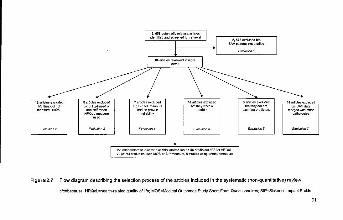
2.7 Flow diagram describing the selection process of the
articles and predictors included in the systematic (non-
quantitative) review..................................................... 31
3.1 Selection of articles and predictors included in meta-
analysis..................................................................... 52
X
3.2 Schematic of the structure of the Medical Outcomes Study
Short Form questionnaires. . . . . . . . . . . . . . . . . . . . . . . . . . . . . . . . . . . . . . . . . . .. 54
3.3 Schematic of the structure of the Sickness Impact Profile.... 55
3.4 Forrest plot of effect size estimates of individual studies
examining the association between age and Physical
HRQoL....................................................................... 68
3.5 Forrest plot of effect size estimates of individual studies
examining the association between bleed severity and
Physical HRQoL ......................................................... 69
3.6 Forrest plot of effect size estimates of individual studies
examining the association between clinical severity and
Physical HRQoL . . . . . . . . . . . . . . . . . . . . . . . . . . . . . . . . . . . . . . . . . . . . . . . . . . . . . . . . . 70
3.7 Forrest plot of effect size estimates of individual studies
examining the association between sex and Physical
HRQoL ....................................................................... 71
3.8 Forrest plot of effect size estimates of individual studies
examining the association between cognitive impairment
and Physical HRQoL . . . . . . . . . . . . . . . . . . . . . . . . . . . . . . . . . . . . . . . . . . . . . . . . . . . . 72
3.9 Forrest plot of effect size estimates of individual studies
examining the association between physical disability and
Physical HRQoL......................................................... 73
3.10 Forrest plot of effect size estimates .of individual studies
examining the association between the time between illness
onset and assessment and Physical HRQoL........... .......... 74
3.11 Forrest plot of effect size estimates of individual studies
examining the association between age and Mental
Xl
HRQoL ...................................................................... 77
3.12 Forrest plot of effect size estimates of individual studies
examining the association between bleed severity and
Mental HRQoL............................................................ 78
3.13 Forrest plot of effect size estimates of individual studies
examining the association between clinical severity and
Mental HRQoL............................................................ 79
3.14 Forrest plot of effect size estimates of individual studies
examining the association between sex and Mental HRQoL. 80
3.15 Forrest plot of effect size estimates of individual studies
examining the association between cognitive impairment
and Mental HRQoL...................................................... 81
3.16 Forrest plot of effect size estimates of individual studies
examining the association between physical disability and
Mental HRQoL......................................................... ... 82
3.17 Forrest plot of effect size estimates of individual studies
examining the association between the time between illness
onset and assessment and Mental HRQoL.................... ... 83
4.1 Decision tree for differential diagnosis of psychiatric
disorders accounting for anxiety symptoms....................... 122
5.1 Graph showing the SF-36 HRQoL profile of patients at
assessments one and two. Deviations from reference data
are expressed in mean standard scores. All deviations from
reference values are significant {P<0.05), except 'Emotional
Role Limitations' at assessment two................................ 159
5.2 Graphs of relationship between 'Mental HRQoL' and 'PTSD
Xll
Symptom Severity' (upper), 'Cognition' (middle) and
'Physical Disability' (lower) at assessment one. ........ .... ..... 170
5.3 Graph depicting the relationship between probability of
PTSD at assessments one and two with use of maladaptive
coping strategies (upper; assessment one OR = 3.670; age
held at mean; assessment two OR= 3.216) and relationship
between PTSD and age at assessment one (lower; OR =
0.917; with use of maladaptive coping strategies held at
mean level).... ....................... .... ............... ......... ....... .. 171
5.4 Graphs of relationship between 'Mental HRQoL' and 'PTSD
Symptom Severity' (upper), 'Cognition' (middle) and
'Physical Disability' (lower) at assessment two...... .... ........ 174
6.1 Graph depicting the relationship between probability of SO
PTSD at assessments one and their use of maladaptive
coping strategies (OR= 4.292).. ... .......... ................... ..... 219
6.2 Use of maladaptive coping skills by significant others and
their PTSD. .... .. . .. .. . . .. . .. . .. ... . .. . . .. .. . .. . . .. . . . . .. .. . .. . . . . .. . . . . ... 221
xiii

Declaration
None of the data or material contained in this thesis has been submitted for
previous or simultaneous consideration for a degree in this or any other university.
The copyright of this thesis rests with the author. No quotation from it should be
published in any format, including electronic and the Internet, with the author's
prior written consent. All information derived from this thesis should be
acknowledged appropriately.
xiv
Acknowledgments
The production of this thesis would have been far more difficult if it were not for the
kind support and assistance of many other people. I should first like to express my
deep and sincere gratitude to Dr. Thomas Schenk, PhD (Department of
Psychology, Durham University) for his committed and unstinting supervision. His
relaxed, but 'always there when needed' approach, meant that I was fortunate
enough to work in an environment which allowed me to independently develop my
ideas and research skills, whilst always knowing that I could ask for support and
guidance when needed. I would also like to thank the staff at the regional
neuroscience centres at Newcastle General Hospital and James Cook University
Hospital for kindly allowing me access to their patients and to participate in their
clinical activities. Particular thanks go to Professor David Mendelow, FRCS, PhD,
(Newcastle Regional Neurosciences Centre), Mr. Phillip Kane, FRCS and Dr.
Lizanne Allen, DPhil, (James Cook University Hospital). I also extend my gratitude
to the staff and postgraduates in the Department of Psychology, Durham
University, for their provision of resources and stimulating discussions.
Furthermore, I am most grateful to those patients and families who so willingly
participated in this research and to the Clarke Lister Brain Haemorrhage
Foundation and the Wolfson Research Institute for their generous financial support
for this research. I am also grateful to the following colleagues for supplying
univariate data or raw data from their studies for use in a meta-analysis which I
present in the thesis: Dr. Maree Hackett, PhD (University of Sydney, Australia),
Ms. Ann-Cathrin Jonsson, RN, (University Hospital, Lund, Sweden), Dr. Joseph
King Jr., MD, MSCE (Yale University School of Medicine, United States of
America), Dr. llonka Kreitschmann-Andermahr, MD, (RWTH Aachen University of
Technology, Germany), Dr. Majed Katati, MD, PhD, (Virgen de las Nieves
Hospital, Spain), Dr. Paul Morris, PhD (University of Edinburgh, Scotland), Dr.
XV
Pedro Rocha Filho, MD, (Clinics Hospital of the University of Sao Paulo Medical
School, Brazil), Dr. Richard Scott, PhD, and Mrs. Katherine Carpenter, Dip Psych,
(John Radcliffe Hospital, Oxford, England) and Mr. Ming-Yuan Tseng, MD,
(Addenbrooke's Hospital, University of Cambridge, England). I also want to thank
my family and friends, especially during the last year, when I had less time for
them. Last, but not least, my most special thanks goes to my partner Fiona. She
not only gave me continuous emotional support, but also her invaluable editorial
skills.
XVI
Abstract
Surviving subarachnoid haemorrhage (SAH) patients' experience significantly
reduced health-related quality of life (HRQoL) in both the short- and long~term,
as well as mysterious symptoms of fatigue and sleep dysfunction. Patients'
family members and friends -who often act as their informal carers - can also
experience psychosocial disability. The cause for these poor outcomes
remains unknown. Traditional explanations focusing on the neurological
sequelae associated with SAH or the characteristics of the illness are not
satisfactory; nor are attempts to explain family members' difficulties on the
basis of carer burden.
The hypothesis which is tested in this thesis is that post-traumatic
stress disorder (PTSD) may be abnormally high in both the SAH patient and
'significant other' (SO) population and that this may explain their outcomes.
SAH patients are known to be at risk of suffering from PTSD, but it is unknown
if this explains their outcome. In terms of patients' SOs, they are known to
experience psychiatric symptoms and I suggest these could be caused by
their development of PTSD, but this has never been examined.
In Part One (Chapter 2-5), I focus on patients' outcomes. Before
examining my PTSD hypothesis, I present a meta-analysis (Chapter 2) I
conducted of studies which have tried to explain patients' outcome using
neurological factors. I conducted the meta-analysis as a tendency for prior
studies to be underpowered and use unreliable statistics could have meant
that the actual importance of traditional factors was obscured. The results of
my meta-analysis however did not support this possibility and instead showed
traditional neurological variables did not explain patients' outcome. With this in
1
mind, I then present a longitudinal study (Chapter 5) in which I examined one
of the largest prospective series of SAH patients to establish PTSD's
explanative importance. Using regression analyses, this study showed PTSD
was the best predictor for patients' mental HRQoL - the domain most
persistently impaired. It also helped predict patients' physical HRQoL.
Moreover, PTSD was linked to sleep problems and may therefore cause
fatigue. Crucially, to establish the cause of PTSD, logistic regression was
performed. This showed that maladaptive stress coping strategies were the
best predictor for PTSD development.
In Part Two of the thesis (Chapter 6), I present my longitudinal study of
one of the largest prospective samples of SOs. All SOs were assessed with a
diagnostic PTSD measure and coping skills were assessed. An elevated
incidence of PTSD was found in both the short- and long-term. Although SOs'
PTSD did not impinge on the recovery of the SAH patients being cared for,
given that it is important to ensure SOs continue caring, regression results are
presented which show the cause of SOs' PTSD was (at least in the short
term) due to the use of maladaptive coping strategies.
The overarching conclusion is that the elevated incidence of PTSD in
SAH patients and SOs helps explain why they experience psychosocial
disability. In the final part of the thesis (Chapter 8) the clinical and theoretical
implications of this conclusion are considered, such as that teaching patients
and their SOs more effective coping skills might prevent PTSD and
psychosocial disability.
2

Chapter 1
General introduction
3
1.1 Introduction
A spontaneous subarachnoid brain hemorrhage (SAH) is a subtype of stroke
that affects mainly middle-aged persons. Advances in treatment have reduced
the mortality associated with this illness. In spite of these medical advances
occurring, patients who survive an SAH experience a particularly poor
psychosocial outcome in the short- and long-term. Evidence also suggests
that patients' family and friends - who often act as their informal carers - can
also experience a substantial reduction in their health and well-being.
After being ignored for a long-time, research has now begun to
consider how best to rehabilitate and improve the outcome of both parties.
Efforts to develop rehabilitation programs are however, hampered by a
paradox. Specifically, patients' and their carers' high levels of psychosocial
dysfunction are disproportionate to the relatively mild physical/ cognitive
impairment induced by the SAH. Moreover, this psychosocial dysfunction
cannot be explained using traditional neurological and illness-related factors.
This thesis looks to try and resolve these paradoxes. Part One of this thesis
primarily focuses upon explaining the paradoxically poor health-related quality
of life (HRQoL) of SAH patients, whilst Part Two addresses the poor outcome
of patients' family and friends.
4
1.2 Overview of Part One: SAH patients' health-related
quality of life, psychosocial outcome and post-traumatic
stress disorder
In Chapter 2, I provide a brief explanation of what SAH is, outline what is
known about its patients' outcome and describe patients' mysterious reduction
in HRQoL. SAH patients' puzzling problems of fatigue and poor sleep quality
are also discussed. I hypothesise that two explanations could explain why
SAH patients' problems remain difficult to explain. The first possibility is that
previous studies have failed to explain HRQoL using traditional neurological
and illness-related factors because they were both statistically underpowered
and used notoriously unreliable statistics by which to judge the importance of
traditional predictors. Specifically, rather than - as is ideal - examining the
degree of variability which these traditional factors share with HRQoL, studies
have typically only looked at the statistical significance of their relationship
with HRQoL. This could have served to obscure the actual explanative value
of traditional factors. The second possibility raised is that SAH researchers in
the past have been too restrictive in the factors they have used to try and
explain SAH HRQoL. It is contended that we may need to consider other,
more novel aspects of SAH patients' symptom profile when trying to explain
their poor outcome. I argue here that the psychiatric problems of an SAH
patient are an aspect which could hold real promise for explaining these
patients' HRQoL reduction, but that this has so-far been ignored.
In Chapter 3, I examine the first possibility- that the explanative value
of traditionally considered variables for SAH patients' HRQoL has been
obscured. In this chapter, I present the first ever meta-analysis on the cause
5
of SAH patients' HRQoL. The use of meta-analytic techniques allows for a
more comprehensive examination of the explanative importance of traditional
factors by considering criteria which are more appropriate than merely
statistical significance - namely their predictive effect sizes for SAH HRQoL.
In Chapter 4, I review the literature to explore possibility two - that
psychiatric disturbance has not yet been taken into account when trying to
explain SAH HRQoL. I review the psychiatric aspects of an SAH and highlight
how post-traumatic stress disorder (PTSD) elicited in patients in response to
an SAH has recently been found to be the main psychiatric disorder affecting
these patients. I then draw on findings from the wider literature and suggest
that PTSD could potentially help us better explain SAH patients' HRQoL, as
well as their puzzling symptoms of fatigue and sleep dysfunction.
Following this, in Chapter 5, I present a longitudinal study of a
prospective sample of SAH patients in which I empirically examine whether
PTSD in SAH patients can help us better explain their HRQoL reduction and
their difficulties with fatigue and sleep. I also present analyses which look to
try and predict which patients develop PTSD following an SAH, with particular
attention being afforded to the role of the stress-coping strategies which a
patient uses.
1.3 Overview of Part Two: Family and friends of SAH
patients - psychosocial outcome and post-traumatic stress
disorder
In Chapter 6, I describe how research to date on the family and friends of
SAH patients has highlighted how these 'significant others' can experience
6
abnormally high levels of psychiatric difficulties and a poor psychosocial
outcome, but that these difficulties remain unexplained. Following this, I detail
my hypothesis that significant others' poor outcome could be explained by the
never examined possibility that significant others could also develop PTSD in
response to the trauma of an SAH. I then present a longitudinal study of a
prospective sample of significant others in which I formally examine whether
PTSD is abnormally present or not in this population. I also examine the
impact of significant others' psychiatric status on SAH patients' cognitive,
physical, psychiatric and psychosocial recovery in the early and later stages of
recovery. Finally, I present analyses which look to determine whether it is
possible to predict which significant others fare worst post-SAH.
1.4 Overview of Final Chapter
In Chapter 7, I summarise the main findings of both parts of this thesis, and
discuss the implications of these findings for the treatment of SAH patients
and significant others, as well the bearing they have on future research.
7
Part One
SAH patients' health-related quality of life,
psychosocial outcome and post-traumatic
stress disorder
8

Chapter 2
The impact of a subarachnoid haemorrhage on
patients' health-related quality of life
9
2.1 Introduction
A spontaneous, non-traumatic subarachnoid brain haemorrhage (SAH) is a
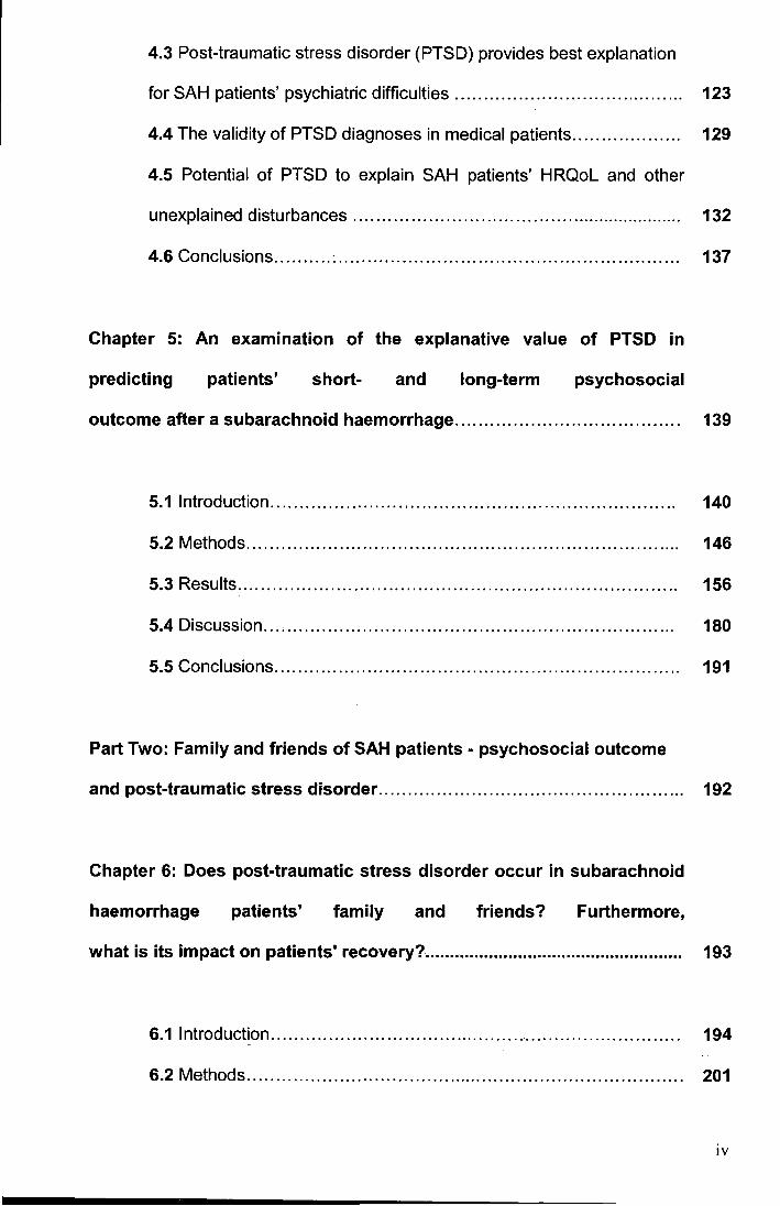
rare type of stroke (6:1 00,000 per year) [4, 403, 730], which has a
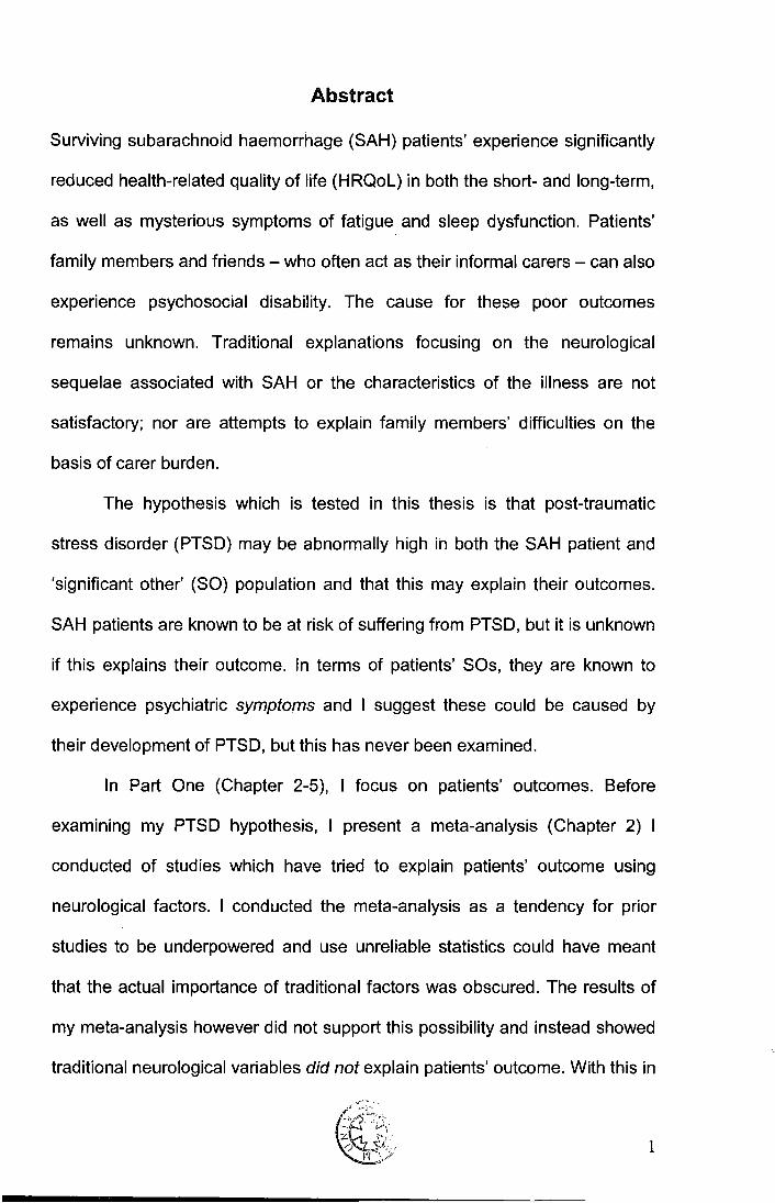
disproportionately high impact upon society [321, 681, 682]. For example, as
Figure 2.1 illustrates, despite its relative infrequency (accounting for only -7%
of all strokes) [19, 32, 672], the loss of productive life years associated with
SAH is approximately double that of much more common types of stroke
[681]. This is largely attributable to the following: (1) SAH affects much
younger persons; with a mean age of 55 [4, 435], SAH strikes persons who
are at the peak of their productivity and often supporting a young family [546,
548], (2) SAH is associated with a high mortality (-50%) [11 0, 284, 653] and
(3) SAH survivors experience a particularly poor psychosocial recovery, in
both the short- and long-term [90, 285, 286, 336, 540, 541, 546, 746]. With
such high societal costs, it is important to look to address the outcome of
surviving patients' and offer them evidence-based rehabilitation tailored to
their disease. Such rehabilitative efforts are however, not currently possible,
as at present we do not know which factors impinge upon SAH patients' well
being and cause their poor recovery. Indeed, we are yet to establish any
factors which reliably predict patients' psychosocial outcome. Stark
differences in patient age (the mean age of SAH patients being 55 years vs.
73 and 75 years for ischemic and intracerebral strokes, respectively [4, 32,
366]), as well as the kinds of brain damage (diffuse and focal vs. ischemic
focal) and medical treatment (SAH patients typically undergo brain surgery
whereas patients of other strokes do not), precludes simply looking to explain
SAH outcome by referring to research which has examined the factors
10
400
350
M' 300 0
~ 250 J!l s: Q) 200 .. cu ~ 150 0
ci z 100
50
$228,030
G,
...................................
7%
..... Incidence -&· U.S. 1990 Cost
........ $123,565 ........
............ "E>----
79%
$90,981
---------E>
0~----~~------------,-------------~------~
SAH ICH IS
300
250 r ~
200 -3' C1)
n 150 0
f/1 --~ 100 ><
...II.
0 w -
50
0
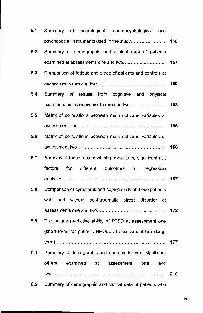
Figure 2.1 Associated life-time costs (U.S. dollars) to society in 1990 of different
types of stroke measured in terms of loss of potential productive life
years. Based on Taylor's ( 1997) data [681].
SAH=subarachnoid haemorrhage; ICH= intracerebral haemorrhage; IS= ischemic stroke
explaining outcome following more common (and better researched) types of stroke.
In this chapter, I introduce the concept of health-related quality of life (HRQoL)
and highlight how it has become the prime criterion by which SAH patients' outcome
should be judged. I highlight how the HRQoL of SAH patients is especially low, not
only compared to normative levels, but also compared to levels in patients suffering
from different illnesses. I then present the results of a systematic (non-quantitative)
review of prior studies on SAH patients which have taken HRQoL as a dependent
variable and have looked to determine which factors are statistically associated with
(and as such, presumed to cause) it. This literature review serves to set up the
subsequent studies as it illustrates how previous attempts to explain SAH patients'
outcome have proved largely futile, with traditional factors appearing to be poorly 11
associated with SAH HRQoL. I then conclude the chapter with a discussion of
whether the factors which are traditionally being used to explain SAH HRQoL are
useful and whether it might not be more promising to examine the psychiatric aspects
of this illness which have previously been ignored. Before discussing all of these
points however, I first need to provide an introduction to SAH and its treatment. Such
an introduction will serve to familiarise the reader with the specific neurological
sequelae and the characteristics of SAH (such as bleed severity/ distribution, origin of
haemorrhage, type of treatment received) as these are factors which have
traditionally been used to try to explain SAH patients' HRQoL. It will also look to draw
to the reader's attention how although an SAH is life-threatening in the immediate
stages, the medical prognosis for a surviving SAH patient, once discharged from
hospital, is very good (in terms of chances of re-bleeding and neurological sequelae),
making such patients' poor psychosocial outcome all the more intriguing. And finally,
the introduction aims to illustrate to the reader how having and surviving an SAH can
potentially be psychologically traumatic.
12
2.2 Subarachnoid haemorrhage tutorial
2.2.1 Pathology
The subarachnoid space which is filled with cerebrospinal fluid is found between the
arachnoid layer and the pia matter. Together with the dura, these comprise the
meninges which loosely encase the brain and spinal cord [666]. A spontaneous non-
traumatic subarachnoid haemorrhage (hereafter referred to as SAH) occurs when one
or more blood vessels (which can run through the subarachnoid space) ruptures and
extravasated blood enters into the space (Figures 2.2 and 2.3[a]) [33]. The rupture of
-· a blood vessel is caused by' a vascular abnormality. The most common cause - in
around 75-85% of cases - is the rupture is of an intracranial aneurysm, which is a
blister-like weakening in the wall of a cerebral artery [4, 335, 712]. The most common
sites for aneurysms are at the bifurcation and junctions of cerebral arteries [71 0],
typically at the base of the brain, near or on the circle of Willis (24-41% of SAH occur
on the internal carotid artery; 30-40% on the middle cerebral artery; 13-33% in the
vertebrobasilar system and 24-41% on a anterior cerebral artery [588, 654, 767]). In
10-20% of patients, repeated investigations fail to identify an origin for the patient's
haemorrhage.[4] These idiopathic SAHs are typically referred to as an SAH of
unknown aetiology, with the bleed typically being perimesencepahlic in nature [71 0].
This form of SAH is considered to be a benign subtype as it has an invariably
uncomplicated clinical course [46, 115, 488, 71 0]. Other causes of SAH in -5% of
cases include leaking from a cerebral arteriovenous malformation, arterial dissection
and dural arteriovenous fistulas [711].
13
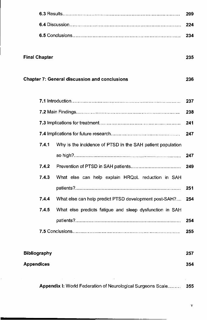
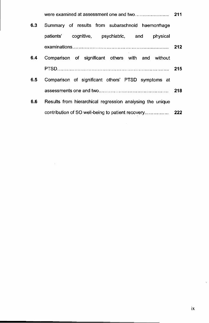
Figure 2.2 The relationship between the basal cisterns (Arabic numbers) and the
ventral cerebral arterial system (p. 17; reprinted by permission) [767].
A = At + A2 + Anterior =Olfactory cistern
communicating artery 2a = Callosal cistern
MCA = Middle cerebral artery 2b = Lamina terminalis cistern
ICA = Internal carotid artery 3 = Chiasmatic cistern
p.co.a. = posterior communicating artery 4 = Carotid cistern
p.c. (P1) = posterior cerebral artery 5 = Sylvian cistern
sea = superior cerebellar artery 6 = Crural cistern
B = Basilar artery 7 = Interpeduncular cistern
AICA = anterior inferior cerebellar artery 8 = Ambient cistern
V.a. = Vertebral artery 9 = Prepontine cistern
PICA = posterior inferior cerebellar artery 10 = Superior cerebellar-pontine cistern
A.sp. = anterior spinal artery 11 = Inferior cerebellar-pontine cistern
12 = Anterior spinal cistern
13 = Posterior spinal cistern
14
Figure 2.3 (a) Computed tomographic (CT) scan showing diffuse subarachnoid blood (basal cisterns; Sylvian fissures; anterior inter
hemispheric fissure) caused by ruptured left pericallosal aneurysm; (b) CT scan showing large temporoparietal
intracerebral haematoma and extensive SAH caused by ruptured right middle cerebral artery aneurysm; (c) CT scan
showing intraventricular blood and subarachnoid blood in interhemispheric fissure from rupture of an anterior
communicating artery aneurysm.
15
The subarachnoid space is not strictly a free-flowing space, but rather a
collection of discrete cisterns formed by arachnoid partitions and trabeculae (Figures
2.2 and2.4) [666, 767]. The walls of the cisterns serve to retard and direct the flow of
any extravasated blood. Consequently, different distributions of bleed are associated
with different anatomically-located sources [712, 767].
Depending on the direction of blood flow, an SAH can also invade adjacent
brain areas, with intracerebral extension occurring in up to 43% of aneurysmal SAH
patients (Figure 2.3[b]) [688], intraventricular extension in around 50% of aneurysmal
SAH patients [306, 391] (Figure 2.3[c]) and more rarely, the occurrence of a subdural
hematoma [295]. These subsequent problems have all been found to worsen
immediate prognosis and can necessitate additional surgical procedures, including
surgical evacuation, hemicraniectomy and the insertion of an external ventricular
catheter [231, 486, 511, 574, 575].
It is postulated that the presence of subarachnoid blood (or substances derived
from its degradation) in the meninges is neurotoxic causing a global encephalopathy
which results in diffuse cortical damage [130, 158, 213, 236, 250, 408, 409, 597, 643,
720]. Although further research is needed [211], clinical and experimental evidence
shows that blood in the subarachnoid space sets in motion a cascade of
pathophysiological processes, including increased intracranial pressure which
reduces cerebral perfusion and leads to ischemic damage [309], brain oedema and
cerebral swelling [213] and blood-brain barrier dysfunction [158]. Germano et al. [213]
for example, reported global brain dysfunction and enduring behavioural deficits in a
rodent model after intracisternal injection of subarachnoid blood. The combination of
this diffuse damage, as well as the focal brain damage resulting from intraventricular
and intracerebral haemorrhage, means that an SAH
16
Para callosal CIS
Chiasma tic cistern Interpeduncular cistern
Velum mterpositum cistern
Cerebellopontine cistern - --../ Prepontine cistern------__,.. Lateral cerebellomedullary cistern-----"
Premedullary Cisterna magna
Figure 2.4 Schematic representation of some of the major subarachnoid cisterns in
lateral view (p. 15. Reprinted with permission) [767].
is associated with a diverse array of difficulties, including cognitive impairment [750] ,
behavioural disturbances [57, 544] and hormonal dysfunction [325, 604, 679] .
2.2.2 Clinical presentation
An SAH precipitates one of the most dramatic and horrific clinical presentations in
medicine [37], with the expelled blood immediately eliciting features of meningism
(Figure 2.5) [338] . Though the initial symptoms depend upon the severity of the
bleeding and the disruption to cerebral function , an unusually severe and sudden
17
O:J1! i nued
possi o I i t y I-
~
....__
l ~ll!DI ons nil nilcki ng
nyocar di al i nf ar ct i on
I
stNt~~o~·~~ ... · ; · .. ·· • • U !<E AEQ.:'J; FFOtfJ:i;6~;
T 01set epi I ept i c Thunder d ap headact-e
seizure in 4·16X "likebeinawtha
I
Emergency hoopi t al adnil ssi on. i nt ensi ve care t r eat nmt
I I
··,.,
S at e of COlla! cr
sonmd ence i ndu:ed i n
I ~ .C: a~ or I unfoar ~.~~e to cer e1x al angi ogr arnt o i dent i t y I l ]
I D agnosed w t hI i fe-t hreat eni ng I I
2- 4 hour sur gi cal andt or r adi d ogi cal
br ai n t r eat nelt t o ocd ucle aneur vsnns>
l
I Pddi t i onal sur gi cal pr ocedur es
i nd udi no cl ot r et11DVal ard I unfoar
J
Typically discharged home wt hI i ttl e or noforllill
suppcrt . Pat i ent exper i enci ng var i ous neur ol ogi cal
"\Nhen the aneurysm bursts it is usually
very sudden. The individual collapses and,
if still conscious, the pressure caused by
the build up of blood will produce a
headache invariably described as
'agonising' and 'like torture'. The waiting
period varies with the philosophy of the
neurosurgical department. The patient is
on bedrest ... It is a very dffficult time for
relatives and patient ... the latter often
semicomatose and confused. The patient
usually remembers only the excruciating
pain .... At the time of discharge
home ... [t]he patient only now begins the
slow process of awareness of the drama of
the past few weeks and usually
experiences symptoms akin to a post
traumatic stress disorder." [449] (p.4)
Figure 2.5 The potentially traumatic experience of suffering and surviving a spontaneous subarachnoid haemorrhage
18
thunderclap headache is characteristic of an SAH [402, 71 0]. This headache is
generally diffuse and described by patients as the most severe headache possible
(e.g., "like being hit with a cricket bat") [295, 449, 555, 707]. Increasingly severe
bleeds result in a clinical presentation that is dominated by coma. Indeed, up to 40%
of SAH patients are in a state of somnolence or coma upon admission to hospital [76,
221]. Other distressing symptoms include neck stiffness, vomiting, photophobia, focal
neurological deficits and electrocardiographic changes mimicking myocardial
infarction [353]. Onset seizures also occur in 4-16% of patients [94, 116, 533].
Importantly, an SAH can occur at any time, in states of rest (e.g., sleep), or in periods
of activity (e.g., sexual intercourse, lifting) [184, 411, 497, 506, 602]. Although risk
factors for SAH include smoking, alcohol consumption and cocaine use and
hypertension [79, 330, 379], evidence suggests that for most patients, the SAH is
their first experience of a serious illness [13, 187, 240, 502].
The severity of the patient's clinical presentation in hospital is judged according
to their state of consciousness and the presence of certain neurological signs.[708]
Two popular scales used to rate the severity are the World Federation of Neurological
Surgeons scale (WFNS) [164] and the Hunt and Hess scale [293]. A higher score on
both of these scales reflects a poor clinical status (for more details on these two
scales which are both used in subsequent chapters, please refer to Appendices I and
II where I provide further details on their contents and scoring). Clinical condition is
largely related to the severity and impact of the initial haemorrhage [711]. The WFNS
scale is the preferable scale because as well as having superior interrater reliability
[401], it affords more weight to level of consciousness, which along with the patient's
age and severity of bleed, is the most important prognostic factor for immediate
outcome [164, 277, 334, 71 0].
19
2.2.3 Diagnosis
A diagnosis of an SAH is made if the presence of subarachnoid blood is confirmed,
either by computed tomography {CT) or by lumbar puncture. In all patients, the first
investigation is aCT scan of the head. The period of diagnosis (and hence access to
appropriate treatment) in reality can be quite protracted for SAH patients (and their
family) as misdiagnosis on first presentation to hospital occurs in 12-51% of cases [5,
112, 332, 369, 436, 482, 603, 715]. The diagnostic yield of the head CT depends on
the time interval since ictus, the resolution of the scanner and the skills of the
radiologist [6, 361, 691, 704]. Patients may therefore, also require a lumbar puncture
to detect the occurrence of an SAH (namely, xanthrochromia in the cerebrospinal
fluid).
Numerous scales exist to quantify and describe the severity and the
anatomical distribution of a patient's bleed as seen on CT scans. Amongst the most
commonly used is Fisher's-scale [191 ]. As Fisher's scale is used and discussed in
subsequent chapters, I provide further details on its content and scoring in Appendix
Ill.
To identify the source of a person's SAH, a comprehensive cerebral
angiography is performed. In the traditional angiography technique, a catheter is fed
through the femoral artery and up to the carotid and vertebral arteries. Contrast dye is
then injected into the catheter so that the vessels of the circle of Willis can be
visualised and any abnormalities, such as a cerebral aneurysm, detected. The
aetiology of the patients bleed is often used to classify (be it 'unknown', aneurysmal,
or the specific arterial location of the ruptured aneurysm) and to try to predict an SAH
patient's outcome
20
2.2.4 Management
The initial haemorrhage is terminated by the "extravascular rise in pressure as a
consequence of the bleeding and/ or by tamponade of the vacant surroundings" (p. 8)
[295]. This termination is, however, only temporary. The cumulative risk of re-bleeding
from a ruptured cerebral aneurysm is around 40% without intervention [278]. As these
re-bleeds hold an extremely poor prognosis [573], the immediate therapeutic goal is
to identify the source of the bleed and, where possible, permanently occlude it from
circulation. Occlusion of a cerebral aneurysm is most commonly achieved via an
invasive procedure, either neurosurgical clipping and/ or endovascular coiling. The
former, more traditional procedure, entails a craniotomy and resection of the brain
under general anaesthesia, where re-bleeding is prevented by the placement of a
small spring-loaded metal clip across the neck of the aneurysm. This serves to
exclude the aneurysm from circulation, whilst still preserving the parent and
perforating arteries [468]. The coiling procedure on the other hand, entails an
angiogram procedure (typically under general anaesthesia), whereby the tip of a
catheter is positioned in the neck of the aneurysm, which is then packed with platinum
coils, through a system of controlled detachment. The intent is to provoke reactive
thrombosis of the aneurysm and so obstruct the lumen. No single craniotomy or
coiling procedure is routine and can each take around 2-6 hours to complete [71 0).
Although both procedures carry independent risks for the life of the patient, the results
from recent randomised controlled trials suggest an absolute advantage for the coiling
technique in terms of the rate of death and dependence [466, 467, 702]. Coiling
negates the need for craniotomy and hence damage to the brain parenchyma.
Furthermore, unlike the clipping techniques, it does not increase the possibility of
secondary ischemic brain damage [77].
21
In contrast to those patients whose haemorrhage is of an identified source,
those with an SAH of an unknown origin, typically receive no surgical intervention and
receive only conservative management. Given the potential differences in treatment
impact, the type of intervention which a patient receives (be it conservative, clipping
or coiling) is often used to classify patients and used to try and predict their outcome.
2.2.5 Risk of re-bleeding and further SAH
Empirical evidence indicates that re-bleeding in SAH patients is very rare [98, 131,
307, 333, 466, 611, 695, 745]. Although former aneurysmal SAH patients have a
statistically higher chance of suffering a further SAH compared to the general
population [702, 7 45], the actual number of re-b leeds is very small. For example, a
recent meta-analysis [702], including data from the largest medical follow-up study to
date (namely, the International Subarachnoid Aneurysm Trial [466]), found that only
1.2% of 1137 clipped patients and 2.6% of 1135 coiled patients had an episode of re
bleeding up to one year following treatment.
It can be concluded therefore, that coiling and surgical clipping procedures are
effective treatments for the prevention of re-bleeding in the vast majority of patients.
This, together with the fact that the majority (>95%) of deaths associated with the
illness occur during the acute hospital stages of the illness [32, 413, 691], means that
patients are naturally informed by their clinicians and patient information sources that
they are very safe and should look to return to a normal, unrestricted life [202, 449,
503]. In addition, patients are often reassured that, unlike most in the general
population, they have had a detailed examination of their cerebral architecture and
had there been any cause for concern this would have been identified. Moreover,
patients treated by coiling, those with multiple aneurysms and those with an
22
aneurysm that was incompletely obliterated by clipping are actually monitored and
undergo repeated angiography following hospital discharge, which allows for pre-
emptive treatment if required [449].
2.3 Health-related quality of life in the context of SAH
2.3.1 Background on HRQoL and the measuring of SAH patients' outcome
Progress in understanding the cause of SAH patients' poor psychosocial outcome
has been particularly slow because research on this topic has only recently begun.
The high mortality following SAH [110, 284, 653], as well as outcome research
traditionally being conducted by neurosurgeons (for whom a "good" outcome
constituted successful occlusion of a cerebral aneurysm), meant that research into
patient recovery was for a long time, not considered a priority. Outcome had only
been indexed in terms of mortality and patient survival time [268]. An improving
survival rate and relatively stable incidence [284, 364, 370, 403, 653] has however,
meant that it has become vital to now consider not only the quantity, but also the
quality of SAH patients' survival [183].
With the increased attention on SAH patients' outcome, it became noticeable
that researchers and clinicians needed to be equipped with sensitive measures to
gauge patients' global outcome [214, 752]. A wealth of evidence has illustrated that
the classic measures of outcome available within neurosurgery and neurology,
measures such as the Glasgow Outcome Scale (GOS) [318], are far from satisfactory
(please see Appendix IV for a description of the content and scoring of the GOS).
Such crude, clinician-led measures were designed only to evaluate patient outcome
with respect to physical disability, and are as such, too restricted in their focus to
capture the broad array of difficulties which SAH patients can experience. An
23
infrequency of profound physical disability in the SAH population for example, means
that although 80-85% of patients are rated as having made either a "good" or
favourable outcome on such scales [303, 434], up to 60% of these patients illustrate
chronic cognitive dysfunction [303, 438, 507, 540, 541, 643], around 50% experience
personality change (including irritability, hostility, emotional lability) [57, 537, 746],
50% do not return to work [90, 540, 541], approximately 20% show reduced social
participation [540, 541], 25% consider death preferable to the quality of their survival
[90], and many also show severe hormonal disturbance [604].
The revelation that a positive appearance [17, 25, 45, 303, 313, 409, 507, 643]
did not necessarily equate with a full recovery, forced neurosurgeons to engage in a
debate on how it defined and measured SAH patients' outcome. Seminal
contributions to this discussion revealed that neurosurgery's definition of outcome had
been, for the most part, atheoretical. It was highlighted how the judgement of patient
outcome following an SAH was not informed by the patient's own opinion, but rather
was centred upon the treating neurosurgeon's own beliefs. Buchanan et al. [90] in a
small, but important study, illustrated that this approach was wholly unsuitable
because the frame of reference used by these two parties drastically differed. Whilst
patients and their families evaluated outcome according to "what was",
neurosurgeons, being acutely aware of the potential of an SAH to kill or profoundly
disable, compared patients' outcomes "with what could have been". Consequently,
though patients often experienced troublesome neurobehavioural sequelae,
difficulties other than obvious physical impairment were viewed by neurosurgeons as
'minor' and permissible [44, 45, 90]. "Good" recovery, it was found, lay very much in
the eye of the beholder.
24
As a result, the neurosurgical community recognised that it was crucial that a
patient's perspective was taken into account when judging outcome after an SAH. It
was seen that a more holistic approach to assessment was needed, whereby the
entire patient as he/she exists and functions in his/her own world was evaluated [17,
44, 45, 295, 313, 383, 397, 398, 592]. As is the case for other illnesses [59, 234, 239,
384, 757], it is now widely accepted that measures of patients' 'health-related quality
of life' (HRQoL) fulfil these aims and that such measures are well-suited to provide a
sensitive index of the personal burden that an SAH places on its survivor [1 03, 137].
Indeed, in line with the US. Food and Drugs Administration's guidelines [137, 214],
HRQoL is now used as "hard" criterion by which to evaluate different treatments for
SAH[3,91, 144,239,362,410,686,693].
Despite this increased emphasis being placed on HRQoL, no single definition
exists in the medical sciences [59, 186, 219, 329]. Nonetheless, HRQoL is commonly
understood to be a multidimensional construct which measures the extent to which
one's usual or expected physical, emotional and social well-being is affected by a
medical condition or treatment [1 09]. HRQoL is distinct from the broader construct of
quality of life which encompasses the influence of other aspects of life not amenable
to health care services, such as adequacy of housing and income [59]. Importantly, it
is recognised that HRQoL can only be effectively accessed by asking the patient
themselves for their opinion [59, 186]. Furthermore, not only does HRQoL measure
for some of the neurophysical impairments resulting from an illness (e.g., loss of
movement in a limb) and its associated disability (i.e., the functional loss associated
with these impairments), but it also heavily loads on the handicap which these
impairments and disabilities cause (i.e., the disadvantages the person experiences as
a result of the illness which limits or prevents them from fulfilling a normal role, such
25
as working). HRQoL therefore, represents an indicator of a patient's overall abilities in
daily life. It exceeds the level of pure physical function and constitutes a more general
outcome criterion than cognitive function [285, 305]. Given this multidimensional
nature, HRQoL accords well with the World Health Organisation's models of disease
[1, 2] which contend that an illness can affect a person in a multitude of ways, that its
effects are context specific and can ultimately often only be evaluated by the patient
themselves [84, 232, 748]. Importantly, HRQoL is a double-sided concept, allowing
for both positive, as well as negative effects, of illness [59].
Both disease specific and generic HRQoL measures have been used to
assess SAH patients' outcome [43, 546, 734]. These self-report questionnaires
evaluate HRQoL by asking the patient to indicate their personal experience of the
presence, frequency, or intensity of various symptoms, behaviours, capabilities and
feelings. The key HRQoL domains which are commonly examined by such
instruments include the person's physical functioning (i.e., limitations in performance
due to their poor health in all physical activities including bathing and dressing),
physical role limitations (i.e., problems with work and/ or daily activities as a result of
their physical health), bodily pain (i.e., severity and limitations of pain), general health
(i.e., evaluate their personal health as poor and asked whether it is likely to get
worse), energy (i.e., whether they feel tired and worn out all the time), social
functioning (i.e., whether they experience extreme and frequent interference with
normal social activities due to physical or emotional problems) and mental health (i.e.,
whether they feel nervous and depressed all of the time) [735]. Principal component
analyses reveal that scores from these domains essentially relate to two broad
HRQoL dimensions - namely physical HRQoL and mental HRQoL [732]. Not only
does the use of these summary scores obviously reduce the number of statistical
26
comparisons which are needed when analysing data, but it also simplifies
interpretation and means that the two aspects can be examined separately.
Although disease-specific HRQoL instruments have the advantage that they
can be highly sensitive, with items being tailored to the disease in question, such
instruments have been slow to develop for SAH patients. To date, only one such SAH
measure has been developed and it is still very much in the early stages of
psychometric evaluation [546]. Consequently, the highly reliable and widely validated
generic HRQoL measures - often referred to as health-status questionnaires [168,
186] - have been favoured in SAH studies. The Medical Outcomes Study Short Form
questionnaires [731, 735] and the Sickness Impact Profile [43] are the most popular
of the generic instruments in the field of SAH (as revealed by my review of the SAH
HRQoL literature presented later in this chapter). The great advantage of such
instruments is that normative data have been collected so that it can be determined to
what degree SAH patients' HRQoL strays from normality [59]. Scores can also be
compared to those reported in relation to patients with/after other illnesses which
allows for SAH patients' impairment to be placed in the context of more thoroughly
researched diseases.
2.3.2 Patients' HRQoL after SAH
Having been employed in SAH research since 1995, HRQoL measures have been
instrumental in exposing the true extent and seriousness of an SAH for its survivors.
Though a few exceptions exist [40, 75, 451], and a minority of patients note some
positive consequences of the illness (e.g., a renewed sense of appreciation for life)
[285], an overwhelming amount of literature now demonstrates that many SAH
patients experience substantial degrees of HRQoL impairment compared to general
27
population controls [116, 144, 189, 240, 283, 285, 286, 302, 305, 336, 377, 438, 609,
639, 703]. This impairment is seen in both the early [144, 285, 336, 358, 438] and the
later stages following the illness [116, 144, 189, 240, 286, 302, 305, 377, 609, 639].
For example, Hutter et al. [302] examined a sample of 58 SAH patients using a study
specific HRQoL measure and found that approximately 50% of those patients still
reported impairment 1-5 years post-SAH.
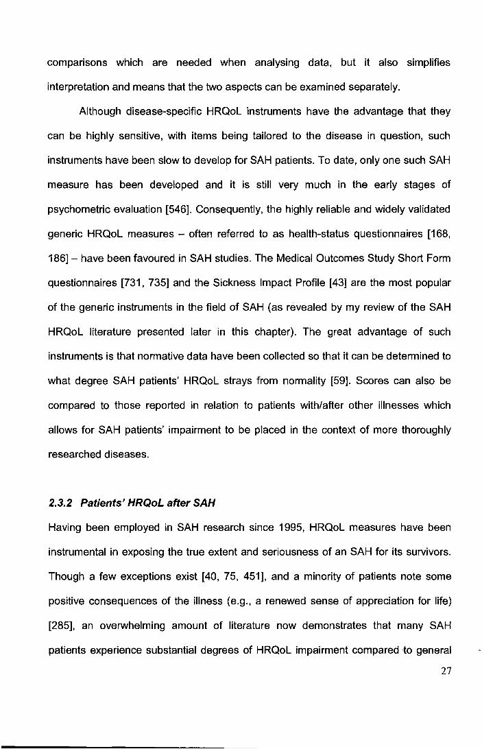
The severity of SAH HRQoL impairment is reflected even at the group mean
level, where it could be expected that its effect would be less pronounced. Using the
data from 10 major studies which together examined over 600 SAH patients [144,
240, 283, 322, 336, 358, 377, 470, 569, 639], I present in Figure 2.6 the (weighted)
average mental and physical HRQoL reported by SAH patients (as measured by the
Medical Outcome Study Short-Form questionnaires [733, 734]). Crucially, I also
include in the figure, the average HRQoL reported on the same measure by persons
with health conditions which have been better researched and are recognised as
being debilitating, such as cancer [676]. The use of a linear t-score transformation
(where the mean normative score for each condition is 50) means that scores across
the different health conditions can be compared. Although some caution should be
exercised when interpreting the summary scores for SAH patients' HRQoL (since I
did not formally examine the homogeneity of the scores from the contributing studies),
the figure does nevertheless, quite clearly illustrate that an SAH - even at group-level
- exerts a negative influence on HRQoL comparable to that of the other chronic
illnesses. In fact, it can be observed that the HRQoL of SAH patients is worse than
that caused by all other illnesses included in the figure, except Major Depressive
Disorder. That an SAH can profoundly reduce a person's HRQoL agrees with those
studies which more formally contrasted SAH patients' psychosocial outcome with
28
Q) ... 0 (,)
"' ~ ns E E :s "' E ... 0
L1. I -... 0
.s::::: en en 0
== "C Q)
"' ns .Q
I
E ... 0 z
1
Pathology
- Mental HRQoL
- Physical HRQoL
Normative mean
Figure 2.6 Comparison of Mental and Physical health-related quality of life
associated with SAH and other illnesses.
SAH scores are weighted averages for 604 patients derived from 10 independent studies [144, 240, 283,
322, 336, 358, 377, 470, 569, 639); Data for comparison health conditions from European Prospective
Investigation into Cancer (676); Scores less than 50 indicate a negative deviation from normality; Higher
scores indicate better outcome; HRQoL= Health-related quality of life; MOS= Medical Outcome Study;
Myocardial infarc.= myocardial infarction.
other patient groups [264, 304, 451, 465, 545, 627]. Though an SAH can cause
significant reductions in all areas of HRQoL [189], the most prominent reduction has
been found to occur in the Mental HRQoL. Katati et al. [336] for example, found that
impairment in the domains mental health (47.1%), energy reduction (42.9%) and role
29
limitations due to emotional problems (40%) were amongst the most impaired areas
of HRQoL post-SAH, whilst Kreitschmann-Andermahr et al. [377] found twice the
impairment in Mental HRQoL compared to Physical HRQoL post-SAH.
2.4 Previous studies failure to explain SAH patients' reduced
HRQoL
As we have seen, a person's HRQoL is especially impaired following an SAH. Does
current the evidence allow us to explain this reduction? In order to answer this
question, I conducted a systematic (non-quantitative) review of the literature to
identify those studies which have looked to explain SAH HRQoL. Figure 2.7 illustrates
the selection process for the articles used in this review. Further methodological
details of this review, such as the terms used for the searching of electronic data
bases and exclusion criteria, are not presented here but instead can be found in the
methods section of the next chapter (Chapter 3), as the current literature review forms
the basis for the articles used in the meta-analysis which I present there.
The results from the systematic (non-quantitative) literature review are shown
in Tables 2.1 and 2.2. These results show that whilst the complexity of an SAH has
meant that a plethora of variables have so far been implicated in trying to explain
SAH HRQoL (in fact, over 45), previous studies have failed to identify any factor
which is reliably associated with SAH HRQoL and so can help explain these patients
impairment (see column titled 'Findings' in the two tables). In addition to showing that
most studies (81 %; see Figure 2.7) on the topic have used either one of the MOS or
SIP HRQoL questionnaires to measure HRQoL, the results also show that most
30
12 articles excluded
I I 8 articles excluded
b/c they did not b/c utility-based or measure HRQoL non self-report
HRQoL measure used
Exclusion 2 I I Exclusion 3
2, 658 potentially relevant articles identified and screened for retrieval
84 articles reviewed in more detail
7 articles excluded 1 0 articles excluded b/c HRQoL measure b/c they were a
had no proven doublet reliability
Exclusion4 Exclusion 5
2, 573 excluded b/c SAH patients not studied
Exclusion 1
6 articles excluded b/c they did not
examine predictors
Exclusion 6
27 independent studies with usable information on 46 predictors of SAH HRQoL. 22 (81 %) of studies used MOS or SIP measure, 5 studies using another measure
14 articles excluded b/c SAH data
merged with other pathologies
I I Exclusion 7
Figure'2.7 Flow diagram describing the selection process of the articles included in the systematic (non-quantitative) review.
b/c=because; HRQoL=health-related quality of life; MOS=Medical Outcomes Study Short-Form Questionnaires; SIP=Sickness Impact Profile.
31
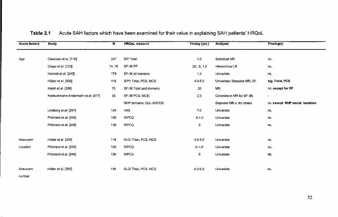
Table2.1 Acute SAH factors which have been examined for their value in explaining SAH patients' HRQoL
Acute factors Study N HRQoL measure Timing (yrs.) Analysis Finding(s)
Age Claassen et al. [116] 247 SIP Total 1.0 Statistical MR ns.
Crago et al. [123] 74, 76 SF-36 PF .25, .5, 1.0 Hierarchical LR ns.
Hackett et al. [240] 174 SF-36 all domains 1.2 Univariate ns.
Hutter et al. [305] 116 SIPt Total, PCS, MCS 4.0-5.0 Univariate; Stepwise MR; OF sig. Total, PCS
Katati et al. [336] 70 SF-36 Total and domains .33 MR ns. except for PF
Kreitschmann-Andermahr et al. [377] 35 SF-36 PCS, MCS; 2.3 Covariate in MR for SF-36;
NHP domains; Qol-AGHDS Stepwise MR ± for others ns. except NHP social isolation
Lindberg et al. [397] 104 VAS 7.0 Univariate ns.
Pritchard et al. [546] 105 WPCQ .5-1.0 Univariate ns.
Pritchard et al. [548] 136 WPCQ .5 Univariate ns.
Aneurysm Hutter et al. [305] 116 ALQI Total, PCS, MCS 4.0-5.0 Univariate ns.
Location Pritchard et al. [546] 105 WPCQ .5-1.0* Univariate ns.
Pritchard et al. [548] 136 WPCQ .5 Univariate Ns
Aneurysm Hutter et al. [305] 116 ALQI Total, PCS, MCS 4.0-5.0 Univariate ns.
number
32
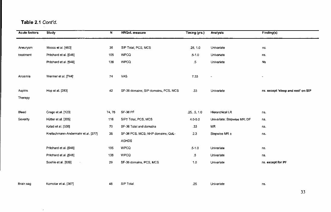
Table 2.1 Cont'd.
Acute factors Study N HRQol measure Timing (yrs.) Analysis Finding(s)
Aneurysm Mocco et al. [463] 35 SIP Total, PCS, MCS .25, 1.0 Univariate ns.
treatment Pritchard et al. [546] 105 WPCQ .5-1.0 Univariate ns.
Pritchard et al. [548] 136 WPCQ .5 Univariate Ns
Anosmia Wermer et al. [744] 74 VAS 7.33
Aspirin Hop et al. [283] 42 SF-36 domains; SIP domains, PCS, MCS .33 Univariate ns. except 'sleep and rest' on SIP
Therapy
Bleed Crago et al. [123] 74, 76 SF-36 PF .25, .5, 1.0 Hierarchical LR ns.
Severity Hutter et al. [305] 116 SIPt Total, PCS, MCS 4.0-5.0 Univariate; Stepwise MR; DF ns.
Katati et al. [336] 70 SF-36 Total and domains .33 MR ns.
Kreitschmann-Andermahr et al. [377] 35 SF-36 PCS, MCS; NHP domains; Qol- 2.3 Stepwise MR ± ns.
AGHDS
Pritchard et al. [546] 105 WPCQ .5-1.0 Univariate ns.
Pritchard et al. [548] 136 WPCQ .5 Univariate ns.
Soehle et al. [63~] 29 SF-36 domains, PCS, MCS 1.0 Univariate ns. except for PF
Brain sag Komotar et al. [367] 48 SIP Total .25 Univariate ns.
33
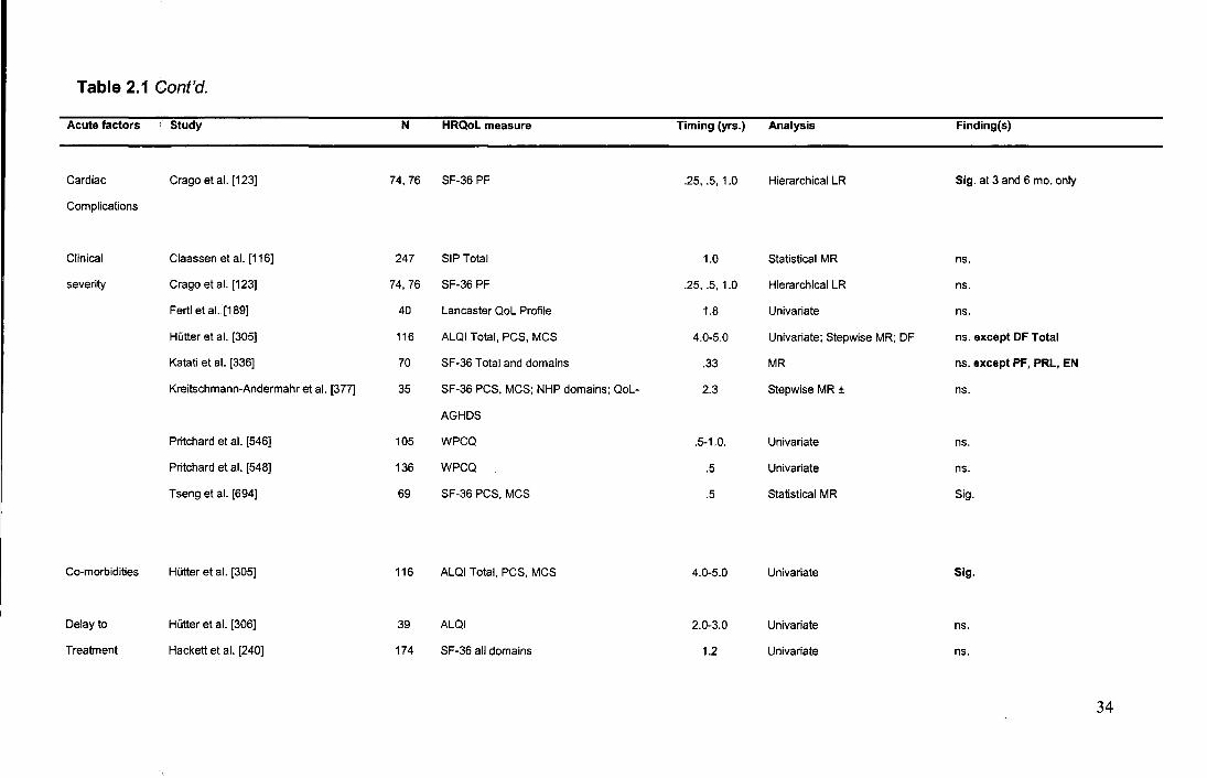
Table 2.1 Gont'd.
Acute factors
Cardiac
Complications
Clinical
severity
Co-morbidities
Delay to
Treatment
· Study
Crago et al. [123]
Claassen et al. [116]
Crago et al. [123]
Fertl et al. [189]
Hutter et al. [305]
Katati et al. [336]
Kreitschmann-Andermahr et al. [377]
Pritchard et al. [546]
Pritchard et al. [548]
Tseng et al. [694]
Hutter et al. [305]
Hutter et al. [306]
Hackett et al. [240]
N
74, 76
247
74, 76
40
116
70
35
105
136
69
116
39
174
HRQol measure
SF-36 PF
SIP Total
SF-36 PF
Lancaster QoL Profile
ALQI Total, PCS, MCS
SF-36 Total and domains
SF-36 PCS, MCS; NHP domains; QoL-
AGHDS
WPCQ
WPCQ
SF-36 PCS, MCS
ALQI Total, PCS, MCS
ALQI
SF-36 all domains
Timing (yrs.)
.25, .5, 1.0
1.0
.25, .5, 1.0
1.8
4.0-5.0
.33
2.3
.5-1.0.
.5
. 5
4.0-5.0
2.0-3.0
1.2
Analysis
Hierarchical LR
Statistical MR
Hierarchical LR
Univariate
Univariate: Stepwise MR; DF
MR
Stepwise MR ±
Univariate
Univariate
Statistical MR
Univariate
Univariate
Univariate
Finding(s)
Sig. at 3 and 6 mo. only
ns.
ns.
ns.
ns. except DF Total
ns. except PF, PRL, EN
ns.
ns.
ns.
Sig .
Sig.
ns.
ns.
34
Table 2.1 Cont'd.
Acute factors Study N HRQoL measure Timing (yrs.) Analysis Finding(s)
Education Claassen et al. [116] 247 SIP Total 1.0 Statistical MR sig.
English fluency Claassen et al. [116] 247 SIP Total 1.0 Statistical MR ns.
Ethnicity Claassen et al. [116] 247 SIP Total 1.0 Statistical MR sig.
Crago et al. [123] 74, 76 SF-36 PF .25, .5, 1.0 Hierarchical LR ns.
Fever Fernandez et al. [188] 353 SIP poor/ good QoL cut-off .25 Hierarchical LR ns.
Hydrocephalus HUtter et al. [305] ±± 116 ALQI Total, PCS, MCS 4.0-5.0 Univariate ns.
Tseng et al. [694] 69 SF-36 PCS, MCS .5 Statistical MR sig. for PCS only
Immediate Tseng eta. [694] 69 SF-36 PCS, MCS .5 Statistical MR sig.
Operative
deficits
Infarction Claassen et al. [116] 247 SIP Total 1.0 Statistical MR ns.
35
Table 2.1 Cont'd.
Acute factors · Study N
Initial Kowalski et al. [369] 482
Misdiagnosis
Intracerebral Hutter et al. [305]* 108
Bleed
Intraoperative Hutter et al. [305] 116
aneurysm
rupture
Intraventricular Hutter et al. [305] 116
Bleed
Pravastatin Tseng et al. [694] 69
treatment
Re-bleed Claassen et al. [116] 247
HRQol measure Timing (yrs.)
SIP .25, 1.0
ALQI Total, PCS, MCS 4.0-5.0
ALQI Total, PCS, MCS 4.0-5.0
ALQI Total, PCS, MCS 4.0-5.0
SF-36 PCS, MCS .5
SIP Total 1.0
Analysis
Univariate
Univariate
Univariate
ANCOVA
Statistical MR
Statistical MR
Finding(s)
ns. except for just good grade
patients at 3 mo.
sig.
ns.
ns.
sig.
sig.
36
Table 2.1 Cont'd.
Acute factors Study N HRQol measure Timing (yrs.) Analysis Finding(s)
Resection of the Hutter et al. [305] 116 ALQI Total, PCS, MCS 4.0-5.0 Univariate ns.
gyrus rectus
Sepsis Tseng et al. [694] 69 SF-36 PCS, MCS .5 Statistical MR sig.
Sex Crago et al. [123] 74, 76 SF-36 PF .25, .5, 1.0 Hierarchical LR ns.
Hackett et al. [240] 174 SF-36 all domains 1.2 Univariate ns.
Katati et al. [336] 70 SF-36 Total and domains .33 MR sig. male better, except SF
Kreitschmann-Anderrnahr et al. [377] 35 SF-36 PCS, MCS; 2.3 Covariate in MR
NHP domains: Qol-AGHDS Stepwise MR ± ns. except NHP social isolation
Lindberg et al. [397] 104 VAS 7.0 Univariate ns.
Pritchard et al. [546] 105 WPCQ 5-1.0 Univariate ns.
Pritchard et al. [548] 136 WPCQ .5 Univariate ns.
Tseng et al. [15] 69 SF-36 PCS, MCS .5 Statistical MR sig. male better
Temporary Hutter et al. [305] 116 ALQI Totai,PCS, MCS 4.0-5.0 Univariate sig. except for MCS
clipping
37
Table 2.1 Cont'd.
Acute factors
Vasospasm
Ventriculitis
E4 allele
Study
Hutter et al. [305]
Soehle et al. [639]
Tseng et al. [15]
Morris et al. [470]
N
116
29
69
70
HRQol measure
ALQI Total, PCS, MCS
SF-36 domains, PCS, MCS
SF-36 PCS, MCS
SF-36
Timing (yrs.) Analysis Finding(s)
4.0-5.0 Univariate ns. except for PCS
1.0 Univariate ns.
.5 Statistical MR sig.forMCS
1.2 Univariate ns.
Note: ALQI= Aachen Life Quality Index; DF=discriminant function analysis; HRQoL=health-related quality of life; LR=Iogistic regression; MCS=Mental/ Psychosocial HRQoL Component Score;
MR= Multiple regression n=number; NHP=Nottingham Health Profile; ns.=statistically non-significant; PCS=Physical HRQoL Component Score; PF=Physical Functioning; Qol-AGHDS=Quality of Life
Assessment of Growth Hormone Deficiency; SF-36=Short Form-36; sig.=statistically significant; SIP=Sickness Impact Profile; VAS= Visual Analogue Scale; WPCQ=Wessex Patient Carer
Questionnaire; yrs.=years; ±=Stepwise type unclear; ±±=Delayed hydrocephalus (3-48 weeks after discharge); *=Location of ICH in cohort was 'frontal' (11.2%), with the 6.8% of patients with ICH at
other locations excluded from analysis.
38
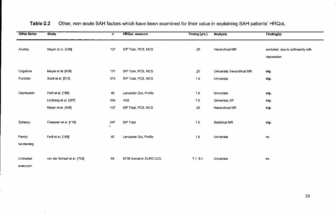
Table:2.2 Other, non-acute SAH factors which have been examined for their value in explaining SAH patients' HRQoL
Other factor Study n HRQol measure Timing (yrs.) Analysis Finding(s)
Anxiety Mayer et al. [438] 137 SIP Total, PCS, MCS .25 Hierarchical MR excluded due to collinearity with
depression
Cognitive Mayer et al. [438] 137 SIP Total, PCS, MCS .25 Univariate: hierarchical MR sig.
Function Scott et al. [613] 573 SIP Total, PCS, MCS 1.0 Univariate sig.
Depression Fertl et al. [189] 40 Lancaster QoL Profile 1.8 Univariate sig.
Lindberg et al. [397] 104 VAS 7.0 Univariate: DF sig.
Mayer et al. [438] 137 SIP Total, PCS, MCS . 25 Hierarchical MR sig .
Epilepsy Claassen et al. [116] 247 SIP Total 1.0 Statistical MR sig. .•.
Family Fertl et al. [189] 40 Lancaster QoL Profile 1.8 Univariate ns.
functioning
Untreated van der Schaaf et al. [703] 69 SF36 domains; EURO-QOL 7.1, 9.1 Univariate ns.
aneurysm
39
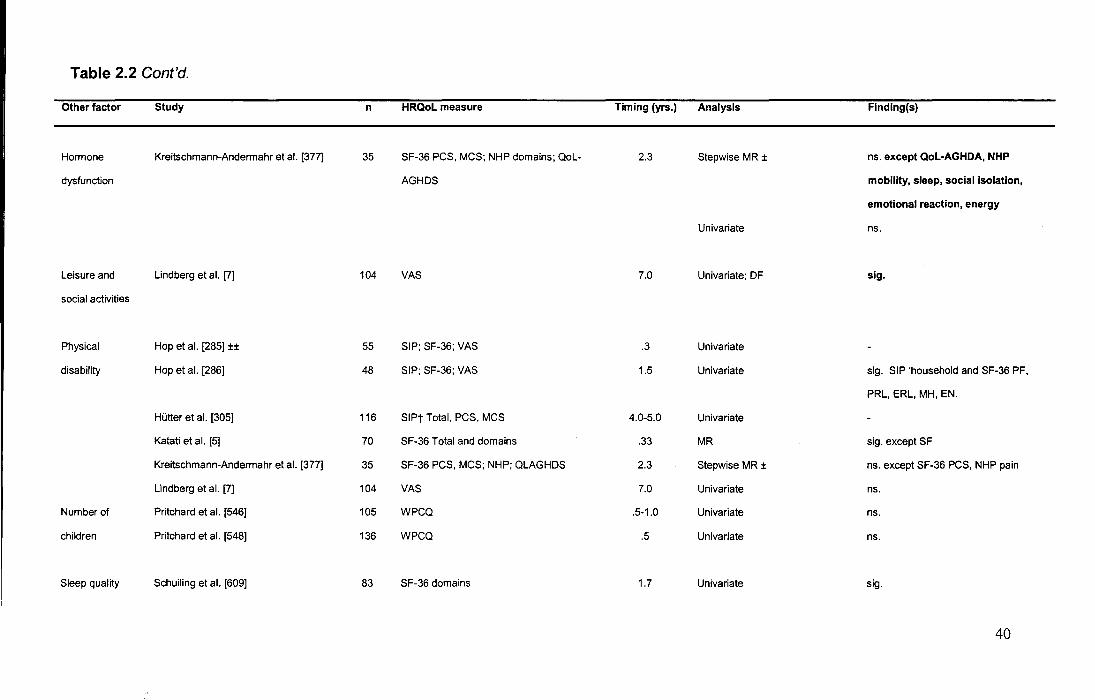
Table 2.2 Cont'd.
Other factor Study n HRQol measure Timing (yrs.) Analysis Finding(s)
Hormone Kreitschmann-Andermahr et al. [377] 35 SF-36 PCS, MCS; NHP domains; QoL- 2.3 Stepwise MR ± ns. except Qol-AGHDA, NHP
dysfunction AGHDS mobility, sleep, social isolation,
emotional reaction, energy
Univariate ns.
Leisure and Lindberg et al. [7] 104 VAS 7.0 Univariate; DF sig.
social activities
Physical Hop et al. [285] ±± 55 SIP; SF-36; VAS .3 Univariate
disability Hop et al. [286] 48 SIP; SF-36; VAS 1.5 Univariate sig. SIP 'household and SF-36 PF,
PRL, ERL, MH, EN.
Hutter et al. [305] 116 SIPt Total, PCS, MCS 4.0-5.0 Univariate
Katati et al. [5] 70 SF-36 Total and domains .33 MR sig. except SF
Kreitschmann-Andermahr et al. [377] 35 SF-36 PCS, MCS; NHP; QLAGHDS 2.3 Stepwise MR ± ns. except SF-36 PCS, NHP pain
Lindberg et al. [7] 104 VAS 7.0 Univariate ns.
Number of Pritchard et al. [546] 105 WPCQ .5-1.0 Univariate ns.
children Pritchard et al. [548] 136 WPCQ .5 Univariate ns.
Sleep quality Schuiling et al. [609] 83 SF-36 domains 1.7 Univariate sig.
40
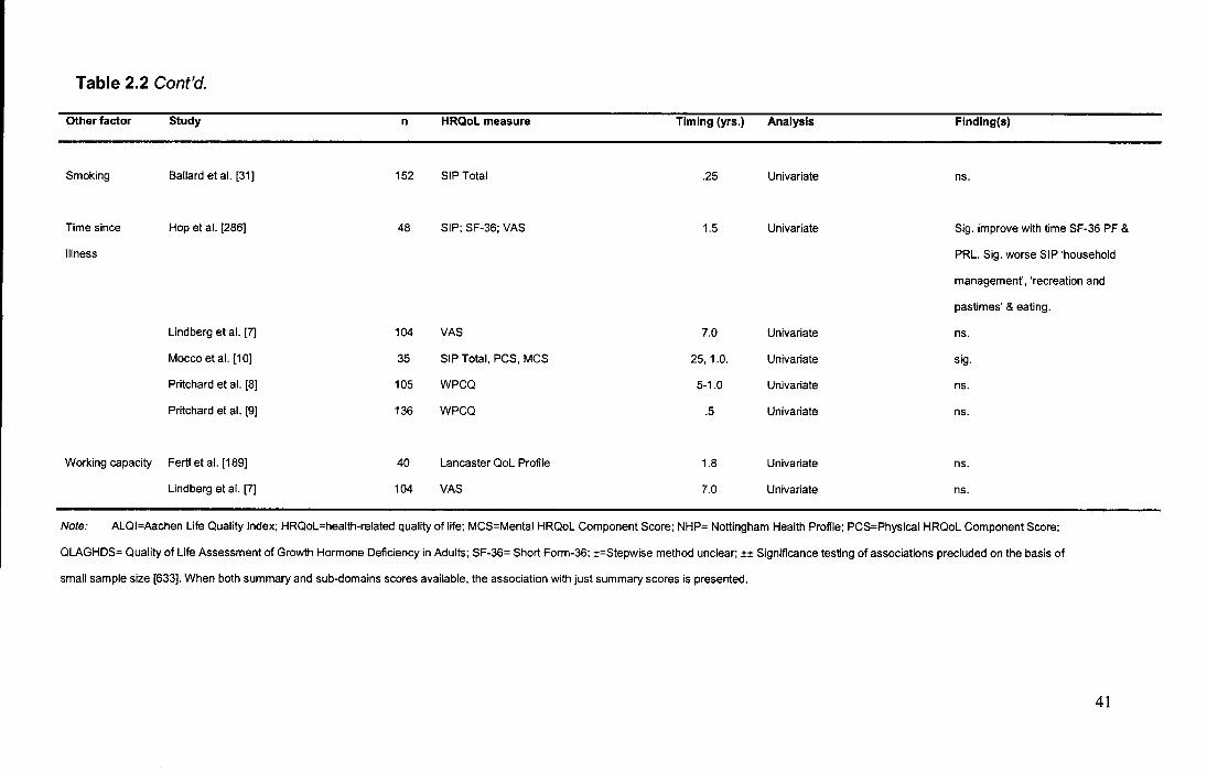
Table 2.2 Cont'd.
Other factor
Smoking
Time since
illness
Working capacity
Study
Ballard et al. [31]
Hop et al. [286]
Lindberg et al. [7]
Maceo et al. [1 OJ
Pritchard et al. [8]
Pritchard et al. [9]
Fertl et al. [189]
Lindberg et al. [7]
n
152
48
104
35
105
136
40
104
HRQol measure Timing (yrs.)
SIP Total .25
SIP; SF-36; VAS 1.5
VAS 7.0
SIP Total, PCS, MCS 25, 1.0.
WPCQ 5-1.0
WPCQ .5
Lancaster QoL Profile 1.8
VAS 7.0
Analysis
Univariate
Univariate
Univariate
Univariate
Univariate
Univariate
Univariate
Univariate
Finding(s)
ns.
Sig. improve with time SF-36 PF &
PRL. Sig. worse SIP 'household
management', 'recreation and
pastimes' & eating.
ns.
sig.
ns.
ns.
ns.
ns.
Note: ALQI=Aachen Life Quality Index; HRQoL=health-related quality of life; MCS=Mental HRQoL Component Score; NHP= Nottingham Health Profile; PCS=Physical HRQoL Component Score;
QLAGHDS= Quality of Life Assessment of Growth Hormone Deficiency in Adults; SF-36= Short Form-36; ±=Stepwise method unclear; ±± Significance testing of associations precluded on the basis of
small sample size [633]. When both summary and sub-domains scores available, the association with just summary scores is presented.
41
previous attempts to explain SAH patients' HRQoL reduction have centred on either
(1) the clinical characteristics of the illness and the patient- such as patient age, sex,
clinical severity on admission to hospital, severity and distribution of the
haemorrhage, its aetiology and its treatment - or (2) on the neurological sequelae
which they experience post-SAH. The reasons why these two sets of predictors have
so dominated prior studies looking to explain SAH HRQoL are quite comprehensible.
In terms of clinical characteristics of the illness and the characteristics of
patient, it is no doubt because these factors have previously been established as the
best indicators for more immediate aspects of outcome following SAH, such as gross
physical morbidity, mortality and some aspects of cognitive impairment [18, 52, 253,
271' 306, 334, 373, 381' 386, 387, 432, 511' 512, 540, 558, 560, 590, 595, 649].
Whilst in terms of the focus on neurological sequelae, it is because whilst SAH does
not cause the cardinal neurological problems of 'stroke' (i.e., hemiplegia, aphasia,
anosognosia and neglect) [302, 303, 382, 399, 460], it does nevertheless still cause
cognitive dysfunction akin to that of closed-head injury in 25-55% of patients and to a
lesser extent, some reduced physical independence [34, 295, 297, 303, 306, 505,
507, 687], which the wider literature on other illnesses suggest cold potentially be
debilitating [35, 61, 180, 282, 344, 523, 71 9]. Specifically, whilst general intellectual
function is unaffected [25, 228, 302, 303, 409, 412, 504, 507, 596, 617, 660],
cognitive tests reveal that SAH patients can experience a pattern of chronic
impairment in the areas of attention (such as in speed of information processing,
sustained concentration and selective attention) [52, 146, 169, 242, 301, 304, 438,
450, 507, 644, 720] executive functioning (such as in tasks of planning, organisation,
sequencing and monitoring of complex behaviours) [52, 146, 169, 242, 297, 300, 304,
382, 438, 450, 507, 644, 649, 720] and, to the largest extent, in learning and memory
42
(such as in figural short-term memory and verbal long-term memory) [13, 34, 145,
169,242,296,300-304,382,388,434,438,460,507,540,541,557,649,685,687,
722]. This impairment it could be speculated could have been disruptive to a patients'
quality of life. Indeed, the central place that neurological factors have been assumed
to have in explaining SAH patients' psychosocial outcome is reflected in the
rehabilitative programmes currently offered to SAH patients which predominantly look
to moderate only the impact of the cognitive and physical consequences of the illness
[159, 160,462,499,555,587,661,734,747, 766)
So, how can the mysterious reduction in SAH patients' HRQoL be explained?
Two possible explanations present themselves. The first possibility is that in most
previous studies, only statistical significance has been used for the evaluation of a
predictor's importance. This, combined with the fact that many studies have been
statistically underpowered, could mean that the actual importance of the traditional
factors which they had considered, may not have been revealed. The second
explanation is that because SAH is a neurological illness, researchers have been
blinkered into concentrating on only classical, neurological and illness-related factors
to explain patient outcome and have not taken into account other possible
explanations. In reading the literature, it is clear that SAH patients' symptom profile is
also very often dominated by psychiatric disturbance [57, 469, 537, 544, 649, 652).
Psychiatric disturbance has however, not been incorporated into explanative models.
This is surprising, considering that evidence from the wider psychiatric literature has
shown that psychiatric disturbance can be highly debilitating [1 0, 49, 93, 532].
In the next three chapters, I empirically examine these two possibilities. In
Chapter 3, I examine possibility one that traditional factors may be important in
explaining SAH HRQoL but that the lack of statistical power in prior studies and their
43

reliance on notoriously unreliable statistics has served to obscure their actual
importance. To do this I present a meta-analysis of prior research on the traditional
predictors. In Chapters 4 and 5, I present a further literature review and my
prospective study of a large sample of SAH patients, which together are used to
explore the second possibility that there might be a so-far ignored consequence of an
SAH which has not been taken into account.
44

Chapter 3
Possibility one - The explanative value of traditionally
considered variables for SAH patients' health-related
quality of life has been obscured
45
3.1 Introduction
A subarachnoid brain haemorrhage (SAH) can not only pose a serious threat to
someone's life, but it can also negatively impinge upon the well-being of those who
survive it [544]. Studies consistently find that a large proportion of its survivors- up to
55%, in fact [302, 377, 397]- report chronic and dramatic reductions on measures of
both their Physical and Mental health-related quality of life (HRQoL). In Chapter 2, I
showed how traditional efforts to explain SAH patients' reduced HRQoL have focused
on the degree of neurological sequelae experienced post-SAH, intrinsic information
about the patient and the characteristics of their haemorrhage (see Tables 2.1 - 2.2 in
that chapter). It has for example, been argued in the SAH literature that increasing
patient age [305, 306, 694], being female [336, 694], presenting in a worse
neurological state on initial admission to hospital [188, 265, 305, 336, 639, 694],
suffering a more severe haemorrhage [305, 306, 377, 639], greater physical disability
and cognitive impairment [285, 286, 305, 336, 377, 438, 613] are associated with
worse HRQoL scores. The time which elapses between haemorrhage occurrence and
HRQoL assessment has also been proposed as being key when explaining patients'
HRQoL outcome. It is those assessments which occur the soonest following
haemorrhage which yield the worse HRQoL scores [286, 397, 463, 546, 548].
These traditional variables have not however, proved to be robust predictors
[105, 116, 189,286, 305, 397,438, 689, 721]. Whilst some studies have found them
to be associated with SAH patient HRQoL, the majority have not [116, 123, 189, 240,
285, 286, 305, 336, 397, 463, 546, 548, 639, 694]. This would suggest that future
studies should focus on more novel aspects of SAH patients' outcome to explain the
reduction in HRQoL. There remains the possibility however, that traditional factors do
explain S.A:H HRQoL, but that the manner in which their importance has been
46
assessed has served to prevent their true importance from being recognised. To date,
studies examining a predictor of HRQoL have almost exclusively relied upon the
statistical significance of its association with HRQoL when evaluating its importance.
By itself, this is however, a notoriously unreliable method. Specifically, any
information which a P-value carries about the practical predictive importance of a
relationship is always confounded by the statistical power of the study in question to
actually detect the effect [108, 117,210,452, 751]. Studies with small samples for
example, may find a large effect but this may not prove to be statistically significant
because of inadequate statistical power [404]. Conversely, a small effect may prove
statistically significant simply as a function of it being measured in a study with a large
sample size. Therefore, the preferable means by which to judge the importance of
these predictors is to examine the size of their predictive effect on SAH patients'
HRQoL. In other words, how much variance in HRQoL can each of the predictors
actually explain? This information is contained in standardised measures of effect
size, of which there are two main families; the d family (such as Cohen's d, Hedges' g
and Glass's delta) and the (correlation coefficient) r family [583]. Unlike P-values,
these measures are relatively unaffected by sample size (although, as with any
quantitative test, the larger the sample, the closer this statistic will approximate the
true effect). Given the evidence that a lack of statistical significance does not
necessarily equate with a lack of practical explanative importance, it is imperative that
the ill-considered effect sizes of traditional predictors of HRQoL are considered before
they are hastily discarded. An examination of their effect sizes will be able to inform
us which, if any, of the predictors are truly important from an explanative perspective
and should be retained.
47
With this in mind, the present chapter reports the results from a meta-analytic
study which I conducted to examine the predictive effect size of each of the following
7 traditional predictors of SAH HRQoL: patient age, bleed severity, clinical severity on
admission, cognitive function, physical disability, sex and time since illness. In the
meta-analysis, the predictive effect sizes for the variables of Physical HRQoL and
Mental HRQoL is examined separately. The basis for which predictors and articles to
include in the meta-analysis were the results from the systematic (non-quantitative)
literature review which I presented in the previous chapter. For each of the 7
predictors included, standard procedures [404, 472, 580] were used to calculate the
size of their predictive effect in each of the included articles. In order to produce the
most accurate estimates of their true effect size, meta-analysis pooled the effect size
estimates from individual studies on the same predictor to produce single overall
effect size estimates. When combining the effect size estimates from separate
studies, the greatest weight was given to those estimates taken from the larger
studies as these will usually be more precise in their measurement than the estimates
from the smaller studies [260].
48
3.2 Methods
The meta-analysis presented in this chapter followed directly on from the results of
the systematic (non-quantitative) literature review which I presented in Chapter 2.
That particular review was conducted to examine which variables had been used in
previous studies to try and explain SAH HRQoL. It revealed that a total of 46
predictors had, to differing extents, been examined for SAH HRQoL. It was this set of
46 predictors which formed the initial sample of predictors which were considered for
inclusion in the meta-analysis. I shall now describe the process by which predictors/
articles were selected for both the initial systematic (non-quantitative) review and for
the present meta-analysis.
Selection Procedures
For the initial systematic review presented in Chapter 2, several techniques were
used to identify full journal articles, published in English, which had examined
potential predictors of SAH patients' HRQoL. Firstly, a search of the Medline (1951 to
June 2008), IS/ Web of Science (1945 to May 2008) and Psyc/NFO (1887 to May
2008) databases was made. The databases were searched using the following key
words in all relevant combinations: aneurysm, subarachnoid h(a)emorrhage,
arteriovenous malformation, perimesencepahlic, quality of life, Qol, recovery, Short
Form 36, SF-36, Sickness Impact Profile, SIP, functional and psychosocial. Secondly,
the journal Stroke (the highest impact journal for SAH studies) was hand-searched
from January 1994 to June 2008 for potentially relevant studies. The starting date for
this hand search was one year before the first SAH HRQoL study by Hutter et el. was
published [302]. Finally, the bibliographies of all retrieved publications were examined
49
and a forward Medline search was made using the names of the author(s) of already
identified studies.
Inclusion and Exclusion Criteria
For the systematic literature review, each identified predictor/ article was scrutinized
and excluded if it met any of the following predetermined exclusion criteria: (1) SAH
patients had not been studied in the article, (2) HRQoL had not actually been
measured (studies measuring more specific aspects of psychosocial outcome, such
as social participation or work status, were excluded), (3) a utility-based, rather than
psychometric-based HRQoL measure had been used, (4) a HRQoL measure which
had no proven reliability or validity had been used, (5) the article was a 'doublet' for
another article in that it used the same data set and addressed the same question
(when such multiple articles existed, I included the most comprehensive article or the
article with the largest sample size), (6) the predictors of SAH HRQoL had not been
examined and finally, (7) the article had merged the HRQoL data of SAH patients with
that of other patient groups. The review initially identified 2,657 potentially relevant
articles. The majority (N=2,573 articles) of these articles were however, excluded at
the outset as their abstracts revealed that they had not studied SAH patients
(Exclusion criteria 1 ). The full articles of the remaining 84 studies were then
examined. Of these studies, 58 were excluded on the basis of the remaining six
exclusion criteria. This resulted in the identification of 46 variables which had, to
differing extents, been examined for their predictive value of SAH HRQoL by 27
articles (see Figure 2.7 in Chapter 2 for flow diagram describing the selection process
for the systematic literature review). In the majority of studies (81.5%), either one of
50
the Sickness Impact Profile questionnaires [43, 298, 299] or Medical Outcomes Study
questionnaires had been used to measure HRQoL (731, 734].
The selection process for the present meta-analysis involved examining the 46
predictors identified by the initial systematic review for possible inclusion. A visual
presentation of the selection process for the meta-analysis is given in Figure 3.1. The
meta-analysis included only predictors which were considered to be 'traditional
predictors' - defined as having been examined in at least 3 independent and eligible
articles (Exclusion criteria 8). Articles were only included if the dependent variable of
HRQoL in the original study had been measured using either (1) the Sickness Impact
Profile (SIP) HRQoL questionnaire (includes Sickness Impact Profile-136 (43], the
Anglicized version known as the Functional Limitations Profile [525, 526]; and the
Aachen Life Quality Index (ALQI) [298, 299]) or (2) the Medical Outcomes Study
(MOS) Short-form HRQoL questionnaires (includes Short Form-36 [734] and Short
Form-12 [731]; Exclusion criteria 9). Articles/ predictors were also excluded if
insufficient data was provided in the article which meant that the effect sizes for its
predictors could not be calculated (Exclusion criteria 1 0), or if only a limited part of the
SIP or MOS instrument had been used to measure HRQoL (Exclusion criteria 11 ). It
should be noted that if a study did fail to report sufficient information for the
calculation of the effect size (or indeed, reported an effect size with more than one
degree of freedom), attempts were made to contact author(s) of these articles, who
were asked to provide either the univariate effect size(s) or the raw data necessary
for their calculation (the positive response rate was 45%, with 9 of the 20 contacted
authors being able to provide the necessary information for inclusion of their study in
my meta-analysis; see Acknowledgements).
51
Figure 3.1
From systematic review: 46 predictors of SAH HRQoL had
been examined to some extent in 27 articles.
Additional data: 19 articles excluded from systematic review under Exclusion criteria 6 or 7
were re-entered for re-consideration in meta-analysis.
46 articles/ 46 predictors reviewed for possible inclusion in meta-analysis on traditional predictors.
37 articles/13 predictors
24 articles/ 7 predictors
13 articles/ 7 predictors
7 predictors included in meta-analysis; 12 articles.
Selection of articles and predictors included in meta-analysis.
b/c=because; HRQoL=health-related quality of life; MOS=Medical Outcomes Study Questionnaires:
SAH=subarachnoid haemorrhage; SIP=Symptom Impact Profile; *Anderson et al. [12], Brilstra et al. [75],
Claassen et al. [116], Czechowsky and Hill [129], Deane et al. [144], Hamedani et al. [244], Hop et al. [286],
Kim et al. [354), Komotar et al. [367], Mayer et al. [438] and Schulling et al. [609].
52
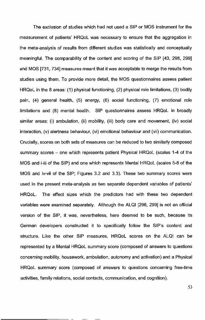
The exclusion of studies which had not used a SIP or MOS instrument for the
measurement of patients' HRQoL was necessary to ensure that the aggregation in
the meta-analysis of results from different studies was statistically and conceptually
meaningful. The comparability of the content and scoring of the SIP [43, 298, 299]
and MOS [731, 734] measures meant that it was acceptable to merge the results from
studies using them. To provide more detail, the MOS questionnaires assess patient
HRQoL in the 8 areas: (1) physical functioning, (2) physical role limitations, (3) bodily
pain, (4) general health, (5) energy, (6) social functioning, (7) emotional role
limitations and (8) mental health. SIP questionnaires assess HRQoL in broadly
similar areas: (i) ambulation, (ii) mobility, (iii) body care and movement, (iv) social
interaction, (v) alertness behaviour, (vi) emotional behaviour and (vii) communication.
Crucially, scores on both sets of measures can be reduced to two similarly composed
summary scores- one which represents patient Physical HRQoL (scales 1-4 of the
MOS and i-iii of the SIP) and one which represents Mental HRQoL (scales 5-8 of the
MOS and iv-vii of the SIP; Figures 3.2 and 3.3). These two summary scores were
used in the present meta-analysis as two separate dependent variables of patients'
HRQoL. The effect sizes which the predictors had with these two dependent
variables were examined separately. Although the ALQI [298, 299] is not an official
version of the SIP, it was, nevertheless, here deemed to be such, because its
German developers constructed it to specifically follow the SIP's content and
structure. Like the other SIP measures, HRQoL scores on the ALQI can be
represented by a Mental HRQoL summary score (composed of answers to questions
concerning mobility, housework, ambulation, autonomy and activation) and a Physical
HRQoL summary score (composed of answers to questions concerning free-time
activities, family relations, social contacts, communication, and cognition).
53
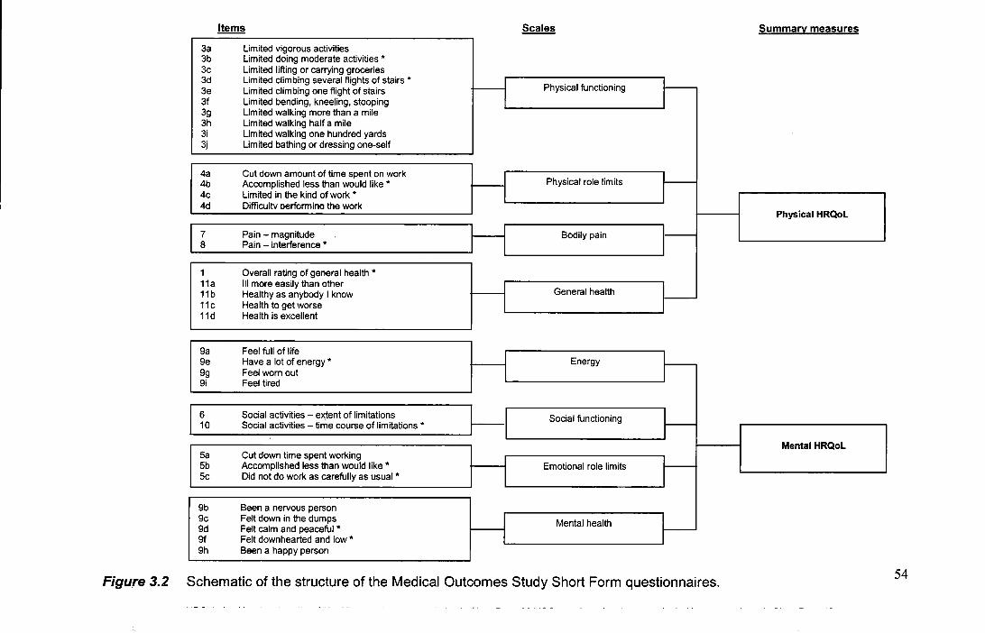
Items Scales Summary measures
3a Limited vigorous activities 3b Limited doing moderate activities * 3c Limited lifting or carrying groceries 3d Limited climbing several flights of stairs * r ,_ 3e Limited climbing one flight of stairs l
Physical functioning
3f Limited bending, kneeling, stooping 3g Limited walking more than a mile 3h Limited walking half a mile 3i Limited walking one hundred yards 3j Limited bathing or dressing one-self
4a Cut down amount of time spent on work 4b Accomplished less than would like * --1 Physical role limits I 4c Limited in the kind of work * 4d Difficultv oerformina the work
Physical HRQoL
7 Pain - magnitude Bodily pain ,_
8 Pain - interference *
1 Overall rating of general health* 11a Ill more easily than other
H ~ 11b Healthy as anybody I know General health
11c Health to get worse 11d Health is excellent
9a Feel full of life 9e Have a lot of energy * I Energy r-9g Feel worn out I 9i Feel tired
6 Social activities -extent of limitations
-I Social functioning ~ 10 Social activities -time course of !_imitations *
Mental HRQoL Sa Cut down time spent working
I I 5b Accomplished less than would like *
I Emotional role limits
5c Did not do work as carefully as usual *
9b Been a nervous person 9c Felt down in the dumps I Mental health ~ 9d Felt calm and peaceful *
I 9f Felt downhearted and low* 9h Been a happy person
Figure 3.2 Schematic of the structure of the Medical Outcomes Study Short Form questionnaires. 54
Selected items
Walk shorter distances or stop to rest often Do not walk at all
Stay within one room Stay away from home only for brief periods of time
Do not bathe self at all, bathed by someone else Very clumsy in body movements
Doing fewer social activities with groups of people Isolate self as much as can from rest of the family
Difficulty reasoning and solving problems, learning new things Behave as if confused or disoriented in place or time
Laugh or cry suddenly Irritable and impatient with self
Trouble writing or typing Do not speak clearly when under stress
Sit during much of the day Sleep or nap during the day
Eating no food, nutrition is taken through tubes or intravenously Eating special or different food
Not working at all Often irritable toward work associates
Not doing prior maintenance/ repair work around house Not doing heavy work around house
I am going out for entertainment less I am not doing any of my usual physical recreation or activities
Scales
Ambulation
Mobility
Body care and movement
Social interaction
Alertness behaviour
Emotion behaviour
Communication
Figure 3.3 Schematic of the structure of the Sickness Impact Profile
Summary measures
Physical HRQol
Mental HRQol
Total HRQoL Score
HRQoL=health-related quality of life. Note the Mental Component of the SIP is officially referred to as 'Psychosocial HRQoL'.
55
Additional motivating factors for including studies using either the SIP or MOS
were that, firstly, they are - by far- the most popular measures of HRQoL used
within SAH studies. This meant that only a minimal number of studies had to be
excluded (see Figure 2.7, Chapter 2). Secondly, both families of measures (including
the ALQI) have been extensively validated and have been found to have satisfactory
psychometric properties [59, 137, 138, 295, 731, 734]. Indeed, they are preferable to
other HRQoL instruments used in SAH studies, such as the Euroqol [237] and the
Nottingham Health Profile [292], as they have been found to illustrate superior
sensitivity to gradations in poor health [59].
The use of all of the exclusion criteria in resulted in the inclusion in the meta
analysis of the following 7 traditional predictors of SAH patients' HRQoL: (1) patient
age, (2) bleed severity, (3) clinical severity on admission to hospital, (4) post-SAH
cognitive impairment, (5) post-SAH physical disability, (6) patient sex and (7) the time
between illness onset and HRQoL assessment. In an attempt to increase the number
of studies in the meta-analysis, the authors of those articles which were already
identified for inclusion, but which had collected data on one (or more) extra
predictor(s), but not included it in the original article, were contacted and asked to
provide either the effect size or the raw data necessary for calculation. In addition,
authors of those studies which had used the SIP or MOS to measure SAH patients'
HRQoL, but which had been excluded from the initial systematic review because they
had not explicitly examined predictors (Exclusion criteria 6) or because they had
merged SAH patient data with that of other patient groups (Exclusion criteria 7), were
also contacted. In total, 12 articles were finally included in the meta-analysis on the 7
predictors.
56
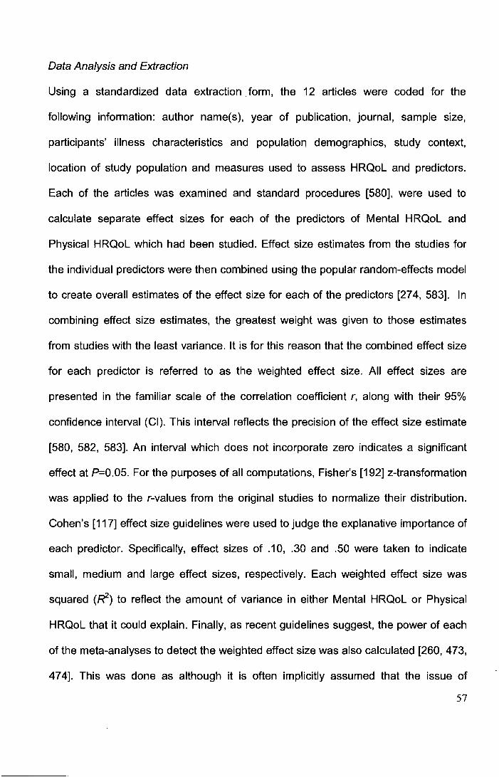
Data Analysis and Extraction
Using a standardized data extraction form, the 12 articles were coded for the
following information: author name(s), year of publication, journal, sample size,
participants' illness characteristics and population demographics, study context,
location of study population and measures used to assess HRQoL and predictors.
Each of the articles was examined and standard procedures [580], were used to
calculate separate effect sizes for each of the predictors of Mental HRQoL and
Physical HRQoL which had been studied. Effect size estimates from the studies for
the individual predictors were then combined using the popular random-effects model
to create overall estimates of the effect size for each of the predictors [274, 583]. In
combining effect size estimates, the greatest weight was given to those estimates
from studies with the least variance. It is for this reason that the combined effect size
for each predictor is referred to as the weighted effect size. All effect sizes are
presented in the familiar scale of the correlation coefficient r, along with their 95%
confidence interval (CI). This interval reflects the precision of the effect size estimate
[580, 582, 583]. An interval which does not incorporate zero indicates a significant
effect at P=O.OS. For the purposes of all computations, Fisher's [192] z-transformation
was applied to the r-values from the original studies to normalize their distribution.
Cohen's [117] effect size guidelines were used to judge the explanative importance of
each predictor. Specifically, effect sizes of .1 0, .30 and .50 were taken to indicate
small, medium and large effect sizes, respectively. Each weighted effect size was
squared (R'2) to reflect the amount of variance in either Mental HRQoL or Physical
HRQoL that it could explain. Finally, as recent guidelines suggest, the power of each
of the meta-analyses to detect the weighted effect size was also calculated [260, 473,
474]. This was done as although it is often implicitly assumed that the issue of
57
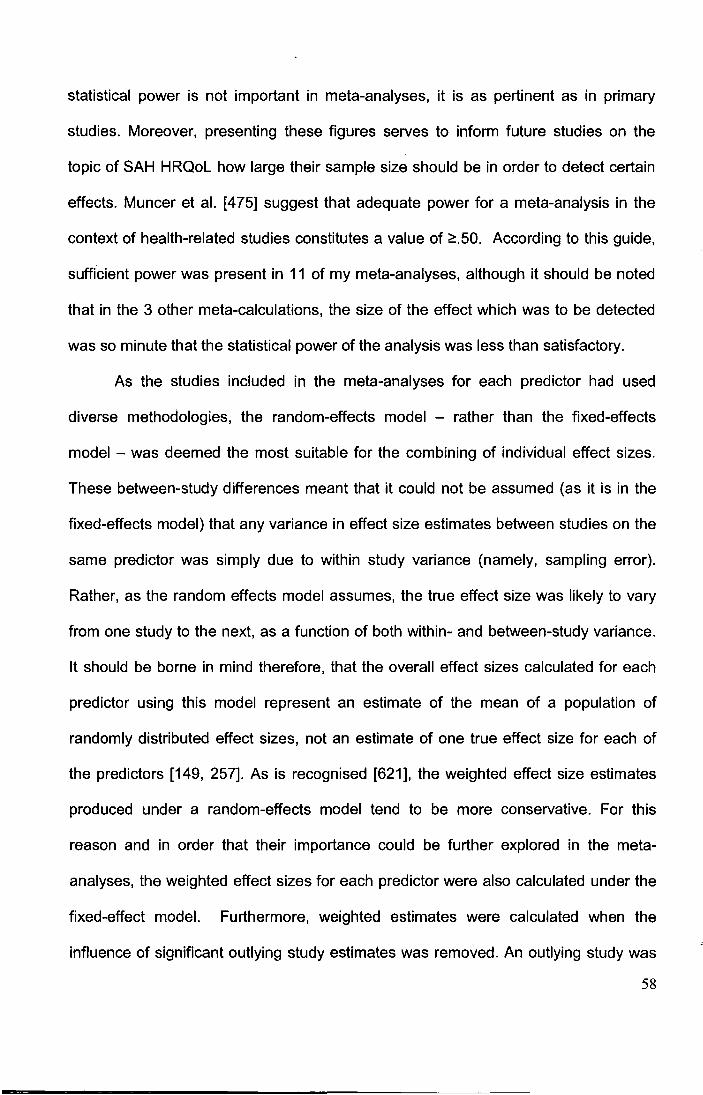
statistical power is not important in meta-analyses, it is as pertinent as in primary
studies. Moreover, presenting these figures serves to inform future studies on the
topic of SAH HRQoL how large their sample size should be in order to detect certain
effects. Muncer et al. [475] suggest that adequate power for a meta-analysis in the
context of health-related studies constitutes a value of ~.50. According to this guide,
sufficient power was present in 11 of my meta-analyses, although it should be noted
that in the 3 other meta-calculations, the size of the effect which was to be detected
was so minute that the statistical power of the analysis was less than satisfactory.
As the studies included in the meta-analyses for each predictor had used
diverse methodologies, the random-effects model - rather than the fixed-effects
model - was deemed the most suitable for the combining of individual effect sizes.
These between-study differences meant that it could not be assumed (as it is in the
fixed-effects model) that any variance in effect size estimates between studies on the
same predictor was simply due to within study variance (namely, sampling error).
Rather, as the random effects model assumes, the true effect size was likely to vary
from one study to the next, as a function of both within- and between-study variance.
It should be borne in mind therefore, that the overall effect sizes calculated for each
predictor using this model represent an estimate of the mean of a population of
randomly distributed effect sizes, not an estimate of one true effect size for each of
the predictors [149, 257]. As is recognised [621], the weighted effect size estimates
produced under a random-effects model tend to be more conservative. For this
reason and in order that their importance could be further explored in the meta
analyses, the weighted effect sizes for each predictor were also calculated under the
fixed-effect model. Furthermore, weighted estimates were calculated when the
influence of significant outlying study estimates was removed. An outlying study was
58

defined as one whose standardised residual effect size estimate exceeded (+/-) 2.00
[258].
The homogeneity between studies in terms of their effect size estimates for
each predictor of Mental and Physical HRQoL was tested using the Q-test, with an
alpha set of 0.05. The Q-test for a given predictor was computed by summing the
squared deviations of each study's effect size estimate from the overall effect size
estimate, with the contribution of each study weighted by its inverse variance.
However, as this test is subject to a lack of power, the parameter /-squared (f) was
also calculated to describe heterogeneity in between study effect sizes as it is not
dependent on the number of studies included in the analysis [252, 275]. f represents
the percentage of the total variation between studies in effect sizes which was due to
heterogeneity rather than chance [272]. Either a significant Q-statistic or a f value of
>50% was taken to indicate that the effect size estimates for a given predictor were
more heterogeneous between studies than expected from sampling error alone [404].
The robustness of the calculated weighted effect size for the predictors from
the (potential) impact of publication bias was assessed using the classic fail safe N
calculation [581]. Publication bias is the tendency for journals to publish only those
studies which report statistically significant results (or those based on larger studies)
[156, 396]. Having largely included only published studies in the present meta
analysis, there was the potential that I introduced upward bias into the meta-analysis
(i.e. significant effects for the traditional predictors were more likely to have been
found than would have been the case if all actual completed studies had been
included). For any significant effects detected by the meta-analysis, I therefore used
the fail-safe N formulae to determine the number of unpublished (or un-retrieved)
studies which on average found no significant effect of the predictor that would be
59
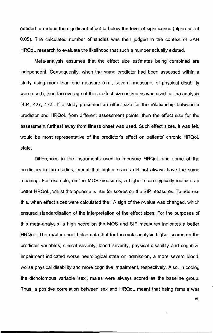
needed to reduce the significant effect to below the level of significance (alpha set at
0.05). The calculated number of studies was then judged in the context of SAH
HRQoL research to evaluate the likelihood that such a number actually existed.
Meta-analysis assumes that the effect size estimates being combined are
independent. Consequently, when the same predictor had been assessed within a
study using more than one measure (e.g., several measures of physical disability
were used), then the average of these effect size estimates was used for the analysis
[404, 427, 472]. If a study presented an effect size for the relationship between a
predictor and HRQoL from different assessment points, then the effect size for the
assessment furthest away from illness onset was used. Such effect sizes, it was felt,
would be most representative of the predictor's effect on patients' chronic HRQoL
state.
Differences in the instruments used to measure HRQoL and some of the
predictors in the studies, meant that higher scores did not always have the same
meaning. For example, on the MOS measures, a higher score typically indicates a
better HRQoL, whilst the opposite is true for scores on the SIP measures. To address
this, when effect sizes were calculated the+/- sign of the r-value was changed, which
ensured standardisation of the interpretation of the effect sizes. For the purposes of
this meta-analysis, a high score on the MOS and SIP measures indicates a better
HRQoL. The reader should also note that for the meta-analysis higher scores on the
predictor variables, clinical severity, bleed severity, physical disability and cognitive
impairment indicated worse neurological state on admission, a more severe bleed,
worse physical disability and more cognitive impairment, respectively. Also, in coding
the dichotomous variable 'sex', males were always scored as the baseline group.
Thus, a positive correlation between sex and HRQoL meant that being female was
60

associated with a better HRQoL, whilst a negative correlation indicated that being
male was associated with a better HRQoL.
The calculation of the effect sizes for a Brazilian study by Rocha-Filo et al.
[569] which was included in this meta-analysis deserves mention here. Specifically,
one of the standard steps used for the calculation of their patients' MOS Mental
HRQoL and Physical HRQoL was not possible to complete exactly as stated by
available guidelines (733]. The step that proved troublesome involved all eight of the
patients' MOS subscale scores being standardised using a linear z-score
transformation. Z-scores are usually calculated by subtracting the MOS subscale
means for the local population from each individual's subscale score and then
dividing the difference by the standard deviation of the local population. The
documented lack of local MOS population scores for Brazil however [717], meant that
it was necessary to use non-local normative data for the z-score calculations. My
choice of which surrogate normative scores to use was informed by the limited
Brazilian MOS normative data which do exist. Specifically, unpublished mean MOS
scores (and not standard deviations) are available [619] and it is these that were used
by Rocha-Filo et al. for crude visual comparisons in their original study. I however,
used published normative scores from a large Swedish sample (N=8930) as these
were the most comparable to the Brazilian population scores in terms of subscale
means (673]. Appendix V presents a breakdown of the exact comparability of the
Swedish MOS normative scores (as well as other countries) to those of Brazil.
Tests analogous to analysis of variance were conducted to examine the
validity of my having combined effect size estimates for predictors from studies which
differed in the HRQoL questionnaire they used. The individual effect sizes for each
predictor were grouped according to whether they were derived from a study using
61
the MOS or the SIP and then contrasted to evaluate whether the measure used,
moderated the effect size estimate. Significant heterogeneity was deemed to exist
between the two groups' average effect size ( Oa statistic) if their mean effect size
differed by more than sampling error [404].
All analyses were conducted using commercially available software (SPSS,
Comprehensive Meta Analysis) and the freeware programme G*Power [174].
3.3 Results
Characteristics of the Studies included in the Meta-Analysis
As previously noted, 12 articles (Table 3.1) studying the 7 traditional SAH HRQoL
predictors formed the basis of the meta-analysis. Of these articles, 10 had examined
the predictor patient age, 9 had examined the predictor sex, 7 had examined clinical
severity, 7 physical disability, 6 had studied bleed severity, 4 the time since illness
and 3 cognitive impairment. Most of the studies reported in the articles had been
conducted in Europe (67%), all were published between 2000 and 2008 (median=
2005) and were typically published in North-American neurological and neurosurgical
journals. Most (67%) of the studies had used a MOS questionnaire to measure
patients' HRQoL. In terms of their sample characteristics, most studies had focused
exclusively on those SAH patients whose haemorrhage was of an aneurysmal origin
(75%) and who were in a favourable clinical state on admission to hospital. Sample
size ranged from 10 to 573 (median=63, interquartile [IQR] range=33-110). Nine of
the 12 studies had assessed the SAH patients' HRQoL at only a single follow-up
assessment: 3 studies had assessed SAH patients <6months post-ictus, 1 ;;::6-12
months, 2 between 13-23 months and 2 ;;::24 months post- SAH. In the studies, the
predictor 'physical disability' had been measured using the Glasgow Outcome Scale 62
Table 3.1 Details of studies included in the meta-analysis
Study Location Treatment Sample HRQol Follow-up Clinical Bleed severity Clinical Treatment Age Sex
period size(s) measure point(s) Severity details
Hackett et al.[240] Australia 1995-1998 174 SF-36 1.2 83% HH I-ll - ASAH; Clipped (46% early) 50.9 Most female
(acute) SAHuo
Hop et al. [286] Netherlands 1995-1996 48 SIP .33, 1.5 86%WFNS 1-11 - ASAH - 51.9 70.3%
female
HOtter et a I. [305] Germany 1989-1992 116 ALQI 4.0-5.0 57% HH 1-11 59% Fisher4 ASAH Clipped (74% early) 50.3 66% female
Jonsson et al. Sweden 2001-2002 10-13 SF-36 .33, 1.3 - - - - 52.0 76.9%
[322] female
Katati et al. [336] Spain 2003-2005 70 SF-36 .33 75.7% HH 1-11; 43% Fisher4 ASAH Clipped or coiled. 48.9 57% male
71.4% WFNS 1-
II King et al. [358] u.s. 2001-2004 91 SF-12 - - - ASAH Most clipped. 51.3 Most female
Kreitsch'. [377] Germany 1997-2002 35 SF-36 2.3 55% HH 1-11 65% Fisher 3 ASAH 95% clipped 43.8 65% female
63
Table 3.1 cont'd
Study Location
Maceo et al. [463] U.S.
Morris et al. [470] U.K.
Rocha-Filo et al. Brazil
[569]
Scott et al. [613] U.K.
Tseng et al. [694] U.K.
Treatment
period
1996-2002
1998-1998
2002-2003
1994-2002
2004-2004
Sample
size(s)
35
32-49
66
512-573
60
HRQol
measure
SIP
SF-36
SF-36
FLP
SF-36
Follow-up
point(s)
.25, 1.0
1.2
.4
1.0
.5
Clinical
Severity
64% HH IV;
36% HH V.
84% WFNS 1-11.
All GCS 15
Most WFNS I-ll*
57.4% WFNS I
ll.
Bleed severity
48% Fisher 3
53% Fisher 3
-
-
86.2% Fisher 3-
4
Clinical
details
ASAH
ASAH;
SAHuo
ASAH
ASAH*
ASAH
Treatment Age Sex
Clipped or coiled. 53.0 Most female
77% clipped 45.2 Most female
Clipped 44.6 64% female
Clipped or coiled.
65% clipped. 52.9 55% female
Note: ALQI=Aachen Life Quality Index; ASAH=aneurysmal subarachnoid haemorrhage; Fisher=Fisher CT grading scale; FLP=Functional Limitations Profile; GCS=Giasgow Coma Scale; HH=Hunt
and Hess scale; Kreitsch'=Kreitschmann-Anderrnahr et al.; SAHuo=subarachnoid haemorrhage of unknown origin; SF-12=Short Forrn-12; SF-36=Short Forrn-36; SIP=Sickness Impact Profile;
WFNS=World Federation of Neurological Surgeons Grading scale; -=unknown.
64
(original and extended version) [318, 758], the modified Rankin Scale [550] and the
Barthel Index [421]. The predictor 'clinical severity' had been measured using either
the World Federation of Neurological Surgeons Scale [164] or the Hunt and Hess
Scale [293]. 'Bleed severity' on the other hand, had been measured in all studies
using Fisher's CT rating scale [191]. For the measurement of the predictor 'cognitive
impairment', one study used the Mini-Mental State Examination [197], a well-known
global measure of cognitive impairment which has been found to be a sensitive and
reliable measure of post-SAH cognitive dysfunction [354, 357, 438]. The other two
studies employed comprehensive batteries of neuropsychological tests chosen
primarily on the basis of their proven ability to detect post-SAH impairment [470, 613].
Measures of cognitive impairment, as well as physical disability, were completed
almost always at the same appointment at which the patients' completed the HRQoL
questionnaires. The measures of physical disability, clinical severity and bleed
severity used in the studies included in the meta-analyses are further described in the
appendices (Appendices I-IV and VI-VII).
Effect size for predictors of HRQoL
Physical HRQoL
Table 3.2 presents the weighted effect size estimates for the 7 traditional predictors of
Physical HRQoL which were calculated using the random effects model. As can be
seen, the weighted effect size for 6 of the 7 variables proved to be significantly
associated with SAH patients' Physical HRQoL. Older age (Z= -4.15, P<0.001 ), a
more severe bleed (Z= -3.86, P<0.001 ), a worse neurological state on admission to
hospital (Z= -2.88, P<0.001 ), more post-SAH cognitive impairment (Z= -3.42,
65
Table 3.2 Summary of weighted effect sizes for predictors of Physical HRQoL
Predictor No. of Population
size
Cl Q-statistic Fail-safe Power
studies N
Age -.20*** 4.00% 10 1224 -.29 -.10 17.71* 87 1.00
Bleed severity -.13*** 1.69% 6 879 -.20 -.06 4.67 8 .98
Clinical severity -.19*** 3.61% 7 1055 -.30 -.06 18.22** 43 .99
Cognitive impairment -.14*** 1.96% 3 615 -.22 -.06 0.03 3 .96
Physical disability -.54*** 29.2% 7 865 -.63 -.44 13.74* 413 1.00
Sex -.13* 1.69% 9 1070 -.24 -.02 19.29* 30 .99
Time since illness .09 0.81% 4 127 -.12 .30 6.88 .26
Total 42.96%
Note: Cl=confidence interval; No.=number;r,.=weighted correlation coefficient (r,.=0.10, r=0.30 and r=0.50 are small, medium
and large effect sizes, respectively); R2 ... =variance explained; *=P<0.05; **=P<0.01; ***=P<0.001. For the dichotomous variable
'sex', males were scored as the baseline group. Thus, a positive correlation means that being female was associated with a
better HRQoL, whilst a negative correlation indicates the opposite.
P<0.001 ), greater physical disability (Z= - 8. 75, P<0.001) and being female (Z= -2.32,
P<0.05) were all associated with a worse Physical HRQoL. Information about the time
which had elapsed between a patient's SAH and assessment of their HRQoL was not
significantly associated with SAH patients' Physical HRQoL: Z= 0.86, P>0.05.
Importantly, with the exception of physical disability, the size of the effect of the
significant predictors of Physical HRQoL was minimal, with each of these predictors
being able to account for only 1.69-4% of the variance in patients' Physical HRQoL
scores. As noted above, physical disability was the exception. It was found to have a
66
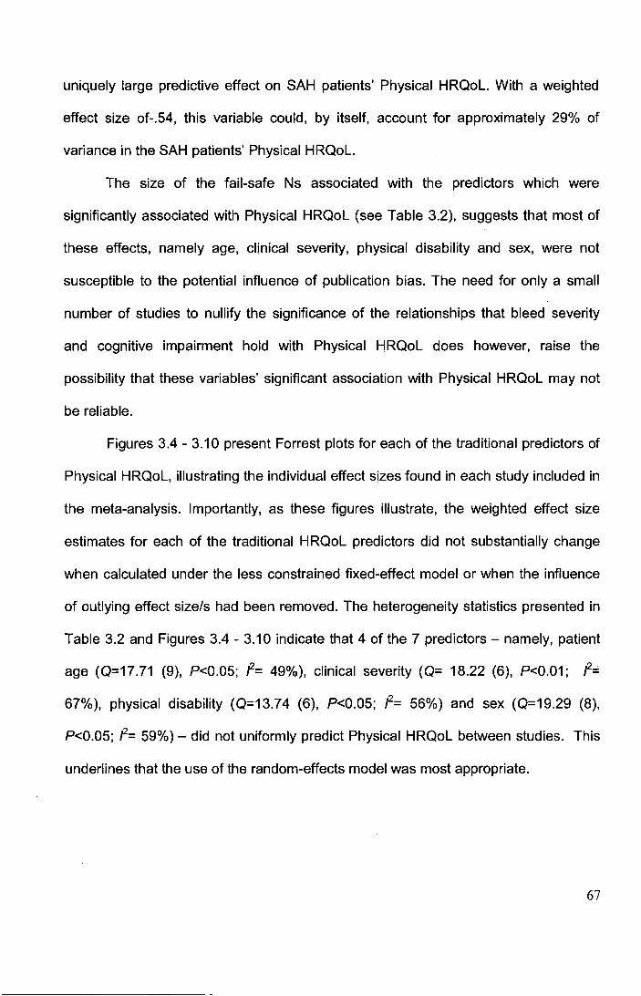
uniquely large predictive effect on SAH patients' Physical HRQoL. With a weighted
effect size of-.54, this variable could, by itself, account for approximately 29% of
variance in the SAH patients' Physical HRQoL.
The size of the fail-safe Ns associated with the predictors which were
significantly associated with Physical HRQoL (see Table 3.2), suggests that most of
these effects, namely age, clinical severity, physical disability and sex, were not
susceptible to the potential influence of publication bias. The need for only a small
number of studies to nullify the significance of the relationships that bleed severity
and cognitive impairment hold with Physical HRQoL does however, raise the
possibility that these variables' significant association with Physical HRQoL may not
be reliable.
Figures 3.4 - 3.10 present Forrest plots for each of the traditional predictors of
Physical HRQoL, illustrating the individual effect sizes found in each study included in
the meta-analysis. Importantly, as these figures illustrate, the weighted effect size
estimates for each of the traditional HRQoL predictors did not substantially change
when calculated under the less constrained fixed-effect model or when the influence
of outlying effect size/s had been removed. The heterogeneity statistics presented in
Table 3.2 and Figures 3.4 - 3.10 indicate that 4 of the 7 predictors- namely, patient
age (0=17.71 (9), P<0.05; f= 49%), clinical severity (0= 18.22 (6), P<0.01; f=
67%), physical disability (0=13.74 (6), P<0.05; /2= 56%) and sex (0=19.29 (8),
P<0.05; f= 59%)- did not uniformly predict Physical HRQoL between studies. This
underlines that the use of the random-effects model was most appropriate.
67
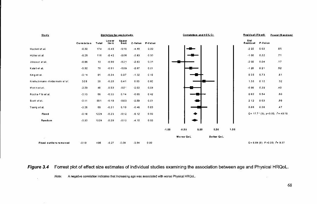
Study Statistics for each study Correlation and 95% Cl Residual !Fixed! Power (R andoml
Lower Upper Correlation Total lim it limit Z-Value P-Value
Std Residual P-Value
Hackett et al. -0.32 174 -0.45 -0.18 -4.40 0.00 --- -2.25 0.02 .85
HOtter et al. -0.26 116 -0.42 -0.08 -2.83 0.00 .....,._
-1.00 0.32 .71
JOnsson et al. -0.68 13 -0.90 -0.21 -2.63 0.01 - -2.08 0.04 .17
Katati et al. -0.32 70 -0.51 -0.09 -2.67 0.01 - -1.26 0.21 .52
King et al. -0.14 91 -0.34 0.07 -1.32 0.19 - 0.35 0.73 .61
Kreitschmann -Andermahr et al. 0.09 35 -0.25 0.41 0.53 0.60 ,_ ,- 1 .55 0.12 .32
Morrisetal. -0.29 48 -0.53 -0.01 -2.03 0.04 -- -0.86 0.39 .40
Rocha-Filo et al. -0.10 66 -0.33 0.14 -0.80 0.42 - 0.62 0.54 .50
Scott et al. -0.11 551 -0.19 -0.03 -2.59 0.01 ... 2.12 0.03 .99
Tseng et al. -0.06 60 -0.31 0.19 -0.48 0.63 0.88 0.38 .47
Axed -0.18 1224 -0.23 -0.12 -6.12 0.00 • Q= 17.71 (9), p<O.OS; f= 49.18
Random -0.20 1224 -0.29 -0.10 -4.15 0.00 .. -1.00 -0.50 0.00 0.50 1.00
Worse Qol Better Qol
Fixed outliers removed -0.18 486 -0.27 -0.09 -3.94 0.00 Q= 6.64 (9), P>0.05; f= 9.57
Figure 3.4 Forrest plot of effect size estimates of individual studies examining the association between age and Physical HRQoL
Note: A negative correlation indicates that increasing age was associated with worse Physical HRQol.
68
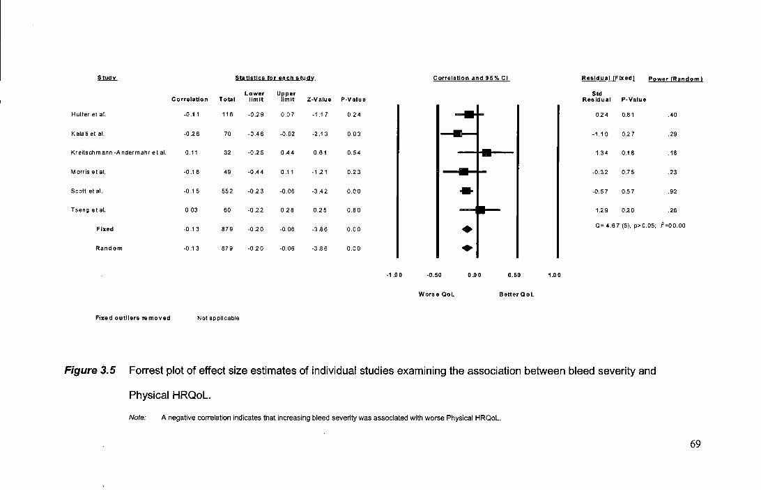
Study Statistics for each study Correlation and 95% Cl Residual !Fixed! Power (Random I
Lower Upper Std Correlation Total limit limit Z-Value P-Value Residual P-Value
Hotter et al. -0.11 116 -0.2 9 0.07 -1 .17 0.24 I I ....... 0.2 4 0.81 .40
Katati et al. -0.26 70 -0.46 -0.02 -2.13 0.03 I I~ -1.10 0.27 .29
Kreitsch mann -A nderm ahr eta!. 0.11 32 -0.25 0.44 0.61 0.54 I I --t- 1.34 0.18 .18
Morris et al. -0.18 49 -0.44 0.11 -1 .2 1 0.23 I 1-- -0.32 0.75 .23
Scott et al. -0.15 552 -0.23 -0.06 -3.42 0.00 I I - -0.57 0.57 .92
Tseng et al. 0.03 60 -0.22 0.28 0.25 0.80 I I -+ Fixed -0.13 879 -0.20 -0.06 -3.86 0.00 I I • 1.29 0.20 .26
Q= 4.67 (5). p>0.05; 1'=00.00
Random -0.13 879 -0.20 -0.06 -3.86 0.00 I I • -1 .0 0 -0.50 0.00 0.50 1.00
Worse Qol Better Q ol
Fixed outliers removed Not applicable
Figure 3.5 Forrest plot of effect size estimates of individual studies examining the association between bleed severity and
Physical HRQoL.
Note: A negative correlation indicates that increasing bleed severity was associated with worse Physical HRQoL.
69
~ Statistics for each study Correlation and 95% Cl Residual IFixedl f'_ower 1Random I
Lower Upper Std Correlation Total limit limit Z-Value P-Value Residual P-Value
Hackettet al. 0.05 174 -0.10 0.20 0.63 0.53 3.22 0.00 .82
HOtter et al. -0.19 116 -0.36 -0.01 -2.04 0.04 -0.17 0.86 .67
Katati et al. -0.34 70 -0.53 -0.11 -2.85 0.00 -1.45 0.15 .48
Kreitschmann -Andermahr et al. 0.0 5 35 -0.29 0.38 0.29 0.77 1.31 0.19 .30
Morris etal. -0.20 49 -0.46 0.08 -1.40 0.16 -0.20 0.84 .38
Scott et al. -0.20 551 -0.28 -0.12 -4.72 0.00
t: -0.84 0.40 .99
Tseng et al. -0.45 60 -0.63 -0.22 -3.69 0.00
I I I -2.42 0.02 .44
Fixed -0.18 1055 -0.23 -0.12 -5.69 0.00 Q= 18.22 (6), p<0.01; /'= 67.10
Random -0.19 10 55 -0.30 -0.06 -2.88 0.00 I I ~ -1.00 -0.50 0.00 0.50 1.00
Worse QoL Better QoL
Fixed outliers removed -0.20 821 -0.27 -0.13 -5.76 0.00 Q= 3.95 (4), p>0.05; /2 = 56.33
Figure 3.6 Forrest plot of effect size estimates of individual studies examining the association between clinical severity and
Physical HRQoL.
Note: A negative correlation indicates that increasing clinical severity was associated with worse Physical HRQoL.
70
.s..tu.d.lL Statistics for aac:h studv Correlation and 95% Cl Residual IFixedl P~wer IRan dom l
Lower Upper Std Correlation Total limit limit Z-Value P-Value Residual P-Value
Hackett et al. -0.15 174 -0.29 -0.00 -1 .96 0.05 --- -0.23 0.82 .53
JOnsson et al. -0.72 13 -0.91 -0.28 -2.88 0.00 - -2.4 7 0.01 .11 -Katati et al. -0.33 70 -0.53 -0.10 -2.82 0.00 - -1.78 0.08 .29 -King et al. -0.03 91 -0.23 0.18 -0 .2 6 0.79 - 1.04 0.30 .34
Kreitschmann -Andermahr et al. 0.16 35 -0.18 0.4 7 0.94 0.35 - 1.72 0.09 .19 -Morris et al. -0.33 49 -0.56 -0.05 -2.33 0.02 - -1.46 0.15 .23 -Rocha- Filo et al. -0.07 66 -0.31 0.17 -0.58 0.56 - 0.50 0.62 .28 -Scott et al. -0.14 512 -0.22 -0.05 -3.1 9 0.00 ... -0.23 0.81 .91
Tseng et al. 0.13 60 -0.13 0.37 1.01 0.31 - 2.08 0.04 .26 -Fixed -0.13 1070 -0.19 -0.07 -4.33 0.00 ........ Q= 19.29 (8), p<O.OS; f= 58.52
Random -0.1 3 1070 -0.24 -0.02 -2.31 0.02 D I I I I
-1.00 -0.50 0.00 0.50 1.00
Worse Qol Better Qol
Fixed outliers removed -0.14 997 -0.20 -0.08 -4,43 0.00 Q= 9.088 (6), p>0.05; f= 33.98
Figure 3.7 Forrest plot of effect size estimates of individual studies examining the association between sex and Physical HRQoL.
Note: For the dichotomous variable 'sex', males were scored as the baseline group. A negative correlation thus means that being female was associated with a worse Physical
HRQoL.
71
Study Statistics for each study Correlation and 95% Cl Residual (Fixed) Power (Random)
Lower Upper Std Correlation Total limit limit Z-Value P-Value Residual P-Value
JOnsson et al. -0.20 10 -0.73 0.50 -0.52 0.60 -- -0.16 0.88 .11
Morris et al. -0.15 32 -0.4 7 0.21 -0.80 0.43 -- .19 -0.05 0.96
Scott et al. -0.14 573 -0.22 -0.06 -3.29 0.00 • 0.12 0.91 .96
Fixed -0.14 615 -0.22 -0.06 -3.42 0.00 • Q= 0.03 (2), p>0.05; i'=OO.OO
Random -0.14 615 -0.2 2 -0.06 -3.42 0.00 • -1.00 -0.50 0.00 0.50 1.00
Worse Qol Better Qol
Fixed outUers removed Not applicable
Figure 3.8 Forrest plot of effect size estimates of individual studies examining the association between cognitive impairment
and Physical HRQoL.
Note: A negative correlation indicates that increasing cognitive impairment was associated with worse Physical HRQoL.
72
~ Statistics for each study Correlation and 95% Cl Residual IFixedl P_owuJRandoml
Lower Upper Std Correlation Total limit lim it Z-Value P-Value Residual P-Value
Jonsson etai. -0.75 13 -0.92 -0.33 -3.04 0.00 I • I I I I -0.97 0.33 .71
Katati et al. -0.55 70 -0.69 -0.36 -5.03 0.00 I I I 0.36 0.72 .99
King et al. -0.35 91 -0.52 -0.15 -3.41 0.00 I I I 2.91 0.00 .99
Kreitschmann -Andermahr et al. -0.34 35 -0.61 -0 01 -2.02 0.04 I I 1.72 0.08 .98
Morris et al. -0.64 48 -0.78 -0.43 -5.09 0.00 I I I -0.70 0.48 .99
Scott et al. -0.61 548 -0.66 -0.56 -16.66 0.00 I .I I I I -224 0.02 1.0
Tseng et al. -0.57 60 -0.72 -0.37 -4.88 0.00 I J I I I 0.08 0.93 .99
Fixed -0.58 865 -0.62 -0.53 -19.07 0.00 I Q= 13.74 (6), p<0.05; f= 56.33
Random -0.54 865 -0.63 -0.44 -8.75 0.00 I .. -1.00 -0.50 0.00 0.50 1.00
Worse Qol Better Qol
Fixed outliers removed -0.56 226 -0.64 -0.46 -9.17 0.00 Q= 4.25 (4 ), p>O .0 5; I'= 5.81
Figure 3.9 Forrest plot of effect size estimates of individual studies examining the association between physical disability and
Physical HRQoL.
Note: A negative correlation indicates that increasing physical disability was associated with worse Physical HRQoL.
73
s.t.w1y_ Statistics for each studv Corrolatlon and 9S% Cl Rosjdua! (Fixed) Power !Rando ml
Lower Upper Std Correlation Total limit lim it Z-Value P-Value Residual P-Value
Hopetal. 0.00 48 -0.28 0.28 0.00 1.00 -1.05 0.29 .16
JOnsson et al. 0.03 13 -0.5 3 0.57 0.09 0.9 3 -0.37 0.71 .09
Mocco et al. 0.29 35 0.13 0.44 3.41 0.00 I--- 2.60 0.01 .14
Rocha-Filoetal. -0.06 66 -0.30 0.18 -0.49 0.62 -1.88 0.06 .19
Fixed 0.14 162 0.02 0.26 2.23 0.03 Q= 6.88 (3), p>0.05; /2= 56.38
Random 0.09 162 -0.12 0.30 0.86 0.39
I I
-1.00 -0.50 0.00 0.50 1.00
Worse Qol Better Qol
Fixed outliers removed -0.03 127 -0.21 0.15 -0.34 0.74 Q= 0.14 (2), p>O.OS; /2= 00.00
Figure 3.10 Forrest plot of effect size estimates of individual studies examining the association between the time between illness
onset and assessment and Physical HRQoL.
Note: A positive correlation indicates that the longer the time between the HRQoL assessment and illness onset, the better the Physical HRQoL.
74
Mental HRQoL
Table 3.3 presents the weighted effect size estimates for each of the traditional
predictors for patients' Mental HRQoL. Of all the predictors, only cognitive impairment
proved to be significantly associated with patients' Mental HRQoL: Z= -3.26, P<0.001
Table 3.3 Summary of weighted effect sizes for predictors of Mental HRQoL
Predictor fw Rw No. of
studies
Population
size
Cl Fail-safe Power
statistic N
Age 0.07 0.59% 10 1225 -0.03 0.17 19.42* .79
Bleed severity 0.01 0.01% 6 879 -0.13 0.14 12.38* .08
Clinical severity -0.08 0.64% 7 1044 -0.18 0.03 12.72* .82
Cognitive impairment -0.13*** 1.69% 3 615 -0.21 -0.05 0.80 n/p .94
Physical disability -0.15 2.25% 7 866 -0.46 0.18 93.69*** .99
Sex -0.05 0.25% 9 1070 -0.17 0.08 24.55** .49
Time since illness 0.10 1.0% 4 162 -0.09 0.29 5.26 .35
Total 6.43%
Note: Cl-confidence interval; No.-number; n/p=calculation not possible due to differences between the formulae for classic
fails safe N and modern meta analysis means that according to fail-safe formulae the weighted effects size was not statistically
significant and so the fail safe N not computable; rw= weighted correlation coefficient (rw=0.10, r =0,30 and r =0.50 are small,
medium and large effect sizes, respectively); R2 w·= variance explained; *=P<0.05, **=P<0.01; ***=P<O.OO. For the dichotomous
variable 'sex', males were scored as the baseline group. A positive correlation thus means that being female was associated
with a better HRQoL, whilst a negative correlation indicates the opposite.
75
(fail-safe N not calculable, see Discussion for comment). The weighted effect size
estimate for this predictor was, nevertheless, still small (weighted r= -.13) - with
cognitive impairment being able to account for only 1.69% of variance in SAH
patients' Mental HRQoL. The Forest plots in Figures 3.11 - 3.17 illustrate that the
effect size estimates for each of the predictors calculated using the random-effects
model were comparable to those calculated using the fixed-effect model. Moreover,
they were also comparable when calculated with any study with an outlying effect size
estimate having been removed. It is also seen that significant heterogeneity existed in
effect size estimates between studies for the predictors: patient age (0=19.42 (9),
P<0.05; P=54%), bleed severity (0=12.38 (5), P<0.05; P=60%), clinical severity
(0=12.72 (6), P<0.05; P=53%), physical disability (0=93.69 (6), P<0.001; P=94%)
and sex (0=24.55 (8), P<0.01; P=67%).
Validity of combining effect sizes from studies using the MOS and studies using the
SIP
Tables 3.4 and 3.5 report the results from the analyses which examined whether the
HRQoL measure used by a study moderated the effect size estimate which it found
for a given predictor. The results show that in the vast majority of cases, the effect
size estimates were homogenous between studies using different HRQoL measures.
This result provides evidence that in most cases, it was valid to combine the results
from studies using the SIP measure with the results from studies using MOS
measures. Although no significant effect was found for any of the predictors of
Physical HRQoL, significantly different effect size estimates were produced by studies
76
Studv Statistics for each study Correlation and 95% Cl Residual !Fixed! power !RJ!ndom I
Lower Upper Std Correlation Total limit lim H Z-Va lue P-Value Residual P-Va lue
Hackett el al. 0.11 174 -0.04 0.25 1.44 0.15 I I -'-- I I 0.53 0.60 .23
HOtter et al. -0.17 116 -0.34 0.01 -1 .82 0.07 -2.73 0.01 .08
Jonsson et al. 0.57 13 0.03 0.85 2.06 0.04 1.84 0.07 .18
Katati et al. -0.08 70 -0.31 0.15 -0.69 0.49 -1.32 0.19 .14
King et al. 0.18 91 -0.02 0.3 8 1.75 0.08 1.11 0.27 .16
Kreltschmann -Andermahr et al. 0.35 35 0.02 0.61 2.05 0.04 1.6 6 0.10 .11
Morris et al. 0.16 48 -0.13 0.43 1.10 0.27 0.62 0.54 .12
Roch a-Fila et al. -0.14 66 -0.3 7 0.11 -1 .09 0.28 -1.71 0.09 .14
Scott et al. 0.10 552 0.01 0.18 2.26 0.02 0.74 0.46 .50
Tseng etal. 0.13 60 -0.1 3 0.3 7 0.96 0.34 0.42 0.68 .13
Fixed 0.07 1225 0.02 0.1 3 2.52 0.01 Q= 19.42 (9), p<0.05: f= 53.66
Random 0.07 1225 -0.03 0.17 1.43 0.15 I I .- I I
-1 ,0 0 -0.50 0.00 0.50 1.00
Worse Qol Better Q ol
Fixed- o uttiers removed 0.10 1109 0.04 0.16 324 0.07 Q= 11.95 (8), p>0.05; t'= 33.07
Figure 3.11 Forrest plot of effect size estimates of individual studies examining the association between age and Mental HRQoL.
Note: A negative correlation indicates that increasing age was associated with worse Mental HRQoL.
77
Study Statistics for each studv Correlation and 95% Cl Residual !Fixed) Power IR andoml
Lower Upper Correlation Total limit lim it Z-Value P-Value
Std Residual P-Value
Hotter et al. -0.11 116 -0.29 0.07 -1.17 0.24 -0.4 8 0.63 .06
Katati et al. 0.20 70 -0.04 0.41 1.62 0.11 2.27 0.02 .06
Kreitsch mann -Andermahr et al. 0.30 32 -0.05 0.59 1.68 0.09 2.09 0.04 .06
Morris et al. -0.06 49 -0.34 0.22 -0.4 1 0.68 0.06 0.95 .06
Scott et al. -0.13 552 -0.21 -0.05 -3.02 0.00 -2.34 0.02 .08
Tsengetal. 0.08 60 -0.18 0.33 0.60 0.55 1.15 0.25 .06
Fixed -0.07 879 -0.13 -0.00 -2.01 0.04 Q= 12.38 (5), p<0.05; !"= 59.64
Random 0.01 879 -0.13 0.14 0.10 0.92
-1.00 -0.50 0.00 0.50 1.00
Worse Qol Better Qol
Fixed outliers removed -0.05 225 -0.18 0.08 -0.73 0.46 Q= 1.36 (2), p>0.05; i'= 00.00
Figure j.12 Forrest plot of effect size estimates of individual studies examining the association between bleed severity and Mental
HRQoL.
Note: A negative correlation indicates that increasing bleed severity was associated with worse Mental HRQoL.
78
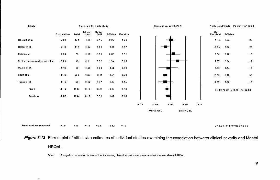
Study Statistics for each stud'L Correlation and 95% Cl Residual IFixedl Power IRan dom 1
Lower Upper Std Correlation Total limit limit Z-Value P-Value Residual P-Value
Hackett et al. 0.00 174 -0.15 0.15 0.00 1.0 0 1.76 0.08 .28
HOtter et al. -0.1 7 116 -0.34 0.01 -1.82 0.07 -0.55 0.58 .22
Katati et al. 0.08 70 -0.16 0.31 0.66 0.51 1.73 0.08 .16
Kreitsch mann -Andermahr et al. 0.23 35 -0.11 0.52 1.34 0.18 2.07 0.04 .12
Morris et al. -0.09 37 -0.40 024 -0.52 0.60 0.20 0.84 .12
Scott et al. -0.19 552 -0.27 -0.11 -4.51 0.00 - -2.39 0.02 .59
Tseng et al. -0.19 60 -0.42 0.07 -1.44 0.15 • I I I -0.52 0.60 .15
Fixed -0.12 1044 -0.18 -0.06 -3.94 0.00
~I I I Q= 12.72 (6), p <0.05; I'= 52.86
Random -0.08 1044 -0.18 0.03 -1.42 0.16
-1 .0 0 -0.50 0.00 0.50 1.00
Worse QoL Better QoL
Fixed outliers·removed -0.06 457 -0.15 0.03 -1.32 0.19 Q= 4.35 (4), p>0.05; f= 8.06
Figure 3.13 Forrest plot of effect size estimates of individual studies examining the association between clinical severity and Mental
HRQoL.
Note: A negative correlation indicates that increasing clinical severity was associated with worse Mental HRQoL.
79
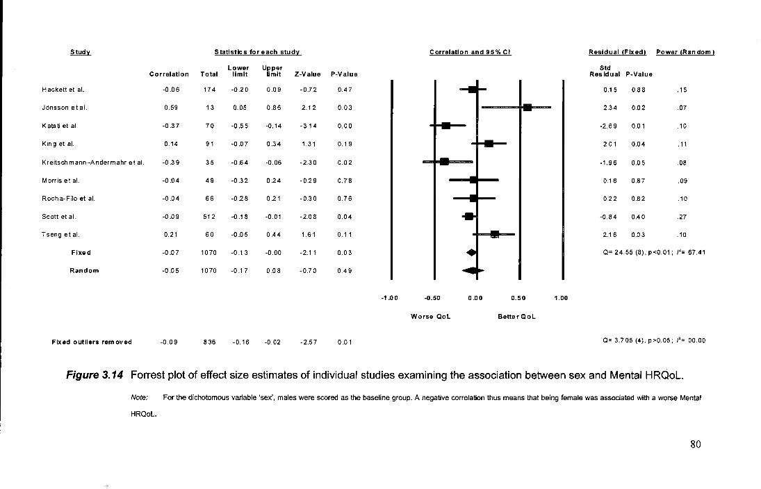
Study Statistics for each stud'L_ Correlation and 95% Cl Residual IFixedl Power IRan dom I
Lower Upper Std Correlation Total limit limit Z-Value P-Value Residual P-Value
Hackett et al. -0.06 174 -0.20 0.09 -0.72 0.4 7 0.15 0.88 .15
JOnsson et al. 0.59 13 0.05 0.86 2.12 0.03 2.34 0.02 .07
Katatietal. -0.37 70 -0.55 -0.14 -3.14 0.0 0 -2.69 0.01 .10
King et al. 0.14 91 -0.07 0.34 1. 31 0.1 9 2.01 0.04 .11
Kreitsch mann -Andermahr et at. -0.39 35 -0.64 -0.06 -2.30 0.02 -1.96 0.05 .08
Morris et al. -0.04 49 -0.32 0.24 -0.29 0.7 8 0.16 0.87 .09
Rocha-Filo et at. -0.04 66 -0.28 0.21 -0.3 0 0.76 0.22 0.82 .10
Scott et al. -0.09 512 -0.18 -0.01 -2.08 0.04 -0.84 0.40 .27
Tseng et at. 0.21 60 -0.05 0.44 1.61 0.11 2.16 0.03 .10
Fixed -0.07 1070 -0.1 3 -0.00 -2.11 0.0 3 Q= 24.55 (8), p<0.01; I'= 67.41
Random -0.05 1070 -0.17 0.08 -0.70 0.49
-1.00 -0.50 0.00 0.50 1.00
Worse Qol Better Qol
Fixed outliers removed -0.09 836 -0.16 -0.02 -2.57 0.01 Q= 3.705 (4), p>0.05; I"= 00.00
Figure 3.14 Forrest plot of effect size estimates of individual studies examining the association between sex and Mental HRQoL.
Note: For the dichotomous variable 'sex', males were scored as the baseline group. A negative correlation thus means that being female was associated with a worse Mental
HRQoL.
80
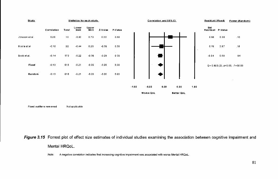
Study Statistics for each studv Correlation and 95% Cl Residual I Fixed) Power !Random)
Lower Upper Std Correlation Total limit limit Z-Value P-Value Residual P-Value
Jonsson et al. 0.20 10 -0.50 0.73 0.52 0.60 -- 0.88 0.38 .10
Morris et al. -0.10 32 -0.44 0.25 -0.56 0.58 -- 0.16 0.87 .18
Scott eta!. -0.14 573 -0.22 -0.06 -3.29 0.00 • -0.54 0.59 .94
Fixed -0.13 615 -0.21 -0.05 -3.26 0.00 • Q= 0.803 (2), p>0.05; f=OO.OO
Random -0.13 615 -0.21 -0.05 -3.26 0.00 • -1.00 -0.50 0.00 0.50 1.00
Worse QoL Better QoL
Fixed outliers removed Not ap piic able
Figure 3.15 Forrest plot of effect size estimates of individual studies examining the association between cognitive impairment and
Mental HRQoL.
Note: A negative correlation indicates that increasing cognitive impairment was associated with worse Mental HRQoL.
81
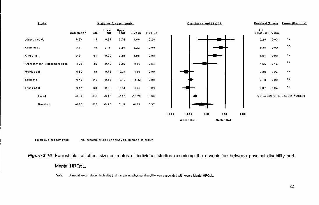
~ Statistics for each study Correlation and 95% Cl Residual !Fixed) Power (Random)
Lower Upper Std Correlation Total limit limit Z-Value P-Value Residual P-Value
JOnsson et al. 0.33 13 -0.2 7 0.74 1.08 0.28 -- 2.20 0.03 .13
Katati et al. 0.37 70 0.15 0.56 3.22 0.00 -- 6.35 0.00 .35
King et al. 0.21 91 -0.00 0.39 1.95 0.05 - 5.54 0.00 .42
Kreitschmann -Andermahr et al. -0.08 35 -0.40 026 -0.46 0.64 -- 1.55 0.12 22
Morris et al. -0.59 48 -0.75 -0.37 -4.56 0.00 -2.26 0.02 27
Scott et al. -0.47 549 -0.53 -0.40 -11.83 0.00 -6.10 0.00 .97
Tseng etal. -0.55 60 -0.70 -0.34 -4.65 0.00 -2.07 0.04 .31
Fixed -0.34 866 -0.40 -0.28 -1 0.20 0.00 • Q= 93.689 (6), p<0.0001; f=93.59
Random -0.15 866 -0.46 0.1 8 -0.89 0.3 7
-1.00 -0.50 0.00 0.50 1.00
Worse Qol Better Qol
Fixed outliers removed Not possible as only onestudynotdeemed an outlier
Figure 3.16 Forrest plot of effect size estimates of individual studies examining the association between physical disability and
Mental HRQoL.
Note: A negative correlation indicates that increasing physical disability was associated with worse Mental HRQoL.
82
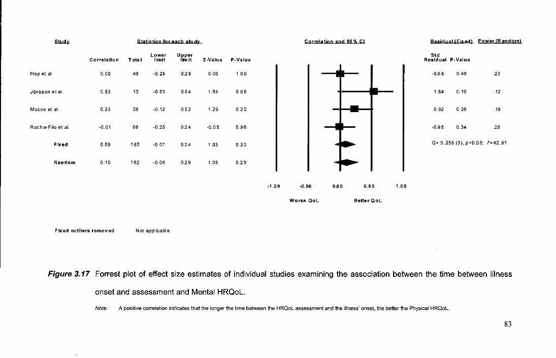
S1u..d.lL Statistics for each stu dv Correlation and 95% Cl Residual (Fixed) Power IR andoml
Lower Upper Std Correlation Total limit lim it Z-Value P-Value Residual P-Value
Hop et al. 0.00 48 -0.28 0.28 0.00 1 .00 -0.68 0.49 .23
JOnsson e I al. 0.53 13 -0.03 0.84 1.85 0.06 1.64 0.10 .12
Mocco et al. 0.23 35 -0.12 0.52 1.29 0.20 0.92 0.36 .19
Rocha- Filo et al. -0.01 66 -0.25 0.24 -0.05 0.96 -0.95 0.34 .28
Fixed 0.09 162 -0.07 0.24 1.05 0.30 Q= 5.255 (3), p>0.05; i'=42.91
Random 0.10 162 -0.09 0.29 1.05 0.29
-1.00 -0.50 0.00 0.50 1.00
Worse Qol BetterQol
Fixed outliers removed Not applicable
Figure 3.17 Forrest plot of effect size estimates of individual studies examining the association between the time between illness
onset and assessment and Mental HRQoL.
Note: A positive correlation indicates that the longer the time between the HRQoL assessment and the illness' onset, the better the Physical HRQoL.
83
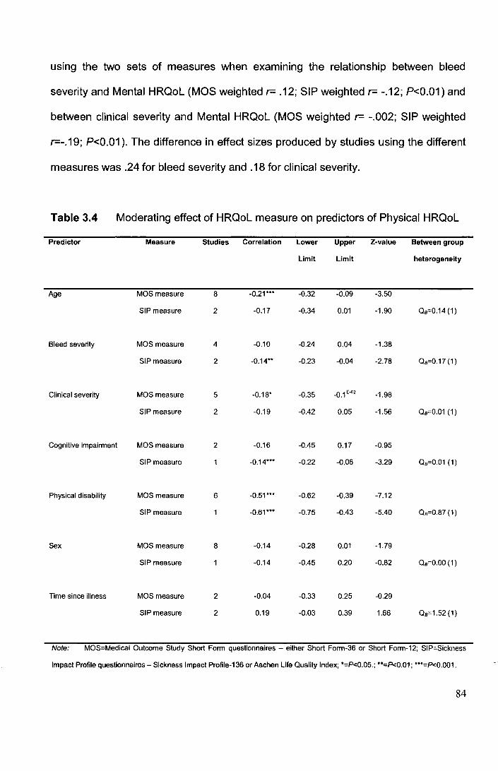
using the two sets of measures when examining the relationship between bleed
severity and Mental HRQoL (MOS weighted r= .12; SIP weighted r= -.12; P<0.01) and
between clinical severity and Mental HRQoL (MOS weighted r= -.002; SIP weighted
r=-.19; P<0.01 ). The difference in effect sizes produced by studies using the different
measures was .24 for bleed severity and .18 for clinical severity.
Table 3.4 Moderating effect of HRQoL measure on predictors of Physical HRQoL
Predictor Measure Studies Correlation Lower Upper Z-value Between group
Limit Limit heterogeneity
Age MOSmeasure 8 -0.21··· -0.32 -0.09 -3.50
SIP measure 2 -0.17 -0.34 O.o1 -1.90 aa=0.14 (1)
Bleed severity MOS measure 4 -0.10 -0.24 0.04 -1.38
SIP measure 2 -0.14 •• -0.23 -0.04 -2.78 Qa=0.17 (1)
Clinical severity MOS measure 5 -0.18. -0.35 -0.1 E-02 -1.98
SIP measure 2 -0.19 -0.42 0.05 -1.56 aa=0.01 (1)
Cognitive impairment MOS measure 2 -0.16 -0.45 0.17 -0.95
SIP measure -0.14··· -0.22 -0.06 -3.29 aa=0.01 (1)
Physical disability MOS measure 6 -0.51··· -0.62 -0.39 -7.12
SIP measure -0.61··· -0.75 -0.43 -5.40 aa=0.87 (1)
Sex MOS measure 8 -0.14 -0.28 0.01 -1.79
SIP measure -0.14 -0.45 0.20 -0.82 Qa=O.OO (1)
Time since illness MOS measure 2 -0.04 -0.33 0.25 -0.29
SIP measure 2 0.19 -0.03 0.39 1.66 Qa=1.52 (1)
Note: MOS=Medical Outcome Study Short Form questionnaires - either Short Form-36 or Short Form-12; SIP=Sickness
Impact Profile questionnaires- Sickness Impact Profile-136 or Aachen Life Quality Index; ·=P<0.05.; ••c:P<0.01; •••=P<0.001.
84
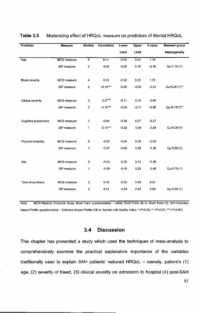
Table 3.5 Moderating effect of HRQoL measure on predictors of Mental HRQoL
Predictor Measure Studies Correlation Lower Upper Z-value Between group
Limit Limit heterogeneity
Age MOS measure 8 0.11 -0.02 0.24 1.70
SIP measure 2 -0.02 -0.22 0.19 -0.16 Qa=1.10 (1)
Bleed severity MOS measure 4 0.12 -0.02 0.25 1.70
SIP measure 2 -0.12*** -0.20 -0.05 -3.23 Oa=9.29 (1)**
Clinical severity MOS measure 5 -0.2E-02 -0.11 0.10 -0.05
SIP measure 2 -0.19*** -0.26 -0.11 -4.86 Oa=8.11(1)**
Cognitive impairment MOS measure 2 -0.04 -0.36 0.27 -0.27
SIP measure -0.14*** -0.22 -0.06 -3.29 Oa=0.29(1)
Physical disability MOS measure 6 -0.09 -0.45 0.30 -0.43
SIP measure -0.47 -0.89 0.39 -1.08 Oa=0.68 (1)
Sex MOS measure 8 -0.03 -0.20 0.14 -0.36
SIP measure -0.09 -0.45 0.29 -0.46 Oa=0.78 (1)
Time since illness MOS measure 2 0.16 -0.22 0.49 0.81
SIP measure 2 0.11 -0.24 0.43 0.60 Os=0.04 (1)
Note: MOS=Medical Outcome Study Short Form questionnaires - either Short Form-36 or Short Form-12; SIP-Sickness
Impact Profile questionnaires -Sickness Impact Profile-136 or Aachen Life Quality Index; *=P<0.05; **=P<0.01; ***=P<0.001.
3.4 Discussion
This chapter has presented a study which used the techniques of meta-analysis to
comprehensively examine the practical explanative importance of the variables
traditionally used to explain SAH patients' reduced HRQoL - namely, patient's (1)
age, (2) severity of bleed, (3) clinical severity on admission to hospital (4) post-SAH
85
cognitive impairment (5) physical disability, (6) sex and (7) the time occurring
between the SAH and the assessment of HRQoL.
In respect to SAH patients' Physical HRQoL, the results from the meta
analysis showed that all, but one, of the traditional predictors were significantly
related to patients' Physical HRQoL in terms of their P-values. Increasing age, a
more severe haemorrhage, worse clinical status on admission to hospital, being
female, and more cognitive impairment and physical disability were all (from a
statistical perspective) significantly associated with a worse Physical HRQoL. The
weighted effect sizes for 6 of these predictors calculated under random effects
models showed that the practical importance of these variables was nevertheless
typically small, thus emphasising the importance of not solely relying on P-values
when judging importance. The variables could typically each explain only 2- 4% of
actual variance in SAH patients' Physical HRQoL scores (effect sizes ranged -.13 to
.20). The exception to the rule was the degree of physical disability which the patient
experienced. Physical disability, it was found, was an important predictor of Physical
HRQoL, both statistically and practically. The variable physical disability could by
itself, account for approximately 30% of variance in patients' Physical HRQOL scores
(weighted r= -.54).
In striking contrast to the numerous significant predictors of Physical HRQoL,
only 1 of the 7 traditional predictors - namely, cognitive impairment - proved to be
associated with SAH patients' Mental HRQoL scores. Increasing cognitive impairment
was seen to be associated with worse Mental HRQoL. The magnitude of this effect
was however still small, with it explaining less than 2% of variance in patients' Mental
HRQoL (weighted r= -.13).
86
To further examine the explanative value of the 7 traditional predictors for SAH
HRQoL, the total variance accounted for by each of these predictors (i.e., the square
of their weighted effect size [R2 w·D was summed together, regardless of whether or
not an effect had proved statistically significant. It was found that together, the 7
variables could explain approximately 43% of variance in SAH patients' Physical
HRQoL, but only 6% of variance in patients' Mental HRQoL scores. Consequently, a
large proportion of variance in SAH patients' Physical HRQoL can be accounted for
(although over 55% still remains mysterious), but nearly 95% of variance in SAH
patients' Mental HRQoL remains unexplained. In fact, two reasons mean that the
estimates of the total variance explained in Physical and Mental HRQoL by the
traditional predictors could actually be too high. Firstly, it remains to be established
whether the variance explained by each of the contributing predictors is actually
unique. The summed R2 w values were based on the squaring of the individual
weighted effect sizes which are simply zero-order correlations . Given that many of
the predictors are closely related to one another, it is highly plausible that much of the
variance accounted for by the individual predictors was actually shared. Clinical
severity on admission is known for instance, to be largely the function of the severity
of a patient's bleed [711], whilst neurological impairment is known to be related to
age, clinical severity and bleed severity [306]. The other reason for the estimate of
total explained variance possibly being too high, is that I included in its calculation the
effect sizes for all predictors regardless of whether they had proved to be statistically
significant or not in the meta-analysis. A more conservative approach of including
only those predictors which had actually proved significant and were not chance
results would have reduced the variance explained. However, as noted earlier in the
'Data Analysis and Extraction' section of this chapter, the diminutive size of some of
87

the effects meant that despite the large sample size, in a few instances the power of
the analysis was below the desired level (<.50). Given the resultant elevated chance
of committing a Type II error for these particular predictors, I felt it wise to adopt a
more relaxed inclusion criteria.
The findings of this meta-analysis are clear: Traditional predictors of SAH
patients' HRQoL on the whole, demonstrate minimal predictive ability and cannot
satisfactorily explain the concerning reduction in HRQoL which many of these
patients illustrate. All traditional predictors (except physical disability when predicting
Physical HRQoL) had only a small effect size (!'S.20). These findings strongly
suggest that the hypothesis that traditional predictors could actually explain patients'
mysterious HRQoL reduction if only studies were sufficiently powered and considered
more than statistical significance, cannot be supported. These findings strongly
support the possibility that the reason researchers have struggled to explain SAH
HRQoL is because they have so far failed to consider more novel aspects of these
patients' outcome- aspects such as their psychiatric disturbance.
The failure of cognitive and physical disability and clinical information to
explain the vast majority of SAH patients' profoundly low levels of HRQoL, although
likely surprising to many people, is in fact, quite understandable. In terms of
neurological sequelae, whilst important neurological changes do occur post-SAH -
and we know from the wider literature that neurological impairment can be debilitating
[35, 61, 180, 282, 344, 523, 719] - the actual nature of SAH patients' impairment
tends only to subtle. Mild to moderate cognitive impairment is only revealed by the
most complex of cognitive tasks [295, 541, 750] and profound physical disability is
uncommon [220, 295, 302, 382, 398, 460]. It would be surprising therefore, if such
mild impairment were to account for the profoundly poor HRQoL which SAH patients
88
experience. Indeed, testimony to the unhelpfulness of using neurological disability to
explain SAH patients' HRQoL, are the findings reported by Pritchard et al. [545].
These researchers compared SAH patients' HRQoL to that of a group of patients with
an acoustic neuroma (a benign form of brain tumour which develops on the
vestibulocochlear nerve) who were broadly similar in demographics. It was reported
that despite the acoustic neuroma patients experiencing greater neurological
disability, it was the SAH patients who reported significantly worse HRQoL. The
failure of clinical information to explain HRQoL even though it can explain immediate
aspects of outcome after an SAH, is also in line with research on other health
conditions (including other types of stroke). In several studies it has been suggested
that those factors affecting long-term outcome are often different to those affecting
immediate outcome [524, 759, 763]. The lack of a meaningful relationship between
aspects of clinical information, such as patient age and sex, and HRQoL is even less
surprising as in health psychology in general, it is recognised that the influence of
these factors on HRQoL though significant, is not profound [1 0, 179].
It is important to note that the results and conclusions of this meta-analytic
study do, however, need to be considered in the context of some potential limitations.
Firstly, the inclusion of diverse studies meant that there was often a lack of
homogeneity existing between studies in terms of their effect size estimates. This
means that there is the possibility that the calculated weighted effect sizes for the
predictors could be unrepresentative of their true effect size. As is typical for clinical
research, the methodology often varied between the studies included in the meta
analyses and so this is a possible cause for the heterogeneity in effect size estimates
for the same predictor. Unfortunately, the small number of studies which have so far
examined SAH HRQoL and which were available for inclusion in the meta-analysis,
89
meant that there was not sufficient statistical power to formally examine which, if any,
of the studies' methodological features moderated the effect size for predictors [259].
As is often the case in meta-analysis, there were simply too few studies to compare
studies with different methodological aspects in a meaningfully way [273, 583]. As
further empirical studies accumulate, patterns in effect sizes will possibly emerge and
moderating variables identified [157, 256, 273]. The search for these moderator
variables could however, be protracted given that the relative infrequency and high
mortality of SAH means that generating a sufficient sample size to complete a HRQoL
study can be difficult. It should also be noted that no index of the studies'
methodological qualities was used because such indices remain to be validated for
observational studies [323, 385, 404]. It is important to note however, that the
standard degree of heterogeneity for the predictors was only moderate. Higgins and
Thompson [275] who developed the P measure used in this study to index
heterogeneity, for example, suggested that notable heterogeneity constituted an /2
value substantially greater than 50 percent. For the predictors included in this meta
analysis, the median heterogeneity value for Physical HRQoL was only 56.33
(IQR=00.00-58.52) and 52.86 (IQR=29.64-67 .41) for Mental HRQoL. Moreover, as I
previously noted, the re-calculated effect sizes under the fixed-effect model when
outlying studies were removed, were broadly similar. Nevertheless, one instance in
which substantial and significant heterogeneity in effect sizes did exist warrants
comment. Specifically, it was found that the effect of physical disability when
predicting Mental HRQoL illustrated a large amount of heterogeneity (see Figure
3.16), with some studies finding that physical disability was associated with worse
Mental HRQoL (up to -.59), whilst an equal number of studies found a positive effect
on Mental HRQoL (up to .37). This discrepancy is hard to explain. One obvious
90
explanation could be that a data entry mistake occurred, especially given that high
scores on the different measures of physical disability which studies used did not
always carry the same meaning (e.g., a high score on the Barthel Index in its original
format indicates less disability, whilst the opposite is true for the modified Rankin
Scale). A re-examination of the data however, showed that the scoring directions had
been suitably standardised. Methodological characteristics of the divergent studies
also do not seem to explain the discrepancy, with the studies being similar in terms of
the samples they studied, the instruments they used and the timing of their HRQoL
assessment. The only discernible difference between the studies finding the different
effects is that those reporting a negative impact of physical disability on Mental
HRQoL were conducted in the U.K. and that two of these were conducted as part of
clinical trials. Still, it is hard to see how this can explain the appreciable discrepancy,
even if the potential for increased bias resulting from demand characteristics
introduced as part of a trial are considered. Instead, the explanation for what
moderates the impact of physical disability on SAH patients' Mental HRQoL needs to
await further study. The large discrepancy between the two groups of studies in their
effect size estimates does nevertheless, raise the possibility that the actual effect of
physical disability on SAH Mental HRQoL is not well represented by the overall
weighted effect size calculated for it by this meta-analysis. The presence of
substantial heterogeneity means that the overall effect size for this predictor is an
average (albeit weighted) of two sets of extreme values and so physical disability may
better explain SAH Mental HRQoL than the results of this meta-analysis would
suggest. Nevertheless, even if we assume one of the largest of the extreme effect
size estimates (i.e., either -.59 or .37) is more representative of the actual effect of
91
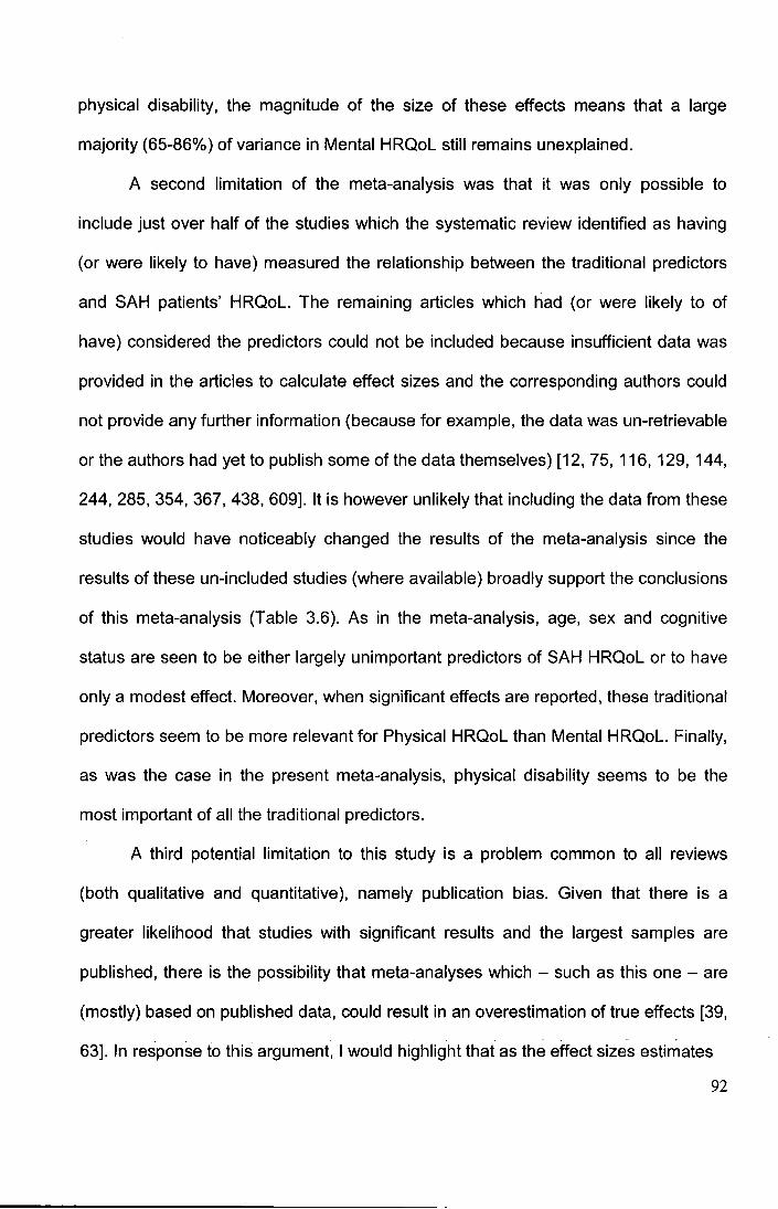
physical disability, the magnitude of the size of these effects means that a large
majority (65-86%) of variance in Mental HRQoL still remains unexplained.
A second limitation of the meta-analysis was that it was only possible to
include just over half of the studies which the systematic review identified as having
(or were likely to have) measured the relationship between the traditional predictors
and SAH patients' HRQoL. The remaining articles which had (or were likely to of
have) considered the predictors could not be included because insufficient data was
provided in the articles to calculate effect sizes and the corresponding authors could
not provide any further information (because for example, the data was un-retrievable
or the authors had yet to publish some of the data themselves) [12, 75, 116, 129, 144,
244, 285, 354, 367, 438, 609]. It is however unlikely that including the data from these
studies would have noticeably changed the results of the meta-analysis since the
results of these un-included studies (where available) broadly support the conclusions
of this meta-analysis (Table 3.6). As in the meta-analysis, age, sex and cognitive
status are seen to be either largely unimportant predictors of SAH HRQoL or to have
only a modest effect. Moreover, when significant effects are reported, these traditional
predictors seem to be more relevant for Physical HRQoL than Mental HRQoL. Finally,
as was the case in the present meta-analysis, physical disability seems to be the
most important of all the traditional predictors.
A third potential limitation to this study is a problem common to all reviews
(both qualitative and quantitative), namely publication bias. Given that there is a
greater likelihood that studies with significant results and the largest samples are
published, there is the possibility that meta-analyses which -such as this one- are
(mostly) based on published data, could result in an overestimation of true effects [39, - - -
63]. In response to this argument, I would highlight that as the effect sizes estimates
92
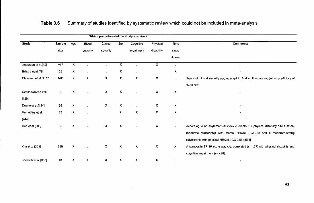
Table 3.6 Summary of studies identified by systematic review which could not be included in meta-analysis
Which predictors did the study examine?
Study Sample Age Bleed Clinical Sex Cognitive Physical Time Comments
size severity severity impairment disability since
illness
Anderson et al.[12] -17 X X X
Brilstra et al.[75] 25 X X X
Claassen et al. [116]* 247* X X X X X X Age and clinical severity not included in final multivariate model as predictors of
Total SIP.
Czechowsky & Hill 2 X X X X X
[129]
Deane et al.[144] 29 X X X X X
Hamedani et al. 83 X X X X X
[244]
Hop et al.[285] 55 X X X X According to an asymmetrical index (Somers' D), physical disability had a small-
moderate relationship with mental HRQoL (0.2-0.4) and a moderate-strong
relationship with physical HRQoL (0.3-0.06).[633]
Kim et al.[354] 385 X X X X X X A composite SF-36 score was sig. correlated (r= -.37) with physical disability and
cognitive impairment (r= -.38).
Komotar et al.[367] 48 X X X X X X
93
Table 3.6 cont'd.
Which predictors did the study examine?
Study Sample Age Bleed Clinical Sex Cognitive Physical Time Comments
size severity severity impairment disability since
illness
Mayer et al.[438] * 113 X X X X X X PCS- Sig. multiple adjusted ?-value of 18.8% for 'demographic variable' (effect
direction unknown). After controlling for demographics and depression, global
cognition and psychomotor function sig. associated with reduced PCS: additive
adjusted r -value of 10. 7%.
MCS- Sig. multiple adjusted r-value of 0.08% for 'demographic variable' (effect
direction unknown). After controlling for demographics and depression, global
cognitive function and visuospatial function were sig. associated with MCS:
additive adjusted r-value of .06%.
Schuiling et al.[609] 247 X X X X X X
Total number of studies 11 4 8 11· 5 10 6
Physical HRQoL Fail-safe N 87 8 43 30 25 45 n/a
Mental HRQoL Fail-safe N n/a n/a n/a n/a n/p n/a n/a
Note: HRQoL=health-related quality of life; n/a=calculation not applicable as weighted effect size not statistically significant; n/p=calculation not possible; r"=variance explained; SF-36= Short
Form-36; sig.=significant; SIP= Sickness Impact Profile; X= variable examined by study; *=these two studies are not independent. Mayer eta I.'s [438] study was an earlier assessment of the same
sample studied by Claassen et al. [116]. =variable not examined by study.
94
produced by the present meta-analysis were typically small and often insignificant,
the possible impact of publication bias for most of this study's results seems
negligible. In fact, it could even be argued, that the data used in this meta-analysis
was less prone to the forces of publication bias (at least directly). For example, the
effect size data on predictors of HRQoL often came from studies where the
examination of the predictors of HRQoL was not actually the prime concern and/or
novelty of published articles [240, 358, 470, 613, 694]. Therefore, a non-significant
effect of a predictor for HRQoL could still have been published as a function of it
being part of a much broader publication which had significant findings in response to
its primary research question. Even with this in mind, for those effects in this meta
analysis which did prove to be statistically significant, the number of unpublished (or
not included studies) with non-significant effects needed to nullify each significant
effect was so high for most that it seemed unlikely that they existed (in the context of
the SAH research field; see last three rows of Table 3.6). In fact, the number of
required studies was often large enough that it approximated the criteria used for
meta-analyses of much broader research fields (i.e., where the potential impact is felt
to be negligible if the fail-safe N>5K+10, where K is the number of studies in the
meta-analysis) [582]. Nevertheless, the significance of the effects of cognitive
impairment and bleed severity when predicting Physical HRQoL were potentially less
robust, with only 3 and 8 studies respectively being needed to nullify their significant
effects. This implies that the ability of traditional variables to explain SAH Physical
HRQoL could be even less important than suggested to be the case by this meta
analysis. Differences in the formulae used by contemporary meta-analysis and the
classic fail safe N to calculate statistical significance (i.e., fail-safe N algorithm
computes a P-value for each study and then combines these P-values, whereas
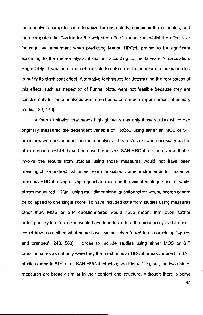
meta-analysis computes an effect size for each study, combines the estimates, and
then computes the P-value for the weighted effect), meant that whilst the effect size
for cognitive impairment when predicting Mental HRQoL proved to be significant
according to the meta-analysis, it did not according to the fail-safe N calculation.
Regrettably, it was therefore, not possible to determine the number of studies needed
to nullify its significant effect. Alternative techniques for determining the robustness of
this effect, such as inspection of Funnel plots, were not feasible because they are
suitable only for meta-analyses which are based on a much larger number of primary
studies [39, 170].
A fourth limitation that needs highlighting is that only those studies which had
originally measured the dependent variable of HRQoL using either an MOS or SIP
measures were included in the meta-analysis. This restriction was necessary as the
other measures which have been used to assess SAH HRQoL are so diverse that to
involve the results from studies using those measures would not have been
meaningful, or indeed, at times, even possible. Some instruments for instance,
measure HRQoL using a single question (such as the visual analogue scale), whilst
others measured HRQoL using multidimensional questionnaires whose scores cannot
be collapsed to one single score. To have included data from studies using measures
other than MOS or SIP questionnaires would have meant that even further
heterogeneity in effect sizes would have introduced into the meta-analysis data and I
would have committed what some have evocatively referred to as combining "apples
and oranges" [243, 583]. I chose to include studies using either MOS or SIP
questionnaires as not only were they the most popular HRQoL measure used in SAH
studies (used in 81% of all SAH HRQoL studies; see Figure 2.7), but, the two sets of
measures are broadly similar in their content and structure. Although there is some
96
preliminary evidence from a few SAH studies (mostly published in abstracts) that the
MOS and SIP measures may exhibit different sensitivities, responsiveness and
discriminative properties [121, 286, 437, 439], by far the majority of evidence from the
literature suggests that these two sets of instruments do measure the same construct
of HRQoL [102, 241, 337, 480, 484, 699, 737]. This conclusion corresponds with the
evaluation undertaken as part of this meta-analysis to assess the validity of
combining results from those studies using these two sets of measures. It was found
that for all but 2 of the 14 predictors, the effect sizes were homogenous between
studies using the different measures. The exceptions were bleed severity and clinical
severity when predicting Mental HRQoL. In these two cases, the use of the SIP was
associated with a larger, more negative effect of bleed severity and clinical severity
on Mental HRQoL. One likely explanation for this is that there are subtle differences
in the content of the Mental HRQoL scales of the two sets of measures. Whilst the
MOS Mental HRQoL summary is composed of questions relating to the patient's
perception of how much their health has impacted upon their energy, social activities
and emotional and mental well-being, the SIP scale in addition includes questions
pertaining to the person's performance on cognitive tasks, such as reasoning,
orientation and communication. Therefore, given that bleed severity and clinical
severity are both known to be related to cognition post-SAH, it could be that the
larger, negative relationship of the predictors with the SIP reflects the undue influence
of the relationship between the predictors and cognition. Other differences between
the SIP and MOS measures, such as the time of reference for their questions (the
SIP uses the day of examination, whilst the MOS uses either the past week or 4-
weeks) and that the SIP questions are more behaviour-based, whilst the MOS are
97
directed more at the subject's perceptions [241 ], could also be involved in explaining
the difference. The reasons for this are however, less clear.
On a final note, I would like to briefly discuss a possible reason why some of
the traditional variables- namely bleed severity and cognitive impairment- proved so
poor in predicting HRQoL (i.e., for bleed severity 1.69% of Physical HRQoL and
0.01% of Mental HRQoL; for cognition 1.96% of Physical HRQoL and 1.69% of
Mental HRQoL). Rather than bleed severity/distribution and cognitive impairment not
being related to HRQoL, the explanation could be that they are actually better related
than the current evidence suggests, but that the instruments used in studies to
measure these aspects are too crude to capture the information needed. In terms of
bleed severity for example, the crudeness of Fisher's CT rating score [191] (which is
almost always used to index bleed severity/distribution) means that it does not
capture the variability in bleed severity. The rationale for this particular speculation
comes from a single study by Hutter et al. [305] which suggested that specific
characteristics of a person's haemorrhage, not sufficiently captured by Fisher's scale,
may be better related to HRQoL than the results of the present meta-analysis would
suggest. Using univariate tests, Hutter et al. examined the predictive value of firstly,
an SAH with intracerebral bleed extension (ICB) on SAH patients' HRQoL and then of
SAH with intraventricular bleed extension (IVB). Crucially, it was found that ICB and
IVB had different predictive values for SAH HRQoL, but Fisher's scale affords equal
weight to ICB and IVB, grouping persons with either haemorrhage extension type
together. Specifically, Hutter and colleagues found that thirteen aneurysmal SAH
patients with frontal intracerebral invasion experienced a significantly worse physical
(r=-.27) and mental (r=-.21) HRQoL compared to 95 SAH patients with no ICB who
were treated contemporaneously and who were socially and clinical comparable. The
98
presence of IVB however, was not associated with HRQoL. Indeed, the effect sizes
for ICB are potentially even larger than those which I cite above as HOtter et al. only
examined the effect of frontal I CBs. The use of the Fisher scale in prior studies could
therefore, serve to obscure the unique HRQoL explanative value associated with
bleed severity and explain why studies (including this meta-analysis) find bleed
severity to explain so little variance in SAH HRQoL. It is quite plausible that ICB and
IVB could have a different impact on patient HRQoL. We already know for instance,
that ICB and IVB each hold unique predictive values for mortality and gross morbidity
following SAH [261, 306, 441, 442], and that they each can cause different types of
focal brain damage [261, 626, 709]. The potential drawback of this scale is not
something which I was able to address in this thesis, but which future studies would
be wise to consider (see discussion section 3.3.3, Chapter 7). In relation to the
measurement of cognitive impairment, previous studies have not assessed cognitive
impairment using instruments with a high degree of ecological validity. Instead prior
studies have typically used only classic neuropsychological tests which are designed
not to predict behaviour, but rather to localise lesions [203, 276]. It may be therefore,
that the use of such measures serves to artificially attenuate the actual relationship
between cognitive impairment and HRQoL. Given this, in my subsequent empirical
study of SAH patients which I present in Chapter 5, I made an explicit effort to use
only cognitive tests with a high degree of ecological validity.
3.5 Conclusions
The results of this meta-analysis lead to the conclusion that traditional predictors (with
the exception of physical disability when predicting Physical HRQoL) cannot
satisfactorily explain SAH HRQoL. Moreover, their importance has not been overly 99
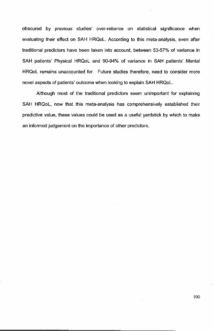
obscured by previous studies' over-reliance on statistical significance when
evaluating their effect on SAH HRQoL. According to this meta-analysis, even after
traditional predictors have been taken into account, between 53-57% of variance in
SAH patients' Physical HRQoL and 90-94% of variance in SAH patients' Mental
HRQoL remains unaccounted for. Future studies therefore, need to consider more
novel aspects of patients' outcome when looking to explain SAH HRQoL.
Although most of the traditional predictors seem unimportant for explaining
SAH HRQoL, now that this meta-analysis has comprehensively established their
predictive value, these values could be used as a useful yardstick by which to make
an informed judgement on the importance of other predictors.
100

Chapter 4
Possibility two: Psychiatric disturbance has not been
taken into account
101
4.1 Introduction
Following a spontaneous subarachnoid haemorrhage (SAH), many patients
experience an extremely poor health-related quality of life (HRQoL). This reduction
however, remains largely unexplained. Classical neurological factors- such as bleed
severity, clinical severity and neurological disability - which have traditionally been
employed to try and explain the reduction have proved (in both primary studies and in
my meta-analysis presented in the previous chapter) inadequate. How then can the
HRQoL reduction be explained? Following injury to the brain, there is an
understandable tendency to assume that it is this damage which is central to a
patient's ultimate outcome. We know however, from pioneering studies following
other illnesses, such as traumatic brain injury and stroke [61, 180, 344, 405, 591], that
while classical neurological factors are key to immediate outcome, a patient's later
recovery and HRQoL is also determined by 'non-neurological' factors. One possibility
therefore, is that the failure to explain much of SAH patients' HRQoL is because
research to date has not considered these 'non-neurological' factors. One highly
promising 'non-neurological' candidate variable which could help us further explain
SAH HRQoL, but which has so-far been ignored is the psychiatric disturbance which
SAH patients can often experience.[57, 469, 537, 746]
In this chapter, I shall provide a review of the psychiatric problems faced by
SAH patients, the studies which have measured these problems, how SAH
psychiatric problems are best conceptualised and finally, discuss the potential of
certain conceptualisations to explain SAH patients' reduced HRQoL (and other
aspects of their psychosocial outcome).
102
4.2 Psychiatric disturbance in SAH patients
As several commentators have concluded [46, 57, 306, 537, 544, 750], it is clear that
SAH patients experience alarmingly high levels of psychiatric disturbance compared
to healthy controls. In Table 4.1, I present a review of studies of psychiatric problems
in SAH patients. It was necessary to exclude many studies from this review as,
although rich in description of patients' psychiatric outcome, they were limited by their
use of unofficial nomenclature, their measurement of nebulous concepts (such as
'emotional disturbance' and 'psychiatric problems') and/or their use of non-validated
instruments [8, 25, 78, 99, 150, 185, 409, 412, 428, 501, 510, 513, 553, 632, 643,
649, 659, 667-669, 678, 685, 721' 722].
My review highlights that the psychiatric profile of SAH patients is largely
dominated by a state of anxiety (:554%) [48, 265, 314, 469, 540, 541, 652, 703, 725,
7 46]. An examination of the literature reveals that a sense of worry and fear over
haemorrhage recurrence dominates SAH patients' anxiety ( 18-89%) [295, 412, 652,
728, 729]. Generalised psychological disturbance [48, 90], personality change (e.g.,
increased irritability, impulsivity, apathy) [8, 58, 302, 419, 504, 510, 643, 659, 685,
721], as well as more peculiar symptoms of sleep dysfunction and fatigue are also
seen to be part of patients' psychiatric profile [313, 314, 409, 510, 609, 649, 746]. As
seen in Table 4.1, although symptoms of depression/ mood reduction are also
reported by SAH patients [44, 45, 105, 189, 265, 280, 302, 377, 397, 418, 504, 507,
508, 540, 609, 687, 703, 746, 752], they have not been found to be the main
symptoms [265, 314, 377, 469, 540, 541, 703, 746]. For example, Jarvis et al. [314] in
one of the largest studies to date (with a sample of 62 aneurysmal SAH patients),
found that 14% of patients reported clinical levels of depressive symptoms, but 35%,
reported clinical levels of anxiety. The tendency of SAH studies to have utilised
103
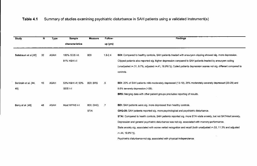
Table 4.1
Study
Bellebaum et al.[40]
Beristain et al. [44,
45)
Berry et al. [48)
Summary of studies examining psychiatric disturbance in SAH patients using a validated instrument(s)
N Type
32 ASAH
15 ASAH
48 ASAH
Sample
characteristics
1 00% GOS 1-11.
81% H&H 1-11
Measure
BDI
53% H&H I-ll; 93% BDI; BRS
GOS 1-11
Most WFNS I-ll BDI; GHQ;
STAI
Follow
up (yrs)
1.9-2.4
.5
.7
Findings
BDI: Compared to healthy controls, SAH patients treated with aneurysm clipping showed sig. more depression.
Clipped patients also reported sig. higher depression compared to SAH patients treated by aneurysm coiling
(unadjusted r=.31, 9.7%; adjusted r=.41, 16.9%*:j:). Coiled patients depression scores not sig. different compared to
controls.
BDI: 20% of SAH patients mild-moderately depressed (13-19), 25% moderately-severely depressed (20-29) and
6.6% severely depression (>29).
BRS: Merging data with other patient groups precludes reporting of results.
BDI: SAH patients were sig. more depressed than healthy controls.
GHQ-28: SAH patients reported sig. more psychological and psychiatric disturbance.
STAI: Compared to health controls, SAH patients reported sig. more STAI-state anxiety, but not SATI-trait anxiety.
Depression and general psychiatric disturbance was not sig. associated with memory performance.
State anxiety sig. associated with worse verbal recognition and recall (both unadjusted r-.33, 11.3% and adjusted
r-.44, 19.9%*:j:).
Psychiatric disturbance not sig. associated with physical independence.
Table 4.1 Cont'.d
Study N
Berry et al. [47] 28
Buchanan et al. [90] 24
Carteret al. [1 05] 179
Claassen et al. [116] 247
Type
ASAH
ASAH;
SAHavm
ASAH
ASAH;
SAHuo
Sample
characteristics
Referrals to clinical
psychology
service.
100% good
neurological
recovery
100% GOS 1-11
96% GOS I-ll; 61%
H&H 1-11
53% H&H 1-11
Measure Follow-
up (yrs)
Diagnostic .5
interview
(DSM-111-R
PTSD)
BSI 1.6
ZSRDS 1.0-6.0
CES-D; 1.0
STAI
Findings
Diagnostic interview: 32% met the diagnostic criteria for PTSD in relation to their SAH.
BSI: 58% met caseness criteria (2:63) for psychological distress. 13% of patients acknowledged suicidal ideation
during the previous 7 days.
ZSRDS: 11.7% of patients reported mild depression (50-59) and 25.1% severe depression (60-1 00).
Time, clinical severity and age not sig. associated with depression.
Physical disability was sig. associated with depression (r= .29, 8.9%*)
CES-D: Depression scores not sig. associated with post-SAH 12-month epilepsy.
105
Table 4.1 Cont'd.
Study N
Claassen et at.
cont'd.
Fertl et al. [189] 40
Fontanella et al. 53
[198]
Germano et al. [212] 20
Type
ASAH
ASAH;
SAHuo
ASAH
Sample
characteristics
100% GOS 1-
11;.65% H&H 1-11
100% WFNS 1-11;
100% GOS I.
100% GOS I;
100% H&H 1-11;
Patients selected
as, considered to
likely make
favourable
recovery:
Measure
BDI
BDI; STAI
HARS;
HDRS
Follow-
up (yrs)
1.8
.5
1.0
Findings
STAI-state: In multiple regression, worse state anxiety sig. associated with non-white ethnicity (OR=4.0) and 12-
month post-SAH epilepsy (OR=4.8). Age, education, fluency in English, clinical grade (GCS), aneurysm re-bleed,
infarction and delayed cerebral ischemia did not enter into final model.
BDI: 27.5% of SAH patients mildly-moderately depressed (12-26).
Depression sig. associated with cognitive and physical impairment (r= .36, 13.5% variance) and reduced working
capacity (r= .56, 31.8% variance)*±.
BDI: No sig. mood disturbance compared to healthy controls.
STAI: No sig. disturbance on both 'state' and 'trait' scales.
Psychiatric state not associated with treatment (clipping vs. coiling) or aetiology of bleed (aneurysmal vs. unknown
origin).
HARS: 100% of SAH patients mildly anxious. SAH patients' mean anxiety score was not sig. different to controls.
HDRS: 15% of SAH patients mildly-moderately depressed, 5% moderately- severely depressed. SAH patients' mean
depression score was not sig. different to controls.
106
Table 4.1 Cont'd.
Study N
Hadjivassiliou et al. 80
[242)
Hellawell et al. [265] 26,
Hillis et al. [280)
23,
19
27,
6
Type
ASAH
ASAH;
SAHuo
ASAH
Sample
characteristics
91% GOS 1-11; 78%
WFNS 1-11
Most WFNS I-ll
Average H&H II
Measure
BDI
HADS
ZSRDS
Follow-
up {yrs)
1.0
.5, 1.0,
2.0
.25
Findings
BDI: Depression scores not sig. associated with aneurysm treatment type (clipping vs. coiling).
HADS-Anxiety:
At 6 months, 11% borderline anxiety, 8% clinically anxious.
At 12 months, 17% were borderline and 13% clinically anxious.
At 24 months, 21% borderline and none were clinically anxious.
HADS-Depression:
At 6 months, 92% scored in the normal range. Just 8% illustrated clinically significant depressive scores.
At 12 months, 9% reported borderline depression and 9% scored in the clinically depressed range.
At 24 months, 5% of patients scores were borderline and 5% of patients' scores were in the clinical range.
Note that at 12 month appointment, total HADS score sig. positively correlated with clinical severity (unadjusted
r-.46, 21.2%; adjusted r-.64, 42.0%*+:1:). Note no corrections were made for multiple comparisons.
ZSRDS: 36% were minimally to moderately depressed (50-59), 23% moderately to markedly depressed (60-69), and
5% severely depressed (<:80).
SAH patients' depression scores not sig. higher than those of non-SAH patients surgically treated for an un-ruptured
aneurysm.
6 patients were seen again at 1 year post-SAH and no sig. change in their depression scores was found.
107
Table 4.1 Cont'd.
Study N
Hutter et al. [297, 58
302]
Hutter et al. [295] 45
Jarvis & Talbot [314] 62
Type
ASAH;
SAHuo
ASAH;
SAHuo
ASAH
Sample
characteristics
100% GOS 1-11
Measure
BDI
BDI; IES;;
Diagnostic
interview
(DSM-IV
PTSD)
HADS; IES
Follow-
up (yrs)
3.0
4.0
.25
Findings
BDI: 30% of patients reported clinically relevant depression scores (BDI >1 0). No sig. differences in depression
scores by SAH type (ASAH vs. SAHuo) or aneurysm location (ACoA vs. other sources).
IES: Compared to a control group of 25 medical studies who had dissected their first corpse a week before, SAH
patients had sig. higher IES avoidance and total traumatisation symptoms, but not intrusion symptoms.
No sig. difference between IES scores by SAH type (ASAH vs. SAHuo).
IES intrusions sig. associated with clinical severity (r=.32, 1 0.2%) and bleed severity (9.6%), but not vasospasm.
Intrusion score also sig. correlated with score on measure offronto-cortical function completed 4 years earlier. (r=
.35, 12.3%). IES intrusion (r-.52, 27.0%) and avoidance (r-.45, 20.3%) scores both sig. correlated with concurrent
BDI depression scores.
Diagnostic interview: 26% of SAH met diagnostic criteria for PTSD.
HADS-Anxiety: 35% of SAH patients reported clinically relevant anxiety (11 + ).
HADS-Depression: 14% of SAH patients reported clinically relevant depression (11 + ).
IES: 37% of SAH patients reported clinically relevant PTSD score (15+).
108
Table 4.1 Cont'd.
Study N
Kreitschmann' (377] 35-
40
Lindberg et al. (397] 104
Madureira et al. [418] 18
Mayer et al. (438] 113
Type
ASAH
ASAH
PMSAH
ASAH;
SAHuo
Sample
characteristics
90% GOS 1-11; 55%
H&H 1-11
-
94% H&H 1-11
84% NIHSS 0;
56% WFNS 1-11
Measure
BDI; IES
ZSRDS
HDRS
CES-D;
STAI
Follow
up (yrs)
2.3
7.0
3.25
.25
Findings
BDI: 27.5% of SAH patients reported mild-moderate depression (11-17) and 10% severe depression (>17).
Depression sig. associated with basal cortisol value (r= -.56, 31.3%) and physical disability (r= -.44, 19.3%).
IES: 34% of SAH patients reported mild-moderate PTSD response (IES<15).
ZSRDS: 22% of SAH patients were reported to be experiencing clinically relevant levels of depression (40-80).
Depression not sig. associated with time between SAH and assessment, age, sex, cognitive impairment or physical
disability.
Depression was sig. associated with ability to work (r-.27, 7.5%* t) and a decrease in leisure activity (r= .26, 7.2%*).
HDRS: 33% of SAH patients scored in the depressed range (~13).
Depression not sig. associated with age, sex or time between SAH and assessment.
Depression sig. correlated with memory deficits (r-.72, 53.0%*±), but not global cognitive function.
CESD: After controlling for age, education and race, global cognitive function sig. associated with depression (r-.24,
5.8%* t). Domain-specific cognition not sig. associated with depression.
STAI: After controlling for age, education and race, global cognitive function sig. associated with state anxiety (r-.29,
8.4%* t), but not trait anxiety. Domain-specific cognition not sig. associated with either state or trait anxiety scores.
Anxiety and depression sig. correlated, but coefficient not provided in article.
109
Table 4.1 Cont'd.
Study N Type Sample Measure Follow- Findings
characteristics up (yrs)
Morris et al. [469] 52 ASAH; 44% had a good BDI; GHQ; 1.4 BDI: 28% of SAH patients reported mild-moderate depression (10-18) and 22% reported moderate-severe
SAHuo GOS-E;84% HADS;STAI depression (~19).
WFNS 1-11 GHQ: 41% of the 46 SAH patients who completed the measure scored above the 4/5 threshold indicating poor
subjective mental health.
HADS-Anxiety: 16% of SAH patients reported mild anxiety (8-1 0) and 38% reported moderate-severe anxiety (~11 ).
HADS-Depression: 21% of SAH patients reported mild depression (8-10) and 17% reported moderate-severe
depression (~11 ).
STAI: 58% of SAH patients reported trait anxiety scoring above 80th percentile and 46% above 80% on state anxiety.
None of the mood scores were sig. associated with clinical severity, bleed
severity, aneurysm location, type of SAH (ASAH vs. SAHuo), delay to treatment or sex.
HADS and STAI scores scales were all sig. correlated with physical disability (unadjusted rs=.42, 18.4%; adjusted
rs=.50, 25.2%).*:tJ:
Morris et al. [470] 70§ ASAH; 84% WFNS 1-11 HADS; 1.2 Possession of E4 allele not sig. associated with any anxiety or depression scores.
SAHuo STAI; BDI
Ogden et al. [504] 16 ASAH; 100% GOS=I; 56% ZSRDS 4.0 ZDRS: 25% of SAH patients reported at least moderate depression (~50).
SAHuo Botterel Grade 1-2
110
Table 4.1 Cont'd.
Study
Ogden et al. (507]
Ogden et al. (508]
N
89,
66
71'
62
Type
ASAH;
SAHuo
ASAH;
SAHuo
Sample
characteristics
Measure
98% GOS 1-11; 89% BDI
GCS 13-15
98% GOSI-11; 57% BDI
good clinical
admission state as
per Mee et al.
scale.
Follow
up (yrs)
.2, 1.0
.20, 1.0
Findings
BDI: At 10 weeks, 19% of SAH patients reported mild-moderate depression (10-19) and 5.6% moderately severe
depression (20-29).
At 12 month assessment, 17% of SAH patients reported mild-moderate depression and 3% moderately severe
depression.
Depression was not sig. associated with cognitive performance.
BDI:
At 10-week assessment, 13% of SAH patients reported mild depression (10-15), 10% moderate depression (16-23)
and 1% severe depression
(24-63).
At 12 month assessment, 14% of SAH patients reported mild depression, 3% moderate depression and 2% severe
depression.
Depression score was notsig. associated with age, sex, aneurysm site, type of SAH (ASAH vs. SAHuo), clinical
complications (vasospasm; ischemic deficit; hydrocephalus), marital status, pre-morbid occupational status, pre
morbid IQ, estimated BDI for month preceding SAH or severity
of life stress for preceding year.
10-week depression scores were sig. associated with worse a clinical grade (effect size not calculable). Significant
reduction in BDI scores with time (r-.16; 2.56%).
111
Table 4.1 Cont'd.
Study
Ogden et al. [51 0]
Oyebode et al. [519]
Powell et al. [540]
N
123
20
44-
50
Type
ASAH;
SAHuo
ASAH;
SAHavm
ASAH
Sample Measure Follow-
characteristics up (yrs)
Most GOS=I; Mean ESS 4.0-7.0
GCS 13.2
(SD=2.14)
Referrals to clinical Diagnostic 8.0
psychiatry unit. interview
100% GOS 1- BDI; HADS; .25, .75
11;.100%1 WFNS I-ll IES
Findings
ESS: 35% of SAH patients reported experiencing excessive daytime sleepiness/ fatigue.
Excessive sleepiness/ fatigue sig. correlated with reports of difficulties maintaining night-time sleep (reported by 26%;
r-.34, 11.6% ), personality changes (r-.30, 9.0%) and complaints of poor memory (r-.32, 10.2% ).
Diagnostic interview: 70% of SAH patients met criteria for major depressive disorder, 5% bipolar depression with
mania, 10% schizoaffective disorder, 10% generalised anxiety and 5% erectile impotence.
BDI:
At the 3 month assessment, 9.1% of SAH patients reported clinical relevant depression (19+).
At the 9 month assessment, 11.4% of SAH patients reported clinically relevant depression.
HADS-Anxiety:
At the 3 month assessment, 16.0% reported clinically relevant anxiety (11+).
At the 9 month assessment, 17.0% reported clinically relevant anxiety.
At both 3 and 9 months, SAH patients reported sig. more anxiety than controls.
HADS-Depression:
At the 3 months assessment, 14.0% of SAH patients reported clinically relevant depression (11 +)
At the 9 months assessment, 8.5% of SAH patients reported clinically relevant depression.
SAH patients' depression scores were sig. higher than controls at 3 month assessment, but not at 9 month
assessment.
112

Table 4.1 Cont'd.
Study N Type Sample Measure Follow- Findings
characteristics up (yrs)
Powell et at. cont'd. IES:
At the 3 month assessment, 30% scored in the clinically indicative PTSD range on both the intrusion (<:12) and
avoidance (<:14) scales.
At the 9 month assessment, 15% scored in the clinically indicative range
on both the intrusion and avoidance scales.
There was a sig. reduction with time on IES scales (both r= .33, 11.5%).
Univariate analysis showed no significant recovery on either HADS scales or BDI with time.
In predicting overall of mood outcome (HADS + IES). 45% of variance in this composite was accounted for by a final
model made up of information about a patient's prior mental health (15%), prior physical health (12%), experience of
dysphasia (8%) and prose recall at the 3 month assessment (5%). Age, sex, physical disability and stresses for the
year proceeding SAH did not enter into final model.
Powell et al. [541] 49 ASAH 100% GOS 1-11; BDI;HADS; 1.5 BDI: 16.3% of SAH patients reported clinically relevant depression (<:19).
100% WFNS 1-11 IES HADS-Anxiety: 18.4% of SAH patients reported clinically relevant anxiety (<:11 ). Compared to controls, SAH patients
reported sig. higher anxiety.
HADS-Depression: 10.2% of SAH patients reported clinically relevant depression (<:11 ). Patients did not report more
depression than controls.
113
Table 4.1 Cont'd.
Study N
Powell et a/. cont'd.
Preiss et al. [543] 75
Salmond et al. [589] 20
Type
ASAH
ASAH
Sample
characteristics
100% GOS 1-11.
Average WFNS I
ll. Only MCA or
PCoA aneurysm
bleeds.
Measure
BDI-11
BDI
Follow
up (yrs)
1.0
5.7
Findings
IES: 6% of SAH patients scored in the clinically indicative PTSD range on both the intrusion (~12) and avoidance
scales (~14).
Compared to 9 month (cf. [540]), was no sig. improvement in HADS Anxiety or Depression scores or BDI scores.
Compared to data from 9 month assessment (cf. Powell et al. 2002), there was a sig. recovery for PTSD IES
intrusion scale score (r=.38; 14.85% variance*), but not on avoidance scale.
No sig. predictive model could be established for overall mood outcome at 18 months (HADS+IES)- potential
variables included age, sex, physical disability, dysphasia, prose recall at 3 months, prior life stresses and prior
physical and mental health problems. In univariate analysis, overall mood was sig. associated with prior mental
health (r= -.29; 8.4% *).
BDI: Mean depression scores of two groups of SAH patients treated differently (clipping vs. coiling) were both within
the minimal depression range.
Depression scores not sig. related to aneurysm treatment or sex.
BDI: Depression scores not sig. higher compared to healthy controls.
114
Table 4.1 Cont'd.
Study N Type Sample
characteristics
Samra et al. [590] · 185 ASAH 96%WFNS Grades
1-99% GOS 1-11.
Satzger et al. [594] 44 ASAH 100% no
neurologic
deficit.91% H&H 1-
II;
Schuiling et al. [609] 83 ASAH; 82% WFNS
SAHuo Grades 1-11,
Measure Follow·
up (yrs)
BDI .75
Befindlickeit 2.9-3.2
s-Skala
APSG; BDI; 1.7
SDL,
Findings
BDI: SAH patients had sig. more symptoms of depression than healthy controls.
Befindlickeits-Skala: Mean score of two SAH patient treatment groups (early surgery vs. late surgery) both
indicated no presence of depression (<56).
Age, timing of surgery, clinical severity and aneurysm location (ACoA vs. other) were not sig. associated with
depression.
SOL: 34% had severe sleep problems (a score of 0!:3 on either the SDL sleepiness or excessive daytime sleepiness
scales).
APSG: In 20 of the patients with SDL scores indicating severe sleep problems scores APSG was conducted. None of
these patients had a normal sleep. Severe sleep fragmentation, sleep apnoea, restlessness leg syndrome/ periodic
limb movement, or a combination these disorders of sleep and wake occurred in 19 patients (95% ).
BDI: Four patients (including the 1 patient without sleep fragmentation) had insomnia from inadequate sleep hygiene
(staying in bed too long and daytime napping). 45% of patients had depression (BDI>16).
Sleep dysfunction on the SDL was not sig. associated with treatment (clipping vs. coiling), physical disability or bleed
severity.
115
Table 4.1 Cont'd.
Study N Type Sample
characteristics
Sheldrick et al. [627] 27- ASAH; -
38 SAHuo
Stegen & Freckman 87 ASAH 93.1% good
[652] neurological
recovery.
Tidswell et al. [687] 37 ASAH -
Measure Follow-
up (yrs)
DTS :S.04, .10-
.15,23-
. 29
STAI 1.0
BDI 2.2
Findings
DTS: At s2 week assessment 18.4% reported clinically relevant PTSD Symptoms (>40).
At 5-7 week assessment, 35.5% reported clinically relevant PTSD symptoms .
At 11-14 week assessment, 18.5% reported clinically relevant PTSD symptoms.
Compared to myocardial infarction patients, SAH patients' scores at all time points were higher, but the difference
was notsig.
A mixed design analysis found time to be sig. related to PTSD scores for both groups. Change over time was
homogenous between groups.
Pair-wise comparisons found there to be a sig. decrease in scores between assessments two and three for the two
groups as a whole (effect size not calculable).
STAI: Increased anxiety noted.
Patients whose SAH was caused by rupture of an MCA aneurysm had sig. worse anxiety than those with ACA
aneurysms (r-.17, 3.1%).
BDI: 16% of patients in the "depressed" range (>13).
Depression was not sig. associated with cognitive performance or aneurysm location (anterior vs. non-anterior).
116
Table 4.1 Cont'd.
Study N Type
van der Schaaf et al. 69 ASAH
[703]
von Voges lang et al. 62 ASAH
[725]
Wermer et al. [746] 584 ASAH
Sample
characteristics
76.8% mRS 0-1
All good
neurological
recovery ( GOS I
ll).
57%mRS 0-1
Measure
HADS
STAI-state
HADS
Follow
up (yrs)
7.1 & 9.1
.08-.25
8.75
Findings
HADS-Anxiety: 17.4% of SAH patients reported possible anxiety (8-1 0) and 5.8% reported probable anxiety (>1 0).
HADS-Depression: 15.9% of SAH patients reported possible depression ( 8-1 0) and 7.2% reported probable
depression (>1 0).
Neither anxiety nor depression was sig. associated with having a small identified aneurysm being left untreated.
STAI-state: Compared to general population guidelines, SAH patients illustrated higher STAI-state scores, but this
was not statistically compared.
Using a quasi-experimental design, anxiety scores were compared between a retrospective 'treatment as usual'
control group and a prospective intervention group who received enhanced oral and written information about their
illness (including its treatment, potential consequences and contact information for patient associations). There was
no sig. difference in anxiety scores between these two groups or between sexes.
HADS-Anxiety: 13.5% of SAH patients reported possible anxiety and 11.1% probable anxiety. SAH patients' mean
anxiety score was not sig. higher than that of controls.
HADS-Depression: 19.5% of SAH patients reported possible depression (8-10 score) and 9.4% probable
depression. SAH patients' mean depression score was significantly higher than controls.
Time since SAH was not sig. associated with either anxiety or depression.
117
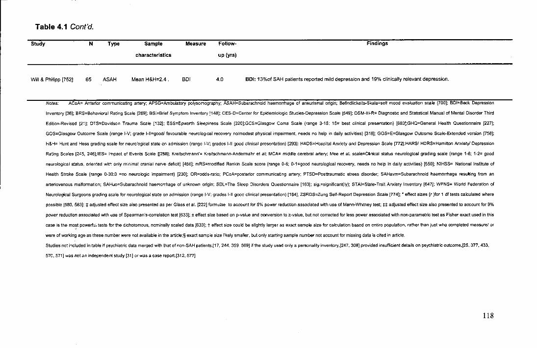
Table 4.1 Cont'd.
Study N Type Sample
characteristics
Measure Follow·
up (yrs)
Findings
Will & Philipp [752] 65 ASAH Mean H&H=2.4 . BDI 4.0 BDI: 13%of SAH patients reported mild depression and 19% clinically relevant depression.
Notes: ACoA= Anterior communicating artery; APSG=Ambulatory polysomography; ASAH=Subarachnoid haemorrhage of aneurismal origin; Befindlickeits-Skala=self mood evaluation scale [700]; BDI=Beck Depression
Inventory [36]; BRS=Behavioral Rating Scale [269]; BSI=Brief Symptom Inventory [148]; CES-D=Center for Epidemiologic Studies-Depression Scale [549]; DSM-111-R= Diagnostic and Statistical Manual of Mental Disorder Third
Edition-Revised [21]; DTS=Davidson Trauma Scale [132]; ESS=Epworth Sleepiness Scale [320];GCS=Giasgow Coma Scale (range 3-15; 15= best clinical presentation) [683];GHQ=General Health Questionnaire [227];
GOS=Giasgow Outcome Scale (range 1-V; grade 1-ll=good/ favourable neurological recovery no/modest physical impairment, needs no help in daily activities) [318]; GOS=E=Giasgow Outcome Scale-Extended version [758];
H&H= Hunt and Hess grading scale for neurological state on admission (range 1-V; grades 1-11 good clinical presentation) [293]; HADS=Hospital Anxiety and Depression Scale [772];HARS/ HDRS=Hamllton Anxiety/ Depression
Rating Scales [245, 246];1ES= Impact of Events Scale [[288]; Kreitschmann'= Kreitschmann-Andermahr et al; MCA= middle cerebral artery; Mea et al. scale=Ciinical status neurological grading scale (range 1-6; 1-2= good
neurological status, oriented with only minimal cranial nerve deficit) [456]; mRS=modified Rankin Scale score (range 0-6; 0-1=good neurological recovery, needs no help In daily activities) [550]; NIHSS= National Institute of
Health Stroke Scale (range 0-30;0 =no neurologic impairment) [230]; OR=odds-ratio; PCoA=posterior communicating artery; PTSD=Posttraumatic stress disorder; SAHavm=Subarachnoid haemorrhage resutting from an
arteriovenous malformation; SAHuo=Subarachnoid haemorrhage of unknown origin; Sl!ll=The Sleep Disorders Questionnaire [163]; sig.=significant(ly); STAI=State-Trait Anxiety Inventory [647]; WFNS= World Federation of
Neurological Surgeons grading scale for neurological state on admission (range 1-V; grades 1-11 good clinical presentation) [164]; ZSRDS=Zung Self-Report Depression Scale [774]; • effect sizes (r )for 1 dftests calculated where
possible [580, 583]; :1: adjusted effect size also presented as per Glass et al. [222] formulae to account for 5% power reduction associated with use of Mann-Whitney test; :1::1: adjusted effect size also presented to account for 9%
power reduction associated with use of Spearman's-correlation test [633]; ±effect size based on p-value and conversion to z-value, but not corrected for less power associated with non-parametric test as Fisher exact used in this
case is the most powerful tests for the dichotomous, nominally scaled data [633]; t effect size could be slightly larger as exact sample size for calculation based on entire population, rather than just who completed measure/ or
were of working age as these number were not available in the article;§ exact sample size likely smaller, but only starting sample number not account for missing data is cited in article.
Studies not included in table if psychiatric data merged with that of non-SAH patients,[17, 244, 359, 569] if the study used only a personality inventory,[247, 308] provided insufficient details on psychiatric outcome,[25, 377, 433,
570, 571] was not an independent study [31] or was a case report.[312, 577]
118
screening measures for depression, rather than anxiety when exploring psychiatric
disturbance in this population (seen in Table 4.1), should not be taken to indicate that
symptoms of depression are most prominent. Instead, it reflects the influence within
the neurosciences of the seminal work by Robinson and colleagues on ischemic
stroke patients, which publicised the possibility of depression following
cerebrovascular incidents [565-568].
It is important to note at this point, that the psychiatric problems of SAH
patients are considered to be new difficulties which occur following their
haemorrhage. There is - despite some early proposals [506] - no convincing
evidence to suggest that this population experienced abnormal levels of
psychopathology or stress prior to their haemorrhage [540].
As noted earlier, one particularly distinctive feature of SAH patients' anxiety is
a sense of fear and a preoccupation with the possibility of a further SAH occurring,
despite their relative safety [25, 47, 57, 75, 99, 295, 313, 314, 412, 449, 465, 559,
643, 652, 728, 729]. "Oh God, I'm on my own, I'm out now. Will it happen to me
again?" (p.1433) is something repeatedly stated by SAH patients [313]. Hutter [295]
for example, found that in a sample of 45 aneurysmal SAH patients assessed 4 years
post-SAH, only 11% never feared recurrence and 33% were either 'frequently' or
'permanently' afraid of a haemorrhage recurring. Crucially, many SAH patients also
have flashbacks and often feel as if the haemorrhage is recurring [47, 313, 314, 449,
716]. This can be precipitated by a tendency in the SAH population to catastrophically
misinterpret normal bodily sensations (such as headaches and tingling sensations in
the operation site) as indicating the onset of another SAH [47, 285, 545, 546, 548].
The sense of fear reported by SAH patients is of clinical concern because it is
considered to be both persistent and potentially debilitating [25, 57, 78, 295, 313, 437,
465]. Its abnormality is illustrated by the fact that significantly less patients following
treatment for other types of brain pathology exhibit such fear [545]. Although it has
often been presumed that SAH patients' worries could reflect a lack of knowledge ,
about their illness [302, 313, 314, 449, 469, 546, 548, 627, 652], it has been found
that this anxiety and fear is not abated through clinicians providing the patients with
extensive reassurance about their relative safety [47, 313, 652, 725]. Stegen and
Freckman (652] for example, assessed a sample of 69 aneurysmal SAH patients 12
months post-SAH and found that despite admitting that the illness had been
sufficiently explained and being aware of their safety, 79% of patients still feared
recurrence. The functional impairment caused by SAH patients' fear is illustrated by
these patients' avoidance of thinking about their haemorrhage and by placing
restrictions on themselves, such as not participating in certain activities (such driving,
lifting, sexual intercourse) in case they elicit sensations which are similar to those
experienced at the time of the haemorrhage and/or trigger harmful processes which
they feel could cause a further bleed [57, 285, 314, 437, 449, 703].
Patients' fears can also place extra strains on relationships with their family
and friends. For instance, patients may continually seek reassurance from their loved
ones about their health and may not allow themselves to be left alone [314, 664].
Their array of symptoms is concisely depicted in the following typical patient comment
derived from Jarvis' [313] qualitative study of SAH patients: "I felt very very (sic)
frightened about it happening again. I thought if it's happened once, it can happen
again. I felt very vulnerable - I had flashbacks of the night it happened. When I went
to bed ... I was reliving everything of it and couldn't get it out of my mind. I learned to
open my eyes when this happened so I would forget about it ... Even though I knew
120
the facts about the chance of another haemorrhage, I still have the flashbacks of the
night it occurred because I'm so frightened of it happening again" (p.1433).
In order for the psychiatric difficulties of SAH patients to be better understood
and ultimately treated, it is necessary to move beyond the simple description of
symptoms and to determine which psychiatric disorder (or disorders) underlies their
symptoms. Whilst the results from studies using screening measures have been
important in highlighting that the dominant presenting symptoms of SAH patients are
anxiety and a sense of fear, the ubiquitous nature of these symptoms to many major
psychiatric disorders [23, 190, 576], means that these studies alone cannot delineate
which specific disorder is responsible for the difficulties experienced by SAH patients
[53, 96, 122, 341, 345, 453, 561, 610, 636, 736]. According to the American
Psychiatric Association's Diagnostic and Statistical Manual of Mental Disorders
(DSM-IV-TR) [23, 190], when patients present with a symptom profile dominated by
anxiety and fear, the set of potentially relevant disorders includes, generalised anxiety
disorder, adjustment disorder and posttraumatic stress disorder (PTSD). As Figure
4.1 illustrates, although these various conditions are associated with symptoms of
anxiety and fear, they are not synonymous and they can be differentiated on the basis
of the nature and content of the anxiety which they produce (e.g., whether or not the
anxiety is in response to a stressor, what the patient is afraid of, what situations are
avoided) [190, 340]. Fortunately, efforts have now been made to determine which of
the potential psychiatric disorders seen in Figure 4.1 best accounts for SAH patients'
symptom profile. Indications from these recent studies suggest that PTSD is the best
explanation for SAH patients' psychiatric symptom profile. Therefore, in the next
section I shall discuss why PTSD is so good at accounting for SAH patients'
psychiatric difficulties.
121

Due to the direct physiolo gical effects of a substance (e.g., a drug ofabuse,a medication, a toxin)
Recurrent unexpected Panic Attacks plus am onth of worry, concern about:attacks, or change in behaviour
No
Ag crap hob ia, i.e. anxiety about being In places from which escape might be difficult or embarrassing in the event of ha v!ng pa nlc -like svm p toms
Yes
Yes
Yes
Yes
Yes
Yes
Yes
Yes
Agoraphobia, i.e. anxiety about being in pia ce s from which escape might bed iflicult or em ba rrassin g In the event of having a Panic Attack
6-month period of excessive anxiety and worry plus anxiety symptoms
No
Anxiety in response to a severe trauma tic event
No
Anxiety that does not meet criteria for one of the above Anxiety Disorders and develops in response to a stressor
Clinically significant symptoms that do not meet criteria for a specific Anxietv Disorder
No
No Anxiety Disorder (symptoms of fear, anxiety, or avoid a nee that are not clinically sign lficant)
Yes
Figure 4.1 Decision tree for differential diagnosis of psychiatric disorders accounting for anxiety symptoms (Adapted from DSM-IV-TR [23]).
122
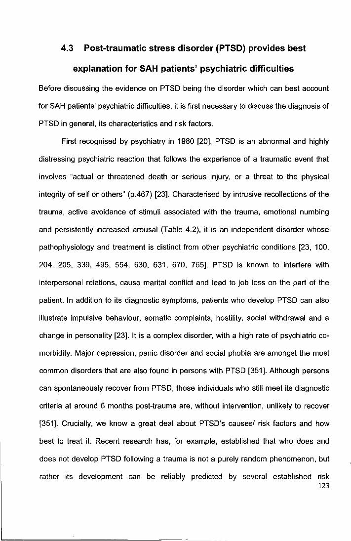
4.3 Post-traumatic stress disorder (PTSD) provides best
explanation for SAH patients' psychiatric difficulties
Before discussing the evidence on PTSD being the disorder which can best account
for SAH patients' psychiatric difficulties, it is first necessary to discuss the diagnosis of
PTSD in general, its characteristics and risk factors.
First recognised by psychiatry in 1980 [20], PTSD is an abnormal and highly
distressing psychiatric reaction that follows the experience of a traumatic event that
involves "actual or threatened death or serious injury, or a threat to the physical
integrity of self or others" (p.467) [23]. Characterised by intrusive recollections of the
trauma, active avoidance of stimuli associated with the trauma, emotional numbing
and persistently increased arousal (Table 4.2), it is an independent disorder whose
pathophysiology and treatment is distinct from other psychiatric conditions [23, 100,
204, 205, 339, 495, 554, 630, 631, 670, 765]. PTSD is known to interfere with
interpersonal relations, cause marital conflict and lead to job loss on the part of the
patient. In addition to its diagnostic symptoms, patients who develop PTSD can also
illustrate impulsive behaviour, somatic complaints, hostility, social withdrawal and a
change in personality [23]. It is a complex disorder, with a high rate of psychiatric co-
morbidity. Major depression, panic disorder and social phobia are amongst the most
common disorders that are also found in persons with PTSD [351]. Although persons
can spontaneously recover from PTSD, those individuals who still meet its diagnostic
criteria at around 6 months post-trauma are, without intervention, unlikely to recover
[351]. Crucially, we know a great deal about PTSD's causes/ risk factors and how
best to treat it. Recent research has, for example, established that who does and
does not develop PTSD following a trauma is not a purely random phenomenon, but
rather its development can be reliably predicted by several established risk 123
Table 4.2 American Psychiatric Association's [23] criteria for PTSD in adults
A. The person has been exposed to a traumatic event in which both of the following were present:
(1) person experienced, witnessed, or was confronted with event or events that involved actual or threatened death or
serious injury, or a threat to the physical integrity of self or others
(2) the person's response involved intense fear, helplessness, or horror
B. The traumatic event is persistently re-experienced in one (or morel of the following ways:
(1) recurrent and intrusive distressing recollections of the event, including images, thoughts, or perceptions.
(2) recurrent distressing dreams of the event
(3) acting or feeling as if traumatic event were recurring (e.g., a sense or reliving the experience, illusions,
hallucinations, and dissociative flashback episodes, including those that occur on awakening or when intoxicated)
(4) intense psychological distress at exposure to internal or external reminders of the traumatic event
(5) physiological reactivity on exposure to internal or external reminders of the traumatic event
C. Persistent avoidance of stimuli associated with the trauma and numbing of general responsiveness (not present
before the trauma), as indicated by three (or morel of the following:
(1) efforts to avoid thoughts, feelings, or conversations associated with the trauma
(2) efforts to avoid activities, places, or people that arouse recollections of the trauma
(3) inability to recall an important part of the trauma
(4) markedly diminished interest or participation in significant activities ("psychic numbing" or "emotional numbing")
(5) feeling of detachment or estrangement from others
(6) restricted ability to feel emotions (especially those associated with intimacy, tenderness and sexuality)
(7) sense of foreshortened future (e.g., does not expect to have a career, marriage, children, or a normal life span)
D. Persistent symptoms of increased arousal (not present before the trauma), as Indicated by two (or morel of the
following:
(1) difficulty falling or staying asleep (often due to recurrent nightmares during which the trauma is relived)
(2) irritability or outbursts of anger
(3) difficulty concentrating or completing tasks
( 4) hypervigilance
(5) exaggerated startie response
E. Duration of the disturbance (symptoms in Criteria B, C and D) is more than 1 month.
F. The disturbance causes clinically significant distress or impairment in social, occupation, or other Important areas of
functioning.
124
factors [521]. It has been found, for example, that key to the development of PTSD is
the manner by which the person reacts and responds to the stress of the trauma [328,
390]. This response is mediated by the coping strategies which the person
consciously employs when confronted with stress [217]. Therefore, whilst meta
analyses find that factors such as age, sex, education and psychiatric history have
only a minimal predictive effect for PTSD development [73, 521], coping strategies
are found to be one of the most powerful and robust predictors for PTSD
development [87, 88, 172, 217, 328, 490, 493]. In a variety of studies, including those
which fortuitously measured coping style prior to exposure to a real-life trauma [217,
490], a causal relation appears to exist between coping and PTSD development.
Those who tend to use denial as a coping strategy (e.g., "I say to myself 'this isn't
real'"), self-distraction (e.g., "I turn to work or other activities to take my mind off
things"), self-blame (e.g., "I blame myself for things that happened"), venting (e.g., "I
express my negative feelings"), substance abuse (e.g., "I use alcohol or other drugs
to help me get through it") and behavioural disengagement (e.g., "I give up trying to
deal with it") when dealing with stress are most likely to develop PTSD. The classic
explanation for why these coping strategies prove maladaptive and promote PTSD
refers to the slightly paradoxical phenomenon that by actively avoiding thoughts or
situations which lead to anxiety and fear, we in fact maintain their fear-inducing
quality. A host of experimental evidence for example, shows that attempts to
suppress unwanted thoughts are doomed to failure, and are, in fact,
counterproductive with the thoughts returning even more strongly [7 43]. In contrast,
successful adaptation to the experience of a trauma (as reflected in most
psychological treatments for PTSD which involve exposure therapy) appears to
involve the accessing of aversive emotions about the event, which permits habituation
125
to anxiety and the modification of one's trauma-related beliefs (i.e., disconfirming
beliefs) through the processing, assimilation and accommodation to the experience
[88, 165, 1 93].
Studies now consistently demonstrate that the experience of having suffered
an SAH can be so traumatic that the patient can develop PTSD (see Table 4.1).
Crucially, recent studies also provide compelling evidence that it is PTSD which is the
most parsimonious explanation for SAH patients' psychiatric disturbance [47, 295,
314, 377, 540, 541, 627], with PTSD being found to account for more of SAH patients'
psychiatric symptoms than any other psychiatric disorder [47]. Supporting PTSD's
ability to explain SAH patients' abnormally high psychiatric disturbance is the finding
of an abnormally high incidence of PTSD in the SAH population. Whilst the life-time
prevalence of PTSD in the general population is between only 1-8% [23, 267, 351],
two studies (using diagnostic criteria) have reported that 26-32% of SAH patients
warrant a clinical diagnosis of PTSD following SAH [47, 295]. This represents at least
three-times the normal rate.
Although there are some obvious caveats to applying the DSM-IV-TR
diagnostic criteria and its associated features in a "rote or cookbook fashion" (p. xv),
[1 90], it is clear that the symptom profile of SAH patients corresponds extremely
closely to PTSD. SAH patients' misinterpretation of bodily sensations and feeling as if
the haemorrhage were recurring for example, can be seen to reflect the PTSD
symptoms of re-experiencing, whereby patients can act or feel as if they are actually
reliving the traumatic experience [47, 313]. In addition, avoidant symptoms can be
seen to be reflected in SAH patients' self-imposed restrictions which prevent them
from thinking about their haemorrhage or from participating in activities and
frequenting places that could remind them of the SAH [437]. Finally, the sense of
126
perpetual fear which SAH patients experience concurs closely with the well-known
feature of PTSD [74, 171], whereby patients are found to feel anxious about the
future, even though the trauma lies in the past. Such patients alter their beliefs,
attributions and expectations about themselves, others and the world, in an extreme
and maladaptive way [372]. For example, in the case of a female rape victim who
develops PTSD, the woman comes to believe that all men could be potential rapists
and/ or that the world is not a safe place. In line with the suggestion that SAH
patients' unfounded fears of re-bleeding are a consequence of the development of
PTSD is Hutter's [295] finding that SAH patients' degree of fear of haemorrhage
recurrence is highly correlated in a positive manner with the degree to which they
experienced different PTSD symptoms (r's ~.62, all p<0.001).
To date, the available evidence suggests that PTSD is the best explanation for
SAH patients' psychiatric symptom profile. Even though most SAH studies have
examined relatively small samples (median size=47, interquartile range 39-59) [47,
295, 314, 377, 540, 541, 571, 627], the effect of PTSD in SAH patients has been of a
sufficient magnitude that all but one of these studies [571] has detected concerning
levels of PTSD (please note that this latter study is not included in Table 4.1 as
insufficient details on SAH patients' psychiatric outcome was provided in the article
which precluded its inclusion). It is important to recognise that the studies identifying
PTSD in SAH patients have not only relied on screening measures, but also used
more rigorous and comprehensive techniques, including the "gold-standard"
technique of differential diagnosis [365]. This is important as many screening
instruments often only measure for the presence of some PTSD symptoms, do not
separate new from pre-existing symptoms, do not ask about the duration of the
symptoms and do not assess the resultant functional impact that these symptoms
127
have [483]. Such measures can, therefore, potentially inflate the number of patients
who are identified as having PTSD [615, 684].
As SAH patients' psychiatric symptoms occur within the context of the patients
having a medical illness, it is important to highlight that the possibility of an alternative
DSM-IV-TR diagnosis of Anxiety due to a General Medical Condition has been
considered. However, it has not been found to be a more appropriate explanation for
patients' symptoms. Such a diagnosis is appropriate only when the psychiatric
disturbance is the direct physiological consequence of a medical condition and
another primary disorder does not better account for the symptoms [23, 637]. No
evidence suggests that SAH patients' psychiatric disturbance is aetiologically related
to the haemorrhage (or its treatments). For example, no acute illness index, such as
the severity and distribution of the person's SAH, its treatment, or its cause, has
proved to be reliably associated with SAH psychiatric disturbance [1 05, 116, 242,
265, 280, 295, 297, 302, 469, 507, 543, 594, 687]. Moreover, indirect mechanisms
by which medical conditions can cause psychiatric disturbance, such as via induced
neuroendocrine dysfunction, have also not proved important [315, 539]. For example,
Kreitschmann-Andermahr et al. [377] found that PTSD symptoms in SAH patients
were not associated with any concurrent measure of their hormonal function.
Psychiatry officially recognises that medical events can precipitate PTSD [23,
455]. This is illustrated by its definition of a traumatic event as one which involves
"actual or threatened death or serious injury, or a threat to the physical integrity of self
or others" (p.467) [23]. Despite this official stance, the conceptualisation of a medical
event (and hence an SAH) within the PTSD nosological framework is not without its
difficulties [92, 476, 684), with some persons having questioned the validity of
diagnoses of PTSD in medical patients. As this is an important point of debate, I will,
128
in the next section, briefly discuss some of the proposed difficulties in diagnosing
PTSD in medical patients. Importantly, I will describe how I believe most criticisms
over these persons' diagnoses can be potentially resolved and how the case for
PTSD in SAH patients should not be viewed as ill-fated.
4.4 The validity of PTSD diagnoses in medical patients
Prior to 1994 [22], psychiatry did not officially recognise that a medical illness could
elicit PTSD because it had been stated that only events 'outside the range of usual
human experience' (p.236) could precipitate such a reaction [21]. It was
epidemiological research however, which forced psychiatry to adopt its current, much
broader definition of what actually constitutes a trauma, as it was repeatedly
illustrated that there are few events which are truly rare from a statistical perspective
and so outside usual human experience. Moreover, the actual events which caused
PTSD were found to be more common than uncommon (e.g., assault, rape, motor
vehicle accidents) [70, 134, 491 ]. Therefore, despite traditionally being associated
with external traumatic events, such as natural disasters [417] and military combat
[640], it is now officially recognised that medical events (30, 81, 89, 128, 167, 476,
534, 684], such as HIV infection (:535%) [355], myocardial infarction (:516%) [380] and
cancer (:532%)[479] can elicit PTSD. In re-examining the clinical events which SAH
patients will typically experience (see Figure 2.5 in Chapter 2), it becomes clear that a
spontaneous SAH and its treatment are (as the DSM-IV-TR's PTSD diagnostic
criteria necessitates) not only life-threatening, but also have the potential to be
intensely frightening [23]. In fact, it is already known that intensive care treatment
(which SAH patients often receive) can alone frequently elicit PTSD [684].
129
Amongst the main reasons for current scepticism over the diagnosing of PTSD
following a medical event are: 1) the methods of diagnosis (e.g., self-report
measures) have not been validated on patients who have experienced a medical
illness and patients could therefore confound traumatological symptoms with the
direct effects of the illness and/ or its treatment (e.g., Criterion D symptoms of
hyperarousal, such as insomnia, concentration difficulties, irritability) [20,21], 2)
differences exist in the manner in which some patients experience some PTSD
symptoms. For example, 're-experiencing' (Criterion B) is defined in terms of thoughts
about past events (e.g. flashbacks to event, feeling like it was happening again), but
some patients (e.g., cancer patients) who face the real possibility of relapse, have
intrusions which are more future-orientated (e.g., 'Will the cancer progress to the
point that I'm in so much pain that I would want to die?' 'Will my family be provided for
once I am gone?') [14,22,23], and 3) it is unclear how a minority of patients develop
PTSD following a medical trauma, despite having lost consciousness at the time. It
has been understood that a person needs to have felt fear, helplessness or horror at
the time of the trauma and that explicit memories of the trauma are necessary for the
generation of and maintenance of PTSD (Criterion A).
There are some clinical difficulties in awarding a diagnosis of PTSD following a
medical trauma (such as SAH). However, I do not believe that these issues raise any
serious threat to the validity of diagnoses of PTSD in SAH patients, rather they
represent theoretical and diagnostic challenges which future studies will need to
address. In support of my argument is the fact that there is still much debate about
the way PTSD expresses itself even in the case of more traditional traumas. For
example, some question the diagnosis of PTSD in children who have experienced a
traditional trauma because their symptomatology can be expressed in divergent ways
130
[136]. With regards to the issue of lack of memory of the event and how it is that
persons following medical events can still develop PTSD, work is already beginning to
emerge (from the field of traumatic brain injury) that helps to explain this. The work
shows that a lack of consciousness at the time of the trauma does not preclude PTSD
because the person can ( 1 ) still experience the traumatic event after they regain
consciousness, (2) processing can occur at an implicit level, by-passing cortical
structures (i.e., through the amygdala and related structures) during periods of
impaired consciousness and (3) because some people appear to reconstruct
memories or experiences which then 'become memories' that may provide the basis
for the generation of PTSD symptoms [86, 218, 599]. Also, in the case of the
difficulties of symptom overlap, evidence from the use of screening measures such as
the Hospital and Anxiety Depression Scale [772], which are used for more general
psychiatric disturbance, suggests that with the refinement of measures, it is possible
to begin to control for the 'noise' of concurrent somatic disorders which can potentially
present themselves when assessing a patient's psychiatric status. One of the most
important pieces of evidence that supports the diagnoses of PTSD in some medical
patients comes from a recent study by Pitman et al. [534] of cancer patients who had
been diagnosed with PTSD using interview techniques. Pitman et al.'s study built
upon the finding that classical PTSD patients are known to illustrate a triad of unique
correlates on psychophysiological measures - namely, elevated arousal during
periods of rest, an exaggerated startle response and physiological reactivity on
exposure to cues symbolising or resembling an aspect of the traumatic event [516,
517, 535, 536, 624]. Crucially, Pitman et al. found that the cancer patients with PTSD
illustrated exactly the same psychophysiological 'signature' as those persons who
had suffered more traditional traumas (including military combat, sexual assault and
131
terrorism). Such findings appear to provide a 'biological' validation of medical patients'
PTSD diagnoses, as the precision of 'objective' psychophysiological tests is not, like
traditional interview techniques, vulnerable to the reliability of patients' reports or a
clinician's ability to differentiate whether patients' symptoms are psychiatric in origin
or not.
In summary, the available evidence supports the diagnosing of PTSD in SAH
patients. With this in mind, in the next section I shall discuss some anecdotal
evidence which motivates me to examine if the abnormally high incidence of PTSD in
the SAH population can help explain SAH patients' poor psychosocial outcome.
4.5 Potential of PTSD to explain SAH patients' HRQol and
other unexplained disturbances?
Although it has never been comprehensively examined, the following tentative
evidence hints that the abnormal incidence of PTSD in the SAH population could help
explain SAH patients' reduced HRQoL. Firstly, PTSD in the wider literature is known
to cause particularly severe levels of disability and handicap [82, 177, 348-350, 352,
459] which lead to a pattern and level of HRQoL impairment very comparable to that
generally reported by SAH patients. Both conditions (i.e., SAH and PTSD in general)
have been found to lead to worse HRQoL than that caused by other illnesses,
including heart disease and diabetes (see Figure 2.6 Chapter 2 for SAH HRQoL
profile compared to other illnesses) [1 0, 249, 422, 459, 514, 551, 585, 608, 662, 760].
In fact, the severity of this reduction in HRQoL in both conditions is second only to
that associated with a Major Depressive Disorder. Secondly, although PTSD
sufferers (like SAH patients) are known to experience impairment in both the Phys_i<:;al
and Mental domains of HRQoL, they too experience especially pronounced 132
reductions in Mental HRQoL [1 0]. Although most evidence on the link between PTSD
in general and HRQoL comes from cross-sectional studies, which precludes causal
statements (e.g., it is not possible to conclude that HRQoL did notpre-date the onset
of PTSD [464]), evidence is amounting to suggest a causal link between PTSD and
HRQoL, as the treatment of PTSD results in improvement in HRQoL [56, 64, 422,
429,454,552,656,696].
In the context of discussing PTSD's potential explanative power for SAH
HRQoL, it is interesting to note that PTSD could also explain two other mysterious
aspects of SAH patients' outcome, namely, post-SAH chronic fatigue and sleep
dysfunction [8, 90, 176, 185, 264, 265, 285, 313, 314, 360, 400, 409, 428, 465, 487,
504, 508, 510, 513, 572, 579, 609, 617, 643, 646, 649, 667, 669, 685, 718, 746].
SAH patients report persistently feeling excessively tired, weak, without energy and
needing rest following even the most menial of tasks [313]. They also note that they
have difficulty falling asleep, repeat awakenings and difficulty returning to sleep [609].
Even the most conservative of estimates suggests that a third of patients experience
debilitating levels of fatigue [579], whilst around 20% report severe sleep dysfunction
up to 7 years after the haemorrhage [51 0].
Although prior studies considering SAH patients' fatigue and sleep quality are
limited by a failure to use validated instruments or to compare patients' complaints to
normative levels, the two sets of difficulties have recently come to the fore as
preliminary evidence suggests that they could be integral to patients' post-SAH social
participation, including their work and socialisation [118, 313, 487, 541].
Unfortunately, these difficulties have gone untreated within clinical practice, often
dismissed because their severity has not been properly gauged, but also because
their origin has never been established [544]. Efforts have been made to explain their
133
presence as a result of the brain damage induced by an SAH (and its treatment) [65,
377, 509], but this mechanism does not stand up to much scrutiny (at least not as the
principle cause of the problems). Kreitschmann-Andermahr et al. [377] for instance,
reported that whilst in a group of 35 patients, SAH-induced neuroendocrine
dysfunction was related to sleep and fatigue, 84% and 70% of variance in these
respective scores was not accounted for by the hormonal dysfunction. In another
study, Brandt et al. [65] found that in 10 patients reporting post-SAH fatigue, only 3
experienced obvious neuroendocrine disturbance. PTSD could potentially much
better explain the development of the fatigue and sleep disturbance. Sleep
dysfunction (as a result of intrusive nightmares and/or a difficulty getting to sleep and
staying asleep) is a DSM-IV-TR symptom of PTSD [23, 251, 420], and increased
somatic complaints, including fatigue, are well known to form part of the PTSD
syndrome following other traumatic events [143, 199, 326, 392, 481]. Despite this
strong indication that PTSD could cause these symptoms, this has never been
examined. Nor has the relationship between sleep dysfunction and fatigue in SAH
patients been determined.
As can be seen in Table 4.3, several small studies have found that psychiatric
symptoms in general could be useful in explaining other aspects of SAH patients'
psychosocial outcome. An exhaustive review of the literature identified only one
(somewhat obscure) study published in a small book chapter [295], which had
considered the value of PTSD symptoms for SAH patients' HRQoL. Although the
study in question did provide some important insights and suggested a potential role
for PTSD, it did not, for several reasons, satisfactorily resolve its importance. Firstly,
134
Table 4.3
Study N
Artiola i Fortuny & 204
Prieto-Valiente[17]
Carteret al. [1 05] 179
Fertl et al. (189] 40
Hutter et al. (294] 58
Hutter [295] 45
Lindberg et al. [397] 104
Studies which formally examined the relation between SAH patients' psychiatric difficulties and psychosocial outcome
Psychiatric Psychosocial
predictor measure
GHQ Work; Leisure
activities
ZSRDS Work; Social
participation (RNL)
BDI Work; Life satisfaction
(LQoLP)
BDI Life satisfaction (FPI-
R)
IES HRQoL (ALQI); Life
satisfaction (FPI-R)
ZSRDS Work; Leisure activity
HRQoL(VAS)
Follow-up
(yrs)
1.5-5.5
1.0-6.0
1.8
3.0
4.0
7.0
Finding(s)
Work: Psychiatric disturbance sig. associated with reduced work capacity. (Effect size not calculable).
Leisure activities: Psychiatric disturbance sig. associated with reduced work capacity. (Effect size not calculable).
Work: In multivariate model, reduced mood sig. associated with reduced working capacity (odds ratio converted r=-.54,
29%*).
Social participation: In multivariate model, reduced mood negatively associated with participation (odds ratio converted r=
.60, 36%*).
Work: Reduced mood sig. associated with a reduced working capacity (r= -.56, 31%).
Life satisfaction: Reduced mood sig. associated with reduced life satisfaction (r= -.46, 21%).
Life satisfaction: Reduced mood sig. associated with reduced life satisfaction (r= -.40, 16%).
HRQoL: PTSD symptoms of avoidance and intrusion sig. associated with reduced HRQoL: Physical HRQoL r= -.40, 16%,
Mental HRQoL r= -.50, 25%.
Work: Reduced mood sig. associated with reduced working capacity (r= -.27, 8%).
Leisure activity: Reduced mood sig. associated with reduced leisure activity (r= -.26, 7%).
HRQoL: Reduced mood sig. associated with reduced HRQoL (r-.21, 4.4%).
135

Table 4.3 Cont'd.
Study N
Mayeret al. 133
[438)
Morris et al. 36-52
[469)
Ogden et al. 16
[504]
Powell et al. 48
[540)
Powell etal. 49
[541)
Psychiatric Psychosocial measure Follow-
predictor up (yrs)
CES-D HRQoL(SIP) .25.
HADS-Anxiety Work; Leisure activities
HADS-
Depression
ZSRDS Broad measures of 5.0
social outcome (PAl)
Composite score Work status (BICR0-39) .75
(HADS; IES)
Composite score Work status (BICR0-39) 1.5
(HADS; IES)
Finding(s)
HRQol: In multivariate model controlling for demographics, reduced mood sig. associated with reduced HRQoL: Physical
HRQoL, additive adjusted 1=15%; Mental HRQoL, additive adjusted 1=56%.
Work: Reduced mood on both scales associated with reduced working capacity: HADS-Anxiety r= -.36, 13%; HADS-
Depression r= -.27, 8%).*
Leisure activities: Reduced mood associated with reduced leisure activities: HADS-Anxiety r= -.48, 23.0; HADS-
Depression r= -.56, 31%.**
Social outcome: ns.
Work: ns.
Work: ns.
Note: ALQI=Aachen Life Quality Index [299, 302);BDI=Beck Depression Inventory [36); BICR0-38=The Brain Injury Community Rehabilitation Outcome-39 scales [542]; CES-D=Center for
Epidemiologic Studies Depression Scale [549]; FPI-R=Freiburger Personality Inventory-Revised [181, 182);GHQ=General Health Questionnaire [226); HADS=Hospital Anxiety and Depression Scale
[772); HRQol=Health-related quality of life; IES=Impact of Events Scale [288); LQoLP=Lancaster Quality of Life Index [515); sig.=significantly; ns=not statistically significant; PAI=Portland Adaptability
Inventory [393); RNL=the Reintegration into Normal Living Index [764]; SIP=Sickness Impact Profile [43); VAS=Visual Analogue Scale [7]; ZSRDS=Zung Self Rating Depression Scale [774); *effect
size r for depression converted from odds ratio from multivariate model using conversion (In( odds ratio)/1.81) [114, 580, 677]; **effect not adjusted for use of non-parametric data as Fisher exact test
most powerful test for nominal data used.[633) Three studies which are doublets for Mayer et al. [438) have also been published as abstracts, but these are not included in the table. [118, 440, 578)
136
the SAH patients' PTSD symptoms were indexed using only a screening measure
(i.e., the Impact of Event Scale [288]), rather than an instrument which was
explicitly tied to the DSM-IV-TR's diagnostic criteria. Secondly, only bivariate
statistical analyses were conducted between PTSD and HRQoL. Therefore, we
know nothing about the relative importance of PTSD compared to the traditional
predictors used for HRQoL, such as age, clinical severity, cognitive impairment
and physical disability. Thirdly, the relationship between PTSD and HRQoL came
only from a single follow-up assessment with the patients, 4-years following their
SAH. Therefore, the study tells us nothing about the importance of PTSD over
time and whether or not it can help us to explain patients' HRQoL in both the
short- and the long-term - information that is clearly important from a clinical,
rehabilitative perspective. Fourthly, the sample on which the results were based
was small (N=45) and seemingly selective, which raises questions as to the
generalisability of the study's findings for the SAH population as a whole. And
finally, the study in question did not examine whether any of the robust risk factors
for PTSD in the wider literature - such as the use of maladaptive coping - were
useful in predicting the development of PTSD in SAH patients, in either the short-
or long-term.
4.6 Conclusions
In this theoretical review chapter I have argued that PTSD seems to hold great
promise in being able to help us better explain SAH HRQoL. With this in mind, in
the next chapter (Chapter 5), I present a comprehensive study in which I
empirically examined the value of PTSD in explaining SAH patients HRQoL. The
study also examined whether SAH patients' trait-coping style can help to predict
the development of PTSD in both the short- and long-term. In respect to patients'
fatigue and sleep dysfunction, I also measured SAH patients' complaints of these 137
difficulties using validated instruments, compared their scores to those of the
general population (i.e. matched control subjects), and finally, evaluated the
usefulness of PTSD in explaining their presence.
138
Chapter 5
An examination of the explanative value of PTSD in
predicting patients' short- and long-term psychosocial
outcome after a subarachnoid haemorrhage
Work presented in this chapter has previously appeared in:
Noble, A.J., Baisch, S., Mendelow, A.D., Allen, L., Kane, P. & Schenk, T. (2008). Posttraumatic
stress disorder explains reduced quality of life in subarachnoid hemorrhage patients in both the
. short- and long-term. Neurosurgery, 63: 1095-1105.
139
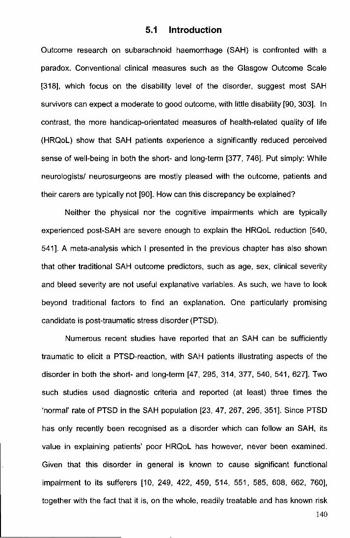
5.1 Introduction
Outcome research on subarachnoid haemorrhage (SAH) is confronted with a
paradox. Conventional clinical measures such as the Glasgow Outcome Scale
[318], which focus on the disability level of the disorder, suggest most SAH
survivors can expect a moderate to good outcome, with little disability [90, 303]. In
contrast, the more handicap-orientated measures of health-related quality of life
(HRQoL) show that SAH patients experience a significantly reduced perceived
sense of well-being in both the short- and long-term [377, 746]. Put simply: While
neurologists/ neurosurgeons are mostly pleased with the outcome, patients and
their carers are typically not [90]. How can this discrepancy be explained?
Neither the physical nor the cognitive impairments which are typically
experienced post-SAH are severe enough to explain the HRQoL reduction [540,
541]. A meta-analysis which I presented in the previous chapter has also shown
that other traditional SAH outcome predictors, such as age, sex, clinical severity
and bleed severity are not useful explanative variables. As such, we have to look
beyond traditional factors to find an explanation. One particularly promising
candidate is post-traumatic stress disorder (PTSD).
Numerous recent studies have reported that an SAH can be sufficiently
traumatic to elicit a PTSD-reaction, with SAH patients illustrating aspects of the
disorder in both the short- and long-term [47, 295, 314, 377, 540, 541, 627]. Two
such studies used diagnostic criteria and reported (at least) three times the
'normal' rate of PTSD in the SAH population [23, 47, 267, 295, 351]. Since PTSD
has only recently been recognised as a disorder which can follow an SAH, its
value in explaining patients' poor HRQoL has however, never been examined.
Given that this disorder in general is known to cause significant functional
impairment to its sufferers [10, 249, 422, 459, 514, 551, 585, 608, 662, 760],
together with the fact that it is, on the whole, readily treatable and has known risk
140
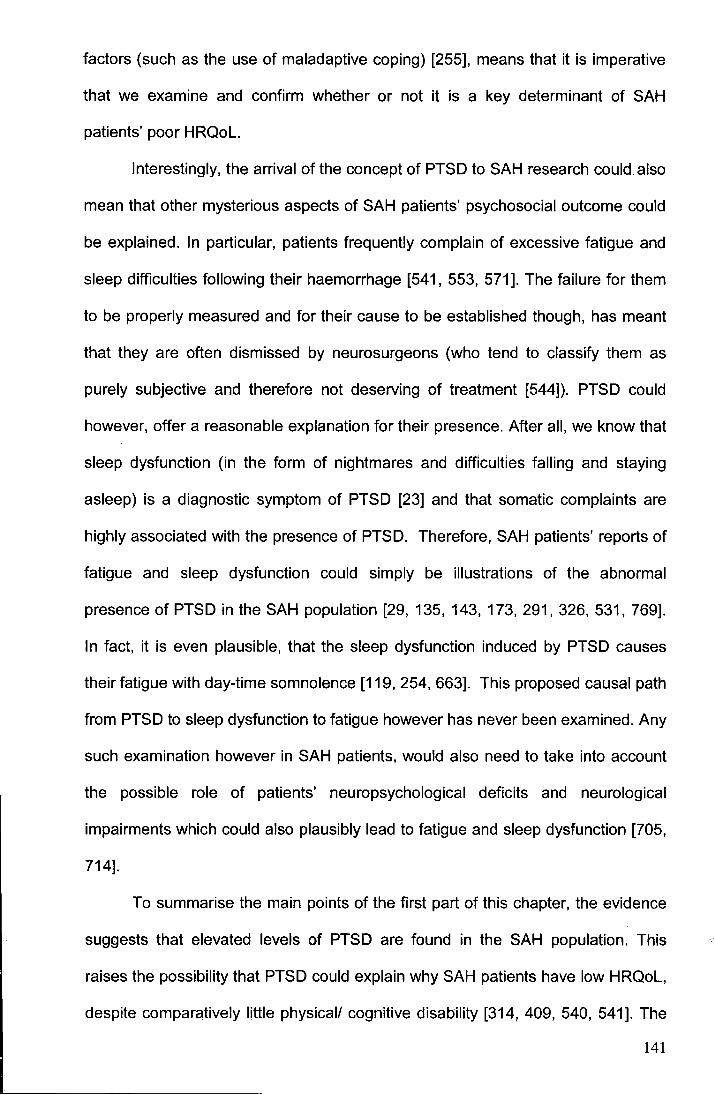
factors (such as the use of maladaptive coping) [255], means that it is imperative
that we examine and confirm whether or not it is a key determinant of SAH
patients' poor HRQoL.
Interestingly, the arrival of the concept of PTSD to SAH research could also
mean that other mysterious aspects of SAH patients' psychosocial outcome could
be explained. In particular, patients frequently complain of excessive fatigue and
sleep difficulties following their haemorrhage [541, 553, 571]. The failure for them
to be properly measured and for their cause to be established though, has meant
that they are often dismissed by neurosurgeons (who tend to classify them as
purely subjective and therefore not deserving of treatment [544]). PTSD could
however, offer a reasonable explanation for their presence. After all, we know that
sleep dysfunction (in the form of nightmares and difficulties falling and staying
asleep) is a diagnostic symptom of PTSD [23] and that somatic complaints are
highly associated with the presence of PTSD. Therefore, SAH patients' reports of
fatigue and sleep dysfunction could simply be illustrations of the abnormal
presence of PTSD in the SAH population [29, 135, 143, 173, 291, 326, 531, 769].
In fact, it is even plausible, that the sleep dysfunction induced by PTSD causes
their fatigue with day-time somnolence [119, 254, 663]. This proposed causal path
from PTSD to sleep dysfunction to fatigue however has never been examined. Any
such examination however in SAH patients, would also need to take into account
the possible role of patients' neuropsychological deficits and neurological
impairments which could also plausibly lead to fatigue and sleep dysfunction [705,
714].
To summarise the main points of the first part of this chapter, the evidence
suggests that elevated levels of PTSD are found in the SAH population. This
raises the possibility that PTSD could explain why SAH patients have low HRQoL,
despite comparatively little physical/ cognitive disability [314, 409, 540, 541]. The
141
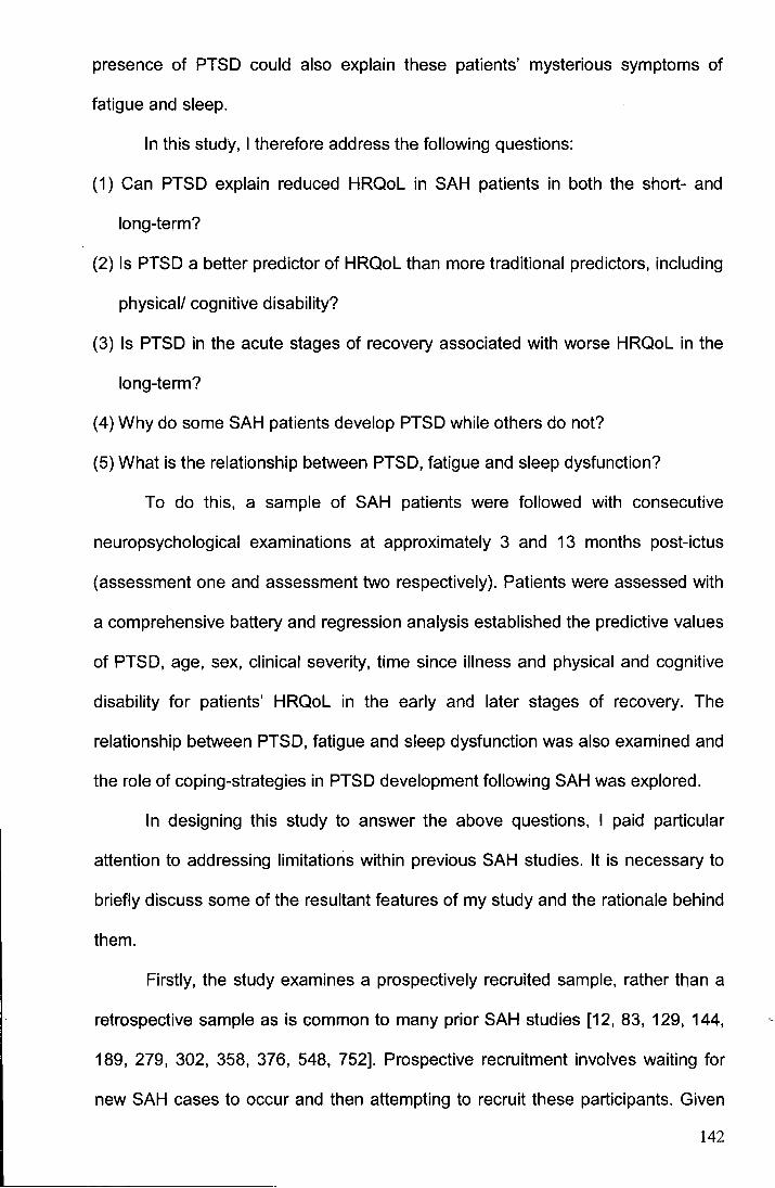
presence of PTSD could also explain these patients' mysterious symptoms of
fatigue and sleep.
In this study, I therefore address the following questions:
(1) Can PTSD explain reduced HRQoL in SAH patients in both the short- and
long-term?
(2) Is PTSD a better predictor of HRQoL than more traditional predictors, including
physical/ cognitive disability?
(3) Is PTSD in the acute stages of recovery associated with worse HRQoL in the
long-term?
(4) Why do some SAH patients develop PTSD while others do not?
(5) What is the relationship between PTSD, fatigue and sleep dysfunction?
To do this, a sample of SAH patients were followed with consecutive
neuropsychological examinations at approximately 3 and 13 months post-ictus
(assessment one and assessment two respectively). Patients were assessed with
a comprehensive battery and regression analysis established the predictive values
of PTSD, age, sex, clinical severity, time since illness and physical and cognitive
disability for patients' HRQoL in the early and later stages of recovery. The
relationship between PTSD, fatigue and sleep dysfunction was also examined and
the role of coping-strategies in PTSD development following SAH was explored.
In designing this study to answer the above questions, I paid particular
attention to addressing limitations within previous SAH studies. It is necessary to
briefly discuss some of the resultant features of my study and the rationale behind
them.
Firstly, the study examines a prospectively recruited sample, rather than a
retrospective sample as is common to many prior SAH studies [12, 83, 129, 144,
189, 279, 302, 358, 376, 548, 752]. Prospective recruitment involves waiting for
new SAH cases to occur and then attempting to recruit these participants. Given
142
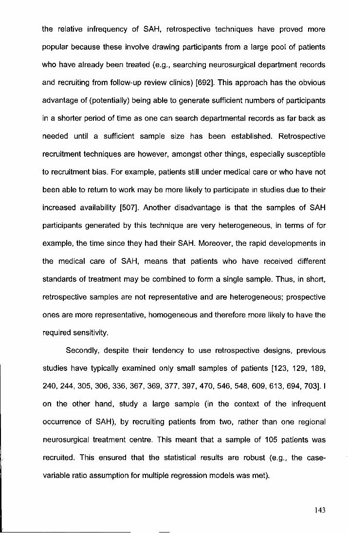
the relative infrequency of SAH, retrospective techniques have proved more
popular because these involve drawing participants from a large pool of patients
who have already been treated (e.g., searching neurosurgical department records
and recruiting from follow-up review clinics) [692]. This approach has the obvious
advantage of (potentially) being able to generate sufficient numbers of participants
in a shorter period of time as one can search departmental records as far back as
needed until a sufficient sample size has been established. Retrospective
recruitment techniques are however, amongst other things, especially susceptible
to recruitment bias. For example, patients still under medical care or who have not
been able to return to work may be more likely to participate in studies due to their
increased availability [507]. Another disadvantage is that the samples of SAH
participants generated by this technique are very heterogeneous, in terms of for
example, the time since they had their SAH. Moreover, the rapid developments in
the medical care of SAH, means that patients who have received different
standards of treatment may be combined to form a single sample. Thus, in short,
retrospective samples are not representative and are heterogeneous; prospective
ones are more representative, homogeneous and therefore more likely to have the
required sensitivity.
Secondly, despite their tendency to use retrospective designs, previous
studies have typically examined only small samples of patients [123, 129, 189,
240,244,305,306,336,367,369,377,397,470,546,548,609,613,694, 703]. I
on the other hand, study a large sample (in the context of the infrequent
occurrence of SAH), by recruiting patients from two, rather than one regional
neurosurgical treatment centre. This meant that a sample of 105 patients was
recruited. This ensured that the statistical results are robust (e.g., the case
variable ratio assumption for multiple regression models was met).
143
Thirdly, a representative sample of patients was recruited. This is in
contrast to nearly all previous studies which used very restrictive inclusion criteria
which resulted in only a select group of the SAH population typically being
included - namely those who presented at hospital in the acute stages, in a good
clinical state and had made a favourable physical recovery at the point of
discharge. Though these patients are less challenging to recruit, the restricted
inclusion criteria meant that a proportion of patients who are perfectly able to
provide informed consent and participate are always excluded. Studies have also
often excluded the 15-25% of SAH patients whose haemorrhage was not due to
the rupture of a cerebral aneurysm. This is because it has been assumed that
most of these excluded patients- namely, those whose bleed was of an unknown
origin - have suffered what is considered a benign sub-form of SAH because it is
associated with a less dramatic clinical course (i.e., a more gradual onset, no
ischemic damage, no surgical intervention, limited extension of blood [71 0]).
Increasing evidence however, suggests that this assumption is incorrect and that
SAH patients' neuropsychological and psychosocial outcome is largely
independent of the aetiology of the bleed, with a common pattern of deficits
resulting regardless of the origin of the haemorrhage [78, 176, 301, 302, 324, 428,
502, 504, 572, 628, 644]. In fact, some contend that those patients whose bleed
was of an unknown origin could experience a worse psychiatric outcome. It has
been argued for example, that a patient whose bleed was of an identifiable origin,
such as a ruptured aneurysm, may leave the hospital more confident as the
source of their haemorrhage has been "cured" and knowing that their chances of a
further SAH are minimal. In contrast, a person whose bleed was of an unidentified
origin and so remains untreated, may worry more about the likelihood of a further
SAH. It has been postulated that these patients may be burdened by this
additionally, (though unfounded) ever-present peril [211, 295, 306, 504, 572].
144

A final limitation of previous studies addressed by the present study is that
patients' neurological disabilities have not previously been measured using
instruments with a high degree of ecological validity. Prior studies for example,
used only classic neuropsychological tests which are designed not to predict
behaviour, but rather to localise lesions [203, 276]. The use of these measures
could have served to attenuate the relationship ·between patients' scores on
cognitive tests and their HRQoL. Since in this chapter I am examining the relative
value of PTSD, I felt it sensible to adopt a conservative approach and compare its
explanative value next to neurological variables indexed using sensitive measures
with a high degree of ecological validity.
145

5.2 Methods
Patients
A prospective and consecutive series of 105 non-institutionalised patients was
studied. Patients were recruited from a potential population of 186 first-time victims
of spontaneous SAH, admitted to Newcastle General or James Cook University
Hospital between April 2005 and August 2006. Of the non-participators, 6 were
excluded for geographic reasons, 1 did not speak English and 7 4 declined
particip9tion.
SAH diagnosis was established by CT, or if negative, xanthrochromia in
cerebrospinal spinal fluid was considered diagnostic. Aneurysms were
demonstrated by spiral computed tomography or catheter angiography.
Age and sex-matched controls were recruited from the U.K. general
population to provide sleep and fatigue data to compare to that of SAH patients.
Control subjects were recruited through personal contacts and recreational clubs.
This study was granted multi-centre approval by the Central Manchester
Research Ethics Committee. Informed consent was obtained from all participants.
Procedure
Patients completed two comprehensive neuropsychological assessments at their
homes or at a laboratory at Durham University. Following a brief introductory
interview, the patient's HRQoL, attention, memory and executive function were
examined. Patients were then left with a series of self-report measures to examine
their psychiatric status, fatigue and sleep dysfunction. These were then either
returned to me using a pre-paid envelope or collected at a later date. The two
appointments were conducted at approximately 3 (Assessment 1) and 13
(Assessment 2) months post-ictus. The neuropsychological appointments lasted
146
approximately 3.5 hours (including regular breaks taken according to the
requirements of the individual patient).
Measures
Instruments were chosen on the basis of having robust psychometric properties
and ecological validity for cognitive tasks [95, 107, 139, 194, 197, 238, 563, 657,
734, 754-756]. Each of these items is detailed below. A complete list of these
instruments along with a shortened description can also be found in Table 5.1.
Please note that parallel versions of the TEA and RBMT-E were used at
assessments one and two.
The Short-Form 36 (SF-36) [734] assesses HRQoL across 8 domains: 1)
'Physical Functioning', 2) 'Physical Role Limitations', 3) 'Bodily Pain', 4) 'General
Health', 5) 'Energy', 6) 'Social Functioning', 7) 'Emotional Role Limitations' and 8)
'Mental Health'. Scores range from 100 (no reduction) to 0 (maximum reduction).
Standard algorithms - derived from principal component analyses - aggregated
scores from the eight domains into two summary scores for regression: 'Physical
HRQoL' and 'Mental HRQoL'. Scores from domains 1-4 mostly contribute to
'Physical HRQoL', whilst domains 5-8 contribute most to 'Mental HRQoL' (see
Figure 3.2, Chapter 3 for schematic of the structure and questions of the SF-36)
[733].
The Post-traumatic Diagnostic Scale [194] assesses whether patients
warrant a DSM-IV-TR diagnosis of PTSD (see Table 4.2, Chapter 4 for diagnostic
symptoms of PTSD) [23]. It provides a PTSD symptom severity score and a
functional impairment index for those diagnosed. For use in some regression
analyses, a 'PTSD Symptom Severity' score was calculated for all patients
regardless of PTSD diagnosis. PTSD is usually only diagnosed if patients have
experienced intense fear, helplessness or horror at the time of the trauma. Given
147
Table 5.1
Purpose
Screening
Global mental status
Health-related quality
of life
Post-traumatic stress
disorder
Fatigue
Summary of neurological, neuropsychological and psychosocial instruments used in the study
Test
Behavioural Inattention Test (BIT) [756];
Short Token Test [139].
Mini-Mental State Examination (MMSE) [197].
Short-Form-36 (SF-36) [734].
Post-traumatic Stress Diagnostic Scale (PDS) [194].
Multidimensional Fatigue Symptom Inventory-Short Form
(MSFI-SF) [657].
Information
These tests screened for unilateral visual neglect and linguistic deficits. They were completed at assessment one and were used
to establish whether administration of the remaining battery was appropriate.
This was completed at assessment one only and was used to establish global cognitive status for all patients. This brief index was
then used in missing value analysis to assess if those failing to complete a subsequent test significantly differed in cognitive status
from those with complete data.
Measured health-related quality of life (HRQoL) across 8 domains (see Figure 3.2, Chapter 3). 'Physical HRQoL' and 'Mental
HRQoL' summary scores were generated for regression analyses using standard algorithms.
This measure assesses whether a patient warranted a DSM-IV diagnosis of post-traumatic stress disorder (PTSD) in relation to
their SAH experience.
It provides a PTSD symptom severity score and a functional impairment index.
PTSD is usually only diagnosed if patients experienced intense fear, helplessness or horror at the time of the trauma. Given SAH
can lead to loss of consciousness at the time of trauma, this criteria was relaxed. The effect of this modification was minimal and
merely permitted diagnosis of PTSD in two more patients at assessment one and three more patients at assessment two.
Fatigue measured across the following scales: General Fatigue, Physical Fatigue, Mental Fatigue, Emotional Fatigue and Vigour.
The sum of the fatigue subscales minus the 'Vigour' scale generated a Total Fatigue Score.
148
Table 5.1 Cont'd.
Purpose
Cognition
Sleep
Physical disability
Coping
Test
Test of Everyday Attention (TEA) [563]; Behavioural
Assessment of the Dysexecutive Syndrome (BADS) [754];
Rivermead Behavioural Memory Test- Extended (RBMT-E)
[755].
Pittsburgh Sleep Quality Index (PSIQ) [95].
Functional Independence Measure- Motor Scale (FIM-MS)
[238].
Brief Cope Inventory [1 07].
Information
Attention tasks: 'Visual Elevator', Telephone Search', Telephone Search Dual Task', 'Elevator Counting' and 'Lottery'.
Executive subtests: 'Rule Shift Cards', 'Key Search', 'Modified Six Elements', Temporal Judgment', and 'Zoo Map'.
Memory subtests: 'Story Immediate' and 'Delayed', 'Picture Recognition', 'Face Recognition', 'Orientation and Date', 'First Name~
'Second
Names', and 'Appointments and Belongings'.
Sleep measured across the following scales: Subjective Sleep Quality, Latency, Duration, Habitual Sleep Efficiency (hours slee
hours in bed}, Disturbances, Sleep Medication Use, Daytime Dysfunction and Global Sleep score.
Independence was measured across 13 tasks. Increasing values indicate greater levels of independence inmobility, self-cal
and continence.
Measured patients' dispositional use of 'Maladaptive Coping Strategies', 'Venting', 'Denial', 'Substance Use', 'Behaviour
Disengagement' and 'Self-Distraction'. This measure was completed at assessment one only.
149
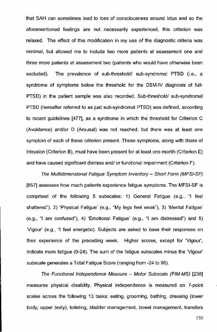
that SAH can sometimes lead to loss of consciousness around ictus and so the
aforementioned feelings are not necessarily experienced, this criterion was
relaxed. The effect of this modification in my use of the diagnostic criteria was
minimal, but allowed me to include two more patients at assessment one and
three more patients at assessment two (patients who would have otherwise been
excluded). The prevalence of sub-threshold/ sub-syndromal PTSD (i.e., a
syndrome of symptoms below the threshold for the DSM-IV diagnosis of fuii
PTSD) in the patient sample was also recorded. Sub-threshold/ sub-syndromal
PTSD (hereafter referred to as just sub-syndromal PTSD) was defined, according
to recent guidelines [477], as a syndrome in which the threshold for Criterion C
(Avoidance) and/or D (Arousal) was not reached, but there was at least one
symptom of each of these criterion present. These symptoms, along with those of
Intrusion (Criterion B), must have been present for at least one month (Criterion E)
and have caused significant distress and/ or functional impairment (Criterion F).
The Multidimensional Fatigue Symptom Inventory - Short Form (MFSI-SF)
[657] assesses how much patients experience fatigue symptoms. The MFSI-SF is
comprised of the following 5 subscales: 1) General Fatigue (e.g., "I feel
shattered"), 2) 'Physical Fatigue' (e.g., "My legs feel weak"), 3) 'Mental Fatigue'
(e.g., "I am confused"), 4) 'Emotional Fatigue' (e.g., "I am distressed") and 5)
'Vigour' (e.g., "I feel energetic). Subjects are asked to base their responses on
their experience of the preceding week. Higher scores, except for 'Vigour',
indicate more fatigue (0-24 ). The sum of the fatigue subscales minus the 'Vigour'
subscale generates a Total Fatigue Score (ranging from -24 to 96).
The Functional Independence Measure - Motor Subscale (FIM-MS) [238]
measures physical disability. Physical independence is measured on ?-point
scales across the following 13 tasks: eating, grooming, bathing, dressing (lower
body, upper body), toileting, bladder management, bowel management, transfers
150
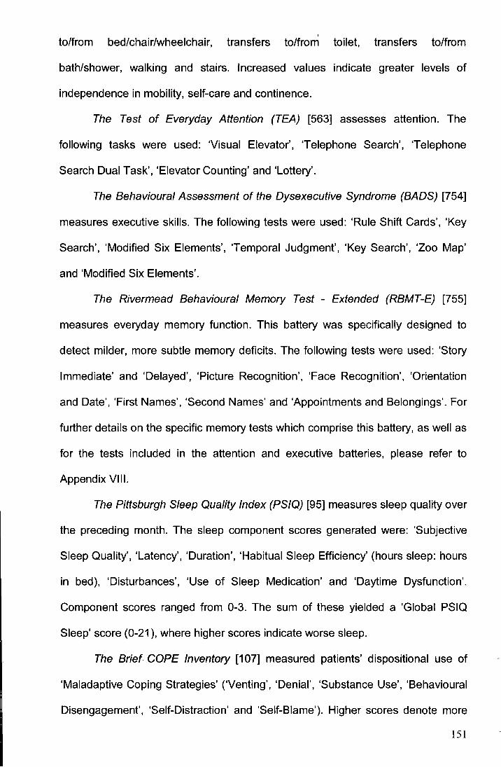
to/from bed/chair/wheelchair, transfers to/from toilet, transfers to/from
bath/shower, walking and stairs. Increased values indicate greater levels of
independence in mobility, self-care and continence.
The Test of Everyday Attention (TEA) [563] assesses attention. The
following tasks were used: 'Visual Elevator', 'Telephone Search', 'Telephone
Search Dual Task', 'Elevator Counting' and 'Lottery'.
The Behavioural Assessment of the Dysexecutive Syndrome (BADS) [754]
measures executive skills. The following tests were used: 'Rule Shift Cards', 'Key
Search', 'Modified Six Elements', 'Temporal Judgment', 'Key Search', 'Zoo Map'
and 'Modified Six Elements'.
The Rivermead Behavioural Memory Test - Extended (RBMT-E) [755]
measures everyday memory function. This battery was specifically designed to
detect milder, more subtle memory deficits. The following tests were used: 'Story
Immediate' and 'Delayed', 'Picture Recognition', 'Face Recognition', 'Orientation
and Date', 'First Names', 'Second Names' and 'Appointments and Belongings'. For
further details on the specific memory tests which comprise this battery, as well as
for the tests included in the attention and executive batteries, please refer to
Appendix VIII.
The Pittsburgh Sleep Quality Index (PSIQ) [95] measures sleep quality over
the preceding month. The sleep component scores generated were: 'Subjective
Sleep Quality', 'Latency', 'Duration', 'Habitual Sleep Efficiency' (hours sleep: hours
in bed), 'Disturbances', 'Use of Sleep Medication' and 'Daytime Dysfunction'.
Component scores ranged from 0-3. The sum of these yielded a 'Global PSIQ
Sleep' score (0-21 ), where higher scores indicate worse sleep.
The Brief COPE Inventory [1 07] measured patients' dispositional use of
'Maladaptive Coping Strategies' ('Venting', 'Denial', 'Substance Use', 'Behavioural
Disengagement', 'Self-Distraction' and 'Self-Blame'). Higher scores denote more
151
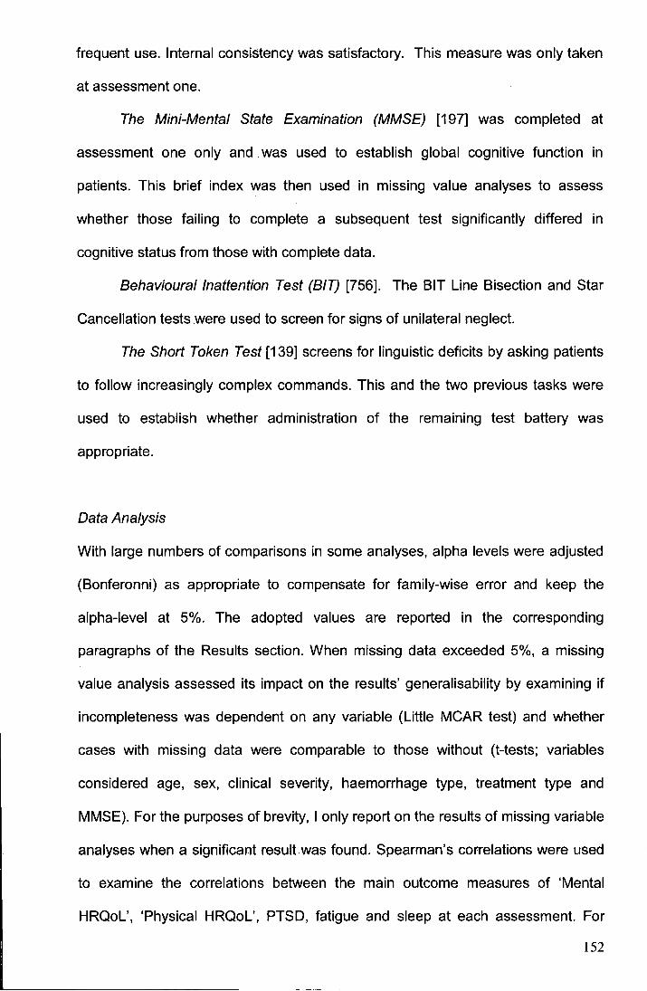
frequent use. Internal consistency was satisfactory. This measure was only taken
at assessment one.
The Mini-Mental State Examination (MMSE) [197] was completed at
assessment one only and was used to establish global cognitive function in
patients. This brief index was then used in missing value analyses to assess
whether those failing to complete a subsequent test significantly differed in
cognitive status from those with complete data.
Behavioural Inattention Test (BIT) [756]. The BIT Line Bisection and Star
Cancellation tests were used to screen for signs of unilateral neglect.
The Short Token Test [139] screens for linguistic deficits by asking patients
to follow increasingly complex commands. This and the two previous tasks were
used to establish whether administration of the remaining test battery was
appropriate.
Data Analysis
With large numbers of comparisons in some analyses, alpha levels were adjusted
(Bonferonni) as appropriate to compensate for family-wise error and keep the
alpha-level at 5%. The adopted values are reported in the corresponding
paragraphs of the Results section. When missing data exceeded 5%, a missing
value analysis assessed its impact on the results' generalisability by examining if
incompleteness was dependent on any variable (Little MCAR test) and whether
cases with missing data were comparable to those without (t-tests; variables
considered age, sex, clinical severity, haemorrhage type, treatment type and
MMSE). For the purposes of brevity, I only report on the results of missing variable
analyses when a significant result was found. Spearman's correlations were used
to examine the correlations between the main outcome measures of 'Mental
HRQoL', 'Physical HRQoL', PTSD, fatigue and sleep at each assessment. For
152
regression analyses, beta values (standardized B) and squared semi-partial
correlations (sr2;) indicated relative and unique predictor contributions. The
backward elimination method of variable entry/ selection (likelihood ratio test for
logistic regression) was used in the regression analyses to identify. the most
parsimonious predictor variables. Stepwise block entry regression was also used
in a few instances to examine the unique explanative ability of a variable once the
variance accounted for by other variables was controlled for. Case-variable ratios
were satisfactory for regression models. Where possible, effect sizes (ES) in the
form of the correlation coefficient r are reported for all results. Analyses were
conducted using commercially available software (SPSS).
Health-Related Quality of Life (HRQoL). Kruskai-Wallis one-way analyses
of variance determined the normality of patients' SF-36 scores by comparing their
scores to reference values [60]. Three- and thirteen month 'Physical HRQoL' and
'Mental HRQoL' determinants were explored using Spearman's correlation and
multiple linear regression. Parametric regression techniques were most
appropriate for this ordinal data due to the number of occupied categories, and
due to the fact that it was fair to assume an underlying continuum and an equal
distance between each ordinal point of the SF-36 [727]. Predictors considered
were: age, sex, clinical severity as indexed by the World Federation of
Neurological Surgeons (WFNS) scale [164], history of previous illness vs. none,
the 'Overall Cognitive Function' index, 'FIM-MS' score and 'PTSD Symptom
Severity' score from the same assessment point. Further regression analyses,
this time using stepwise variable entry (2 blocks), were conducted to examine 1)
the unique ability of SAH patients' PTSD detected at the short-term assessment to
predict the HRQoL these patients reported at the long-term assessment and 2)
any change that had occurred between patients' short-term HRQoL and long-term
HRQoL (indexed as a patient's long-term HRQoL minus their short-term HRQoL).
153
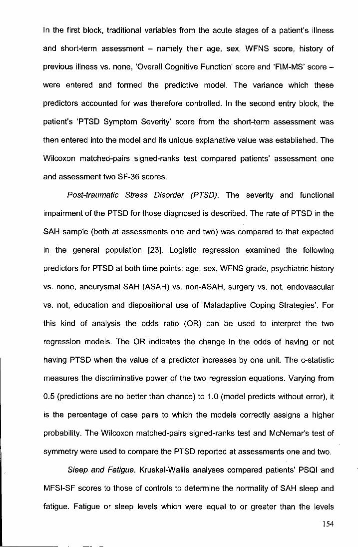
In the first block, traditional variables from the acute stages of a patient's illness
and short-term assessment - namely their age, sex, WFNS score, history of
previous illness vs. none, 'Overall Cognitive Function' score and 'FIM-MS' score -
were entered and formed the predictive model. The variance which these
predictors accounted for was therefore controlled. In the second entry block, the
patient's 'PTSD Symptom Severity' score from the short-term assessment was
then entered into the model and its unique explanative value was established. The
Wilcoxon matched-pairs signed-ranks test compared patients' assessment one
and assessment two SF-36 scores.
Post-traumatic Stress Disorder (PTSD). The severity and functional
impairment of the PTSD for those diagnosed is described. The rate of PTSD in the
SAH sample (both at assessments one and two) was compared to that expected
in the general population [23]. Logistic regression examined the following
predictors for PTSD at both time points: age, sex, WFNS grade, psychiatric history
vs. none, aneurysmal SAH (ASAH) vs. non-ASAH, surgery vs. not, endovascular
vs. not, education and dispositional use of 'Maladaptive Coping Strategies'. For
this kind of analysis the odds ratio (OR) can be used to interpret the two
regression models. The OR indicates the change in the odds of having or not
having PTSD when the value of a predictor increases by one unit. The c-statistic
measures the discriminative power of the two regression equations. Varying from
0.5 (predictions are no better than chance) to 1.0 (model predicts without error), it
is the percentage of case pairs to which the models correctly assigns a higher
probability. The Wilcoxon matched-pairs signed-ranks test and McNemar's test of
symmetry were used to compare the PTSD reported at assessments one and two.
Sleep. and Fatigue. Kruskai-Wallis analyses compared patients' PSQI and
MFSI-SF scores to those of controls to determine the normality of SAH sleep and
fatigue. Fatigue or sleep levels which were equal to or greater than the levels
154
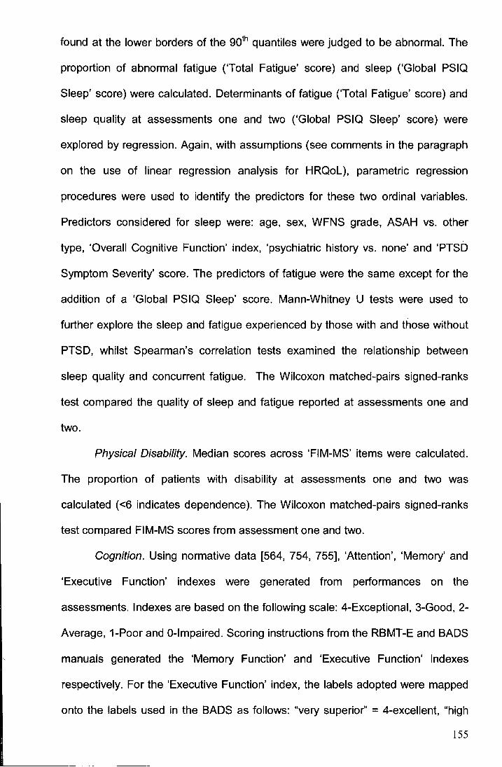
found at the lower borders of the 90th quantiles were judged to be abnormal. The
proportion of abnormal fatigue ('Total Fatigue' score) and sleep ('Global PSIQ
Sleep' score) were calculated. Determinants of fatigue ('Total Fatigue' score) and
sleep quality at assessments one and two ('Global PSIQ Sleep' score) were
explored by regression. Again, with assumptions (see comments in the paragraph
on the use of linear regression analysis for HRQoL), parametric regression
procedures were used to identify the predictors for these two ordinal variables.
Predictors considered for sleep were: age, sex, WFNS grade, ASAH vs. other
type, 'Overall Cognitive Function' index, 'psychiatric history vs. none' and 'PTSD
Symptom Severity' score. The predictors of fatigue were the same except for the
addition of a 'Global PSIQ Sleep' score. Mann-Whitney U tests were used to
further explore the sleep and fatigue experienced by those with and those without
PTSD, whilst Spearman's correlation tests examined the relationship between
sleep quality and concurrent fatigue. The Wilcoxon matched-pairs signed-ranks
test compared the quality of sleep and fatigue reported at assessments one and
two.
Physical Disability. Median scores across 'FIM-MS' items were calculated.
The proportion of patients with disability at assessments one and two was
calculated ( <6 indicates dependence). The Wilcoxon matched-pairs signed-ranks
test compared FIM-MS scores from assessment one and two.
Cognition. Using normative data [564, 754, 755], 'Attention', 'Memory' and
'Executive Function' indexes were generated from performances on the
assessments. Indexes are based on the following scale: 4-Exceptional, 3-Good, 2-
Average, 1-Poor and 0-lmpaired. Scoring instructions from the RBMT-E and BADS
manuals generated the 'Memory Function' and 'Executive Function' indexes
respectively. For the 'Executive Function' index, the labels adopted were mapped
onto the labels used in the BADS as follows: "very superior'' = 4-excellent, "high
155
average/ superior" = 3-good, "average" = 2-average, "borderline/low average" = 1-
poor and "impaired" = 0-impaired. As no standard TEA procedure exists for
generating an 'Attention Function' index, I scored TEA subtest performances
(directed by accepted guidelines [248]) as follows: TEA '=:98% = 4-excellent, '=:75%
= 3-good, '=:25% = 2-average, '=:2% = 1-poor and <2% = 0-impaired. TEA lottery
task performance, which in its original format is scored according to cut-offs, was
awarded the following: TEA "impaired" = 0-impaired; "borderline" = 1-poor and
"normal" = 3-good. The 'Attention Function' index was the median score on the
subtests. For the purposes of regression analyses, 'Overall Cognitive Function'
indices were generated for patients to represent cognitive performance on each
respective assessment. These indices were calculated by summing the 'Attention',
'Memory' and 'Executive' indexes from the respective assessment. Changes in
cognitive performance from assessment one to assessment two were analysed by
Wilcoxon test.
5.3 Results
The recruited patient group consisted of 60 women and 45 men. No differences
existed between participators and non-participators in age, sex, WFNS grade,
haemorrhage type, or treatment (all P>0.05; Little's MCAR: P=.082). All 105
patients were examined at assessment one, but 10 patients (9.5%) were lost from
the sample at assessment two (Table 5.2). The reason for the reduced sample
was due to one patient death and the remaining nine patients declining to
participate in assessment two. No significant differences existed between the
patients who were examined at assessment two and those who were not (P>0.05),
with no pattern to the missing data being found (Little's MCAR: P=.549). Average
follow-up for assessment one was 109 days after SAH (SO 49.95; range 24-251)
and 406 days after SAH for assessment two (SO 47.82; range 335-672). In total,
156
Table 5.2 Demographic and clinical data of patients examined at
assessments one and two
Factor
Mean age at assessment± SD (range) (yr)
Women
Education (yr)
Aneurysmal SAH
Location of ruptured aneurysm
ACoMIACA
/CA
MCA
Vertebrobasi/ar
Unknown origin
Other origin
WFNS grade
II
Ill
IV
v
Primary treatment
Endovascular
Surgery
Conservative
Significant history *
Physical only
Psychiatric only
Both
Value(%)
Assessment one (N=1 05)
52.4 ± 11 (31-80)
60(57.1)
11.6± 1.51 (10-20)
77 (73.3)
34 (44.2)
19 (24.7)
19 (24.7)
5 (6.5)
25 (23.8)
3 (2.9)
74 (70.5)
15 (14.3)
5 (4.8)
10 (9.5)
1 (1)
47 (44.8)
32 (30.5)
26 (24.8)
18(17.1)
15 (14.3)
16(15.2)
Assessment two (N=95)
53.0 ± 11 (32-81)
54 (56.8)
11.6± 1.6 (10-20)
67 (70.5)
31 (46.3)
14 (20.9)
17 (25.4)
5 (7.5)
25 (26.3)
3 (3.2)
66 (69.5)
13 (13.7)
5 (5.3)
10 (10.5)
1 (1.1)
40 (42.1)
29 (30.5)
26 (27.4)
15 (15.8)
15 (15.8)
15 (15.8)
Note: ACA=anterior cerebral artery; ACoM=anterior communicating artery; ICA=internal carotid artery; n=number;
MCA=middle cerebral artery; N=number; SD=standard deviation; Vertebrobasilar =vertebrobasilar system artery;
WFNS=World Federation of Neurological Surgeons; yr=year. *Self-reported illness receiving/ received formal treatment.
Physical illness: benign intracranial hypertension; cancer; epilepsy; Guillain-Barre syndrome; human immunodeficiency
virus; hypothyroidism; ischemic stroke; kidney cyst; meningioma; multiple sclerosis; myocardial infarction; pulmonary
emphysema; Reynaud's phenomenon; recurrent pneumothorax; rheumatoid arthritis; road traffic accident Injuries; sleep
apnoea; spondylosis; transient ischemic attack; tuberculosis. Psychiatric illness: Anxiety unspecified diagnosis; Depression
unspecified diagnosis and Alcohol abuse.
157
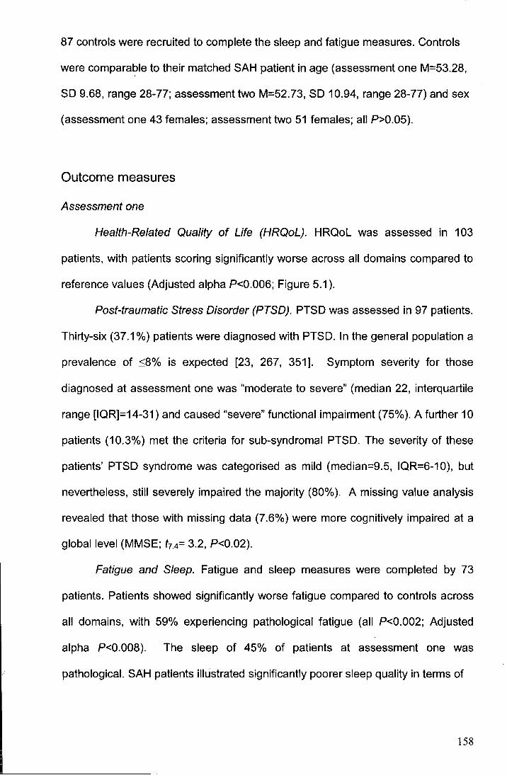
87 controls were recruited to complete the sleep and fatigue measures. Controls
were comparable to their matched SAH patient in age (assessment one M=53.28,
SD 9.68, range 28-77; assessment two M=52.73, SD 10.94, range 28-77) and sex
(assessment one 43 females; assessment two 51 females; all P>0.05).
Outcome measures
Assessment one
Health-Related Quality of Life (HRQoL). HRQoL was assessed in 103
patients, with patients scoring significantly worse across all domains compared to
reference values (Adjusted alpha P<0.006; Figure 5.1 ).
Post-traumatic Stress Disorder (PTSD). PTSD was assessed in 97 patients.
Thirty-six (37.1 %) patients were diagnosed with PTSD. In the general population a
prevalence of ::;8% is expected [23, 267, 351]. Symptom severity for those
diagnosed at assessment one was "moderate to severe" (median 22, interquartile
range [IQR]=14-31) and caused "severe" functional impairment (75%). A further 10
patients (10.3%) met the criteria for sub-syndromal PTSD. The severity of these
patients' PTSD syndrome was categorised as mild (median=9.5, IQR=6-10), but
nevertheless, still severely impaired the majority (80%). A missing value analysis
revealed that those with missing data (7.6%) were more cognitively impaired at a
global level (MMSE; t7.4= 3.2, P<0.02).
Fatigue and Sleep. Fatigue and sleep measures were completed by 73
patients. Patients showed significantly worse fatigue compared to controls across
all domains, with 59% experiencing pathological fatigue (all P<0.002; Adjusted
alpha P<0.008). The sleep of 45% of patients at assessment one was
pathological. SAH patients illustrated significantly poorer sleep quality in terms of
158
-U) ('I)
I u.. en -Q) ... 0 u en "C ... ca
"C c ca -en s:: ca Q)
:!:
0.5
0.3
0.1
-0.1
-0.3
-0.5
-0.7
-0.9
-1.1
-1.3
-1.5
-1.7
-1.9
Health Domains
----o- Assessment one
···-D-··· Assessment two
Figure 5.1 Graph showing the SF-36 HRQoL profile of patients at assessments
one and two. Deviations from reference data are expressed in mean
standard scores. All deviations from reference values are significant
(P<0.05), except 'Emotional Role Limitations' at assessment two.
159
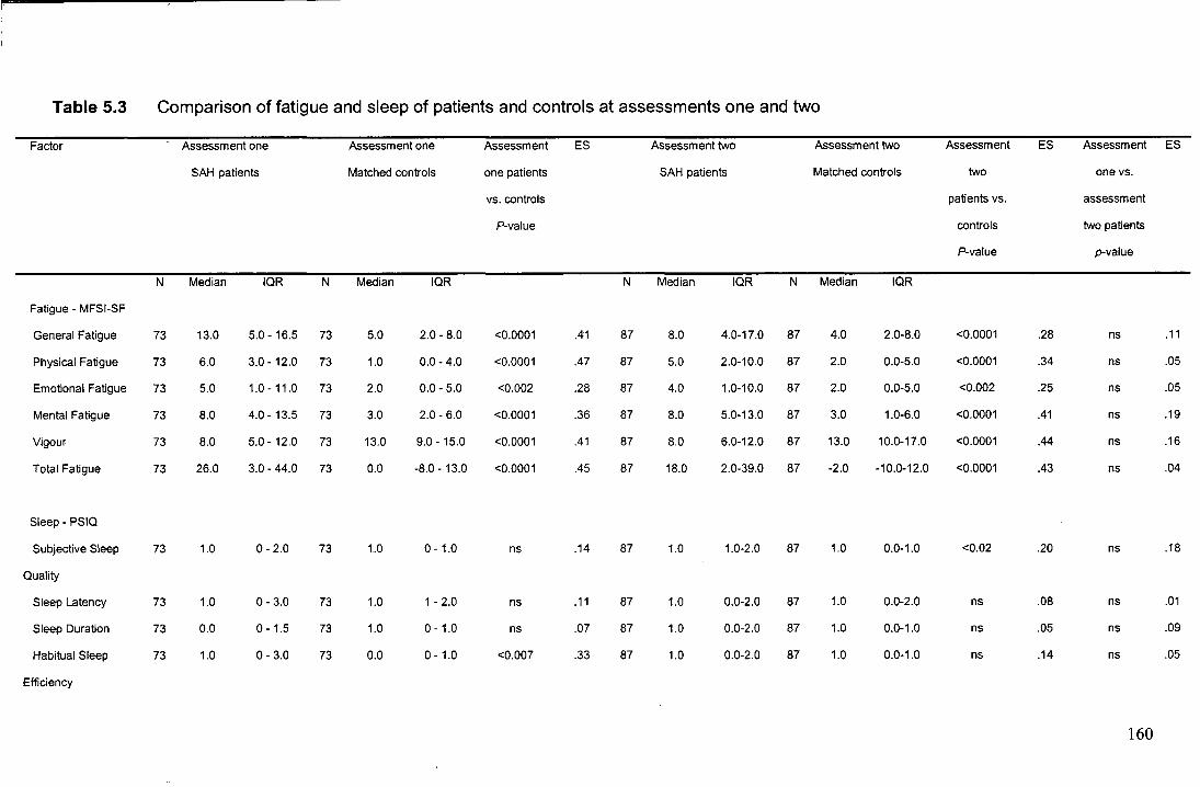
Table 5.3 Comparison of fatigue and sleep of patients and controls at assessments one and two
Factor Assessment one Assessment one Assessment ES Assessment two Assessment two Assessment ES Assessment ES
SAH patients Matched controls one patients SAH patients Matched controls two one vs.
vs. controls patients vs. assessment
P-value controls two patients
P-value p-value
N Median IQR N Median IQR N Median IQR N Median IQR
Fatigue- MFSI-SF
General Fatigue 73 13.0 5.0- 16.5 73 5.0 2.0- 8.0 <0.0001 .41 87 8.0 4.0-17.0 87 4.0 2.0-8.0 <0.0001 .28 ns .11
Physical Fatigue 73 6.0 3.0- 12.0 73 1.0 0.0-4.0 <0.0001 .47 87 5.0 2.0-10.0 87 2.0 0.0-5.0 <0.0001 .34 ns .05
Emotional Fatigue 73 5.0 1.0- 11.0 73 2.0 0.0-5.0 <0.002 .28 87 4.0 1.0-10.0 87 2.0 0.0-5.0 <0.002 .25 ns .05
Mental Fatigue 73 8.0 4.0- 13.5 73 3.0 2.0- 6.0 <0.0001 .36 87 8.0 5.0-13.0 87 3.0 1.0-6.0 <0.0001 .41 ns .19
Vigour 73 8.0 5.0- 12.0 73 13.0 9.0- 15.0 <0.0001 .41 87 8.0 6.0-12.0 87 13.0 10.0-17.0 <0.0001 .44 ns .16
Total Fatigue 73 26.0 3.0-44.0 73 0.0 -8.0- 13.0 <0.0001 .45 87 18.0 2.0-39.0 87 -2.0 -10.0-12.0 <0.0001 .43 ns .04
Sleep- PSIQ
Subjective Sleep 73 1.0 0-2.0 73 1.0 0- 1.0 ns .14 87 1.0 1.0-2.0 87 1.0 0.0-1.0 <0.02 .20 ns .18
Quality
Sleep Latency 73 1.0 0-3.0 73 1.0 1-2.0 ns .11 87 1.0 0.0-2.0 87 1.0 0.0-2.0 ns .08 ns .01
Sleep Duration 73 0.0 0- 1.5 73 1.0 0- 1.0 ns .07 87 1.0 0.0-2.0 87 1.0 0.0-1.0 ns .05 ns .09
Habitual Sleep 73 1.0 0-3.0 73 0.0 0- 1.0 <0.007 .33 87 1.0 0.0-2.0 87 1.0 0.0-1.0 ns .14 ns .05
Efficiency
160
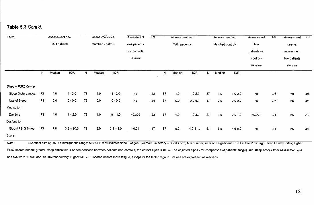
Table 5.3 Cont'd.
Factor Assessment one Assessment one Assessment ES Assessment two Assessment two Assessment ES Assessment ES
SAH patients Matched controls one patients SAH patients Matched controls two one vs.
vs. controls patients vs. assessment
P-value controls two patients
P-value P-value
N Median lOR N Median lOR N Median IQR N Median lOR
Sleep - PS/Q Cont'd.
Sleep Disturbances 73 1.0 1-2.0 73 1.0 1-2.0 ns .13 87 1.0 t.0-2.0 87 1.0 1.0-2.0 ns .06 ns .08
Use of Sleep 73 0.0 0-0.0 73 0.0 0-0.0 ns .14 87 0.0 0.0-0.0 87 0.0 0.0-0.0 ns .07 ns .04
Medication
Daytime 73 1.0 1-2.0 73 1.0 0-1.0 <0.009 .22 87 1.0 1.0-2.0 87 1.0 0.0-1.0 <0.007 .21 ns .10
Dysfunction
Global PSIQ Sleep 73 7.0 3.5- 10.5 73 6.0 3.5-8.0 <0.04 .17 87 6.0 4.0-11.0 87 6.0 4.0-8.0 ns .14 ns .01
Score
Note: ES=effect size (r); IQR = interquartile range; MFSI-SF = Multidimensional Fatigue Symptom Inventory- Short Form; N =number; ns =non significant. PSIQ =The Pittsburgh Sleep Quality Index; higher
PSIQ scores denote greater sleep difficulties. For comparisons between patients and controls, the critical alpha =<0.05. The adjusted alphas for comparison of patients' fatigue and sleep scores from assessment one
and two were <0.008 and <0.006 respectively. Higher MFSI-SF scores denote more fatigue, except for the factor 'vigour'. Values are expressed as medians
161
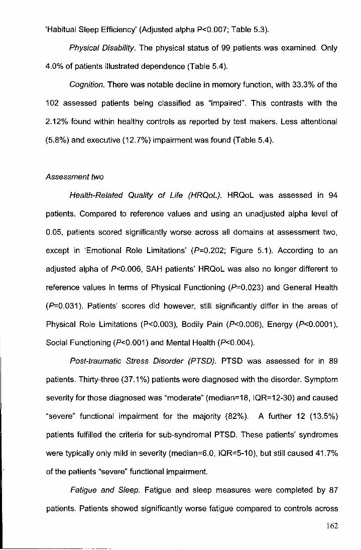
'Habitual Sleep Efficiency' (Adjusted alpha P<0.007; Table 5.3).
Physical Disability. The physical status of 99 patients was examined. Only
4.0% of patients illustrated dependence (Table 5.4).
Cognition. There was notable decline in memory function, with 33.3% of the
102 assessed patients being classified as "impaired". This contrasts with the
2.12% found within healthy controls as reported by test makers. Less attentional
(5.8%) and executive (12.7%) impairment was found (Table 5.4).
Assessment two
Health-Related Quality of Life (HRQoL). HRQoL was assessed in 94
patients. Compared to reference values and using an unadjusted alpha level of
0.05, patients scored significantly worse across all domains at assessment two,
except in 'Emotional Role Limitations' (P=0.202; Figure 5.1 ). According to an
adjusted alpha of P<0.006, SAH patients' HRQoL was also no longer different to
reference values in terms of Physical Functioning (P=0.023) and General Health
(P=0.031 ). Patients' scores did however, still significantly differ in the areas of
Physical Role Limitations (P<0.003), Bodily Pain (P<0.006), Energy (P<0.0001 ),
Social Functioning (P<0.001) and Mental Health (P<0.004).
Post-traumatic Stress Disorder (PTSD). PTSD was assessed for in 89
patients. Thirty-three (37.1 %) patients were diagnosed with the disorder. Symptom
severity for those diagnosed was "moderate" (median=18, IQR=12-30) and caused
"severe" functional impairment for the majority (82%). A further 12 (13.5%)
patients fulfilled the criteria for sub-syndromal PTSD. These patients' syndromes
were typically only mild in severity (median=6.0, IQR=5-1 0), but still caused 41.7%
of the patients "severe" functional impairment.
Fatigue and Sleep. Fatigue and sleep measures were completed by 87
patients. Patients showed significantly worse fatigue compared to controls across
162
Table 5.4 Summary of results from cognitive and physical examinations in assessments one and two
Factor
N
Cognition
RBMT-E Memory Function 102
BADS Executive Function 102
TEA Attention Function 103
Physical
FIM-motor scale 99
Assessment one
SAH patients
Median IQR Percentage in
clinical range
1.0 0.0- 1.0 33.3
2.0 1.0-2.0 12.7
2.0 1.0-2.0 5.8
7.0 7.0- 7.0 4.0
N
95
95
95
87
Assessment two
SAH patients
Median IQR Percentage in
clinical range
1.0 0.0- 1.0 29.5
2.0 1.0-3.0 13.7
2.0 1.0-3.0 8.4
7.0 7.0- 7.0 8.0
Assessment
one versus two
P-value
ns
ns
ns
ns
ES
.15
.06
.04
.09
Note: BADS=Behavioural Assessment of the Dysexecutive Syndrome; FIM-motor scale=Functional Independence Measure motor scale items. FIM
motor scale scores range from 1 (total dependence) to 7 (totally independent); IQR=interquartile range; N=number; ns=non significant; RBMT-E=Rivermead
Behavioural Memory Test; TEA=Test of Everyday Attention. Cognitive function indexes range from 0 (impaired) to 4 (Excellent). Percent in clinical range for
FIM was a median total motor score <6 (dependent on other person) and for cognitive tests it was a function score of 0 ("impaired"). For comparison of
assessment one and two cognitive performances adjusted alpha P=0.01. Alpha for comparison of FIM-motor scale score P=0.05. Values are expressed as
medians.
163
all domains - 36% reported pathological levels of fatigue (all P <0.002; Adjusted
alpha P<0.0008). The only significantly reduced sleep score was seen in the
domain of 'Daytime Dysfunction' (Adjusted alpha P<0.007). Sleep function was
pathological in 37% of patients (Table 5.3).
Physical Disability. The physical status of 87 patients was examined. Eight
percent of patients illustrated dependence (Table 5.4 ).
Cognition. In total, 95 patients were cognitively assessed. There was
substantial memory dysfunction, with 29.5% of patients classified as "impaired" at
assessment two. Much less attentional impairment (8.4%) and executive
impairment (13.7% at assessment two) impairment was found (Table 5.4).
Comparison of assessment one and two performances
Health-Related Quality of Life (HRQoL). For the 93 patients who completed
the SF-36 at both assessments, significant improvements in scores were seen at
assessment two in the domains of 'Physical Functioning' (P<0.001, r-.36),
'Physical Role Limitations' (P<0.0001, r-.54), 'Bodily Pain' (P<0.006, r-.29),
'Social Functioning' (P<0.001, r-.57) and 'Emotional Role Limitations' (P<0.001,
r-.36; Adjusted alpha P<0.006). Importantly, no significant improvement in SF-36
was seen in 'General Health' (P= 0.77, r-.03), 'Energy' (P= 0.03, r-.22) or 'Mental
Health' (P= 0.13, r-.16).
Post-traumatic Stress Disorder (PTSD). For the 82 patients who completed
PTSD assessments at both time points, the Wilcoxon test did not detect any
significant difference in the number (P= 0.80; r-.03) or severity of PTSD symptoms
experienced (P= 0.33; r-.11 ). Nor did it detect any significant difference in the
experience of intrusive (P= 0.632; r-.05), avoidant (P= 0.189; r-.15) and arousal
symptoms (P= 0.693; r-.04) at the two assessments (Adjusted alpha P< 0.01 ). A
non-significant McNemar's test of symmetry indicated that the proportion of
164
patients with and without PTSD at the two assessments was comparable (P>0.05):
Specifically, 75.6% of patients had the same PTSD status at the two assessments.
A missing value analysis found that those without complete PTSD data (21.9%)
from the first and second assessment were more cognitively impaired according to
the MMSE (t2s.1= 2.9, P<0.009).
Fatigue and Sleep. Wilcoxon analyses of the Global PSIQ Sleep score and
MFSI-SF Total Fatigue score from the 58 patients examined at both assessment
points, revealed no significant change in sleep quality or fatigue levels (both
P>0.05; Table 5.3).
Physical Disability. A Wilcoxon analysis of the physical disability scores of
the 86 patients with complete 'FIM-MS' data from both assessments did not detect
any significant change in physical status between assessments (P>0.05; Table
5.4).
Cognition. Wilcoxon analyses found there to be no significant improvement
between assessments one and two, as measured by the 'Memory' (n=94; P=
0.157), 'Executive' (n=93; P= 0.542) or 'Attention' (n=93; P= 0.694) indexes
(Adjusted alpha P<0.02; Table 5.4).
Correlation matrix
Tables 5.5 and 5.6 present the correlations between the main outcome
variables of 'Mental HRQoL', 'Physical HRQoL', 'PTSD Symptom Severity', 'Total
Fatigue' score and 'Global Sleep Quality' score at assessment one and
assessment two respectively.
165
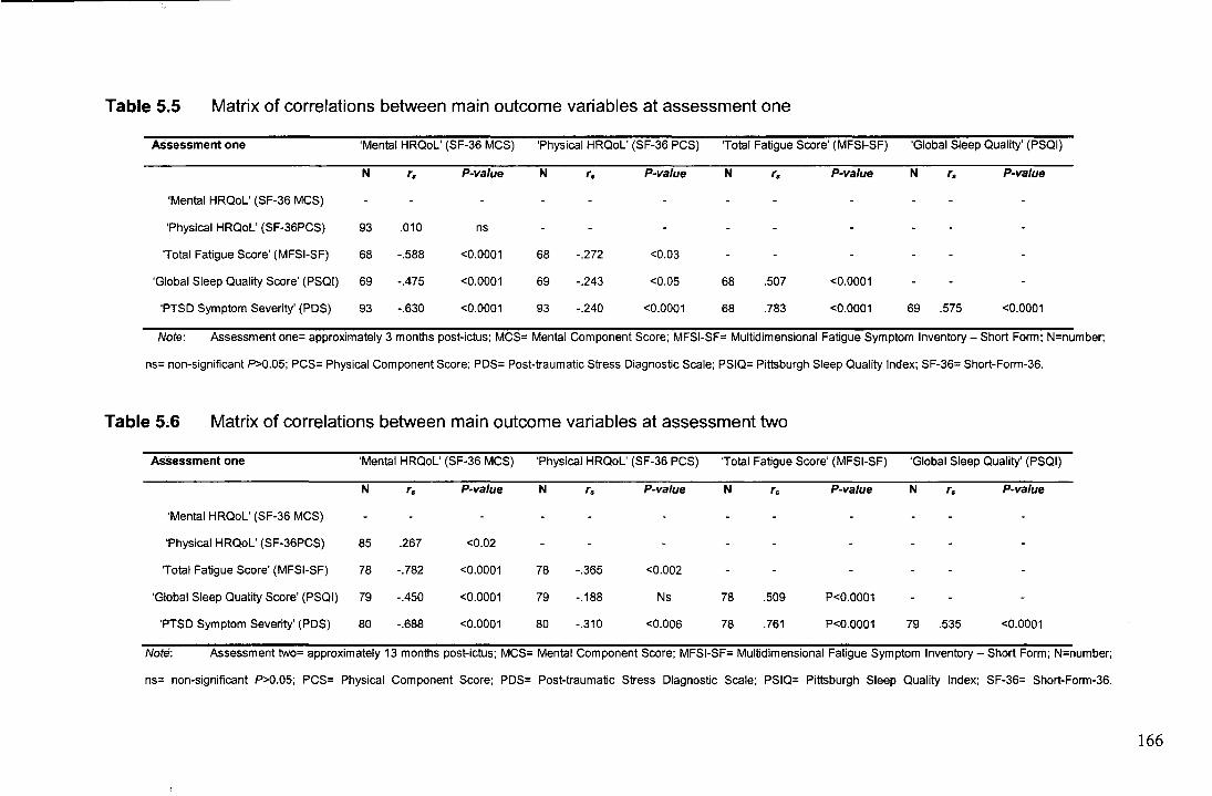
Table 5.5 Matrix of correlations between main outcome variables at assessment one
Assessment one 'Mental HRQoL' (SF-36 MCS) 'Physical HRQoL' (SF-36 PCS) 'Total Fatigue Score' (MFSI-SF) 'Global Sleep Quality' (PSQI)
N '· P-value N '· P-value N '· P-va/ue N '· P-value
'Mental HRQoL' (SF-36 MCS)
'Physical HRQoL' (SF-36PCS) 93 .010 ns
'Total Fatigue Score' (MFSI-SF) 68 -.588 <0.0001 68 -.272 <0.03
'Global Sleep Quality Score' (PSQI) 69 -.475 <0.0001 69 -.243 <0.05 68 .507 <0.0001
'PTSD Symptom Severity' (PDS) 93 -.630 <0.0001 93 -.240 <0.0001 68 .783 <0.0001 69 .575 <0.0001
Note: Assessment one= approximately 3 months post-ictus: MCS= Mental Component Score; MFSI-SF= Multidimensional Fatigue Symptom Inventory- Short Form: N=number:
ns= non-significant P>0.05; PCS= Physical Component Score: PDS= Post-traumatic Stress Diagnostic Scale; PSIQ= Pittsburgh Sleep Quality Index; SF-36= Short-Form-36.
Table 5~6 Matrix of correlations between main outcome variables at assessment two
Assessment one 'Mental HRQoL' (SF-36 MCS) 'Physical HRQoL' (SF-36 PCS) 'Total Fatigue Score' (MFSI-SF) 'Global Sleep Quality' (PSQI)
N '· P-value N '· P-value N '· P-value N '· P-value
'Mental HRQoL' (SF-36 MCS)
'Physical HRQoL' (SF-36PCS) 85 .267 <0.02
'Total Fatigue Score' (MFSI-SF) 78 -.782 <0.0001 78 -.365 <0.002
'Global Sleep Quality Score' (PSQI) 79 -.450 <0.0001 79 -.188 Ns 78 .509 P<0.0001
'PTSD Symptom Severity' (PDS) 80 -.688 <0.0001 80 -.310 <0.006 78 .761 P<0.0001 79 .535 <0.0001
Note: Assessment two= approximately 13 months post-ictus; MCS= Mental Component Score; MFSI-SF= Multidimensional Fatigue Symptom Inventory- Short Form: N=number;
ns= non-significant P>0.05; PCS= Physical Component Score; PDS= Post-traumatic Stress Diagnostic Scale; PSIQ= Pittsburgh Sleep Quality Index: SF-36= Short-Form-36.
166
Predictive models
Table 5.7 provides a survey of those factors which proved significantly
predictive in regression analyses for each outcome measure.
Table 5.7 A survey of those factors which proved to be significant risk factors in
regression analyses
Outcome Risk factor
Mento! HRQoL' Age
(SF-36 MCS) Sex
Clinical severily (WFNS)
His10ty of P<IMOUS iDness
Overall ~we fmction (TEA: RBMT·E: BAOS)
Physical dlsabthly (FIM-MS)
PTSD symplom severily (PDS)
Model Summary
Physical HRQoL' Age
(SF.J6 PCS) Sex
PTSO" (POS) §
Clinical sevorlly (WFNS)
HlsiOry of prov'oous Illness
Ovenll a>gni~ve fmction (TEA: RBMT ·E: BAOS)
Physical dlsabthly (FIM-MS)
PTSD symplom severily (POS)
Model Summary
Sex
Clinical severily (WFNS)
Ps)'(:hialrlc history
Aneurysmal vs. non aneurysmal haomorrhage
SUrgery vs non·surgical inletVention
Endovascular vs. noo-endovascuar lniA!<VenllOn
Education
Maladapllve coping (Brief COPE)
Model Summary
Assessment one As.sassment two
IS/OR s?, P-value IS/OR S?, P-vatu•
ns Ns
ns Ns
ns Ns
ns Ns
ns Ns
ns Ns
·.581 30% <0.0001 · .626 35% <0.0001
R' = 0.44; F,,.•16.70, P<0.0001 R' = 0.48; Fut= 35.98. P<0.0001
ns Ns
ns Ns
ns Ns
-.334 11% <0.0001 Ns
ns Ns
.424 18% <0.0001 .429 18% <0.0001
ns ·.411 15% <0.0001
R'= 0.30; Fz.oo• 18.80, P<0.0001 R' = 0.43; F,,,. = 19.42, P<0.0001
0.92 <0.005 Ns
ns Ns
ns Ns
ns Ns
ns Ns
ns Ns
ns Ns
ns Ns
3.67 <0.0001 3.22 <0.0001
c=.85; x' l3. N• 9SJ=32.n. P<0.0001 c•.78: x'11. N=8tJ =t8 41 . P<o.ooo1
167
Table 5.7 Cont'd.
Outcome
(MFSI·SF}
Risk factor
Age
Sex
Clinical seve<ity (WFNS)
Aneurysmal vs. non aneurysmal haemorrhage
Ove<all cognitive function (TEA; RBMT-E:
BADS)
Psyclliatric his lory
Sleep function (Global PSIQ Score)
PTSD symptom severity (PDS)
Model Summary
Assessment one
41 OR s?J P-value
Ns
Ns
Ns
Ns
Ns
Ns
Ns
.786 49% <0.0001
R'• 0.64; F, .. = 37.75. P<0.0001
Assessment two
IJIOR s?1 P·valuo
Ns
Ns
ns
ns
ns
ns
ns
.796 58% <0.0001
R1• 0.58; F, .... 58.31. P<O.OOOI
Sleep• (PSIQ) Age Ns ns
Sex
Clinical severity (WFNS)
Aneurysmal vs. non aneurysmal haemorrhage
Overall cognilive funclion (TEA: RBMT-E:
BADS)
Psyclllalrlc hlslory
PTSD symplom severity (PDS)
MOdel Summary
.532 27%
Ns
Ns
Ns
Ns
Ns
<0.0001
R'• 0.38: F1.or= 20.71 P<0.0001
.652 42%
ns
ns
ns
ns
ns
<0.0001
R' = 0.45: F,...• 34.98 P<0.0001
Nole: ·= <0.007: ··= <0.005; ···= <0.006; Assessmenl one= approxlmalely 3 monlhs posi·SAH; assessmenllwo= approximalely 13 monlhs
posi-SAH: B=slandardlsed bela coeffiCient MFSI·SF• Mullidimensional Faligue Symptom Inventory - Short Form; OR• Odds.rallo: P= P-value;
PDS= Post-trauma lie Stress [);agnostic Scale; PSIQ• Pittsburgh Sleep Qualily Index: SF·36 MCS= Short-Form·36 Menial oomponenl heallh-relaled
quality of life soore: SF-36 PCS• Shorl Fonm·36 Physical heallh·relaled quallly of life componenl Soore: sr2i= squared semi-partial correlations; §=
Logistic: regression. rather than linear regression. -; PredictOf not significant This table preseniS the explanative Importance and P·vatues of
significant predictors in the regression analyses. Backward elimination methods (likelihood ratio 1ests In the case of logistic regressions) were used to
identify parsimonious predictors. Please refer to the 'Data anafysis' section for further information on the predictors included In eaCh regression model.
Assessment one
Health-Related Quality of Life (HRQoL). Due to missing data of some of the
patients, regression analysis was based on 93 patients. Bonferonni-correction led
to the adoption of a test-wise alpha of P<0.007.
168
'Mental HRQoL': 44% of variance in 'Mental HRQoL' was accounted for
(R2= 0.443; F4,84= 16.698, P<0.0001 ). Only 'PTSD Symptom Severity' scores were
significantly predictive (squared semi-partial correlation [sli]=30%; standardized
B= -.581; P<0.0001 ). Demographic, cognitive and physical disability information
demonstrated no predictive value for 'Mental HRQoL' (Figure 5.2).
'Physical HRQoL': 30% of variance in 'Physical HRQoL' was accounted for
(R2= 0.304; F2,86= 18.801, P<0.0001 ). Physical disability contributed most (FIM
MS; sli= 18%; standardized B = .424; P<0.0001 ), followed by history of significant
illness (sli = 11 %; standardized B = -.334; P<0.0001 ).
Post-traumatic Stress Disorder (PTSD). Due to missing data, this
regression analysis was based on 95 patients. Bonferonni-correction led to the
adoption of a test-wise alpha of P<0.005.
A significantly effective logistic model to predict PTSD was generated (X2 [3,
N= 95] = 32.767, P< 0.0001 ). Predominant use of 'Maladaptive Coping Strategies'
(OR= 3.670, Cl 1.967 - 6.845; Wald= 16.707 (1) P<0.0001) and younger age
(OR= 0.917, Cl 0.867- 0.971; Wald= 8.726 (1 ), P<0.005) significantly increased
the probability of PTSD. Using a probability criterion of .5, predictive classification
was excellent (c= 0.85), with the model assigning 85% of all possible pairs the
correct probability of having PTSD (sensitivity= .71; specificity= .87; Figure 5.3).
Table 5.8 presents a comparison of the symptoms and coping skills of those
patients with and without PTSD.
Fatigue. Bonferonni-correction led to the adoption of a test-wise alpha of
P<0.006.
A significantly predictive model for the 73 who completed the MFSI-SF at
assessment one was generated -accounting for 64% of variance in 'Total Fatigue'
scores (r= 0.797; R2= 0.635; F3,65= 37.750, P<0.0001 ). The model was based
solely
169
80
70
~ 60 u.. II) - 50 ...J 0
~ 40 J: 'i 30 -c Ql ::!: 20
10
0 1 2 3 4 5 6 7
Physical Disability (FIM-MS)
80
70
10 ' M 60 ~
~ I
' u.. ' II) • ' -50 • ' ...J • 0 CJ 40 0:: J: 'i 30 ..... c ~ Q) . ::!: 20 • • ' •
• 10
0 1 2 3 4 5 6 7 8
Overal Cognitive Function
80~--------------------------.
70
~ 60 u.. ~ 50 ...J 0
~ 40 J:
~ 30 c Q)
::!: 20
10
i ... • I ·=· . . . . .. . . ..
• . .. . . . . . .. .. : .. .. .. ..
..
. .
0 10 20 30 40 50
PTSD Symptom Severity (PDS)
Figure 5.2 Graphs of relationship between 'Mental HRQoL' and 'PTSD
Symptom Severity' (upper), 'Cognition' (middle) and 'Physical
Disability' (lower) at assessment one.
170
c ~ ll.
'0 ~ :0
"' .0 0 ... ll.
0.0 .....
0 2 3 4 5 6 7
Use of Maladaptive Coping Strategies
--Assessment one .... n ... Assessment two
8
1. 0-1--'---+--'---+-......__-+-......__-+-_..__-+-_..__-+-_._-+-_.._+ l · ~ =~= 1 r - =; - l
o. 9 .;; ... ~: ·· :· t· .. ··"·:·· .. j· · · ::: ~ :,~· .. ·.:· i ~:.·""::;:,~i ·[ E;::: ..... - ::t~ -;:~~~;: · · · ··)·.~·· : · : ····-· !··:······ · ... 0.8 .............. : ..... , ....... j ..... : ........ i .............. f . :~ ....... , .. h ........... i ............ + ........... .
c .. , '··· ! t· ; ! .. i ~• ! • ! . ~ o. 7 ............. t ....... .... 1 .............. 1 ..... ~~·--t ............. ~ ............. ] .............. r ............. . ll. o.6 ..... ' .. , ..... { ............. i-............ j.: .. : ......... f ............. F ...... : ... j .............. j ............ . 0 ' .. ' : ' ' : : .~- 0.5 ............. + .... ......... { ............. j ....... = ..... f·····""""f'·:·: ......... j .............. f"""'"'"'
:::. i ~ ~ ~ ~ ~ ;: =~ " • l ~"-;:I::,~. ;, i'' ,,. "·; ~;:• l: -~i ·" :0 0.4 ....... : ..... + ............. ; .............. , ............ -f-............ l""""'"'"l""'""""+''"'"''"' ~ : :· : ! : ! : 0 0.3 ............. + ............. { .............. ; ....... '""f""""'""i""'"'"'"j ........ , _.j ........... .. a: : -!- . ! ! ! ! !
0.2 ............. + ............. + ............. j .......... :,:":" .......... + ............. ! .............. f ............ .. : f ! : : ! ;
0·1 ....... : .... + ........... t .. : .......... ! ............. + ............ T .......... + ........... + ........... . i f .. l f 1 t
0. 0-1--.-+--.-+---r-+---r--l---r---l-.....--+-.....-.+:~~ 10 20 30 40 50 60 70 80 90
Age at Time of SAH (years)
--Assessment one
Figure 5.3 Graph depicting the relationship between probability of PTSD at
assessments one and two with use of maladaptive coping strategies
(upper; assessment one OR = 3.670; age held at mean; assessment
two OR = 3.216) and relationship between PTSD and age at
assessment one (lower; OR = 0.917; with use of maladaptive coping
strategies held at mean level).
171
Table 5.8 Comparison of symptoms and coping skills of patients with and without post-traumatic stress disorder at assessments
Factor Assessment one Assessment one P-value ES Assessment two Assessment two P-value ES
With PTSD Without PTSD With PTSD Without PTSD
N Median IQR N Median lOR N Median lOR N Median lOR
PTSD- PDS
Intrusions 36 4.0 2.0- 5.0 61 1.0 0.0-2.0 <0.0001 .62 33 3.0 2.0-5.0 56 1.0 0.0-2.0 <0.0001 .66
Avoidance 36 5.0 4.0-6.0 61 2.0 1.0- 3.0 <0.0001 .67 33 4.0 4.0-7.0 56 1.0 0.3-2.8 <0.0001 .74
Arousal 36 4.0 3.0- 5.0 61 2.0 1.0-3.0 <0.0001 .58 33 4.0 3.0-5.0 56 2.0 1.0- 3.0 <0.0001 .60
Symptom Severity 36 22.0 14.0- 31.0 61 5.0 3.0- 11.0 <0.0001 .69 33 18.0 12.0- 30.0 56 5.0 3.0- 8.0 <0.0001 .73
Coping - Brief COPE §
Maladaptive coping 35 4.0 3.2-4.7 59 2.8 2.3- 3.3 <0.0001 .45 29 3.7 3.0-4.6 53 2.5 2.3-3.4 <0.0001 .47
Self Distraction 35 5.0 3.0- 7.0 59 4.0 3.0- 5.0 <0.005 .32 29 5.0 3.5-7.0 53 4.0 2.0-5.0 <0.01 .29
Denial 35 4.0 2.0-6.0 59 2.0 2.0- 3.0 <0.0001 .45 29 3.0 2.0-5.0 53 2.0 2.0-3.0 <0.0001 .39
Substance Use 35 2.0 2.0- 3.0 59 2.0 2.0- 2.0 Ns .10 29 2.0 2.0-4.0 53 2.0 2.0-2.0 ns .14
Behavioural Disengagement 35 3.0 2.0- 5.0 59 2.0 2.0- 3.0 <0.005 .30 29 2.0 2.0-4.0 53 2.0 2.0-3.0 ns .18
Venting 35 3.0 2.0- 5.0 59 3.0 2.0-4.0 Ns .16 29 3.0 3.0-4.5 53 2.0 2.0- 3.0 <0.005 .33
Self-Blame 35 4.0 3.0- 6.0 59 2.0 2.0-4.0 <0.002 .35 29 4.0 3.0- 5.5 53 2.0 2.0-4.5 <0.003 .34
Notes: ; lOR- interquartile range; N- number;; ns =non significant p>0.05; PDS- Posttraumatic Stress Diagnostic Scale; PDS scoring is presented as the median number of intrusive (e.g., "reliving the brain
haemorrhage, acting or feeling as if it was happening again"), avoidant (e.g., "trying to avoid activities, people or places that remind me of the brain haemorrhage") and arousal (e.g., "feeling irritable or having fits of
anger") symptoms experienced; § Brief COPE results taken at assessment one (3-months post-SAH) used for prediction of PTSD at both assessment points. Brief COPE scoring ranges from 1 ("strategy usually not
used") to 4 ("strategy usually used a lot"). Values are expressed as medians.
172
on 'PTSD Symptom Severity' scores (si;= 49%; standardised B= .786; P<0.0001 ).
Patients with PTSD reported significantly more 'Total Fatigue' than those without
(Mann Whitney, P<0.0001; r-.44).
Sleep. Bonferonni-correction led to the adoption of a test-wise alpha of
P<0.007.
A significantly predictive model for the 73 who completed the PSIQ
accounted for 38% of variance in 'Global PSIQ Sleep' scores (r= 0.618; R2= 0.382;
F2.61= 20.708 P<0.0001). The model was based solely on 'PTSD Symptom
Severity' scores (si;= 27%; standardised B= .532; P<0.0001 ). Significantly worse
sleep ('Global PSIQ Sleep' score) was reported by patients with PTSD (Mann
Whitney, P<0.0001; r-.28). Sleep quality was significantly associated with
concurrent fatigue (rs= .49, P<0.0001 ).
Assessment two
Health-Related Quality of Life (HRQoL). Due to missing data, regression
analysis was based on 89 patients. Bonferonni-correction led to the adoption of a
test-wise alpha of P<0.007.
'Mental HRQoL': 48% of variance in 'Mental HRQoL' was accounted for (R2
= 0.477; F2.19 = 35.979, P<0.0001 ). Only the 'PTSD Symptom Severity' score was
significantly predictive (si; = 35%; standardised B = -.626; P<0.0001 ).
Demographic, cognitive and physical disability information demonstrated no
predictive value (Figure 5.4 ).
'Physical HRQoL': 43% of variance in 'Physical HRQoL' was accounted for
(R2 = 0.428; F3.1a = 19.415, P<0.0001 ). Physical disability scores (FIM-MS; si; =
18%; standardized B = .429; P<0.0001) contributed most, followed closely by
'PTSD Symptoms Severity' scores (si; = 15%; standardised B = -.411; P<0.0001 ).
173
60,-----------------------------.
70
i oo u. (/) -50 ..J 0 8 0
~ 40 L-----------c-___o.-n ::z::
~30 ~ 20 0 0
0
10
012 3 4 56 7
Physical Disability (FIM-MS)
80,.-------------------------------,
70
«> M 6Q
Li.. (/) -so ..J 0 0 40 0:: ::z:: IU 30 .... c: Cll :E 20
10
v v
v
v ~ v v v v
' v v v v v ljl v
Q
v
0 2 4 6 8
Overal Cognitive Function (SF-36)
80,-------------------------------,
70
C:O oo M
u. ~ 50 ..J 0
~ 40 ::z:: ~ 30 c: Cll
:iE 20
10
0 0 0
0 10 20 30 40 50
PTSD Symptom Severity (PDS)
Figure 5.4 Graphs of relationship between 'Mental HRQoL' and 'PTSD
Symptom Severity' (upper), 'Cognition' (middle) and 'Physical Disability' (lower) at
assessment two.
174
Prediction of patients' long-term HRQoL by their experience of PTSD in the
acute stages: Due to missing data, analysis was based on 83 patients.
Bonferonni-correction led to the adoption of a test-wise alpha of P<0.007.
'Mental HRQoL': A patient's experience of PTSD in the short-term ('PTSD
Symptom Severity' assessment one) held a large and negative relationship with
the 'Mental HRQoL' which they experienced in the later stages (assessment two):
rs= -.597, P<0.0001.
Regression analysis using stepwise block entry revealed that the degree to
which a patient experienced PTSD in the short-term had a unique predictive effect
for their longer-term 'Mental HRQoL' (Table 5.9). An initial predictive model based
on information about the severity of a patient's illness, their demographics and the
degree to which they experienced neurological sequelae in the short-term could
explain around 23% of variance in patients' long-term 'Mental HRQoL': R2 = 0.226,
F6,76= 3.703, P<0.004). The addition of information about the patient's experience
of PTSD in the acute stages - namely 'PTSD Symptom Severity' score from
assessment one - significantly improved the model's overall predictive ability:
F1,7s= 20.101, P<0.0001. With its inclusion, a total 39% of variance in 'Mental
HRQoL' could be accounted for (R2 = 0.390, F7.75= 6.843, P<0.0001 ). Increasing
PTSD severity was associated with a worse 'Mental HRQoL': 13= -.484; t= -4.483,
P<0.0001.
'Physical HRQoL': A patient's experience of PTSD in the short-term ('PTSD
Symptom Severity' assessment one) held only a small and negative relationship
with the 'Physical HRQoL' which they experienced in the later stages (assessment
two): r5= -.248, P<0.03.
Stepwise regression analysis revealed that once the variance accounted for
by traditional predictors was controlled for, patients' PTSD symptomatology in the
short-term holds no unique significant predictive effect for long-term 'Physical
175

HRQoL': F1.15= 3.595, P>0.05; Table 5.9. The initial predictive model based on
traditional predictive information was however, significantly predictive - explaining
approximately 30% of variance in patients' long-term ' Physical HRQoL': R2 =
0.284, F6,76= 5.031 I P<0.0001.
Prediction of improvement in patients' HRQoL between assessment one
and two by information about the patients' experience of PTSD in the acute
stages: Due to missing data, analysis was .based on 83 patients. Bonferonni
correction led to the adoption of a test-wise alpha of P<O.OO?.
'Mental HRQoL': A patient's experience of PTSD in the short-term ('PTSD
Symptom Severity' assessment one) did not hold a significant bivariate
relationship with the change in their 'Mental HRQoL' that occurred between
assessment one and two: rs= .148, N=85 P>0.05.
The subsequent stepwise regression analysis revealed that it was not
possible to significantly predict the change in patients' 'Mental HRQoL' that
occurred between assessments one and two using either the initial model based
on traditional predictors (R2 = 0.133, F6,76= 1.939, P>0.05), or with the addition of
information about the patients' experience of PTSD at assessment one (R2 =
0.137, F1,16= 0.368, P>0.05; Table 5.9).
'Physical HRQoL': A patient's experience of PTSD in the short-term ('PTSD
Symptom Severity' assessment one) did not hold a significant relationship with the
change in their 'Physical HRQoL' that occurred between assessments one and two:
rs= -.032, N=85 P>0.05.
176
Table 5.9 The unique predictive ability of PTSD at assessment one (short-
term) for patients' HRQoL at assessment two (long-term)
Factor Model1 Model2
N n s?1 P-value N ll s?1 P-value
Predicting improvement in HRQol
'Mental HRQol' (SF-36 MCS)
Age 83 -.152 .022 ns 83 ·.128 .014 Ns
Sex 83 .188 .032 ns 83 .164 .021 Ns
Clinical severity (WFNS) 83 .175 .025 ns 83 .175 .030 Ns
History of previous illness 83 .039 .001 ns 83 .022 .441 E-03 Ns
Overall cognitive function (TEA; RBMT-E; BADS) 83 -.038 .001 ns 83 -.034 .961E-03 Ns
Physical disability (FIM-MS) 83 .273 .062 <0.03 83 .291 .070 <0.02
PTSD Symptom Severity 83 078 .004 Ns
Model summary R2= 0.13; Fs.7s= 1.94, P>0.05 R2= 0.14; F1.1s= 0.37, P>0.05
'Physical HRQol' (SF-36 PCS)
Age 83 -.156 .023 ns 83 -.196 .032 Ns
Sex 83 .005 .256E-03 ns 83 .046 .002 Ns
Clinical severity (WFNS) 83 .018 .289E-03 ns 83 .017 .256E·03 Ns
History of previous illness 83 .120 .013 ns 83 .148 .020 Ns
Overall cognitive function (TEA; RBMT-E; BADS) 83 .110 .010 ns 83 .104 .010 Ns
Physical disability (FIM-MS) 83 .032 .001 ns 83 .003 .100E-04 Ns
PTSD symptom severity (PDS) 83 -.129 .012 Ns
Model summary R2= 0.05; Fs.1s= 0.61, P>0.05 R2= 0.06; F1.1s= 0.93, P>0.05
Predicting long-term HRQol
'Mental HRQol' (SF-36 MCS)
Age 83 .247 0.060 <0.02 83 .099 0.008 Ns
Sex 83 -.036 0.001 ns 83 .133 0.010 ns
Clinical severity (WFNS) 83 .190 0.030 ns 83 .186 0.030 ns
History of previous illness 83 -.274 0.070 <0.02 83 -.169 0.024 ns
Overall cognitive function (TEA; RBMT-E; BADS) 83 .166 0.022 ns 83 .144 0.016 ns
Physical disability (FIM-MS) 83 .185 0.030 ns 83 .075 0.005 ns
PTSD Symptom Severity 83 -.484 0.163 <0.0001
Model Summary R2= 0.23; Fs.1s= 3.70, P<0.004 R2= 0.39; F1.7s= 20.10, P<0.0001
177
Table 5.9 Cont'd.
Factor Model1 Model2
N 8 s?, P-value N 8 s?, P-value
Predicting long-term HRQoL Cont'd.
'Physical HRQol' (SF-36 PCS)
Age 83 -.115 0.013 ns 83 -.180 0.030 83
Sex 83 -.009 0.640E4! ns 83 0.058 0.003 83
Clinical severity (WFNS) 83 .046 0.002 ns 83 .044 0.002 83
History of previous illness 83 -.247 0.060 <0.02 83 -.201 0.034 83
Overall cognitive function (TEA; RBMT-E; BADS) 83 .010 0.810E4! ns 83 .000 0.000 83
Physical disability (FIM-MS) 83 .435 0.160 <0.0001 83 .386 0.120 83
PTSD Symptom Severity 83 -.216 0.033 83
Model Summary R2= 0.28; Fs.1s= 5.03, P<0.0001 R2= 0.32; F1.1s= 3.60, P>0.05
Note: Assessment one= approximately 3-months post-ictus; Assessment two= approximately 13-months post-ictus; !!.= standardise
beta coefficient; BADS=Behavioural Assessment of the Dysexecutive Syndrome; FIM-MS=Functional Independence Measure-Molal
HADS-A=Hospital Anxiety and Depression Scale - Anxiety score; HADS-D=Hospital Anxiety and Depression Scale - Depression scorE
HRQol= Health-related quality of life; MCS= Mental Component Score; MFSI-SF=Multidimensional Fatigue Symptom lnventory-Sho1
Form; N=sample size; ns= non-significant (P>0.05); PCS=Physical Component Score; PDS=Posttraumatic Stress Diagnostic Scale
PSQI=Pittsburgh Sleep Quality; f?!=variance explained by model; RBMT-E=Rivermead Behavioural Memory Test-Extended; SF
36=Short Form-36; sr,=squared semi-partial correlation; TEA=Test of Everyday Attention. Hierarchical step-wise entry (2 blocks) wa
used to examine the unique predictive ability of patients' experience of PTSD in the acute stages for their later HRQoL. In Model
traditional predictors of SAH patients' outcome from assessment one were entered into the model and the variance in assessment tw
HRQoL which these predictors accounted for was controlled (constant+ Patient age+ Sex, Clinical severity+ History of previous illnes
+ Overall cognitive function + Physical disability). In Model 2 patients' experience of PTSD at assessment one (PTSD Symptor
Severity) was entered into the predictive model and its additive explanative importance established.
Stepwise regression analysis revealed that it was not possible to
significantly predict the change in patients' 'Physical HRQoL' that occurred
between assessments one and two using the initial model based on traditional
predictors (R2 = 0.046, F6,76= 0.611, P>0.05), nor with the addition of information
about patients' experience of PTSD at assessment one (R2 = 0.058, F7,76= 0~929,
P>0.05; Table 5.9).
178
Post-traumatic Stress Disorder (PTSD). Due to missing data this regression
analyses was based on 81. Bonferonni-correction led to the adoption of a test-wise
alpha of P<0.005.
A significantly effective logistic model to predict PTSD was generated (X2 [1,
N= 81] = 18.409, P< 0.0001). The only variable included in the final model was
information about the patients' coping skills. A disposition to use 'Maladaptive
Coping Strategies' (OR= 3.216, Cl 1.761 - 5.873; Wald= 14.452 (1), P<0.0001)
significantly increased the probability of having PTSD. Using a probability criterion
of .5, predictive classification was excellent (c= 0.78), with the model assigning
78% of all possible pairs the correct probability of having PTSD (sensitivity= .48;
specificity= .79; Figure 5.3). Table 5.8 presents a comparison of the symptoms
and coping skills of those patients with and without PTSD.
Fatigue. Bonferonni-correction led to the adoption of a test-wise alpha of
P<0.006.
A significantly predictive model for the 87 patients who completed the
MFSI-SF at assessment two was generated - accounting for 57% of 'Total
Fatigue' variance (r= . 762; R2= 0.581; F2.84= 58.310, P<0.0001 ). The model was
based solely on 'PTSD Symptom Severity' (s?i= 58%; standardised B= .796;
P<0.0001 ). Patients with PTSD reported significantly more Total Fatigue than
those without (Mann Whitney, P<0.0001; r-.56).
Sleep. Bonferonni-correction led to the adoption of a test-wise alpha of
P<0.007.
A significantly predictive model for the 88 who completed the PSIQ at
assessment two accounted for 45% of variance in 'Global PSIQ Sleep' scores (r=
0.672; R2= 0.451; F2.as= 34.977 P<0.0001 ). The model was based solely on 'PTSD
Symptom Severity' scores (s?i= 42%; standardised B= .652; P<0.0001 ).
Significantly worse sleep ('Global PSIQ Sleep' score) was reported by patients
179
with PTSD (Mann Whitney, P<0.0001; r-.38). Sleep quality was significantly
associated concurrent fatigue (rs= .51, P<0.0001 ).
5.4 Discussion
Post-traumatic stress disorder (PTSD) is a severe psychiatric condition that can be
elicited by the experience of traumatic, life-threatening events. In line with earlier
reports, this study has confirmed that the experience of having and surviving a
subarachnoid haemorrhage (SAH) can elicit PTSD [23, 47, 295]. Moreover, PTSD
was found to be abnormally prevalent in the SAH population. Crucially, PTSD was
found to be the single best predictor of patients' Mental health-related quality of life
(HRQoL) - the domain most persistently impaired - in both the short- and long
term. In accord with the results of my meta-analysis presented in Chapter 3, none
of the other variables in the model (including clinical grade, physical and cognitive
disability) made significant contributions to the patients' Mental HRQoL. PTSD
also proved to be significantly predictive of Physical HRQoL at the second follow
up assessment. Importantly, I also found that the degree to which patients
experienced PTSD in the short-term held a large and unique predictive effect for
patients' long-term HRQoL. Specifically, it was those patients who experienced
more severe PTSD symptoms in the early stages post-ictus who experienced
worse Mental HRQoL in the long-term. Together the findings from my study
suggest that it is the frequent occurrence of PTSD (37%) after SAH which is partly
to blame for the surprisingly low HRQoL found in many SAH patients who, in other
respects, make a good recovery. Although it has not traditionally been considered
as a high priority within SAH care, my findings highlight the importance of
recognizing and addressing the psychological adjustment of patients to their
experience of SAH. In particular, given that the experience of PTSD in the short
term is uniquely predictive of a poor long-term Mental HRQoL, the evidence
180
indicates that patients' psychiatric dysfunction should be treated aggressively as
early as possible.
The proposal that PTSD can affect a patient's Mental HRQoL seems
intuitive given that this domain is concerned with the degree to which the person
feels their health has impinged upon their mood and vitality and the degree to
which their emotional well-being has limited their social interaction and ability to
work [23, 514]. The means by which PTSD can affect the physical aspects of an
SAH patient's HRQoL may however, seem counterintuitive, given that Physical
HRQoL concerns patients' physical functioning (e.g., "my health severely limits me
from lifting or carrying"), physical role limitations (e.g., "my physical health has
caused me to cut down on the amount of time spent on work"), bodily pain and
general health (e.g., "I expect my health to get worse"), whereas PTSD
presumably causes primarily non-physical impairment. Interestingly though, the
association between PTSD and Physical HRQoL which I found is not unique to the
present study. Indeed, several reviews in the wider literature have now concluded
that in contrast with other psychiatric disorders, PTSD can impact on Physical
HRQoL. In fact, its effect on Physical HRQoL can at times, be almost as severe as
its effect on Mental HRQoL [514, 614]. The means by which PTSD could reduce
Physical HRQoL include the fact that those who develop PTSD are known to
experience increased physical symptoms, including elevated pain, headaches,
joint and muscle aches, bowel symptoms and dizziness, which together with core
PTSD symptoms, such as avoidance and anxiety, would simply make it harder for
the patient to complete physical tasks, or to simply do things as one commentator
notes[69, 133,175,229,289,368,406,415,437,438,448,530,622,641, 761].
PTSD was highly prevalent in my sample at both assessment points post
haemorrhage and most patients who warranted a diagnosis of PTSD at
approximately 3 months, still warranted the diagnosis at 13 months. Obviously, my
181
conclusions on the incidence of PTSD are limited to 13 months post-SAH, but they
are in line with evidence that PTSD is often a persistent and chronic disorder [417,
529]. Evidence from two recent studies does however, suggest that mood
disturbance is a persistent problem post-SAH [295, 746]. Hutter, for example,
assessed patients four years post-SAH and found PTSD in a similar proportion of
patients [295]. It is likely that the largely stable nature of SAH patients' PTSD
explains why in regression analyses, I found that PTSD could not account for the
improved HRQoL which patients reported at the second assessment. It is
interesting to note that I also found that more traditional predictors, such as
patients' cognitive function and physical disability, also could not account for the
improvement in patients' HRQoL between assessments. This is not surprising
given that these factors, as revealed by my comparison of patients' performances
at the two assessments, also had not significantly improved at the second
assessment. The improvement in patients' HRQoL scores is instead, likely due to
improvement between assessments in other aspects of the patients' outcome
which I did not measure/ incorportate into the predictive models (for example, the
de-conditioning associated with their convalescence) and/ or the possible
insensitivity to change of my measures.
It is interesting to note that in the present study the incidence of PTSD
detected in the SAH patient population is remarkably high. At both the short- and
long-term follow-up appointments, 37% of SAH patients were found to warrant a
diagnosis of fuii-PTSD. It is usually assumed that that the prevalence of PTSD
(and psycho-pathology in general) following such internal traumas is lower relative
to that which follows traditional traumas [476, 701]. For example, whilst
approximately 35-50% of patients are expected to develop PTSD following
traditional traumas such rape or military combat [71, 125, 351 ], 0-16% develop
PTSDfollowing myocardial infarction [41, 42,152,380,500,616, 706], 10-18%
182
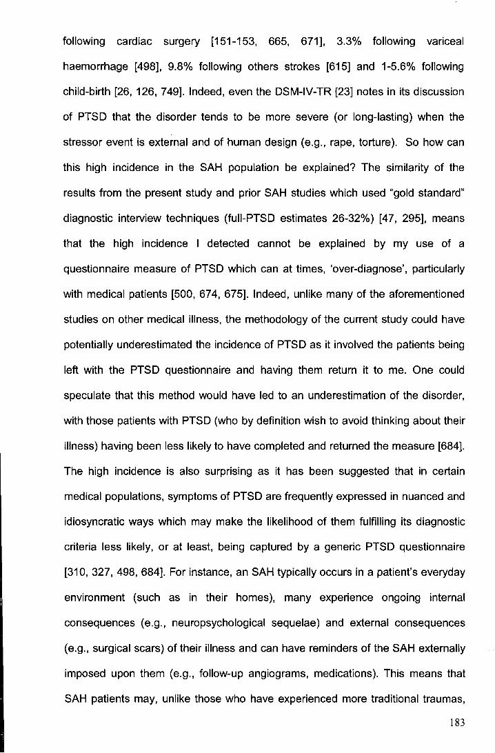
following cardiac surgery [151-153, 665, 671], 3.3% following variceal
haemorrhage [498], 9.8% following others strokes [615] and 1-5.6% following
child-birth [26, 126, 749]. Indeed, even the DSM-IV-TR [23] notes in its discussion
of PTSD that the disorder tends to be more severe (or long-lasting) when the
stressor event is external and of human design (e.g., rape, torture). So how can
this high incidence in the SAH population be explained? The similarity of the
results from the present study and prior SAH studies which used "gold standard"
diagnostic interview techniques (fuii-PTSD estimates 26-32%) [47, 295], means
that the high incidence I detected cannot be explained by my use of a
questionnaire measure of PTSD which can at times, 'over-diagnose', particularly
with medical patients [500, 674, 675]. Indeed, unlike many of the aforementioned
studies on other medical illness, the methodology of the current study could have
potentially underestimated the incidence of PTSD as it involved the patients being
left with the PTSD questionnaire and having them return it to me. One could
speculate that this method would have led to an underestimation of the disorder,
with those patients with PTSD (who by definition wish to avoid thinking about their
illness) having been less likely to have completed and returned the measure [684].
The high incidence is also surprising as it has been suggested that in certain
medical populations, symptoms of PTSD are frequently expressed in nuanced and
idiosyncratic ways which may make the likelihood of them fulfilling its diagnostic
criteria less likely, or at least, being captured by a generic PTSD questionnaire
[310, 327, 498, 684]. For instance, an SAH typically occurs in a patient's everyday
environment (such as in their homes), many experience ongoing internal
consequences (e.g., neuropsychological sequelae) and external consequences
(e.g., surgical scars) of their illness and can have reminders of the SAH externally
imposed upon them (e.g., follow-up angiograms, medications). This means that
SAH patients may, unlike those who have experienced more traditional traumas,
183

find it more difficult to actually avoid trauma reminders and so not fulfill the
'avoidance' criteria for the diagnosis of PTSD. SAH patients do however, still often
fulfill this criteria and this could be explained by the fact that in reality the questions
which ask patients about their avoidance behaviours only ask whether they have
made efforts to avoid reminders and not about the success of their attempts [23].
Given that methodological features do not seem able to explain the high incidence
of PTSD in the SAH population, it could be that there is something uniquely
traumatising about the experience of an SAH. This is something deserving of
future study, a point which I discuss further in Chapter 7 (see section 7.4.1 ).
Studies in the past had reported a comparatively good physical and
cognitive recovery after SAH [432, 507, 558, 590, 649]. It was suspected that the
relative infrequency of severe problems post-SAH might in part, be a consequence
of those studies having used insensitive tests and pre-selected samples. I avoided
these problems in the present study by investigating a large unselected sample
and using ecologically valid instruments. Nevertheless my findings confirm that on
average, patients surviving SAH show a good physical and cognitive recovery.
This underlines the importance of shifting focus from classical
neurological/neuropsychological problems to the less examined field of
neuropsychiatric sequelae in order to understand the poor HRQoL experienced by
patients. Furthermore, where considered, no significant value was found to be
associated with clinical information for HRQoL [306, 469, 510, 543, 546, 577, 590,
609,649, 722].
That I did not detect any significant improvement in cognition between
assessment one and assessment two in the present study is not entirely
unexpected. Whilst some prospective studies have reported improvements [507, "
590, 649], others, like mine, have not [432, 558]. When improvements in cognition
have been found, it appears that the effect is only modest and tends to be
184
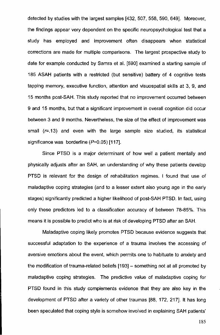
detected by studies with the largest samples [432, 507, 558, 590, 649]. Moreover,
the findings appear very dependent on the specific neuropsychological test that a
study has employed and improvement often disappears when statistical
corrections are made for multiple comparisons. The largest prospective study to
date for example conducted by Samra et al. [590] examined a starting sample of
185 ASAH patients with a restricted (but sensitive) battery of 4 cognitive tests
tapping memory, executive function, attention and visuospatial skills at 3, 9, and
15 months post-SAH. This study reported that no improvement occurred between
9 and 15 months, but that a significant improvement in overall cognition did occur
between 3 and 9 months. Nevertheless, the size of the effect of improvement was
small (r-.13) and even with the large sample size studied, its statistical
significance was borderline (P=0.05) [117].
Since PTSD is a major determinant of how well a patient mentally and
physically adjusts after an SAH, an understanding of why these patients develop
PTSD is relevant for the design of rehabilitation regimes. I found that use of
maladaptive coping strategies (and to a lesser extent also young age in the early
stages) significantly predicted a higher likelihood of post-SAH PTSD. In fact, using
only these predictors led to a classification accuracy of between 78-85%. This
means it is possible to predict who is at risk of developing PTSD after an SAH.
Maladaptive coping likely promotes PTSD because evidence suggests that
successful adaptation to the experience of a trauma involves the accessing of
aversive emotions about the event, which permits one to habituate to anxiety and
the modification of trauma-related beliefs [193] - something not at all promoted by
maladaptive coping strategies. The predictive value of maladaptive coping for
PTSD found in this study complements evidence that they are also key in the
development of PTSD after a variety of other traumas [88, 172, 217]. It has long
been speculated that coping style is somehow involved in explaining SAH patients'
185
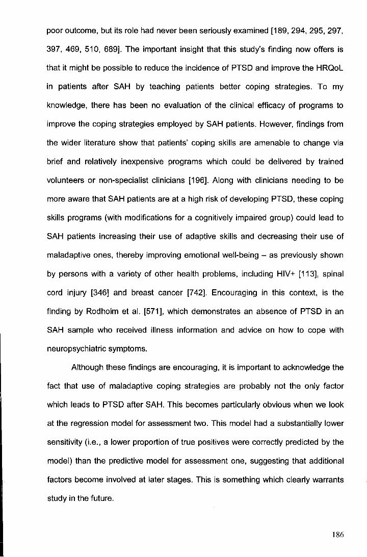
poor outcome, but its role had never been seriously examined [189, 294, 295, 297,
397, 469, 510, 689]. The important insight that this study's finding now offers is
that it might be possible to reduce the incidence of PTSD and improve the HRQoL
in patients after SAH by teaching patients better coping strategies. To my
knowledge, there has been no evaluation of the clinical efficacy of programs to
improve the coping strategies employed by SAH patients. However, findings from
the wider literature show that patients' coping skills are amenable to change via
brief and relatively inexpensive programs which could be delivered by trained
volunteers or non-specialist clinicians [196]. Along with clinicians needing to be
more aware that SAH patients are at a high risk of developing PTSD, these coping
skills programs (with modifications for a cognitively impaired group) could lead to
SAH patients increasing their use of adaptive skills and decreasing their use of
maladaptive ones, thereby improving emotional well-being - as previously shown
by persons with a variety of other health problems, including HIV+ [113], spinal
cord injury [346] and breast cancer [7 42]. Encouraging in this context, is the
finding by Rodholm et al. [571], which demonstrates an absence of PTSD in an
SAH sample who received illness information and advice on how to cope with
neuropsychiatric symptoms.
Although these findings are encouraging, it is important to acknowledge the
fact that use of maladaptive coping strategies are probably not the only factor
which leads to PTSD after SAH. This becomes particularly obvious when we look
at the regression model for assessment two. This model had a substantially lower
sensitivity (i.e., a lower proportion of true positives were correctly predicted by the
model) than the predictive model for assessment one, suggesting that additional
factors become involved at later stages. This is something which clearly warrants
study in the future.
186
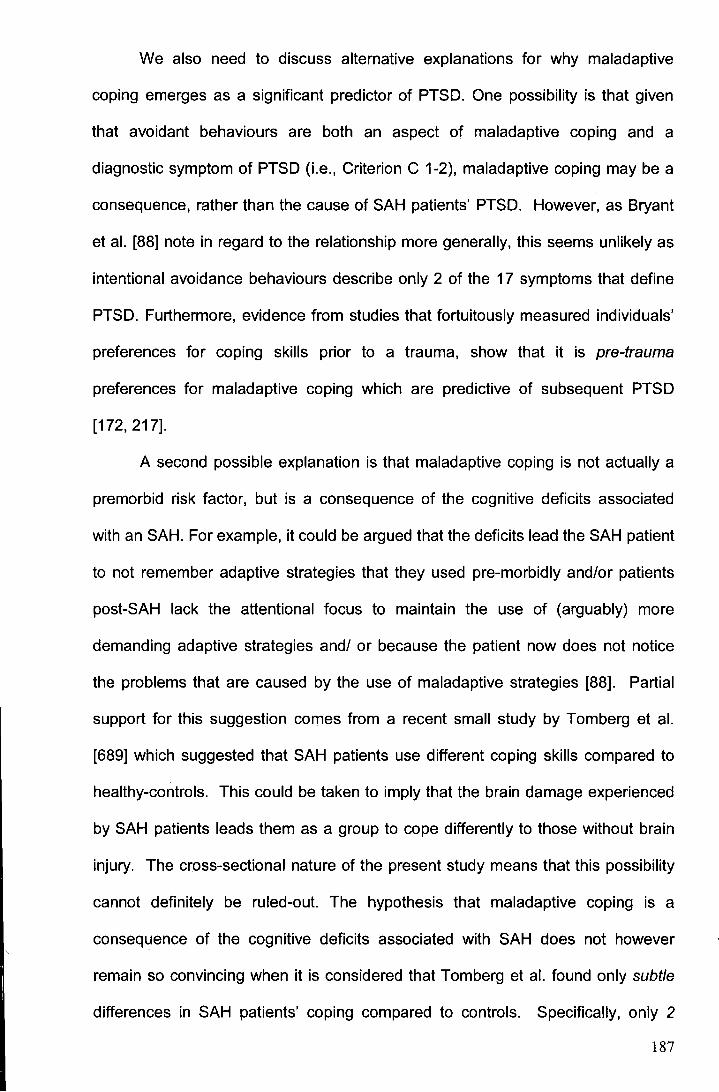
We also need to discuss alternative explanations for why maladaptive
coping emerges as a significant predictor of PTSD. One possibility is that given
that avoidant behaviours are both an aspect of maladaptive coping and a
diagnostic symptom of PTSD (i.e., Criterion C 1-2), maladaptive coping may be a
consequence, rather than the cause of SAH patients' PTSD. However, as Bryant
et al. [88) note in regard to the relationship more generally, this seems unlikely as
intentional avoidance behaviours describe only 2 of the 17 symptoms that define
PTSD. Furthermore, evidence from studies that fortuitously measured individuals'
preferences for coping skills prior to a trauma, show that it is pre-trauma
preferences for maladaptive coping which are predictive of subsequent PTSD
[172, 217].
A second possible explanation is that maladaptive coping is not actually a
premorbid risk factor, but is a consequence of the cognitive deficits associated
with an SAH. For example, it could be argued that the deficits lead the SAH patient
to not remember adaptive strategies that they used pre-morbidly and/or patients
post-SAH lack the attentional focus to maintain the use of (arguably) more
demanding adaptive strategies and/ or because the patient now does not notice
the problems that are caused by the use of maladaptive strategies [88]. Partial
support for this suggestion comes from a recent small study by Tomberg et al.
[689] which suggested that SAH patients use different coping skills compared to
healthy-controls. This could be taken to imply that the brain damage experienced
by SAH patients leads them as a group to cope differently to those without brain
injury. The cross-sectional nature of the present study means that this possibility
cannot definitely be ruled-out. The hypothesis that maladaptive coping is a
consequence of the cognitive deficits associated with SAH does not however
remain so convincing when it is considered that Tomberg et al. found only subtle
differences in SAH patients' coping compared to controls. Specifically, only 2
187
meaningful and significant differences in SAH patients' coping compared to
controls (both P<0.05) were found from a measure of 14 aspects of patients'
coping. Indeed, it is even debatable whether these 2 significant differences in
SAH coping were not simply chance results given no correction was made by the
authors for the potential family-wise error that resulted from the 14 t-tests they
conducted for the analysis. In any case, teaching patients more adaptive coping
skills could still reduce the prevalence of PTSD in the SAH population.
Should the reader wish to consider another explanation for why
maladaptive coping emerged as a significant predictor of patients' PTSD in the
current study, they are directed to Appendix IX. There, I empirically examine and
refute the possibility that the relationship was detected due to many patients
having suffered a psychiatric illness prior to the SAH which could have promoted
the use of such coping and so be the real predictive factor. In discussing the cause
of SAH patients' PTSD, the reader may also wish to refer to Appendix X where I
give empirical consideration to the predictive value of the location of SAH patients'
ruptured cerebral aneurysms. Aneurysm location was not included as a potential
predictive factor in my main examination of the cause of SAH patients' PTSD, as
contemporary SAH studies (and indeed my own, presented in the supplementary
analyses) have consistently found it unhelpful.
Fatigue and sleep disorders are two important aspects of the SAH sequelae
which have not received sufficient attention. Although previous studies reported
that patients complain about fatigue [507, 541, 553] and sleep difficulties [609],
these problems were not properly assessed. Using standardised measures, I have
reported, for the first time within the literature, that fatigue and sleep problems
occur significantly more often in SAH PC1tients than in a matched control sample.
Fatigue and sleep dysfunction were persistent problems for SAH patients with over
30% of patients being afflicted by each difficulty more than a year post-
188
haemorrhage. No significant improvement in these domains was detected
between assessments. Alarmingly, the fatigue in the SAH population was at times,
in fact, even higher than in cancer patients undergoing chemotherapy - a group
known to experience incapacitating fatigue [407]. Regrettably, due to restrictions in
recording of control subjects' educational/ occupational levels, the recruited fatigue
and sleep controls were matched to patients only on the basis of age and sex.
This less-than-favourable matching procedure could potentially lead to an
overestimation of fatigue and sleep dysfunction. However, as no appropriate
normative values for the fatigue and sleep measures exist, my calculation of
impairment results in a more conservative estimate than that which would result
from the only alternative, namely employing non U.K.-based cut-off scores
associated with the measures [95, 407].
Interestingly, as shown by my regression analyses, PTSD seems to
consistently play a major role in the development of both fatigue and sleep
problems. The likely mechanism linking these disorders is that PTSD induces
sleep problems, which in turn leads to much of the fatigue. This model
corresponds with previous findings where sleep disturbance is noted as core to
PTSD and a much ignored association between sleeplessness and
fatigue/daytime sleepiness which was previously found in SAH patients [510]. In
fact, the nature of the studied SAH patients' sleep dysfunction (i.e. reduced sleep
efficiency) accords with measurement of sleep functions in PTSD patients in
general where persistent arousals cause impaired sleep continuity [420]. Indeed,
in the only objective SAH sleep study, patients reporting primary insomnia were
most afflicted by 'sleep fragmentation' [609]. However, PTSD cannot explain all
SAH fatigue and sleep dysfunction. I would suggest on the basis of the literature
[65, 377, 509] that diffuse neuronal damage and/ or neuroendocrine dysfunction
are also involved and could help explain some of the remaining 36-43% and 55-
189
I I
62% variance in fatigue and sleep dysfunction, respectively. In discussing the !
important predictive role which PTSD has for SAH patients' experience of fatigue . I
:i
'and ·~leep dysfunction, it is important to draw attention to the high correlations
whicr) I found between patients' fatigue, sleep and Mental HRQoL at each I
respeJctive assessment (Spearman's correlation coefficients ranged from= -.45 to-'
, . 78). ,~lthough the correlations were not large enough to indicate that the three
. concepts are synonymous with one another, these high correlations do, ' '
, neveritheless, indicate that the variance which PTSD is found to explain in patients'
'i · fatigue, sleep and Mental HRQoL scores may not be independent, but partly the
l I '
same. The likely explanation for this is that the content of the fatigue (MFSI-SF),
' sleep (PSQI) and Mental HRQoL (SF-36) instruments have a degree of overlap !
with bne another. For example, in addition to asking the patient questions about
• their isocial functioning, emotional role limitations and mental health, the SF-36
Ment~l HRQoL scale also includes questions about vitality and tiredness. Fatigue
and sleep in this sense, should be considered correlated sub-components of a
more global concept of outcome which is being captured by the Mental HRQoL
scale. It is important to emphasise this point so as not to risk exaggerating the
explanative power which PTSD holds for SAH patients' outcomes.
i There are two potential limitations to the present study. Firstly, in assessing '
the relative predictive value of clinical indices for patients' outcome it was possible
to ilildude ·only a selection of key factors. An obvious absence from the regression ' 1 ., ' i~iJ
· 'models was an index of bleed severity. Although this variable did not prove
parti~ularly useful in the meta-analysis I conducted (see Chapter 3), it would
nevertheless have been preferable to have included it in the models .
. Vnfqrtunately, with initial CT scans often being conducted in outside accident
emergency and departments, scans were not available for most patients and so
could not be systematically examined. The only CT scans which were accessible
190
for most of the patients were those derived from the angiographic examination
which the SAH patients underwent. These are however not suitable for the rating
of bleed distribution because not only are the angiograms conducted several
hours/ days after the initial haemorrhage (which renders scans unreliable as blood
rapidly becomes isodense [618]), but also the technical quality of these scans is
not necessarily satisfactory [7 46]. A second limitation to the study which needs to
be acknowledged is that given some SAH symptoms (such as anxiety and sleep
disturbance) are ubiquitous to a variety of other psychiatric conditions, and that
PTSD has a high psychiatric co-morbidity, there is the possibility that an alternate
or concurrent disorder could be responsible for these symptoms. This is something
my study cannot rule out.
5.5 Conclusions
It can be concluded that PTSD is largely responsible for the reduced HRQoL
observed in SAH patients in both the short- and long-term. Moreover, PTSD
appears to consistently lead to sleep problems and thereby contribute to patients'
excessive fatigue. As such, it is important to prevent the development of PTSD
post-SAH. Maladaptive coping strategies seem to be the best predictor for later
PTSD. Teaching patients better coping strategies could potentially reduce the
incidence of PTSD and fatigue -thus significantly improving HRQoL.
191

Part Two
Family and friends of SAH patients - psychosocoal
outcome and post ... traumatic stress disorder
192
Chapter 6
Does post-traumatic stress disorder occur in
subarachnoid haemorrhage patients' family and
friends? Furthermore, what is its impact on patients'
recovery?
Work presented in this chapter has previously appeared in:
Noble, A.J. & Schenk, T. (2008). Post-traumatic stress disorder in family and friends of
spontaneous subarachnoid hemorrhage patients. Journal of Neurosurgery, 109: 1027-1033.
&
Noble, A.J. & Schenk, T. (2008). The impact of spontaneous subarachnoid haemorrhage on
patient's families and friends (Invited review). British Journal of Neuroscience Nursing, 4: 278-285.
193
6.1 Introduction
Increasing evidence suggests that it is not only subarachnoid haemorrhage (SAH)
patients who are affected by the illness. Recent studies provide compelling
evidence that the families and friends of these patients are also traumatised by
this disorder [461, 710, 750]. Considering the life-threatening nature of a
spontaneous subarachnoid haemorrhage (SAH), its dramatic clinical presentation
and neuropsychological sequelae, it is understandable that overwhelming stress
and distress can be experienced by an SAH patient's family and friends [90, 264,
285, 286, 461' 545, 547].
Although two studies note positive consequences of the SAH for family and
friends, such as gratitude for having been given a 'second chance' with their loved
one [285] and a closer marriage [90], most concur in finding that the negative
consequences far outweigh the positives. Patients' 'significant others' (SOs) report
persistent psychiatric symptoms, with anxiety [285, 545], depression (18%) [90,
545], stress (25-40%) [48, 264, 265, 461, 546] and sleep disturbance [461]
featuring heavily. The aforementioned symptoms often co-occur with impairment in
an SO's work [286, 545, 546], quality of life [285, 286], and family functioning [90,
286, 451, 461, 501, 502, 519]. Buchanan et al. [90], even noted that 26% of these
SOs reported that death was preferable to life in their current state. Importantly,
these difficulties are not experienced solely by those SOs of patients with a poor
outcome [48, 90, 264, 265, 286, 461, 545, 546, 746] and SOs can even
experience higher levels of emotional distress than the patient does [90, 285, 546].
Pritchard et al.[545], for example, noted that 50% of SOs considered that the
family had suffered as much as the patient as a result of the SAH.
Two particularly remarkable features of SOs difficulties are the following.
Firstly, they report elevated feelings of being frightened (27%) [545] and report
perpetual fears over their loved-one suffering a further haemorrhage ( 17% ),
194
despite expert reassurance that the chances are extremely low [285, 652].
Secondly, they report finding discussing the event particularly harrowing [313]. In
fact, over 25% have been found to require medication to help them with their
anxieties [546].
The psychological problems of the friends and family of SAH patients
present a significant concern for two main reasons. Firstly, it shows that the
consequences of SAH extend beyond the patient, causing psychological problems
also in the patient's friends and families. As things stand though, SOs of SAH
patients hardly receive any medical attention [461 ]. On discharge from acute
services, most SAH patients require continued care. With referral to rehabilitation
centres restricted to a subset (10%) of patients [398, 579, 746], and involvement of
a professional carer appearing rare [189, 264, 398, 752] the majority of care is
therefore left to SOs [189, 264, 398, 461, 752]. Two studies for example, found
that even up to 6.5 years after SAH, long after acute support is withdrawn, around
55% of patients still require a high level of care [264, 461]. A second concern
therefore, is that with SOs often acting as informal carers [90, 264, 461, 545, 546],
that their ill psychological-health could act as a deterrent to assuming this role and
may mean that SOs become unable or unwilling to continue in the position.
Replacing them with professional carers would be both difficult and costly.
Furthermore, there is tenuous evidence suggesting that poor SO health can
reduce the quality of the care which they might offer and thus reduce their patient's
chance of a good recovery [423, 538, 690]. For example, in a cross-sectional
study of 27 patients examined 15 months after SAH, Toomela et al. [690] reported
that the degree to which SAH patients reported cognitive symptoms was
associated with a low level of social support.
In light of these points, it is important to find ways to reduce, or even better,
prevent the development of the psychological problems in SOs of SAH patients.
195
This however requires a better understanding of the origin of these difficulties. In
the past, research on the impact of SAH has typically focused on the wellbeing of
the patients only. A systematic review which I previously published [489] (see
Appendix XI for further results of this review) showed that only 6 studies have
looked into this aspect of SAH and out of this sample only one article focused
exclusively on the SOs rather than the patients. It also showed that it is not useful
to refer to the wider literature on stroke carers for any direction. With SAH being
considered so different from the norm, SAH patients/ their SOs are typically
excluded from studies which look to learn about the impact of stroke in general on
SOs [14 7, 155, 235, 527, 7 41]. Our current lack of understanding of the cause of
SOs' difficulties and hence how they should be treated, is no doubt the reason why
the only study [548] so far which has examined the effect of an intervention (use of
a specialist liaison nurse) for these persons, found the benefits of the treatment to
be only negligible.[547, 548]
The most obvious explanation for SOs' reduced well-being is that providing
instrumental and emotional support for an SAH patient with physical, cognitive,
and/or emotional problems can be extremely draining and leads the SO to
experience depression and other symptoms of psychological distress. This
explanation is supported by a number of studies showing that SOs of patients who
have had an SAH report significant degrees of carer burden-at times as high as
those of dementia carers [90]. The studies also show that this burden is, in turn,
linearly associated with the degree of neurobehavioural sequelae that the patient
illustrates [90, 264, 265, 285, 286, 461, 504, 510, 746]. The explanation that SAH
SOs' psychosocial difficulties could be explained by the burden of the SAH
patients' condition is given some plausibility by evidence from the wider literature
on other conditions, which shows that the neurobehavioural sequelae associated
with injury to the brain hold unique stress-inducing qualities. For example, it has
196
been documented that families living with brain-injured patients experience greater
stress than those families living with the effects of physical disability (e.g. spinal
cord injury) or other debilitating illnesses (e.g. cystic fibrosis) [9].
The empirical support for this explanation for SAH SOs' difficulties is
however, not as compelling as one might expect. Firstly, many of the family
members and friends of SAH patients who have previously been found to illustrate
psychiatric disturbance had not actually assumed a caring role for a patient [48,
90, 264, 265, 285, 286]. SOs therefore appear to be significantly affected by an
SAH even if they do not directly provide the patient with care [48, 265, 285, 51 0].
Secondly, in the group of SOs who had been caring, if burden of care was the
main determinant of their wellbeing, there should be a robust correlation between
the patient's disability and the SO's wellbeing. This is not the case. Although
correlations are found, they are not robust. They are based on small-to-moderate
sized samples (median=52 participants), vary dramatically in size between studies
[90, 265, 461] and in some cases, burden of care accounts for less than 10% of
the variability in SOs' wellbeing [51 0]. It is thus clear that burden of care on its
own, cannot explain the psychological impact of SAH on the SOs of affected
patients.
A study by Pritchard et a1.[545] provides a clue as to what might be in part
responsible for much of these SOs problems. These workers found that compared
to SOs of other neurosurgical patients with similar needs and disabilities, but
which followed a non-emergency illness (namely, an acoustic neuroma), SOs of
SAH patients have a significantly worse psychopathological outcome. SOs of SAH
patients reported significantly more anxiety, depression and stress and took more
time off from their formal occupation. Pritchard et al. postulated that it is the
unpredictable (emergency) nature and sudden onset of SAH which is probably
responsible for the SOs' increased psychopathology. This phenomenon of
197
persistent psychological problems following the experience of an unpredictably
occurring traumatic event has been well-described in the psychiatric literature and
is now recognised within the Diagnostic and Statistical Manual of Mental Disorders
(DSM-IV-TR} as an independent psychiatric illness called post-traumatic stress
disorder (PTSD) [23]. In my view, PTSD provides a plausible and attractive
hypothesis in the search for an explanation of SAH patents' significant others'
reduced well-being for several reasons: Firstly, the psychological problems
typically described by SOs of SAH patients (e.g. anxiety, fear, stress and sleep
dysfunction) are similar to those of the PTSD syndrome (see Table 4.2, Chapter 4
for PTSD's diagnostic symptoms) [23]. Secondly, it is known (as seen in my own
study in Chapter 5) that the onset of an SAH often causes PTSD in the patients
themselves [47, 295], illustrating that an SAH can be as traumatic an event as
events which are more traditionally associated with PTSD. And finally, psychiatry
formally recognises that PTSD can be experienced not only by those involved in a
traumatic event (such as a medical illness), but also by those who witness it, or
learn of its occurrence [23, 27, 771]. Evidence that SOs of SAH patients
experience uniquely high (and unappreciated) degrees of stress in the acute
stages of the illness could also be seen to lend credence to this possible
explanation [90, 545]. However, since no study has formally assessed the
incidence of PTSD in SOs of SAH patients, we do not know whether PTSD could
be responsible for their observed psychiatric problems.
If it were established that the psychosocial difficulties observed in SOs of
SAH patients correspond to PTSD, it would mean that the host of information
already uncovered about this disorder by psychiatry could be made use of. In
particular, it could be used to provide a more accurate prognosis and more
effective treatment for the SOs [23]. It could also mean that the possibility of being
able to improve these particular SOs' outcome is arguably more likely than it is for
198
SOs of different groups of patients. As although many interventions (such as
employing the use of specialized community support, social workers and
occupational therapists) for this group of persons are commonly cited as being
effective, several recent reviews have now concluded that these interventions
have in reality failed to actually prove effective for any carer population [67, 201,
723]. One finding that could have significant implications for the support of SOs is
that the use of 'maladaptive' coping strategies (such as distraction, denial, venting
and self-blame) can help to predict PTSD development. This means that there is
the possibility of designing interventions to improve the coping strategies of people
who are at risk of developing PTSD and thereby help them to avoid that fate [193,
217]. Importantly, such prediction of SOs' later well-being could alert practitioners
to 'at-risk' SOs, allowing monitoring and targeting of appropriate interventions
before, rather than after, a crisis has occurred. The predictive value of maladaptive
coping strategies does, however, need to be considered alongside other potential
predictors identified in the wider literature on the wellbeing of SOs. It has, as
noted, for example, been suggested that the extent of neuropsychological and
neuropsychiatric deficits in the patient can be related to the extent of psychosocial
problems observed in the SO [264, 285, 286].
The aim of this study therefore is to address the following questions:
1) Can the experience of having a loved-one suffer and survive an SAH elicit
post-traumatic stress disorder in SOs?
2) Is the incidence of SO PTSD in the short- and long-term beyond that normally
expected?
3) Why do some SOs develop PTSD while others do not? In particular, are
maladaptive coping strategies good predictors of PTSD in SOs of SAH
patients?
199
4) What is the impact of SO well-being on the recovery of the patient they care
for?
To do this, a representative sample of 86 SAH patients and their SOs were
examined twice over the course of 13-months. Assessment one occurred at
approximately 3 months post-ictus and assessment two approximately 13 months
post-ictus. All SOs were assessed with a diagnostic measure for PTSD and were
given a questionnaire to establish their dominant coping style. All patients
underwent a comprehensive assessment of their cognitive, physical and emotional
state. Regression analyses were then used to explore the predictive values of the
patients' level of impairment and the SOs' coping style for the development of
PTSD in the SOs of SAH patients. Finally, the unique contribution of SOs well
being - as indexed by their experience of PTSD symptomatology - to their
patient's cognitive, neurological and emotional recovery was determined using
sequential multiple regression analyses.
200
6.2 Methods
Patients and Significant Others
In this study, 86 SAH patients and their SOs were prospectively examined. All
patients were non-institutionalised first-time victims of spontaneous SAH, admitted
to Newcastle General Hospital or James Cook University Hospital, Middlesbrough
between May 2005 and August 2006. SOs were approached through the patients,
who passed an invitation on to the SO if they wished them to participate. An SO
was defined as the person/s the patient felt closest to and who was most involved
in helping to informally support them since their illness. Patients were recruited
from a total of 188 patients admitted and who were eventually discharged home
and who had a significant other. Of non-participating patient/ SO pairs, 6 were
excluded for geographic reasons, 1 patient did not speak English, 16 recruited
pairs had too much missing data and 79 declined to participate or were not
available for testing.
Diagnosis of the patents' SAH was established by computed tomography
(CT), or if negative, xanthrochromia of cerebrospinal fluid was diagnostic. In
aneurysmal SAH patients (ASAH), aneurysms were demonstrated by spiral CT or
catheter angiography.
This study was granted multi-centre approval by the Central Manchester
Research Ethics Committee. Informed consent was obtained from all participants.
Measures
Instruments were chosen on the basis of having robust psychometric properties
and ecological validity for cognitive tasks [53, 95, 107, 194, 238, 563, 657, 734,
754, 755]. Parallel versions of the attention and memory tests were used at
assessments one and two.
201
Significant others
At both assessment one and two, SOs underwent a brief interview to determine
their medical and psychiatric history, relationship with the patient, years of
marriage (where appropriate) and whether the SO resided with the patient. The
measures described below were then completed at both assessments (except the
Brief COPE Inventory which was only completed at each SO's first appointment).
The Post-traumatic Diagnostic Scale (PDS) [194] assessed whether SOs
warranted a DSM-IV-TR diagnosis of PTSD [23]. The PDS also provides a PTSD
severity score and a functional impairment index. In order for a diagnosis of fuii
PTSD to be warranted SOs must have satisfied the following diagnostic criteria in
response to their loved-ones SAH: experienced intense fear, helplessness, or
horror at time of the event (Criterion A); persistently re-experienced the event
(Criterion B Intrusion); persistently avoided stimuli associated with the event and
numbing of responsiveness (Criterion C Avoidance); and had persistent symptoms
of increased arousal (Criterion D Arousal). This full symptom picture must have
been present for more than 1 month (Criterion E Duration) and the disturbance
must have caused clinically significant distress or impairment in social,
occupational or other important areas of functioning (Criterion F Functioning). The
prevalence of sub-threshold/ sub-syndromal PTSD as defined by recent guidelines
[477] was also recorded. In short, this syndrome was defined as one in which the
threshold for Criterion C (Avoidance) and/or D (Arousal) was not reached, but
there was at least one symptom of each of these criterion present. These
symptoms, along with those of Intrusion (Criterion B), must have been present for
at least one month (Criterion E) and have caused significant distress and/ or
functional impairment (Criterion F). Finally, a 'PTSD Symptom Severity' score was
calculated for all SOs regardless of PTSD diagnosis to examine the link between
SO well-being and patient recovery through regression analyses.
202
The Brief COPE Inventory [1 07] measured SOs' dispositional use of
'maladaptive coping strategies' (Venting, Denial, Substance Use, Behavioural
Disengagement, Self Distraction and Self Blame). Higher scores denote more
frequent use of the strategies. Internal consistency of this scale was satisfactory
(a=0.7) [494].
Patients
Patients' neurosurgical notes were examined and details of their medical and
psychiatric history and SAH were recorded. Details obtained about patients' SAH
included clinical severity on admission to hospital (World Federation of
Neurological Surgeons grade; WFNS) [164], the origin of their haemorrhage, its
primary treatment and, if appropriate, aneurysm location. Following a brief
introductory interview, the following measures were completed at both
assessments.
The Functional Independence Measure - Motor Subscale (FIM-MS)[238]
was completed based upon information from the SOs to index their loved-ones'
physical disability. Measured across 13 tasks on 7-point scales, increasing values
indicate greater levels of independence in mobility, self-care and continence.
The Test of Everyday Attention (TEA) [563] assessed attention. The
following tasks were used: Visual Elevator, Telephone Search, Telephone Search
Dual Task, Elevator Counting and Lottery.
The Behavioural Assessment of the Dysexecutive Syndrome (BADS) [754]
measured executive skills. All but one of the tests was used: Rule Shift Cards, Key
Search, Modified Six Elements, Temporal Judgment and Zoo Map.
The Rivermead Behavioural Memory Test - Extended Version (RBMT-E)
[755] measured everyday memory function. The following tests were used: Story
Immediate, Story Delayed, Picture Recognition, Face Recognition, Orientation and
203
Date, First Names, Second Names, and Appointments and Belongings (see
Appendix VIII for further details on all cognitive measures).
For the purposes of analyses, an Overall Cognitive Function index for each
assessment was generated from the performances on . the TEA, RBMT -E and
BADS at the respective assessment. This was achieved by using normative data
to generate attention, memory and executive function indexes for performance on
the batteries [563, 754, 755]. Indexes were based on the following scale: 4=
Exceptional, 3= Good, 2= Average, 1 = Poor and 0= Impaired. These indexes were
then summed to provide the Overall Cognitive Function index. Further details on
how the two Overall Cognitive Function indices were computed can be found in
the methods section in Chapter 5 (section 5.2).
The Hospital Anxiety and Depression Scale (HADS) [772] is a self report
measure of patients' experiences of 7 anxiety (HADS Anxiety Score) and 7
depression (HADS Depression Score) symptoms over the preceding week. It
reduces 'noise' from somatic disorders by excluding symptoms also related to
physical disorders. Scores were compared to UK normative data [124]. The Total
HADS score (anxiety and depression scores combined) was used as an index of a
patient's overall mood disturbance.
The Post-traumatic Diagnostic Scale (PDS) [194] assessed to what degree
patients experienced DSM-IV PTSD symptoms.[23] A 'PTSD Symptom Severity'
score was calculated for all patients regardless of PTSD diagnosis to examine the
link between SO well-being and patient recovery through regression analyses.
The Pittsburgh Sleep Quality Index (PSIQ) [95] measures sleep quality over
the preceding month. Subjective 'Sleep Quality', 'Latency', 'Duration', 'Habitual
Sleep Efficiency' (hours sleep: hours in bed), 'Disturbances', 'Use of Sleep
Medication' and 'Daytime Dysfunction' are all measured. A 'Global PSIQ Sleep'
204
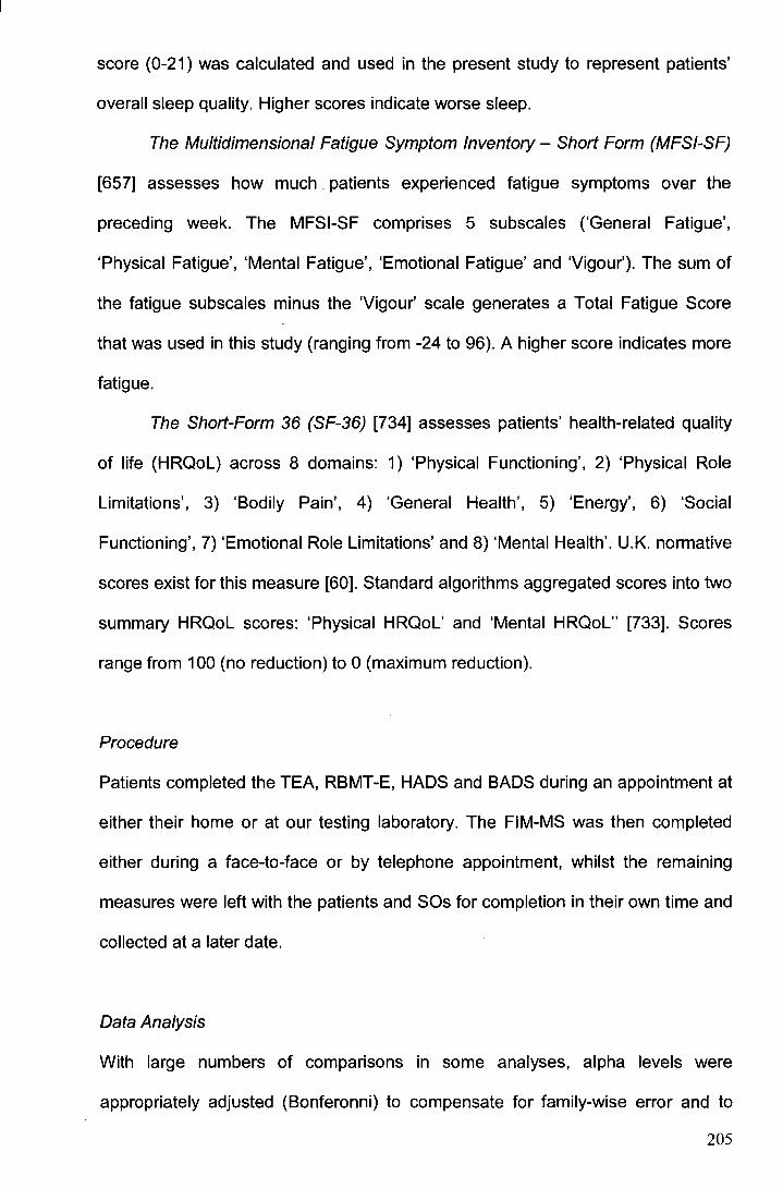
score (0-21) was calculated and used in the present study to represent patients'
overall sleep quality. Higher scores indicate worse sleep.
The Multidimensional Fatigue Symptom Inventory - Short Form (MFSI-SF)
[657] assesses how much. patients experienced fatigue symptoms over the
preceding week. The MFSI-SF comprises 5 subscales ('General Fatigue',
'Physical Fatigue', 'Mental Fatigue', 'Emotional Fatigue' and 'Vigour'). The sum of
the fatigue subscales minus the 'Vigour' scale generates a Total Fatigue Score
that was used in this study (ranging from -24 to 96). A higher score indicates more
fatigue.
The Short-Form 36 (SF-36) [734] assesses patients' health-related quality
of life (HRQoL) across 8 domains: 1) 'Physical Functioning', 2) 'Physical Role
Limitations', 3) 'Bodily Pain', 4) 'General Health', 5) 'Energy', 6) 'Social
Functioning', 7) 'Emotional Role Limitations' and 8) 'Mental Health'. U.K. normative
scores exist for this measure [60]. Standard algorithms aggregated scores into two
summary HRQoL scores: 'Physical HRQoL' and 'Mental HRQoL" [733). Scores
range from 100 (no reduction) to 0 (maximum reduction).
Procedure
Patients completed the TEA, RBMT-E, HADS and BADS during an appointment at
either their home or at our testing laboratory. The FIM-MS was then completed
either during a face-to-face or by telephone appointment, whilst the remaining
measures were left with the patients and SOs for completion in their own time and
collected at a later date.
Data Analysis
With large numbers of comparisons in some analyses, alpha levels were
appropriately adjusted (Bonferonni) to compensate for family-wise error and to
205
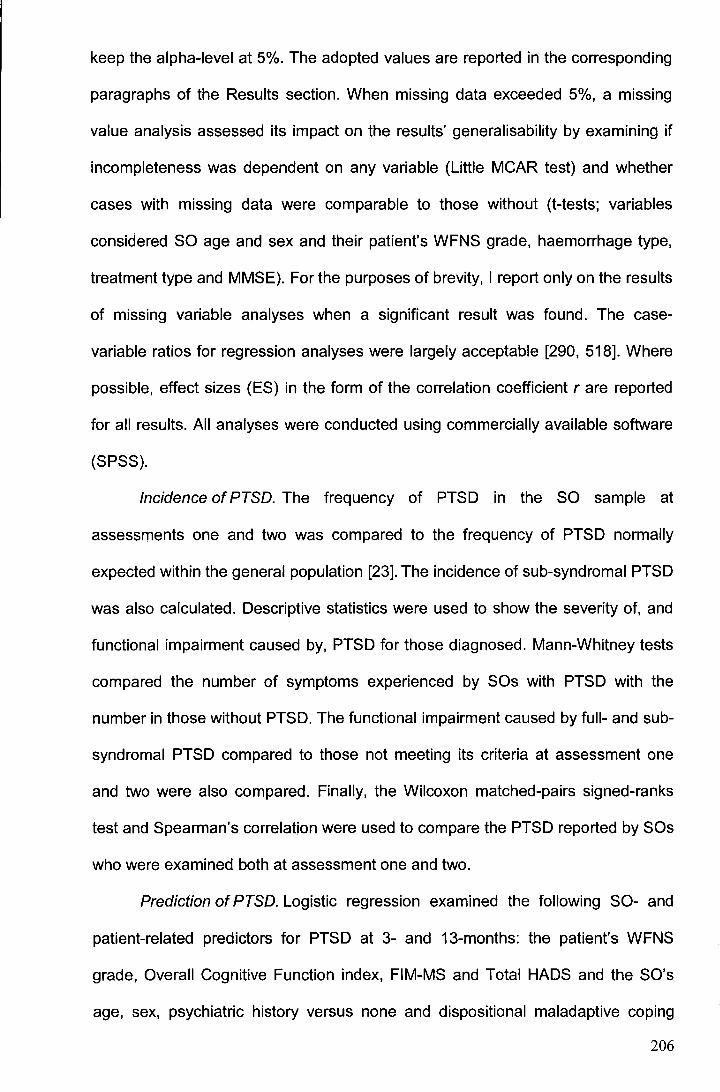
keep the alpha-level at 5%. The adopted values are reported in the corresponding
paragraphs of the Results section. When missing data exceeded 5%, a missing
value analysis assessed its impact on the results' generalisability by examining if
incompleteness was dependent on any variable (Little MCAR test) and whether
cases with missing data were comparable to those without (t-tests; variables
considered SO age and sex and their patient's WFNS grade, haemorrhage type,
treatment type and MMSE). For the purposes of brevity, I report only on the results
of missing variable analyses when a significant result was found. The case
variable ratios for regression analyses were largely acceptable [290, 518]. Where
possible, effect sizes (ES) in the form of the correlation coefficient rare reported
for all results. All analyses were conducted using commercially available software
(SPSS).
Incidence of PTSD. The frequency of PTSD in the SO sample at
assessments one and two was compared to the frequency of PTSD normally
expected within the general population [23]. The incidence of sub-syndromal PTSD
was also calculated. Descriptive statistics were used to show the severity of, and
functional impairment caused by, PTSD for those diagnosed. Mann-Whitney tests
compared the number of symptoms experienced by SOs with PTSD with the
number in those without PTSD. The functional impairment caused by full- and sub
syndromal PTSD compared to those not meeting its criteria at assessment one
and two were also compared. Finally, the Wilcoxon matched-pairs signed-ranks
test and Spearman's correlation were used to compare the PTSD reported by SOs
who were examined both at assessment one and two.
Prediction of PTSD. Logistic regression examined the following SO- and
patient-related predictors for PTSD at 3- and 13-months: the patient's WFNS
grade, Overall Cognitive Function index, FIM-MS and Total HADS and the SO's
age, sex, psychiatric history versus none and dispositional maladaptive coping
206

use. The patients' Overall Cognitive Function Index score, FIM-MS and Total
HADS score from assessment one were used in the model to predict PTSD in SOs
at assessment one, whilst the patients' scores on these measures from
assessment two were used when predicting SO PTSD at assessment two. A
backward elimination method (likelihood ratio test) revealed the most
parsimonious predictors. The odds ratio (OR) was used here to indicate the
change in the odds of having or not having PTSD when the value of a predictor
increased by one unit. The c-statistic measured the discriminative power of the
logistic equation. Varying from 0.5 (predictions are no better than chance) to 1.0
(model predicts without error), it is the percentage of case pairs to which the model
correctly assigns a higher probability. Mann-Whitney U tests, box-plots and
Kendall's tau ('r) measures of association were used where necessary to further
explore relationships highlighted by regression.
Please note that due to a reduced SO sample size at assessment two and
a smaller found frequency of SO PTSD, the full- and sub-syndromal SO PTSD
categories were collapsed for regression analyses for assessment two to increase
the number of cases of the rarer PTSD event (and hence satisfy an assumption of
logistic regression). This merging of groups was deemed acceptable since at
assessment two (unlike at assessment one), SOs with sub-syndromal and fuii
PTSD illustrated comparable levels of functional/ clinical impairment as a result of
their PTSD syndromes (see Results incidence sections).
Impact of significant other well-being on patient recovery. Multiple linear
and logistic regression using two sequential blocks of entry of variables was used.
In the first block, patients' demographics (age, sex, education) and illness
characteristics (clinical severity, time since illness and pre-morbid health) were
entered and formed the predictive model. The variance in patient recovery which
these predictors accounted for was therefore, controlled. In the second entry
207
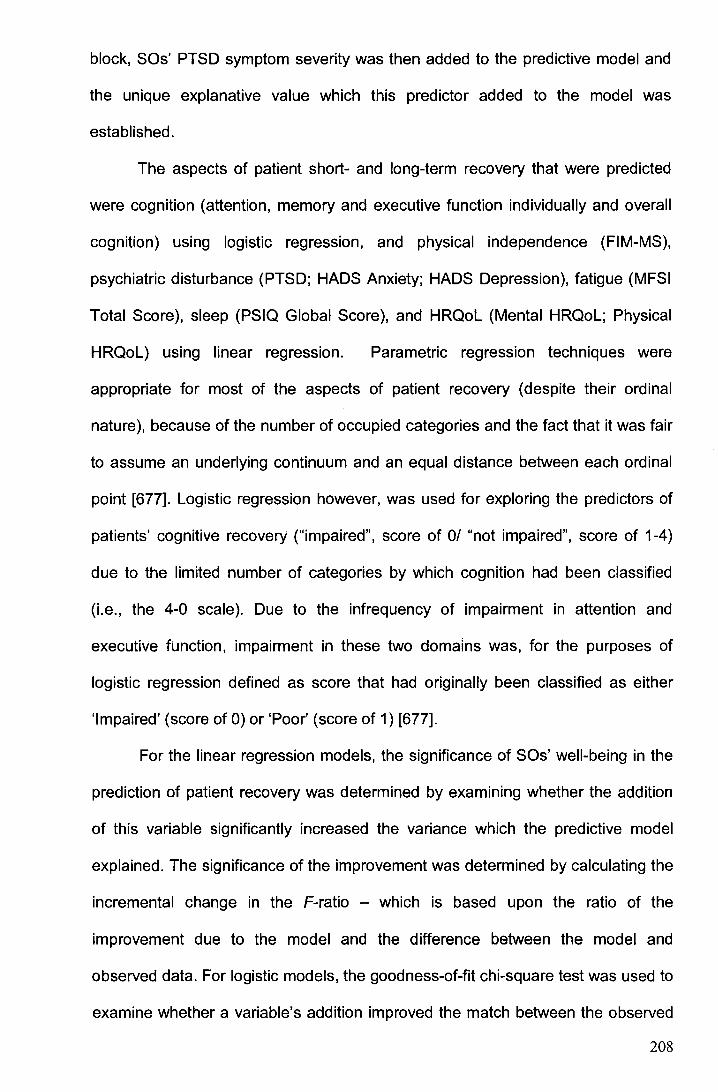
block, SOs' PTSD symptom severity was then added to the predictive model and
the unique explanative value which this predictor added to the model was
established.
The aspects of patient short- and long-term recovery that were predicted
were cognition (attention, memory and executive function individually and overall
cognition) using logistic regression, and physical independence (FIM-MS),
psychiatric disturbance (PTSD; HADS Anxiety; HADS Depression), fatigue (MFSI
Total Score), sleep (PSIQ Global Score), and HRQoL (Mental HRQoL; Physical
HRQoL) using linear regression. Parametric regression techniques were
appropriate for most of the aspects of patient recovery (despite their ordinal
nature), because of the number of occupied categories and the fact that it was fair
to assume an underlying continuum and an equal distance between each ordinal
point [677]. Logistic regression however, was used for exploring the predictors of
patients' cognitive recovery ("impaired", score of 0/ "not impaired", score of 1-4)
due to the limited number of categories by which cognition had been classified
(i.e., the 4-0 scale). Due to the infrequency of impairment in attention and
executive function, impairment in these two domains was, for the purposes of
logistic regression defined as score that had originally been classified as either
'Impaired' (score of 0) or 'Poor' (score of 1) [677].
For the linear regression models, the significance of SOs' well-being in the
prediction of patient recovery was determined by examining whether the addition
of this variable significantly increased the variance which the predictive model
explained. The significance of the improvement was determined by calculating the
incremental change in the F-ratio - which is based upon the ratio of the
improvement due to the model and the difference between the model and
observed data. For logistic models, the goodness-of-fit chi-square test was used to
examine whether a variable's addition improved the match between the observed
208
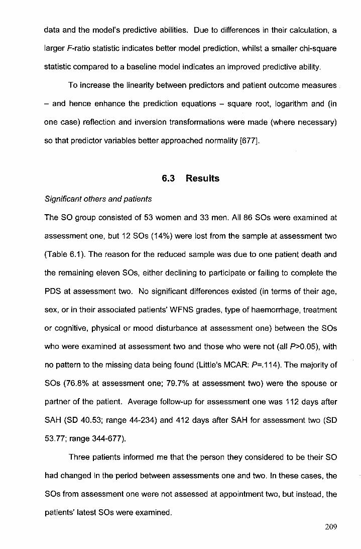
data and the model's predictive abilities. Due to differences in their calculation, a
larger F-ratio statistic indicates better model prediction, whilst a smaller chi-square
statistic compared to a baseline model indicates an improved predictive ability.
To increase the linearity between predictors and patient outcome measures
- and hence enhance the prediction equations - square root, logarithm and (in
one case) reflection and inversion transformations were made (where necessary)
so that predictor variables better approached normality [677].
6.3 Results
Significant others and patients
The SO group consisted of 53 women and 33 men. All 86 SOs were examined at
assessment one, but 12 SOs (14%) were lost from the sample at assessment two
(Table 6.1 ). The reason for the reduced sample was due to one patient death and
the remaining eleven SOs, either declining to participate or failing to complete the
PDS at assessment two. No significant differences existed (in terms of their age,
sex, or in their associated patients' WFNS grades, type of haemorrhage, treatment
or cognitive, physical or mood disturbance at assessment one) between the SOs
who were examined at assessment two and those who were not (all P>0.05), with
no pattern to the missing data being found (Little's MCAR: P=.114 ). The majority of
SOs (76.8% at assessment one; 79.7% at assessment two) were the spouse or
partner of the patient. Average follow-up for assessment one was 112 days after
SAH (SD 40.53; range 44-234) and 412 days after SAH for assessment two (SD
53. 77; range 344-677).
Three patients informed me that the person they considered to be their SO
had changed in the-period between assessments one and two. In these cases, the
SOs from assessment one were not assessed at appointment two, but instead, the
patients' latest SOs were examined.
209
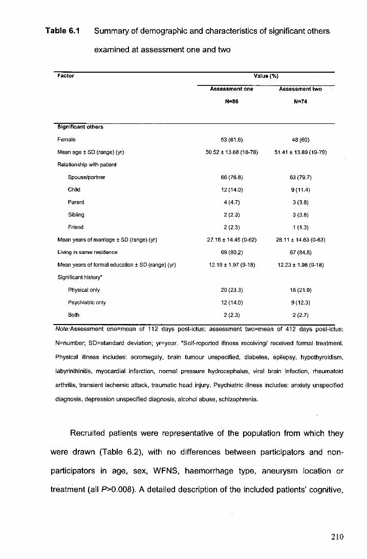
Table 6.1 Summary of demographic and characteristics of significant others
examined at assessment one and two
Factor Value(%)
Assessment one Assessment two
N=86 N=74
Significant others
Female 53 (61.6) 48 (60)
Mean age± SO (range) (yr) 50.52 ± 13.68 (18-78) 51.41 ± 13.89 (19-79)
Relationship with patient
Spouse/partner 66 (76.8) 63 (79.7)
Child 12 (14.0) 9(11.4)
Parent 4 (4.7) 3 (3.8)
Sibling 2 (2.3) 3 (3.8)
Friend 2 (2.3) 1 (1.3)
Mean years of marriage± SO (range) (yr) 27.16 ± 14.45 (0-62) 28.11 ± 14.63 (0-63)
Living in same residence 69 (80.2) 67 (84.8)
Mean years of formal education± SO (range) (yr) 12.19 ± 1.97 (9-18) 12.23 ± 1.98 (9-18)
Significant history•
Physical only 20 (23.3) 16 (21.9)
Psychiatric only 12 (14.0) 9 (12.3)
Both 2 (2.3) 2 (2.7)
Note:Assessment one=mean of 112 days post-ictus; assessment two=mean of 412 days post-ictus;
N=number; SD=standard deviation; yr=year. *Self-reported illness receiving/ received formal treatment.
Physical illness includes: acromegaly, brain tumour unspecified, diabetes, epilepsy, hypothyroidism,
labyrinthinitis, myocardial infarction, normal pressure hydrocephalus, viral brain infection, rheumatoid
arthritis, transient ischemic attack, traumatic head injury. Psychiatric illness includes: anxiety unspecified
diagnosis, depression unspecified diagnosis, alcohol abuse, schizophrenia.
Recruited patients were representative of the population from which they
were drawn (Table 6.2), with no differences between participators and non
participators in age, sex, WFNS, haemorrhage type, aneurysm location or
treatment (all P>O.OOB). A detailed description of the included patients' cognitive,
210
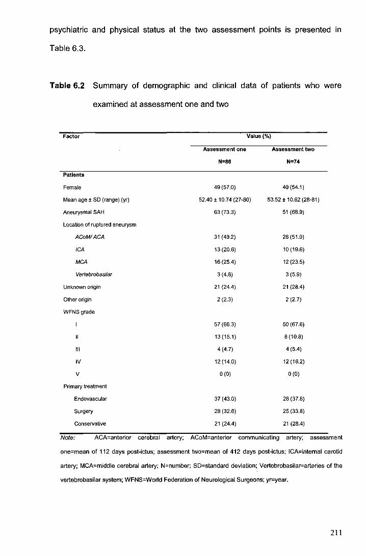
psychiatric and physical status at the two assessment points is presented in
Table 6.3.
Table 6.2 Summary of demographic and clinical data of patients who were
examined at assessment one and two
Factor Value (%)
Assessment one Assessment two
N=86 N=74
Patients
Female 49 (57.0) 40 (54.1)
Mean age± SD (range) (yr) 52.40 ± 10.74 (27-80) 53.52 ± 10.62 (28-81)
Aneurysmal SAH 63 (73.3) 51 (68.9)
Location of ruptured aneurysm
ACoMIACA 31 (49.2) 26 (51.0)
ICA 13 (20.6) 10(19.6)
MCA 16 (25.4) 12 (23.5)
Vertebrobasilar 3 (4.8) 3 (5.9)
Unknown origin 21 (24.4) 21 (28.4)
Other origin 2 (2.3) 2 (2.7)
WFNSgrade
57 (66.3) 50 (67.6)
II 13(15.1) 8 (10.8)
Ill 4 (4.7) 4 (5.4)
IV 12 (14.0) 12 (16.2)
v 0 (0) 0 (0)
Primary treatment
Endovascular 37 (43.0) 28 (37.8)
Surgery 28 (32.6) 25 (33.8)
Conservative 21 (24.4) 21 (28.4)
Note: ACA-anterior cerebral artery; ACoM-anterior communicating artery; assessment
one=mean of 112 days post-ictus; assessment two=mean of 412 days post-ictus; ICA=intemal carotid
artery; MCA=middle cerebral artery; N=number; SD=standard deviation; Vertebrobasilar=arteries of the
vertebrobasilar system; WFNS=World Federation of Neurological Surgeons; yr=year.
211
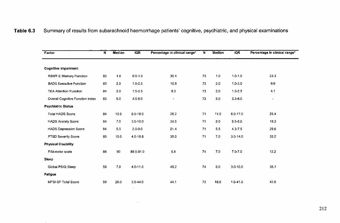
Table 6.3 Summary of results from subarachnoid haemorrhage patients' cognitive, psychiatric, and physical examinations
Factor N Median IQR Percentage in clinical range* N Median IQR Percentage in clinical range*
Cognitive Impairment
RBMT-E Memory Function 83 1.0 0.0-1.0 30.1 73 1.0 1.0-1.0 23.3
BADS Executive Function 83 2.0 1.0-2.0 10.8 73 2.0 1.0-3.0 9.6
TEA Attention Function 84 2.0 1.5-2.5 8.3 73 2.0 1.3-2.5 4.1
Overall Cognitive Function index 83 5.0 4.0-6.0 - 73 5.0 3.3-6.0
Psychiatric Status
Total HADS Score 84 12.0 6.0-18.0 26.2 71 11.0 6.0-17.0 25.4
HADS Anxiety Score 84 7.0 3.0-10.0 34.5 71 8.0 6.5-8.0 18.3
HADS Depression Score 84 5.0 2.0-9.0 21.4 71 5.5 4.3-7.5 29.6
PTSD Severity Score 80 10.0 4.0-18.8 35.0 71 7.0 3.0-14.0 35.2
Physical Disability
FIM-motor scale 86 90 89.0-91.0 5.8 74 7.0 7.0-7.0 12.2
Sleep
Global PSIQ Sleep 59 7.0 4.0-11.0 49.2 74 6.0 3.0-10.0 35.1
Fatigue
MFSI-SF Total Score 59 26.0 3.0-44.0 44.1 73 18.0 1.0-41.0 42.5
212
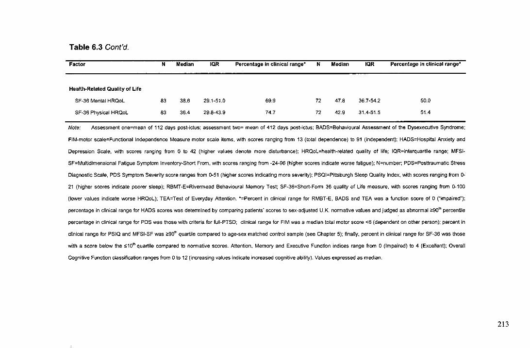
Table 6.3 Cont'd.
Factor N Median IQR Percentage in clinical range* N Median IQR Percentage in clinical range*
Health-Related Quality of Life
SF-36 Mental HRQoL 83 38.6 2g,1-51.0 6g,g 72 47.8 36.7-54.2 50.0
SF-36 Physical HRQoL 83 36.4 2g.8-43.g 74.7 72 42.g 31.4-51.5 51.4
Note: Assessment one=mean of 112 days post-ictus; assessment two= mean of 412 days post-ictus; BADS=Behavioural Assessment of the Dysexecutive Syndrome;
FIM-motor scale=Functional Independence Measure motor scale items, with scores ranging from 13 (total dependence) to g1 (independent); HADS=Hospital Anxiety and
Depression Scale, with scores ranging from 0 to 42 (higher values denote more disturbance); HRQoL=health-related quality of life; IQR=interquartile range; MFSI
SF=Multidimensional Fatigue Symptom Inventory-Short From, with scores ranging from -24-g6 (higher scores indicate worse fatigue); N=number; PDS=Posttraumatic Stress
Diagnostic Scale, PDS Symptom Severity score ranges from 0-51 (higher scores indicating more severity); PSQI=Pittsburgh Sleep Quality Index, with scores ranging from 0-
21 (higher scores indicate poorer sleep); RBMT-E=Rivermead Behavioural Memory Test; SF-36=Short-Form 36 quality of Life measure, with scores ranging from 0-100
(lower values indicate worse HRQoL); TEA=Test of Everyday Attention. *=Percent in clinical range for RMBT-E, BADS and TEA was a function score of 0 ("impaired");
percentage in clinical range for HADS scores was determined by comparing patients' scores to sex-adjusted U.K. normative values and judged as abnormal ~goth percentile
percentage in clinical range for PDS was those with criteria for fuii-PTSD; clinical range for FIM was a median total motor score <6 (dependent on other person); percent in
clinical range for PSIQ and MFSI-SF was ~goth quartile compared to age-sex matched control sample (see Chapter 5); finally, percent in clinical range for SF-36 was those
with a score below the ::>10th quartile compared to normative scores. Attention, Memory and Executive Function indices range from 0 (Impaired) to 4 (Excellent); Overall
Cognitive Function classification ranges from 0 to 12 (increasing values indicate increased cognitive ability). Values expressed as median.
213
PTSD Incidence in significant others
Assessment one
PTSD was assessed in 86 SOs. Twenty-two (25.6%) SOs were diagnosed with
PTSD. In the general population a prevalence of 8% is expected. Median symptom
severity for those with PTSD was moderate (median=19.0, interquartile range
[IQR]=14.0-30.8) and caused "severe" functional impairment for the majority
(77.3%). Those SOs with fuii-PTSD exhibited significantly higher levels of
intrusive, avoidant and arousal symptoms and more severe levels of
symptomatology overall compared to those without (all P<0.0001; adjusted alpha=
P<0.01; Table 6.4). An additional 12 (14%) SOs fulfilled the criteria for sub
syndromal PTSD. This caused mild functional impairment for 5 of these patients
(41.7%), moderate impairment for 3 (25.0%) and severe impairment for the
remaining 4 (33.3%) patients. Those with sub-syndromal PTSD experienced
significantly less functional impairmenU distress as a result of their syndrome
(median=4.0, IQR=2.0-9.0) compared to those with fuii-PTSD (median=9.0,
IQR=8.0-9.0; P<0.003).
Assessment two
PTSD was assessed in 74 SOs. Twelve (16.2%) SOs were diagnosed with PTSD.
The median symptom severity for those with PTSD was moderate (median=17.0,
IQR=13.3-23.0) and caused "severe" functional impairment for the majority
(75.0%). Those SOs with PTSD exhibited significantly higher levels of intrusive,
avoidant and arousal symptoms and more severe levels of symptomatology
overall (all P<0.0001; adjusted alpha= P<0.01; Table 6.4). An additional 5 (6.8%)
of SOs met the criteria for sub-syndromal PTSD. This
214
Table 6.4 Comparison of significant others with and without PTSD
Factor Assessment one Assessment two
50s with PT5D 50s without PT5D P-value E5 50s with PT5D** 50s without PT5D P-value ES
N Median IQR N Median IQR N Median IQR N Median IQR
PT5D-PD5 *
Intrusions 22 4.0 2.8-4.3 64 2.0 1.0-3.0 <0.0001 -.45 17 3.0 2.0-5.0 57 1.0 0.0-2.0 <0.0001 -.53
Avoidance 22 5.0 3.0-6.0 64 1.0 0.0-2.0 <0.0001 -.64 17 4.0 1.5-5.5 57 0.0 0.0-2.0 <0.0001 -.54
Arousal 22 4.0 3.0-5.0 64 1.0 0.0-2.0 <0.0001 -.60 17 3.0 2.0-5.0 57 0.0 0.0-2.0 <0.0001 -.55
Symptom severity 22 19.0 14.0-30.8 64 4.5 2.0-9.0 <0.0001 -.62 17 14.0 10.5-20.0 57 3.0 0.0-6.5 <0.0001 -.61
Coping -Brief COPE *
Maladaptive coping 21 3.6 3.1-4.5 64 2.7 2.2-3.1 <0.0001 -.47 17 3.3 2.9-4.2 56 2.6 2.2-3.2 <0.007 -.32
Self distraction 21 5.0 3.0-6.0 64 3.0 2.0-7.0 <0.004 -.32 17 4.0 2.5-5.5 56 3.0 2.0-5.0 ns -.16
Denial 21 4.0 2.0-6.0 64 2.0 2.0-3.0 <0.003 -.32 17 3.0 2.0-4.0 56 2.0 2.0-3.0 ns -.22
Substance abuse 21 2.0 2.0-4.0 64 2.0 2.0-2.0 ns -.23 17 2.0 2.0-3.0 56 2.0 2.0-2.0 ns -.08
Behavioural disengagement 21 3.0 2.0-3.5 64 2.0 2.0-2.0 <0.0001 -.37 17 2.0 2.0-3.0 56 2.0 2.0-2.0 ns -.14
Venting 21 4.0 2.0-5.0 64 2.0 2.0-3.0 ns -.26 17 4.0 3.0-5.0 56 2.0 2.0-3.0 <0.0001 -.44
Self- blame 21 3.0 2.0-5.5 64 2.0 2.0-2.0 <0.004 -.33 17 3.0 2.0-4.0 56 2.0 2.0-3.8 ns -.17
50s demographics
Mean age ± SD § 22 47.6 12.1 64 51.9 14.3 ns .14 17 44.7 14.5 57 52.8 13.6 <0.05" .06
215
Table 6.4 Cont'd.
Assessment one Assessment two
50s with PT5D 50s without PT5D P-value E5 50s with PT5D** 50s without PT5D P-value E5
N Median IQR N Median IQR N Median IQR N Median IQR
SOs demographics cont'd.
Females (%) :t: 22 12 55% 64 41 64% ns .06 17 13 76.5 57 25 43.9 ns
With psychiatric history(%) :t: 22 4 18% 64 10 16% ns - 17 5 29.4 57 6 10.5 ns
Note: ES=effect size (r); IQR=interquartile range; N=number; PDS=Posttraumatic Stress Diagnostic Scale; PDS scoring is presented as the number of intrusive (e.g., "reliving the brain
haemorrhage, acting or feeling as if it was happening again"), avoidant (e.g., "trying to avoid activities, people or places that remind me of the brain haemorrhage") and arousal (e.g., "feeling
irritable or having fits of anger") symptoms experienced; SOs=significant others; *=Comparisons made using Mann-Whitney tests. §=Comparison made using t-test for unequal groups; **PTSD
group in assessment two comparisons includes 5 SOs with sub-syndromal PTSD in order to increase cell count for statistical purposes; :!:=Comparison made using i test/ Fisher exact test;
"=Equality of variances assumed. Brief COPE scoring ranges from 2 ("strategy usually not used") to 8 ("strategy usually used a lot"). Critical alpha level adjusted for each set of family
comparisons: PTSD block P= 0.0125, Coping block P= 0.007 and SOs' demographic block P= 0.016. All values, other than age, expressed as medians.
216
caused severe functional impairment for the majority (60.0%). In fact, those with
sub-syndromal PTSD illustrated levels of functional impairment/ distress as a
result of their disorder comparable to those with fuii-PTSD (sub-syndromal
median= 9.0, IQR 1.5-9.0 vs. fuii-PTSD median= 9.0, IQR 6.8-9.0, P>0.05).
Comparison of significant others' PTSD at assessment one and two
Missing data (17.4%), due to a reduced sample size at assessment two, meant
that this analysis was based on the 71 SOs who completed the PTSD
assessments at both assessments. There was no significant difference in PTSD
prevalence (P>0.05), experience of intrusive {P>0.05), avoidant (P>0.05) or
arousal symptoms {P>0.05) at assessments one and two (Adjusted alpha P<
0.01 ). The Wilcoxon test however, detected a significant reduction with time in the
number (P<0.007) and severity of PTSD symptoms {P<0.01) experienced by SOs
(Table 6.5). Spearman's correlation between SOs' individual PTSD status (full,
sub-syndromal or no PTSD) at the two assessments revealed that PTSD status
was stable for many: rs= .317, P<0.0007. In total, 67.1% of patients had the same
status at both assessments.
Prediction of significant others' PTSD
Assessment one
Analysis was based on 82 patient/ SO pairs due to missing data (4.6%) on some
measures considered. A logistic model that provided a significant relationship
between predictors and the occurrence of PTSD was generated <l [2] = 26.195,
P<0.0001 ). It was found that increased use of maladaptive coping strategies (OR=
4.292, Cl 2.070 - 8.899; Wald statistic= 15.329 [1] P<0.0001) significantly
increased the probability of PTSD (Adjusted alpha=P<0.006; Figure 6.1 ). Using a
probability criterion of .5, predictive classification was excellent (c = 0.84), with the
217
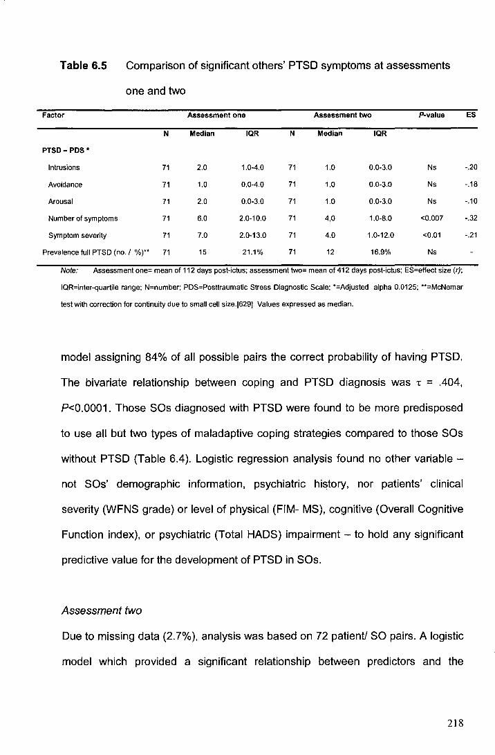
Table 6.5 Comparison of significant others' PTSD symptoms at assessments
one and two
Factor Assessment one Assessment two P-value
N Median IQR N Median IQR
PTSD- PDS*
Intrusions 71 2.0 1.0-4.0 71 1.0 0.0-3.0 Ns
Avoidance 71 1.0 0.0-4.0 71 1.0 0.0-3.0 Ns
Arousal 71 2.0 0.0-3.0 71 1.0 0.0-3.0 Ns
Number of symptoms 71 6.0 2.0-10.0 71 4.0 1.0-8.0 <0.007
Symptom severity 71 7.0 2.0-13.0 71 4.0 1.0-12.0 <0.01
Prevalence full PTSD (no. I %)** 71 15 21.1% 71 12 16.9% Ns
Note: Assessment one= mean of 112 days post-ictus; assessment two= mean of 412 days post-ictus; ES=effect size (r);
IQR=inter-quartile range; N=number; PDS=Posttraumatic Stress Diagnostic Scale; *=Adjusted alpha 0.0125; **=McNemar
test with correction for continuity due to small cell size.[629] Values expressed as median.
model assigning 84% of all possible pairs the correct probability of having PTSD.
The bivariate relationship between coping and PTSD diagnosis was -r = .404,
P<0.0001. Those SOs diagnosed with PTSD were found to be more predisposed
to use all but two types of maladaptive coping strategies compared to those SOs
without PTSD (Table 6.4). Logistic regression analysis found no other variable
not SOs' demographic information, psychiatric history, nor patients' clinical
severity (WFNS grade) or level of physical (FIM- MS), cognitive (Overall Cognitive
Function index), or psychiatric (Total HADS) impairment - to hold any significant
predictive value for the development of PTSD in SOs.
Assessment two
Due to missing data (2. 7% ), analysis was based on 72 patient/ SO pairs. A logistic
model which provided a significant relationship between predictors and the
218
ES
-.20
-.18
-.10
-.32
-.21
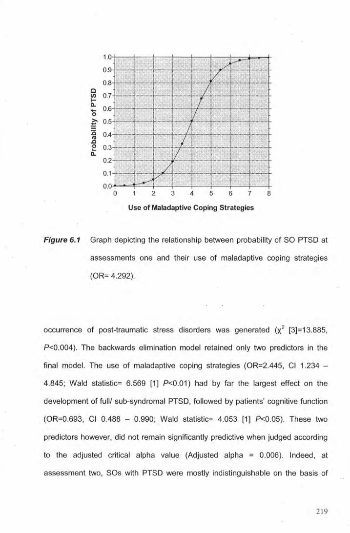
c en 1-a. -0 >. ~
.0 ~ .0 0 ... a.
O.O-+-..IO==F=..::....._+-...:....._+--+--+---.!~+--+--+
0 1 2 3 4 5 6 7 8
Use of Maladaptive Coping Strategies
Figure 6.1 Graph depicting the relationship between probability of SO PTSD at
assessments one and their use of maladaptive coping strategies
(OR= 4.292).
occurrence of post-traumatic stress disorders was generated (X2 [3]=13.885,
P<0.004). The backwards elimination model retained only two predictors in the
final model. The use of maladaptive coping strategies (OR=2.445, Cl 1.234 -
4.845; Wald statistic= 6.569 [1] P<0.01) had by far the largest effect on the
development of full/ sub-syndromal PTSD, followed by patients' cognitive function
(OR=0.693, Cl 0.488 - 0.990; Wald statistic= 4.053 [1] P<0.05). These two
predictors however, did not remain significantly predictive when judged according
to the adjusted critical alpha value (Adjusted alpha = 0.006). Indeed, at
assessment two, SOs with PTSD were mostly indistinguishable on the basis of
219
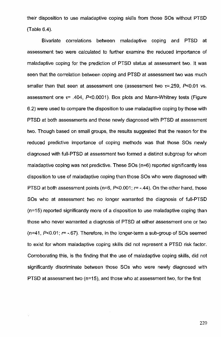
their disposition to use maladaptive coping skills from those SOs without PTSD
(Table 6.4).
Bivariate correlations between maladaptive coping and PTSD at
assessment two were calculated to further examine the reduced importance of
maladaptive coping for the prediction of PTSD status at assessment two. It was
seen that the correlation between coping and PTSD at assessment two was much
smaller than that seen at assessment one (assessment two t=.259, P<0.01 vs.
assessment one t= .404, P<0.0001 ). Box plots and Mann-Whitney tests (Figure
6.2) were used to compare the disposition to use maladaptive coping by those with
PTSD at both assessments and those newly diagnosed with PTSD at assessment
two. Though based on small groups, the results suggested that the reason for the
reduced predictive importance of coping methods was that those SOs newly
diagnosed with fuii-PTSD at assessment two formed a distinct subgroup for whom
maladaptive coping was not predictive. These SOs (n=6) reported significantly less
disposition to use of maladaptive coping than those SOs who were diagnosed with
PTSD at both assessment points (n=6, P<0.001; r= -.44). On the other hand, those
SOs who at assessment two no longer warranted the diagnosis of fuii-PTSD
(n=15) reported significantly more of a disposition to use maladaptive coping than
those who never warranted a diagnosis of PTSD at either assessment one or two
(n=41, P<0.01; r= -.67). Therefore, in the longer-term a sub-group of SOs seemed
to exist for whom maladaptive coping skills did not represent a PTSD risk factor.
Corroborating this, is the finding that the use of maladaptive coping skills, did not
significantly discriminate between those SOs who were newly diagnosed with
PTSD at assessment two (n=15), and those who at assessment two, for the first
220
!/) 8 C1)
C) 7 C1) -n:s * .... I -en 6
C) c ·c.. 5 0 u
C1) 4 > ; c. 3 n:s
"C n:s n:s 2 ~ -0 1
C1) !/)
::::J 0
ns I
*~ I I
**
~ B
PTSD status
• p<0.01 •• p<0.001 ••• p<0.0001
Figure 6.2 Use of maladaptive coping skills by significant others and their
PTSD.
time, did not meet the diagnosis for PTSD (n=6, P>0.05; r= -.37). It should be
noted that persons with sub-syndromal PTSD were excluded from these analyses
due to their small number.
Impact of significant other well-being on patient recovery
Assessment one
The SOs' experience of PTSD symptomatology reported at assessment one did
not significantly add to the predictive value of any aspect of patient recovery apart
from patient anxiety (F[1]=8.286, P<0.005) (Table 6.6). Even in this one significant
221
Table 6.6 Results from hierarchical regression analysing the unique contribution of SO well-being to patient recovery
Factor Assessment one Assessment two
Model N Rz; Step Ft)? p Model N R2* Step Ft)? p
Improvement Improvement
Attention (TEA)* 1 84 32.0 19.795 0.003 1 73 23.3 12.418 0.05
2 84 32.0 0.009 ns 2 73 23.3 0.032 ns
Memory(RBMT-E)* 1 83 12.1 7.430 ns 1 73 19.3 9.997 ns
2 83 21.8 6.446 0.011 2 73 20.0 0.392 ns
Executive (BADS)* 1 83 9.3 5.827 ns 1 73 18.2 10.210 ns
2 83 14.6 3.494 ns 2 73 20.0 1.132 ns
Physical (FIM-MS)* 1 86 18.7 3.031 0.01 1 72 28.2 4.326 0.001
2 86 21.9 3.213 ns 2 72 33.0 4.594 0.04
Sleep (PSQI) 1 60 12.1 1.218 ns 1 70 32.2 5.071 0.0001
2 60 15.8 2.246 ns 2 70 33.1 0.795 ns
Fatigue (MFSI-SF) 1 59 29.3 3.592 0.005 1 69 19.2 2.489 0.04
2 59 29.9 0.400 ns 2 69 19.5 0.284 ns
Physical HRQoL (SF-36) 1 83 13.6 1.988 ns 1 71 14.9 1.893 ns
2 83 15.7 1.890 ns 2 71 17.2 1.799 ns
Mental HRQoL (SF-36) 1 83 27.8 4.884 0.0001 1 71 17.0 2.212 0.05
2 83 30.9 3.363 ns 2 71 20.9 3.161 ns
Anxiety (HADS-A) 1 84 29.1 5.275 0.0001 1 70 18.2 2.375 0.04
2 84 36.1 8.286 0.005 2 70 19.2 0.736 ns
222
Table 6.6 Cont'd.
Factor Assessment one Assessment two Factor
Model N R2* Step Fit p Model N R2* Step Fit p
Improvement Improvement
Depression (HADS-0) 1 84 23.1 3.859 0.002 1 70 12.9 1.578 ns
2 84 25.2 2.074 ns 2 70 15.9 2.242 ns
PTSD (PDS) 1 80 26.1 4.300 0.001 1 70 18.5 2.422 0.05
2 80 29.2 3.146 ns 2 70 25.4 5.851 0.03
Note: BADS=Behavioural Assessment of the Dysexecutive Syndrome; FIM-MS=Functional Independence Measure-Motor; HADS-A=Hospital Anxiety and
Depression Scale -Anxiety score; HADS-D=Hospital Anxiety and Depression Scale - Depression score; MFSI-SF=Multidimensional Fatigue Symptom Inventory
Short Form; N=sample size; PDS=Posttraumatic Stress Diagnostic Scale; PSQI=Pittsburgh Sleep Quality; f?!=variance explained by model; RBMT-E=Rivermead
Behavioural Memory Test-Extended; TEA= Test of Everyday Attention; SF-36=Short Form-36; *=The f?1 approximation for logistic regression model is the f?1 version
devised by Nagelkerke (note this statistic can achieve a value of 1).[478] Model 1=Constant + Covariates (education, age, sex, pre-morbid health, WFNS grade,
time since ictus), Modei2=Constant + Covariates +concurrent SO PTSD symptom severity.
223
aspect of patient recovery, only 7% of unique variance in patients' recovery was
accounted for by SO well-being. SO wellbeing did explain 9. 7% of unique variance in
patients memory performance (F[1 ]=6.446, P=0.01 ), but this proved non-significant
when judged according to the adjusted test-wise alpha (P=0.007).
Assessment two
The SOs' experience of PTSD symptomatology did not significantly predict any
aspect of their patients' longer-term recovery (Table 6.6). Small associations of 5%
and 7% were found between SO well-being and patients' physical recovery (F[1 ]=
4.595, P=0.04) and patients' own experience of PTSD symptoms respectively (F[1 ]=
5.851, P=0.03). Again though, these associations proved non-significant when judged
according to the corrected alpha level (P=0.007).
6.4 Discussion
This study has shown for the first time that having a loved-one suffer, and survive a
spontaneous subarachnoid haemorrhage (SAH) is sufficient to elicit post-traumatic
stress disorder in patients' family members and friends. It was found that
approximately 26% (over three times the normal rate) of these 'significant others'
(SOs) warranted a diagnosis of PTSD in the early-stages following an SAH and 16%
warranted a diagnosis over one year post-SAH. A further 14% (in the early stages)
and 7% (at around 1 year post-SAH) met the criteria for its preclinical, debilitating
sub-syndromal form. Given this considerably high prevalence and that PTSD is a
disorder known to cause medical and psychosocial disability, it is clear that much
more attention nE;leds to be given to the outcome of SOs [23). The present findings
help to explain why SOs have previously been documented as exhibiting abnormal
224
levels of psychiatric symptoms and as having a reduced sense of well-being [48, 90,
264, 265, 285, 286, 461, 545, 546]. The need for an increased focus on SOs is
underscored by evidence that they are the persons who typically act as informal
carers to the patient [90, 264, 461, 545, 546]. If support is not provided, there may
end up being two patients to support, where previously there was only one, supported
by a SO [520]. The importance of this is highlighted when it is considered that the
service rendered by families and other informal carers is invaluable and dramatically
reduces the economic burden of SAH on society [154, 215]. Moreover, recent social
trends, such as changes in family structure, increased geographical mobility and
greater participation of women in the workforce, means that there are fewer potential
replacements for those SOs who become ill and end up having to abdicate from the
caring role [522]. Also, the incidence of SAH - unlike other stroke types - remains
stable [140, 403, 711], but the number of people who survive an SAH and who
require subsequent care is increasing all the time [284, 364, 653]. Therefore, the
number of SOs who will be needed to act as informal carers can be expected to rise
in the future.
My finding that SAH can elicit PTSD in an SO compliments the work of
Pritchard et al. [545]. As noted in the introduction to this chapter, Pritchard and
colleagues found that informal carers of aneurysmal SAH patients experienced a
significantly worse psychosocial outcome (for example, feeling more frightened,
depressed and stressed) than carers of acoustic neuroma (AN) patients, even though
socially and clinically well-matched. The authors suggested that this difference in
outcome could be due to the emergency (rather than elective manner as is the case
for AN) onset of the SAH and its treatment. Pritchard et al. considered that the
emergency onset of the SAH, which by its very nature, came without prior warning
225
and caused major disruption, was sufficiently traumatic for the SO to elicit a post
traumatic stress-reaction. Pritchard et al. however, did not use a diagnostic measure
for PTSD and were therefore unable to confirm their hypothesis about PTSD. The
present study did use such a diagnostic measure and can, therefore, confirm for the
first time that an SAH leads to significantly elevated levels of PTSD in SOs of SAH
patients.
It is important to highlight that it is not presumed that the finding of elevated
PTSD is restricted to family members and friends of patients with SAH [23]. A few
studies have indeed documented an abnormal level of PTSD symptoms in other
populations of family and friends of patients, such as in parents of children involved in
accidents [97, 142, 698, 771]. It is also likely that other neurological conditions could
be sufficiently psychologically traumatic to elicit a PTSD-reaction in family or friends.
The present findings therefore, serve to underscore the importance of considering the
impact of an illness on persons other than the patient.
The finding that there was not a reduction in the prevalence of PTSD in those
SOs examined at both assessment points, and that an SOs PTSD diagnosis was
largely stable, leads to the conclusion that PTSD in this sample is, for the most part, a
chronic problem. This concurs with evidence from the wider literature. One large
epidemiological study of 5877 persons in the U.S., for example, found that the
average duration of PTSD for those who did not seek treatment was over 5 years,
and over one-third of persons suffering PTSD never fully recover [351]. There is some
evidence to suggest that the greatest remission of PTSD occurs within the first 3
months [584] and that those persons who fulfil the diagnostic criteria beyond this point
have a significantly high likelihood of their PTSD taking on a chronic course [266,
446, 458]. Indeed, the DSM-IV-TR does recommend that if PTSD endures in a person
226
for 3 months or more, then the treating clinician should consider it to have assumed a
chronic course [23].
The existence ·of sub-syndromal PTSD in this study's SO sample (7-14%) also
warrants discussion. The inclusion of these persons in the formal incidence of PTSD
in the SAH SO population would obviously increase the prevalence of significant
psychological impact of an SAH on SOs. It should be borne in mind, however, that a
consensus on the definition of sub-syndromal PTSD has only recently begun to
emerge [477], with some [55, 101] advocating less stringent diagnostic criteria for
sub-syndromal PTSD than employed in the present study. Using such alternative
criteria would have further increased the incidence of stress reactions found in SAH
patients SOs. Previous research in other populations has illustrated that such sub
syndromal disorders are associated with similar levels of functional impairment and
psychosocial difficulties as full-blown PTSD [72, 658, 739, 773], that it often assumes
a chronic path [31 9, 605], and is a risk factor for delayed onset fuii-PTSD (i.e., PTSD
which occurs more than 6 months following the trauma) [1 06]. In line with these
findings, sub-syndromal PTSD in the present study was found to cause many SOs
severe degrees of distress and/ or functional impairment. In fact, at the second
assessment, the functional impairment caused to SOs by sub-syndromal PTSD was
comparable to that experienced by SOs with fuii-PTSD. Findings such as these have,
in fact, led many to criticise the diagnostic criteria for PTSD as being too restrictive
and leaving many short of appropriate treatment [477, 586].
The high rate of PTSD in the current study clearly indicates a need to
understand why SOs develop PTSD. It was found that for SOs of SAH patients,
coping strategies are the single best predictor of PTSD in the short-term. The greater
the use of maladaptive coping strategies, the higher the likelihood that an SO will
227
develop post-SAH PTSD. This mirrors similar findings obtained in persons who
developed PTSD as a consequence of other traumatic events (such as accidents,
terrorist incidents and having a medically ill child) [87, 206, 207, 217]. In fact, in this
study it was found that use of information about an SO's coping strategies alone,
allows a classification accuracy of 84%. The classic explanation for why maladaptive
coping strategies promote PTSD refers to the slightly paradoxical phenomenon that
by actively avoiding thoughts or situations which lead to anxiety and fear we in fact
maintain the fear-inducing quality of those thoughts and situations. In contrast,
successful adaptation to the experience of a trauma involves accessing of aversive
emotions about the event, which permits habituation to anxiety and modification of
trauma-related beliefs [88, 193]. It is therefore possible to predict at an early stage,
which SOs are at risk of developing PTSD. Moreover, appropriate interventions, such
as teaching SOs better coping strategies can be implemented, which should make
possible a reduction in SO PTSD and its associated psychosocial disability.
Encouraging in this context, are the findings from another of Pritchard et al.'s studies,
which illustrated the potential to improve the psychosocial outcome of SAH carers
through the enhancement of the support they are offered [547, 548]. Specifically, this
enhanced service employed the use of a specialist liaison nurse who provided
support and counselling for SAH patients and families from the first day of admission.
Those receiving this enhanced support fared significantly better in their emotional
response and employment than did a matched group of carers receiving the normal
support. Knowing that the underlying problem in SOs is PTSD and identifying their
use of maladaptive coping strategies should allow us to design a more specific and
even more effective system for preventing and treating the psychosocial problems in
SOs (the effect sizes for Pritchard et al.'s intervention were only modest).
228
It could be argued though, that maladaptive coping, which incorporates
avoidant behaviors, rather than representing a PTSD risk factor, could instead be a
symptom of it, especially given that avoidance behaviours are a diagnostic symptom
of PTSD. As commented by other workers however, this seems unlikely given that
avoidance behaviours describe only 2 of the 17 symptoms that define PTSD [88].
Furthermore, evidence from studies which fortuitously measured individuals'
preferences for coping skills prior to a trauma, show that pre-trauma preferences for
maladaptive coping are predictive of subsequent PTSD, suggesting that maladaptive
coping is a cause rather than a consequence of PTSD [172, 217]. For the analysis in
question, coping skills had been measured at the same time as PTSD. Thus, it was
not possible to determine the causal relationship between these two variables.
However, in light of the above-mentioned findings, it is more likely that maladaptive
coping skills cause PTSD than the other way around. Ultimately, an intervention study
would be required to settle this question.
Unfortunately, it was not possible to develop a significantly effective prediction
model for longer-term SO PTSD. It was found that the dispositional use of
maladaptive coping strategies, when judged against the adjusted critical alpha, were
no longer sufficient to discriminate between those with and those without PTSD at
assessment two. In exploring the reasons for the change in the association between
coping and PTSD, the data suggests that those SOs with PTSD at only assessment
two were unique in that their PTSD developed regardless of whether or not they used
maladaptive coping. Further work on predicting the development of PTSD in the
longer-term is obviously therefore needed. Fortunately, the results from the present
study have illustrated that (at least in the first 13 months post-SAH) only a minority
(-30%) of SOs' PTSD status actually changes between the short- and long-term.
229
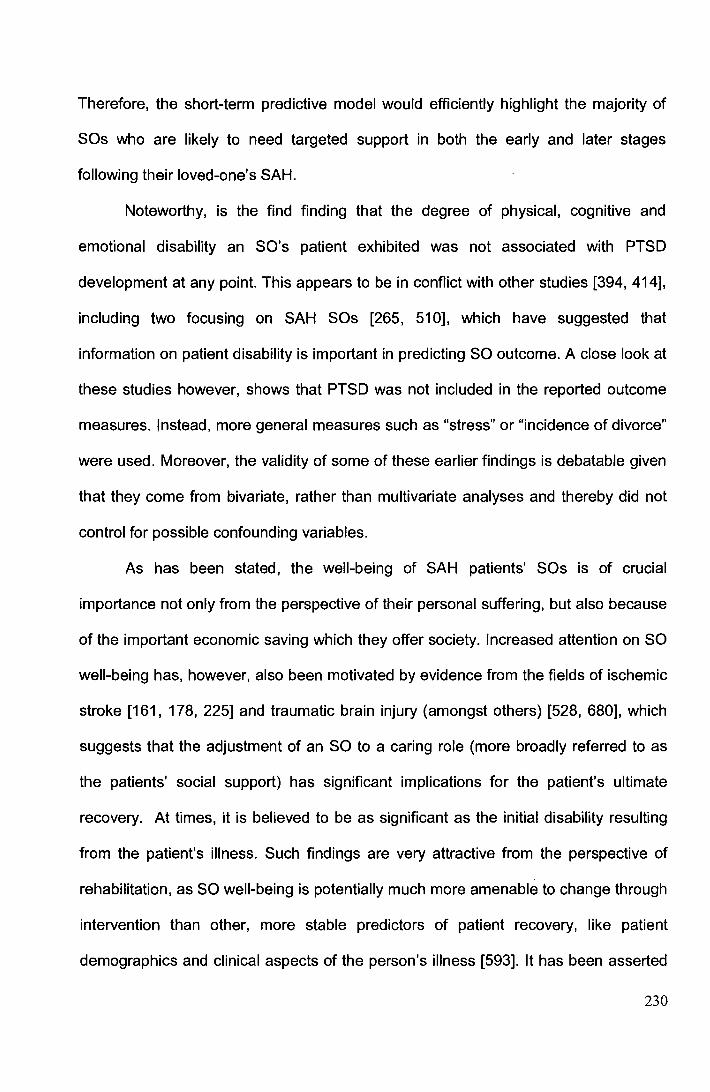
Therefore, the short-term predictive model would efficiently highlight the majority of
SOs who are likely to need targeted support in both the early and later stages
following their loved-one's SAH.
Noteworthy, is the find finding that the degree of physical, cognitive and
emotional disability an SO's patient exhibited was not associated with PTSD
development at any point. This appears to be in conflict with other studies [394, 414],
including two focusing on SAH SOs [265, 510], which have suggested that
information on patient disability is important in predicting SO outcome. A close look at
these studies however, shows that PTSD was not included in the reported outcome
measures. Instead, more general measures such as "stress" or "incidence of divorce"
were used. Moreover, the validity of some of these earlier findings is debatable given
that they come from bivariate, rather than multivariate analyses and thereby did not
control for possible confounding variables.
As has been stated, the well-being of SAH patients' SOs is of crucial
importance not only from the perspective of their personal suffering, but also because
of the important economic saving which they offer society. Increased attention on SO
well-being has, however, also been motivated by evidence from the fields of ischemic
stroke [161, 178, 225] and traumatic brain injury (amongst others) [528, 680], which
suggests that the adjustment of an SO to a caring role (more broadly referred to as
the patients' social support) has significant implications for the patient's ultimate
recovery. At times, it is believed to be as significant as the initial disability resulting
from the patient's illness. Such findings are very attractive from the perspective of
rehabilitation, as SO well-being is potentially much more amenable to change through
intervention than other, more stable predictors of patient recovery, like patient
demographics and clinical aspects of the person's illness [593]. It has been asserted
230
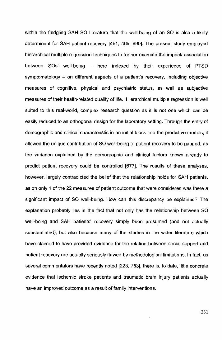
within the fledgling SAH SO literature that the well-being of an SO is also a likely
determinant for SAH patient recovery [461, 469, 690). The present study employed
hierarchical multiple regression techniques to further examine the impact/ association
between SOs' well-being - here indexed by their experience of PTSD
symptomatology - on different aspects of a patient's recovery, including objective
measures of cognitive, physical and psychiatric status, as well as subjective
measures of their health-related quality of life. Hierarchical multiple regression is well
suited to this real-world, complex research question as it is not one which can be
easily reduced to an orthogonal design for the laboratory setting. Through the entry of
demographic and clinical characteristic in an initial block into the predictive models, it
allowed the unique contribution of SO well-being to patient recovery to be gauged, as
the variance explained by the demographic and clinical factors known already to
predict patient recovery could be controlled [677]. The results of these analyses,
however, largely contradicted the belief that the relationship holds for SAH patients,
as on only 1 of the 22 measures of patient outcome that were considered was there a
significant impact of SO well-being. How can this discrepancy be explained? The
explanation probably lies in the fact that not only has the relationship between SO
well-being and SAH patients' recovery simply been presumed (and not actually
substantiated), but also because many of the studies in the wider literature which
have claimed to have provided evidence for the relation between social support and
patient recovery are actually seriously flawed by methodological limitations. In fact, as
several commentators have recently noted [223, 753], there is, to date, little concrete
evidence that ischemic stroke patients and traumatic brain injury patients actually
have an improved outcome as a result of family interventions.
231
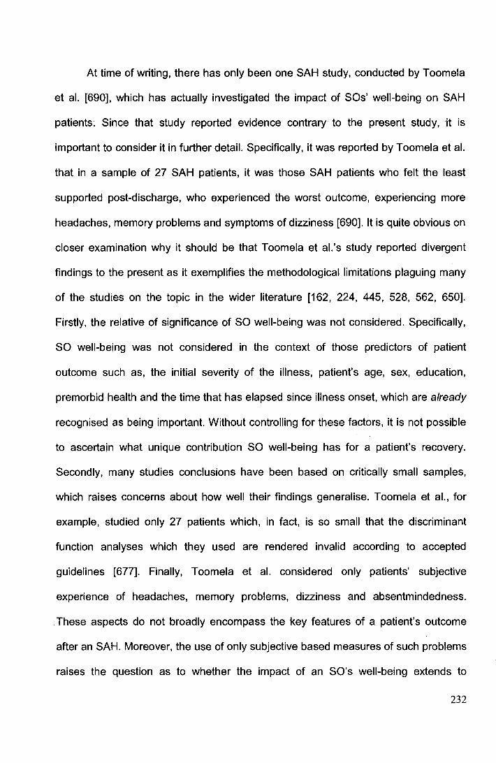
At time of writing, there has only been one SAH study, conducted by Toomela
et al. [690], which has actually investigated the impact of SOs' well-being on SAH
patients~ Since that study reported evidence contrary to the present study, it is
important to consider it in further detail. Specifically, it was reported by Toomela et al.
that in a sample of 27 SAH patients, it was those SAH patients who felt the least
supported post-discharge, who experienced the worst outcome, experiencing more
headaches, memory problems and symptoms of dizziness [690]. It is quite obvious on
closer examination why it should be that Toomela et al.'s study reported divergent
findings to the present as it exemplifies the methodological limitations plaguing many
of the studies on the topic in the wider literature [162, 224, 445, 528, 562, 650].
Firstly, the relative of significance of SO well-being was not considered. Specifically,
SO well-being was not considered in the context of those predictors of patient
outcome such as, the initial severity of the illness, patient's age, sex, education,
premorbid health and the time that has elapsed since illness onset, which are already
recognised as being important. Without controlling for these factors, it is not possible
to ascertain what unique contribution SO well-being has for a patient's recovery.
Secondly, many studies conclusions have been based on critically small samples,
which raises concerns about how well their findings generalise. Toomela et al., for
example, studied only 27 patients which, in fact, is so small that the discriminant
function analyses which they used are rendered invalid according to accepted
guidelines [677]. Finally, Toomela et al. considered only patients' subjective
experience of headaches, memory problems, dizziness and absentmindedness.
These aspects do not broadly encompass the key features of a patient's outcome
after an SAH. Moreover, the use of only subjective based measures of such problems
raises the question as to whether the impact of an SO's well-being extends to
232
objective measures. In the same study it was in fact reported that these patients'
subjective reports of memory problems and absentmindedness did not correlate
significantly with objective measures of the same problems. Therefore, given the
greater methodological rigor of the present study, it is not surprising that results
contrary to Toomela et al.'s were found. Although a larger sample size and a longer
follow-up is required to rule out that SO well-being has no bearing on patients'
recovery, the results of the present study do still strongly suggest that the influence of
SO recovery is small. Indeed, the effect size of the only significant relationship that
was found - namely between SO well-being and patient anxiety in the early stages of
recovery - was of a negligible size, explaining only 7% of variance in patients' anxiety
scores.
Two potential limitations to the present study require highlighting. Firstly, as
SOs were recruited through the patients, there is the possibility of a selection bias.
Secondly, it would have been preferable to have measured the SOs' psychiatric
status using an interview, rather than questionnaire-based technique. Although the
PDS is known to possess sensitivity and specificity levels which approach those
associated with "gold-standard" interview techniques [54, 193], there exists the
possibility, due to the ubiquitous nature of many of the SOs' symptoms to a variety of
other disorders [23], that another disorder may be more appropriate to explain SOs'
psychological distress. On the basis of my findings, this possibility cannot be ruled
out. In any case, regardless of whether the observed psychological distress is most
appropriately labelled as PTSD or not, it remains a fact that a substantial portion of
SOs experience significant distress subsequent to a loved-one's SAH. More
importantly, my findings suggest that the psychological distress of SOs might be
233
treatable and potentially preventable as maladaptive coping skills seem key to its
development.
6.5 Conclusions
In conclusion, PTSD was found to be common in SOs of SAH patients at both 3%
and 13-months post-ictus. The elevated incidence of this disorder helps to account for
the previously noted psychiatric symptoms in and psychosocial disability of SOs.
Greater attention and support needs to be given to assist with the adjustment of these
persons to their recent experience, especially considering that they often act as
informal carers to SAH patients. It is important to find ways to prevent the
development of PTSD which would likely interfere with the willingness/ ability of SOs
to care. Maladaptive coping strategies seem to be the best predictor of SO PTSD in
the short-term, but it is unclear what predicts PTSD in the later stages. It is hoped that
by teaching SOs better coping strategies early on, the incidence of PTSD could be
reduced.
234

Final Chapter
235

Chapter 7
General discussion and conclusions
236
7.1 Introduction
Previous studies examining spontaneous subarachnoid haemorrahge (SAH) patients
and theirfamily and friends (who often act as their informal carers), had found that
despite a seemingly good prognosis for surviving patients, many patients and their
family and friends nevertheless suffered an particularly poor psychosocial outcome.
The main aim of this thesis was to look to explain the mysterious difficulties of
patients and their 'significant others' (SOs) by building upon previous evidence which
led me to hypothesise that an abnormal prevalence of post-traumatic stress disorder
(PTSD) in both groups- elicited by the trauma of an SAH -could be a way to explain
their respective difficulties.
Two longitudinal studies were conducted- one focusing on SAH patients and
one focusing on their SOs. Overall, the results show that the best explanation for the
poor outcome of members of both groups is that an abnormal number of them find the
experience of an SAH so traumatizing that they develop PTSD. However, rather than
simply determining what the difficulties of these persons were, this thesis has also
provided some important insights into how we could potentially treat/ prevent their
difficulties. Specifically, I established that it is possible to reliably predict which
patients and SOs develop PTSD on the basis of information about the stress-coping
strategies that they employ. In the next section (7.2), I shall discuss the main findings
and conclusions to this thesis. These are then followed by a discussion of their
implications for the treatment of SAH patients and their SOs in section 7 .3. Finally,
future research directions are discussed in Section 7 .4.
237
7.2 Main findings
The overarching finding emerging from the studies presented in this thesis is that the
(previously mysterious) poor psychosocial outcome of SAH patients (defined in terms
of health-related quality of life [HRQoL], fatigue and sleep dysfunction) and their SOs
(defined in terms of their psychiatric disturbance) can, to a large extent be explained
by the fact that an abnormal number of them find the experience of an SAH so
traumatizing that they develop PTSD.
Evidence from Chapters 3, 5 and 6 showed that despite their very different
roles in the event, both patients and significant others are united by the fact that they
can experience an SAH as not only a medical emergency, but also as a
psychologically stressful event. Overall, these findings support prior research which
proposes that medical events can be highly traumatic for patients and that it is also
important to consider the wider impact of medical events for patients' family and
friends. The striking parallels between SAH patients' and their loved-ones' outcome
accords well with previous research where similarly poor reactions have been found
in both patients and their family members following other medical events, including
breast cancer and heart transplant [28, 62, 85, 671].
Crucially, insights were provided into how patients and their family and friends
PTSD could be reduced as findings from Chapters 5 and 6 showed that for both
parties the degree to which they used certain psychological stress-coping strategies
largely determined whether or not they developed PTSD as a result of the SAH.
Those with a disposition to use 'maladaptive' coping strategies such as denial,
avoidance, distraction, venting and behavioural disengagement, were the most likely
to experience PTSD. Importantly, the SAH patients' PTSD was not found to be
238
associated with any characteristics of their illness, which strongly implies that the
origins of these patients' PTSD are not neurological, but rather psychological.
In Chapter 5, I examined SAH patients' sleep and fatigue. For the first time
within the literature, these symptoms were both measured using validated measures
and the patients' scores were compared to those of a control group. Using these
techniques evidence was found which strongly suggests that many SAH patients
experience highly abnormal levels of fatigue and sleep. Importantly, PTSD helped
explain many of the difficulties that these patients had with their sleep and energy
levels. This evidence means that clinicians can no longer dismiss these symptoms
and that they symptoms require medical attention.
Behavioural disturbances such as delirium, major depression, anxiety disorder,
mania, psychosis, personality change and fatigue following neurological diseases
have typically been assumed by clinicians to be the direct result of the brain damage
associated with the disease and not a psychological disturbance [416, 531]. The
finding of this thesis, that a psychiatric disturbance, namely PTSD (whose origins
were psychological), is integral to understanding a patient's behavioural outcome
following a neurological disease, is therefore highly novel and significant. In showing
that not all behavioural disturbances or psychosocial impairment following
neurological diseases can be understood as being the result of 'organic' brain
damage, the findings of this thesis imply that a significantly different approach to the
treatment of neurological patients' disturbances may be warranted. My study is
amongst only a few studies which have considered such a 'non-organic' explanation
for neurological patients' behavioural disturbances. For example, whilst a recent
review article by Tedstone and Tarrier [684] revealed that a host of studies have
considered the role of PTSD in explaining the behavioural disturbances following a
239

variety of medical incidents, including cardiac events, cancer and certain medical
treatments, only a handful of primary studies were identified as having considered the
possibility that this 'non-organic' disorder might explain the behavioural disturbances
seen after neurological disease [89, 111, 615].
In Chapter 7, I looked at the outcome of patients' SOs in more detail. I felt that
it was relevant to do so (a) because their health status is important for their own
sakes and (b) because their health status is important due to their valuable role in
supporting SAH patients. Contrary to many assertions, I found that although SO well
being was reduced, their poor psychiatric state, at least within the time frame of this
particular study, did not appear to have any meaningful impact on patient recovery.
The discrepancy between the finding of this study and the findings of other studies
may at first seem concerning. However, close examination of the literature reveals
that the relationship between significant others' psychiatric state and patient recovery,
had largely been assumed, or based on research in the wider literature on other carer
groups which have not adequately controlled for obvious confounding explanations
for patients' poor recovery.
The key overall conclusion is that the treatment of PTSD in both SAH patients
and their SOs could lead to a much improved outcome for both groups following an
SAH.
240
7.3 Implications for treatment
The fact that both SAH patients and their SOs are often afflicted by PTSD and that
the use of maladaptive coping reliably predicts its development, has significant
bearing on neurosurgical practice and how these persons can be better treated.
One immediate implication of the finding that 37% of SAH patients and 16% of
their SOs develop fuii-PTSD (and yet more its sub-syndromal form) post-SAH, is that
health-professionals now urgently need to recognise and treat the psychiatric
consequences of an SAH experienced by patient and significant other. At present,
post-SAH rehabilitation programmes typically focus on only the patient, address only
the neurological impact of an SAH and are offered to only a restricted subset of
patients (-10%). The persistence and consequences of PTSD in the SAH population
also add credence to calls for health-service providers to no longer simply view an
SAH as an acute event, but rather a chronic disorder which necessitates the on-going
support which is typically afforded to other such illnesses [240]. It is interesting to note
when considering what support should be offered to SOs, that in my studies, I
restricted the inclusion of SOs to only the person(s) which the patient felt were most
involved in their support and excluded significant others whose loved-one had died. It
is possible that additional members of the surviving patients' social networks
(including their children) [762), as well as bereaved family members and friends,
could have also developed PTSD as a result of the SAH [23).
PTSD following other traumas is known to be a readily treatable disorder [255].
This means that there is now a real possibility that the well-being of SAH patients and
their SOs could be significantly improved [255]. It also provides a crucial insight for
the rehabilitation of SAH patients and SOs, as traditionally it has been assumed that
241

the principle difficulties for this group were the consequence of patients' 'organic'
brain damage, which is known to be extremely difficult to treat.
The failure to deliver an intervention targeted at treating PTSD could also help
to explain why the effectiveness of interventions to improve SAH patients' and SOs'
outcome in two previous studies proved disappointing [548, 725]. Only one of the
studies [54 7, 548] - a non-randomised examination of a fairly non-specific
intervention (support by a liaison nurse) - reported significant effects, but the benefit
of this treatment (as revealed by calculated effect sizes) was small (r range=.12-. 21 ).
An array of formal psychological interventions [195] and pharmacological
treatments (typically selective serotonin re-up-take inhibitors) [51, 429, 656, 696] are
available for the treatment of PTSD and could prove much more efficacious in
improving SAH patients' and significant others' well-being. But which specific
treatment(s) should SAH patients and their SOs be offered? A psychological, rather
than pharmacological treatment would seem preferable [24], as persons with brain
damage in general are at increased risk of experiencing side effects (for example,
nausea, dizziness and insomnia) from pharmacological treatments [281]. Medications
could also increase patients' already heightened risk of suffering from seizures and
the cognitive side-effects common to such treatments could serve to further impair
patients' compromised cognitive function [645]. Moreover, the National Institute for
Health and Clinical Excellence recently concluded from a comparison of treatment
efficacy that psychological interventions are preferable to pharmacological
interventions for the treatment of PTSD [255].
Psychological therapies for PTSD come in a variety of forms, but their central
theme is similar. Like treatments for other anxiety disorders, psychological treatments
consider that the best way to reduce or eliminate patient fear and anxiety is by
242
exposing (in a controlled and structured manner) the patient to those stimuli which
provoke their fear and which they so ardently try to avoid. In the case of SAH PTSD,
these stimuli are related to the "brain haemorrhage"'. In psychological interventions,
the traumatized person is encouraged to confront the initial trauma in order to gain
mastery over it and extinguish the considerable anxiety associated with the horrific
event. A recent Cochrane review concluded that eye-movement desensitization and
reprocessing (EMDR) and trauma-focused cognitive behavioural therapy (CBT) were
the most efficacious of the psychological treatments [50]. In EMDR, the PTSD patient
is asked to imagine an image, thought, emotion and a bodily sensation associated
with the traumatic event whilst making rapid eye movements (which some consider
facilitate processing of the traumatic event) [625]. Trauma-focused CBT on the other
hand, generally employs an approach in which, (with the help of a therapist) the
patient identifies distorted thinking patterns regarding themselves, the traumatic event
and the world. The patient is encouraged to challenge these distorted thoughts by
weighing up available evidence [50]. Future treatment of SAH patients and their SOs
could look to use either of these two therapies for the treatment of PTSD.
An alternative treatment option would be to specifically focus on coping, since
it was this which was found to be the best predictor of which SAH patients and SOs
developed PTSD. In line with prior evidence, it was found that the use of coping
strategies such as avoidance, denial, distraction and self-blame proved maladaptive
to a patient's! SO's mental health, increasing the risk of PTSD development. Mental
health following an SAH may therefore be improved by the facilitation of more
adaptive coping styles. This finding is very promising as a raft of evidence from the
wider literature suggests that coping is amenable to intervention using relatively
243
inexpensive, psychosocial 'coping-effectiveness' group-based interventions [14, 16,
127,216,424,556,638].
'Coping-effectiveness' interventions are highly structured and draw on some
elements of CST. The interventions typically focus on teaching the individuals a
structured problem-solving technique/ framework for choosing from various coping
strategies. The central approach converts the major tenets of Lazarus and Folkman's
[390] stress and coping theory into a series of practical and straightforward steps.
Participants are encouraged to actively deal with problems by seeking information or
by talking with others, rather than engaging in avoidant behaviours such as alcohol or
drug use. Homework exercises and written handouts are used to encourage
individuals to practise the strategies at home and to develop personal understanding
of the effectiveness of the strategies.
Importantly, not only have coping effectiveness interventions proved to
increase peoples' use of adaptive coping strategies (e.g., active coping, acceptance)
and reduce the use of maladaptive coping (e.g., avoidance, denial, distraction), but
these changes in coping are associated with improvements in mental health [16, 104,
113, 127, 216, 346, 424, 556]. Moreover, this finding has been illustrated in persons
following a variety of medical events, including cancer, spinal cord injury, sickle cell
disease and HIV+ infection [1 04, 113, 127, 216, 346, 556, 612]. Studies have also
involved patients' family members and friends [342, 343, 424, 485, 612]. One study
[634] even found a significant and clinically meaningful reduction in PTSD symptoms
in a sample of adults who had experienced childhood sexual abuse and had
HIV/AIDS. This improvement was found in comparison to both a wait-list and a
support group control condition.
244
If coping effectiveness training proved effective for SAH patients and SOs, it
could be extremely advantageous compared to traditional psychological treatments.
As whilst EMDR and CBT are offered on a one-to-one basis (typically with repeated
sessions often over the course of weeks or months, but sometimes years), coping
training can be delivered to a group, including both SAH patients and their SOs. This
means that such a treatment could be more cost-effective and allow a greater number
of persons to be treated [14, 195, 483]. Furthermore, in several studies, the 'coping
effectiveness' intervention has been delivered by non-specialist staff [1 04, 263]. This
raises the possibility that such programmes could be delivered to SAH patients and
SOs by non-psychologist/ psychiatrists. It could even potentially be delivered by the
large network of support groups and charities dedicated to brain damage which exist
throughout the world.
Given that the maladaptive coping models developed in Chapters 4 and 6
proved to be quite reliable in predicting which SAH patients and SOs were most likely
to develop PTSD, the possibility that the coping effectiveness training could actually
be offered as a preemptive therapy is also raised. The Brief COPE measure [1 07]
could be administered to all patients who are to be discharged and their SOs and
those identified according to their score as being most 'at-risk' could be offered the
intervention before the full PTSD syndrome develops. Indeed, preliminary evidence
from one study on spinal cord injury suggests that the efficacy of coping effectiveness
training interventions for mental health is maximized by reducing the time between
onset of injury and the start of the intervention [347]. Moreover, early supportive
interventions (but not single session debriefing techniques [444]) have long been
considered helpful for acutely traumatized persons in general [24].
245
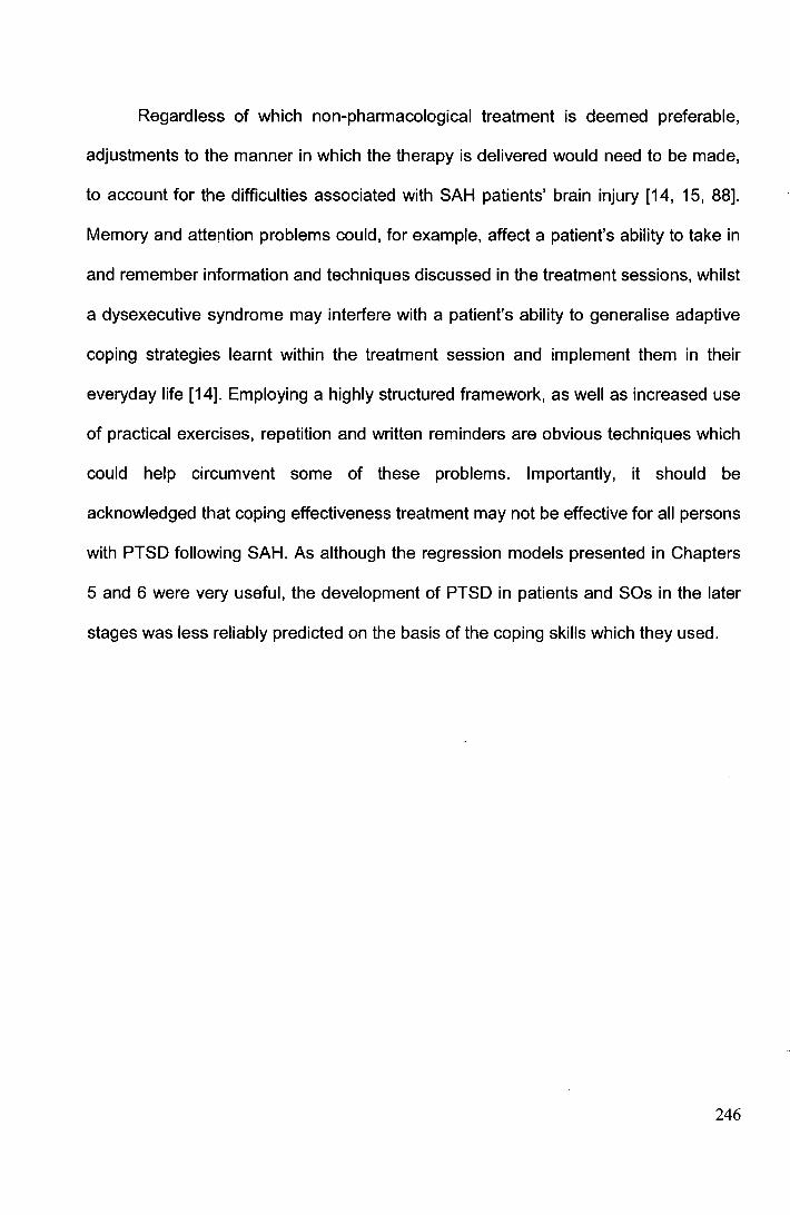
Regardless of which non-pharmacological treatment is deemed preferable,
adjustments to the manner in which the therapy is delivered would need to be made,
to account for the difficulties associated with SAH patients' brain injury [14, 15, 88].
Memory and attention problems could, for example, affect a patient's ability to take in
and remember information and techniques discussed in the treatment sessions, whilst
a dysexecutive syndrome may interfere with a patient's ability to generalise adaptive
coping strategies learnt within the treatment session and implement them in their
everyday life [14]. Employing a highly structured framework, as well as increased use
of practical exercises, repetition and written reminders are obvious techniques which
could help circumvent some of these problems. Importantly, it should be
acknowledged that coping effectiveness treatment may not be effective for all persons
with PTSD following SAH. As although the regression models presented in Chapters
5 and 6 were very useful, the development of PTSD in patients and SOs in the later
stages was less reliably predicted on the basis of the coping skills which they used.
246
7.4 Implications for future research
The findings of the research presented in this thesis lead to extremely interesting
directions for future studies. Some of the key questions which would it be useful to
address are now described.
7. 4. 1 Why is the incidence of PTSD in the SAH patient population so
high?
As I have already noted in the discussion section to Chapter 5, it is interesting that I
detected a remarkably high incidence of PTSD in the SAH patient population -
namely 37% in the both short- and long-term. This high incidence appears to be at
odds with the usual pattern of low prevalence of PTSD (and psychopathology in
general) that follows internal medical traumas [23, 476, 701]. So how can this high
incidence in the SAH population be explained? The similarity of the incidence of
PTSD in patients detected in this thesis and that of prior studies (full PTSD estimates
26-32% [47, 295]) on SAH which used "gold standard" interview techniques means
that the high incidence cannot be explained by my use of a questionnaire measure of
PTSD which can at times, 'over-diagnose', particularly with medical patients [500,
67 4, 675]. Rather, in my view, two (potentially complimentary) explanations could
account for the high incidence. Firstly, I recruited a fairly large sample of patients and
used broad inclusion sample. Previous studies examining PTSD following other
medical events which have reported finding much lower incidences have often
employed highly restrictive inclusion criteria which could serve to reduce their
likelihood of finding PTSD. For example in cancer studies, it is often only early stage
female breast cancer samples which are studied because of the relative ease of their
recruitment [327, 476]. Evidence suggests that the relatively good prognosis of these
247

participants limits the likelihood of them developing PTSD and that the inclusion of
persons with more advanced cancers would have increased the incidence of PTSD
[311).
The second explanation for the high incidence of PTSD following an SAH is its
unusually severe nature and its aftermath. Although the severity of an event is not by
itself sufficient for the development of PTSD (hence 63% of SAH patients did not its
fulfill its diagnostic criteria in this thesis), nor is it the most important determinant for
who develops the disorder in general [73, 521 ], a dose-response model is
nevertheless predictive to some extent [576]. Specifically, events of a higher a
severity, intensity and physical proximity have been found to pose the greatest risk for
the development of PTSD [23, 576]. This phenomenon has most recently been
illustrated by an epidemiological study of the incidence of PTSD in residents in
different areas of Manhattan, following the terrorist attacks of September 11th 2001 in
New York on the World Trade Center. It was found in multivariate analysis that
proximity to the World Trade Center was a significant predictor of PTSD development.
Judged according to a U.S. normative population estimate of 3.6% of PTSD [620], it
was found that following the attacks 6.8% of persons developed PTSD who lived
towards the north of Manhattan and furthest away from the World Trade Center,
whilst 20% of those persons living towards the southern end of Manhattan, in the
vicinity of the attacks, developed PTSD [209). This latter group was presumably more
directly exposed to the trauma and/or its consequences. The phenomenon that being
directly affected by a disaster carries a greater risk of developing PTSD [233, 492]
explains why the incidence of PTSD is lower in SAH SOs than it is in SAH patients.
So what is it about an SAH that makes it so severe? Not only would an answer to this
question have significant implications for how we could possibly prevent SAH PTSD
248
(as I discuss in the next section), but it could have important ramifications for trauma
research in general as it could help us to better understand the origins of PTSD
reactions.
7.4.2 Prevention of PTSD in SAH patients
As noted by Weinert and Meller [738] with respect to the trauma of intensive care
treatment, we are in a unique position with SAH because "[u]nlike subjects exposed
to the stressors of combat, childhood neglect or physical assault who almost always
present for treatment after the stressor is over ... we can ... intervene while the stressor
is occurring ... almost no other PTSD field can feasibly do this" (p.2). This means that
identifying what it is about an SAH that precipitates PTSD in SAH patients could have
significant implications for prevention as attempts could be made to make the evenUs
less traumatic. This idea of pre-emptive intervention for PTSD has previously been
discussed by Bryant et al. [85] in the context of road-traffic accidents, who on the
basis of their study, suggested that relatively simple changes in the future, such as
the rapid removal of neck and back restraints on arrival to hospital and better
communication about the trauma and its consequences, could help to reduce the
incidence of PTSD.
Identifying the initial stressor event which precipitates PTSD in SAH patients is
however, challenging compared to traditional traumatic events such as rape or
assault, as it is not a discrete event. Instead, the nature of an SAH and its requisite
treatments means that the event of having a 'brain haemorrhage' can be protracted,
with patients potentially being exposed to multiple stressors (including the initial
distressing onset of symptoms, the invasive treatments, the diagnosis, etcetera).
Each of these individual events could be considered to satisfy the DSM-IV-TR
249
definition of a traumatic event (i.e., one which poses a threat to either a person's life
or physical integrity). Pinning down the event which precipitated SAH patients PTSD
has not so far been achieved by SAH patient studies (including the one in this thesis)
because awarding the diagnosis does not necessitate knowing the exact stressor.
Criterion A for a diagnosis of PTSD rather only requires that the person has a history
of exposure to one or more traumatogenic events and that this event or events
subsequently mobilises the three symptom clusters of re-experiencing, avoidance and
hyperarousal which define PTSD [23]. The criterion A event can be as specific as a
physical assault, or as broad as being involved in military combat. SAH studies, as
was the case in this thesis, simply ask patients about their experience of PTSD
symptoms with reference to their "brain haemorrhage". Future studies could employ
two techniques to potentially help determine which event(s) is the actual stressor for
SAH patients. The first would be to use qualitative interview techniques to explore the
content of patients' intrusions. This could help ascertain which event(s) the patients'
psychiatric response is centred upon. Alternatively, a novel approach would be to use
psychophysiological techniques to determine which specific events of the whole SAH
trauma are associated with the most stress. Patients could for example, be asked to
generate personal scripts of their SAH experience. These could then be recorded in a
neutral voice and recounted back to the patient. It may then be possible to identify the
events which precipitated SAH patients' PTSD by determining which aspects of the
recounted trauma were associated with the greatest physiological reaction in the
patients.
It is interesting to note however, that it could actually be the complexity of an
SAH and the fact that it exposes its patients to multiple traumas in a short space of
time that causes an SAH to lead to such a high incidence of PTSD. Evidence is
250
available for instance, to suggest that individuals exposed to prolonged, repeated or
multiple stressful events exhibit greater PTSD symptoms compared to individuals who
experience discrete and single events [356, 447, 642].
In discussing the prevention of PTSD in SAH patients, it is also interesting to
note that there has been recent interest in the administration of medication in the
initial stages of a trauma to prevent PTSD- something possible in the case of SAH.
Specifically, there is some evidence, albeit preliminary [51], that PTSD symptoms can
be reduced following intravenous administration of hydrocortisone in victims of septic
shock in intensive care [601 ], as well as in those undergoing cardiac surgery [7 40].
Such treatment is informed by studies - though not recent ones [770] - which found
PTSD patients show neuroendocrine system alteration (such as lower basal cortisol
concentrations [768] and high noradrenergic activity [457]). It is suggested that
disrupting memory retrieval mechanisms with glucocorticoids during the initial stages
of a trauma could act protectively against the development of PTSD by preventing
recall of traumatic memories [600]. Future studies could consider the role of
hydrocortisone and its ability to reduce PTSD in SAH patients.
7.4.3 What else can help explain HRQoL reduction in SAH patients?
Including PTSD into this thesis' explanative models of HRQoL substantially improves
our ability to comprehend SAH patients' outcome. Nevertheless, even with the
inclusion of this factor, there remains a proportion of variability in SAH patients'
HRQoL that cannot be explained (52% of variance in Mental HRQoL and 67% of
variance in Physical HRQoL). It is important for future studies to identify the factors
that explain this reduction beca,use currently there are some patients with a reduced
HRQoL who we do not know how to help.
251
Given that conceptualising an SAH as a potential psychological stressor has
proved useful in helping us to better explain SAH HRQoL, one possibility is to explore
whether such a concepualisation could even further help to explain SAH patients'
HRQoL. One interesting point raised by trauma-research in general is that it could be
useful for future SAH studies to consider what psychiatric disorders other than PTSD
are being elicited in patients by the trauma of their SAH [443, 607, 623]. A recent
meta-analysis of psychiatric disorders following civilian traumas for instance, found
that although PTSD was the most common psychiatric disorder to develop,
generalised anxiety disorder, substance abuse, phobias and major depressive
disorder are also common [80]. It could be that by measuring only PTSD symptoms, a
group of patients who do not develop PTSD, but who do experience another
psychiatric disorder post-SAH are excluded. This could explain their unexplained
HRQoL reduction. Partial support for this possibility comes from three SAH studies
which examined SAH patients' PTSD symptoms and found only modest correlations
between the degree to which patients experienced PTSD and their scores on broader
measures of psychiatric disturbance [295, 540, 541]. Hutter [295] for instance,
measured the PTSD symptoms of intrusion and avoidance in a sample of 45 SAH
patients and reported that their experience of these symptoms shared around only
25% of variance with their scores on a measure of mood disturbance (namely the
Beck Depression Inventory [36]). This suggests that although a relationship exists
between SAH patients' PTSD and mood disturbance, the two are relatively
independent. Continuing along this theme of taking guidance from trauma research in
general, future studies could also consider the added explanative value that comes
from incorporating information into predictive models about the psychiatric co
morbidity experienced by those SAH patients with PTSD. It is well known from the
252
wider literature for example, that an additional formal psychiatric diagnosis is often
warranted in PTSD patients [68, 125, 351] and that co-morbidity worsens a patient's
HRQoL and functioning [431, 623].
With respect to which non-psychiatric factors could also potentially be involved,
it is difficult to sift through the plethora of factors which have been implicated (see
Tables 2.1 and 2.2 in Chapter 2) and suggest which hold the most promise. Studies
examining them have typically not reported effect size data (or information necessary
for its calculation). However, as I debated in the discussion section of my meta
analysis which I presented in Chapter 3 (see section 3.4), there is some evidence to
suggest that it might be prudent for future studies to re-examine the relationship
between bleed severity and SAH patients' HRQoL, but when bleed severity is indexed
using a more detailed scale. The rationale for this suggestion being that Fisher's CT
rating score [191] which is almost always used to index SAH bleed severity is
potentially too crude to reflect the true variability in anatomical distribution and
quantity of extravasated blood seen on patients' CT scans [306, 363, 371]. Indeed, it
should be highlighted at this point that the Fisher scale was not designed to predict
long-term HRQoL or describe bleed severity, but rather to simply categorise patients
according to the risk that their bleed carried for the development of cerebral
vasospasm [363, 496]. More appropriate scales for future HRQoL studies are
however available. For example, Hidjra et al.'s [270] scale individually quantifies the
presence of blood in the 10 major subarachnoid cisterns. The use of such scales
could reveal that bleed distribution is better correlated with HRQoL than presently
suggested.
253
7.4.4 What else can help predict PTSD development post-SAH?
Dispositional maladaptive coping proved to be highly predictive for the development
of PTSD in SAH patients and SOs in the early stages post-ictus, as well as for most
patients in the longer-term. This finding accords well with extensive evidence- from
both prospective and retrospective studies - that shows that PTSD is highly
associated with these types of coping strategies [87, 88, 165, 172, 217, 328, 493,
635, 655]. The failure however, to predict a subset of those SOs who develop PTSD
in the later stages post-SAH and the reduced discriminative abilities of the model for·
long-term patient prediction, suggests that future work needs to look to refine the
predictive models developed in Chapters 5 and 7 of this thesis and consider including
additional, potentially predictive variables which have been implicated in the wider
literature [73, 521].
7.4.5 What else predicts fatigue and sleep dysfunction in SAH patients?
In the current thesis, PTSD was found to be able to explain a staggering 58% and
42% of variance in SAH patients' experience of chronic fatigue and sleep symptoms,
respectively. Previously these symptoms had remained unexplained. A large
correlation in Chapter 5 between SAH patients' concurrent quality of sleep and fatigue
(assessment one r-.53; assessment two r-.55) suggests that some of the fatigue was
the consequence of impaired sleep which resulted from PTSD. This concurs with
more general research on fatigue in sleep disorders,[166] where fatigue has been
found to be elevated in those with sleep disorders (e.g., apnea, restless leg
syndrome, narcolepsy [395, 471]) and a causal pathway is suggested, as treatment of
these disorders leads to reduced fatigue [471]. Nevertheless, future research should
examine how it is that PTSD can produce the remainder of SAH patients' fatigue, not
254
explained by induced sleep dysfunction. Possibilities could include the constant state
of arousal which a PTSD patient can experience and the adverse health behaviours
associated with the disorder (e.g., smoking, lack of exercise, alcohol use etc.) [351,
389,606].
The finding that a proportion of fatigue and sleep dysfunction cannot be
explained by PTSD, means that future studies need to determine which other factors
are also involved. The fact that a proportion of fatigue remains unexplained is
however, not unexpected as findings from the wider literature suggest that its cause is
often multifactorial, with a variety of causes, including other health conditions (e.g.,
bacterial or viral infections, autoimmune illness) [120, 426], life-style choices (e.g.,
alcohol or caffeine intake, psychosocial stressors) [120, 426] and medications (e.g.,
anticonvulsants, analgesics) [38, 200, 648].
7.5 Conclusions
At the beginning of this thesis, it was stated that SAH is associated with very high
societal and personal costs because the illness affects relatively young persons and
because it causes its survivors and their family and friends to experience significant
psychosocial disability. Unfortunately, it was not known what treatment could be
offered to the survivors and their SOs to improve their outcome because the cause of
their problems remained mysterious. This thesis has however, helped resolve this
mystery by illustrating that the poor psychosocial outcome of SAH patients and their
SOs is caused, to a large extent, by the fact that an abnormally high number of these
persons develop PTSD as a result of the trauma of an SAH. Future studies are
urgently needed to consider the implications of this finding. Importantly, studies need
to determine whether offering treatment for PTSD to SAH survivors and their family
255
and friends reduces the burden associated with this illness. Studies would also be
wise to determine whether teaching patients and their SOs more effective stress
coping strategies can reduce/ prevent PTSD and what it is that makes SAH so
traumatizing that the illness stands out from other medical traumas as causing such a
high degree of PTSD.
256
Bibliography
1. (WHO), W.H.O., The international classification of impairments, disabilities and
handicaps. 1980, Geneva: World Health Organisation.
2. (WHO), W.H.O., International classification of functioning, disability and health
(icf). 2001, Geneva: World Health Organisation.
3. Aboderin, I. and G. Venables, Stroke management in europe. Journal of
Internal Medicine, 1996. 240: p. 173-180.
4. ACROSS, Epidemiology of aneurysmal subarachnoid hemorrhage in australia
and new zealand. Incidence and case fatality from the australasian cooperative
research on subarachnoid hemorrhage study. Stroke, 2000. 31: p. 1843-1850.
5. Adams, H.P., D.D. Jergenson, N.F. Kassel!, and A.L. Sahs, Pitfalls in the
recognition of subarachnoid hemorrhage. Journal of the American Medical
Association, 1980. 244: p. 794-796.
6. Adams, H.P., N.F. Kassel, J.C. Tomer, and A.L. Sahs, Ct and clinical
correlations in recent aneurysmal subarachnoid hemorrhage.
A preliminary report of the cooperative aneurysm study. Neurology, 1983. 33:
p. 981-988.
7. Ahlsj6, B., M. Britton, V. Murray, and T. Theorell, Disablement and quality of
life after stroke. Stroke, 1984. 15: p. 886-890.
8. Alexander, M.P. and M. Freedman, Amnesia after anterior communicating
artery aneurysm rupture. Neurology, 1984. 34: p. 752-757.
9. Allen, K., R.T. Linn, H. Gutierrez, and B.S. Willer, Family burden following
traumatic brain injury. Rehabilitation Psychology, 1994. 39: p. 29-48.
10. Alonso, J., M.C. Angermeyer, S. Bernert, R. Bruffaerts, T.S. Brugha, H.
Bryson, G. de Girolamo, R. Graaf, K. Demyttenaere, I. Gasquet, J.M. Haro,
257
S.J. Katz, R.C. Kessler, V. Kovess, J.P. Lepine, J. Ormel, G. Polidori, L.J.
Russo, G. Vilagut, J. Almansa, S. Arbabzadeh-Bouchez, J. Autonell, M.
Bernal, M.A. Buist-Bouwman, M. Codony, A. Domingo-Salvany, M. Ferrer, S.S. ·
Joo, M. Martinez-Aionso, H. Matschinger, F. Mazzi, Z. Morgan, P. Morosini, C.
Palacin, B. Romera, N. Taub, and W.A. Vollebergh, Disability and quality of life
impact of mental disorders in europe: Results from the european study of the
epidemiology of mental disorders (esemed) project. Acta Psychiatrica
Scandinavica, 2004. 420: p. 38-46.
11. Alonso, J., E. Regidor, G. Barrio, L. Prieto, C. Rodrigues, and L. de Ia Fuente,
Valores poblacionales de referencia de Ia version espanola del cuestionario de
salud sf-36. Medicina Clinica 1998. 111: p. 410-416.
12. Anderson, C., S. Laubscher, and R. Burns, Validation of the short form 36 (sf-
36) health survey questionnaire among stroke patients. Stroke, 1996. 27: p.
1812-1816.
13. Anderson, S.W., M.M. Todd, B.J. Hindman, W.R. Clarke, J.C. Torner, D.
Tranel, B. Yoo, J. Weeks, K.W. Manzel, and S. Samra, Effects of
intraoperative hypothermia on neuropsychological outcomes after intracranial
aneurysm surgery. Annals of Neurology, 2006. 60: p. 518-527.
14. Anson, K. and J. Ponsford, Evaluation of a coping skills group following
traumatic brain injury. Brain Injury, 2006. 20: p. 167-178.
15. Anson, K. and J. Ponsford, Who benefits? Outcome following a coping skills
group intervention for traumatically brain injured individuals. Brain Injury, 2006.
20: p. 1-13.
16. Antoni, M.H., J.M. Lehman, K.M. Kilbourn, A.E. Boyers, J.L. Culver, S.M.
Alferi, S.E. Yount, B.A. McGregor, P.L. Arena, S.D. Harris, A.A. Price, and C.S.
258
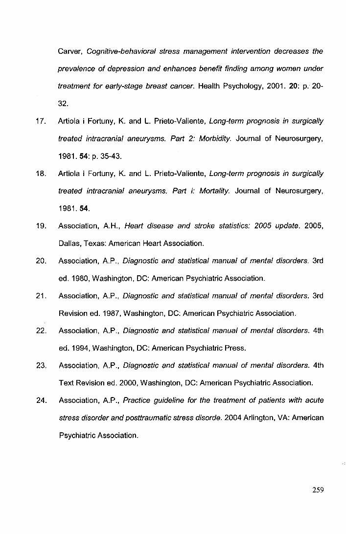
Carver, Cognitive-behavioral stress management intervention decreases the
prevalence of depression and enhances benefit finding among women under
treatment for early-stage breast cancer. Health Psychology, 2001. 20: p.· 20-
32.
17. Artiola i Fortuny, K. and L. Prieto-Valiente, Long-term prognosis in surgically
treated intracranial aneurysms. Part 2: Morbidity. Journal of Neurosurgery,
1981.54: p. 35-43.
18. Artiola i Fortuny, K. and L. Prieto-Valiente, Long-term prognosis in surgically
treated intracranial aneurysms. Part i: Mortality. Journal of Neurosurgery,
1981.54.
19. Association, A.H., Heart disease and stroke statistics: 2005 update. 2005,
Dallas, Texas: American Heart Association.
20. Association, A.P., Diagnostic and statistical manual of mental disorders. 3rd
ed. 1980, Washington, DC: American Psychiatric Association.
21. Association, A.P., Diagnostic and statistical manual of mental disorders. 3rd
Revision ed. 1987, Washington, DC: American Psychiatric Association.
22. Association, A.P., Diagnostic and statistical manual of mental disorders. 4th
ed. 1994, Washington, DC: American Psychiatric Press.
23. Association, A.P., Diagnostic and statistical manual of mental disorders. 4th
Text Revision ed. 2000, Washington, DC: American Psychiatric Association.
24. Association, A.P., Practice guideline for the treatment of patients with acute
stress disorder and posttraumatic stress disorde. 2004 Arlington, VA: American
Psychiatric Association.
259
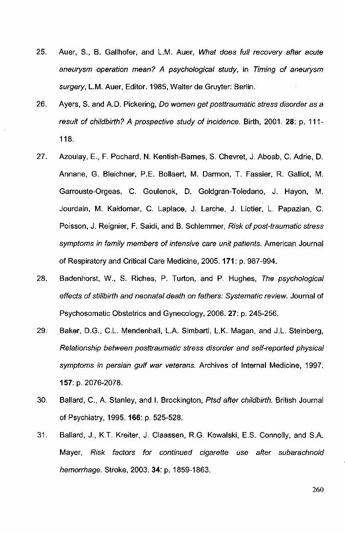
25. Auer, S., B. Gallhofer, and L.M. Auer, What does full recovery after acute
aneurysm operation mean? A psychological study, in Timing of aneurysm
surgery, L.M. Auer, Editor. 1985, Walter de Gruyter: Berlin.
26. Ayers, S. and A.D. Pickering, Do women get posttraumatic stress disorder as a
result of childbirth? A prospective study of incidence. Birth, 2001. 28: p. 111-
118.
27. Azoulay, E., F. Pochard, N. Kentish-Barnes, S. Chevret, J. Aboab, C. Adrie, D.
Annane, G. Bleichner, P.E. Bollaert, M. Darmon, T. Fassier, R. Galliot, M.
Garrouste-Orgeas, C. Goulenok, D. Goldgran-Toledano, J. Hayon, M.
Jourdain, M. Kaidomar, C. Laplace, J. Larche, J. Liotier, L. Papazian, C.
Poisson, J. Reignier, F. Saidi, and B. Schlemmer, Risk of post-traumatic stress
symptoms in family members of intensive care unit patients. American Journal
of Respiratory and Critical Care Medicine, 2005. 171: p. 987-994.
28. Badenhorst, W., S. Riches, P. Turton, and P. Hughes, The psychological
effects of stillbirth and neonatal death on fathers: Systematic review. Journal of
Psychosomatic Obstetrics and Gynecology, 2006. 27: p. 245-256.
29. Baker, D.G., C.L. Mendenhall, L.A. Simbartl, L.K. Magan, and J.L. Steinberg,
Relationship between posttraumatic stress disorder and self-reported physical
symptoms in persian gulf war veterans. Archives of Internal Medicine, 1997.
157: p. 2076-2078.
30. Ballard, C., A. Stanley, and I. Brockington, Ptsd after childbirth. British Journal
of Psychiatry, 1995. 166: p. 525-528.
31. Ballard, J., K.T. Kreiter, J. Claassen, R.G. Kowalski, E.S. Connolly, and S.A.
Mayer, Risk factors for continued cigarette use after subarachnoid
hemorrhage. Stroke, 2003. 34: p. 1859-1863.
260
32. Bamford, J.M., P.A.G. Sandercock, M.S.D. Dennis, J.P.S. Burn, and C.P.
Warlow, A prospective study of acute cerebrovascular disease in the
community: The oxfordshire community stroke project 1981-1982 - 2
incidence, case fatality rates and overall outcome at one year of cerebral
infarction, primary intracerebral and subarachnoid haemorrhage. Journal of
Neurology, Neurosurgery and Psychiatry, 1990. 53: p. 16-22.
33. Bannister, R., Brain's clinical neurology. 1978, Oxford: Oxford University Press.
34. Barbarotto, R., A. Desantis, M. Laiacona, and e. al., Neuropsychological
follow-up of patients operated upon for aneurysms of the middle cerebral artery
and posterior communicating artery. Cortex, 1989. 25: p. 275-288.
35. Bays, C.L., Quality of life of stroke survivors: A research synthesis. Journal of
Neuroscience Nursing, 2001. 31: p. 310-316.
36. Beck, A.T., C.H. Ward, M. Mendeson, J. Mock, and J. Erbaugh, An inventory
for measuring depression. Archives of General Psychiatry, 1961. 4: p. 561-
571.
37. Bederson, J.B., Preface, in Subarachnoid haemorrhage: Pathophysiology and
management, J.B. Bederson, Editor. 1997, The American Association of
Neurological Surgeons: Park Ridge. p. xi-xii.
38. Beenen, L.F.M., L. J., D.J. Touw, H.J. Ader, and H.A.M. van Alphen,
Comparative double blind clinical trial of phenytoin and sodium valporate as
anticonvulsant prophylaxis after craniotomy: Efficacy, tolerability and cognitive
effects. Journal of Neurology, Neurosurgery and Psychiatry, 1999. 67: p. 47 4-
480.
261
39. Begg, C.B., Publication bias, in The handbook of research synthesis, H.
Cooper and L.V. Hedges, Editors. 1994, Russell Sage Foundation: New York.
p. 399-409.
40. Bellebaum, C., L. Schafers, B. Schoch, I. Wanke, D. Stolke, M. Forsting, and I.
Daum, Clipping versus coiling: Neuropsychological follow up after aneurysmal
subarachnoid haemorrhage (sah). Journal of Clinical and Experimental
Neuropsychology, 2004. 26: p. 1081-1092.
41. Bennett, P. and S. Brooke, Intrusive memories, post-traumatic stress disorder
and myocardial infarction. British Journal of Clinical Psychology, 1999. 38: p.
411-416.
42. Bennett, P., M. Conway, J. Clatworthy, S. Brooke, and R. Owen, Predicting
post-traumatic symptoms in cardiac patients. Heart & Lung, 2001. 30: p. 458-
465.
43. Bergner, M., R.A. Bobbitt, W.B. Carter, and G. BS., The sickness impact
profile: Development and final revision of a health status measure. Medical
Care, 1981. 19: p. 787-805.
44. Beristain, X., M. Gaviria, M. Dujovny, T.H.A. EIBary, J.L. Stark, and J.l.
Ausman, Evaluation of outcome after intracranial aneurysm surgery: The
neuropsychiatric approach. Surgical Neurology, 1996. 45: p. 422-428.
45. Beristain, X., M. Gaviria, M. Dujovny, J.L. Stark, and J.l. Ausman, Do we
evaluate outcome appropriately? Neurological Research, 1996. 18: p. 248-250.
46. Bernardini, G.L. and S.A. Mayer, Subarachnoid hemorrhage: Clinical
presentation and neuropsychological outcome. Medical Update for
Psychiatrists, 1998. 3: p. 71-76
262
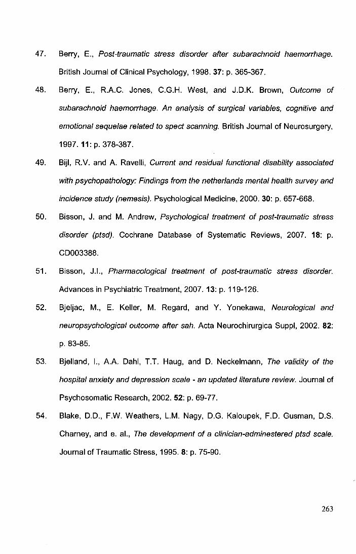
47. Berry, E., Post-traumatic stress disorder after subarachnoid haemorrhage.
British Journal of Clinical Psychology, 1998. 37: p. 365-367.
48. Berry, E., R.A.C. Jones, C.G.H. West, and J.D.K. Brown, Outcome of
subarachnoid haemorrhage. An analysis of surgical variables, cognitive and
emotional sequelae related to spect scanning. British Journal of Neurosurgery,
1997. 11:p.378-387.
49. Bijl, R.V. and A. Ravelli, Current and residual functional disability associated
with psychopathology: Findings from the netherlands mental health survey and
incidence study (nemesis). Psychological Medicine, 2000. 30: p. 657-668.
50. Bisson, J. and M. Andrew, Psychological treatment of post-traumatic stress
disorder (ptsd). Cochrane Database of Systematic Reviews, 2007. 18: p.
CD003388.
51. Bisson, J.l., Pharmacological treatment of post-traumatic stress disorder.
Advances in Psychiatric Treatment, 2007. 13: p. 119-126.
52. Bjeljac, M., E. Keller, M. Regard, and Y. Yonekawa, Neurological and
neuropsychological outcome after sah. Acta Neurochirurgica Suppl, 2002. 82:
p. 83-85.
53. Bjelland, 1., A.A. Dahl, T.T. Haug, and D. Neckelmann, The validity of the
hospital anxiety and depression scale - an updated literature review. Journal of
Psychosomatic Research, 2002. 52: p. 69-77.
54. Blake, D.D., F.W. Weathers, L.M. Nagy, D.G. Kaloupek, F.D. Gusman, D.S.
Charney, and e. al., The development of a clinician-adminestered ptsd scale.
Journal of Traumatic Stress, 1995. 8: p. 75-90.
263
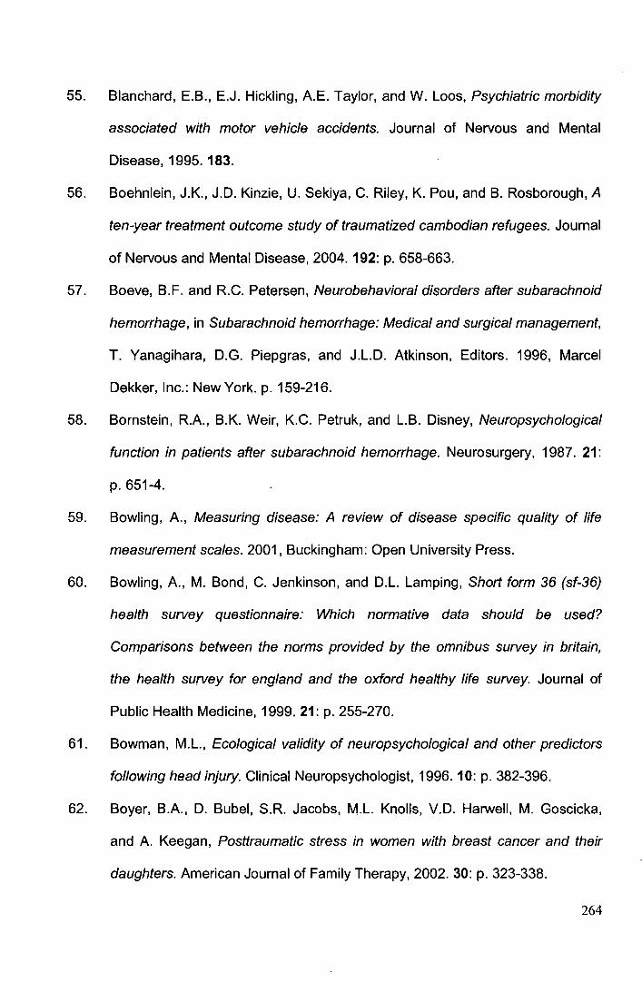
55. Blanchard, E.B., E.J. Hickling, A.E. Taylor, and W. Laos, Psychiatric morbidity
associated with motor vehicle accidents. Journal of Nervous and Mental
Disease, 1995. 183.
56. Boehnlein, J.K., J.D. Kinzie, U. Sekiya, C. Riley, K. Pou, and B. Rosborough, A
ten-year treatment outcome study of traumatized cambodian refugees. Journal
of Nervous and Mental Disease, 2004. 192: p. 658-663.
57. Boeve, B.F. and R.C. Petersen, Neurobehavioral disorders after subarachnoid
hemorrhage, in Subarachnoid hemorrhage: Medical and surgical management,
T. Yanagihara, D.G. Piepgras, and J.L.D. Atkinson, Editors. 1996, Marcel
Dekker, Inc.: New York. p. 159-216.
58. Bornstein, R.A., B.K. Weir, K.C. Petruk, and L.B. Disney, Neuropsychological
function in patients after subarachnoid hemorrhage. Neurosurgery, 1987. 21:
p. 651-4.
59. Bowling, A., Measuring disease: A review of disease specific quality of life
measurement scales. 2001, Buckingham: Open University Press.
60. Bowling, A., M. Bond, C. Jenkinson, and D.L. Lamping, Short form 36 (sf-36)
health survey questionnaire: Which normative data should be used?
Comparisons between the norms provided by the omnibus survey in britain,
the health survey for england and the oxford healthy life survey. Journal of
Public Health Medicine, 1999. 21: p. 255-270.
61. Bowman, M.L., Ecological validity of neuropsychological and other predictors
following head injury. Clinical Neuropsychologist, 1996. 10: p. 382-396.
62. Boyer, B.A., D. Bubel, S.R. Jacobs, M.L. Knolls, V.D. Harwell, M. Goscicka,
and A. Keegan, Posttraumatic stress in women with breast cancer and their
daughters. American Journal of Family Therapy, 2002. 30: p. 323-338.
264
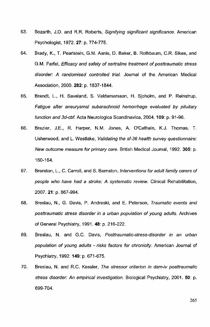
63. Bozarth, J.D. and R.R. Roberts, Signifying significant significance. American
Psychologist, 1972. 27: p. 774-775.
64. Brady, K., T. Pearlstein, G.M. Asnis, D. Baker, B. Rothbaum, C.R. Sikes, and
G.M. Farfel, Efficacy and safety of sertraline treatment of posttraumatic stress
disorder: A randomised controlled trial. Journal of the American Medical
Association, 2000. 282: p. 1837-1844.
65. Brandt, L., H. Saveland, S. Valdemarsson, H. Sjoholm, and P. Reinstrup,
Fatigue after aneurysmal subarachnoid hemorrhage evaluated by pituitary
function and 3d-cbf. Acta Neurologica Scandinavica, 2004. 109: p. 91-96.
66. Brazier, J.E., R. Harper, N.M. Jones, A. O'Cathain, K.J. Thomas, T.
Usherwood, and L. Westlake, Validating the sf-36 health survey questionnaire:
New outcome measure for primary care. British Medical Journal, 1992. 305: p.
160-164.
67. Brereton, L., C. Carroll, and S. Barnston, Interventions for adult family carers of
people who have had a stroke: A systematic review. Clinical Rehabilitation,
2007. 21: p. 867-994.
68. Breslau, N., G. Davis, P. Andreski, and E. Peterson, Traumatic events and
posttraumatic stress disorder in a urban population of young adults. Archives
of General Psychiatry, 1991. 48: p. 216-222.
69. Breslau, N. and G.C. Davis, Posttraumatic-stress-disorder in an urban
population of young adults - risks factors for chronicity. American Journal of
Psychiatry, 1992. 149: p. 671-675.
70. Breslau, N. and R.C. Kessler, The stressor criterion in dsm-iv posttraumatic
stress disorder: An empirical investigation. Biological Psychiatry, 2001. 50: p.
699-704.
265
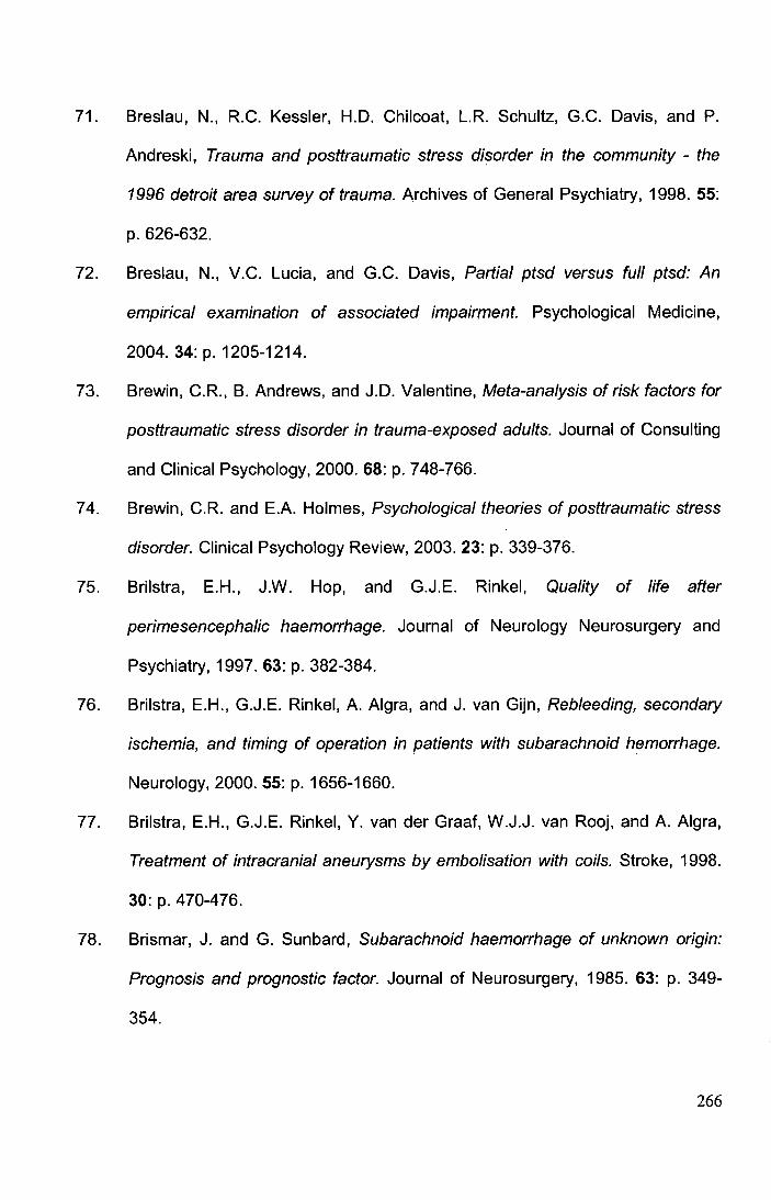
71. Breslau, N., R.C. Kessler, H.D. Chilcoat, L.R. Schultz, G.C. Davis, and P.
Andreski, Trauma and posttraumatic stress disorder in the community- the
1996 detroit area survey of trauma. Archives of General Psychiatry, 1998. 55:
p. 626-632.
72. Breslau, N., V.C. Lucia, and G.C. Davis, Partial ptsd versus full ptsd: An
empirical examination of associated impairment. Psychological Medicine,
2004. 34: p. 1205-1214.
73. Brewin, C.R., B. Andrews, and J.D. Valentine, Meta-analysis of risk factors for
posttraumatic stress disorder in trauma-exposed adults. Journal of Consulting
and Clinical Psychology, 2000. 68: p. 748-766.
7 4. Brewin, C.R. and E.A. Holmes, Psychological theories of posttraumatic stress
disorder. Clinical Psychology Review, 2003. 23: p. 339-376.
75. Brilstra, E.H., J.W. Hop, and G.J.E. Rinkel, Quality of life after
perimesencephalic haemorrhage. Journal of Neurology Neurosurgery and
Psychiatry, 1997. 63: p. 382-384.
76. Brilstra, E.H., G.J.E. Rinkel, A. Algra, and J. van Gijn, Rebleeding, secondary
ischemia, and timing of operation in patients with subarachnoid hemorrhage.
Neurology, 2000. 55: p. 1656-1660.
77. Brilstra, E.H., G.J.E. Rinkel, Y. van der Graaf, W.J.J. van Rooj, and A. Algra,
Treatment of intracranial aneurysms by embolisation with coils. Stroke, 1998.
30: p. 470-476.
78. Brismar, J. and G. Sunbard, Subarachnoid haemorrhage of unknown origin:
Prognosis and prognostic factor. Journal of Neurosurgery, 1985. 63: p. 349-
354.
266
79. Broderick, J.P., C.M. Viscoli, T. Brott, W.N. Kernan, L.M. Brass, E. Feldman,
L.B. Morgenstern, J.L. Wilterdink, and R.I. Horwitz, Major risk factors for
aneurysmal subarachnoid hemorrhage in the young are modifiable. Stroke,
2003. 34: p. 1375-1381.
80. Brown, E.S., M.K. Fulton, A. Wilkeson, and F. Petty, The psychiatric sequelae
of civilian trauma. Comprehensive Psychiatry, 2000. 41: p. 19-23.
81. Bruggimann, L., J.M. Annoni, F. Staub, N. von Steinbuchel, M. Vander Linden,
and J. Bogousslavsky, Chronic posttraumatic stress symptoms after nonsevere
stroke. Neurology, 2006. 66: p. 513-516.
82. Brunella, N., J.R.T. Davidson, M. Deahl, R.C. Kessler, J. Mendlewicz, G.
Racagni, A.Y. Shalev, and J. Zohar, Posttraumatic stress disorder: Diagnosis
and epidemiology, comorbidity and social consequences, biology and
treatment. Neuropsychobiology, 2001.43: p. 150-162.
83. Bruun Wyller, T., J. Holmen, P. Lakke, and K. Laake, Correlates of subjective
well-being in stroke patients. Stroke, 1998. 29: p. 363-367.
84. Bruyere, S.M., S.A. Van Looy, and D.B. Peterson, The international
classification of functioning, disability and health: Contemporary literature
overview. Rehabilitation Psychology, 2005. 50: p. 113-121.
85. Bryant, B., R. Mayou, L. Wiggs, A. Ehlers, and G. Stores, Psychological
consequences of road traffic accidents for children and mothers. Psychological
Medicine, 2004. 34: p. 335-346.
86. Bryant, R.A., Posttraumatic stress disorder and traumatic brain injury: Can they
co-exist? Clinical Psychology Review, 2001. 21: p. 931-948.
267
87. Bryant, R.A. and A.G. Harvey, Avoidant coping style and post-traumatic stress
disorder following motor vehicle accidents. Behaviour Research and Therapy,
1995. 33: p. 631-635.
88. Bryant, R.A., J.E. Marosszeky, J. Crooks, I. Baguley, and J. Gurka, Coping
style and post-traumatic stress disorder following severe traumatic brain injury.
Brain Injury, 2000. 14: p. 175-180.
89. Bryant, R.A., J.E. Marosszeky, J. Crooks, I. Baguley, and J. Gurka,
Posttraumatic stress disorder and psychosocial functioning after severe
traumatic brain injury. Journal of Nervous and Mental Disease, 2001. 189: p.
109-113.
90. Buchanan, K.M., L.J. Elias, and G.B. Goplen, Differing perspectives on
outcome after subarachnoid hemorrhage: The patient, the relative, the
neurosurgeon. Neurosurgery, 2000. 46: p. 831-838.
91. Buck, D., A. Jacoby, A. Massey, and G. Ford, Evaluation of measures used to
assess quality of life after stroke. Stroke, 2000. 31: p. 2004-2010.
92. Buckley, T.C., B.L. Green, and P.P. Schnurr, Trauma, ptsd, and physical
health: Clinical issues, in Assessing psychological trauma and ptsd J.P. Wilson
and T.M. Keane, Editors. 2004, Guilford Publications: New York, NY. p. 441-
465.
93. Buist-Bouwman, M.A., R. DeGraaf, W.A. Vollebergh, J. Alonso, R. Bruffaerts,
J. Ormel, and E.M. Investigators., Functional disability of mental disorders and
comparison with physical disorders: A study among the general population of
six european countries. Acta Psychiatrica Scandinavica, 2006. 113: p. 492-
500.
268
94. Butzkueven, H., A.H. Evans, A. Pitman, C. Leopold, D.J. Jolley, A.H. Kaye,
C.J. Kilpatrick, and S.M. Davis, Onset seizures independently predict poor
outcome after subarachnoid hemorrhage. Neurology, 2000. 55: p. 1315-1320.
95. Buysse, D.J., C.F. Reynolds, T.H. Monk, S.R. Berman, and D.J. Kupfer, The
pittsburgh sleep quality index - a new instrument for psychiatric practice and
research. Psychiatry Research, 1989. 28: p. 193-213.
96. Caci, H., F.H. Bayle, C. Dossios, P. Robert, and P. Boyer, The spielberger trait
anxiety inventory measures more than anxiety. European Psychiatry, 2003. 18:
p. 394-400.
97. Cadell, S., Trauma and growth in canadian carers. Aids Care-Psychological
and Socio-Medical Aspects of Aids/Hiv, 2003. 15: p. 639-648.
98. Campi, A., N. Ramzi, A.J. Molyneux, P.E. Summers, R.S. Kerr, M. Sneade,
J.A. Yarnold, J. Rischmiller, and J.V. Byrne, Retreatment of ruptured cerebral
aneurysms in patients randomised by coiling or clipping in the international
subarachnoid aneurysm trial (isat). Stroke 2007. 38: p. 1538-1544.
99. Canhao, P., J.M. Ferro, A.N. Pinto, T.P. Melo, and J.G. Campos,
Perimesencephalic and nonperimesencephalic subarachnoid hemorrhages
with negative angiograms. Acta Neurochirurgica, 1995. 132: p. 14-19.
100. Cannistraro, P.A., C.l. Wright, M.M. Wedig, B. Martis, L.M. Shin, S. Wilhelm,
and S.L. Rauch, Amygdala responses to human faces in obsessive
compulsive disorder. Biological Psychiatry, 2004. 56: p. 916-920.
101. Carlier, I.V. and B.P. Gerson, Partial posttraumatic stress disorder (ptsd): The
issue of psychological scars and the occurrence of ptsd symptoms. Journal of
Nervous and Mental Disease, 1995. 183: p. 107-109.
269
102. Carod-Artal, J., J.A. Egido, J.L. Gonzalez, and V. de Seijas, Quality of life
among stroke survivors evaluated 1 year after stroke. Stroke, 2000. 31: p.
2995-3000.
103. Carr, A.J., P.W. Thompson, and J.R. Kirwan, Quality of life measures. British
Journal of Rheumatology, 1996. 35: p. 275-281.
104. Carrico, A.W., M.H. Antoni, R.E. Duran, G. lronson, F. Penedo, M.A. Fletcher,
N. Klimas, and N. Schneiderman, Reductions in depressed mood and denial
coping during cognitive behavioral stress management with hiv-positive gay
men treated with haart. Annals of Behavioral Medicine, 2006. 31: p. 155-164.
105. Carter, B.S., D. Buckley, R. Ferraro, G. Rordorf, and C.S. Ogilvy, Factors
associated with reintegration to norma/living after subarachnoid hemorrhage.
Neurosurgery, 2000. 46: p. 1326-1333.
106. Carty, J., M.L. O'Donnell, and M. Creamer, Delayed onset ptsd: A prospective
study of injured survivors. Journal of Affective Disorders, 2006. 90: p. 257-261.
107. Carver, C.S., You want to measure coping but your protocol's too long:
Consider the brief cope. International Journal of Behavioral Medicine, 1997. 4:
p. 92-100.
108. Carver, R.P., The case against statistical significance testing, revisited. The
Journal of Experimental Education, 1993. 61: p. 287-292.
109. Cella, D.F. and A.E. Bonomi, Measuring quality of life, in Cancer management:
A multidisciplinary approach, R. Pazdur, et al., Editors. 1996, PRR Inc.:
Huntington. p. 773-787.
110. Cesarini, K.G., H.G. Hardemark, and L. Persson, Improved survival after
aneurysmal subarachnoid hemorrhage: Review of case management during a
12-year period. J Neurosurg, 1999. 90: p. 664-72.
270
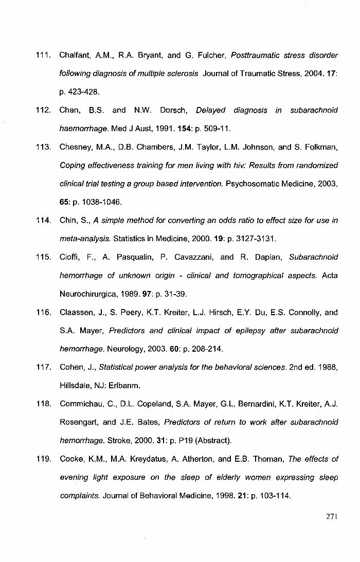
111. Chalfant, A.M., R.A. Bryant, and G. Fulcher, Posttraumatic stress disorder
following diagnosis of multiple sclerosis Journal of Traumatic Stress, 2004. 17:
p. 423-428.
112. Chan, B.S. and N.W. Dorsch, Delayed diagnosis in subarachnoid
haemorrhage. Med J Aust, 1991. 154: p. 509-11.
113. Chesney, M.A., D.B. Chambers, J.M. Taylor, L.M. Johnson, and S. Folkman,
Coping effectiveness training for men living with hiv: Results from randomized
clinical trial testing a group based intervention. Psychosomatic Medicine, 2003.
65: p. 1038-1046.
114. Chin, S., A simple method for converting an odds ratio to effect size for use in
meta-analysis. Statistics in Medicine, 2000. 19: p. 3127-3131.
115. Cioffi, F., A. Pasqualin, P. Cavazzani, and R. Dapian, Subarachnoid
hemorrhage of unknown origin - clinical and tomographical aspects. Acta
Neurochirurgica, 1989. 97: p. 31-39.
116. Claassen, J., S. Peery, K.T. Kreiter, L.J. Hirsch, E.Y. Du, E.S. Connolly, and
S.A. Mayer, Predictors and clinical impact of epilepsy after subarachnoid
hemorrhage. Neurology, 2003. 60: p. 208-214.
117. Cohen, J., Statistical power analysis for the behavioral sciences. 2nd ed. 1988,
Hillsdale, NJ: Erlbanm.
118. Commichau, C., D.L. Copeland, S.A. Mayer, G.L. Bernardini, K.T. Kreiter, A.J.
Rosengart, and J.E. Bates, Predictors of return to work after subarachnoid
hemorrhage. Stroke, 2000.31: p. P19 (Abstract).
119. Cooke, K.M., M.A. Kreydatus, A. Atherton, and E.B. Thoman, The effects of
evening light exposure on the sleep of elderly women expressing sleep
complaints. Journal of Behavioral Medicine, 1998. 21: p. 103-114.
271
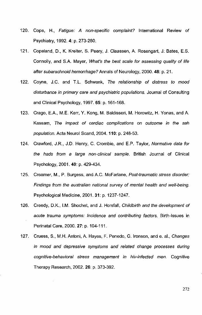
120. Cope, H., Fatigue: A non-specific complaint? International Review of
Psychiatry, 1992. 4: p. 273-280.
121. Copeland, D., K. Kreiter, S. Peery, J. Claassen, A. Rosengart, J. Bates, E.S.
Connolly, and S.A. Mayer, What's the best scale for assessing quality of life
after subarachnoid hemorrhage? Annals of Neurology, 2000. 48: p. 21.
122. Coyne, J.C. and T.L. Schwenk, The relationship of distress to mood
disturbance in primary care and psychiatric populations. Journal of Consulting
and Clinical Psychology, 1997. 65: p. 161-168.
123. Crago, E.A., M.E. Kerr, Y. Kong, M. Baldisseri, M. Horowitz, H. Yonas, and A.
Kassam, The impact of cardiac complications on outcome in the sah
population. Acta Neural Scand, 2004. 110: p. 248-53.
124. Crawford, J.R., J.D. Henry, C. Crombie, and E.P. Taylor, Normative data for
the hads from a large non-clinical sample. British Journal of Clinical
Psychology, 2001. 40: p. 429-434.
125. Creamer, M., P. Burgess, and A.C. McFarlane, Post-traumatic stress disorder:
Findings from the australian national survey of mental health and well-being.
Psychological Medicine, 2001. 31: p. 1237-1247.
126. Creedy, O.K., I.M. Shochet, and J. Horsfall, Childbirth and the development of
acute trauma symptoms: Incidence and contributing factors. Birth-Issues in
Perinatal Care, 2000. 27: p. 104-111.
127. Cruess, S., M.H. Antoni, A. Hayes, F. Penedo, G. lronson, and e. al., Changes
in mood and depressive symptoms and related change processes during
cognitive-behavioral stress management in hiv-infected men. Cognitive
Therapy Research, 2002. 26: p. 373-392.
272
128. Cuthbertson, B.H., A. Hull, M. Strachan, and J. Scott, Post-traumatic stress
disorder after critical illness requiring general intensive care. Intensive Care
Medicine, 2004. 30: p. 450-455.
129. Czechowsky, D. and M.D. Hill, Neurological outcome and quality of life after
stroke due to vertebral artery dissection. Cerebrovasc Dis, 2002. 13: p. 192-
197.
130. d'Avella, D., R. Cicciarello, M. Zuccarello, F. Albiero, A. Romano, F.F. Angileri,
F.M. Salpietro, and F. Tomasello, Brain energy metabolism in the acute stage
of experimental subarachnoid haemorrhage: Local changes in cerebral
glucose utilization. Acta Neurochirurgica, 1996. 138: p. 737-743.
131. David, C.A., A.G. Vishteh, R.F. Spetzler, M. Lemole, M.T. Lawton, and S.
Partovi, Late angiographic follow-up review of surgically treated aneurysms.
Journal of Neurosurgery, 1999. 91: p. 396-340 ..
132. Davidson, J.R.T., Davidson trauma scale. 1996: Multi-Health Systems, Inc.
133. Davidson, J.R.T., D. Hughes, D.G. Blazer, and L.K. George, Posttraumatic
stress disorder in the community - an epidemiological study. Psychological
Medicine, 1991.21: p. 713-721.
134. Davidson, J.R.T., D. Hughes, D.G. Blazer, and L.K. George, Posttraumatic
stress disorder in the community: An epidemiological study. Psychological
Medicine, 1991.21: p. 1-19.
135. Davidson, J.R.T., B.O. Rothbaum, B.A. van der Kolk, C.R. Sikes, and G.M.
Farfel, Multicenter, double-blind comparison of sertraline and placebo in the
treatment of posttraumatic stress disorder. Archives of General Psychiatry,
2001. 58:p. 485-492.
273
136. Davis, L. and L.J. Siegel, Posttraumatic stress disorder in children and
adolescents: A review and analysis. Clinical Child and Family Psychology
Review, 2000.3: p. 135-154.
137. De Haan, R., J. Horn, M. Limburg, J. Van Der Meulen, and P. Bossuyt, A
comparison of five stroke scales with measures of disability, handicap, and
quality oflife. Stroke, 1993. 24: p. 1178-1181.
138. de Haan, R.J., Measuring quality of life after stroke using the sf-36. Stroke,
2002. 33:p. 1176-1177.
139. De Renzi, E. and P. Faglioni, Normative data and screening power of a
shortened version of the token test. Cortex, 1978. 14: p. 41-9.
140. de Rooij, N.K., F.H. Linn, J.A. van der Plas, A. Algra, and G.J. Rinkel,
Incidence of subarachnoid haemorrhage: A systematic review with emphasis
on region, age, gender and time trends. Journal of Neurology, Neurosurgery
and Psychiatry., 2007.78: p. 1365-1372.
141. De Santis, A., M. Laiacona, R. Barbarotto, A. Basso, R. Villani, D. Spagnoli,
and E. Capitani, Neuropsychological outcome of patients operated upon for an
intracranial aneurysm: Analysis of general prognostic factors and of the effects
of the location of the aneurysm. Journal of Neurology, Neurosurgery and
Psychiatry, 1989. 52: p. 1135-1140.
142. de Vries, A.P.J., N. Kassam-Adams, A. Cnaan, E. Sherman-Slate, P.R.
Gallagher, and F .K. Winston, Looking beyond the physical injury:
Posttraumatic stress disorder in children and parents after pediatric traffic
injury. Pediatrics, 1999. 104: p. 1293-1299.
274
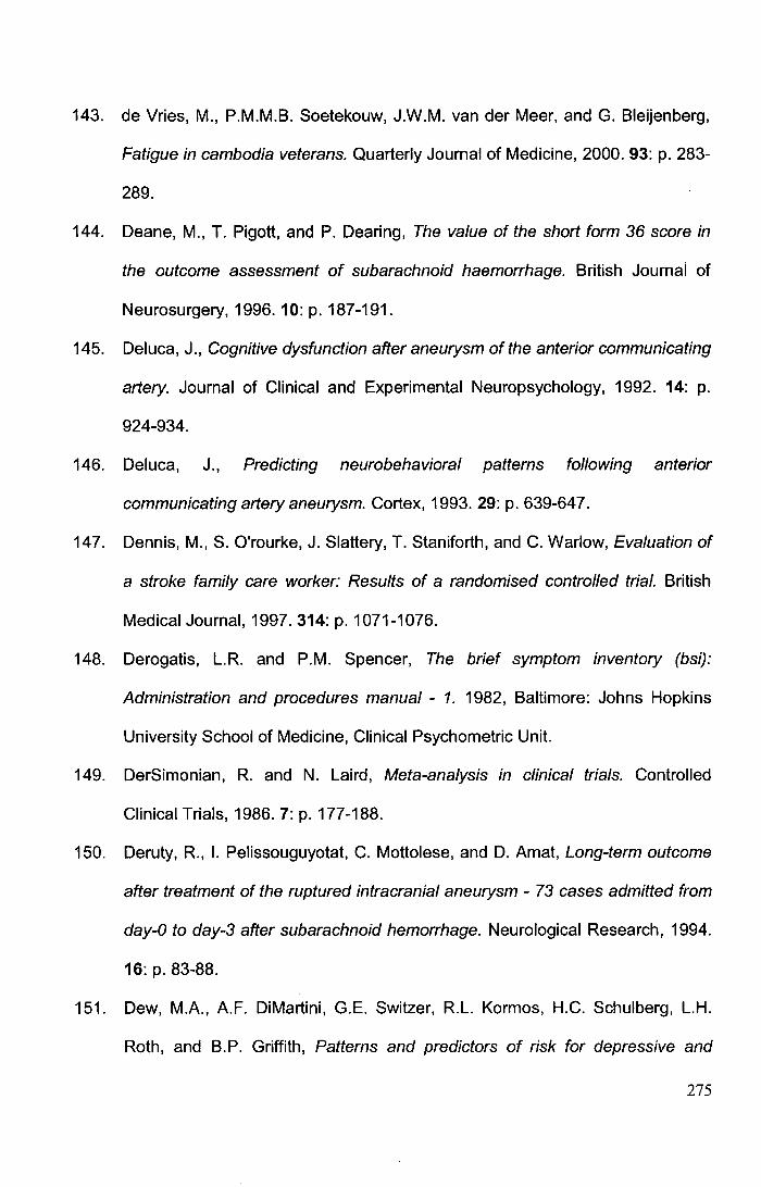
143. de Vries, M., P.M.M.B. Soetekouw, J.W.M. van der Meer, and G. Bleijenberg,
Fatigue in cambodia veterans. Quarterly Journal of Medicine, 2000. 93: p. 283-
289.
144. Deane, M., T. Pigott, and P. Dearing, The value of the short form 36 score in
the outcome assessment of subarachnoid haemorrhage. British Journal of
Neurosurgery, 1996. 10: p.187-191.
145. Deluca, J., Cognitive dysfunction after aneurysm of the anterior communicating
artery. Journal of Clinical and Experimental Neuropsychology, 1992. 14: p.
924-934.
146. Deluca, J., Predicting neurobehavioral patterns following anterior
communicating artery aneurysm. Cortex, 1993. 29: p. 639-64 7.
147. Dennis, M., S. O'rourke, J. Slattery, T. Staniforth, and C. Warlow, Evaluation of
a stroke family care worker: Results of a randomised controlled trial. British
Medical Journal, 1997.314: p. 1071-1076.
148. Derogatis, L.R. and P.M. Spencer, The brief symptom inventory (bsi):
Administration and procedures manual - 1. 1982, Baltimore: Johns Hopkins
University School of Medicine, Clinical Psychometric Unit.
149. DerSimonian, R. and N. Laird, Meta-analysis in clinical trials. Controlled
Clinical Trials, 1986.7: p.177-188.
150. Deruty, R., I. Pelissouguyotat, C. Mottolese, and D. Amat, Long-term outcome
after treatment of the ruptured intracranial aneurysm - 73 cases admitted from
day-0 to day-3 after subarachnoid hemorrhage. Neurological Research, 1994.
16: p. 83-88.
151. Dew, M.A., A.F. DiMartini, G.E. Switzer, R.L. Kormos, H.C. Schulberg, L.H.
Roth, and B.P. Griffith, Patterns and predictors of risk for depressive and
275
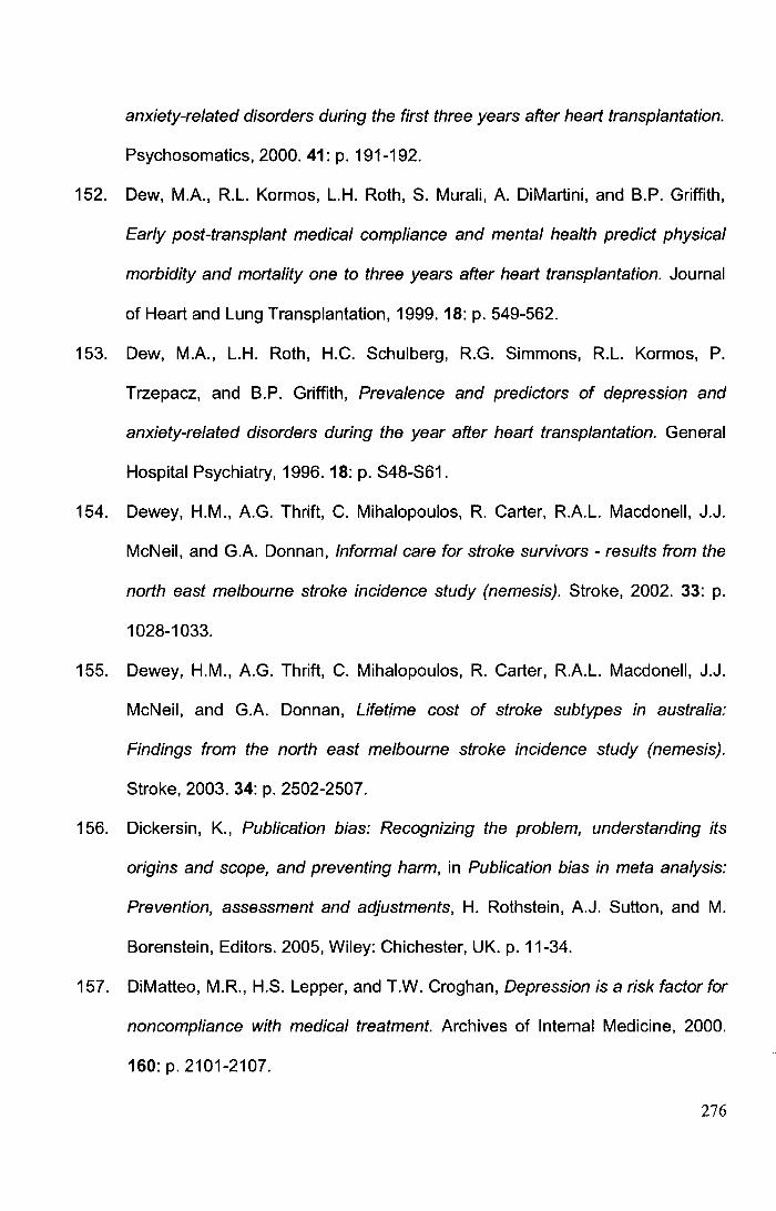
anxiety-related disorders during the first three years after heart transplantation.
Psychosomatics, 2000.41: p. 191-192.
152. Dew, M.A., R.L. Kormos, L.H. Roth, S. Murali, A. DiMartini, and B.P. Griffith,
Early post-transplant medical compliance and mental health predict physical
morbidity and mortality one to three years after heart transplantation. Journal
of Heart and Lung Transplantation, 1999. 18: p. 549-562.
153. Dew, M.A., L.H. Roth, H.C. Schulberg, R.G. Simmons, R.L. Kormos, P.
Trzepacz, and B.P. Griffith, Prevalence and predictors of depression and
anxiety-related disorders during the year after heart transplantation. General
Hospital Psychiatry, 1996. 18: p. S48-S61.
154. Dewey, H.M., A.G. Thrift, C. Mihalopoulos, R. Carter, R.A.L. Macdonell, J.J.
McNeil, and G.A. Donnan, Informal care for stroke survivors- results from the
north east melbourne stroke incidence study (nemesis). Stroke, 2002. 33: p.
1028-1033.
155. Dewey, H.M., A.G. Thrift, C. Mihalopoulos, R. Carter, R.A.L. Macdonell, J.J.
McNeil, and G.A. Donnan, Lifetime cost of stroke subtypes in australia:
Findings from the north east melbourne stroke incidence study (nemesis).
Stroke, 2003. 34: p. 2502-2507.
156. Dickersin, K., Publication bias: Recognizing the problem, understanding its
origins and scope, and preventing harm, in Publication bias in meta analysis:
Prevention, assessment and adjustments, H. Rothstein, A.J. Sutton, and M.
Borenstein, Editors. 2005, Wiley: Chichester, UK. p. 11-34.
157. DiMatteo, M.R., H.S. Lepper, and T.W. Croghan, Depression is a risk factor for
noncompliance with medical treatment. Archives of Internal Medicine, 2000.
160: p. 2101-2107.
276

158. Doczi, T., The pathogenetic and prognostic-significance of blood-brain- barrier
damage at the acute stage of aneurysmal subarachnoid hemorrhage - clinical
and experimental studies. Acta Neurochirurgica, 1985. 77: p. 110-132.
159. Dombovy, M.L., J. Drew-Cates, and R. Serdans, Recovery and rehabilitation
following subarachnoid haemorrhage. Part i: Outcome after inpatient
rehabilitation. Brain Injury, 1998. 12: p. 443-454.
160. Dombovy, M.L., J. Drew-Cates, and R. Serdans, Recovery and rehabilitation
following subarachnoid haemorrhage: Part ii long-term follow-up. Brain Injury,
1998. 12:p.887-894.
161. Dombovy, M.L., B.A. Sandok, and J.R. Basford, Rehabilitation for stroke: A
review. Stroke, 1986. 17: p. 363-369.
162. Douglas, J.M. and F.J. Spellacy, Correlates of depression in adults with severe
traumatic brain injury and their carers. Brain Injury, 2000. 14: p. 71-88.
163. Douglass, A.B., R. Bornstein, G. Ninomurcia, S. Keenan, L. Miles, V.P.
Zarcone, C. Guilleminault, and W.C. Dement, The sleep disorders
questionnaire-i - creation and multivariate structure of sdq. Sleep, 1994. 17: p.
160-167.
164. Drake, C.G., Report of world-federation-of-neurological-surgeons committee
on a universal subarachnoid hemorrhage grading scale. Journal of
Neurosurgery, 1988. 68: p. 985-986.
165. Dunmore, E., D.M. Clark, and A. Ehlers, A prospective investigation of the role
of cognitive factors in persistent posttraumatic stress disorder (ptsd) after
physical or sexaul assault. Behavior Research and Therapy, 2001. 39: p.
1063-1084.
277
166. Duntley, S.P., Fatigue and sleep, in Fatigue as a window to the brain J.
Deluca, Editor. 2005, MIT Press: Cambridge, Mass. p. 209-227.
167. Dyster-Aas, J., M. Willebrand, B. Wikehult, B. Gardin, and L. Ekselius, Major
depression and posttraumatic stress disorder symptoms following severe burn
injury in relation to lifetime psychiatric history. Journal of Trauma, 2008. 64: p.
1349-1356.
168. Edelman, D., R. Williams, M. Rothman, and G.P. Samsa, A comparison of
three health status measures in primary care outpatients Journal of Geriatric
and Internal Medicine, 1999. 14: p. 759-762.
169. Egge, A., K. Waterloo, H. Sjoholm, T. lngebrigtsen, S. Forsdahl, E.A.
Jacobsen, and B. Romner, Outcome 1 year after aneurysmal subarachnoid
hemorrhage: Relation between cognitive performance and neuroimaging. Acta
Neurologica Scandinavica, 2005. 112: p. 76-80.
170. Egger, M., G. Davey-Smith, M. Schneider, and C. Minder, Bias in meta
analysis detected by a simple, graphical test. British Medical Journal, 1997.
315: p. 629-634.
171. Ehlers, A. and D.M. Clark, A cognitive model of posttraumatic stress disorder.
Behaviour Research and Therapy, 2000. 38: p. 319-345.
172. Eid, J., The course of ptsd symptoms following military training accidents and
brief psychosocial interventions. Personality and Individual Differences, 2003.
35: p. 771-783.
173. Engel, C.C., X. Liu, B.D. McCarthy, R.F. Miller, and R. Ursano, Relationship of
physical symptoms to posttraumatic stress disorder among veterans seeking
care for gulf war-related health concerns. Psychosomatic Medicine, 2000. 62:
p. 739-745.
278
174. Erdfelder, E., F. Faul, and A. Buchner, Gpower: A general power analysis
program. Behavior Research Methods, Instruments and Computers, 1996. 28:
p.1-11.
175. Escobar, J.l., G. Canino, M. Rubiostipec, and M. Bravo, Somatic symptoms
after a natural disaster- a prospective study. American Journal of Psychiatry,
1992. 149: p. 965-967.
176. Eskesen, V., E.B. Sorensen, J. Rosenorn, and K. Schmidt, The prognosis in
subarachnoid hemorrhage of unknown etiology. Journal of Neurosurgery,
1984. 61: p. 1029-1031.
177. Ettner, S.L., R.G. Frank, and R.C. Kessler, The impact of psychiatric disorders
on labor market outcomes. Industrial & Labor Relations Review, 1997. 51: p.
64-81.
178. Evans, R.L. and L.K. Northwood, Social support needs in adjustment to stroke.
Archives of Physical Medicine and Rehabilitation, 1983. 64: p. 61-64.
179. Evans, S. and P. Huxley, Studies of quality of life in the general population.
International Review of Psychiatry, 2002. 14: p. 203-211.
180. Ezrachi, 0., Y. Ben-Yishay, T. Kay, L. Diller, and J. Rattok, Predicting
employment in traumatic brain injury following neuropsychological
rehabilitation. Journal of Head Trauma and Rehabilitation, 1991. 6: p. 71-84.
181. Fahrenberg, J., R. Hampel, and H. Selg, Das freiburger personlichkeitsinventar
fpi. Revidierte fassung fpi-r. 1984, Gottingen: Verlag fUr Psychologie, CJ
Hogrefe.
182. Fahrenberg, J., R. Hampel, and H. Selg, Die revidierte form des freiburger
personlichkeitsinventars fpi. Diagnostica, 1985. 31: p. 1-21.
279
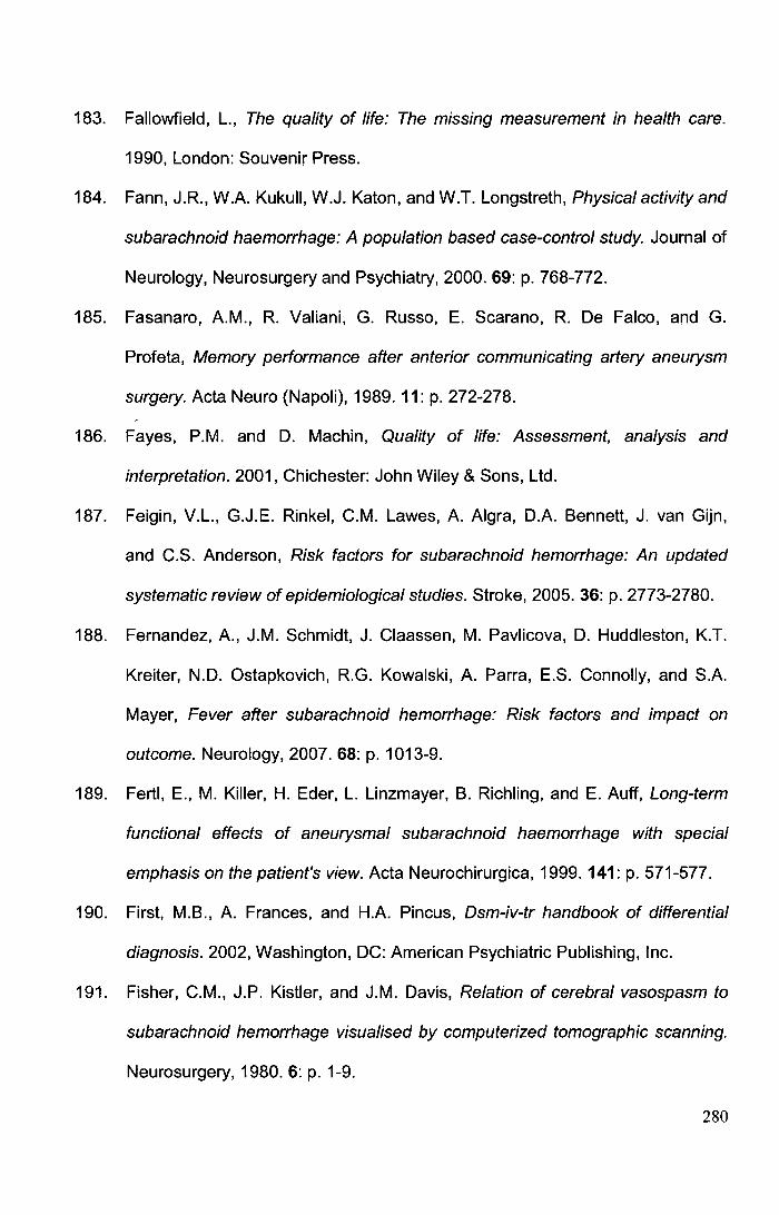
183. Fallowfield, L., The quality of life: The missing measurement in health care.
1990, London: Souvenir Press.
184. Fann, J.R., W.A. Kukull, W.J. Katon, and W.T. Longstreth, Physical activity and
subarachnoid haemorrhage: A population based case-control study. Journal of
Neurology, Neurosurgery and Psychiatry, 2000. 69: p. 768-772.
185. Fasanaro, A.M., R. Valiani, G. Russo, E. Scarano, R. De Falco, and G.
Profeta, Memory performance after anterior communicating artery aneurysm
surgery. Acta Neuro (Napoli), 1989. 11: p. 272-278.
186. Fayes, P.M. and D. Machin, Quality of life: Assessment, analysis and
interpretation. 2001, Chichester: John Wiley & Sons, Ltd.
187. Feigin, V.L., G.J.E. Rinkel, C.M. Lawes, A. Algra, D.A. Bennett, J. van Gijn,
and C.S. Anderson, Risk factors for subarachnoid hemorrhage: An updated
systematic review of epidemiological studies. Stroke, 2005. 36: p. 2773-2780.
188. Fernandez, A., J.M. Schmidt, J. Claassen, M. Pavlicova, D. Huddleston, K.T.
Kreiter, N.D. Ostapkovich, R.G. Kowalski, A. Parra, E.S. Connolly, and S.A.
Mayer, Fever after subarachnoid hemorrhage: Risk factors and impact on
outcome. Neurology, 2007. 68: p. 1013-9.
189. Fertl, E., M. Killer, H. Eder, L. Linzmayer, B. Richling, and E. Auff, Long-term
functional effects of aneurysmal subarachnoid haemorrhage with special
emphasis on the patient's view. Acta Neurochirurgica, 1999. 141: p. 571-577.
190. First, M.B., A. Frances, and H.A. Pincus, Dsm-iv-tr handbook of differential
diagnosis. 2002, Washington, DC: American Psychiatric Publishing, Inc.
191. Fisher, C.M., J.P. Kistler, and J.M. Davis, Relation of cerebral vasospasm to
subarachnoid hemorrhage visualised by computerized tomographic scanning.
Neurosurgery, 1980. 6: p. 1-9.
280
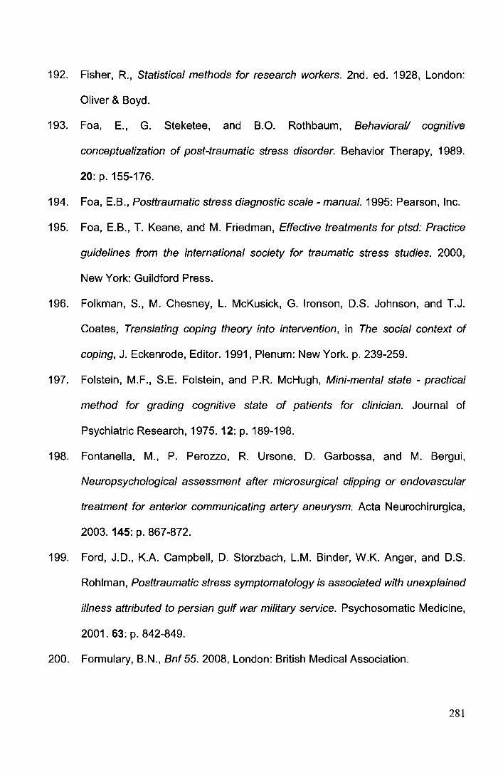
192. Fisher, R., Statistical methods for research workers. 2nd. ed. 1928, London:
Oliver & Boyd.
193. Foa, E., G. Steketee, and B.O. Rothbaum, Behavioral/ cognitive
conceptualization of post-traumatic stress disorder. Behavior Therapy, 1989.
20: p. 155-176.
194. Foa, E.B., Posttraumatic stress diagnostic scale- manual. 1995: Pearson, Inc.
195. Foa, E.B., T. Keane, and M. Friedman, Effective treatments for ptsd: Practice
guidelines from the international society for traumatic stress studies. 2000,
New York: Guildford Press.
196. Folkman, S., M. Chesney, L. McKusick, G. lronson, D.S. Johnson, and T.J.
Coates, Translating coping theory into intervention, in The social context of
coping, J. Eckenrode, Editor. 1991, Plenum: New York. p. 239-259.
197. Folstein, M.F., S.E. Folstein, and P.R. McHugh, Mini-mental state -practical
method for grading cognitive state of patients for clinician. Journal of
Psychiatric Research, 1975. 12: p. 189-198.
198. Fontanella, M., P. Perozzo, R. Ursone, D. Garbossa, and M. Bergui,
Neuropsychological assessment after microsurgical clipping or endovascular
treatment for anterior communicating artery aneurysm. Acta Neurochirurgica,
2003. 145: p. 867-872.
199. Ford, J.D., K.A. Campbell, D. Storzbach, L.M. Binder, W.K. Anger, and D.S.
Rohlman, Posttraumatic stress symptomatology is associated with unexplained
illness attributed to persian gulf war military service. Psychosomatic Medicine,
2001. 63:p. 842-849.
200. Formulary, B.N., Bnf 55. 2008, London: British Medical Association.
281
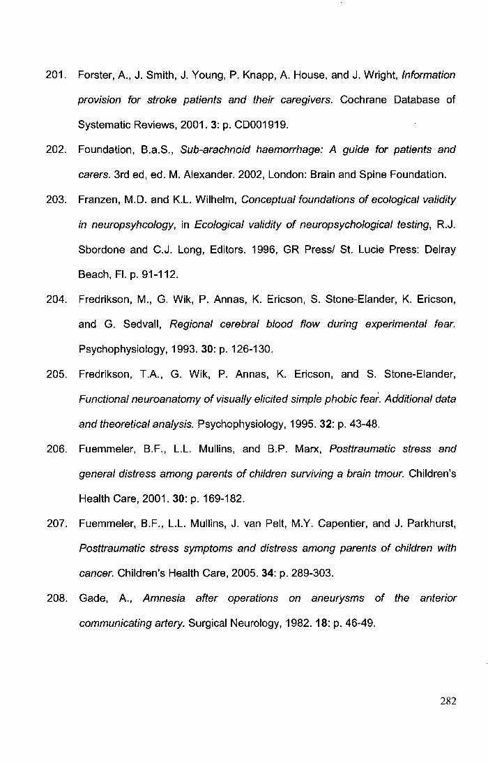
201. Forster, A., J. Smith, J. Young, P. Knapp, A. House, and J. Wright, Information
provision for stroke patients and their caregivers. Cochrane Database of
Systematic Reviews, 2001. 3: p. CD001919.
202. Foundation, B.a.S., Sub-arachnoid haemorrhage: A guide for patients and
carers. 3rd ed, ed. M. Alexander. 2002, London: Brain and Spine Foundation.
203. Franzen, M.D. and K.L. Wilhelm, Conceptual foundations of ecological validity
in neuropsyhcology, in Ecological validity of neuropsychological testing, R.J.
Sbordone and C.J. Long, Editors. 1996, GR Press/ St. Lucie Press: Delray
Beach, Fl. p. 91-112.
204. Fredrikson, M., G. Wik, P. Annas, K. Ericson, S. Stone-Eiander, K. Ericson,
and G. Sedvall, Regional cerebral blood flow during experimental fear.
Psychophysiology, 1993. 30: p. 126-130.
205. Fredrikson, T.A., G. Wik, P. Annas, K. Ericson, and S. Stone-Eiander,
Functional neuroanatomy of visually elicited simple phobic fear. Additional data
and theoretical analysis. Psychophysiology, 1995. 32: p. 43-48.
206. Fuemmeler, B.F., L.L. Mullins, and B.P. Marx, Posttraumatic stress and
general distress among parents of children surviving a brain tmour. Children's
Health Care, 2001. 30: p. 169-182.
207. Fuemmeler, B.F., L.L. Mullins, J. van Pelt, M.Y. Capentier, and J. Parkhurst,
Posttraumatic stress symptoms and distress among parents of children with
cancer. Children's Health Care, 2005. 34: p. 289-303.
208. Gade, A., Amnesia after operations on aneurysms of the anterior
communicating artery. Surgical Neurology, 1982. 18: p. 46-49.
282
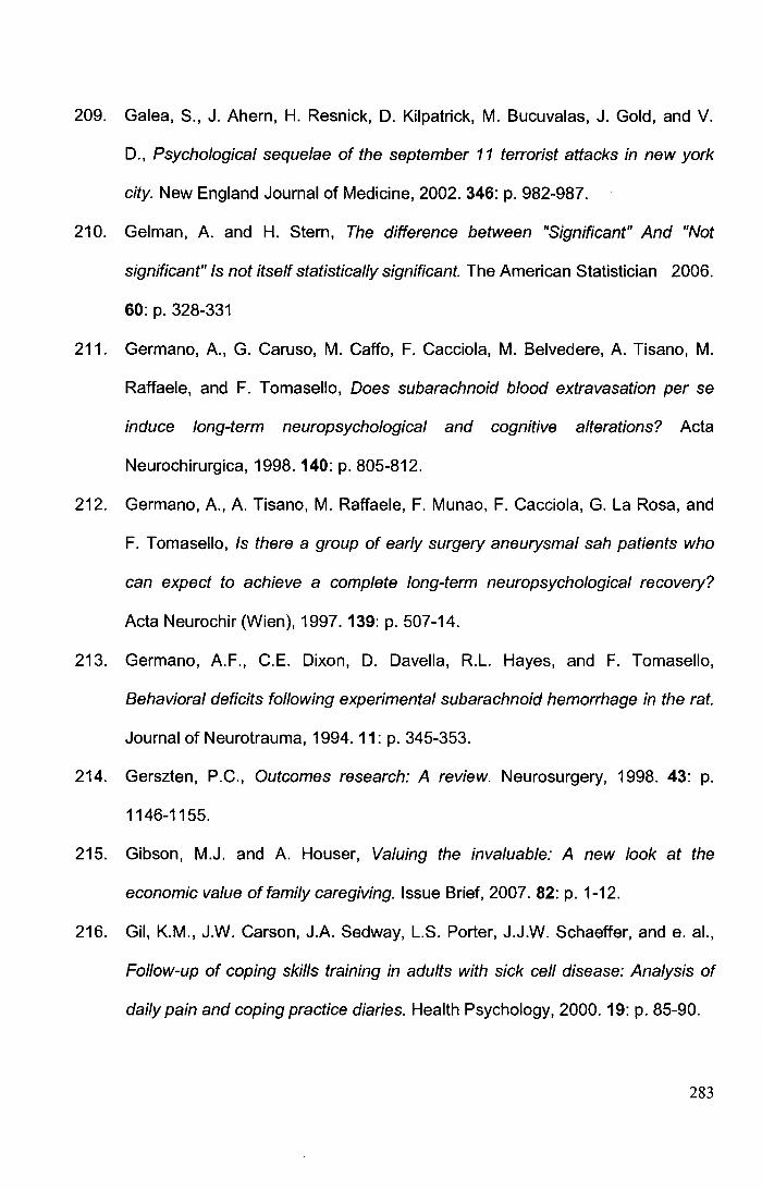
209. Galea, S., J. Ahern, H. Resnick, D. Kilpatrick, M. Bucuvalas, J. Gold, and V.
D., Psychological sequelae of the september 11 terrorist attacks in new york
city. New England Journal of Medicine, 2002. 346: p. 982-987.
210. Gelman, A. and H. Stern, The difference between "Significant" And "Not
significant" Is not itself statistically significant. The American Statistician 2006.
60: p. 328-331
211. Germano, A., G. Caruso, M. Caffo, F. Cacciola, M. Belvedere, A. Tisano, M.
Raffaele, and F. Tomasello, Does subarachnoid blood extravasation per se
induce long-term neuropsychological and cognitive alterations? Acta
Neurochirurgica, 1998. 140: p. 805-812.
212. Germano, A., A. Tisano, M. Raffaele, F. Munao, F. Cacciola, G. La Rosa, and
F. Tomasello, Is there a group of early surgery aneurysmal sah patients who
can expect to achieve a complete long-term neuropsychological recovery?
Acta Neurochir (Wien), 1997. 139: p. 507-14.
213. Germano, A.F., C.E. Dixon, D. Davella, R.L. Hayes, and F. Tomasello,
Behavioral deficits following experimental subarachnoid hemorrhage in the rat.
Journal of Neurotrauma, 1994. 11: p. 345-353.
214. Gerszten, P.C., Outcomes research: A review. Neurosurgery, 1998. 43: p.
1146-1155.
215. Gibson, M.J. and A. Houser, Valuing the invaluable: A new look at the
economic value offamily caregiving. Issue Brief, 2007. 82: p. 1-12.
216. Gil, K.M., J.W. Carson, J.A. Sedway, L.S. Porter, J.J.W. Schaeffer, and e. al.,
Follow-up of coping skills training in adults with sick cell disease: Analysis of
daily pain and coping practice diaries. Health Psychology, 2000. 19: p. 85-90.
283
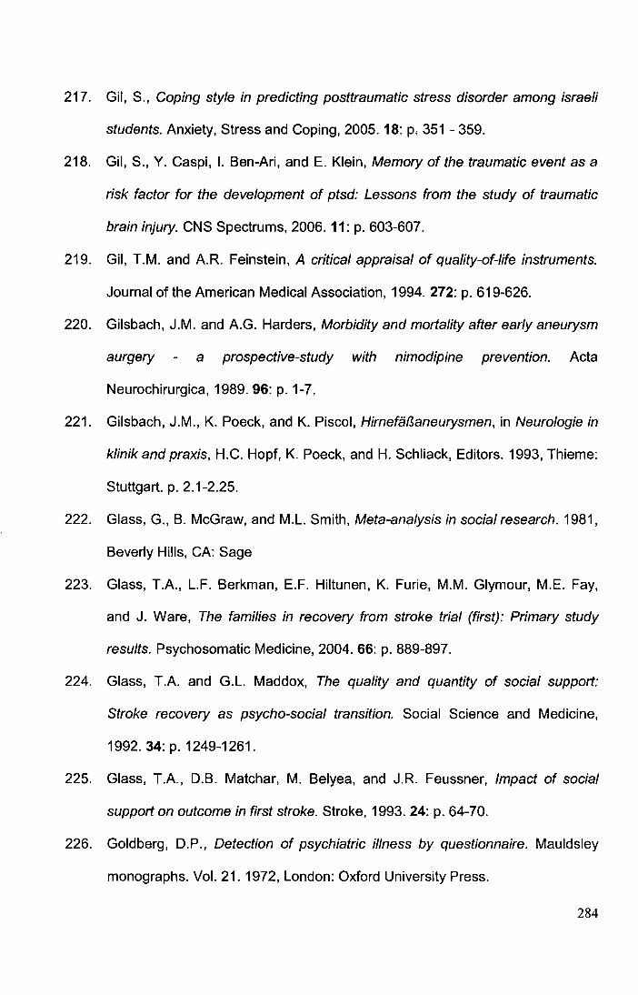
217. Gil, S., Coping style in predicting posttraumatic stress disorder among israeli
students. Anxiety, Stress and Coping, 2005. 18: p. 351 - 359.
218. Gil, S., Y. Caspi, I. Ben-Ari, and E. Klein, Memory of the traumatic event as a
risk factor for the development of ptsd: Lessons from the study of traumatic
brain injury. CNS Spectrums, 2006. 11: p. 603-607.
219. Gil, T.M. and A.R. Feinstein, A critical appraisal of quality-of-life instruments.
Journal of the American Medical Association, 1994. 272: p. 619-626.
220. Gilsbach, J.M. and A.G. Harders, Morbidity and mortality after early aneurysm
aurgery a prospective-study with nimodipine prevention. Acta
Neurochirurgica, 1989. 96: p. 1-7.
221. Gilsbach, J.M., K. Poeck, and K. Piscol, Hirnefal3aneurysmen, in Neurologie in
klinik and praxis, H.C. Hopf, K. Poeck, and H. Schliack, Editors. 1993, Thieme:
Stuttgart. p. 2.1-2.25.
222. Glass, G., B. McGraw, and M.L. Smith, Meta-analysis in social research. 1981,
Beverly Hills, CA: Sage
223. Glass, T.A., L.F. Berkman, E.F. Hiltunen, K. Furie, M.M. Glymour, M.E. Fay,
and J. Ware, The families in recovery from stroke trial (first): Primary study
results. Psychosomatic Medicine, 2004. 66: p. 889-897.
224. Glass, T.A. and G.L. Maddox, The quality and quantity of social support:
Stroke recovery as psycho-social transition. Social Science and Medicine,
1992.34:p. 1249-1261.
225. Glass, T.A., D.B. Matchar, M. Belyea, and J.R. Feussner, Impact of social
support on outcome in first stroke. Stroke, 1993. 24: p. 64-70.
226. Goldberg, D.P., Detection of psychiatric illness by questionnaire. Mauldsley
monographs. Vol. 21. 1972, London: Oxford University Press.
284
227. Goldberg, D.P., Manual for the general health questionnaire. 1978, Windsor:
NFER-Nelson.
228. Goldflam, S., Beitrag zue atiologie und symptomatologie der spontanen
subarachnoidalen blutungen. Deutsche Zeitschrift fUr Nervenheilkunde, 1923.
76: p. 158-182.
229. Golding, J.M., Sexual assault history and physical health in randomly selected
los-angeles women. Health Psychology, 1994. 13: p. 130-138.
230. Goldstein, L.B., C. Bertels, and J.N. Davis, lnterrater reliability of the nih stroke
scale. Archives of Neurology, 1989. 39.
231. Graff-Radford, N.R., J. Tomer, H.P. Adams, and N.F. Kassel, Factors
associated with hydrocephalus after subarachnoid hemorrhage. A report of the
cooperative aneurysm study. Archives of Neurology, 1989. 46: p. 744-752.
232. Granger, C.V. and G.E. Gresham, Functional assessment in rehabilitation
medicine. 1984, Baltimore: Williams and Wilkins.
233. Green, B., M. Grace, J. Lindy, G.C. Gieser, A.C. Leonard, and T.L. Kramer,
Buffalo creek survivors in the second decade: Comparison with unexposed
and nonlitigant groups. Journal of Applied Social Psychology, 1990. 20: p.
1033-1050.
234. Greenfield, S. and E.C. Nelson, Recent developments and future issues in the
use of health status assessment measures in clinical settings. Journal of
Clinical Epidemiology, 1992. 30: p. MS23-41.
235. Greveson, G.C., C.S. Gray, J.M. French, and O.F.W. James, Long-term
outcome for patients and carers following hospital admission for stroke. Age
and Ageing, 1991. 20: p. 337-344.
285
236. Grote, E. and W. Hassler, The critical first minutes after subarachnoid
hemorrhage. Neurosurgery, 1988. 22: p. 654-661.
237. Group, T.E., Euroqol: A new facility for the measurement of health related
qualityoflife. Health Policy, 1990. 16: p. 199-208.
238. Guide for use of the uniform data set for medical rehabilitation (Adult FIM), V.
1993, Buffalo, NY 14214: State University of New York at Buffalo.
239. Guyatt, G., D. Feeney, and D. Patrick, Measuring health-related quality of life.
Annals of Internal Medicine, 1993. 118: p. 622-629.
240. Hackett, M.L. and C.S. Anderson, Health outcomes 1 year after subarachnoid
hemorrhage- an international population-based study. Neurology, 2000. 55: p.
658-662.
241. Hacking, H.G.A., M.W.M. Post, V.P.M. Schepers, J.M.A. visser-Meily, and E.
Lindeman, A comparison of 3 generic health status questionnaires among
stroke patients. Journal of Stroke and Cerebrovascular Diseases, 2006. 15: p.
235-240.
242. Hadjivassiliou, M., C.L. Tooth, C.A.J. Romanowski, J. Byrne, R.D.E. Battersby,
S. Oxbury, C.S. Crewswell, E. Burkitt, N.A. Stokes, C. Paul, A.R. Mayes, and
H.J. Sagar, Aneurysmal sah -cognitive outcome and structural damage after
clipping or coiling. Neurology, 2001. 56: p. 1672-1677.
243. Hall, M., How science takes stock. 1997, New Tork: Russell Sage Found.
244. Hamedani, A.G., C.K. Wells, L.M. Brass, W.N. Kernan, C.M. Viscoli, J.N.
Maraire, I.A. Awad, and R.I. Horwitz, A quality-of-life instrument for young
hemorrhagic stroke patients. Stroke, 2001. 32: p. 687-695.
245. Hamilton, M., A rating scale for depression. Journal of Neurology,
Neurosurgery and Psychiatry, 1960. 23: p. 56-62.
286
246. Hamilton, M., Standardised assessment and recording of depressive
symptoms. Psychiatric and Neurological Neurochiropracty, 1969. 72: p. 201-
205.
247. Hampl, J., H. Berger, M. Degenhard, E. lrle, J. Kunert, and S. Kunze,
Postoperative long-term results after aneurysm surgery among patients treated
with nimodipine, in Advances in neurosurgery, K.A. Bushe, M. Brock, and M.
Klinger, Editors. 1990, Springer: Berlin. p. 227-235.
248. Hannay, H.J. and M.D. Lezak, The neuropsychological examination:
Interpretation, in Neuropsychological assessment, M.D. Lezak, D. B. Howieson,
and D.W. Loring, Editors. 2004, Oxford University Press: New York. p. 133-
156.
249. Hansoon, L., Quality of life in depression and anxiety. International Review of
Psychiatry, 2002. 14: p. 185-189.
250. Hardemark, H.G., 0. Almqvist, T. Johansson, S. Pahlman, and L. Persson, S-
1 00 protein in cerebrospinal fluid after aneurysmal subarachnoid haemorrhage:
Relation to functional outcome, late ct and spect changes, and signs of higher
cortical dysfunction. Acta Neurochir (Wien), 1989. 99: p. 135-44.
251. Harvey, A.G., C. Jones, and D.A. Schmidt, Sleep and posttraumatic stress
disorder: A review. Clinical Psychology Review, 2003. 23: p. 377-407.
252. Harwell, M., An empirical study of hedge's homogeneity test. Psychological
Methods, 1997.2: p. 219-231.
253. Haug, T., A. Sorteberg, W. Sorteberg, K.F. Lindegaard, T. Lundar, and A.
Finset, Cognitive outcome after aneurysmal subarachnoid hemorrhage: Time
course of recovery and relationship to clinical, radiological, and management
parameters. Neurosurgery, 2007. 60: p. 649-56; discussion 656-7.
287
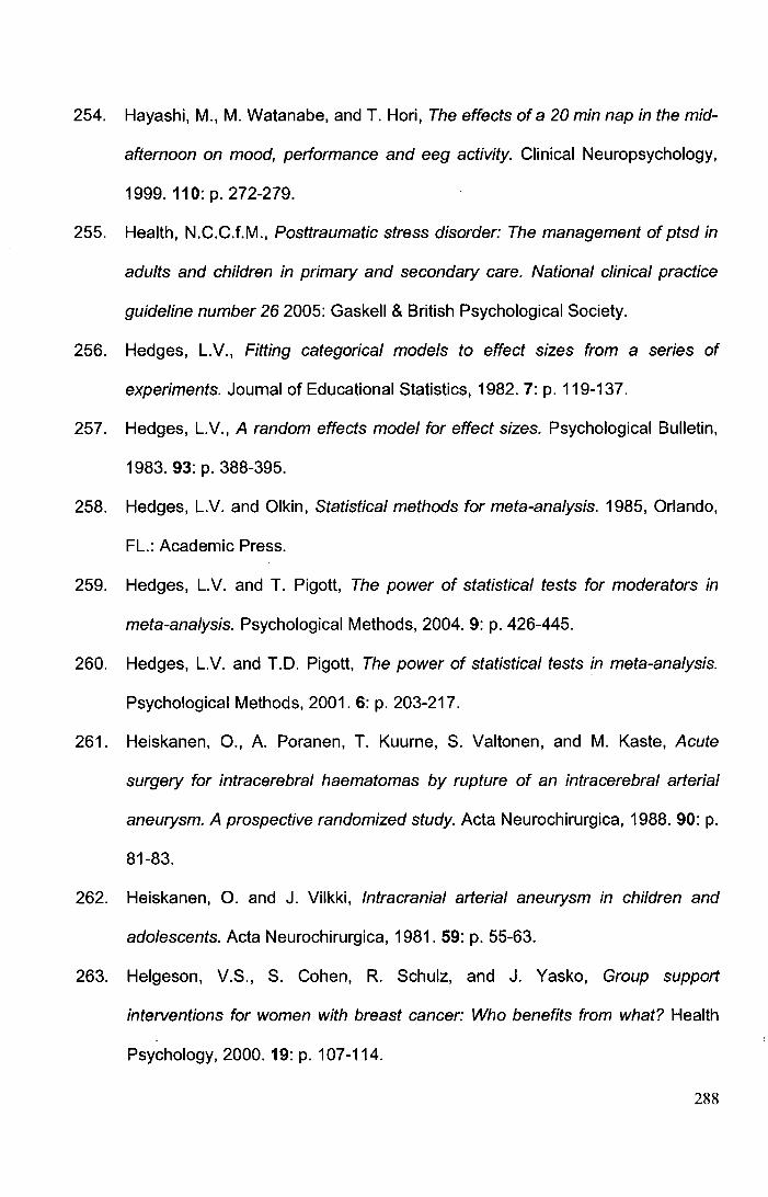
254. Hayashi, M., M. Watanabe, and T. Hori, The effects of a 20 min nap in the mid
afternoon on mood, performance and eeg activity. Clinical Neuropsychology,
1999. 110: p. 272-279.
255. Health, N.C.C.f.M., Posttraumatic stress disorder: The management of ptsd in
adults and children in primary and secondary care. National clinical practice
guideline number 26 2005: Gaskell & British Psychological Society.
256. Hedges, L.V., Fitting categorical models to effect sizes from a series of
experiments. Journal of Educational Statistics, 1982. 7: p. 119-137.
257. Hedges, L.V., A random effects model for effect sizes. Psychological Bulletin,
1983. 93: p. 388-395.
258. Hedges, L.V. and Olkin, Statistical methods for meta-analysis. 1985, Orlando,
FL.: Academic Press.
259. Hedges, L.V. and T. Pigott, The power of statistical tests for moderators in
meta-analysis. Psychological Methods, 2004. 9: p. 426-445.
260. Hedges, L.V. and T.D. Pigott, The power of statistical tests in meta-analysis.
Psychological Methods, 2001.6: p. 203-217.
261. Heiskanen, 0., A. Poranen, T. Kuume, S. Valtonen, and M. Kaste, Acute
surgery for intracerebral haematomas by rupture of an intracerebral arterial
aneurysm. A prospective randomized study. Acta Neurochirurgica, 1988. 90: p.
81-83.
262. Heiskanen, 0. and J. Vilkki, Intracranial arterial aneurysm in children and
adolescents. Acta Neurochirurgica, 1981. 59: p. 55-63.
263. Helgeson, V.S., S. Cohen, R. Schulz, and J. Yasko, Group support
interventions for women with breast cancer: Who benefits from what? Health
Psychology, 2000. 19: p. 107-114.
288
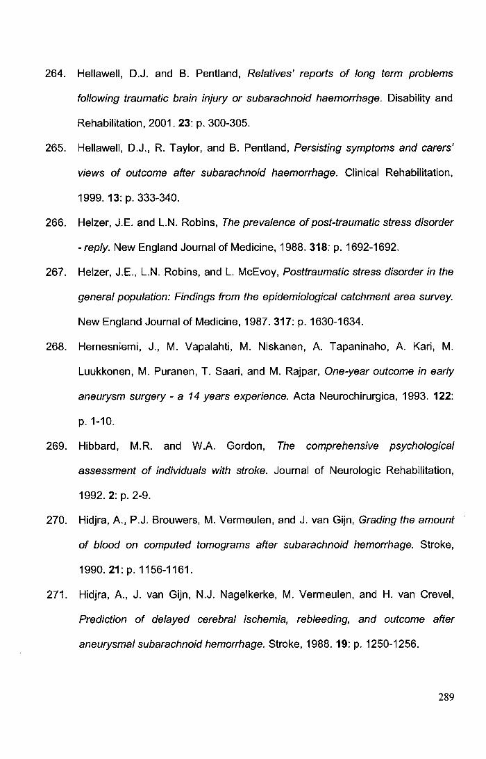
264. Hellawell, D.J. and B. Pentland, Relatives' reports of long term problems
following traumatic brain injury or subarachnoid haemorrhage. Disability and
Rehabilitation, 2001. 23: p. 300-305.
265. Hellawell, D.J., R. Taylor, and B. Pentland, Persisting symptoms and carers'
views of outcome after subarachnoid haemorrhage. Clinical Rehabilitation,
1999. 13: p. 333-340.
266. Helzer, J.E. and L.N. Robins, The prevalence of post-traumatic stress disorder
-reply. New England Journal of Medicine, 1988.318: p. 1692-1692.
267. Helzer, J.E., L.N. Robins, and L. McEvoy, Posttraumatic stress disorder in the
general population: Findings from the epidemiological catchment area survey.
New England Journal of Medicine, 1987.317: p. 1630-1634.
268. Hernesniemi, J., M. Vapalahti, M. Niskanen, A. Tapaninaho, A. Kari, M.
Luukkonen, M. Puranen, T. Saari, and M. Rajpar, One-year outcome in early
aneurysm surgery- a 14 years experience. Acta Neurochirurgica, 1993. 122:
p. 1-10.
269. Hibbard, M.R. and W.A. Gordon, The comprehensive psychological
assessment of individuals with stroke. Journal of Neurologic Rehabilitation,
1992. 2: p. 2-9.
270. Hidjra, A., P.J. Brouwers, M. Vermeulen, and J. van Gijn, Grading the amount
of blood on computed tomograms after subarachnoid hemorrhage. Stroke,
1990.21: p. 1156-1161.
271. Hidjra, A., J. van Gijn, N.J. Nagelkerke, M. Vermeulen, and H. van Crevel,
Prediction of delayed cerebral ischemia, rebleeding, and outcome after
aneurysmal subarachnoid hemorrhage. Stroke, 1988. 19: p. 1250-1256.
289
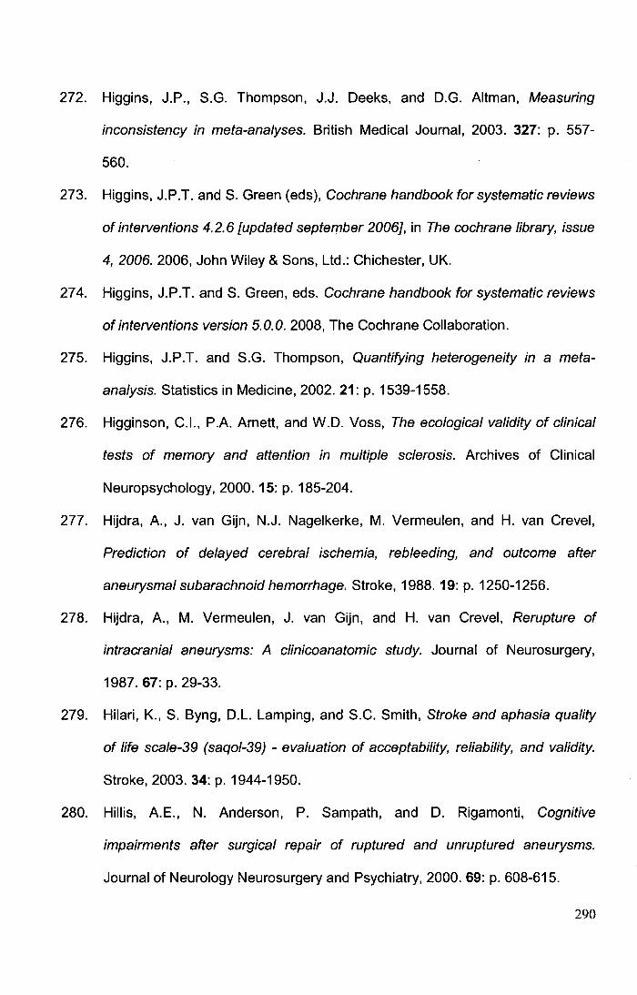
272. Higgins, J.P., S.G. Thompson, J.J. Deeks, and D.G. Altman, Measuring
inconsistency in meta-analyses. British Medical Journal, 2003. 327: p. 557-
560.
273. Higgins, J.P.T. and S. Green (eds), Cochrane handbook for systematic reviews
of interventions 4.2.6 [updated september 2006}, in The cochrane library, issue
4, 2006. 2006, John Wiley & Sons, Ltd.: Chichester, UK.
274. Higgins, J.P.T. and S. Green, eds. Cochrane handbook for systematic reviews
of interventions version 5.0.0. 2008, The Cochrane Collaboration.
275. Higgins, J.P.T. and S.G. Thompson, Quantifying heterogeneity in a meta
analysis. Statistics in Medicine, 2002. 21: p. 1539-1558.
276. Higginson, C.l., P.A. Arnett, and W.D. Voss, The ecological validity of clinical
tests of memory and attention in multiple sclerosis. Archives of Clinical
Neuropsychology, 2000. 15: p. 185-204.
277. Hijdra, A., J. van Gijn, N.J. Nagelkerke, M. Vermeulen, and H. van Crevel,
Prediction of delayed cerebral ischemia, rebleeding, and outcome after
aneurysmal subarachnoid hemorrhage. Stroke, 1988. 19: p. 1250-1256.
278. Hijdra, A., M. Vermeulen, J. van Gijn, and H. van Crevel, Rerupture of
intracranial aneurysms: A clinicoanatomic study. Journal of Neurosurgery,
1987. 67: p. 29-33.
279. Hilari, K., S. Byng, D.L. Lamping, and S.C. Smith, Stroke and aphasia quality
of life scale-39 (saqol-39) - evaluation of acceptability, reliability, and validity.
Stroke, 2003. 34: p. 1944-1950.
280. Hillis, A.E., N. Anderson, P. Sampath, and D. Rigamonti, Cognitive
impairments after surgical repair of ruptured and unruptured aneurysms.
Journal of Neurology Neurosurgery and Psychiatry, 2000. 69: p. 608-615.
290
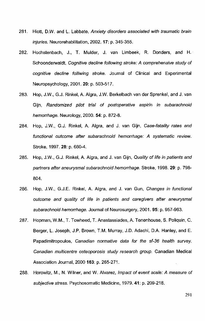
281. Hiott, D.W. and L. Labbate, Anxiety disorders associated with traumatic brain
injuries. Neurorehabilitation, 2002. 17: p. 345-355.
282. Hochstenbach, J., T. Mulder, J. van Limbeek, R. Danders, and H.
Schoonderwaldt, Cognitive decline following stroke: A comprehensive study of
cognitive decline folliwing stroke. Journal of Clinical and Experimental
Neuropsychology, 2001. 20: p. 503-517.
283. Hop, J.W., G.J. Rinkel, A. Algra, J.W. Berkelbach van der Sprenkel, and J. van
Gijn, Randomized pilot trial of postoperative aspirin in subarachnoid
hemorrhage. Neurology, 2000. 54: p. 872-8.
284. Hop, J.W., G.J. Rinkel, A. Algra, and J. van Gijn, Case-fatality rates and
functional outcome after subarachnoid hemorrhage: A systematic review.
Stroke, 1997. 28: p. 660-4.
285. Hop, J.W ., G.J. Rinkel, A. Algra, and J. van Gijn, Quality of life in patients and
partners after aneurysmal subarachnoid hemorrhage. Stroke, 1998. 29: p. 798-
804.
286. Hop, J.W., G.J.E. Rinkel, A. Algra, and J. van Gun, Changes in functional
outcome and quality of life in patients and caregivers after aneurysmal
subarachnoid hemorrhage. Journal of Neurosurgery, 2001. 95: p. 957-963.
287. Hopman, W.M., T. Towhead, T. Anastassiades, A. Tenenhouse, S. Poliquin, C.
Berger, L. Joseph, J.P. Brown, T.M. Murray, J.D. Adachi, D.A. Hanley, and E.
Papadimitropoulos, Canadian normative data for the sf-36 health survey.
Canadian multicentre osteoporosis study research group. Canadian Medical
Association Journal, 2000 163: p. 265-271.
288. Horowitz, M., N. Wilner, and W. Alvarez, Impact of event scale: A measure of
subjective stress. Psychosomatic Medicine, 1979.41: p. 209-218.
291
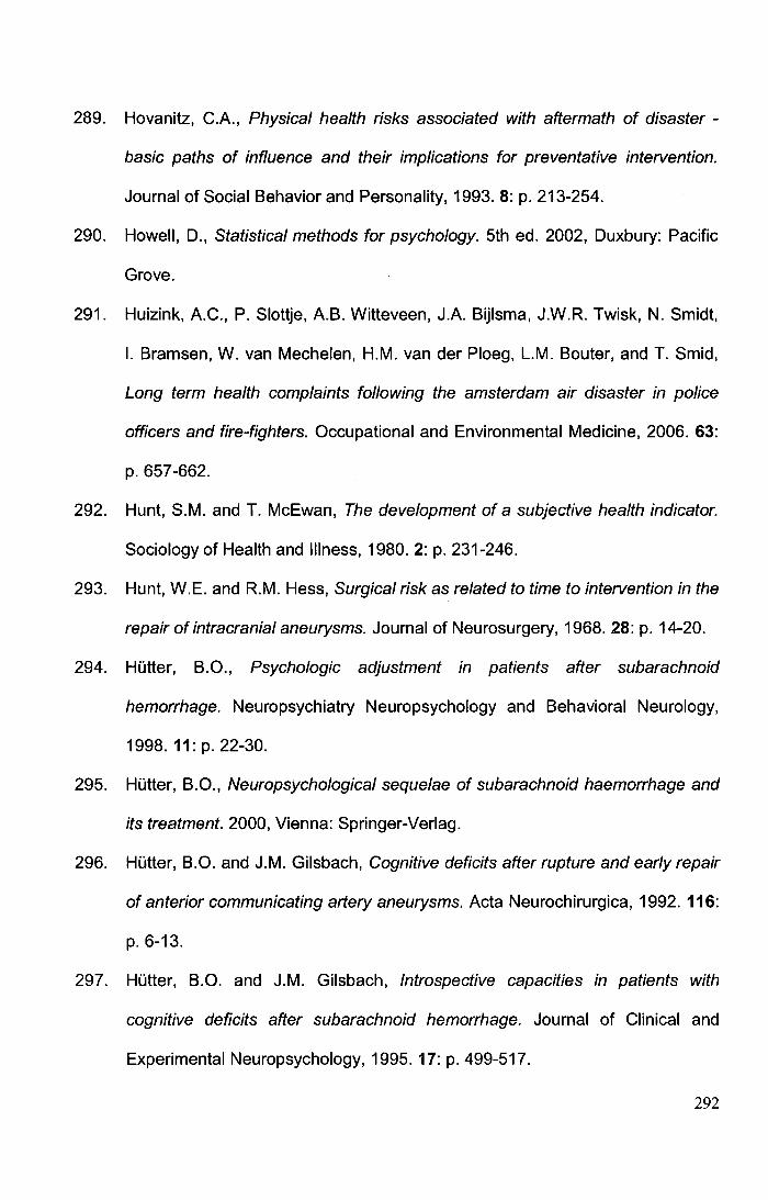
289. Hovanitz, C.A., Physical health risks associated with aftermath of disaster -
basic paths of influence and their implications for preventative intervention.
Journal of Social Behavior and Personality, 1993. 8: p. 213-254.
290. Howell, D., Statistical methods for psychology. 5th ed. 2002, Duxbury: Pacific
Grove.
291. Huizink, A.C., P. Slottje, A.B. Witteveen, J.A. Bijlsma, J.W.R. Twisk, N. Smidt,
I. Bramsen, W. van Mechelen, H.M. van der Ploeg, L.M. Souter, and T. Smid,
Long term health complaints following the amsterdam air disaster in police
officers and fire-fighters. Occupational and Environmental Medicine, 2006. 63:
p. 657-662.
292. Hunt, S.M. and T. McEwan, The development of a subjective health indicator.
Sociology of Health and Illness, 1980. 2: p. 231-246.
293. Hunt, W.E. and R.M. Hess, Surgical risk as related to time to intervention in the
repair of intracranial aneurysms. Journal of Neurosurgery, 1968. 28: p. 14-20.
294. Hutter, B.O., Psychologic adjustment in patients after subarachnoid
hemorrhage. Neuropsychiatry Neuropsychology and Behavioral Neurology,
1998. 11: p. 22-30.
295. Hutter, B.O., Neuropsychological sequelae of subarachnoid haemorrhage and
its treatment. 2000, Vienna: Springer-Verlag.
296. Hutter, B.O. and J.M. Gilsbach, Cognitive deficits after rupture and early repair
of anterior communicating artery aneurysms. Acta Neurochirurgica, 1992. 116:
p. 6-13.
297. Hutter, B.O. and J.M. Gilsbach, Introspective capacities in patients with
cognitive deficits after subarachnoid hemorrhage. Journal of Clinical and
Experimental Neuropsychology, 1995. 17: p. 499-517.
292
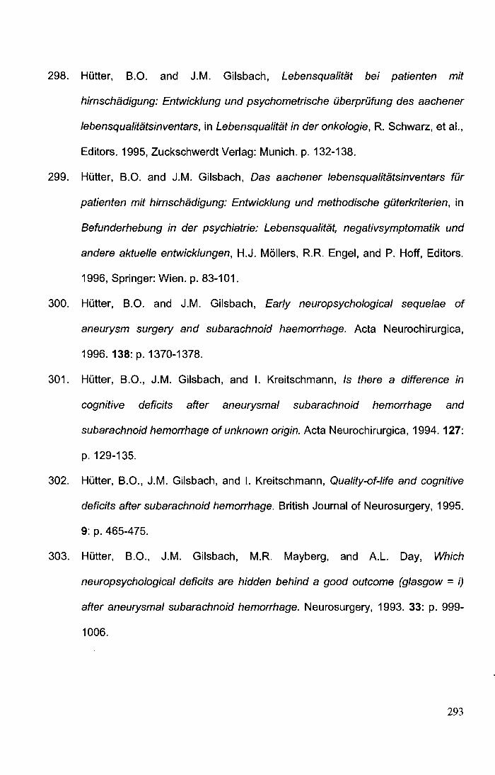
298. HOtter, B.O. and J.M. Gilsbach, Lebensqualitat bei patienten mit
hirnschadigung: Entwicklung und psychometrische iiberpriifung des aachener
lebensqualitatsinventars, in Lebensqualitat in der onkologie, R. Schwarz, et al.,
Editors. 1995, Zuckschwerdt Verlag: Munich. p. 132-138.
299. HOtter, B.O. and J.M. Gilsbach, Das aachener lebensqualitatsinventars fOr
patienten mit hirnschadigung: Entwicklung und methodische giiterkriterien, in
Befunderhebung in der psychiatrie: Lebensqualitat, negativsymptomatik und
andere aktuel/e entwicklungen, H.J. Moilers, R.R. Engel, and P. Hoff, Editors.
1996, Springer: Wien. p. 83-101.
300. HOtter, B.O. and J.M. Gilsbach, Early neuropsychological sequelae of
aneurysm surgery and subarachnoid haemorrhage. Acta Neurochirurgica,
1996. 138:p. 1370-1378.
301. HOtter, B.O., J.M. Gilsbach, and I. Kreitschmann, Is there a difference in
cognitive deficits after aneurysmal subarachnoid hemorrhage and
subarachnoid hemorrhage of unknown origin. Acta Neurochirurgica, 1994. 127:
p. 129-135.
302. HOtter, B.O., J.M. Gilsbach, and I. Kreitschmann, Quality-of-life and cognitive
deficits after subarachnoid hemorrhage. British Journal of Neurosurgery, 1995.
9: p. 465-475.
303. HOtter, B.O., J.M. Gilsbach, M.R. Mayberg, and A.L. Day, Which
neuropsychological deficits are hidden behind a good outcome (glasgow = i)
after aneurysmal subarachnoid hemorrhage. Neurosurgery, 1993. 33: p. 999-
1006.
293
304. Hutter, B.O., I. Kreitschmann-Andermahr, and J.M. Gilsbach, Cognitive deficits
in the acute stage after subarachnoid hemorrhage. Neurosurgery, 1998. 43: p.
1054-1064.
305. Hutter, B.O., I. Kreitschmann-Andermahr, and J.M. Gilsbach, Health-related
quality of life after aneurysmal subarachnoid hemorrhage: Impacts of bleeding
severity, computerized tomography findings, surgery, vasospasm, and
neurological grade. Journal of Neurosurgery, 2001. 94: p. 241-251.
306. Hutter, B.O., I. Kreitschmann-Andermahr, L. Mayfrank, V. Rohde, U. Spetzger,
and J.M. Gilsbach, Functional outcome after aneurysmal subarachnoid
haemorrhage. Acta Neurochirugica Supplement (Wien), 1999. 72: p. 157-174.
307. Investigators, C., Rates of delayed rebleeding from intracranial aneurysms are
low after surgical and endovascular treatment. Stroke 2006. 37: p. 1437-1442.
308. lrle, E., B. Wowra, H.J. Kunert, J. Hampl, and S. Kunze, Memory disturbances
following anterior communicating artery rupture. Annals of Neurology, 1992.
31: p. 473-480.
309. Jackowski, A., A. Crockard, G. Burnstock, R.R. Russell, and F. Kristek, The
time course of intracranial pathophysiological changes following experimental
subarachnoid hemorrhage in the rat. Journal of Cerebral Blood Flow and
Metabolism, 1990. 10: p. 835-849.
310. Jackson, J.C., R.P. Hart, S.M. Gordon, R.O. Hopkins, T.D. Girard, and E.W.
Ely, Post-traumatic stress disorder and post-traumatic stress symptoms
following critical illness in medical intensive care unit patients: Assessing the
magnitude of the problem. Critical Care 2007. 11: p. R27.
311. Jacobsen, P.B., M.R. Widows, D.M. Hann, M.A. Andrykowski, L.E. Kronish,
and K.K. Fields, Posttraumatic stress disorder symptoms after bone marrow
294
transplantation for breast cancer. Psychosomatic Medicine, 1998. 60: p. 366-
371.
312. Jampala, V.C. and R. Abrams, Mania secondary to left and right hemisphere
damage. American Journal of Psychiatry, 1983.140: p.1197-1199.
313. Jarvis, A., Recovering from subarachnoid haemorrhage: Patients' perspective.
British Journal of Nursing, 2002.11: p. 1430-1437.
314. Jarvis, A. and L. Talbot, Multiprofessional follow up of patients after
subarachnoid haemorrhage. British Journal of Nursing, 2004. 13: p. 1262-
1267.
315. Jefferson, J.W. and J.R. Marshall, Neuropsychiatric features of medical
disorders. 1981, New York, NY: Plenum.
316. Jenkinson, C., A. Coulter, and L. Wright, Short form-36 (sf-36) health survey
questionnaire - normative data for adults of working age. British Medical
Journal, 1993. 306: p. 1437-1440.
317. Jenkinson, C., R. Layte, L. Wright, and A. Coulter, The uk sf-36: An analysis
and interpretation manual. A guide to health status measurment with particular
reference to the short form 36 health survey. 1996, Oxofrd: University of
Oxford.
318. Jennett, B. and M. Bond, Assessment of outcome after severe brain-damage
practical scale. Lancet, 1975. 1: p. 480-484.
319. Jeon, H.J., T. Suh, H.J. Lee, B.J. Hahm, J.Y. Lee, S.J. Cho, Y.R. Lee, S.M.
Chang, and M.J. Cho, Partial versus full ptsd in the korean community:
Prevalence, duration, correlates, comorbidity, and dysfunctions. Depression
and Anxiety, 2007. 24: p. 577-585.
295
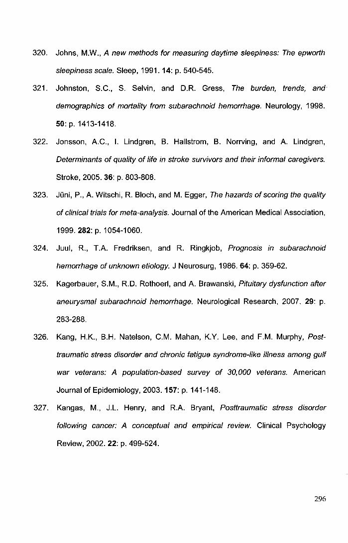
320. Johns, M.W., A new methods for measuring daytime sleepiness: The epworth
sleepiness scale. Sleep, 1991. 14: p. 540-545.
321. Johnston, S.C., S. Selvin, and D.R. Gress, The burden, trends, and
demographics of mortality from subarachnoid hemorrhage. Neurology, 1998.
50: p. 1413-1418.
322. Jonsson, A.C., I. Lindgren, B. Hallstrom, B. Norrving, and A. Lindgren,
Determinants of quality of life in stroke survivors and their informal caregivers.
Stroke, 2005. 36: p. 803-808.
323. JOni, P., A. Witschi, R. Bloch, and M. Egger, The hazards of scoring the quality
of clinical trials for meta-analysis. Journal of the American Medical Association,
1999.282: p. 1054-1060.
324. Juul, R., T.A. Fredriksen, and R. Ringkjob, Prognosis in subarachnoid
hemorrhage of unknown etiology. J Neurosurg, 1986. 64: p. 359-62.
325. Kagerbauer, S.M., R.D. Rothoerl, and A. Brawanski, Pituitary dysfunction after
aneurysmal subarachnoid hemorrhage. Neurological Research, 2007. 29: p.
283-288.
326. Kang, H.K., B.H. Natelson, C.M. Mahan, K.Y. Lee, and F.M. Murphy, Post
traumatic stress disorder and chronic fatigue syndrome-/ike illness among gulf
war veterans: A population-based survey of 30,000 veterans. American
Journal of Epidemiology, 2003. 157: p. 141-148.
327. Kangas, M., J.L. Henry, and R.A. Bryant, Posttraumatic stress disorder
following cancer: A conceptual and empirical review. Clinical Psychology
Review, 2002. 22: p. 499-524.
296
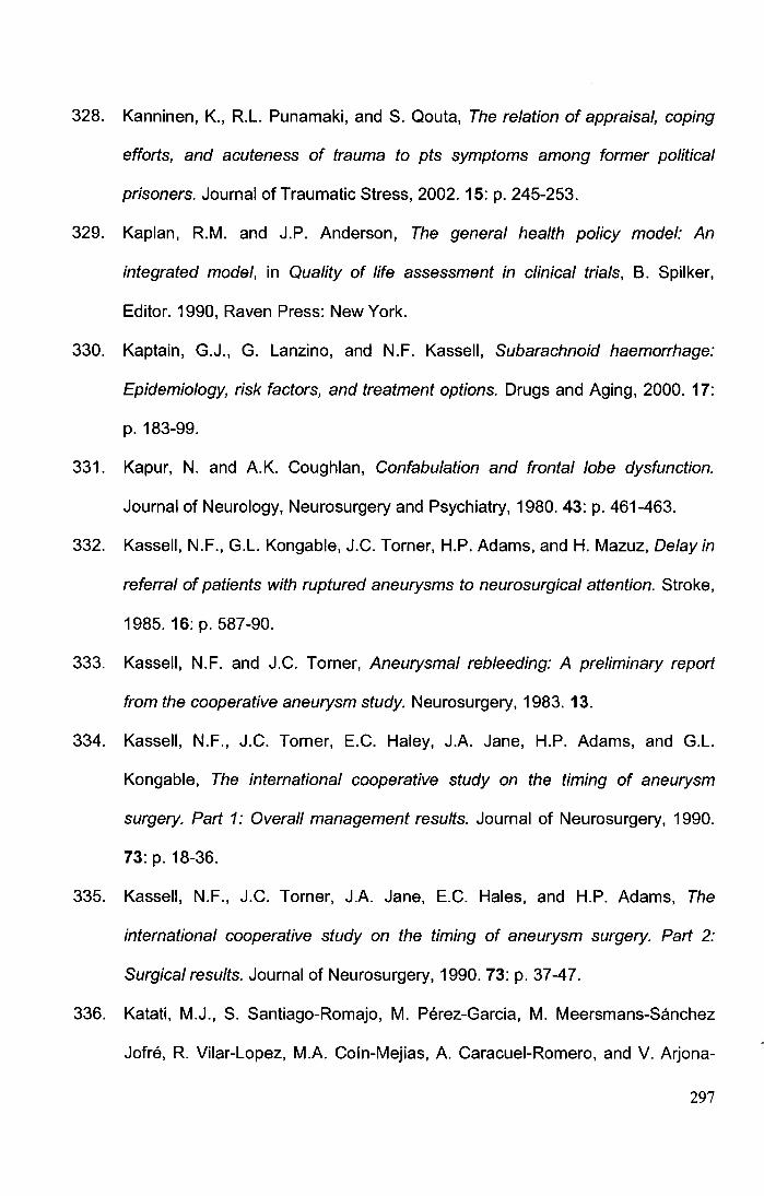
328. Kanninen, K., R.L. Punamaki, and S. Qouta, The relation of appraisal, coping
efforts, and acuteness of trauma to pts symptoms among former political
prisoners. Journal of Traumatic Stress, 2002. 15: p. 245-253.
329. Kaplan, R.M. and J.P. Anderson, The general health policy model: An
integrated model, in Quality of life assessment in clinical trials, B. Spilker,
Editor. 1990, Raven Press: New York.
330. Kaptain, G.J., G. Lanzino, and N.F. Kassell, Subarachnoid haemorrhage:
Epidemiology, risk factors, and treatment options. Drugs and Aging, 2000. 17:
p. 183-99.
331. Kapur, N. and A.K. Coughlan, Confabulation and frontal lobe dysfunction.
Journal of Neurology, Neurosurgery and Psychiatry, 1980. 43: p. 461-463.
332. Kassell, N.F., G.L. Kongable, J.C. Torner, H.P. Adams, and H. Mazuz, Delay in
referral of patients with ruptured aneurysms to neurosurgical attention. Stroke,
1985. 16: p. 587-90.
333. Kassel!, N.F. and J.C. Torner, Aneurysmal rebleeding: A preliminary report
from the cooperative aneurysm study. Neurosurgery, 1983. 13.
334. Kassel!, N.F., J.C. Torner, E.C. Haley, J.A. Jane, H.P. Adams, and G.L.
Kongable, The international cooperative study on the timing of aneurysm
surgery. Part 1: Overall management results. Journal of Neurosurgery, 1990.
73: p. 18-36.
335. Kassell, N.F., J.C. Torner, J.A. Jane, E.C. Hales, and H.P. Adams, The
international cooperative study on the timing of aneurysm surgery. Part 2:
Surgical results. Journal of Neurosurgery, 1990. 73: p. 37-47.
336. Katati, M.J., S. Santiago-Romajo, M. Perez-Garcia, M. Meersmans-Sanchez
Jofre, R. Vilar-Lopez, M.A. Coin-Mejias, A. Caracuei-Romero, and V. Arjona-
297
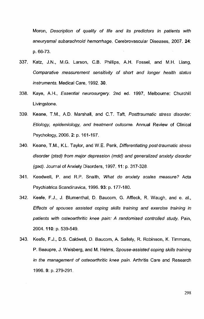
Moron, Description of quality of life and its predictors in patients with
aneurysmal subarachnoid hemorrhage. Cerebrovascular Diseases, 2007. 24:
p. 66-73.
337. Katz, J.N., M.G. Larson, C.B. Phillips, A.H. Fossel, and M.H. Liang,
Comparative measurement sensitivity of short and longer health status
instruments. Medical Care, 1992. 30.
338. Kaye, A.H., Essential neurosurgery. 2nd ed. 1997, Melbourne: Churchill
Livingstone.
339. Keane, T.M., A.D. Marshall, and C.T. Taft, Posttraumatic stress disorder:
Etiology, epidemiology, and treatment outcome. Annual Review of Clinical
Psychology, 2006. 2: p. 161-197.
340. Keane, T.M., K.L. Taylor, and W.E. Penk, Differentiating post-traumatic stress
disorder (ptsd) from major depression (mdd) and generalized anxiety disorder
(gad). Journal of Anxiety Disorders, 1997. 11: p. 317-328.
341. Keedwell, P. and R.P. Snaith, What do anxiety scales measure? Acta
Psychiatrica Scandinavica, 1996. 93: p. 177-180.
342. Keefe, F.J., J. Blumenthal, D. Baucom, G. Affleck, R. Waugh, and e. al.,
Effects of spouses assisted coping skills training and exercise training in
patients with osteoarthritic knee pain: A randomised controlled study. Pain,
2004. 110: p. 539-549.
343. Keefe, F.J., D.S. Caldwell, D. Baucom, A. Sallely, R. Robinson, K. Timmons,
P. Beaupre, J. Weisberg, and M. Helms, Spouse-assisted coping skills training
in the management of osteoarthritic knee pain. Arthritis Care and Research
1996. 9: p. 279-291.
298
344. Kendall, E. and D.J. Terry, Psychosocial adjustment following closed head
injury. A model for understanding individual differences and predicting
outcome. Neuropsychological Rehabilitation, 1996. 6: p. 101-132.
345. Kennedy, B.L., J.J. Schwab, R.L. Morris, and G. Beldia, Assessment of state
and trait anxiety in subjects with anxiety and depressive disorders. Psychiatric
Quarterly, 2001. 72: p. 263-276.
346. Kennedy, P., J. Duff, M. Evans, and A. Beedie, Coping effectiveness training
reduces depression and anxiety following traumatic spinal cord injuries. British
Journal of Clinical Psychology, 2003. 42: p. 41-52.
347. Kennedy, P., N.M. Taylor, and J. Duff, Characteristics predicting effective
outcomes after coping effectiveness training for patients with spinal cord
injuries. Journal of Consulting and Clinical Psychology, 2005. 12: p. 93-98.
348. Kessler, R.C., P.A. Berglund, C.L. Foster, W.B. Saunders, P.E. Stang, and
E.E. Walters, Social consequences of psychiatric disorders .2. Teenage
parenthood. American Journal of Psychiatry, 1997. 154: p. 1405-1411.
349. Kessler, R.C., C.L. Foster, W.B. Saunders, and P.E. Stang, Social
consequences of psychiatric disorders. 1. Educational attainment. American
Journal of Psychiatry, 1995. 152: p. 1026-1032.
350. Kessler, R.C., C.L. Foster, W.B. Saunders, and P.E. Stang, The social
consequences of psychiatric disorders. 2. Teenage childbearing. American
Journal of Epidemiology, 1995. 141: p. S77-S77.
351. Kessler, R.C., A. Sonnega, E. Bromet, M. Hughes, and C.B. Nelson,
Posttraumatic stress disorder in the national comorbidity survey. Archives of
General Psychiatry, 1995. 52: p. 1 048-1 060.
299
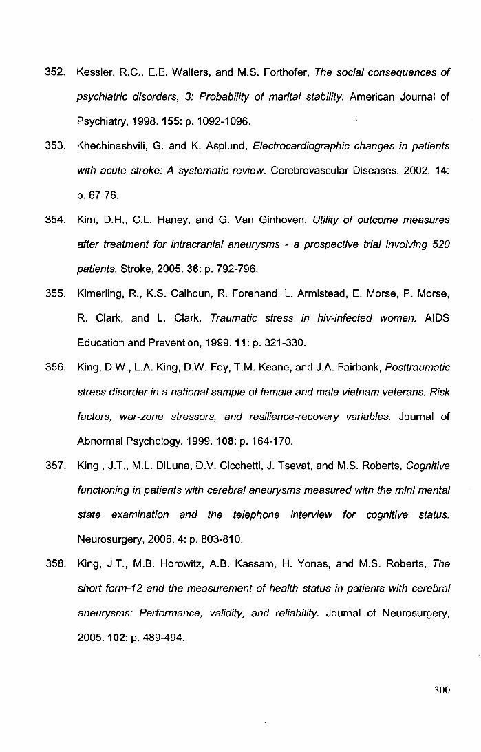
352. Kessler, R.C., E.E. Walters, and M.S. Forthofer, The social consequences of
psychiatric disorders, 3: Probability of marital stability. American Journal of
Psychiatry, 1998. 155: p. 1092-1096.
353. Khechinashvili, G. and K. Asplund, Electrocardiographic changes in patients
with acute stroke: A systematic review. Cerebrovascular Diseases, 2002. 14:
p. 67-76.
354. Kim, D.H., C.L. Haney, and G. Van Ginhoven, Utility of outcome measures
after treatment for intracranial aneurysms - a prospective trial involving 520
patients. Stroke, 2005. 36: p. 792-796.
355. Kimerling, R., K.S. Calhoun, R. Forehand, L. Armistead, E. Morse, P. Morse,
R. Clark, and L. Clark, Traumatic stress in hiv-infected women. AIDS
Education and Prevention, 1999. 11: p. 321-330.
356. King, D.W., L.A. King, D.W. Foy, T.M. Keane, and J.A. Fairbank, Posttraumatic
stress disorder in a national sample of female and male vietnam veterans. Risk
factors, war-zone stressors, and resilience-recovery variables. Journal of
Abnormal Psychology, 1999. 108: p. 164-170.
357. King , J.T., M.L. Diluna, D.V. Cicchetti, J. Tsevat, and M.S. Roberts, Cognitive
functioning in patients with cerebral aneurysms measured with the mini mental
state examination and the telephone interview for cognitive status.
Neurosurgery, 2006. 4: p. 803-810.
358. King, J.T., M.B. Horowitz, A.B. Kassam, H. Yonas, and M.S. Roberts, The
short form-12 and the measurement of health status in patients with cerebral
aneurysms: Performance, validity, and reliability. Journal of Neurosurgery,
2005. 102: p. 489-494.
300
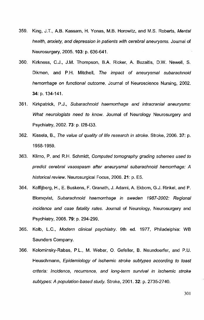
359. King, J.T., A.B. Kassam, H. Yonas, M.B. Horowitz, and M.S. Roberts, Mental
health, anxiety, and depression in patients with cerebral aneurysms. Journal of
Neurosurgery, 2005. 103: p. 636-641.
360. Kirkness, C.J., J.M. Thompson, B.A. Ricker, A. Buzaitis, D.W. Newell, S.
Dikmen, and P.H. Mitchell, The impact of aneurysmal subarachnoid
hemorrhage on functional outcome. Journal of Neuroscience Nursing, 2002.
34: p. 134-141.
361. Kirkpatrick, P.J., Subarachnoid haemorrhage and intracranial aneurysms:
What neurologists need to know. Journal of Neurology Neurosurgery and
Psychiatry, 2002. 73: p. 128-133.
362. Kissela, B., The value of quality of life research in stroke. Stroke, 2006. 37: p.
1958-1959.
363. Klimo, P. and R.H. Schmidt, Computed tomography grading schemes used to
predict cerebral vasospasm after aneurysmal subarachnoid hemorrhage: A
historical review. Neurosurgical Focus, 2006. 21: p. E5.
364. Koffijberg, H., E. Buskens, F. Granath, J. Adami, A. Ekbom, G.J. Rinkel, and P.
Blomqvist, Subarachnoid haemorrhage in sweden 1987-2002: Regional
incidence and case fatality rates. Journal of Neurology, Neurosurgery and
Psychiatry, 2008. 79: p. 294-299.
365. Kolb, L.C., Modern clinical psychiatry. 9th ed. 1977, Philadelphia: WB
Saunders Company.
366. Kolominsky-Rabas, P.L., M. Weber, 0. Gefeller, B. Neundoerfer, and P.U.
Heuschmann, Epidemiology of ischemic stroke subtypes according to toast
criteria: Incidence, recurrence, and long-term survival in ischemic stroke
subtypes: A population-based study. Stroke, 2001. 32: p. 2735-2740.
301
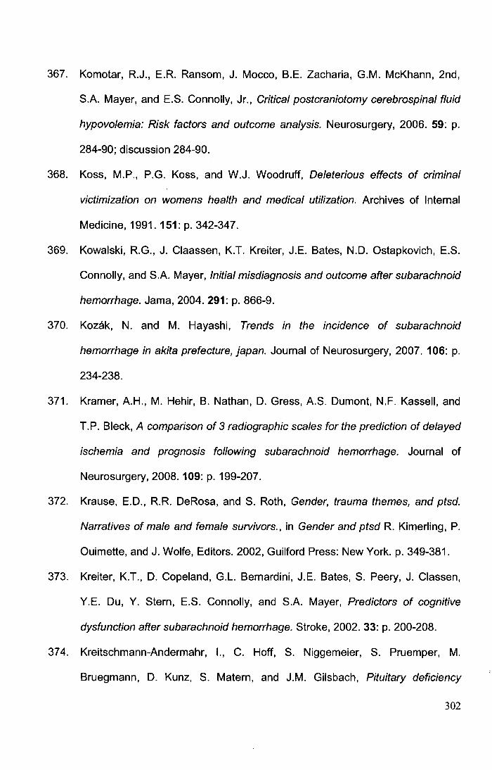
367. Komotar, R.J., E.R. Ransom, J. Mocco, B.E. Zacharia, G.M. McKhann, 2nd,
S.A. Mayer, and E.S. Connolly, Jr., Critical postcraniotomy cerebrospinal fluid
hypovolemia: Risk factors and outcome analysis. Neurosurgery, 2006. 59: p.
284-90; discussion 284-90.
368. Koss, M.P., P.G. Koss, and W.J. Woodruff, Deleterious effects of criminal
victimization on womens health and medical utilization. Archives of Internal
Medicine, 1991.151: p. 342-347.
369. Kowalski, R.G., J. Claassen, K.T. Kreiter, J.E. Bates, N.D. Ostapkovich, E.S.
Connolly, and S.A. Mayer, Initial misdiagnosis and outcome after subarachnoid
hemorrhage. Jama, 2004. 291: p. 866-9.
370. Kozak, N. and M. Hayashi, Trends in the incidence of subarachnoid
hemorrhage in akita prefecture, japan. Journal of Neurosurgery, 2007. 106: p.
234-238.
371. Kramer, A.H., M. Hehir, B. Nathan, D. Gress, A.S. Dumont, N.F. Kassell, and
T.P. Bleck, A comparison of 3 radiographic scales for the prediction of delayed
ischemia and prognosis following subarachnoid hemorrhage. Journal of
Neurosurgery, 2008. 109: p. 199-207.
372. Krause, E.D., R.R. DeRosa, and S. Roth, Gender, trauma themes, and ptsd.
Narratives of male and female survivors., in Gender and ptsd R. Kimerling, P.
Ouimette, and J. Wolfe, Editors. 2002, Guilford Press: New York. p. 349-381.
373. Kreiter, K.T., D. Copeland, G.L. Bernardini, J.E. Bates, S. Peery, J. Classen,
Y.E. Du, Y. Stern, E.S. Connolly, and S.A. Mayer, Predictors of cognitive
dysfunction after subarachnoid hemorrhage. Stroke, 2002. 33: p. 200-208.
374. Kreitschmann-Andermahr, 1., C. Hoff, S. Niggemeier, S. Pruemper, M.
Bruegmann, D. Kunz, S. Matern, and J.M. Gilsbach, Pituitary deficiency
302
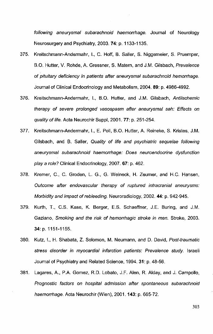
following aneurysmal subarachnoid haemorrhage. Journal of Neurology
Neurosurgery and Psychiatry, 2003. 74: p. 1133-1135.
375. Kreitschmann-Andermahr, 1., C. Hoff, B. Saller, S. Niggemeier, S. Pruemper,
B.O. Hutter, V. Rohde, A. Gressner, S. Matern, and J.M. Gilsbach, Prevalence
of pituitary deficiency in patients after aneurysmal subarachnoid hemorrhage.
Journal of Clinical Endocrinology and Metabolism, 2004. 89: p. 4986-4992.
376. Kreitschmann-Andermahr, 1., B.O. Hutter, and J.M. Gilsbach, Antiischemic
therapy of severe prolonged vasospasm after aneurysmal sah: Effects on
quality of life. Acta Neurochir Suppl, 2001. 77: p. 251-254.
377. Kreitschmann-Andermahr, 1., E. Poll, B.O. Hutter, A. Reineke, S. Kristes, J.M.
Gilsbach, and B. Saller, Quality of life and psychiatric sequelae following
aneurysmal subarachnoid haemorrhage: Does neuroendocrine dysfunction
play a role? Clinical Endocrinology, 2007. 67: p. 462.
378. Kremer, C., C. Groden, L. G., G. Weineck, H. Zeumer, and H.C. Hansen,
Outcome after endovascular therapy of ruptured intracranial aneurysms:
Morbidity and impact of rebleeding. Neuroradiology, 2002. 44: p. 942-945.
379. Kurth, T., C.S. Kase, K. Berger, E.S. Schaeffner, J.E. Suring, and J.M.
Gaziano, Smoking and the risk of hemorrhagic stroke in men. Stroke, 2003.
34: p. 1151-1155.
380. Kutz, 1., H. Shabata, Z. Solomon, M. Neumann, and D. David, Post-traumatic
stress disorder in myocardial infarction patients: Prevalence study. Israeli
Journal of Psychiatry and Related Science, 1994. 31: p. 48-56.
381. Lagares, A., P.A. Gomez, R.D. Lobato, J.F. Alen, R. Alday, and J. Campollo,
Prognostic factors on hospital admission after spontaneous subarachnoid
haemorrhage. Acta Neurochir (Wien), 2001. 143: p. 665-72.
303
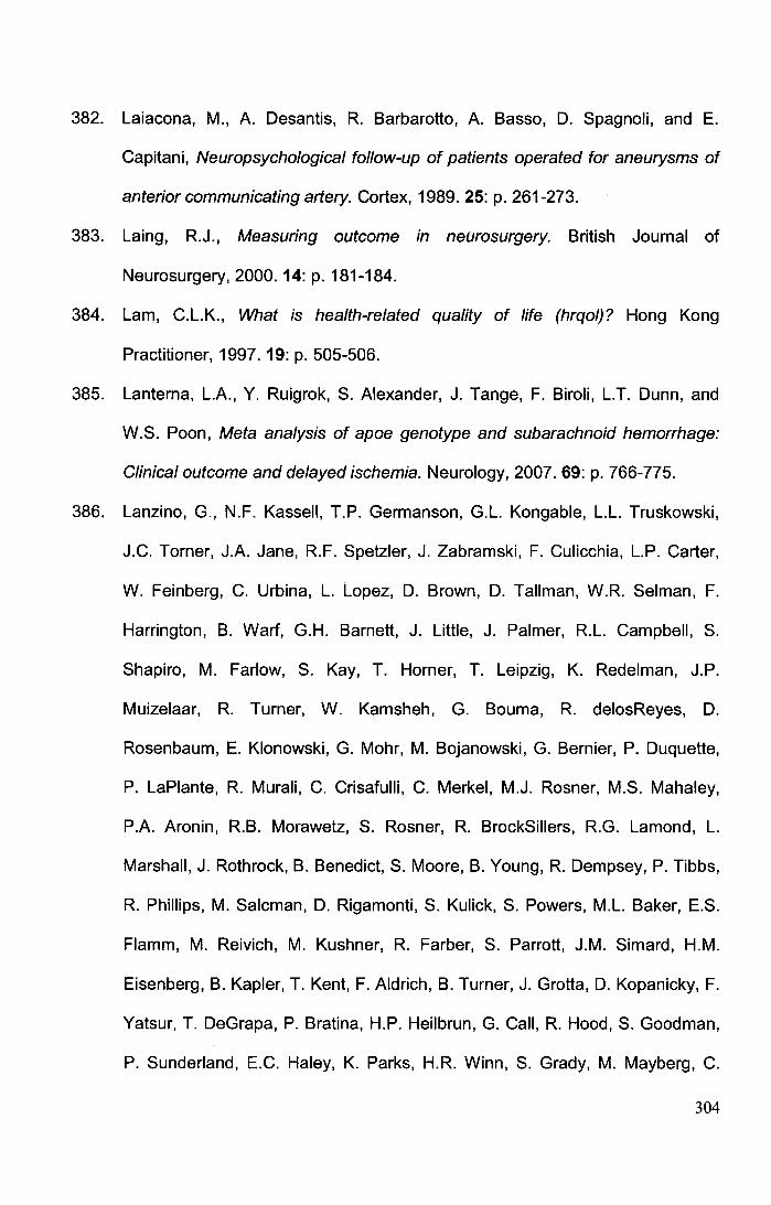
382. Laiacona, M., A. Desantis, R. Barbarotto, A. Basso, D. Spagnoli, and E.
Capitani, Neuropsychological follow-up of patients operated for aneurysms of
anterior communicating artery. Cortex, 1989. 25: p. 261-273.
383. Laing, R.J., Measuring outcome in neurosurgery. British Journal of
Neurosurgery, 2000. 14: p. 181-184.
384. Lam, C.L.K., What is health-related quality of life (hrqol)? Hong Kong
Practitioner, 1997. 19: p. 505-506.
385. Lanterna, L.A., Y. Ruigrok, S. Alexander, J. lange, F. Biroli, L.T. Dunn, and
W.S. Poon, Meta analysis of apoe genotype and subarachnoid hemorrhage:
Clinical outcome and delayed ischemia. Neurology, 2007. 69: p. 766-775.
386. Lanzino, G., N.F. Kassell, T.P. Germanson, G.L. Kongable, L.L. Truskowski,
J.C. Torner, J.A. Jane, R.F. Spetzler, J. Zabramski, F. Culicchia, L.P. Carter,
W. Feinberg, C. Urbina, L. Lopez, D. Brown, D. Tallman, W.R. Selman, F.
Harrington, B. Wart, G.H. Barnett, J. Little, J. Palmer, R.L. Campbell, S.
Shapiro, M. Farlow, S. Kay, T. Horner, T. Leipzig, K. Redelman, J.P.
Muizelaar, R. Turner, W. Kamsheh, G. Bouma, R. delosReyes, D.
Rosenbaum, E. Klonowski, G. Mohr, M. Bojanowski, G. Bernier, P. Duquette,
P. LaPlante, R. Murali, C. Crisafulli, C. Merkel, M.J. Rosner, M.S. Mahaley,
P.A. Aronin, R.B. Morawetz, S. Rosner, R. BrockSillers, R.G. Lamond, L.
Marshall, J. Rothrock, B. Benedict, S. Moore, B. Young, R. Dempsey, P. Tibbs,
R. Phillips, M. Salcman, D. Rigamonti, S. Kulick, S. Powers, M.L. Baker, E.S.
Flamm, M. Reivich, M. Kushner, R. Farber, S. Parrott, J.M. Simard, H.M.
Eisenberg, B. Kapler, T. Kent, F. Aldrich, B. Turner, J. Grotta, D. Kopanicky, F.
Yatsur, T. DeGrapa, P. Bratina, H.P. Heilbrun, G. Call, R. Hood, S. Goodman,
P. Sunderland, E.C. Haley, K. Parks, H.R. Winn, S. Grady, M. Mayberg, C.
304
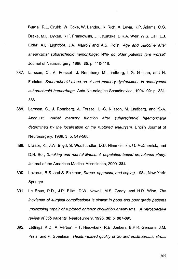
Burnal, R.L. Grubb, W. Coxe, W. Landou, K. Rich, A. Levin, H.P. Adams, C.G.
Drake, M.L. Dyken, R.F. Frankowski, J.F. Kurtzke, B.K.A. Weir, W.S. Gail, L.J.
Elder, A.L. Lightfoot, J.A. Marron and A.S. Polin, Age and outcome after
aneurysmal subarachnoid hemorrhage: Why do older patients fare worse?
Journal of Neurosurgery, 1996. 85: p. 410-418.
387. Larsson, C., A. Forssell, J. Ronnberg, M. Lindberg, L.G. Nilsson, and H.
Fodstad, Subarachnoid blood on ct and memory dysfunctions in aneurysmal
subarachnoid hemorrhage. Acta Neurologica Scandinavica, 1994. 90: p. 331-
336.
388. Larsson, C., J. Ronnberg, A. Forssel, L.-G. Nilsson, M. Lindberg, and K.-A.
Angguist, Verbal memory function after subarachnoid haemorrhage
determined by the localisation of the ruptured aneurysm. British Journal of
Neurosurgery, 1989. 3: p. 549-560.
389. Lasser, K., J.W. Boyd, S. Woolhandler, D.U. Himmelstein, D. McCormick, and
D.H. Bor, Smoking and mental illness: A population-based prevalence study.
Journal of the American Medical Association, 2000. 284.
390. Lazarus, R.S. and S. Folkman, Stress, appraisal, and coping. 1984, New York:
Springer.
391. Le Roux, P.D., J.P. Elliot, D.W. Newell, M.S. Grady, and H.R. Winn, The
incidence of surgical complications is similar in good and poor grade patients
undergoing repair of ruptured anterior circulation aneurysms: A retrospective
review of 355 patients. Neurosurgery, 1996. 38: p. 887-895.
392. Lettinga, K.D., A. Verbon, P.T. Nieuwkerk, R.E. Jonkers, B.P.R. Gersons, J.M.
Prins, and P. Speelman, Health-related quality of life and posttraumatic stress
305
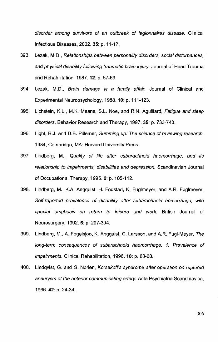
disorder among survivors of an outbreak of legionnaires disease. Clinical
Infectious Diseases, 2002. 35: p. 11-17.
393. Lezak, M.D., Relationships between personality disorders, social disturbances,
and physical disability following traumatic brain injury. Journal of Head Trauma
and Rehabilitation, 1987. 12: p. 57-69.
394. Lezak, M.D., Brain damage is a family affair. Journal of Clinical and
Experimental Neuropsychology, 1988. 10: p. 111-123.
395. Lichstein, K.L., M.K. Means, S.L. Noe, and R.N. Aguillard, Fatigue and sleep
disorders. Behavior Research and Therapy, 1997. 35: p. 733-740.
396. Light, R.J. and D.B. Pillemer, Summing up: The science of reviewing research.
1984, Cambridge, MA: Harvard University Press.
397. Lindberg, M., Quality of life after subarachnoid haemorrhage, and its
relationship to impairments, disabilities and depression. Scandinavian Journal
of Occupational Therapy, 1995. 2: p. 105-112.
398. Lindberg, M., K.A. Angquist, H. Fodstad, K. Fuglmeyer, and A.R. Fuglmeyer,
Self-reported prevalence of disability after subarachnoid hemorrhage, with
special emphasis on return to leisure and work. British Journal of
Neurosurgery, 1992. 6: p. 297-304.
399. Lindberg, M., A. Fogelsjoo, K. Angguist, C. Larsson, and A.R. Fugi-Meyer, The
long-term consequences of subarachnoid haemorrhage. 1: Prevalence of
impairments. Clinical Rehabilitation, 1996. 10: p. 63-68.
400. Lindqvist, G. and G. Norlen, Korsakoff's syndrome after operation on ruptured
aneurysm of the anterior communicating artery. Acta Psychiatria Scandinavica,
1966. 42: p. 24-34.
306
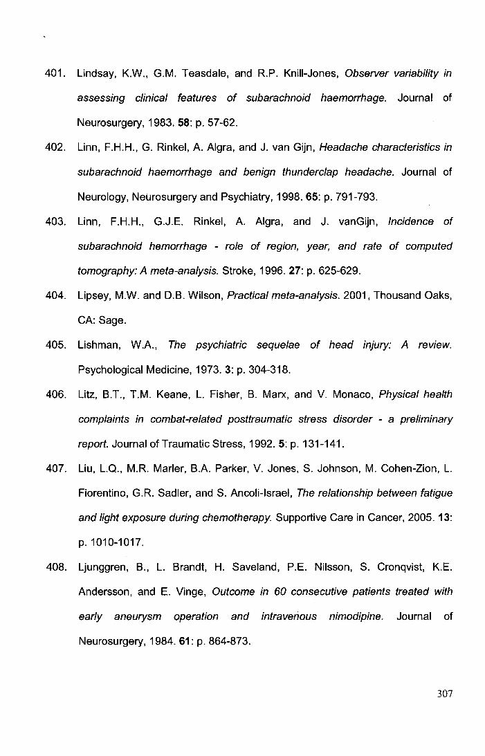
401. Lindsay, K.W., G.M. Teasdale, and R.P. Knili-Jones, Observer variability in
assessing clinical features of subarachnoid haemorrhage. Journal of
Neurosurgery, 1983. 58: p. 57-62.
402. Linn, F.H.H., G. Rinkel, A. Algra, and J. van Gijn, Headache characteristics in
subarachnoid haemorrhage and benign thunderclap headache. Journal of
Neurology, Neurosurgery and Psychiatry, 1998. 65: p. 791-793.
403. Linn, F.H.H., G.J.E. Rinkel, A. Algra, and J. vanGijn, Incidence of
subarachnoid hemorrhage - role of region, year, and rate of computed
tomography: A meta-analysis. Stroke, 1996. 27: p. 625-629.
404. Lipsey, M.W. and D.B. Wilson, Practical meta-analysis. 2001, Thousand Oaks,
CA: Sage.
405. Lishman, W.A., The psychiatric sequelae of head injury: A review.
Psychological Medicine, 1973. 3: p. 304-318.
406. Litz, B.T., T.M. Keane, L. Fisher, B. Marx, and V. Monaco, Physical health
complaints in combat-related posttraumatic stress disorder - a preliminary
report. Journal of Traumatic Stress, 1992. 5: p. 131-141.
407. Liu, L.Q., M.R. Marler, B.A. Parker, V. Jones, S. Johnson, M. Cohen-Zion, L.
Fiorentino, G.R. Sadler, and S. Ancoli-lsrael, The relationship between fatigue
and light exposure during chemotherapy. Supportive Care in Cancer, 2005. 13:
p. 1010-1017.
408. Ljunggren, B., L. Brandt, H. Saveland, P.E. Nilsson, S. Cronqvist, K.E.
Andersson, and E. Vinge, Outcome in 60 consecutive patients treated with
early aneurysm operation and intravenous nimodipine. Journal of
Neurosurgery, 1984. 61: p. 864-873.
307
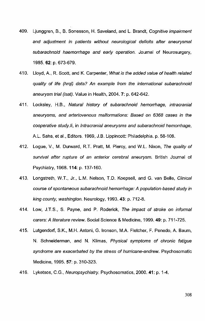
409. Ljunggren, B., B. Sonesson, H. Saveland, and L. Brandt, Cognitive impairment
and adjustment in patients without neurological deficits after aneurysmal
subarachnoid haemorrhage and early operation. Journal of Neurosurgery,
1985. 62: p. 673-679.
410. Lloyd, A., R. Scott, and K. Carpenter, What is the added value of health related
quality of life (hrql) data? An example from the international subarachnoid
aneurysm trial (isat). Value in Health, 2004. 7: p. 642-642.
411. Locksley, H.B., Natural history of subarachnoid hemorrhage, intracranial
aneurysms, and arteriovenous malformations: Based on 6368 cases in the
cooperative study./i, in Intracranial aneurysms and subarachnoid hemorrhage,
A.L. Sahs, et al., Editors. 1969, J.B. Lippincott: Philadelphia. p. 58-108.
412. Logue, V., M. Durward, R.T. Pratt, M. Piercy, and W.L. Nixon, The quality of
survival after rupture of an anterior cerebral aneurysm. British Journal of
Psychiatry, 1968. 114: p. 137-160.
413. Longstreth, W.T., Jr., L.M. Nelson, T.D. Koepsell, and G. van Belle, Clinical
course of spontaneous subarachnoid hemorrhage: A population-based study in
king county, washington. Neurology, 1993. 43: p. 712-8.
414. Low, J.T.S., S. Payne, and P. Roderick, The impact of stroke on informal
carers: A literature review. Social Science & Medicine, 1999. 49: p. 711-725.
415. Lutgendorf, S.K., M.H. Antoni, G. lronson, M.A. Fletcher, F. Penedo, A. Baum,
N. Schneiderman, and N. Klimas, Physical symptoms of chronic fatigue
syndrome are exacerbated by the stress of hurricane-andrew. Psychosomatic
Medicine, 1995. 57: p. 310-323.
416. Lyketsos, C.G., Neuropsychiatry. Psychosomatics, 2000.41: p. 1-4.
308
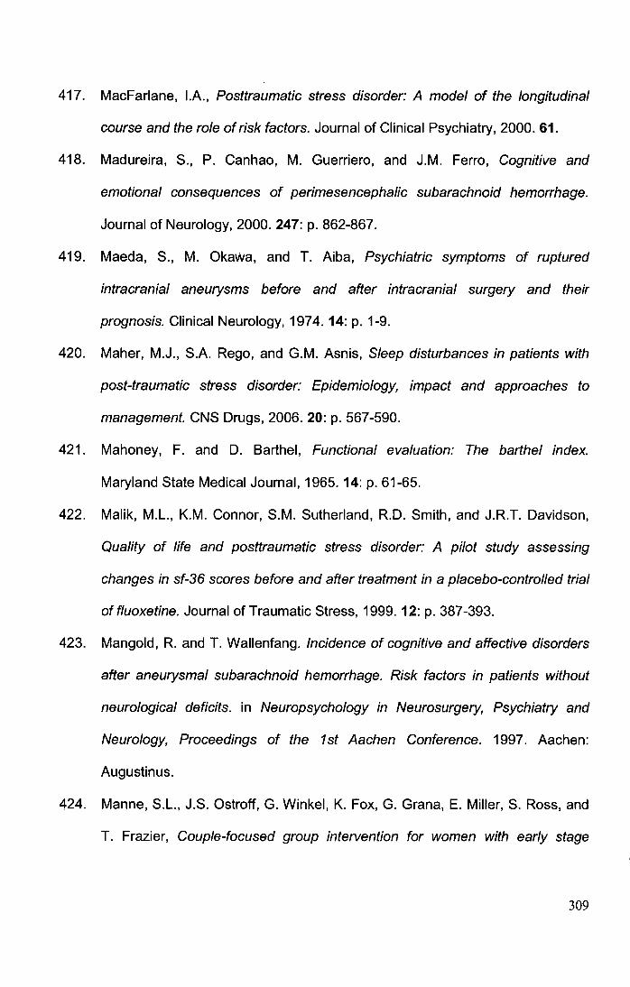
417. MacFarlane, I.A., Posttraumatic stress disorder: A model of the longitudinal
course and the role of risk factors. Journal of Clinical Psychiatry, 2000. 61.
418. Madureira, S., P. Canhao, M. Guerriero, and J.M. Ferro, Cognitive and
emotional consequences of perimesencephalic subarachnoid hemorrhage.
Journal of Neurology, 2000. 247: p. 862-867.
419. Maeda, S., M. Okawa, and T. Aiba, Psychiatric symptoms of ruptured
intracranial aneurysms before and after intracranial surgery and their
prognosis. Clinical Neurology, 1974. 14: p. 1-9.
420. Maher, M.J., S.A. Rego, and G.M. Asnis, Sleep disturbances in patients with
post-traumatic stress disorder: Epidemiology, impact and approaches to
management. CNS Drugs, 2006. 20: p. 567-590.
421. Mahoney, F. and D. Barthel, Functional evaluation: The barthel index.
Maryland State Medical Journal, 1965. 14: p. 61-65.
422. Malik, M.L., K.M. Connor, S.M. Sutherland, R.D. Smith, and J.R.T. Davidson,
Quality of life and posttraumatic stress disorder: A pilot study assessing
changes in sf-36 scores before and after treatment in a placebo-controlled trial
offluoxetine. Journal of Traumatic Stress, 1999. 12: p. 387-393.
423. Mangold, R. and T. Wallenfang. Incidence of cognitive and affective disorders
after aneurysmal subarachnoid hemorrhage. Risk factors in patients without
neurological deficits. in Neuropsychology in Neurosurgery, Psychiatry and
Neurology, Proceedings of the 1st Aachen Conference. 1997. Aachen:
Augustin us.
424. Manne, S.L., J.S. Ostroff, G. Winkel, K. Fox, G. Grana, E. Miller, S. Ross, and
T. Frazier, Couple-focused group intervention for women with early stage
309
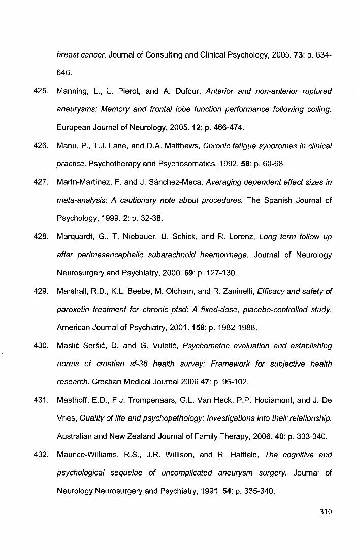
breast cancer. Journal of Consulting and Clinical Psychology, 2005. 73: p. 634-
646.
425. Manning, L, L. Pierot, and A. Dufour, Anterior and non-anterior ruptured
aneurysms: Memory and frontal lobe function performance following coiling.
European Journal of Neurology, 2005. 12: p. 466-474.
426. Manu, P., T.J. Lane, and D.A. Matthews, Chronic fatigue syndromes in clinical
practice. Psychotherapy and Psychosomatics, 1992. 58: p. 60-68.
427. Marin-Martinez, F. and J. Sanchez-Meca, Averaging dependent effect sizes in
meta-analysis: A cautionary note about procedures. The Spanish Journal of
Psychology, 1999. 2: p. 32-38.
428. Marquardt, G., T. Niebauer, U. Schick, and R. Lorenz, Long term follow up
after perimesencephalic subarachnoid haemorrhage. Journal of Neurology
Neurosurgery and Psychiatry, 2000. 69: p. 127-130.
429. Marshall, R.D., K.L. Beebe, M. Oldham, and R. Zaninelli, Efficacy and safety of
paroxetin treatment for chronic ptsd: A fixed-dose, placebo-controlled study.
American Journal of Psychiatry, 2001. 158: p. 1982-1988.
430. Maslic Sersic, D. and G. Vuletic, Psychometric evaluation and establishing
norms of croatian sf-36 health survey: Framework for subjective health
research. Croatian Medical Journal 2006 47: p. 95-102.
431. Masthoff, E.D., F.J. Trompenaars, G.L. Van Heck, P.P. Hodiamont, and J. De
Vries, Quality of life and psychopathology: Investigations into their relationship.
Australian and New Zealand Journal of Family Therapy, 2006. 40: p. 333-340.
432. Maurice-Williams, R.S., J.R. Willison, and R. Hatfield, The cognitive and
psychological sequelae of uncomplicated aneurysm surgery. Journal of
Neurology Neurosurgery and Psychiatry, 1991. 54: p. 335-340.
310
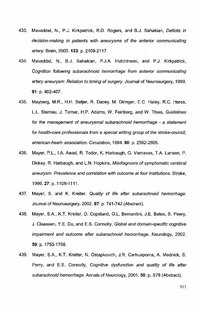
433. Mavaddat, N., P.J. Kirkpatrick, R.D. Rogers, and B.J. Sahakian, Deficits in
decision-making in patients with aneurysms of the anterior communicating
artery. Brain,2000.123: p. 2109-'2117.
434. Mavaddat, N., B.J. Sahakian, P.J.A. Hutchinson, and P.J. Kirkpatrick,
Cognition following subarachnoid hemorrhage from anterior communicating
artery aneurysm: Relation to timing of surgery. Journal of Neurosurgery, 1999.
91: p. 402-407.
435. Mayberg, M.R., H.H. Batjer, R. Dacey, M. Diringer, E.C. Haley, R.C. Heros,
L.L. Sternau, J. Torner, H.P. Adams, W. Feinberg, and W. Thies, Guidelines
for the management of aneurysmal subarachnoid hemorrhage - a statement
for health-care professionals from a special writing group of the stroke-council,
american-heart- association. Circulation, 1994. 90: p. 2592-2605.
436. Mayer, P.L., I.A. Awad, R. Todor, K. Harbaugh, G. Varnavas, T.A. Lansen, P.
Dickey, R. Harbaugh, and L.N. Hopkins, Misdiagnosis of symptomatic cerebral
aneurysm. Prevalence and correlation with outcome at four institutions. Stroke,
1996.27:p. 1108-1111.
437. Mayer, S. and K. Kreiter, Quality of life after subarachnoid hemorrhage.
Journal of Neurosurgery, 2002. 97: p. 741-742 (Abstract).
438. Mayer, S.A., K.T. Kreiter, D. Copeland, G.L. Bernardini, J.E. Bates, S. Peery,
J. Claassen, Y.E. Du, and E.S. Connolly, Global and domain-specific cognitive
impairment and outcome after subarachnoid hemorrhage. Neurology, 2002.
59: p. 1750-1758.
439. Mayer, S.A., K.T. Kreiter, N. Ostapkovich, J.R. Carhuapoma, A. Mednick, S.
Perry, and E.S. Connolly, Cognitive dysfunction and quality of life after
subarachnoid hemorrhage. Annals of Neurology, 2001. 50: p. S78 (Abstract).
311
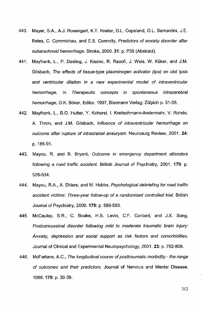
440. Mayer, S.A., A.J. Rosengart, K.T. Kreiter, D.L. Copeland, G.L. Bernardini, J.E.
Bates, C. Commichau, and E.S. Connolly, Predictors of anxiety disorder after
subarachnoid hemorrhage. Stroke, 2000. 31: p. P35 (Abstract).
441. Mayfrank, L., P. Desling, J. Kissler, R. Raoofi, J. Weis, W. Kuker, and J.M.
Gilsbach, The effects of tissue-type plasminogen activator (tpa) on clot lysis
and ventricular dilation in a new experimental model of intraventricular
hemorrhage, in Therapeutic concepts in spontaneous intracerebral
hemorrhage, D.K. Boker, Editor. 1997, Biermann Verlag: Zulpich p. 31-35.
442. Mayfrank, L., B.O. Hutter, Y. Kohorst, I. Kreitschmann-Andermahr, V. Rohde,
A. Thron, and J.M. Gilsbach, Influence of intraventricular hemorrhage on
outcome after rupture of intracranial aneurysm. Neurosurg Review, 2001. 24:
p. 185-91.
443. Mayou, R. and B. Bryant, Outcome in emergency department attenders
following a road traffic accident. British Journal of Psychiatry, 2001. 179: p.
528-534.
444. Mayou, R.A., A. Ehlers, and M. Hobbs, Psychological debriefing for road traffic
accident victims: Three-year follow-up of a randomised controlled trial. British
Journal of Psychiatry, 2000. 176: p. 589-593.
445. McCauley, S.R., C. Boake, H.S. Levin, C.F. Contant, and J.X. Song,
Postconcussinal disorder following mild to moderate traumatic brain injury:
Anxiety, depression and social support as risk factors and comorbidities.
Journal of Clinical and Experimental Neuropsychology, 2001. 23: p. 792-808.
446. McFarlane, A. C., The longitudinal course of posttraumatic morbidity- the range
of outcomes and their predictors. Journal of Nervous and Mental Disease,
1988. 176: p. 30-39.
312
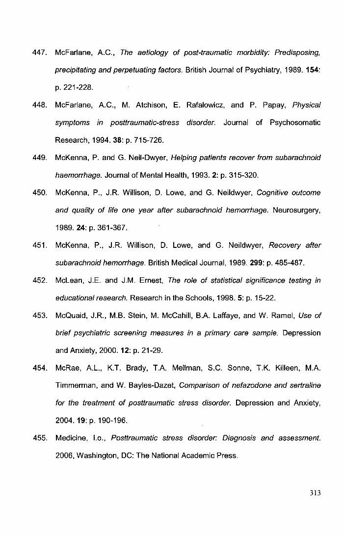
447. McFarlane, A.C., The aetiology of post-traumatic morbidity: Predisposing,
precipitating and perpetuating factors. British Journal of Psychiatry, 1989. 154:
p. 221-228.
448. McFarlane, A.C., M. Atchison, E. Rafalowicz, and P. Papay, Physical
symptoms in posttraumatic-stress disorder. Journal of Psychosomatic
Research, 1994. 38: p. 715-726.
449. McKenna, P. and G. Neil-Dwyer, Helping patients recover from subarachnoid
haemorrhage. Journal of Mental Health, 1993. 2: p. 315-320.
450. McKenna, P., J.R. Willison, D. Lowe, and G. Neildwyer, Cognitive outcome
and quality of life one year after subarachnoid hemorrhage. Neurosurgery,
1989. 24: p. 361-367.
451. McKenna, P., J.R. Willison, D. Lowe, and G. Neildwyer, Recovery after
subarachnoid hemorrhage. British Medical Journal, 1989. 299: p. 485-487.
452. Mclean, J.E. and J.M. Ernest, The role of statistical significance testing in
educational research. Research in the Schools, 1998. 5: p. 15-22.
453. McQuaid, J.R., M.B. Stein, M. McCahill, B.A. Laffaye, and W. Ramel, Use of
brief psychiatric screening measures in a primary care sample. Depression
and Anxiety, 2000. 12: p. 21-29.
454. McRae, A.L., K.T. Brady, T.A. Mellman, S.C. Sonne, T.K. Killeen, M.A.
Timmennan, and W. Bayles-Dazet, Comparison of nefazodone and sertraline
for the treatment of posttraumatic stress disorder. Depression and Anxiety,
2004. 19: p. 190-196.
455. Medicine, l.o., Posttraumatic stress disorder: Diagnosis and assessment.
2006, Washington, DC: The National Academic Press.
313
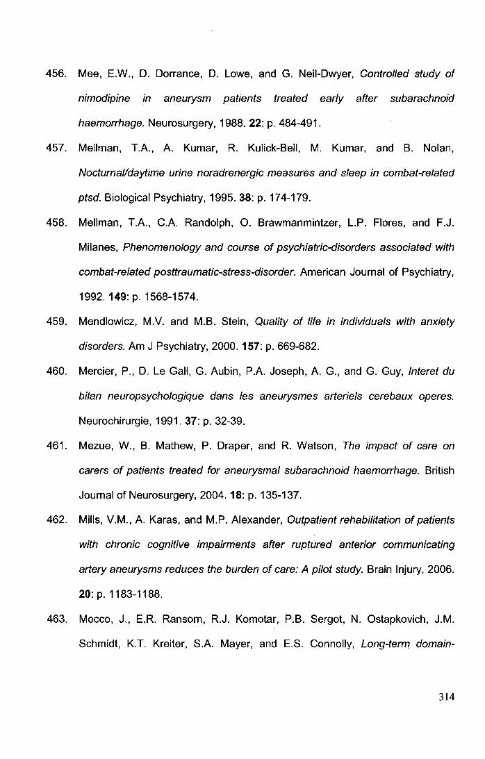
456. Mee, E.W., D. Dorrance, D. Lowe, and G. Neil-Dwyer, Controlled study of
nimodipine in aneurysm patients treated early after subarachnoid
haemorrhage. Neurosurgery, 1988. 22: p. 484-491.
457. Mellman, T.A., A. Kumar, R. Kulick-Bell, M. Kumar, and B. Nolan,
Nocturnal/daytime urine noradrenergic measures and sleep in combat-related
ptsd. Biological Psychiatry, 1995. 38: p. 174-179.
458. Mellman, T.A., C.A. Randolph, 0. Brawmanmintzer, L.P. Flores, and F.J.
Milanes, Phenomenology and course of psychiatric-disorders associated with
combat-related posttraumatic-stress-disorder. American Journal of Psychiatry,
1992. 149: p. 1568-1574.
459. Mendlowicz, M.V. and M.B. Stein, Quality of life in individuals with anxiety
disorders. Am J Psychiatry, 2000. 157: p. 669-682.
460. Mercier, P., D. Le Gall, G. Aubin, P.A. Joseph, A. G., and G. Guy, Interet du
bilan neuropsychologique dans les aneurysmes arteriels cerebaux operes.
Neurochirurgie, 1991. 37: p. 32-39.
461. Mezue, W., B. Mathew, P. Draper, and R. Watson, The impact of care on
carers of patients treated for aneurysmal subarachnoid haemorrhage. British
Journal of Neurosurgery, 2004. 18: p. 135-137.
462. Mills, V.M., A. Karas, and M.P. Alexander, Outpatient rehabilitation of patients
with chronic cognitive impairments after ruptured anterior communicating
artery aneurysms reduces the burden of care: A pilot study. Brain Injury, 2006.
20: p. 1183-1188.
463. Maceo, J., E.R. Ransom, R.J. Komotar, P.B. Sergot, N. Ostapkovich, J.M.
Schmidt, K.T. Kreiter, S.A. Mayer, and E.S. Connolly, Long-term domain-
314
specific improvement following poor grade aneurysmal subarachnoid
hemorrhage. Journal of Neurology, 2006. 253: p. 1278-1284.
464. Mogotsi, M., D. Kaminer, and D.J. Stein, Quality of life in anxiety disorders.
Harvard Review of Psychiatry, 2000. 8: p. 273-282.
465. Mohanty, A., B.S. Das, C.R. Mukundan, and N. Jamuna, Cognitive outcome
and quality of life after aneurysmal subarachnoid haemorrhage: li. Quality of
life. NIMHANS Journal, 1993. 11: p. 119-124.
466. Molyneux, A., R. Kerr, I. Stratton, P.A.G. Sandercock, M. Clarke, J. Shrimpton,
R. Holman, and I.S.A.T.I.C. Group., International subarachnoid aneurysm trial
(isat) of neurosurgical clipping versus endovascular coiling in 2143 patients
with ruptured intracranial aneurysms: A randomised trial. Lancet, 2002. 360: p.
1267-1274.
467. Molyneux, A.J., R.S. Kerr, L.M. Yu, M. Clarke, M. Sneade, J.A. Yarnold, and P.
Sandercock, International subarachnoid aneurysm trial (isat) of neurosurgical
clipping versus endovascular coiling in 2143 patients with ruptured intracranial
aneurysms: A randomised comparison of effects on survival, dependency,
seizures, rebleeding, subgroups, and aneurysm occlusion. Lancet, 2005. 366:
p. 809-17.
468. Morita, A., M.R. Puumala, and F.B. Meyer, Intracranial aneurysms and
subarachnoid hemorrhage, in Outcomes in neurological and neurosurgical
disorders, M. Swash, Editor. 1998, Cambridge University Press: Cambridge. p.
93-122.
469. Morris, P.G., J.T.L. Wilson, and L. Dunn, Anxiety and depression after
spontaneous subarachnoid hemorrhage. Neurosurgery, 2004. 54: p. 47-52.
315
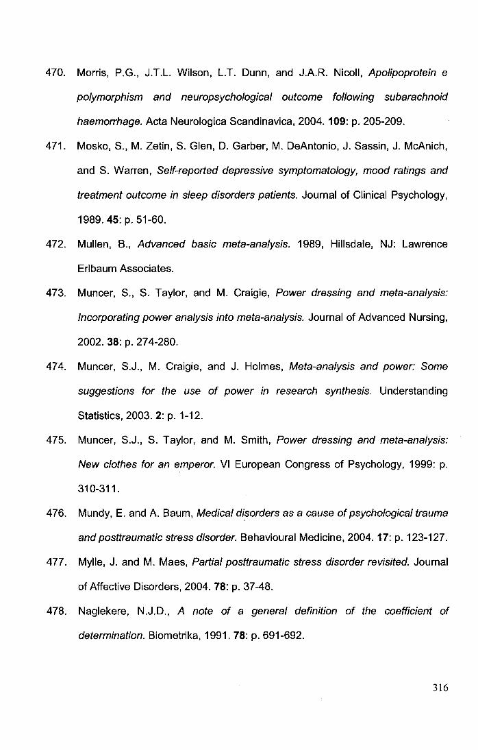
470. Morris, P.G., J.T.L. Wilson, L.T. Dunn, and J.A.R. Nicoll, Apolipoprotein e
polymorphism and neuropsychological outcome following subarachnoid
haemorrhage. Acta Neurologica Scandinavica, 2004. 109: p. 205-209.
471. Mosko, S., M. Zetin, S. Glen, D. Garber, M. DeAntonio, J. Sassin, J. McAnich,
and S. Warren, Self-reported depressive symptomatology, mood ratings and
treatment outcome in sleep disorders patients. Journal of Clinical Psychology,
1989. 45: p. 51-60.
472. Mullen, B., Advanced basic meta-analysis. 1989, Hillsdale, NJ: Lawrence
Erlbaum Associates.
473. Muncer, S., S. Taylor, and M. Craigie, Power dressing and meta-analysis:
Incorporating power analysis into meta-analysis. Journal of Advanced Nursing,
2002. 38:p.274-280.
474. Muncer, S.J., M. Craigie, and J. Holmes, Meta-analysis and power: Some
suggestions for the use of power in research synthesis. Understanding
Statistics, 2003. 2: p. 1-12.
475. Muncer, S.J., S. Taylor, and M. Smith, Power dressing and meta-analysis:
New clothes for an emperor. VI European Congress of Psychology, 1999: p.
310-311.
476. Mundy, E. and A. Baum, Medical disorders as a cause of psychological trauma
and posttraumatic stress disorder. Behavioural Medicine, 2004. 17: p. 123-127.
477. Mylle, J. and M. Maes, Partial posttraumatic stress disorder revisited. Journal
of Affective Disorders, 2004. 78: p. 37-48.
478. Naglekere, N.J.D., A note of a general definition of the coefficient of
determination. Biometrika, 1991. 78: p. 691-692.
316
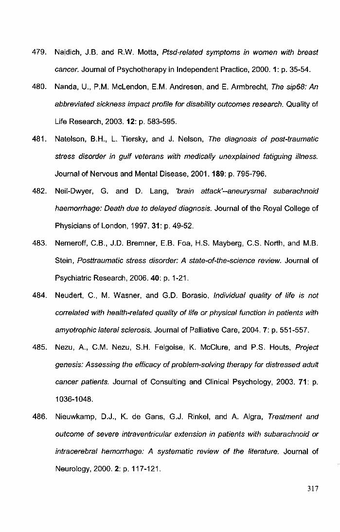
479. Naidich, J.B. and R.W. Motta, Ptsd-related symptoms in women with breast
cancer. Journal of Psychotherapy in Independent Practice, 2000. 1: p. 35-54.
480. Nanda, U., P.M. Mclendon, E.M. Andresen, and E. Armbrecht, The sip68: An
abbreviated sickness impact profile for disability outcomes research. Quality of
Life Research, 2003. 12: p. 583-595.
481. Natelson, B.H., L. Tiersky, and J. Nelson, The diagnosis of post-traumatic
stress disorder in gulf veterans with medically unexplained fatiguing illness.
Journal of Nervous and Mental Disease, 2001. 189: p. 795-796.
482. Neil-Dwyer, G. and D. Lang, 'brain attack'--aneurysmal subarachnoid
haemorrhage: Death due to delayed diagnosis. Journal of the Royal College of
Physicians of London, 1997.31: p. 49-52.
483. Nemeroff, C.B., J.D. Bremner, E.B. Foa, H.S. Mayberg, C.S. North, and M.B.
Stein, Posttraumatic stress disorder: A state-of-the-science review. Journal of
Psychiatric Research, 2006. 40: p. 1-21.
484. Neudert, C., M. Wasner, and G.D. Borasio, Individual quality of life is not
correlated with health-related quality of life or physical function in patients with
amyotrophic lateral sclerosis. Journal of Palliative Care, 2004. 7: p. 551-557.
485. Nezu, A., C.M. Nezu, S.H. Felgoise, K. McClure, and P.S. Houts, Project
genesis: Assessing the efficacy of problem-solving therapy for distressed adult
cancer patients. Journal of Consulting and Clinical Psychology, 2003. 71: p.
1036-1048.
486. Nieuwkamp, D.J., K. de Gans, G.J. Rinkel, and A. Algra, Treatment and
outcome of severe intraventricular extension in patients with subarachnoid or
intracerebral hemorrhage: A systematic review of the literature. Journal of
Neurology, 2000.2: p.117-121.
317
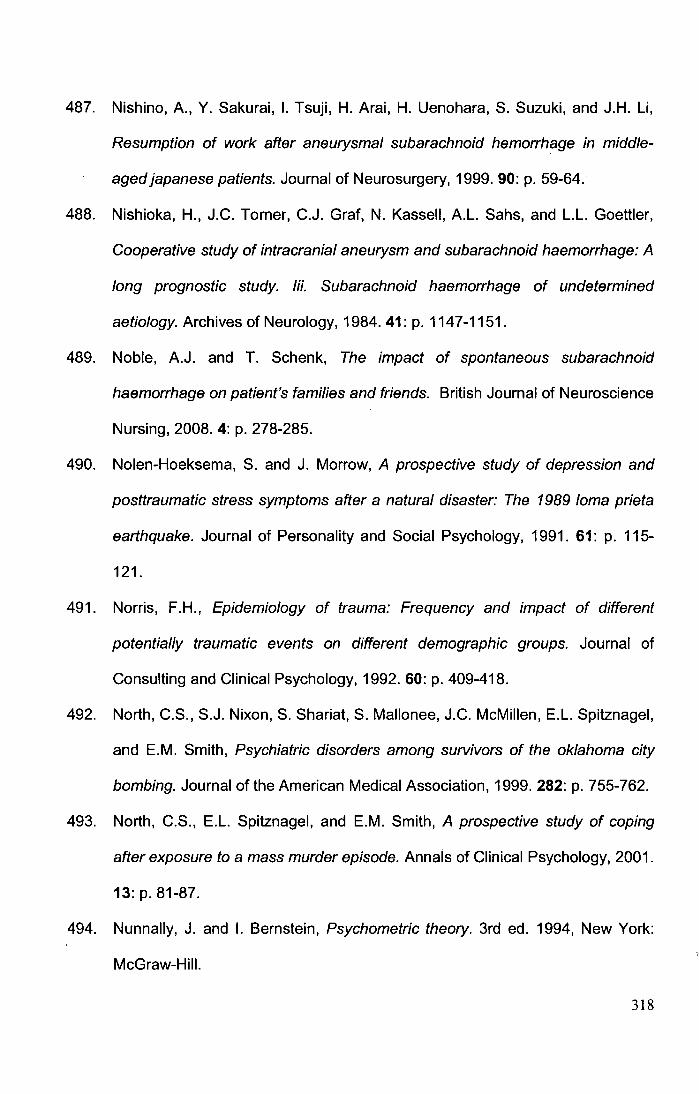
487. Nishino, A., Y. Sakurai, I. Tsuji, H. Arai, H. Uenohara, S. Suzuki, and J.H. Li,
Resumption of work after aneurysmal subarachnoid hemorrhage in middle
aged japanese patients. Journal of Neurosurgery, 1999. 90: p. 59-64.
488. Nishioka, H., J.C. Tomer, C.J. Graf, N. Kassel!, A.L. Sahs, and L.L. Goettler,
Cooperative study of intracranial aneurysm and subarachnoid haemorrhage: A
long prognostic study. Iii. Subarachnoid haemorrhage of undetermined
aetiology. Archives of Neurology, 1984. 41: p. 1147-1151.
489. Noble, A.J. and T. Schenk, The impact of spontaneous subarachnoid
haemorrhage on patient's families and friends. British Journal of Neuroscience
Nursing, 2008. 4: p. 278-285.
490. Nolen-Hoeksema, S. and J. Morrow, A prospective study of depression and
posttraumatic stress symptoms after a natural disaster: The 1989 lorna prieta
earthquake. Journal of Personality and Social Psychology, 1991. 61: p. 115-
121.
491. Norris, F.H., Epidemiology of trauma: Frequency and impact of different
potentially traumatic events on different demographic groups. Journal of
Consulting and Clinical Psychology, 1992. 60: p. 409-418.
492. North, C.S., S.J. Nixon, S. Shariat, S. Mallonee, J.C. McMillen, E.L. Spitznagel,
and E.M. Smith, Psychiatric disorders among survivors of the oklahoma city
bombing. Journal of the American Medical Association, 1999. 282: p. 755-762.
493. North, C.S., E.L. Spitznagel, and E.M. Smith, A prospective study of coping
after exposure to a mass murder episode. Annals of Clinical Psychology, 2001.
13: p. 81-87.
494. Nunnally, J. and I. Bernstein, Psychometric theory. 3rd ed. 1994, New York:
McGraw-Hill.
318
495. Nutt, D.J. and A.L. Malizia, Structural and functional brain changes in
posttraumatic stress disorder. Journal of Clinical Psychiatry, 2004. 65: p. 11-
17.
496. Nyquist, P., S. LeDroux, and R. Geocadin, Thrombolytics in intraventricular
hemorrhage. Current Neurology and Neuroscience Reports, 2007. 7: p. 522-
528
497. Nystrom, S.H.M., Psychodynamics in subarachnoid hemorrhage: A study
based on 1183 patients. Helsinki, finland. Sahalan kirjapaino, 1973. 39: p. 39-
47.
498. O'Carroll, R.E., G. Masterton, R. Gooday, J.A. Cassar, M.C. Couston, and P.C.
hayes, Variceal haemorrhage and post-traumatic stress disorder. British
Journal of Clinical Psychology, 1999. 38: p. 203-208.
499. O'Dell, M.W., T.K. Watanabe, S.T. De Roos, and C. Kager, Functional
outcome after inpatient rehabilitation in persons with subarachnoid
hemorrhage. Archives of Physical Medicine and Rehabilitation, 2002. 83: p.
678-682.
500. O'Reilly, S.M., N. Grubb, and R.E. O'Carroll, Long-term emotional
consequences of in-hospital cardiac arrest and myocardial infarction. British
Journal of Clinical Psychology, 2004. 43: p. 83-96.
501. Oder, W., P. Dalbianco, H. Kollegger, K. Zeiler, H. Binder, and L. Deecke,
Spontaneous subarachnoid hemorrhage - prognostic factors for social
readjustment. Scandinavian Journal of Rehabilitation Medicine, 1990. 22: p.
85-91.
319
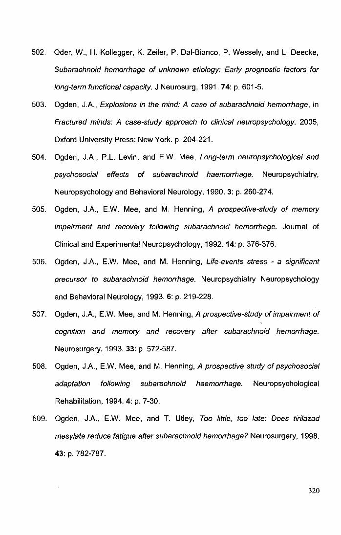
502. Oder, W., H. Kollegger, K. Zeiler, P. Dai-Bianco, P. Wessely, and L. Deecke,
Subarachnoid hemorrhage of unknown etiology: Early prognostic factors for
long-term functional capacity. J Neurosurg, 1991. 74: p. 601-5.
503. Ogden, J.A., Explosions in the mind: A case of subarachnoid hemorrhage, in
Fractured minds: A case-study approach to clinical neuropsychology. 2005,
Oxford University Press: New York. p. 204-221.
504. Ogden, J.A., P.L. Levin, and E.W. Mee, Long-term neuropsychological and
psychosocial effects of subarachnoid haemorrhage. Neuropsychiatry,
Neuropsychology and Behavioral Neurology, 1990. 3: p. 260-274.
505. Ogden, J.A., E.W. Mee, and M. Henning, A prospective-study of memory
impairment and recovery following subarachnoid hemorrhage. Journal of
Clinical and Experimental Neuropsychology, 1992. 14: p. 376-376.
506. Ogden, J.A., E.W. Mee, and M. Henning, Life-events stress - a significant
precursor to subarachnoid hemorrhage. Neuropsychiatry Neuropsychology
and Behavioral Neurology, 1993. 6: p. 219-228.
507. Ogden, J.A., E.W. Mee, and M. Henning, A prospective-study of impairment of
cognition and memory and recovery after subarachnoid hemorrhage.
Neurosurgery, 1993. 33: p. 572-587.
508. Ogden, J.A., E.W. Mee, and M. Henning, A prospective study of psychosocial
adaptation following subarachnoid haemorrhage. Neuropsychological
Rehabilitation, 1994. 4: p. 7-30.
509. Ogden, J.A., E.W. Mee, and T. Utley, Too little, too late: Does tirilazad
mesylate reduce fatigue after subarachnoid hemorrhage? Neurosurgery, 1998.
43: p. 782-787.
320
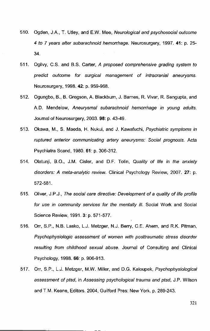
510. Ogden, J.A., T. Utley, and E.W. Mee, Neurological and psychosocial outcome
4 to 7 years after subarachnoid hemorrhage. Neurosurgery, 1997. 41: p. 25-
34.
511. Ogilvy, C.S. and B.S. Carter, A proposed comprehensive grading system to
predict outcome for surgical management of intracranial aneurysms.
Neurosurgery, 1998. 42: p. 959-968.
512. Ogungbo, B., B. Gregson, A. Blackburn, J. Barnes, R. Vivar, R. Sengupta, and
A.D. Mendelow, Aneurysmal subarachnoid hemorrhage in young adults.
Journal of Neurosurgery, 2003. 98: p. 43-49.
513. Okawa, M., S. Maeda, H. Nukui, and J. Kawafuchi, Psychiatric symptoms in
ruptured anterior communicating artery aneurysms: Social prognosis. Acta
Psychiatra Scand, 1980. 61: p. 306-312.
514. Olatunji, B.O., J.M. Cisler, and D.F. Tolin, Quality of life in the anxiety
disorders: A meta-analytic review. Clinical Psychology Review, 2007. 27: p.
572-581.
515. Oliver, J.P.J., The social care directive: Development of a quality of life profile
for use in community services for the mentally ill. Social Work and Social
Science Review, 1991. 3: p. 571-577.
516. Orr, S.P., N.B. Lasko, L.J. Metzger, N.J. Berry, C.E. Ahern, and R.K. Pitman,
Psychophysiologic assessment of women with posttraumatic stress disorder
resulting from childhood sexual abuse. Journal of Consulting and Clinical
Psychology, 1998. 66: p. 906-913.
517. Orr, S.P., L.J. Metzger, M.W. Miller, and D.G. Kaloupek, Psychophysiological
assessment of ptsd, in Assessing psychological trauma and ptsd, J.P. Wilson
and T.M. Keane, Editors. 2004, Guilford Pres: New York. p. 289-243.
321
518. Ottenbacher, K.J., H.R. Ottenbacher, L. Tooth, and G.V. Ostir, A review of two
journals found that articles using multivariable logistic regression frequently did
not report commonly recommended assumptions. Journal of Clinical
Epidemiology, 2004. 57: p. 1147-1152.
519. Oyebode, F., S. Kennedy, and K. Davison, Psychiatric sequelae of
subarachnoid hemorrhage. British Journal of Hospital Medicine, 1986. 36: p.
104, 106-108.
520. Oyebode, J., Assessment of carers' psychological needs. Advances in
Psychiatric Treatment, 2003. 9: p. 45-53.
521. Ozer, E.J., S.R. Best, T.l. Lipsey, and D.S. Weiss, Predictors of posttraumatic
stress disorder and symptoms in adults: A meta-analysis. Psychological
Bulletin, 2003. 129: p. 52-73.
522. Parker, G., With due care and attention: A review of research on informal care.
2nd ed. 1990, London: Family Policy Studies Centre.
523. Patel, M.D., C. Coshall, A.G. Rudd, and C. D. Wolfe, Cognitive impairment after
stroke: Clinical determinants and its associations with long-term stroke
outcomes. Journal of the American Geriatrics Society, 2002. 50.
524. Patel, M.D., C. McKevitt, E. Lawrence, A.G. Rudd, and C.D. Wolfe, Clinical
determinants of long-term quality of life after stroke. Age and Ageing, 2007. 36:
p. 316-322.
525. Patrick (ed), D.L., Health and care of the physical disabled in Iambeth: Phase i
report. 1982, London: Department of Community Medicine, St Thomas'
Hospital Medical School.
322
526. Patrick (ed), D.L., Health and care of the physically disabled in Iambeth: Report
of phase ii of the longitudinal disability interview survey. 1982, London:
Department of Community Medicine, StThomas' Hospital Medical School.
527. Paul, S.L., J.W. Sturm, H.M. Dewey, G.A. Donnan, R.A. Macdonell, and A.G.
Thrift, Long-term outcome in the north east melbourne stroke incidence study:
Predictors of quality of life at 5 years after stroke. Stroke, 2005. 36: p. 2082-6.
528. Pelletier, P.M. and D.P. Alfano, Depression, social support, and family coping
following traumatic brain injury. Brain and Cognition, 2000. 44: p. 45-49.
529. Perkonigg, A., H. Pfister, M.B. Stein, M. Hoefler, R. Lieb, A. Maercker, and
H.U. Wittchen, Longitudinal course of posttraumatic stress disorder and
posttraumatic stress disorder symptoms in a community sample of adolescents
and young adults. European Neuropsychopharmacology, 2006. 16: p. S453-
S454.
530. Phifer, J.F., Psychological distress and somatic symptoms after natural
disaster - differential vulnerability among older adults. Psychology and Aging,
1990. 5: p. 412-420.
531. Pincus, J.H. and G.J. Tucker, Behavioral neurology. 4th ed. 2003, New York:
Oxford University Press.
532. Pinto-Meza, A., J.M. Haro, C. Palacln, J.V. Torres, S. Ochoa, G. Vilagut, M.
Martinez-Aionso, M. Codony, and J. Alonso, The impact of mood and anxiety
disorders, and physical chronic conditions in the quality of life of general
population of spain. Results of the esemed-spain study. Aetas Espaflolas de
Psiquiatria, 2007. 35: p. 12-20.
533. Pinto, A.N., P. Canhao, and J.M. Ferro, Seizures at the onset of subarachnoid
haemorrhage. Journal of Neurology, 1996. 243: p. 161-164.
323
534. Pitman, R.K., D.M. Lanes, S.K. Williston, J.L. Guillaume, L.J. Metzger, G.M.
Gehr, and S.P. Orr, Psychophysiologic assessment of posttraumatic stress
disorder in breast cancer patients. Psychosomatics, 2001. 42: p. 133-140.
535. Pitman, R.K., S.P. Orr, D.F. Forgue, J.B. de Jong, and J.M. Claiborn,
Psychophysiology of ptsd imagery in vietnam combat veterans. Archives of
General Psychiatry, 1987. 44: p. 49-54.
536. Pole, N., The psychophysiology of posttraumatic stress disorder: A meta
analysis. Psychological Bulletin, 2007. 133: p. 725-746.
537. Politynska, B.E., G.E. Berrios, and J.L. Lewko, Neuropsychiatric aspects of
subarachnoid haemorrhage: A review. Neurology Psychiatry and Brain
Research, 1995. 3: p. 111-120.
538. Ponsford, J., S. Sloan, and P. Snow, Traumatic brain injury. Rehabilitation for
everyday adaptive living. 1996, Hove: Psychology Press.
539. Popkin, M.K. and T.B. Mackenzie, Psychiatric presentations of endocrine
dysfunction, in Psychiatric presentations of medical illness, R.C.W. Hall, Editor.
1980, Spectrum: New York, NY. p. 139-156.
540. Powell, J., N. Kitchen, J. Heslin, and R. Greenwood, Psychosocial outcomes at
three and nine months after good neurological recovery from aneurysmal
subarachnoid haemorrhage: Predictors and prognosis. Journal of Neurology
Neurosurgery and Psychiatry, 2002. 72: p. 772-781.
541. Powell, J., N. Kitchen, J. Heslin, and R. Greenwood, Psychosocial outcomes at
18 months after good neurological recovery from aneurysmal subarachnoid
haemorrhage. Journal of Neurology Neurosurgery and Psychiatry, 2004. 75: p.
1119-1124.
324
542. Powell, J.H., K. Beckers, and R.J. Greenwood, Measuring progress and
outcome in community rehabilitation after brain injury with a new assessment
measure: The bicro-39 scales. · Archives of Physical Medicine and
Rehabilitation, 1998.79: p. 1213-1225.
543. Preiss, M., J. Koblihova, D. Netuka, J. Klose, F. Charvat, and V. Benes,
Ruptured cerebral aneurysm patients treated by clipping or coiling:
Comparison of long-term neuropsychological and personality outcomes.
Zentralblatt Fur Neurochirurgie, 2007. 68: p. 169-175.
544. Prigatano, G.P. and S. Henderson, Cognitive outcome after subarachnoid
hemorrhage, in Subarachnoid hemorrhage: Pathophysiology and
management, J.B. Bederson, Editor. 1997, American Association of
Neurological Surgeons: Park Ridge, Illinois. p. 27-40.
545. Pritchard, C., L. Clapham, L. Foulkes, D.A. Lang, and G. Neil-Dwyer,
Comparison of cohorts of elective and emergency neurosurgical patients:
Psychosocial outcomes of acoustic neuroma and aneurysmal sub arachnoid
hemorrhage patients and carers. Surgical Neurology, 2004. 62: p. 7-16.
546. Pritchard, C., L. Foulkes, D.A. Lang, and G. Neil-Dwyer, Psychosocial
outcomes for patients and carers after aneurysmal subarachnoid
haemorrhage. British Journal of Neurosurgery, 2001. 15: p. 456-463.
547. Pritchard, C., L. Foulkes, D.A. Lang, and G. Neil-Dwyer, Cost-benefit analysis
of an integrated approach to reduce psychosocial trauma following
neurosurgery compared with standard care: Two-year prospective comparative
study of enhanced specialist liaison nurse service for aneurysmal
subarachnoid hemorrhage (asah) patients and carers. Surgical Neurology,
2004. 62: p. 17-27.
325
548. Pritchard, C., L. Foulkes, D.A. Lang, and G. Neil-Dwyer, Two-year prospective
study of psychosocial outcomes and a cost- analysis of 'treatment-as-usual'
versus an 'enhanced' (specialist liaison nurse) service for aneurysmal sub
arachnoid haemorrhage (asah) patients and families. British Journal of
Neurosurgery, 2004. 18: p. 347-356.
549. Radloff, L.S. and S. Lenore, The ces-d sea/e. A self-report depression scale for
research in the general population. Applied Psychological Measurement, 1977.
1: p. 385-401.
550. Rankin, J., Cerebral vascular accidents in patients over the age of 60. Scottish
Medical Journal, 1957. 2: p. 200-215.
551. Rapaport, M.H., C. Clary, R. Fayyad, and J. Endicott, Quality of life impairment
in depressive and anxiety disorders. American Journal of Psychiatry, 2005.
162: p.1171-1178.
552. Rapaport, M.H., J. Endicott, and C.M. Clary, Posttraumatic stress disorder and
quality of life: Results across 64 weeks of sertraline treatment. Journal of
Clinical Psychiatry, 2002. 157: p. 1014-1016.
553. Rasmussen, P., H. Busch, J. Haase, J. Hansen, A. Harmsen, V. Knudsen, E.
Marcussen, S.T. Midholm, R.B. Olsen, J. Rosenorn, K. Schmidt, B. Voldby,
and L. Hansen, Intracranial saccular aneurysms. Results of treatment of 851
patients. Acta Neurochir (Wien), 1980.53: p. 1-17.
554. Rauch, S.L., C.R. Savage, N.M. Alpert, E.C. Miguel, L. Baer, H.C. Breiter, A.
Fischman, J, P. Manzo, A, C. Moretti, and M. Jenike, A positron emission
tomographic study of simple phobic symptom provocation. Archives of General
Psychiatry, 1995. 52: p. 20-28.
326
555. Rees, G., S. Shah, C. Hanley, and C. Brunker, Subarachnoid haemorrhage: A
clinical review. Nursing Standard, 2002. 16: p. 47-54.
556. Rhee, S.H., J.C. Parker, K.L. Smarr, G.F. Petroski, J.C. Johnson, and e. al.,
Stress managment in rheumatoid arthritis: What is the underlying mechanism?
Arthritis Care Research, 2000. 13: p. 435-442.
557. Richardson, J.T., Performance in free recall following rupture and repair of
intracranial aneurysm. Brain and Cognition, 1989. 9: p. 210-226.
558. Richardson, J.T., Cognitive performance following rupture and repair and
repair ofintracranial aneurysm. Acta Neurol Scand, 1991. 83: p. 110-122.
559. Rinkel, G.J., E.F. Wijdicks, M. Vermeulen, L.M. Hageman, J.T. Tans, and J.
van Gijn, Outcome in perimesencephalic (non-aneurysmal) subarachnoid
hemorrhage: A follow up study in 37 patients. Neurology, 1990. 40: p. 1130-
1132.
560. Rinne, J., J. Hernesniemi, M. Niskanen, and M. Vapalahti, Analysis of 561
patients with 690 middle cerebral artery aneurysms: Anatomic and clinical
features as correlated to management outcome. Neurosurgery, 1996. 38: p. 2-
11.
561. Riskind, J.H., A.T. Beck, G. Brown, and R.A. Steer, Taking the measure of
anxiety and depression: Validity of reconstructed hamilton scales. Journal of
Nervous and Mental Disease, 1987. 175: p. 474-479.
562. Robertson, E.K. and R.M. Sunn, The determination of rate of progress of
stroke patients through empathy measures of patient and family. Journal of
Psychosomatic Research, 1968.12: p. 189-191.
563. Robertson, I.H., T. Ward, V. Ridgeway, and I. Nimmo-Smith, The test of
everyday attention. 1994, Bury St. Edmonds: Thames Valley Test Company.
327
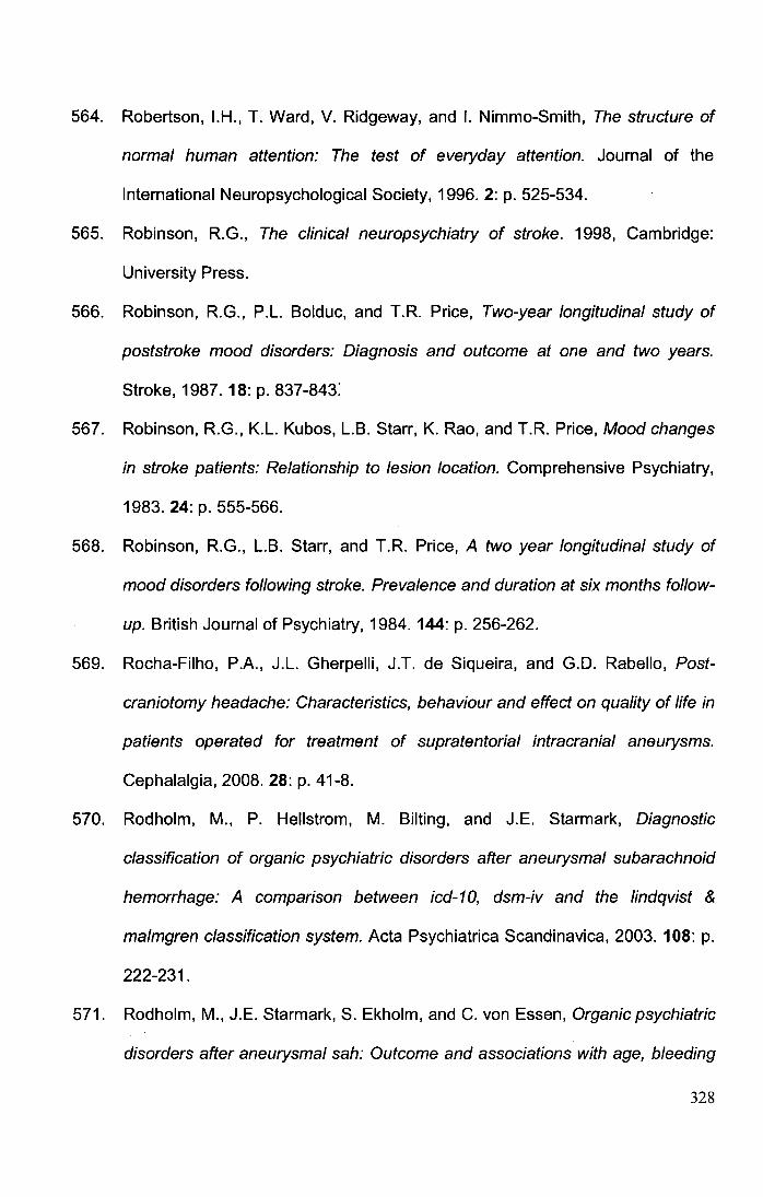
564. Robertson, I.H., T. Ward, V. Ridgeway, and I. Nimmo-Smith, The structure of
normal human attention: The test of everyday attention. Journal of the
International Neuropsychological Society, 1996. 2: p. 525-534.
565. Robinson, R.G., The clinical neuropsychiatry of stroke. 1998, Cambridge:
University Press.
566. Robinson, R.G., P.L Bolduc, and T.R. Price, Two-year longitudinal study of
poststroke mood disorders: Diagnosis and outcome at one and two years.
Stroke, 1987. 18: p. 837-843:
567. Robinson, R.G., K.L Kubos, LB. Starr, K. Rao, and T.R. Price, Mood changes
in stroke patients: Relationship to lesion location. Comprehensive Psychiatry,
1983. 24: p. 555-566.
568. Robinson, R.G., LB. Starr, and T.R. Price, A two year longitudinal study of
mood disorders following stroke. Prevalence and duration at six months follow
up. British Journal of Psychiatry, 1984. 144: p. 256-262.
569. Rocha-Filho, P.A., J.L Gherpelli, J.T. de Siqueira, and G.D. Rabello, Post
craniotomy headache: Characteristics, behaviour and effect on quality of life in
patients operated for treatment of supratentorial intracranial aneurysms.
Cephalalgia, 2008. 28: p. 41-8.
570. Rodholm, M., P. Hellstrom, M. Silting, and J.E. Starmark, Diagnostic
classification of organic psychiatric disorders after aneurysmal subarachnoid
hemorrhage: A comparison between icd-10, dsm-iv and the lindqvist &
malmgren classification system. Acta Psychiatrica Scandinavica, 2003. 108: p.
222-231.
571. Rodholm, M., J.E. Starmark, S. Ekholm, and C. von Essen, Organic psychiatric
disorders after aneurysmal sah: Outcome and associations with age, bleeding
328
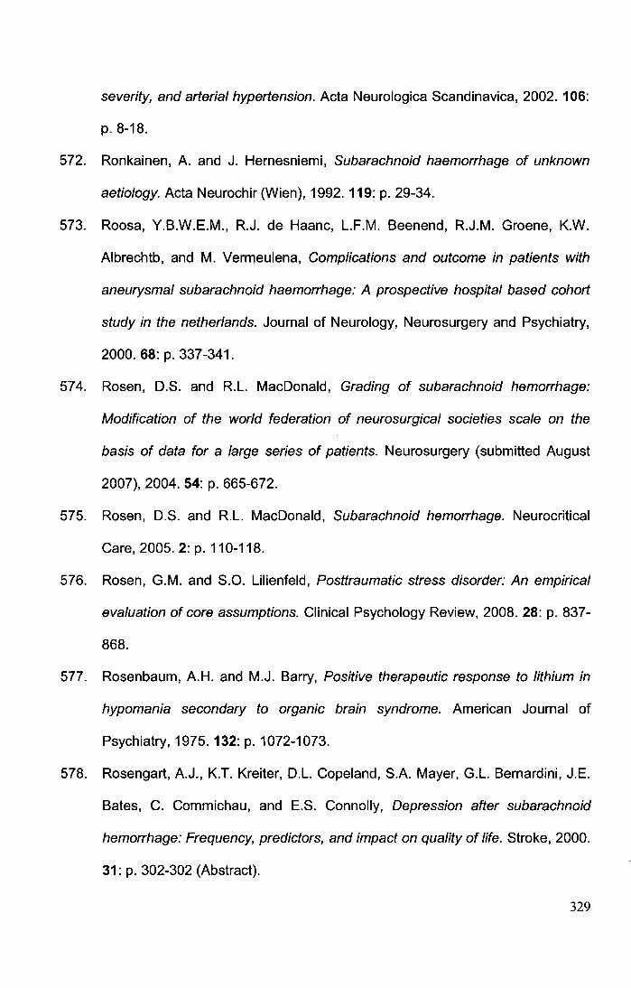
severity, and arterial hypertension. Acta Neurologica Scandinavica, 2002. 106:
p. 8-18.
572. Ronkainen, A. and J. Hernesniemi, Subarachnoid haemorrhage of unknown
aetiology. Acta Neurochir (Wien), 1992. 119: p. 29-34.
573. Roosa, Y.B.W.E.M., R.J. de Haanc, L.F.M. Beenend, R.J.M. Groene, K.W.
Albrechtb, and M. Vermeulena, Complications and outcome in patients with
aneurysmal subarachnoid haemorrhage: A prospective hospital based cohort
study in the netherlands. Journal of Neurology, Neurosurgery and Psychiatry,
2000. 68: p. 337-341.
574. Rosen, D.S. and R.L. MacDonald, Grading of subarachnoid hemorrhage:
Modification of the world federation of neurosurgical societies scale on the
basis of data for a large series of patients. Neurosurgery (submitted August
2007), 2004. 54: p. 665-672.
575. Rosen, D.S. and R.L. MacDonald, Subarachnoid hemorrhage. Neurocritical
Care, 2005. 2: p. 110-118.
576. Rosen, G.M. and S.O. Lilienfeld, Posttraumatic stress disorder: An empirical
evaluation of core assumptions. Clinical Psychology Review, 2008. 28: p. 837-
868.
577. Rosenbaum, A.H. and M.J. Barry, Positive therapeutic response to lithium in
hypomania secondary to organic brain syndrome. American Journal of
Psychiatry, 1975. 132: p. 1072-1073.
578. Rosengart, A.J., K.T. Kreiter, D.L. Copeland, S.A. Mayer, G.L. Bernardini, J.E.
Bates, C. Commichau, and E.S. Connolly, Depression after subarachnoid
hemorrhage: Frequency, predictors, and impact on quality of life. Stroke, 2000.
31: p. 302-302 (Abstract).
329
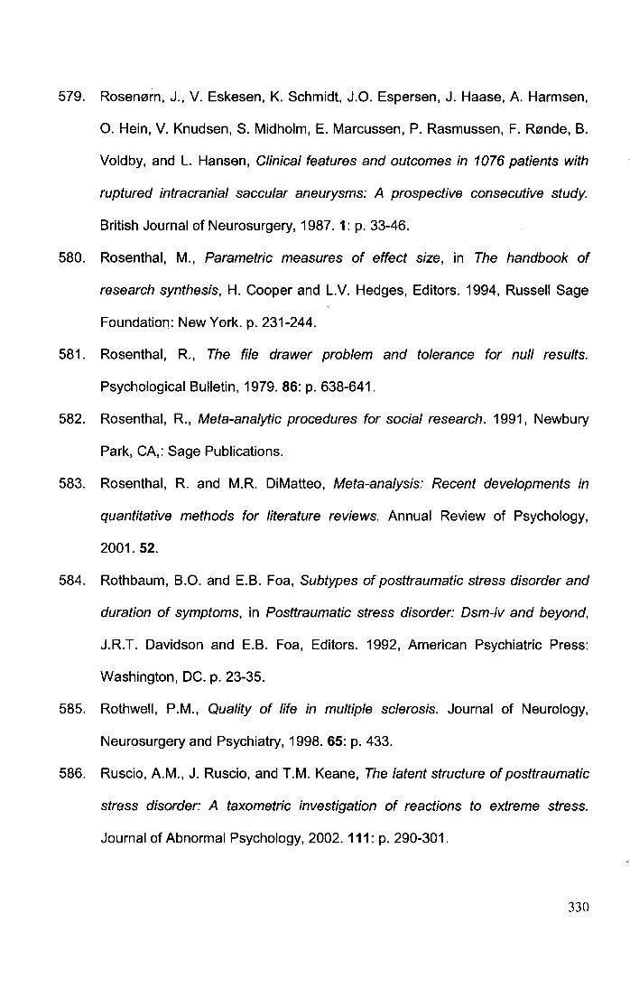
579. Rosen0rn, J., V. Eskesen, K. Schmidt, J.O. Espersen, J. Haase, A. Harmsen,
0. Hein, V. Knudsen, S. Midholm, E. Marcussen, P. Rasmussen, F. R0nde, B.
Voldby, and L. Hansen, Clinical features and outcomes in 1076 patients with
ruptured intracranial saccular aneurysms: A prospective consecutive study.
British Journal of Neurosurgery, 1987. 1: p. 33-46.
580. Rosenthal, M., Parametric measures of effect size, in The handbook of
research synthesis, H. Cooper and L.V. Hedges, Editors. 1994, Russell Sage
Foundation: New York. p. 231-244.
581. Rosenthal, R., The file drawer problem and tolerance for null results.
Psychological Bulletin, 1979. 86: p. 638-641.
582. Rosenthal, R., Meta-analytic procedures for social research. 1991, Newbury
Park, CA,: Sage Publications.
583. Rosenthal, R. and M.R. DiMatteo, Meta-analysis: Recent developments in
quantitative methods for literature reviews. Annual Review of Psychology,
2001. 52.
584. Rothbaum, B.O. and E.B. Faa, Subtypes of posttraumatic stress disorder and
duration of symptoms, in Posttraumatic stress disorder: Dsm-iv and beyond,
J.R.T. Davidson and E.B. Foa, Editors. 1992, American Psychiatric Press:
Washington, DC. p. 23-35.
585. Rothwell, P.M., Quality of life in multiple sclerosis. Journal of Neurology,
Neurosurgery and Psychiatry, 1998. 65: p. 433.
586. Ruscio, A.M., J. Ruscio, and T.M. Keane, The latent structure of posttraumatic
stress disorder: A taxometric investigation of reactions to extreme stress.
Journal of Abnormal Psychology, 2002. 111: p. 290-301.
330
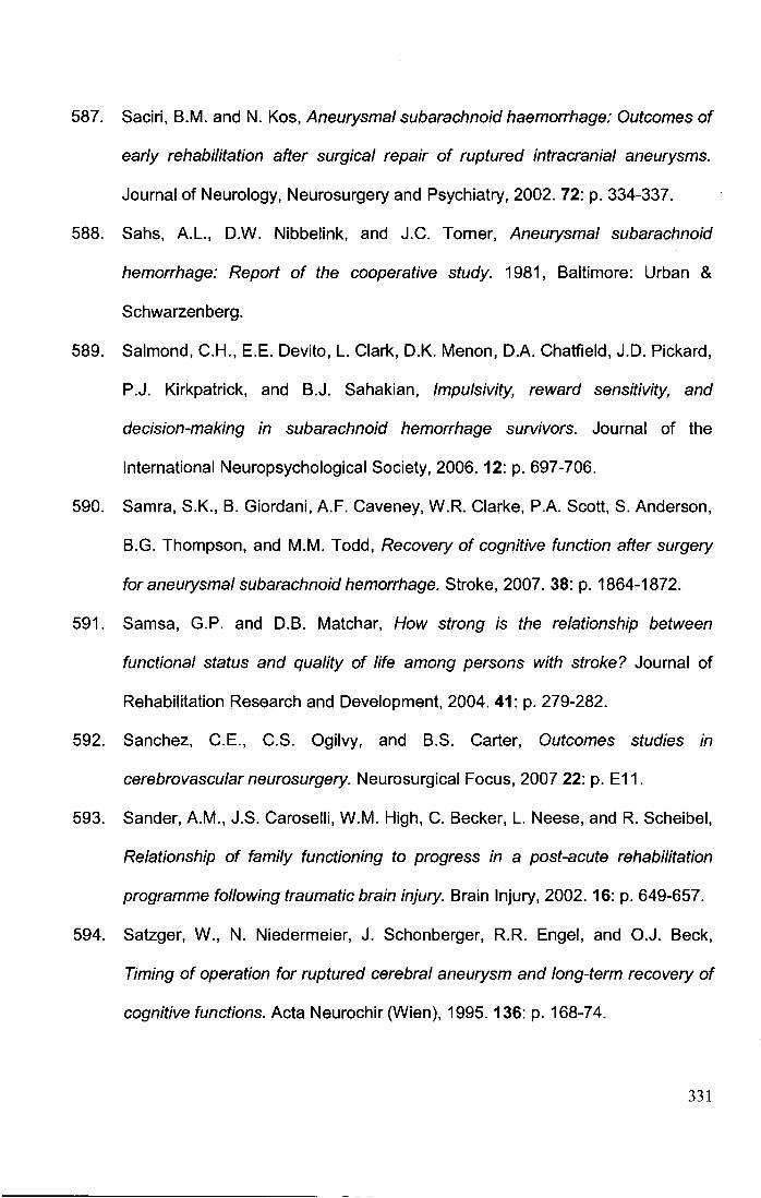
587. Saciri, B.M. and N. Kos, Aneurysmal subarachnoid haemorrhage: Outcomes of
early rehabilitation after surgical repair of ruptured intracranial aneurysms.
Journal of Neurology, Neurosurgery and Psychiatry, 2002. 72: p. 334-337.
588. Sahs, A.L., D.W. Nibbelink, and J.C. Tomer, Aneurysmal subarachnoid
hemorrhage: Report of the cooperative study. 1981, Baltimore: Urban &
Schwarzenberg.
589. Salmond, C.H., E.E. Devito, L. Clark, O.K. Menon, D.A. Chatfield, J.D. Pickard,
P.J. Kirkpatrick, and B.J. Sahakian, Impulsivity, reward sensitivity, and
decision-making in subarachnoid hemorrhage survivors. Journal of the
International Neuropsychological Society, 2006. 12: p. 697-706.
590. Samra, S.K., B. Giordani, A.F. Caveney, W.R. Clarke, P.A. Scott, S. Anderson,
B.G. Thompson, and M.M. Todd, Recovery of cognitive function after surgery
for aneurysmal subarachnoid hemorrhage. Stroke, 2007. 38: p. 1864-1872.
591. Samsa, G.P. and D.B. Matchar, How strong is the relationship between
functional status and quality of life among persons with stroke? Journal of
Rehabilitation Research and Development, 2004. 41: p. 279-282.
592. Sanchez, C.E., C.S. Ogilvy, and B.S. Carter, Outcomes studies in
cerebrovascular neurosurgery. Neurosurgical Focus, 2007 22: p. E11.
593. Sander, A.M., J.S. Caroselli, W.M. High, C. Becker, L. Neese, and R. Scheibel,
Relationship of family functioning to progress in a post-acute rehabilitation
programme following traumatic brain injury. Brain Injury, 2002. 16: p. 649-657.
594. Satzger, W., N. Niedermeier, J. Schonberger, R.R. Engel, and O.J. Beck,
Timing of operation for ruptured cerebral aneurysm and long-term recovery of
cognitive functions. Acta Neurochir (Wien), 1995. 136: p. 168-74.
331
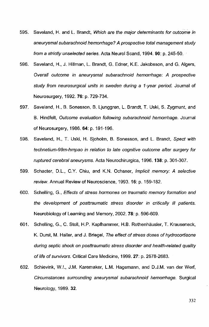
595. Saveland, H. and L. Brandt, Which are the major determinants for outcome in
aneurysmal subarachnoid hemorrhage? A prospective total management study
from a strictly unselected series. Acta Neurol Scand, 1994. 90: p. 245-50.
596. Saveland, H., J. Hillman, L. Brandt, G. Edner, K.E. Jakobsson, and G. Algers,
Overall outcome in aneurysmal subarachnoid hemorrhage: A prospective
study from neurosurgical units in sweden during a 1-year period. Journal of
Neurosurgery, 1992. 76: p. 729-734.
597. Saveland, H., B. Sonesson, B. Ljunggren, L. Brandt, T. Uski, S. Zygmunt, and
B. Hindfelt, Outcome evaluation following subarachnoid hemorrhage. Journal
of Neurosurgery, 1986. 64: p. 191-196.
598. Saveland, H., T. Uski, H. Sjoholm, B. Sonesson, and L. Brandt, Spect with
technetium-99m-hmpao in relation to late cognitive outcome after surgery for
ruptured cerebral aneurysms. Acta Neurochirurgica, 1996. 138: p. 301-307.
599. Schacter, D.L., C.Y. Chiu, and K.N. Ochsner, Implicit memory: A selective
review. Annual Review of Neuroscience, 1993. 16: p. 159-182.
600. Schelling, G., Effects of stress hormones on traumatic memory formation and
the development of posttraumatic stress disorder in critically ill patients.
Neurobiology of Learning and Memory, 2002. 78: p. 596-609.
601. Schelling, G., C. Stoll, H.P. Kapfhammer, H.B. Rothenhausler, T. Krauseneck,
K. Durst, M. Haller, and J. Briegel, The effect of stress doses of hydrocortisone
during septic shock on posttraumatic stress disorder and health-related quality
of life of survivors. Critical Care Medicine, 1999. 27: p. 2678-2683.
602. Schievink, W.l., J.M. Karemaker, L.M. Hagemann, and D.J.M. van der Werf,
Circumstances surrounding aneurysmal subarachnoid hemorrhage. Surgical
Neurology, 1989. 32.
332
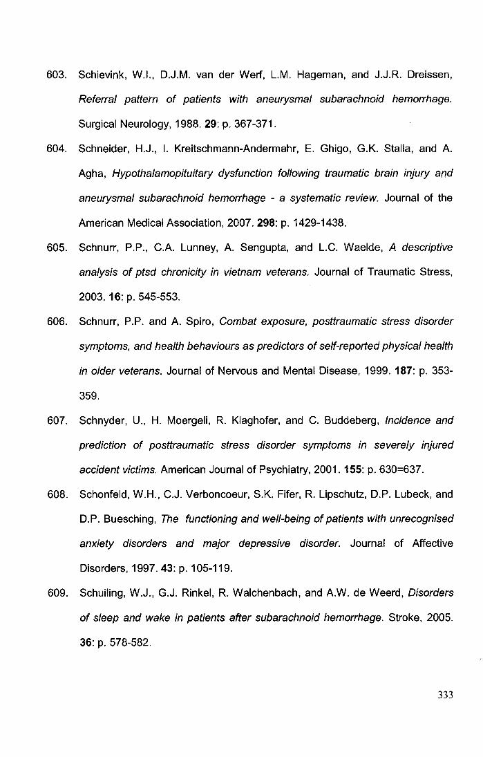
603. Schievink, W.l., D.J.M. van der Wert, L.M. Hageman, and J.J.R. Dreissen,
Referral pattern of patients with aneurysmal subarachnoid hemorrhage.
Surgical Neurology, 1988.29: p. 367-371.
604. Schneider, H.J., I. Kreitschmann-Andermahr, E. Ghigo, G.K. Stalla, and A.
Agha, Hypothalamopituitary dysfunction following traumatic brain injury and
aneurysmal subarachnoid hemorrhage - a systematic review. Journal of the
American Medical Association, 2007. 298: p. 1429-1438.
605. Schnurr, P.P., C.A. Lunney, A. Sengupta, and L.C. Waelde, A descriptive
analysis of ptsd chronicity in vietnam veterans. Journal of Traumatic Stress,
2003. 16: p. 545-553.
606. Schnurr, P.P. and A. Spiro, Combat exposure, posttraumatic stress disorder
symptoms, and health behaviours as predictors of self-reported physical health
in older veterans. Journal of Nervous and Mental Disease, 1999. 187: p. 353-
359.
607. Schnyder, U., H. Moergeli, R. Klaghofer, and C. Buddeberg, Incidence and
prediction of posttraumatic stress disorder symptoms in severely injured
accident victims. American Journal of Psychiatry, 2001. 155: p. 630=637.
608. Schonfeld, W.H., C.J. Verboncoeur, S.K. Fifer, R. Lipschutz, D.P. Lubeck, and
D.P. Buesching, The functioning and well-being of patients with unrecognised
anxiety disorders and major depressive disorder. Journal of Affective
Disorders, 1997. 43: p. 105-119.
609. Schuiling, W.J., G.J. Rinkel, R. Walchenbach, and A.W. de Weerd, Disorders
of sleep and wake in patients after subarachnoid hemorrhage. Stroke, 2005.
36: p. 578-582.
333
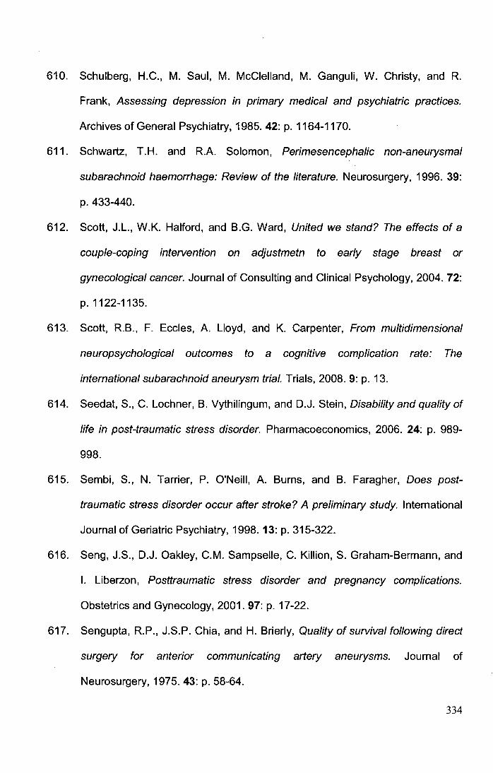
610. Schulberg, H.C., M. Saul, M. McClelland, M. Ganguli, W. Christy, and R.
Frank, Assessing depression in primary medical and psychiatric practices.
Archives of General Psychiatry, 1985. 42: p. 1164-1170.
611. Schwartz, T.H. and R.A. Solomon, Perimesencephalic non-aneurysmal
subarachnoid haemorrhage: Review of the literature. Neurosurgery, 1996. 39:
p. 433-440.
612. Scott, J.L., W.K. Halford, and B.G. Ward, United we stand? The effects of a
couple-coping intervention on adjustmetn to early stage breast or
gynecological cancer. Journal of Consulting and Clinical Psychology, 2004. 72:
p.1122-1135.
613. Scott, R.B., F. Eccles, A. Lloyd, and K. Carpenter, From multidimensional
neuropsychological outcomes to a cognitive complication rate: The
international subarachnoid aneurysm trial. Trials, 2008. 9: p. 13.
614. Seedat, S., C. Lochner, B. Vythilingum, and D.J. Stein, Disability and quality of
life in post-traumatic stress disorder. Pharmacoeconomics, 2006. 24: p. 989-
998.
615. Sembi, S., N. Tarrier, P. O'Neill, A. Burns, and B. Faragher, Does post
traumatic stress disorder occur after stroke? A preliminary study. International
Journal of Geriatric Psychiatry, 1998. 13: p. 315-322.
616. Seng, J.S., D.J. Oakley, C.M. Sampselle, C. Killion, S. Graham-Bermann, and
I. Liberzon, Posttraumatic stress disorder and pregnancy complications.
Obstetrics and Gynecology, 2001. 97: p. 17-22.
617. Sengupta, R.P., J.S.P. Chia, and H. Brierly, Quality of survival following direct
surgery for anterior communicating artery aneurysms. Journal of
Neurosurgery, 1975. 43: p. 58-64.
334
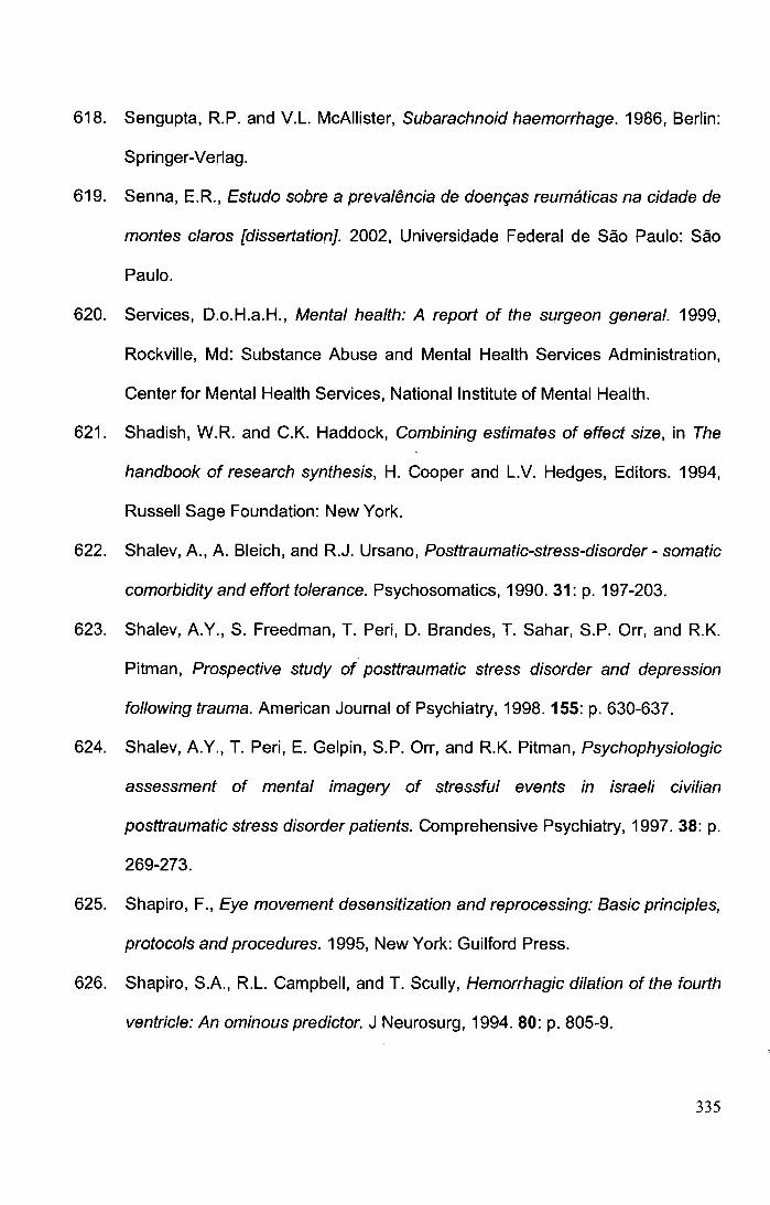
618. Sengupta, R.P. and V.L. McAllister, Subarachnoid haemorrhage. 1986, Berlin:
Springer-Verlag.
619. Senna, E.R., Estudo sobre a preval{mcia de doem;;as reumaticas na cidade de
montes claros [dissertation]. 2002, Universidade Federal de Sao Paulo: Sao
Paulo.
620. Services, D.o.H.a.H., Mental health: A report of the surgeon general. 1999,
Rockville, Md: Substance Abuse and Mental Health Services Administration,
Center for Mental Health Services, National Institute of Mental Health.
621. Shadish, W.R. and C.K. Haddock, Combining estimates of effect size, in The
handbook of research synthesis, H. Cooper and L.V. Hedges, Editors. 1994,
Russell Sage Foundation: New York.
622. Shalev, A., A. Bleich, and R.J. Ursano, Posttraumatic-stress-disorder- somatic
comorbidity and effort tolerance. Psychosomatics, 1990. 31: p. 197-203.
623. Shalev, A.Y., S. Freedman, T. Peri, D. Brandes, T. Sahar, S.P. Orr, and R.K.
Pitman, Prospective study of posttraumatic stress disorder and depression
following trauma. American Journal of Psychiatry, 1998. 155: p. 630-637.
624. Shalev, A.Y., T. Peri, E. Gelpin, S.P. Orr, and R.K. Pitman, Psychophysiologic
assessment of mental imagery of stressful events in israeli civilian
posttraumatic stress disorder patients. Comprehensive Psychiatry, 1997. 38: p.
269-273.
625. Shapiro, F., Eye movement desensitization and reprocessing: Basic principles,
protocols and procedures. 1995, New York: Guilford Press.
626. Shapiro, S.A., R.L. Campbell, and T. Scully, Hemorrhagic dilation of the fourth
ventricle: An ominous predictor. J Neurosurg, 1994. 80: p. 805-9.
335
627. Sheldrick, R., N. Tarrier, E. Berry, and J. Kincey, Post-traumatic stress
disorder and illness perceptions over time following myocardial infarction and
subarachnoid haemorrhage. British Journal of Health Psychology, 2006. 11: p.
387-400.
628. Shepard, R.H., Prognosis of spontaneous (nontraumatic) subarachnoid
haemorrhage of unknown cause. Lancet, 1984. 7: p. 777-778.
629. Sheskin, D.J., Parametric and nonparametric statistical procedures. 1997,
Boca Raton, FL: CRC Press.
630. Shin, L.M., S.L. Rauch, and R.K. Pitman, Structural and functional anatomy of
ptsd: Findings from neuroimaging research, in Neuropsychology of ptsd:
Biological, cognitive, and clinical perspectives, J.J. Vasterlin and C.R. Brewin,
Editors. 2005, Guilford Press: New York. p. 59-82.
631. Shin, L.M., C.l. Wright, P.A. Cannistraro, M.M. Wedig, K. McMullin, B. Martis,
M.L. Macklin, N.B. Lasko, S.R. Cavanagh, T.S. Krangel, S.P. Orr, R.K. Pitman,
P.J. Whalen, and S.L. Rauch, A functional magnetic resonance imaging study
of amygdala and medial prefrontal cortex responses to overtly presented
fearful faces in posttraumatic stress disorder. Archives of General Psychiatry,
2005. 62: p. 273-281.
632. Short, M.J., W.P. Wilson, and G.L. Odom, Psychiatric sequelae of
subarachnoid hemorrhage. Southern Medical Journal, 1968. 61: p. 87-91.
633. Siegel, S. and N.J. Castellan, Nonparametric statistics for the behavioural
science. 2nd ed. 1988, New York: McGraw-Hill Book Company.
634. Sikkema, K.J., N.B. Hansen, A. Kochman, N. Tarakeshwar, S. Neufeld, C.S.
Meade, and A.M. Fox, Outcomes from a group intervention for coping with
336
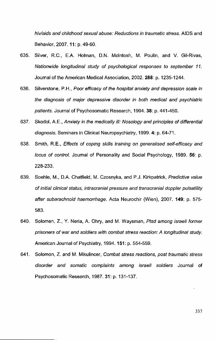
hiv/aids and childhood sexual abuse: Reductions in traumatic stress. AIDS and
Behavior, 2007. 11: p. 49-60.
635. Silver, R.C., E.A. Holman, D.N. Mcintosh, M. Poulin, and V. Gii-Rivas,
Nationwide longitudinal study of psychological responses to september 11.
Journal of the American Medical Association, 2002.288: p. 1235-1244.
636. Silverstone, P.H., Poor efficacy of the hospital anxiety and depression scale in
the diagnosis of major depressive disorder in both medical and psychiatric
patients. Journal of Psychosomatic Research, 1994. 38: p. 441-450.
637. Skodol, A. E., Anxiety in the medically ill: Nosology and principles of differential
diagnosis. Seminars in Clinical Neuropsychiatry, 1999. 4: p. 64-71.
638. Smith, R.E., Effects of coping skills training on generalised self-efficacy and
locus of control. Journal of Personality and Social Psychology, 1989. 56: p.
228-233.
639. Soehle, M., D.A. Chatfield, M. Czosnyka, and P.J. Kirkpatrick, Predictive value
of initial clinical status, intracranial pressure and transcranial doppler pulsatility
after subarachnoid haemorrhage. Acta Neurochir (Wien), 2007. 149: p. 575-
583.
640. Solomen, Z., Y. Neria, A. Ohry, and M. Waysman, Ptsd among israeli former
prisoners of war and soldiers with combat stress reaction: A longitudinal study.
American Journal of Psychiatry, 1994. 151: p. 554-559.
641. Solomon, Z. and M. Mikulincer, Combat stress reactions, post traumatic stress
disorder and somatic complaints among israeli soldiers Journal of
Psychosomatic Research, 1987. 31: p. 131-137.
337
642. Solomon, Z., M. Mikulincer, and H. Flum, Negative life events, coping
response, and combat-related psychopathology: A prospective study. Journal
of Abnormal Psychology, 1988. 97: p. 302-307.
643. Sonesson, B., B. Ljunggren, H. Saveland, and L. Brandt, Cognition and
adjustment after late and early operation for ruptured aneurysm. Neurosurgery,
1987.21: p. 279-281.
644. Sonesson, B., H. Saveland, B. Ljunggren, and L. Brandt, Cognitive functioning
after subarachnoid haemorrhage of unknown origin. Acta Neurol Scand, 1989.
80: p. 400-410.
645. Soo, C. and R. Tate, Psychological treatment for anxiety in people with
traumatic brain injury. Cochrane Database of Systematic Reviews, 2007. 3: p.
CD005239.
646. Soryal, 1., R.L. Sloan, C. Skelton, and B. Pentland, Rehabilitation needs after
haemorrhagic brain injury: Are they similar to those after traumatic brain injury?
Clinical Rehabilitation, 1992. 6: p. 103-110.
647. Spielberger, C.D., R.L. Gorusch, R. Lushene, and e. al., Manual for the state
trait anxiety inventory. 1983, Palo Alto, CA: Consulting Psychologists Press.
648. Spigset, 0., Adverse reactions of selective serotonin reuptake inhibitors:
Reports from a spontaneous reporting system. Drug Safety, 1999. 20: p. 277-
287.
649. Stabell, K.E. and B. Magnres, Neuropsychological course after surgery for
intracranial aneurysms: A prospective study and a critical review. Scandinavian
Journal of Psychology, 1997. 38: p. 127-137.
338

650. Stalnacke, B.M., Community integration, social support and life satisfaction in
relation to symptoms 3 years after mild traumatic brain injury. Brain Injury,
2007. 21: p. 933-942.
651. Statistics, A.B.a., National health survey. Sf-36 population norms, australia.
1995, Australian Bureau of Statistics: Canberra.
652. Stegen, G. and N. Freckmann, Outcome and rehabilitation after aneurysmal
subarachnoid haemorrhage. Zentralbl Neurochir, 1991. 52: p. 37-39.
653. Stegmayr, B., M. Eriksson, and K. Asplund, Declining mortality from
subarachnoid hemorrhage - changes in incidence and case fatality from 1985
through 2000. Stroke, 2004. 35: p. 2059-2063.
654. Stehbens, W., Intracranial arterial aneurysms, in Pathology of the cerebral
blood vessels. 1972, Mosby: Saint Louis. p. 351-470.
655. Steil, R. and A. Ehlers, Dysfunctional meaning of posttraumatic intrusions in
chronic ptsd. Behavior Research and Therapy, 2000. 38: p. 537-558.
656. Stein, D.J., J.C. lpser, and S. Seedat, Pharmacotherapy for post traumatic
stress disorder (ptsd). Cochrane Database of Systematic Reviews, 2006. 25:
p. CD002795.
657. Stein, K.D., P.B. Jacobsen, C.M. Blanchard, and C. Thors, Further validation of
the multidimensional fatigue symptom inventory-short form (mfsi-sf). Journal of
Pain and Symptom Management, 2004. 27: p. 14-23.
658. Stein, M.B., J.R. Walker, A.L. Hazen, and D.R. Forde, Full and partial
posttraumatic stress disorder: Findings from a community survery. American
Journal of Psychiatry, 1997. 154: p. 1114-1119.
339
659. Steinmann, D.R. and E.D. Bigler, Neuropsychological sequelae of ruptured
anterior communicating artery aneurysm. International Journal of Clinical
Neuropsychology, 1986. 8: p. 135-140.
660. Stenhouse, L.M., R.G. Knight, B.E. Longmore, and S.N. Bishara, Long-term
cognitive deficits in patients after surgery on aneurysms of the anterior
communicating artery. JNNP, 1991. 54: p. 909-1014.
661. Stern, M., D. Chang, M. Odell, and K. Sperber, Rehabilitation implications of
non-traumatic subarachnoid haemorrhage. Brain Injury, 2006. 20: p. 679-685.
662. Stewart, A.L., S. Greenfield, R.D. Hays, K. Wells, W.H. Rogers, S.D. Berry,
E.A. McGlynn, and J.E. Ware, Functional status and well-being of patients with
chronic conditions: Results from the medical outcomes study. Journal of the
American Medical Association, 1989 262: p. 907-913.
663. Stewart, D., S. Abbey, M. Meana, and K.M. Boydell, What makes women
tired? A community sample. Journal of Women's Health, 1998. 7: p. 69-76.
664. Stolke, D., B. Hoppe, V. Seifert, N. Ruckert, and H. Dietz, How good are the
results in aneurysm surgery? Psychological aspects of rehabilitation of early
and late operated patients with cerebral aneurysm, in Advances in
neurosurgery, R. Wullenweber, M. Klinger, and M. Brock, Editors. 1987,
Springer Verlag: Berlin. p. 297-301.
665. Stoll, C., G. Schelling, A. Goetz, E. Kilger, A. Bayer, H.P. Kapfhammar, H.B.
Rothenhausler, E. Kreuzer, B. Reichart, and K. Peter, Health-related quality of
life and post-traumatic stress disorder in patients after cardiac surgery and
intensive care treatment. Journal of Thoracic and Cardiovacular Surgery, 2000.
120: p. 505-512.
340

666. Stoltenburg-Didinger, G., Neuropathology of subarachnoid hemorrhage, in
Subarachnoid hemorrhage: Pathophysiology and management, J.B. Bederson,
Editor. 1997, ANNS publications: Park Ridge. p. 41-60.
667. Storey, P., Emotional disturbances before and after subarachnoid
haemorrhage. Ciba Found Symp, 1972. 8: p. 337-343.
668. Storey, P.B., Psychiatric sequelae of subarachnoid haemorrhage. British
Medical Journal, 1967. 69: p. 261-266.
669. Storey, P.B., Brain damage and personality change after subarachnoid
haemorrhage. British Journal of Psychiatry, 1970. 117: p. 129-142.
670. Straube, T., H.J. Mentzel, and W.H. Miltner, Common and distinct brain
activation to threat and safety signals in social phobia. Neuropsychobiology,
2005. 52:p. 163-168.
671. Stukas, A.A., M.A. Dew, G.E. Switzer, A. DiMartini, R.L. Kormos, and B.P.
Griffith, Ptsd in heart transplant recipients and their primary family caregivers.
Psychosomatics, 1999. 40: p. 212-221.
672. Sudlow, C.L. and C.P. Warlow, Comparable studies of the incidence of stroke
and its pathological types: Results from an international collaboration. Stroke,
1997. 28: p. 491-499.
673. Sullivan, M. and J. Karlsson, The swedish sf-36 health survey iii. Evaluation of
criterion-based validity: Results from normative population. Journal of Clinical
Epidemiology, 1998 41: p. 1349-1358.
674. Sumpter, R.E. and T.M. McMillan, Errors in self-report of post-traumatic stress
disorder after severe traumatic brain injury. Brain Injury, 2005. 20: p. 93-99.
341
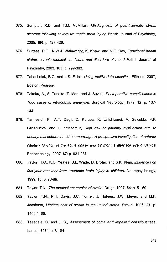
675. Sumpter, R.E. and T.M. McMillan, Misdiagnosis of post-traumatic stress
disorder following severe traumatic brain injury. British Journal of Psychiatry,
2005. 186: p. 423-426.
676. Surtees, P.G., N.W.J. Wainwright, K. Khaw, and N.E. Day, Functional health
status, chronic medical conditions and disorders of mood. British Journal of
Psychiatry, 2003. 183: p. 299-303.
677. Tabachnick, B.G. and L.S. Fidell, Using multivariate statistics. Fifth ed. 2007,
Boston: Pearson.
678. Takaku, A., S. Tanaka, T. Mori, and J. Suzuki, Postoperative complications in
1000 cases of intracranial aneurysm. Surgical Neurology, 1979. 12: p. 137-
144.
679. Tanriverdi, F., A.T. Dagli, Z. Karaca, K. Unluhizarci, A. Selcuklu, F.F.
Casanueva, and F. Kelestimur, High risk of pituitary dysfunction due to
aneurysmal subarachnoid haemorrhage: A prospective investigation of anterior
pituitary function in the acute phase and 12 months after the event. Clinical
Endocrinology, 2007. 67: p. 931-937.
680. Taylor, H.G., K.O. Yeates, S.L. Wade, D. Drotar, and S.K. Klein, Influences on
first-year recovery from traumatic brain injury in children. Neuropsychology,
1999. 13: p. 76-89.
681. Taylor, T.N., The medical economics of stroke. Drugs, 1997. 54: p. 51-59.
682. Taylor, T.N., P.H. Davis, J.C. Tomer, J. Holmes, J.W. Meyer, and M.F.
Jacobson, Lifetime cost of stroke in the united states. Stroke, 1996. 27: p.
1459-1466.
683. Teasdale, G. and J. B., Assessment of coma and impaired consciousness.
Lancet, 1974: p. 81-84
342

684. Tedstone, J.E. and N. Tarrier, Posttraumatic stress disorder following medical
illness and treatment. Clinical Psychology Review, 2003. 23: p. 409-448.
685. Teissier du Cros, J. and F. Lhermitte, Neuropsychological analysis of ruptured
saccular aneurysms of the anterior communicating artery after radical therapy
(32 cases). Surgical Neurology, 1984. 22: p. 353-359.
686. Testa, M.A. and D.C. Simonson, Assessment of quality of life outcomes. New
England Journal of Medicine, 1996. 334: p. 835-840.
687. Tidswell, P., P.S. Dias, H.J. Sagar, A.R. Mayes, and R.D. Battersby, Cognitive
outcome after aneurysm rupture: Relationship to aneurysm site and
preoperative complications. Neurology, 1995. 45: p. 875-882.
688. Tokuda, Y., T. lnagawa, Y. Katho, K. Kumano, N. Ohbayashi, and H.
Yoshioka, Intracerebral hematoma in patients with rutpured cerebral
aneurysms. Surgical Neurology, 1995. 43: p. 272-277.
689. Tomberg, T., A. Orasson, U. Linnamagi, A. Toomela, A. Pulver, and T. Asser,
Coping strategies in patients following subarachnoid haemorrhage. Acta
Neurologica Scandinavica, 2001. 104: p. 148-155.
690. Toomela, A., A. Pulver, T. Tomberg, A. Orasson, A. Tikk, and T. Asser,
Possible interpretation of subjective complaints in patients with spontaneous
subarachnoid haemorrhage. Journal of Rehabilitation Medicine, 2004. 36: p.
63-69.
691. Tomer, J.C. and P.H. Davis, Epidemiology and clinical manifestations of
subarachnoid hemorrhage, in Subarachnoid hemorrhage: Pathophysiology and
management, J.B. Bederson, Editor. 1997, The American Association of
Neurological Surgeons: Park Ridge.
343
692. Towgood, K., J.A. Ogden, and E. Mee, Neurological, neuropsychological, and
psychosocial outcome following treatment of unruptured intracranial
aneurysms: A review and commentary. Journal of the International
Neuropsychological Society, 2004. 10: p. 114-134.
693. Trust, M.O., Pharmaceutical companies use quality of life measures. Medical
Outcomes Trust Bulletin, 1995. 3: p. 1.
694. Tseng, M.Y., P.J. Hutchinson, M. Czosnyka, H. Richards, J.D. Pickard, and
P.J. Kirkpatrick, Effects of acute pravastatin treatment on intensity of rescue
therapy, length of inpatient stay, and 6-month outcome in patients after
aneurysmal subarachnoid hemorrhage. Stroke, 2007. 38: p. 1545-50.
695. Tsutsumi, K., K. Ueki, M. Usui, S. Kwak, and T. Kirino, Risk of recurrent
subarachnoid hemorrhage after complete obliteration of cerebral aneurysms.
Stroke, 1998. 29: p. 2511-2513.
696. Tucker, P., R. Zaninelli, R. Yehuda, L. Ruggiero, K. Dillingham, and C.D. Pitts,
Paroxetine in the treatment of chronic posttraumatic stress disorder: Results
from a placebo-controlled trial. Journal of Clinical Psychiatry, 2001. 62: p. 860-
868.
697. TOre, U., M.G. Yasargil, and A. F. Krisht, The arteries of the corpus callosum: A
microsurgical anatomic study. Neurosurgery, 1996. 39: p. 1075-1085.
698. Turton, P., W. Badenhorst, P. Hughes, J. Ward, S. Riches, and S. White,
Psychological impact of stillbirth on fathers in the subsequent pregnancy and
puerperium. British Journal of Psychiatry, 2006. 188: p. 165-172.
699. Unal, G., J.B. de Boer, G. Borsboom, J.T. Brouwer, M.L. Essink-Bot, and R.A.
de Man, A psychometric comparison of health-related quality of life measures
in chronic liver disease. Journal of Clinical Epidemiology, 2001. 54: p. 587-596.
344
700. v Zerssen, D., Die befindlichkeits-skala. 1976, Beltz: Weinheim.
701. Van't Spijker, A., R.W. Trijsburg, and H.J. Duivenvoorden, Psychological
sequelae of cancer diagnosis: A meta-analytic review of 58 studies after 1980.
Psychosomatic Medicine, 1997. 59: p. 280-293.
702. van der Schaaf, 1., A. Algra, M. Warmer, A. Molyneux, M. Clarke, J. van Gijn,
and G. Rinkel, Endovascular coiling versus neurosurgical clipping for patients
with aneurysmal subarachnoid haemorrhage. Cochrane Database of
Systematic Reviews, 2005. 19: p. CD003085.
703. van der Schaaf, I.C., M.J.H. Warmer, B.K. Velthuis, E. Buskens, P.M.M.
Bossuyt, and G.J.E. Rinkel, Psychosocial impact of finding small aneurysms
that are left untreated in patients previously operated on for ruptured
aneurysms. Journal of Neurology Neurosurgery and Psychiatry, 2006. 77: p.
748-752.
704. van der Wee, N., G.J. Rinkel, D. Hasan, and J. van Gijn, Detection of
subarachnoid haemorrhage on early ct: Is lumbar puncture still needed after a
negative scan? Journal of Neurology, Neurosurgery and Psychiatry, 1995. 58:
p. 357-359.
705. van der Werf, S.P., H.L.P. van den Broek, H.W.M. Anten, and G. Bleijenberg,
Experience of severe fatigue long after stroke and its relation to depressive
symptoms and disease characteristics. European Neurology, 2001. 45: p. 28-
33.
706. Van Oriel, R.C. and W. Op den Velde, Myocardial infaction and post-traumatic
stress disorder. Journal of Traumatic Stress, 1995.8: p. 151-159.
707. van Gijn, J., Subarachnoid haemorrhage. Lancet, 1992. 339: p. 653-655.
345
708. van Gijn, J., J.E. Bromberg, K.W. Lindsay, D. Hasan, and M. Vermeulen,
Definition of initial grading, specific events, and overall outcome in patients
with aneurysmal subarachnoid hemorrhage: A survey. Stroke 1994. 25: p.
1623-1627.
709. van Gijn, J., A. Hidjra, E.F.M. Wijdicks, M. Vermeulen, and H. van Crevel,
Acute hydrocephalus after subarachnoid hemorrhage. Journal of
Neurosurgery, 1985. 63: p. 355-362.
710. van Gijn, J., R.S. Kerr, and G.J.E. Rinkel, Subarachnoid haemorrhage. Lancet,
2007. 369: p. 306-318.
711. van Gijn, J. and G.J.E. Rinkel, Subarachnoid haemorrhage: Diagnosis, causes
and management. Brain, 2001. 124: p. 249-278.
712. van Gijn, J. and K.J. van Dongen, Computerised tomography in the diagnosis
of subarachnoid haemorrhage and ruptured aneurysm. Clinical Neurology and
Neurosurgery, 1980. 82: p. 11-24.
713. Van Swieten, J.C., P.J. Koudstaal, M.C. Visser, H.J.A. Schouten, and J. van
Gijn, lnterobserver agreement for the assesment of handicap in stroke
patients. Stroke, 1988. 19: p. 604-607.
714. Van Zandvoort, M.J.E., J. Kappelle, A. Algra, and E.H.F. DeHaan, Decreased
capacity for mental effort after single supratentorial lacunar infarct may affect
performance in everyday life Journal of Neurology, Neurosurgery and
Psychiatry, 1998. 65: p. 697-702.
715. Vannemreddy, P.S.S.V., A. Nanda, R. Kelley, and M.K. Baskaya, Delayed
diagnosis of intracranial aneurysms: Confounding factors in clinical
presentation and the influence of misdiagnosis on outcome. Southern Medical
Journal, 2001. 94: p. 1108-1111.
346
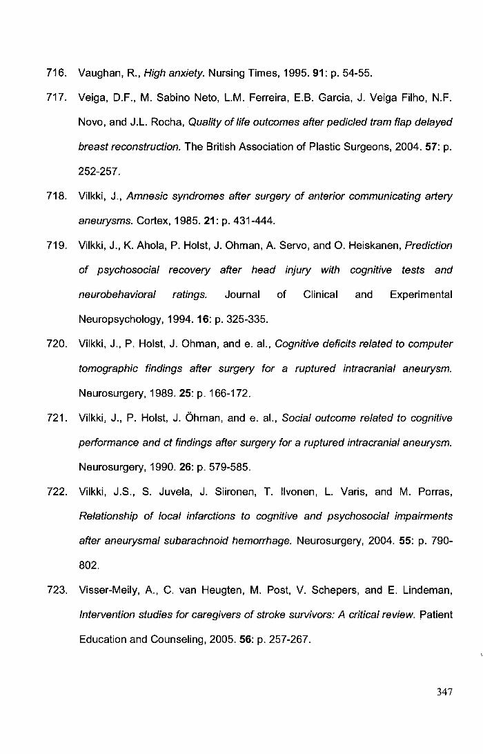
716. Vaughan, R., High anxiety. Nursing Times, 1995.91: p. 54-55.
717. Veiga, D.F., M. Sabino Neto, L.M. Ferreira, E.B. Garcia, J. Veiga Filho, N.F.
Novo, and J.L. Rocha, Quality of life outcomes after pedicled tram flap delayed
breast reconstruction. The British Association of Plastic Surgeons, 2004. 57: p.
252-257.
718. Vilkki, J., Amnesic syndromes after surgery of anterior communicating artery
aneurysms. Cortex, 1985.21: p. 431-444.
719. Vilkki, J., K. Ahola, P. Holst, J. Ohman, A. Servo, and 0. Heiskanen, Prediction
of psychosocial recovery after head injury with cognitive tests and
neurobehavioral ratings. Journal of Clinical and Experimental
Neuropsychology, 1994. 16: p. 325-335.
720. Vilkki, J., P. Holst, J. Ohman, and e. al., Cognitive deficits related to computer
tomographic findings after surgery for a ruptured intracranial aneurysm.
Neurosurgery, 1989. 25: p. 166-172.
721. Vilkki, J., P. Holst, J. Ohman, and e. al., Social outcome related to cognitive
performance and ct findings after surgery for a ruptured intracranial aneurysm.
Neurosurgery, 1990. 26: p. 579-585.
722. Vilkki, J.S., S. Juvela, J. Siironen, T. llvonen, L. Varis, and M. Porras,
Relationship of local infarctions to cognitive and psychosocial impairments
after aneurysmal subarachnoid hemorrhage. Neurosurgery, 2004. 55: p. 790-
802.
723. Visser-Meily, A., C. van Heugten, M. Post, V. Schepers, and E. Lindeman,
Intervention studies for caregivers of stroke survivors: A critical review. Patient
Education and Counseling, 2005. 56: p. 257-267.
347
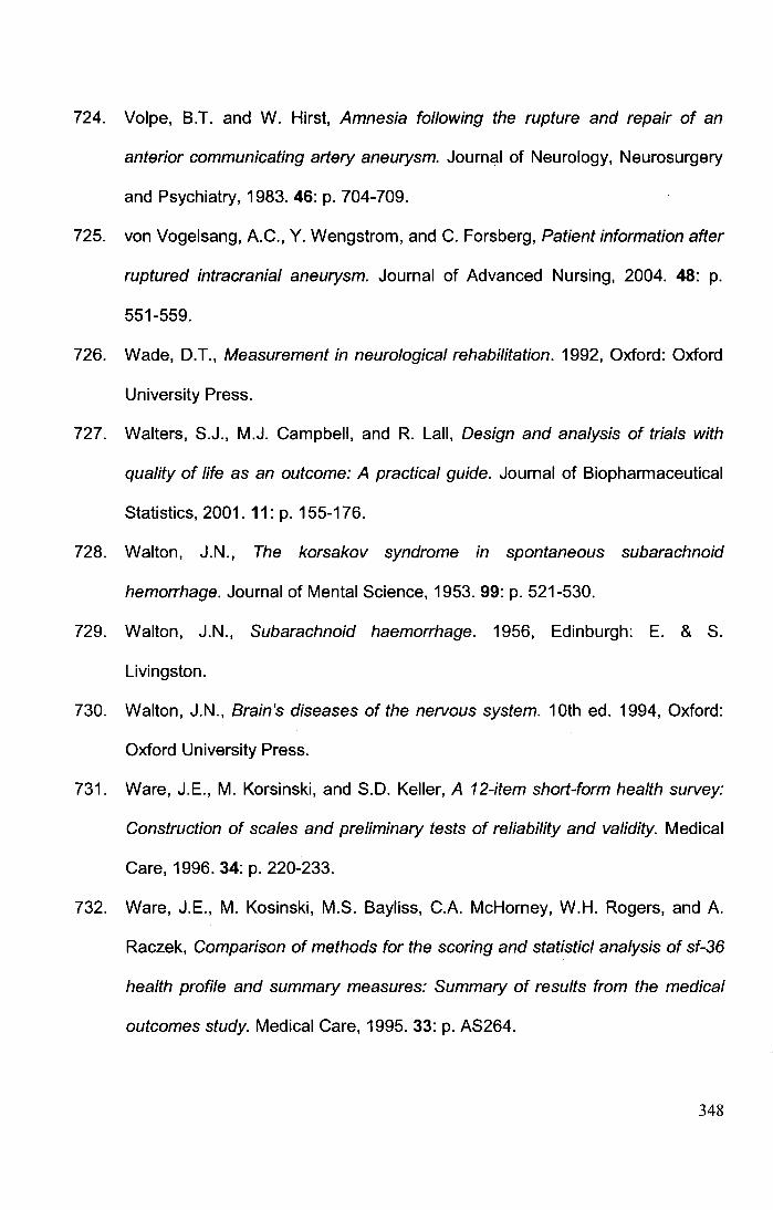
724. Volpe, B.T. and W. Hirst, Amnesia following the rupture and repair of an
anterior communicating artery aneurysm. Journal of Neurology, Neurosurgery
and Psychiatry, 1983. 46: p. 704-709.
725. von Vogelsang, A.C., Y. Wengstrom, and C. Forsberg, Patient information after
ruptured intracranial aneurysm. Journal of Advanced Nursing, 2004. 48: p.
551-559.
726. Wade, D.T., Measurement in neurological rehabilitation. 1992, Oxford: Oxford
University Press.
727. Walters, S.J., M.J. Campbell, and R. Lall, Design and analysis of trials with
quality of life as an outcome: A practical guide. Journal of Biopharmaceutical
Statistics, 2001. 11: p. 155-176.
728. Walton, J.N., The korsakov syndrome in spontaneous subarachnoid
hemorrhage. Journal of Mental Science, 1953. 99: p. 521-530.
729. Walton, J.N., Subarachnoid haemorrhage. 1956, Edinburgh: E. & S.
Livingston.
730. Walton, J.N., Brain's diseases of the nervous system. 10th ed. 1994, Oxford:
Oxford University Press.
731. Ware, J.E., M. Korsinski, and S.D. Keller, A 12-item short-form health survey:
Construction of scales and preliminary tests of reliability and validity. Medical
Care, 1996. 34: p. 220-233.
732. Ware, J.E., M. Kosinski, M.S. Bayliss, C.A. McHorney, W.H. Rogers, and A.
Raczek, Comparison of methods for the scoring and statistic/ analysis of sf-36
health profile and summary measures: Summary of results from the medical
outcomes study. Medical Care, 1995. 33: p. AS264.
348
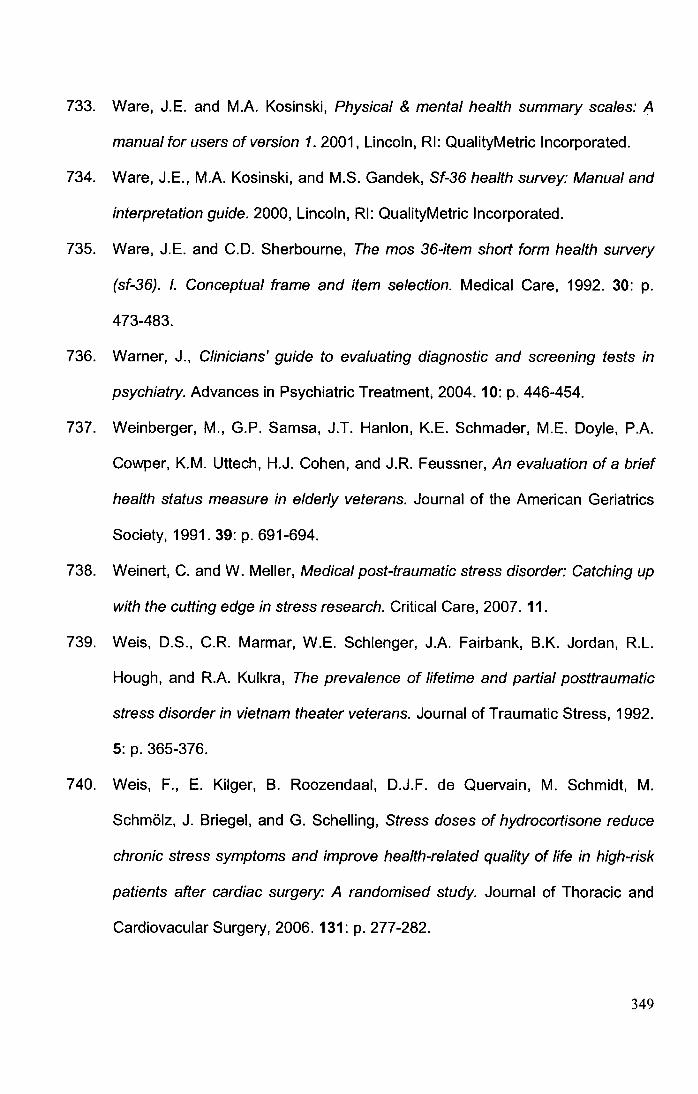
733. Ware, J.E. and M.A. Kosinski, Physical & mental health summary scales: A
manual for users of version 1. 2001, Lincoln, Rl: QualityMetric Incorporated.
734. Ware, J.E., M.A. Kosinski, and M.S. Gandek, Sf-36 health survey: Manual and
interpretation guide. 2000, Lincoln, Rl: QualityMetric Incorporated.
735. Ware, J.E. and C.D. Sherbourne, The mos 36-item short form health survery
(sf-36). I. Conceptual frame and item selection. Medical Care, 1992. 30: p.
473-483.
736. Warner, J., Clinicians' guide to evaluating diagnostic and screening tests in
psychiatry. Advances in Psychiatric Treatment, 2004. 10: p. 446-454.
737. Weinberger, M., G.P. Samsa, J.T. Hanlon, K.E. Schmader, M.E. Doyle, P.A.
Cowper, K.M. Uttech, H.J. Cohen, and J.R. Feussner, An evaluation of a brief
health status measure in elderly veterans. Journal of the American Geriatrics
Society, 1991. 39: p. 691-694.
738. Weinert, C. and W. Meller, Medical post-traumatic stress disorder: Catching up
with the cutting edge in stress research. Critical Care, 2007. 11.
739. Weis, D.S., C.R. Marmar, W.E. Schlanger, J.A. Fairbank, B.K. Jordan, R.L.
Hough, and R.A. Kulkra, The prevalence of lifetime and partial posttraumatic
stress disorder in vietnam theater veterans. Journal of Traumatic Stress, 1992.
5: p. 365-376.
740. Weis, F., E. Kilger, B. Roozendaal, D.J.F. de Quervain, M. Schmidt, M.
Schmalz, J. Briegel, and G. Schelling, Stress doses of hydrocortisone reduce
chronic stress symptoms and improve health-related quality of life in high-risk
patients after cardiac surgery: A randomised study. Journal of Thoracic and
Cardiovacular Surgery, 2006. 131: p. 277-282.
349
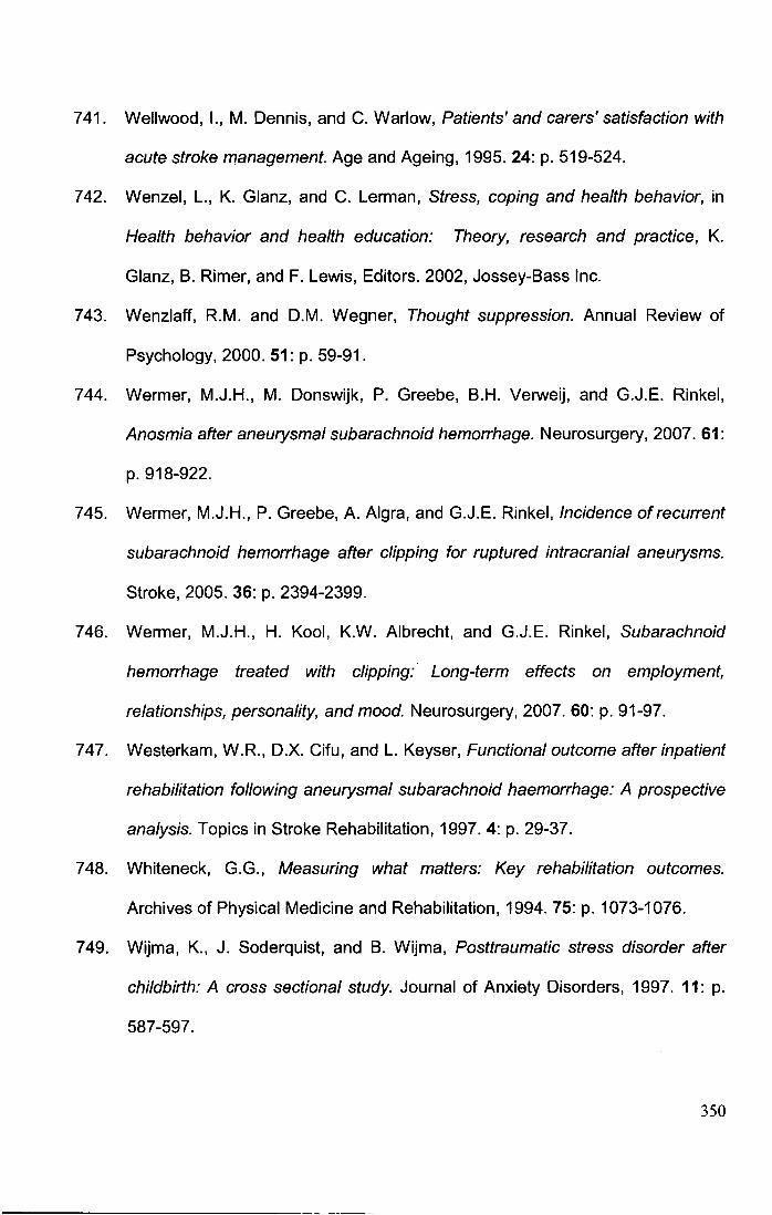
741. Wellwood, 1., M. Dennis, and C. Warlow, Patients' and carers' satisfaction with
acute stroke management. Age and Ageing, 1995. 24: p. 519-524.
742. Wenzel, L., K. Glanz, and C. Lerman, Stress, coping and health behavior, in
Health behavior and health education: Theory, research and practice, K.
Glanz, B. Rimer, and F. Lewis, Editors. 2002, Jossey-Bass Inc.
743. Wenzlaff, R.M. and D.M. Wegner, Thought suppression. Annual Review of
Psychology, 2000. 51: p. 59-91.
744. Wermer, M.J.H., M. Donswijk, P. Greebe, B.H. Verweij, and G.J.E. Rinkel,
Anosmia after aneurysmal subarachnoid hemorrhage. Neurosurgery, 2007. 61:
p. 918-922.
745. Wermer, M.J.H., P. Greebe, A. Algra, and G.J.E. Rinkel, Incidence of recurrent
subarachnoid hemorrhage after clipping for ruptured intracranial aneurysms.
Stroke, 2005. 36: p. 2394-2399.
746. Wermer, M.J.H., H. Kool, K.W. Albrecht, and G.J.E. Rinkel, Subarachnoid
hemorrhage treated with clipping:· Long-term effects on employment,
relationships, personality, and mood. Neurosurgery, 2007. 60: p. 91-97.
747. Westerkam, W.R., D.X. Cifu, and L. Keyser, Functional outcome after inpatient
rehabilitation following aneurysmal subarachnoid haemorrhage: A prospective
analysis. Topics in Stroke Rehabilitation, 1997. 4: p. 29-37.
748. Whitenack, G.G., Measuring what matters: Key rehabilitation outcomes.
Archives of Physical Medicine and Rehabilitation, 1994. 75: p. 1073-1076.
749. Wijma, K., J. Soderquist, and B. Wijma, Posttraumatic stress disorder after
childbirth: A cross sectional study. Journal of Anxiety Disorders, 1997. 11: p.
587-597.
350
750. Wilde, M.C., Neuropsychological outcome in subarachnoid hemorrhage due to
aneurysm. Critical Reviews in Physical and Rehabilitation Medicine, 2006. 18:
p.117-131.
751. Wilkinson, L. and T.F.O.S. Inference, Statistical methods in psychology
journals: Guidelines and explanations. American Psychologist, 1999. 54: p.
594-604.
752. Will, B.E. and A. Philipp, Quality of life after sah and clipping of cerebral
aneurysm. Neural Neurochir Pol, 2000. 34: p. 61-63.
753. Willer, B., P.M. Flaherty, and S. Coallier, Families living with the effects of
acquired brain injury, in Neurobehavioural disability and social handicap
following traumatic brain injury, R.L. Wood and T.M. McMillan, Editors. 2001,
Psychology Press: Hove. p. 47-63.
754. Wilson, B.A., N. Alderman, P.W. Burgess, H. Emslie, and J.J. Evans,
Behavioural assessment of the dysexecutive syndrome. 1996, Bury St.
Edmunds: Thames Valley Test Company.
755. Wilson, B.A., L. Clare, A.D. Baddeley, J. Cockburn, P. Watson, and R. Tate,
The rivermead behavioural memory test - extended version (rmbt-e). 1999,
Bury St. Edmunds: Thames Valley Test Company.
756. Wilson, B.A., J. Cockburn, and P. Halligan, Behavioural inattention test. 1987,
Bury St. Edmunds, UK: Thames Valley Test Company.
757. Wilson, I.B. and P.O. Cleary, Linking clinical variables with health related
quality of life. A conceptual model of patient outcomes. Journal of the
American Medical Association, 1995. 273: p. 59-65.
351
758. Wilson, J.T., L.E. Pettigrew, and G.M. Teesdale, Structured interviews for the
glasgow outcome scale and the extended glasgow outcome scale: Guidelines
for their use. Journal of Neurotrauma, 1998. 15: p. 573-585.
759. Wilz, G., Predictors of subjective impairment after stroke: Influence of
depression, gender and severity of stroke. Brain Injury, 2007. 21: p. 39-45.
760. Wolfe, F. and D.J. Hawley, Measurement of the quality of life in rheumatic
disorders using the euroqol. British Journal of Rheumatology, 1997. 36: p. 786-
793.
761. Wolfe, J., P.P. Schnurr, P.J. Brown, and J. Furey, Posttraumatic-stress
disorder and war-zone exposure as correlates of perceived health in female
vietnam veterans. Journal of Consulting and Clinical Psychology, 1994. 62: p.
1235-1240.
762. Wong, M., E. Looney, J. Michaels, 0. Palesh, and C. Koopman, A preliminary
study of peritraumatic dissociation, social support, and coping in relation to
posttraumatic stress symptoms for a parent's cancer. Psycho-Oncology, 2006.
15: p. 1093-1098.
763. Woo, J., R. Kay, Y.K. Yuen, and M.G. Nicholls, Factors influencing long-term
survival and disability among three-month stroke surivivors.
Neuroepidemiology, 1992. 11: p. 143-150.
764. Wood-Dauphinee, S.L., M.A. Opzoomer, J.l. Williams, B. Marchand, and W.O.
Spitzer, Assessment of global function: The reintegration to normal living
index. Arch Phys Med Rehabil, 1988. 69: p. 583-590.
765. Wright, C.l., B. Martis, K. McMullin, L.M. Shin, and S.L. Rauch, Amygdala and
insula responses to emotionally valenced human faces in small animal specific
phobia. Biological Psychiatry, 2003. 54: p. 1067-1076.
352
766. Yap, E.C. and K.S. Chua, Rehabilitation outcome after primary subarachnoid
haemorrhage. Brain Injury, 2002. 16: p. 491-499.
767. Yasargil, M.G., Microneurosurgery. Vol. I. 1984, Stuttgart: Thieme Verlag.
768. Yehuda, R., S.M. Southwick, G. Nussbaum, V. Wahby, E.L. Giller, and J.W.
Mason, Low urinary cortisol excretion in patients with posttraumatic stress
disorder. Journal of Nervous and Mental Disease, 1990. 178: p. 366-369.
769. Yokoyama, K., S. Araki, K. Murata, M. Nishikitani, T. Okumura, S. lshimatsu,
N. Takasu, and R.F. White, Chronic neurobehavioral effects of tokyo subway
sarin poisoning in relation to posttraumatic stress disorder. Archives of
Environmental Health, 1998. 53: p. 249-256.
770. Young, E.A. and N. Breslau, Cortisol and catecholamines in posttraumatic
disorder: An epidemiology community study. Archives of General Psychiatry,
2005. 61: p. 394-401.
771. Young, G.S., L.L. Mintzer, D. Seacord, M. Castaneda, V. Mesrkhani, and M.L.
Stuber, Symptoms of posttraumatic stress disorder in parents of transplant
recipients: Incidence, severity, and related factors. Pediatrics, 2003. 111: p.
725-731.
772. Zigmond, A.S. and R.P. Snaith, The hospital anxiety and depression scale.
Acta Psychiatr Scand, 1983. 67: p. 361-370.
773. Zlotnick, C., C.L. Franklin, and M. Zimmerman, Does "Subthreshold"
Posttraumatic stress disorder have any clinical relevance? Comprehensive
Psychiatry, 2002. 43: p. 413-419.
774. Zung, W.W.K., A self-rating depression scale. Archives of General Psychiatry,
1965. 12: p. 63-70.
353

Appendices
354
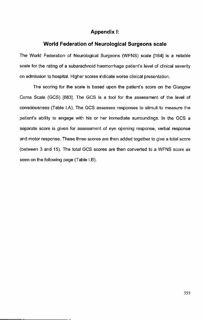
Appendix 1:
World Federation of Neurological Surgeons scale
The World Federation of Neurological Surgeons (WFNS) scale [164] is a reliable
scale for the rating of a subarachnoid haemorrhage patient's level of clinical severity
on admission to hospital. Higher scores indicate worse clinical presentation.
The scoring for the scale is based upon the patient's score on the Glasgow
Coma Scale (GCS) [683]. The GCS is a tool for the assessment of the level of
consciousness (Table I.A). The GCS assesses responses to stimuli to measure the
patient's ability to engage with his or her immediate surroundings. In the GCS a
separate score is given for assessment of eye opening response, verbal response
and motor response. These three scores are then added together to give a total score
(between 3 and 15). The total GCS scores are then converted to a WFNS score as
seen on the following page (Table I. B).
355
Table I.A The Glasgow Coma Scale
Glasgow Coma Scale
Eyes
Verbal
Motor
Table I.B
2 3
Does not open Opens eyes in Opens eyes in
eyes
Makes no
sound
Makes no
movements
response to response to
painful stimuli voice
lncomprehensi Utters words,
ble sounds
Extensor
(rigid)
response,
decerebrate
posture
but not
coherent
Abnormal
(spastic)
flexion,
decorticate
posture
4
Opens eyes
spontaneously
Confused,
disoriented
conversation
Flexion I
withdraws to
painful stimuli
5
Orientated,
converses
normally
Localizes pain
The conversion of GCS to WFNS scores
WFNS Grade
Glasgow Coma Scale score Motor deficit
1 15 Absent
2 13-14 Absent
3 13-14 Present
4 7-12 Absent or present
5 3-6 Absent or present
6
Obeys
Commands
356
Appendix II:
The Hunt and Hess Scale
The Hunt and Hess scale [293] is a useful measure for the rating of a patient's clinical
severity on admission to hospital (Table II.A). Higher scores indicate a worse clinical
presentation.
Table II.A Hunt and Hess Scale
Hunt and Hess Grade
Grade 0 Non-ruptured aneurysm
Grade 1 Asymptomatic or mild headache and/ or slight nuchal stiffness
Grade 2 Moderate to severe headache and/ or pronounced nuchal stiffness and/ or cranial nerve deficits,
photophobia
Grade 3 Drowsiness and/ or mild disorientation and/ or slight pareses and/ or dysphasia
Grade 4 Stupor, moderate to severe hemiparesis and/ or dysphasia
Grade 5 Deep coma, decerebrate rigidity, moribund appearance
357
Appendix m:
Fisher's Rating Scale for Bleed Severity
Fisher's scale [191] for the rating of head computed tomographic scans for
subarachnoid haemorrhage is presented below (Table III.A). A higher score indicates
a more severe haemorrhage.
Table III.A Fisher's CT rating scale
Fisher Grade
0 No scan available
1 No blood detected on CT
2 Diffuse deposition or thin, localised layer of blood in all vertical layers (interhemispheric fissure, insular
cistem, ambient cistem), less than 1 mm in thickness
3 Localised blood clots and/or vertical layers of blood, 1 mm or greater in thickness
4 Diffuse or no subarachnoid blood, with intracerebral or intraventricular blood present
358
Appendix IV:
Glasgow Outcome Scale
The Glasgow Outcome Scale (GOS) [318] for the rating of physical recovery following
a subarachnoid haemorrhage is presented below (Table IV.A). A higher grade
indicates more physical disability.
Table IV.A The Glasgow Outcome Scale
GOS Score
Excellent recovery- Independent, no/ minimal neurological deficit
2 Good recovery - Moderate disability, independent with moderate deficit (e.g., aphasia, hemiparesis,
ataxia or memory deficit)?
3 Poor recovery- Severe disability, dependent but conscious with severe deficits
4 Poor recovery- Vegetative state, dependent, persistent vegetative state
5 Dead
It should be noted that the scale is presented in the form in which it is typically
used within SAH research. In its original form however, the scores were placed in
reverse order (i.e., 5="good recovery", 1 ="death").
In the extended version of the GOS [758), the first three categories are each
subdivided into two to increase its sensitivity for those patients with ratings in the
upper end of the scale.
359
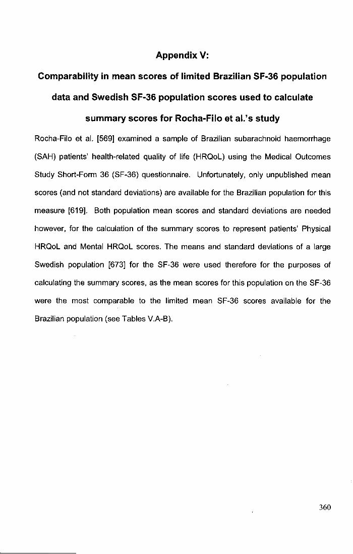
Appendix V:
Comparability in mean scores of limited Brazilian SF-36 population
data and Swedish SF-36 population scores used to calculate
summary scores for Rocha-Filo et al.'s study
Rocha-Filo et al. [569] examined a sample of Brazilian subarachnoid haemorrhage
(SAH) patients' health-related quality of life (HRQoL) using the Medical Outcomes
Study Short-Form 36 (SF-36) questionnaire. Unfortunately, only unpublished mean
scores (and not standard deviations) are available for the Brazilian population for this
measure [619]. Both population mean scores and standard deviations are needed
however, for the calculation of the summary scores to represent patients' Physical
HRQoL and Mental HRQoL scores. The means and standard deviations of a large
Swedish population [673] for the SF-36 were used therefore for the purposes of
calculating the summary scores, as the mean scores for this population on the SF-36
were the most comparable to the limited mean SF-36 scores available for the
Brazilian population (see Tables V.A-8).
360
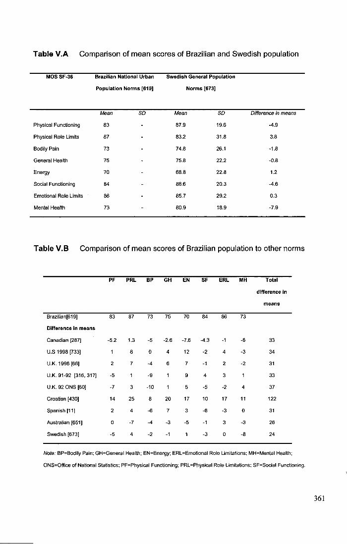
Table V.A Comparison of mean scores of Brazilian and Swedish population
MOS SF-36
Physical Functioning
Physical Role Limits
Bodily Pain
General Health
Energy
Social Functioning
Emotional Role Limits
Mental Health
Brazilian National Urban
Population Norms [619)
Mean so
83
87
73
75
70
84
86
73
Swedish General Population
Norms [673]
Mean so
87.9 19.6
83.2 31.8
74.8 26.1
75.8 22.2
68.8 22.8
88.6 20.3
85.7 29.2
80.9 18.9
Difference in means
-4.9
3.8
-1.8
-0.8
1.2
-4.6
0.3
-7.9
Table V.B Comparison of mean scores of Brazilian population to other norms
PF PRL BP GH EN SF ERL MH Total
difference in
means
Brazilian[619) 83 87 73 75 70 84 86 73
Difference in means
Canadian [287] -5.2 1.3 -5 -2.6 -7.6 -4.3 -1 -6 33
u.s 1998 [733) 8 0 4 12 -2 4 -3 34
U.K. 1996 [66) 2 7 -4 6 7 -1 2 -2 31
U.K. 91-92 [316, 317] -5 -9 9 4 3 33
U.K. 92 ONS [60) -7 3 -10 5 -5 -2 4 37
Croatian [430) 14 25 8 20 17 10 17 11 122
Spanish [11] 2 4 -6 7 3 -6 -3 0 31
Australian [651] 0 -7 -4 -3 -5 -1 3 -3 26
Swedish [673] -5 4 -2 -1 -3 0 -8 24
Note: BP=Bodily Pain; GH=Generai Health; EN=Energy; ERL=Emotional Role Limitations; MH=Mental Health;
ONS=Office of National Statistics; PF=Physical Functioning; PRL=Physical Role Limitations; SF=Social Functioning.
361
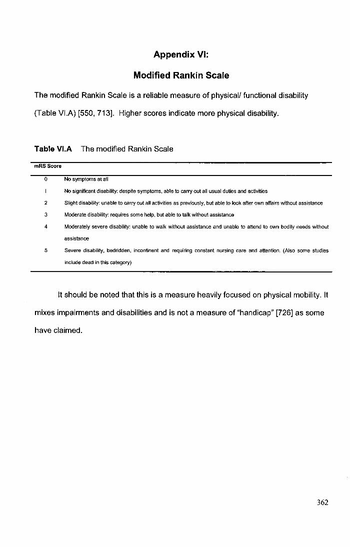
Appendix VI:
Modified Rankin Scale
The modified Rankin Scale is a reliable measure of physical/ functional disability
(Table VI.A) [550, 713]. Higher scores indicate more physical disability.
Table VI.A The modified Rankin Scale
mRS Score
0 No symptoms at all
No significant disability: despite symptoms, able to carry out all usual duties and activities
2 Slight disability: unable to carry out all activities as previously, but able to look after own affairs without assistance
3 Moderate disability: requires some help, but able to talk without assistance
4 Moderately severe disability: unable to walk without assistance and unable to attend to own bodily needs without
assistance
5 Severe disability, bedridden, incontinent and requiring constant nursing care and attention. (Also some studies
include dead in this category)
It should be noted that this is a measure heavily focused on physical mobility. It
mixes impairments and disabilities and is not a measure of "handicap" [726] as some
have claimed.
362
Appendix VII:
Barthel Index
The Barthel Index is a measure of functional/ physical disability (Table VII.A) [421].
Although higher scores on this scale (in its original format) indicate less physical
disability, for the purposes of my meta-analysis the direction of the effect size was
reversed to standardise its interpretation with other measures of physical disability.
Table VII.A The Barthel Index
Barthel Area
Bladder
Grooming
Toilet use
Feeding
Transfer
(bed to chair and back)
Mobility
O=incontinent, or catheterised and unable to manage alone
1 =occasional accident (max. once per 24 hours)
2=continent
O=needs help with personal care
1 =independent face/ hair/ teeth/ shaving (implements provided)
O=dependent
1 =needs some help, but can do some things alone
2=independent (on and off, dressing, wiping)
O=unable
1 =needs help cutting, spreading butter, etc.
2=independent
O=unable, no sitting balance
1 =major help (one or two people, physical} can sit
2=minor help (verbal or physical)
3=independent
O=immobile
1 = independent in a wheelchair, including comers
2=walks with help of another person (verbal or physical)
363
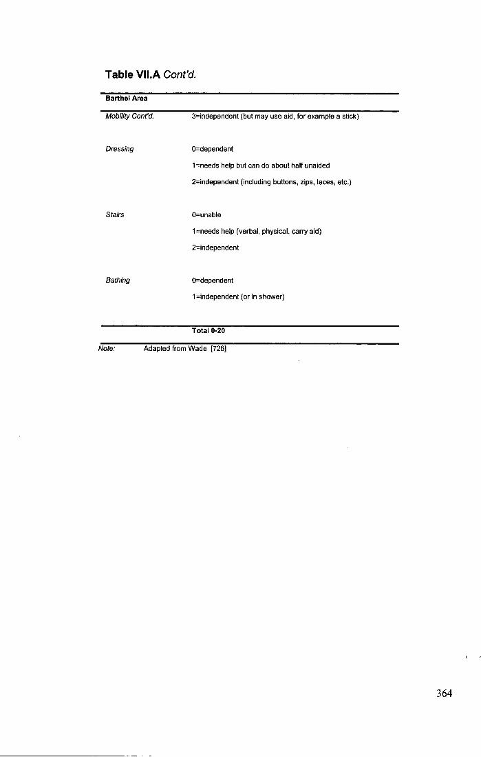
Table VII.A Cont'd.
Barthel Area
Mobility Cont'd.
Dressing
Stairs
Bathing
3=independent (but may use aid, for example a stick)
O=dependent
1 =needs help but can do about half unaided
2=independent (including buttons, zips, laces; etc.)
O=unable
1 =needs help (verbal, physical, carry aid)
2=independent
O=dependent
1 =independent (or in shower)
Total 0-20
Note: Adapted from Wade [726]
364
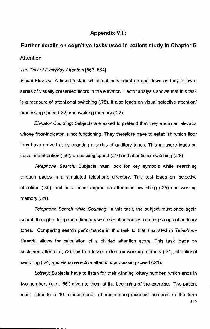
Appendix VIII:
Further details on cognitive tasks used in patient study in Chapter 5
Attention
The Test of Everyday Attention [563, 564]
Visual Elevator. A timed task in which subjects count up and down as they follow a
series of visually presented floors in the elevator. Factor analysis shows that this task
is a measure of attentional switching (.78). It also loads on visual selective attention/
processing speed (.22) and working memory (.22).
Elevator Counting: Subjects are asked to pretend that they are in an elevator
whose floor-indicator is not functioning. They therefore have to establish which floor
they have arrived at by counting a series of auditory tones. This measure loads on
sustained attention (.56), processing speed (.27) and attentional switching (.28).
Telephone Search: Subjects must look for key symbols while searching
through pages in a simulated telephone directory. This test loads on 'selective
attention' (.80), and to a lesser degree on attentional switching (.25) and working
memory (.21 ).
Telephone Search while Counting: In this task, the subject must once again
search through a telephone directory while simultaneously counting strings of auditory
tones. Comparing search performance in this task to that illustrated in Telephone
Search, allows for calculation of a divided attention score. This task loads on
sustained attention (.72) and to a lesser extent on working memory (.31 ), attentional
switching (.24) and visual selective attention/ processing speed (.21 ).
Lottery: Subjects have to listen for their winning lottery number, which ends in
two numbers (e.g., '55') given to them at the beginning of the exercise. The patient
must listen to a 1 0 minute series of audio-tape-presented numbers in the form
365
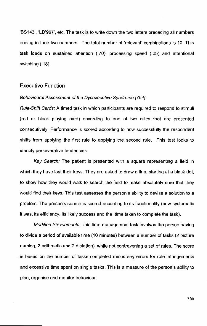
'BS 143', 'LD'967', etc. The task is to write down the two letters preceding all numbers
ending in their two numbers. The total number of 'relevant' combinations is 1 0. This
task loads on sustained attention (.70), processing speed (.25) and attentional
switching ( .18 ).
Executive Function
Behavioural Assessment of the Dysexecutive Syndrome [754]
Rule-Shift Cards: A timed task in which participants are required to respond to stimuli
(red or black playing card) according to one of two rules that are presented
consecutively. Performance is scored according to how successfully the respondent
shifts from applying the first rule to applying the second rule. This test looks to
identify perseverative tendencies.
Key Search: The patient is presented with a square representing a field in
which they have lost their keys. They are asked to draw a line, starting at a black dot,
to show how they would walk to search the field to make absolutely sure that they
would find their keys. This test assesses the person's ability to devise a solution to a
problem. The person's search is scored according to its functionality (how systematic
it was, its efficiency, its likely success and the time taken to complete the task).
Modified Six Elements: This time-management task involves the person having
to divide a period of available time (10 minutes) between a number of tasks (2 picture
naming, 2 arithmetic and 2 dictation), while not contravening a set of rules. The score
is based on the number of tasks completed minus any errors for rule infringements
and excessive time spent on single tasks. This is a measure of the person's ability to
plan, organise and monitor behaviour.
366
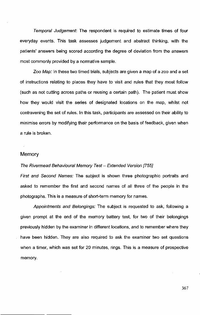
Temporal Judgement: The respondent is required to estimate times of four
everyday events. This task assesses judgement and abstract thinking, with the
patients' answers being scored according the degree of deviation from the answers
most commonly provided by a normative sample.
Zoo Map: In these two timed trials, subjects are given a map of a zoo and a set
of instructions relating to places they have to visit and rules that they most follow
(such as not cutting across paths or reusing a certain path). The patient must show
how they would visit the series of designated locations on the map, whilst not
contravening the set of rules. In this task, participants are assessed on their ability to
minimise errors by modifying their performance on the basis of feedback, given when
a rule is broken.
Memory
The Rivermead Behavioural Memory Test- Extended Version [755]
First and Second Names: The subject is shown three photographic portraits and
asked to remember the first and second names of all three of the people in the
photographs. This is a measure of short-term memory for names.
Appointments and Belongings: The subject is requested to ask, following a
given prompt at the end of the memory battery test, for two of their belongings
previously hidden by the examiner in different locations, and to remember where they
have been hidden. They are also required to ask the examiner two set questions
when a timer, which was set for 20 minutes, rings. This is a measure of prospective
memory.
367

Picture Recognition: The subject must recognise 20 previously-seen line
drawings and distinguish them from distractor items (a filled -5 minute delay occurs
between presentation and recall). This is a measure of visual memory.
Face Recognition: The subject must recognise 15 previously-seen faces and
distinguish them from distractor items. This is a measure of visual memory.
Story Immediate and Delayed: The subject must recall a short passage of
newspaper prose, immediately following it being read to them and then again after a
delay (filled delay of 20 minutes). These two tests are measures of verbal memory.
Orientation and Date: The subject is asked 12 orientation questions and to give
the correct date.
368
Appendix IX:
Supplementary analysis on the possible confounding impact of
psychiatric history on the relationship between coping and SAH
patients' PTSD
In Chapter 5, I contend that the dispositional use of maladaptive coping strategies is a
risk factor for post-traumatic stress disorder {PTSD) subsequent to a subarachnoid
haemorrhage (SAH). This conclusion was motivated by the results of the logistic
regression analyses of assessments one and two PTSD which showed that
dispositional use of maladaptive coping emerged as the most powerful predictor of
PTSD in both the short- and long-term. Further supporting evidence for this
conclusion is derived from previous studies which fortuitously measured individuals'
preferences for coping skills prior to a trauma and found that pre-trauma preferences
for maladaptive coping were predictive of subsequent PTSD [172, 217]. Up to
approximately 30% of patients in the patient sample examined as part of the study
presented in Chapter 5 do, nevertheless, have a history of other psychiatric illness,
including depression, anxiety, and/or alcohol abuse (Table IX.A). Although this is not
unusual and similar rates are reported in prior SAH studies [31, 159, 268, 305], there
does remain the possibility that use of maladaptive coping skills does not represent a
risk factor for PTSD, but that a significant association between coping and PTSD is
found because those patients with PTSD could have experienced a prior psychiatric
history which had promoted the use of maladaptive coping skills. This alternative
explanation of the relationship between coping and PTSD was, however, not
supported by the results of Chapter 5. 'Prior psychiatric history' had been included as
a potential predictor in the original regression models for PTSD, but was eliminated
369
Table IX.A Psychiatric history in SAH patients examined for PTSD at assessment
one and assessment two
Factor Value(%)
Assessment one Assessment two
N=95 N=81
Significant psychiatric history* 25 (26.3) 22 (27.2)
Note: N=number. *=Self-reported illness receiving/ received formal treatment.
Psychiatric illnesses included: Anxiety unspecified diagnosis, Depression unspecified
diagnosis, Alcohol abuse.
from the final model as it did not prove to be associated with PTSD in either the short
or long-term. In any case, the possibility that prior psychiatric history is a confounding
variable for the relationship between coping and PTSD warrants further examination
given the importance of establishing risk factors for the debilitating stress disorders
which SAH patients experience. To do this, the unique and shared variance between
PTSD, psychiatric history and coping was examined in order to further elucidate the
relationship between these factors.
Methods
Nonparametric zero-order correlation and partial correlation coefficients were
calculated between the following combinations of variables at both assessment one
and assessment two: PTSD symptomatology and coping, PTSD symptomatology and
prior psychiatric history and PTSD symptomatology and psychiatric history.
370
Results
The results of these supplementary correlation analyses are presented in Table IX.B.
The results firstly show that the magnitude/ effect size of the relationship between
coping and PTSD is consistently larger than the relationship between coping and
psychiatric history. This substantial difference in effect sizes serves to underline the
relative triviality of psychiatric history in explaining the presence of maladaptive
coping. Secondly, the minimal reduction in the size of the correlation coefficients
between PTSD and coping and between coping and psychiatric history when partial
correlations are calculated which control for the shared variance between PTSD and
psychiatric history, illustrates that these two factors are largely independent of one
another in their relation to coping (Figures IX.A-8).
Discussion and conclusion
The results from the correlation analyses illustrate that PTSD holds a unique and
substantial relationship with coping skills. Having controlled for the potential shared
variance which PTSD and psychiatric history hold with coping, it is apparent that
PTSD and psychiatric history are largely independent variables. In conclusion,
psychiatric history holds little relevance for explaining the presence of poor coping
skills or PTSD. The evidence therefore serves to corroborate the conclusion of
Chapter 5, that coping skills seem largely the cause of PTSD in SAH patients in both
the short- and long-term.
371
Table IX:B. Zero-order and partial correlation coefficients between PTSD, psychiatric history and maladaptive coping strategies
Variable N Zero- P-value Partial P-value
order correlation
correlation controlling for
with coping other variable
r. '·2 faY(X1.x2) fsY(X1.x2)2
PTSD assessment 1 91 0.636 40.4% <0.0001 0.623 38.8% <0.0001
Psychiatric history assessment 1 91 0.216 4.67% <0.05 0.066 0.4% >0.05
PTSD assessment 2 81 0.459 21.1% <0.0001 0.452 20.4% <0.0001
Psychiatric history assessment 2 81 0.234 5.5% <0.05 0.156 0.2% >0.05
Note: N=sample size; PTSD=post-traumatic stress disorder; r.=Spearman's rank order correlation; r} =Spearman's correlation r-squared equivalent;
r.v(X1.x2,=Spearman's partial rank order correlation; r.v(x1.x2/=Spearman's partial rank order correlation r-squared equivalent..
372
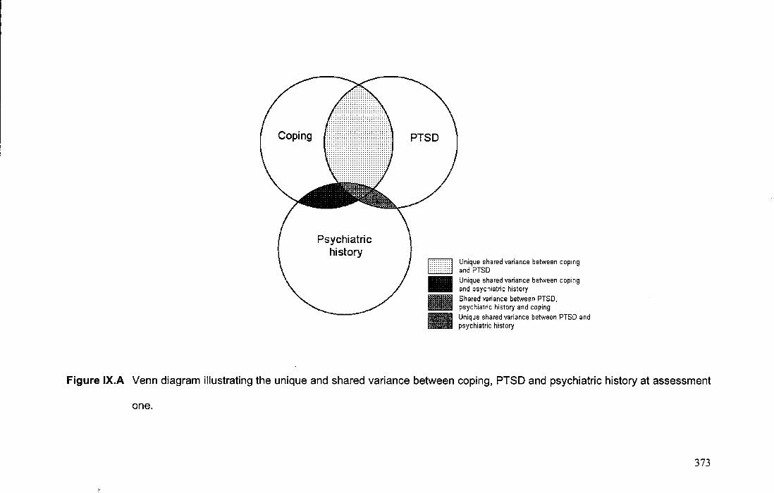
Coping
Psychiatric history
PTSD
F7'l L.l.2ld •
Unique shared variance between coping and PTSD Unique shared variance between coping and psychiatric history Shared variance between PTSD, psychiatric history and coping Unique shared variance between PTSD and psychiatric history
Figure IX.A Venn diagram illustrating the unique and shared variance between coping, PTSD and psychiatric history at assessment
one.
373
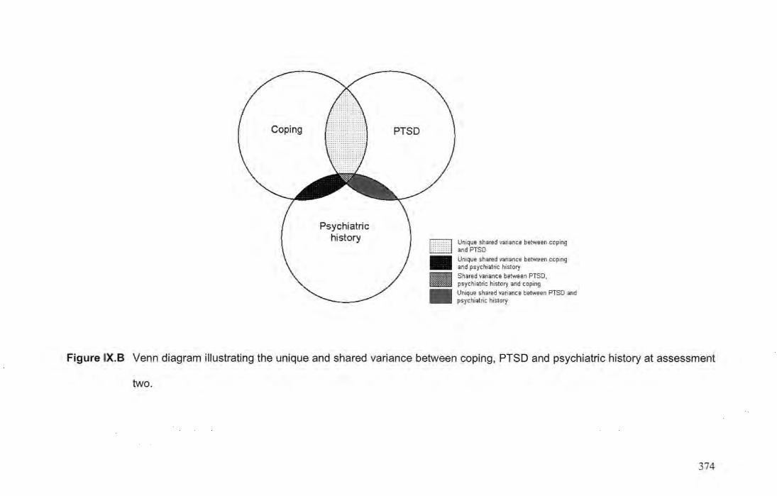
Coping
Psychiatric history
DI2J • •
Unique shared variance between coping and PTSD
Unique shared variance between coping and psychiatric history
Shared variance between PTSD, psychiatric history and coping
Unique shared variance between PTSO and psychiatric history
Figure IX.B Venn diagram illustrating the unique and shared variance between coping, PTSD and psychiatric history at assessment
two.
374
Appendix X:
Supplementary analysis on the predictive value of aneurysm
location for PTSD in SAH patients
The majority of surviving subarachnoid haemorrhage (SAH) patients examined in
Chapter 5 had suffered their haemorrhage as the result of a ruptured cerebral
aneurysm. As is typical, the neuroanatomical location of a patient's ruptured
aneurysm varied between patients (Table X.A). Before further investigating other
clinical explanations/ risk factors for post-traumatic stress disorder (PTSD) in SAH
patients, it is important to comprehensively evaluate the effect of aneurysm location.
Historically, it has been believed that differently located aneurysms pose different
risks to the psychiatric sequelae which SAH patients' experience [57, 295, 537]. In
particular, many traditionally believed that those patients whose SAH was due to a
ruptured anterior cerebral artery aneurysm - especially those on the anterior
communicating artery (ACoA) [8] - were likely to experience a unique pattern of
psychiatric sequelae [295]. Older studies suggested that these patients could
experience a psychiatric outcome akin to Korsakoff's syndrome [400, 513, 724].
ACoA patients for example, were seen to classically illustrate a distinctive
neuropsychiatric profile of amnesia, confabulation and altered personality (including
impulsivity, disinhibited behaviour, apathy, emotional lability and depression) [433].
The parallels between this profile and that of patients who had suffered damage to
the prefrontal lobe [412] led to the belief that the type of neurobehavioural sequelae
experienced post-SAH was largely due to the result of ischemic/ haemorrhagic
damage in the territory supplied by the artery on which the aneurysm had ruptured
[433]. Other factors, such as the fact that different locations of ruptured aneurysms
375
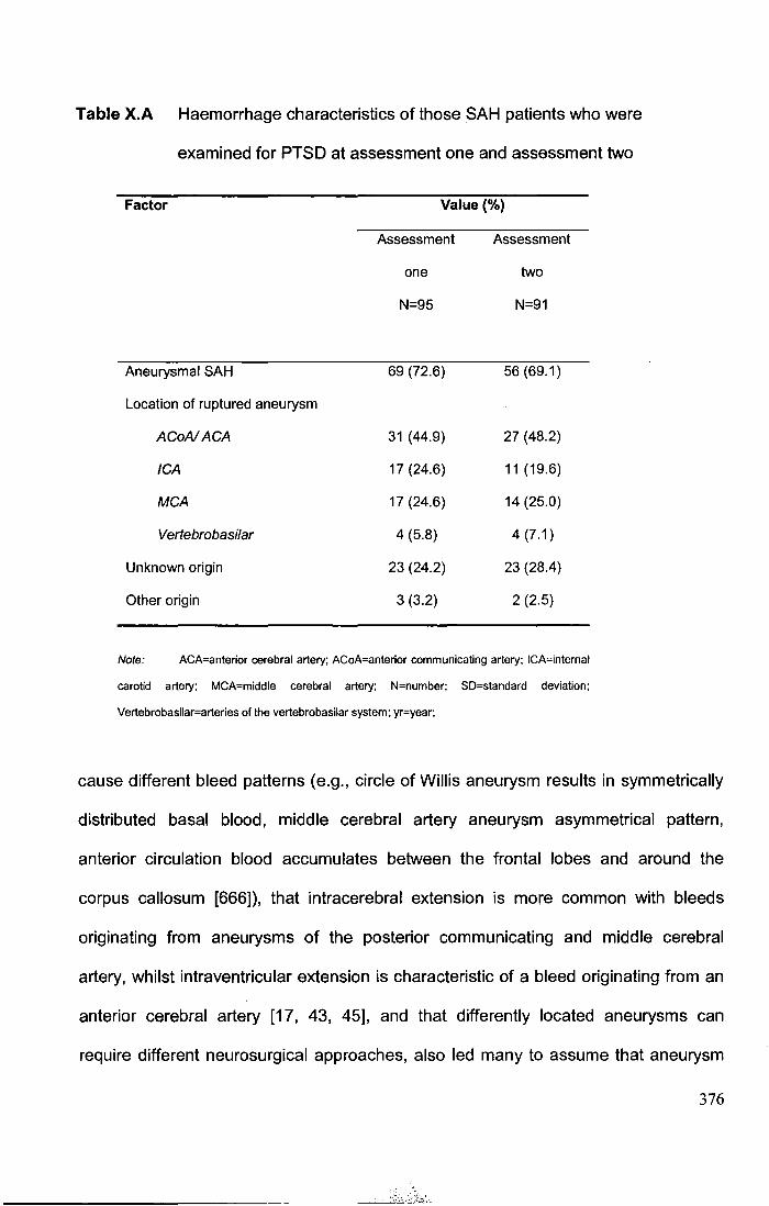
Table X. A Haemorrhage characteristics of those SAH patients who were
examined for PTSD at assessment one and assessment two
Factor
Aneurysmal SAH
Location of ruptured aneurysm
ACoAIACA
ICA
MCA
Vertebrobasilar
Unknown origin
Other origin
Value(%)
Assessment
one
N=95
69 (72.6)
31 (44.9)
17 (24.6)
17 (24.6)
4 (5.8)
23 (24.2)
3 (3.2)
Assessment
two
N=91
56 (69.1)
27 (48.2)
11 (19.6)
14 (25.0)
4 (7.1)
23 (28.4)
2 (2.5)
Note: ACA=anterior cerebral artery; ACoA=anterior communicating artery; ICA=intemal
carotid artery; MCA=middle cerebral artery; N=number; SD=standard deviation;
Vertebrobasilar=arteries of the vertebrobasilar system; yr=year;
cause different bleed patterns (e.g., circle of Willis aneurysm results in symmetrically
distributed basal blood, middle cerebral artery aneurysm asymmetrical pattern,
anterior circulation blood accumulates between the frontal lobes and around the
corpus callosum [666]), that intracerebral extension is more common with bleeds
originating from aneurysms of the posterior communicating and middle cerebral
artery, whilst intraventricular extension is characteristic of a bleed originating from an
anterior cerebral artery [17, 43, 45], and that differently located aneurysms can
require different neurosurgical approaches, also led many to assume that aneurysm
376
location was important in determining SAH outcome [295]. Nonetheless, the
importance of the location of the ruptured aneurysm for SAH patients'
neurobehavioural sequelae has in contemporary studies, proved debatable, with the
majority studies, despite being well-powered, failing to detect a difference between
the outcomes of patients with differently located aneurysms [57, 295, 433]. Instead,
most now advocate that an SAH leads to unspecific brain damage that is, for the
greater part, independent of the localisation of the ruptured aneurysm. In support of
this position, is evidence that patients whose SAH was of an unknown origin,
experience difficulties comparable to those patients whose bleed was caused by a
ruptured aneurysm [34, 141, 296, 300, 302, 382, 409, 504, 507, 597, 643]. Yet further
support can be found in the results presented in Chapter 5 which showed that
aneurysmal SAH patients were no more, or no less prone to develop PTSD than
other SAH patients. Even so, given the benefits of being able to reliably predict which
patients will develop PTSD and also being able to understand the aetiology of the
disorder, it is still important to explore the association between of aneurysm location
and SAH PTSD.
With this in mind, this supplementary section now presents the results of
bivariate/ univariate analyses of the association between aneurysm location (anterior
vs. posterior; ACoA vs. other) and PTSD in both the short- and long-term. Also, to
further explore the role of aneurysm location, PTSD severity and prevalence was
compared between those patients whose bleed was of an aneurysmal origin and
those patients whose bleed was of an unknown, non-aneurysmal origin.
377
Methods
The chi-square test (corrected for continuity [633]) and the Fisher exact test were
used to determine the association between the experience of PTSD post-SAH and
the location of a patient's ruptured aneurysm. The Mann-Whitney test examined the
degree to which PTSD symptom severity in SAH patients was dependent on the
location of the ruptured aneurysm. Comparisons were made between 1) ACoA
aneurysms and all other locations, 2) anterior cerebral artery aneurysms (including
ACoA) and all other locations and 3) between patients with an SAH of an aneurysmal
origin and those who bleed was of an unknown origin. It was not possible to conduct
a more detailed analysis of PTSD by specific aneurysm location because too small a
number of patients had aneurysms of vertebrobasilar and too small a number of
patients had aneurysms on arteries of locations other than the anterior cerebral
artery. This is not however, a limitation specific to the present study, but a difficulty
pervasive to many studies of SAH outcome which reflect the natural distribution of
ruptured cerebral aneurysms in SAH patients [48, 300, 357, 374, 378, 507, 510, 687,
689].
Results
No significant association was found between PTSD prevalence or symptom severity
and aneurysm location at either assessment one or assessment two (all P>0.05).
Furthermore, there was no significant difference between the prevalence of PTSD or
the severity of those patients' PTSD which resulted from a ruptured aneurysm and
those whose bleed was of an unknown origin (Table X.B).
378
TableX.B The impact of aneurysm location on PTSD prevalence and severity
Haemorrhage aetiology
ACoA Other ACA Other ASAH Unknown
No.(%) no.t(%) P·value no.(%) no.:t: (%) P-value no.(%) no.(%) P-value
Assessment one
PTSD
Yes 5 22 11 16 27 7
No 12 30 .510. 16 26 1.00 42 16 .618
PTSD severity
Median 10.0 10.5 11.0 10.0 10.0 10.0
JQR 5.5-22.0 5.0-22.5 .775 5-28 4.5-19.5 .386 5.0-22.0 3.0-14.0 .308
Assessment two
PTSD
Yes 6 11 10 7 17 9
No 10 29 .527. 13 26 .137 39 14 .599
PTSD severity
Median 10.5 6 10. 6 6.5 8
IQR 3.5-12.0 3.0-11.75 .561 3.0-12.0 3.5-10.0 .339 3.0-12.0 5.0-14.0 .402
Note: ACoA = anterior communicating artery; ACA = anterior cerebral artery. • Fisher's exact test. t 'Other' location group includes aneurysms located on the internal carotid
artery, middle cerebral artery, in the vertebrobasilar system and those of the anterior cerebral artery, excluding anterior communicating aneurysms. + 'Other' location group includes
aneurysms located on the internal carotid artery, middle cerebral artery and in the vertebrobasilar system.
379
Discussion
The results. of this supplementary analysis show that the location of subarachnoid
haemorrhage (SAH) patients' ruptured aneurysms does not significantly determine
the likelihood of SAH patients developing post-traumatic stress disorder (PTSD), nor
the severity of the PTSD that an SAH patient experiences. These findings concur
with the multiple regression analyses presented in Chapter 5, where aetiology of
bleed did not significantly add to the final predictive models for PTSD. It also accords
with the majority of contemporary studies which have not found an association
between long-term psychosocial outcome (such as anxiety, depression) and the
location of a patient's ruptured aneurysm [17, 242, 24 7, 294, 297, 302, 425, 469, 507,
508, 546, 594, 649, 687, 722]. Indeed, aneurysm location is increasingly being seen
as unrelated too to other aspects of SAH outcome, such as cognitive function [189,
247, 262, 357, 388, 432, 507, 543, 558, 590, 598, 649, 746, 750], fatigue [65, 247,
510, 649, 722], sleep dysfunction [510] and neuroendocrine dysfunction [375].
Historically however, many have proposed/ found that different aneurysm
locations - particularly those of the anterior communicating artery (ACoA) - can
cause different neurobehavioural sequelae of SAH [8, 331, 513, 617, 652]. How then
can the discrepancy between these older and newer studies (including the current
one) be explained? I would concur with the conclusions of two recent reviews on this
important question produced by Hutter and colleagues [295, 306], who argue that
aneurysm location (at least indirectly) was once an important explanation for SAH
patients' neuropsychiatric changes, but that it is now largely irrelevant (and so not
found to be important in contemporary studies) because of the drastically improved
management of SAH which has occurred with the passage of time. These
improvements include the introduction of the operating microscope, the use of
380
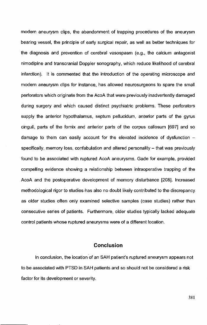
modern aneurysm clips, the abandonment of trapping procedures of the aneurysm
bearing vessel, the principle of early surgical repair, as well as better techniques for
the diagnosis and prevention of cerebral vasospasm (e.g., the calcium antagonist
nimodipine and transcranial Doppler sonography, which reduce likelihood of cerebral
infarction). It is commented that the introduction of the operating microscope and
modern aneurysm clips for instance, has allowed neurosurgeons to spare the small
perforators which originate from the AcoA that were previously inadvertently damaged
during surgery and which caused distinct psychiatric problems. These perforators
supply the anterior hypothalamus, septum pellucidum, anterior parts of the gyrus
cinguli, parts of the fornix and anterior parts of the corpus callosum [697] and so
damage to them can easily account for the elevated incidence of dysfunction -
specifically, memory loss, confabulation and altered personality- that was previously
found to be associated with ruptured AcoA aneurysms. Gade for example, provided
compelling evidence showing a relationship between intraoperative trapping of the
AcoA and the postoperative development of memory disturbance [208]. Increased
methodological rigor to studies has also no doubt likely contributed to the discrepancy.
as older studies often only examined selective samples (case studies) rather than
consecutive series of patients. Furthermore, older studies typically lacked adequate
control patients whose ruptured aneurysms were of a different location.
Conclusion
In conclusion, the location of an SAH patient's ruptured aneurysm appears not
to be associated with PTSD in SAH patients and so should not be considered a risk
factor for its development or severity.
381
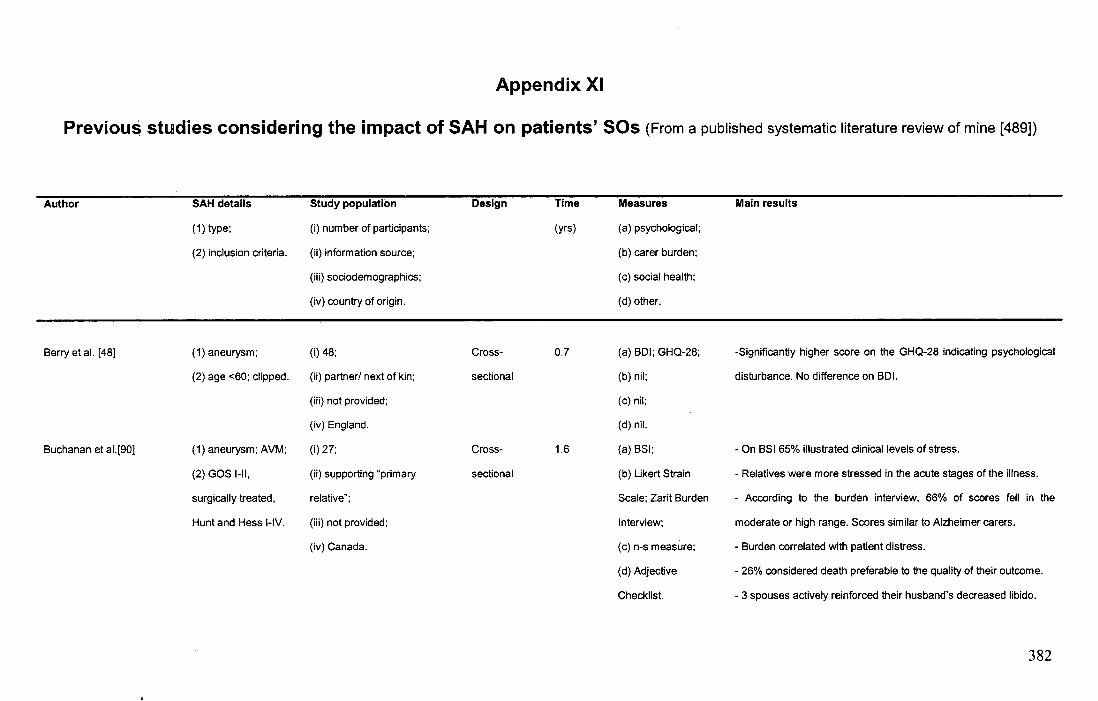
Appendix XI
Previous studies considering the impact of SAH on patients' SOs (From a published systematic literature review of mine [489])
Author SAH details Study population Design Time Measures Main results
(1)type; (i) number of participants; (yrs) (a) psychological;
(2) inclusion criteria. (ii) information source; (b) carer burden;
(iii) sociodemographics; (c) social health;
(iv) country of origin. (d) other.
Berry et al. [48] ( 1) aneurysm; (i) 48; Cross- 0.7 (a) BDI; GHQ-28; -Significantly higher score on the GHQ-28 indicating psychological
(2) age <60; clipped. (ii) partner/ next of kin; sectional (b) nil; disturbance. No difference on BDl.
(iii) not provided; (c) nil;
(iv) England. (d) nil.
Buchanan et al.[90] (1) aneurysm; AVM; (i)27; Cross- 1.6 (a) BSI; - On BSI 65% illustrated clinical levels of stress.
(2) GOS 1-11, (ii) supporting "primary sectional (b) Likert Strain - Relatives were more stressed in the acute stages of the illness.
surgically treated, relative"; Scale; Zarit Burden - According to the burden interview, 66% of scores fell in the
Hunt and Hess I-IV. (iii) not provided; Interview; moderate or high range. Scores similar to Alzheimer carers.
(iv) Canada. (c) n-s measure; - Burden correlated with patient distress.
(d) Adjective - 26% considered death preferable to the quality of their outcome.
Checklist. - 3 spouses actively reinforced their husband's decreased libido.
382
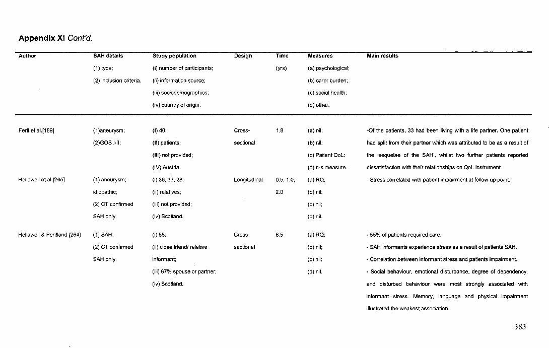
Appendix XI Cont'd.
Author
Fertl et al.[189]
Hellawell et al.[265]
Hellawell & Pentland [264]
SAH details
(1) type;
(2) inclusion criteria.
( 1 )aneurysm;
(2)GOS 1-11;
( 1) aneurysm;
idiopathic;
(2) CT confirmed
SAH only.
(1) SAH;
(2) CT confirmed
SAH only.
Study population
(i) number of participants;
(ii) information source;
(iii) sociodemographics;
(iv) country of origin.
(I) 40;
(II) patients;
(Ill) not provided;
(IV) Austria.
(i) 36, 33, 28;
(ii) relatives;
(iii) not provided;
(iv) Scotland.
(i) 58;
(ii) close friend/ relative
informant;
(iii) 67% spouse or partner;
(iv) Scotland.
Design
Cross-
sectional
Longitudinal
Cross-
sectional
Time
(yrs)
1.8
0.5, 1.0,
2.0
6.5
Measures
(a) psychological;
(b) carer burden;
(c) social health;
(d) other.
(a) nil;
(b) nil;
(c) Patient QoL;
(d) n-s measure.
(a)RQ;
(b) nil;
(c) nil;
(d) nil.
(a) RQ;
(b) nil;
(c) nil;
(d) nil.
Main results
-Of the patients, 33 had been living with a life partner. One patient
had split from their partner which was attributed to be as a result of
the 'sequelae of the SAH', whilst two further patients reported
dissatisfaction with their relationships on QoL instrument.
- Stress correlated with patient impairment at follow-up point.
- 55% of patients required care.
- SAH informants experience stress as a result of patients SAH.
- Correlation between informant stress and patients impairment.
- Social behaviour, emotional disturbance, degree of dependency,
and disturbed behaviour were most strongly associated with
informant stress. Memory, language and physical impairment
illustrated the weakest association.
383
Appendix XI Cont'd.
Author
Hop et al. [285, 286]*
;;,
SAH details
(1) type;
(2) inclusion criteria.
(1) aneurysm;
(2) nil.
Study population
(i) number of participants;
(ii) information source;
(iii) sociodemographics;
(iv) country of origin.
(i) 51;
(ii) patient's closest significant
other- 'partner';
(iii) 90% spouse or partner;
(iv) Netherlands.
Design
Longitudinal
Time
(yrs)
0.3, 1.5
Measures
(a) psychological;
(b) carer burden;
(c) social health;
(d) other.
(a) nil;
(b) nil;
(c) SF-36; SIP; VAS;
(d) n-s measure.
Main results
- At assessment one, carers illustrated reduced Qol on SIP,
particularly psychosocial reductions. At assessment two, reductions
found for SIP domains sleep and rest, emotional behaviour and
recreation and pastimes.
- SF-36 and SIP scores between 4 and 18 months varied, with
some improvement (in social functioning, mental health), but also
reductions (in pain, general health). Overall QoL, assessed by the
VAS, did not improve between assessments one and two.
- At assessment one, partners reported anxiety and uneasiness.
Partners were afraid to leave the patient alone, especially if they
had witnessed the initial event. Some expressed fear of having sex.
especially if SAH was coital.
384
Table XI Cont'd.
Author SAH details Study population Design Time Measures Main results
(1)type; (i) number of participants; (yrs) (a) psychological;
(2) inclusion criteria. (ii) information source; (b) carer burden;
(iii) sociodemographics; (c) social health;
(iv) country of origin. (d) other.
Hop et al. [285, 286]* - At assessment two, 50% of employed carers reported occupation
Cont'd. change related to SAH. 3 carers reported divorce/ "severe marital
difficulties" caused by the patients' personality change.
- Hop et al. [285] explored partners' QoL at assessment one and
reported an association with patient dependence. Nevertheless,
even partners of independent patients still experienced emotional
role limitations- at times more than their patient.
Jarvis [313] (1) SAH; (I) 8; Cross- 1.1-1.5 (a) nil; - Common themes from qualitative interviews included: relationship
(2) nil. (II) patients; sectional (b) nil; difficulties as a result of the SAH and relatives/ friends finding it very
(Ill) not reported; (c) nil; traumatic to talk about the SAH event with the patient.
(IV) England. (d) n-s measure.
385
Table X~ Cont'd.
Author
Mezue et al. [461]
SAH details
(1)type;
(2) inclusion criteria.
( 1) aneurysm;
(2) GOS 1-11.
Study population
(i) number of participants;
(ii) information source;
(iii) sociodemographics;
(iv) country of origin.
(i) 52;
(ii) carers;
(iii) 74.6% spouse or partner;
13.6% other family member;
(iv) England.
Design
Cross
sectional
Time
(yrs)
3.0
Measures
(a) psychological;
(b) carer burden;
(c) social health;
(d) other.
(a) GHQ-12;
(b) Caregiver Strain
Index;
(c) nil;
(d) Adjective
Checklist.
Main results
- 54% complained of care-related strain, particularly in emotional
and social areas. Emotional problems included upsetting patient
behaviour (44.2%), emotional adjustment (28.9%), feeling
overwhelmed (25%) and disturbed sleep (19.3%).
-Social problems included changes in personal plans (28.9%), work
(17.3%) and family (17.3%).
- 25% of carers reported that they were distressed to an
'overwhelming' degree.
- Carer distress/ strain correlated with neurobehavioural change in
the patient.
- No relation between clinical/ demographic variables and stress.
386
Table XI Cont'd.
Author
Oder et al. [501]
Oder et al. (502]
SAH details
(1)type;
(2) inclusion criteria.
~I
(1)SAH;
(2) Less severe and
idiopathic cases
overrepresented
(61%).
(1) idiopathic;
(2) Less severe
patients.
Study population
(i) number of participants:
(ii) information source;
(iii) sociodemographics;
(iv) country of origin.
(i) 67;
(ii) patients:
(iii) not reported;
(iv) Austria.
(i) 41;
(ii) patients;
(iii) not reported;
(iv) Austria.
Design
Cross
sectional
Cross
sectional
Time
(yrs)
7.1
7.6
Measures
(a) psychological;
(b) carer burden;
(c) social health;
(d) other.
(a) nil;
(b) nil;
(c) n-s measure:
(d) nil.
(a) nil;
(b) nil;
(c) n-s measure;
(d) nil.
Main results
- Patients rated the degree of disability in family relationships.
- Association between amount of familial disturbance and patient
disability.
- Association between patients working status and familial
handicap.
- No relationship between clinical/ demographic variables and carer
stress, although neurologic deficit on admission was predictive of
later family disturbance.
-In total17.1% of patients reported persistent "family tensions" after
the SAH.
387
Table XI Cont'd.
Author
Ogden et al. [51 0]
Pritchard et al.[546]
SAH details
(1)type;
(2) inclusion criteria.
(1) aneurysm;
idiopathic;
(2) nil.
( 1) aneurysm;
(2) nil.
Study population
(i) number of participants;
(ii) information source;
(iii) sociodemographics;
(iv) country of origin.
(i) 123;
(ii) patients;
(iii) not reported;
(iv) New Zealand.
(i) 98;
(ii) carers;
(iii) 76% partners, 63% female,
53% aged <54 years;
(iv) England.
Design
Cross
sectional
Cross
sectional
Time
(yrs)
4.0-7.0
0.5-1.6
Measures
(a) psychological;
(b) carer burden;
(c) social health;
(d) other.
(a) nil;
(b) nil;
(c) n-s measure;
(d) nil.
(a) nil;
(b) nil;
(c) Wessex
Questionnaire;
(d) nil.
Main results
- Of the patients asked about changes in family structure, 4.1%
reported divorcing as a result of SAH. Most due to partners failure
to cope with SAH changes.
- Association between family disruption and neurological status in
the early stages of illness and experience of seizures.
- Carers were positive about in-patient care, though 63% remained
fearful during the patient's hospital stay and 30% felt depressed.
- Post-discharge, 50% felt unsupported, 40% stressed, 25%
required medication and 14% said they made 'unnecessary' calls to
their GP. Carers reported significantly more psychological burdens
than patients.
388
Table XI Cont'd.
Author
Pritchard et al.[546] Cont'd.
Pritchard et al.[545]
:-··
SAH details
(1) type;
(2) inclusion criteria.
(1) aneurysm;
(2) nil.
Study population
(i) number of participants;
(ii) information source;
(iii) sociodemographics;
(iv) country of origin.
(i) 98;
(ii) primary carers:
(iii) 63% male;
(iv) England.
Design
Cross
sectional
Time
(yrs)
0.5-2.0
Measures
(a) psychological;
(b) carer burden;
(c) social health;
(d) other.
(a) nil;
(b) nil;
(c) Wessex
Questionnaire;
(d) nil.
Main results
- No association between outcomes and clinical variables. -
Criticism of 'Community Care'. It was the patients' families (96%)
and neighbours (68%) who were the most involved in supporting
patients.
- 33% reported financial difficulties
- 86% of carers lost 2 or more weeks and 15% lost 17 or more.
-'Lost productivity' was estimated at £182,000.
- Compared outcome of a well-matched emergency cohort of SAH
carers and an elective cohort of acoustic neuroma carers (AN).
- SAH carers reported greater psychosocial disturbance than AN
carers. In both the inpatient and post-discharge stages, SAH carers
were more depressed, frightened, anxious and stressed
- Economic cost: 85% of AN compared to 77% of SAH lost S16
weeks, but 23% of ASAH carers lost between 17-30 weeks. For
AN, time off work cost £52,440. For SAH carers, it was £182,000.
389
Table XI Cont'd.
Author SAH details Study population
(1)type; (i) number of participants;
(2) inclusion criteria. (ii) information source;
(iii) sociodemographics;
(iv) country of origin.
Stegen & Freckman [652] (1) aneurysm; (i) 87;
(2) nil. (ii) next of kin;
(iii) not reported;
(iv) Germany.
Wermer et al.[746] (1) aneurysm; (i) 610;
(2) mRS s 3; <71 (ii) patients;
years old; clipping (iii) not reported;
only. (iv) Netherlands.
Design Time
(yrs)
Cross- 1.0
sectional
Cross- 8.9
sectional
Measures
(a) psychological;
(b) carer burden;
(c) social health;
(d) other.
(a) n-s measure;
(b) nil;
(c) nil;
(d) nil.
(a) nil;
(b) nil;
(c) nil;
(d) n-s measure.
~-
Main results
- 17% reported concerns about the possibility of re-bleeding.
- 75 of the 520 (14%) formerly married patients had divorced after
the SAH, 34 of whom (7%) reported that this was the result of the
SAH. The main reason was that the partner could not cope with the
changes in the SAH patient.
Note: AN=acoustic neuroma; AVM=arteriovenous malformation; BDI=Beck Depression Inventory; BSI=Brief Symptom Inventory; GHQ-28=General Health Questionnaire-28; GOS !=Glasgow Outcome Scale I-
no/ minimal impairment, "good recovery"; GOS II=Giasgow Outcome Scale II - modest impairment/ needs no impairment, "fair recovery; Hunt and Hess !=Grade I - mild headache and/ or nick stiffness on initial
presentation; Hunt and Hess II=Grade II -severe headache and/ or pronounced neck stiffness and/ or cranial nerve deficits, photophobia; mRS=modified Rankin Scale; n-s measure=non-standardised measure;
RQ=Relatives Questionnaire; SAH=subarachnoid haemorrhage; SF-36=Short-Form 36 Health Survey; SIP=Sickness Impact Profile; TBI=traumatic brain injury; Qol=Quality of Life; VAS=Visual Analogue Scale.
*=Hop et al. (2001) contains data from 4 month assessment originally presented by Hop et al. (1998). For this review, searches of the Medline (1951 to April 2008), IS/ Web of Science (1945 to April 2008) and
Psyc/NFO databases (1887 to April 2008) was made using the term 'subarachnoid' plus: 'family', 'friend', 'carer', 'partner', 'relative' and 'rehabilitation'. The searches excluded 'stroke' as a term because SAH is
considered a .distinct form of stroke due to its differences in pathology (requiring differences in careJ:·Yo~ger mean age of occurrence and differences in intervention (surgery for SAH) [71 0].
~/;.1-r~. !:~;., ~~·-~. ·' -·
390