anatomical eligibility of the renal vasculature for catheter-based renal denervation in hypertensive...
TRANSCRIPT

J A C C : C A R D I O V A S C U L A R I N T E R V E N T I O N S V O L . 7 , N O . 2 , 2 0 1 4
ª 2 0 1 4 B Y T H E A M E R I C A N C O L L E G E O F C A R D I O L O G Y F O U N D A T I O N I S S N 1 9 3 6 - 8 7 9 8 / $ 3 6 . 0 0
P U B L I S H E D B Y E L S E V I E R I N C . h t t p : / / d x . d o i . o r g / 1 0 . 1 0 1 6 / j . j c i n . 2 0 1 3 . 1 0 . 0 1 3
Anatomical Eligibility of the RenalVasculature for Catheter-Based RenalDenervation in Hypertensive Patients
Stefano F. Rimoldi, MD,* Niklaus Scheidegger, BSC,* Urs Scherrer, MD,*yStefan Farese, MD,z Emrush Rexhaj, MD,* Aris Moschovitis, MD,*
Stephan Windecker, MD,* Bernhard Meier, MD,* Yves Allemann, MD*
Bern and Solothurn, Switzerland; and Arica, Chile
Objectives This study sought to determine the vascular anatomical eligibility for catheter-based renalartery denervation (RDN) in hypertensive patients.
Background Arterial hypertension is the leading cardiovascular risk factor for stroke and mortalityglobally. Despite substantial advances in drug-based treatment, many patients do not achieve targetblood pressure levels. To improve the number of controlled patients, novel procedure- and device-based strategies have been developed. RDN is among the most promising novel techniques. However,there are few data on the vascular anatomical eligibility.
Methods We retrospectively analyzed 941 consecutive hypertensive patients undergoing coronaryangiography and selective renal artery angiography between January 1, 2010, and May 31, 2012.Additional renal arteries were divided into 2 groups: hilar (accessory) and polar (aberrant) arteries.Anatomical eligibility for RDN was defined according to the current guidelines: absence of renal arterystenosis, renal artery diameter �4 mm, renal artery length �20 mm, and only 1 principal renal artery.
Results A total of 934 hypertensive patients were evaluable. The prevalence of renal artery stenosiswas 10% (n ¼ 90). Of the remaining 844 patients without renal artery stenosis, 727 (86%) hadnonresistant hypertension and 117 (14%) had resistant hypertension; 62 (53%) of the resistanthypertensive and 381 (52%) of the nonresistant hypertensive patients were anatomically eligible forsympathetic RDN.
Conclusions The vascular anatomical eligibility criteria of the current guidelines are a major limitingfactor for the utilization of RDN as a therapeutic option. Development of new devices and/ortechniques may significantly increase the number of candidates for these promising therapeuticoptions. (J Am Coll Cardiol Intv 2014;7:187–92) ª 2014 by the American College of CardiologyFoundation
From the *Department of Cardiology, Bern University Hospital, Bern, Switzerland; yFacultad de Ciencias, Departamento de
Biología, Universidad de Tarapacá, Arica, Chile; and the zDepartment of Nephrology, Bürgerspital Solothurn, Solothurn,
Switzerland. Dr. Windecker has received research grants from Abbott Vascular, Cordis, Medtronic, Boston Scientific, Biotronik,
Biosensors, and St. Jude Medical. All other authors have reported that they have no relationships relevant to the contents of this
paper to disclose.
Manuscript received July 25, 2013; revised manuscript received October 12, 2013, accepted October 15, 2013.

Rimoldi et al. J A C C : C A R D I O V A S C U L A R I N T E R V E N T I O N S , V O L . 7 , N O . 2 , 2 0 1 4
Anatomical Eligibility for Renal Denervation F E B R U A R Y 2 0 1 4 : 1 8 7 – 9 2
188
Arterial hypertension is the major cardiovascular risk factorworldwide (1). Despite substantial advances in drug-basedtreatment, arterial hypertension therapy remains a majorchallenge. According to recent surveys in the United Statesand Europe, only 26% to 63% of patients achieve targetblood pressure (BP) values (2). To improve the number ofwell-controlled patients, novel procedure- and device-basedstrategies have been developed (3–5), particularly renal arterydenervation (RDN).
See page 193
Exaggerated systemic sympathetic activity has been shownto be a major pathophysiological mechanism triggeringthe development, maintenance, and progression of arterialhypertension (6). Furthermore, recent preliminary studiessuggest that RDN may also have favorable effects in otherconditions associated with exaggerated sympathetic drive,such as obstructive sleep apnea syndrome, insulin resistance,and chronic renal or heart failure (7–9). Particularly,increased renal sympathetic activity has been shown to playan important role in the regulation of systemic BP (10).
Table 1. Anatomical Eligibility C
No renal artery stenosis (�30%)
No extrarenal artery
Diameter of the renal artery: �4 m
No early division: �20 mm
Abbreviationsand Acronyms
BP = blood pressure
RAS = renal artery stenosis
RDN = catheter-based renal
artery denervation
Consistent with this observation,surgical sympathectomy has beenshown to significantly reduce BPin hypertensive patients (11).However, this procedure wasaccompanied by severe ortho-static hypotension and inconti-nence in some patients (12). To
obviate these surgery-related complications, several mini-mally-invasive, catheter-based renal sympathetic denervationtechniques have been developed in recent years (3,13). Afterplacement of a catheter in the renal arteries, radiofrequencyenergy is applied endoluminally to induce thermal injuryof the renal sympathetic fibers located in the adventitia ofthe renal arteries. To use this technique and to minimize therisk of complications (i.e., renal artery stenosis and dissection),several anatomical criteria of the renal arteries have to befulfilled (Table 1). According to the guidelines (14,15), thediameter of the renal arteries should not be inferior to 4 mm,and the length before division should be�20 mm.Moreover,no renal artery stenosis (RAS) (�30%) and no extrarenalartery should be present.
The SYMPLICITY (Renal Sympathetic Denervation inPatients With Treatment-Resistant Hypertension) studyand others (16–18) reported 10% to 37% ineligibility among
riteria of the Renal Vasculature
m
screened patients, because the anatomical criteria were notmet. However, these data were limited due to the selectedpatient populations and the lack of detailed anatomical data.
In the present study, we aimed to determine theanatomical eligibility for RDN in a large population ofconsecutive patients with arterial hypertension undergoingrenal angiography.
Methods
Patient selection and definitions. We retrospectivelyanalyzed 941 consecutive hypertensive patients undergoingcoronary angiography and concomitant selective renal arte-riography between January 1, 2010, and May 31, 2012.Seven patients were excluded from the analysis, as theirimages were not suitable for evaluation (Fig. 1).
The study was approved by the institutional ethics com-mittee, and informed consent was obtained from all patients.
Imaging of the renal arteries was performed by the inva-sive cardiologist performing coronary arteriography inpatients with a history of (treated) hypertension, knownhypertensive heart disease as diagnosed by echocardiogra-phy, or persistently elevated BP (>140/90 mm Hg), pro-vided there was no previous assessment of the renal arteries.
Resistant hypertension was defined according to thecurrent guidelines (19). Coronary artery disease and peri-pheral artery disease were defined as previously described(20). Impaired renal function was defined by a glomerularfiltration rate of <60 ml/min/1.73 m2, as estimated by theMDRD (Modification of Diet in Renal Disease) equations(21). Diagnosis of dyslipidemia and of diabetes mellituswere based on the 2013 European Society of Hypertension/European Society of Cardiology guidelines (19).
Significant RAS was defined as lumen narrowing �50%diameter, as previously described (20).Selective and quantitative renal arteriography. Eight expe-rienced staff cardiologists performed the 934 selective renalarteriographies considered in the study. The percutaneousfemoral approach was used with standard 4- to 7-F Judkins orAmplatz catheters (Cordis, Miami Lakes, Florida). Aftercoronary angiography, renal arteriography was performed byselectively injecting contrast medium into main and accessoryrenal arteries. All images were recorded digitally. The pro-jection that best showed the anatomy was used for all analyses.Measurements were performed on cineangiograms. Thecontrast-filled, nontapered tip of the catheter was used forcalibration. Digital angiograms were analyzed with the use ofan automated edge-detection system (CAAS II, Pie MedicalImaging, Maastricht, the Netherlands), as previouslydescribed (22). Quantitative measurements included thediameter and length of the reference vessel. The intraobserverand interobserver variability of the quantitative measurementshas been reported previously (23). The intraobserver (N.S.)and interobserver (N.S., S.F.R.) coefficients of variation for

Figure 1. Flow Chart of the Study
RAS ¼ renal artery stenosis; RDN ¼ catheter-based renal artery denervation.
J A C C : C A R D I O V A S C U L A R I N T E R V E N T I O N S , V O L . 7 , N O . 2 , 2 0 1 4 Rimoldi et al.
F E B R U A R Y 2 0 1 4 : 1 8 7 – 9 2 Anatomical Eligibility for Renal Denervation
189
renal artery length were 1.3% and 2.8% and for renal arterydiameter were 2.5% and 3.4%, respectively.
Extrarenal arteries were divided into 2 groups: hilar(accessory) and polar (aberrant) arteries (Fig. 2). Anatomicaleligibility for catheter-based renal sympathetic denervationwas defined according to previous studies and currentguidelines (14–17): absence of renal artery stenosis, renalartery diameter �4 mm, renal artery length �20 mm, andonly 1 main renal artery (Table 1).
Early division was defined as the presence of an additionalrenal artery departing at �20 mm from the orifice of themain renal artery.
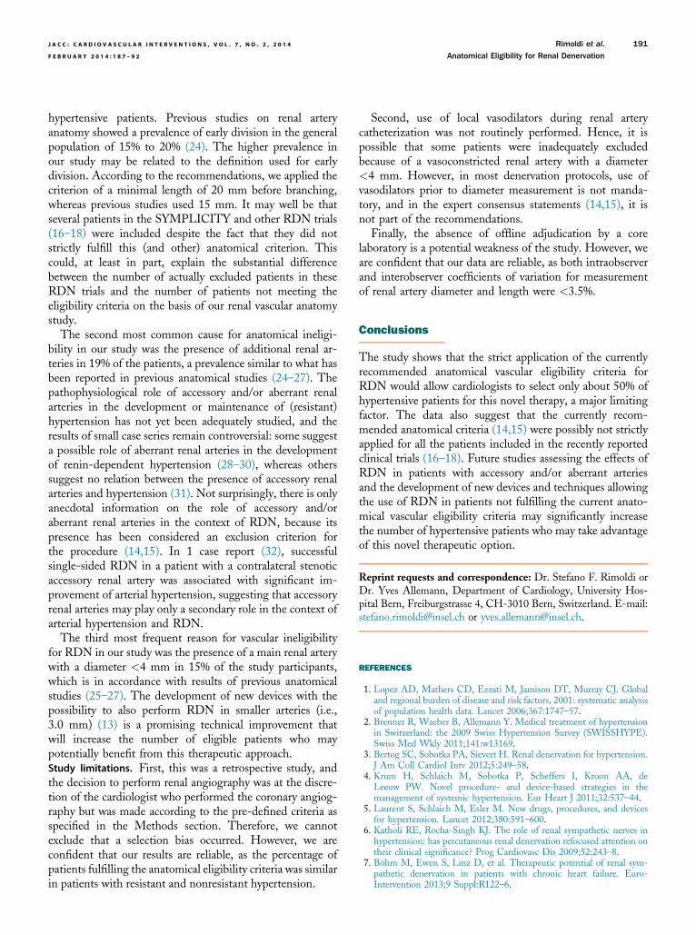
Figure 2. Example of Additional Renal Arteries
Accessory (hilar) renal artery (A) and aberrant (polar) renal artery (B).
Statistical analysis. Data were analyzed using GraphPadPrism version 5.0 (GraphPad Software Inc., La Jolla, Cali-fornia). Chi-square analysis was done for comparison of cat-egorical data between groups. Data are expressed as mean �SD. Statistical significance was defined at a p value of <0.05.
Results
The study included 941 patients (Fig. 1) of whom 934 hadgood quality images. Patient characteristics are summarizedin Table 2. The number and anatomy of the renal arteriesin the study population is summarized in Table 3. A single

Table 3. Number and Anatomy of the Renal Arteries in the StudyPopulation (N ¼ 934)
Number of Arteries Left Kidney Right Kidney Total*
Single artery 828 (88.7) 837 (89.6) 731 (78.3)
Diameter, mm 5.1 � 1.0 5.1 � 0.9
Double artery 103 (11.0) 94 (10.0) 197 (21.1)
Main artery þ accessory 53 47
Diameter accessory, mm 2.9 � 0.8 2.9 � 0.8
Main artery þ aberrant 50 47
Diameter aberrant, mm 2.9 (0.8) 2.9 (0.8)
Triple artery 3 (0.3) 3 (0.3) 6 (0.6)
Values are n (%), mean � SD, or n. *Total of the study population: single artery left and right:
731; double artery left or right: 197.
Aberrant ¼ polar; Accessory ¼ hilar.
Rimoldi et al. J A C C : C A R D I O V A S C U L A R I N T E R V E N T I O N S , V O L . 7 , N O . 2 , 2 0 1 4
Anatomical Eligibility for Renal Denervation F E B R U A R Y 2 0 1 4 : 1 8 7 – 9 2
190
renal artery was present in 731 patients (78%), and 197patients (21%) had a double artery. Among them were 100accessory (Fig. 2A) and 97 aberrant arteries (Fig. 2B). In6 cases, 3 arteries vascularizing 1 kidney were present.
The prevalence of angiographically significant RAS was7% (n ¼ 65), and 25 patients (3%) had a nonsignificantRAS. Of the remaining 844 patients without RAS, 727(86%) had nonresistant hypertension and 117 (14%) hadresistant hypertension (Fig. 1). Based on the eligibilitycriteria (Table 1), 62 (53%) of the resistant hypertensive and381 (52%) of the nonresistant hypertensive patients wouldhave qualified for sympathetic RDN (Fig. 1, Table 4).
The most frequent reason for anatomical ineligibility(in 24% of nonresistant and in 28% of resistant hypertensivepatients) was length of the main renal artery �20 mm (95%confidence limits: 3.6 to 18.2 mm in nonresistant and 3.0 to17.9 mm in resistant hypertensive patients). In 19% of thepatients, there was >1 main renal artery, and in 15% ofpatients, the diameter of the renal artery was <4 mm (95%confidence limits: 1.4 to 3.6 mm in nonresistant and 1.6 to3.8 mm in resistant hypertensive patients) (Table 4).
The mean diameter of the left and right main renal ar-teries was 5.1 � 1.0 and 5.1 � 0.9 mm, respectively. Themean diameter of the accessory or aberrant arteries was 2.9� 0.8 mm on both sides (Table 3).
Discussion
Catheter-based RDN is a novel, promising, minimally-invasive technique for the treatment of hypertension thatwas developed with the goal of achieving sustained BPreduction and control, particularly in patients with resistant
Table 2. Baseline Clinical Characteristics of the Study Population(N ¼ 934)
Age, yrs 65 � 10
Female 438 (47)
Systolic blood pressure, mm Hg 136 � 24
Diastolic blood pressure, mm Hg 72 � 12
Impaired renal function 144 (15)
Diabetes mellitus 182 (19)
Dyslipidemia 738 (80)
Current and former smoker 356 (38)
Obesity 182 (19)
Coronary artery disease 411 (44)
Peripheral vascular disease 75 (8)
Number of antihypertension medications 2.3 � 1.2
Beta-blockers 530 (57)
ACE inhibitor/ARB 578 (62)
Calcium-channel blockers 176 (19)
Diuretics 378 (40)
Values are mean � SD or n (%).
ACE ¼ angiotensin-converting enzyme; ARB ¼ angiotensin receptor blocker.
hypertension. To be selected for RDN according to currentrecommendations (14,15), patients should satisfy severalclinical criteria. Moreover, several anatomical characteristicsof the renal arterial vascularization need to be fulfilled andplay an important role in the selection process (Table 1).However, there are few detailed data on this issue. Previousstudies just reported “global” rates of excluded patientsowing to unfavorable vascular anatomical characteristics(16–18). Therefore, we studied in detail the anatomicalcharacteristics of the renal arteries of almost 1,000 consec-utive hypertensive patients undergoing coronary angiog-raphy and selective renal arteriography at our institutionand applied the anatomical vascular inclusion and exclusioncriteria used in previous RDN studies recommended by thecurrent guidelines (14,15).
The main finding of this study was that anatomicalvascular eligibility for RDN according to current recom-mendations (14,15) was met in 52% of patients withnonresistant and in 53% of patients with resistant hyper-tension. This figure is substantially lower than that reportedin previous studies (16–18), with approximately 10% to 37%of the patients being excluded due to vascular anatomicalcriteria.
In the present study, the most common cause foranatomical ineligibility was an early division of the mainrenal artery both in nonresistant (24%) and resistant (28%)
Table 4. Renal Anatomical Eligibility of Hypertensive Patients
Anatomy
NonresistantHypertensive(n ¼ 727)
ResistantHypertensive(n ¼ 117) p Value
Eligible/not eligible 381 (52)/346 (48) 62 (53)/55 (47) 0.92
Reason for noneligibility
More than 1 main artery 142 (19) 23 (19) 1.0
<4 mm diameter 109 (15) 17 (15) 1.0
<20 mm length 177 (24) 33 (28) 0.25
Values are n (%).
J A C C : C A R D I O V A S C U L A R I N T E R V E N T I O N S , V O L . 7 , N O . 2 , 2 0 1 4 Rimoldi et al.
F E B R U A R Y 2 0 1 4 : 1 8 7 – 9 2 Anatomical Eligibility for Renal Denervation
191
hypertensive patients. Previous studies on renal arteryanatomy showed a prevalence of early division in the generalpopulation of 15% to 20% (24). The higher prevalence inour study may be related to the definition used for earlydivision. According to the recommendations, we applied thecriterion of a minimal length of 20 mm before branching,whereas previous studies used 15 mm. It may well be thatseveral patients in the SYMPLICITY and other RDN trials(16–18) were included despite the fact that they did notstrictly fulfill this (and other) anatomical criterion. Thiscould, at least in part, explain the substantial differencebetween the number of actually excluded patients in theseRDN trials and the number of patients not meeting theeligibility criteria on the basis of our renal vascular anatomystudy.
The second most common cause for anatomical ineligi-bility in our study was the presence of additional renal ar-teries in 19% of the patients, a prevalence similar to what hasbeen reported in previous anatomical studies (24–27). Thepathophysiological role of accessory and/or aberrant renalarteries in the development or maintenance of (resistant)hypertension has not yet been adequately studied, and theresults of small case series remain controversial: some suggesta possible role of aberrant renal arteries in the developmentof renin-dependent hypertension (28–30), whereas otherssuggest no relation between the presence of accessory renalarteries and hypertension (31). Not surprisingly, there is onlyanecdotal information on the role of accessory and/oraberrant renal arteries in the context of RDN, because itspresence has been considered an exclusion criterion forthe procedure (14,15). In 1 case report (32), successfulsingle-sided RDN in a patient with a contralateral stenoticaccessory renal artery was associated with significant im-provement of arterial hypertension, suggesting that accessoryrenal arteries may play only a secondary role in the context ofarterial hypertension and RDN.
The third most frequent reason for vascular ineligibilityfor RDN in our study was the presence of a main renal arterywith a diameter <4 mm in 15% of the study participants,which is in accordance with results of previous anatomicalstudies (25–27). The development of new devices with thepossibility to also perform RDN in smaller arteries (i.e.,3.0 mm) (13) is a promising technical improvement thatwill increase the number of eligible patients who maypotentially benefit from this therapeutic approach.Study limitations. First, this was a retrospective study, andthe decision to perform renal angiography was at the discre-tion of the cardiologist who performed the coronary angiog-raphy but was made according to the pre-defined criteria asspecified in the Methods section. Therefore, we cannotexclude that a selection bias occurred. However, we areconfident that our results are reliable, as the percentage ofpatients fulfilling the anatomical eligibility criteria was similarin patients with resistant and nonresistant hypertension.
Second, use of local vasodilators during renal arterycatheterization was not routinely performed. Hence, it ispossible that some patients were inadequately excludedbecause of a vasoconstricted renal artery with a diameter<4 mm. However, in most denervation protocols, use ofvasodilators prior to diameter measurement is not manda-tory, and in the expert consensus statements (14,15), it isnot part of the recommendations.
Finally, the absence of offline adjudication by a corelaboratory is a potential weakness of the study. However, weare confident that our data are reliable, as both intraobserverand interobserver coefficients of variation for measurementof renal artery diameter and length were <3.5%.
Conclusions
The study shows that the strict application of the currentlyrecommended anatomical vascular eligibility criteria forRDN would allow cardiologists to select only about 50% ofhypertensive patients for this novel therapy, a major limitingfactor. The data also suggest that the currently recom-mended anatomical criteria (14,15) were possibly not strictlyapplied for all the patients included in the recently reportedclinical trials (16–18). Future studies assessing the effects ofRDN in patients with accessory and/or aberrant arteriesand the development of new devices and techniques allowingthe use of RDN in patients not fulfilling the current anato-mical vascular eligibility criteria may significantly increasethe number of hypertensive patients who may take advantageof this novel therapeutic option.
Reprint requests and correspondence: Dr. Stefano F. Rimoldi orDr. Yves Allemann, Department of Cardiology, University Hos-pital Bern, Freiburgstrasse 4, CH-3010 Bern, Switzerland. E-mail:[email protected] or [email protected].
REFERENCES
1. Lopez AD, Mathers CD, Ezzati M, Jamison DT, Murray CJ. Globaland regional burden of disease and risk factors, 2001: systematic analysisof population health data. Lancet 2006;367:1747–57.
2. Brenner R, Waeber B, Allemann Y. Medical treatment of hypertensionin Switzerland: the 2009 Swiss Hypertension Survey (SWISSHYPE).Swiss Med Wkly 2011;141:w13169.
3. Bertog SC, Sobotka PA, Sievert H. Renal denervation for hypertension.J Am Coll Cardiol Intv 2012;5:249–58.
4. Krum H, Schlaich M, Sobotka P, Scheffers I, Kroon AA, deLeeuw PW. Novel procedure- and device-based strategies in themanagement of systemic hypertension. Eur Heart J 2011;32:537–44.
5. Laurent S, Schlaich M, Esler M. New drugs, procedures, and devicesfor hypertension. Lancet 2012;380:591–600.
6. Katholi RE, Rocha-Singh KJ. The role of renal sympathetic nerves inhypertension: has percutaneous renal denervation refocused attention ontheir clinical significance? Prog Cardiovasc Dis 2009;52:243–8.
7. Böhm M, Ewen S, Linz D, et al. Therapeutic potential of renal sym-pathetic denervation in patients with chronic heart failure. Euro-Intervention 2013;9 Suppl:R122–6.

Rimoldi et al. J A C C : C A R D I O V A S C U L A R I N T E R V E N T I O N S , V O L . 7 , N O . 2 , 2 0 1 4
Anatomical Eligibility for Renal Denervation F E B R U A R Y 2 0 1 4 : 1 8 7 – 9 2
192
8. Mahfoud F, Schlaich M, Kindermann I, et al. Effect of renal sympa-thetic denervation on glucose metabolism in patients with resistanthypertension: a pilot study. Circulation 2011;123:1940–6.
9. Witkowski A, Prejbisz A, Florczak E, et al. Effects of renal sympatheticdenervation on blood pressure, sleep apnea course, and glycemic controlin patients with resistant hypertension and sleep apnea. Hypertension2011;58:559–65.
10. Converse RL Jr., Jacobsen TN, Toto RD, et al. Sympathetic overactivityin patients with chronic renal failure. N Engl J Med 1992;327:1912–8.
11. Morrissey DM, Brookes VS, Cooke WT. Sympathectomy in thetreatment of hypertension; review of 122 cases. Lancet 1953;1:403–8.
12. Allen TR. Current status of lumbar sympathectomy. Am Surg 1976;42:89–91.
13. Bunte MC, Infante de Oliveira E, Shishehbor MH. Endovasculartreatment of resistant and uncontrolled hypertension: therapies on thehorizon. J Am Coll Cardiol Intv 2013;6:1–9.
14. Mahfoud F, Luscher TF, Andersson B, et al. Expert consensus docu-ment from the European Society of Cardiology on catheter-based renaldenervation. Eur Heart J 2013;34:2149.
15. Schmieder RE, Redon J, Grassi G, et al. ESH position paper: renaldenervationdan interventional therapy of resistant hypertension.J Hypertens 2012;30:837–41.
16. Esler MD, Krum H, Sobotka PA, et al., for the Symplicity HTN-2Investigators. Renal sympathetic denervation in patients with treatment-resistant hypertension (The Symplicity HTN-2 Trial): a randomisedcontrolled trial. Lancet 2010;376:1903–9.
17. Krum H, Schlaich M, Whitbourn R, et al. Catheter-based renal sym-pathetic denervation for resistant hypertension: a multicentre safety andproof-of-principle cohort study. Lancet 2009;373:1275–81.
18. Savard S, Frank M, Bobrie G, Plouin PF, Sapoval M, Azizi M.Eligibility for renal denervation in patients with resistant hypertension:when enthusiasm meets reality in real-life patients. J Am Coll Cardiol2012;60:2422–4.
19. Mancia G, Fagard R, Narkiewicz K, et al. 2013 ESH/ESC guidelinesfor the management of arterial hypertension: the Task Force for theManagement of Arterial Hypertension of the European Society ofHypertension (ESH) and of the European Society of Cardiology (ESC).Eur Heart J 2013;34:2159–219.
20. Rimoldi SF, de Marchi SF, Windecker S, Meier B, Allemann Y.Screening renal artery angiography in hypertensive patients undergoingcoronary angiography and 6-month follow-up after ad hoc percutaneousrevascularization. J Hypertens 2010;28:842–7.
21. Levey AS, Bosch JP, Lewis JB, et al., for the Modification of Diet inRenal Disease Study Group. A more accurate method to estimateglomerular filtration rate from serum creatinine: a new predictionequation. Ann Intern Med 1999;130:461–70.
22. Windecker S, Remondino A, Eberli FR, et al. Sirolimus-eluting andpaclitaxel-eluting stents for coronary revascularization. N Engl J Med2005;353:653–62.
23. Togni M, Windecker S, Wenaweser P, et al. Deleterious effect ofcoronary brachytherapy on vasomotor response to exercise. Circulation2004;110:135–40.
24. Kadir S. Kidneys: Atlas of Normal and Varial Angiographic Anatomy.Philadelphia, PA: W. B. Saunders, 1991.
25. Khamanarong K, Prachaney P, Utraravichien A, Tong-Un T,Sripaoraya K. Anatomy of renal arterial supply. Clin Anat 2004;17:334–6.
26. Ozkan U, O�guzkurt L, Tercan F, Kizilkiliç O, Koç Z, Koca N. Renalartery origins and variations: angiographic evaluation of 855 consecutivepatients. Diagn Interv Radiol 2006;12:183–6.
27. Satyapal KS, Haffejee AA, Singh B, Ramsaroop L, Robbs JV,Kalideen JM. Additional renal arteries: incidence and morphometry.Surg Radiol Anat 2001;23:33–8.
28. Derrick JR, Tyson KR. The surgical significance of aberrant renalarteries in relation to systemic hypertension. Circulation 1961;24:1192–6.
29. Glodny B, Cromme S, Reimer P, Lennarz M, Winde G, Vetter H.Hypertension associated with multiple renal arteries may be renin-dependent. J Hypertens 2000;18:1437–44.
30. Kem DC, Lyons DF, Wenzl J, Halverstadt D, Yu X. Renin-dependenthypertension caused by nonfocal stenotic aberrant renal arteries: proof ofa new syndrome. Hypertension 2005;46:380–5.
31. Gupta A, Tello R. Accessory renal arteries are not related to hyper-tension risk: a review of MR angiography data. Am J Roentgenol 2004;182:1521–4.
32. Himmel F, Bode F, Mortensen K, et al. Successful single-sided renaldenervation approach in a patient with stenosis of an accessory renalartery. J Clin Hypertens (Greenwich) 2012;14:187–8.
Key Words: arterial hypertension - anatomical eligi-bility - renal vascular anatomy - sympathetic renaldenervation.