an important adjunct in airway assessment of adult ... - mdpi
TRANSCRIPT
Journal of
Clinical Medicine
Article
Hyo-Mental Angle and Distance: An Important Adjunct inAirway Assessment of Adult Mucopolysaccharidosis
Chaitanya Gadepalli 1,* , Karolina M. Stepien 2 and Govind Tol 3
�����������������
Citation: Gadepalli, C.; Stepien,
K.M.; Tol, G. Hyo-Mental Angle and
Distance: An Important Adjunct in
Airway Assessment of Adult
Mucopolysaccharidosis. J. Clin. Med.
2021, 10, 4924. https://doi.org/
10.3390/jcm10214924
Academic Editor: César Picado
Received: 2 October 2021
Accepted: 21 October 2021
Published: 25 October 2021
Publisher’s Note: MDPI stays neutral
with regard to jurisdictional claims in
published maps and institutional affil-
iations.
Copyright: © 2021 by the authors.
Licensee MDPI, Basel, Switzerland.
This article is an open access article
distributed under the terms and
conditions of the Creative Commons
Attribution (CC BY) license (https://
creativecommons.org/licenses/by/
4.0/).
1 Ear Nose and Throat Department, Salford Royal NHS Foundation Trust, Manchester M6 8HD, UK2 Adult Inherited Metabolic Department, Salford Royal NHS Foundation Trust, Manchester M6 8HD, UK;
[email protected] Anaesthetics Department, Salford Royal NHS Foundation Trust, Manchester M6 8HD, UK;
[email protected]* Correspondence: [email protected]
Abstract: Background: Mucopolysaccharidosis (MPS) is a rare congenital lysosomal storage dis-order with complex airways. High anterior larynx is assessed by thyromental distance (TMD)nasendoscopy. A simpler method to assess this hyoid bone is described. The distance between thecentral-hyoid and symphysis of the mandible (hyo-mental distance; HMD) and inclination of thisline to the horizontal axis (hyo-mental angle; HMA) in neutrally positioned patients is investigated.Methods: HMA, HMD in MPS, and non-MPS were compared, and their correlation with heightand weight were assessed. Results: 50 adult MPS patients (M = 32, F = 18, age range = 19–66 years;mean BMI = 26.8 kg/m2) of MPS I, II, III, IV, and VI were compared with 50 non-MPS (M = 25, F = 25;age range = 22–84 years; mean BMI = 26.5 kg/m2). Mean HMA in MPS was 25.72◦ (−10 to +50)versus 2.42◦ (−35 to +28) in non-MPS. Mean HMD was 46.5 (25.7–66) millimeters in MPS versus41.8 (27–60.3) in non-MPS. HMA versus height and weight showed a moderate correlation (r = −0.4,p < 0.05) in MPS and no significant correlation (r < 0.4, p > 0.05) in non-MPS. HMD versus height andweight showed no correlation (r < 0.4, p > 0.05) in both groups. Conclusions: HMA seems more acutein MPS despite nearly the same HMD as non-MPS, signifying a high larynx, which may be missedby TMD.
Keywords: mucopolysaccharidosis; airway management; radiology; hyoid bone; chin;intubation; intratracheal
1. Introduction
Mucopolysaccharidosis are a group of inherited congenital multisystem diseases dueto a deficiency in enzymes required for the breakdown of complex mucopolysaccharides.Mucopolysaccharidoses (MPSs) are rare, inherited, lysosomal storage diseases with acombined incidence of 1 in 22,000 [1]. The disease is characterized by the accumulationof glycosaminoglycans (GAGs) in almost all parts of the body. There are seven types ofMPS depending on the type of enzyme deficiency (Table 1). The manifestations of thisdisease are multisystemic, resulting in shortened longevity [2]. Along with other systems,airways are commonly involved. Knowledge about the airway abnormalities is importantas these patients can pose airway problems. With advances in treatment modalities, such ashematopoietic stem cell transplantation (HSCT) [3] and enzyme replacement therapy(ERT) [4], the longevity of these patients has increased posing newer problems. Most ofthese patients will need general anesthetic for surgery at some point in their lifetime due tomulti-system involvement. In our experience, we noted that most adult MPS patients havea large and bulky tongue, large lower jaw, and short neck [5]. A high or anterior larynxposes difficulty in access to the airway. A difficult laryngoscopy is defined as an inabilityto visualize any part of the vocal cords on conventional laryngoscopy [6]. Various bedsidemeasures have been described to assess a high or anterior larynx. The method commonly
J. Clin. Med. 2021, 10, 4924. https://doi.org/10.3390/jcm10214924 https://www.mdpi.com/journal/jcm
J. Clin. Med. 2021, 10, 4924 2 of 12
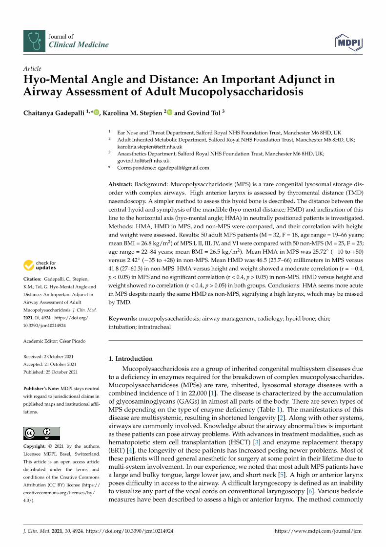
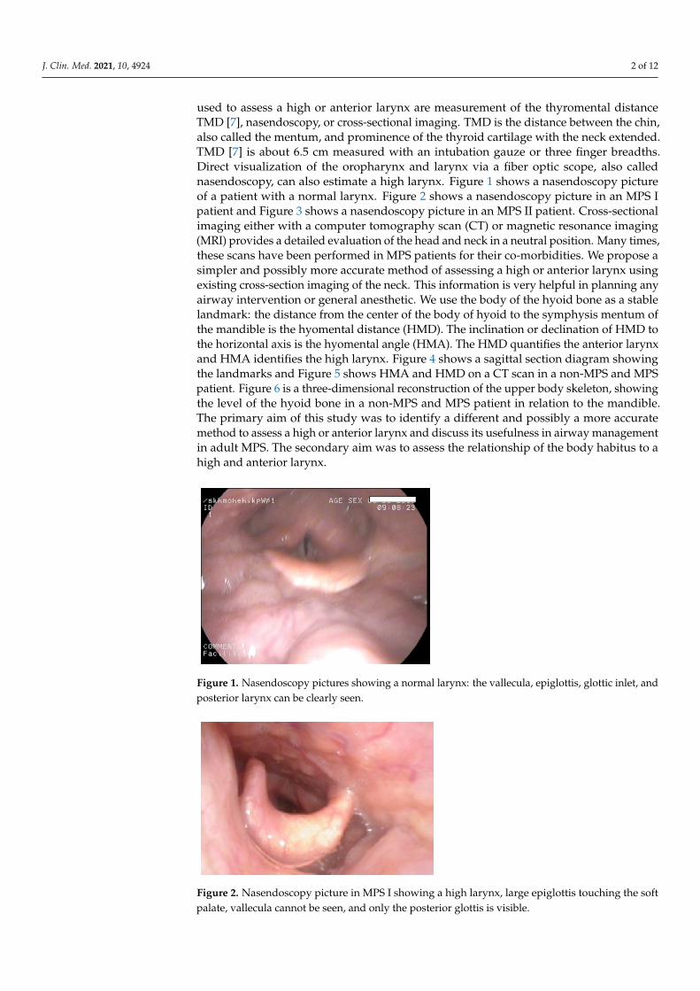
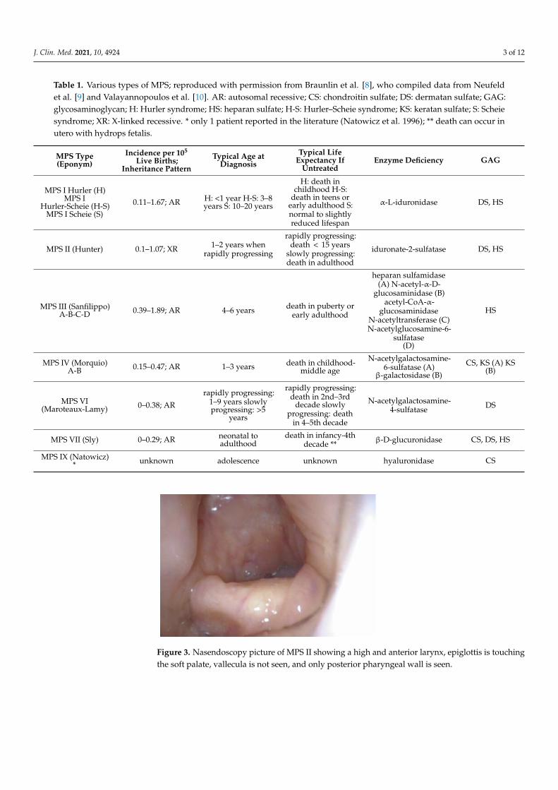
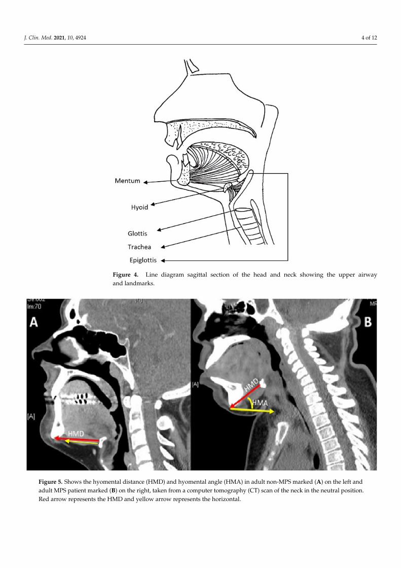
used to assess a high or anterior larynx are measurement of the thyromental distanceTMD [7], nasendoscopy, or cross-sectional imaging. TMD is the distance between the chin,also called the mentum, and prominence of the thyroid cartilage with the neck extended.TMD [7] is about 6.5 cm measured with an intubation gauze or three finger breadths.Direct visualization of the oropharynx and larynx via a fiber optic scope, also callednasendoscopy, can also estimate a high larynx. Figure 1 shows a nasendoscopy pictureof a patient with a normal larynx. Figure 2 shows a nasendoscopy picture in an MPS Ipatient and Figure 3 shows a nasendoscopy picture in an MPS II patient. Cross-sectionalimaging either with a computer tomography scan (CT) or magnetic resonance imaging(MRI) provides a detailed evaluation of the head and neck in a neutral position. Many times,these scans have been performed in MPS patients for their co-morbidities. We propose asimpler and possibly more accurate method of assessing a high or anterior larynx usingexisting cross-section imaging of the neck. This information is very helpful in planning anyairway intervention or general anesthetic. We use the body of the hyoid bone as a stablelandmark: the distance from the center of the body of hyoid to the symphysis mentum ofthe mandible is the hyomental distance (HMD). The inclination or declination of HMD tothe horizontal axis is the hyomental angle (HMA). The HMD quantifies the anterior larynxand HMA identifies the high larynx. Figure 4 shows a sagittal section diagram showingthe landmarks and Figure 5 shows HMA and HMD on a CT scan in a non-MPS and MPSpatient. Figure 6 is a three-dimensional reconstruction of the upper body skeleton, showingthe level of the hyoid bone in a non-MPS and MPS patient in relation to the mandible.The primary aim of this study was to identify a different and possibly a more accuratemethod to assess a high or anterior larynx and discuss its usefulness in airway managementin adult MPS. The secondary aim was to assess the relationship of the body habitus to ahigh and anterior larynx.
J. Clin. Med. 2021, 10, x FOR PEER REVIEW 3 of 13
MPS IV (Morquio) A-B 0.15–0.47; AR 1–3 years
death in childhood- middle age
N-acetylgalactosamine-6-sulfatase (A) β-galacto-
sidase (B)
CS, KS (A) KS (B)
MPS VI (Maroteaux-Lamy)
0–0.38; AR
rapidly progress-ing: 1–9 years
slowly progress-ing: >5 years
rapidly progressing: death in 2nd–3rd
decade slowly pro-gressing: death in 4–
5th decade
N-acetylgalactosamine-4-sulfatase
DS
MPS VII (Sly) 0–0.29; AR neonatal to adult-hood
death in infancy-4th decade **
β-D-glucuronidase CS, DS, HS
MPS IX (Natowicz) * unknown adolescence unknown hyaluronidase CS
Figure 1. Nasendoscopy pictures showing a normal larynx: the vallecula, epiglottis, glottic inlet, and posterior larynx can be clearly seen.
Figure 2. Nasendoscopy picture in MPS I showing a high larynx, large epiglottis touching the soft palate, vallecula cannot be seen, and only the posterior glottis is visible.
Figure 1. Nasendoscopy pictures showing a normal larynx: the vallecula, epiglottis, glottic inlet, andposterior larynx can be clearly seen.
J. Clin. Med. 2021, 10, x FOR PEER REVIEW 3 of 13
MPS IV (Morquio) A-B 0.15–0.47; AR 1–3 years
death in childhood- middle age
N-acetylgalactosamine-6-sulfatase (A) β-galacto-
sidase (B)
CS, KS (A) KS (B)
MPS VI (Maroteaux-Lamy)
0–0.38; AR
rapidly progress-ing: 1–9 years
slowly progress-ing: >5 years
rapidly progressing: death in 2nd–3rd
decade slowly pro-gressing: death in 4–
5th decade
N-acetylgalactosamine-4-sulfatase
DS
MPS VII (Sly) 0–0.29; AR neonatal to adult-hood
death in infancy-4th decade **
β-D-glucuronidase CS, DS, HS
MPS IX (Natowicz) * unknown adolescence unknown hyaluronidase CS
Figure 1. Nasendoscopy pictures showing a normal larynx: the vallecula, epiglottis, glottic inlet, and posterior larynx can be clearly seen.
Figure 2. Nasendoscopy picture in MPS I showing a high larynx, large epiglottis touching the soft palate, vallecula cannot be seen, and only the posterior glottis is visible.
Figure 2. Nasendoscopy picture in MPS I showing a high larynx, large epiglottis touching the softpalate, vallecula cannot be seen, and only the posterior glottis is visible.
J. Clin. Med. 2021, 10, 4924 3 of 12
Table 1. Various types of MPS; reproduced with permission from Braunlin et al. [8], who compiled data from Neufeldet al. [9] and Valayannopoulos et al. [10]. AR: autosomal recessive; CS: chondroitin sulfate; DS: dermatan sulfate; GAG:glycosaminoglycan; H: Hurler syndrome; HS: heparan sulfate; H-S: Hurler–Scheie syndrome; KS: keratan sulfate; S: Scheiesyndrome; XR: X-linked recessive. * only 1 patient reported in the literature (Natowicz et al. 1996); ** death can occur inutero with hydrops fetalis.
MPS Type(Eponym)
Incidence per 105
Live Births;Inheritance Pattern
Typical Age atDiagnosis
Typical LifeExpectancy If
UntreatedEnzyme Deficiency GAG
MPS I Hurler (H)MPS I
Hurler-Scheie (H-S)MPS I Scheie (S)
0.11–1.67; AR H: <1 year H-S: 3–8years S: 10–20 years
H: death inchildhood H-S:
death in teens orearly adulthood S:normal to slightlyreduced lifespan
α-L-iduronidase DS, HS
MPS II (Hunter) 0.1–1.07; XR 1–2 years whenrapidly progressing
rapidly progressing:death < 15 years
slowly progressing:death in adulthood
iduronate-2-sulfatase DS, HS
MPS III (Sanfilippo)A-B-C-D 0.39–1.89; AR 4–6 years death in puberty or
early adulthood
heparan sulfamidase(A) N-acetyl-α-D-
glucosaminidase (B)acetyl-CoA-α-
glucosaminidaseN-acetyltransferase (C)N-acetylglucosamine-6-
sulfatase(D)
HS
MPS IV (Morquio)A-B 0.15–0.47; AR 1–3 years death in childhood-
middle age
N-acetylgalactosamine-6-sulfatase (A)
β-galactosidase (B)
CS, KS (A) KS(B)
MPS VI(Maroteaux-Lamy) 0–0.38; AR
rapidly progressing:1–9 years slowlyprogressing: >5
years
rapidly progressing:death in 2nd–3rd
decade slowlyprogressing: death
in 4–5th decade
N-acetylgalactosamine-4-sulfatase DS
MPS VII (Sly) 0–0.29; AR neonatal toadulthood
death in infancy-4thdecade ** β-D-glucuronidase CS, DS, HS
MPS IX (Natowicz)* unknown adolescence unknown hyaluronidase CSJ. Clin. Med. 2021, 10, x FOR PEER REVIEW 4 of 13
Figure 3. Nasendoscopy picture of MPS II showing a high and anterior larynx, epiglottis is touching the soft palate, vallecula is not seen, and only posterior pharyngeal wall is seen.
Figure 4. Line diagram sagittal section of the head and neck showing the upper airway and land-marks.
Figure 3. Nasendoscopy picture of MPS II showing a high and anterior larynx, epiglottis is touchingthe soft palate, vallecula is not seen, and only posterior pharyngeal wall is seen.
J. Clin. Med. 2021, 10, 4924 4 of 12
J. Clin. Med. 2021, 10, x FOR PEER REVIEW 4 of 13
Figure 3. Nasendoscopy picture of MPS II showing a high and anterior larynx, epiglottis is touching the soft palate, vallecula is not seen, and only posterior pharyngeal wall is seen.
Figure 4. Line diagram sagittal section of the head and neck showing the upper airway and land-marks. Figure 4. Line diagram sagittal section of the head and neck showing the upper airwayand landmarks.
J. Clin. Med. 2021, 10, x FOR PEER REVIEW 5 of 13
Figure 5. Shows the hyomental distance (HMD) and hyomental angle (HMA) in adult non-MPS marked (A) on the left and adult MPS patient marked (B) on the right, taken from a computer tomography (CT) scan of the neck in the neutral position. Red arrow represents the HMD and yellow arrow represents the horizontal.
Figure 5. Shows the hyomental distance (HMD) and hyomental angle (HMA) in adult non-MPS marked (A) on the left andadult MPS patient marked (B) on the right, taken from a computer tomography (CT) scan of the neck in the neutral position.Red arrow represents the HMD and yellow arrow represents the horizontal.
J. Clin. Med. 2021, 10, 4924 5 of 12J. Clin. Med. 2021, 10, x FOR PEER REVIEW 6 of 13
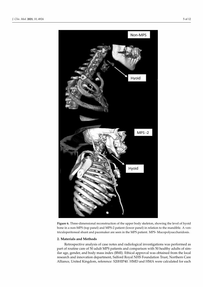
Figure 6. Three-dimensional reconstruction of the upper body skeleton, showing the level of hyoid bone in a non-MPS (top panel) and MPS-2 patient (lower panel) in relation to the mandible. A ven-triculoperitoneal shunt and pacemaker are seen in the MPS patient. MPS- Mucopolysaccharidosis.
Figure 6. Three-dimensional reconstruction of the upper body skeleton, showing the level of hyoidbone in a non-MPS (top panel) and MPS-2 patient (lower panel) in relation to the mandible. A ven-triculoperitoneal shunt and pacemaker are seen in the MPS patient. MPS- Mucopolysaccharidosis.
2. Materials and Methods
Retrospective analysis of case notes and radiological investigations was performed aspart of routine care of 50 adult MPS patients and comparison with 50 healthy adults of sim-ilar age, gender, and body mass index (BMI). Ethical approval was obtained from the localresearch and innovation department, Salford Royal NHS Foundation Trust, Northern CareAlliance, United Kingdom, reference: S20HIP40. HMD and HMA were calculated for each
J. Clin. Med. 2021, 10, 4924 6 of 12
group calculated in the picture archiving communications system (PACS) using the rulertool. A smaller HMD was considered to be an anterior larynx and acute inclination of theHMA to the horizontal axis was considered to represent a high larynx. Both HMD andHMA reflect the difficulty in accessing an airway. The impact of body habitus on a high oranterior larynx was investigated by calculating the Pearsons correlation between HMD,HMD, and weight and height.
3. Results
Radiological cross-sectional images of 50 MPS and 50 non-MPS patients were includedin the study. The MPS group included patients with types I, II, III, IV, and VI. Table 2depicts the demographics in both groups. It may be noted that even though age doesnot match between both groups, BMI is comparable. The non-MPS group of patientsincluded a range of patients of various ENT (ear, nose, and throat) pathologies who hadimaging studies. All the patients in the non-MPS group had no pathology in the oral cavity,neck, oropharynx, supraglottis, and hypopharynx. The non-MPS group had a normalsupra-glottic airway. This enabled us to investigate the abnormal hyomental region in MPSpatients. Table 3 depicts the pathology subtypes in both groups. The HMA and HMD werecalculated and compared between the two groups.
Table 2. Demographics of the study in both the MPS and non-MPS groups.
MPS Non-MPS
Number of patients 50 50Males 32 25
Females 18 25Age range in years 19–66 22–84Mean age in years 31.7 59.9
Mean Body Mass index 26.8 26.5MPS—Mucopolysaccharidosis.
Table 3. Clinical diagnosis of different patients in the MPS and non-MPS groups. MPS—Mucopolysaccharidosis.
Pathology Number Males Females
MPS groupMPSI 16 8 8MPSII 13 13 0MPSIII 1 1 0MPSIV 14 6 8MPSVI 6 4 2Total 50 32 18
Non-MPS groupSubglottic stenosis 10 1 9Tracheal stenosis 8 5 3
Vasculitis 7 4 3Malignancy not involving supraglottis, oropharynx 16 12 4
Bilateral vocal fold immobility 5 0 5Vocal cord leukoplakia 4 3 1
Total 50 25 25MPS—Mucopolysaccharidosis.
Table 4 depicts HMA and HMD in both MPS and non-MPS groups. It may be notedthat the HMD is slightly less in the MPS depicting slightly anterior larynx in the MPS group.The HMA is more acute in the MPS group compared to the non-MPS group, depicting thatlarynx is higher in the MPS groups. The MPS group have a shorter stature and lower bodymass, this is a recognized feature of MPS due to multisystemic involvement of the disease.
J. Clin. Med. 2021, 10, 4924 7 of 12
Table 4. HMD and HMA in the MPS and non-MPS groups.
MPS
Age inYears
HMD inMillime-
ters
HMA inDegrees
Height inCentime-
ters
Weight inKilograms
BodyMassIndex
N = 50Mean 31.74 46.5 27.6 135.8 50.47 26.8
Median 29.50 48.5 25.0 136.8 48 25.7Range 19–66 25.7–66.0 −10.0 to 50 91.0–182 17.4–125.2 16.5–43.6
Non-MPS
Age inYears
HMD inMillime-
ters
HMA inDegrees
Height inCentime-
ters
Weight inKilograms
BodyMassIndex
N = 50Mean 59.9 41.9 2.420 166.1 72.6 26.5
Median 63 40.9 0.0 166.0 71.9 26.9Range 22–99 27.0–60.3 −35.0 to 28 150.0–188 39.3–130 14.2–47.3
HMD—Hyomental distance, HMA—Hyomental angle. MPS—Mucopolysaccharidosis.
It can be assumed that a bulky upper airway may be attributable to BMI, thereby affect-ing HMA or HMD. To test this hypothesis, the Pearson correlation between the BMI versusHMA and HMD was calculated and Table 5 depicts the results. HMD shows no correlationwith height, weight, and BMI in the non-MPS group but reveals a statistically significantcorrelation with height and weight in MPS (p = 0.05; p = 0.009). It must be noted that therho value in the MPS group is only 0.3 at best. The Pearson correlation between HMAversus height and weight showed a moderate negative correlation in the MPS group andno correlation in the non-MPS group. Thus, HMA shows a better correlation with heightand weight in the MPS group, compared to HMD. The significant results are highlighted inthe table.
Table 5. Pearson correlation between HMA, HMD, height, weight, and BMI in both the MPS andnon-MPS groups.
Correlations MPS
HMD HMA HT WT BMI
HMDrho 1 −0.2 0.28 * 0.3 * 0.2
p-value 0.15 0.05 * 0.009 * 0.13
HMArho −0.2 1 −0.45 * −0.41 * −0.1
p-value 0.146 0.0001 * 0.003 * 0.73
Correlations Non-MPS
HMD HMA HT WT BMI
HMDrho 1 0.15 0.1 0.35 0.27
p-value 0.3 0.49 0.014 0.06
HMArho 0.15 1 −0.03 −0.03 0.02
p-value 0.296 0.85 0.862 0.91* Represents significant results. HMA—Hyomental angle, HMD—Hyomental distance, HT—height, WT—weight,BMI—Body Mass Index, rho—Pearsons correlation coefficient value.
4. Discussion
4.1. Difficult Airway
Airway complications are a common feature of MPS I, II, IV, and VI and considerablycontribute to morbidity and premature mortality [11,12]. Airway assessment is ideallyperformed holistically, taking into account all the factors in the upper and lower airways
J. Clin. Med. 2021, 10, 4924 8 of 12
with various methods, including medical history, clinical examination, radiological eval-uation, and endoscopy [13]. A high and anterior larynx is one of the important aspectsin the upper airway, which can lead to difficult intubation due to poor access, also calleddifficult laryngoscopy. Failure to recognize airway problems pre-operatively or during theplanning of airway intervention can lead to unfavorable outcomes. MPS is a rare disease,and awareness amongst health professionals regarding adult MPS patients is poor. Once apatient is paralyzed and anaesthetized, the tongue falls backwards, and the oropharynxcollapses inwards. In this situation, a high or an anterior larynx makes access to the larynxdifficult if not impossible. In a patient who is paralyzed and anaesthetized, this can leadto situation of “cannot intubate- cannot ventilate”. The Difficult Airway Society (DAS;UK) has produced guidelines on this difficult situation [14]. This difficult situation canbe prevented by recognition of the problem of difficult access by existing cross-sectionimages. Metanalysis of 35 studies representing 50,760 patients revealed the incidence ofdifficult intubation is about 5.8 % in normal patients, 3.1% in obstetric patients, and 14.8in obese patients [15]. This may be higher in MPS due to deposition of GAGs in the softtissues and musculoskeletal system, leading to bulky upper airways and bony abnormali-ties. A combination of abnormalities in the soft tissue, cervical spine, and skull leads to ahigh larynx and anterior larynx, resulting in difficulty accessing the airway. In our study,an attempt was made to match the MPS and non-MPS groups. It is not possible to obtainan exact match as age-related changes are faster in the MPS group with a shortened lifespan. Most of the MPS patients have short stature; however, it can be noted that both MPSand non-MPS groups have nearly similar BMI. We must also understand that MPS patientshave a short stature and have truncal obesity [16], and non-MPS patients in our group aretaller. Hence, BMI may be a misleading airway health measure in MPS patients. The PACShas in-built tools to measure the distances and angles in the cross-sectional images. As thismeasurement tool is computerized, we can assume that inter or intra-rater variability isreduced. Bias may be observed if the landmarks are not correctly identified by the clinician.In situations where the clinician does not have access to PACS, an angle measure and aruler can be used to obtain HMD or HMA on existing cross-sectional imaging. In our study,we note that HMD was slightly less in the MPS group, indicating that the larynx is mildlyanterior. The overall difference, however, in HMD in the MPS and non-MPS groups isminimal. On the other hand, HMA was more acute in the MPS group, indicating that thelarynx is higher in the MPS group. It is interesting to note that HMA can vary in the MPSgroup despite nearly the same HMD as non-MPS.
Moreover, it was observed that some MPS patients did not have acute HMA andnearly the same HMD as non-MPS. The reasons for this could be multifactorial. Firstly,16 results from MPS type I had milder upper airway abnormalities; secondly, the severity ofMPS is dependent on mutations, the length, or their therapies, such as enzyme replacementtherapy (ERT) or hematopoietic stem cell transplantation (HSCT). ERT in MPS I Hurler–Scheie (HS) and Scheie, II, IVA, and VI and HSCT in MPS I Hurler (H) have demonstratedorgan-specific and systemic metabolic correction [17–20]; hence, the severity of the diseaseis variable. Advances in treatment strategies have improved life expectancy, and theaverage age of our cohort was 31.7 years. So, our study may be representative of variedMPS phenotypes and younger adult MPS patients.
4.2. Clinical Measures of Difficult Airway
TMD is a commonly used tool in airway assessment. The sensitivity of TMD is about25% (95% confidence interval: 23–28) and specificity is 90.2% (95% confidence interval: 90–91) [21]. In MPS patients, the lower jaw may be disproportionately large, resulting in a normalthyromental distance despite a high and anterior larynx. TMD may also be of limited use inpatients with facial and skeletal dysmorphism, and bulky soft tissues of the neck and submental region as commonly noted in adult MPS. The sternomental distance (SMD) [22] iscalculated by measuring the distance between the mentum and the manubrium sternum withthe mouth closed. It may indicate the degree of neck extension, which is important in access
J. Clin. Med. 2021, 10, 4924 9 of 12
to the airway. The authors [22] conclude that a sternomental distance of 13.5 cm or less was66.7% sensitive and 71.1% specific, and the positive and negative predictive values were 7.6%and 98.4%, for difficult laryngoscopy [22]. In their study, there was no association betweensternomental distance and age, weight, height, or BMI. Sternomental distance may indirectlyreflect a high larynx, and further research can be done to invesitgate this association. The TMDand SMD may be normal in MPS patients due to a large lower jaw, giving a false sense ofsecurity of a normal airway. The distance between the hyoid and thyroid prominence maynot be as variable, so HMD and TMD may represent the same measure. HMD may be moreaccurate as it does not take into account the sub cutaneous soft tissue of the neck. In our study,we observed that the HMD is nearly the same in the MPS and non-MPS groups but the HMAwas more acute. This may indicate that HMA is a more accurate measure of a high or anteriorlarynx than HMD or TMD. The results of this study showed that there is no correlationbetween the HMA and HMD in both the MPS and non-MPS groups, suggesting that HMAmay be a completely independent entity, not related to the distance between the mentum tothe laryngeal framework. The HMA correlates negatively with height and weight but has nocorrelation with BMI. This is because correlation is a linear measure and BMI is weight dividedby height squared. Hence, observing the weight and height independently may be more usefulthan BMI in airway assessments. This may be more relevant to the MPS population as they areknown to have short stature and central obesity [23,24]. The other commonly used bedsideairway assessments methods are neck movements, neck circumference, Wilson’s score [25],mallampati [26], and modified mallampati grade [27]. Mallampati and modified mallampatigrade assess the size of the tongue in relation to the opened oral cavity. Dalewski [28] et al., in astudy of 129 adults, suggested the combination of Mallampati grade, CT scan upper airwayvolume, and Berlin score to calculate snoring and breathlessness. The authors noted a positivecorrelation between high modified mallampati grade, BMI, and reduced oxygen saturationsand upper airway volume. The pre-operative assessment aims to assess the difficult airwayand plan a difficult situation. Based on laryngoscopy views, Cormack [29] graded the airwayinto three grades: grade 1 being full view of the glottis, grade 2—partial view of the glottis,grade 3—only epiglottis is visible, and grade 4—neither epiglottis nor glottis are visible.Modifications [25,30] of grade 2 resulted in 2a being part of the glottis visible and 2b beingarytenoids or posterior cords only just visible. Cook [31] suggested the grading system as“E” as easy view—grade 1 and 2a “R” restricted view—grade 2b and 3a and “D” difficultview—grade 3b and 4, where the grade being epiglottis can be elevated with a gum elasticbougie and 3b being epiglottis cannot be elevated by a gum elastic bougie. Knowledge of agrade R or D prior to intubation in the pre-assessment clinics is very useful; HMD and HMAwill provide this information. Radiology plays an important role in airway assessment foran anesthetist [32]. Although lateral radiographs, chest X-rays, and ultrasonography [33,34]are useful, in our experience, we found that the use of MRI scans is helpful in upper airwaysand CT scans in lower airways. The images from CT and MRI scans can be used to performthree-dimensional reconstruction of the airways in MPS [35] and perform virtual endoscopy.We also found nasendoscopy to be very useful in adult MPS [36]. Imaging of the airway isnot routinely performed for unsuspected airway problems. Any additional investigationsshould be carefully considered, keeping patient comfort in mind. Although HMD has beenreported [37] as a predictor of difficult airway in patients with cervical spondylosis, HMA,to the best of our knowledge, has not been reported so far. We feel that this easily available toolis an adjunct to airway assessment and can be adopted in adult MPS and any other difficultairway situation. In our personal experience of adult airway assessment, we feel HMA closeto zero or less than zero indicates a larynx that is not high. HMA could be considered asanother important adjunct in upper airway assessment; however, holistic airway assessmentshould include both upper and lower airways.
J. Clin. Med. 2021, 10, 4924 10 of 12
4.3. Limitations of the Study
4.3.1. Head and Neck Position
Most of our MPS patients had cervical spine issues so we chose to take radiologicalimages at the neutral position of the head, keeping patient comfort as the priority. To keepthe upper airway open, the natural instinct of any patient is to adopt a sniffing posture.In all our MPS patients, some form of airway and cervical spine abnormality was noted,which may have prompted patients to adopt a comfortable posture. These factors couldhave skewed some of our measurements. It may be argued that HMA, which is acute inthe neutral comfortable position in MPS compared to non-MPS, may be more acute ina standard position of the head and neck. Future studies could include standardizationof head positions to obtain radiological images to obtain accurate measures of the HMAand HMD to test this hypothesis. Extension of the neck will improve laryngoscopic views.Future studies could also incorporate measurement of HMA and HMD in maximumextension and comparison with HMA and HMD in the neutral position to assess the degreeof improvement of laryngoscopy views by neck extension.
4.3.2. Thyro-Hyoid Distance
We made the assumption that the distance between the hyoid and thyroid is smallenough to assume that HMD and TMD reflect the same measure of a high or anteriorlarynx. Future studies could also assess the thyrohyoid distance in flexion and extension ofthe neck to test this hypothesis.
4.3.3. Numbers
Our cohort examined only 50 adult MPS patients of various types and varying severity,which was compared with 50 adults with no upper airway issues. Considering the rarityof the disease, this may appear a significant number; however, a larger study group couldhave produced more significant results. Future studies may incorporate larger number ofpatients in both pediatric and adult MPS by a multi-center collaboration.
4.4. Wider Implications
HMD and HMA application can be extended to wider use of difficult airway assess-ment in any patient due to its simplicity in use. This may play a special role in those withcervical spine or any craniofacial anomalies.
5. Conclusions
HMA and HMD are useful measurements that can be obtained from existing cross-section imaging, providing important information about an anterior or high larynx. This isvery helpful in pre-planning during airway assessment as part of the pre-operative work-up. HMA may be a better indicator than HMD in MPS patients. The use of HMA and HMDcan be extrapolated to airway assessments in other patients with or without head and neckdysmorphism. This simple airway assessment tool is a useful adjunct in the managementof complex airways, such as adult MPS. Further investigation into the sensitivity andspecificity of HMA and HMA with a standardized head position and its correlation withdifficult intubation will be useful.
Author Contributions: Authors C.G. (Otolaryngologist), K.M.S. (Adult inherited Metabolic Medicine),G.T. (Anesthetist) come from different specialties with common interest in adult Mucopolysacchari-dosis (MPS). C.G., G.T. have special interest with airway diseases came up with the conceptualizationof this unique idea airway assessment in MPS. The methodology of the study was devised by C.G.and K.M.S. Formal analysis of the data was performed by C.G., K.M.S. The resources, data curationwas performed by C.G., K.M.S., G.T.; writing original draft preparation by C.G.; writing—reviewand editing was performed by C.G., K.M.S., G.T. Visualization of the project was planned by C.G.,K.M.S. The project was supervised by C.G. All authors contributed towards project administration.All authors have read and agreed to the published version of the manuscript.

J. Clin. Med. 2021, 10, 4924 11 of 12
Funding: This research received no external funding, the article processing fee was supported byBioMarin Pharmaceutical Inc.
Institutional Review Board Statement: The study was conducted according to the guidelines ofthe Declaration of Helsinki, and Ethical approval from the local research and development de-partment form Salford Royal NHS Foundation trust, Manchester, UK was obtained, reference:S20HIP40. The study did not involve any animals. This study was retrospective case notes review ofadult patients.
Informed Consent Statement: Informed consent was obtained from all subjects involved in thestudy. However, no personal identifiable information has been used in this study.
Data Availability Statement: All the data required to understand and the data supporting reportedresults this project has been provided in the paper.
Acknowledgments: We would like to thank Amit Herwadkar, Consultant Radiologist, Salford RoyalNHS foundation Trust, Manchester, UK, for his expertise in this project.
Conflicts of Interest: The authors declare no conflict of interest.
References1. Mehta, A.B.; Winchester, B. Lysosomal Storage Disorders: A Practical Guide; Wiley-Blackwell Chichester: Hoboken, NJ, USA, 2012.2. Mucopolysaccharidoses Fact Sheet. Available online: https://www.ninds.nih.gov/Disorders/Patient-Caregiver-Education/Fact-
Sheets/Mucopolysaccharidoses-Fact-Sheet (accessed on 31 July 2020).3. Taylor, M.; Khan, S.; Stapleton, M.; Wang, J.; Chen, J.; Wynn, R.; Yabe, H.; Chinen, Y.; Boelens, J.J.; Mason, R.W. Hematopoietic
stem cell transplantation for mucopolysaccharidoses: Past, present, and future. Biol. Blood Marrow Transplant. 2019, 25, e226–e246.[CrossRef] [PubMed]
4. Concolino, D.; Deodato, F.; Parini, R. Enzyme replacement therapy: Efficacy and limitations. Ital. J. Pediatrics 2018, 44, 117–126.[CrossRef] [PubMed]
5. Gadepalli, C.; Stepien, K.M.; Sharma, R.; Jovanovic, A.; Tol, G.; Bentley, A. Airway Abnormalities in Adult Mucopolysaccharidosisand Development of Salford Mucopolysaccharidosis Airway Score. J. Clin. Med. 2021, 10, 3275. [CrossRef]
6. Apfelbaum, J.; Hagberg, C.; Caplan, R.; Blitt, C.; Connis, R.; Nickinovich, D.; Benumof, J.; Berry, F. American Society ofAnesthesiologists Task Force on Management of the Difficult Airway Practice guidelines for management of the difficultairway: An updated report by the American Society of Anesthesiologists Task Force on Management of the Difficult Airway.Anesthesiology 2013, 118, 251–270.
7. Patil, V. Predicting the difficulty of intubation utilizing an intubation gauge. Anesth. Rev. 1983, 10, 32–33.8. Braunlin, E.A.; Harmatz, P.R.; Scarpa, M.; Furlanetto, B.; Kampmann, C.; Loehr, J.P.; Ponder, K.P.; Roberts, W.C.; Rosenfeld, H.M.;
Giugliani, R. Cardiac disease in patients with mucopolysaccharidosis: Presentation, diagnosis and management. J. Inherit. Metab.Dis. 2011, 34, 1183–1197. [CrossRef]
9. Neufeld, E.; Muenzer, J. The mucopolysaccharidoses. In The Metabolic and Molecular Bases of Inherited Diseases, 8th ed.; Scriver, C.R.,Beaudet, A.L., Sly, W.S., Valle, D., Childs, R., Kinzler, K.W., Eds.; McGraw-Hill: New York, NY, USA, 2001; pp. 3421–3452.
10. Valayannopoulos, V.; Nicely, H.; Harmatz, P.; Turbeville, S. Mucopolysaccharidosis vi. Orphanet J. Rare Dis. 2010, 5, 5. [CrossRef]11. Berger, K.I.; Fagondes, S.C.; Giugliani, R.; Hardy, K.A.; Lee, K.S.; McArdle, C.; Scarpa, M.; Tobin, M.J.; Ward, S.A.; Rapoport, D.M.
Respiratory and sleep disorders in mucopolysaccharidosis. J. Inherit. Metab. Dis. 2013, 36, 201–210. [CrossRef] [PubMed]12. Muhlebach, M.S.; Wooten, W.; Muenzer, J. Respiratory manifestations in mucopolysaccharidoses. Paediatr. Respir. Rev. 2011, 12,
133–138. [CrossRef]13. Crawley, S.; Dalton, A. Predicting the difficult airway. BJA Educ. 2015, 15, 253–257. [CrossRef]14. Frerk, C.; Mitchell, V.S.; McNarry, A.F.; Mendonca, C.; Bhagrath, R.; Patel, A.; O’Sullivan, E.P.; Woodall, N.M.; Ahmad, I.
Difficult Airway Society 2015 guidelines for management of unanticipated difficult intubation in adults. Br. J. Anaesth. 2015, 115,827–848. [CrossRef]
15. Shiga, T.; Wajima, Z.i.; Inoue, T.; Sakamoto, A. Predicting difficult intubation in apparently normal patients: A meta-analysis ofbedside screening test performance. J. Am. Soc. Anesthesiol. 2005, 103, 429–437. [CrossRef] [PubMed]
16. Mitchell, J.; Berger, K.I.; Borgo, A.; Braunlin, E.A.; Burton, B.K.; Ghotme, K.A.; Kircher, S.G.; Molter, D.; Orchard, P.J.; Palmer, J.Unique medical issues in adult patients with mucopolysaccharidoses. Eur. J. Intern. Med. 2016, 34, 2–10. [CrossRef]
17. Hendriksz, C.J.; Burton, B.; Fleming, T.R.; Harmatz, P.; Hughes, D.; Jones, S.A.; Lin, S.-P.; Mengel, E.; Scarpa, M.;Valayannopoulos, V. Efficacy and safety of enzyme replacement therapy with BMN 110 (elosulfase alfa) for Morquio Asyndrome (mucopolysaccharidosis IVA): A phase 3 randomised placebo-controlled study. J. Inherit. Metab. Dis. 2014, 37, 979–990.[CrossRef] [PubMed]
18. Clarke, L.A.; Wraith, J.E.; Beck, M.; Kolodny, E.H.; Pastores, G.M.; Muenzer, J.; Rapoport, D.M.; Berger, K.I.; Sidman, M.;Kakkis, E.D. Long-term efficacy and safety of laronidase in the treatment of mucopolysaccharidosis I. Pediatrics 2009, 123, 229–240.[CrossRef] [PubMed]
J. Clin. Med. 2021, 10, 4924 12 of 12
19. Harmatz, P.; Giugliani, R.; Schwartz, I.V.D.; Guffon, N.; Teles, E.L.; Miranda, M.C.S.; Wraith, J.E.; Beck, M.; Arash, L.; Scarpa, M.Long-term follow-up of endurance and safety outcomes during enzyme replacement therapy for mucopolysaccharidosis VI: Finalresults of three clinical studies of recombinant human N-acetylgalactosamine 4-sulfatase. Mol. Genet. Metab. 2008, 94, 469–475.[CrossRef]
20. Aldenhoven, M.; Wynn, R.F.; Orchard, P.J.; O’Meara, A.; Veys, P.; Fischer, A.; Valayannopoulos, V.; Neven, B.; Rovelli, A.;Prasad, V.K. Long-term outcome of Hurler syndrome patients after hematopoietic cell transplantation: An international multicen-ter study. Blood J. Am. Soc. Hematol. 2015, 125, 2164–2172. [CrossRef] [PubMed]
21. Baker, P.; Depuydt, A.; Thompson, J. Thyromental distance measurement–fingers don’t rule. Anaesthesia 2009, 64, 878–882.[CrossRef] [PubMed]
22. Al Ramadhani, S.; Mohamed, L.; Rocke, D.; Gouws, E.; Ramadhani, S. Sternomental distance as the sole predictor of difficultlaryngoscopy in obstetric anaesthesia. Br. J. Anaesth. 1996, 77, 312–316. [CrossRef]
23. Braunlin, E.; Steinberger, J.; DeFor, T.; Orchard, P.; Kelly, A.S. Metabolic syndrome and cardiovascular risk factors after hematopoi-etic cell transplantation in severe mucopolysaccharidosis type I (Hurler syndrome). Biol. Blood Marrow Transpl. 2018, 24, 1289–1293.[CrossRef] [PubMed]
24. Lin, H.-Y.; Lee, C.-L.; Chiu, P.C.; Niu, D.-M.; Tsai, F.-J.; Hwu, W.-L.; Lin, S.J.; Lin, J.-L.; Chang, T.-M.; Chuang, C.-K.Relationships among Height, Weight, Body Mass Index, and Age in Taiwanese children with different types of mucopolysaccha-ridoses. Diagnostics 2019, 9, 148. [CrossRef]
25. Wilson, M.; Spiegelhalter, D.; Robertson, J.; Lesser, P. Predicting difficult intubation. Br. J. Anaesth. 1988, 61, 211–216. [CrossRef]26. Mallampati, S.R.; Gatt, S.P.; Gugino, L.D.; Desai, S.P.; Waraksa, B.; Freiberger, D.; Liu, P.L. A clinical sign to predict difficult
tracheal intubation; a prospective study. Can. Anaesth. Soc. J. 1985, 32, 429–434. [CrossRef]27. Huang, H.-H.; Lee, M.-S.; Shih, Y.-L.; Chu, H.-C.; Huang, T.-Y.; Hsieh, T.-Y. Modified Mallampati classification as a clinical
predictor of peroral esophagogastroduodenoscopy tolerance. BMC Gastroenterol. 2011, 11, 12. [CrossRef]28. Dalewski, B.; Kaminska, A.; Syrico, A.; Kałdunska, A.; Pałka, Ł.; Sobolewska, E. The Usefulness of Modified Mallampati Score
and CT Upper Airway Volume Measurements in Diagnosing OSA among Patients with Breathing-Related Sleep Disorders.Appl. Sci. 2021, 11, 3764. [CrossRef]
29. Cormack, R.; Lehane, J. Difficult tracheal intubation in obstetrics. Anaesthesia 1984, 39, 1105–1111. [CrossRef] [PubMed]30. Yentis, S. The effects of single-handed and bimanual cricoid pressure on the view at laryngoscopy. Anaesthesia 1997, 52, 332–335.
[CrossRef] [PubMed]31. Cook, T. A new practical classification of laryngeal view. Anaesthesia 2000, 55, 274–279. [CrossRef]32. Jain, K.; Gupta, N.; Yadav, M.; Thulkar, S.; Bhatnagar, S. Radiological evaluation of airway—What an anaesthesiologist needs to
know! Indian J. Anaesth. 2019, 63, 257–264. [PubMed]33. Hui, C.; Tsui, B. Sublingual ultrasound as an assessment method for predicting difficult intubation: A pilot study. Anaesthesia 2014,
69, 314–319. [CrossRef]34. Ezri, T.; Gewürtz, G.; Sessler, D.; Medalion, B.; Szmuk, P.; Hagberg, C.; Susmallian, S. Prediction of difficult laryngoscopy in obese
patients by ultrasound quantification of anterior neck soft tissue. Anaesthesia 2003, 58, 1111–1114. [CrossRef] [PubMed]35. Sharma, R.; Tol, G.; Stepien, K.; Yadthore, S.; Watson, S.; Samraj, P.; Gadepalli, C. Role of 3-dimensional (3D) reconstruction of
radiology images and virtual endoscopy in the assessment of airways in adult mucopolysaccharidosis patients. Mol. Genet. Metab.2020, 129, S147–S148. [CrossRef]
36. Gadepalli, C.; Tol, G.; Yadthore, S.; Sharma, R.; Jovanovic, A.; Palmer, J.; Stepien, K.M. Nasendoscopy findings in adult patientswith mucopolysaccharidosis: A tertiary UK centre experience. Mol. Genet. Metab. 2020, 129, S59–S60. [CrossRef]
37. Han, Y.; Tian, Y.; Zhang, H.; Zhao, Y.; Xu, M.; Guo, X. Radiologic indicators for prediction of difficult laryngoscopy in patientswith cervical spondylosis. Acta Anaesthesiol. Scand. 2018, 62, 474–482. [CrossRef] [PubMed]