an atypical pkc directly associates and colocalizes at the epithelial tight junction with asip, a...
TRANSCRIPT
The Rockefeller University Press, 0021-9525/98/10/95/12 $2.00The Journal of Cell Biology, Volume 143, Number 1, October 5, 1998 95–106http://www.jcb.org 95
An Atypical PKC Directly Associates and Colocalizes at theEpithelial Tight Junction with ASIP, a Mammalian
Homologue of
Caenorhabditis
elegans
Polarity Protein PAR-3
Yasushi Izumi,* Tomonori Hirose,* Yoko Tamai,* Syu-ichi Hirai,* Yoji Nagashima,
‡
Toyoshi Fujimoto,
§
Yo Tabuse,
i
Kenneth J. Kemphues,
¶
and Shigeo Ohno*
*Department of Molecular Biology and
‡
Department of Pathology, Yokohama City University School of Medicine, Yokohama 236-0004, Japan;
§
Department of Anatomy and Cell Biology, Gunma University School of Medicine, Maebashi 371-8511, Japan;
i
Fundamental Research Laboratories, NEC Corporation, Tsukuba 305-0841, Japan; and
¶
Section of Genetics and Development, 101 Biotechnology Building, Cornell University, Ithaca, New York 14853
Abstract.
Cell polarity is fundamental to differentia-tion and function of most cells. Studies in mammalian epithelial cells have revealed that the establishment and maintenance of cell polarity depends upon cell ad-hesion, signaling networks, the cytoskeleton, and pro-tein transport. Atypical protein kinase C (PKC) iso-
types PKC
z
and PKC
l
have been implicated in signaling through lipid metabolites including phos-phatidylinositol 3-phosphates, but their physiological role remains elusive. In the present study we report the identification of a protein, ASIP (atypical PKC iso-type–specific interacting protein), that binds to aPKCs, and show that it colocalizes with PKC
l
to the cell junc-tional complex in cultured epithelial MDCKII cells and rat intestinal epithelia. In addition, immunoelectron microscopy revealed that ASIP localizes to tight junc-tions in intestinal epithelial cells. Furthermore, ASIP
shows significant sequence similarity to
Caenorhabditis
elegans
PAR-3. PAR-3 protein is localized to the ante-rior periphery of the one-cell embryo, and is required for the establishment of cell polarity in early embryos. ASIP and PAR-3 share three PDZ domains, and can both bind to aPKCs. Taken together, our results sug-gest a role for a protein complex containing ASIP and aPKC in the establishment and/or maintenance of epi-thelial cell polarity. The evolutionary conservation of the protein complex and its asymmetric distribution in polarized cells from worm embryo to mammalian-dif-ferentiated cells may mean that the complex functions generally in the organization of cellular asymmetry.
Key words: ASIP • atypical PKC •
par
• cell polarity • tight junction
T
he
stimulus-coupled turnover of membrane lipids isan important event during cellular signal transduc-tion (Nishizuka, 1995). Members of the protein ki-
nase C (PKC)
1
family are among the prominent candi-dates that transduce a variety of extracellular stimuli tothe cellular signaling networks through lipid-derived sec-ond messenger molecules. PKC comprises at least 10closely related isoforms that can be subdivided into threeclasses: cPKC (PKC
a
, PKC
b
I, PKC
b
II, and PKC
g
),
nPKC (PKC
d
, PKC
e
, PKC
h
, and PKC
u
), and aPKC(PKC
z
and PKC
l
/
i
) (Nishizuka, 1995). The cPKC andnPKC isotypes can be positively regulated by a variety oflipid-derived messenger molecules including diacylglycer-ols, and are receptors for phorbol esters such as TPA. Un-like other PKC isotypes, aPKCs do not seem to be regu-lated directly by Ca
2
1
, phorbol esters, or diacylglycerols(Nishizuka, 1995). Some studies have shown that ceramideregulates the activity of PKC
z
(Lozano et al., 1994; Mülleret al., 1995). Purified PKC
z
is also regulated by phosphati-dylinositol 3,4,5-trisphosphate (PI-3,4,5-P
3
), a product ofphosphatidylinositol 3-kinase (PI3-kinase) (Nakanishi etal., 1993), and PKC
l
is activated upon PDGF receptorstimulation through a pathway involving PI3-kinase (Aki-moto et al., 1996; Toker and Cantley, 1997). Stimulation ofinsulin receptors also results in PKC
z
activation throughthe PI3-kinase pathway (Standaert et al., 1997; Bandyo-padhyay et al., 1997). A series of studies using a pseu-
Address all correspondence to Dr. Shigeo Ohno, Department of Molecu-lar Biology, Yokohama City University School of Medicine, Kanazawa-ku, Yokohama 236-0004, Japan. Tel.: 81-45-787-2596. Fax: 81-45-785-4140.E-mail: [email protected]
1.
Abbreviations used in this paper
: ASIP, atypical PKC isotype–specificinteracting protein; GST, glutathione-S-transferase; PKC, protein kinase C;PVDF, polyvinylidene difluoride; tag-ASIP, epitope-tagged-ASIP.
on January 21, 2016jcb.rupress.org
Dow
nloaded from
Published October 5, 1998

The Journal of Cell Biology, Volume 143, 1998 96
dosubstrate peptide or kinase-deficient dominant negativeforms of PKC
z
and PKC
l
suggest that atypical PKC iso-types (aPKC) plays roles distinct from those of the cPKCand nPKC isotypes in a variety of cellular functions. Theseinclude
Xenopus
oocyte maturation (Dominguez et al.,1992; Berra et al., 1993), proliferation and survival of fi-broblasts (Berra et al., 1993; Diaz-Meco et al., 1996), dif-ferentiation of PC12 (Wooten et al., 1994) and leukemiccells (Ways et al., 1994), activation of mitogen-activatedprotein kinase (MAPK) (Berra et al., 1995) and gene ex-pression (Lozano et al., 1994; Akimoto et al., 1996; Xu etal., 1996), and insulin-induced glut4 translocation (Stan-daert et al., 1997). Furthermore, several proteins havebeen shown to interact directly with aPKC isotypes (Diaz-Meco et al., 1994; Diaz-Meco et al., 1996
a
; Diaz-Meco etal., 1996
b
; Puls et al., 1997; Sanchez et al., 1998). However,the downstream effectors and the exact cellular functionof aPKCs remain elusive. In this paper we present evi-dence consistent with a hypothesis that atypical PKC iso-type–specific interacting protein (ASIP) and aPKC com-plex is involved in establishment or maintenance of cellpolarity.
Cell polarity is the reflection of complex mechanismsthat establish and maintain functionally specialized do-mains in the plasma membrane and cytoplasm, and is fun-damental to differentiation and function of most cells(Drubin and Nelson, 1996; Caplan, 1997). Cell polarity isbeing studied in model genetic systems like
Caenorhabdi-tis elegans
and in tissue culture in epithelial cells. Studiesof asymmetric cell division in embryogenesis have pro-vided evidence that transient asymmetric distribution ofproteins at the cell periphery is essential for cell polarity(Knoblich, 1997). In early
C
.
elegans
embryos, PAR pro-teins such as PAR-3 are required for embryonic polarity,and become localized asymmetrically at the periphery ofthe one-cell embryo (Etemad-Moghadam et al., 1995; Guoand Kemphues, 1996). The cue that triggers cell polariza-tion and determines the axis of polarity is provided by thesperm (Goldstein and Hird, 1996). Mutations in the
par-3
gene affect the asymmetric distribution of other proteinsinvolved in cell fate determination and the orientation ofmitotic spindles in successive cell cycle (Guo and Kem-phues, 1996; Bowerman et al., 1997). How the sperm cuetriggers asymmetric distribution of PAR proteins is notclear; neither is it clear how the asymmetric distribution ofPAR proteins leads to other cellular asymmetries.
Mammalian epithelial cells provide an experimental sys-tem that has revealed essential features of cell polarity(Eaton and Simons, 1995; Drubin and Nelson, 1996; Gum-biner, 1996). Epithelial cells respond to asymmetric celladhesion to organize cytoskeletal and membrane proteinsinto distinct apical and basal-lateral membrane domains;this apical/basal polarity provides a basis for directedtransport across the epithelium. Tight junctions are spe-cialized structures that play an essential role in epithelialcell polarity by creating a barrier to diffusion between cellsin the epithelial sheet and forming an intramembrane dif-fusion fence that restricts intermixing of apical and basal-lateral membrane components (Balda and Matter, 1998).As in the
C
.
elegans
one-cell embryos, establishing of cellpolarity in epithelial cells starts with a cortical spatial cue.The spatial cue in epithelial cells is cell adhesion. E-cad-
herin–mediated cell–cell contact and the contact betweenintegrins and the extracellular matrix trigger the special-ized assembly of actin-based cytoskeleton and signalingnetworks around the adhesion receptors and tight junc-tions, and position other cytoskeletal complexes and pro-tein-sorting compartments (Eaton and Simons, 1995; Dru-bin and Nelson, 1996; Gumbiner, 1996). How adhesionreceptors trigger the establishment of cellular asymmetryis not clear; neither is it clear how tight junctions reinforceand maintain the cellular asymmetry.
During experiments to clarify the role of aPKC isotypes,we searched for aPKC-interacting proteins using an inter-action cloning approach using purified recombinant PKC
z
as a probe. In the present study, we show that a novel pro-tein, ASIP, interacts with aPKC isotypes, and that the in-teraction involves the kinase domain of aPKC and occurswithin a region of 225 amino acids of ASIP. ASIP showssignificant sequence similarity to a
C
.
elegans
polarity pro-tein, PAR-3. Furthermore, the direct interaction withaPKC is conserved from worm PAR-3 to mammalianASIP. Endogenous ASIP and PKC
l
form a complex inNIH3T3 cells and epithelial MDCKII cells. In addition,ASIP colocalizes with PKC
l
to the tight junction and ad-herens junction–containing junctional complex in MDCKIIcells. ASIP also colocalizes with ZO-1, a tight junctionprotein, in MDCKII cells, and localizes to the tight junc-tion in rat intestinal epithelium.
Materials and Methods
Materials and Chemicals
PKC expression vectors encoding PKC
a
(YK504), PKC
d
(M241), PKC
z
(M246), PKC
l
(MLNP45), and PKC
l
RD (MLRD) have been describedpreviously (Akimoto et al., 1994; Ohno et al., 1994; Akimoto et al., 1996;Izumi et al., 1997). PKC
l
KD encoding amino acid (a.a.) residues 191–586of PKC
l
was constructed by K. Akimoto. Tag-ASIP (SRHis-ASIP) en-codes the entire ASIP sequence fused downstream of the six histidine res-idues and a 12–amino acid sequence from the T7 gene 10 leader sequence.Anti-PKC
z
(
z
Rb2) was raised against the COOH-terminal peptide ofmouse PKC
z
(from residues 578–592). Anti-glutathione-S-transferase(GST) was raised against the purified protein for glutathione-S-trans-ferase. Monoclonal antibodies (mAb) of anti-PKC
a
and anti-PKC
d
werepurchased from Transduction Laboratories (Lexington, KY), and anti-T7tag was from Novagen. The mAb of PKC
l
was purchased from Transduc-tion Laboratories. This antibody was generated against the kinase domainof PKC
i
, a human homologue of mouse PKC
l
, and specifically recognizesPKC
l
, but not PKC
z
. The anti-PKC
l
(
l
2) antibody is a polyclonal anti-body against the D2/D3 region of mouse PKC
l
. Both anti-PKC
l
(mAb)and anti-PKC
l
(
l
2) recognize cellular PKC
l
specifically, and not PKC
z
(data not shown). The anti-ASIP antibody (C1 and C2) was raised againstthe GST fusion protein of aPKC-binding region (a.a. residues 712–936),and P1 was raised against the GST fusion protein of the third PDZ do-main (a.a. residues 584–708). Anti-ZO-1 and MDCKII cells were kindlyprovided by Dr. S. Tsukita (Kyoto University, Kyoto, Japan). FITC-con-jugated goat anti–rabbit and Cy3-conjugated goat anti–mouse antibodieswere purchased from E.Y. Laboratories, Inc. (San Mateo, CA) and Amer-sham Life Science, Inc. (Arlington Heights, IL), respectively.
Purification and
32
P-labeling of PKC
z
Mouse PKC
z
(2.1
m
g) purified from recombinant baculovirus-infectedSf21 cells (Fujise et al., 1994) was labeled with
32
P by autophosphorylationin 155
m
l of kinase buffer containing 20 mM Tris-HCl, pH 7.5, 5 mMMgCl
2
, 10% glycerol, 0.6
m
Ci/ml [
g
-
32
P]ATP, and 40
m
g/ml phosphati-dylserine at 30
8
C for 2 h. After the reaction, the [
32
P]-labeled PKC
z
wasseparated from the unreacted ATP by gel filtration on Sephadex G-50equilibrated with 50 mM Tris-HCl, pH 7.5, 150 mM NaCl, 0.1 mM DTT,
on January 21, 2016jcb.rupress.org
Dow
nloaded from
Published October 5, 1998

Izumi et al.
Association of ASIP with aPKC in Tight Junction
97
and 0.5% Triton X-100, and was used as a probe for screening a
l
EX
lox
cDNA expression library.
Isolation of cDNA Clones Encoding ASIP
Poly(A)
1
RNA was isolated from NIH3T3 cells, and randomly primedcDNA was synthesized using a HindIII primer adapter (Novagen Inc.,Madison, WI). cDNA was ligated to
l
EX
lox
vector arm (Novagen Inc.),and was packaged in phage particles using a Phage Marker System(Novagen Inc.). This library contains
z
0.95
3
10
6
independent clones.Approximately 1.8
3
10
5
clones were plated at 10,000 phages per plate us-ing
Escherichia
coli
strain BL21 (DE3) pLysE as host cells. After incuba-tion for 6 h at 37
8
C, the plaques were covered with Hybond-C extra (Am-ersham Life Science, Inc.), impregnated with 10 mM IPTG, and allowedto grow for an additional 3.5 h at 37
8
C. The filters were then washed withTBS buffer (50 mM Tris-HCl, pH 7.5, 0.5 M NaCl), blocked with 5% skimmilk at 4
8
C overnight, and incubated with 1 mM ATP solution (TBSbuffer containing 1 mM ATP) at room temperature for 2 h to saturate anyautophosphorylation sites or ATP-binding sites on the proteins. The fil-ters were then incubated with [
32
P]-labeled PKC
z
(10
6
cpm/ml) in 50 mMTris-HCl, pH 7.5, 0.5 M NaCl, 50
m
g/ml phosphatidylserine, and 1% BSAfor 5 h at room temperature. After washing in TBS buffer, the filters wereexposed for autoradiography. Positive phage clones were converted toplasmids (pEX
lox
1
cDNA insert) using
E
.
coli
strain BM25.5 accordingto the manufacturer’s instructions. The initial isolate (clone I-1), which en-codes amino acid residues 710–924, was used as a probe to screen a rat3Y1-
l
ZAP II cDNA library to obtain a cDNA clone encoding the full-length ASIP coding sequence. The nucleotide sequence of the ASIPcDNA has been submitted to the Genbank/EMBL databank with acces-sion no. AB005549.
Northern Blot Analysis
Total RNA preparations from various mouse tissues were obtained as de-scribed (Osada et al., 1992). Poly(A)
1 RNA preparations from variouscell lines were isolated using a QuickPrep mRNA Purification Kit (Phar-macia Biotech, Inc., Piscataway, NJ). Northern blot analysis was per-formed according to the standard protocol.
FISH Mapping of ASIP on ChromosomesLymphocytes isolated from human blood were cultured in a-MEM sup-plemented with 10% FBS and phytohemagglutinin at 378C for 68–72 h.The lymphocyte cultures were treated with BrdU (0.18 mg/ml: SigmaChemical Co.) to synchronize the cell population. The synchronized cellswere washed three times with serum-free medium to release the block,and were recultured at 378C for 6 h in a-MEM containing thymidine (2.5mg/ml; Sigma Chemical Co.). The cells were harvested, and slides weremade by standard procedures including hypotonic treatment, fixation, andair-drying. For in situ hybridization and FISH detection, the rat ASIPcDNA probe was biotinylated with dATP using a BioNick labeling kit(158C, 1 h; GIBCO BRL, Rockville, MD; Heng et al., 1992). The procedurefor FISH detection was performed according to Heng et al. (1992) andHeng and Tsui (1993). In brief, the slides were baked at 558C for 1 h. AfterRNase treatment, the slides were denatured in 70% formamide in 23 SSCfor 2 min at 708C, and were dehydrated with ethanol. Probes were dena-tured at 758C for 5 min in a hybridization mix consisting of 50% formamideand 10% dextran sulphate. The probes were loaded on denatured chromo-somal slides. After overnight hybridization, the slides were washed andamplified for detection. FISH signals and the DAPI banding pattern wererecorded separately by photograph, and assignment of the FISH mappingdata to the chromosomal bands was achieved by superimposing the FISHsignals on the DAPI banded chromosomes (Heng and Tsui, 1993).
Cell CulturesCOS and MDCKII cells were grown in DME containing 10% FCS, peni-cillin, and streptomycin in an air-5% CO2 atmosphere at constant humid-ity. NIH3T3 cells were grown in DME containing 7% calf serum, penicil-lin, and streptomycin in an air-5% CO2 atmosphere at constant humidity.For the Ca21 switch assay, subconfluent MDCKII cells were grown in nor-mal growth medium, and were transferred to low Ca21 medium (growthmedium containing 4 mM EGTA) for 6 h and then transferred back to thenormal Ca21 medium.
Transfection, Cell Lysis, Immunoprecipitation,and ImmunoblottingCOS cells were transfected with cDNA expression plasmids by electropo-ration (Gene Pulser; Bio-Rad Laboratories, Hercules, CA). Cells(NIH3T3, MDCKII, and cDNA-transfected COS cells) in 10-cm disheswere suspended in 200 ml of lysis buffer containing 20 mM Hepes, pH 7.5,150 mM NaCl, 1 mM EDTA, 50 mM NaF, 1 mM Na3VO4, 10 mg/ml leu-peptin, 1 mM PMSF, 1.8 mg/ml aprotinin, 1% Triton X-100, 0.1% deoxy-cholate, and 0.1% SDS. After a 30-min incubation on ice, the lysates wereclarified by centrifugation at 14,000 rpm for 30 min, and were incubatedwith antibodies preabsorbed on Protein G-Sepharose (Pharmacia Bio-tech, Inc., Piscataway, NJ) for 1 h at 48C. The immunocomplexes onSepharose were washed six times with lysis buffer, after which the pro-teins were separated by SDS-PAGE, transferred to polyvinylidene difluo-ride (PVDF) membranes, and probed with the indicated antibodies.
Gel Electrophoresis and Western Blot AnalysisSDS-PAGE was based on the discontinuous Tris-glycine system of Laem-mli (1970). Western blot analysis was performed by one-dimensional elec-trophoresis, followed by electrophoretic transfer to PVDF membraneswhich were then incubated with various antibodies. Antibodies weredetected by chemiluminescence ECL (Amersham Life Science, Inc., Ar-lington Heights, IL).
Blot Overlay Assay Using Recombinant aPKC,GST-ASIPs, and GST-PAR-3GST-fusion proteins were produced in Escherichia coli, and were purifiedon Glutathione Sepharose 4B (Pharmacia Biotech, Inc.) by a standardprocedure. GST-ASIP 584/708 and GST-ASIP 712/936 encode amino acidresidues 584–708 and 712–936, respectively, of ASIP fused downstream ofGST. GST-PAR-3 (p30-5) encodes amino acid residues 678–932 of PAR-3.These proteins were subjected to SDS-PAGE and blotted onto a PVDFmembrane. After treatment with a 5% skim milk solution, the PVDFmembrane was incubated with 4 mg/ml of purified mouse PKCz in 50 mMTris-HCl, pH 7.5, containing 0.5 M NaCl and 1% BSA for 3 h at roomtemperature. The membrane was washed with TBS buffer, and the boundproteins were detected with an anti-PKCz antibody (zRb2), an alkalinephosphatase-conjugated second antibody (Tago Inc., Burlingame, CA),and an artificial substrate for alkaline phosphatase (Vector Labs, Inc.,Burlingame, CA).
Immunofluorescence MicroscopyMDCKII and NIH3T3 cells plated on glass coverslips were fixed withmethanol-acetone (1:1) for 10 min at 2208C, and were then air-dried. Thecells were incubated with TBST (20 mM Tris-HCl, pH 8.0, 150 mM NaCl,0.05% Tween 20) containing 10% calf serum for 10 min at room tempera-ture, incubated with primary antibodies for 45 min at 378C, and washedthree times for 5 min each with TBST. After the first incubation, the cellswere incubated for 45 min at 378C with secondary antibodies (FITC-con-jugated goat anti–rabbit and Cy3-conjugated goat anti–mouse antibodies),and were washed three times for 5 min each with TBST. Samples wereviewed using an Olympus BX50. Images were captured with a PrincetonInstruments digital camera. Images were manipulated using Adobe Pho-toshop software.
Immunocytochemistry of Rat Intestinal Epithelium and Hepatic Bile CapillaryRat (10-wk-old) tissue samples were frozen in liquid nitrogen, and the fro-zen sections (z7 mM) were cut in a cryostat. The samples were mountedon glass slides, fixed with methanol/acetone (1:1) for 10 min at 2208C, andair-dried. After the samples were incubated with TBST containing 10%calf serum for 10 min at room temperature, they were incubated with pri-mary antibodies for 45 min at 378C, and were washed three times for 5 mineach with TBST. After the first incubation, the cells were incubated for 45min at 378C with secondary antibodies (FITC-conjugated goat anti–rabbitand Cy3-conjugated goat anti–mouse antibodies) and washed three timesfor 5 min with TBST. Samples were viewed using an Olympus BX50. Im-ages were captured with a Princeton Instruments digital camera (Prince-ton Instruments, Trenton, NJ). Images were manipulated using AdobePhotoshop software.
on January 21, 2016jcb.rupress.org
Dow
nloaded from
Published October 5, 1998
The Journal of Cell Biology, Volume 143, 1998 98
Immunoelectron MicroscopySmall pieces of rat small intestine were fixed in 2% formaldehyde (freshlydepolymerized from paraformaldehyde; Merck & Co., Whitehouse Sta-tion, NJ) in 0.1 M sodium phosphate buffer, pH 7.4, and 5% sucrose for 30min at 48C. After rinsing, the tissue specimens were infused with a mixtureof sucrose and polyvinylpyrrolidone (Sigma Chemical Co.; Tokuyasu,1989) and frozen rapidly in liquid nitrogen. Ultrathin (50–70 nm) cryosec-tions were prepared as described (Liou et al., 1996) and subjected to indi-rect immunolabeling with 10 nm colloidal gold-conjugated goat anti–rab-bit IgG antibody (Amersham Life Science, Inc., Arlington Heights, IL).The specimens were embedded in a mixture of 2% methylcellulose (Na-calai Tesque, Kyoto, Japan) and 0.4% uranyl acetate (Griffiths et al., 1986).
Results
Molecular Cloning, Primary Structure, and mRNA Expression of ASIP
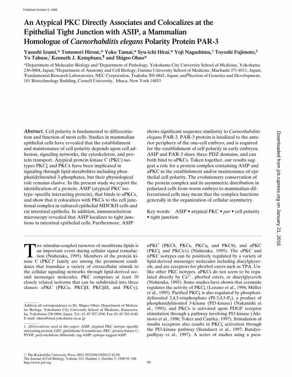
To identify proteins that interact directly with aPKC, wescreened an NIH3T3 cDNA expression library using puri-fied and autophosphorylated PKCz as a probe. OnecDNA found, clone I-1, encoded a truncated form of anovel protein, and we next screened a rat fibroblast 3Y1cDNA library to obtain a full-length cDNA clone encod-ing the entire protein-coding sequence. This cDNA was5,500 nucleotides in length, and contained a 1,337–aminoacid open reading frame. The open reading frame is pre-ceded by 259 nucleotides containing an in-frame termina-tion codon at nucleotide position -96 (compared with A atthe putative initiation codon), and is followed by a 1,227-nucleotide 39 noncoding sequence. Since the encoded pro-tein shows binding for aPKC isotypes of the PKC family(see below), we named it ASIP (aPKC-specific interactingprotein). The sequence of ASIP does not contain any ap-parent structural motif suggesting catalytic activity, butcontains three PDZ domains that are potentially involvedin protein–protein interactions (Saras and Heldin, 1996;Sheng, 1996) in addition to the aPKC-binding region (seebelow; Fig. 1 d). A database search for sequence similarityrevealed Caenorhabditis elegans PAR-3 as the only pro-tein with a significant overall sequence similarity (Fig. 1, aand b). PAR-3 localizes asymmetrically to the periphery ofthe C. elegans one-cell embryo, and is required for embry-onic cell polarity (Etemad-Moghadam et al., 1995). Thesimilarity between ASIP and PAR-3 involves an NH2-ter-minal conserved region (CR1), the three PDZ domains(CR2), and a conserved sequence (CR3) in the aPKC-binding region. A Drosophila EST cDNA clone, AA439413,also contains a sequence that shows significant similarityto the sequence of CR1 (Fig. 1 c).
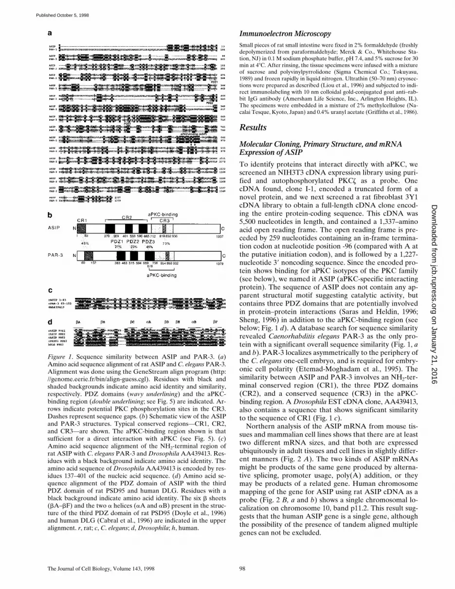
Northern analysis of the ASIP mRNA from mouse tis-sues and mammalian cell lines shows that there are at leasttwo different mRNA sizes, and that both are expressedubiquitously in adult tissues and cell lines in slightly differ-ent manners (Fig. 2 A). The two kinds of ASIP mRNAsmight be products of the same gene produced by alterna-tive splicing, promoter usage, poly(A) addition, or theymay be products of a related gene. Human chromosomemapping of the gene for ASIP using rat ASIP cDNA as aprobe (Fig. 2 B, a and b) shows a single chromosomal lo-calization on chromosome 10, band p11.2. This result sug-gests that the human ASIP gene is a single gene, althoughthe possibility of the presence of tandem aligned multiplegenes can not be excluded.
Figure 1. Sequence similarity between ASIP and PAR-3. (a)Amino acid sequence alignment of rat ASIP and C. elegans PAR-3.Alignment was done using the GeneStream align program (http://genome.eerie.fr/bin/align-guess.cgl). Residues with black andshaded backgrounds indicate amino acid identity and similarity,respectively. PDZ domains (wavy underlining) and the aPKC-binding region (double underlining; see Fig. 5) are indicated. Ar-rows indicate potential PKC phosphorylation sites in the CR3.Dashes represent sequence gaps. (b) Schematic view of the ASIPand PAR-3 structures. Typical conserved regions—CR1, CR2,and CR3—are shown. The aPKC-binding region shown is thatsufficient for a direct interaction with aPKC (see Fig. 5). (c)Amino acid sequence alignment of the NH2-terminal region ofrat ASIP with C. elegans PAR-3 and Drosophila AA439413. Res-idues with a black background indicate amino acid identity. Theamino acid sequence of Drosophila AA439413 is encoded by res-idues 137–401 of the nucleic acid sequence. (d) Amino acid se-quence alignment of the PDZ domain of ASIP with the thirdPDZ domain of rat PSD95 and human DLG. Residues with ablack background indicate amino acid identity. The six b sheets(bA–bF) and the two a helices (aA and aB) present in the struc-ture of the third PDZ domain of rat PSD95 (Doyle et al., 1996)and human DLG (Cabral et al., 1996) are indicated in the upperalignment. r, rat; c, C. elegans; d, Drosophila; h, human.
on January 21, 2016jcb.rupress.org
Dow
nloaded from
Published October 5, 1998
Izumi et al. Association of ASIP with aPKC in Tight Junction 99
Association Between ASIP and aPKC in COS Cells
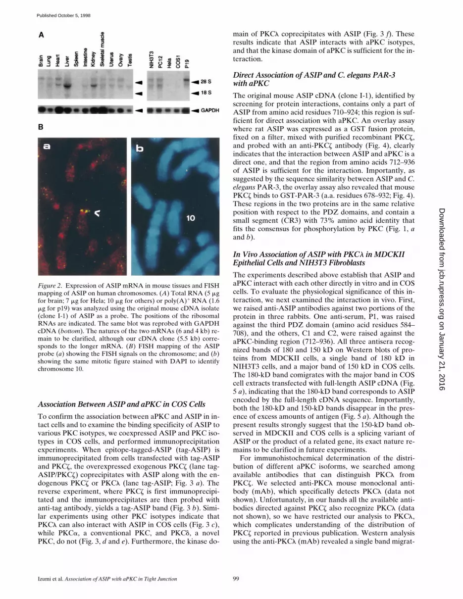
To confirm the association between aPKC and ASIP in in-tact cells and to examine the binding specificity of ASIP tovarious PKC isotypes, we coexpressed ASIP and PKC iso-types in COS cells, and performed immunoprecipitationexperiments. When epitope-tagged-ASIP (tag-ASIP) isimmunoprecipitated from cells transfected with tag-ASIPand PKCz, the overexpressed exogenous PKCz (lane tag-ASIP/PKCz) coprecipitates with ASIP along with the en-dogenous PKCz or PKCl (lane tag-ASIP; Fig. 3 a). Thereverse experiment, where PKCz is first immunoprecipi-tated and the immunoprecipitates are then probed withanti-tag antibody, yields a tag-ASIP band (Fig. 3 b). Simi-lar experiments using other PKC isotypes indicate thatPKCl can also interact with ASIP in COS cells (Fig. 3 c),while PKCa, a conventional PKC, and PKCd, a novelPKC, do not (Fig. 3, d and e). Furthermore, the kinase do-
main of PKCl coprecipitates with ASIP (Fig. 3 f). Theseresults indicate that ASIP interacts with aPKC isotypes,and that the kinase domain of aPKC is sufficient for the in-teraction.
Direct Association of ASIP and C. elegans PAR-3with aPKC
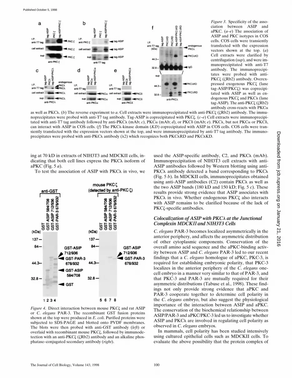
The original mouse ASIP cDNA (clone I-1), identified byscreening for protein interactions, contains only a part ofASIP from amino acid residues 710–924; this region is suf-ficient for direct association with aPKC. An overlay assaywhere rat ASIP was expressed as a GST fusion protein,fixed on a filter, mixed with purified recombinant PKCz,and probed with an anti-PKCz antibody (Fig. 4), clearlyindicates that the interaction between ASIP and aPKC is adirect one, and that the region from amino acids 712–936of ASIP is sufficient for the interaction. Importantly, assuggested by the sequence similarity between ASIP and C.elegans PAR-3, the overlay assay also revealed that mousePKCz binds to GST-PAR-3 (a.a. residues 678–932; Fig. 4).These regions in the two proteins are in the same relativeposition with respect to the PDZ domains, and contain asmall segment (CR3) with 73% amino acid identity thatfits the consensus for phosphorylation by PKC (Fig. 1, aand b).
In Vivo Association of ASIP with PKCl in MDCKII Epithelial Cells and NIH3T3 Fibroblasts
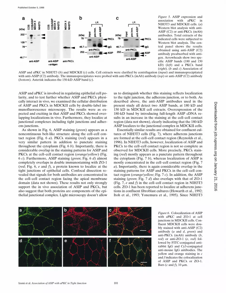
The experiments described above establish that ASIP andaPKC interact with each other directly in vitro and in COScells. To evaluate the physiological significance of this in-teraction, we next examined the interaction in vivo. First,we raised anti-ASIP antibodies against two portions of theprotein in three rabbits. One anti-serum, P1, was raisedagainst the third PDZ domain (amino acid residues 584–708), and the others, C1 and C2, were raised against theaPKC-binding region (712–936). All three antisera recog-nized bands of 180 and 150 kD on Western blots of pro-teins from MDCKII cells, a single band of 180 kD inNIH3T3 cells, and a major band of 150 kD in COS cells.The 180-kD band comigrates with the major band in COScell extracts transfected with full-length ASIP cDNA (Fig.5 a), indicating that the 180-kD band corresponds to ASIPencoded by the full-length cDNA sequence. Importantly,both the 180-kD and 150-kD bands disappear in the pres-ence of excess amounts of antigen (Fig. 5 a). Although thepresent results strongly suggest that the 150-kD band ob-served in MDCKII and COS cells is a splicing variant ofASIP or the product of a related gene, its exact nature re-mains to be clarified in future experiments.
For immunohistochemical determination of the distri-bution of different aPKC isoforms, we searched amongavailable antibodies that can distinguish PKCl fromPKCz. We selected anti-PKCl mouse monoclonal anti-body (mAb), which specifically detects PKCl (data notshown). Unfortunately, in our hands all the available anti-bodies directed against PKCz also recognize PKCl (datanot shown), so we have restricted our analysis to PKCl,which complicates understanding of the distribution ofPKCz reported in previous publication. Western analysisusing the anti-PKCl (mAb) revealed a single band migrat-
Figure 2. Expression of ASIP mRNA in mouse tissues and FISHmapping of ASIP on human chromosomes. (A) Total RNA (5 mgfor brain; 7 mg for Hela; 10 mg for others) or poly(A)1 RNA (1.6mg for p19) was analyzed using the original mouse cDNA isolate(clone I-1) of ASIP as a probe. The positions of the ribosomalRNAs are indicated. The same blot was reprobed with GAPDHcDNA (bottom). The natures of the two mRNAs (6 and 4 kb) re-main to be clarified, although our cDNA clone (5.5 kb) corre-sponds to the longer mRNA. (B) FISH mapping of the ASIPprobe (a) showing the FISH signals on the chromosome; and (b)showing the same mitotic figure stained with DAPI to identifychromosome 10.
on January 21, 2016jcb.rupress.org
Dow
nloaded from
Published October 5, 1998
The Journal of Cell Biology, Volume 143, 1998 100
ing at 70 kD in extracts of NIH3T3 and MDCKII cells, in-dicating that both cell lines express the PKCl isoform ofaPKC (Fig. 5 a).
To test the association of ASIP with PKCl in vivo, we
used the ASIP-specific antibody, C2, and PKCl (mAb).Immunoprecipitation of NIH3T3 cell extracts with anti-ASIP antibodies followed by Western blotting using anti-PKCl antibody detected a band corresponding to PKCl(Fig. 5 b). In MDCKII cells, immunoprecipitates obtainedusing anti-ASIP antibodies (C2) contain PKCl as well asthe two ASIP bands (180 kD and 150 kD; Fig. 5 c). Theseresults provide strong evidence that ASIP associates withPKCl in vivo. Whether endogenous PKCz also interactswith ASIP remains to be clarified because of the lack ofPKCz-specific antibodies.
Colocalization of ASIP with PKCl at the Junctional Complexin MDCKII and NIH3T3 Cells
C. elegans PAR-3 becomes localized asymmetrically in theanterior periphery, and affects the asymmetric distributionof other cytoplasmic components. Conservation of theoverall amino acid sequence and the aPKC-binding activ-ity between ASIP and C. elegans PAR-3 led to our recentfindings that a C. elegans homologue of aPKC, PKC-3, isrequired for establishing embryonic polarity, that PKC-3localizes in the anterior periphery of the C. elegans one-cell embryo in a manner very similar to that of PAR-3, andthat PKC-3 and PAR-3 are mutually required for theirasymmetric distributions (Tabuse et al., 1998). These find-ings not only provide strong evidence that aPKC andPAR-3 cooperate together to determine cell polarity inthe C. elegans embryo, but also suggest the physiologicalimportance of the interaction between ASIP and aPKC.The conservation of the biochemical relationship betweenASIP/PAR-3 and aPKC/PKC-3 led us to investigate whetherASIP and PKCl are involved in regulating cell polarity asobserved in C. elegans embryos.
In mammals, cell polarity has been studied intensivelyusing cultured epithelial cells such as MDCKII cells. Toevaluate the above possibility that the protein complex of
Figure 3. Specificity of the asso-ciation between ASIP andaPKC. (a–e) The association ofASIP and PKC isotypes in COScells. COS cells were transientlytransfected with the expressionvectors shown at the top. (a)Cell extracts were clarified bycentrifugation (sup), and were im-munoprecipitated with anti-T7antibody. The immunoprecipi-tates were probed with anti-PKCz (zRb2) antibody. Overex-pressed exogenous PKCz (lanetag-ASIP/PKCz) was coprecipi-tated with ASIP as well as en-dogenous PKCz and PKCl (lanetag-ASIP). The anti-PKCz (zRb2)antibody cross-reacts with PKCa
as well as PKCl. (b) The reverse experiment to a. Cell extracts were immunoprecipitated with anti-PKCz (zRb2) antibody. The immu-noprecipitates were probed with anti-T7 tag antibody. Tag-ASIP is coprecipitated with PKCz. (c–e) Cell extracts were immunoprecipi-tated with anti-T7 tag antibody followed by anti-PKCl (mAb; c), PKCa (mAb; d), or PKCd (mAb; e). PKCl, but not PKCa or PKCd,can interact with ASIP in COS cells. (f) The PKCl-kinase domain (KD) coprecipitated with ASIP in COS cells. COS cells were tran-siently transfected with the expression vectors shown at the top, and were immunoprecipitated by anti-T7 tag antibody. The immuno-precipitates were probed with anti-PKCl antibody (l2) which recognizes both PKClRD and PKClKD.
Figure 4. Direct interaction between mouse PKCz and rat ASIPor C. elegans PAR-3. The recombinant GST fusion proteinsshown at the top were produced in E. coli. Purified proteins weresubjected to SDS-PAGE and blotted onto PVDF membranes.The blots were then probed with anti-GST antibody (left) oroverlaid with recombinant mouse PKCz, followed by immunode-tection with an anti-PKCz (zRb2) antibody and an alkaline phos-phatase–conjugated secondary antibody (right).
on January 21, 2016jcb.rupress.org
Dow
nloaded from
Published October 5, 1998
Izumi et al. Association of ASIP with aPKC in Tight Junction 101
ASIP and aPKC is involved in regulating epithelial cell po-larity, and to test further whether ASIP and PKCl physi-cally interact in vivo, we examined the cellular distributionof ASIP and PKCl in MDCKII cells by double-label im-munofluorescence microscopy. The results were as ex-pected and exciting in that ASIP and PKCl showed over-lapping localizations in vivo. Furthermore, they localize atjunctional complexes including tight junctions and adher-ens junctions.
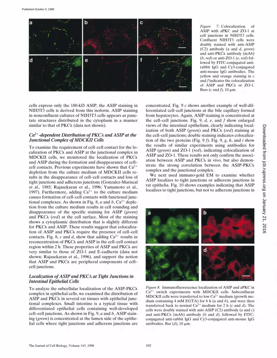
As shown in Fig. 6, ASIP staining (green) appears as anoncontinuous belt-like structure along the cell–cell con-tact region (Fig. 6 a). PKCl staining (red) appears in avery similar pattern in addition to punctate stainingthroughout the cytoplasm (Fig. 6 b). Importantly, there isconsiderable overlap in the staining patterns for ASIP andPKCl at the cell–cell contact region (orange/yellow) (Fig.6 c). Furthermore, ASIP staining (green; Fig. 6 d) almostcompletely overlaps in double immunostaining with ZO-1(red; Fig. 6, e and f), a protein known to localize at thetight junctions of epithelial cells. Confocal dissection re-vealed that signals for both antibodies are concentrated inthe cell–cell contact region facing the apical membranedomain (data not shown). These results not only stronglysupport the in vivo association of ASIP and PKCl, butalso suggest that both proteins are components of the epi-thelial junctional complex. Light microscopy doesn’t allow
us to distinguish whether this staining reflects localizationto the tight junction, the adherens junction, or to both. Asdescribed above, the anti-ASIP antibodies used in thepresent study all detect two ASIP bands, at 180 kD and150 kD in MDCKII cell extracts. Overexpression of the180-kD band by introducing full-length ASIP cDNA re-sults in an increase in the staining at the cell–cell contactregion (data not shown), clearly indicating that the 180-kDASIP localizes to the junctional complex in MDCKII cells.
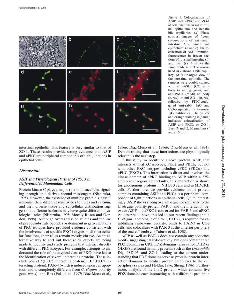
Essentially similar results are obtained for confluent cul-tures of NIH3T3 cells (Fig. 7), where adherens junctionsare formed at the cell–cell contact region (Reynolds et al.,1996). In NIH3T3 cells, however, localization of ASIP andPKCl to the cell–cell contact region is not so complete asobserved for MDCKII cells. More precisely, PKCl stain-ing (red) mostly appears as a punctate pattern throughoutthe cytoplasm (Fig. 7 b), whereas localization of ASIP ismostly concentrated in the cell–cell contact region (Fig. 7a). Importantly, there is again considerable overlap in thestaining patterns for ASIP and PKCl in the cell–cell con-tact region (orange/yellow; Fig. 7 c). In addition, the ASIPstaining (green; Fig. 7 d) also overlaps with that of ZO-1(Fig. 7, e and f) in the cell–cell contact region in NIH3T3cells. ZO-1 has been reported to localize at adherens junc-tions in confluent fibroblast cultures (Howarth et al., 1992;Itoh et al., 1993; Yonemura et al., 1995). Since NIH3T3
Figure 5. ASIP expression andassociation with aPKC inNIH3T3 and MDCKII cells. (a)Western blot analysis with anti-ASIP (C2) or anti-PKCl (mAb)antibodies. Total extracts of theindicated cells were subjected toWestern blot analysis. The cen-tral panel shows the resultsobtained using anti-ASIP (C2)antibody preabsorbed with anti-gen. Arrowheads show two spe-cific ASIP bands (180 and 150kD) (left) and a PKCl band(right). (b and c) Association of
ASIP and aPKC in NIH3T3 (b) and MDCKII (c) cells. Cell extracts were clarified by centrifugation (input) and immunoprecipitatedwith anti-ASIP (C2) antibody. The immunoprecipitates were probed with anti-PKCl (mAb) antibody (top) or anti-ASIP (C2) antibody(bottom). Asterisk indicates the 150-kD ASIP band (c).
Figure 6. Colocalization of ASIPwith aPKC and ZO-1 at celljunctions in MDCKII cells. Con-fluent MDCKII cells were dou-bly stained with anti-ASIP (C2)antibody (a and d, green) andanti-PKCl (mAb) antibody (b,red) or anti-ZO-1 (e, red) fol-lowed by FITC-conjugated anti-rabbit IgG and Cy3-conjugatedanti-mouse IgG antibodies. Theyellow and orange staining in cand f indicates the colocalizationof ASIP and PKCl or ZO-1.Bars (c and f), 10 mm.
on January 21, 2016jcb.rupress.org
Dow
nloaded from
Published October 5, 1998
The Journal of Cell Biology, Volume 143, 1998 102
cells express only the 180-kD ASIP, the ASIP staining inNIH3T3 cells is derived from this isoform. ASIP stainingin nonconfluent cultures of NIH3T3 cells appears as punc-tate structures distributed in the cytoplasm in a mannersimilar to that of PKCl (data not shown).
Ca21-dependent Distribution of PKCl and ASIP at the Junctional Complex of MDCKII Cells
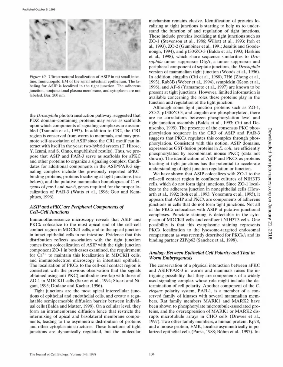
To examine the requirement of cell–cell contact for the lo-calization of PKCl and ASIP at the junctional complex inMDCKII cells, we monitored the localization of PKCland ASIP during the formation and disappearance of cell–cell contacts. Previous experiments have shown that Ca21
depletion from the culture medium of MDCKII cells re-sults in the disappearance of cell–cell contacts and loss oftight junctions and adherens junctions (Gonzalez-Mariscalet al., 1985; Rajasekaran et al., 1996; Yamamoto et al.,1997). Furthermore, adding Ca21 to the culture mediumcauses formation of cell–cell contacts with functional junc-tional complexes. As shown in Fig. 8, a and b, Ca21 deple-tion from the culture medium results in cell rounding anddisappearance of the specific staining for ASIP (green)and PKCl (red) at the cell surface. Most of the stainingshows a cytoplasmic distribution that is slightly differentfor PKCl and ASIP. These results suggest that colocaliza-tion of ASIP and PKCl require the presence of cell–cellcontacts. Fig. 8, c and d, show that adding Ca21 results inreconcentration of PKCl and ASIP in the cell–cell contactregion within 2 h. These properties of ASIP and PKCl arevery similar to those of ZO-1 and E-cadherin (data notshown; Rajasekaran et al., 1996), and support the notionthat ASIP and PKCl are peripheral components of cell–cell junctions.
Localization of ASIP and PKCl at Tight Junctions in Intestinal Epithelial Cells
To analyze the subcellular localization of the ASIP-PKClcomplex in epithelial cells, we examined the distribution ofASIP and PKCl in several rat tissues with epithelial junc-tional complexes. Small intestine is a typical tissue withdifferentiated epithelial cells containing well-developedcell–cell junctions. As shown in Fig. 9, a and b, ASIP stain-ing (green) is concentrated at the lumen side of the epithe-lial cells where tight junctions and adherens junctions are
concentrated. Fig. 9 c shows another example of well-dif-ferentiated cell–cell junctions at the bile capillary formedfrom hepatocytes. Again, ASIP staining is concentrated atthe cell–cell junctions. Fig. 9, d, e, and f show enlargedviews of the intestinal epithelium, clearly indicating local-ization of both ASIP (green) and PKCl (red) staining atthe cell–cell junctions; double staining indicates colocaliza-tion of the two proteins (Fig. 9 f). Fig. 9, g, h, and i showthe results of similar experiments using antibodies forASIP (green) and ZO-1 (red), indicating colocalization ofASIP and ZO-1. These results not only confirm the associ-ation between ASIP and PKCl in vivo, but also demon-strate the strong correlation between the ASIP-PKClcomplex and the junctional complex.
We next used immuno-gold EM to examine whetherASIP localizes to tight junctions or adherens junctions inrat epithelia. Fig. 10 shows examples indicating that ASIPlocalizes to tight junctions, but not to adherens junctions in
Figure 7. Colocalization ofASIP with aPKC and ZO-1 atcell junctions in NIH3T3 cells.Confluent NIH3T3 cells weredoubly stained with anti-ASIP(C2) antibody (a and d, green)and anti-PKCl antibody (mAb)(b, red) or anti-ZO-1 (e, red) fol-lowed by FITC-conjugated anti-rabbit IgG and Cy3-conjugatedanti-mouse IgG antibodies. Theyellow and orange staining in cand f indicates the colocalizationof ASIP and PKCl or ZO-1.Bars (c and f), 10 mm.
Figure 8. Immunofluorescence localization of ASIP and aPKC inCa21 switch experiments with MDCKII cells. SubconfluentMDCKII cells were transferred to low Ca21 medium (growth me-dium containing 4 mM EGTA) for 6 h (a and b), and were thentransferred back to normal Ca21 medium for 2 h (c and d). Thecells were doubly stained with anti-ASIP (C2) antibody (a and c)and anti-PKCl (mAb) antibody (b and d), followed by FITC-conjugated anti-rabbit IgG and Cy3-conjugated anti-mouse IgGantibodies. Bar (d), 10 mm.
on January 21, 2016jcb.rupress.org
Dow
nloaded from
Published October 5, 1998
Izumi et al. Association of ASIP with aPKC in Tight Junction 103
intestinal epithelia. This feature is very similar to that ofZO-1. These results provide strong evidence that ASIPand aPKC are peripheral components of tight junctions inepithelial cells.
Discussion
ASIP is a Physiological Partner of PKCl in Differentiated Mammalian Cells
Protein kinase C plays a major role in intracellular signal-ing through lipid-derived second messengers (Nishizuka,1995). However, the existence of multiple protein kinase Cisoforms, their different sensitivities to lipids and calcium,and their diverse tissue and subcellular distribution sug-gest that different isoforms may have quite different phys-iological roles (Nishizuka, 1995; Mochly-Rosen and Gor-don, 1998). Although overexpression studies and the useof pseudosubstrate peptides or dominant negative mutantsof PKC isotypes have provided evidence consistent withthe involvement of specific PKC isotypes in distinct cellu-lar functions, their roles remain mostly elusive. As an al-ternative way to sort out these roles, efforts are beingmade to identify and study proteins that interact directlywith different PKC isotypes. For example, attempts to un-derstand the role of the atypical class of PKCs have led tothe identification of several interacting proteins. These in-clude p62/ZIP (PKCz interacting protein), LIP (PKCl in-teracting protein), PAR-4 (which is induced upon cell apop-tosis and is completely different from C. elegans polaritygene par-4), and Ras (Puls et al., 1997; Diaz-Meco et al.,
1996a; Diaz-Meco et al., 1996b; Diaz-Meco et al., 1994).Demonstrating that these interactions are physiologicallyrelevant is the next step.
In this study, we identified a novel protein, ASIP, thatinteracts with aPKC isotypes, PKCz and PKCl, but notwith other PKC isotypes including cPKC (PKCa) andnPKC (PKCd). This interaction is direct and involves thekinase domain of aPKC binding to ASIP within a 225–amino acid region. Importantly, this interaction is shownfor endogenous proteins in NIH3T3 cells and in MDCKIIcells. Furthermore, we provide evidence that a proteincomplex containing ASIP and PKCl is a peripheral com-ponent of tight junctions in epithelial cells. Quite interest-ingly, ASIP shows strong overall sequence similarity to theC. elegans polarity protein PAR-3, and the interaction be-tween ASIP and aPKC is conserved for PAR-3 and aPKC.As described above, this led to our recent findings that aC. elegans homologue of aPKC, PKC-3, is required for es-tablishing embryonic polarity, binds to PAR-3 in COScells, and colocalizes with PAR-3 at the anterior peripheryof the one-cell embryo (Tabuse et al., 1998).
ASIP as well as PAR-3 does not contain any sequencemotifs, suggesting catalytic activity, but does contain threePDZ domains in CR2. PDZ domains (also called DHR orGLGF) are found in many proteins such as the DrosophilaDlg, PSD-95, and ZO-1, leading to the current under-standing that PDZ domains serve as protein–protein inter-action domains to localize protein complexes to the cellperiphery (Saras and Heldin, 1996; Sheng, 1996). Further-more, analysis of the InaD protein, which contains fivePDZ domains each interacting with a different protein in
Figure 9. Colocalization ofASIP with aPKC and ZO-1at cell junctions in rat intesti-nal epithelium and hepaticbile capillaries. (a) Phasecontrast images of frozencryosections of rat smallintestine. lum, lumen; ept,epithelium. (b and c) The lo-calization of ASIP immuno-fluorescence in frozen sec-tions of rat small intestine (b)and liver (c). b shows thesame fields as a. The arrow-head in c shows a bile capil-lary. (d–i) Enlarged view ofthe intestinal epithelia. Thesamples were doubly stainedwith anti-ASIP (C2) anti-body (d and g, green) andanti-PKCl (mAb) antibody(e, red) or anti-ZO-1 (h, red)followed by FITC-conju-gated anti-rabbit IgG andCy3-conjugated anti-mouseIgG antibodies. The yellowand orange staining in f and iindicates colocalization ofASIP and PKCl or ZO-1.Bars (b and c), 20 mm; bars (fand i), 5 mm.
on January 21, 2016jcb.rupress.org
Dow
nloaded from
Published October 5, 1998
The Journal of Cell Biology, Volume 143, 1998 104
the Drosophila phototransduction pathway, suggested thatPDZ domain–containing proteins may serve as scaffoldsupon which components of signaling complexes are assem-bled (Tsunoda et al., 1997). In addition to CR2, the CR1region is conserved from worm to mammals, and may pro-mote self-association of ASIP since the CR1 motif can in-teract with itself in the yeast two-hybrid system (T. Hirose,Y. Izumi, and S. Ohno, unpublished results). Thus, we pro-pose that ASIP and PAR-3 serve as scaffolds for aPKCand other proteins to organize a signaling complex. Candi-dates for additional components in the ASIP/PAR-3 sig-naling complex include the previously reported aPKC-binding proteins, proteins localizing at tight junctions (seebelow), and the putative mammalian homologues of C. el-egans of par-5 and par-6, genes required for the proper lo-calization of PAR-3 (Watts et al., 1996; Guo and Kem-phues, 1996).
ASIP and aPKC are Peripheral Components ofCell–Cell Junctions
Immunofluorescence microscopy reveals that ASIP andPKCl colocalize to the most apical end of the cell–cellcontact region in MDCKII cells, and to the apical junctionin intact epithelial cells in rat intestine. Evidence that thisdistribution reflects association with the tight junctioncomes from colocalization of ASIP with the tight junctioncomponent ZO-1 in both cases examined, the requirementfor Ca21 to maintain this localization in MDCKII cells,and immunoelectron microscopy in intestinal epithelia.The localization of PKCl to the cell–cell contact region isconsistent with the previous observation that the signalsobtained using anti-PKCz antibodies overlap with those ofZO-1 in MDCKII cells (Saxon et al., 1994; Stuart and Ni-gam, 1995; Dodane and Kachar, 1996).
Tight junctions are the most apical intercellular junc-tions of epithelial and endothelial cells, and create a regu-latable semipermeable diffusion barrier between individ-ual cells (Balda and Matter, 1998). On a cellular level, theyform an intramembrane diffusion fence that restricts theintermixing of apical and basolateral membrane compo-nents, leading to the asymmetric distribution of proteinsand other cytoplasmic structures. These functions of tightjunctions are dynamically regulated, but the molecular
mechanism remains elusive. Identification of proteins lo-calizing at tight junctions is starting to help us to under-stand the function of and regulation of tight junctions.These include proteins localizing at tight junctions such asZO-1 (Stevenson et al., 1986; Willott et al., 1993; Itoh etal., 1993), ZO-2 (Gumbiner et al., 1991; Jesaitis and Goode-nough, 1994), and p130/ZO-3 (Balda et al., 1993; Haskinset al., 1998), which share sequence similarities to Dro-sophila tumor suppressor DlgA, a tumor suppressor andperipheral component of septate junctions, the Drosophilaversion of mammalian tight junction (Woods et al., 1996).In addition, cingulin (Citi et al., 1988), 7H6 (Zhong et al.,1993), Rab3B (Weber et al., 1994), symplekin (Keon et al.,1996), and AF-6 (Yamamoto et al., 1997) are known to bepresent at tight junctions. However, limited information isavailable concerning the roles these proteins play in thefunction and regulation of the tight junction.
Although some tight junction proteins such as ZO-1,ZO-2, p130/ZO-3, and cingulin are phosphorylated, thereare no correlations between phosphorylation level andtight junction assembly (Balda et al., 1993; Citi and De-nisenko, 1995). The presence of the consensus PKC phos-phorylation sequence in the CR3 of ASIP and PAR-3suggests that PKCl regulates this complex through phos-phorylation. Consistent with this notion, ASIP domains,expressed as GST-fusion proteins in E. coli, are efficientlyphosphorylated by recombinant mouse PKCz (data notshown). The identification of ASIP and PKCl as proteinslocating at tight junctions has the potential to accelerateunderstanding of tight junction regulation or function.
We have shown that ASIP colocalizes with ZO-1 to thecell–cell contact region in confluent cultures of NIH3T3cells, which do not form tight junctions. Since ZO-1 local-izes to the adherens junction in nonepithelial cells (How-arth et al., 1992; Itoh et al., 1993; Yonemura et al., 1995), itappears that ASIP and PKCl are components of adherensjunctions in cells that do not form tight junctions. Not allof the PKCl colocalizes with ASIP at putative junctionalcomplexes. Punctate staining is detectable in the cyto-plasm of MDCKII cells and confluent NIH3T3 cells. Onepossibility is that this cytoplasmic staining representsPKCl localization to the lysosome-targeted endosomalcompartment as was recently described for PKCl/i and itsbinding partner ZIP/p62 (Sanchez et al., 1998).
Analogy Between Epithelial Cell Polarity and That in Worm Embryogenesis
The conservation of a physical interaction between aPKCand ASIP/PAR-3 in worms and mammals raises the in-triguing possibility that they are components of a widelyused signaling complex whose role might include the de-termination of cell polarity. Another component of the C.elegans polarity system, PAR-1, is a member of a con-served family of kinases with several mammalian mem-bers. Rat family members MARK1 and MARK2 havebeen shown to phosphorylate microtubule-associated pro-teins, and the overexpression of MARK1 or MARK2 dis-rupts microtubule arrays in CHO cells (Drewes et al.,1997). Two other family members, a human protein, Kp78,and a mouse protein, EMK, localize asymmetrically in po-larized epithelial cells (Parsa, 1988; Böhm et al., 1997). In-
Figure 10. Ultrastructural localization of ASIP in rat small intes-tine. Immunogold EM of the small intestinal epithelium. The la-beling for ASIP is localized in the tight junction. The adherensjunction, nonjunctional plasma membrane, and cytoplasm are notlabeled. Bar, 200 nm.
on January 21, 2016jcb.rupress.org
Dow
nloaded from
Published October 5, 1998
Izumi et al. Association of ASIP with aPKC in Tight Junction 105
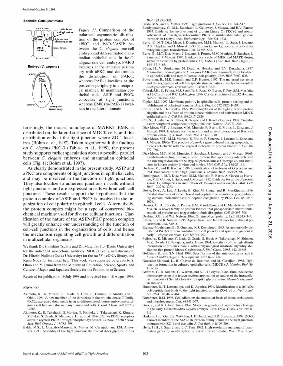
terestingly, the mouse homologue of MARK2, EMK, isdistributed on the lateral surface of MDCK cells, and thisdistribution ends at the tight junction where ZO-1 local-izes (Böhm et al., 1997). Taken together with the findingson C. elegans PKC-3 (Tabuse et al., 1998), the presentstudy supports earlier speculation about a possible parallelbetween C. elegans embryos and mammalian epithelialcells (Fig. 11; Böhm et al., 1997).
As clearly demonstrated in the present study, ASIP andaPKC are components of tight junctions in epithelial cells,and may be involved in the function of tight junctions.They also localize to adherens junctions in cells withouttight junctions, and are expressed in cells without cell–celljunctions. These results support the hypothesis that theprotein complex of ASIP and PKCl is involved in the or-ganization of cell polarity in epithelial cells. Alternatively,the protein complex might be a type of conserved bio-chemical machine used for diverse cellular functions. Clar-ification of the nature of the ASIP-aPKC protein complexwill greatly enhance our understanding of the function ofcell–cell junctions in the organization of cells, and hencethe mechanism regulating cell growth and differentiationin multicellular organisms.
We thank Dr. Shoichiro Tsukita and Dr. Masahiko Ito (Kyoto University)for the anti-ZO-1 monoclonal antibody, MDCKII cells, and discussion,Dr. Hiroshi Nojima (Osaka University) for the rat 3Y1-cDNA library, andKumi Noda for technical help. This work was supported by grants to S.Ohno and Y. Izumi from the Ministry of Education, Science, Sports, andCulture of Japan and Japanese Society for the Promotion of Science.
Received for publication 19 July 1998 and in revised form 18 August 1998.
References
Akimoto, K., K. Mizuno, S. Osada, S. Hirai, S. Tanuma, K. Suzuki, and S.Ohno. 1994. A new member of the third class in the protein kinase C family,PKCl, expressed dominantly in an undifferentiated mouse embryonal carci-noma cell line and also in many tissues and cells. J. Biol. Chem. 269:12677–12683.
Akimoto, K., R. Takahashi, S. Moriya, N. Nishioka, J. Takayanagi, K. Kimura,Y. Fukui, S. Osada, K. Mizuno, S. Hirai, et al. 1996. EGF or PDGF receptorsactivate atypical PKCl through phosphatidylinositol 3-kinase. EMBO (Eur.Mol. Biol. Organ.) J. 15:788–798.
Balda, M.S., L. Gonzalez-Mariscal, K. Matter, M. Cereijido, and J.M. Ander-son. 1993. Assembly of the tight junction: the role of diacylglycerol. J. Cell
Biol. 123:293–302.Balda, M.S., and K. Matter. 1998. Tight junctions. J. Cell Sci. 111:541–547.Bandyopadhyay, G., M.L. Standaert, L. Galloway, J. Moscat, and R.V. Farese.
1997. Evidence for involvement of protein kinase C (PKC)-z and nonin-volvement of diacylglycerol-sensitive PKCs in insulin-stimulated glucosetransport in L6 myotubes. Endocrinology. 138:4721–4731.
Berra, E., M.T. Diaz-Meco, I. Dominguez, M.M. Municio, L. Sanz, J. Lozano,R.S. Chapkin, and J. Moscat. 1993. Protein kinase Cz isoform is critical formitogenic signal transduction. Cell. 74:555–563.
Berra, E., M.T. Diaz-Meco, J. Lozano, S. Frutos, M.M. Municio, P. Sanchez, L.Sanz, and J. Moscat. 1995. Evidence for a role of MEK and MAPK duringsignal transduction by protein kinase Cz. EMBO (Eur. Mol. Biol. Organ.) J.14:6157–6163.
Böhm, H., V. Brinkmann, M. Drab, A. Henske, and T.V. Kurzchalia. 1997.Mammalian homologues of C. elegans PAR-1 are asymmetrically localizedin epithelial cells and may influence their polarity. Curr. Biol. 7:603–606.
Bowerman, B., M.K. Ingram, and C.P. Hunter. 1997. The maternal par genesand the segregation of cell fate specification activities in early Caenorhabdi-tis elegans embryos. Development. 124:3815–3826.
Cabral, J.H., C. Petosa, M.J. Sutcliffe, S. Raza, O. Byron, F. Poy, S.M. Marfatia,A.H. Chishti, and R.C. Liddington. 1996. Crystal structure of a PDZ domain.Nature. 382:649–652.
Caplan, M.J. 1997. Membrane polarity in epithelial cells: protein sorting and es-tablishment of polarized domains. Am. J. Physiol. 272:F425–F429.
Citi, S., and N. Denisenko. 1995. Phosphorylation of the tight junction proteincingulin and the effects of protein kinase inhibitors and activators in MDCKepithelial cells. J. Cell Sci. 108:2917–2926.
Citi, S., H. Sabanay, R. Jakes, B. Geiger, and J. Kendrick-Jones. 1988. Cingulin,a new peripheral component of tight junctions. Nature. 333:272–276.
Diaz-Meco, M.T., J. Lozano, M.M. Municio, E. Berra, S. Frutos, L. Sanz, and J.Moscat. 1994. Evidence for the in vitro and in vivo interaction of Ras withprotein kinase Cz. J. Biol. Chem. 269:31706–31710.
Diaz-Meco, M.T., M.M. Municio, S. Frutos, P. Sanchez, J. Lozano, L. Sanz, andJ. Moscat. 1996a. The product of par-4, a gene induced during apoptosis, in-teracts selectively with the atypical isoforms of protein kinase C. Cell. 86:777–786.
Diaz-Meco, M.T., M.M. Municio, P. Sanchez, J. Lozano, and J. Moscat. 1996b.Lambda-interacting protein, a novel protein that specifically interacts withthe zinc finger domain of the atypical protein kinase C isotype l/i and stimu-lates its kinase activity in vitro and in vivo. Mol. Cell. Biol. 16:105–114.
Dodane, V., and B. Kachar. 1996. Identification of isoforms of G proteins andPKC that colocalize with tight junctions. J. Membr. Biol. 149:199–209.
Dominguez, I., M.T. Diaz-Meco, M.M. Municio, E. Berra, A. Garcia de Herre-ros, M.E. Cornet, L. Sanz, and J. Moscat. 1992. Evidence for a role of proteinkinase Cz subspecies in maturation of Xenopus laevis oocytes. Mol. Cell.Biol. 12:3776–3783.
Doyle, D.A., A. Lee, J. Lewis, E. Kim, M. Sheng, and R. MacKinnon. 1996.Crystal structures of a complexed and peptide-free membrane protein-bind-ing domain: molecular basis of peptide recognition by PDZ. Cell. 85:1067–1076.
Drewes, G., A. Ebneth, U. Preuss, E.M. Mandelkow, and E. Mandelkow. 1997.MARK, a novel family of protein kinases that phosphorylate microtubule-associated proteins and trigger microtubule disruption. Cell. 89:297–308.
Drubin, D.G., and W.J. Nelson. 1996. Origins of cell polarity. Cell. 84:335–344.Eaton, S., and K. Simons. 1995. Apical, basal, and lateral cues for epithelial po-
larization. Cell. 82:5–8.Etemad-Moghadam, B., S. Guo, and K.J. Kemphues. 1995. Asymmetrically dis-
tributed PAR-3 protein contributes to cell polarity and spindle alignment inearly C. elegans embryos. Cell. 83:743–752.
Fujise, A., K. Mizuno, Y. Ueda, S. Osada, S. Hirai, A. Takayanagi, N. Shimizu,M.K. Owada, H. Nakajima, and S. Ohno. 1994. Specificity of the high affinityinteraction of protein kinase C with a physiological substrate, myristoylatedalanine-rich protein kinase C substrate. J. Biol. Chem. 269:31642–31648.
Goldstein, B., and S.N. Hird. 1996. Specification of the anteroposterior axis inCaenorhabditis elegans. Development. 122:1467–1474.
Gonzalez-Mariscal, L., B. Chavez de Ramirez, and M. Cereijido. 1985. Tightjunction formation in cultured epithelial cells (MDCK). J. Membr. Biol. 86:113–125.
Griffiths, G., K. Simons, G. Warren, and K.T. Tokuyasu. 1986. Immunoelectronmicroscopy using thin frozen section: application to studies of the intracellu-lar transport of Semliki forest virus spike glycoproteins. Methods Enzymol.96:466–483.
Gumbiner, B., T. Lowenkopf, and D. Apatira. 1991. Identification of a 160-kDapolypeptide that binds to the tight junction protein ZO-1. Proc. Natl. Acad.Sci. USA. 88:3460–3464.
Gumbiner, B.M. 1996. Cell adhesion: the molecular basis of tissue architectureand morphogenesis. Cell. 84:345–357.
Guo, S., and K.J. Kemphues. 1996. Molecular genetics of asymmetric cleavagein the early Caenorhabditis elegans embryo. Curr. Opin. Genet. Dev. 6:408–415.
Haskins, J., L. Gu, E.S. Wittchen, J. Hibbard, and B.R. Stevenson. 1998. ZO-3,a novel member of the MAGUK protein family found at the tight junction,interacts with ZO-1 and occludin. J. Cell Biol. 141:199–208.
Heng, H.H., J. Squire, and L.C. Tsui. 1992. High-resolution mapping of mam-malian genes by in situ hybridization to free chromatin. Proc. Natl. Acad.
Figure 11. Comparison of thepolarized asymmetric distribu-tion of the protein complex ofaPKC and PAR-3/ASIP be-tween the C. elegans one-cellembryo and differentiated mam-malian epithelial cells. In the C.elegans one-cell embryo, PAR-3localizes at the anterior periph-ery with aPKC and determinesthe distribution of PAR-1,whereas PAR-1 localizes at theposterior periphery in a recipro-cal manner. In mammalian epi-thelial cells, ASIP and PKClcolocalize at tight junctions,whereas EMK (m-PAR-1) local-izes in the lateral domain.
on January 21, 2016jcb.rupress.org
Dow
nloaded from
Published October 5, 1998
The Journal of Cell Biology, Volume 143, 1998 106
Sci. USA. 89:9509–9513.Heng, H.H., and L.C. Tsui. 1993. Modes of DAPI banding and simultaneous in
situ hybridization. Chromosoma. 102:325–332.Howarth, A.G., M.R. Hughes, and B.R. Stevenson. 1992. Detection of the tight
junction-associated protein ZO-1 in astrocytes and other nonepithelial celltypes. Am. J. Physiol. 262:C461–C469.
Itoh, M., A. Nagafuchi, S. Yonemura, T. Kitani-Yasuda, S. Tsukita, and S. Tsu-kita. 1993. The 220-kD protein colocalizing with cadherins in non-epithelialcells is identical to ZO-1, a tight junction-associated protein in epithelialcells: cDNA cloning and immunoelectron microscopy. J. Cell Biol. 121:491–502.
Izumi, Y., S. Hirai, Y. Tamai, A. Fujise-Matsuoka, Y. Nishimura, and S. Ohno.1997. A protein kinase Cd-binding protein SRBC whose expression is in-duced by serum starvation. J. Biol. Chem. 272:7381–7389.
Jesaitis, L.A., and D.A. Goodenough. 1994. Molecular characterization and tis-sue distribution of ZO-2, a tight junction protein homologous to ZO-1 andthe Drosophila discs-large tumor suppressor protein. J. Cell Biol. 124:949–961.
Keon, B.H., S. Schafer, C. Kuhn, C. Grund, and W.W. Franke. 1996. Symple-kin, a novel type of tight junction plaque protein. J. Cell Biol. 134:1003–1018.
Knoblich, J.A. 1997. Mechanisms of asymmetric cell division during animal de-velopment. Curr. Opin. Cell Biol. 9:833–841.
Laemmli, U.K. 1970. Cleavage of structural proteins during the assembly of thehead of bacteriophage T4. Nature. 227:680–685.
Liou, W., H.J. Geuze, and J.W. Slot. 1996. Improving structural integrity ofcryosections for immunogold labeling. Histochem. Cell Biol. 106:41–58.
Lozano, J., E. Berra, M.M. Municio, M.T. Diaz-Meco, I. Dominguez, L. Sanz,and J. Moscat. 1994. Protein kinase Cz isoform is critical for kB-dependentpromoter activation by sphingomyelinase. J. Biol. Chem. 269:19200–19202.
Mochly-Rosen, D., and A.S. Gordon. 1998. Anchoring proteins for protein ki-nase C: a means for isozyme selectivity. FASEB J. 12:35–42.
Müller, G., M. Ayoub, P. Storz, J. Rennecke, D. Fabbro, and K. Pfizenmaier.1995. PKCz is a molecular switch in signal transduction of TNF-a, bifunc-tionally regulated by ceramide and arachidonic acid. EMBO (Eur. Mol. Biol.Organ.) J. 14:1961–1969.
Nakanishi, H., K.A. Brewer, and J.H. Exton. 1993. Activation of the z isozymeof protein kinase C by phosphatidylinositol 3,4,5-trisphosphate. J. Biol.Chem. 268:13–16.
Nishizuka, Y. 1995. Protein kinase C and lipid signaling for sustained cellularresponses. FASEB J. 9:484–496.
Ohno, S., K. Mizuno, Y. Adachi, A. Hata, Y. Akita, K. Akimoto, S. Osada, S.Hirai, and K. Suzuki. 1994. Activation of novel protein kinases Cd and eupon mitogenic stimulation of quiescent rat 3Y1 fibroblasts. J. Biol. Chem.269:17495–17501.
Osada, S., K. Mizuno, T.C. Saido, K. Suzuki, T. Kuroki, and S. Ohno. 1992. Anew member of the protein kinase C family, nPKCu, predominantly ex-pressed in skeletal muscle. Mol. Cell Biol. 12:3930–3938.
Parsa, I. 1988. Loss of a Mr 78,000 marker in chemically induced transplantablecarcinomas and primary carcinoma of human pancreas. Cancer Res. 48:2265–2272.
Puls, A., S. Schmidt, F. Grawe, and S. Stabel. 1997. Interaction of protein kinaseCz with ZIP, a novel protein kinase C-binding protein. Proc. Natl. Acad. Sci.USA. 94:6191–6196.
Rajasekaran, A.K., M. Hojo, T. Huima, and E. Rodriguez-Boulan. 1996.Catenins and zonula occludens-1 form a complex during early stages in theassembly of tight junctions. J. Cell Biol. 132:451–463.
Reynolds, A.B., J.M. Daniel, Y.Y. Mo, J. Wu, and Z. Zhang. 1996. The novelcatenin p120cas binds classical cadherins and induces an unusual morpholog-ical phenotype in NIH3T3 fibroblasts. Exp. Cell Res. 225:328–337.
Sanchez, P., G. De Carcer, I.V. Sandoval, J. Moscat, and M.T. Diaz-Meco. 1998.Localization of atypical protein kinase C isoforms into lysosome-targetedendosomes through interaction with p62. Mol. Cell Biol. 18:3069–3080.
Saras, J., and C.H. Heldin. 1996. PDZ domains bind carboxy-terminal se-quences of target proteins. Trends Biochem. Sci. 21:455–458.
Saxon, M.L., X. Zhao, and J.D. Black. 1994. Activation of protein kinase Cisozymes is associated with post-mitotic events in intestinal epithelial cells insitu. J. Cell Biol. 126:747–763.
Sheng, M. 1996. PDZs and receptor/channel clustering: rounding up the latestsuspects. Neuron. 17:575–578.
Standaert, M.L., L. Galloway, P. Karnam, G. Bandyopadhyay, J. Moscat, andR.V. Farese. 1997. Protein kinase C-z as a downstream effector of phosphati-dylinositol 3-kinase during insulin stimulation in rat adipocytes. Potentialrole in glucose transport. J. Biol. Chem. 272:30075–30082.
Stevenson, B.R., J.D. Siliciano, M.S. Mooseker, and D.A. Goodenough. 1986.Identification of ZO-1: a high molecular weight polypeptide associated withthe tight junction (zonula occludens) in a variety of epithelia. J. Cell Biol.103:755–766.
Stuart, R.O., and S.K. Nigam. 1995. Regulated assembly of tight junctions byprotein kinase C. Proc. Natl. Acad. Sci. USA. 92:6072–6076.
Tabuse, Y., Y. Izumi, F. Piano, K.J. Kemphues, J. Miwa, and S. Ohno. 1998.Atypical Protein Kinase C Cooperates with PAR-3 to Establish EmbryonicPolarity In Caenorhabditis elegans. Development. 125:3607–3614.
Toker, A., and L.C. Cantley. 1997. Signaling through the lipid products of phos-phoinositide-3-OH kinase. Nature. 387:673–676.
Tokuyasu, K.T. 1989. Use of poly(vinylpyrrolidone) and poly(vinyl alcohol) forcryoultramicrotomy. Histochem. J. 21:163–171.
Tsunoda, S., J. Sierralta, Y. Sun, R. Bodner, E. Suzuki, A. Becker, M. Socolich,and C.S. Zuker. 1997. A multivalent PDZ-domain protein assembles signal-ing complexes in a G-protein-coupled cascade. Nature. 388:243–249.
Watts, J.L., B. Etemad-Moghadam, S. Guo, L. Boyd, B.W. Draper, C.C. Mello,J.R. Priess, and K.J. Kemphues. 1996. par-6, a gene involved in the establish-ment of asymmetry in early C. elegans embryos, mediates the asymmetric lo-calization of PAR-3. Development. 122:3133–3140.
Ways, D.K., K. Posekany, J. deVente, T. Garris, J. Chen, J. Hooker, W. Qin, P.Cook, D. Fletcher, and P. Parker. 1994. Overexpression of protein kinase C-zstimulates leukemic cell differentiation. Cell Growth Differ. 5:1195–1203.
Weber, E., G. Berta, A. Tousson, P. St. John, M.W. Green, U. Gopalokrishnan,T. Jilling, E.J. Sorscher, T.S. Elton, D.R. Abrahamson, et al. 1994. Expres-sion and polarized targeting of a rab3 isoform in epithelial cells. J. Cell Biol.125:583–594.
Willott, E., M.S. Balda, A.S. Fanning, B. Jameson, C. Van Itallie, and J.M.Anderson. 1993. The tight junction protein ZO-1 is homologous to theDrosophila discs-large tumor suppressor protein of septate junctions. Proc.Natl. Acad. Sci. USA. 90:7834–7838.
Woods, D.F., C. Hough, D. Peel, G. Callaini, and P.J. Bryant. 1996. Dlg proteinis required for junction structure, cell polarity, and proliferation control inDrosophila epithelia. J. Cell Biol. 134:1469–1482.
Wooten, M.W., G. Zhou, M.L. Seibenhener, and E.S. Coleman. 1994. A rolefor z protein kinase C in nerve growth factor-induced differentiation of PC12cells. Cell Growth Differ. 5:395–403.
Xu, J., M.M. Zutter, S.A. Santoro, and R.A. Clark. 1996. PDGF induction ofa2 integrin gene expression is mediated by protein kinase C-z. J. Cell Biol.134:1301–1311.
Yamamoto, T., N. Harada, K. Kano, S. Taya, E. Canaani, Y. Matsuura, A. Mi-zoguchi, C. Ide, and K. Kaibuchi. 1997. The ras target AF-6 interacts withZO-1 and serves as a peripheral component of tight junctions in epithelialcells. J. Cell Biol. 139:785–795.
Yonemura, S., M. Itoh, A. Nagafuchi, and S. Tsukita. 1995. Cell-to-cell adher-ens junction formation and actin filament organization: similarities and dif-ferences between non-polarized fibroblasts and polarized epithelial cells. J.Cell Sci. 108:127–142.
Zhong, Y., T. Saitoh, T. Minase, N. Sawada, K. Enomoto, and M. Mori. 1993.Monoclonal antibody 7H6 reacts with a novel tight junction-associated pro-tein distinct from ZO-1, cingulin and ZO-2. J. Cell Biol. 120:477–483.
on January 21, 2016jcb.rupress.org
Dow
nloaded from
Published October 5, 1998