amputation and prosthesis implantation shape body and peripersonal space representations
TRANSCRIPT

Amputation and prosthesis implantationshape body and peripersonal spacerepresentationsElisa Canzoneri1,3*, Marilena Marzolla1, Amedeo Amoresano2, Gennaro Verni2 & Andrea Serino1,3*
1CsrNC, Centro studi e ricerche in Neuroscienze Cognitive, Polo Scientifico-Didattico di Cesena, ALMA MATER STUDIORUM,Universita di Bologna, 2Centro Protesi INAIL, Vigorso di Budrio, Bologna, 3Dipartimento di Psicologia, ALMA MATER STUDIORUM,Universita di Bologna.
Little is known about whether and how multimodal representations of the body (BRs) and of the spacearound the body (Peripersonal Space, PPS) adapt to amputation and prosthesis implantation. In order toinvestigate this issue, we tested BR in a group of upper limb amputees by means of a tactile distanceperception task and PPS by means of an audio-tactile interaction task. Subjects performed the tasks withstimulation either on the healthy limb or the stump of the amputated limb, while wearing or not wearingtheir prosthesis. When patients performed the tasks on the amputated limb, without the prosthesis, theperception of arm length shrank, with a concurrent shift of PPS boundaries towards the stump. Conversely,wearing the prosthesis increased the perceived length of the stump and extended the PPS boundaries so as toinclude the prosthetic hand, such that the prosthesis partially replaced the missing limb.
Our physical body represents the interface between the self and the external world, in that it mediates everyinteraction with external stimuli. The physical body is represented at different levels in the human brain,from unimodal somatosensory, motor and visual body maps1–3, to multimodal areas representing the
shape, dimensions and position of body parts4–8 and the space immediately around them (Peripersonal Space,PPS)9–12. Brain systems involved in body and PPS representation have been localized within a common fronto-parietal network, encompassing the ventral premotor cortex and the posterior parietal cortex, both in mon-keys13–15 and in humans16–20; see21 for a review. Body representations in the brain depend on the structure of thephysical body, and must dynamically update to changes of the physical body. While there is extensive evidence ofplasticity in unimodal body representations following a sudden change in the physical body, such as in the case oftraumatic amputation22–25, little is known about the extent to which multimodal body and PPS representations aredependent on the structure of the physical body and dynamically adapt to changes in body structure26–28.
Here we study a group of 10 upper limb amputees to show how a sudden change in the structure of the physicalbody affects a critical feature of body representations (BRs), i.e., the perceived dimension of the residual body part,and the extension of PPS around the affected body part. In addition, the effects of amputation are partiallypalliated by means of prostheses that at the same time physically replace the amputated body part and extend thefunctionality of the residual limb. Little is known on whether and how partially restoring the function andstructure of the physical body by means of prosthesis implantation affects BRs and PPS representations. Tostudy this issue, we selected amputee patients who underwent traumatic amputation of an upper limb at least 24months before testing, and were implanted with and used a functional prosthesis. Patients performed a tactiledistance perception task in order to assess the perceived length of the stump and of the healthy arm (seeExperiment 1) and an audio-tactile interaction task in order to measure the extent of PPS representation aroundthe stump and the healthy limb (see Experiment 2). The comparison between the results for the two hemisomaprovided evidence about the effects of amputation. The same experiments were also run while patients were orwere not wearing their prosthesis during testing, and the results from these two conditions were compared inorder to study the effect of prosthesis implantation on body and PPS representation.
ResultsExperiment 1 - amputation and prosthesis implantation affect BR. In order to assess the perceived length of theresidual part of the upper limb and of the homologous region of the healthy limb, we used a tactile distanceperception task. In each trial, subjects received two pairs of tactile stimuli, one pair on the forehead (serving as a
OPEN
SUBJECT AREAS:PERCEPTION
PSYCHOLOGY
NEUROSCIENCE
SENSORIMOTOR PROCESSING
Received25 June 2013
Accepted11 September 2013
Published3 October 2013
Correspondence andrequests for materials
should be addressed toE.C. (elisa.canzoneri@
epfl.ch) or A.S.(andrea.serino@epfl.
ch)
*Current address:Laboratory of
CognitiveNeuroscience & Center
for Neuroprosthetics,Ecole Polytechnique
Federale de Lausanne,Lausanne, Switzerland
SCIENTIFIC REPORTS | 3 : 2844 | DOI: 10.1038/srep02844 1
reference body part) and one pair on the upper arm (target bodypart). Participants were asked to judge whether the distance betweenthe two stimuli was longer on the forehead or on the arm. In thepresent experiment, we administered the tactile distance perceptiontask with tactile stimuli longitudinally delivered on the upper arm,along the arm axis, in order to measure the perceived length of thearm. Amputees performed the task in 3 blocked conditions, run incounterbalanced between-subjects order, on the healthy limb and onthe stump, with or without the prosthesis. For each subject, wecalculated the mean probability of reporting the distance on theupper arm as longer for all combinations of inter-point distances(P-Arm). We predicted that P-Arm would vary depending on theperceived size of the stimulated arm, in line with a context dependentbias, well documented in the field of visual perception [e.g.29,30], andsuggested also for haptic exploration [e.g.31; but see also32 for adifferent interpretation] and recently confirmed also for a visual33,34
and a tactile distance perception task34. According to this bias, higheror lower P-Arm would indicate, respectively, that the upper arm isperceived as shorter or as longer.
We first compared mean P-Arm between the amputees’ healthylimb and the right arm of healthy controls, by means of an independ-ent samples t-test. Data were normally distributed (Kolmogorov-Smirnov: p 5 .20). Scores did not differ significantly between thetwo groups [t(12) 5 1.41, p 5 .29] suggesting that amputation of oneupper limb did not affect the implicitly perceived length of thehealthy arm.
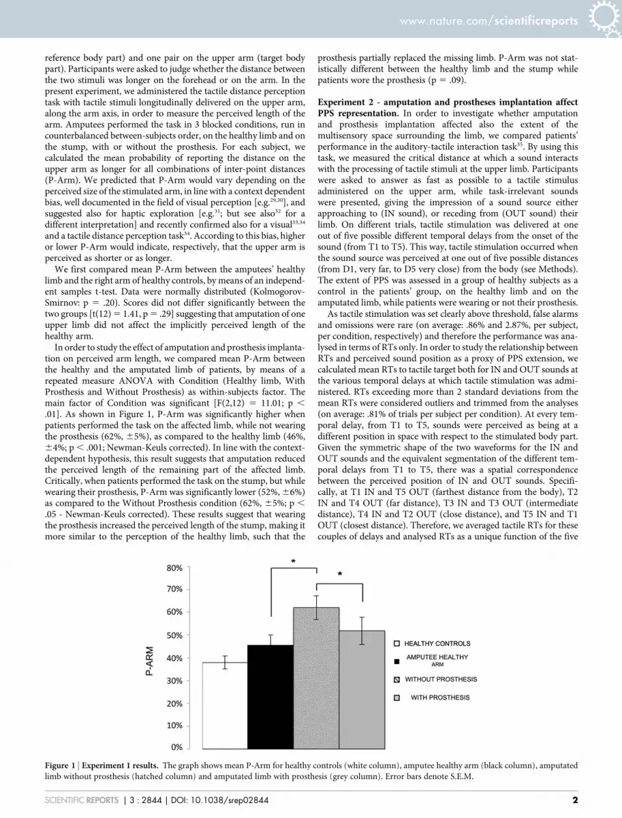
In order to study the effect of amputation and prosthesis implanta-tion on perceived arm length, we compared mean P-Arm betweenthe healthy and the amputated limb of patients, by means of arepeated measure ANOVA with Condition (Healthy limb, WithProsthesis and Without Prosthesis) as within-subjects factor. Themain factor of Condition was significant [F(2,12) 5 11.01; p ,
.01]. As shown in Figure 1, P-Arm was significantly higher whenpatients performed the task on the affected limb, while not wearingthe prosthesis (62%, 65%), as compared to the healthy limb (46%,64%; p , .001; Newman-Keuls corrected). In line with the context-dependent hypothesis, this result suggests that amputation reducedthe perceived length of the remaining part of the affected limb.Critically, when patients performed the task on the stump, but whilewearing their prosthesis, P-Arm was significantly lower (52%, 66%)as compared to the Without Prosthesis condition (62%, 65%; p ,
.05 - Newman-Keuls corrected). These results suggest that wearingthe prosthesis increased the perceived length of the stump, making itmore similar to the perception of the healthy limb, such that the
prosthesis partially replaced the missing limb. P-Arm was not stat-istically different between the healthy limb and the stump whilepatients wore the prosthesis (p 5 .09).
Experiment 2 - amputation and prostheses implantation affectPPS representation. In order to investigate whether amputationand prosthesis implantation affected also the extent of themultisensory space surrounding the limb, we compared patients’performance in the auditory-tactile interaction task35. By using thistask, we measured the critical distance at which a sound interactswith the processing of tactile stimuli at the upper limb. Participantswere asked to answer as fast as possible to a tactile stimulusadministered on the upper arm, while task-irrelevant soundswere presented, giving the impression of a sound source eitherapproaching to (IN sound), or receding from (OUT sound) theirlimb. On different trials, tactile stimulation was delivered at oneout of five possible different temporal delays from the onset of thesound (from T1 to T5). This way, tactile stimulation occurred whenthe sound source was perceived at one out of five possible distances(from D1, very far, to D5 very close) from the body (see Methods).The extent of PPS was assessed in a group of healthy subjects as acontrol in the patients’ group, on the healthy limb and on theamputated limb, while patients were wearing or not their prosthesis.
As tactile stimulation was set clearly above threshold, false alarmsand omissions were rare (on average: .86% and 2.87%, per subject,per condition, respectively) and therefore the performance was ana-lysed in terms of RTs only. In order to study the relationship betweenRTs and perceived sound position as a proxy of PPS extension, wecalculated mean RTs to tactile target both for IN and OUT sounds atthe various temporal delays at which tactile stimulation was admi-nistered. RTs exceeding more than 2 standard deviations from themean RTs were considered outliers and trimmed from the analyses(on average: .81% of trials per subject per condition). At every tem-poral delay, from T1 to T5, sounds were perceived as being at adifferent position in space with respect to the stimulated body part.Given the symmetric shape of the two waveforms for the IN andOUT sounds and the equivalent segmentation of the different tem-poral delays from T1 to T5, there was a spatial correspondencebetween the perceived position of IN and OUT sounds. Specifi-cally, at T1 IN and T5 OUT (farthest distance from the body), T2IN and T4 OUT (far distance), T3 IN and T3 OUT (intermediatedistance), T4 IN and T2 OUT (close distance), and T5 IN and T1OUT (closest distance). Therefore, we averaged tactile RTs for thesecouples of delays and analysed RTs as a unique function of the five
Figure 1 | Experiment 1 results. The graph shows mean P-Arm for healthy controls (white column), amputee healthy arm (black column), amputated
limb without prosthesis (hatched column) and amputated limb with prosthesis (grey column). Error bars denote S.E.M.
www.nature.com/scientificreports
SCIENTIFIC REPORTS | 3 : 2844 | DOI: 10.1038/srep02844 2
possible perceived distances, from D1, farthest distance, to D5, clos-est distance, along a continuum.
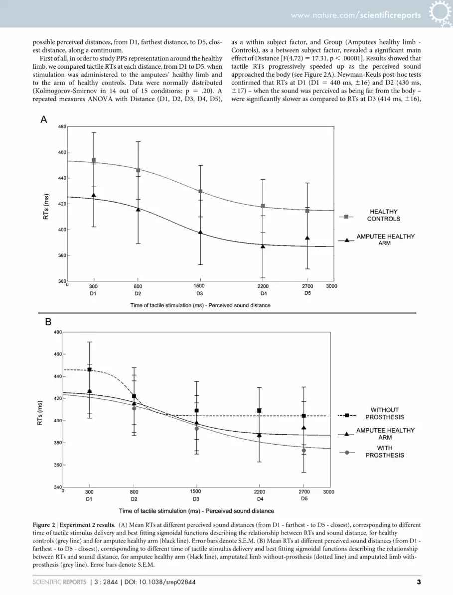
First of all, in order to study PPS representation around the healthylimb, we compared tactile RTs at each distance, from D1 to D5, whenstimulation was administered to the amputees’ healthy limb andto the arm of healthy controls. Data were normally distributed(Kolmogorov-Smirnov in 14 out of 15 conditions: p 5 .20). Arepeated measures ANOVA with Distance (D1, D2, D3, D4, D5),
as a within subject factor, and Group (Amputees healthy limb -Controls), as a between subject factor, revealed a significant maineffect of Distance [F(4,72) 5 17.31, p , .00001]. Results showed thattactile RTs progressively speeded up as the perceived soundapproached the body (see Figure 2A). Newman-Keuls post-hoc testsconfirmed that RTs at D1 (D1 5 440 ms, 616) and D2 (430 ms,617) – when the sound was perceived as being far from the body –were significantly slower as compared to RTs at D3 (414 ms, 616),
Figure 2 | Experiment 2 results. (A) Mean RTs at different perceived sound distances (from D1 - farthest - to D5 - closest), corresponding to different
time of tactile stimulus delivery and best fitting sigmoidal functions describing the relationship between RTs and sound distance, for healthy
controls (grey line) and for amputee healthy arm (black line). Error bars denote S.E.M. (B) Mean RTs at different perceived sound distances (from D1 -
farthest - to D5 - closest), corresponding to different time of tactile stimulus delivery and best fitting sigmoidal functions describing the relationship
between RTs and sound distance, for amputee healthy arm (black line), amputated limb without-prosthesis (dotted line) and amputated limb with-
prosthesis (grey line). Error bars denote S.E.M.
www.nature.com/scientificreports
SCIENTIFIC REPORTS | 3 : 2844 | DOI: 10.1038/srep02844 3

D4 (402 ms, 616) and D5 (404 ms, 616; all ps , .01) – when thesound was perceived as being close to the body. This pattern of resultswas equivalent between patients and healthy controls. Indeed, nei-ther the main effect of Group [F(1,18) 5 0.81, p 5 .38] nor theDistance X Group interaction [F(4,72) 5 0.32, p 5 .86] were signifi-cant. Taken together these results suggest that there is a criticalspatial range (in this case between D2 and D3) within which auditorystimuli begin interacting with tactile stimuli administered on thebody surface, resulting in quicker tactile RTs. This spatial range couldbe considered as the boundaries of the PPS. The present analysissuggests that the boundaries of PPS representation around the upperlimb do not differ between healthy controls and amputees, for whatconcerned the non-affected side of their body.
In order to study the effect of amputation and prosthesis implanta-tion on PPS representation, we compared the results betweenpatients’ healthy and amputated limb, while wearing or not wearingtheir prosthesis. We entered tactile RTs in a repeated measureANOVA with Condition (Healthy Limb, With Prosthesis andWithout Prosthesis) and Distance (D1, D2, D3, D4, D5) as the withinsubject factors. The main effect of Distance was significant [F(4,36)5 19.92, p , .00001], resembling the pattern of responses found forthe healthy limb and for controls: RTs became faster when the soundwas perceived as being closer to the body (see Figure 2B). Critically,the main effect of Condition was also significant [F(2,18) 5 3.64, p ,
.05]. A Newman-Keuls post-hoc test showed that when patientsperformed the task without the prosthesis, RTs were slower(418 ms, 629) as compared to when they performed the task withthe healthy limb (404 ms, 629; p , .05; one-tailed). When patientsperformed the task with the amputated limb, RTs were faster whenthey wore the prosthesis (398 ms, 626) as compared to when theydid not wear the prosthesis (p , .05). RTs were not significantlydifferent between the Healthy limb and the With Prosthesis condi-tions (p 5 .45). Taken together these results suggest that amputationaffected PPS representation around the stump, as compared to thenon-affected limb. Wearing a prosthesis compensated this effect,making PPS representation around the stump more similar to PPSrepresentation around the healthy limb.
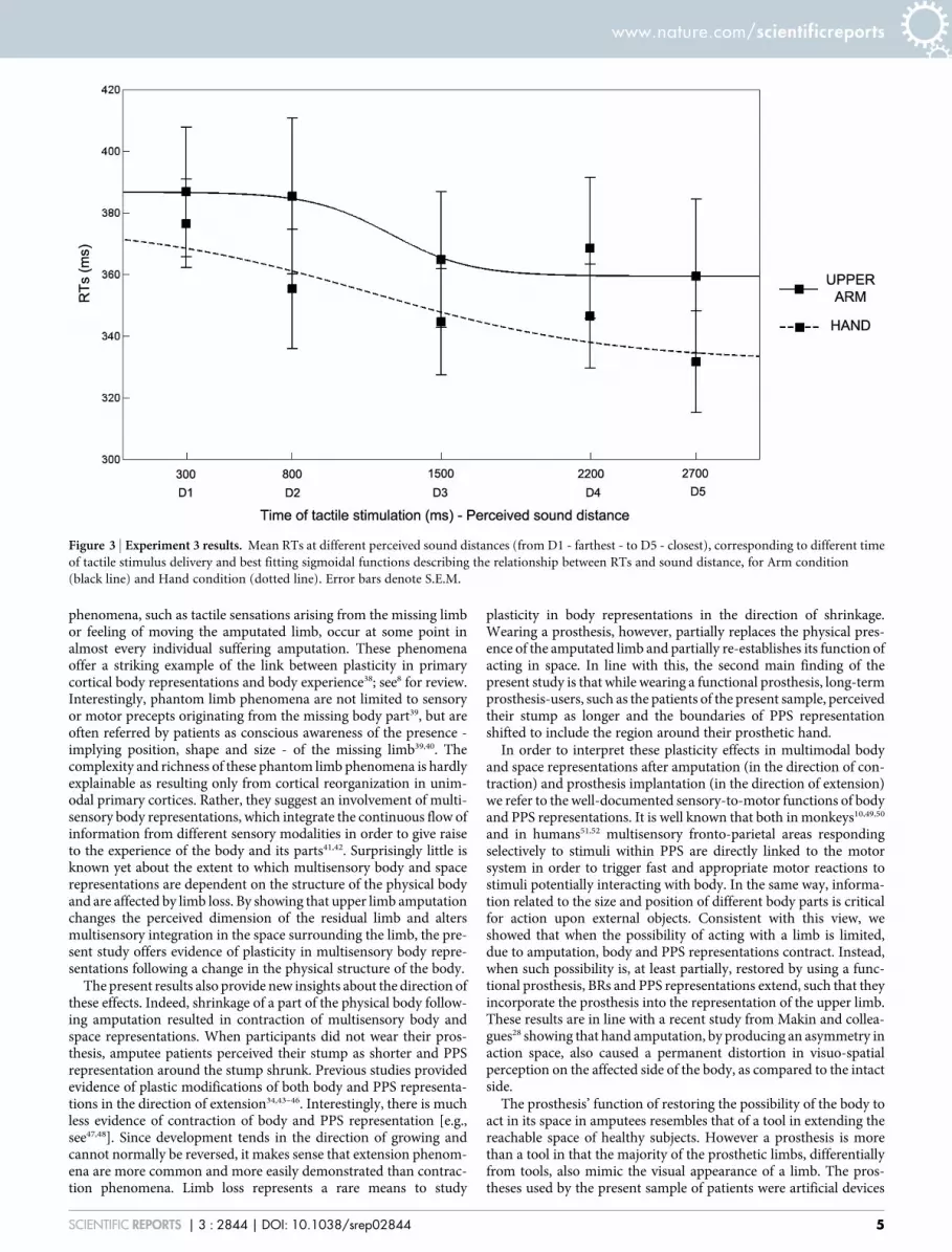
Experiment 3 - prosthesis implantation shifts PPS coding fromthe stump to the prosthetic hand. In order to interpret the differ-ential effect on RTs found between the with - and without prosthesisconditions in amputee patients, we hypothesized that, when patientsdid not wear the prosthesis, sound position was codified with respectto the stump, which represented the boundaries of the physical body.Instead, when patients wore their prosthesis, the perceived positionof sound in space was re-calibrated with respect to the prosthetichand, such that the prosthetic hand itself represented the new bodyboundary. In other words, audio-tactile interaction was coded withrespect to the stump in the Without Prosthesis condition and withrespect to the prosthetic hand in the With Prosthesis condition,resulting in a general reduction of RTs in every temporal delay(i.e., at each sound distance) when the prosthesis was on. In orderto test this hypothesis, we ran a further experiment on healthyparticipants. The task was the same as for Experiment 2, inexception for a manipulation in the location of tactile stimuli.Sound positions were kept constant in relationship to the upperarm (i.e., the near loudspeaker was close to the upper arm and thefar loudspeaker was at 100 cm). Tactile targets, however, wereadministered in two different experimental conditions either at thearm (Upper Arm condition), or at the hand (Hand conditions). Thisway we aimed at simulating, respectively, the stump stimulation inthe Without Prosthesis condition and the recoding of auditory-tactile interaction to the prosthetic hand in the With Prosthesiscondition in amputee patients.
Subjects performed two blocks for each experimental condition(Upper Arm and Hand condition), ran in a counterbalanced order.
False alarms and omissions were rare (on average: .45 and 1.89%, persubject, per condition, respectively). Data were normally distributed(Kolmogorov- Smirnov in 9 out of 10 conditions, p..09). Mean RTs(after trimming outliers, .54% of trails per subject per condition) totactile stimulation were entered in a repeated-measures ANOVAwith Condition (Upper Arm – Hand) and Distance (from D1 toD5), as within subject factors, and Order of administration (UpperArm – Hand; Hand – Upper Arm) as between subject factor. As inthe previous experiment, the main effect of Distance was significant[F(4,40) 5 11.64, p , .0001], replicating the modulation of tactileRTs depending on the position of sound in space (see Figure 3). TheCondition X Distance interaction was not significant [F(4,40) 5 1.56,p 5 .20]. Critically, also the main effect of Condition was significant[F(1,10) 5 5.47, p , .05]. Newman-Keuls post-hoc tests showed thatwhen subjects performed the task while receiving the tactile stimu-lation on their hand they were faster (351 ms, 615) in every tem-poral delay as compared to when they received the tactile stimulationon their upper arm (373 ms, 621). These results suggest that whenthe tactile stimulation was administered at the hand, while the nearsound source was placed close to the upper arm, sounds were pro-cessed as if they were closer to the boundaries of the stimulated limb,i.e., the hand. This pattern of results evidenced a general reduction oftactile RTs in every temporal delay as compared to when tactilestimuli were administered at the upper arm. This effect suggests thatthe perceived position of sound was computed with respect to thepart of the limb tactilely stimulated and clearly resembles the effectfound in amputee patients when task was performed while wearing,as compared to not wearing, the prosthesis.
DiscussionTwo main results have been obtained by the present study. First, amodification in the physical structure of the body, such as limb lossdue to traumatic amputation, affects high-order multisensory repre-sentations of the body and of the space around the body. Second,such effects are, at least partially, compensated by prosthesis implan-tation substituting the lost body part. Results from Experiment 1show that, following amputation, the implicitly perceived length ofthe residual part of the upper arm decreased, such that patientsperceived their stump as shorter as compared to the healthy arm.Wearing a prosthesis increased the perceived length of the arm,making the perception of the stump length more similar to that ofthe healthy limb. Amputation and prosthesis implantation also affec-ted the representation of PPS around the stump. In Experiment 2,task-irrelevant sounds boosted tactile RTs in so far as they wereperceived as being closer to the stimulated body part, both in healthycontrols and in amputees tested on their healthy limb. This multi-sensory effect was reduced when amputated patients were tested ontheir amputated limb (without-prosthesis); in this condition patientsshowed slower RTs as compared to conditions involving healthylimb assessment or healthy controls, suggesting that, after ampu-tation, the boundaries of PPS shifted towards the stump. However,when the task was administered on the stump while patients woretheir prosthesis, there was again the same speeding effect on tactileRTs depending on the position of sounds in space, as for the healthylimb and in controls. These results suggested that prosthesisimplantation restored the boundaries of PPS so that they includedthe prosthetic hand. Experiment 3 supports this conclusion, byshowing that in healthy subjects, dynamic sounds coupled to tactilestimulation of the upper arm or of the hand resulted in the samemodulation of tactile RT similar as that found in amputees respect-ively when they did not wear or they wore their prosthesis.
Until now, an extensive body of evidence has demonstrated thatamputation modifies unimodal motor and somatosensory represen-tations of the body in the brain, both in monkeys22,36,37 and inhumans8,23–25 suggesting a strong dependency of unimodal bodyrepresentations on the structure of the physical body. Phantom limb
www.nature.com/scientificreports
SCIENTIFIC REPORTS | 3 : 2844 | DOI: 10.1038/srep02844 4
phenomena, such as tactile sensations arising from the missing limbor feeling of moving the amputated limb, occur at some point inalmost every individual suffering amputation. These phenomenaoffer a striking example of the link between plasticity in primarycortical body representations and body experience38; see8 for review.Interestingly, phantom limb phenomena are not limited to sensoryor motor precepts originating from the missing body part39, but areoften referred by patients as conscious awareness of the presence -implying position, shape and size - of the missing limb39,40. Thecomplexity and richness of these phantom limb phenomena is hardlyexplainable as resulting only from cortical reorganization in unim-odal primary cortices. Rather, they suggest an involvement of multi-sensory body representations, which integrate the continuous flow ofinformation from different sensory modalities in order to give raiseto the experience of the body and its parts41,42. Surprisingly little isknown yet about the extent to which multisensory body and spacerepresentations are dependent on the structure of the physical bodyand are affected by limb loss. By showing that upper limb amputationchanges the perceived dimension of the residual limb and altersmultisensory integration in the space surrounding the limb, the pre-sent study offers evidence of plasticity in multisensory body repre-sentations following a change in the physical structure of the body.
The present results also provide new insights about the direction ofthese effects. Indeed, shrinkage of a part of the physical body follow-ing amputation resulted in contraction of multisensory body andspace representations. When participants did not wear their pros-thesis, amputee patients perceived their stump as shorter and PPSrepresentation around the stump shrunk. Previous studies providedevidence of plastic modifications of both body and PPS representa-tions in the direction of extension34,43–46. Interestingly, there is muchless evidence of contraction of body and PPS representation [e.g.,see47,48]. Since development tends in the direction of growing andcannot normally be reversed, it makes sense that extension phenom-ena are more common and more easily demonstrated than contrac-tion phenomena. Limb loss represents a rare means to study
plasticity in body representations in the direction of shrinkage.Wearing a prosthesis, however, partially replaces the physical pres-ence of the amputated limb and partially re-establishes its function ofacting in space. In line with this, the second main finding of thepresent study is that while wearing a functional prosthesis, long-termprosthesis-users, such as the patients of the present sample, perceivedtheir stump as longer and the boundaries of PPS representationshifted to include the region around their prosthetic hand.
In order to interpret these plasticity effects in multimodal bodyand space representations after amputation (in the direction of con-traction) and prosthesis implantation (in the direction of extension)we refer to the well-documented sensory-to-motor functions of bodyand PPS representations. It is well known that both in monkeys10,49,50
and in humans51,52 multisensory fronto-parietal areas respondingselectively to stimuli within PPS are directly linked to the motorsystem in order to trigger fast and appropriate motor reactions tostimuli potentially interacting with body. In the same way, informa-tion related to the size and position of different body parts is criticalfor action upon external objects. Consistent with this view, weshowed that when the possibility of acting with a limb is limited,due to amputation, body and PPS representations contract. Instead,when such possibility is, at least partially, restored by using a func-tional prosthesis, BRs and PPS representations extend, such that theyincorporate the prosthesis into the representation of the upper limb.These results are in line with a recent study from Makin and collea-gues28 showing that hand amputation, by producing an asymmetry inaction space, also caused a permanent distortion in visuo-spatialperception on the affected side of the body, as compared to the intactside.
The prosthesis’ function of restoring the possibility of the body toact in its space in amputees resembles that of a tool in extending thereachable space of healthy subjects. However a prosthesis is morethan a tool in that the majority of the prosthetic limbs, differentiallyfrom tools, also mimic the visual appearance of a limb. The pros-theses used by the present sample of patients were artificial devices
Figure 3 | Experiment 3 results. Mean RTs at different perceived sound distances (from D1 - farthest - to D5 - closest), corresponding to different time
of tactile stimulus delivery and best fitting sigmoidal functions describing the relationship between RTs and sound distance, for Arm condition
(black line) and Hand condition (dotted line). Error bars denote S.E.M.
www.nature.com/scientificreports
SCIENTIFIC REPORTS | 3 : 2844 | DOI: 10.1038/srep02844 5

that faithfully reproduced the exterior appearance of a real arm andhand; they were also controlled myoelectrically or kinematically byresidual muscles in order to allow quite complex limb movements.Since prosthetic limbs share more features with an anatomical limbas compared to tools, prosthesis-use and tool-use could have differ-ential effects on the plasticity of BRs. In particular, distinction hasbeen proposed between the effects of body extension (e.g., in the caseof tool-use) and body incorporation (e.g., in the case of prosthesis-use)53,54. It is still not clear, however, whether and to what extent aprosthetic limb can be embodied such that it becomes in some senseindistinguishable from a real body part. We did not directly test thesubjective experience of prosthesis embodiment, but the presentresults suggest that although the effect of prosthesis implantationmight overcome that of tool-use, prosthetic limbs cannot be totallyconceived as a real part the body, at least because they are known tobe attachments that can be taken off. Accordingly, our data show thatin amputee patients, two different body representations coexistedand were differentially activated when patients did or did not weartheir prosthesis. Indeed, the perceived length of the stump and theextension of PPS immediately shrunk or elongated, depending onwhether the prosthesis was respectively on or off. The coexistence ofmultiple body representations depending on different body states
(with or without a tool) resembles other forms of plasticity shownafter long-term tool-use experiences55,56.
Understanding the mechanisms of prostheses embodiment andidentifying key features of prosthetic devices favouring prosthesis-use and acceptance are key issues for rehabilitation of limb loss andthe new field of neuroprosthetics. The present study might contrib-ute to research in this field: on the one hand, it demonstrates strikingeffects of amputation and prosthesis implantation on the perceptionof body part size and on multisensory integration in the space aroundthe body. On the other hand, it proposes sensitive and easy-to-applytasks to measure the effects of using prosthetic devices on BRs andPPS representations.
MethodsAmputee subjects. Ten volunteers participated in the study (8 males and 2 females,mean age 45 years, range 21–66 years), recruited at the INAIL Prostheses Centre,Budrio, Bologna (http://www.inail-ricerca.it/index.aspx). They were healthy exceptthat they had all one upper limb amputated either below or above the elbow, followinga traumatic accident. Before the accident all patients were right-handed. Theinclusion criterion was that they must have been using a functional prosthesis,cosmetically designed, so they resembled arm appearance, at least 4–8 h daily for 5–7days per week for at least 1 year. Patients’ demographic and clinical data are reportedin Table 1. All subjects gave their informed consent to participate in the study, whichwas approved by the Ethical commission of the INAIL Prostheses Centre andperformed in accordance with the Declaration of Helsinki.
Control subjects. Twenty-nine healthy volunteers participated in Experiment 1 (N 5
7, 5 males, mean age 5 33.7 years, range 25–62 years), Experiment 2 (N 5 10, allfemales, mean age 5 22.1 years, range 19–24 years) and Experiment 3 (N 5 12, 2males, mean age 5 23.4 years, range 20–26 years) as control groups. All subjects gavetheir informed consent to participate in the study, which was approved by the Ethicalcommission of the INAIL Prostheses Centre and performed in accordance with theDeclaration of Helsinki.
Experimental procedure: overall structure. Amputee patients participated in twoexperiments, assessing BRs (Experiment 1) and PPS representations (Experiment 2),performed in a single 2-hour and a half session. Seven out of ten patients participatedin Experiment 1: two patients were excluded because of the very high level of theamputation (above the elbow; P1 and P10, see Table 1), thus not allowingadministering tactile stimuli of sufficient dimensions (see Experiment 1 methods),because the prosthesis covered most of the stump surface. One patient could notperform the experiment for matter of time (P3, see Table 1). All patients participatedin Experiment 2.
Patients performed Experiment 1 and Experiment 2 in three different experimentalconditions: stimuli were administered on the healthy limb (Healthy limb condition),on the amputated limb without prosthesis (Without Prosthesis condition) and on theamputated limb with prosthesis (With Prosthesis condition). All healthy subjectsfrom the control group performed the tasks in Experiment 1 and 2 on their rightupper arm, on a skin region matching the site of stump stimulation used for amputeepatients.
Procedure and materials. Experiment 1 - tactile distance perception task. In order toassess the perceived length of the residual part of the upper limb and of thehomologous region of the healthy limb, we used a tactile distance perception task. Oneach trial of the tactile distance perception task, participants were touched with a pairof dots on the forehead and then with a pair of dots on the upper arm, longitudinallydelivered along the arm axis. Participants made untimed two-alternative forced-choice judgments of whether the two points felt farther apart on the forehead or onthe upper arm, responding verbally ‘‘forehead’’ or ‘‘arm’’. Subjects were lain down
Table 1 | Patients’ demographic and clinical data. All patients but P1, P3 and P10 participated in Experiment 1. All patients participated inExperiment 2
Patient Age Gender Handiness Amputation side, level Prosthesis type Years since amputation Phantom limb symptoms
P1 41 F Dx Right, above elbow Kinematic 2 YesP2 56 M Dx Left, above elbow Myoelectric 21 YesP3 50 M Dx Right, above elbow Myoelectric 18 NoP4 30 M Dx Right, below elbow Kinematic 1.5 NoP5 21 F Dx Right, below elbow Kinematic 2.5 YesP6 38 M Dx Right, below elbow Myoelectric 18 NoP7 66 M Dx Right, below elbow Myoelectric 16 YesP8 62 M Dx Right, below elbow Myoelectric 42 NoP9 41 M Dx Right, below elbow Kinematic 4 NoP10 43 M Dx Left, above elbow Kinematic 3 Yes
Figure 4 | Experimental set up for Experiment 2 and Experiment 3.
www.nature.com/scientificreports
SCIENTIFIC REPORTS | 3 : 2844 | DOI: 10.1038/srep02844 6

with the tested arm resting in a prone position. In order to set the spatial distancebetween the stimuli administered to the forehead and the upper arm, we initiallymeasured the two-point discrimination threshold (2 pdt) on the upper arm forlongitudinal orientation by using a staircase method, as described in57. For eachsubject, the individual 2 pdt on the arm was used to set the distance between the pairsof dots used during the tactile distance task. Three different inter-point distances wereused: at the 2 pdt; 1.5 the 2 pdt; and twice the 2 pdt. The task comprised a total of 36trials. An experimenter administered the stimuli manually for approximately onesecond, with an inter-stimulus interval of one second between taps on the foreheadand the arm. Subjects were blindfolded throughout the procedure.
The perceived size of tactile stimuli touching the body depends on the perceiveddimension of the body part tactilely stimulated6,7,58,59 and is influenced by visual33 orproprioceptive60 information about the stimulated body part. Thus, the tactile dis-tance perception task can be used as an indirect measure of the internal representationof body part size [see also34].
Experiment 2 and 3- audio-tactile interaction task. During the task, subjects wereblindfolded and were seated with the tested arm resting prone on a table beside them.During each trial a sound (pink noise) was presented for 3000 ms. The sounds weregenerated by two loudspeakers, one placed on the table in proximity of the upper arm,the other one placed on the table, at a distance of ,100 cm from the near loudspeaker,thus far from the upper arm. Sound intensity was manipulated so that IN sounds hadexponentially rising acoustic intensity, while OUT sounds had exponentially fallingacoustic intensity. In this way, IN sounds gave the impression of a sound sourcemoving from the far to the near loudspeaker, i.e., towards the subject’s body, whileOUT sounds gave the impression of a sound source moving in the opposite direction,i.e. receding from the body. Along with the auditory stimulation, in half of the trialssubjects were also presented with a tactile stimulation, delivered by means of aconstant-current electrical stimulator (DS7A, Digitimer, Hertfordshire, UnitedKingdom), via a pair of neurological electrodes (Neuroline, Ambu, Ballerup,Denmark), placed on the dorsal surface of the upper arm. The remaining trials werecatch trials with auditory stimulation only. Subjects were asked to respond vocally tothe tactile target, when present, saying ‘‘TAH’’ as rapidly as possible, trying to ignorethe sound. Tactile RTs were recorded by means of a voice-activated relay. A PCrunning C.I.R.O. software (www.cnc.unibo.psice.unibo/ciro) was used to control thepresentation of the stimuli and to record responses [see35].
The critical experimental manipulation was that the tactile stimulus was deliveredat different temporal delays (from T1 to T5, see below) from the onset of the auditorystimulus, both for IN and OUT sounds. In this way, tactile stimulation occurred whenthe sound source was perceived at different locations with respect to the body: i.e.,close to the body, at high temporal delays for the IN sound and at low temporal delaysfor the OUT sound; and far from the body, at low temporal delays for the IN soundand at high temporal delays for the OUT sound. For each trial, the sound was pre-ceded and followed by 1000 ms of silence. Temporal delays from sound onset for thetactile stimulus were set as follows: T1, tactile stimulation administered at 300 ms, T2at 800 ms, T3 at 1500 ms, T4 at 2200 ms and T5 at 2700 ms (see Figure 4).
The task used for Experiment 2 and Experiment 3 consisted in a random com-bination of 8 target stimuli for each temporal delay, for the IN and OUT sounds,resulting in a total of 76 trials with a tactile target, randomly intermingled with 76catch trials. Trials were equally divided into 2 blocks, each block lasting about 8minutes.
Funding. This work was supported by a Volkswagen Stiftung grant (theUnBoundBody project, ref. 85 639) to AS and by a Spinner 2013 grant (Prosthesis andbrain) by Regione Emilia Romagna to EC and AS.
1. Penfield, W. & Boldrey, E. Somatic motor and sensory representation in thecerebral cortex of man as studied by electrical stimulation. Brain. 60, 389–443(1937).
2. Downing, P. E., Jiang, Y., Shuman, M. & Kanwisher, N. A cortical area selective forvisual processing of the human body. Science. 293, 2470–2473 (2001).
3. Orlov, T., Makin, T. R. & Zohary, E. Topographic representation of the humanbody in the occipitotemporal cortex. Neuron. 68, 586–600 (2010).
4. Head, H. & Holmes, G. Sensory disturbances from cerebral lesions. Brain. 34, 102(1911).
5. Gallagher, S. How the Body Shapes the Mind. New York: Oxford University Press.284, 0199271941 (2005).
6. Medina, J. & Coslett, H. B. From maps to form to space: touch and the bodyschema. Neuropsychologia. 48, 645–654 (2010).
7. Longo, M. R., Azanon, E. & Haggard, P. More than skin deep: body representationbeyond primary somatosensory cortex. Neuropsychologia. 48, 655–668 (2010).
8. Serino, A. & Haggard, P. Touch and the body. Neurosci Biobehav Rev. 34, 224–236(2010).
9. Rizzolatti, G., Fadiga, L., Fogassi, L. & Gallese, V. The space around us. Science.277, 190–191 (1997).
10. Graziano, M. S. & Cooke, D. F. Parieto-frontal interactions, personal space, anddefensive behavior. Neuropsychologia. 44, 845–859 (2006).
11. Ladavas, E. Functional and dynamic properties of visual peripersonal space.Trends Cogn Sci. 6, 17–22 (2002).
12. Ladavas, E. & Serino, A. Action-dependent plasticity in peripersonal spacerepresentations. Cogn Neuropsychol. 25, 1099–1113 (2008).
13. Duhamel, J. R., Colby, C. L. & Goldberg, M. E. Ventral intraparietal area of themacaque:congruent visual and somatic response properties. J Neurophysiol 79,126–136 (1998)
14. Graziano, M. S. & Cooke, D. F. Parieto-frontal interactions, personal space, anddefensive behavior. Neuropsychologia 44, 845–859 (2006).
15. Graziano, M. S. A. & Botvinick, M. M. How the brain represents the body: insightsfrom neurophysiology and psychology. In: Common Mechanisms in Perceptionand Action: Attention and Performance XIX. Eds. Prinz, W. and Hommel, B.Oxford University Press, Oxford England, pp. 136–157 (2002).
16. Bremmer, F., Schlack, A., Shah, N. J., Zafiris, O., Kubischik, M., Hoffmann, K. P.et al. Polymodal Motion Processing in Posterior Parietal and Premotor Cortex: AHuman fMRI Study Strongly Implies Equivalencies between Humans andMonkeys. Neuron 29, 287–296 (2001).
17. Filimon, F., Nelson, J. D., Huang, R. S., Sereno, M. I. Multiple parietal reachregions in humans: cortical representations for visual and proprioceptivefeedback during on-line reaching. J Neurosci 29, 2961–71 (2009).
18. Makin, T. R., Holmes, N. P., Zohary, E. Is That Near My Hand? MultisensoryRepresentation of Peripersonal Space in Human Intraparietal Sulcus. J Neurosci27, 731–740 (2007).
19. Sereno, M. I. & Huang, R. S. A human parietal face area contains aligned head-centered visual and tactile maps. Nat Neurosci 9, 1337–1343 (2006).
20. Serino, A., Canzoneri, E., Avenanti, A. Fronto-parietal Areas Necessary for aMultisensory Representation of Peripersonal Space in Humans: An rTMS Study.J Cogn Neurosci 23, 2956–2967 (2011).
21. Blanke, O. Multisensory brain mechanisms of bodily self-consciousness. NatNeurosci Rev 13, 556–571 (2012).
22. Buonomano, D. V. & Merzenich, M. M. Cortical plasticity: from synapses to maps.Annu Rev Neurosci. 21, 149–186 (1998).
23. Chen, R., Corwell, B., Yaseen, Z., Hallett, M. & Cohen, L. G. Mechanisms ofcortical reorganization in lower-limb amputees. J Neurosci. 18, 3443–3450 (1998).
24. Reilly, K. T. & Sirigu, A. The motor cortex and its role in phantom limbphenomena. Neuroscientist. 14, 195–202 (2008).
25. Borsook, D., Becerra, L., Fishman, S., Edwards, A., Jennings, C. L. et al. Acuteplasticity in the human somatosensory cortex following amputation. Neuroreport.9, 1013–1017 (1998).
26. Nico, D., Daprati, E., Rigal, F., Parsons, L. & Sirigu, A. Left and right handrecognition in upper limb amputees. Brain. 127, 120–132 (2004).
27. Ehrsson, H. H., Rosen, B., Stockselius, A., Ragno, C., Kohler, P. et al. Upper limbamputees can be induced to experience a rubber hand as their own. Brain. 131,3443–3452 (2008).
28. Makin, T. R., Wilf, M., Schwartz, I, Zohary, E. Amputees ‘‘neglect’’ the space neartheir missing hand. Psychol Sci. 21, 55–7 (2010).
29. Ebbinghaus, H. Memory: A Contribution to Experimental Psychology. New York,NY: Dover Publications (1987).
30. Linkenauger, S. A., Ramenzoni, V. & Proffitt, D. R. Illusory shrinkage and growth:body-based rescaling affects the perception of size. Psychol Sci. 21, 1318–25(2010).
31. Linkenauger, S. A., Witt, J. K. & Proffitt, D. R. Taking a hands-on approach:apparent grasping ability scales the perception of object size. J Exp Psychol HumPercept Perform. 37, 1432–41 (2011).
32. Bruno, N. & Bertamini, M. Haptic perception after a change in hand size.Neuropsychologia. 48, 1853–6 (2010)
33. Taylor-Clarke, M., Jacobsen, P. & Haggard, P. Keeping the world a constant size:object constancy in human touch. Nat Neurosci. 7, 219–220 (2004).
34. Canzoneri, E., Ubaldi, S., Rastelli, V., Finisguerra, A., Bassolino, M. & Serino, A.Tool-use reshapes the boundaries of body and peripersonal space representations.Exp Brain Res. 228, 25–42 (2013).
35. Canzoneri, E., Magosso, E. & Serino, A. Dynamic sounds capture the boundariesof peripersonal space representation in humans. PLoS One. 7, e44306 (2012).
36. Wu, C. W. & Kaas, J. H. Reorganization in primary motor cortex of primates withlong-standing therapeutic amputations. J Neurosci. 19, 7679–7697 (1999).
37. Merzenich, M. M., Nelson, R. J., Stryker, M. P., Cynader, M. S., Schoppmann, A.et al. Somatosensory cortical map changes following digit amputation in adultmonkeys. J Comp Neurol. 224, 591–605 (1984).
38. Karl, A., Birbaumer, N., Lutzenberger, W., Cohen, L. G. & Flor, H. Reorganizationof motor and somatosensory cortex in upper extremity amputees with phantomlimb pain. J Neurosci. 21, 3609–3618 (2001).
39. Hunter, J. P., Katz, J. & Davis, K. D. The effect of tactile and visual sensory inputson phantom limb awareness. Brain. 126, 579–589 (2003).
40. Flor, H., Nikolajsen, L. & Staehelin, Jensen Phantom limb pain: a case ofmaladaptive CNS plasticity? Nat Rev Neurosci. 7, 873–881 (2006).
41. Ehrsson, H. H. The Concept of Body Ownership. In: Stein, B. E., editor. The NewHandbook of Multisensory Processing. MIT Press (in press).
42. Ionta, S., Gassert, R. & Blanke, O. Multi-sensory and sensorimotor foundation ofbodily self-consciousness - an interdisciplinary approach. Front Psychol. 2, 383(2011).
43. Maravita, A., Spence, C., Kennett, S. & Driver, J. 2002 Tool-use changesmultimodal spatial interactions between vision and touch in normal humans.Cognition. 83, B25–34 (2002).
44. Holmes, N. P., Calvert, G. A. & Spence, C. Extending or projecting peripersonalspace with tools? Multisensory interactions highlight only the distal and proximalends of tools. Neurosci Lett. 372, 62–67 (2004).
www.nature.com/scientificreports
SCIENTIFIC REPORTS | 3 : 2844 | DOI: 10.1038/srep02844 7

45. Farne, A. & Ladavas, E. Dynamic size-change of hand peripersonal spacefollowing tool use. Neuroreport. 11, 1645–1649 (2000).
46. Maravita, A., Husain, M., Clarke, K. & Driver, J. Reaching with a tool extendsvisual-tactile interactions into far space: evidence from cross-modal extinction.Neuropsychologia. 39, 580–585 (2001).
47. Di Russo, F., Committeri, G., Pitzalis, S., Spitoni, G., Piccardi, L. et al. Corticalplasticity following surgical extension of lower limbs. NeuroImage. 30, 172–183(2006).
48. Longo, M. R., Kammers, M. P., Gomi, H., Tsakiris, M. & Haggard, P. Contractionof body representation induced by proprioceptive conflict. Curr Biol. 19, R727–8(2009).
49. Graziano, M. S., Taylor, C. S. & Moore, T. Complex movements evoked bymicrostimulation of precentral cortex. Neuron. 34, 841–851 (2002).
50. Stepniewska, I., Fang, P. C. & Kaas, J. H. Microstimulation reveals specializedsubregions for different complex movements in posterior parietal cortex ofprosimian galagos. Proc Natl Acad Sci USA. 102, 4878–4883 (2005).
51. Serino, A., Annella, L. & Avenanti, A. Motor properties of peripersonal space inhumans. PLoS One. 4, e6582 (2009).
52. Makin, T. R., Holmes, N. P., Brozzoli, C., Rossetti, Y. & Farne, A. Coding of visualspace during motor preparation: Approaching objects rapidly modulatecorticospinal excitability in hand-centered coordinates. J Neurosci. 29,11841–11851 (2009).
53. De Preester, H. & Tsakiris, M. 2009 Body-extension versus body-incorporation: Isthere a need for a body-model? Phenom Cogn Sci. 8, 307–319.
54. Giummarra, M. J., Gibson, S. J., Georgiou-Karistianis, N. & Bradshaw, J. L.Mechanisms underlying embodiment, disembodiment and loss of embodiment.Neurosci Biobehav Rev. 32, 143–60 (2008).
55. Serino, A., Bassolino, M., Farne, A. & Ladavas, E. 2007 Extended multisensoryspace in blind cane users. Psychol Sci. 18, 642–648 (2007).
56. Bassolino, M., Serino, A., Ubaldi, S. & Ladavas, E. Everyday use of the computermouse extends peripersonal space representation. Neuropsychologia. 48, 803–811(2010).
57. Serino, A., Padiglioni, S., Haggard, P. & Ladavas, E. 2009 Seeing the hand boostsfeeling on the cheek. Cortex. 45, 602–9.
58. de Vignemont, F., Majid, A., Jola, C. & Haggard, P. Segmenting the body intoparts: evidence from biases in tactile perception. Q J Exp Psychol (Colchester). 62,500–512 (2009).
59. Longo, M. R. & Haggard, P. 2011 Weber’s illusion and body shape: Anisotropy oftactile size perception on the hand. J Exp Psychol Hum Percept Perform. 37,720–726 (2011).
60. de Vignemont, F., Ehrsson, H. H. & Haggard, P. Bodily illusions modulate tactileperception. Curr Biol. 15, 1286–1290 (2005).
AcknowledgementsThe authors thank patients for their participation in the study, Angelo Davalli for hissupport and collaboration, Francesca Baldassarri for patients recruitment, ValentinaTedeschi for her contribution in data collection, Adrian Smith and Ana Tajadura for theircomments and Elisa Magosso for her help in data analysis.
Author contributionsThe experiment was conceived by AS and EC. Data were collected by EC and MM, and theyhave been analysed by EC. Recruiting of the patients was done by AA and GV. Themanuscript text was written by EC and AS.
Additional informationCompeting financial interests: The authors declare no competing financial interests.
How to cite this article: Canzoneri, E., Marzolla, M., Amoresano, A., Verni, G. & Serino, A.Amputation and prosthesis implantation shape body and peripersonal spacerepresentations. Sci. Rep. 3, 2844; DOI:10.1038/srep02844 (2013).
This work is licensed under a Creative Commons Attribution-NonCommercial-NoDerivs 3.0 Unported license. To view a copy of this license,
visit http://creativecommons.org/licenses/by-nc-nd/3.0
www.nature.com/scientificreports
SCIENTIFIC REPORTS | 3 : 2844 | DOI: 10.1038/srep02844 8