amifostine induces anaerobic metabolism and hypoxia-inducible factor 1α
TRANSCRIPT
Cancer Chemother Pharmacol (2004) 53:8-14 DOI 10.1007/s00280-003-0691-z
O R I G I N A L A R T I C L E
Michael I. Koukourakis �9 Alexandra Giatromanolaki Wen Chong �9 Costantinos S imopoulos Alexandros Polychronidis �9 Ef lhimios Sivridis Adrian L. Harris
Amifostine induces anaerobic metabolism and hypoxia-inducible factor
Received: 8 May 2003 / Accepted: 11 July 2003/Published online: 22 October 2003 �9 Springer-Verlag 2003
Abstract Purpose: The cytoprotective mechanism of amifostine (WR-2721) implies free radical scavenging and DNA repair activities. We investigated additional cytoprotective pathways involving intracellular hypoxia and the activation of the hypoxia-inducible factor (HIF) pathway, a key transcription factor regulating glycolysis, angiogenesis and apoptosis, which is also linked with radioresistance. Materials and methods: The glucose and oxygen levels in the peripheral blood of patients receiving 1000 mg amifostine were determined at vari- ous time-points in order to investigate the metabolic changes induced by amifostine. MDA468 breast tumor cell lines were incubated with a high amifostine con- centration (10 mM) to overcome the natural resistance of cancer cells to influx of the non-hydrolyzed WR-2721, and the HIFI~ protein levels were determined by Wes- tern blot analysis. In vivo experiments with Wistar rats were performed in order to assess immunohistochemi- cally changes in the intracellular accumulation of HIFlc~ induced by amifostine (200 mg/kg). Results: By 30 min following amifostine administration, the hemoglobin oxygen saturation and pO2 levels had increased in the peripheral blood while glucose levels had reduced, pro- viding evidence that normal tissue metabolism switches to glycolytic pathways. Incubation of cell lines with amifostine resulted in HIFlc~ induction. In Wistar rats administration of amifostine resulted in increased
This work is presented on behalf of the Tumour and Angiogenesis Research Group.
M. I. Koukourakis ([~) �9 A. Giatromanolaki �9 C. Simopoulos A. Polychronidis �9 E. Sivridis Departments of Radiotherapy/Oncology, Pathology and Surgery, Democritus University of Thrace, 68100 Alexandroupolis, Greece E-mail: [email protected] Tel.: + 30-6932-480808 Fax: + 30-25510-74623
W. Chong �9 A. L. Harris Cancer Research UK, Molecular Oncology Laboratories, Institute of Molecular Medicine, John Radcliffe Hospital, Oxford, OX3 9DS UK
HIFlc~ accumulation in normal tissues. Conclusions: Since it is doubtful whether dephosphorylation of ami- fostine to the active metabolite WR-1065 occurs within tumoral tissues (an acidic environment that lacks vas- cular alkaline phosphatase activity), intracellular hy- poxia and upregulation of HIFI~ represents an additional, normal tissue-specific, amifostine cytopro- tective pathway.
Keywords Amifostine. HIFlc~ �9 Hypoxia �9 Glycolysis
Introduction
Amifostine (Ethyol, WR-2721), an organic triphosphate, is the first broad-spectrum selective cytoprotective drug approved for clinical use in conjunction with radio- therapy and chemotherapy. The mechanism of cyto- protection is complicated and, to a certain extent, unclear. Free radical scavenging activities of the thiolic metabolite WR-1065 [1] and enhancement of the DNA repair process by the disulfide metabolite WR-33278 [2] are well-characterized mechanisms. More recently, additional cytoprotective pathways involving transcrip- tional regulation of genes involved in cellular apoptosis (nuclear factor-kappaB, activator protein-l, and p53) have been postulated [3, 4].
Microcalorimetric studies have shown that incuba- tion of cells with WR-1065 results in heat production that lasts for at least 90 rain [5]. This heat, which is probably a result of the oxidation of WR-1065 to disulfides, leads to a rapid consumption of intracellular oxygen and to hypoxia. Such intracellular hypoxia could be easily overcome in vivo by an increase in oxygen extraction from the blood. Several year ago, however, Glover et al. observed that, in contrast to what was expected, the mean venous oxygen tension and the hemoglobin oxygen saturation rise following amifostine administration [6]. Allalunis-Turner et al. also noted that administration of amifostine at maximally radio- protective doses significantly increases the binding of the
hypoxia marke r [3H]misonidazole to bone mar row cells, suggesting that hypoxia may con t r ibu te to amifost ine- media ted cy topro tec t ion [7].
In the present study, we found evidence that ami- fostine induces no rma l tissue hypoxia and glycolysis, and upregula tes hypoxia- induc ib le factor H I F l e , a key t ranscr ip t ion factor regulat ing the expression of a vari- ety of hypoxia-responsive genes.
Materials and methods
Climcal study
A group of 15 breast cancer patients with locally advanced inop- erable or recurrent disease were recruited into a phase I/II trial to test the safety and efficacy of hypofractionated and accelerated radiotherapy supported with high-dose daily amifostine [8]. An additional group of patients with operable high-risk breast cancer were treated with a similar postoperative regimen [9]. From among the patients recruited into the above-mentioned studies, 15 were entered into a parallel phase I study to assess changes in oxygen and glucose blood levels following amifostine administration.
Radiotherapy consisted of I2 consecutive fractions of radio- therapy, 3.5--4 Gy per fraction, delivered to the breast/chest and axlllary area, while a flat dose of 1000 mg amifostine was admin- istered 20 min before each radiotherapy fraction. The amifostine dose was diluted in 50 ml normal saline and rapidly infused within 5 min. No steroids were used.
The glucose levels in the peripheral blood were determined before amifostine infusion and at 30, 60 and 120 rain following infusion. A blood gas report was obtained by analyzing the peripheral venous blood (hemoglobin saturation, pO2, pCO2, HCO3, pH) at 30 and 60 min. Lactate serum levels were also re- corded before and at 1 h following amifostine administration. In order to avoid an eventual interference of hypotension with the results, patients with a drop in blood pressure of > 30 mmHg (or to below 80 mmHg) following amifostine administration were ex- cluded from the study.
In vitro study
Cell lines
MDA468 breast tumor cell lines were obtained from Imperial Cancer Research Fund ([CRF) Cell Services (London, UK), and passaged in E4 medium (ICRF Cell Services) supplemented with glutamine and 10% fetal calf serum. The decision to use the MDA468 breast tumor cell line instead of a normal cell culture for the present study was based on the experience in our laboratory with the response of this tumor cell line to hypoxia, as a clear detection of HIFlc~ signal can be obtained under hypoxic stress.
Amifostine
Amifostine was obtained from Sigma (Sigma-Aldrich, Poole, UK). A stock solution of 100 mM was prepared by dissolving in distilled water and the solution was stored at 4~ Stock solutaon was added to culture medium (1 ml solution to 9 ml medium) to give a final concentration of 10 mM. The choice of this specific concentration of amifostine was based on a previous study by Pardie et al., in which at this amifostine concentration, cell lines were able to im- port the drug intracellularly and hydrolyze it to WR-1065 [5]. Using this amifostine concentration, the authors were able to clearly detect rapid consumption of the intracellular oxygen that paralleled the WR-2721 hydrolysis, and we therefore tried to
simulate the conditions used in this microcalorimetric experiment in our experiments. If, under these conditions, our cancer cell line responded by consuming the intracellular oxygen, then HIFlc~ upregulation would be expected.
Normoxia/hypoxia incubators
Cells were exposed in a C O 2 water-jacketed incubator (Forma Scientific, Marietta, Ohio) at 37~ to an atmosphere containing 5% CO2 and 21% oxygen (normoxic conditions). Cells were also exposed in nitrogen-flushed hypoxic chambers (Cellhouse 170 HI; Heto-Holten, Allerod, Denmark) for 16 h to an atmosphere con- taining 5% CO2 and 0.1% oxygen (hypoxlc conditions) at 37~ and 95% humidity . The 16-h time-frame was chosen for optimal induction of hypoxia-responsive genes.
Western blot
Cells were obtained at various time-points (starting from 30 min up to 24 h), lysed in 8 M urea lysis buffer, and the homogenized ly- sates were stored at -20~ All samples were assayed and stan- dardized for protein levels (protein assay kit; Bio-Rad Laboratories, Hercules, Calif.) before separation on 8% SDS polyacrylamide gels by electrophoresis at 110 V for 2 h. Proteins were transferred onto Millipore Immobilon membranes using a semidry transfer technique over 45 rain. The membranes were washed in phosphate-buffered saline (PBS)/milk protein (Marvel) and 0.1%Tween 20, and blotted with primary antibodies: (1) HIF lc~ (mouse anti-human H IF1 c~ antibody, 1:1000; Transduction Laboratories, Becton Dickinson UK, Oxford, UK) and (2) /3-tubulin (mouse anti-human/~-tubulin antibody, 1:10,000; Sigma- Aldrich, Dorset, UK). Blots were then rewashed in PBS/ 0.1%Tween 20. A secondary goat anti-mouse antibody with horseradish peroxidase (DAKO, Glostrup, Denmark) was applied for 1 h at room temperature between washes. Immunodetection was performed with an ECL Western blotting kit (Amersham, Little Chalfont, UK), and blots exposed to X-ray film. Experiments were repeated to test the reproducibility of the results.
Animal studies
Animal studies were conducted at the Department of Experimental Surgery, Democritus University of Thrace, according to Hellenic laws (FI.& 160/91). Approval of the study was obtained by the local veterinary medicine authorities. Male pathogen-free Wistar rats at 3 months of age were used for the experiments. Under general anesthesia six animals underwent medial abdominal incision fol- lowed by ligation of the kidney vessels in four, or immediate kidney excision in two. Right kidneys were excised 20 min and left kidneys 60 rain following ligation. Animals remained under light anesthesia throughout the experiment.
Amifostine (Ethyol; Schering-Plough, Alimos, Greece) diluted in normal saline was injected intravenously or subcutaneously (200 mg/kg) into five animals, and 40 min after injection, kidneys, liver, intestine and lungs were excised under general anesthesia.
Immediately following excision of the organs, small thin slices of the tissues were fixed in 10% formalin solution and were embedded in paraffin 24 h later. Tissue sections were cut at 3 pm and mounted on poly-L-lysine-coated slides. The HIFlc~ protein expression was assessed immunohistochemically using the ab463 monoclonal antibody which recognizes the HIF 1 c~ protein of many animal species including that of rats (Abcam, Cambridge, UK). Sections were deparaffinized and peroxidase was quenched with methanol and 3% H202 for 15 min. Microwaving was used for antigen retrieval (3x4 rain). The primary antibody was applied overnight. Following washing with Tris-buffered saline (TBS), sections were incubated with a secondary antibody (Kwik bioti- nylated secondary, 0.69A; Shandon-Upshaw, Warrington. Pa.) for 15 min and washed in TBS. Kwik streptavidin peroxidase reagent (039A; Shandon-Upshaw) was applied for 15 min and sections
10
were again washed in TBS. The color was developed by a 15-min incubation with diaminobenzidine solution and sections were weakly counterstained with hematoxylin. Breast cancer tissue sec- tions with strong nuclear HIFI~ expression were used as positive controls. Normal mouse IgG was substituted for primary antibody as the negative control (same concentration as the test antibody).
Results
Clinical study
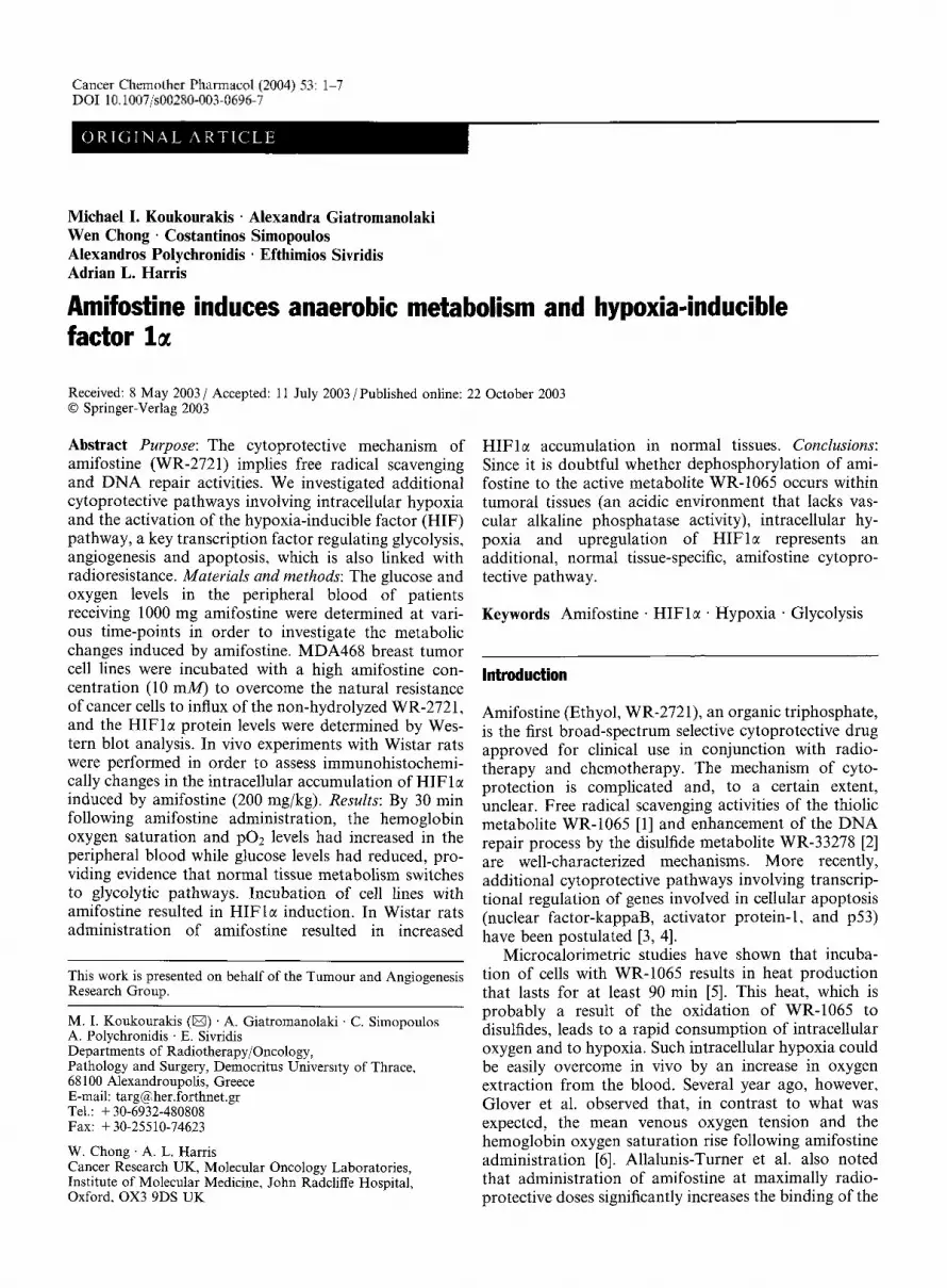
In breast cancer patients, 30 min following amifostine administration, the peripheral blood hemoglobin oxygen saturation had increased (67.8+13% vs 79.64-12%, means + SD; P < 0.04), but by 60 rain it had dropped to lower than the pretreatment levels (63.3 4- 16%; P < 0.00l). Similar changes were noted for the pO2 val- ues (45.94- 11 vs 58.64- 14 vs 40.54- 11 mmHg; P<0.01; Fig. la). The glucose levels had dropped from 1094-25 mg/dl to 844- 18 and 734- 14 mg/dl at 30 and 60 min following amifostine administration, respectively (P < 0.0001). At 2 h after injection, the glucose levels approached the normal levels (93 4-25 mg/dl; P = 0.006; Fig. lb). No significant changes were noted in the pH or pCO2 values or in the HCO3 and lactate concentrations.
In vitro study
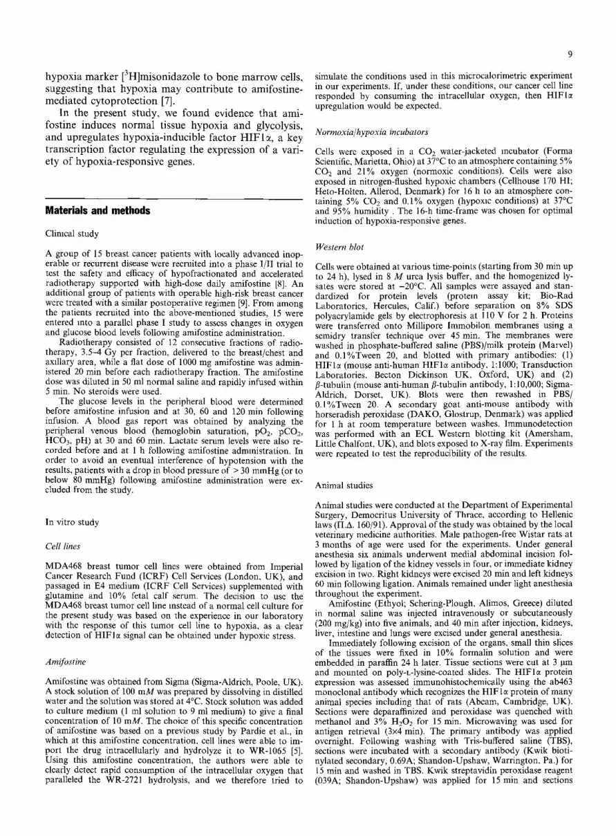
Figure 2 shows the results of the Western blot analysis in the MDA468 cells. The loading control was fl-tubulin and is shown in all cases. It was clear that hypoxia led to the induction of HIFlct protein. The maximum induction of H I F l e by amifostine was noted at 2 h, although less strong than in cells exposed to prolonged hypoxia. Normal HIFI~ levels were restored at 4 h and remained at normal levels thereafter. Spot density measurements showed a 1.8-fold increase in H I F l e levels following 16 h exposure of cells to 0.1% hypoxia, compared to those exposed to normoxia. The H I F l e levels had risen by 1.2-fold by 2 h following incubation with amifostine and had returned to normal
at 4 and 8 h (1.06 and 0.93-fold compared to the levels under normoxic conditions).
Animal study
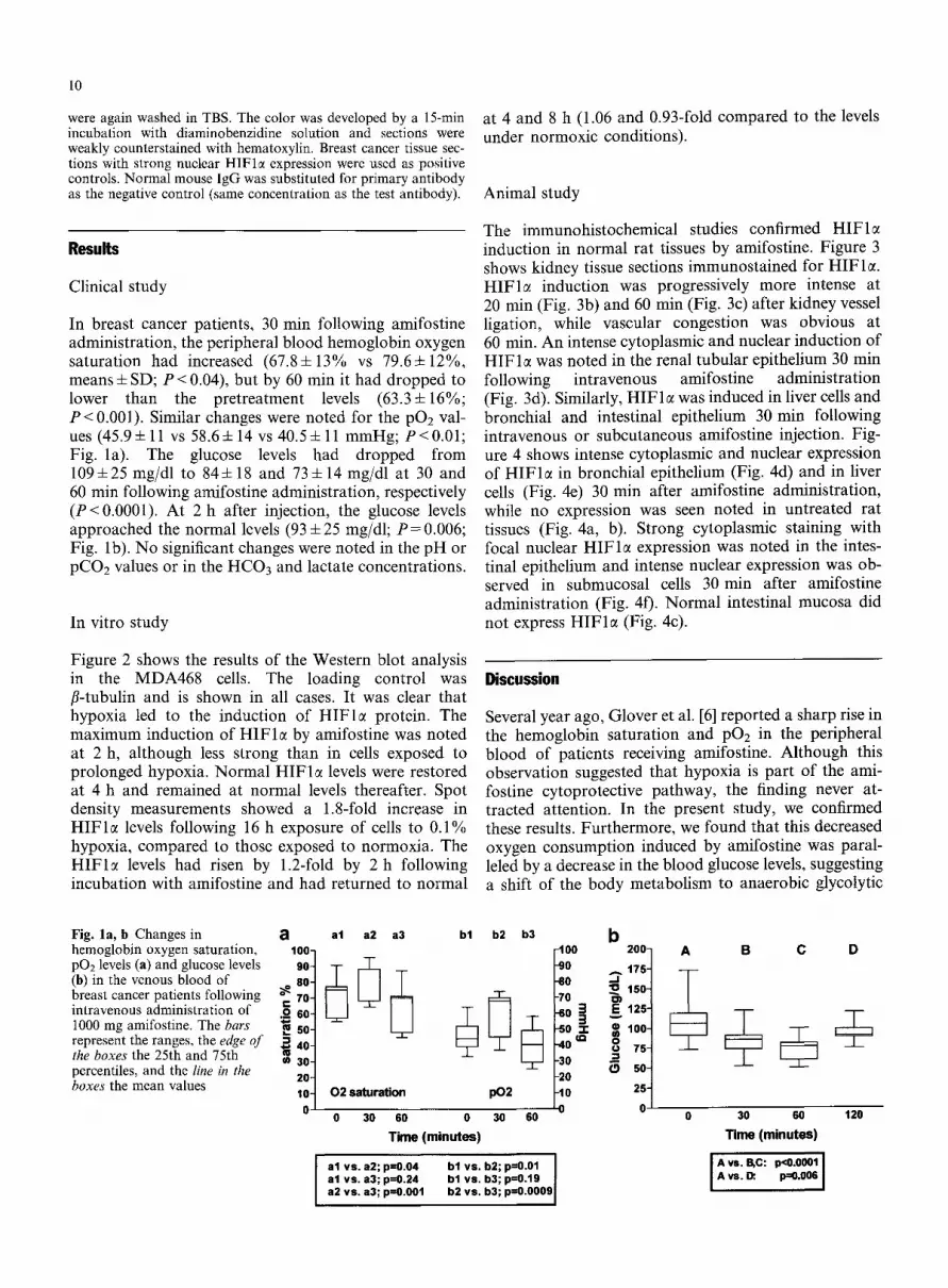
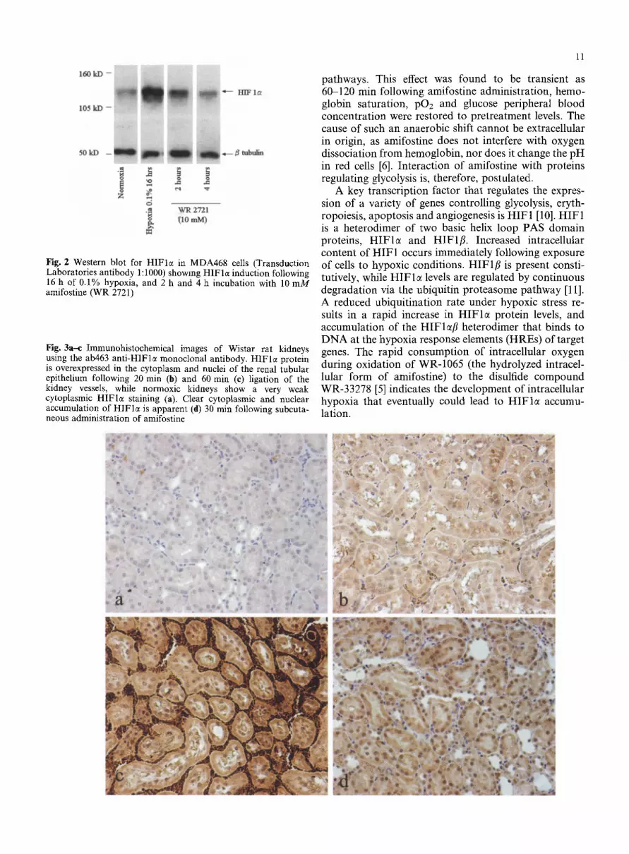
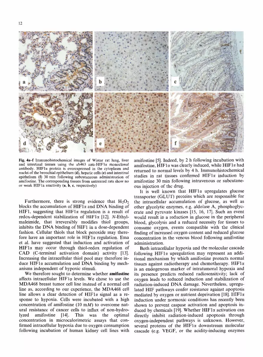
The immunohistochemical studies confirmed HIFlct induction in normal rat tissues by amifostine. Figure 3 shows kidney tissue sections immunostained for HIFlm HIFlc~ induction was progressively more intense at 20 rain (Fig. 3b) and 60 min (Fig. 3c) after kidney vessel ligation, while vascular congestion was obvious at 60 rain. An intense cytoplasmic and nuclear induction of HIFlct was noted in the renal tubular epithelium 30 min following intravenous amifostine administration (Fig. 3d). Similarly, HIF 1 ~ was induced in liver cells and bronchial and intestinal epithelium 30 min following intravenous or subcutaneous amifostine injection. Fig- ure 4 shows intense cytoplasmic and nuclear expression of HIFI~ in bronchial epithelium (Fig. 4d) and in liver cells (Fig. 4e) 30 min after amifostine administration, while no expression was seen noted in untreated rat tissues (Fig. 4a, b). Strong cytoplasmic staining with focal nuclear HIFlct expression was noted in the intes- tinal epithelium and intense nuclear expression was ob- served in submucosal cells 30 min after amifostine administration (Fig. 40. Normal intestinal mucosa did not express HIF 1 ~ (Fig. 4c).
Discussion
Several year ago, Glover et al. [6] reported a sharp rise in the hemoglobin saturation and pO2 in the peripheral blood of patients receiving amifostine. Although this observation suggested that hypoxia is part of the ami- fostine cytoprotective pathway, the finding never at- tracted attention. In the present study, we confirmed these results. Furthermore, we found that this decreased oxygen consumption induced by amifostine was paral- leled by a decrease in the blood glucose levels, suggesting a shift of the body metabolism to anaerobic glycolytic
Fig. la, b Changes in a hemoglobin oxygen saturation, 100- p02 levels (a) and glucose levels 90- (b) in the venous blood of so- breast cancer patients following ~ 70- intravenous administration of .~- 60- 1000 mg amifostine. The bars ~ SO- represent the ranges, the edge of 5.. 40- the boxes the 25th and 75th percentiles, and the line in the boxes the mean values
(u w 30-
20- 10- 0
bl b2 b3 al a2 a3
6@6 02 saturation I)02
0 30 60 0 30 60
T ime (minutes)
b .100 200] A
-,o 0 15o 1 .5o 3 ~ 1,51 -50 ~ ; 1001 -40 ~
'5 t -30 0 50 -20 -10 25
0 0
al vs. a2; p=O.04 al vs. a3; p=0.24 a2 vs. a3; p=O.O01
bl vs. b2; p=O.01 I bl vs. b3; p=0.19 b2 vs. b3; p=O.O0091
B C D
30 60 120
T ime (minutes)
I A,..,c: p<O.O0Ol l A vs. D: p=0.006 |
11
160 kD -
105 kD -- : , , :
" - - - I t l F Ic~
ci .~ ~ 2721 o~ (to m ~
Fig. 2 Western blot for HIFlc( in MDA468 cells (Transduction Laboratories antibody 1:1000) showing HIFle induction following 16 h of 0.1% hypoxia, and 2 h and 4 h incubation with 10 mM amifostine (WR 2721)
Fig. 3a--c Immunohistochemical images of Wistar rat kidneys using the ab463 ant i -HIFle monoclonal antibody�9 HIFlc~ protein is overexpressed in the cytoplasm and nuclei of the renal tubular epithelium following 20 rain (b) and 60 min (e) ligation of the kidney vessels, while normoxic kidneys show a very weak cytoplasmic H I F l e staining (a). Clear cytoplasmic and nuclear accumulation of HIFlc~ is apparent (d) 30 min following subcuta- neous administration of amifostine
pathways. This effect was found to be transient as 60-120 min following amifostine administration, hemo- globin saturation, pO2 and glucose peripheral blood concentration were restored to pretreatment levels. The cause of such an anaerobic shift cannot be extracellular in origin, as amifostine does not interfere with oxygen dissociation from hemoglobin, nor does it change the pH in red cells [6]. Interaction of amifostine with proteins regulating glycolysis is, therefore, postulated�9
A key transcription factor that regulates the expres- sion of a variety of genes controlling glycolysis, eryth- ropoiesis, apoptosis and angiogenesis is HIF1 [10]. HIF1 is a heterodimer of two basic helix loop PAS domain proteins, HIF la and HIFlfi. Increased intracellular content of HIF1 occurs immediately following exposure of cells to hypoxic conditions. HIFlfl is present consti- tutively, while HIF la levels are regulated by continuous degradation via the ubiquitin proteasome pathway [11]. A reduced ubiquitination rate under hypoxic stress re- sults in a rapid increase in HIFlc~ protein levels, and accumulation of the HIFlefi heterodimer that binds to DNA at the hypoxia response elements (HREs) of target genes. The rapid consumption of intracellular oxygen during oxidation of WR-1065 (the hydrolyzed intracel- lular form of amifostine) to the disulfide compound WR-33278 [5] indicates the development of intracellular hypoxia that eventually could lead to HIFI~ accumu- lation.
, ,~,~. ' ,~, .. , % . ~ ,=, ~ ~ . , " , ,
J ,:~ , .,c , Ul,~ ~
12
: ~ . . . . . . ,,...,. . . . . . ~ , t,, ~ ' ~ ~ . ,+.r . , . , + , , . . . . .
0+..+ . ,, i+~ ...... , .+. . ~ . ~ + .
. " + . . . . . ' ' . - " s , ," ,+ ,,' ,
.1, ;~ ,+ , . . r 1~ ~ .~ * ~ , ~ ' ~ ,+2"~ " ' , '~ 4 , , " '+,4+,,., ~o:7 '.~'~',~'~,~(
+ +.+'~+-',+ ..... '*;"-'. i+,X,-,.+t~ k . ~ m + t,+m+ -.+,+ +,+ +, .++. ...... .+.,:*..: . . . . . '~ r �9 'rod- ~" ,, .~ . , ' ' ' a ' ' ~ + 1 ~ ; ' ; ' , ,1~.,"- ~'+," , , ' 4 �9 , , ,~ ~,'+.r,,.+ "' 2.,,,:" ,+-,,- '% ,,.....~+';r a , ? - , + ~ / ~ ~ +. , , ,++-;~, , , , .+ .~ ,, . . . . . . . . . ~ :,:.+.,++./+[;~,.+,+.. ",. "+ "~'+''+'.*+ -+~s ~ . m ~ , " ~ + + ~ " " 'e -'+ ,+',+ +,,,'.1,,,,.';,,"~'+.'.+.,. ,,",,.,+i'+.m . . . . ,i.~++~'+~,,+..-: ~, + , i ~ , I . + , i i ~ , i -+ +,i~
- . . p ~
; t
Fig. 4 a - f I m m u n o h i s t o c h e m i c a l i m a g e s o f W i s t a r r a t l ung , l iver and intestinal tissues using the ab463 antl-HIFle monoclonal antibody. HIFlc+ protein is overexpressed in the cytoplasm and nuclei of the bronchial epithelium (fl), hepatic cells (e) and intestinal epithelium (t) 30 min following subcutaneous administration of amifostine. The corresponding tissues from untreated rats show no or weak HIFla reactivity (a, b, e, respectively)
Furthermore, there is strong evidence that H202 blocks the accumulation of HIFlc+ and DNA binding of HIF1, suggesting that HIF la regulation is a result of redox-dependent stabilization of HIFla [12]. N-Ethyl- maleimide, that irreversibly modifies thiol groups, inhibits the DNA binding of HIF1 in a dose-dependent fashion. Cellular thiols that block peroxide may there- fore have an important role in HIFlc+ regulation. Ema et al. have suggested that induction and activation of HIFlc+ may occur through thiol-redox regulation of CAD (C-terminal activation domain) activity [13]. Increasing the intracellular thiol pool may therefore in- duce HIFla accumulation and DNA binding by mech- anisms independent of hypoxic stimuli.
We therefore sought to determine whether amifostine affects intracellular HIFla levels, We chose to use the MDA468 breast tumor cell line instead of a normal cell line as, according to our experience, the MDA468 cell line allows a clear detection of HIFI++ signal as a re- sponse to hypoxia. Cells were incubated with a high concentration of amifostine (10 raM) to overcome nat- ural resistance of cancer cells to influx of non-hydro- lyzed amifostine [14]. This was the optimal concentration in microcalorimetric assays that con- firmed intracellular hypoxia due to oxygen consumption following incubation of human kidney cell lines with
amifostine [5]. Indeed, by 2 h following incubation with amifostine, HIF le was clearly induced, while HIFlc~ had returned to normal levels by 4 h. Immunohistochemical studies in rat tissues confirmed HIFle induction by amifostine 30 rain following intravenous or subcutane- ous injection of the drug.
It is well known that HIFI++ upregulates glucose transporter (GLUT) proteins which are responsible for the intracellular accumulation of glucose, as well as other glycolytic enzymes, e.g. aldolase A, phosphoglyc- erate and pyruvate kinases [15, 16, 17]. Such an event would result in a reduction in glucose in the peripheral blood, glycolysis and a reduced necessity for tissues to consume oxygen, events compatible with the clinical finding of increased oxygen content and reduced glucose concentration in the venous blood following amifostine administration.
Both intracellular hypoxia and the molecular cascade following HIFle upregulation may represent an addi- tional mechanism by which amifostine protects normal tissues against radiotherapy and chemotherapy. HIFIc+ is an endogenous marker of intratumoral hypoxia and its presence predicts reduced radiosensitivity; lack of oxygen leads to reduced induction and stabilization of radiation-induced DNA damage. Nevertheless, upregu- lated HIF pathways confer resistance against apoptosis mediated by oxygen or nutrient deprivation [18]. HIFlc+ induction under normoxic conditions has recently been shown to prevent caspase activation and apoptosis in- duced by chemicals [19]. Whether HIFlc+ activation can directly inhibit radiation-induced apoptosis through oxygen-independent pathways is unknown. However, several proteins of the HIFlc+ downstream molecular cascade (e.g. VEGF, or the acidity-inducing enzymes

13
lactate dehydrogenase and carbonic anhydrase 9) are directly involved in radioresistance [20, 21, 22, 23, 24].
Although it could be argued that amifostine may also induce H I F l e in tumor tissues, therefore increasing tu- mor resistance to radiotherapy and chemotherapy, this is unlikely to occur. Poor drug availability, in t ra tumoral acidity and loss of alkaline phosphatase expression by the tumoral vasculature and s t roma prevent the hydro- lysis of amifostine within the tumor environment (re- viewed in reference 25). Intracellular hypoxia and upregulation of the H I F molecular cascade should, therefore, exclusively occur in normal tissues and is probably part of such selectivity mechanisms. On the other hand, hypoxia is a common tumoral feature, and H I F I ~ is upregulated in more than 60% of tumors [26, 27, 28, 29, 30, 31, 32] due to intra tumoral hypoxia or even constitutively as a result of cellular t ransformation. Lu et al. have recently shown that aerobic glycolysis, a tumor-specific phenomenon also known as the Warburg effect, activates H I F 1 ~ and promotes survival of cancer cells [33]. Clinical studies have confirmed that upregu- lation of the H I F pathway in tumors is linked to tumor resistance to radiotherapy [30, 31, 32]. Even if WR-1065 reaches the tumor environment, it is questionable whe- ther further upregulation of the H I F molecular cascade can occur or can be clinically meaningful.
I t could be suggested that amifostine counteracts the undesirable advantage of tumors in terms of radio- and chemoresistance (conferred by the intra tumoral hypoxia or the intrinsic anaerobic metabolism) by triggering the H I F cascade in normal tissues. Intracellular hypoxia or thiol-redox activity may account for this event. In this way, amifostine targets hypoxia using a quite inverse rationale f rom policies aimed at improving tumor oxy- genation. Due to the selective nature of amifostine activity on normal tissues, combining amifostine with hyperbaric oxygen and blood flow modifiers would seem a reasonable approach. As amifostine induces anaerobic metabol ism in normal cells and peripheral blood pO2 increases, it is evident that normal tissue oxygen con- sumption is prevented, and it seems unlikely that in- creased oxygen disposal or blood flow may reverse this effect. In this way, hyperbaric oxygen or blood flow modifiers given concurrently with amifostine would sensitize predominant ly tumors, as the metabolic effect of amifostine mainly concerns normal tissues. On the contrary, the concurrent use of amifostine with biore- ductive drugs should probably be avoided, as amifos- tine-induced normal tissue hypoxia predisposes to an increased intracellular drug activation.
The present findings bring forward and enrich old observations suggesting that intracellular hypoxia may be an important component of amifostine-induced cytoprotect ion and reveal a potential role of amifostine as an inducer of the HIF-regula ted molecular cascade. As H I F I ~ is involved in the pathogenesis of several human diseases (e.g. myocardial and cerebral ischemia, pulmonary hypertension), an eventual therapeutic role for amifostine as a H I F I ~ activator is postulated [34].
References
1. Grdina DJ, Sigdestad CP (1989) Radiation protectors: the unexpected benefits. Drug Metab Rev 20:1342
2. Holwitt EA, Koda E, Swenberg CE (1990) Enhancement of topoisomerase I-mediated unwinding of supercoiled DNA by the radioprotector WR-33278. Radiat Res 124:107-109
3. Shen H, Chen Z J, Zilfou JT, Hopper E, Murphy M, Tew KD (2001) Binding of the aminothiol WR-1065 to transcription factors influences cellular response to anticancer drugs. J Pharmacol Exp Ther 297:1067-1073
4. Grdina D J, Murley JS, Kataoka Y, Calvin DP (2002) Differ- ential activation of nuclear transcription factor kappaB, gene expression, and proteins by amifostine's free thiol in human microvascular endothelial and glioma cells. Semin Radiat On- col 12 [Suppl 1]:103-111
5. Purdie JW, Inhaber ER (1983) Interacnon of cultured mam- malian cells with WR-2721 and its thiol, WR-1065: implica- tions for mechanisms of radioprotection. Int J Radiat Biol 43:517 527
6. Glover D, Negendank W, Delivoria-Papadopoulos M, Glick JH (l 984) Alterations in oxygen transport following WR-2721. Int J Radlat Oncol Biol Phys 10:1565 1568
7. Allalunis-Turner M J, Walden TL Jr, Sawich C (1989) Induc- tion of marrow hypoxia by radioprotective agents. Radiat Res 118:581-586
8. Koukourakis MI, Giatromanolaki A, Kouroussis C, Kakolyris S, Sivridis E, Frangiadaki C, Retalis G, Georgoulias V (2002) Hypofractionated and accelerated radiotherapy with cytopro- tecnon (HypoARC): a short, safe and effective postoperative regimen for high-risk breast cancer patients. Int J Radiat Oncol Biol Phys 52:14~155
9. Koukourakis MI, Yannakakis D (2001) High dose daily ami- fostine and hypofractionated intensively accelerated radio- therapy for locally advanced breast cancer. A phase I/II study and report of early and late sequelae. Anticancer Res 21:2973- 2978
10. Semenza GL (1998) Hypoxia-inducible factor 1: master regu- lator of 02 homeostasis. Curr Opin Genet Dev 8:588 594
11. Huang LE, Gu J, Schau M, Bunn HF (1998) Regulation of hypoxia-inducible factor 1~ is mediated by an O2-dependent degradation domain via the ubiquitin-proteasome pathway. Proc Natl Acad Sci U S A 95:7987-7992
12. Huang E, Arany Z, Livingston DM, Bunn F (1996) Activation of hypoxia-inducible transcription factor depends primarily upon redox-sensitive stabilization of its ~ subunit. J Biol Chem 271:32253-32259
13. Ema M, Hirota K, Mimura J, Abe, H, Yodoi J, Sogawa K, Poellinger, Fujli-Kuruyama Y (1999) Molecular mechanisms of transcription activation by HLF and HIFla in response to hypoxia: their stabilization and redox signal-induced interac- tion with CBP/p300. EMBO J 18:1905-1914
14. Yuhas JM (1980) Active versus passive absorption kinetics as the basis for selective protection of normal tissue by S-2-3- aminopropylamino-ethylphosphorotbioic acid. Cancer Res 40:1519-1524
15. Okino ST, Chichester CH, Whitlock JP Jr (1998) Hypoxia- inducible mammalian gene expression analyzed in vivo at a TATA-dnven promoter and at an initiator-driven promoter. J Biol Chem 273:2383223843
16. Chen C, Pore N, Behrooz A, Ismail-Beigi F, Maity A (2001) Regulation of glutl mRNA by hypoxia-inducible factor-1. Interaction between H-ras and hypoxia. J Biol Chem 276:9519 9525
17. Semenza GL, Roth PH, Fang HM, Wang GL (1994) Tran- scriptional regulation of genes encoding glycolytic enzymes by hypoxia-inducible factor 1. J Biol Chem 269:23757-23763
18. Akakura N, Kobayashi M, Horiuchi I, Suzuki A, Wang J, Chen J, Niizeki H, Kawamura K, Hosokawa M, Asaka M (2001) Constitutive expression of hypoxia-inducible factor- lalpha renders pancreatic cancer cells resistant to apoptosis

14
induced by hypoxia and nutrient deprivation. Cancer Res 61:6548-6554
19. Piret JP, Mottet D, Raes M, Michiels C (2002) COC12, a chemical inducer of hypoxia-inducible factor-l, and hypoxia reduce apoptotic cell death in hepatoma cell line HepG2. Ann N Y Acad Sci 973:443-447
20. Geng L, Donnelly E, McMahon G, Lin PC, Sierra-Rivera E, Oshinkan H, Hallahan DE (2001) Inhibition of vascular endothehal growth factor receptor signaling leads to reversal of tumor resistance to radiotherapy. Cancer Res 6:2413-2419
21. Gorski DH, Beckett MA, Jaskowiak NT, Calvin DP, Mauceri H J, Salloum RM, Seetharam S, Koons A, Hari DM, Kufe DW, Weichselbaurn RR (1999) Blockage of the vascular endothelial growth factor stress response increases the antitumor effects of ionizing radiation. Cancer Res 59:3374-3378
22. Lee HS, Park H J, Lyons JC, Griffin RJ, Auger EA, Song CW (1997) Radiation-induced apoptosis in different pH environ- ments in vitro. Int J Radiat Oncol Biol Phys 38:1079-1087
23. Park H J, Lee SH, Chung H, Rhee YH, Lim BU, Ha SW, Griffin RJ, Lee HS, Song CW, Choi EK (2003) Influence of environmental pH on G(2)-phase arrest caused by ionizing radiation. Radiat Res 159:86-93
24. Brizel DM, Schroeder T, Scher RL, Walenta S, Clough RW, Dewhirst MW, MueUer-Klieser W (2001) Elevated tumor lac- tate concentrations predict for an increased risk of metastases m head-and-neck cancer. Int J Radiat Oncol Biol Phys 51:349- 353
25. Koukourakis MI (2002) Amifostine in clinical oncology: current use and future applications. Anticancer Drugs 13:181- 209
26. Zhong H, De Marzo AM, Laughner E, Lira M, Hilton DA, Zagzag D, Buechler P, Isaacs WB, Semenza GL, Simons JW (1999) Overexpression of hypoxia-inducible factor la in com- mon human cancers and their metastases. Cancer Res 59:5830- 5835
27. Talks KL, Turley H, Gatter KC, Maxwell PH, Pugh CW, Ratcliffe P J, Harris AL (2000) The expression and distribution of the hypoxia inducible factors HIF- la and HIF-2a in normal human tissues, cancers and tumor associated macrophages. Am J Pathol 157:411-421
28. Giatromanolaki A, Koukourakis MI. Sivridis E, Turley H, Talks K, Pezzella F, Gatter KC, Harris AL (2001) Relation of hypoxia inducible factor 1 alpha and 2 alpha in operable non- small cell lung cancer to angiogenic/molecular profile of tumours and survival. Br J Cancer 85:881-890
29. Vukovic V, Haugland HK, Nicklee T, Morrison A J, Hedley DW (2001) Hypoxia-inducible factor-lalpha is an intrinsic marker for hypoxia in cervical cancer xenografts. Cancer Res 61:7394-7398
30. Aebersold DM, Burri P, Beer KT, Laissue J, Djonov V, Gre- iner RH. Semenza GL (2001) Expression of hypoxia-inducible factor-latpha: a novel predictive and prognostic parameter in the radiotherapy of oropharyngeal cancer. Cancer Res 61:2911-2916
31. Koukourakis MI, Giatromanolaki A, Skarlatos J Corti L, Blandamura S, Piazza M, Gatter KC, Harris AL (2001) Hy- poxia inducible factor HIF- la and HIF-2a expression in early esophageal cancer and response to photodynamic therapy and radiotherapy. Cancer Res 61:1830-1832
32. Koukourakis MI, Giatromanolaki A, Sivridis E, Simopoulos C, Turley H, Talks K, Garter KC, Harris AL (2002) Hypoxia- inducible factor (HIFIA and HIF2A), angiogenesis, and che- moradiotherapy outcome of squamous cell head-and-neck cancer. Int J Radiat Oncol Biol Phys 53:1192-1202
33. Lu H, Forbes RA, Verma A (2002) Hypoxia-inducible factor 1 activation by aerobic glycolysis implicates the Warburg effect in carcinogenesis. J Biol Chem 277:23111-23115
34. Semenza GL (2001) Hypoxia-inducible factor 1: oxygen homeostasis and disease pathophysiology. Trends Mol Med 7:345 350