altered properties of human t-lymphoblast soluble low km 5′-nucleotidase: comparison with...
TRANSCRIPT

Leukemia Research Vol. 17, No. 3, pp. 231-240, 1993. 0145 2126/93 $6.00 + .00 Printed in Great Britain. Pergamon Press Ltd
ALTERED PROPERTIES OF HUMAN T-LYMPHOBLAST SOLUBLE LOW Km 5 ' -NUCLEOTIDASE: COMPARISON WITH
B-LYMPHOBLAST ENZYME
VICENTE MADRID-MARINA, BORIS LESTAN, PAUL J. NOWAK, IRVING H . FOX a n d JOZEF SPYCHALA*
The University of Michigan, Department of Internal Medicine, Division of Rheumatology, Ann Arbor, MI 48109, U.S.A. and *The University of North Carolina, Department of Pharmacology,
Chapel Hill, NC 27599, U.S.A.
(Received 1 September 1992. Revision accepted 9 November 1992)
Abstract--Soluble low Km 5'-nucleotidases have been purified from human cultured T- and B- lymphoblasts to compare their properties and to examine the mechanism of different rates of nucleotide dephosphorylation. The enzyme from B-lymphoblasts (MGL-8) was 4385-fold purified with a specific activity of 114 ~tmol/min/mg, while the enzyme from T-lymphoblasts (CEM, MOLT-4) was 4355-fold purified with a specific activity of 35 ~tmol/min/mg. The activity of both enzymes have an absolute requirement for Mg ÷÷. The B-cell enzyme has maximum activity with Mg 2÷ > Mn 2÷ > Co 2÷, while the T-cell enzyme had maximum activity with Co 2÷ > Mn 2÷ > Mg 2÷. The optimum activity was at pH 7.4-9.0 for the B-cell enzyme and pH 9.0 for the T-cell enzyme. Substrate specificity was the same for both enzymes with the following relative Vmax values:
CMP > UMP > dUMP > dCMP > dAMP > IMP > GMP > dlMP > dGMP. The Km values for AMP and IMP were 12 and 25 IxM for the B-cell enzyme, and 7.0 and 12 ~tM for the T-cell enzyme. ATP and ADP are competitive inhibitors of these enzymes with apparent Ki values of 100 and 20 ~tM for the B-cell enzyme, and 44 ~tM and 8 pxM for the T-cell enzyme, respectively. The apparent molecular mass by gel filtration column chromatography is 145 kD for the B-ceU enzyme and 72 kDa for the T-cell enzyme. The subunit molecular masses by Western blots are 69.2 kD for both enzymes.
These properties suggest that the B-lymphoblast enzyme is identical or similar to the enzyme from human placenta. However, the T-cell enzyme has some different properties. We conclude that these differences plus a lower content of low Km 5'-nucleotidase in T-cells may account for the decreased ability of T-lymphoblasts to dephosphorylate nucleotides and may contribute to the selective cyto- toxicity of deoxyribonucleosides for T-lymphoblasts as compared to B-lymphoblasts.
Key words: 5'-nucleotidase, nucleotide degradation, nucleoside toxicity, leukemic cells.
I N T R O D U C T I O N
INTRACELLULAR nucleotide degradation in human cells is highly regulated [1-3]. A key step in this pathway is dephosphorylation of nucleotide 5 '-mono- phosphates catalyzed by 5'-nucleotidase (5'-ribo- nucleotide phosphohydrolase, EC3.1.3.5) . This enzyme is important for intracellular nucleotide metabolism and adenosine release [4-6].
Abbreviations: BCIP, 5-bromo-4-chloro-3-indolyl phos- phate; NBT, nitro blue tetrazolium; MBS, m-maleimido- benzoyl-N-hydroxysuccinate ester; E-64, N-[N-(L-3-trans- carboxyoxiran-2-carbonyl)-L-leucyl]-agmatine; EG TA, ethyleneglycol-bis-(/3-aminoethyl ether) N,N,N 1,N 1- tetraacetic acid; PMSF, phenylmethylsulfonylflouride; SDS-PAGE, sodium dodecyl phosphate polyacrylamide gel electrophoresis.
Correspondence to: Dr 3ozef Spychala, University of North Carolina, Department of Pharmacology, 1106 FLOB, Chapel Hill, NC 27599-7365, U.S.A.
231
Available data indicate that 5'-nucleotidase from human T-lymphoblastoid cell lines has a low activity as compared to B-lymphoblastoid cell lines [7]. In T- lymphoblasts, intracellular nucleoside 5 '-monophos- phate dephosphorylation is lower compared with B- lymphoblasts or with T- and B-lymphocytes [5, 8- 10]. Consequently, under stress conditions, B-cells selectively release adenosine [11]. In addition, the activity of 5'-nucleotidase may have prognostic value in leukemias [12-15] and other diseases: it is decreased in the lymphocytes of immunodeficient patients with hypogammaglobulinemia [16-18] and patients with chronic lymphocytic leukemia [19], and in lymphoblasts of patients with acute T-cell lympho- blastic leukemia [12, 20]. Low intracellular 5'-nucleo- tidase activity may contribute to the accumulation of toxic deoxyribonucleoside triphosphates in immuno- deficient patients with adenosine deaminase deft-
232 V. MADRID-MARINA et al.
ciency, or in the presence of adenosine deaminase inhibitors and with purine nucleoside phosphorylase deficiency [21-23].
The basis for the decreased dephosphorylat ing activity in human T-lymphoblasts is unclear. The 5 ' -phosphomonoes terase activity is a composite of nonspecific phosphatases and specific 5'- nucleotidases. There are two soluble specific 5'- nucleotidases in lymphoid tissue: low Km and high Km forms [24]. Our previous studies determined that the decrease in 5 '-nucleotidase in T-lymphoblasts is apparently related to the low Km form [24]. There- fore, as one approach to examine the decreased nucleoside 5 ' -monophospha te activity of these cells, we have tested the hypothesis that there is an alter- ation of specific soluble low Km 5'-nucleotidase in T- lymphoblasts as compared to B-lymphoblasts. To accomplish this, we have adapted our purification and characterization of human placental and rat liver low K,, 5 '-nucleotidase [25, 26] to cultured B-lym- phoblast and T-lymphoblas t soluble 5'-nucleotidases.
E X P E R I M E N T A L P R O C E D U R E
Materials [8-14C]AMP (50mCi/mmol) and [8-14C]IMP (50mCi/
mmol) were purchased from Amersham Corporation (Chi- cago, IL). All nucleosides, 5'-monophosphates, ADP, ATP, adenosine, inosine, p-nitrophenylphosphate, gel fil- tration protein standards, imidazole, /3-mercaptoethanol, trizma base, ~r-methyl-D-mannoside, bovine serum albumin, goat anti-rabbit affinity purified IgG conjugated with alkaline phosphatase trypsin inhibitor (soybean), 1,10- phenanthroline, ol,/J-methylene adenosine diphosphate (AMPCP), ol,3-methylene adenosine triphosphate (AMPCPP), 3,~,-methylene adenosine triphosphate (AMPPCP), 5-bromo-4-chloro-3-indolyl phosphate (BCIP) and nitro blue tetrazolium (NBT) were purchased from Sigma Chemical Co. (St Louis, MO). AMP- Sepharose 4B, Concanavalin A (Con A)-Sepharose., Sephacryl S-300, Sephadex G-25, blue dextran and columns were obtained from Pharmacia Fine Chemicals (Pisca- taway, N J). Bradford protein reagent, acrylamide, N,N'- methylene bisacrylamide, N,N,N',N'-tetramethylethyl- enediamine, ammonium persulfate, bromophenol blue, SDS-polyacrylamide gel protein standard kits of high and low molecular weight, biotinylated molecular weight stan- dards and streptavidin conjugated with alkaline phospha- tase, and the silver stain reagent kit were purchased from Bio Rad (Rockville Center, NY). The pressure ultrafiltr- ation unit with YM-10 membranes was obtained from Amicon (Danvers, MA). All other reagents used were of the highest quality generally available.
Cell lines and cell culture Three human lymphoblastoid cell lines were used for
our studies including T-lymphoblasts (CEM and MOLT- 4) and B-lymphoblasts (MGL-8). The MGL-8 (B-lympho-
blasts) cell line was derived from a normal individual and was a gift from the laboratory of Dr John Littlefield, Johns Hopkins University (Baltimore, MD). MOLT-4 and CEM (T-lymphoblasts) cell lines were obtained from HEM Research, Inc., Rockville, MD, and were originally derived from patients with acute lymphoblastic leukemia. All cell lines were grown in suspension culture using RPMI-1640 media supplemented with 10% donor calf serum and 2 mM L-glutamine. The cells were maintained in logarithmic growth phase at 37°C and 5% CO2. Mycoplasma con- tamination was excluded by periodic microbiology tests.
Enzyme assays Protein containing extracts of soluble 5'-nucleotidase
were incubated for 30 min or 60 min at 37°C in a reaction mixture containing 62.5 mM imidazole-HC1 pH 7.4, 20 mM MgC12, 1 mg/ml bovine serum albumin, and 0.2 mM [8- 14C]AMP or [8-14C]IMP in a total volume of 100 ~tl. After stopping the reaction by heating at 85°C for 2 min, pre- cipitated protein was removed by centrifugation. Twenty microliters were spotted on Whatman 3MM chroma- tography paper with 20 ~tl nonradioactive nucleosides and nucleotides (1 mg/ml). Nucleosides and nucleotides were separated by high voltage electrophoresis at 4000 V and 250 mA for 30 min in 50 mM sodium borate pH 9. Nucleo- side and nucleotide spots were located with an ultraviolet light, cut out and counted in LKB Rackbeta liquid scin- tillation spectrometer. In addition, enzyme activity was measured by release of inorganic phosphate (Pi) in incu- bation media of 200 p~l containing 67 mM imidazole-HCl pH 7.4, 20 mM MgCI2, 1 mg/ml bovine serum albumin and 5 mM AMP. The reaction was stopped by the addition of 50 Ixl of 10% trichloroacetic acid. The amount of Pi lib- erated was determined by the method of Chen et al. [27].
Non-specific phosphatase activity was measured in an incubation mixture containing 62.5 mM imidazole pH 7.4, 20mM MgCI2, l mg/ml bovine serum albumin and 12.5 mM p-nitrophenylphosphate. The reaction was incu- bated for 30 min at 37°C and stopped with the addition of 0.8 ml 0.4M NaOH. The samples were clarified by centrifugation and the optical density measured at 400 nm on a Gilford 260 spectrophotometer.
Protein assay Protein concentration was estimated by the method of
Bradford [28] with alcohol dehydrogenase as the standard. Proteins eluting from the columns during the various puri- fication steps were monitored by absorbance at 280 nm or by the Bradford method.
Preparation of cell extracts Cells were collected by centrifugation, washed three
times, and resuspended in 0.25 M sucrose, 1 mM MgCI2, and 10 mM Tris-HCl pH 7.4. The cell pellet was stored at -70°C. The frozen cells were thawed, suspended in 20 mM imidazole-HCl, pH 7.0, 20 mM MgCI2 (buffer A) to 5 × 10 ~ cells/ml, and were lysed by nitrogen bomb cavitation (1000lb./in. 2 for 30min at 4°C) and by Dounce tissue homogenizer (about 30-40 strokes). The lysate was spun for 10 min at 10,000 × g to remove cellular debris. The supernatant was spun at 50,000 × g for 2 h to eliminate the remaining cellular membranes and then was dialyzed overnight at 4°C against 10 mM Tris-HCl pH 7.4, and 20 mM MgCI 2. This lymphoblast supernatant was then used immediately for enzyme purification.

Lymphoblast low K,, 5'-nucleotidase 233
Enzyme purification The purification used was similar to our procedure for
human placental soluble 5'-nucleotidase [25]. The lym- phoblast supernatant (50 ml) was passed through an AMP- Sepharose 4B affinity chromatography column. Soluble 5'- nucleotidase activity was eluted from the column with 60 ml of 10 mM ADP in 20 mM imidazole-HC1 pH 7.0 and 20 mM MgCI2 (buffer A). The eluate was concentrated from 25 ml to 2 ml using an AMICON pressure ultrafiltration unit with YM-10 membrane.
Concentrate (2 ml) from the AMP-Sepharose column was then applied to a Sephacryl S-300 gel filtration column equilibrated with buffer A. The fractions with the highest activity were pooled (17 ml) and applied to concanavalin A-Sepharose 4B column. This column was then washed with 45 ml of 20 mM imidazole-HC1 pH 7.0 and 20 mM MgCI2. The enzyme was eluted in a single peak with 45 ml of 0.5 M o~-methylmannoside at 37°C in buffer A. The eluate from the concanavalin A-Sepharose was immedi- ately concentrated to 1.5 ml to preserve enzyme stability. This preparation was stored at 4°C and used for our charac- terization studies.
Modifications of the purification were made to obtain proteins for subunit molecular weight determinations by Western blot. The T- and B-lymphoblast extract was applied to 20ml concanavalin A-Sepharose columns according to the procedure described for rat liver 5'-nucleo- tidase purification [26]. The pooled, concentrated and dia- lyzed active fractions were passed through a DE-52 ion exchange column. The preparations after DE-52 chroma- tography were taken for Western blot analysis.
Molecular weight determination The molecular weight of the native purified proteins
were determined by gel filtration chromatography on a Sephacryl S-300 column (1.6 × 70cm) equilibrated with 5% glycerol, 20 mM MgCIz, 60 mM imidazole-HC1 pH 7.0, and 0.02% sodium azide. Two milliliter samples were applied, the column flow rate was 6 ml/h, and 1.75 ml fractions were collected. We used blue dextran and di- n~trophenylalanine to determine the void volume and exclusion volume, respectively. The void volume was 55 ml and the excluded volume was 150ml. The column was standardized with aldolase (Mr = 140,000), bovine serum albumin (Mr = 67,000), ovalbumin (Mr = 43,000), alpha- chymotrypsinogen A (Mr = 25,000), and cytochrome C (Mr = 12,000). The protein was estimated by absorbance at 280 nm. Kay was calculated for each standard according to the method of Laurent and Kilander [29] and Siegel and Monty [30]. The molecular weights and Stokes radius were determined from the plot of Kay vs the molecular weight and Stokes radius of the standard proteins.
Subunit molecular weight determination Western blot procedure was used to determine the mol-
ecular weights of T- and B-lymphoblast 5'-nucleotidase. Rabbit antibodies raised against a peptide derived from
human placental soluble low Km 5'-nucleotidase were used to bind to the enzyme protein immobilized on nitro- cellulose. A peptide for amino acid sequence analysis was prepared by trypsin digestion of pure 5'-nucleotidase puri- fied to 800 ~tmol/min/mg protein specific activity. The sequence analysis yielded a 17 amino acid sequence. The synthetic peptide with attached cysteine at the N-terminus was conjugated with keyhole limpet hemocyanin (KLH,
50:50w/w). One milligram of conjugate with complete Freund's adjuvant (1 ml of total volume) was injected subcutaneously to the rabbit at 13 different spots. One- half milligram of peptide-KLH conjugate and incomplete Freund's adjuvant were injected every 4 weeks and the blood was drawn 10 days after each injectio n.
Polyacrylamide slab gel electrophoresis with human pla- centa, B-and T-lymphoblast soluble low Km 5'-nucleotidase and biotynylated standards was run with sodium dode- cylsulfate according to standard procedure [31] and the gel was electroblotted with Bio-Rad Trans-blot cell at 60 V for 6h. The nitrocellulose was blocked with 5% dry milk overnight and incubated with 1:1000 rabbit serum for 3 h. The bands were developed with goat anti-rabbit alkaline phosphatase conjugated antibodies. Biotinylated standards with avidin conjugated with alkaline phosphatase were used to determine molecular weight. Color reaction was developed with BCIP and NBT as described [32]. The molecular weight of the enzymes was determined by meas- uring R/values and comparing them to the Rf values of known biotinylated protein standards (Bio-Rad): phos- phorylase B (97.4 kD), bovine serum albumin (66.2 kD), ovalbumin (42.7kD), carbonic anhydrase (31.0kD) soybean trypsin inhibitor (21.5 kD), and lysozyme (14.4 kD).
Kinetics and calculations The Km values were determined from initial velocity
studies. For each study there were at least six different substrate concentrations. Each determination was carried out 2-4 times in duplicate. Double reciprocal plots of initial velocity vs the substrate concentration were used to estimate the Km values. Linearity with time was established
>.. I -
>- 25 pT B~Jffer C~NaC I5M ~Buffer llOmM ADP I.-
I- o ~ - ~ '¢~ > Z
2 0 - o co
, t ie ,~ (/) -
_ o ~ o -
z m- ~ . 0
o.. u) • o . .,, l
i Z O 20 40 60 80 I00 •
FRACTION NUMBER
FIG. 1. 5'-AMP-Sepharose 4B affinity chromatography. Forty milliliters of either T- or B-lymphoblast supernatants (shown for B-lymphoblasts only) were applied to 20 ml 5'-AMP-Sepharose 4B affinity columns (2.5 × 7 cm). The column was equilibrated with buffer A (see Experimental Procedure). After application of the enzyme extract, the column was washed with 120ml buffer A to remove unbound proteins and then was washed with 60 ml 0.5 M NaC1 in buffer A to remove non-specifically bound proteins. Subsequently, the bound soluble 5'-nucleotidase was eluted with about 90 ml of 10 mM ADP in buffer A. The active fractions were pooled, concentrated using an
Amicon YM-10 filter.

234 V. MADRID-MARINA el al.
TABLE 1. PURIFICATION OF SOLUBLE LOW g m 5'-NUCLEOT1DASE FROM B-LYMPHOBLASTS (MGL-8)
Total Specific Total protein activity enzyme Purification Recovery
Step (mg) (~tmol/min/mg) 0xmol/min) (fold) (%)
Lymphoblast supernatant 351 AMP Sepharose-4B 7.62 AMP-Sepharose concentrate 5.18 Sephacryl S-300 0.15 Con A-Sepharose concentrate 0.04
0.026 8.3 - - 100 0.840 6.4 32 76 0.820 4.3 32 51
33.36 4.2 1283 50 114.0 4.0 4385 48
TABLE 2. PURIFICATION OF SOLUBLE LOW K m 5'-NUCLEOTIDASE FROM T-LYMPHOBLASTS (MOLT-4)
Step
Total Specific Total protein activity enzyme Purification Recovery
(mg) (ixmol/min/mg) (p, mol/min) (fold) (%)
Lymphoblasts supernatant 637 AMP Sepharose-4B 20.5 AMP-Sepharose concentrate 20.0 Sephacryl S-300 0.10 Con A-Sepharose concentrate 0.05
0.008 5.1 - - 100 0.212 4.4 27 87 0.220 4.3 30 83
17.4 1.8 2176 36 34.7 1.7 4335 34
for different concentrations used in initial velocity studies for K,, determinations. Lineweaver-Burk plots of initial velocity vs concentration of substrate were linear. Kinetic data were fitted to the simple Michaelis-Menten equation, i.e. a hyperbola, by a modification of Cleland's program (Cleland, 1967), on a Vax 11/730 minicomputer.
RESULTS
Purification Both enzymes showed a similar purification profile
(Fig. 1). The initial step of purification with AMP- Sepharose affinity chromatography removes all non- specific phosphatase activity from the crude prep- aration, since these enzymes pass through the column. This step shows consistent elution of a single peak of 5'-nucleotidase activity. The enzyme activity peak elutes from the gel filtration column in a same major protein peak (data not shown) separated from other proteins.
The purification of soluble 5'-nucleotidase from B- lymphoblasts yielded a 4385-fold purified enzyme with a specific activity of 114 ~tmoles/min/mg protein (Table 1). The final recovery was 48%. Gel filtration column chromatography yielded a 40-fold puri- fication with a recovery of 68%. Con-A-Sepharose affinity chromatography yielded a 3.5-fold puri- fication with a consistent 96% recovery.
The purification of cytoplasmic 5'-nucleotidase from T-lymphoblasts (MOLT-4) yielded 4335-fold purified enzyme with a specific activity of 40 Ixmol/ min/mg, and final recovery of 34% (Table 2). In this case the gel filtration column also yielded a 72-fold purification, with a 43% recovery. Concanavalin A-
TABLE 3. EFFECT OF DIVALENT CATIONS ON CYTOPLASMIC LOW g m 5'-NUCLEOTIDASE ACTIVITY FROM T-AND B-
LYMPHOBLASTS
Enzyme (% of control) activity*
T-lymphoblasts Divalent cation B-lymphoblasts
(7.5 mM) (MGL-8) (CEM) (MOLT-4)
Mg 100 100 100 Mn 79 107 115 Co 78 142 132 Ba 74 23 28 Ca 72 12 7 Cu 57 60 57 Ni 55 53 47 Zn 34 56 53 Fe 33 15 14
* The activity is expressed as the percent of the value with Mg which have the following in Ixmol/min/mg: MGL- 8, 110; CEM, 37: MOLT-4, 35.
Sepharose chromatography yielded a 2.0-fold puri- fication with a 90% recovery. The enzyme purified from CEM T-lymphoblasts had identical properties.
Divalent cations The enzyme was dialyzed against 10 mM Tris-HC1
pH7.4 and 0 .2mM EDTA and was assayed with different divalent cations at 7.5 mM (Table 3). There was no activity of these enzymes without Mg +÷. As compared to Mg ++, the B-lymphoblast enzyme was 79% effective with Mn and was 78% effective with

Lymphoblast low K,, 5'-nucleotidase 235
• DO, • MGL-8
i o MOLT-4
200
z
pH
o
o~
7
TABLE 4. ACTIVITY OF CYTOPLASMIC LOW g m 5P-NUCLEO -
TIDASE FROM T- AND B-LYMPHOBLASTS WITH VARIOUS
SUBSTRATES
Enzyme activity (% of control)
B-lymphoblasts T-lymphoblasts Substrate (MGL-8) (CEM) (MOLT-4) (2.5 mM) (p.mol/min/mg) (~tmol/min/mg)
CMP 240.8 (247)* 25.3 (248) 14.7 (233) UMP 237.4 (244) 24.1 (236) 13.3 (211) dUMP 209.0 (215) 13.7 (135) 7.3 (116) dCMP 203.0 (208) 16.3 (160) 7.8 (124) dAMP 101.0 (104) 12.3 (119) 6.4 (102) AMP 97.4 (100) 10.2 (100) 6.3 (100) IMP 95.0 (98) 8.9 (87) 4.4 (70) GMP 91.0 (93) 8.3 (81) 4.2 (67) dIMP 66.0 (68) 8.0 (78) 3.1 (49) dGMP 46.0 (47) 6.6 (65) 6.9 (110) TMP - - 4.9 (48) 3.0 (48)
FIG. 2. pH activity profile of T- and B-lymphoblasts soluble low Km 5'-nucleotidases. The enzymes were assayed under standard conditions at different pH values using 100 ~tM AMP as substrate. The B-lymphoblast enzyme has an optimum pH plateau from pH 7.4 to 9.0, while both T-
lymphoblast enzymes have a pH optimum of 9.0.
Co ++. In contrast, the T-cell enzyme showed a greater preference for cobalt (132-142%) and manganese (107-115 %). The B-lymphoblast enzyme had substantially more activity than the T-lym- phoblast enzyme with the addition of Ba, Ca or Fe.
Both enzymes displayed an activation curve with magnesium, reaching its maximum activity between 10 and 20 mM MgCIz-. No AMP dephosphorylation occurred without magnesium.
* The values in brackets are percentages calculated using the activity with AMP as 100%.
blasts had maximum activity at pH 7.4 and remained at a plateau up to pH 9.0 (Fig. 2). In contrast, the T-lymphoblast enzyme with the same experimental conditions has its maximum activity peak at pH 9.0.
Substrate specificity When soluble 5'-nucleotidases from T- and B-
lymphoblasts were assayed with different nucleoside 5 '-monophosphates at the same concentration of 2.5 mM, the enzymes showed high activity with CMP, UMP, dCMP and dUMP (Table 4). There is no difference in the substrate preference.
pH optimum Soluble 5'-nucleotidase purified from B-lympho-
Kinetic properties Initial velocity studies were performed with dif-
TABLE 5. KINETIC VARIABLES FOR CYTOPLASMIC LOW g m 5CNUCLEOTIDASES FROM T- AND B-LYMPHOBLASTS
B-lymphoblast T-lymphoblast Compound enzyme (MGL-8) enzyme (CEM)
AMP IMP ADP ATP Pi
Km (~tM) Ki app (~tM) K,, (,M) Ki app (~tM) 12 - - 7.0 - - 25 - - 10 - - - - 2 0 - - 8.0 - - 1 1 0 - - 55 - - 23,000 - - 16,000
% inhibition
AMPCP (100 ~tM) 70 73 AMPCPP (100 ixM) 10 25 AMPPCP (100 ixM) 10 12

236 V. MADRID-MARINA et al.
"_o K I-" -r" (.9 m Q:: .J 0 LIJ _1 0
IO 8
6
4
2
~ e A
l ~ T- Lymphoblost B ,y p ,o os, \ / 5 Nuoleotid°,,
5'-Nucleolidose • ~ e A Ovolbumin
a Chymotrypsinogen A ~
. . . . Cyt°chr°TC ~ '
0.2 0.5 0.4 0.5 Kov
o,,~ 50 03 Q < n., o3 W 3¢: ~ z5 o3
B- Lyrnphob ~ 1 Aldolose 5'-Nucleotidose ~ X ~
5T-NLYumlPehotidoseOblOst J '~alburnin
aChyrnolrypsinogen A eN~ Cytochrome C ~ X ~
0 0.1 0.2 0 0.4 05 06 0.7 Kov
FIG. 3. Molecular weight and Stokes radii of T- and B- lymphoblast soluble low K,. 5'-nucleotidases on Sephacryl S-300. A Sephacryl S-300 gel filtration column was cali- brated as described in Experimental Procedure, using cyto- chrome C (12.4kD), alpha-chymotrypsinogen (25kD), ovalbumin (43 kD), bovine serum albumin (67 kD), and aldolase (140 kD). The apparent molecular mass of the B- iymphoblast enzyme is 145 kD as compared to the T- lymphoblast enzyme of 72.5 kD (panel A). The Stokes radius of the B-lymphoblast enzyme is 47.4 A, while the
T-lymphoblast enzyme is 37.0 (panel B).
ferent concentrations of AMP or IMP. Soluble 5'- nucleotidases purified from T- and B-lymphoblasts displayed hyperbolic kinetics. Double reciprocal plots of initial velocity studies are linear. The K,, values for the B-lymphoblast enzyme are 12 IxM for AMP and 25 IxM for IMP. The T-lymphoblast enzyme showed Km values of 7 I~M and 12 txM for AMP and IMP, respectively (Table 5).
Inhibition studies of soluble 5'-nucleotidases were performed at different concentrations of AMP and fixed concentrations of ATP (100 IxM), ADP (25 IxM) and Pi (25 mM). With double reciprocal plots of initial velocity data, ATP or ADP produce changes in
the slope only consistent with competitive inhibition. The apparent Ki values were 100 and 20 I~M for ATP and ADP, respectively, for the B-lymphoblast enzyme. The apparent Ki values were 55 IxM for ATP and 8 IIM for ADP for the T-lymphoblast enzyme. Inhibition studies with Pi show changes in both the slope and intercept, suggesting non-competitive inhi- bition. The apparent Ki values were 23 mM (slope), and 44 mM (intercept) for the B-cell enzyme, and 16 mM (slope) and 38 mM (intercept) for the T-cell enzyme (Table 5).
Molecular weight determination The native molecular weights of T- and B-lym-
phoblast cytoplasmic 5'-nucleotidases were esti- mated from the elution of the known molecular weight standards from the Sephacryl S-300 gel fil- tration column. The elution volume of B-lymphoblast soluble 5'-nucleotidase indicates a molecular mass of 145 kD with a Stokes radius of 47.4/~ (Figs 3 and 4). The enzyme from T-lymphoblasts was found to have a molecular mass of 72.5 kD with a Stokes radius of 37.0/~ (Figs 3 and 4).
Subunit molecular weight The Western blot analysis yielded a molecular
mass of 69.2 kD for B- and T-lymphoblast 5'-nucleo- tidases. This compares with 70.6 kD by the same procedure for human placental enzyme (Fig. 5).
DISCUSSION
Cultured human T- and B-lymphoblasts provide a model system for critical examination of the relation- ships between nucleotide degradation and 5'-nucleo- tidase activities. T-lymphoblasts have a decreased ability to dephosphorylate intracellular nucleotides as compared to B-lymphoblasts [5,8,10]. In addition, T-lymphoblasts have a lower activity of soluble and membrane bound 5'-nucleotidases (low K,, type) relative to B-lymphoblasts [5, 7-10, 24, 34]. The discovery of leukemias and immunodeficiency states with absent or low levels of 5'-nucleotidase activity has created further interest in its metabolic significance [12, 16-21, 23].
Our observations have elucidated a number of distinguishing properties between soluble low K,, 5'- nucleotidase from T- and B-lymphoblasts on the one hand and between lymphoblast and human placental enzyme on the other.
Firstly, there is a substantial difference in the AMP-dephosphorylating activity between these cells. The B-cell enzyme is several times more active than the T-cell enzyme in crude extracts as previously described [5, 10, 20]. Furthermore, the pH activity
Lymphoblast low Km 5'-nucleotidase 237
>- I-
I--, U ~ O
_ - 6
. J
z g.) Q
>-
IJJ c
---6 ~E .J
D Z i
20.0
15.0
10.0
5.0
Buffer IOmM CMP
I I0 20 .30 40
A 0.100
0.075
0.050
0.025
50
0 E
O~ io
z n,- 0 gO
0
4.0
3.0
2.0
1.0
FRACTION NUMBER
Bufferl IOmM~CMP B ] 0.04
0 0.05 E c
to O~ to
] 0.02 0 if) II1
0.01 o
I0 20 5 0 4 0 50
FRACTION NUMBER
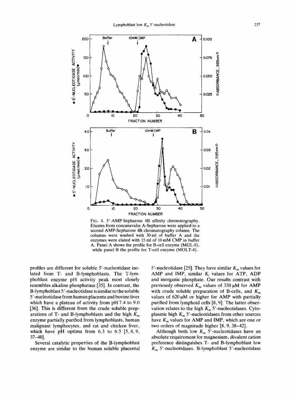
FIG. 4. 5'-AMP-Sepharose 4B affinity chromatography. Eluates from concanavalin A-Sepharose were applied to a second AMP-Sepharose 4B chromatography column. The columns were washed with 30 ml of buffer A and the enzymes were eluted with 15 ml of 10 mM CMP in buffer A. Panel A shows the profile for B-cell enzyme (MGL-8), while panel B the profile for T-cell enzyme (MOLT-4).
profiles are different for soluble 5'-nucleotidase iso- lated from T- and B-lymphoblasts. The T-lym- phoblast enzyme pH activity peak most closely resembles alkaline phosphatase [35]. In contrast, the B-lymphoblast 5'-nucleotidase is similar to the soluble 5'-nucleotidase from human placenta and bovine liver which have a plateau of activity from pH 7.4 to 9.0 [36]. This is different from the crude soluble prep- arations of T- and B-lymphoblasts and the high Km enzyme partially purified from lymphoblasts, human malignant lymphocytes, and rat and chicken liver, which have pH optima from 6.3 to 6.5 [5, 8,9, 37-40].
Several catalytic properties of the B-lymphoblast enzyme are similar to the human soluble placental
5'-nucleotidase [25]. They have similar Km values for AMP and IMP, similar K i values for ATP, ADP and inorganic phosphate. Our results contrast with previously observed Km values of 330 ~tM for AMP with crude soluble preparation of B-cells, and Km values of 620 txM or higher for AMP with partially purified from lymphoid cells [8, 9]. The latter obser- vation relates to the high Km 5'-nucleotidases. Cyto- plasmic high K,, 5'-nucleotidases from other sources have Km values for AMP and IMP, which are one or two orders of magnitude higher [8, 9, 38-42].
Although both low Km 5'-nucleotidases have an absolute requirement for magnesium, divalent cation preference distinguishes T- and B-lymphoblast low Km 5'-nucleotidases. B-lymphoblast 5'-nucleotidase

238 V. MADRID-MARINA et al.
A B C
FIG. 5. Western blot of soluble low Km 5'-nucleotidase from human B-lymphoblasts (A), T-lymphoblasts (B) and placenta (C). A 7.5% gel was electroblotted onto nitro- cellulose, reacted with rabbit anti-peptide monospecific antibodies and visualized with alkaline phosphatase-con- jugated goat anti-rabbit IgG and BCIP and NBT staining [32]. Pure soluble low Km 5'-nucleotidase from human
placenta was included for comparison.
has the highest activity with magnesium as has been observed with other soluble 5'-nucleotidases and membrane 5'-nucleotidases [9, 25, 37-40]. In contrast, the T-cell enzyme has maximum activity with cobalt and manganese. In addition, the enzymes are different by virtue of their preference for Ca, Ba or Fe.
The substantial differences in catalytic properties of T- and B-lymphoblast enzymes was supported by a major alteration in molecular weight. Soluble 5'- nucleotidase isolated from B-cells is similar to a num- ber of mammalian enzymes with low Km 5'-nucleo- tidase activity [25, 26, 44]. The native protein is a dimer of 145 kD of molecular mass and 69.2 kD for each subunit. On the other hand, the enzyme from T-cells appears to be a monomer with molecular mass of 69.2-72.5 kD. Structural differences related to carbohydrate moiety have been already shown for
structurally related plasma membrane 5'-nucleo- tidase [45]. Whether this is the case for these two enzymes and whether the altered catalytic properties of the enzyme from T-cells are related to the inability to form a dimeric structure remains to be established. In an attempt to investigate if there is another and/or different species of mRNA coding for low Km 5'- nucleotidase in T-cells we have performed a Northern blot analysis. We have been unable to detect any level of mRNA for low Km 5'-nucleotidase in B- and T-cells with up to 30 ~tg total RNA and human placenta low Km cDNA as a probe (data not shown).
There are some important biological implications of the reduced and altered low Km 5'-nucleotidase activity in T-lymphoblasts. Firstly, if the low K,, soluble 5'-nucleotidase particles in intracellular nucleotide degradation, it seems possible that the altered properties of the enzyme may explain the decreased dephosphorylating activity of these cells. Secondly, the preference of soluble low Km 5'-nucleo- tidase for deoxycytidine 5'-monophosphate may facilitate the generation of deoxycytidine. This may explain the selective toxicity of deoxyadenosine for T-lymphoblasts as compared to B-lymphoblasts [7- 9, 34, 46]. The soluble low Km 5'-nucleotidase in B- lymphoblasts may generate levels of deoxycytidine sufficient to saturate deoxycytidine kinase and inhibit deoxyadenosine and deoxyguanosine phosphoryl- ation [47, 48]. In T-lymphoblasts, the reduced and kinetically altered activity of soluble low Km 5'- nucleotidase and the resultant decreased ability for the removal of excess of nucleoside monophosphates optimizes the conditions for the preferential phos- phorylation of accumulated deoxyadenosine and deoxyguanosine to toxic deoxynucleoside triphos- phates. Similar metabolic conditions may contribute to the selective cytotoxicity of deoxyribonucleosides for T-cells in immunodeficient patients with adeno- sine deaminase deficiency or purine nucleoside phos- phorylase deficiency [21,23] or to the selective adenosine release from B cells [11].
Acknowledgements--This research is supported by grants from the U.S. Public Health Service 2RO1 AM19674 and 5MO1 RR42. Dr Madrid-Marina was sup- ported by a John Fogarty International Award FO5 TWO3371. Boris Lestan was supported by a Fellowship from Slovenia. Jozef Spychala is a recipient of the Inves- tigator Award from the Arthritis Foundation.
REFERENCES
1. Bagnara A. & Hershfield M. (1982) Mechanism of deoxyadenosine induced catabolism of adenine ribo- nucleotides in adenosine deaminase-inhibited human T-lymphoblastoid cells. Proc. natn. Acad. Sci. U.S.A. 79, 2673-2677.

Lymphoblast low K,, 5'-nucleotidase 239
2. Fox I. H. & Kelley W. N. (1978) The role of adenosine and 2'-deoxyadenosine in mammalian cells. Ann. Rev. Biochem. 47, 655-686.
3. Fox I. H. (1981) Metabolic basis for disorders of purine nucleotide degradation. Metabolism 30, 616-634.
4. Bontemps F., Van den Berghe G. & Hers H.-G. (1983) Evidence for a substrate cycle between AMP and adenosine in isolated hepatocytes. Proc. natn. Acad. Sci. U.S.A. 80, 2829-2833.
5. Edwards L. N., Recker D., Manfredi J., Rembercki R. & Fox I. H. (1982) Regulation of purine metabolism by plasma membrane and cytoplasmic 5'-nucleotidases. Am. J. Physiol. 243, C270-C277.
6. Lin H.-Y., Fox I. H. & Spychala J. (1990) ATP degra- dation and depletion of adenine nucleotides. In Clinical Ischemic Syndromes, Mechanism and Consequences o f Tissue Injury (Zelenock G. B. Ed.), pp. 159-186. The C. V. Mosby Company.
7. Carson D. A., Kaye J., Matsumoto S., Seegmiller J. E. & Thompson L. (1979) Biochemical basis for the enhanced toxicity of deoxyribonucleotides toward malignant human T-cell lines. Proc. natn. Acad. Sci. U.S.A. 76, 2430-2433.
8. Carson D. A., Kaye J. & Wasson D. B. (1981) The potential importance of soluble deoxynucleotidase activity in mediating deoxyadenosine toxicity in human lymphoblasts. J. Immun. 126, 348-352.
9. Carson D. A. & Wasson B. D. (1982) Characterization of an adenosine 5'-triphosphate- and deoxyadenosine 5'-triphosphate-activated nucleotidase from human malignant lymphocytes. Cancer Res. 42, 4321-4324.
10. Van Laarhoven J. P. R. M., Spierenburg G. T., deGast G. C., Schouten T. J. & DeBruyn C. H. M. N. (1984)Adv. exp. med. Biol. 165B, 233-239.
11. Barankiewicz J., Ronlov G., Jiminez R. & Gruber H. E. (1990) Selective adenosine release from human B- but not T-lymphoid cell line. J. biol. Chem. 265, 15,738-15,743.
12. Reaman G. H., Levin N., Muchmore A., Holiman B. J. & Poplack D. G. (1979) Diminished lymphoblast 5'- nucleotidase activity in acute lymphoblastic leukemia with T-cell characteristic. N. Engl. J. Med. 300, 1374- 1377.
13. Gutensohn W., Thiel E. & Emmerich B. (1983) Evaluation of 5'-Nucleotidase as biochemical marker in leukemias and lymphomas. Klin. Wochenschr. 61, 57-62.
14. Veerman A. J. P., Hogeman P. H. G., van Zantwijk C. H. & Bezemer P. D. (1985) Prognostic value of 5'- nucleotidase in acute lymphoblastic leukemia with the common-ALL phenotype. Leukemia Res. 9, 1227- 1229.
15. Gutensohn W. & Thiel E. (1990) Prognostic impli- cation of ecto-5'-nucleotidase activity in acute lym- phoblastic leukemia. Cancer 66, 1755-1758.
16. Edwards N. L., Magilavy J. T., Cassidy J. T. & Fox I. H. (1978) Lymphocyte ecto-5'-nucleotidase deficiency in agammaglobulinemia. Science 201, 628-630.
17. Edwards L. N., Cassidy J. T. & Fox I. H. (1980) Lymphocyte 5'-Nucleotidase deficiency in hypogam- maglobulinemia: clinical characteristics. Clin. Immun. lmmunopath. 17, 76-88.
18. Johnson S. M., North M. E., Asherson G. L., Allsop J., Watts R. W. E. & Webster A. D. B. (1977) Lym- phocyte 5'-nucleotidase deficiency in primary hyperg- ammaglobulinemia. Lancet I, 168-170.
19. Quagliata F., Faig D., Conklyn M. & Silber R. (1974) Studies on the lymphocyte 5'-nucleotidase in chronic lymphocytic leukemia, infectious mononucleosis, nor- mal subpopulations, and phytohemagglutinin-stimu- lated cells. Cancer Res. 34, 3197-3202.
20. Dornand J., Bonnafous J.-C., Favero J. & Mani J.-C. (1982) Ecto-5'-nucleotidase and adenosine deaminase activities of lymphoid cells. Biochem. Med. 28, 144- 156.
21. Cohen A., Hirschhorn R., Horowitz S. D., Rubenstein A., Polmar S. H., Hong R. & Martin D. W. Jr. (1978) Deoxyadenosine triphosphate as a potentially toxic metabolite in adenosine deaminase deficiency. Proc. natn. Acad. Sci. U.S.A. 75, 472-476.
22. Mitchell B. S. & Edwards L. N. (1984) Purine metab- olizing enzymes as predictors of lymphoblast sensitivity to deoxyadenosine. J. Lab. clin. Med. 104, 414-424.
23. Martin D. W. & Gelfand E. W. (1981) Biochemistry of diseases of immunodevelopment. Ann. Rev. Biochem. 50, 845-877.
24. Spychala J., Madrid-Marina V., Nowak P. J. & Fox I. H. (1989) AMP and IMP dephosphorylation by soluble high-and low-Kin 5'-nucleotidases. Am. J. Physiol. 256, E386-E391.
25. Madrid-Marina V. & Fox I. H. (1986) Human placental cytoplasmic 5'-nucleotidase. Kinetic properties and inhibition. J. biol. Chem. 261, 444-452.
26. Park J. K., Curran D., Lestan B., Fox I. H. & Spychala J. (1992) Properties of soluble low Km 5'-nucleotidase from rat liver: role in adenosine formation. Am. J. Physiol. (submitted).
27. Chen P. S., Toribara T. Y. & Warner H. (1956) Micro- determination of phosphorus. Anal. Chem. 28, 1756- 1758.
28. Bradford M. M. (1976) A rapid and sensitive method for the quantitation of microgram quantities of protein utilizing the principle of protein-dye binding. Anal. Biochem. 72, 248-254.
29. Laurent T. C. & Killander J. (1964) A theory of gel filtration and its experimental verification. J. Chromat. 14, 317-330.
30. Siegel L. M. & Monty K. J. (1965) Determination of molecular weights and frictional ratios of macro- molecules in impure systems: aggregation of urease. Biochem. Biophys. Res. Commun. 19, 494-499.
31. Laemmli U. K. (1970) Cleavage of structural proteins during the assembly of the head of bacteriophage T4. Nature 227, 680-685.
32. Mierendorf R. C., Percy C. & Young R. A. (1987) Gene isolation by screening lambda gtll libraries with antibodies. In. Methods in Enzymology, Vol. 152, pp. 458-469. Academic Press, New York.
33. Cleland W. W. (1967) The statistical analysis of enzyme kinetic data. Ado. Enzymol. 19, 1-32.
34. Wortmann R. L., Mitchell B. S., Edwards N. L. & Fox I. H. (1979) Biochemical basis for differential deoxyadenosine toxicity to T- and B-lymphoblasts: role for 5'-nucleotidase. Proc. nam. Acad. Sci. U.S.A. 76, 2434-2437.
35. Coleman J. E. & Gettins P. (1983) Alkaline phos- phatase, solution structure, and mechanism. Ado. Enzymol. 55, 381-452
36. Itoh R. & Tsushima K. (1971) 5'-nucleotidase of bovine liver. Int. J. Biochem. 2, 651-656.
37. Fritzson P. (1969) Nucleotidase activities in the soluble fraction of rat liver homogenate. Partial purification

240 V. MADRID-MARINA et al.
and properties of a 5'-nucleotidase with pH optimum 6.3. Biochim. Biophys. Acta 178, 534-541.
38. Itoh R., Usami C., Nishino T. & Tsushima K. (1978) Kinetic properties of cytosol 5'-nucleotidase from chicken liver. Biochim. Biophys. Acta 526, 154-162.
39. Itoh R. (1981) Purification and some properties of cytosol 5'-nucleotidase from rat liver. Biochim. Biophys. Acta 657, 402-410.
40. Naito Y. & Tsushima K. (1976) Cytosol 5'-nucleotidase from chicken liver. Purification and some properties. Biochim. Biophys. Acta 438, 159-168.
41. Itoh R., Mitsui A. & Tsushima K. (1967) 5'-Nucleo- tidase of chicken liver. Biochim. Biophys. Acta 146, 151-159.
42. Van den Berghe G., Van Pottelsberghe C. & Hers H.- G. (1977) Biochem. J. 162, 611-616.
43. Spychala J., Madrid-Marina V. & Fox I. H. (1988) High Km soluble 5'-nucleotidase from human placenta. Properties and allosteric regulation by IMP and ATP.
J. biol. Chem. 263, 18,759-18,765. 44. Wada I., Himeno M., Furuno K. & Kato K. (1986)
Biosynthesis and intracellular transport of rat liver 5'- nucleotidase. J. biol. Chem. 261, 2222-2227.
45. Harb J., Meflah K. & Bernard S. (1985) Structural differences between plasma-membrane 5'-nucleotidase in different cell types as evidenced by antibodies. Bio- chem. J. 232, 859-862.
46. Cohen A., Barankiewicz J., Lederman H. M. & Gel- fand E. W. (1983) Purine and pyrimidine metabolism in human T-lymphocytes. Regulation of deoxy- ribonucleotide metabolism. J. biol. Chem. 258, 12,334- 12,340.
47. Hurley M. C., Palella T. D. & Fox I. H. (1983) Human placental deoxyadenosine and deoxyguanosine phos- phorylating activity. J. biol. Chem 258, 15,021-15,027.
48. Yamada Y., Goto H. & Ogasawara Nobuaki (1983) Purine nucleoside kinases in human T- and B-lympho- blasts. Biochim. Biophys. Acta 761, 34--40.