alopecia in wegener's granulomatosis
TRANSCRIPT

LETTERS
Peptostreptococcal pericarditis complicating anti-tumournecrosis factor α treatment in rheumatoid arthritisS Harney, F D O’Shea, O FitzGerald. . . . . . . . . . . . . . . . . . . . . . . . . . . . . . . . . . . . . . . . . . . . . . . . . . . . . . . . . . . . . . . . . . . . . . . . . . . . . . . . . . . . . . . . . . . . . . . . . . . . . . . . . . . . . . . . . . . . . . . . . . . . .
Ann Rheum Dis 2002;61:653–654
Rheumatoid arthritis (RA) is a common cause of disability
and deformity for which treatment is often of limited
value in controlling the disease process and outcome.1
Infliximab (chimeric antibody to tumour necrosis factor α(TNFα)) is clearly efficacious in up to 70% of patients, but treat-
ment may be complicated by the development of infections that
are occasionally serious and life threatening. Pooled analysis
reported a 21% incidence of infection among 453 patients
treated with infliximab compared with an 11% incidence in 109
placebo recipients.2 Infections considered serious occurred in
3.4% and 1.8% of patients, respectively. As of August 2001, 84 of
170 000 patients treated with infliximab world wide had devel-
oped active tuberculosis, including 14 deaths.
CASE REPORTHere we present the case of a 57 year old man with a five year
history of RA who was admitted with a two week history of
anorexia and nausea accompanied by pale stools and dark
urine. Previous treatment for RA had included Salazopyrin (3
g/day), methotrexate (20 mg/week), cyclosporin (5 mg/kg),
and a matrix metalloproteinase inhibitor (Trocade), with
inadequate response to each of these agents..
Treatment with infliximab at a dose of 3 mg/kg was started
in combination with methotrexate 7.5 mg/week and Deltacor-
tril 5 mg/day. He responded well to this treatment regimen.
Three weeks before his admission, he reported feeling very
well. He had low grade synovitis in his metacarpophalangeal
joints only, both erythrocyte sedimentation rate (ESR) and C
reactive protein (CRP) were normal, as were full blood count
and liver function studies. The patient’s occupation was that of
a distributor of farm equipment.
On admission, he was apyrexial, tachycardiac, and normo-
tensive. He was icteric with a 5 cm hepatomegaly. Clinical
examination, including cardiovascular and respiratory sys-
tems, was normal. Poor oral hygiene was noted. Initial inves-
tigations showed a raised white cell count of 13.5×10⁹/l with
a neutrophilia; ESR 78 mm/1st h, CRP 159 mg/l, and
transaminases were grossly abnormal with an aspartate
aminotransferase of 1558 U/l and an alanine aminotrans-
ferase of 1525 U/l. An electrocardiogram was normal and a
chest radiograph was also normal apart from showing cardi-
omegaly. Abdominal ultrasound showed hepatomegaly with
minimal ascites. The initial diagnosis was drug induced
hepatitis. He admitted to consumption of 20–25 units of alco-
hol weekly.
The day after admission, the patient collapsed and became
hypotensive. An echocardiogram showed a large pericardial
effusion. A computed tomographic (CT) scan of the thorax (fig
1) confirmed the effusion and also showed bilateral pleural
effusions. He proceeded to pericardiocentesis and one litre of
purulent fluid was drained. Treatment was started empirically
with teicoplanin and gentamicin. Pericardial fluid subse-
quently grew peptostreptococci, and the antibiotics were
changed to amoxycillin 2 g four times a day based on
sensitivities. A repeat echocardiogram and CT scan at two
weeks showed a residual pericardial effusion, and a fenestra-
tion procedure was carried out before the patient’s discharge.
Three months after discharge a further echocardiogram
showed pericardial thickening, but no effusion was present.
Treatment with infliximab and methotrexate was withheld
at the time of the patients’ presentation with purulent
pericarditis, but within two months his arthritis flared, neces-
sitating an increase in steroids and the introduction of
leflunomide.
His presentation initially suggested a drug-induced hepati-
tis. The subsequent development of pericardial tamponade
was fortunately promptly recognised and treated, leading to a
satisfactory outcome in this patient.
DISCUSSIONPeptostreptococcus is a rare anaerobe most commonly isolated
from peritoneal fluid, followed by joint fluid, abscess and
endometrial materials, soft tissue biopsy, and draining
material.3 To date, no cases of pericarditis caused by this
organism have been reported. Although poor oral hygiene was
evident, it is possible that this patient’s occupation exposed
him to the organism.
With increased use of anti-TNFα treatment, serious infec-
tions have been increasingly recorded. Although treatment
successfully controlled the infection, this serious adverse
event further highlights concerns about anti-TNFα treat-
ment and emphasises the need for vigilance and prompt
treatment.
. . . . . . . . . . . . . . . . . . . . .Authors’ affiliationsS Harney, F D O’Shea, O FitzGerald, Department of Rheumatology, StVincent’s University Hospital, Elm Park, Dublin 4, Ireland
Correspondence to: Dr O FitzGerald; [email protected]
Accepted 18 December 2001
Figure 1 CT scan of the thorax showing marked pericardialeffusion (arrow) and bilateral pleural effusions (left greater than right)(arrow): mediastinal windows.
653
www.annrheumdis.com
group.bmj.com on July 15, 2011 - Published by ard.bmj.comDownloaded from

REFERENCES1 Maini RN, Elliott MJ, Brennan FM , Williams RO, Chu CQ, Paleolog E,
et al. Monoclonal anti TNF alpha antibody as a probe of pathogenesisand therapy of rheumatoid disease. Immunol Rev 1995;144:195–223.
2 Hanauer SB. Safety of infliximab in clinical trials. Aliment PharmacolTher 1999;13(suppl 4):16–22.
3 Durmaz B, Durmaz R, Tastekin N. Evaluation of culture results ofspecimens from patients with suspected anaerobic infection. NewMicrobiol 1999;22:155–9.
Alopecia in Wegener’s granulomatosisC Hidalgo-Tenorio, J M Sabio, J Jiménez-Alonso. . . . . . . . . . . . . . . . . . . . . . . . . . . . . . . . . . . . . . . . . . . . . . . . . . . . . . . . . . . . . . . . . . . . . . . . . . . . . . . . . . . . . . . . . . . . . . . . . . . . . . . . . . . . . . . . . . . . . . . . . . . . .
Ann Rheum Dis 2002;61:654–655
Alopecia is not a distinctive clinical sign in Wegener’s
granulomatosis and, as far as we know, to date no cases
have been published describing this phenomenon.
CASE REPORTWe present the case of a 54 year old woman diagnosed with
Wegener’s granulomatosis, who in the first stage of her disease
had alopecia and improved after treatment with cyclophos-
phamide and prednisone.
Nine months before her admission to our service, she had
had paraesthesias, and leg pain and dysfunction. Electromyog-
raphy showed some signs of sensorimotor polyneuropathy.
She was given prednisone for 10 days (90 mg/day) and
improved partially. Five months later, she started coughing up
haemoptysic sputum, and had arthralgias in both hands, con-
stitutional symptoms, and intense and diffuse hair loss (trac-
tion positive). Her temperature was 36.5ºC, blood pressure
130/60 mm Hg, respirations 16, pulse 80 beats/min, and her
weight was 44 kg.
Physical examination showed 2 cm abdomen hepatomegaly
and leg distal muscular atrophy. 4/5 upper limb distal weakness,
normal positional and vibratory sensitivity, paretic-spastic walk,
and deep tendon reflexes increased diffusely with clonic reply.
The erythrocyte sedimentation rate was 91 mm/1st h, platelets
678×109/l, C reactive protein 229 mg/l, rheumatoid factor 1 U/ml.
A chest x ray examination showed a bilateral interstitial pattern
with multiple fibre tracts of hilar origin and ulterior segment
atelectasis. Chest computed tomography showed three small
nodules located in the front segment of the right upper lobe and
right middle lobe—one was 5 cm and the other two were 1 cm.
We also noted scarring fibre tracts in the left upper lobe and
lower lobe back basal regions. The antineutrophil cytoplasmic
antibody cANCA titre was 1/160 U/ml (normal range 0–20),
proteinase 3 was 133 U/l, and myeloperoxidase, antinuclear
antibodies, SSA/Ro, SSB/La, RNP, and Scl-70 were negative. ECA
was 23 U/l (normal range 8–55).
A diagnosis of Wegener’s granulomatosis was made. The
patient was treated with prednisone (60 mg/day) with normal
tapering and 10 monthly cyclophosphamide pulses (each 500
mg/m2 each). The patient’s symptoms, including respiratory,
neurological involvement, and alopecia, improved. As Lang-
ford et al have described,1 after the last pulse of cyclophospha-
mide we added methotrexate (10 mg/week); the patient’s
condition deteriorated and she had mild atrophy on her right
leg lateral side and slight alopecia, and the proteinase 3 level
reached 100 U/l. We stopped methotrexate and gave oral
cyclophosphamide (2 mg/kg/day); the neurological symptoms
and alopecia then improved (traction negative) and proteinase
3 became normal.
DISCUSSIONWegener’s granulomatosis is one type of vasculitis whose
mortality rate, if not treated, can be high (82%) with a survival
rate of 5–12 months.2 Treatment of this disease with
cyclophosphamide has increased the survival rate of
patients,3 and the daily combination of high doses of
prednisone and oral cyclophosphamide has proved to be very
effective in more than 90% of cases, especially when used
from its initial stage to its remission,.1 Because this combina-
tion has many side effects (in 42% of cases), other
alternatives may need to be used, such as high doses of daily
prednisone and monthly cyclophosphamide pulses, which
have a higher degree of remission with fewer side effects, but
also more relapses.4 The use of methotrexate for maintenance
of remission is a successful alternative to oral cyclophospha-
mide with a lower percentage of relapses (16%),1 but in our
patient that regimen was ineffective. Cyclophosphamide is an
alkylating agent with cytotoxicity and immunosuppressive
activity. Its main side effects are leucopenia, infections,
vomiting5 and haemorrhagic cystitis.6 Alopecia is deemed
to be one of the most common side effects of
cyclophosphamide.5 The side effects are directly related to
the doses given, so that these can be reduced with a weekly
dose of a 500 mg pulse given for three months2; the length
of exposure to the drug may be another factor to take
into account.1 In our patient, alopecia appeared during the
active stage of the disease. Once corticosteroids and
cyclophosphamide were given, we were able to control the
disease activity and cranial hair loss. We believe that the
pilose follicle is another organ which nay be affected in
Wegener’s granulomatosis by a vasculitis of the scalp vessels;
and although we did not perform a scalp biopsy, it seems
likely that this disease might have caused the patient’s hair
loss.
The interesting aspect of this case is that the patient had
Wegener’s granulomatosis and alopecia and she improved
with a treatment which included prednisone and cyclophos-
phamide.
. . . . . . . . . . . . . . . . . . . . .Authors’ affiliationsC Hidalgo-Tenorio, J M Sabio, J Jiménez-Alonso, Service of InternalMedicine, University Hospital “Virgen de las Nieves”, Granada, Spain
Correspondence to: Dr J Jiménez-Alonso, Hospital Universitario Virgen delas Nieves, Avda de las Fuerzas Armas, No 2, Granada, 18014 Spain;[email protected]
Accepted 18 December 2001
REFERENCES1 Langford CA, Talar-Williams C, Barron KS, Sneller MC. A staged
approach to the treatment of Wegener’s granulomatosis: induction ofremission with glucocorticoids and daily cyclophosphamide switching tomethotrexate for remission maintenance. Arthritis Rheum1999;42:2666–73.
2 Martin-Suarez I, D’Cruz D, Mansoor M, Fernandes AP, KhamashtaMA, Hughes GR. Immunosuppressive treatment in severe connectivetissue diseases: effects of low dose intravenous cyclophosphamide. AnnRheum Dis 1997;56:481–7.
3 Fauci A S, Katz P, Haynes BF, Wolf SM. Cyclophosphamide therapy ofsevere systemic necrotizing vasculitis. N Engl J Med 1979;301:235–8.
654 Letters
www.annrheumdis.com
group.bmj.com on July 15, 2011 - Published by ard.bmj.comDownloaded from

4 Guillevin L, Cordier JF, Lhote F, Cohen P, Jarrousse B, Royer I, et al.Prospective, multicenter, randomized trial comparing steroids and pulsecyclophosphamide versus steroids and oral cyclophosphamide in thetreatment of generalized Wegener’s granulomatosis. Arthritis Rheum1997;40:2187–98.
5 Le Thi Huong D, Papo T, Piette JC, Wechsler B, Bletry O, Lamas G, etal. Monthly intravenous pulse cyclophosphamide therapy in Wegener’sgranulomatosis. Clin Exp Rheumatol 1996;14:9–16.
6 Ahmed AR, Hombal SM. Cyclophosphamide (Cytoxan). A review onrelevant pharmacology and clinical uses. J Am Acad Dermatol1984;11:1115–26.
Paediatric Behçet’s disease in FranceI Koné-Paut, A Gorchakoff-Molinas, B Weschler, I Touitou. . . . . . . . . . . . . . . . . . . . . . . . . . . . . . . . . . . . . . . . . . . . . . . . . . . . . . . . . . . . . . . . . . . . . . . . . . . . . . . . . . . . . . . . . . . . . . . . . . . . . . . . . . . . . . . . . . . . . . . . . . . . .
Ann Rheum Dis 2002;61:655–656
Our objective was to assess the increase in the number
of children with Behçet’s disease in France. To our
knowledge, this survey is the most extensive reported
from a single country.
Children with Behçet’s disease from any part of France were
referred to one of three medical centres: Marseille, Montpel-
lier, and Paris. Information was obtained from the medical
charts and from the patient’s interview. A specific question-
naire was designed to determine the following demographic
features: sex, age, city of residence, ethnicity, and familial his-
tory with complete pedigree; and the clinical variables: oral
aphthous ulcers, genital ulceration, skin lesion, skin hypersen-
sitivity plus other organ involvement—nervous system,
gastrointestinal tract, eye, vessels, lungs, heart, joints, genito-
urinary tract, and fever. The date of onset of the disease was
recorded together with the date of appearance of each symp-
tom, and the date at which the patient met the international
criteria for Behçet’s disease.1 A specific database was set up.
Fifty five children with Behçet’s disease met the inter-
national criteria before the age of 16 years: 33 white subjects
(27 French), nine North Africans, five Turks, three West Indi-
ans, three mixed white/North African subjects, one Asian, and
one Ashkenazi Jew. The male to female ratio was 0.89. The
mean age of onset was 7.5 years (median 8 years, SD 4.3). The
mean age at which patients met the criteria for Behçet’s
disease was 11.6 years (median 12, SD 3.7; fig 1). The mean
time between the appearance of the first and last criterion was
3.5 years (median 3, SD 3.7).
Initial symptoms were oral ulcers in 41 (74%) (at a mean
age of 6.8 years), genital ulcers in 13 (24%) (at a mean age of
6.8 years), bipolar aphthosis in nine (16%), skin lesions in
eight (14%), and uveitis in two (4%). At least two criteria were
present in nine (16%) patients.
Recurrent oral ulcers were present at a mean age of 7.44
years. Genital ulceration occurred in 43 (79%) patients, at a
mean age of 10.8 years. Cutaneous signs included erythema
nodosum (26%), necrotic folliculitis (38%), and aphthosis
(14%). Ocular signs were uveitis (36%), retinal vasculitis
(24%), conjunctivitis (17%), papilloedema (7%), and keratitis
(3%). Arthralgia was the main articular sign, arthritis was
present in 17% of patients. Headaches were common (35%)
and associated with aseptic meningitis (10%), benign
intracranial hypertension (10%), and hemiparesis in two
patients. Abdominal pain was reported in 40% of cases, with
digestive ulceration in 14%. Ulcerative colitis was diagnosed in
one patient. Venous thrombosis occurred in 21% of patients.
Figure 1 Age at which the first symptom appeared and age at which the children satisfied the criteria for Behçet’s disease.
14
12
10
8
6
4
2
0
Years
Onset
Num
ber o
f pat
ient
s
0 1 2 3 4 5 6 7 8 9 10 11 12 13 14 15
Criteria
Letters 655
www.annrheumdis.com
group.bmj.com on July 15, 2011 - Published by ard.bmj.comDownloaded from
One 13 year old boy died of multiple deep vein thrombosis.
Familial aggregation was present in four families which
included eight patients (9%).
The epidemiology of paediatric Behçet’s disease is difficult
to evaluate because there is no formal agreement about the
age of onset or the age of completed disease.2 3 Previous epide-
miological studies have shown that the proportion of patients
in whom the onset of symptoms occurs under the age of 16
years varies from 3% to 24%.4–7
Selection of patients according to international criteria gave
high percentages of patients with mucocutaneous and ocular
symptoms (reaching 60%, of which 36% were uveitis). The
spread of clinical signs in our patients was similar to that
obtained by other studies for patients of the same mean age
and recruited similarly. The familial occurrence of BD, 9%, has
been reported to be high in children and also in patients from
endemic areas such as Turkey, Korea, and Tunisia.8–10 Therefore
genetic linkage studies are needed to examine the genetic
component of Behçet’s disease further.
The number of recognised cases of Behçet’s disease in chil-
dren in France is increasing, probably reflecting an increase in
doctors’ awareness of this disease. Worldwide collaborations
are now needed to delineate this subgroup of patients and to
establish accurate sets of criteria.
ACKNOWLEDGEMENTS
Contributing doctors and nurses: Christine Bodemer, Brigitte Chabrol,
Michel Cointin, Louis David, Paul Fisbach, Ariane Freychet, Catherine
Glastre, Eric Grouteau, Jean Robert Harlé, Gilles Kaplanski, Irène
Lemelle, Alain Le Quellec, Josette Mancini, Anne Marie Prieur,
Michèle Monticelli, Pierre Quartier dit Maire, Danièle Sommelet, Jean
Louis Stephan; The Association Behçet “B7” France and the
Assistance Publique Hôpitaux de Marseille.
. . . . . . . . . . . . . . . . . . . . .Authors’ affiliationsI Koné-Paut, A Gorchakoff-Molinas, Department of Paediatrics, CHUNord, Marseilles, FranceB Weschler, Department of Internal Medicine, Hôpital Pitié Salpétrière,Paris, FranceI Touitou, Laboratory of Genetics, Hôpital Arnaud de Villeneuve,Montpellier, France
Correspondence to: Dr I Koné-Paut, Hôpital Nord, Department ofPaediatrics, Chemin des Bourrelys, 13915 Marseille cedex 20, France;[email protected]
Accepted 17 January 2002
REFERENCES1 International study group for Behçet’s disease. Criteria for the
diagnosis of Behçet’s disease. Lancet 1990;335:1078–80.2 Bang D, Han Yoon K, Chung HG, Cho EU, Lee ES, Lee S.
Epidemiological and clinical features of Behçet’s disease in Korea.Yonsei Med J 1997;38:428–36.
3 Zouboulis CC, Kötter I, Djawari D, Kirch W, Khol PK, Ochsendorf FR, etal. Epidemiological features of Adamantiades Behçets disease inGermany and in Europe. Yonsei Med J. 1997;38:411–22.
4 Kim DK, Chang SN, Bang D, Lee ES, Lee S. Clinical analysis of 40cases of childhood onset Behçet’s disease. Pediatr Dermatol1994;11:95–101.
5 Sarica R, Azizerli G, Kose A, Disci R, Ovul C, Kural Z. Juvenile Behçet’sdisease among 1784 Turkish Behçet’s patients. Int J Dermatol1996;35:109–11.
6 Benamour S, TakTak T, Moudatir A, Hamdani M, Mikou N, HadjKhalifa H, et al. Juvenile Behçet’s disease and juvenile onset Behçet;sdisease in Morocco. In: Proceedings of the 7th international congress onBehçet’s disease. Reggio Emilia1998:176.
7 Treudler RR, Orfanos CE, Zouboulis CC. Twenty eight cases of juvenileonset Adamantiades Behçet’s disease in Germany. Dermatology1999;199:15–19.
8 Koné-Paut I, Geisler I, Wechsler B, Ozen S, Ozdogan H, RozenbaumM, et al Characteristics of familial aggregation in Behçet’s disease: highfrequency of BD in siblings and parents of pediatric probands. J Pediatr1999;135:89–93.
9 Gul A, Inanc M, Ocal L, Aral O, Konice M. Familial aggregation ofBehçet’s disease in Turkey. Ann Rheum Dis 2000;59:622–5.
10 Fresko I, Soy M, Hamuryudan V, Yurdakul S, Yavuz S, Tuner Z, et al.Genetic anticipation in Behçet’s syndrome. Ann Rheum Dis1998;57:45–4.
Intra-articular and soft tissue injections: assessment of theservice provided by nursesJ Edwards, B Hannah, K Brailsford-Atkinson, T Price, T Sheeran, D Mulherin. . . . . . . . . . . . . . . . . . . . . . . . . . . . . . . . . . . . . . . . . . . . . . . . . . . . . . . . . . . . . . . . . . . . . . . . . . . . . . . . . . . . . . . . . . . . . . . . . . . . . . . . . . . . . . . . . . . . . . . . . . . . .
Ann Rheum Dis 2002;61:656–657
Local steroid injections have traditionally been given bydoctors in rheumatology practice, with varying accuracyand success.1–3 The first joint injection course for nurses
approved by the English National Board (ENB) was estab-lished at Cannock Chase Hospital in 1995, jointly led by arheumatology consultant and nursing sister, and has runannually since then (ENB-N78).4 Over 50 nurses havecompleted the course, including many from this unit. Theynow give an increasing proportion of these injections at thishospital (following medical prescription), releasing doctorsfor other activities. Our audit assessed this service increasinglyprovided by nurses, measuring the frequency and type ofnurse injection and patient satisfaction. Injections given bynurses and doctors at this unit were compared, as we requireda standard of service from the nurses at least equal to that ofthe doctors.
The audit included all patients who underwent anintra-articular or soft tissue cortiocosteroid injection at thishospital over one calendar month. Injectors recorded theirprofessional background and the site of injection(s). After the
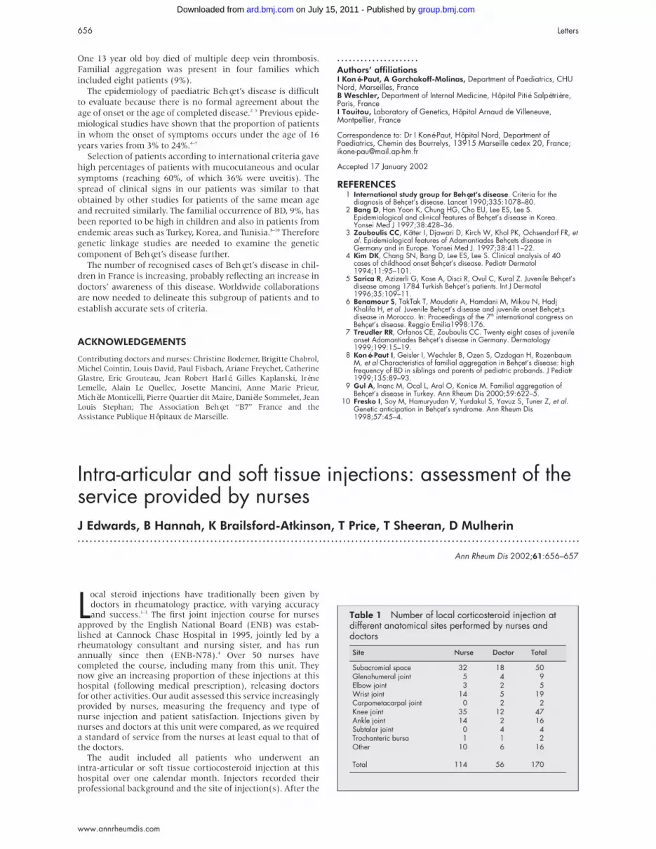
Table 1 Number of local corticosteroid injection atdifferent anatomical sites performed by nurses anddoctors
Site Nurse Doctor Total
Subacromial space 32 18 50Glenohumeral joint 5 4 9Elbow joint 3 2 5Wrist joint 14 5 19Carpometacarpal joint 0 2 2Knee joint 35 12 47Ankle joint 14 2 16Subtalar joint 0 4 4Trochanteric bursa 1 1 2Other 10 6 16
Total 114 56 170
656 Letters
www.annrheumdis.com
group.bmj.com on July 15, 2011 - Published by ard.bmj.comDownloaded from

injection, the patient completed an anonymous questionnaire
in their home within 3–7 days for return by prepaid envelope.
This also recorded the injector’s professional background; the
patient’s opinion of the adequacy of the information given
before and after the injection (on a four point scale, from “no
information” to “very detailed information”); the comfort (on
a four point scale, from “very comfortable” to “very
uncomfortable”) and efficacy (on a four point scale, from
“very helpful” to “made worse”) of the procedure; their over-
all satisfaction (marked on a 10 cm visual analogue scale
(VAS)); and the professional group from whom they would
prefer to receive any future injections. The approval of the local
research ethics committee was given for this audit.
In one month, 170 corticosteroid injections were given to
103 patients: trained nurses gave 114 (67%) of these injections
to 63 patients. Nurses gave a mean (range) of 1.8 (1–8) injec-
tions to each patient compared with 1.4 (1–4) given by
doctors. Nurses gave most of the ankle, knee, wrist, elbow,
glenohumeral, and subacromial injections (table 1). Doctors
gave all subtalar and carpometacarpal joint injections. Ninety
three (90%) completed questionnaires were returned by
patients. Almost all (96%) described preinjection information
as “adequate” or “very detailed”, but more described it as
“very detailed” when given by a nurse (48%) than when given
by a doctor (32%) (fig 1). A greater proportion also described
postinjection information as “very detailed” when given by a
nurse (47% v 34%) (fig 1). Most (93%) described the injection
as “fairly comfortable” or “very comfortable” but were more
likely to have described the injection given by a nurse as “very
comfortable” (61% v 43%). Almost all the injections given by
both nurses and doctors were described as “helpful” or “very
helpful” (93% and 88%, respectively). Overall satisfaction of
injections given by nurses and doctors was similar (mean
(range) VAS 8.0 (1.1–10.0) cm and 7.8 (0.6–10) cm,
respectively). Only one patient injected by a nurse expressed a
preference for any future injections to be given by a doctor; the
rest preferred a nurse (38%) to give any future injections or
had no preference.
This audit confirmed that trained nurses were performing
the vast majority of local corticosteroid injections at this unit
and that their standards were at least as good as those of doc-
tors. Patients felt well informed by nurse injectors, found their
injections were effective, were highly satisfied with their
treatment, and were willing to have further treatment from
nurses, if necessary. The results show that trained nurses can
deliver a service previously provided by doctors in training,
whose working hours are now greatly restricted.
There might be concerns about the experience acquired by
such doctors during their rheumatology attachment, but fur-
ther audit has shown that they now learn the techniques of
steroid injection largely from trained nursing staff (results not
shown). Rheumatology nurses now fulfil a wide range of roles
in education, counselling, monitoring, and giving treatment.5
Debate continues over whether such developments represent
progress or a dilution of the nurturing role traditionally
ascribed to nurses.6 7 The cost effectiveness of an injection
service provided by nurses was not considered by this audit
and would be influenced by salary and patient throughput.
The length of time spent with patients during this audit was
not recorded, but it is worth noting that nursing staff were
performing multiple intra-articular injections on some pa-
tients, which is time-consuming in itself and certainly does
not represent “cherry picking” the easy cases.
ACKNOWLEDGEMENTThe authors acknowledge and thank Dr A Hassell, consultantrheumatologist, for his foresight and determination in establishingthe ENB-N78 joint injection course.
. . . . . . . . . . . . . . . . . . . . .Authors’ affiliationsJ Edwards, B Hannah, K Brailsford-Atkinson, T Price, T Sheeran,D Mulherin, Department of Rheumatology, Cannock Chase Hospital,United Kingdom
Correspondence to: Dr D Mulherin, Department of Rheumatology,Cannock Chase Hospital, Brunswick Road, Cannock, WS11 2XY, UK;[email protected]
Accepted 19 December 2001
REFERENCES1 Dixon A, Emery P. Local injection therapy in rheumatic diseases. 4th ed.
Basle: Eular Publishers, 1992.2 Haslock I, MacFarlane D, Speed C. Intra-articular and soft tissue
injections: a survey of current practice. Br J Rheumatol 1995;34:449–52.3 Eustace JA, Brophy DP, Gibney RP, Bresnihan B, FitzGerald O.
Comparison of the accuracy of steroid placement with clinical outcome inpatients with shoulder symptoms. Ann Rheum Dis 1997;56:59–63.
4 Edwards J, Hassell A. Intra-articular and soft tissue injections by nurses:preparation for expanded practice. Nurs Standard 2000;14:43–6.
5 Hill J. The expanding role of the nurse in rheumatology. Br J Rheumatol1997;36:410–12.
6 Gardiner PV. Nurse practitioner clinics. Br J Rheumatol 1994;33:893.7 A Carr, ed. Defining the extended clinical role for allied health
professionals in rheumatology. Chesterfield: The Arthritis ResearchCampaign, 2001.
Figure 1 Quality of information provided to a patient by a nurse or doctor injector as rated by the patient before (A) or after (B) localcorticosteroid injection. Ratings were provided by 38 patients who received their injections from doctors and 58 patients who received theirinjections from trained nurses.
80
60
40
0
20
A
Nurse
Perc
enta
ge
Very
detai
led
Adequ
ateNon
e
Inade
quate
Doctor
80
60
40
0
20
B
Very
detai
led
Adequ
ateNon
e
Inade
quate
Letters 657
www.annrheumdis.com
group.bmj.com on July 15, 2011 - Published by ard.bmj.comDownloaded from

Manual jobs increase the risk of patients with ankylosingspondylitis withdrawing from the labour force, also whenadjusted for job related withdrawal in the generalpopulationA Boonen, A Chorus, R Landewé, D van der Heijde, H Miedema, H van der Tempel,Sj van der Linden. . . . . . . . . . . . . . . . . . . . . . . . . . . . . . . . . . . . . . . . . . . . . . . . . . . . . . . . . . . . . . . . . . . . . . . . . . . . . . . . . . . . . . . . . . . . . . . . . . . . . . . . . . . . . . . . . . . . . . . . . . . . .
Ann Rheum Dis 2002;61:658
In 1997 we studied labour force participation among 658
Dutch patients with ankylosing spondylitis (AS).1 In those
who had a paid job before onset of disease (n=529), age and
sex adjusted withdrawal rate was 3.0 times (95% CI 2.5 to 3.6)
higher than expected in the general Dutch population.2 Within
patients with AS, those with a manual job had a 2.3 (95% CI
1.5 to 3.4) times increased risk of withdrawal compared with
those with a non-manual job after correction for age at onset
of disease, gender, educational level, and coping strategies.2
However, the question remained whether a manual job was a
specific risk factor for withdrawal from work in patients with
AS or if a manual job is a non-specific risk factor for
withdrawal in every working subject.
Recently, Dutch population figures for 1998 on withdrawal
from the labour force, for the same work categories as used in
our study, became available (Dutch Bureau of Statistics).
Therefore, we were then able to calculate the job adjusted
ratios for withdrawal from the labour force in patients with AS
by comparing the job-specific annual withdrawals in patients
with those from the general population. Five major classes of
work were distinguished: agriculture, industry, transport,
commerce, and services/management. The first three classes
were later grouped as manual jobs and the last two classes as
non-manual jobs. This classification has some limitations
because jobs like housekeepers and waiters are also included
within the servicing jobs and therefore considered as
non-manual jobs. However, this was similar in patients and in
controls. All data were stratified by gender. The 95% CIs for the
ratios were calculated using Poisson’s distribution.
Table 1 presents for each job class, the observed rates of
withdrawal from the labour force as well as the ratios when
compared with the general population. While the overall risk
for a patient with AS to withdraw from the labour force is 3.0
(95% CI 2.5 to 3.6) times higher than expected in the general
population, this risk is 4.9 (95% CI 3.5 to 5.9) times higher for
those with a manual job versus 2.2 (95% CI 1.6 to 2.7) times
higher for those with a non-manual job. The observed effect
was stronger in male than in female patients, probably
because of the smaller number of female patients in the study
sample. In the general population withdrawal rates were not
significantly different among the professional classes studied
either for men or for women.We conclude that patients with AS with a manual job have an
increased risk for withdrawal from the labour force, also when
adjusted for job related withdrawal in the general population.
ACKNOWLEDGEMENTThe authors acknowledge the Dutch rheumatologists participating inthe Standardised Diagnositic Register of Rheumatic Diseases and allpatients who took part in this study.
. . . . . . . . . . . . . . . . . . . . .Authors’ affiliationsA Boonen, R Landewé, D van der Heijde, Sj van der Linden,Department of Internal Medicine, Division of Rheumatology, UniversityHospital Maastricht, The NetherlandsA Chorus, Division of Public Health, TNO Prevention and Health, Leiden,The NetherlandsR Landewé, Atrium Medical Centre, Heerlen, The NetherlandsD van der Heijde, Limburg University Centre, Diepenbeek, BelgiumH Miedema, Netherlands Expert Centre for Workrelated MusculoskeletalDisorders, University Hospital Dijkzigt and Erasmus University Rotterdam,The NetherlandsH van der Tempel, Maasland Ziekenhuis Sittard, The Netherlands
Correspondence to: Dr A Boonen, Department of Internal Medicine,Division of Rheumatology, University Hospital Maastricht, PO Box 5800,6202 AZ Maastricht, The Netherlands; [email protected]
Accepted 7 January 2002
REFERENCES1 Boonen A, Chorus A, Miedema H, van der Heijde D, van der Tempel H,
van der Linden Sj. Employment, work disability and work days lost inpatients with ankylosing spondylitis: a cross sectional study of Dutchpatients. Ann Rheum Dis 2001;60:353–8.
2 Boonen A, Chorus A, Miedema H, van der Heijde D, Landewé R,Schouten H, et al. Withdrawal from labour force due to work disability inpatients with ankylosing spondylitis. Ann Rheum Dis 2001;60:1033–9.
Table 1 Annual withdrawal rate from the labour force due to official work disability in patients with ankylosingspondylitis for five classes of work and ratios when compared with the general population
Male patients Female patients All patients
Withdrawal rate (%) SIR Withdrawal rate (%) SIR (95% CI) Withdrawal rate (%) SIR (95% CI)
Manual job 3.9 5.2 (3.6 to 6.4) 3.5 2.4 (0.1 to 4.7) 4.2 4.9 (3.5 to 5.9)Agriculture 0 – 11.6 5.4 1.3 1.0Industry 4.1 5.3 3.2 2.2 4.3 4.9Transport 4.5 7.2 0 – 4.9 6.8
Non-manual job 1.3 2.1 (1.3 to 2.8) 2.8 2.2 (1.3 to 3.1) 2.0 2.2 (1.6 to 2.7)Commercial 1.0 1.9 3.5 3.0 1.9 2.3Service 1.4 2.1 2.7 2.1 2.1 2.1
All jobs 2.3 3.4 (2.6 to 4.1) 2.9 2.2 (1.4 to 3.1) 2.7 3.0 (2.5 to 3.6)
SIR, standardised incidence ratios of withdrawal.
658 Letters
www.annrheumdis.com
group.bmj.com on July 15, 2011 - Published by ard.bmj.comDownloaded from

Breast implants and illness: a model of psychologicalillnessM Ahern, M Smith, H Chua, P Youssef. . . . . . . . . . . . . . . . . . . . . . . . . . . . . . . . . . . . . . . . . . . . . . . . . . . . . . . . . . . . . . . . . . . . . . . . . . . . . . . . . . . . . . . . . . . . . . . . . . . . . . . . . . . . . . . . . . . . . . . . . . . . .
Ann Rheum Dis 2002;61:659
We read with interest the hypothesis of Dush in the
Annals of the Rheumatic Diseases1 and provide some
evidence to support it. Dush suggested that many of
the symptoms of women with silicone breast implants might
be attributed to somatisation, stress, and mass somatisation.
We recently reviewed 179 women with silicone breast
implants who were involved in product liability and litigation.
Their ages ranged from 29 to 74 years (mean (SD) 46.8
(8.1)). The indications for surgery were cosmetic in 146 (82%),
followed by cancer in 17 (9%), fibrocystic disease in 12 (7%),
and congenital hypoplasia in 4 (2%) women. The most
common symptoms were burning breast pain in 142 (79%),
chronic fatigue in 142 (79%), arthralgia in 134 (75%), sleep
disturbance in 127 (71%), cognitive dysfunction in 102 (57%),
sicca symptoms in 100 (56%), night sweats in 97 (54%), and
myalgia in 91 (51%). Findings on clinical examination were
few, including chest wall abnormalities in 60 (34%), tender
trigger points in 31 (17%), and carpal tunnel syndrome in 6
(3%) women. Sixty five (36%) women had radiological and/or
surgical proof of implant leakage or rupture and 61 (34%) had
signs of implant contractures. We found no evidence of
increased occurrence of any connective tissue disorder such as
rheumatoid arthritis (RA), systemic lupus erythematosus
(SLE), or Sjögren’s syndrome.
The women were asked to complete two questionnaires and
return them by mail: (a) the General Health Questionnaire
(GHQ), possibly the most widely used and extensively
validated screening test for functional psychiatric illness—a
score above 12 denoting significant psychiatric morbidity; (b)
the Speilberger State-Trait Anxiety Inventory (STAI). This
comprises two self report scales for measuring two distinct
anxiety concepts: (i) state anxiety (A state), which may vary
over time and (ii) trait anxiety (A trait), a relatively enduring
personality characteristic which does not tend to change
much over time.
Of the 179 questionnaires sent, 117 were returned, giving a
response rate of (65%). The women had a mean (SD) score of
18.3 (8.2) on the GHQ; 75% of women having scores above 12.
Table 1 shows the means (SD) of the A state and A trait.
Patients with breast implant had significantly higher Astate and A trait scores than female undergraduate students(p<0.001) and medical/surgical inpatients (p<0.001) and asmuch anxiety reaction as psychiatric patients with anxietydisorders.
Thus these women were found to have significant psychiat-ric morbidity and to show significant state and trait anxiety.The causes for these high anxiety levels may be related to thereasons these women sought breast implants—for example,poor self esteem, interpersonal and psychological problems.Possibly, these high anxiety levels are exacerbated by the liti-gation and media attention. The raised trait-anxiety levelsmaybe a risk factor for somatisation.
We attempted to reassure these women that there was noevidence of any underlying connective tissue disorder orsystemic disease but could not allay their fears. Nevertheless,we identified the fact that these woman were distressed andrequired medical and psychological rehabilitation. Unfortu-nately, many of these women had been “dismissed” and theirsymptoms not taken seriously by their treating doctorsbecause epidemiological research had not shown any associ-ation with recognisable systemic disease.
Because of the uncontrolled nature of our observations wecan only propose a hypothesis for the causes of theirsymptoms. As rheumatologists encountering women withsilicone breast implants, we need to reassure them that theyare unlikely to have an underlying connective tissue disorder.We also must provide access to treatments that will alleviatetheir anxiety and distress, as suggested by Dush.
. . . . . . . . . . . . . . . . . . . . .Authors’ affiliationsM Ahern, M Smith, H Chua, P Youssef, Department of Rheumatology,Repatriation General Hospital, Adelaide, South Australia
Correspondence to: Professor M Ahern, Department of Rheumatology,Repatriation General Hospital, Flinders University of South Australia,Daws Road, Daw Park, South Australia 5041, Australia;[email protected]
Accepted 18 December 2001
REFERENCE1 Dush DM. Breast implants and illness: a model of psychological illness.
Ann Rheum Dis 2001;60:653–7.
Table 1 A state and A trait scores, mean (SD)
Silicone breastimplant (n=116)
Femaleundergraduatestudents (n=231)
Medical/surgicalpatients (n=161)
Anxious psychiatricpatients (n=60)
A trait 51.5 (14.3) 38.25 (9.14) 41.91 (12.7) 48.08 (10.61)A state 53.1 (13.6) 35.16 (9.3) 43.28 (13.8) 49.02 (11.6)
Letters 659
www.annrheumdis.com
group.bmj.com on July 15, 2011 - Published by ard.bmj.comDownloaded from

Fibromyalgia in patients with rheumatoid arthritis isassociated with higher scores of disabilityA Naranjo, S Ojeda, F Francisco, C Erausquin, I Rúa-Figueroa, C Rodríguez-Lozano. . . . . . . . . . . . . . . . . . . . . . . . . . . . . . . . . . . . . . . . . . . . . . . . . . . . . . . . . . . . . . . . . . . . . . . . . . . . . . . . . . . . . . . . . . . . . . . . . . . . . . . . . . . . . . . . . . . . . . . . . . . . .
Ann Rheum Dis 2002;61:660–661
Rheumatoid arthritis (RA) is a chronic polyarticular
disease characterised by pain in peripheral joints accom-
panied by swelling, stiffness, and functional impairment.
In some cases it is associated with fibromyalgia (FM), a
syndrome defined by chronic, widespread pain, asthenia, and
sleep disorders. When a patient has both RA and FM,
determining the degree of RA activity may be difficult, because
these patients typically have higher scores for pain and
disability.
This study aimed at evaluating whether there were
differences in functional disability, extra-articular manifesta-
tions, and use of disease modifying antirheumatic drugs
(DMARDs), between patients with RA with and without FM.
PATIENTS AND METHODSA cross sectional study was conducted with 386 patients with
RA, 94 men and 292 women, with a mean age of 53 years. All
the patients met the criteria of the American College of Rheu-
matology (ACR) for the diagnosis of the disease.1 The mean
duration of the disease was nine years. All the patients
received treatment in a hospital outpatient clinic and were
included in a database between 1991 and 2000. To diagnose
FM, ACR criteria had to be fulfilled on at least two consecutive
visits.2 The following assessment was made in all patients par-
ticipating in the study: a clinical history, evaluation of
functional status using the Health Assessment Questionnaire
(HAQ),3 conventional laboratory measurements, and evalua-
tion of the rheumatoid factor. In addition to these assess-
ments, extra-articular manifestations were diagnosed. Sec-
ondary Sjögren’s syndrome was diagnosed when, in addition
to subjective xerophthalmia and xerostomia, Schirmer’s test or
the rose bengal staining were pathological.4 The number of
previous DMARDs was counted, independently of whether the
patient received a single drug or a combination.
Contingency tables were used to compare the frequency of
categorical variables among the different groups. To compare
numerical variables we used Student’s t test when the data
followed a normal distribution and the equivalent Wilcoxon
non-parametric test when they did not.
RESULTSOf the total, 57 (14.8%) patients fulfilled FM criteria. No
differences were found in age or disease duration between
patients with RA without FM (RA group) and patients with RA
and FM (RA-FM group) (table 1). In the RA-FM group there
was a higher percentage of women (p=0.03); HAQ scores
(p=0.002) were also higher. The incidence of extra-articular
manifestations such as serositis, pneumonitis, or Sjögren’s
syndrome was similar in both groups. Rheumatoid nodules
and the rheumatoid factor were more common in the RA
group, although differences were not significant. The RA-FM
group had received a greater number of DMARDs (p=0.04).
DISCUSSIONThe results of this study indicate that patients with RA who
also have FM are more often women, have higher disability
scores, and receive DMARDs more frequently.
Wolfe et al studied 242 patients with RA and 38 who had FM
occurring in association with RA. The RA-FM group had more
abnormal measures of function, pain, disease activity, and
psychological status, but the disease severity in RA-FM and
RA was similar.5 In patients with RA, FM tender points have
been found to correlate mainly with daily stress and with
higher joint tenderness count scores, indicating that patients
with RA and FM have a lower pain threshold.6 7 Patients with
RA and depression often perform fewer daily life activities.8
In summary, FM is found to be associated in one of seven
patients with RA; the presence of FM may constitute a marker
of a worse prognosis for subjective functional disability.
. . . . . . . . . . . . . . . . . . . . .Authors’ affiliationsA Naranjo, S Ojeda, F Francisco, C Erausquin, I Rúa-Figueroa, CRodríguez-Lozano, Rheumatology Service, Hospital de Gran CanariaDr Negrín, Las Palmas de Gran Canaria, Spain
Correspondence to: Dr A Naranjo, Rheumatology Service, Hospital deGran Canaria Dr Negrín, C/ Barranco de la Ballena s/n 35020, LasPalmas de Gran Canaria, Spain; [email protected]
Accepted 11 December 2001
Table 1 Comparison of patients with rheumatoid arthritis (RA) and rheumatoidarthritis and fibromyalgia (RA-FM)
RA-FM RA p Value
Number of patients 57 329Women (No (%)) 50 (88) 242 (74) 0.03Mean (SD) age (years) 52 (9.6) 53 (15) 0.42Duration of RA disease (SD), years 8 (8) 9 (9) 0.35HAQ (mean (SD)) 1.62 (0.70) 1.21 (0.77) 0.002Patients with rheumatoid factor (No (%)) 37 (65) 247 (75) 0.13Extra-articular manifestations
Rheumatoid nodules (No (%)) 10 (18) 82 (25) 0.36Secondary Sjögren’s syndrome (No (%)) 15 (26) 80 (24) 0.97Serositis (No (%)) 3 (5) 12 (4) 0.99Interstitial lung disease (No (%)) 0 17 (5)
Number of DMARDs (mean (SD)) 2.64 (1.6) 2.17 (1.5) 0.04
660 Letters
www.annrheumdis.com
group.bmj.com on July 15, 2011 - Published by ard.bmj.comDownloaded from

REFERENCES
1 Arnett FC, Edworthy SM, Bloch DA, McShane DJ, Fries JF, Cooper NS,et al. The American Rheumatism Association 1987 revised criteriafor the classification of rheumatoid arthritis. Arthritis Rheum1988;31:315–24.
2 Wolfe F, Smythe HA, Yunus MB, Bennett RM, Bombardier C,Goldenberg DL, et al. The American College of Rheumatology 1990criteria for the classification of fibromyalgia. Report of the multicentercriteria committee. Arthritis Rheum 1990;33:160–72.
3 Esteve-Vives J, Batlle-Gualda E, Reig A. Spanish version of the HealthAssessment Questionnaire: reliability, validity and transculturalequivalency. (Grupo para la adaptación del HAQ a la poblaciónespañola.) J Rheumatol 1993;20:2116–22.
4 Vitali C, Bombardieri S, Moutsopoulos HM, Balestrieri G, Bencivelli W,Bernstein RM, et al. Preliminary criteria for the classification of Sjögren’ssyndrome. Results of a prospective concerted action supported by theEuropean Community. Arthritis Rheum 1993;36:340–7.
5 Wolfe F, Cathey MA, Kleinheksel SM. Fibrositis (fibromyalgia) inrheumatoid arthritis. J Rheumatol 1984;11:814–18.
6 Urrows S, Affleck G, Tennen H, Higgins P. Unique clinical andpsychological correlates of fibromyalgia tender points and jointtenderness in rheumatoid arthritis. Arthritis Rheum 1994;37:1513–20.
7 Konttinen YT, Honkanen VE, Gronblad M, Keinonen M, Santavirta N,Santavirta S. The relation of extraarticular tenderness to inflammatoryjoint disease and personality in patients with rheumatoid arthritis. JRheumatol 1992;19:851–5.
8 Katz PP, Yelin EH. Life activities of persons with rheumatoid arthritis withand without depressive symptoms. Arthritis Care Res 1991;7:69–77.
Shingles following infliximab infusionD C Baumgart, A U Dignass. . . . . . . . . . . . . . . . . . . . . . . . . . . . . . . . . . . . . . . . . . . . . . . . . . . . . . . . . . . . . . . . . . . . . . . . . . . . . . . . . . . . . . . . . . . . . . . . . . . . . . . . . . . . . . . . . . . . . . . . . . . . .
Ann Rheum Dis 2002;61:661
Infliximab is a chimeric IgG1κ monoclonal antibody that
binds specifically to human tumour necrosis factor alpha
(TNFα). Infliximab, in combination with methotrexate, is
approved for reducing signs and symptoms and inhibiting the
progression of structural damage in patients with moderately
to severely active rheumatoid arthritis who have had an inad-
equate response to methotrexate. It will also reduce the signs
and symptoms of Crohn’s disease in patients with moderately
to severely active Crohn’s disease who have had an inadequate
response to conventional treatment and will reduce the
number of draining enterocutaneous fistulas in patients with
fistulising Crohn’s disease.1 2 Owing to its mechanism of
action infliximab can lead to a number of complications.
CASE REPORTHere we report a case of shingles, an infectious complication,
currently not included in the product labelling.
A 45 year old man with steroid dependent Crohn’s disease
presented to the outpatient clinic with an acute flare up. At
that time he had already been receiving 150 mg of
azathioprine and 1000 mg mesalamine by mouth three times
a day for about 17 months. Prednisolone had been tapered to
5 mg a day. High resolution intestinal ultrasound showed a
subtotal small bowel stenosis. Power Doppler demonstrated
mucosal hyperaemia, suggesting an inflammatory process.
It was therefore decided to switch his treatment to
infliximab. His condition slightly improved, but after the third
course of 5 mg/kg bodyweight infliximab he developed a pain-
ful, pustular skin rash on the left side of his chest involving
several dermatomas. Varicella zoster IgM titres were raised,
confirming an acute shingles infection. He was treated intra-
venously with 5 mg/kg bodyweight acyclovir every eight hours
for seven days. He recovered and was later referred for surgery
to resect the inflamed segment.
DISCUSSIONAdult varicella can be a severe illness complicated by
pneumonia, encephalitis, hepatitis, thrombocytopenia, and
prolonged fever.3 Blood levels of TNFα have been shown to be
raised in patients with acute varicella infection.4 In vitro stud-
ies have shown that replication of varicella zoster virus and
varicella zoster virus antigen expression are inhibited by TNFαand that this antiviral activity can be completely blocked by
monoclonal antibodies against TNFα.5
The use of monoclonal antibodies against TNFα in patients
with inflammatory bowel disease increases the risk of viral
infections by inhibiting an adequate TNFα response. Doctors
should be cautious when prescribing infliximab for patients
who are already receiving immunosuppressant drugs. We sug-
gest that varicella zoster virus infection should be included as
an infectious complication in the drug labelling.
. . . . . . . . . . . . . . . . . . . . .Authors’ affiliationsD C Baumgart, A U Dignass, Universitätsklinikum Charité, CampusVirchow-Klinikum, Medizinische Fakultät der Humboldt-Universität zuBerlin, Medizinische Klinik mit Schwerpunkt Hepatologie undGastroenterologie, D-13344 Berlin, Germany
Correspondence to: Dr D C Baumgart, Charité-Campus Virchow-Klinikum,Humboldt-Universität zu Berlin, Hepatologie und Gastroenterologie,D-13344 Berlin, Germany; [email protected]
Accepted 3 January 2002
REFERENCES1 Lipsky PE, van der Heijde DM, St Clair EW, Furst DE, Breedveld FC,
Kalden JR, et al. Infliximab and methotrexate in the treatment ofrheumatoid arthritis. Anti-tumor necrosis factor trial in rheumatoid arthritiswith concomitant therapy study group. N Engl J Med 2000;343:1594–602.
2 Targan SR, Hanauer SB, van Deventer SJ, Mayer L, Present DH,Braakman T, et al. A short-term study of chimeric monoclonal antibodycA2 to tumor necrosis factor alpha for Crohn’s disease. Crohn’s diseasecA2 Study Group. N Engl J Med 1997;337:1029–35.
3 Liesegang TJ. Varicella zoster viral disease. Mayo Clin Proc1999;74:983–98.
4 Wallace MR, Woelfl I, Bowler WA, Olson PE, Murray NB, Brodine SK,et al. Tumor necrosis factor, interleukin-2, and interferon-gamma in adultvaricella. J Med Virol 1994;43:69–71.
5 Ito M, Nakano T, Kamiya T, Kitamura K, Ihara T, Kamiya H, et al. Effectsof tumor necrosis factor alpha on replication of varicella-zoster virus.Antiviral Res 1991;15:183–92.
Letters 661
www.annrheumdis.com
group.bmj.com on July 15, 2011 - Published by ard.bmj.comDownloaded from

Self limiting lupus-like symptoms in patients withparvovirus B19 infectionE Tóvári, I Mezey, K Hedman, L Czirják. . . . . . . . . . . . . . . . . . . . . . . . . . . . . . . . . . . . . . . . . . . . . . . . . . . . . . . . . . . . . . . . . . . . . . . . . . . . . . . . . . . . . . . . . . . . . . . . . . . . . . . . . . . . . . . . . . . . . . . . . . . . .
Ann Rheum Dis 2002;61:662–663
Parvovirus B19 causes polyarthritis in adults.1 Several
observers have noted a lupus-like syndrome2 3 and
production of autoantibodies associated with this
infection.4 We describe here two white patients with acute
parvovirus B19 infection and a transient lupus-like syndrome.
CASE REPORTSCase 1A 41 year old woman had a transient polyarthritis of the
metacarpophalangeal (MCP) and proximal interphalangeal
(PIP) joints after her first delivery at the age of 30, and she
developed photosensitivity, and reported dryness of the eyes
for some years.
In 1998 she had an influenza-like illness with polyarthral-
gia, myalgia, and fever. She also had a massive UV light expo-
sure with tanning. Two days later she developed polyarthritis
of the wrists, MCP, and PIP joints. Her 4 year old child had a
fever and facial skin rash just before her disease.
One week after the start of her polyarthritis she had a nor-
mal blood count, erythrocyte sedimentation rate (ESR), rheu-
matoid factor, complement, immunoglobulin levels, urine
analysis, liver enzymes, creatinine kinase, and slightly raised
(15 mg/l) C reactive protein (CRP). Anti-dsDNA, anticardioli-
pin, IgG and IgM were increased, and anti-SS-A, anti-SS-B
antibodies were also detected by enzyme linked immuno-
sorbent assay (ELISA). An antinuclear antibody (ANA) test
was negative, with an anti-cytoskeletal staining on HEp-2
cells. Both IgM and IgG antibodies to human parvovirus B19
were positive, and parvovirus DNA was detected by nested
polymerase chain reaction (PCR) in serum.
After eight weeks the autoantibody tests became negative,
and her symptoms completely resolved. At two years follow up
she remained asymptomatic, and the parvovirus PCR was
negative.
Case 2A 34 year old woman was examined in June 1998 with an
acute painful occipital lymph node and polyarthritis in the
MCP and PIP joints. She had had photosensitivity from the
age of 10. Two weeks before symptoms, her child had a rash
and flu-like illness, and in the meantime she was exposed to
UV light with tanning.Urine analysis, blood count, liver enzymes were normal, but
the ESR and CRP were slightly raised (22 mm/1st h, and 11
mg/l, respectively). No rheumatoid factor was detected.
Anti-dsDNA and anti-Sm autoantibody were positive by
ELISA. An ANA test was negative on HEp-2 cells, with an
atypical cytoplasmic staining. IgM and IgG antibodies to
human parvovirus B19 were present, and parvovirus DNA was
detected by PCR in serum. The patient was three weeks’ preg-
nant but she decided to terminate the pregnancy. After 10
weeks the autoantibody ELISA became negative, but the ANA
test became positive with homogeneous staining pattern.
Apart from periodic myalgia and arthralgia the patient
remained without symptoms for two years. The diagnosis may
be systemic lupus erythematosus (SLE) in view of the photo-
sensitivity, polyarthritis, ANA, and anti-dsDNA positivity. At
500 days after the onset PCR was positive in serum.
DISCUSSIONAfter the infection four criteria of SLE were fulfilled in case 2,
and case 1 was compatible with the diagnosis of undifferenti-
ated connective tissue disease.5 The autoimmune process was
self limited in both patients.
The patients’ children had symptoms of erythema infectio-
sum, and the mothers were examined for parvovirus B19.
They were IgM and IgG positive (table 1). The VP1-IgG
avidity6 and VP2-IgG epitope type specificity7 results (table 1)
confirmed acute B19 infection in our patients. Nested PCR was
performed on the patients’ sera.8 Both patients were PCR
positive in the acute phase (table 1). In immunocompetent
patients PCR may remain positive for four months, but more
prolonged persistence in healthy subjects is unusual. Parvo-
virus DNA was not detectable in the first patient after 400
days, but in patient 2 the blood sample taken at 500 days was
positive.
Table 1 Antibodies and nested PCR findings of two patients with parvovirus B19infection
Days after onsetof polyarthritis VP2-IgM EIA VP2-IgG EIA VP2-ETS ratio
VP1-IgGavidity (%) PCR
Case 1 7 Pos Pos 0.38 6.3 +20 ND Pos 0.41 6.6 +
400 Neg Pos 232.57 52.8 –
Case 2 4 Pos Pos 0.28 – +24 ND Pos 0.49 6.2 ND47 Pos Pos 0.81 11.8 +
110 Neg Pos 4.98 24.7 +144 – Pos 13.42 26.5 +500 ND ND ND ND +
VP2, major capsid protein; VP1, minor capsid protein; ETS, epitope type-specific IgG; pos, positive; neg,negative; ND, not determined.Zone of acute infection in VP2/ETS ratio 0–10; zone of past immunity >10.Zone of acute infection in VP1 IgG avidity 0–15%; borderline zone 16–25%, zone of past immunity: >25%.Signs of acute parvovirus infection are shown in bold.
662 Letters
www.annrheumdis.com
group.bmj.com on July 15, 2011 - Published by ard.bmj.comDownloaded from
At the time of the infection other provoking factors for SLE
could also be identified. The second patient had an early phase
pregnancy. Although the patients had earlier had photosensi-
tivity, they were tanning during the initial period of infection.
The typical rash of parvovirus infection, like the skin manifes-
tations of SLE, can be provoked by UV light. Possibly, the virus
and the UV light had additional effects on the autoimmune
process.
Typing for HLA-A, B, C, and DR, DQ showed common alle-
les. They had DR2 (DRB1*1501) and DQ6 (DQB1*0602) with
the same subtypes (table 2). Four HLA alleles of the patients
have been previously described as associated with SLE.9
In conclusion, we have found several common provoking
factors of the autoimmune process in our cases. The patients
had a genetic predisposition to lupus and also signs of
autoimmune diseases in the past. They had also been exposed
to UV light and pregnancy, which are known provoking factors
for SLE. Furthermore, parvovirus infection seemed to be also
a provoking factor for the development of SLE and lupus-like
disease, although the symptoms of connective tissue diseases
were transient.
. . . . . . . . . . . . . . . . . . . . .Authors’ affiliationsE Tóvári, L Czirják, 2nd Department of Internal Medicine, University ofPécs, Medical Faculty, Pécs, HungaryI Mezey, Bela Johan’ National Centre for Epidemiology, Division ofVirology, Budapest, HungaryK Hedman, Department of Virology, Haartman Institute and HUCHDiagnostic, University of Helsinki, Finland
Correspondence to: Professor L Czirják, University of Pécs, MedicalFaculty Nephrology Centre and 2nd Department of Internal Medicine,Clinical Immunology Unit, H-7624 Pécs, Pacsirta u.1., Hungary;[email protected]
Accepted 14 December 2001
REFERENCES1 White DG, Woolf AD, Mortimer PP, Cohen BJ, Blake DR, Bacon PA.
Human parvovirus arthropathy. Lancet 1985;i:419–21.2 Gran JT, Johnsen V, Myklebust G, Nordbo SA. The variable clinical
picture of arthritis induced by human parvovirus B19. Report of sevenadult cases and review of the literature. Scand J Rheumatol1995;24:174–9.
3 Nesher G, Osborn TG, Moore TL. Parvovirus infection mimickingsystemic lupus erythematosus. Semin Arthritis Rheum 1995;24:297–303.
4 Loizou S, Cazabon JK, Walport MJ, Tait D, So AK. Similarities ofspecificity and cofactor dependence in serum antiphospholipidantibodies from patients with human parvovirus B19 infection and fromthose with systemic lupus erythematosus. Arthritis Rheum1997;40:103–8.
5 Calvo Alen J, Alarcón GS, Burgard SL, Burst N, Bartolucci AA, WilliamsHJ. Systemic lupus erythematosus: predictors of its occurrence among acohort of patients with early undifferentiated connective tissue disease:multivariate analyses and identification of risk factors. J Rheumatol1996;23:469–75.
6 Söderlund M, Brown CS, Cohen BJ, Hedman K. Accurate serodiagnosisof B19 parvovirus infections by measurement of IgG avidity. J Infect Dis1995;171:710–13.
7 Kaikkonen L, Lankinen H, Harjunpää I, et al. Acute-phase-specificheptapeptide epitope for diagnosis of parvovirus B19 infection. J ClinMicrobiol 1999;37:3952–6.
8 Hornsleth A, Carlsen KM, Christensen LS, Gundestrup M, Heegaard ED,Myhre J. Estimation of serum concentration of parvovirus B-19 DNA byPCR in patients with chronic anaemia. Res Virol 1994;145:379–86.
9 Arnett FC Jr. The genetics of human lupus. In: Wallace DJ, Hahn BH,ed. Dubois’ lupus erythematosus. Baltimore: William and Wilkins,1997:77–117.
Dermatomyositis in a patient with adenocarcinoma of thegall bladderG Yiannopoulos, P Ravazoula, N Meimaris, M Stavropoulos, A P Andonopoulos. . . . . . . . . . . . . . . . . . . . . . . . . . . . . . . . . . . . . . . . . . . . . . . . . . . . . . . . . . . . . . . . . . . . . . . . . . . . . . . . . . . . . . . . . . . . . . . . . . . . . . . . . . . . . . . . . . . . . . . . . . . . .
Ann Rheum Dis 2002;61:663–664
Acase of dermatomyositis in an elderly woman, found to
have gall bladder adenocarcinoma, is presented. As far
as we know this is the first report of an association
between the myopathy and this specific malignancy.
CASE REPORTA 75 year old white woman was admitted to our department,
with a seven month history of proximal muscle weakness,
dysphagia, dysphonia, facial erythema, and oedema of the
eyelids. Atypical dyspeptic symptoms had been present for the
same time period. At that time, an abdominal ultrasound
showed, besides multiple gall stones, increased echogenicity of
the gall bladder content. Simultaneous routine laboratory
tests were normal, except for a minimal increase of serum
aspartate aminotransferase (AST) and alanine aminotrans-
ferase (ALT) and a creatine kinase (CK) value of 350 IU/ml
(upper normal 190). Apparently, that had gone unnoticed,
until her admission.
On physical examination, she was found to be a relatively
obese elderly woman, in no distress, with normal vital signs. A
“heliotrope” rash was present in her eyelids, and Gottron’s
papules were noticed on the dorsal surface of her metacar-
pophalangeal and proximal interphalangeal joints bilaterally.
Proximal muscle weakness of the neck, shoulder, and pelvic
girdles was prominent. The rest of the physical examination
was unremarkable.
Routine laboratory tests, including CK, were normal, except
for a mild increase of AST and lactate dehydrogenase (LDH).
Table 2 HLA types of patients
Patient HLA-A HLA-B HLA-C HLA-DR DRB1 DRw DQ DQA1 DQB1
1 1 8(Bw6) 7 2 1501 51 6 0101-05 0602/10-11/1337(Bw4) 13 1302 52 6 0101-05 0603-09/12/14
2 2 18(Bw6) 2 2 1501 51 6 0101-05 0602/10-11/139 7(Bw6) 7 11 1104 52 7 0501-04 0301
HLA haplotypes, which have been described as associated with SLE are shown in bold.
Letters 663
www.annrheumdis.com
group.bmj.com on July 15, 2011 - Published by ard.bmj.comDownloaded from

Thyroid function was within the normal range. Serum
antinuclear antibodies were positive at a titre of 1/160 with a
fine speckled pattern. The rest of the serological profile was
normal.
A chest x ray examination was unremarkable. Gastroscopy
showed mild gastritis. Abdominal ultrasound was essentially
the same as the one performed seven months previously. An
electromyogram of the proximal muscles disclosed decreased
amplitude and duration of motor unit action potentials and an
increased proportion of polyphasic potentials, increased inser-
tional activity and fibrillation. A deltoid muscle biopsy showed
typical findings of dermatomyositis.
The patient was treated with methylprednisolone 48 mg
daily, and asked to be discharged. We continued the investiga-
tions to detect a possible underlying malignancy, during a
close follow up. She improved clinically with methylpred-
nisolone, and the steroid dose was gradually tapered.
Meanwhile, a bone scan and a mammogram were read as
normal. However, an abdominal computed tomography (CT)
scan showed thickening of the gall bladder wall and enlarge-
ment of the lymph nodes around the portal vein and hepatic
artery.
Two months after her initial presentation to us, she was
admitted to the surgical department for exploratory
laparotomy; her muscle strength had improved, and she had
no rash or jaundice, while taking methylprednisolone 16
mg/day. At that time, her alkaline phosphatase and
γ-glutamyltransferase (γGT) were markedly raised, by three
and eight times normal, respectively. At surgery, cancer of the
gall bladder was found, with intrahepatic metastases and
involvement of the lymph nodes of the hepatoduodenal
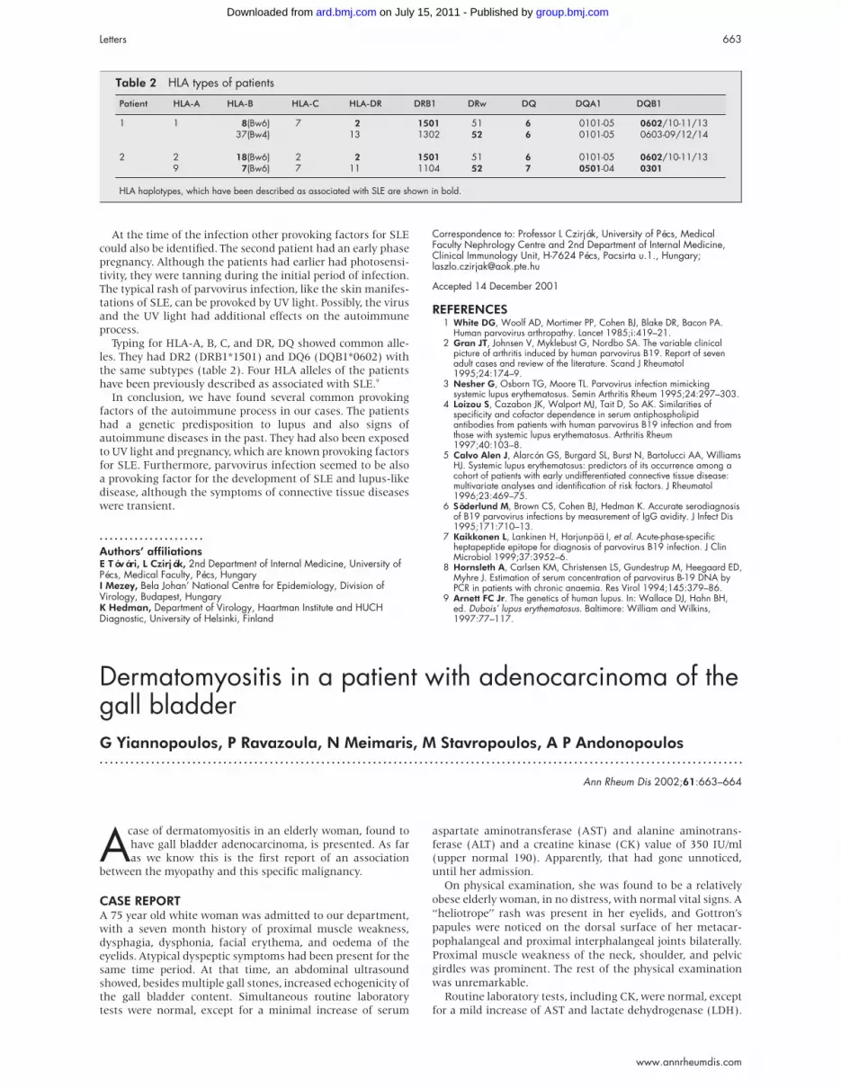
ligament; a cholecystectomy was performed. The specimen
disclosed a moderately differentiated adenocarcinoma, affect-
ing most of the gall bladder wall, and at its full thickness
expanding to the surrounding fatty tissue (fig 1).
Ten weeks after discharge, she was readmitted to the medi-
cal service, severely debilitated, with profound muscle
weakness and jaundice, nodular hepatomegaly, and oedema.
Laboratory tests showed mild anaemia, markedly raised alka-
line phosphatase, γGT, and LDH, bilirubin of 548 µmol/l, but
normal CK. The patient’s already serious condition deterio-
rated further, she developed a left lower lobe pneumonia and
died 10 days after her admission.
DISCUSSIONA number of studies have suggested an association between
inflammatory myopathy and malignancy, which seems
stronger with dermatomyositis and weaker with
polymyositis.1–4 A temporal relationship has also been sug-
gested, with the diagnosis of malignancy coinciding with, pre-
ceding, or following that of myositis, usually within two
years,2 3 5 although longer periods of surveillance have been
recommended.4
With the exception of gastric and ovarian carcinoma, which
seem to more commonly underlie myositis, the spectrum of
the various kinds of cancer in patients with inflammatory
myopathy is similar to that in the general population.3
However, an association of dermatomyositis with tumours of
the biliary tract is extremely rare. According to the available
published reports, there are only three cases of dermatomyosi-
tis in patients with cholangiocarcinoma (two of them in non-
English literature),6–8 and in no patient with gall bladder car-
cinoma has inflammatory myopathy been described.
Furthermore, in a recent excellent population based study9 of
618 patients with dermatomyositis, of whom 198 were found
to have cancer, no gall bladder carcinoma was identified.
Therefore, our patient seems to be the first report of an associ-
ation between dermatomyositis and adenocarcinoma of the
gall bladder. Her myopathy responded initially to steroids,
despite the advanced stage of her cancer, in accordance with
some observations, suggesting occasionally independent
courses of dermatomyositis and malignancy.10
In conclusion, carcinoma of the gall bladder should be
added to the list of malignancies, which dermatomyositis can
complicate, as a paraneoplastic process.
. . . . . . . . . . . . . . . . . . . . .Authors’ affiliationsG Yiannopoulos, N Meimaris, A P Andonopoulos, Division ofRheumatology, Department of Medicine, University of Patras School ofMedicine, Patras, GreeceP Ravazoula, Department of Pathology, University of Patras School ofMedicineM Stavropoulos, Department of Surgery, University of Patras School ofMedicine
Correspondence to: Professor A P Andonopoulos, Division ofRheumatology, Department of Medicine, University of Patras School ofMedicine, 265 00 Rio, Patras, Greece; [email protected]
Accepted 28 January 2002D van der Heijde, Limburg University Centre, Diepenbeek, BelgiumH Miedema, Netherlands Expert Centre for Workrelated MusculoskeletalDisorders, University Hospital Dijkzigt and Erasmus University Rotterdam,The NetherlandsH van der Tempel, Maasland Ziekenhuis Sittard, The Netherlands
Correspondence to: Dr A Boonen, Department of Internal Medicine,Division of Rheumatology, University Hospital Maastricht, PO Box 5800,6202 AZ Maastricht, The Netherlands; [email protected]
Accepted 7 January 2002
REFERENCES1 Barnes BE. Dermatomyositis and malignancy. Ann Intern Med
1976;84:68–76.2 Manchul LA, Jin A, Pritchard KI, Tenenbaum J, Boyd NF, Lee P, et al.
The frequency of malignant neoplasms in patients withpolymyositis-dermatomyositis. Arch Intern Med 1985;145:1835–9.
3 Sigurgeirsson B, Lindelof B, Edhag O, Allander E. Risk of cancer inpatients with dermatomyositis or polymyositis. N Engl J Med1992;326:363–7.
4 Richardson JB, Callen JP. Dermatomyositis and malignancy. Med ClinNorth Am 1989;73:1211–20.
5 Tymms KE, Webb J. Dermatomyositis and other connective tissuediseases: a review of 105 cases. J Rheumatol 1985;12:1140–8.
6 Rorive A, Fraipont V, Quatresooz P, Cataldo D, Dubois B, Fillet G.Clinical case of the month. A case of acute rhabdomyolysis. Rev MedLiege 1999;54:143–8.
7 Horie Y, Yamada M, Nakai K, Kawasaki H, Hirayama C, Matsui K, etal. Combined hepatocellular-cholangiocarcinoma associated withdermatomyositis. J Gastroenterol Hepatol 1989;4:101–4.
8 Llinares Mondejar P, Amador Barciela L, Arnal Monreal F, BerazaMilicua A, Toro Santos M, del Rio Vazquez A. Associateddermatomyositis and cholangiocarcinoma. Rev Clin Esp1982;164:335–7.
9 Hill CL, Zhang Y, Sigurgeirsson B, Pukkala E, Mellemkjaer L, Airio A, etal. Frequency of specific cancer types in dermatomyositis andpolymyositis: a population-based study. Lancet 2001;357:96–100.
10 Yazici Y, Kagen LJ. The association of malignancy with myositis. CurrOpin Rheumatol 2000;12:498–500.
Figure 1 Histology of the surgical specimen of the patient(haematoxylin and eosin, ×200), showing moderately differentiatedadenocarcinoma of the gall bladder.
664 Letters
www.annrheumdis.com
group.bmj.com on July 15, 2011 - Published by ard.bmj.comDownloaded from

Coincidence of asymptomatic avascular necrosis andfracture of the femoral neck: a rare combination ofglucocorticoid induced side effectsK Loddenkemper, C Perka, G-R Burmester, F Buttgereit. . . . . . . . . . . . . . . . . . . . . . . . . . . . . . . . . . . . . . . . . . . . . . . . . . . . . . . . . . . . . . . . . . . . . . . . . . . . . . . . . . . . . . . . . . . . . . . . . . . . . . . . . . . . . . . . . . . . . . . . . . . . .
Ann Rheum Dis 2002;61:665–666
Glucocorticoids have profound anti-inflammatory and
immunosuppressive actions when used therapeuti-
cally. Unfortunately, these drugs have adverse
effects—for example, on bone metabolism. Osteoporosis is
well known to be a common side effect, whereas a glucocorti-
coid associated avascular osteonecrosis is rarely diagnosed.
However, as far as we know, the coincidence of manifest
osteoporosis with fracture and avascular osteonecrosis in the
same area is unique.
CASE REPORTWe present the case of a 47 year old postmenopausal woman
who had had mixed connective tissue disease (MCTD) for
more than 20 years. The MCTD was complicated by
progressive vasculitis, and had required immunosuppressive
treatment with prednisolone (maximum dose 500 mg/day
(pulse therapy; average dose 15 mg/day) for 20 years and aza-
thioprine (75 mg/day). In 1995, she complained for the first
time of severe back pain and noticed a height loss of 10 cm
within two years. There was no history of previous fractures.
Glucocorticoid induced osteoporosis was suspected. Indeed,
bone densitometry (DXA-LUNAR) showed decreased bone
mineral density (BMD) of the lumbar spine (L2–4) and left
femoral neck (table 1). An x ray examination demonstrated
osteoporotic changes—for example, end plate fractures, osteo-
penia. Treatment with fluorides, vitamin D, and calcium was
started. One year later the BMD of the lumbar spine had
increased, whereas the BMD of the femoral neck and back
pain remained unchanged. Consequently, the treatment was
changed to bisphosphonates (alendronate) supplemented by
calcium and vitamin D. Another year later her back pain was
reduced. The BMD of lumbar spine had become stable while
decreasing by 30% at the femoral neck (table 1). No evidence
of fluorosis, secondary hyperparathyroidism, or abnormal
vitamin D metabolism was found.
Shortly after this visit the patient was admitted to hospital
for alprostadil treatment of her Raynaud’s phenomenon asso-
ciated with the MCTD. She complained of sudden severe pain
in her left hip during a forest walk. Surprisingly, an x ray
examination showed left femoral head fracture and avascular
necrosis (early radiological Ficat stage IV) (fig 1).1 After
successful hip replacement, the patient was able to walk with-
out pain.
DISCUSSIONThis patient with MCTD demonstrates typical side effects of
long term glucocorticoid treatment, but several points are of
particular interest:
(1) Osteoporosis treatment had been started at a time when
the BMD was already decreased. Other risk factors were early
menopause without hormone replacement therapy and
inflammatory rheumatic disease (cytokines can affect bone
turnover). Official guidelines for the prevention of glucocorti-
coid induced osteoporosis were published by the American
Table 1 Bone mineral density of lumbar spine andfemoral neck over the course of three years
1995 1996 1997
Lumbar spine (mg/cm2) 0.800 0.875 0.874Lumbar spine (t score) −3.33 −2.71 −2.83Femoral neck (mg/cm2) 0.538 0.549 0.381Femoral neck (t score) −3.68 −3.68 −4.99
Figure 1 An x ray image of the left hip showing femur headnecrosis (early radiological Ficat stage IV) and fracture of the leftfemoral neck.
Letters 665
www.annrheumdis.com
group.bmj.com on July 15, 2011 - Published by ard.bmj.comDownloaded from

College of Rheumatology in 1996.2 Today it is clear that early
prophylaxis can prevent manifest osteoporosis. Potent drugs
for the prevention and treatment of osteoporosis are bisphos-
phonates and substitution of calcium and vitamin D.3
(2) Another important aspect is osteoporosis with intercur-
rent avascular osteonecrosis of the left femoral head. The
absence of pain in this condition is uncommon. To the best of
our knowledge, unilateral osteonecrosis of the hip at an
advanced stage without pain has not been previously
reported. Only early stage forms or one hip without pain in
bilateral osteonecrosis have been described.4 5 Avascular
necrosis is not a specific disease entity. Predisposing factors
are trauma, glucocorticoids, alcohol abuse, and connective tis-
sue disorders. It may also be idiopathic.6 Avascular necrosis is
a rare side effect of glucocorticoid treatment and is normally
painful. In autoimmune diseases a correlation has been shown
between glucocorticoids and bone death, which appears to be
due to blood stasis and ischaemia in the trabecular bone.
Theories of thrombotic formation or fat embolism have proved
to be invalid.7 A further suspected mechanism could be the
increase of osteocyte apoptosis owing to microdamage in the
bone.8 Ischaemic vasculopathy as part of the underlying
disease may also have a role.9 However, if these mechanisms
are correct, then the association reported here should occur
more often than it does. Active vasculitis or mechanical ortho-
paedic factors were excluded in our case. The absence of pain
might be the result of peripheral nerve dysfunction secondary
to polyneuropathy caused by previous MCTD associated
vasculitis. Glucocorticoid treatment might also have decreased
the pain by reducing femoral head synovialitis.
(3) The simultaneous occurrence of osteonecrosis and
atraumatic fracture of the femoral neck was even more
surprising than the rare occurrence of severe, yet asympto-
matic, osteonecrosis. Revascularisation of dead bone begins
shortly after interruption of the blood supply. It promotes
removal of necrotic bone by osteoclasts while inducing osteo-
blasts to synthesise new bone, thus creating more stability.9
The combination of osteonecrosis and fracture in the same
region is therefore extremely rare.
In conclusion, osteoporosis is a common side effect of
glucocorticoid treatment, and early prevention and treatment
are necessary. Osteonecrosis and osteoporosis with a fracture
in the same area as the side effects of glucocorticoid treatment
is extremely rare. Only imaging techniques will allow differentia-
tion.
. . . . . . . . . . . . . . . . . . . . .Authors’ affiliationsK Loddenkemper, C Perka, G-R Burmester, F Buttgereit,Departments of Rheumatology/Clinical Immunology and Orthopaedics,Charité University Hospital, Humboldt University of Berlin, Germany
Correspondence to: Dr K Loddenkemper, Department ofRheumatology/Clinical Immunology, Charité University Hospital,Schumannstrasse 20–21, 10117 Berlin, Germany;[email protected]
Accepted 27 December 2001
REFERENCES1 Ficat RP. Idiopathic bone necrosis of the femoral head: early diagnosis
and treatment; J Bone Joint Surg Br 1985;67:3–9.2 Anonymous. Recommendation for prevention and treatment of
glucocorticoid-induced osteoporosis. American College of RheumatologyTask Force on Osteoporosis Guidelines. Arthritis Rheum1996;39:1791–801.
3 Adachi JD, Bensen WG, Brown J, Hanley D, Hodsman A, Josse R, et al.Intermittent etidronate therapy to prevent corticosteroid-inducedosteoporosis. N Engl J Med 1997;337:382–7.
4 Steinberg ME, Brighton CT, Steinberg DR, Tooze SE, Hayken GD.Treatment of avascular necrosis of the femoral head by a combination ofbone grafting, decompression, and electrical stimulation. Clin Orthop1984;(186):137–53.
5 Desforges JF. Nontraumatic necrosis of bone (osteonecrosis). Reviewarticle. N Engl J Med 1992;326:1473–9.
6 Pavelka K. Osteonecrosis. Baillieres Best Pract Res Clin Rheumatol2000;14:399–414.
7 Felson DT, Anderson JJ. A cross-study evaluation of association betweensteroid dose and bolus steroid and avascular necrosis of bone. Lancet1997;i:902.
8 Manolagas SC, Weinstein RS. New developments in pathogenesis andtreatment of steroid-induced osteoporosis. J Bone Miner Res1999;14:1061–6.
9 Akiyama K, Hirota J, Taniyasu N, Asano S. Unusual complications inan inflammatory abdominal aortic aneurysm. Jpn Circ J 1999;63:914–16.
666 Letters
www.annrheumdis.com
group.bmj.com on July 15, 2011 - Published by ard.bmj.comDownloaded from

doi: 10.1136/ard.61.7.654 2002 61: 654-655Ann Rheum Dis
C Hidalgo-Tenorio, J M Sabio and J Jiménez-Alonso Alopecia in Wegener's granulomatosis
http://ard.bmj.com/content/61/7/654.full.htmlUpdated information and services can be found at:
These include:
References http://ard.bmj.com/content/61/7/654.full.html#ref-list-1
This article cites 6 articles, 1 of which can be accessed free at:
serviceEmail alerting
box at the top right corner of the online article.Receive free email alerts when new articles cite this article. Sign up in the
Notes
http://group.bmj.com/group/rights-licensing/permissionsTo request permissions go to:
http://journals.bmj.com/cgi/reprintformTo order reprints go to:
http://group.bmj.com/subscribe/To subscribe to BMJ go to:
group.bmj.com on July 15, 2011 - Published by ard.bmj.comDownloaded from