akt/protein kinase b prevents injury-induced motoneuron death and accelerates axonal regeneration
TRANSCRIPT
Akt/Protein Kinase B Prevents Injury-Induced Motoneuron Deathand Accelerates Axonal Regeneration
Kazuhiko Namikawa,1 Masaru Honma,1,2 Koji Abe,1,3 Masumi Takeda,1,4 Khalil Mansur,1 Tatsuo Obata,1Akiko Miwa,5,6 Haruo Okado,5,6 and Hiroshi Kiyama1,6
Departments of 1Anatomy, 2Dermatology,3Psychiatry and Neurology, and 4Ophthalmology, Asahikawa Medical College,Asahikawa, Hokkaido, 078–8510 Japan, 5Department of Neurobiology, Tokyo Metropolitan Institute for Neuroscience,Fuchu, Tokyo, 183–8526 Japan, and 6Core Research for Evolutional Science and Technology (CREST), Japan Scienceand Technology, Kawaguchi, Saitama, 332–0012 Japan
Motoneurons require neurotrophic factors for their survival andaxonal projection during development, as well as nerve regen-eration. By using the axotomy-induced neuronal death paradigmand adenovirus-mediated gene transfer, we attempted to gaininsight into the functional significances of major growth factorreceptor downstream cascades, Ras–extracellular signal-regulated kinase (Ras-ERK) pathway and phosphatidylinositol-3kinase–Akt (PI3K-Akt) pathway. After neonatal hypoglossal nervetransection, the constitutively active Akt-overexpressing neuronscould survive as well as those overexpressing Bcl-2, whereasthe constitutively active ERK kinase (MEK)-overexpressing ones
failed to survive. A dominant negative Akt experiment demon-strated that inhibition of Akt pathway hastened axotomy-inducedneuronal death in the neonate. In addition, the dominant activeAkt-overexpressing adult hypoglossal neurons showed accel-erated axonal regeneration after axotomy. These results sug-gest that Akt plays dual roles in motoneuronal survival andnerve regeneration in vivo and that PI3K-Akt pathway is prob-ably more vital in neuronal survival after injury than Ras-ERKpathway.
Key words: cell death; nerve injury; adenoviral gene transfer;hypoglossal; neuronal survival; nerve regeneration
An organized expression of various kinds of molecules would beessential for injured neurons to survive and regenerate (for re-view, see Persson and Ibanez, 1993; Snider, 1994; Oppenheim,1996; Pettmann and Henderson, 1998). In an attempt to explorethe molecular basis of this process, we have used differentialdisplay–PCR to identify genes whose mRNA expression wasupregulated in injured hypoglossal motoneurons and succeededin isolating both novel and known molecules with possible asso-ciation with neuronal survival and regeneration (Kiryu et al.,1995b; Morita et al., 1996; Su et al., 1997; Namikawa et al., 1998;Toki et al., 1998). In such gene screening studies, moleculesrestricted to certain intracellular signaling pathways were repeat-edly hit. These are, for instance, Shc, 14-3-3, extracellular signal-regulated kinase 1 (ERK1), ERK kinase 1 (MEK1), phosphatidy-linositol-3 kinase (PI3K), and Akt (Kiryu et al., 1995a; Ito et al.,1996; Owada et al., 1997; Namikawa et al., 1998; Tanabe et al.,1998), which are molecules located downstream of growth factorreceptor signaling pathways, in particular Ras-ERK and PI3K-Akt cascades (for review, see Kaplan and Miller, 1997). The in
vitro studies using culture cells have also indicated that these twopathways are vital for neuronal survival (for review, see Pettmannand Henderson, 1998). Currently, serine/threonine kinase Akt,also known as PKB (protein kinase B) or RAC-PK (related to Aand C protein kinase) (Burgering and Coffer, 1995; Franke et al.,1995), has attracted much attention as a survival signal mediator,stimulated not only by growth factors but also by calcium influx(Dudek et al., 1997; Crowder and Freeman, 1998; Yano et al.,1998). Akt inactivates BAD, caspase-9, and FKHRL1 by phos-phorylation and thereby blocks BAD-, caspase-9-, or FKHRL1-induced cell death in vitro (Datta et al., 1997; del Peso et al., 1997,Cardone et al., 1998, Brunet et al., 1999). In addition to PI3K-Aktpathway, Ras-ERK pathway is another well established survivalsignaling pathway, at least in differentiated PC12 cells and Dro-sophila (Xia et al., 1995; Bergmann et al., 1998; Kurada andWhite, 1998). So far, both Ras-ERK and PI3K-Akt pathways havebeen considered to be the most vital for neuronal survival, andthis fact strongly suggests some crucial roles of these pathways ininjured motoneurons for their survival. However, the controversyremains as to whether they contribute equally to neuronal sur-vival in vivo (in mammal). In the present study, we tried toaddress this issue by using axotomy-induced neuronal death par-adigm (Hamburger, 1934; Romanes, 1946; Snider et al., 1992).Axotomy or the removal of peripheral targets in the neonatecauses neuronal death, whereas axotomy in mature animals in-duces a regenerative response. In the present experimental par-adigm, we attempted to gain insight into the functional signifi-cance of Ras-ERK and PI3K-Akt pathways by using adenovirusvectors (Akli et al., 1993; Davidson et al., 1993), carrying recom-binant genes for the pivotal molecules to actively manipulatethese cascades in specific ways, in injured motoneurons of neo-nate rats. Here, we have revealed distinct functional differences
Received Oct. 19, 1999; revised Jan. 20, 2000; accepted Jan. 28, 2000.This work was supported in part by Grant-in-Aid for Scientific Research from the
Ministry of Education, Science, and Culture, Ministry of Heath and Welfare, andCREST. K.M. is a fellow of Ministry of Education, Science, and Culture Japan. Wethank Drs. I. Saito and Y. Kanegae (University of Tokyo, Tokyo, Japan) forpAxCAwt, pAxCALNLw, AxCANLacZ, AxCALNLNZ, and AxCANCre; Dr. J.Miyazaki (Osaka University, Osaka, Japan) for CAG promoter in adenoviralvectors; Dr. S. J. Korsmeyer (Harvard Medical School, Boston, MA) for SSFV Bcl-2expression plasmid; Drs. M. Kasuga and W. Ogawa (Kobe University, Kobe, Japan)for Akt-AA plasmid; and Drs. U. Kikkawa and H. Konishi (Kobe University) foranti-Akt antibody. We are grateful to Prof. U. Kikkawa for the critical reading ofthis manuscript, and T. Sasaki and K. Hazawa for technical assistance.
Correspondence should be addressed to Prof. Hiroshi Kiyama, Department ofAnatomy, Asahikawa Medical College, 2-1-1-1, Midorigaokahigashi, Asahikawa,Hokkaido, 078–8510 Japan. E-mail: [email protected] © 2000 Society for Neuroscience 0270-6474/00/202875-12$15.00/0
The Journal of Neuroscience, April 15, 2000, 20(8):2875–2886
between these two signaling pathways. In addition, we have alsorevealed a novel activity of Akt on neurite elongation both in vitroand in vivo.
MATERIALS AND METHODSAnimals and surgery. The hypoglossal nerve was cut unilaterally in bothneonate (3-d-old) and adult (6-week-old) Wistar rats. Briefly, rats wereanesthetized with pentobarbital and positioned supine, and the unilateralhypoglossal nerve was cut with a pair of scissors.
In situ hybridization and immunohistochemistry. In situ hybridization onbrain sections was performed using digoxigenin (DIG)-UTP-labeledcRNA probe. For Akt mRNA detection, a cDNA (1147–1610) fragmentof rat Akt1 (Konishi et al., 1994) was isolated from rat whole braincDNA by using PCR. Hybridization were performed on fresh frozensections (18 mm) of neonatal rats 3 d after axotomy or adult rats 7 d afteraxotomy. Briefly, the sections were prehybridized and then hybridized at58°C for 16 hr in hybridization buffer [50% deionized formamide, 0.3 MNaCl, 20 mM EDTA, 10 mM phosphate buffer (PB), 10% dextran sulfate,13 Denhardt’s solution, 0.2% sarcosyl, 500 mg/ml yeast tRNA, and 200mg/ml denatured salmon sperm DNA] containing 40 ng/ml DIG-labeledRNA probe. After hybridization, washing was performed twice at 65°Cin 50% formamide and 23 SSC for each 30 min. The signal was detectedby using the DIG nucleic acid detection kit (Boehringer Mannheim,Indianapolis, IN) according to the manufacturer’s protocol. Immunohis-tochemical studies on brain sections were performed as described previ-ously (Kiryu et al., 1995a). Neonate rats 3 d after axotomy or adult rats7 d after axotomy were fixed in Zamboni’s fixative (0.1 M PB containing2% paraformaldehyde and 0.2% picric acid), and brains were cryopro-tected in 20% sucrose and then sectioned (20 mm). Anti-phospho Aktpolyclonal antibody (1:300; New England Biolabs, Beverly, MA) specificto phosphorylated Akt at Ser 473 was used.
Construction of adenoviral vectors. Recombinant adenoviral vectorswere constructed in the following manner. The cDNA fragments com-prising the entire coding regions for human MEK1 and human Akt1,were isolated from human embryonic kidney 293 (HEK293) cDNA byusing PCR. Constitutively active MEK, which lacks its nuclear exportsignal (Fukuda et al., 1997) (amino acid 32–51) and which has thesubstitution of glutamic acid for two phosphorylation sites, Ser 218 andSer 222, were prepared by site-directed mutagenesis as described previ-ously (Mansour et al., 1994). After that, c-Myc tag sequence was fusedto its N terminal by using PCR. Constitutively active Akt, which lacksits pleckstrin homology domain (amino acid 4–129) but has src-myristoylation signal sequence (MGSSKSKPKDPSQRR) (Resh, 1994)fused to its N-terminal end and hemagglutinin epitope tag (HA tag) to itsC-terminal end (Kohn et al., 1996a), was also prepared by using PCR.Akt-AA (rat Akt1 T308A/S473A), which also contains HA tag in itsN terminal and works as dominant negative mutant of Akt (Kitamura etal., 1998), was kindly provided by Drs. M. Kasuga and W. Ogawa (KobeUniversity, Kobe, Japan). Bcl-2 expression plasmid SSFV Bcl-2, carryingthe entire coding sequence of human Bcl-2, was kindly provided by Dr.S. J. Korsmeyer (Harvard Medical School, Boston, MA). da-MEK wassubcloned into pAxCAwt (Miyake et al., 1996), an expression cosmidcassette, which was created from the human type 5 adenovirus genomefrom which the E1A, E1B, and E3 regions were deleted and whichreplaced the expression unit under the control of CAG promoter (Niwaet al., 1991) On the other hand, the fragments of myr-Akt, Akt-AA andBcl-2 were subcloned into pAxCALNLw Cre-lox P system mediatedexpression cassette (Sato et al., 1998a) as described below. The recombi-nant adenovirus vectors AxCAda-M EK , AxCAL N L myr-Akt,AxCALNLAkt-AA, and AxCALNLBcl-2 were constructed by theCOS-terminal protein complex (TPC) method (Miyake et al., 1996).Each expression cosmid cassette and EcoT22I digested adenovirusDNA-TPC (Ad5dlX DNA-TPC) were cotransfected into HEK293 cells,and the recombinant adenoviruses were generated by homologous re-combination and amplified in HEK293 cells. Finally, high titered recom-binant viral stocks were generated in HEK293 cells, purified by cesiumgradient centrifugation (Kanegae et al., 1994), and stored at 280°C untiluse. The viral titers were determined by plaque-forming assay in HEK293cells. AxCANLacZ (Terashima et al., 1997), AxCALNLNZ (Sato et al.,1998a), and AxCANCre (Kanegae et al., 1995) were kindly provided byDrs. I. Saito and Y. Kanegae (University of Tokyo, Tokyo, Japan).
Cell culture. PC12 cells were maintained in RPMI 1640 mediumcontaining 5% fetal bovine serum and 10% heat-inactivated horse serum.The cells were differentiated for 9–10 d in the same medium containing
0.5% fetal bovine serum and nerve growth factor (NGF) (50 ng/ml;Promega, Madison, WI).
Western blotting. Western blotting was done according to the followingprocedure. Protein (20 mg) from adenoviral-infected whole-cell extracts(the infections were performed as the described below) were separatedby SDS-PAGE, and blots were prepared on polyvinylidene difluoridemembranes (Bio-Rad, Richmond, CA). The following primary antibod-ies were used as probes: for myr-Akt, anti-phospho Akt polyclonalantibody (1:1000; New England Biolabs) and anti-HA monoclonal anti-body (12CA5) (1:2000; Boehringer Mannheim); for Akt-AA, anti-Aktpolyclonal antibody (Kitamura et al., 1998) (kindly provided by Drs. U.Kikkawa and H. Konishi) and anti-HA monoclonal antibody (12CA5);for da-MEK, anti-c-Myc monoclonal antibody (9E10); for phosphoryla-tion of ERKs, anti-phospho ERK polyclonal antibody (1:1000; NewEngland Biolabs); for Bcl-2, anti-Bcl-2 polyclonal antibody (1:1000; SantaCruz Biotechnology, Santa Cruz, CA); and for phosphorylation of c-JunN-terminal protein kinases (JNKs) or p38, anti-phospho JNK polyclonalantibody (1:1000; New England Biolabs) or anti-phospho p38 polyclonalantibody (1:1000; New England Biolabs), respectively. Blots were thenprobed with horseradish peroxidase-conjugated goat anti-mouse or anti-rabbit secondary antibody (Amersham, Arlington Heights, IL) and visu-alized by using chemiluminescence system (ECL; Amersham)
In vitro kinase assay. To detect the activity of adenovirus-expressedmyr-Akt or Akt-AA under the presence or absence of NGF (100 ng/ml;3 min after stimulation), we lysed the cells 36 hr after the infection in asolution containing 20 mM Tris-HCl at pH 7.5, 1 mM EDTA, 1 mMEGTA, 10 mM 2-mercaptoethanol, 1% Triton X-100, 150 mM NaCl,10 mM NaF, 1 mM Na3VO4, and 25 mM phenylmethylsulfonyl fluoride.The lysates were immunoprecipitated with protein A-Sepharose (Phar-macia, Uppsala, Sweden) coupled with anti-HA polyclonal antibody(1:100; Santa Cruz Biotechnology) for 3 hr at 4°C. After washing threetimes in 20 mM Tris-HCl at pH 7.5 containing 150 mM NaCl and 1%Triton X-100, the final immunoprecipitates were incubated for 30 min at30°C in a reaction buffer (25 ml) containing 20 mM Tris-HCl at pH 7.5,10 mM MgCl2, 20 mM cold ATP, 50 kBq g- 32P ATP (DuPont NEN,Boston, MA), 1 mM protein kinase inhibitor (Sigma, St. Louis, MO), and5 mg of histone H2B (Boehringer Mannheim) or 3 mg of glutathioneS-transferase (GST)-BAD fusion protein as substrates. The reactionswere terminated by adding SDS-PAGE sample buffer to the supernatantsand analyzed by 10% (for visualization of GST-BAD) or 15% (forvisualization of histone H2B) SDS-PAGE, followed by autoradiography.The same immunoprecipitations were also used for Western blot analysiswith anti-Akt antibody. On the other hand, dominant negative effectof Akt-AA was estimated by measuring endogenous Akt activity inAkt-AA-overexpressing PC12 cells. In this study, LY294002 (Sigma) wasadded into some dishes at the concentration of 100 mM 15 min beforeNGF stimulation. The lysates were subjected to three sequential immu-noprecipitations with anti-HA polyclonal antibody (1:100; Santa CruzBiotechnology) for 90 min to remove adenovirus-expressed Akt-AA.The final supernatants were then subjected to immunoprecipitation withpolyclonal antibody to Akt, and Akt kinase assay with the resultingimmunoprecipitates and Western blot analysis using anti-Akt antibodywere performed as described above.
Immunostaining and survival assays using adenoviral expression in dif-ferentiated PC12 cells. To estimate the survival activity of each virus,neuronally differentiated PC12 cells were infected with AxCANLacZ[multiplicity of infection (MOI) 100], AxCAda-MEK (MOI 100),AxCALNLmyr-Akt plus AxCANCre (MOI 100/30), or AxCALNLBcl-2plus AxCANCre (MOI 100/30) and then maintained in NGF-containing(50 ng/ml) medium. Thirty-six hours later, cells were washed two timeswith NGF-free medium followed by incubation in NGF-free mediumcontaining neutralizing antibody to 2.5S NGF (Sigma) at a 1:1000 dilu-tion. Then, 18 hr later, immunocytochemistry was done to detectb-galactosidase (b-Gal) or myr-Akt-expressing cells, using the anti-b-Gal polyclonal antibody (1:500; Organon Teknika, West Chester, PA) orthe anti-HA polyclonal antibody (1:300; Santa Cruz Biotechnology),respectively. FITC-conjugated antibody (Vector Laboratories, Burlin-game, CA) was used as the secondary antibody. Subsequently, Hoechststaining was done (Hoechst 33258, 2.5 mg/ml; Wako, Tokyo, Japan) tovisualize their nuclei. For cell viability assay, quantitative determinationsof surviving cells were done 0, 24, or 48 hr after NGF withdrawal. Thebiochemical method using a highly water-soluble tetrazolium salt (WST-1), neutral red and crystal violet (Ishiyama et al., 1996), was used todetermine the percent ratio of surviving cells following the manufactur-er’s protocol (Nakalai Tesque, Kyoto, Japan). On the other hand, to
2876 J. Neurosci., April 15, 2000, 20(8):2875–2886 Namikawa et al. • Akt Promotes Neuronal Survival and Regeneration
evaluate the pro-apoptotic ability of Akt-AA under the presence of NGF,differentiated PC12 cells were infected with AxCALNLAkt-AA plusAxCANCre (MOI 100/30) or AxCALNLNZ plus AxCANCre (MOI100/30). Twenty-four hours after infection, virus-infected cells werestained as described above. Cell viability assay was done after 0, 24, 48,and 72 hr after the virus infection under the presence of NGF using thesame method described above. In addition, the effect of LY294002 (100mM) on the same cells was also estimated 0, 24, and 48 hr after thetreatment.
Effects of the adenoviral infection on rat neonatal hypoglossal motoneu-rons af ter axotomy. AxCANLacZ (1.7 3 10 8 pfu/5 ml), AxCAda-MEK(1.0 3 10 8 pfu/5 ml), AxCALNLmyr-Akt plus AxCANCre (1.3 3 10 8
pfu/5 ml and 1.9 3 10 7 pfu/5 ml, respectively), AxCALNLBcl-2 plusAxCANCre (3.3 3 10 7 pfu/5 ml and 1.9 3 10 7/5 ml, respectively),AxCALNLAkt-AA plus AxCANCre (1.8 3 10 8 pfu/5 ml and 1.9 3 10 7
pfu/5 ml, respectively), or AxCALNLNZ plus AxCANCre (3.3 3 10 8
pfu/5 ml and 1.9 3 10 7 pfu/5 ml, respectively) was injected unilaterallyinto tongues of neonatal rats (1-d-old). Efficiency of viral infection wasassessed by 5-bromo-4-chloro-3-indolyl-b-D-galactopyranoside (X-Gal)histochemistry (Terashima et al., 1997), counterstained with neutral red,using the brainstem sections of AxCANLacZ-infected nonoperated an-imals. Two days after injection of each virus, pups were operated (3-d-old). For evaluation of survival activity by da-MEK-, myr-Akt-, Bcl-2-,and LacZ-expressing virus, the infected animals were perfused transcar-dially with 4% paraformaldehyde in 0.1 M PB on postaxotomy day 12. Onthe other hand, for evaluation of death-promoting activity by Akt-AA,the rats infected with AxCALNLAkt-AA plus AxCANCre were decap-itated at 36 hr or 3, 5, or 7 d after axotomy. In this study, AxCALNLNZand AxCANCre were used as a control viruses. Sections (20 mm) weretaken through hypoglossal nuclei region and stained with thionin, andneurons were counted on both operated and control sides. Survivaleffects of recombinant viral infections were determined by calculating thepercent ratio of the surviving motoneurons in the operated side com-pared with those on control side. In some experiments, the sections wereprocessed for immunohistochemistry as described above. To identifyrecombinant adenoviral-infected neurons, anti-c-Myc antibody (9E10) oranti-HA polyclonal antibody (1:300; Santa Cruz Biotechnology) was usedfor the detection of c-Myc-tagged da-MEK, HA-tagged myr-Akt, orHA-tagged Akt-AA, respectively, followed by thionin staining.
Death-promoting assay of Akt-AA adenovirus in the adult rat hypoglossalmotoneurons af ter axotomy. AxCALNLNZ plus AxCANCre(3.3 3 10 8
pfu/5 ml and 1.9 3 10 7 pfu/5 ml, respectively) or AxCALNLAkt-AAplus AxCANCre (1.8 3 10 8 pfu/5 ml and 1.9 3 10 7 pfu/5 ml, respec-
tively) was infected through the cut axon tips immediately after thetransection of unilateral hypoglossal nerve in adult rats. Animals werekilled at 6, 10, or 14 d after axotomy, and the methods for preparationsof brain sections and the calculation of percent survival ratio of injuredmotoneurons were performed as described above.
Infection of myr-Akt-expressing adenovirus into undifferentiated PC12cells. AxCANLacZ (MOI 100) or AxCALNLmyr-Akt plus AxCANCre(MOI 100/30) was infected into undifferentiated PC12 cells. For immu-nostaining, cells were fixed 2 d after the infection and stained as de-scribed above. For an estimation of neurite outgrowth, the number ofcells whose neurites are longer than their cell bodies was counted, andthe percent ratio of the number of neurite bearing cells was calculated.(Four different experiments were performed for each value.)
In vivo assay for nerve regeneration. AxCANLacZ (1.7 3 10 8 pfu/5 ml)or AxCALNLmyr-Akt plus AxCANCre (1.3 3 10 8 pfu/5 ml or 1.9 3 10 7
pfu/5 ml, respectively) was infected into injured motoneurons in adultrats as described above. One week later, immunohistochemistry was donewith the brainstem sections for detection of b-Gal-positive neurons orHA-positive myr-Akt-expressing neurons as described above. Nerveregeneration was assessed by using the retrograde tracer Fluoro-Gold(FG) (Fluorochrom Inc., Englewood, CO), 2, 3, and 4 weeks afteraxotomy as described previously (Hirota et al., 1996).
RESULTSAkt activation in response to nerve injury is oppositelyregulated between newborn and adultIn adult injured motoneurons, upregulated expressions of theligands (neurotrophic factors), the receptors, and the intracellularsignaling molecules have been proved; however, these responsesin neonate have not been so well characterized yet. If the activa-tion of Akt is observed in the neonate paradigm in which theinjured neuron is fated to die, Akt may not be the crucialmolecule in survival. Therefore, before addressing the main is-sue, we examined the possibility of differences in Akt responseafter axotomy in neonate and adult hypoglossal motoneurons.After hypoglossal nerve axotomy, Akt1 mRNA (Fig. 1A) wasmarkedly upregulated in injured hypoglossal neurons in adult rat.For activation of Akt, the phosphorylation of Akt1 at serine 473(Ser473) after the phosphorylation of threonine 308 (Thr308) is
Figure 1. Expression and activation of Akt in injured hypoglossal motoneurons are oppositely regulated between neonate and adult. A, Expression ofAkt1 mRNA demonstrated by in situ hybridization using DIG-labeled Akt1 antisense RNA probe. Photograph indicates markedly upregulated signalin injured hypoglossal motoneurons (lef t) of adult rat 7 d after axotomy. B, C, Immunohistochemical demonstrations of phosphorylated Akt at Ser 473
(P-Akt). B, In the adult rat, enhanced P-Akt expression was observed in injured hypoglossal nucleus (lef t) compared with the control hypoglossal nucleus(right) 7 d after axotomy. C, High-power magnification indicates that most of the injured motoneurons show intense P-Akt immunoreactivity. D, E,Decreased Akt expression and activity in the neonate injured motoneurons in response to axotomy. D, Expression of Akt1 mRNA was downregulatedon the injured side (lef t) 3 d after axotomy. E, Immunohistochemistry also showed few P-Akt-positive neurons in the injured side (lef t), whereas manyimmunostained motoneurons are observed on control side (right) 3 d after axotomy. Scale bars: A, 600 mm; B, 500 mm; C, 50 mm; D, E, 200 mm.
Namikawa et al. • Akt Promotes Neuronal Survival and Regeneration J. Neurosci., April 15, 2000, 20(8):2875–2886 2877
necessary through the growth factor receptor stimulation (forreview, see Downward, 1998). Immunohistochemistry using anantibody specific to phosphorylated Akt at Ser473 revealed thatphosphorylated Akt protein dramatically increased in injuredadult motoneurons (surviving after axotomy) (Fig. 1B,C),whereas the expression of Akt1 mRNA and phosphorylatedAkt decreased after axotomy in injured neonate motoneurons(Fig. 1D,E). Thus, injured neonate motoneurons that were des-tined to die somehow failed to upregulate and phosphorylate Aktin response to nerve injury.
Construction of adenoviral vectors and their activityTo activate two different pathways in injured motoneurons, weattempted to use a dominant active form of Akt (for activation ofPI3K-Akt pathway) or MEK (for activation of Ras-ERK path-way). A dominant negative form of Akt was also prepared toinhibit the Akt signaling cascade in a specific manner. In addition,Bcl-2, a well known molecule that promotes motoneuron survivalafter axotomy as described previously (Farlie et al., 1995), wasalso used for the quantitative analysis of the efficacy of ourexperimental paradigm. By using adenovirus vectors, we con-structed four recombinant genes, namely, HA-tagged constitu-tively active Akt [myristoylated Akt1(myr-Akt): D4–129, desig-nated AxCALNLmyr-Akt] (Kohn et al., 1996b), HA-taggeddominant negative Akt [Akt1 T308A/S473A (Akt-AA), desig-nated AxCALNLAkt-AA] (Kitamura et al., 1998), c-Myc-taggedconstitutively active MEK [dominant active MEK1(da-MEK):D32–51; S218E/S222E, designated AxCAda-MEK] (Mansour etal., 1994), and Bcl-2 (designated AxCALNLBcl-2).
By using an ordinary procedure (see Materials and Methods),we failed to obtain recombinant viruses expressing myr-Akt andBcl-2. In addition, although we succeeded in getting Akt-AA-representing adenovirus, it could not become a high-titered one(,1010 pfu/ml) and was not effective enough to be used in animalexperiments. We concluded that very high expression of theserecombinant molecules under the control of the CAG promoter(composed of the cytomegalovirus enhancer plus the chickenb-actin promoter, and the rabbit b-globin polyadenylation signalsequence), might be producing some deleterious effects in theiramplification in HEK293 cells. Consequently, these three genesequences were inserted into a novel type of adenoviral vector,which bears an ON–OFF switching unit for activation by Crerecombinase (Sato et al., 1998a,b). The switching unit in the vectorcontained a stuffer sequence encoding Neo with a functional poly-adenylation signal between the promoter and the inserted cDNAfragment, thereby blocking its expression during viral amplificationin HEK293 cells. The stuffer is flanked by a pair of loxP sites,allowing its excision by Cre leading to expression of the insertedcDNA sequence. To obtain the expression of myr-Akt, Akt-AA, orBcl-2, we performed coinfection of the nuclear localization signal(NLS)-tagged Cre recombinase expressing virus (AxCANCre)(Kanegae et al., 1995) plus AxCALNLmyr-Akt, AxCANCre plusAxCALNLAkt-AA or AxCANCre plus AxCALNLBcl-2, respec-tively. The NLS-LacZ virus (AxCANLacZ) or Cre-mediatedNLS-LacZ virus (AxCALNLNZ) (Terashima et al., 1997; Sato etal., 1998a) were used as control vectors.
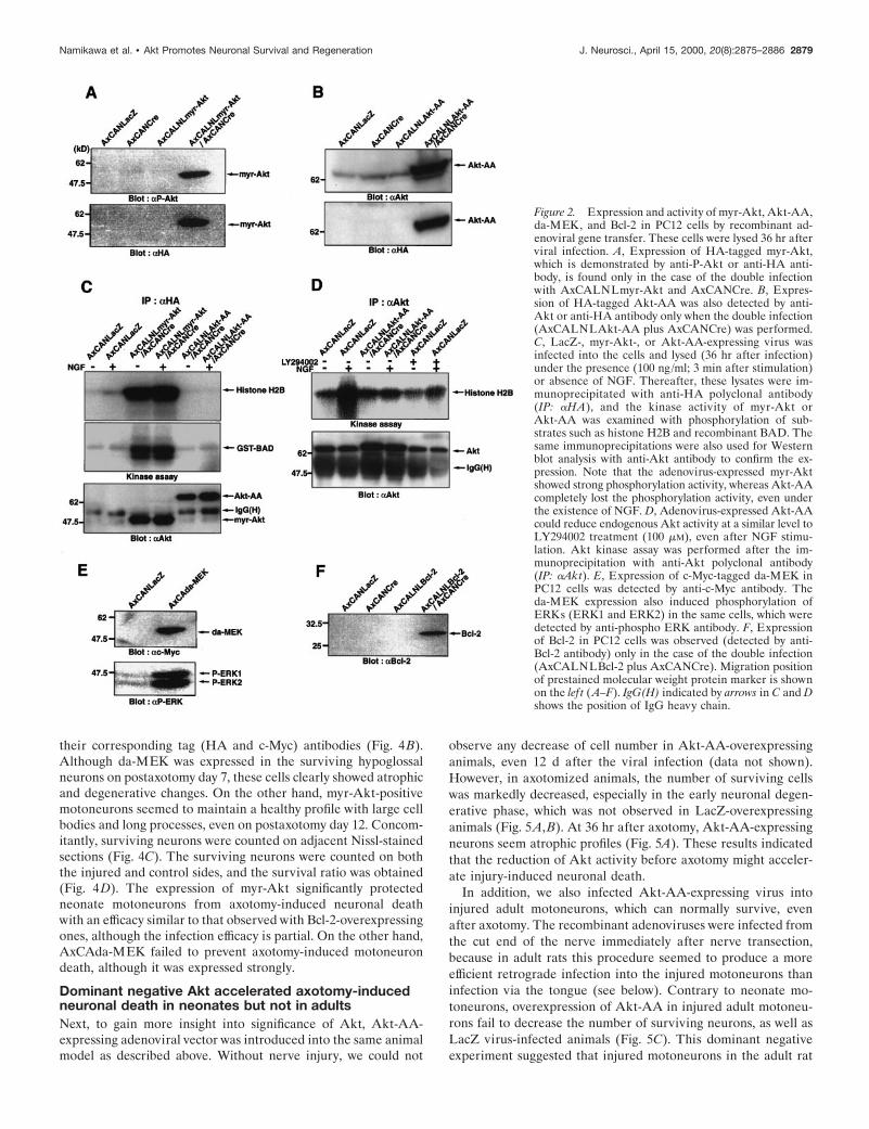
The expression and efficiency of these recombinant viruseswere examined in PC12 cells. The expression of HA-taggedmyr-Akt (as detected by anti-phospho Akt at Ser473 or anti-HAantibody) and Akt-AA (as detected by anti-Akt antibody oranti-HA antibody) were observed only when they were coinfectedwith AxCANCre (Fig. 2A,B). The functional efficacy of the
expressed myr-Akt and Akt-AA were assessed by phosphoryla-tion of substrates, such as histone H2B and recombinant BADprotein, in vitro. Although myr-Akt expressed by adenovirus hada vital activity under both the absence and presence of NGF,Akt-AA completely lost its activity, even after NGF stimulation(Fig. 2C). In addition, overexpression of Akt-AA could succeedin decreasing the endogenous Akt activity in NGF-stimulatedPC12 cells with a similar efficacy as the PI3K inhibitor LY294002(Fig. 2D). The expressed c-Myc-tagged da-MEK (as detectedby anti-c-Myc antibody) was also effective in phosphorylatingERK1 and ERK2 (as detected by anti-phospho ERK antibody)(Fig. 2E). Furthermore, the expression of Bcl-2 was observed onlywhen both AxCALNLBcl-2 and AxCANCre were coinfected(as detected by anti-Bcl-2 antibody) (Fig. 2F).
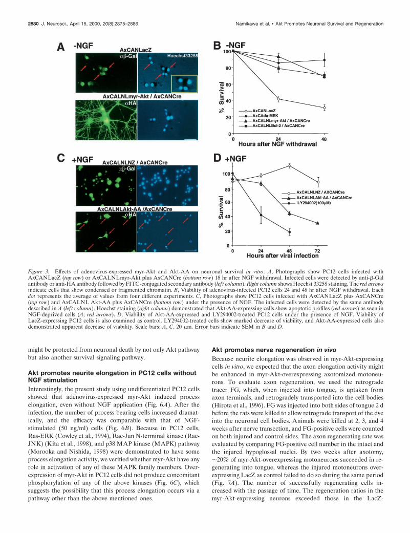
Survival activities of the recombinant viruses indifferentiated PC12 cellsSurvival activities of myr-Akt-, da-MEK-, and Bcl-2-expressingrecombinant adenoviruses were evaluated in differentiated PC12cells, because the previous studies demonstrated that these mol-ecules are survival inducers in PC12 cells and/or sympatheticprimary neurons after NGF withdrawal (Batistatou et al., 1993;Xia et al., 1995; Philpott et al., 1997; Crowder and Freeman,1998). Before NGF deprivation, these adenoviruses were infectedinto the differentiated cells. Then the medium was changed toNGF-free medium. The Hoechst staining (Hoechst 33258) readilydemonstrated apoptotic profiles in the dying cells infected withLacZ-expressing virus (Fig. 3A), and more than half of the virusinfected cells died within 24 hr (Fig. 3B). On the other hand, cellsinfected with myr-Akt, da-MEK, and Bcl-2 carrying recombinantadenoviruses showed almost no signs of any physical deteriora-tion (Fig. 3B). Furthermore, Hoechst staining also showed noapoptotic profiles in myr-Akt-overexpressing cells (Fig. 3A).These observations indicated that adenoviral expression of bothmyr-Akt and da-MEK had similar efficacies equivalent to that ofBcl-2 in survival of NGF-deprived PC12 cells.
A dominant negative effect by Akt-AA-expressing virus wasalso evaluated in PC12 cells with NGF. The cells infectedAxCALNLNZ plus AxCANCre were not represented apoptoticprofiles under the presence of NGF, but Akt-AA-overexpressedcells did show shrunken cell soma, fragmented neurite, and con-densed chromatin in their nuclei similar to NGF-deprived cells(Fig. 3C). The survival ratios of the cells after the infectionof Akt-AA virus and that of the cells treated with LY294002(100 mM) decreased substantially with the passage of time(Fig. 3D). The results indicated that activation of PI3K-Aktpathway is necessary for survival of NGF-dependent PC12 cells.
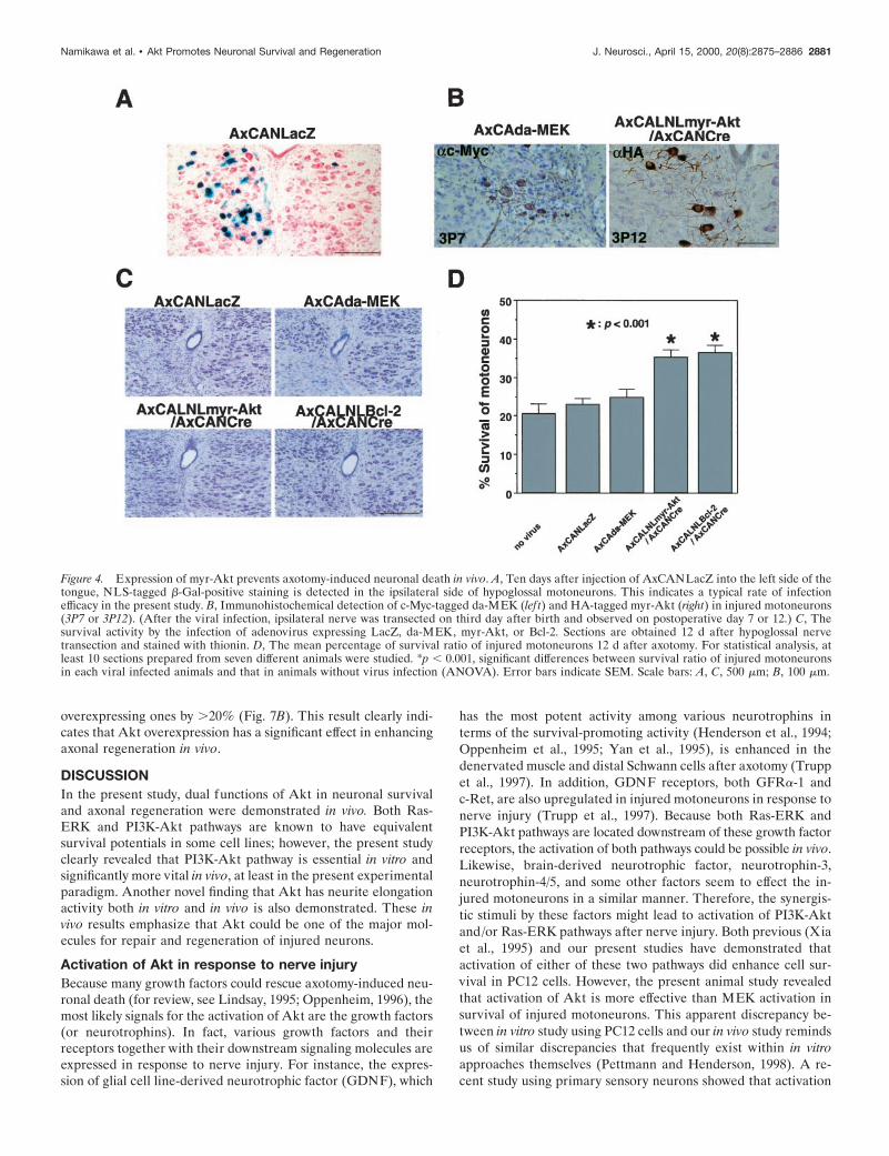
Akt, but not MEK, can prevent axotomy-inducedneuronal deathTo further clarify the roles of PI3K-Akt and Ras-ERK pathwaysin the prevention of axotomy-induced neuronal death in vivo,these recombinant adenoviruses were injected into the tongues ofneonate rats (1-d-old) to allow retrograde infection into thehypoglossal motoneurons. The efficacy of this method was pre-liminarily verified in neonate rat pups using AxCANLacZ. WhenAxCANLacZ was injected into unilateral sides of tongues ofneonates, .20% of ipsilateral hypoglossal motoneurons werelabeled as b-Gal-positive (Fig. 4A). Two days after injection,ipsilateral hypoglossal nerve was axotomized (on postnatal day3). Adenoviral expressions of myr-Akt and da-MEK in injuredmotoneurons were observed immunohistochemically by using
2878 J. Neurosci., April 15, 2000, 20(8):2875–2886 Namikawa et al. • Akt Promotes Neuronal Survival and Regeneration
their corresponding tag (HA and c-Myc) antibodies (Fig. 4B).Although da-MEK was expressed in the surviving hypoglossalneurons on postaxotomy day 7, these cells clearly showed atrophicand degenerative changes. On the other hand, myr-Akt-positivemotoneurons seemed to maintain a healthy profile with large cellbodies and long processes, even on postaxotomy day 12. Concom-itantly, surviving neurons were counted on adjacent Nissl-stainedsections (Fig. 4C). The surviving neurons were counted on boththe injured and control sides, and the survival ratio was obtained(Fig. 4D). The expression of myr-Akt significantly protectedneonate motoneurons from axotomy-induced neuronal deathwith an efficacy similar to that observed with Bcl-2-overexpressingones, although the infection efficacy is partial. On the other hand,AxCAda-MEK failed to prevent axotomy-induced motoneurondeath, although it was expressed strongly.
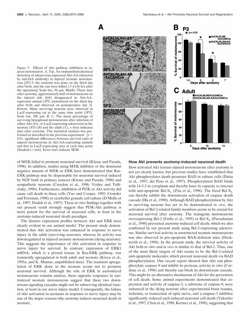
Dominant negative Akt accelerated axotomy-inducedneuronal death in neonates but not in adultsNext, to gain more insight into significance of Akt, Akt-AA-expressing adenoviral vector was introduced into the same animalmodel as described above. Without nerve injury, we could not
observe any decrease of cell number in Akt-AA-overexpressinganimals, even 12 d after the viral infection (data not shown).However, in axotomized animals, the number of surviving cellswas markedly decreased, especially in the early neuronal degen-erative phase, which was not observed in LacZ-overexpressinganimals (Fig. 5A,B). At 36 hr after axotomy, Akt-AA-expressingneurons seem atrophic profiles (Fig. 5A). These results indicatedthat the reduction of Akt activity before axotomy might acceler-ate injury-induced neuronal death.
In addition, we also infected Akt-AA-expressing virus intoinjured adult motoneurons, which can normally survive, evenafter axotomy. The recombinant adenoviruses were infected fromthe cut end of the nerve immediately after nerve transection,because in adult rats this procedure seemed to produce a moreefficient retrograde infection into the injured motoneurons thaninfection via the tongue (see below). Contrary to neonate mo-toneurons, overexpression of Akt-AA in injured adult motoneu-rons fail to decrease the number of surviving neurons, as well asLacZ virus-infected animals (Fig. 5C). This dominant negativeexperiment suggested that injured motoneurons in the adult rat
Figure 2. Expression and activity of myr-Akt, Akt-AA,da-MEK, and Bcl-2 in PC12 cells by recombinant ad-enoviral gene transfer. These cells were lysed 36 hr afterviral infection. A, Expression of HA-tagged myr-Akt,which is demonstrated by anti-P-Akt or anti-HA anti-body, is found only in the case of the double infectionwith AxCALNLmyr-Akt and AxCANCre. B, Expres-sion of HA-tagged Akt-AA was also detected by anti-Akt or anti-HA antibody only when the double infection(AxCALNLAkt-AA plus AxCANCre) was performed.C, LacZ-, myr-Akt-, or Akt-AA-expressing virus wasinfected into the cells and lysed (36 hr after infection)under the presence (100 ng/ml; 3 min after stimulation)or absence of NGF. Thereafter, these lysates were im-munoprecipitated with anti-HA polyclonal antibody(IP: aHA), and the kinase activity of myr-Akt orAkt-AA was examined with phosphorylation of sub-strates such as histone H2B and recombinant BAD. Thesame immunoprecipitations were also used for Westernblot analysis with anti-Akt antibody to confirm the ex-pression. Note that the adenovirus-expressed myr-Aktshowed strong phosphorylation activity, whereas Akt-AAcompletely lost the phosphorylation activity, even underthe existence of NGF. D, Adenovirus-expressed Akt-AAcould reduce endogenous Akt activity at a similar level toLY294002 treatment (100 mM), even after NGF stimu-lation. Akt kinase assay was performed after the im-munoprecipitation with anti-Akt polyclonal antibody(IP: aAkt). E, Expression of c-Myc-tagged da-MEK inPC12 cells was detected by anti-c-Myc antibody. Theda-MEK expression also induced phosphorylation ofERKs (ERK1 and ERK2) in the same cells, which weredetected by anti-phospho ERK antibody. F, Expressionof Bcl-2 in PC12 cells was observed (detected by anti-Bcl-2 antibody) only in the case of the double infection(AxCALNLBcl-2 plus AxCANCre). Migration positionof prestained molecular weight protein marker is shownon the lef t (A–F). IgG(H) indicated by arrows in C and Dshows the position of IgG heavy chain.
Namikawa et al. • Akt Promotes Neuronal Survival and Regeneration J. Neurosci., April 15, 2000, 20(8):2875–2886 2879
might be protected from neuronal death by not only Akt pathwaybut also another survival signaling pathway.
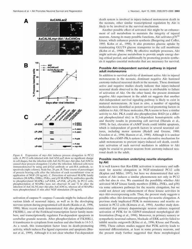
Akt promotes neurite elongation in PC12 cells withoutNGF stimulationInterestingly, the present study using undifferentiated PC12 cellsshowed that adenovirus-expressed myr-Akt induced processelongation, even without NGF application (Fig. 6A). After theinfection, the number of process bearing cells increased dramat-ically, and the efficacy was comparable with that of NGF-stimulated (50 ng/ml) cells (Fig. 6B). Because in PC12 cells,Ras-ERK (Cowley et al., 1994), Rac-Jun N-terminal kinase (Rac-JNK) (Kita et al., 1998), and p38 MAP kinase (MAPK) pathway(Morooka and Nishida, 1998) were demonstrated to have someprocess elongation activity, we verified whether myr-Akt have anyrole in activation of any of these MAPK family members. Over-expression of myr-Akt in PC12 cells did not produce concomitantphosphorylation of any of the above kinases (Fig. 6C), whichsuggests the possibility that this process elongation occurs via apathway other than the above mentioned ones.
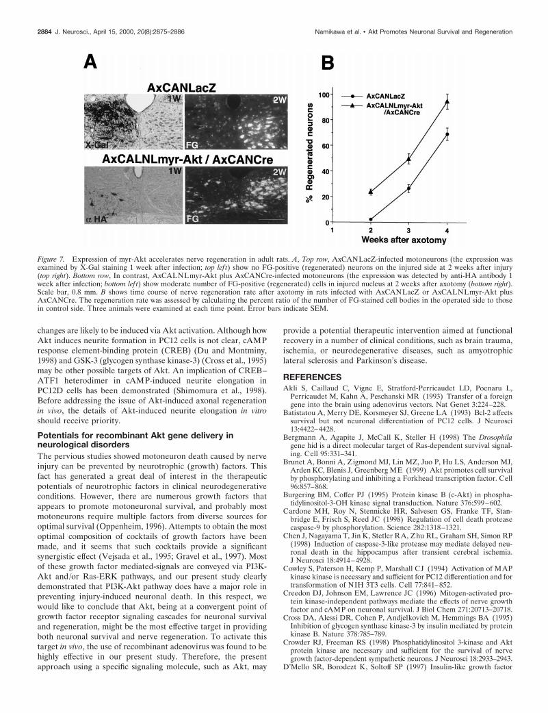
Akt promotes nerve regeneration in vivoBecause neurite elongation was observed in myr-Akt-expressingcells in vitro, we expected that the axon elongation activity mightbe enhanced in myr-Akt-overexpressing axotomized motoneu-rons. To evaluate axon regeneration, we used the retrogradetracer FG, which, when injected into tongue, is uptaken fromaxon terminals, and retrogradely transported into the cell bodies(Hirota et al., 1996). FG was injected into both sides of tongue 2 dbefore the rats were killed to allow retrograde transport of the dyeinto the neuronal cell bodies. Animals were killed at 2, 3, and 4weeks after nerve transection, and FG-positive cells were countedon both injured and control sides. The axon regenerating rate wasevaluated by comparing FG-positive cell number in the intact andthe injured hypoglossal nuclei. By two weeks after axotomy,;20% of myr-Akt-overexpressing motoneurons succeeded in re-generating into tongue, whereas the injured motoneurons over-expressing LacZ as control failed to do so during the same period(Fig. 7A). The number of successfully regenerating cells in-creased with the passage of time. The regeneration ratios in themyr-Akt-expressing neurons exceeded those in the LacZ-
Figure 3. Effects of adenovirus-expressed myr-Akt and Akt-AA on neuronal survival in vitro. A, Photographs show PC12 cells infected withAxCANLacZ (top row) or AxCALNLmyr-Akt plus AxCANCre (bottom row) 18 hr after NGF withdrawal. Infected cells were detected by anti-b-Galantibody or anti-HA antibody followed by FITC-conjugated secondary antibody (lef t column). Right column shows Hoechst 33258 staining. The red arrowsindicate cells that show condensed or fragmented chromatin. B, Viability of adenovirus-infected PC12 cells 24 and 48 hr after NGF withdrawal. Eachdot represents the average of values from four different experiments. C, Photographs show PC12 cells infected with AxCANLacZ plus AxCANCre(top row) and AxCALNL Akt-AA plus AxCANCre (bottom row) under the presence of NGF. The infected cells were detected by the same antibodydescribed in A (lef t column). Hoechst staining (right column) demonstrated that Akt-AA-expressing cells show apoptotic profiles (red arrows) as seen inNGF-deprived cells (A; red arrows). D, Viability of Akt-AA-expressed and LY294002-treated PC12 cells under the presence of NGF. Viability ofLacZ-expressing PC12 cells is also examined as control. LY294002-treated cells show marked decrease of viability, and Akt-AA-expressed cells alsodemonstrated apparent decrease of viability. Scale bars: A, C, 20 mm. Error bars indicate SEM in B and D.
2880 J. Neurosci., April 15, 2000, 20(8):2875–2886 Namikawa et al. • Akt Promotes Neuronal Survival and Regeneration
overexpressing ones by .20% (Fig. 7B). This result clearly indi-cates that Akt overexpression has a significant effect in enhancingaxonal regeneration in vivo.
DISCUSSIONIn the present study, dual functions of Akt in neuronal survivaland axonal regeneration were demonstrated in vivo. Both Ras-ERK and PI3K-Akt pathways are known to have equivalentsurvival potentials in some cell lines; however, the present studyclearly revealed that PI3K-Akt pathway is essential in vitro andsignificantly more vital in vivo, at least in the present experimentalparadigm. Another novel finding that Akt has neurite elongationactivity both in vitro and in vivo is also demonstrated. These invivo results emphasize that Akt could be one of the major mol-ecules for repair and regeneration of injured neurons.
Activation of Akt in response to nerve injuryBecause many growth factors could rescue axotomy-induced neu-ronal death (for review, see Lindsay, 1995; Oppenheim, 1996), themost likely signals for the activation of Akt are the growth factors(or neurotrophins). In fact, various growth factors and theirreceptors together with their downstream signaling molecules areexpressed in response to nerve injury. For instance, the expres-sion of glial cell line-derived neurotrophic factor (GDNF), which
has the most potent activity among various neurotrophins interms of the survival-promoting activity (Henderson et al., 1994;Oppenheim et al., 1995; Yan et al., 1995), is enhanced in thedenervated muscle and distal Schwann cells after axotomy (Truppet al., 1997). In addition, GDNF receptors, both GFRa-1 andc-Ret, are also upregulated in injured motoneurons in response tonerve injury (Trupp et al., 1997). Because both Ras-ERK andPI3K-Akt pathways are located downstream of these growth factorreceptors, the activation of both pathways could be possible in vivo.Likewise, brain-derived neurotrophic factor, neurotrophin-3,neurotrophin-4/5, and some other factors seem to effect the in-jured motoneurons in a similar manner. Therefore, the synergis-tic stimuli by these factors might lead to activation of PI3K-Aktand/or Ras-ERK pathways after nerve injury. Both previous (Xiaet al., 1995) and our present studies have demonstrated thatactivation of either of these two pathways did enhance cell sur-vival in PC12 cells. However, the present animal study revealedthat activation of Akt is more effective than MEK activation insurvival of injured motoneurons. This apparent discrepancy be-tween in vitro study using PC12 cells and our in vivo study remindsus of similar discrepancies that frequently exist within in vitroapproaches themselves (Pettmann and Henderson, 1998). A re-cent study using primary sensory neurons showed that activation
Figure 4. Expression of myr-Akt prevents axotomy-induced neuronal death in vivo. A, Ten days after injection of AxCANLacZ into the left side of thetongue, NLS-tagged b-Gal-positive staining is detected in the ipsilateral side of hypoglossal motoneurons. This indicates a typical rate of infectionefficacy in the present study. B, Immunohistochemical detection of c-Myc-tagged da-MEK (lef t) and HA-tagged myr-Akt (right) in injured motoneurons(3P7 or 3P12). (After the viral infection, ipsilateral nerve was transected on third day after birth and observed on postoperative day 7 or 12.) C, Thesurvival activity by the infection of adenovirus expressing LacZ, da-MEK, myr-Akt, or Bcl-2. Sections are obtained 12 d after hypoglossal nervetransection and stained with thionin. D, The mean percentage of survival ratio of injured motoneurons 12 d after axotomy. For statistical analysis, atleast 10 sections prepared from seven different animals were studied. *p , 0.001, significant differences between survival ratio of injured motoneuronsin each viral infected animals and that in animals without virus infection (ANOVA). Error bars indicate SEM. Scale bars: A, C, 500 mm; B, 100 mm.
Namikawa et al. • Akt Promotes Neuronal Survival and Regeneration J. Neurosci., April 15, 2000, 20(8):2875–2886 2881
of MEK failed to promote neuronal survival (Klesse and Parada,1998). In addition, studies using MEK inhibitor or the dominantnegative mutant of MEK or ERK have demonstrated that Ras-ERK pathway may be dispensable for neuronal survival inducedby NGF both in primary sensory (Klesse and Parada, 1998) andsympathetic neurons (Creedon et al., 1996; Virdee and Tolk-ovsky, 1996). Furthermore, inhibition of PI3K or Akt activity didcause cell death in these cells (Yao and Cooper, 1995; Crowderand Freeman, 1998) or cerebellar granule cell culture (D’Mello etal., 1997; Dudek et al., 1997). These in vitro findings together withour present result strongly suggest that PI3K-Akt pathway ismore potent for the survival of neuronal cells, at least in theaxotomy-induced neuronal death paradigm.
The distinct expression profiles between Akt and ERK wereclearly evident in our animal model. The present study demon-strated that Akt activation was enhanced in response to nerveinjury in the adult (surviving neurons), whereas its activity wasdownregulated in injured neonate motoneurons (dying neurons).This suggests the importance of Akt activation in response tonerve injury for survival. In contrast, expression of ERK1mRNA, which is a pivotal kinase in Ras-ERK pathway, wastransiently upregulated in both adult and neonate (Kiryu et al.,1995a; and K. Mansur, unpublished data). The transient upregu-lation of ERK alone in the neonate seems not to promoteneuronal survival. Although the role of ERK in axotomizedmotoneurons remains unclear, these opposite responses in axo-tomized neonate motoneurons suggest that these two down-stream signaling cascades might not be subserving identical func-tion, at least in our nerve injury model. Consequently, the failureof Akt activation in neonates in response to nerve injury may beone of the major reasons why axotomy induces neuronal death inneonate.
How Akt prevents axotomy-induced neuronal deathHow activated Akt rescues injured motoneurons after axotomy isnot yet clearly known, but previous studies have established thatAkt phosphorylates death promoter BAD in culture cells (Dattaet al., 1997; del Peso et al., 1997). Phosphorylated BAD bindswith 14-3-3 in cytoplasm and thereby loses its capacity to interactwith anti-apoptotic Bcl-XL (Zha et al., 1996). The freed Bcl-XL
can thereby inhibit the downstream activation of caspase deathcascade (Hu et al., 1998). Although BAD phosphorylation by Aktin surviving neurons has yet to be demonstrated in vivo, theactivation of Bcl-2-related family members seems to be crucial forneuronal survival after axotomy. The transgenic motoneuronsoverexpressing Bcl-2 (Farlie et al., 1995) or Bcl-XL (Parsadanianet al., 1998) prevented axotomy-induced cell death, which we alsoconfirmed by our present study using Bcl-2-expressing adenovi-rus. Similar survival activity in axotomized neonate motoneuronswas also observed in pro-apoptotic BAX-deficient mice (Deck-werth et al., 1996). In the present study, the survival activity ofAkt both in vitro and in vivo is similar to that of Bcl-2. Thus, oneof the most likely targets of Akt seems to be the Bcl-2-relatedanti-apoptotic molecules, which prevent neuronal death via BADphosphorylation. One recent report showed that Akt can phos-phorylate caspase-9 and inhibit its protease activity in vitro (Car-done et al., 1998) and thereby can block its downstream cascade.This might be an alternative mechanism of Akt for the preventionof cell death. Some animal experiments demonstrated that ex-pression and activity of caspase-3, a substrate of caspase-9, wereenhanced in the dying neurons after experimental brain trauma,ischemia, or transection of optic nerve, and a caspase-3 inhibitorsignificantly reduced such induced neuronal cell death (Yakovlevet al., 1997; Chen et al., 1998; Kermer et al., 1998), suggesting that
Figure 5. Effects of Akt pathway inhibition in in-jured motoneuron. A, Top, An immunohistochemicaldetection of adenovirus-expressed Akt-AA (detectedby anti-HA antibody) in injured neonate motoneu-rons [3P1.5; the axotomy was done on the third dayafter birth, and the rats were killed 1.5 d (36 hr) afterthe operation]. Scale bar, 50 mm. Middle, Three daysafter axotomy, approximately half of motoneurons onthe injured side (left) disappeared in Akt-AA-expressed animal (3P3; axotomized on the third dayafter birth and observed on postoperative day 3).Bottom, Many surviving neurons were observed inLacZ-expressing rat at the same time point (3P3).Scale bar, 200 mm. B, C, The mean percentage ofsurviving hypoglossal motoneurons after infection ofeither Akt-AA- or LacZ-expressing adenovirus in theneonate (P3) (B) and the adult (C). x-Axis indicatesdays after axotomy. The statistical analysis was per-formed as described in the previous experiment. *p ,0.01, significant differences between survival ratio ofinjured motoneurons in Akt-AA-expressing animalsand that in LacZ-expressing ones at each time point(Student’s t test). Error bars indicate SEM.
2882 J. Neurosci., April 15, 2000, 20(8):2875–2886 Namikawa et al. • Akt Promotes Neuronal Survival and Regeneration
activation of caspase-9–caspase-3 cascade might be induced aftervarious kinds of neuronal injury, as well as in the developingnervous system during programmed cell death (Kuida et al., 1996,1998). More recent study demonstrated Akt also phosphorylatesFKHRL1, one of the Forkhead transcription factor family mem-bers, and transcriptionally regulates Fas-dependent apoptosis incerebellar granule neurons. After phosphorylation of FKHRL1,it translocates to cytoplasm from nucleus and also binds to 14-3-3.As a result, this transcription factor loses its death-promotingactivity, which induces Fas ligand expression and apoptosis (Bru-net et al., 1999). Although it is not clear whether Fas-dependent
death system is involved in injury-induced motoneuron death inthe neonate, other similar transcriptional regulation by Akt islikely to be involved in the survival mechanism.
Another possible effect of activated Akt might be an enhance-ment of cell metabolism to maintain the integrity of injuredneurons. Among its many possible functions, Akt activates p70S6
kinase, which enhances protein synthesis (Burgering and Coffer,1995; Kohn et al., 1998). It also promotes glucose uptake bytranslocating GLUT4 glucose transporter to the cell membrane(Kohn et al., 1996b, 1998). By effective multiple processes, Aktmight activate glucose metabolism to provide ample energy dur-ing critical period, and additionally by promoting protein synthe-sis it supplies essential molecules that are necessary for survival.
Possible Akt-independent survival pathway in injuredadult motoneuronsIn addition to survival activity of dominant active Akt in injuredmotoneurons in the neonate, dominant negative Akt hastenedaxotomy-induced neuronal death in the neonate. These dominantactive and negative studies clearly indicate that injury-inducedneuronal death observed in the neonate is attributable to failureof activation of Akt. On the other hand, the present dominantnegative Akt experiment in the adult rat suggests that anotherAkt-independent survival signaling pathway is likely to exist inmatured motoneurons. At least in vitro, a number of signalingmolecules were identified as potent survival-promoting factors inaddition to Akt. Of these molecules, PKA seems to have a similaractivity to Akt. PKA could also phosphorylate BAD (at a differ-ent phosphorylated site) in IL3-dependent hematopoietic cellsand thereby results in promoting cell survival (Harada et al.,1999). In fact, elevation of cAMP levels could inhibit apoptosis,which is independent of growth factors in several neuronal cul-tures, including motor systems (Rydell and Greene, 1988;Creedon et al., 1996; Hanson et al., 1998). Although it is unclearwhether the cAMP–PKA system is an alternative mechanism foradult motoneurons to prevent injury-induced cell death, coordi-nate activation of such survival mediators in addition to Aktmight be crucial to protect neurons from axotomy-induced neu-ronal death in the adult.
Possible mechanism underlying neurite elongationby AktIt is well known that Ras-ERK activation is necessary and suffi-cient for neurite elongation in NGF-stimulated PC12 cells(Kaplan and Miller, 1997), but here we demonstrated that acti-vation of Akt induces a similar phenomenon not only in PC12cells but also in vivo. We examined the possibility whether Aktactivated MAP kinase family members (ERKs, JNKs, and p38)via some unknown pathways for the neurite elongation, but wecould not detect any enhancement of these kinase activities inmyr-Akt-overexpressing cells. Thus, the present study suggests apossible other Akt downstream pathway for neurite elongation. Aprevious study implicated PI3K in maintenance and neurite ex-tension in PC12 cells (Kimura et al., 1994). Another study dem-onstrated that primary dopaminergic neuron cultures requiredactivation of PI3K for its GDNF-stimulated morphological dif-ferentiation (Pong et al., 1998). Moreover, in primary sensory orsympathetic neuronal cultures, blockade of ERK activity failed toinhibit neurite outgrowth (Klinz et al., 1996; Klesse and Parada,1998). These culture studies suggest that PI3K activity leads toneuronal differentiation, at least in some primary neurons, andthe present study further suggested that these morphological
Figure 6. Expression of myr-Akt induces process elongation in PC12cells. A, PC12 cells infected with AxCANLacZ show no significant changein cell shapes, but the infection with AxCALNLmyr-Akt plus AxCANCrecaused clear process elongation 2 d after the infection. Infected cells wereimmunodetected by using anti-b-Gal antibody or anti-HA antibody, re-spectively (right column). Scale bar, 20 mm. B, Time course of percentagesof process bearing cells after the infection of each recombinant virus orapplication of NGF (50 ng/ml). C, Detection of activated MAPK familymembers (MAPKs, ERKs, JNKs, and p38 MAPK) by antibodies specificto phosphorylated MAPKs (aP-ERK, aP-JNK, aP-p38). In PC12 cells,phosphorylation of MAPKs were not observed 36 and 72 hr after theinfection of AxCALNLmyr-Akt plus AxCANCre, whereas all of MAPKswere phosphorylated 15 min after NGF stimulation (50 ng/ml).
Namikawa et al. • Akt Promotes Neuronal Survival and Regeneration J. Neurosci., April 15, 2000, 20(8):2875–2886 2883
changes are likely to be induced via Akt activation. Although howAkt induces neurite formation in PC12 cells is not clear, cAMPresponse element-binding protein (CREB) (Du and Montminy,1998) and GSK-3 (glycogen synthase kinase-3) (Cross et al., 1995)may be other possible targets of Akt. An implication of CREB–ATF1 heterodimer in cAMP-induced neurite elongation inPC12D cells has been demonstrated (Shimomura et al., 1998).Before addressing the issue of Akt-induced axonal regenerationin vivo, the details of Akt-induced neurite elongation in vitroshould receive priority.
Potentials for recombinant Akt gene delivery inneurological disordersThe pervious studies showed motoneuron death caused by nerveinjury can be prevented by neurotrophic (growth) factors. Thisfact has generated a great deal of interest in the therapeuticpotentials of neurotrophic factors in clinical neurodegenerativeconditions. However, there are numerous growth factors thatappears to promote motoneuronal survival, and probably mostmotoneurons require multiple factors from diverse sources foroptimal survival (Oppenheim, 1996). Attempts to obtain the mostoptimal composition of cocktails of growth factors have beenmade, and it seems that such cocktails provide a significantsynergistic effect (Vejsada et al., 1995; Gravel et al., 1997). Mostof these growth factor mediated-signals are conveyed via PI3K-Akt and/or Ras-ERK pathways, and our present study clearlydemonstrated that PI3K-Akt pathway does have a major role inpreventing injury-induced neuronal death. In this respect, wewould like to conclude that Akt, being at a convergent point ofgrowth factor receptor signaling cascades for neuronal survivaland regeneration, might be the most effective target in providingboth neuronal survival and nerve regeneration. To activate thistarget in vivo, the use of recombinant adenovirus was found to behighly effective in our present study. Therefore, the presentapproach using a specific signaling molecule, such as Akt, may
provide a potential therapeutic intervention aimed at functionalrecovery in a number of clinical conditions, such as brain trauma,ischemia, or neurodegenerative diseases, such as amyotrophiclateral sclerosis and Parkinson’s disease.
REFERENCESAkli S, Caillaud C, Vigne E, Stratford-Perricaudet LD, Poenaru L,
Perricaudet M, Kahn A, Peschanski MR (1993) Transfer of a foreigngene into the brain using adenovirus vectors. Nat Genet 3:224–228.
Batistatou A, Merry DE, Korsmeyer SJ, Greene LA (1993) Bcl-2 affectssurvival but not neuronal differentiation of PC12 cells. J Neurosci13:4422–4428.
Bergmann A, Agapite J, McCall K, Steller H (1998) The Drosophilagene hid is a direct molecular target of Ras-dependent survival signal-ing. Cell 95:331–341.
Brunet A, Bonni A, Zigmond MJ, Lin MZ, Juo P, Hu LS, Anderson MJ,Arden KC, Blenis J, Greenberg ME (1999) Akt promotes cell survivalby phosphorylating and inhibiting a Forkhead transcription factor. Cell96:857–868.
Burgering BM, Coffer PJ (1995) Protein kinase B (c-Akt) in phospha-tidylinositol-3-OH kinase signal transduction. Nature 376:599–602.
Cardone MH, Roy N, Stennicke HR, Salvesen GS, Franke TF, Stan-bridge E, Frisch S, Reed JC (1998) Regulation of cell death proteasecaspase-9 by phosphorylation. Science 282:1318–1321.
Chen J, Nagayama T, Jin K, Stetler RA, Zhu RL, Graham SH, Simon RP(1998) Induction of caspase-3-like protease may mediate delayed neu-ronal death in the hippocampus after transient cerebral ischemia.J Neurosci 18:4914–4928.
Cowley S, Paterson H, Kemp P, Marshall CJ (1994) Activation of MAPkinase kinase is necessary and sufficient for PC12 differentiation and fortransformation of NIH 3T3 cells. Cell 77:841–852.
Creedon DJ, Johnson EM, Lawrence JC (1996) Mitogen-activated pro-tein kinase-independent pathways mediate the effects of nerve growthfactor and cAMP on neuronal survival. J Biol Chem 271:20713–20718.
Cross DA, Alessi DR, Cohen P, Andjelkovich M, Hemmings BA (1995)Inhibition of glycogen synthase kinase-3 by insulin mediated by proteinkinase B. Nature 378:785–789.
Crowder RJ, Freeman RS (1998) Phosphatidylinositol 3-kinase and Aktprotein kinase are necessary and sufficient for the survival of nervegrowth factor-dependent sympathetic neurons. J Neurosci 18:2933–2943.
D’Mello SR, Borodezt K, Soltoff SP (1997) Insulin-like growth factor
Figure 7. Expression of myr-Akt accelerates nerve regeneration in adult rats. A, Top row, AxCANLacZ-infected motoneurons (the expression wasexamined by X-Gal staining 1 week after infection; top lef t) show no FG-positive (regenerated) neurons on the injured side at 2 weeks after injury(top right). Bottom row, In contrast, AxCALNLmyr-Akt plus AxCANCre-infected motoneurons (the expression was detected by anti-HA antibody 1week after infection; bottom lef t) show moderate number of FG-positive (regenerated) cells in injured nucleus at 2 weeks after axotomy (bottom right).Scale bar, 0.8 mm. B shows time course of nerve regeneration rate after axotomy in rats infected with AxCANLacZ or AxCALNLmyr-Akt plusAxCANCre. The regeneration rate was assessed by calculating the percent ratio of the number of FG-stained cell bodies in the operated side to thosein control side. Three animals were examined at each time point. Error bars indicate SEM.
2884 J. Neurosci., April 15, 2000, 20(8):2875–2886 Namikawa et al. • Akt Promotes Neuronal Survival and Regeneration
and potassium depolarization maintain neuronal survival by distinctpathways: possible involvement of PI 3- kinase in IGF-1 signaling.J Neurosci 17:1548–1560.
Datta SR, Dudek H, Tao X, Masters S, Fu H, Gotoh Y, Greenberg ME(1997) Akt phosphorylation of BAD couples survival signals to thecell- intrinsic death machinery. Cell 91:231–241.
Davidson BL, Allen ED, Kozarsky KF, Wilson JM, Roessler BJ (1993)A model system for in vivo gene transfer into the central nervous systemusing an adenoviral vector. Nat Genet 3:219–223.
Deckwerth TL, Elliott JL, Knudson CM, Johnson Jr EM, Snider WD,Korsmeyer SJ (1996) BAX is required for neuronal death after tro-phic factor deprivation and during development. Neuron 17:401–411.
del Peso L, Gonzalez-Garcia M, Page C, Herrera R, Nunez G (1997)Interleukin-3-induced phosphorylation of BAD through the proteinkinase Akt. Science 278:687–689.
Downward J (1998) Lipid-regulated kinases: some common themes atlast. Science 279:673–674.
Du K, Montminy M (1998) CREB is a regulatory target for the proteinkinase Akt/PKB. J Biol Chem 273:32377–32379.
Dudek H, Datta SR, Franke TF, Birnbaum MJ, Yao R, Cooper GM,Segal RA, Kaplan DR, Greenberg ME (1997) Regulation of neuronalsurvival by the serine-threonine protein kinase Akt. Science275:661–665.
Farlie PG, Dringen R, Rees SM, Kannourakis G, Bernard O (1995) bcl-2transgene expression can protect neurons against developmental andinduced cell death. Proc Natl Acad Sci USA 92:4397–4401.
Franke TF, Yang SI, Chan TO, Datta K, Kazlauskas A, Morrison DK,Kaplan DR, Tsichlis PN (1995) The protein kinase encoded by theAkt proto-oncogene is a target of the PDGF-activated phosphatidyl-inositol 3-kinase. Cell 81:727–736.
Fukuda M, Gotoh Y, Nishida E (1997) Interaction of MAP kinase withMAP kinase kinase: its possible role in the control of nucleocytoplas-mic transport of MAP kinase. EMBO J 16:1901–1908.
Gravel C, Gotz R, Lorrain A, Sendtner M (1997) Adenoviral genetransfer of ciliary neurotrophic factor and brain-derived neurotrophicfactor leads to long-term survival of axotomized motor neurons. NatMed 3:765–770.
Hamburger V (1934) The effects of wing bud extirpation on the devel-opment of the central nervous system in chick embryos. J Exp Zool68:449–494.
Hanson Jr MG, Shen S, Wiemelt AP, McMorris FA, Barres BA (1998)Cyclic AMP elevation is sufficient to promote the survival of spinalmotor neurons in vitro. J Neurosci 18:7361–7371.
Harada H, Becknell B, Wilm M, Mann M, Huang LJ, Taylor SS, Scott JD,Korsmeyer SJ (1999) Phosphorylation and inactivation of BAD bymitochondria-anchored protein kinase A. Mol Cell 3:413–422.
Henderson CE, Phillips HS, Pollock RA, Davies AM, Lemeulle C,Armanini M, Simmons L, Moffet B, Vandlen RA, Simpson LC, MoffetB, Vandlen RA, Koliatsos VE, Rosenthal A (1994) GDNF: a potentsurvival factor for motoneurons present in peripheral nerve and mus-cle. Science 266:1062–1064.
Hirota H, Kiyama H, Kishimoto T, Taga T (1996) Accelerated nerveregeneration in mice by upregulated expression of interleukin (IL) 6and IL-6 receptor after trauma. J Exp Med 183:2627–2634.
Hu Y, Benedict MA, Wu D, Inohara N, Nunez G (1998) Bcl-XL inter-acts with Apaf-1 and inhibits Apaf-1-dependent caspase-9 activation.Proc Natl Acad Sci USA 95:4386–4391.
Ishiyama M, Tominaga H, Shiga M, Sasamoto K, Ohkura Y, Ueno K(1996) A combined assay of cell viability and in vitro cytotoxicity witha highly water-soluble tetrazolium salt, neutral red and crystal violet.Biol Pharm Bull 19:1518–1520.
Ito Y, Sakagami H, Kondo H (1996) Enhanced gene expression forphosphatidylinositol 3-kinase in the hypoglossal motoneurons followingaxonal crush. Mol Brain Res 37:329–332.
Kanegae Y, Makimura M, Saito I (1994) A simple and efficient methodfor purification of infectious recombinant adenovirus. Jpn J Med SciBiol 47:157–166.
Kanegae Y, Lee G, Sato Y, Tanaka M, Nakai M, Sakaki T, Sugano S,Saito I (1995) Efficient gene activation in mammalian cells by usingrecombinant adenovirus expressing site-specific Cre recombinase. Nu-cleic Acids Res 23:3816–3821.
Kaplan DR, Miller FD (1997) Signal transduction by the neurotrophinreceptors. Curr Opin Cell Biol 9:213–221.
Kermer P, Klocker N, Labes M, Bahr M (1998) Inhibition of CPP32-like
proteases rescues axotomized retinal ganglion cells from secondary celldeath in vivo. J Neurosci 18:4656–4662.
Kimura K, Hattori S, Kabuyama Y, Shizawa Y, Takayanagi J, NakamuraS, Toki S, Matsuda Y, Onodera K, Fukui Y (1994) Neurite outgrowthof PC12 cells is suppressed by wortmannin, a specific inhibitor ofphosphatidylinositol 3-kinase. J Biol Chem 269:18961–18967.
Kiryu S, Morita N, Ohno K, Maeno H, Kiyama H (1995a) Regulation ofmRNA expression involved in Ras and PKA signal pathways during rathypoglossal nerve regeneration. Mol Brain Res 29:147–156.
Kiryu S, Yao GL, Morita N, Kato H, Kiyama H (1995b) Nerve injuryenhances rat neuronal glutamate transporter expression: identificationby differential display PCR. J Neurosci 15:7872–7878.
Kita Y, Kimura KD, Kobayashi M, Ihara S, Kaibuchi K, Kuroda S, Ui M,Iba H, Konishi H, Kikkawa U, Nagata S, Fukui Y (1998) Microinjec-tion of activated phosphatidylinositol-3 kinase induces process out-growth in rat PC12 cells through the Rac-JNK signal transductionpathway. J Cell Sci 111:907–915.
Kitamura T, Ogawa W, Sakaue H, Hino Y, Kuroda S, Takata M, Matsu-moto M, Maeda T, Konishi H, Kikkawa U, Kasuga M (1998) Require-ment for activation of the serine-threonine kinase Akt (protein kinaseB) in insulin stimulation of protein synthesis but not of glucose trans-port. Mol Cell Biol 18:3708–3717.
Klesse LJ, Parada LF (1998) p21 ras and phosphatidylinositol-3 kinaseare required for survival of wild-type and NF1 mutant sensory neurons.J Neurosci 18:10420–10428.
Klinz FJ, Wolff P, Heumann R (1996) Nerve growth factor-stimulatedmitogen-activated protein kinase activity is not necessary for neuriteoutgrowth of chick dorsal root ganglion sensory and sympathetic neu-rons. J Neurosci Res 46:720–726.
Kohn AD, Takeuchi F, Roth RA (1996a) Akt, a pleckstrin homologydomain containing kinase, is activated primarily by phosphorylation.J Biol Chem 271:21920–21926.
Kohn AD, Summers SA, Birnbaum MJ, Roth RA (1996b) Expression ofa constitutively active Akt Ser/Thr kinase in 3T3–L1 adipocytes stim-ulates glucose uptake and glucose transporter 4 translocation. J BiolChem 271:31372–31378.
Kohn AD, Barthel A, Kovacina KS, Boge A, Wallach B, Summers SA,Birnbaum MJ, Scott PH, Lawrence Jr JC, Roth RA (1998) Construc-tion and characterization of a conditionally active version of the serine/threonine kinase Akt. J Biol Chem 273:11937–11943.
Konishi H, Shinomura T, Kuroda S, Ono Y, Kikkawa U (1994) Molec-ular cloning of rat RAC protein kinase alpha and beta and theirassociation with protein kinase C zeta. Biochem Biophys Res Commun205:817–825.
Kuida K, Zheng TS, Na S, Kuan C, Yang D, Karasuyama H, Rakic P,Flavell RA (1996) Decreased apoptosis in the brain and prematurelethality in CPP32- deficient mice. Nature 384:368–372.
Kuida K, Haydar TF, Kuan CY, Gu Y, Taya C, Karasuyama H, Su MS,Rakic P, Flavell RA (1998) Reduced apoptosis and cytochromec-mediated caspase activation in mice lacking caspase 9. Cell94:325–337.
Kurada P, White K (1998) Ras promotes cell survival in Drosophila bydownregulating hid expression. Cell 95:319–329.
Lindsay RM (1995) Neuron saving schemes. Nature 373:289–290.Mansour SJ, Matten WT, Hermann AS, Candia JM, Rong S, Fukasawa
K, Vande Woude GF, Ahn NG (1994) Transformation of mammaliancells by constitutively active MAP kinase kinase. Science 265:966–970.
Miyake S, Makimura M, Kanegae Y, Harada S, Sato Y, Takamori K,Tokuda C, Saito I (1996) Efficient generation of recombinant adeno-viruses using adenovirus DNA–terminal protein complex and a cosmidbearing the full-length virus genome. Proc Natl Acad Sci USA93:1320–1324.
Morita N, Kiryu S, Kiyama H (1996) p53-independent cyclin G expres-sion in a group of mature neurons and its enhanced expression duringnerve regeneration. J Neurosci 16:5961–5966.
Morooka T, Nishida E (1998) Requirement of p38 mitogen-activatedprotein kinase for neuronal differentiation in PC12 cells. J Biol Chem273:24285–24288.
Namikawa K, Su Q, Kiryu-Seo S, Kiyama H (1998) Enhanced expres-sion of 14–3-3 family members in injured motoneurons. Mol Brain Res55:315–320.
Niwa H, Yamamura K, Miyazaki J (1991) Efficient selection for high-expression transfectants with a novel eukaryotic vector. Gene108:193–199.
Namikawa et al. • Akt Promotes Neuronal Survival and Regeneration J. Neurosci., April 15, 2000, 20(8):2875–2886 2885
Oppenheim RW (1996) Neurotrophic survival molecules for motoneu-rons: an embarrassment of riches. Neuron 17:195–197.
Oppenheim RW, Houenou LJ, Johnson JE, Lin LF, Li L, Lo AC,Newsome AL, Prevette DM, Wang S (1995) Developing motor neu-rons rescued from programmed and axotomy-induced cell death byGDNF. Nature 373:344–346.
Owada Y, Utsunomiya A, Yoshimoto T, Kondo H (1997) Expression ofmRNA for Akt, serine-threonine protein kinase, in the brain duringdevelopment and its transient enhancement following axotomy of hy-poglossal nerve. J Mol Neurosci 9:27–33.
Parsadanian AS, Cheng Y, Keller-Peck CR, Holtzman DM, Snider WD(1998) Bcl-xL is an antiapoptotic regulator for postnatal CNS neuronsJ Neurosci 18:1009–1019.
Persson H, Ibanez CF (1993) Role and expression of neurotrophins andthe trk family of tyrosine kinase receptors in neural growth and rescueafter injury. Curr Opin Neurol Neurosurg 6:11–18.
Pettmann B, Henderson CE (1998) Neuronal cell death. Neuron 20:633– 647.
Philpott KL, McCarthy MJ, Klippel A, Rubin LL (1997) Activatedphosphatidylinositol 3-kinase and Akt kinase promote survival of su-perior cervical neurons. J Cell Biol 139:809–815.
Pong K, Xu RY, Baron WF, Louis JC, Beck KD (1998) Inhibition ofphosphatidylinositol 3-kinase activity blocks cellular differentiation me-diated by glial cell line-derived neurotrophic factor in dopaminergicneurons. J Neurochem 71:1912–1919.
Resh MD (1994) Myristylation and palmitylation of Src family mem-bers: the fats of the matter. Cell 76:411–413.
Romanes GJ (1946) Motor localization and the effects of nerve injury onthe ventral horn cells of the spinal cord. J Anat 80:117–131.
Rydell RE, Greene LA (1988) cAMP analogs promote survival andneurite outgrowth in cultures of rat sympathetic and sensory neuronsindependently of NGF. Proc Natl Acad Sci USA 85:1257–1261.
Sato Y, Tanaka K, Lee G, Kanegae Y, Sakai Y, Kaneko S, NakabayashiH, Tamaoki T, Saito I (1998a) Enhanced and specific gene expressionvia tissue-specific production of Cre recombinase using adenovirusvector. Biochem Biophys Res Commun 244:455–462.
Sato N, Wang S, Li L, Okabe K, Hashimoto M, Yaginuma H, MikoshibaK, Uchiyama Y, Uetsuki T, Yoshikawa K, Milligan CE, OppenheimRN (1998b) A novel strategy for introducing exogenous bcl-2 intoneuronal cells: the Cre/ loxP system-mediated activation of bcl-2 forpreventing programmed cell death using recombinant adenoviruses.Mol Cell Neurosci 12:65–78.
Shimomura A, Okamoto Y, Hirata Y, Kobayashi M, Kawakami K, KiuchiK, Wakabayashi T, Hagiwara M (1998) Dominant negative ATF1blocks cyclic AMP-induced neurite outgrowth in PC12D cells. J Neu-rochem 70:1029–1034.
Snider WD (1994) Functions of the neurotrophins during nervous sys-tem development: what the knockouts are teaching us. Cell 77:627–638.
Snider WD, Elliott JL, Yan Q (1992) Axotomy-induced neuronal deathduring development. J Neurobiol 23:1231–1246.
Su QN, Namikawa K, Toki H, Kiyama H (1997) Differential displayreveals transcriptional up-regulation of the motor molecules for bothanterograde and retrograde axonal transport during nerve regenera-tion. Eur J Neurosci 9:1542–1547.
Tanabe K, Kiryu-Seo S, Nakamura T, Mori N, Tsujino H, Ochi T,Kiyama H (1998) Alternative expression of Shc family members innerve-injured motoneurons. Mol Brain Res 53:291–296.
Terashima T, Miwa A, Kanegae Y, Saito I, Okado H (1997) Retrogradeand anterograde labeling of cerebellar afferent projection by the injec-tion of recombinant adenoviral vectors into the mouse cerebellar cor-tex. Anat Embryol 196:363–382.
Toki H, Namikawa K, Su Q, Kiryu-Seo S, Sato K, Kiyama H (1998)Enhancement of extracellular glutamate scavenge system in injuredmotoneurons. J Neurochem 71:913–919.
Trupp M, Belluardo N, Funakoshi H, Ibanez CF (1997) Complementaryand overlapping expression of glial cell line-derived neurotrophic factor(GDNF), c-ret proto-oncogene, and GDNF receptor-a indicates mul-tiple mechanisms of trophic actions in the adult rat CNS. J Neurosci17:3554–3567.
Vejsada R, Sagot Y, Kato AC (1995) Quantitative comparison of thetransient rescue effects of neurotrophic factors on axotomized mo-toneurons in vivo. Eur J Neurosci 7:108–115.
Virdee K, Tolkovsky AM (1996) Inhibition of p42 and p44 mitogen-activated protein kinase activity by PD98059 does not suppress nervegrowth factor-induced survival of sympathetic neurones. J Neurochem67:1801–1805.
Xia Z, Dickens M, Raingeaud J, Davis RJ, Greenberg ME (1995) Op-posing effects of ERK and JNK-p38 MAP kinases on apoptosis. Sci-ence 270:1326–1331.
Yakovlev AG, Knoblach SM, Fan L, Fox GB, Goodnight R, Faden AI(1997) Activation of CPP32-like caspases contributes to neuronal ap-optosis and neurological dysfunction after traumatic brain injury.J Neurosci 17:7415–7424.
Yan Q, Matheson C, Lopez OT (1995) In vivo neurotrophic effects ofGDNF on neonatal and adult facial motor neurons. Nature 373:341–344.
Yano S, Tokumitsu H, Soderling TR (1998) Calcium promotes cellsurvival through CaM-K kinase activation of the protein-kinase-Bpathway. Nature 396:584–587.
Yao R, Cooper GM (1995) Requirement for phosphatidylinositol-3 ki-nase in the prevention of apoptosis by nerve growth factor. Science267:2003–2006.
Zha J, Harada H, Yang E, Jockel J, Korsmeyer SJ (1996) Serine phos-phorylation of death agonist BAD in response to survival factor resultsin binding to 14–3-3 not BCL-X(L). Cell 87:619–628.
2886 J. Neurosci., April 15, 2000, 20(8):2875–2886 Namikawa et al. • Akt Promotes Neuronal Survival and Regeneration