activation of the nuclear factor-kappab by rho, cdc42, and rac-1 proteins
TRANSCRIPT
Activation of the nuclear factor-KB by Rho, CDC42, and Rac-1 proteins Rosario Perona/'^ Silvia Montaner/'^ Luisa Saniger/ Isabel Sanchez-Perez/ Rodrigo Bravo, and Juan Carlos Lacal*''* ^Institute de Investigaciones Biomedicas, Consejo Superior de Investigaciones Cientificas (CSIC), 28029 Madrid, Spain; ^Department of Molecular Oncology, Bristol-Myers Squibb Pharmaceutical Research Institute, Princeton, New Jersey 08543-4000 USA
The Rho family of small GTPases are critical elements involved in the regulation of signal transduction cascades from extracellular stimuli to the cell nucleus, including the JNK/SAPK signaling pathway, the c-/os serum response factor, and the p70 S6 kinase. Here we report a novel signaling pathway activated by the Rho proteins that may be responsible for their biological activities, including cytoskeleton organization, transformation, apoptosis, and metastasis. The human RhoA, CDC42, and Rac-1 proteins efficiently induce the transcriptional activity of nuclear factor KB (NF-KB) by a mechanism that involves phosphorylation of iKBa and translocation of p50/p50 and p50/p65 dimers to the nucleus, but independent of the Ras GTPase and the Raf-1 kinase. We also show that activation of NF-KB by TNFa depends on CDC42 and RhoA, but not Rac-1 proteins, because this activity is drastically inhibited by their respective dominant-negative mutants. In contrast, activation of NF-KB by UV light was not affected by Rho, CDC42, or Rac-1 dominant-negative mutants. Thus, members of the Rho family of GTPases are involved specifically in the regulation of NF-KB-dependent transcription.
[Key Words: Rho; Rac; CDC42; NF-KB; transcription factor; TNFa] Received September 23, 1996; revised version accepted December 23, 1996.
In response to extracellular stimuli, several parallel and interconnected intracellular pathways are activated whose final destination is the regulation of transcription factors (Karin 1994). Among the components of these intracellular pathways, several independent but interconnected kinase cascades have been identified from yeast to mammals. These include the STE20/STE11/ STE7/FUS3/KSS1 cascade, which regulates mating and morphology in Saccharomyces cerevisiae; STE6/GT-Pases/Byr2/Byrl/Spkl cascade, which regulates mating in Schizosaccharomyces pombe; the PAK/MEKK/SEK/ JNK/SAPK (P21 activating protein kinase/MAPK ERK kinase kinase/stress-activated ERK kinase/Jun N-termi-nal kinase/stress-activated protein kinase) cascade, which is involved in response to stress in mammalian cells; and the growth-regulating Raf/MEK/MAPK (Raf/ MAPK ERK kinase/mitogen-activated protein kinase) cascade in mammals (Blumer and Johnson 1994; Karin 1994). Upon activation of these kinase cascades, transcription factors, such as c-Fos, c-Jun, NF-KB, and others, are transcriptionally activated following a phosphorylation event (Karin 1994; Thanos and Maniatis 1995). Sev-
^These authors contributed equally to this work. "Corresponding author. E-MAIL [email protected]; FAX (34-1)585.4606.
eral families of GTPases play a prominent role in the transmission of the signals from the extracellular environment to the cell nucleus (Bourne et al. 1990, 1991; Clapham and Neer 1993). Thus, the Ras GTPases, which are involved in the regulation of cell growth and differentiation, are activated in response to growth factors and hormones through specific factors, such as guanine nucleotide exchange factors (GEFs), that catalyze the exchange of GDP by GTP (Quilliam et al. 1995). Once in its active GTP-bound state, Ras proteins activate the Raf kinase by recruiting it to the plasma membrane by a mechanism that is still poorly understood (Boguski and McCormick 1993; Pawson 1995). Activation of the Raf kinase leads to the activation of MAPK and the further up-regulation of specific transcription factors (Karin 1994; Pawson 1995).
Rho proteins are members of the Ras superfamily of GTPases (Bourne et al. 1991). This family includes at least nine members designated as RhoA, RhoB, RhoC, RhoG, RhoE, CDC42, Rac-1, Rac-2, and TCIO (Bourne et al. 1991). Recently it has been reported that Rho proteins are either oncogenic themselves or are critical components of the Ras-dependent signaling pathways leading to transformation (Perona et al. 1993; Khosravi-Far et al. 1995; Prendergast et al. 1995; Qiu et al. 1995), apoptosis (Esteve et al. 1995; Jimenez et al. 1995; Moorman et al.
GENES & DEVELOPMENT 11:463-475 © 1997 by Cold Spring Harbor Laboratory Press ISSN 0890-9369/97 $5.00 463
Cold Spring Harbor Laboratory Press on February 13, 2015 - Published by genesdev.cshlp.orgDownloaded from
Perona et al.
1996; Na et al. 1996), invasion (Michiels et al. 1995), and stress (Fritz et al. 1995). Rac-1 and CDC42 proteins, but not RhoA, regulate the activation of JNK/SAPKs, a family of intracellular kinases that are part of the internal cascades leading to the nucleus through modulation of the Jun transcription factor (Coso et al. 1995; Minden et al. 1995) as well as the pp70 S6 kinase (Chou and Blenis 1996). Also, RhoA, CDC42, or Rac-1 proteins regulate the c-/os serum response element (SRE) by a still unknown mechanism (Hill et al. 1995) and are involved in the regulation of relevant enzymes for signal transduction such as PLA2 (Peppelenbosch et al. 1995), PLD (Bowman et al. 1993), PI3K and PI5K (Kumagai et al. 1993; Zhang et al. 1993; Chong et al. 1994), and other intracellular kinases (Manser et al. 1994; Martin et al. 1995; Amano et al. 1996; Ishizaki et al. 1996; Matsui et al. 1996; Watanabe et al. 1996). These results strongly implicate Rho proteins in the regulation of signaling pathways leading to the nucleus. Finally, several observations indicate that Ras- and Rho-mediated signaling pathways may be mutually dependent, because different members of the rho family induce alteration of growth properties and transformation of N1H-3T3 cells and dominant-negative mutants of the rho family block ras transformation (Perona et al. 1993; Khosravi-Far et al. 1995; Prendergast et al. 1995; Qiu et al. 1995).
We have demonstrated recently that overexpression of Rho proteins triggers apoptosis after serum removal (Jimenez et al. 1995), an effect that may be mediated through the generation of ceramides (Esteve et al. 1995). Ceramide production is strongly activated by tumor necrosis factor-a (TNFa), ionizing radiation, and interleu-kin-1 (IL-1), which are not only potent inducers of ceramide production but also activators of the transcription factor N F - K B (Schiitze et al. 1992; Grimm and Baeuerle 1993; Mathias et al. 1993; Obeid et al. 1993; Haimovitz-Friedman et al. 1994; Jarvis et al. 1994; Faucheau et al. 1995). In this report we have explored whether different members of the Rho family induce the activation of NF-KB, including the Aplysia and the human RhoA, RhoB, RhoC, CDC42, and Rac-1 proteins. We have also investigated the mechanism of NF-KB activation by expression of the Rho proteins with the NF-KB inhibitor, IKBO, or dominant-negative mutants of the Ras GTPase and the Raf-1 kinase. Finally, the physiological relevance of this effect has been investigated using dominant-negative mutants of different Rho proteins in response to TNFa or UV light.
Results
Rho proteins constitutively activate NF-KB in NIH-3T3 cells
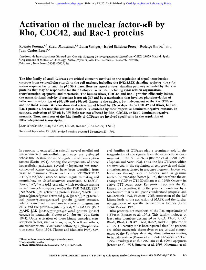
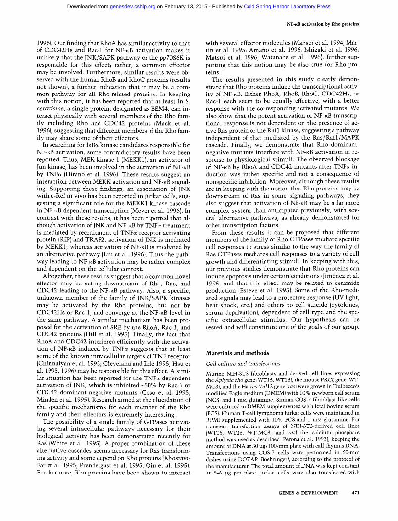
To test whether Rho proteins activate N F - K B , we first used previously characterized NIH-3T3 cells stably transfected with the Aplysia californica rho gene and expressing high levels of the Rho product. To that end, two independent clones overexpressing the wild-type rho gene, WT15 and WT16, were used, which are trans
forming in nude mice (Perona et al. 1993), have potent apoptotic activity after serum removal (Jimenez et al. 1995), and have been demonstrated to trigger ceramide production under these conditions (Esteve et al. 1995). As shown in Figure 1, extracts from both cell lines had a constitutive activation of the NF-KB-dependent (-453/ +80) HIV-luciferase (HIV-LUC) reporter (Devary et al. 1993). This activity was even more pronounced than the one produced by UV exposure of the corresponding control, G418-resistant cells generated by transfection with the empty vector (pZip-Neo). In contrast, cells overexpressing the PKC^ isoenzyme did not present activation of the N F - K B factor, as described previously (Genot et al. 1995; Montaner et al. 1995). Finally, trans-activation of the HIV promoter requires intact NF-KB binding sites because no activation was observed when a HIV-LUC reporter containing 3-bp substitutions in each N F - K B binding site was used (data not shown).
Activation of NF-KB by Rho induces translocation of p65/RelA to the nucleus
N F - K B transcription factor may be composed of homo- or
Cell line
Figure 1. Constitutive activation of NF-KB transcriptional activity by Rho expression. Cells stably overexpressing the rho gene from A. californica (WT15, WT16), or the wild-type PKC^ gene (WT-MC3), or cells transformed by the H-ras-Valll oncogene [las], were analyzed for constitutive NF-KB activation by transfection of the reporter plasmid HIV-LUC. Where indicated, control cells were treated with UV light (pZipNeo+UV) as indicated under Materials and Methods. The results are represented as fold induction above the basal (-453/+80) HIV-LUC expression in the control pZIP-Neo cells, and are the average ± s.D. of a single experiment performed in triplicate. The luciferase values were corrected by cotransfection with the pSV2-CAT plasmid and the stimation of CAT activity as indicated in Materials and Methods. Similar results were obtained in three independent experiments.
464 GENES & DEVELOPMENT
Cold Spring Harbor Laboratory Press on February 13, 2015 - Published by genesdev.cshlp.orgDownloaded from
NF-KB activation by Rho proteins
pZip NEO WT15
p50/p65
p5Q/p50
I I t I ! ■■i | f l | 4P '«• "^
I ill Comp.
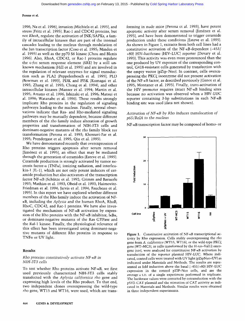
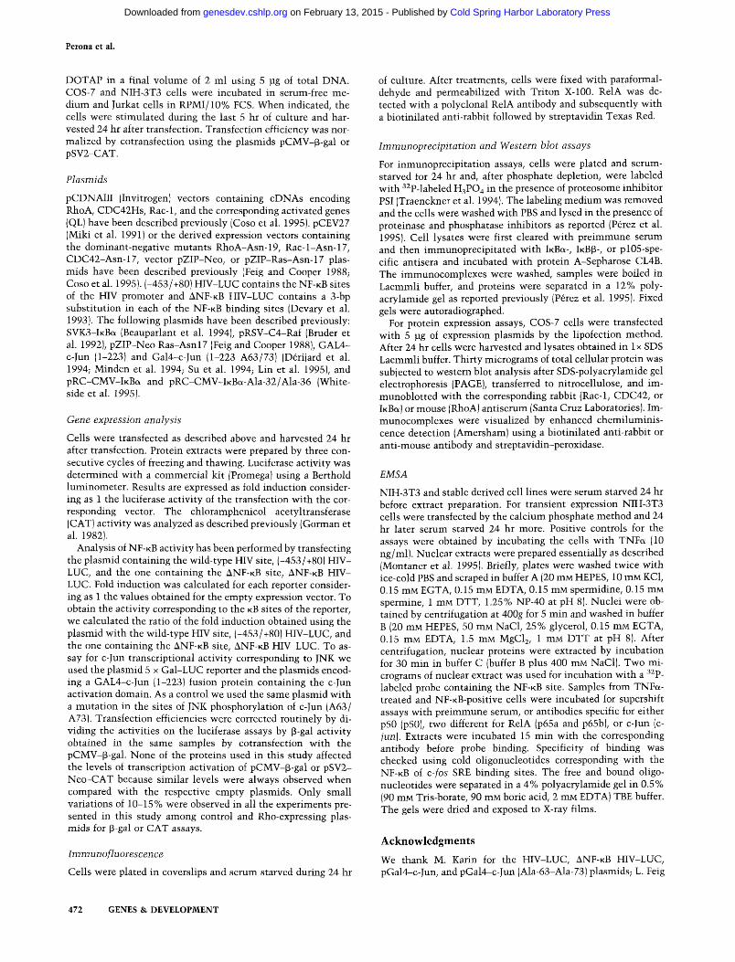
Figure 2. Analysis of NF-KB complexes activated by rho expression. Gel retardation analysis was carried out in the same cell lines as in Fig. 1. The arrows indicate the p50/p50 or p50/ RelA complexes. (-TNF) Control cells; (+TNF) TNFa-treated cells (10 ng/ml). Samples from TNFa-treated cells were incubated with pre-immune serum (p.s.), or antibodies specific for either p50 (p50), RelA (p65a and p65b), or c-Jun (c-jun). (WT-MC3I A mass culture of NIH-3T3 cells overexpressing PKC^, characterized previously (Montaner et al. 1995). The experiment was repeated once more with similar results.
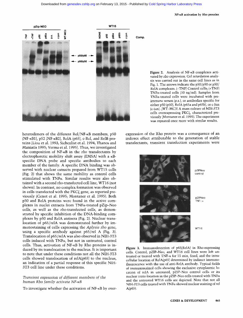
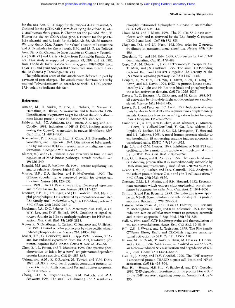
heterodimers of the different Rel/NF-KB members, p50 (NF-KBI) , p52 (NF-KB2), RelA (p65), c-Rel, and RelB proteins (Liou et al. 1993; Siebenlist et al. 1994; Thanos and Maniatis 1995; Verma et al. 1995). Thus, we investigated the composition of NF-KB in the rho transfectants by electrophoretic mobility shift assay (EMSA) with a KB-specific DNA probe and specific antibodies to each member of the family. A specific DNA binding was observed with nuclear extracts prepared from WT15 cells (Fig. 2) that shows the same mobility as control cells stimulated with TNFa. Similar results were also obtained with a second rio-transfected cell line, WT16 (not shown). In contrast, no complex formation was observed in cells transfected with the PKC^ gene, as reported previously (Genot et al. 1995; Montaner et al. 1995). Both p50 and RelA proteins were found in the active complexes in nuclei extracts from TNFa-treated pZip-Neo cells, as well as the riio-transfected cells, as demonstrated by specific inhibition of the DNA-binding complexes by p50 and RelA antisera (Fig. 2). Nuclear translocation of p65/relA was demonstrated further by im-munostaining of cells expressing the Aplysia rho gene, using a specific antibody against p65/rel A (Fig. 3). Translocation of p65/relA was also observed in NIH-3T3 cells induced with TNFa, but not in untreated, control cells. Thus, activation of N F - K B by Rho proteins is induced by its translocation to the nucleus. It is important to note that under these conditions not all the NM-3T3 cells showed translocation of relA(p65) to the nucleus, an indication of a partial response of this specific NIH-3T3 cell line under these conditions.
Transient expression of different members of the human Rho family activate NF-KB
To investigate whether the activation of N F - K B by over-
expression of the Rho protein was a consequence of an indirect effect attributable to the generation of stable transfectants, transient transfection experiments were
pZIPNeo control
pZIPNeo TNF-a
m M' WT16
Figure 3. Immunodetection of p65(RelA) in Rho-expressing cells. Control, pZIP-Neo, and WT16 cell lines were left untreated or treated with TNF-a for 15 min, fixed, and the intracellular location of RelA(p65) determined by indirect immunofluorescence with the use of anti-RelA antibody. Typical fields of immunostained cells showing the exclusive cytoplasmic location of relA in untreated, pZIP-Neo control cells or its nuclear trans-location in the pZIP-Neo cells treated with TNFa and the untreated WT16 cells are depicted. Note that not all NIH-3T3 cells treated with TNFa showed nuclear staining of rel A(p65).
GENES & DEVELOPMENT 465
Cold Spring Harbor Laboratory Press on February 13, 2015 - Published by genesdev.cshlp.orgDownloaded from
Perona et al.
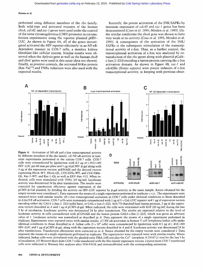
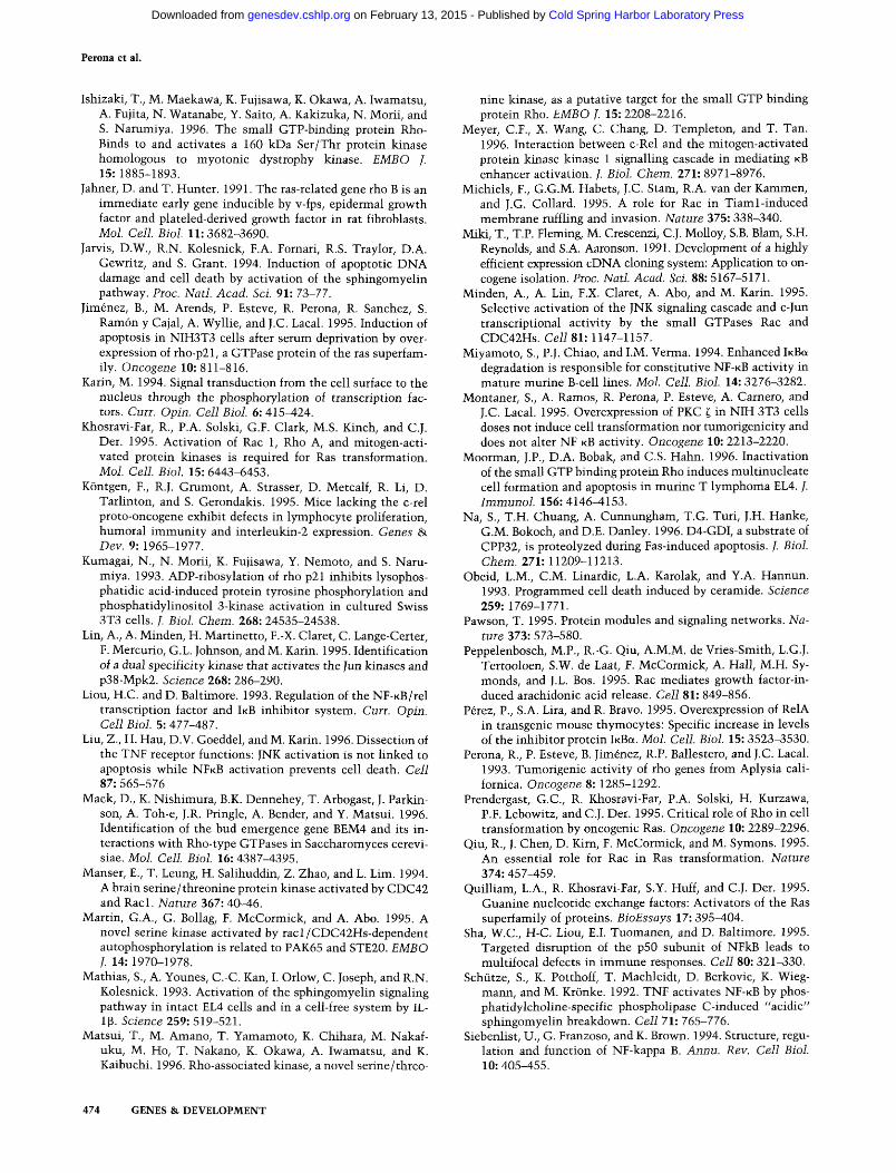
performed using different members of the rho family. Both wild-type and activated mutants of the human rho A, cdc42, and iac-1 genes were used under the control of the same cytomegalovirus (CMV) promoter in cotrans-fection experiments using the reporter plasmid pHIV-LUC. As shown in Figure 4A, all of the genes investigated activated the HIV reporter efficiently in an N F - K B -dependent manner in COS-7 cells, a monkey kidney fibroblast-like cellular system. Similar results were observed when the Aplysia gene as well as the human rhoB and ihoC genes were used in this assay (data not shown). Finally, as positive controls, the activated H-Ras protein (Ras-VaP^) and TNFa induction were also used with the expected results.
Recently, the potent activation of the JNK/SAPKs by transient expression of cdc42 and rac-1 genes has been demonstrated (Coso et al. 1995; Minden et al. 1995). Under similar conditions the rhoA gene was shown to have very weak or no activity (Coso et al. 1995; Minden et al. 1995). A consequence of the activation of the JNK/ SAPKs is the subsequent stimulation of the transcriptional activity of c-Jun. Thus, as a further control, the transcriptional activation of c-Jun was analyzed by co-transfection of the rho genes along with plasmid pGal4-c-Jun (1-223) encoding a fusion protein carrying the c-Jun activation domain. As shown in Figure 4B, rac-1 and cdc42Hs {Homo sapiens) were potent inducers of c-Jun transcriptional activity, in keeping with previous obser-
B NF-KB dependent transcription c-jun transcriptional activation NF-KB dependent transcription
DNA
antibody anti-rhoA anti-rad anti-ccic42
Figure 4. Activation of NF-KB and c-Jun transcriptional activity by different members of the rho family. [A] NF-KB activity in transient experiments performed in the simian COS-7 cells. COS-7 cells were cotransfected by lipofection with 0.5 |ag of (-453/+80) HIV-LUC per 60 mm per plate and 1 ]ug of pCMV-^-gal along with 3 jLig of the expression vectors: pcDNAIII and the derived vectors expressing RhoA-WT, RhoA-QL, CDC42Hs-WT, and CDC42Hs-QL, Rac-l-WT, and Rac-l-QL as well as EXV-Ras-V12. When indicated, cells were stimulated with TNFa (10 ng/ml). Luciferase activity was determined 24 hr after transfection. The results were corrected for transfection efficiency against expression of the pCMV-p-Gal plasmid, by dividing the activity on HIV-LUC reporter by p-gal activity in the same sample. Ratios obtained for the empty vectors were considered 1. Data represent the means of a single experiment performed in triplicate ± S.D.. The experiments were repeated twice with similar results. [B] c-Jun transcriptional activation in COS-7 cells under identical conditions to those described in A for NF-KB activation. COS-7 cells were transiently cotransfected with 1 lag of 5 x Gal-LUC reporter and 1 pg of expression vectors encoding either the GAL4-c-Jun (1-223) (solid bars), or GAL-c-Jun (1-223; A63/73) (hatched bars) fusion proteins, 2 pg of the expression vectors described in A and 1 jug of pCMV-p-gal. When indicated, the cells were stimulated with EGF (20 ng/ml) during the last 5 hr of incubation. Luciferase activity was determined 24 hr after transfection. The results are expressed relative to the level of luciferase activity in cells cotransfected with pCDNAIII and the fusion protein GAL4-c-Jun (1-223), which was given an arbitrary value of 1. Luciferase activity was normalized as described in A. Data represent the means of a single experiment performed in triplicate. Experiments were repeated twice with similar results. (C) NF-KB activation in human T-cell lymphoma Jurkat cells under identical conditions to those described in A: Jurkat cells (5 x 10* cells) were cotransfected by lipofection with 0.5 pg of (-453/+80) HFV-LUC and 1 pg of pCMV-^-gal, along with the expression vectors described in A and B. Luciferase activity was determined 24 hr after transfections. Transfection efficiencies were corrected as in A. Ratios obtained for the empty vectors were considered 1. Data represent the means of a single experiment performed in triplicate. The experiments were repeated twice with similar results. Where indicated, Jurkat cells were stimulated with the phorbol ester PMA (100 nM) plus the Ca^* ionophore A-23187 (1 mM) for the last 5 hr of incubation. [D] Western blots from COS-7 cells transfected with the Rho family expression vectors. Lysates from COS-7 transfected cells were subjected to Western blot analysis after SDS-PAGE and immunoblotted with the corresponding antiserum.
466 GENES & DEVELOPMENT
Cold Spring Harbor Laboratory Press on February 13, 2015 - Published by genesdev.cshlp.orgDownloaded from
N F - K B activation by Rho proteins
vations by other groups (Coso et al. 1995; Minden et al. 1995). A mutated version of the c-Jun fusion protein, carrying a double mutation Ala-63-Ala-73, was not responsive, a strong indication that the observed activation was dependent on c-Jun phosphorylation by the JNK cascade. A much less efficient induction of the c-Jun tr^225-activation was observed with rhoA in this cell line, in agreement with previous reports (Coso et al. 1995; Minden et al. 1995). Finally, as controls, the activated las oncogene and epidermal growth factor (EGF)-treated cells showed activation of the transcription factor that was abrogated by the Ala-63-Ala-73 mutations.
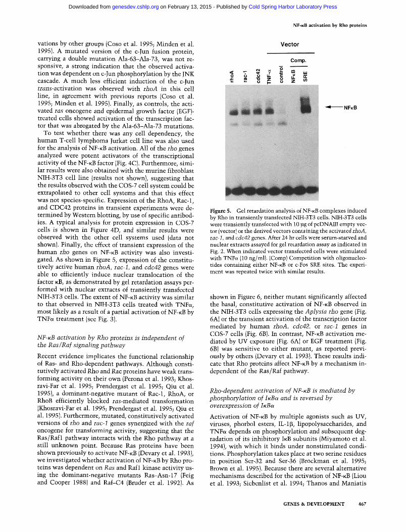
To test whether there was any cell dependency, the human T-cell lymphoma Jurkat cell line was also used for the analysis of N F - K B activation. All of the rho genes analyzed were potent activators of the transcriptional activity of the NF-KB factor (Fig. 4C). Furthermore, similar results were also obtained with the murine fibroblast NIH-3T3 cell line (results not shown), suggesting that the results observed with the COS-7 cell system could be extrapolated to other cell systems and that this effect was not species-specific. Expression of the RhoA, Rac-1, and CDC42 proteins in transient experiments were determined by Western blotting, by use of specific antibodies. A typical analysis for protein expression in COS-7 cells is shown in Figure 4D, and similar results were observed with the other cell systems used (data not shown). Finally, the effect of transient expression of the human rho genes on N F - K B activity was also investigated. As shown in Figure 5, expression of the constitu-tively active human rho A, rac-1, and cdc42 genes were able to efficiently induce nuclear translocation of the factor KB, as demonstrated by gel retardation assays performed with nuclear extracts of transiently transfected NIH-3T3 cells. The extent of N F - K B activity was similar to that observed in NIH-3T3 cells treated with TNFa, most likely as a result of a partial activation of N F - K B by TNFa treatment (see Fig. 3).
NF-KB activation by Rho proteins is independent of the Ras/Raf signaUng pathway
Recent evidence implicates the functional relationship of Ras- and Rho-dependent pathways. Although consti-tutively activated Rho and Rac proteins have weak transforming activity on their own (Perona et al. 1993; Khos-ravi-Far et al. 1995; Prendergast et al. 1995; Qiu et al. 1995), a dominant-negative mutant of Rac-1, RhoA, or RhoB efficiently blocked ras-mediated transformation (Khosravi-Far et al. 1995; Prendergast et al. 1995; Qiu et al. 1995). Furthermore, mutated, constitutively activated versions of rho and rac-1 genes synergized with the raf oncogene for transforming activity, suggesting that the Ras/Raf 1 pathway interacts with the Rho pathway at a still unknown point. Because Ras proteins have been shown previously to activate N F - K B (Devary et al. 1993), we investigated whether activation of N F - K B by Rho proteins was dependent on Ras and Raf 1 kinase activity using the dominant-negative mutants Ras-Asn-17 (Feig and Cooper 1988) and Raf-C4 (Bruder et al. 1992). As
Vector
Comp.
i 11 I H mM^ ^ I NFKB
Figure 5. Gel retardation analysis of NF-KB complexes induced by Rho in transiently transfected NIH-3T3 cells. NIH-3T3 cells were transiently transfected with 10 lag of pcDNAIII empty vector (vector) or the derived vectors containing the activated ihoA, iac-1, and cdc42 genes. After 24 hr cells were serum-starved and nuclear extracts assayed for gel retardation assay as indicated in Fig. 2. When indicated vector transfected cells were stimulated with TNFa (10 ng/ml). (Comp) Competition with oligonucleotides containing either NF-KB or c-Fos SRE sites. The experiment was repeated twice with similar results.
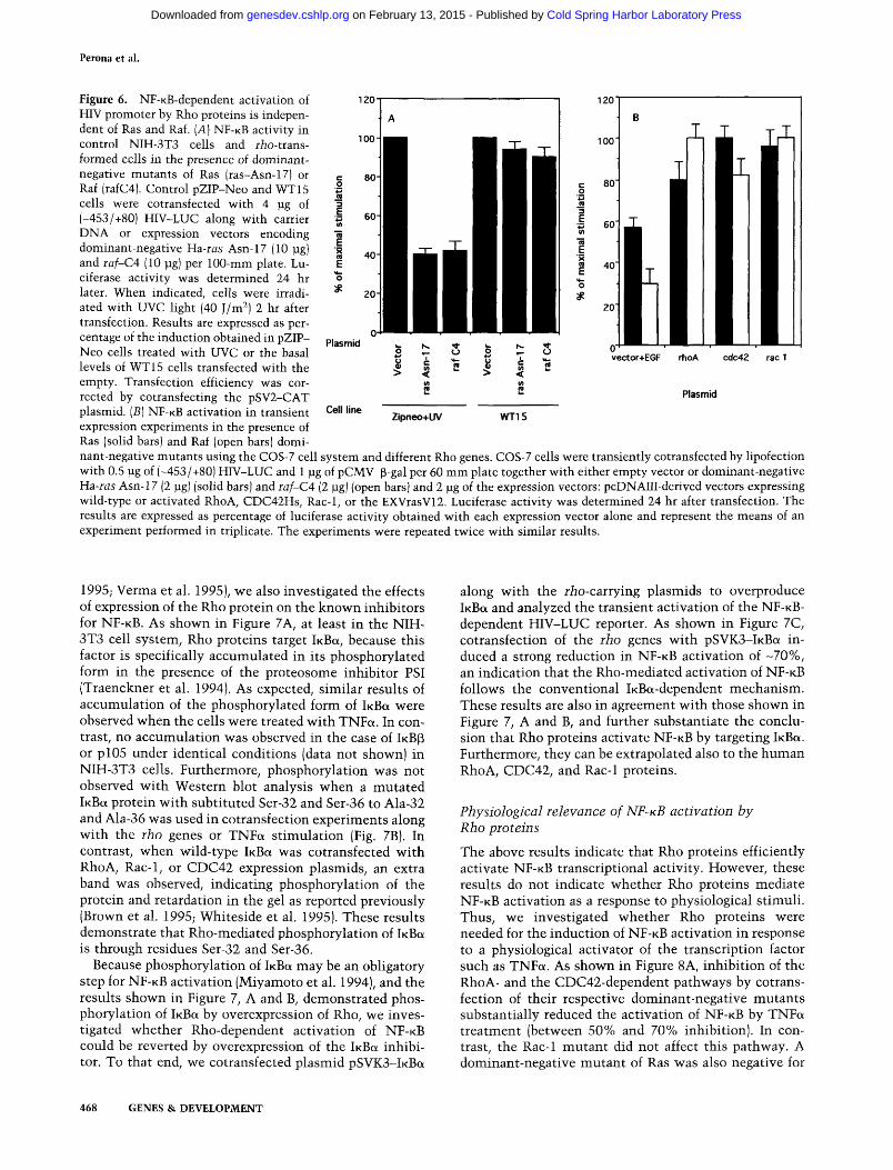
shown in Figure 6, neither mutant significantly affected the basal, constitutive activation of N F - K B observed in the NIH-3T3 cells expressing the Aplysia rho gene (Fig. 6A) or the transient activation of the transcription factor mediated by human rho A, cdc42, or rac-1 genes in COS-7 cells (Fig. 6B). In contrast, N F - K B activation mediated by UV exposure (Fig. 6A) or EGF treatment (Fig. 6B) was sensitive to either mutant, as reported previously by others (Devary et al. 1993). These results indicate that Rho proteins affect N F - K B by a mechanism independent of the Ras/Raf pathway.
Rho-dependent activation of NF-KB is mediated by phosphorylation of iKBa and is reversed by overexpression of iKBa
Activation of N F - K B by multiple agonists such as UV, viruses, phorbol esters, IL-1(B, lipopolysaccharides, and TNFa depends on phosphorylation and subsequent degradation of its inhibitory IKB subunits (Miyamoto et al. 1994), with which it binds under nonstimulated conditions. Phosphorylation takes place at two serine residues in position Ser-32 and Ser-36 (Brockman et al. 1995; Brown et al. 1995). Because there are several alternative mechanisms described for the activation of N F - K B (Liou et al. 1993; Siebenlist et al. 1994; Thanos and Maniatis
GENES & DEVELOPMENT 467
Cold Spring Harbor Laboratory Press on February 13, 2015 - Published by genesdev.cshlp.orgDownloaded from
Perona et al.
120
100-
120
100
Figure 6. NF-KB-dependent activation of HIV promoter by Rho proteins is independent of Ras and Raf. [A] NF-KB activity in control NIH-3T3 cells and rio-trans-formed cells in the presence of dominant-negative mutants of Ras (ras-Asn-17) or Raf (rafC4). Control pZIP-Neo and WT15 cells were cotransfected w ith 4 jig of (-453/+80) HIV-LUC along with carrier DNA or expression vectors encoding dominant-negative Ha-ras Asn-17 (10 ]ig] and raf-C4 (10 pg) per 100-mm plate. Lu-ciferase activity was determined 24 hr later. When indicated, cells were irradiated with UVC light (40 J/m^) 2 hr after transfection. Results are expressed as percentage of the induction obtained in pZIP-Neo cells treated with UVC or the basal levels of WT15 cells transfected with the empty. Transfection efficiency was corrected by cotransfecting the pSV2-CAT plasmid. [B] NF-KB activation in transient expression experiments in the presence of Ras (solid bars) and Raf (open bars) dominant-negative mutants using the COS-7 cell system and different Rho genes. COS-7 cells were transiently cotransfected by lipofection with 0.5 pg of (-453/-(-80) HFV-LUC and 1 jug of pCMV-p-gal per 60 mm plate together with either empty vector or dominant-negative Ha-ras Asn-17 (2 ]ig] (solid bars) and laf-CA (2 pg) (open bars) and 2 \xg of the expression vectors: pcDNAIII-derived vectors expressing wild-type or activated RhoA, CDC42Hs, Rac-1, or the EXVrasV12. Luciferase activity was determined 24 hr after transfection. The results are expressed as percentage of luciferase activity obtained with each expression vector alone and represent the means of an experiment performed in triplicate. The experiments were repeated twice with similar results.
Plasmid
Cell line
o :i 1 s > <
VI IB
Zipneo+UV
* u iS
O
>
N.
C V)
< (/I
WTIS
* o S
vector+E6F cdc42 rac 1
Plasmid
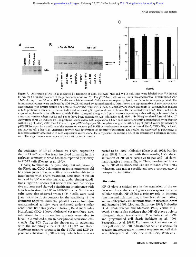
1995; Verma et al. 1995), we also investigated the effects of expression of the Rho protein on the know^n inhibitors for N F - K B . A S shown in Figure 7A, at least in the NIH-3T3 cell system, Rho proteins target iKBa, because this factor is specifically accumulated in its phosphorylated form in the presence of the proteosome inhibitor PSI (Traenckner et al. 1994). As expected, similar results of accumulation of the phosphorylated form of iKBa were observed when the cells were treated with TNFa. In contrast, no accumulation was observed in the case of IKB^ or pl05 under identical conditions (data not shown) in NIH-3T3 cells. Furthermore, phosphorylation was not observed with Western blot analysis when a mutated iKBct protein with subtituted Ser-32 and Ser-36 to Ala-32 and Ala-36 was used in cotransfection experiments along with the iho genes or TNFa stimulation (Fig. 7B). In contrast, when wild-type iKBa was cotransfected with RhoA, Rac-1, or CDC42 expression plasmids, an extra band was observed, indicating phosphorylation of the protein and retardation in the gel as reported previously (Brown et al. 1995; Whiteside et al. 1995). These results demonstrate that Rho-mediated phosphorylation of IKBU is through residues Ser-32 and Ser-36.
Because phosphorylation of IKBU may be an obligatory step for N F - K B activation (Miyamoto et al. 1994), and the results shown in Figure 7, A and B, demonstrated phosphorylation of iKBa by overexpression of Rho, we investigated whether Rho-dependent activation of N F - K B could be reverted by overexpression of the iKBa inhibitor. To that end, we cotransfected plasmid pSVK3-lKBa
along with the r/io-carrying plasmids to overproduce iKBa and analyzed the transient activation of the NF-KB-dependent HIV-LUC reporter. As shown in Figure 7C, cotransfection of the iho genes with pSVK3-lKBa induced a strong reduction in N F - K B activation of -70%, an indication that the Rho-mediated activation of N F - K B follows the conventional iKBa-dependent mechanism. These results are also in agreement with those shown in Figure 7, A and B, and further substantiate the conclusion that Rho proteins activate N F - K B by targeting iKBa. Furthermore, they can be extrapolated also to the human RhoA, CDC42, and Rac-1 proteins.
Physiological relevance of NF-KB activation by Rho proteins
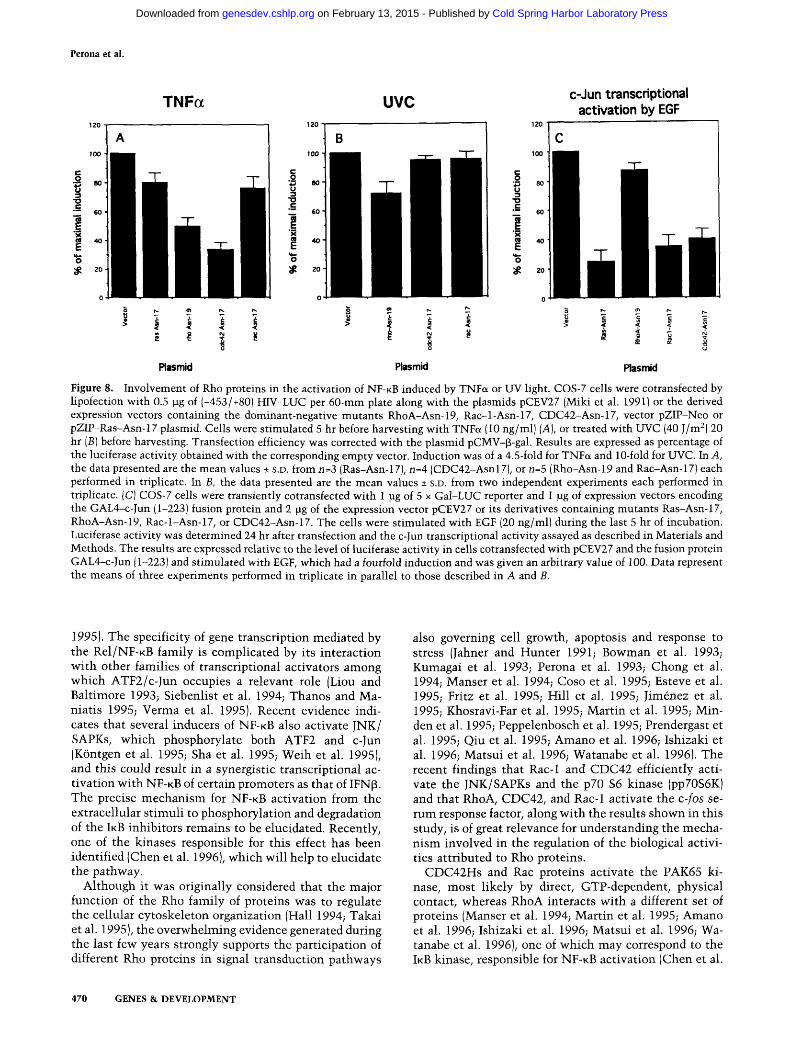
The above results indicate that Rho proteins efficiently activate NF-KB transcriptional activity. However, these results do not indicate whether Rho proteins mediate N F - K B activation as a response to physiological stimuli. Thus, we investigated whether Rho proteins were needed for the induction of N F - K B activation in response to a physiological activator of the transcription factor such as TNFa. As shown in Figure 8A, inhibition of the RhoA- and the CDC42-dependent pathways by cotransfection of their respective dominant-negative mutants substantially reduced the activation of N F - K B by TNFa treatment (between 50% and 70% inhibition). In contrast, the Rac-1 mutant did not affect this pathway. A dominant-negative mutant of Ras was also negative for
468 GENES & DEVELOPMENT
Cold Spring Harbor Laboratory Press on February 13, 2015 - Published by genesdev.cshlp.orgDownloaded from
NF-KB activation by Rho proteins
B pZIP-neo
Jl III
lA
m hcBa
k B a ala 32/36
w *
8
> t O
>
MW ft .« " V
- J _ J - 1
o a o < - 5! o 2 o »- *- o
f
1
kBaWT
a u. z »-+ o
r.--,!r
< 1
mm
o u cB -a »- o rho A cdc42 rac ras V I2
Plasmid
Figure 7. Activation of NF-KB is mediated by targeting of iKBa. [A] pZIP-Neo and WT15 cell lines were labeled with ^^P-labeled H3PO4 for 4 hr in the presence of the proteosome inhibitor PSI. The pZIP-Neo cells were either untreated (control) or stimulated with TNFa during 10 or 30 min. WT15 cells were left untreated. Cells were subsequently lysed, and iKBa immunoprecipitated. The immunoprecipitates were analyzed by SDS-PAGE followed by autoradiography. Data shown are representative of two independent experiments with similar results. For simplicity, only the results with the iKBa antibody are shown (see text). [B] Western blot analysis of iKBa proteins in transiently transfacted COS-7 cells using 30 pg of total protein from cells transfected with RhoA, Rac-1, or CDC42 expression plasmids or in cells treated with TNFa (10 ng/ml) along with 2 ]ig of vectors expressing either wild-type human iKBa or a mutated version where Ser-32 and Ser-36 have been changed to Ala (Whiteside et al. 1995). (•) Phosphorylated form of IKEK. (C) Activation of NF-KB induced by Rho proteins is blocked by IKBU expression. COS-7 cells were transiently cotransfected by lipofection with 0.5 jjg of (-453/+80) FilV-LUC and 1 ]ig of pCMV-^-gal per 60-mm plate along with either 2 ]ig of pSVK3 vector (sohd bars) or pSVKSIKBa (open bars) and 2 jig of the expression vectors: pcDNAIII-derived vectors expressing activated RhoA, CDC42Hs, or Rac-1, and EXVrasVall2 (rasV12). Luciferase activity was determined 24 hr after transfection. The results are expressed as percentage of luciferase activity obtained with each expression vector alone. Data represent the means ± s.D. of an experiment performed in triplicate. The experiments were repeated twice with similar results.
the activation of N F - K B induced by TNFa, suggesting that in COS-7 cells, Ras is not involved primarily in this pathway, contrary to what has been reported previously in PC-12 cells (Devary et al. 1993).
Finally, to eliminate the possibility that inhibition by the RhoA and CDC42 dominant-negative mutants could be a consequence of nonspecific effects attributable to its interference with TNFa treatment, activation of N F - K B induced by UV was also analyzed under similar conditions. Figure 8B shows that none of the dominant-negative mutants used showed a significant interference with NF-KB activation by UV in NIH-3T3 cells. Similar results were also observed when COS-/ cells were used (data not shown). As controls for the specificity of the dominant-negative mutants, parallel assays for c-Jun transcriptional activity were performed under similar conditions. Both Ras (75% inhibition), Rac-1 (65% inhibition), and CDC42 (60% inhibition) but not RhoA (13% inhibition) dominant-negative mutants were able to block EGF-induced c-Jun transcriptional activation efficiently (Fig. 8C). The results shown are in agreement with the inhibitory effects of the CDC42 and Rac-1 dominant-negative mutants in the TNFa- and EGF-de-pendent activation of JNK activity, which has been re
ported to be -50% inhibition (Coso et al. 1995; Minden et al. 1995). In contrast with these results, UV-induced activation of N F - K B is sensitive to Ras and Raf dominant-negative mutants (Fig. 6). Thus, the observed blockage of N F - K B by RhoA and CDC42 mutants after TNFa induction was rather specific and not a consequence of nonspecific inhibition.
Discussion
N F - K B plays a critical role in the regulation of the expression of specific sets of genes as a response to extracellular signals. NF-KB has a primary role in immune function and inflammation, in lymphoid differentiation, and in embryonic axis determination in insects (Grimm and Baeuerle 1993; Liou and Baltimore 1993; Siebenlist et al. 1994; Thanos and Maniatis 1995; Verma et al. 1995). There is also evidence that N F - K B plays a role in mitogenic signal transduction (Miyamoto et al. 1994) and programmed cell death (Baldwin et al. 1991; Beauparlant et al. 1994). Furthermore, knockouts of either p50, c-rel, or RelB indicate that N F - K B is critical for specific and nonspecific immune response and cell division (Kontgen et al. 1995; Sha et al. 1995; Weih et al.
GENES & DEVELOPMENT 469
Cold Spring Harbor Laboratory Press on February 13, 2015 - Published by genesdev.cshlp.orgDownloaded from
Perona et al.
TNFa UVC c-Jun transcriptional activation by EGF
•3 80-
#
_ 60-
SS 4 0 -
# 20
■^ 80
^ 20
Piasmid Plasmid Piasmid
Figure 8. Involvement of Rho proteins in the activation of NF-KB induced by TNFa or UV light. COS-7 cells were cotransfected by lipofection with 0.5 pg of (-453/+80! HIV-LUC per 60-mni plate along with the plasmids pCEV27 (Miki et al. 1991) or the derived expression vectors containing the dominant-negative mutants RhoA-Asn-19, Rac-l-Asn-17, CDC42-Asn-17, vector pZIP-Neo or pZIP-Ras-Asn-17 piasmid. Cells were stimulated 5 hr before harvesting with TNFa (10 ng/ml) [A], or treated with UVC (40 J/m^) 20 hr [B] before harvesting. Transfection efficiency was corrected with the piasmid pCMV-p-gal. Results are expressed as percentage of the luciferase activity obtained with the corresponding empty vector. Induction was of a 4.5-fold for TNFa and 10-fold for UVC. In A, the data presented are the mean values + S.D. from n=3 (Ras-Asn-17), n=4 (CDC42-Asnl7), or n=5 (Rho-Asn-19 and Rac-Asn-17) each performed in triplicate. In B, the data presented are the mean values ± S.D. from two independent experiments each performed in triplicate. (C) COS-7 cells were transiently cotransfected with 1 pg of 5 x Gal-LUC reporter and 1 ]ig of expression vectors encoding the GAL4-c-Jun (1-223) fusion protein and 2 pg of the expression vector pCEV27 or its derivatives containing mutants Ras-Asn-17, RhoA-Asn-19, Rac-l-Asn-17, or CDC42-Asn-17. The cells were stimulated with EGF (20 ng/ml) during the last 5 hr of incubation. Luciferase activity was determined 24 hr after transfection and the c-Jun transcriptional activity assayed as described in Materials and Methods. The results are expressed relative to the level of luciferase activity in cells cotransfected with pCEV27 and the fusion protein GAL4-c-Jun (1-223) and stimulated with EGF, which had a fourfold induction and was given an arbitrary value of 100. Data represent the means of three experiments performed in triplicate in parallel to those described in A and B.
1995). The specificity of gene transcription mediated by the R C I / N F - K B family is complicated by its interaction w^ith other families of transcriptional activators among which ATF2/c-Jun occupies a relevant role (Liou and Baltimore 1993; SiebenUst et al. 1994; Thanos and Ma-niatis 1995; Verma et al. 1995). Recent evidence indicates that several inducers of N F - K B also activate JNK/ SAPKs, which phosphorylate both ATF2 and c-Jun (Kontgen et al. 1995; Sha et al. 1995; Weih et al. 1995), and this could result in a synergistic transcriptional activation with N F - K B of certain promoters as that of IFN(3. The precise mechanism for NF-KB activation from the extracellular stimuli to phosphorylation and degradation of the IKB inhibitors remains to be elucidated. Recently, one of the kinases responsible for this effect has been identified (Chen et al. 1996), which will help to elucidate the pathway.
Although it was originally considered that the major function of the Rho family of proteins was to regulate the cellular cytoskeleton organization (Hall 1994; Takai et al. 1995), the overwhelming evidence generated during the last few years strongly supports the participation of different Rho proteins in signal transduction pathways
also governing cell growth, apoptosis and response to stress (Jahner and Hunter 1991; Bowman et al. 1993; Kumagai et al. 1993; Perona et al. 1993; Chong et al. 1994; Manser et al. 1994; Coso et al. 1995; Esteve et al. 1995; Fritz et al. 1995; Hill et al. 1995; Jimenez et al. 1995; Khosravi-Far et al. 1995; Martin et al. 1995; Min-den et al. 1995; Peppelenbosch et al. 1995; Prendergast et al. 1995; Qiu et al. 1995; Amano et al. 1996; Ishizaki et al. 1996; Matsui et al. 1996; Watanabe et al. 1996). The recent findings that Rac-1 and CDC42 efficiently activate the JNK/SAPKs and the p70 S6 kinase (pp70S6K) and that RhoA, CDC42, and Rac-1 activate the c-fos serum response factor, along with the results shown in this study, is of great relevance for understanding the mechanism involved in the regulation of the biological activities attributed to Rho proteins.
CDC42HS and Rac proteins activate the PAK65 kinase, most likely by direct, GTP-dependent, physical contact, whereas RhoA interacts with a different set of proteins (Manser et al. 1994; Martin et al. 1995; Amano et al. 1996; Ishizaki et al. 1996; Matsui et al. 1996; Watanabe et al. 1996), one of which may correspond to the IKB kinase, responsible for N F - K B activation (Chen et al.
470 GENES & DEVELOPMENT
Cold Spring Harbor Laboratory Press on February 13, 2015 - Published by genesdev.cshlp.orgDownloaded from
NF-KB activation by Rho proteins
1996). Our finding that Rho A has similar activity to that of CDC42HS and Rac-1 for NF-KB activation makes it unlikely that the JNK/SAPK pathway or the pp70S6K is responsible for this effect; rather, a common effector may be involved. Furthermore, similar results were observed with the human RhoB and RhoC proteins (results not shown), a further indication that it may be a common pathway for all Rho-related proteins. In keeping with this notion, it has been reported that at least in S. ceievisiae, a single protein, designated as BEM4, can interact physically with several members of the Rho family including Rho and CDC42 proteins (Mack et al. 1996), suggesting that different members of the Rho family may share some of their effectors.
In searching for iKBa kinase candidates responsible for NF-KB activation, some contradictory results have been reported. Thus, MEK kinase 1 (MEKKl), an activator of Jun kinase, has been involved in the activation of N F - K B by TNFa (Hirano et al. 1996). These results suggest an interaction between MEKK activation and N F - K B signaling. Supporting these findings, an association of JNK with c-Rel in vivo has been reported in Jurkat cells, suggesting a significant role for the MEKKl kinase cascade in NF-KB-dependent transcription (Meyer et al. 1996). In contrast with these results, it has been reported that although activation of JNK and NF-KB by TNFa treatment is mediated by recruitment of TNFa receptor activating protein (RIP) and TRAF2, activation of JNK is mediated by MEKKl, whereas activation of N F - K B is mediated by an alternative pathway (Liu et al. 1996). Thus the pathway leading to NF-KB activation may be rather complex and dependent on the cellular context.
Altogether, these results suggest that a common novel effector may be acting downstream of Rho, Rac, and CDC42 leading to the N F - K B pathway. Also, a specific, unknown member of the family of JNK/SAPK kinases may be activated by the Rho proteins, but not by CDC42Hs or Rac-1, and converge at the NF-KB level in the same pathway. A similar mechanism has been proposed for the activation of SRE by the RhoA, Rac-1, and CDC42 proteins (Hill et al. 1995). Finally, the fact that RhoA and CDC42 interfered efficiently with the activation of NF-KB induced by TNFa suggests that at least some of the known intracellular targets of TNF receptor (Chinnaiyan et al. 1995; Cleveland and Ihle 1995; Hsu et al. 1995, 1996) may be responsible for this effect. A similar situation has been reported for the TNFa-dependent activation of JNK, which is inhibited -50% by Rac-1 or CDC42 dominant-negative mutants (Coso et al. 1995; Minden et al. 1995). Research aimed at the elucidation of the specific mechanisms for each member of the Rho family and their effectors is extremely interesting.
The possibility of a single family of GTPases activating several intracellular pathways necessary for their biological activity has been demonstrated recently for Ras (White et al. 1995). A proper combination of these alternative cascades seems necessary for Ras transforming activity and some depend on Rho proteins (Khosravi-Far et al. 1995; Prendergast et al. 1995; Qiu et al. 1995). Furthermore, Rho proteins have been shown to interact
with several effector molecules (Manser et al. 1994; Martin et al. 1995; Amano et al. 1996; Ishizaki et al. 1996; Matsui et al. 1996; Watanabe et al. 1996), further supporting that this notion may be also true for Rho proteins.
The results presented in this study clearly demonstrate that Rho proteins induce the transcriptional activity of N F - K B . Either RhoA, RhoB, RhoC, CDC42Hs, or Rac-1 each seem to be equally effective, with a better response with the corresponding activated mutants. We also show that the potent activation of N F - K B transcriptional response is not dependent on the presence of active Ras protein or the Raf 1 kinase, suggesting a pathway independent of that mediated by the Ras/Rafl/MAPK cascade. Finally, we demonstrate that Rho dominant-negative mutants interfere with NF-KB activation in response to physiological stimuli. The observed blockage of NF-KB by RhoA and CDC42 mutants after TNFa induction was rather specific and not a consequence of nonspecific inhibition. Moreover, although these results are in keeping with the notion that Rho proteins may be downstream of Ras in some signaling pathways, they also suggest that activation of NF-KB may be a far more complex system than anticipated previously, with several alternative pathways, as already demonstrated for other transcription factors.
From these results it can be proposed that different members of the family of Rho GTPases mediate specific cell responses to stress similar to the way the family of Ras GTPases mediates cell responses to a variety of cell growth and differentiating stimuli. In keeping with this, our previous studies demonstrate that Rho proteins can induce apoptosis under certain conditions (Jimenez et al. 1995) and that this effect may be related to ceramide production (Esteve et al. 1995). Some of the Rho-medi-ated signals may lead to a protective response (UV light, heat shock, etc.) and others to cell suicide (cytokines, serum deprivation), dependent of cell type and the specific extracellular stimulus. Our hypothesis can be tested and will constitute one of the goals of our group.
Materials and methods
Cell culture and transfections Murine NIH-3T3 fibroblasts and derived cell lines expressing the Aplysia rho gene (WT15, WT16), the mouse PKC^ gene (WT-MC3), and the Ha-ras Valll gene [las] were grown in Dulbecco's modified Eagle medium (DMEM) with 10% newborn calf serum (NCS) and 1 mM glutamine. Simian COS-7 fibroblast-like cells were cultured in DMEM supplemented with fetal bovine serum (PCS). Human T-cell lymphoma Jurkat cells were maintained in RPMI supplemented with 10% PCS and 1 mM glutamine. For transient transfection assays of NIH-3T3-derived cell lines (WT15, WT16, WT-MC3, and ras] the calcium phosphate method was used as described (Perona et al. 1993), keeping the amount of DNA at 30 jag/ 100-mm plate with calf thymus DNA. Transfections using COS-7 cells were performed in 60-mm dishes using DOTAP (Boehringer), according to the protocol of the manufacturer. The total amount of DNA was kept constant at 5-6 iig per plate. Jurkat cells were also transfected with
GENES & DEVELOPMENT 471
Cold Spring Harbor Laboratory Press on February 13, 2015 - Published by genesdev.cshlp.orgDownloaded from
Perona et al.
DOTAP in a final volume of 2 ml using 5 jig of total DNA. COS-7 and NIH-3T3 cells were incubated in serum-free medium and Jurkat cells in RPMI/10% PCS. When indicated, the cells were stimulated during the last 5 hr of culture and harvested 24 hr after transfection. Transfection efficiency was normalized by cotransfection using the plasmids pCMV-p-gal or pSV2-CAT.
Plasmids
pCDNAIII (Invitrogen) vectors containing cDNAs encoding RhoA, CDC42Hs, Rac-1, and the corresponding activated genes (QL) have been described previously (Coso et al. 1995). pCEV27 (Miki et al. 1991) or the derived expression vectors containing the dominant-negative mutants RhoA-Asn-19, Rac-l-Asn-17, CDC42-Asn-17, vector pZIP-Neo, or pZIP-Ras-Asn-17 plasmids have been described previously (Feig and Cooper 1988; Coso et al. 1995). (-453/+80) HIV-LUC contains the NF-KB sites of the HIV promoter and A N F - K B HIV-LUC contains a 3-bp substitution in each of the N F - K B binding sites (Devary et al. 1993). The following plasmids have been described previously: SVK3-lKBa (Beauparlant et al. 1994), pRSV-C4-Raf (Bruder et al. 1992), pZIP-Neo Ras-Asnl7 (Feig and Cooper 1988), GAL4-c-Jun (1-223) and Gal4-c-Jun (1-223 A63/73) (Derijard et al. 1994; Minden et al. 1994; Su et al. 1994; Lin et al. 1995), and pRC-CMV-lKBa and pRC-CMV-lKBa-Ala-32/Ala-36 (Whiteside et al. 1995).
Gene expression analysis
Cells were transfected as described above and harvested 24 hr after transfection. Protein extracts were prepared by three consecutive cycles of freezing and thawing. Luciferase activity was determined with a commercial kit (Promega) using a Berthold luminometer. Results are expressed as fold induction considering as 1 the luciferase activity of the transfection with the corresponding vector. The chloramphenicol acetyltransferase (CAT) activity was analyzed as described previously (Gorman et al. 1982).
Analysis of N F - K B activity has been performed by transfecting the plasmid containing the wild-type HIV site, (-453/+80) HIV-LUC, and the one containing the ANF-KB site, ANF-KB HIV-LUC. Fold induction was calculated for each reporter considering as 1 the values obtained for the empty expression vector. To obtain the activity corresponding to the KB sites of the reporter, we calculated the ratio of the fold induction obtained using the plasmid with the wild-type HIV site, (-453/+80) HFV-LUC, and the one containing the ANF-KB site, ANF-KB H F V - L U C . To assay for c-Jun transcriptional activity corresponding to JNK we used the plasmid 5 x Gal-LUC reporter and the plasmids encoding a GAL4-c-Jun (1-223) fusion protein containing the c-Jun activation domain. As a control we used the same plasmid with a mutation in the sites of JNK phosphorylation of c-Jun (A63/ A73). Transfection efficiencies were corrected routinely by dividing the activities on the luciferase assays by 3-gal activity obtained in the same samples by cotransfection with the pCMV-3-gal. None of the proteins used in this study affected the levels of transcription activation of pCMV-^-gal or pSV2-Neo-CAT because similar levels were always observed when compared with the respective empty plasmids. Only small variations of 10-15% were observed in all the experiments presented in this study among control and Rho-expressing plasmids for p-gal or CAT assays.
Immunofluorescence
Cells were plated in coverslips and serum starved during 24 hr
of culture. After treatments, cells were fixed with paraformaldehyde and permeabilized with Triton X-100. RelA was detected with a polyclonal RelA antibody and subsequently with a biotinilated anti-rabbit followed by streptavidin Texas Red.
Immunoprecipitation and Western blot assays
For inmunoprecipitation assays, cells were plated and serum-starved for 24 hr and, after phosphate depletion, were labeled with ^^P-labeled H3PO4 in the presence of proteosome inhibitor PSI (Traenckner et al. 1994). The labeling medium was removed and the cells were washed with PBS and lysed in the presence of proteinase and phosphatase inhibitors as reported (Perez et al. 1995). Cell lysates were first cleared with preimmune serum and then immunoprecipitated with IKBK-, IKB^-, or pl05-spe-cific antisera and incubated with protein A-Sepharose CL4B. The immunocomplexes were washed, samples were boiled in Laemmli buffer, and proteins were separated in a 12% poly-acrylamide gel as reported previously (Perez et al. 1995). Fixed gels were autoradiographed.
For protein expression assays, COS-7 cells were transfected with 5 ]ig of expression plasmids by the lipofection method. After 24 hr cells were harvested and lysates obtained in 1 x SDS Laemmli buffer. Thirty micrograms of total cellular protein was subjected to western blot analysis after SDS-polyacrylamide gel electrophoresis (PAGE), transferred to nitrocellulose, and im-munoblotted with the corresponding rabbit (Rac-1, CDC42, or IKBU) or mouse (RhoA) antiserum (Santa Cruz Laboratories). Immunocomplexes were visualized by enhanced chemiluminis-cence detection (Amersham) using a biotinilated anti-rabbit or anti-mouse antibody and streptavidin-peroxidase.
EMSA
NIH-3T3 and stable derived cell lines were serum starved 24 hr before extract preparation. For transient expression NIH-3T3 cells were transfected by the calcium phosphate method and 24 hr later serum starved 24 hr more. Positive controls for the assays were obtained by incubating the cells with TNFa (10 ng/ml). Nuclear extracts were prepared essentially as described (Montaner et al. 1995). Briefly, plates were washed twice with ice-cold PBS and scraped in buffer A (20 mM HEPES, 10 mM KCl, 0.15 mM EGTA, 0.15 mM EDTA, 0.15 mM spermidine, 0.15 mM spermine, 1 mM DTT, 1.25% NP-40 at pH 8). Nuclei were obtained by centrifugation at 400g for 5 min and washed in buffer B (20 mM HEPES, 50 mM NaCl, 25% glycerol, 0.15 mM EGTA, 0.15 mM EDTA, 1.5 mM MgClj, 1 mM DTT at pH 8). After centrifugation, nuclear proteins were extracted by incubation for 30 min in buffer C (buffer B plus 400 mM NaCl). Two micrograms of nuclear extract was used for incubation with a ' ^P-labeled probe containing the N F - K B site. Samples from TNFa-treated and NF-KB-positive cells were incubated for supershift assays with preimmune serum, or antibodies specific for either p50 (p50), two different for RelA (p65a and p65b), or c-Jun (c-iun). Extracts were incubated 15 min with the corresponding antibody before probe binding. Specificity of binding was checked using cold oligonucleotides corresponding with the NF-KB of c-/os SRE binding sites. The free and bound oligonucleotides were separated in a 4% polyacrylamide gel in 0.5% (90 mM Tris-borate, 90 mM boric acid, 2 mM EDTA) TBE buffer. The gels were dried and exposed to X-ray films.
Acknowledgments
We thank M. Karin for the HIV-LUC, ANF-KB HIV-LUC, pGal4-c-Jun, and pGal4-c-Jun (Ala-63-Ala-73) plasmids; L. Feig
472 GENES & DEVELOPMENT
Cold Spring Harbor Laboratory Press on February 13, 2015 - Published by genesdev.cshlp.orgDownloaded from
NF-KB activation by Rho proteins
for the Ras-Asn-17; U. Rapp for the pRSV-C4-Raf plasmid; S. Gutkind for the pCDNAIII plasmids carrying the cdc42 Hs, rac-1, and human rhoA geneS; P. Chardin for the pGEM3-rioB; T. Hunter for the rat cDNA rhoB genc; J. Hiscott for the pSVK-iKBa plasmid, and A. Israel for the iKBa Ala-32/Ala-36 mutant . We also thank M.A. Ramos for valuable technical assistance and A. Fernandez for the art work. S.M. and I.S.-P. are Fellows from Dirrecion General de Investigacion en Ciencia y Tecnolo-gia (DGICYT) and L.S. is a fellow from Fundacion Ramon Are-ces. This study is supported by grants 93/0293 and 95/0882 from Fondo de Investigacion Sanitaria, grant PB94-0009 from DGICYT, and grant SAF/93-0142 from Comision Interministe-rial de Ciencia y Tecnologia(CICYT).
The publication costs of this article were defrayed in part by payment of page charges. This article must therefore be hereby marked "advertisement" in accordance with 18 USC section 1734 solely to indicate this fact.
References
Amano, M., H. Mukai, Y. Ono, K. Chihara, T. Matsui, Y. Hamajima, K. Okawa, A. Iwamatsu, and K. Kaibuchi. 1996. Identification of a putative target for Rho as the serine-threonine kinase protein kinase N. Science 271: 648-650
Baldwin, A.S., J.C. Azizkhan, D.E. Jensen, A.A. Beg, and L.R. Goodly. 1991. Induction of NF-KB DNA-binding activity during the Go-to-Gi transtition in mouse fibroblasts. Mol. Cell. Biol. 11:4943-4951.
Beauparlant, P., I. Kwan, R. Bitar, P. Chou, A.E. Koromilas, N. Sonenberg, and J. Hiscott. 1994. Disruption of IKBU regulation by antisense RNA expression leads to malignant transformation. Oncogene 9: 3189-3197.
Blumer, K.J. and G.L. Johnson. 1994. Diversity in function and regulation of MAP kinase pathways. Trends Biochem. Sci. 19: 236-240.
Boguski, M.S. and F. McCormick. 1993. Proteins regulating Ras and its relatives. Nature 366: 643-654.
Bourne, H.R., D.A. Sanders, and F. McCormick. 1990. The GTPase superfamily: A conserved switch for diverse cell function. Nature 348: 125-132.
. 1991. The GTPase superfamily: Conserved structure and molecular mechanism. Nature 349: 117-127.
Bowman, E.P., D.J. Uhlinger, and J.D. Lambeth. 1993. Neutrophil phospholipase D is activated by a membrane-associated Rho family small molecular weight GTP-binding protein. /. Biol. Chem. 268: 21509-21512.
Brockman, J.A., D.C. Scherer, T.A. McKinsey, S.M. Hall, X. Qi, W.Y. Lee, and D.W. Ballard. 1995. Coupling of signal response domain in iKBa to multiple pathways for N F K B activation. Mol. Cell. Biol. 15: 2809-2818.
Brown, K., S. Gerstberger, L. Carlson, G. Franzoso, and U. Sieben-list. 1995. Control of iKB-a proteolysis by site-specific, signal-induced phosphorylation. Science 267: 1485-1488.
Bruder, T.B., G. Heidecker, and U. Rapp. 1992. Serum-, TPA-, and Ras-induced expression from the APl/Ets-driven promoters requires Raf-1 kinase. Genes &. Dev. 6: 545-556.
Chen, Z.J., L. Parent, and T. Maniatis. 1996. Site-specific phosphorylation of iKB-a by a novel ubiquitination-dependent protein kinase activity. Cell 84: 853-862.
Chinnaiyan, A.M., K. O'Rourke, M. Tewari, and V.M. Dixit. 1995. FADD, a novel death domain-containing protein, interacts with the death domain of Fas and initiates apoptosis. Ceil 81: 505-512.
Chong, L.D., A. Traynor-Kaplan, G.M. Bokoch, and M.A. Schwartz. 1994. The small GTP-binding Rho A regulates a
phosphatidylinositol 4-phosphate 5-kinase in mammalian cells. Celi 79: 507-513.
Chou, M.M. and J. Blenis. 1996. The 70 kDa S6 kinase complexes with and is activated by the Rho family G proteins CDC42 and Racl. Cell 85: 573-583.
Clapham, D.E. and E.J. Neer. 1993. New roles for G-protein P7-dimers in transmembrane signalling. Nature 365: 403 -406.
Cleveland, J.L. and J.N. Ihle. 1995. Contenders in FasL/TNF death signaling. Cell 81: 479-482.
Coso, O.A., M. Chiariello, J. Yu, H. Teramoto, P. Crespo, N. Xu, T. Miki, and J.S. Gutkind. 1995. The small GTP-binding proteins Racl and CDC42Hs regulate the activity of the JNK/SAPK signaling pathway. Cell 81: 1137-1146.
Derijard, B., M. Hibi, I.-H. Wu, T. Barret, B. Su, T. Deng, M. Karin, and R.J. Davis. 1994. JNKl: A protein kinase stimulated by UV light and Ha-Ras that binds and phosphorylates the c-Jun activation domain. Cell 76: 1025-1037.
Devary, Y., C. Rosette, J.A. DiDonato, and M. Karin. 1993. NF-KB activation by ultraviolet light not dependent on a nuclear signal. Science 261: 1442-1445.
Esteve, P., L. del Peso, and J.C. Lacal. 1995. Induction of apoptosis by rho in NIH 3T3 cells requires two complementary signals. Ceramides function as a progression factor for apoptosis. Oncogene 11: 2657-2665.
Faucheau, C , A. Diu, A.W.E. Chan, A.-M. Blanchet, C. Miossec, F. Herve, V. CoUard-Dutilleul, Y. Gu, R.A. Aldape, J.A. Lippke, C. Rocher, M.S.-S. Su, D.J. Livingston, T. Hercend, and J.-L. Lalanne. 1995. A novel human protease similar to the interleukin-lB converting enzyme induces apoptosis in transfected cells. EMBO J. 9: 1914-1922.
Feig, L.A. and G.M. Cooper. 1988. Inhibition of NIH 3T3 cell proliferation by a mutant ras protein with preferential affinity to GDP. Mol. Cell. Biol. 8: 3235-3243.
Fritz, G., B. Kaina, and K. Aktories. 1995. The Ras-related small GTP-binding protein Rho B is immediate-early inducible by DNA damaging treatments. /. Biol. Chem. 270: 25172-25177.
Genot, E.M., P.J. Parker, and D.A. Cantrell. 1995. Analysis of the role of protein kinase C-a, e and ^ in T cell activation. /. Biol. Chem. 270: 9833-9839.
Gorman, CM. , L.F. Moffat, and B.H. Howard. 1982. Recombinant genomes which express chloramphenicol acetyltrans-ferase in mammalian cells. Mol. Cell. Biol. 2: 1044-1051.
Grimm, S. and P.A. Baeuerle. 1993. The inducible transcripton factor N F - K B : Structure-function relationship of its protein subunits. Biochem. J. 290: 297-308.
Haimovitz-Friedman, A., C.C. Kan, D. Ehleiter, R.S. Persaud, M. McLoughlin, Z. Fuks, and R.N. Kolesnick. 1994. Ionizing radiation acts on cellular membranes to generate ceramide and initiate apoptosis. /. Exp. Med. 180: 525-535.
Hall, A. 1994. Small GTP-binding proteins and the regulation of the actin cytoskeleton. Annu. Rev. Cell Biol. 10: 31-54.
Hill, C.S., J. Wynne, and R. Treisman. 1995. The Rho family GTPases RhoA, Racl, and CDC42Hs regulate transcriptional activation by SRF. Cell 81: 1159-1170.
Hirano, M., S. Osada, T. Aoki, S. Hirai, M. Hosaka, J. Onoue, and S. Ohno. 1996. MEK kinase is involved in tumor necrosis factor-a-induced N F K B activation and degradation of IKB-a. J. Biol. Chem. 271: 13234-13238.
Hsu, H., J. Xiong, and D.V. Goeddel. 1995. The TNF receptor 1-associated protein TRADD signals cell death and N F - K B activation. Cell 81: 495-504.
Hsu, H., J. Huang, H.B. Shu, V. Baichwal, and D.V. Goeddel. 1996. TNF-dependent recruitment of the protein kinase RIP to the TNF receptor-1 signaling complex. Immunity 4: 387-396.
GENES a DEVELOPMENT 473
Cold Spring Harbor Laboratory Press on February 13, 2015 - Published by genesdev.cshlp.orgDownloaded from
Perona et al.
Ishizaki, T., M. Maekawa, K. Fujisawa, K. Okawa, A. Iwamatsu, A. Fujita, N. Watanabe, Y. Saito, A. Kakizuka, N. Morii, and S. Narumiya. 1996. The small GTP-binding protein Rho-Binds to and activates a 160 kDa Ser/Thr protein kinase homologous to myotonic dystrophy kinase. EMBO J. 15:1885-1893.
Jahner, D. and T. Hunter. 1991. The ras-related gene rho B is an immediate early gene inducible by v-fps, epidermal growth factor and plateled-derived growth factor in rat fibroblasts. Mol. Cell. Biol. 11:3682-3690.
Jarvis, D.W., R.N. Kolesnick, F.A. Fornari, R.S. Traylor, D.A. Gewritz, and S. Grant. 1994. Induction of apoptotic DNA damage and cell death by activation of the sphingomyelin pathway. Proc. Natl. Acad. Sci. 91: 73-77.
Jimenez, B., M. Arends, P. Esteve, R. Perona, R. Sanchez, S. Ramon y Cajal, A. Wyllie, and J.C. Lacal. 1995. Induction of apoptosis in NIH3T3 cells after serum deprivation by over-expression of rho-p21, a GTPase protein of the ras superfam-ily. Oncogene 10: 811-816.
Karin, M. 1994. Signal transduction from the cell surface to the nucleus through the phosphorylation of transcription factors. Curr. Opin. Cell Biol. 6 : 4 1 5 ^ 2 4 .
Khosravi-Far, R., P.A. Solski, G.F. Clark, M.S. Kinch, and C.J. Der. 1995. Activation of Rac 1, Rho A, and mitogen-acti-vated protein kinases is required for Ras transformation. Mol. Cell. Biol. 15: 6443-6453.
Kontgen, F., R.J. Grumont, A. Strasser, D. Metcalf, R. Li, D. Tarlinton, and S. Gerondakis. 1995. Mice lacking the c-rel proto-oncogene exhibit defects in lymphocyte proliferation, humoral immunity and interleukin-2 expression. Genes &. Dev. 9: 1965-1977.
Kumagai, N., N. Morii, K. Fujisawa, Y. Nemoto, and S. Narumiya. 1993. ADP-ribosylation of rho p21 inhibits lysophos-phatidic acid-induced protein tyrosine phosphorylation and phosphatidylinositol 3-kinase activation in cultured Swiss 3T3 cells. /. Biol. Chem. 268: 24535-24538.
Lin, A., A. Minden, H. Martinetto, F.-X. Claret, C. Lange-Certer, F. Mercurio, G.L. Johnson, andM. Karin. 1995. Identification oi a dual specificity kinase that activates the ]un kinases and p38-Mpk2. Science 268: 286-290.
Liou, FI.C. and D. Baltimore. 1993. Regulation of the NF-KB/rel transcription factor and IKB inhibitor system. Curr. Opin. Cell Biol. 5: 477-487.
Liu, Z., H. Hau, D.V. Goeddel, and M. Karin. 1996. Dissection of the TNF receptor functions: JNK activation is not linked to apoptosis while N F K B activation prevents cell death. Cell 87: 565-576
Mack, D., K. Nishimura, B.K. Dennehey, T. Arbogast, J. Parkinson, A. Toh-e, J.R. Pringle, A. Bender, and Y. Matsui. 1996. Identification of the bud emergence gene BEM4 and its interactions with Rho-type GTPases in Saccharomyces cerevi-siae. Mol. Cell. Biol. 16: 4387-4395.
Manser, E., T. Leung, H. Salihuddin, Z. Zhao, and L. Lim. 1994. A brain serine/threonine protein kinase activated by CDC42 and Racl. Nature 367: 40-46.
Martin, G.A., G. Bollag, F. McCormick, and A. Abo. 1995. A novel serine kinase activated by racl/CDC42Hs-dependent autophosphorylation is related to PAK65 and STE20. EMBO f. 14: 1970-1978.
Mathias, S., A. Younes, C.-C. Kan, I. Orlow, C. Joseph, and R.N. Kolesnick. 1993. Activation of the sphingomyelin signaling pathway in intact EL4 cells and in a cell-free system by IL-Ip. Science 259: 519-521.
Matsui, T., M. Amano, T. Yamamoto, K. Chihara, M. Nakaf-uku, M. Ho, T. Nakano, K. Okawa, A. Iwamatsu, and K. Kaibuchi. 1996. Rho-associated kinase, a novel serine/threo
nine kinase, as a putative target for the small GTP binding protein Rho. EMBO J. 15: 2208-2216.
Meyer, C.F., X. Wang, C. Chang, D. Templeton, and T. Tan. 1996. Interaction between c-Rel and the mitogen-activated protein kinase kinase 1 signalling cascade in mediating KB enhancer activation. /. Biol. Chem. T7\: 8971-8976.
Michiels, F., G.G.M. Habets, J.C. Stam, R.A. van der Kammen, and J.G. Collard. 1995. A role for Rac in Tiaml-induced membrane ruffling and invasion. Nature 375: 338-340.
Miki, T., T.P. Fleming, M. Crescenzi, C.J. MoUoy, S.B. Blam, S.H. Reynolds, and S.A. Aaronson. 1991. Development of a highly efficient expression cDNA cloning system: Application to oncogene isolation. Proc. Natl. Acad. Sci. 88: 5167-5171.
Minden, A., A. Lin, F.X. Claret, A. Abo, and M. Karin. 1995. Selective activation of the JNK signaling cascade and c-Jun transcriptional activity by the small GTPases Rac and CDC42HS. Cell 81: 1147-1157.
Miyamoto, S., P.J. Chiao, and I.M. Verma. 1994. Enhanced iKBa degradation is responsible for constitutive N F - K B activity in mature murine B-cell lines. Mol. Cell. Biol. 14: 3276-3282.
Montaner, S., A. Ramos, R. Perona, P. Esteve, A. Carnero, and J.C. Lacal. 1995. Overexpression of PKC i in NIH 3T3 cells doses not induce cell transformation nor tumorigenicity and does not alter NF KB activity. Oncogene 10: 2213-2220.
Moorman, J.P., D.A. Bobak, and C.S. Hahn. 1996. Inactivation of the small GTP binding protein Rho induces multinucleate cell formation and apoptosis in murine T lymphoma EL4. /. Immunol. 156: 4146-4153.
Na, S., T.H. Chuang, A. Cunnungham, T.G. Turi, J.H. Hanke, G.M. Bokoch, and D.E. Danley. 1996. D4-GDI, a substrate of CPP32, is proteolyzed during Fas-induced apoptosis. /. Biol. Chem. 271: 11209-11213.
Obeid, L.M., C M . Linardic, L.A. Karolak, and Y.A. Hannun. 1993. Programmed cell death induced by ceramide. Science 259: 1769-1771.
Pawson, T. 1995. Protein modules and signaling networks. Nature 373: 573-580.
Peppelenbosch, M.P., R.-G. Qiu, A.M.M. de Vries-Smith, L.G.J. Tertooloen, S.W. de Laat, F. McCormick, A. Hall, M.H. Sy-monds, and J.L. Bos. 1995. Rac mediates growth factor-induced arachidonic acid release. Cell 81: 849-856.
Perez, P., S.A. Lira, and R. Bravo. 1995. Overexpression of RelA in transgenic mouse thymocytes: Specific increase in levels of the inhibitor protein IKEQ. Mol. Cell. Biol. 15: 3523-3530.
Perona, R., P. Esteve, B. Jimenez, R.P. Ballestero, and J.C. Lacal. 1993. Tumorigenic activity of rho genes from Aplysia cali-fornica. Oncogene 8: 1285-1292.
Prendergast, G.C., R. Khosravi-Far, P.A. Solski, H. Kurzawa, P.P. Lebowitz, and C.J. Der. 1995. Critical role of Rho in cell transformation by oncogenic Ras. Oncogene 10: 2289-2296.
Qiu, R., J. Chen, D. Kim, F. McCormick, andM. Symons. 1995. An essential role for Rac in Ras transformation. Nature 374: 457-459.
QuilHam, L.A., R. Khosravi-Far, S.Y. Huff, and C.J. Der. 1995. Guanine nucleotide exchange factors: Activators of the Ras superfamily of proteins. BioEssays 17: 395-404.
Sha, W.C, H-C. Liou, E.I. Tuomanen, and D. Baltimore. 1995. Targeted disruption of the p50 subunit of NFkB leads to multifocal defects in immune responses. Cell 80: 321-330.
Schiitze, S., K. Potthoff, T. Machleidt, D. Berkovic, K. Wieg-mann, and M. Kronke. 1992. TNF activates N F - K B by phos-phatidylcholine-specific phospholipase C-induced "acidic" sphingomyelin breakdown. Cell 71: 765-776.
Siebenlist, U., G. Franzoso, and K. Brown. 1994. Structure, regulation and function of NF-kappa B. Annu. Rev. Cell Biol. 10: 405-455.
474 GENES & DEVELOPMENT
Cold Spring Harbor Laboratory Press on February 13, 2015 - Published by genesdev.cshlp.orgDownloaded from
NF-KB activation by Rho proteins
Su, B., E. Jacinto, M. Hibi, T. Kallunki, M. Karin, and Y. Ben-Neriah. 1994. JNK is involved in signal integration during costimulation of T lymphocytes. Cell 77: 717-736.
Takai, Y., T. Sasaki, K. Tanaka, and H. Nakanishi. 1995. Rho as a regulator of the cytoskeleton. Trends Biochem. Sci. 20:227-231.
Thanos, D. and T. Maniatis. 1995. N F - K B : A lesson in family values. Cell 80: 529-532.
Traenckner, E.B.-M., S. Wilk, and P.A. Baeuerle. 1994. A pro-teasome inhibitor prevents activation of N F K B and stabilizes a newly phosphorylated form of IKB-U that is still bound to N F K B . EMBO J. 13: 5433-5441.
Verma, I.M., J.K. Stevenson, E.M. Schwarz, D. Van Antwerp, and S. Miyamoto. 1995. Rel/NF-KB/lKB family: Intimate tales of association and dissociation. Genes &. Dev. 9: 2723-2735.
Watanabe, G., Y. Saito, P. Madaule, T. Ishizaki, K. Fujisawa, N. Morii, H. Mukai, Y. Ono, A. Kakizuka, and S. Narumiya. 1996. Protein kinase N (PKN) and PKN-related protein Rhophilin as targets of small GTPase Rho. Science 271: 645-648.
Weih, F., D. Carrasco, S.K. Durham, D.S. Barton, C.A. Rizo, R.-P. Ryseck, S.A. Lira, and R. Bravo. 1995. Multiorgan inflammation and hematopoietic abnormalities in mice with a targeted disruption of Rel B, a member of the NFkB/Rel family. Ceil 80: 331-340.
White, M.A., C. Nicolette, A. Minden, A. Polverino, L. Van Aelst, M. Karin, and M. Wigler. 1995. Multiple Ras functions can contribute to mammalian cell transformation. Cell 80:533-541.
Whiteside, S.T., M.K. Ernst, O. LeBail, C. Laurent-Winter, N. Rice, and A. Israel. 1995. N- and C-terminal sequences control degradation of MAD3/lKBa in response to inducers of N F - K B activity. Mol. Cell. Biol. 15: 5339-5345.
Zhang, J., W. King, S. Dillon, A. Hall, L. Feig, and S. Eritten-house. 1993. Activation of platelet phosphatidylinositide 3-kinase requires the small GTP-binding protein Rho. /. Biol. Chem. 268: 22251-22254.
GENES & DEVELOPMENT 475
Cold Spring Harbor Laboratory Press on February 13, 2015 - Published by genesdev.cshlp.orgDownloaded from
10.1101/gad.11.4.463Access the most recent version at doi: 1997 11: 463-475 Genes Dev.
R Perona, S Montaner, L Saniger, et al. proteins.Activation of the nuclear factor-kappaB by Rho, CDC42, and Rac-1
References
http://genesdev.cshlp.org/content/11/4/463.full.html#ref-list-1
This article cites 78 articles, 32 of which can be accessed free at:
ServiceEmail Alerting
click here.top right corner of the article orReceive free email alerts when new articles cite this article - sign up in the box at the
http://genesdev.cshlp.org/subscriptionsgo to: Genes & Development To subscribe to
Copyright © Cold Spring Harbor Laboratory Press
Cold Spring Harbor Laboratory Press on February 13, 2015 - Published by genesdev.cshlp.orgDownloaded from