abstracts of the 4th international congress of
TRANSCRIPT
in vivo 25: 467-576 (2011)
467
ABSTRACTS OF THE 4TH INTERNATIONAL CONGRESS
OF MOLECULAR MEDICINE
27-30 June, 2011Istanbul, Turkey
Under the Auspices and Support of
Yeditepe University, Istanbul, Turkey The Scientific and Technological Research Council of Turkey (TÜBITAK)
The Turkish Society of Molecular MedicineThe International Institute of Anticancer Research, Athens, Greece
Congress President
Turgay İsbir, Istanbul, Turkey
Honorary Presidents
Nurcan Baç, Istanbul, TurkeyÜlker Turgut, Istanbul, TurkeyAyça Vitrinel, Istanbul, Turkey
Vice Presidents
Nezih Hekim, Istanbul, TurkeyNecip İlhan, Elazig, Turkey
Mehmet İsbir, Antalya, Turkeyİlhan Yaylım Eraltan, Istanbul, Turkey

Welcome to the 4th International Congress of Molecular Medicine
Dear Colleagues,
On behalf of the Organizing Committee, it is my great pleasure to welcome you to the 4th International Congress ofMolecular Medicine.
The congress has brought together recognized scientists and physicians from all around the world to discuss trends,technologies and clinical applications in Molecular Medicine. For the clinician, this is an opportunity to focus on advancesin the basic sciences forming the basis of clinical practice. For the scientist, there is a perspective to appreciate the wide-reaching clinical relevance of scientific discovery.
Istanbul is such a vibrant and fascinating city, ancient and modern, religious and secular, Asian and European, mystical andearthly. Therefore, we hope that during the congress you will enjoy a rich scientific program and feel the spirit of Istanbul.
We encourage every participant to be active and use this occasion to exchange ideas, and build collaborations; and we hopethat your stay in Istanbul will be academically, educationally and socially rewarding.
On behalf of the Organizing Committee,
Prof. Turgay IsbirChairperson of the Turkish Society of Molecular Medicine Department of Medical Biology, Faculty of Medicine, Yeditepe University, Istanbul, Turkey

Congress SecretariatBedia Ağaçhan, Istanbul, TurkeyBurak Dalan, Istanbul, TurkeyArzu Ergen, Istanbul, TurkeyOğuz Öztürk, Istanbul, TurkeyHülya Yılmaz, Istanbul, TurkeyÜmit Zeybek, Istanbul, TurkeyTriga Tourism Congress and Organization
Congress Assistant SecretariatCanan Cacına, Istanbul, TurkeyAlev Cumbul, Istanbul, TurkeyBerna Demircan, Istanbul, TurkeyE. Çiğdem Kaspar, Istanbul, TurkeyDeniz Kıraç, Istanbul, TurkeyÖzlem Küçükhüseyin, Istanbul, TurkeyÖzlem Timirci Kahraman, Istanbul, TurkeyBahar Toptaş, Istanbul, Turkey
Organizing CommitteeAlper Tunga Akarsubaşı, Istanbul, TurkeyRukset Attar, Istanbul, TurkeyMakbule Aydın, Istanbul, TurkeyNesrin Erçelen, Istanbul, TurkeyUzay Görmüş, Istanbul, TurkeyYeşim Gürol, Istanbul, TurkeyNevin İlhan, Elazig, TurkeyAyten Kandilci, Istanbul, TurkeyFehmi Narter, Istanbul, TurkeyTülin Öztürk, Istanbul, TurkeyNilüfer Yiğit, Istanbul, Turkey
International Scientific Steering CommitteeAngelo Azzi, Boston, USAPeter Butterworth, London, United KingdomJohn G. Delinassios, Kapandriti, Attica, GreeceShant Kumar, Manchester, United KingdomFranz Theuring, Berlin, GermanyAldo Tomasi, Modena, Italy
Scientific CommitteeNejat Akar, Ankara, TurkeyTevfik Akoğlu, Istanbul, TurkeyFigen Aksoy, Istanbul, TurkeyKemal Altaş, Istanbul, TurkeyErkut Attar, Istanbul, TurkeyFiliz Aydın, Istanbul, TurkeyCanan Aykut Bingöl, Istanbul, TurkeyGülseren Bağcı, Denizli, TurkeyHüseyin Bağcı, Denizli, Turkey
Hüveyda Başağa, Istanbul, TurkeyAslı Baykal, Antalya, TurkeyBülent Bayraktar, Istanbul, TurkeyAhmet Belce, Istanbul, TurkeyYusuf Özgür Çakmak, Istanbul, TurkeyMahmut Çarin, Istanbul, TurkeyGülden Çelik, Istanbul, TurkeyReha Cengizler, Istanbul, TurkeyFeyza Darendeliler, Istanbul, TurkeyGülnür Deniz, Istanbul, TurkeyAhmed El-Sohemy, Toronto, CanadaNesrin Emekli, Istanbul, TurkeyÖzcan Erel, Ankara, TurkeyZehra Eren, Istanbul, TurkeyBora Farsak, Istanbul, TurkeyEce Genç, Istanbul, TurkeyMehmet Güven, Istanbul, TurkeyTuncay Hatun, Istanbul, TurkeyOsman Hayran, Istanbul, TurkeySerap İnal, Istanbul, TurkeySelim İsbir, Istanbul, TurkeyCem İyibozkurt, Istanbul, TurkeyGüldal İzbırak, Istanbul, TurkeyGülçin Kantarcı, Istanbul, TurkeyKubilay Karşıdağ, Istanbul, TurkeyGüldal Kırkali, Istanbul, TurkeyEmine Kökoğlu, Istanbul, TurkeyÖmer Küçük, Atlanta, USACihat Küçükhüseyin, Istanbul, TurkeyMehmet Kurtoğlu, Istanbul, TurkeyAkif Maharramov, Istanbul, TurkeyAdile Muz, Elazig, Turkey Fatma Oğuz, Istanbul, TurkeyUğur Özbek, Istanbul, TurkeyTomris Özben, Antalya, Turkeyİnci Özden, Istanbul, TurkeyFerda Özkan, Istanbul, TurkeyHatice Paşaoğlu, Ankara, TurkeyFikrettin Şahin, Istanbul, TurkeyHüseyin Sönmez, Istanbul, TurkeyAsuman Sunguroğlu, Ankara, TurkeyKadirhan Sunguroğlu, Ankara, TurkeyAzmi Telefoncu, Izmir, TurkeySeyhan, Tükel, Adana, TurkeyTurgut, Ulutin, Istanbul, TurkeyÜnal, Uslu, Istanbul, TurkeyElif, Vatanoğlu, Istanbul, TurkeyAyşen, Yarat, Istanbul, TurkeyTuray, Yardımcı, Istanbul, TurkeyBayram, Yılmaz, Istanbul, TurkeyGülden, Yılmaz, Istanbul, TurkeyGüzide, Yücebilgiç, Adana, Turkey
Abstracts of the 4th International Congress of Molecular Medicine, 27-30 June, 2011, Istanbul, Turkey
469
Oral Presentations(Alphabetically by presenter’s surname)
1APOPTOTIC TENDENCY AND GENOMICABNORMALITIES OF SMOOTH MUSCLE CELLS IN THORACIC AORTIC ANEURYSMS
Ceyda Açılan1, Müge Serhatlı1, Zelal Adıgüzel1, Ömer Kaçar1, Altug Tuncer2 and Kemal Baysal1,3
1Genetic Engineering and Biotechnology Institute,TÜBİTAK Marmara Research Center, Gebze, Kocaeli,Turkey;2Kartal Koşuyolu Advanced Training and Research Hospital,T.C. Ministry of Health, Kartal, İstanbul, Turkey;3Department of Biochemistry, Medical Faculty, Dokuz EylülUniversity, İnciraltı, Izmir, Turkey
Background: During the development of aortic aneurysms,degradation of extracellular matrix and loss of smooth musclecells (SMCs) through apoptosis appear to be the major factorsthat lead to structural deterioration, leading to progressivedilation. Reactive oxygen species (ROS) are produced wellabove physiological levels in aneurysm tissues and are knownto regulate both of these changes. Here, we hypothesized that:(i) SMCs are more susceptible to apoptosis, (ii) at least somecells undergo apoptosis in response to elevated ROS in theaortic wall and (iii) p53 may be an important player in ROS-induced apoptosis. Methods: Cell death in response to H2O2was measured by WST-1 assay and apoptosis was confirmedby AnnexinV/PI, DNA condensation and live-cell microscopy.DNA damage was measured by micronuclei frequency andcompared to the percentage of binucleate cells, age, gender,hypertension or aortic diameter. The role of p53 was tested bysiRNA silencing. Results: While the tendency for apoptosisdid not appear to be significantly different when compared tonormal cells, the percentage of micronuclei was higher inaneurismal SMCs. Moreover, cell death was unchanged whenp53 was reduced. Conclusion: Apoptosis of SMCs can stilltake place in the absence of p53 and there is increased DNAdamage in aneurysm samples.
2FACTOR V LEIDEN AND NATURAL SELECTION
Nejat Akar
TOBB-ETU Hospital, Ankara, Turkey
Background: Factor V Leiden ([FVL] 1691G-A) is athrombophilic single-point mutation causing activated proteinC resistance. Theoretically, high prevalence of the mutation
makes it plausible to ask whether FVL has caused selectiveadvantages during evolution. The Turkish population seems tobe a good candidate to study the possible selectivedisadvantages with significantly high FVL. Results: Frequencywas 11.5 and 8.0% in newborns (57 FVL in 494) and adults(357 FVL in 4,431), respectively. The difference wasstatistically significant (p=0.015). A question arises: “Didsome of the infants with FVL mutation die of clinicalconditions related to thromboembolism before reaching adultage and without receiving a specific diagnosis?”. This mayexplain the frequency difference between the newborns andadults. We compared the effect of FVL in thrombotic childrenwith and without FVL. Kaplan–Meier analysis revealed thatFVL affects morbidity (p<0.05). Also, FVL increased the riskfor pediatric stroke significantly. FVL was also increasedamong the elderly, aged over 70 years old (29 FVL in 266(11.5 %)). The difference between the two groups was notsignificant (p=0.8; p=0.13). Conclusion: If these data andhypothesis are verified by other studies, it will be a veryimportant finding to explain advantageous role of FVL fornatural selection from an evolutionary point of view.
3RECOMBINANT FERROPORTIN INFLUENCES THE PARASITE LOAD ANDIMMUNE RESPONSES IN BALB/C MICE INFECTED WITH LEISHMANIA MAJOR
Mohammad Hossein Alimohammadian1, Aras Rafiei2, Seyed Mohammad Reza Fatemi2, Farhad Riazi-Rad1,Haiedeh Darabi1, Vahid Khazee1 and Soheila Ajdary1
1Pasteur Institute of Iran, Tehran, Iran; 2Department of Marine Biology, Sciences and ResearchBranch, Islamic Azad University, Hesarak, Tehran, Iran
Inoculation of inbred mice by Leishmania major results in twodistinct patterns. In contrast to resistant mice, BALB/c miceshow susceptibility to Leishmania major with visceralmanifestation, anemia and death. The purpose of this studywas to express the recombinant ferroportin (rFpn), a majorregulator of iron homeostasis, in enterocytes of L. major-infected BALB/c mice. cDNA was prepared from total RNAof ferroportin (Fpn) extracted from zebrafish duodenum. ThePCR products were cloned in Topo TA vector and sub-clonedinto pEGFP–N1vector. The resulted plasmid (pEGFP-ZFpn)encapsulated in chitosan/alginate nanoparticles wasadministered orally to L. major-infected BALB/c mice.Treated and untreated mice were analyzed for bloodhematocrit and serum iron, pathogenicity and cytokineproduction by ELISA. The results obtained showed highhematocrit and iron levels, a decrease in footpad size and asignificant reduction in parasite load in treated mice.
471
in vivo 25: 467-576 (2011)
0258-851X/2011 $2.00+.40
Moreover, lower levels of IL-4 and IL-10 and higher ratios ofIFN-/IL-4 were shown in treated mice in comparison to theuntreated mice. These data suggest that the expression of rFpnhas the ability to increase blood hematocrit and iron, decreasethe pathogenicity of the L. major infection and suppress theTh2 response and may be used to control leishmaniasis.
4DNA FINGERPRINTING – A NEW APPROACH INFORENSIC IDENTIFICATION
Davood Eradatmand Asli1 and I.S. Dua2
1Department of Agronomy, Islamic Azad University SavehBranch, Saveh, Iran;2Department of Botany, Punjab University, Chandigarh,Punjab, India
It has long been the ambition of a forensic scientist to be ableto identify the origin of blood and body fluid stains in order toestablish the relationship between a victim and a suspect withthe same degree of certainty as the popular technique offingerprinting can. The discovery of the “wonder molecule”DNA in the second half of last century and the recognition thatit is virtually the universal genetic material made it imperativethat, sooner or later, this knowledge would be appliedextensively. The methods used in DNA profiling areconventional techniques of molecular biology. The source ofDNA can be whole blood, blood stains, semen stains, vaginalswabs, saliva bone marrow, hairs and other muscle tissues. Thecells, essentially, must be nucleated as the source of DNA is inthe nucleus. There are a number of alternative techniques eachmost appropriate with a specific source for DNA extraction.For cases of murder and rape and specifically for multiple rapecases, where a single locus probe is used, DNA fingerprintingis the answer to the problem.
5ANALYSIS OF TAU, MBP, MOG, NFL AND GFAPPROTEINS FROM CEREBROSPINAL FLUIDS OF CLINICALLY DIFFERENT MULTIPLESCLEROSIS PATIENTS USING COMPUTATIONALCLASSIFICATION METHODS
Timucin Avsar1, Didem Korkmaz1, Melih Tütüncü2, N. Onat Demirci2, Sabahattin Saip2, Mustafa Kamasak3,Aksel Siva2 and Eda Tahir Turanlı1,4
1Dr. Orhan Öcalgiray Molecular Biology-Biotechnology andGenetics Research Center, 3Computer EngineeringDepartment, Electric and Electronics Faculty, 4MolecularBiology and Genetics Department, Science and LetterFaculty, Istanbul Technical University, Istanbul, Turkey;
2Department of Neurology, Cerrahpasa School of Medicine,Istanbul University, Istanbul, Turkey
Background: The complex pathophysiology of multiplesclerosis (MS) combined with unpredictable prognosis requireidentification of disease-specific diagnostic and prognosticbiomarkers. The aim of this study was to investigate thebiomarker potential of candidate inflammatory andneurodegenerative proteins for different clinical subtypes ofMS. Methods: Cerebrospinal fluid (CSF) and serum sampleswere collected from relapsing-remitting (RRMS) (n=67),clinically-isolated syndrome (CIS) (n=46), primary-progressive(PPMS) (n=22) patients and control non-MS subjects (n=22).Western blot analyses were performed for GFAP, MOG, NFL,Tau and MBP proteins. Protein levels were compared withANOVA and the K nearest-neighbor (KNN) algorithm wasfurther used to assess the predictive use of these proteins forclinical subtype classification. Results: Tau, GFAP, MOG andNFL protein concentrations were significantly different in MS-subtypes compared to the control group (p<0.001), whereasMBP protein was not. When these proteins were used togetherfor classification, kNN resulted to 94.25%±6.44% accuracy fordifferentiating MS from non-MS subjects. Conclusion: Ourstudy showed the biomarker potential of those proteins inclassifying the MS-subtypes and predicting the prognosisincreases when they are investigated altogether. Furthermore,prognosis of CIS to RRMS transition can be predicted byevaluating Tau, MOG and NFL protein concentrations in CSF.
6ANTIOXIDANTS: THEY MAY BE USEFUL, BUT…
Angelo Azzi
Vascular Biology Laboratory, JM USDA-HNRCA at TuftsUniversity, Boston, Massachusetts, U.S.A.
A molecule with antioxidant properties in vitro might havealternative or additional properties in vivo. For example,estrogens have been implied as antioxidants for lipoproteinand neural protection. However, estrogens act mainly throughreceptor-mediated signaling and not by the weak antioxidantproperties of the molecule. Retinol, an antioxidant in vitro,acts in its association with opsin, to produce a reversiblecomplex, rhodopsin needed for the process of vision.Genistein, soy isoflavonoid, inhibits protein-tyrosine kinaseand topoisomerase-II and induces G2 phase cell arrest.Curcumin anti-inflammatory properties after O-conjugationare lost, but not its antioxidant property. Tea polyphenolsEGCG, EGC and EC are candidates for tea beneficial effects.Except for EGCG, all other catechins are highly conjugated inplasma. Flavonoid-rich foods cause a plasma antioxidant burstby increasing the level of uric acid. Flavonoids and their
in vivo 25: 467-576 (2011)
472
metabolites are unlikely to act as major antioxidants in vivo.Their concentrations in vivo are sufficiently high to haveactivity at the receptor, enzyme, miRNA and transcriptionfactor levels. In cells, production of reactive oxygen species isprecisely regulated with specific targets and redox-dependentpathways, affecting diverse processes from tumorigenesis toageing. Excess or uncontrolled administration of antioxidantsmay result in their malfunctioning.
7DIAGNOSTIC IMPLICATIONS OF PUTRESCINE,SPERMIDINE AND SPERMINE IN PLEURALEFFUSIONS
Amal A. Baalash1 and Bedir M. Mohammed2
Departments of 1Medical Biochemistry and 2Cardiothoracic Surgery, Faculty of Medicine, Tanta University, Tanta, Egypt
Background: Pleural effusions are caused by a wide variety ofdiseases. It is important to elucidate their precise etiology todifferentiate benign from malignant effusions. The polyaminesare important molecules governing cell proliferation, survivaland apoptosis. Consistent with their elevated levels in cancer,it seemed reasonable that patients with active cancer may haveelevated levels of these compounds in some of their bodyfluids. The aim of the present study was to investigate thediagnostic efficacy of measuring pleural effusion concentrationof the polyamines (putrescine, spermidine and spermine) forthe discrimination of malignant and benign pleural effusions.Patients and Methods: Pleural effusions were collected from138 consecutive patients in whom the diagnosis wasconfirmed with cytological and/or histological examinations.Cytological samples were classified as malignant (n=78) orbenign (n=60). Polyamine concentrations were measuredusing the ion exchange chromatography method. Results: Theresults showed that the levels of the three polyamines weresignificantly higher in malignant pleural effusions whencompared to the benign effusions. Conclusion: Thepolyamines putrescine, spermidine and spermine are of greatvalue in the diagnosis of malignancy and may be used as anadjunct to cytological findings in determining malignantpleural effusions.
8METALLOTHIONEIN (MT I & MT II) GENEEXPRESSION IN HEPATOCELLULAR CARCINOMA
Hala Hamouda1, Amal Baalash1, Saber Ismail2, Osama Negm2, Sherif El Saadany2, Gamal Byoomy2, Amal Abdel Hameed3, Manal Hamouda3 and Ghada Ismail4
Departments of 1Medical Biochemistry, 2Tropical Medicine,3Clinical Pathology and 4Physiology, Faculty of Medicine,Tanta University, Egypt
Background: Available information has suggested thatmetallothioneins (MTs) may play an important role in thecarcinogenic and apoptotic processes of some tumors. Thisstudy aimed to detect the association between the levels ofzinc, copper and metallothioneine as well as metallothioneins(MTs) I and II mRNA expression and the development ofhepatocellular carcinoma. Patients and Methods: This studywas carried out on 45 patients with liver diseases (15 patientswith chronic hepatitis, 15 with liver cirrhosis and 15 withHCC) as well as 15 healthy controls. We measured copper,zinc and metallothionein (MT I and MT II) mRNA levelsserum for all subjects; also the tissue levels of Cu, Zn andmetallothionine were estimated in all patients. Results: Theresults showed a significant decrease in serum and tissue levelsof zinc and MT and an increase in copper levels in all patientgroups. More changes were documented in HCC patients. ThePCR results of MT gene expression showed a significantdecrease in MT I and MT II mRNA expression in HCCpatients compared to other groups. Conclusion: Serum zinc,copper and MT levels may be used as non-invasivebiochemical markers for the early detection of the progressionof chronic liver diseases.
9A MULTIPLEX ALLELE-SPECIFIC POLYMERASECHAIN REACTION FOR THE DETECTION OFFACTOR V LEIDEN AND PROTHROMBIN G20210A
Morteza Bagheri1 and Isa Abdi Rad2
1Genetics Department, 2Cellular and Molecular ResearchCenter, Urmia University of Medical Sciences, Urmia, Iran
Background: The aim of this study was to determine thefrequencies of factor V Leiden and prothrombin G20210Apoint mutations in the Iranian population with Azeri Turkishorigin. Methods: A total of 120 unrelated individuals from thegeneral population were randomly selected and examined forfactor V Leiden and prothrombin G20210A mutations using amultiplex allele-specific polymerase chain reaction assay.Results: The frequency of prothrombin G20210A mutationwas 2.08%, meaning that, out of 240 chromosomes, 5chromosomes had prothrombin G20210A mutation. Thedistribution of prothrombin 20210 GG, GA and AA genotypesand prothrombin 20210A allele were 37 (92.5%), 3 (7.5%), 0(0%) and 3(3.75%) in males and 78 (97.5%), 2 (2.5%), 0(0%)and 2(1.25%) in females, respectively. Factor V Leiden wasnot found in the tested group. Analysis of the observedfrequencies in the studied group indicated that there is no
Abstracts of the 4th International Congress of Molecular Medicine, 27-30 June, 2011, Istanbul, Turkey
473
statistically significant difference between females and males,regarding prothrombin G20210A mutation (p>0.05).Conclusion: This is the first study of its own kind in thispopulation and implies that the frequency of factor V LeidenG1691A (R506Q, FV-Leiden) allele is extremely low but theprothrombin G20210A mutation is more frequent in the testedgroup.
10FACTOR XIII GENE VAL34LEU MUTATION IN THEIRANIAN AZERI TURKISH HEALTHY CONTROLS
Morteza Bagheri1 and Isa Abdi Rad2
1Department of Genetics, Faculty of Medicine, 2Cellular and Molecular Research Center Urmia University of Medical Sciences, Urmia, Iran
Background: The present study investigated the factor XIIIgene Val34Leu mutation in the Iranian Azeri Turkish females.Methods: Dde I-based RFLP-PCR was performed todetermine the factor XIII gene Val34Leu mutation. Results:The observed frequencies of G (Val) and T (Leu) allele andG/G (Val/Val), G/T (Val/Leu) and T/T (Leu/Leu) genotypedistributions of factor XIII gene Val34Leu were 165 (84.18%),31 (15.82%), 67 (68.37%), 31 (31.63%) and 0(0%) in thetested population, respectively. Factor XIII gene Val34Leugenotypes showed an excellent fit to the Hardy-Weinbergequilibrium in the tested population (χ2=3.45 <3.84, p-value(with 2 degrees of freedom)=0.177 >0.05). Factor XIII G (Val)and T (Leu) allele frequencies were 0.84 and 0.16,respectively. Conclusion: Our findings suggested that thefactor XIII Val34Leu mutation has a very low prevalence inthe tested population. These results are reported for the firsttime for the Iranian Azeri Turkish females, which may beconsidered as a control group for studies relating theprevalence of factor XIII gene Val34Leu mutation with humanprimary hemostatic, spontaneous bleeding and thromboticdisorders.
11ANALYSIS AND MODELLING OF CANCER USING ARTIFICIAL NEURAL NETWORK BASED BIOINFORMATICS
Graham Ball
John van Geest Cancer Research Centre, School of Scienceand Technology, Nottingham Trent University, Nottingham,U.K.
Cancer is a complex disease. While it may present at a givensite there may be a myriad of forms presenting at that site.
For example, in breast cancer, using genomic profiling, inexcess of 80 sub-types have been identified. In such acomplex disease state there is no “one treatment fits all”solution. The ability to characterise the disease for eachpatient may offer the potential to assess the molecular sub-type of the disease, determine the patient’s prognosticoutcome or determine the likelihood of the patientresponding to a particular therapy. The post-genomic eraoffers a significant potential for the characterisation of manydiseases. Methodologies such as mass spectrometry-basedproteomics and gene expression arrays, offer the potential forcharacterisation of disease-derived samples using a largenumber of proteins or genes. This depth of information,while providing a comprehensive overview of a disease statealso proves problematic in its complexity. One has to searchthrough potentially hundreds of thousands of pieces ofinformation for consistent features that address a clinicalquestion in the population. The human mind is very good atfinding patterns in a system but is not able to conduct thetask repetitively for large numbers of parameters. Conversely,computers are very good at searching for features in suchdata spaces, but previously defined statistical methods arenot able to cope with the high complexity. Here we presentthe application of artificial neural networks (a form ofartificial intelligence having the characteristics of bothhuman pattern recognition and computer automatedsearching) to finding genomic solutions to questions incancer.
12SMALL INTERFERING RNAS (SIRNAS),RIBOZYMES, CATALYTIC DNA, LENTIVIRALVECTORS AND STEM CELL BASED GENE THERAPY IN HIV/AIDS
Akhil C. Banerjea
National Institute of Immunology, New Delhi, India
Background: Catalytic nucleic acid, siRNA, ribozyme (Rz)and DNA-enzyme (Dz) based approaches offer new promiseto selectively target any RNA. They have significantadvantages over protein based approaches that often result insuboptimal immune response, high toxicity and tolerance.Small interfering RNA technology is expected to revolutionizetherapy because of its efficacy, specificity and no immuneresponse. Methods: Hammerhead Rzs, which are small hairpinRNAi constructs, were synthesized and cloned into anexpression vector and lentiviral vectors for gene therapy. Invitro RNA (Tat, Rev) were cleaved by Rzs and Dzs.Hematopoietic stem cells were prepared from fetal liver andtransduced with VSV-G pseudotyped lentiviral vectors.Results: Rzs cleaved the target RNA in a sequence-specific
in vivo 25: 467-576 (2011)
474
manner and stem cell derived T cells and macrophages showedprotection against HIV-1 challenge. Conclusion: Shortcatalytic RNA and DNA are effective in controlling HIV-1challenge and siRNAS were very effective. These antiviralapproaches may be combined to obtain a greater level ofprotection. They may be exploited against HIV or any otherviral pathogen.
13DIFFERENTIAL PROTEIN EXPRESSION ANALYSISOF SH-SY5Y CELL MODELS TO ELUCIDATENEURODEGENERATIVE MECHANISMS
Betul Baykal1,2, Omer Kacar1, Huseyin Ozkaya1
and Ahmet Tarik Baykal1
1Genetic Engineering and Biotechnology Institute,TUBITAK Marmara Research Center, Gebze, Kocaeli Turkey; 2Department of Molecular Biology and Medical Genetics, Medical School, Kocaeli University, Kocaeli, Turkey
Background: The central nervous system carries complexprocesesses and requires methods to understand theaberrant mechanisms that define the deviation from normalphysiology. Liquid chromatography (LC) coupled to high-definition mass spectrometry (MS) has found increased usein neuroproteomics with an aim to pinpoint alteredpathways and disease-specific biomarkers and to find newdrug targets. This study involved an in vitro neuroblastomacell disease model aiming to differentiate the specificpathways related to Alzheimer’s, Parkison’s, andHuntington’s diseases. Methods: LC-MS-based bottom-up,label-free differential proteomics expression analysis wasapplied to electron transport complex specific neurotoxin-induced neuroblastoma cells. The generated tryptic peptideswere fractionated by nanochromatography and amino acidsequences for the identification, while absolutequantification was achieved by the MS and MS/MSexperiments. Results: A total of 400 high-confidenceprotein identifications were achieved and absolute amountswere quantified. Automated classification of identifiedproteins based on biological processes, molecular functionand cellular compartmentation showed that there are cell-model-specific alterations in the pathways related to energymetabolism, stress response, regulation and others.Conclusion: Neurotoxin-treated neuroblastoma cells areviable in vitro mitochondrial dysfunction models to studyneurodegeneration. LC-MS/MS is a powerfull method toquantify protein expression changes and can helpunderstand the pathways involved in neurodegenerativemechanisms.
14
INHIBITION OF CANDIDA RUGOSA LIPASE BY SECONDARY METABOLITES EXTRACTS(PHENOLIC COMPOUNDS AND SAPONINS) OF ACHILLEA SANTOLINA
K. Benarous, A. Djeridane and M. Yousfi
Laboratoire des Sciences Fondamentale, Université AmarTelidji. Laghouat, Algérie
Background: The polyphenols compounds and saponinspossess many beneficial properties, such as reducing the riskof cancer and obesity. To explore these properties, the effectof phenolic and saponins extracts of Achillea santolina on theactivity of Candida rugosa lipase was investigated. Methods:The phenolic compounds and the saponins were extracted byethyl acetate and butanol, respectively. Lipase inhibition wasdetermined using a spectrophotometric method. Results: UV-analysis of the polyphenols extracts from Achillea santolinaindicated that the total phenol content was 6.35±0.30 mg/g forethyl acetate fraction (EAF) and 0.82±0.03 mg/g for butanolfraction (BF) of gallic acid equivalent, while the saponincontent was 21.92±2.09 mg/g in BF of digitonin equivalent.The effect of these extracts on the enzymatic activity of lipaseshowed a significant inhibitory potency with values of IC50and Ki of 0.376±0.005 g/l and 0.023±0 g/l, respectively forEAF and 1.187±0.030 g/l and 0.071±0.002 g/l, respectivelyfor BF. Both EAF and BF presented uncompetitive inhibition.Conclusion: We suggest that these extracts of Achilleasantolina may possess antiobesity and antiacnes properties.
15INHIBITION OF CANDIDA RUGOSA LIPASE BYPHENOLIC OF MARRUBIUM VULGARE
K. Benarous, A. Djeridane and M. Yousfi
Laboratoire des Sciences Fondamentale, Université AmarTelidji. Laghouat, Algérie
Background: Many studies have focused on polyphenols fromteas and herbal sources for lipase inhibition to treat obesity.As a result, the effect of Marrubium vulgare phenolic extractson the activity of Candida rugosa lipase and their antioxidantactivity were investigated in this study. Methods: The phenoliccompounds were extracted by ethyl acetate and the enzymaticinhibition of lipase was determined using a spectrophotometricmethod. Results: UV analysis of the polyphenols extracts fromMarrubium vulgare indicated that the total phenol content was1.52±0.09 mg/g for the ethyl acetate fraction of gallic acidequivalent, while the flavonoid content was 0.53±0.05 mg/gfor the ethyl acetate fraction of quercetin equivalent.
Abstracts of the 4th International Congress of Molecular Medicine, 27-30 June, 2011, Istanbul, Turkey
475
Furthermore, these extracts showed a hydrogen-donatingability in the presence of DPPH stable radical with an EC50value of 12±1.3 μg/ml in the ethyl acetate fraction. The effectof these extracts on the enzymatic activity of lipase showed asignificant inhibitory potency with IC50=0.453± 0.064 g/l andKi=0.640±0.170 g/l in the ethyl acetate fraction and the extractpresented competitive inhibition. Conclusion: We suggest thatthese extracts of the plant Marrubium vulgare may possessantiobesity and antiacnes properties.
16SOLUTE CARRIER TRANSPORTERS:PHARMACOGENOMICS RESEARCHOPPORTUNITIES IN AFRICA
Mongi Benjeddou
Department of Biotechnology, University of the WesternCape, South Africa
Membrane transporters play a critical role in drug response asthey provide the target for many commonly-used drugs and aremajor determinants of drug absorption, distribution andelimination. Most of them belong to one of the two majorsuper-families of membrane transport proteins, the ATP-binding cassette (ABC) transporters and the solute carrier(SLC) transporters. They are subject to both genotypic andphenotypic polymorphism and variation in drug transportersmay be the reason for inter-individual variability inpharmacokinetic disposition, efficacy and toxicity of drugtransporter substrates. The growing number of publicationsreporting genetic population data for the SLC transporters, inparticular, shows their importance, as well as, the increasedinterest in investigating them in most recentpharmacogenetics/genomics research projects. However, thereis little or no genetic data available from the Southern and sub-Saharan African populations for the SLC transporters.Excellent research opportunities to investigate the geneticdiversity of the SLC transporters and its pharmacogeneticimplications are, therefore, available in this region of the world.
17INDUCTION OF APOPTOSIS IN HUMANLYMPHOBLASTOID CELLS BY DIGALLIC ACIDFROM PISTACIA LENTISCUS L FRUITS
Wissem Bhouri1,2, Jihed Boubaker1,2, Ines Skandrani1,2,Kamel Ghedira2 and Leila Chekir Ghedira1,2
1Laboratoire de Βiologie Cellulaire et Moléculaire, Facultéde Medecine Dentaire, Monastir, Tunisia;2Unité de Pharmacognosie 99/UR/07, Faculté de Pharmacie,Monastir, Tunisia
Background: The digallic acid (DGA) purified from Pistacialentiscus L fruits was investigated for its antiproliferative andapoptotic activity on human lymphoblastoid cells. Weattempted to characterize the apoptotic pathway activated byDGA. Materials and Methods: Apoptosis of TK6 cell line wasdetected by DNA fragmentation and PARP cleavage and byevaluating caspase activity. Results: Apoptosis was observedafter 24 and 48 h incubation of the TK6 humanlymphoblastoid cells with the tested compound. In fact, DNAfragmentation was observed, after treatment with the testedcompound and it was confirmed by PARP cleavage. Caspase-3 and caspase-8 activity was induced by DGA, showing thehighest activity for a compound concentration of 10 μg/ml.Conclusion: DGA exhibited an apoptosis induction effect incells with officious tumor suppressive p53 gene, revealing,thus, its potential as a cancer-preventive agent.
18GATA3 ANTAGONIZES PROSTATE CANCER PROGRESSION
Alana H.T. Nguyen1, Katharina Haigh2, Ismaël Hervé Koumakpayi3, Mathieu Tremblay1, Marilène Paquet4, Pier Paolo Pandolfi5, Anne-Marie Mes-Masson6, Fred Saad2,6, Jody J. Haigh2 and Maxime Bouchard1
1Goodman Cancer Research Centre, 4Comparative Medicineand Animal Resources Centre, McGill University, Montreal,Quebec, Canada;2Department of Biomedical Molecular Biology, GhentUniversity and Vascular Cell Biology Unit, Department forMolecular Biomedical Research, VIB, Ghent, Belgium;3Department of Surgery/Urology, 6Institut du Cancer deMontréal, Université de Montréal, Montréal, Québec,Canada;5Cancer Genetics Program, Harvard Medical School,Harvard University, Boston, MA, U.S.A.
The identification and treatment of prostate cancer progressingto castration-resistant stage is a major challenge. Using aprostate-specific Pten-inactivation model, we showed that thetranscription factor Gata3 is lost during tumor progression. Tounderstand the consequences of this loss, we inactivated Gata3in the adult prostate, which revealed roles in androgen receptor(AR) regulation and maintenance of epithelial differentiation.Gata3-mutant prostates mislocalized AR to the cytoplasm andprogressively lose their glandular epithelium in a mannerreminiscent of AR-deficient prostates. To determine the role ofGata3 in tumors, we developed a conditional mouse model tore-express GATA3. Strikingly, enforced GATA3 expression inPten-deficient prostates antagonized tumor progression bypreventing polarity loss, EMT and cellular dedifferentiation,
in vivo 25: 467-576 (2011)
476
when GATA3 was present in the nucleus. In contrast, prostaticducts expressing cytoplasmic or no GATA3 developedcarcinoma. We showed that GATA3 prevents PI3K/Aktpathway up-regulation. Remarkably, similar mislocalizaion ofGATA3 was observed in human prostate tumors, whereGATA3 was lost or sequestered to the cytoplasm in 85% ofcastration-resistant tumors. In addition, GATA3 expressionlevels at the hormone-sensitive stage held a predictive valuefor prostate cancer recurrence. Together, these resultsconclusively identified GATA3 as a critical player in prostatehomeostasis and cancer progression.
19ANTIGENOTOXIC AND ANTIOXIDANT ACTIVITIES OF ISORHAMNETIN 3-ONEOHESPERIDOSIDE FROM ACACIA SALICINA
Ines Bouhlel1, Ines Skandrani1, Aicha Nefatti1, Kita Valenti2,Kamel Ghedira1, Anne Marie Mariotte3, Isabelle Hininger-Favier4, Francois Laporte2, Marie G. Dijoux-Franca5 andLeila Chekir-Ghedira1
1Unité de Pharmacognosie/Biologie Moléculaire 99/UR/07-03, Faculté de Pharmacie/Médecine Dentaire de Monastir,Monastir, Tunisie;2Département de Biologie Intégrée, Centre HospitalierUniversitaire de Grenoble, France;3Département de Pharmacochimie Moléculaire, UniversitéJoseph Fourier-Grenoble I UMR/CNRS n˚ 5063, France; 4Laboratoire de Nutrition, Vieillissement et MaladiesCardiovasculaires, Université Joseph Fourier-Grenoble1,France;5Université de Lyon, UMR 5557 CNRS-UCBL EcologieMicrobienne, Department de Botanique et Pharmacognosie,Lyon, France
Background: The isorhamnetin 3-O neohesperidoside (I3ON),isolated from the leaves of Acacia salicina, was investigatedfor its antioxidant and antigenotoxic activity. To furtherexplore the mechanism of action of I3ON, we characterizedthe expression profiles of genes involved in antioxidantprotection and DNA repair in the human lymphoblastic cellline K562 exposed to H2O2. Materials and Methods: Theantioxidant activity of isorhamnetin 3-O neohesperidoside(I3ON), isolated from the leaves of Acacia salicina, wasdetermined by the ability of this compound to inhibit lipidperoxidation. Antigenotoxic activity was assessed by using thecomet assay. The protective effect exhibited by this moleculewas also determined by analysis of gene expression as aresponse to an oxidative stress, using a cDNA microarray.Results: The IC50 value of the inhibitory activity toward lipidperoxidation by I3ON was 0.6 mM. This compound was alsoable induce an inhibitory activity toward H2O2-induced
genotoxicity. Transcription of several genes related to theantioxidant system (HMOX2 and TXNL) and to the DNArepair pathway (XPC, POLD1, POLD2, PCNA, DDIT3,APEX and LIG4) was up-regulated after incubation withI3ON. Conclusion: I3ON, isolated from the leaves of A.salicina, is able to protect cells against oxidative stress.
20HUMAN Α-AMYLASE GENES AND THEIRSIGNIFICANCE IN UNDERSTANDING THE ROLEOF AMYLASE IN THE DIGESTION OF STARCH
Peter J. Butterworth, Frederick J. Warren and Peter R. Ellis
King’s College London, School of Medicine, Diabetes andNutritional Sciences Division, Biopolymers Group, Franklin-Wilkins Building, 150 Stamford Street, London SE1 9NH,U.K.
α-Amylase catalyses the first step in the digestion of starch, amain source of dietary carbohydrate in humans. Duringevolution, significant starch consumption probably did not occuruntil the development of agricultural societies. Native starchgranules are relatively difficult to digest and granules ofdifferent botanical origin can differ greatly in their susceptibilityto attack by amylase. With the advent of cooking, starchdigestion would have become much easier and starch was thenable to become a favourable source of dietary energy. It hasbeen suggested that selective pressures have acted on amylase.Two distinct but related genes code for amylase. AMY1produces the enzyme present in human saliva and AMY2produces the enzyme synthesised in the pancreas and secretedinto the duodenum in pancreatic juices. AMY1 is also expressedin lactating mammary gland and active α-amylase is present inhuman milk. An interesting feature of amylase genes is that theyare present in multiple copies and the number of copies variesbetween individuals and population groups. It has been claimedthat individuals that are characterised by a high starch diet havea greater number of gene copies. The physiological role ofsalivary amylase still remains to be fully understood, however.
21EFFECT OF SURVIVIN PROMOTER (-31 G/C) GENE VARIATION IN ORAL CARCINOMA
Canan Cacına1, Kıvanç Bektaş-Kayhan2, Gülay Tuna1, Meral Ünür2, Diğdem Kafadar1, Bedia Çakmakoğlu1, Ilhan Yaylım Eraltan1 and Turgay Isbir3
1Department of Molecular Medicine, Institute ofExperimental Medicine, 2Department of Oral Surgery andMedicine, Faculty of Dentistry, Istanbul University, Istanbul,Turkey;
Abstracts of the 4th International Congress of Molecular Medicine, 27-30 June, 2011, Istanbul, Turkey
477
3Department of Medical Biology, Medical Faculty, YeditepeUniversity, Istanbul Turkey
Background: Most cancer types show alterations in genes,including the regulation of growth control and apoptosis.Survivin is a multifunctional protein, member of the inhibitorof apoptosis protein family, involved in the regulation of thecell cycle and the inhibition of the apoptotic pathway. Thepresent study was designed to investigate the role of survivingene promoter (-31 G/C) polymorphism in oral carcinogenesis.Materials and Methods: Survivin gene promoter (-31 G/C)genotypes were determined using polymerase chain reaction-restriction fragment lenght polymorphism analysis. A total of61 patients with oral cancer and 68 healthy individuals wereenrolled in the study. Results: The survivin (-31 G/C) genepolymorphism GG,GC,CC genotype frequencies for controlsand patients were 54.4%, 35.3%, 10.3%, and 42.6%, 49.2%,8.2%, respectively. There were no significant differences in thedistribution of survivin (-31 G/C) genotypes between patientsand controls. Stratification analysis was also performed usingprognostic parameters and it was noted that carrying the Callele was increased in patients with advanced tumor stage andthis difference was statistically significant (p=0.022,OR=1.736, 95% CI: 1.025-2.940). Conclusion: These resultssuggest that the survivin (-31 G/C) gene polymorphism maybe associated with advanced tumor stage in oral cancerpatients. Further studies in a larger population are needed toconfirm these results.
22POLYMORPHISMS OF V762A OF PARP1 AND V384D OF HMLH1 ARE ASSOCIATED WITHINCREASED COLORECTAL CARCINOMA RISK IN KOREAN WOMEN
Xue Mei Jin1,2, Hee Nam Kim1, Il-Kwon Lee1, Hyeoung-Joon Kim1,3, Jin-Su Choi4, Sang Woo Juhng2 and Chan Choi1,2
1Genome Research Center for Hematopoietic Diseases,Chonnam National University Hwasun Hospital, 160, Ilsim-ni, Hwasun-eup, Hwasun-gun, Chonnam, Republic of Korea; Departments of 2Pathology, 3Hematology/Oncology and4Preventive Medicine, Chonnam National University MedicalSchool, 5Hak-dong, Dong-gu, Gwangju, Republic of Korea
Alterations of DNA repair genes are associated with thedevelopment of colorectal carcinoma (CRC). We tested theassociation between the CRC risk and polymorphisms ofV762A of poly ADP-ribose polymerase 1 (PARP-1) andV384D of human mutL homolog 1 (hMLH1) in 507 KoreanCRC patients and 736 healthy controls. The polymorphismswere analyzed using high-resolution melting PCR. The V762A
polymorphism of PARP1 was not associated with the overallCRC risk. However, both TC (OR, 1.83; 95% CI, 1.20-2.79)and CC genotypes (OR, 1.75; 95% CI, 1.02-2.98) wereassociated with the CRC risk but not with the TT genotype inwomen (p for interaction, 0.01). The V384D of hMLH1polymorphism was not associated with the overall CRC risk.The interaction of TA/TC (TA genotype of V384D of hMLH1and TC genotype of V762A of PARP1) was associated withincreased risk both in the overall study group (OR, 2.01; 95%CI, 1.06-3.81) and in women (OR, 3.87; 95% CI, 1.43-10.50)but not with the TT/TT genotype. These results suggested thatV762A of PARP1 may be associated with the CRC risk inwomen, while V384D of hMLH1 and V762A of PARP1 mayinteract to increase the CRC risk both in the overall populationand in Korean women.
23TREATMENTS OF OVARIAN CANCER CELLS WITHPRODRUGS RESULTED IN AN INHIBITION OFTHEIR GROWTH IN THE PRESENCE OFENGINEERED STEM CELLS EXPRESSINGCYTOSINE DEAMINASE AND CARBOXYLESTERASE VIA TUMOR-TROPIC EFFECT
Kyung-Chul Choi
Laboratory of Veterinary Biochemistry and Immunology,College of Veterinary Medicine, Chungbuk NationalUniversity, Cheongju, Chungbuk, 361-763 Republic ofKorea
Background: Recent studies have shown that geneticallyengineered stem cells (GESTECs) to produce suicide enzymesthat convert non-toxic prodrugs to toxic metabolites selectivelymigrate toward tumor sites and reduce tumor growth. In thepresent study, we evaluated whether these GESTECs arecapable of migrating to human ovarian cancer cells andexamined the potential therapeutic efficacy of the gene-directed enzyme prodrug therapy against ovarian cancer cellsin vitro. Methods: Ovarian cancer SKOV-3 and engineeredstem cells were cultured in DMEN with 10% FBS. Afterevaluation of cytosine deaminase (CD) and carboxyl esterase(CE) gene expression in the stem cells using RT-PCR, weconfirmed the expressions of chemoattractant molecules, suchas SCF, CXCR4, c-kit, VEGF and VEGFR2 in ovarian cancercells. To determine the migration ability of these engineeredstem cells in comparison to primary cells, we performed amodified transwell assay. Using a co-culture system and MTTassay, we examined the therapeutic efficacy of the engineeredstem cells with the prodrug 5-FC to selectively targetendometrial cancer cells in vitro. Results: GESTECs(HB1.F3.CD or HB1.F3.CE cells) engineered to express asuicide gene (CD or CE) selectively migrated toward ovarian
in vivo 25: 467-576 (2011)
478
cancer cells. A [3H] thymidine incorporation assay wasconducted to measure the proliferative index. Treatment of ahuman epithelial ovarian cancer cell line (SKOV-3, an ovarianadenocarcinoma derived from the ascites of an ovarian cancerpatient) with the prodrugs 5-FC or camptothecin-11 (CPT-11)in the presence of HB1.F3.CD or HB1.F3.CE cells resulted inthe inhibition of ovarian cancer cell growth. Conclusion: Theresults of this study have shown that GESTECs expressingCD/CE have a potent advantage of selective migration towardovarian cancer cells. Moreover, these engineered stem cellsresulted in an anti-proliferative effect on ovarian cancer cells,suggesting that these GESTECs expressing suicide genescombined with the application of prodrugs may have atherapeutic potential to selectively treat ovarian cancers.
24MATRIX METALLOPROTEINASE-7 GENEPOLYMORPHISM AND THE RISK OF CHRONIC OBSTRUCTIVE PULMONARY DISEASE IN TURKISH POPULATION
Ender Coskunpinar1, Umit Mogulkoc2, Emel Caglar2,Mediha Gonenc Ortakoylu2, Gokhan Ozkan1, Engin Aynaci2
and Ilhan Yaylim Eraltan1
1Department of Molecular Medicine, DETAE, IstanbulUniversity, Istanbul, Turkey;2Yedikule Chest Diseases and Thoracic Surgery TrainingHospital, Istanbul, Turkey
Background: Chronic obstructive pulmonary disease (COPD)is a major cause of morbidity and mortality worldwide.Among the various proteinases, matrix metalloproteinases(MMPs) digest the extracellular matrix of the lung and play asignificant role in the development of COPD. MMPs play akey role in tissue remodeling and repair and there is significantevidence that members of the MMP family may also play animportant role in COPD pathology. Methods: A total of 85COPD patients and 85 healthy individuals were enrolled inthis study. Genomic DNA was extracted from peripheralwhole blood with a Roche High Pure Preparation Template Kit(Roche Applied Science, Mannheim, Germany) according tothe manufacturer’s instructions. MMP7 -181 A/Gpolymorphism was determined by RT-PCR and melting-curveanalysis, using fluorescence-labeled hybridization probes(LightCycler; Roche Diagnostics, Mannheim, Germany).Results: There were significant differences in the distributionof MMP7 genotypes between the COPD patients and thecontrols (p=0.049, χ2=6.012). MMP7 AA genotype wassignificantly associated with an increased risk of COPD in thestudy groups (OR=1.8; 95% CI: 1.033-3.135; p=0.033).Conclusion: These findings suggest that MMP7 -181 A/Gpolymorphism may be associated with an increased risk for
COPD. This study will be performed in a larger patient seriesin the near future.
25HSSB2, A NOVEL SINGLE-STRANDED DNA BINDING PROTEIN IMPLICATED IN THE DNA DAMAGE RESPONSE
Ruvini Fernando1, Roland Gamsjaeger1, Kum Kum Khanna2
and Liza Cubeddu1
1School of Molecular Bioscience, University of Sydney,Sydney, New South Wales, Australia;2Signal Transduction Laboratory, Queensland Institute ofMedical Research, Brisbane, Queensland, Australia
Single-stranded DNA binding (SSB) proteins are ubiquitousand essential for a variety of DNA metabolic processesincluding DNA replication, recombination, transcription andrepair, as well as for the recruitment of other repair proteinsto sites of DNA damage. SSB proteins from the three domainsof life are evolutionarily conserved and utilise oligonucleotide-binding (OB) domains for DNA binding. We recentlydescribed two new human SSBs (hSSB1 and 2), with adomain organisation closer to the archaeal SSB than to theeukaryotic replication protein A (RPA) (Richard et al. (2008)Nature 453: 677-681). While hSSB1 is critical for the cellularresponse to double-stranded DNA breaks, our recent dataindicated that hSSB2 is involved in the cellular response toultraviolet radiation and, hence, in the Nucleotide ExcisionRepair (NER) pathway. We have probed the molecularmechanism of hSSB2 by surface plasmon resonance, NMRand biochemical approaches. Unlike RPA, hSSB2 showednon-cooperative, distributive binding, which was similar to itsarchaeal counterpart (Cubeddu and White (2005) JMB 353:507-516). We showed that hSSB2 binds with high affinity tobulky lesions that are processed by NER, indicating a likelyrole in the detection of damage. Like many early participantsin the damage response, hSSB2 may be involved intumorigenesis and may significantly affect the response ofpatients to certain cancer therapies.
26INFLAMMATION RESULTS IN LOSS OFANDROGEN RECEPTOR (AR), ANDCONSEQUENTLY UNCONTROLLEDPROLIFERATION IN PROSTATE CELLS
Bilge Debelec-Butuner and Kemal Sami Korkmaz
Cancer Biology Laboratory, Department of Bioengineering,Faculty of Engineering, Ege University, Bornova, Izmir,Turkey
Abstracts of the 4th International Congress of Molecular Medicine, 27-30 June, 2011, Istanbul, Turkey
479
Inflammation is associated with the development of carcinomaand a decrease in androgen receptor (AR) expression orresponsive factors in the presence of testosterone causeprolonged activation of a redox-sensitive transcription factor,nuclear factor kappa B (NFkB). This initiates and amplifies aninflammatory cascade within the prostate and results insustained oxidative damage. The inflammatory cytokines, suchas tumor necrosis factor-alpha (TNF-α) and interleukin-1beta(IL-1β) accelerate the loss of AR and NKX3.1 expressions,which is found in pre-invasive prostate cancer. Therefore, aninflammation model of prostate using androgen-responsiveLNCaP cells was established to investigate the inflammatoryproliferative atrophy (PIA) and the subsequent molecularalterations in cancer development. In our model, the U937monocyte cell line was used for cytokine secretion, andfurther, conditioned media were used to feed LNCaP cells tocreate an microenvironment of inflammatory prostatitis.Significant down-regulation of AR, loss of AR-mediatedtransactivation, down-regulation of p53 as well as increases inp-Akt and B-catenin stabilizations were observed. At certaindoses of TNF-α, AR-regulated apoptosis was down-regulated,while DNA damage and proliferative capacity were increased,suggesting that the PIA favors genomic instability and thecellular heterogeneity that eventually makes cells moreresistant to apoptosis.
27CANCER EPIGENETICS AND THERAPEUTIC INTERVENTIONS
Berna Demircan and Turgay Isbir
Department of Medical Biology, Yeditepe University,Istanbul, Turkey
Epigenetics is defined as a set of heritable changes in geneexpression that are not accompanied by the gene’s nucleotidesequence. During the past decade, the somatic mutation theoryof cancer has been revolutionized, as it became evident thatepigenetic modifications play a role as equally important asgenetics in cancer development. Cancer cells have genome-wideaberrations in terms of epigenetic alterations, including globalhypomethylation, promoter-specific hypermethylation, histonedeacetylation and global down-regulation of miRNAs. Thesedifferent modifications are closely interconnected. DNAhypermethylation and histone modifications involved inchromatin remodeling are the most studied epigeneticmechanisms. Modifications of DNA and histones are reversible,making them good targets for therapeutic intervention. Severalinhibitors of histone deacetylation or DNA methylation areapproved for hematological malignancies by the US Food andDrug Administration and have been in clinical use for severalyears. Combining traditional cancer therapy with the use of
epigenetic therapy holds a strong potential for the successfultreatment in many types of cancer. Also, the development ofbiomarker panels based upon changes in the epigenetic patternbetween non-malignant and malignat genomes is extremelyimportant in the selection of the right therapy.
28APPROACHING SPORADIC OVARIAN CANCER BY EXPLOITING DEFECTS INHOMOLOGOUS RECOMBINATION
Joseph A. De Soto
Department of Pharmaceutical Sciences, AppalachianCollege of Pharmacy, Oakwood, VA, U.S.A.
Background: Adjuvant chemotherapy in the treatment ofovarian cancer consists of cisplatin, carboplatin, doxorubicin,5-fluorouracil, gemcitabine and/or paclitaxel. Most epithelialovarian cancers have defects in homologous recombination.Poly(ADP-ribose) polymerase (PARP) inhibitors takeadvantage of these defects in homologous recombination andmay sensitize standard chemotherapy in the treatment ofovarian cancer. Methods: Ovarian cancer cells were incubatedwith standard chemotherapeutic agents, PARP inhibitors alone(Olaparib, Veliparib and AG143461) or in combination for 72h. The IC50 values we calculated and compared through SigmaPlot. Additionally, the amount of PAR in each cell line wasmeasured and compared to the IC50 values. Results: The abilityof each class of drug to inhibit ovarian cancer growth afterbeing sensitized by PARP inhibition was as follows: Paclitaxel94×; platinum agents 1,644×; doxorubicin 35.7×; 5-andfluorouracil 6,800×. Gemcitabine was inhibited by PARPinhibitors 0.27×. Linear regression of the IC50 values of PARPinhibition and the endogenous quantity of PAR indicated nocorrelation between the two. Conclusion: PARP inhibitorsincreased the efficacy of cisplatin, carboplatin, doxorubicin, 5-fluorouracil and paclitaxel in the treatment of ovarian cancer,while antagonizing gemcitabine. There was no correlationbetween PAR and the IC50 values of PARP inhibitors.
29TARGETTING DNA REPAIR TO TREAT BREAST CANCER
Joseph A. De Soto
Department of Pharmaceutical Sciences, AppalachianCollege of Pharmacy, Oakwood, VA, U.S.A.
Background: Most chemotherapeutic agents used to treatbreast cancer either damage DNA, inhibit the production ofDNA or inhibit the duplication of DNA. Poly(ADP-ribose)
in vivo 25: 467-576 (2011)
480
polymerase (PARP) is important in the repair of DNA. In thisstudy, we used Olaparib, Veliparib and AG143461, alone or incombination with chemotherapy, to treat breast cancer. PARPinhibitors have a high therapeutic index as single agents.Methods: Twenty human breast cancer cells representing thefour Perou subtypes (er+/pg+ slow growing, er+/pg+ fastgrowing, her2/neu+ and triple negative) of breast cancer weretreated with standard chemotherapy agents alone or incombination with PARP inhibitors for 72 h. Each experimentwas repeated 10 times and the respective IC50 values obtained.Results: PARP inhibitors greatly enhanced the efficacy ofalkylating agents, topoisomerase inhibitors, platinum-typealkylating agents and microtubule inhibitors against all fourPerou subtypes of breast cancer. Anti-metabolites with theexception of 5-FU were antagonized by PARP inhibitors intheir ability to inhibit breast cancer. Conclusion: PARPinhibitors effectively enhanced the ability of DNA andchromosome damaging agents to inhibit breast cancer, whileinhibiting the ability of anti-metabolites to treat breast cancer.
30KAEMPFEROL GLYCOSIDES: EMERGING STARS IN THE FLAVONOID GALAXY?
Konstantinos Dimas
Laboratory of Pharmacology, Faculty of Medicine,University of Thessaly, Larisa, Greece
Flavonoids, a group of more than 4,000 polyphenoliccompounds that occur ubiquitously in most plants, have beenshown to exhibit physiological, pharmaceutical, antioxidant,antiviral as well as anticancer activities. Flavonols compriseone of the most important flavonoid classes, mainly due totheir abundance in the diet of Western populations. Recently,it has been demonstrated that the flavonols, kaempferol andits glycosides, may specifically inhibit the serine/threoninekinases, known as p90 ribosomal s6 kinases (RSKs), whichare involved in signal transduction through the MAPK/ERKpathway. Kaempferol glycosides have already been reportedto interfere with the cell cycle, the DNA synthesis pathwayand to induce apoptosis in human leukemic cell lines. Asemisynthetic derivative kaempferol glycoside, named Tac,that has been studied by our group demonstrated as wellpromising in vitro and in vivo anticancer activity against solidtumors via a mechanism involving the MAPK pathway and,more specifically, RSKs and GSK 3α/β. Further analysis ofthe mechanism of action of Tac suggests that it sharessimilarities with anthracyclins and flavopiridol, linking, thus,RSKs to DNA damage and CDKs. Taking into considerationall these novel data, it would be of great interest to furtherinvestigate whether these agents represent a new class oftargeted cancer chemotherapeutics.
31MIDBODIES ACCUMULATE IN STEM CELLS AND CANCER ‘STEM CELLS’ AND CONTRIBUTE TO PLURIPOTENCY AND TUMORIGENESIS
Tse-Chun Kuo1, Chun-Ting Chen1, Desiree Baron1,Tamer Onder2, Sabine Loewer2, George Daley2
and Stephen Doxsey1
1Program in Molecular Medicine, 373 Plantation Street,Worcester, Massachusetts 01605 U.S.A.;2Stem Cell Program, Children’s Hospital Boston, Boston,Massachusetts 02115, U.S.A.
The midbody (MB) is a nonmembranous organelle formedbetween daughter cells during cell division and is required fortheir final separation during cytokinesis. Here we show thatMBs persist in cells long after division, but their fate isunclear. We also show that MBs segregate asymmetrically tothe daughter cell with the older centrosome. They selectivelyaccumulate in stem cells in mouse and human tissues, and incultured stem cells and induced pluripotent cells in vitro.Differentiation of stem cells is accompanied by an eight-folddecrease in MB+ cells, which is mediated by autophagicdegradation of MBs via the autophagy receptor, NBR1 and theMB ligand, Cep55. Reprogramming of fibroblasts to inducedpluripotent stem (iPS) cells is accompanied by an eight-foldincrease in MB+ cells, which involves evasion ofautophagosome encapsulation of MBs. Experimental elevationof MBs in fibroblasts accelerates reprogramming to iPS cells.The subpopulation of MB+ cancer cells shows enhanced invitro tumorigenesis (three-dimensional growth) and co-segregates with cancer ‘stem’ cells. We conclude that MBscontribute to properties of stem cells and cancer ‘stem cells’,possibly by scaffolding stem cell molecules (example,CD133/prominin) and activities.
32COMPARING THE OXIDATIVE STRESS INDEXES OF OBESITY IN OBESE AND NON-OBESE PEOPLE
Anoosh Eghdami and Fatemeh Sadeghi
Biochemistry Department, Islamic Azad University, SavehBranch, Saveh, Iran
Obesity is a principal causative factor in the development ofmetabolic syndromes and the major risk factor forcardiovascular diseases. The present study aimed to assess thesystematic oxidative stress, as reflected by plasma activities ofantioxidant enzymes in erythrocytes, namely glutathione
Abstracts of the 4th International Congress of Molecular Medicine, 27-30 June, 2011, Istanbul, Turkey
481
peroxidase (GPX), superoxide dismutase (SOD) and totalantioxidant capacity of plasma by FRAP method, totalcholesterol (TC), triglyceride (TG), low-density lipoprotein(LDL) and high-density lipoprotein (HDL) of plasma. In thiscase control study, twenty-five overweight volunteers(BMI>30 kg/m2) were studied. Twenty-five non-obesevolunteers were used as controls. The results showed that thetotal antioxidant capacity of plasma, SOD activity and plasmaHDL were lower in the overweight group in comparison to thecontrol group (p<0.05), but plasma levels of TC and LDLwere higher in the overweight group than in the control group(p<0.05). Differences in plasma TG and GPX levels betweenthe overweight and the control group were not statisticallysignificant. The study findings demonstrated the presence ofoxidative stress in the plasma of obese people. This isprobably the result of an imbalance in oxidant/antioxidanthomeostasis.
33ACUPUNCTURE AND SPORTS INJURIES
E. Hakan Eraltan
Department of Physical Medicine and Rehabilitation,Azerbaijan Medicine University, Baku, Azerbaijan
Acupuncture is a therapeutic procedure that is applied uponpenetration to the specific parts of the body with fineneedles. There are some studies suggesting that traditionalacupuncture is more effective on pain relief thanpharmacological treatment. Despite the pain reductionachieved with acupuncture, the qualitative immunorepressiveeffect of acupuncture is not yet understood. According toinitial results regarding the calcitonin gene-related peptide(CGRP), acupuncture has a significant role in expanding avessel special to neuropeptides, involved in the regulation ofacute, subacute and chronic inflammation. In an effort toprovide strong evidence for the immunological effects ofacupuncture, clinical studies, which have providedmeasurement of inflammation with various mediators andassessed the signs of inflammatory and surrounding findings,may provide some explanation of the influence mechanismsof acupuncture. Many scientific studies have been publishedfocusing on the analgesic effect of acupuncture. In manytypes of pain, the acupuncture is more effective than placeboin a meaningful way, and its efficiency in chronic pain hasbeen compared with morphine in controlled studies.Evaluating the effect of acupuncture on a broad spectrum ofsports injuries, such as femoral adductor syndrome, tenniselbow or lateral epicondylitis, osteoarthritis, patellartendonitis and plantar fasciitis, may render acupuncture amethod of alternative treatment for sport injuries.
34MITRAGYNINE PREVENTS THE DEVELOPMENT OF TOLERANCE FOLLOWING MORPHINE TREATMENT
Sharida Fakurazi1, Mohammad Taufik Hidayat Baharuldin1,Mohammad Aris Mokhlas1, Hairuszah Ithnin2 and Shamima Abdul Rahman1
1Department of Human Anatomy, 2Department of Pathology,Faculty of Medicine and Health Sciences, Universiti PutraMalaysia, Serdang, Selangor, Malaysia
Aim: Morphine is one of the most effective analgesics availableto date. However, the effectiveness of morphine lessenssignificantly with repeated doses. The objective of this studywas to investigate the role of mitragynine in reducing thetolerance to morphine which will result in effective pain relief.Methodology: Mitragynine was administered with morphine andthe effect on morphine tolerance was investigated. Male ICRmice were randomly divided into groups of 7 animals each. Theanimals were administered with morphine alone or morphinewith mitragynine for nine consecutive days via intraperitonealinjection. The antinociceptive effect was estimated using hot-plate test (Ugo Basile Model 7280; 50±0.2˚C). Results:Comparing the treated group, a combination of mitragynine andmorphine gave a significant increase (p<0.05) in the latencyperiod. The antinociceptive action was maintained for nine dayscompared to group treated with morphine alone (p<0.05).Western blotting and densitometry analysis showed that therewas a significant elevation of CREB protein (p<0.001) inanimals treated with morphine alone compared to the othergroups. In animals treated with mitragynine, the elevation of theprotein was prohibited. Conclusion: The results suggested thatmitragynine is able to prevent the development of tolerance tomorphine and sustain its antinociceptive action.
35EFFECTS OF VITAMIN D AND DOXORUBICIN ONEXPRESSION OF INTERFERON REGULATORYFACTOR-4 AND VITAMIN D RECEPTOR INANAPLASTIC LARGE CELL LYMPHOMA
Sertac Kip1, Julie C. Porcher2, James R. Cerhan4, Steven C. Ziesmer2, Stephen M. Ansell3, Ahmet Dogan1 and Andrew L. Feldman1
1Department of Laboratory Medicine and Pathology,2Hematology Research, 3Department of Hematology,4Department of Epidemiology, Mayo Clinic, Rochester, MN,U.S.A.
Anaplastic large cell lymphomas (ALCLs) are T-celllymphomas (TCLs) that depend on interferon regulatory factor-
in vivo 25: 467-576 (2011)
482
4 (IRF4), an oncogenic transcription factor, for their growth.As IRF4 expression in myeloid cells is down-regulated by1,25-dihydroxy-Vitamin D3 (VD3), we hypothesized VD3might inhibit IRF4 expression and proliferation in ALCL.Normal T cells required stimulation to induce IRF4 expression.In contrast, IRF4 was constitutively expressed in ALCL. InSUDHL-1, an ALCL cell line with low IRF4 expression andhigh vitamin D receptor (VDR) expression, VD3 (100 nM)inhibited IRF4 expression by 63% and decreased proliferationby 40% (p=0.006, t-test). In contrast, Karpas 299 (highIRF4/low VDR) was resistant to these effects of VD3 alone;however, VD3 reduced proliferation by 48% (p=0.002) in thepresence of doxorubicin (2 ng/mL). IRF4 siRNAs increasedVDR expression in ALCL cells; IRF4-negative TCL tissuesalso showed increased VDR expression by immunohisto-chemistry. These findings suggest VD3 and VDR decreaseIRF4-driven proliferation in ALCL, and help explain recentdata that vitamin D insufficiency is a poor prognostic factor inTCL. The effects of VD3 in ALCL cells were greatest whencombined with doxorubicin. Therefore, ensuring vitamin Dsufficiency in ALCL patients may be particularly critical duringadministration of doxorubicin-based chemotherapy.
36STRUCTURAL BASIS AND SPECIFICITY OFACETYLATED TRANSCRIPTION FACTOR GATA1 RECOGNITION BY BET-FAMILY BROMODOMAIN PROTEIN BRD3
Roland Gamsjaeger1, Sarah R. Webb1, Janine M. Lamonica2,Andrew Billin3, Gerd A. Blobel2 and Joel P. Mackay1
1School of Molecular Bioscience, University of Sydney,Sydney, New South Wales, Australia;2Division of Hematology, The Children’s Hospital ofPhiladelphia, The University of Pennsylvania, PA, U.S.A.;3GlaxoSmithKline, Medicines Research Centre, GunnelsWood Road, Stevenage, U.K.
Recent data have demonstrated that small syntheticcompounds, specifically targeting bromodomain proteins, canmodulate the expression of cancer-related or inflammatorygenes. Although these studies have focused on the ability ofbromodomains to recognize acetylated histones, it isincreasingly becoming clear that histone-like modificationsexist on other important proteins, such as transcription factors.However, our understanding of the molecular mechanismsthrough which these modifications modulate protein functionis far from complete. The transcription factor GATA1 can beacetylated at lysine residues adjacent to the zinc fingerdomains and this acetylation is essential for normal chromatinoccupancy of GATA1. We have recently identified thebromodomain-containing protein Brd3 as a co-factor that
interacts with acetylated GATA1 and shown that thisinteraction is essential for the targeting of GATA1 tochromatin. Here, we describe the structural basis for thisinteraction. These data reveal for the first time the specificityof and molecular basis for an interaction between atranscription factor bearing a histone-like post-translationalmodification and its cognate recognition module. We alsoshow that this interaction can be inhibited by an acetyllysinemimic, highlighting the importance of further increasing thespecificity of compounds that target BET bromodomains inorder to fully realize their therapeutic potential.
37ENHANCED ULTRASOUND IMAGING ANDTARGET-DELIVERY OF BONE MARROW STEM CELLS TO ATHEROMA WITH IMMUNOGENIC LIPOSOMES
Yong-Jian Geng1, Melvin Klegerman1, Rosalinda Madonna2,Harnath Shelat1, Zekeriye Arslan1, Ali Denktas1 and DavidMcPherson1
1Department of Internal Medicine, University of TexasMedical School at Houston, Texas, U.S.A.;2Texas Heart Institute at St. Luke’s Episcopal Hospital,Houston, Texas, U.S.A.
Background: Imaging and delivery of stem cells to vascularlesions remain a major challenge. Targeted molecular imagingoffers a novel technology for better identification andtreatment of vulnerable plaques. Targeted stem cell deliverywith echogenic immunoliposomes (ELIPs) may possess theability to function as both an imaging and a therapeutic agent.Methods and Results: Bone marrow stem cells isolated fromtransgenic mice and engineered to express green fluorescentprotein were mixed with ELIPs conjugated to anti-CD34 andanti-ICAM-1 antibodies; the mixture was intravenouslytransfused into C57BL/6J mice and atheroma-proneapolipoprotein-E (apoE) knockout mice. Vascular ultrasoundimaging showed little or no plaque signals in wild-typeC57BL/6J mice but detected atheroma enhancement in apoE-null mice prior to injection of ELIP-targeted stem cells.Following injection of anti-CD34/ICAM-1 ELIPs into theapoE-null mice, marked acoustic enhancement was observedin the region with intimal thickness, but not in other areas.Histological examination of aortas one month after treatmentrevealed reduced lesion sizes and atheroma lipid content inanimals receiving ELIP-stem cell injections. Conclusion: Wehave demonstrated that ELIP-targeted delivery of bonemarrow stem cells provides a novel, minimally invasivestrategy to image and treat atheroma. The data indicates agreater efficacy of the directed stem cell therapy and points toits potential outside of the vascular field.
Abstracts of the 4th International Congress of Molecular Medicine, 27-30 June, 2011, Istanbul, Turkey
483
38RADIATION TREATMENT: A MODEL FOR THESTUDY OF FREE RADICAL-MEDIATED DAMAGE
Antonia Gounaris
Research Center, Hellenic Anticancer Institute, 11 Valtetsioust, 106 80, Athens, Greece
Modern medicine is becoming increasingly aware of thesignificance of free radicals in both physiological andpathological processes. Nowadays, understanding andmonitoring oxidative stress represents an extremely challengingtask. Oxidative stress is regarded as an imbalance between freeradical production and existing antioxidant capacity. Sincethere is no clinical presentation of this imbalance and no testis available, in order to assess whether an individual is underattack by free radicals or has a depleted antioxidant capacity,the study of predefined patients or diseases as models of freeradicals effects is important. Radiotherapy exerts its antitumoreffects through increased formation of free radicals, provokingcell death as a result of massive cellular damage. Free radicals,among other factors, induce or facilitate cell apoptosis and arecounteracted by defense mechanisms designated to reduce theirlevels. An understanding of the mechanisms activated duringand after tissue irradiation might be important for theelucidation of the events sequence after oxidant statusimbalance. Having selected a study population of breast cancerpatients under radiotherapy, we are interested in exploringpathways participating in oxidant status imbalance afterradiation exposure. The significant amount of data,documenting the effects of combining radiotherapy withantioxidants, and the broad administration of antioxidantsupplements for prevention and/or treatment of disorders raisecrucial aspects to be considered.
39AUTOPHAGY, CELL DEATH AND DISEASE
Devrim Gozuacik
Faculty of Engineering and Natural Sciences, SabanciUniversity, Tuzla, Istanbul, Turkey
Autophagy is characterized by sequestration of bulk cytoplasmand organelles in double- or multi-membrane autophagicvesicles and their delivery to and subsequent degradation bythe cell's own lysosomal system. This cellular phenomenon hasmultiple physiological functions including protein degradationand organelle turnover. Increasing lines of evidence indicatethat the autophagy machinery may be recruited by analternative, caspase-independent and non-apoptotic form ofprogrammed cell death named "autophagic cell death". In somesettings, autophagy and apoptosis seem to be interconnected
positively or negatively, introducing the concept of "molecularswitches" or "integration points" between them. Consequently,autophagy abnormalities are frequent in various humandiseases, including cancer. Therefore, understanding themolecular mechanisms regulating human autophagy is crucialfor the development of new approaches to diagnose and treatmajor health problems. In our lab in Sabanci University,Istanbul, we performed several unbiased screens to identifynew regulators of human autophagy. Several new proteins,molecules and pathways were discovered and analyzedfunctionally. The consequences of autophagy abnormalities inhuman disease and the potential of drug-mediated autophagymodulation in disease treatment will be discussed from a basicand clinical scientific point of view.
40CLINICAL IMPORTANCE OF MICRORNAS INEARLY DETECTION OF CANCER
Lülüfer Tamer Gümüs
Mersin University, Faculty of Medicine, Department ofMedical Biochemistry, Mersin, Turkey
MicroRNAs (miRNAs) are a class of about 19-24 nt non-coding single-stranded small RNA molecules. miRNAs haverecently emerged as key post-transcriptional regulators of geneexpression. miRNAs are predicted to control the activity ofmore than 60% of all protein-coding genes. Recent studieshave revealed that a single miRNA might bind to as many as200 gene targets. The levels and types of miRNA expressionin normal tissues are significantly different from those intumor tissues and miRNAs in different tumors have their ownspecific expression pattern. These features of miRNAexpression have been confirmed in the liver, lung, colorectaland ovarian cancer, leukemia and other malignancies. miRNAshave been demonstrated to play an important role in themultistep processes of carcinogenesis either by oncogenic ortumor suppressor function. Study of miRNA has beenextended into many kinds of tumors. miRNA expressionpanels may be used to classify cancer to identify tissue originfor cancer of unknown primaries and for early detection ofcancer. miRNAs may be used as potential diagnostic andprognostic tools or in the follow-up treatment for cancer.
41GENE EXPRESSION PATTERN CHANGES IN CANCER CELLS DEPENDING ON RESISTANCE LEVELS TO DOCETAXEL
Özlem Darcansoy İşeri1,2, Meltem Demirel Kars1,3
and Ufuk Gündüz1
in vivo 25: 467-576 (2011)
484
1Department of Biological Sciences, Middle East TechnicalUniversity, Ankara, Turkey;2Institute of Transplantation and Gene Sciences, BaşkentUniversity, Ankara, Turkey;3Health and Sciences, Selçuk University, Konya, Turkey
Background: Docetaxel is used in the treatment of breastcancer. Tumor cells acquire multi-drug resistance which is aserious limitation in cancer chemotherapy. In this study, geneexpression patterns of MCF7 sublines, which have differentlevels of resistance to docetaxel, were investigated. Methods:Docetaxel-resistant sublines (MCF-7/30nMDoc and MCF-7120nMDoc) were stepwise selected by concentrationincrements. Cell proliferation assay was used to quantify thelevel of resistance. cDNA microarray analysis was performedin order to reveal the gene expression patterns. Results: MCF-7 sublines resistant to 30 nM and 120 nM docetaxel weresubjected to cDNA microarray analysis and it was shown thatmore than 2,800 and 4,000 genes were significantly altered inthese sublines, respectively. Approximately 850 genes werefound to change in common in both sublines. Dıscussıon:Expression profiles in resistant sublines were significantlydifferent, although some of the altered genes were common inboth sublines. However, it was clear that the molecularmechanisms of resistance were apparently different. Stepwisealteration in gene expression levels seems to be the basis forthe development of increasing degrees of drug resistance.
42GENETICS OF MALE INFERTILITY
A.İlter Güney
Marmara University School of Medicine, Pendik, Istanbul,Turkey
Infertility can be defined as the inability to become pregnantafter one year of unprotected sexual intercourse. It is one ofthe most significant health problems today, affecting up to15% of couples of reproductive age and an increasing numberof couples require assisted reproductive technologies toachieve pregnancy. About 50% of infertility is caused by amale factor and it can be diagnosed by a decrease in certainmale biological parameters such as ejaculate volume andspermatozoa number. Genetic factors, problems in spermproduction, delivery and motility, hormone and/or receptordeficiencies compromise the common causes for maleinfertility. Although 30% of severe male infertility cases canbe attributed to genetic defects, for a majority of patients thecause of the dysfunction remains unknown. Several differentgenetic factors have been described to affect male fertility.Only two genes can so far be undoubtedly linked to humanmale infertility: SPATA16 and AURKC. It is believed that a
better understanding of the spermatogenesis including theeffects of all the related genes will allow us to identify agenotype-phenotype correlation in human infertility.
43ULTRA-SHORT SINGLE-WALLED CARBONNANOTUBES (US-TUBES) AS “SMART”ACTIVATABLE DRUG DELIVERY PLATFORM
Adem Guven1, Irene Rusakova2 and Lon J. Wilson1
1Department of Chemistry, Smalley Institute for NanoscaleScience and Technology and the Center for Biological andEnvironmental Nanotechnology, Rice University, Houston,TX, U.S.A.; 2Texas Center for Superconductivity of the University ofHouston, Houston, TX, U.S.A.
Chemotherapy is one of the major approaches for thetreatment of cancer. To carry a sufficient amount of drugmolecules to the tumor cells is essential for efficient cancertherapy. However, the use of many existing anticancer drugsin cancer therapy is often limited by complications inadministration, such as limited solubility, rapid elimination,inefficient distribution, inability to cross cellular barriers andinability to differentiate between healthy and cancer cells,leading to systemic toxicity and adverse side effects thatgreatly limit the dosage level. Current research in the area ofdrug delivery focuses on the development of efficient drugdelivery systems that selectively increase the concentration ofanticancer drugs in tumor cells alone, thereby eliminating sideeffects while improving efficacy. Carbon nanotubes (CNTs)are well-known nanostructures with unique physical andchemical properties that make them desirable for manyapplications. Their use as a carrier of drug molecules offersseveral advantages over current drug delivery systems. Herein,we present the synthesis, characterization, and study of a newdrug delivery system for the treatment of cancer, based onultra-short singled-walled CNTs (US-tubes). The system(CDDP@US-tubes) is comprised of the chemotherapeuticdrug, cisplatin (CDDP), loaded inside US-tubes, which willselectively unload solely in cancer cells by activation of aspecific peptide sequence with a cancer-specific antigen whichis wrapped around the CDDP@US-tubes agent (Figure 1). TheCDDP@US-tubes were characterized by high-resolutiontransmission electron microscopy (Figure 2a, b), energy-dispersive spectroscopy (Figure 2c), X-ray powder diffraction,X-ray photoelectron spectroscopy and inductively-coupledoptical emission spectroscopy. Dialysis studies performed inphosphate-buffered saline (PBS) at 37˚C demonstrated thatCDDP@US-tubes leak CDDP much more slowly whenCDDP@US-tubes are wrapped with pluronic-F108 as asurfactant (Figure 3).
Abstracts of the 4th International Congress of Molecular Medicine, 27-30 June, 2011, Istanbul, Turkey
485
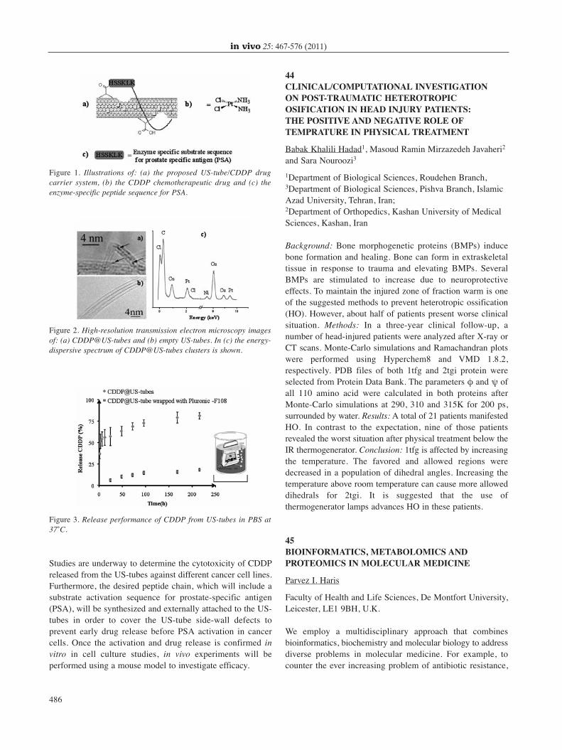
Studies are underway to determine the cytotoxicity of CDDPreleased from the US-tubes against different cancer cell lines.Furthermore, the desired peptide chain, which will include asubstrate activation sequence for prostate-specific antigen(PSA), will be synthesized and externally attached to the US-tubes in order to cover the US-tube side-wall defects toprevent early drug release before PSA activation in cancercells. Once the activation and drug release is confirmed invitro in cell culture studies, in vivo experiments will beperformed using a mouse model to investigate efficacy.
44CLINICAL/COMPUTATIONAL INVESTIGATION ON POST-TRAUMATIC HETEROTROPICOSIFICATION IN HEAD INJURY PATIENTS: THE POSITIVE AND NEGATIVE ROLE OFTEMPRATURE IN PHYSICAL TREATMENT
Babak Khalili Hadad1, Masoud Ramin Mirzazedeh Javaheri2
and Sara Nouroozi3
1Department of Biological Sciences, Roudehen Branch,3Department of Biological Sciences, Pishva Branch, IslamicAzad University, Tehran, Iran;2Department of Orthopedics, Kashan University of MedicalSciences, Kashan, Iran
Background: Bone morphogenetic proteins (BMPs) inducebone formation and healing. Bone can form in extraskeletaltissue in response to trauma and elevating BMPs. SeveralBMPs are stimulated to increase due to neuroprotectiveeffects. To maintain the injured zone of fraction warm is oneof the suggested methods to prevent heterotropic ossification(HO). However, about half of patients present worse clinicalsituation. Methods: In a three-year clinical follow-up, anumber of head-injured patients were analyzed after X-ray orCT scans. Monte-Carlo simulations and Ramachandran plotswere performed using Hyperchem8 and VMD 1.8.2,respectively. PDB files of both 1tfg and 2tgi protein wereselected from Protein Data Bank. The parameters φ and ψ ofall 110 amino acid were calculated in both proteins afterMonte-Carlo simulations at 290, 310 and 315K for 200 ps,surrounded by water. Results: A total of 21 patients manifestedHO. In contrast to the expectation, nine of those patientsrevealed the worst situation after physical treatment below theIR thermogenerator. Conclusion: 1tfg is affected by increasingthe temperature. The favored and allowed regions weredecreased in a population of dihedral angles. Increasing thetemperature above room temperature can cause more alloweddihedrals for 2tgi. It is suggested that the use ofthermogenerator lamps advances HO in these patients.
45BIOINFORMATICS, METABOLOMICS ANDPROTEOMICS IN MOLECULAR MEDICINE
Parvez I. Haris
Faculty of Health and Life Sciences, De Montfort University,Leicester, LE1 9BH, U.K.
We employ a multidisciplinary approach that combinesbioinformatics, biochemistry and molecular biology to addressdiverse problems in molecular medicine. For example, tocounter the ever increasing problem of antibiotic resistance,
in vivo 25: 467-576 (2011)
486
Figure 1. Illustrations of: (a) the proposed US-tube/CDDP drugcarrier system, (b) the CDDP chemotherapeutic drug and (c) theenzyme-specific peptide sequence for PSA.
Figure 2. High-resolution transmission electron microscopy imagesof: (a) CDDP@US-tubes and (b) empty US-tubes. In (c) the energy-dispersive spectrum of CDDP@US-tubes clusters is shown.
Figure 3. Release performance of CDDP from US-tubes in PBS at37˚C.
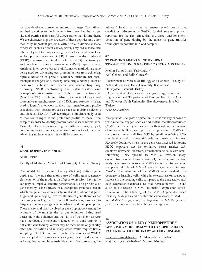
we have developed a novel antimicrobial strategy. This utilisessynthetic peptides to block bacteria from reaching their targetsite and exerting their harmful effects rather than killing them.We are characterising the structure of these peptides and othermedically important proteins, with a role in diverse diseaseprocesses such as dental caries, prion, amyloid disease andothers. Physical techniques being used in these studies includesurface plasmon resonance (SPR), Fourier transform infrared(FTIR) spectroscopy, circular dichroism (CD) spectroscopyand nuclear magnetic resonance (NMR) spectroscopy.Artificial intelligence-based bioinformatics methods are alsobeing used for advancing our proteomics research, achievingrapid elucidation of protein secondary structure for high-throughput analysis and, thereby, obtaining a better picture oftheir role in health and disease and accelerating drugdiscovery. NMR spectroscopy and matrix-assisted laserdesorption/ionisation-time of flight mass spectrometry(MALDI-TOF) are being utilised for metabolomics andproteomics research, respectively. NMR spectroscopy is beingused to identify alterations in the urinary metabolomic profileassociated with disease processes such as multiple sclerosisand diabetes. MALDI-TOF technique is simultaneously usedto monitor changes in the proteomic profile of these urinesamples in order to identify protein-based disease biomarkers.Examples of results from our highly multidisciplinary project,combining bioinformatics, proteomics and metabolomics foradvancing molecular medicine will be presented.
46GENE DOPING IN SPORTS
Nezih Hekim
Faculty of Medicine, Yeni Yuzyil University, Istanbul, Turkey
The World Anti- Doping Agency (WADA) defines genedoping as "the non-therapeutic use of cells, genes, geneticelements, or of the modulation of gene expression, having thecapacity to improve athletic performance". The principle ofgene therapy is the delivery of a therapeutic gene to a cell inwhich the gene may compensate an absent or abnormal gene.In general, gene doping involves the use of gene therapies forincreasing muscle growth, blood cell production, resistance tofatigue, endurance, oxygen accumulation and pain perception.There are several risks involved in gene doping concerning theaccuracy of the transfer, the various techniques being usedunder the right guidance and the skills of the scientists whohave therapeutic proficiency. Detection of gene doping isdifficult. Gene therapy vectors may be measurable only shortlyafter administration and in many cases would require tissuesampling. The International Sports Federations and WADAhave accepted performance-enhancing substances and methodsas being doping and have forbidden them from protecting the
athletes’ health in order to ensure equal competitiveconditions. Moreover, a WADA funded research projectreported, for the first time, that the direct and long-termdetection of gene doping by the abuse of gene transfertechniques is possible in blood samples.
47TARGETING MMP-3 GENE BY siRNATRANSFECTION IN GASTRIC CANCER AGS CELLS
Meliha Burcu Irmak-Yazicioglu1,3*, Anıl Cebeci1 and Salih Gencer2,3
1Department of Molecular Biology and Genetics, Faculty ofArts and Sciences, Halic University, Kaptanpasa,Okmeydanı, Istanbul, Turkey;2Department of Genetics and Bioengineering, Faculty ofEngineering and 3Department of Biology, Faculty of Artsand Sciences, Fatih University, Buyukcekmece, Istanbul,Turkey*Previous address
Background: The gastric epithelium is continuously exposed totoxic reactive oxygen species and matrix metalloproteinases(MMPs) are the enzymes known for their role in the invasionof tumor cells. Here, we report the suppression of MMP-3 inthe gastric cancer cell line AGS by small interfering RNAtransfection and its potential role in gastric carcinomas.Methods: Oxidative stress in the cells was assessed followingH2O2 exposure via the oxidative stress marker 2,7-dichlorofluorescein diacetate. Transfection of cells with smallinterfering RNA specific to MMP-3, invasion assay,quantitative reverse transcriptase polymerase chain reactionanalysis and overexpression of MMP-3 were used to determinethe potential role of MMP-3 gene in gastric carcinomas.Results: The silencing of the MMP-3 gene resulted in adecrease of invading cells, while its overexpression caused anincrease in the invading cells, compared to the untreated controlcells. Moreover, it caused a 4.1-fold increase in MMP-10 anda 7.4-fold decrease in MMP-15 mRNA expression levels.Conclusion: The silencing of the MMP-3 gene decreasedinvading AGS cells and affected the expressions of MMP-10and MMP-15, suggesting that targeting the MMP-3 gene ingastric carcinomas may be a therapeutic approach.
48ASSOCIATION OF 1128T>C NEUROPEPTIDE YGENE POLYMORPHISM WITH DYSLIPIDEMIA INPATIENTS WITH CORONARY ARTERY DISEASE
Khadijeh Jamialahmadi1, Ali Masoudi Kazemabad2, Majid Ghayour Mobarhan1, Mohsen Mouhebati3,
Abstracts of the 4th International Congress of Molecular Medicine, 27-30 June, 2011, Istanbul, Turkey
487
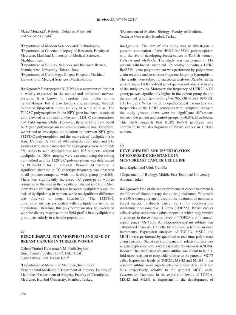
Majid Mojarrad4, Raheleh Dehghan Manshadi1
and Saeed Akhlaghi5
1Department of Modern Sciences and Technologies,4Department of Genetics, 5Deputy of Research, Faculty ofMedicine, Mashhad University of Medical Sciences,Mashhad, Iran;2Department of Biology, Sciences and Research Branch,Islamic Azad University, Tehran, Iran;3Department of Cardiology, Ghaem Hospital, MashhadUniversity of Medical Sciences, Mashhad, Iran
Background: Neuropeptide Y (NPY) is a neurotransmitter thatis widely expressed in the central and peripheral nervoussystems. It is known to regulate food intake in thehypothalamus, but it also favours energy storage throughincreased lipoprotein lipase activity in white adipose. TheT1128C polymorphism in the NPY gene has been associatedwith elevated serum total cholesterol, LDL-C concentrationsand TAG among adults. However, there is little data aboutNPY gene polymorphism and dyslipidemia in Iran. Therefore,we wished to investigate the relationship between NPY gene1128T>C polymorphism and the outbreak of dyslipidemia inIran. Methods: A total of 485 subjects (270 men and 215women) who were candidates for angiography were recruited;380 subjects with dyslipidemia and 105 subjects withoutdyslipidemia. DNA samples were extracted using the saltingout method and the 1128T>C polymorphism was determinedby PCR-RFLP for all subjects. Results: At baseline, asignificant increase in TC genotype frequency was observedin all patients compared with the healthy group (p<0.05).There was significantly increased TC genotype in womencompared to the men in the population studied (p<0.05). Also,there was significant difference between dyslipidemia and thelack of dyslipidemia in women, while no significant differencewas observed in men. Conclusion: The 1128T>Cpolymorphism was associated with dyslipidemia in Iranianpopulation. Therefore, this polymorphism may be associatedwith the dietary response to the lipid profile in a dyslipidemiagroup particularly in a female population.
49HER2 ILE655VAL POLYMORPHISM AND RISK OFBREAST CANCER IN TURKISH WOMEN
Ozlem Timirci Kahraman1, M. Fatih Seyhan1, Emel Canbay2, Cihan Uras3, Hilal Unal3, Oguz Ozturk1 and Turgay Isbir4
1Department of Molecular Medicine, Institute ofExperimental Medicine, 2Department of Surgery, Faculty ofMedicine, 3Department of Surgery, Faculty of CerrahpasaMedicine, Istanbul University, Istanbul, Turkey;
4Department of Medical Biology, Faculty of Medicine,Yeditepe University, Istanbul, Turkey
Background: The aim of this study was to investigate apossible association of the HER2 Ile655Val polymorphismwith the risk of developing breast cancer in Turkish women.Patients and Methods: The study was performed in 118patients with breast cancer and 128 healthy individuals. HER2Ile655Val gene polymorphism was performed by polymerasechain reaction and restriction fragment length polymorphism.The results were subject to statistical analyses. Results: In thepresent study, HER2 Val/Val genotype was not observed in anyof the study groups. Moreover, the frequency of HER2 Ile/Valgenotype was significantly higher in the patient group than inthe control group (p=0.009, χ2=6.785, OR=1.983 95% CI:1.181-3.328). When the clinicopathological parameters andfrequencies of the HER2 genotypes were compared betweenthe study groups, there were no significant differencesbetween the patient and control groups (p>0.05). Conclusion:This study suggests that HER2 Ile/Val genotype maycontribute to the development of breast cancer in Turkishwomen.
50DEVELOPMENT AND INVESTIGATION OF ETOPOSIDE RESISTANCE IN MCF7 BREAST CANCER CELL LINE
Esra Kaplan and Ufuk Gündüz
1Department of Biology, Middle East Technical University,Ankara, Turkey
Background: One of the major problems in cancer treatment isthe failure of chemotherapy due to drug resistance. Etoposideis a DNA damaging agent used in the treatment of metastaticbreast cancer. It directs cancer cells into apoptosis viainhibiting topoisomerase II alpha (TOP2A). Breast cancercells develop resistance against etoposide which may involvealterations in the expression levels of TOP2A and mismatchrepair genes. Methods: An etoposide-resistant subline wasestablished from MCF7 cells by stepwise selection in doseincrements. Expression analyses of TOP2A, MSH2 andMLH1 were performed by quantitative real time polymerasechain reaction. Statistical significance of relative differencesin gene expression levels were estimated by one-way ANOVA.Results: The established resistant subline was found to be 2.3-fold more resistant to etoposide relative to the parental MCF7cells. Expression levels of TOP2A, MSH2 and MLH1 in theresistant subline were significantly decreased 90%, 82% and82% respectively, relative to the parental MCF7 cells.Conclusion: Decrease in the expression levels of TOP2A,MSH2 and MLH1 is important in the development of
in vivo 25: 467-576 (2011)
488
chemotherapeutic resistance to etoposide in breast cancer.These genes may be considered for the development of newstrategies to overcome resistance against topoisomerase IIinhibitors.
51PROMOTER ANALYSIS OF THE P80-KATANIN GENE
Güher Işık Cesur, Derya Canbaz and Arzu Karabay
Department of Molecular Biology and Genetics, IstanbulTechnical University, Istanbul, Turkey
Background: The microtubule-severing protein katanin is aheterodimer of 60 kDa (p60) and 80 kDa (p80) subunits.The p60 subunit can break microtubules (MTs) whereas thep80 subunit targets p60 to the centrosomes. The ratio of thetwo subunits varies markedly in different tissues and atdifferent stages of development, suggesting that the activityof the p60 subunit might be influenced by the levels of thep80. Promoter analysis of the p80-katanin gene which iscrucial to understand the temporal gene expression patternof katanin has not been studied yet. Therefore, we aimed toidentify the critical promoter regions of KATNB1 (p80)gene required for its expression. Methods: For thecharacterization of the promoter of KATNB1 gene,polymerase chain reaction (PCR) was used to amplify 1000bp upstream nucleotides of KATNB1 gene. By using nestedPCR, shorter fragments were obtained. These promoterdeletion constructs were cloned into a luciferase reporterplasmid vector which lacked eukaryotic promoter andenhancer sequences. Obtained deletion constructs weretransfected to SHSY-5Y neuroblastoma cells and theluminometrical measurement was performed. Results: Weidentified 518 bp TATA-less promoter including a criticalCpG island as an optimal promoter and 160 bp minimalpromoter required for KATNB1 gene expression.Conclusion: 518 bp TATA-less promoter is sufficient forKATNB1 gene expression.
52SPEEDY/RINGO INHIBITS CALPAIN-DIRECTEDAPOPTOSIS IN NEURONS
Aysegul Yildiz and Arzu Karabay
Molecular Biology and Genetics Department, IstanbulTechnical University, İstanbul, Turkey
Background: While according to the classic view neurons are
post-mitotic and cannot reenter the cell cycle, recent studiesshowed mitotic reentry due to neurotoxic agents. Especiallyamyloid-beta aggregation destabilizes calcium homeostasis.Elevation of intracellular calcium levels overactivates thecalpain and causes mitotic reentry of neurons. However,neurons cannot complete their cycle and undergo apoptosis.Recent studies indicated the ability of a novel cell-cycleprotein, Speedy/RINGO, to inhibit apoptosis. Since calpainoveractivation is one of the important factors causingapoptosis, our aim was to identify the protective effect ofSpeedy/RINGO on calpain-induced neurodegeneration.Methods: Speedy/RINGO was transfected into neurons.Calpain activation was then performed and cell cycle markerswere analyzed by Q-PCR and immunocytochemistry.Caspase-3 activity was analyzed by Western-blotting todetermine if apoptotic pathway was inhibited bySpeedy/RINGO. Results: Q-PCR analysis showed that innone of the cell-cycle markers mRNA level was changed.Immunostaining results confirmed the Q-PCR analysis thatthere is no difference in expression levels. Western-blottingresults determined no change in protein level of cleaved-caspase-3 in the presence of Speedy/RINGO. Conclusion:The lack of change in mRNA/protein levels implied thatSpeedy/RINGO keeps neurons under stable conditions.Furthermore, no change in cleaved caspase-3 level indicatedthat Speedy/RINGO may protect neurons against calpain’sapoptotic effect.
53INVESTIGATION OF THE CYTOTOXIC EFFECTS OF EXTRACTS FROM ENDEMICBUPLEURUM SPECIES IN RESISTANT BREAST CANCER CELL LINES
Gökhan Kars1, Meltem Demirel Kars2, Mehtap Akın1, Hatice Taner Saraçoğlu1 and Ufuk Gündüz3
1Department of Biology, Faculty of Science, 2SarayönüVocational High School, Selçuk University, Konya, Turkey;3Department of Biological Sciences, Middle East TechnicalUniversity, Ankara, Turkey
The resistance developed to a broad spectrum ofchemotherapeutic agents during cancer chemotherapy iscalled multiple drug resistance (MDR). The MDRphenotype acquired by drug-unresponsive patients remains amajor hindrance to chemotherapy. P-gp overexpression isone form of MDR phenotype. Paclitaxel and vincristine areantimitotic chemotherapeutic agents. MCF-7 is a mammaryepithelium cell line used as a model for breast cancer. Theaim of this study was to investigate the cytotoxic effects ofplant extracts in P-gp overexpressing paclitaxel-resistant(MCF-7/Pac) and vincristine-resistant (MCF-7/Vinc) MCF-
Abstracts of the 4th International Congress of Molecular Medicine, 27-30 June, 2011, Istanbul, Turkey
489
7 mammary carcinoma cell lines and their parental line(MCF-7/S). In this study, the roots of five endemicBupleurum species (B. turcicum, B. sulphureum, B.lycaonicum, B. pauciradiatum and B. heldrechii) werecollected and grinded. The plant powder was extracted in70% ethanol by sonication and lyophilized. The effect of thetotal extracts on the viability of paclitaxel- and vincristine-resistant MCF-7 cells were determined by XTT cytotoxicitytests after incubation of cells in different doses of plantextracts for 72 h. IC50 values of total extracts for sensitiveand resistant cell lines were calculated. The degree oftoxicity of the total extracts to the cell lines was determinedaccording to these values.
54DETERMINATION OF PHENOLIC COMPOUND,SAIKOSAPONIN AND PODOPHYLLOTOXINCONTENTS IN THE ROOTS OF ENDEMIC BUPLEURUM SPECIES
Gökhan Kars1, Meltem Demirel Kars2, Mehtap Akın1 andHatice Taner Saraçoğlu1
1Department of Biology, Faculty of Science, 2SarayönüVocational High School, Selçuk University, Konya, Turkey
Multiple drug resistance (MDR) acquired againstchemotherapy by cancer cells causes most of thechemotherapeutic agents not to show the expected impact onthe patients and leads to the progression of the disease.Therefore, making the drug-resistant cells re-sensitive to thechemotherapeutic agents is extremely important for thesuccess of the treatment. While podophyllotoxin is known tobe an effective antitumor agent, types of phenolics andsaponins derived from plants were shown to reverse theresistance by modulating the cell membrane protein P-glycoprotein (P-gp). The aim of this study was to determinethe phenolic compound, saikosaponin and podohyllotoxincontents of total extracts from Bupleurum species by HPLC.The total phenolic content and DPPH antioxidant activity ofthe extracts were also investigated. In this study, the roots offive endemic Bupleurum species (B. turcicum, B.sulphureum, B. lycaonicum, B. pauciradiatum and B.heldrechii) were collected and grinded. The plant powderwas extracted in 70% ethanol by sonication and the extractswere lyophilized. After dissolving the lyophilized extracts inmethanol, the extracts were subjected to gradient elution byHPLC and the contents were determined. Total phenoliccontent and the DPPH antioxidant activity of extracts werealso investigated. The MDR reversal activities of thecompounds collected by HPLC will be further determined byfluorescent microscopy.
55INVOLVEMENT OF mTOR SIGNALLINGPATHWAYS IN HEALTH AND DISEASE IN REPRODUCTIVE TISSUES
Emmanouil Karteris
Cellular Endocrinology Laboratory, Biosciences, BrunelUniversity, Uxbridge, UB8 3PH, U.K.
The mammalian target of rapamycin (mTOR) is a Ser/Thrprotein kinase that functions as energy, stress, amino acid, andnutrient sensor to balance metabolism and cell growth. mTORforms two complexes (mTORC1 and mTORC2) which differin their composition and regulation and exert distinctbiological effects. Recently, a modulator of mTOR that bindsto mTOR complexes, termed DEPTOR was described.However, the precise functions of DEPTOR are not fullyelucidated. We have mapped in detail mTOR components inreproductive tissues. At placental level where mTOR appearsto act as a nutrient sensor, DEPTOR is the predominanttranscript compared with mTOR, rictor and raptor.Interestingly, a significant inverse correlation betweenDEPTOR and maternal stress was noted. Moreover,syncytialization of placental cells also exerts an effect in theexpression of mTOR-related genes. Finally, a limitation tosuccessful ovarian cancer chemotherapy treatments is theacquisition of drug-resistance. We have shown that inpaclitaxel resistant ovarian cancer cell lines the activity ofmTOR signalling pathway is altered. Collectively our datasuggest that there is a higher order of complexity of mTORsignalling in the human placenta that can affect pregnancyoutcome, and mTOR pathway may also mediate paclitaxelresistance in ovarian cancer.
56SAMPLE SIZE AND POWER CALCULATION IN MOLECULAR MEDICINE: A SIMULATION STUDY
Elif Çiğdem Kaspar
Department of Biostatistics, Faculty of Medicine, YeditepeUniversity, Istanbul, Turkey
Background: Sample size and power calculation is a criticalprocedure when designing new studies, including those in thefield of molecular medicine. Power analysis involvesdetermining the statistical power of a design, given asignificance level, sample size and effect size and it can beused to calculate the minimum sample size required to acceptthe outcome of a statistical test with a particular level ofconfidence. Sample size and power of a study can becalculated using statistical formulas. However, these formulas
in vivo 25: 467-576 (2011)
490
have their own limitations and to judge about the accuracy ofthese calculations, knowledge of statistics is required.Methods: Sample size and power calculation methods wereshown with the illustrated data sets. Results: The statisticalpower calculated from the small sample size and thatcalculated from the sufficient sample size of the same studywere compared. It was found that the sufficient sample sizeprovides higher power than the small sample size.Conclusion: Researchers can use these calculations as a toolto increase the strength of their inferences, and editors andreviewers of scientific publications may demand thatstatistical power be reported in all cases where a non-significant result is obtained.
57TWO HOMOLOGOUS PROTEINS CHI3L1 ANDCHI3L2 OVEREXPRESSED IN GLIAL TUMORSACTIVATE THE MAPK SIGNALING PATHWAY BUT LEAD TO DIFFERENT CELL FATE
Pavlo Areshkov1, Stanislav Avdieiev1, Olena Balynska1, Yuri Zozulya2 and Vadym Kavsan1
1Department of Nucleic Acids Biosynthesis, Institute ofMolecular Biology and Genetics, Kyiv, Ukraine;2A.P. Romodanov Institute of Neurosurgery, Kyiv, Ukraine
Background: Serial analysis of gene expression, Northern andWestern analyses revealed the closely related chitinase-likegenes CHI3L1 and CHI3L2 among genes with the mostincreased expression in glioblastoma. Methods: Recombinantproteins, cell line stably producing CHI3L1, soft agar colonyformation, Erk1/2 phosphorylation and nuclear translocation,mitogenic, proliferation and apoptosis assays were used inthis study. Results: It was found that Chi3l1 possessesoncogenic properties. The influence of CHI3L2 protein oncell proliferation was opposite to the CHI3L1 effect.Evaluated parameters were diminished in cells treated byCHI3L2, in contrast to those stimulated by CHI3L1application. Both proteins activated Erk1/2, but the activationduration was different: in cells treated by CHI3L2, sustainedErk1/2 activation was associated with their nuclearaccumulation, while activation triggered by CHI3L1 led tothe transient Erk1/2 phosphorylation with only brief nucleartranslocation. Nuclear accumulation of active Erk1/2 resultedin phosphorylation of certain transcription factors.Conclusion: The consequences of events in Erk-mediatedcascade depend on the duration of Erk phosphorylation aswell as on the cellular context, namely the transcriptionfactors activated by Erk1/2. Thus, the cellular responsedetermines which factors are present in the cell. Activation ofthe phosphatidylinositol 3-kinase (PI3K) pathway is currentlyunder investigation.
58USE OF MOLECULAR BIOLOGY IN MEDICALDEFENSE AGAINST BIOTERRORISM ANDRELATED EXPERIENCES OF THE MILITARYMEDICAL ACADEMY CBRN LABORATORY
Levent Kenar and Mesut Ortatatli
Department of Medical CBRN Defense, Gulhane MilitaryMedical Academy, Ankara, Turkey
Bioterrorism is the intentional use of microorganisms andtoxins, generally of microbial, plant or animal origin toproduce disease and death in humans, livestock and crops. Thedeliberate use of biological agents including bacteria, viruses,rickettsia and toxins has emerged as significant threat in thelast decade and after the anthrax cases that occurred followingthe terrorist attack in 11th September, 2001. Rapid andaccurate detection and identification of bioterror agents isimportant not only to confirm that a bioterrorism event hasoccurred, but also to determine the required proper protectivemeasures. Molecular analysis of samples such as powder-containing envelopes upon their arrival to the laboratoryshould be carried out for instant confirmation. Molecularassays should be sensitive and specific to detect lowconcentrations of agents. In general, nucleic acid-basedsystems are more sensitive. Improvement of rapid andadvanced diagnostic systems and facilities such as battlefielddetection systems, biosensors and fully automated biodetectorsfor real time sample collection, detection and identification inthe field have been developed. On the other hand, a miniatureflow cytometer (known as miniFlo) using an immunoassaysystem and a portable PCR identifying the DNA inside the cellare also available for the detection and identification ofbiological warfare agents. This lecture aims to give briefinformation about detection technologies for biothreat agents.In addition, the studies about biothreat detection in ourmedical CBRN Department which is unique in Turkey will bebriefly mentioned herein.
59INVESTIGATION AND COMPARISON OF THEEXISTENCE OF MULTI-DRUG RESISTANCE (MDR1) GENE MUTATIONS AT GENOTYPE 1B AND DIFFERENT GENOTYPES OF HCV
Recep Kesli1 and Nadir Kocak2
1Department of Microbiology, Molecular Diagnostic Unit,2Department of Genetics, Konya Research and EducationHospital, Konya, Turkey
Background: P-glycoprotein (Pgp) is encoded by the humanmultidrug-resistance (MDR1, ABCB1) gene. Hepatitis C virus
Abstracts of the 4th International Congress of Molecular Medicine, 27-30 June, 2011, Istanbul, Turkey
491
species is classified into six genotypes that are clinicallyimportant in determining the potential response to interferon-based therapy. Methods: HCV-RNA assay was performedusing real-time PCR. HCV genotyping and MDR1 mutationswere both performed by using the reverse hybridizationmethod. Results: The study included 85 HCV-RNA positivepatients; among them 81 (95.2%) were identified as HCV-1b,2 (2.4%) were HCV-2a and 2 (2.4%) were HCV-3a subtypes.Of the 81 HCV-1b cases, 51 (62.9%) were found to be positivewith MDR1 mutations; 47 (58%) were heterozygous and 4(4.9%) were homozygous. No MDR1 mutations were foundin patients with HCV-2a and HCV-3a subtypes. Statisticallysignificant differences were found between genotypes of HCV-1b and HCV-2a (p<0.001), HCV-1b and HCV-3a (p<0.001)for the occurrence of MDR1 mutations. Of all 81 cases withHCV-1b, 45 (58%) were female and 36 (44%) were male.Statistically significant differences were found between maleand female with genotypes of HCV-1b for the occurrence ofheterozygous (p<0.05) and homozygous (p<0.05) MDR1mutations. The two–sample proportion test was used for allstatistical analyses. Conclusion: The results of this studyindicated a significant difference between the two groupsHCV-1b/2a and HCV-1b/3a for the occurrence of MDR1mutations. These results may be related with the tendency ofpatients infected with HCV-1b to exhibit poor IFNresponsiveness compared with those infected with HCV-2a/3a.
60COMPARISON OF HPV PREVALENCE AND TYPES OF HPV IN WOMEN AT HIGH RISK OF CERVICAL CANCER WITH RISKFACTORS AND CERVICAL SMEAR
Celalettin Eroglu1, Recep Kesli2, Mehmet Ali Eryılmaz3,Yaşar Unlu4, Oguzhan Gonenc5 and Cetin Celik6
1Radiation Oncology Department, Faculty of Medicine,Erciyes University, Kayseri, Turkey;2Microbiology Laboratory, Molecular Diagnostic Unit,3Cancer Early Diognasis-Treatment and Education Center,4Pathology Department, 5Obstetric and GynecologyDepartment, Konya Research and Education Hospital,Konya, Turkey;6Obstetrics and Gynecology Department, Meram Faculty ofMedicine, Selcuk Universty, Konya, Turkey
Background: Human papilloma virus (HPV) is considered tobe involved in cervical cancer and precancerous lesions. Thisstudy aimed to determine the prevalence and the types of HPVin women at high risk of cervical cancer and the correlationbetween HPV positivity and risk factors for developingcervical cancer. Methods: A total of 404 healthy women atrisk were investigated in terms of risk factors. The existence of
HPV was determined and 16 types were identified by usingDNA-reverse hybridization. Cervical smear samples wereobtained and biopsies were performed with colposcopicexamination. Chi-square and multiple logistic regressionanalyses were used for statistical processing of the data.Results: HPV frequency rate was found to be 32.5% (n=131).HPV18 was found in 60 cases (45.8%) and HPV16 in 22cases (16.8%) as the most frequently types. According to HPVtypes, high-risk rate for cervical cancer was found to be 78.7%(n=103). In cervical smear evaluations, epithelial cellabnormalities were found in 91 cases (22.5%), atypicalsquamous cells of undetermined significance in 82 cases(20.3%), low-grade squamous intraepithelial lesions in 7 cases(1.7%) and high-grade squamous intraepithelial lesions in 2cases (0.5%). Conclusion: Women at high positivity inoncogenic HPV types should be followed and necessaryprecautions should be taken.
61HEREGULIN INDUCES HEAT SHOCK PROTEINS IN AN HSF1 DEPENDENT MANNER IN CANCERCELLS AND LEADS TO PROTECTION FROM APOPTOSIS
Md Abdul Khaleque1, Ajit Bharti2 and Stuart K. Calderwood3
1Department of Life Sciences, North South University,Dhaka, Bangladesh;2Department of Medicine, Boston University School ofMedicine, Boston, MA, U.S.A.; 3Department of Radiation Oncology, Beth Israel DeaconessMedical Center, Harvard Medical School, Boston, MA,U.S.A.
Elevated HSP levels are observed in many cancer types andsignal a poor prognosis in terms of survival and response totherapy. HSP expression protects cells from spontaneousapoptosis during transformation, in addition to apoptosisinduced by therapy. These mechanisms may underlie the roleof HSP in tumor progression and resistance to treatment. HSPtranscription requires the heat shock transcription factor (hsf1),which is itself overexpressed in cancer and plays a role ininvasion and metastasis. We show that HSP elevation is linkedto the highly malignant factor heregulin β1 (HRGβ1), whichinduces HSP expression through HSF1. HSP expression isinduced through a cascade response initiated by HRGβ1binding to c-erbB receptors and leads to the inhibition ofHSF1 antagonist glycogen synthase kinase3. Activated HSF1plays an essential role in the protection of cells from apoptosisby HRGβ1, indicating the role for HSF1 in tumor progressionpathway. Methods: Mammalian cell culture, immunoblotanalysis, luciferase and β-galactosidase activity assays,
in vivo 25: 467-576 (2011)
492
apoptosis assay and anchorage independent growth assay wereused in this study. Results: HRGβ1 induced HSP expressionthrough hsf1-dependent transcription. Signal transductionsteps led to hsf1-dependent activation of HSP expression byHRGβ1. HRGβ1-induced, c-erbB-2-dependent and anchorage-independent growth involved significant participation of hsf1.The cytoprotective effects of HRGβ1 protection by heat shockled to HSP expression. Conclusion: Heregulin induces Hsp70in an HSF1-dependent manner. Heregulin activates HSF1through the ErbB/Pi3k/Akt pathway. Heregulin/ErbB/HSF1-mediated hsp synthesis inhibits apoptosis and prevents tumorprogression.
62CYP21A2 ANALYSIS OF CONGENITAL ADRENAL HYPERPLASIA PATIENTS DUE TO 21-HYDROXYLASE DEFICIENCY
D. Kiraç1, K. Ulucan2, D. Ergeç3, T. Güran4, T. Akçay4, F. Eren5, G. Koç2, E.Ç. Kaspar6, A. Bereket4, T. Isbir1 and A.I. Güney2
Departments of 1Medical Biology and 6Biostatistics, Facultyof Medicine, Yeditepe University, Istanbul, Turkey; Departments of 2Medical Genetics, Pediatric Endocrinology and 5Medical Biology, Faculty of Medicine, Marmara University Istanbul, Turkey;3Department of Molecular Biology and Genetics, Faculty ofScience and Art, Yeni Yuzyıl University, Istanbul, Turkey
Introduction: Congenital adrenal hyperplasia (CAH) is anautosomal recessive disorder mainly caused by 21-hydroxylase deficiency. Females with severe 21-hydroxylase deficiency are exposed to excess androgensprenatally and are born with ambiguous genitalia.Moreover, most patients cannot synthesize sufficientaldosterone to maintain sodium balance and may developpotentially fatal “salt-wasting” crises, if not treated.Methods: A total of 100 patients and 80 controls wererecruited to the study. CYP21 gene regions starting frompromoter to the end of exon 4 were amplified by PCR.Mutations were detected by direct sequencing. The resultswere evaluated statistically. Results: A total of 10 novelmutations were detected. In addition to these mutations, 16different polymorphisms were also found. G→A mutationat position 711 (13%) (novel mutation) and A/C→Gmutation at position 777 (38%) were found to bestatistically significant when compared to controls.Conclusion: Prenatal diagnosis has been utilized for over20 years, especially by screening for nine known mutations.In order to establish the presence of G→A mutation atposition 711 in different populations, the test for thismutation may be included in the screening protocol of
prenatal diagnosis. By using prenatal treatment techniquesin the prenatal stage, genital ambiguity, subsequentproblems of sex misassignment, gender confusion and“salt-wasting” can be reduced in CAH patients.
63MOLECULAR REGULATION OF LYMPHATICVASCULAR DEVELOPMENT: IMPLICATIONS FORHUMAN DISEASE AND CANCER THERAPY
Mathias Francois, Tam Duong, Frank Fontaine, Ben Hogan,Matt Cooper, Robert Capon and Peter Koopman
Institute for Molecular Bioscience, The University ofQueensland, Brisbane, QLD 4072, Australia
Background: Aberrant formation of lymphatic vessels(lymphangiogenesis) is associated with a wide range ofpathological conditions such as inflammatory disease,obesity and lymphedema. Further, most tumours depend onlymphangiogenesis for metastasis. Understanding themolecular control of lymphangiogenesis will suggest newways to manage it pharmaceutically. Results: We discoveredthat Sox18 encodes a transcription factor that acts as themaster regulator of lymphangiogenesis in the embryo. Wenow find that Sox18 is reactivated during tumourlymphangiogenesis, and that mice mutant for Sox18 showreduced metastasis. We have therefore developed ascreening strategy to identify small inhibitory moleculesable to modulate SOX18 protein activity, and havediscovered a novel class of small molecules that disruptSOX18 function in vitro and in vivo. Conclusion: Ourfindings provide a basis for innovative pharmacologicalSOX18-based strategies to modulate lymphangiogenesis,raising the prospect of complementing anti-angiogenictreatment with an anti-lymphangiogenic approach to cancertherapy.
64ANTIHYPERPLASTIC EFFECT OF LYSOZYMEGENE IN A TESTOSTERONE-INDUCED RAT MODEL OF BENIGN PROSTATİC HYPERPLASIA (BPH)
Pınar Aslan Koşar1, Osman Ergün2, İbrahim Onaran1, Taylan Oksay2 and Alim Koşar2
1Department of Medical Biology, and2Department of Urology, Süleyman Demirel University,Isparta, Turkey
Background: The effects of lysozyme gene treatments onprostatic tissue were studied in an experimental model of
Abstracts of the 4th International Congress of Molecular Medicine, 27-30 June, 2011, Istanbul, Turkey
493

BPH. Methods: Adult Wistar rats weighing 200 to 400 gwere used. They were completely randomized into 3 groupsas control, testosterone, and testosterone+ lysozyme (Lys)gene. After mRNA isolation, reverse transcriptasepolymerase chain reaction, restriction digestions andligations, recombinant pHM6 vector harboring mouselysozyme gene (pHM6mLys) was constructed. Single dosetestosterone was administered intramuscularly, except group1. 40 days after the beginning of experiment in group 3, Lyswas injected into rat prostates tissue. A week later (day 47),all rats were sacrificed. Results: Testosterone treatmentincreased by 39% the prostate weight in rats. Lys therapydecreased by 38.7% the prostate weight in rats. Testosteroneapplication caused an irregular acinar growth pattern in theprostate tissue with variable amout of stroma. İsolated ormultiple stacked cell populations (pilling up formation) wasidentified in the epithelial layer and it was lined by cuboidalor cylindrical cells. Conclusion: We suggest that Lys may beuseful in the treatment of BPH.
65THE EXPERIMENTAL MODEL OF TESTESTORSION AND APOPTOSIS
Pınar Aslan Koşar1, Hamdi Tuncer2, İbrahim Onaran1,Osman Ergün2, Alim Koşar2
1Department of Medical Biology, Süleyman DemirelUniversity, Isparta, Turkey;2Department of Urology, Süleyman Demirel University,Isparta, Turkey
Background: Apoptosis response was investigated in mildischemic reperfusion damage in germ cells due to testiculartorsion. Methods: A total of 44 Wistar albino rats were dividedinto control, sham, and 1, 2 and 4 hour torsion groups. Lefttestes were subjected to 720˚ of torsion followed by 4 hours ofreperfusion. The ischemic reperfusion damage and theapoptosis level in left and right testes were evaluatedhistopathologically. Results: JTBS was normal in the controlsand the sham group, accompanied by very low levels ofapoptosis. In the 1 hour torsion group, the JTBS was lowercompared to the controls and the sham group, and the averageapoptosis level was higher. However, these levels were notdrastically high and comparable to the right testes levels of the2 and 4 hour torsion groups. In 2 and 4 hour torsion groups,the JTBS was also lower and the apoptosis levels higher incomparison to the controls as well as the sham and 1 hourtorsion groups. Conclusion: The apoptotic examination wasmore sensitive to determine the testicular damage in mildischemia and the opposing testicular damage compared to theJTBS.
66THE EFFECTS OF RECEPTOR FOR ADVANCEDGLYCATION END PRODUCTS (RAGE) -374 T/A AND GLY82 SER AND PEROXISOMEPROLIFERATOR-ACTIVATED RECEPTOR GAMMA (PPAR-γ) PRO12ALA POLYMORPHISMSON CORONARY ARTERY DISEASE
Özlem Küçükhüseyin1, Atike Tekeli2, Hülya Yilmaz Aydoğan1 and Turgay Isbir3
1Department of Molecular Medicine, Istanbul University,Institute of Experimental Medicine, Istanbul, Turkey;2Department of Cardiovascular Surgery, Marmara UniversityHospital, Istanbul, Turkey;3Department of Medical Biology, Yeditepe University,School of Medicine, Istanbul, Turkey
Background: The present study aimed to investigate theindividual and combined effects of RAGE -374T/A, RAGEGly82Ser and PPAR-γ Pro12Ala polymorphisms on thedevelopment of coronary artery disease (CAD). Methods:Polymerase chain reaction, restriction fragment lengthpolymorphism and agarose gel electrophoresis techniqueswere used to determine RAGE -374T/A, RAGE Gly82Serand PPAR-γ Pro12Ala. Results: Individual allele andgenotype frequencies of RAGE -374T/A, RAGE Gly82Serand PPAR-γ Pro12Ala polymorphisms were not significantlydifferent between the study groups. However, when comparedto the control group, wild-type T allele frequency was foundhigher in the patients with diabetes. To investigate thecombined effects of RAGE and PPAR-γ polymorphisms,haplotype analysis was performed and there was no statisticaldifference between the haplotypes of RAGE Gly82Ser withRAGE -374T/A or PPAR-γ Pro12Ala. However, thefrequency of RAGE-374T/PPAR12Ala haplotype was foundto be higher in both patient groups with and without diabetescompared with the control group. Conclusion: The results ofthe present study demonstrated that possessing the A allele ofRAGE -374T/A polymorphism for diabetic CAD patients andpossessing the -374T/Ala12 haplotype of RAGE -374T/A andPPAR-γ Pro12Ala polymorphisms for the patient groups arethe most important risk factors for CAD.
67THERAPEUTIC APPLICATIONS OF ANTI-ANGIOGENIC STRATEGIES
Shant Kumar
Department of Pathology, Medical School, ManchesterUniversity, Manchester M13 9PT, U.K.
Angiogenesis, the formation of new blood vessels, is essential
in vivo 25: 467-576 (2011)
494
for tumour growth and metastasis. Angiogenesis also has acrucial role in a variety of physiological conditions and‘angiogenic diseases’ such as embryogenesis, wound healing,psoriasis, myocardial infarction, scleroderma, low back pain,rheumatic arthritis, stroke etc. Over the past 30 years or so anenormous amount of work has been undertaken to findspecific markers for angiogenic endothelial cells. We haveraised a panel of monoclonal antibodies to activation/proliferation associated antigens such as CD105 which presenton the luminal surface of vascular endothelial cells. Whenreactivity of our anti CD105 mab in normal and tumour tissuewas compared with a number of routinely employed pan – ECmarkers, blood vessels within and in the immediate vicinity ofthe tumour mass strongly stained with our CD105 antibody(E9), whereas the same blood vessels were either weaklypositive or not stained by pan-EC antibodies. Similarly thisantibody was highly specific for angiogenic EC in humanstroke tissues and myocardial infarcts. In vivo animal studiesshowed that CD105 antibody E9 localised to tumour vascularendothelial cells and CD105 based immunoconjugats inhibitedthe growth of the human breast tumours. An ex vivo study thatutilized freshly excised kidneys from patients with renalcancer resulted in specific labelling of tumour vasculature byradio labelled anti-CD105 mab E9. Unpublished data of aphase 1 clinical trial revealed evidence of localisation of antiCD105 antibody in tumours and in some cases even hadbeneficial therapeutic effects. Similar experimental work isunderway in other ‘angiogenic diseases’ in man.
68ETS DOMAIN TRANSCRIPTION FACTORS INNEURODEGENERATIVE DISORDERS
Işil Kurnaz
Department of Genetics and Bioengineering, YeditepeUniversity, Istanbul, Turkey
Background: Elk-1 is a transcription protein that belongs tothe ETS domain superfamily. Elk-1 is traditionally studied asa mitogenic transcription factor that gets phosphorylated inresponse to the activation of the mitogenic MAPK signalingpathway, translocates to the nucleus and induces transcriptionfrom immediate-early genes, such as c-fos. However, recentlyour lab and others have shown the survival-related functionsof Elk-1, while one group has shown cell death responseinitiated by Elk-1 in neurons. These findings, thus, put Elk-1in a peculiar position. Methods: We have employed moleculartechniques such as promoter cloning, site-directedmutagenesis, luciferase reporter assays and chromatin IP(ChIP) assays among many other techniques to study theeffect of Elk-1 on the transcriptional activation from variousneurodegeneration-related promoters. Results: In this study we
analyzed the transcriptional regulation of various neuro-degeneration-related genes that contain Elk-1-binding motifsin their promoter sequences. We have shown that Elk-1regulates the survival of motor neuron gene (SMN), which iscommonly mutated in spinomuscular atrophy, and we haveprovided some evidence that Elk-1 can also regulate thetranscription of SOD1 gene that is associated with familiaramyotrophic lateral sclerosis. Conclusion: We believe thatElk-1 is at the center stage of many neurodegenerativedisorders, and our conclusion (combined with our previousdata) is that this helps protect neurons against apoptotic celldeath.
69GENETIC FACTORS AND HUMAN PHYSICAL CAPABILITY
Hizir Kurtel
Marmara University School of Medicine, Haydarpasa,Istanbul, Turkey
Human athletic performance is essentially multifactorial anddetermined by a wide range of factors, including training,nutrition, technological aid and genetic factors. Hereditaryinfluences on endurance phenotypes such as maximal oxygenuptake and ventilatory thresholds are estimated to be around50% in sedentary individuals. Understanding the genetics ofendurance performance may provide valuable information todetermine "advantageous" polygenic profiles. Although over150 DNA polymorphisms have been found to be associatedwith some form of human physical performance, theprobability of an individual possessing an optimal polygenicprofile seems very small. Furthermore, the genetic basis ofpsychological phenotypes that would characterize thephysiological trait is equally important in determining theathletic elite performance and this topic requires furtherattention. Overall, this brief presentation tries to summarizesome notable polymorphisms in the light of evidence thatcomplex genetic interactions are yet to be identified.
70ROLE OF APOPTOSIS IN THE RESPONSE OF SOLIDTUMOR CELLS TO THERAPY
Stig Linder
Karolinska Cancer Center R8:00, Karolinska Hospital andInstitute, Stockholm, Sweden
Apoptosis has been widely proposed as a major mechanism ofthe anti-proliferative effects of various clinically usedtherapeutic agents, including DNA damaging agents and
Abstracts of the 4th International Congress of Molecular Medicine, 27-30 June, 2011, Istanbul, Turkey
495
mitotic inhibitors. A problem with these studies is that mostavailable evidence is based on in vitro studies using high drugconcentrations. The mechanisms of cell death induction byanticancer drugs in vivo are less clear. Apoptosis has beenreported, but may be a secondary phenomenon to mitoticcatastrophe. Therefore, whereas apoptosis is a relevantpharmacodynamic read-out for the efficacy of many drugs,apoptosis signaling may be of secondary importance as adeterminant of drug sensitivity. The cellular response tocisplatin will here be described and used to illustrate thecomplex cellular response to clinically used chemotherapeuticagents. I will also discuss the therapy response of quiescentand hypoxic cells in the deep parenchyma of solid tumors.Drugs may not reach these cells at sufficient concentrationsand the cells respond poorly to drugs that target proliferatingcells. Our laboratory has found that hypoxic cells in the deeptumor parenchyma may not respond to therapy by inductionof apoptosis. Strategies to target these cells will be discussed.
71LIPID PEROXIDATION PRODUCTS ANDANTIOXIDANT ACTIVITIES IN PREGNANTANEMIC WOMEN BEFORE AND AFTER ORAL IRON SUPPLEMENTATION
Abbas Ali Mahdi1, Amit Kumar Mani Tiwari1, Fatima Zahra2, Farzana Mahdi3 and Shyam Pyari Jaiswar4
Departments of 1Biochemistry and 4Obstetrics andGynecology, CSM Medical University, Lucknow, UP, India;Department of 2Obstetrics and Gynecology and3Biochemistry, Era’s Lucknow Medical College andHospital, Lucknow, UP, India
Background: Iron deficiency anemia (IDA) is one of the mostcommon nutritional disorders worldwide, affecting all ages.Oral iron supplementation is a commonly-used strategy tomeet the increased iron requirements of the risk groups.However, if provided in excess, iron supplementation mayinduce peroxidative damage through the production ofreactive oxygen species. The present study was designed toevaluate the markers of oxidative stress and the antioxidativeenzymes along with serum ferritin and transferrin receptorlevels in iron-deficient pregnant anemic women and theirresponse to oral iron supplementation. Methods: Iron (100mg) and folic acid (500 μg) supplementation were given toall 133 anemic women (mild anemia n=50, moderate anemian=50 and severe anemia n=33) along with 100 age-matchednon-anemic pregnant women, taken as controls, daily for 100days. Blood index, oxidative stress parameters, antioxidantenzymes, vitamins, serum ferritin and transferrin receptorswere estimated as per standard protocols. Results:Haemoglobin (Hb) levels along with antioxidant enzymes,
namely catalase, superoxide dismutase, glutathione reductase,reduced glutathione, total antioxidant capacity and ferritinlevels were found significantly increased (p<0.01) in alltreated groups. However, glutathione peroxidase andantioxidant vitamins, namely A, C and E and transferrinreceptor levels were found significantly decreased in alltreated subjects. Lipid peroxide levels, protein carbonylcontents, conjugated dienes, lipid hydroperoxide and oxidizedglutathione levels were found significantly increased (p<0.01)after oral iron supplementation. Conclusion: On the basis ofthese results, it may be safely concluded that, although therecommended oral iron dose is effective for improving bloodindices, this happens at the cost of increased oxidative stress.Therefore, it may be suggested that the doses of oral iron maybe given according to individual requirements.
72INCIDENCE AND PROGNOSTIC SIGNIFICANCE OF C-KIT EXON 9 MUTATIONS IN NEOPLASIA IN NORTHERN INDIA
Farzana Mahdi1, Hena Naqvi1, Syed Rizwan Hussain1, Sunil G Babu2, Abbas Ali Mahdi3 and Syed Tasleem Raza1
1Department of Biochemistry, Era’s Lucknow MedicalCollege, Lucknow, UP, India;2Department of Biotechnology, BBA University, Lucknow,UP, India;3Department of Biochemistry, CSM Medical University,Lucknow, UP, India
Background: The proto-oncogene c-kit encodes a tyrosinekinase receptor. Abnormal activation of tyrosine kinases or thesignaling pathways they control is thought to play a critical rolein the neoplastic process of many human malignancies. Exon 9is the frequent site for mutations in different kinds of neoplasia.Materials and Methods: PCR-SSCP and sequencing. Results:Of 147 neoplasia cases, 67 were male and 80 were female withages ranging from 2 to 70 years (mean±SD, 41.6±6.47 years).We have detected a total of 20 mutations (16 point mutationsand 4 deletions) in 14 cases. Mutations Ser451Cyst, Ala452Ser,Ser453Leu, Pro456Gln Asp458Tyr, Gln460Trp, Pro467His,Asp496Val, Lys499Gln were found in acute myeloid leukemia,prostate carcinoma, acute lymphoblastic leukemia and chronicmyeloid leukemia. Mutations Pro467His and Lys499Gln werefound in two cases. Mutation Asp496Val was found in sixindependent cases. Deletion at codons 480, 481, 482 and 483were detected in two cases. Conclusion: This is the first reportof c-kit gene mutations in neoplasia cases in Northern India.These observations suggest that mutations in exon 9 of the c-kitgene may be useful to determine the prognostic implicationsand how these mutations are related with progression andpathogenesis of malignancy.
in vivo 25: 467-576 (2011)
496
73TURKISH POPULATION STRUCTURE ANDGENETIC ANCESTRY: IMPACT OF MIDDLEEASTERN AND ASIAN RELATEDNESS AFFECTSPLASMA LIPIDS AND HEART DISEASE RISK
Robert W. Mahley
Gladstone Institute for Cardiovascular Disease, University ofCalifornia, San Francisco, California, U.S.A.
Background: Turkey connects the Middle East, Europe andAsia and has experienced major population movements,especially admixture from Asia between the 11th and 13thcenturies. Methods: The study population included 40 three-generation families and unrelated cohorts from several regionsof Turkey as part of the Turkish Heart Study. Fine geneticmapping and detailed single nucleotide polymorphism (SNP)analyses (500,000 SNP genotyped; Illumina) were conducted.Results: Supervised STRUCTURE analyses (K=4)demonstrated a parental ancestry coefficient for Turks to be38% European, 35% Middle Eastern, 18% South Asian, and9% Central Asian. Unique lipid and lipoprotein profiles amongthe different populations reflect the diversity of genetic ancestryamong individuals in the Middle East and Central Asia. Plasmahigh-density lipoprotein cholesterol (HDL-C) levels, low inTurks, were mapped to chromosome 15q21-23 in atherogenicdyslipidemic Turkish families. In addition to hepatic lipase, finemapping revealed the importance of glucuronic acid epimerase(GLCE), a heparan sulfate proteoglycan biosynthesis enzyme,at this locus. SNP analyses demonstrated significant affects onboth HDL-C and triglyceride levels. Interestingly, at the GLCElocus, bounded by recombination hotspots, Turks had a minorallele frequency of SNPs resembling Asian more thanEuropean ancestry. The importance of GLCE in lipoproteinmetabolism was further supported by transgenic mouse studies.Conclusion: Genetic heterogeneity across population groupscan provide insights into new metabolic pathways and indicatesthe need for detailed population-based studies to define riskfactors unique to subgroups of people.
74THE NOVEL QUINUCLIDINONE DERIVATIVE 8AINDUCED APOPTOSIS IN THE HUMAN MCF-7BREAST CANCER CELL LINES VIA THESPHINGOMYELINASE PATHWAY
Ahmed Malki1,2 and Stephen C. Bergmeier3
1Edison Institute of Biotechnology, 3Department ofChemistry and Biochemistry, Ohio University, Athens, OH45701, U.S.A.;2Biochemistry Department, Faculty of Science, AlexandriaUniversity, Alexandria, Egypt
Previously, we reported novel quinuclidinone derivatives thatcause cytotoxicity in human non-small lung carcinomaepithelial cells, null for p53 (H1299). The goal of recentinvestigations is to develop target-based therapeutics and thepresent study aimed to investigate the effect of thesederivatives on the cytotoxicity of human MCF-7 cells andnormal lung epithelial cells (MCF-12 a). Here, we report thatthe quinuclidinone derivatives 8a and 8b induced growthinhibition mainly through apoptosis of breast cancer cells(MCF-7) with less cytotoxic effect in normal lung epithelialcells (MCF-12a) for derivative 8a, while 8b induced similarcytotoxicity for both breast cancer cells and normal breastepithelial cells. Accordingly, we chose 8a for furtherinvestigation; 8a induced G1 phase arrest presumably tosensitize the breast cancer cells to apoptosis by increasing theexpression levels of p21 and cyclin E. Moreover, 8a increasedthe expression levels of ERK1, p53 and Bax and it reducedthe expression levels of Akt and Bcl-2. By investigating thesphingomyelinase apoptosis pathway, we observed that 8asignificantly increased sphingomyelinase activity andincreased the formation of ceramide, as well as it increasedthe expression levels of JNK phosphorylation, caspase 8 andcaspase 9. Based on previous results, we propose that thequinuclidinone derivative 8a provokes apoptosis in humanbreast cancer cells (MCF-7) via the sphingomyelinasepathway.
75IN VIVO INTERACTIONS OF S. CEREVISIAELINKER HISTONE WITH CHROMATINREMODELING COMPLEXES
Milena Georgieva, Dessislava Staneva and George Miloshev
Molecular Genetics Lab, R. Tsanev Institute of MolecularBiology, Bulgarian Academy of Sciences, Sofia, Bulgaria
Background: Chromatin structure is an ambiguous player inthe processes of cellular transformation and cancerpropagation. Complex interactions exist among histones andchromatin-modifying complexes. Any aberrations in thesefunctional networks would potentially lead to chromatinrearrangements and changes in gene expression. An excellentmodel for chromatin structure and dynamics studies hasalways been the yeast (Saccharomyces cerevisiae). Thisstudy was focused on Arp4 – an abundant yeast nuclearprotein which is an essential part of several chromatin-remodeling complexes and Hho1p, the yeast linker histone.Methods: Gene cloning techniques led to the development ofdouble arp4 deltahho1 mutant yeast cells. Studies of cellgrowth were performed under different stress conditions. Thechromatin structure of the double mutant cells was studiedbiochemically. The hybrid system of the double mutant yeast
Abstracts of the 4th International Congress of Molecular Medicine, 27-30 June, 2011, Istanbul, Turkey
497
cells revealed the physical interactions among both proteinsof interest. Results: The obtained double arp4 deltahho1mutant cells revealed defects in the ability to overcomereplicative and temperature stress when compared to wild-type. Interestingly, the comparison with the single arp4mutants showed that the deletion of the gene for the linkerhistone allows the double mutant cells to better survivestress. Conclusion: Here, we presented in vivo data that theyeast linker histone, Hho1p and chromatin-remodelingcomplexes interact physically, thus potentiating the processesof gene expression.
76THE INVESTIGATION OF GENETICPOLYMORPHISM IN THE CARBONIC ANHYDRASE VI GENE EXON 2 IN TYPE II DIABETIC PATIENTS AND HEALTHY ADULTS: A PRELIMINARY STUDY
Leyla Koç Öztürk1, Korkut Ulucan1, Serap Akyuz2, HalitFuruncuoglu3, Hikmet Bayer4 and Ayşen Yarat1
Departments of 1Basic Medical Sciences and 2ClinicalSciences, Faculty of Dentistry, Marmara University, İstanbul,Turkey;3Department of Clinical Sciences, Faculty of Dentistry,Ondokuz Mayis University, Samsun, Turkey;4Turkish Diabetes Association, İstanbul, Turkey
Background: Salivary carbonic anhydrase (CA) VI has beenimplicated in taste and gastrointestinal dysfunctions, tootherosion, and dental caries. Diabetes mellitus is a chronicmetabolic disease with serious oral health problems. Thegenetic polymorphism of the CA gene exon 2 wasdetermined in diabetic patients compared to healthy adults.Methods: A total of 23 Type II diabetic patients and 20healthy aged matched adults were recruited in this study.Unstimulated whole saliva and blood samples were taken.Gene polymorphism of CA gene exon 2 was determined byPCR and DNA sequencing. Salivary CA activity wasdetermined by the method of Verpoorte. Results: Salivary CAactivity did not differ between groups (p>0.05). Three SNPswere detected as T55M, G70A, T71S with correspondingamino acid changes and also the change from G to C incodon 76 without corresponding amino acid change. Thesealterations were not significantly different between groups(p>0.05). Conclusion: This is the first study investigating thegene polymorphism of CA gene exon 2 in diabetic patients.These genetic alterations may be specific polymorphismsaffecting the Turkish population and may contribute to oralproblems seen in diabetic patients. Further investigations arenecessary.
77APOPTOSIS IN TYPE-2 DIABETES AND DIABETES COMPLICATIONS
Melek Öztürk
Department of Medical Biology, Cerrahpaşa Faculty ofMedicine, İstanbul University, Turkey
Dysregulation of apoptotic signaling can play a primary orsecondary role in various diseases. Type-2 diabetes ischaracterized by a significant deficit in beta cell mass. Beta-cell mass is regulated by proliferation, neogenesis andapoptosis. Excessive apoptosis in beta-cell plays animportant role in diabetes. Various therapeutic strategies arebeing developed that target beta cells to restore their functionby promoting neogenesis and regeneration or by preventingtheir apoptosis. Exendin-4 enhances the expression of thePDX-1 gene in islets and duct cells, increases beta-cellreplication, neogenesis and beta-cell mass. The kidney is animportant target organ in diabetes. Apoptosis contributes toprogressive renal-cell depletion in diabetic nephropathy.Increased expression of nuclear-clusterin and TGF-β1 intubuli and glomeruli cells, podocyte injury andmicroalbuminuria are seen in diabetic nephropathy. ACEinhibitors and angiotensin receptor blockers suppressingRAS provide renal hemodynamic control by blockingapoptosis, proteinuria and TGF-β1 expression by preventingpodocyte loss, which leads to a decrease of bax and caspase-3 expressions. Further detailed analysis of targets andregulators of apoptosis may reveal novel therapeutic optionsfor the management and treatment of diabetes and itscomplications.
78YERBA MATÉ (ILEX PARAGUARIENSIS)SUPPRESSES OXIDATIVE STRESS AND ENHANCES THE ANTIOXIDANT DEFENSE SYSTEM IN EXPERIMENTAL MODELS OF CARCINOGENESIS
Lucio Fabio Caldas Ferraz, Demétrius Paiva Arçari,Waldemar Bartchewsky Jr, Tanila Wood dos Santos, Karim Aline Oliveira, Patricia de Oliveira Carvalho andMarcelo Lima Ribeiro
Molecular Biology Laboratory, Clinical Pharmacology andGastroenterolgy Unit, Sao Francisco University MedicalSchool, Bragança Paulista, SP, Brazil
Background: Yerba maté (MT, Ilex paraguariensis) is apopular South American beverage that has been reported tohave antioxidant properties. The aim of the present study wasto evaluate the chemopreventive potential of MT on liver and
in vivo 25: 467-576 (2011)
498
colon carcinogenesis initiated by dietylnitrosamine andazoxymethane, respectively. Methods: Hepatocarcinogenesiswas induced with dietylnitrosamine in thirty 6-week-oldmale Wistar rats. Colon carcinogenesis was induced withazoxymethane in thirty 6-week-old male Fisher 344 rats.After the initiation period, the animals received MT extract(0.5 mg/kg) or vehicle for ten weeks. Liver and colonbiopsies were collected for histological and DNA damageanalysis. Tissues were also examined to determine themRNA levels and the enzymatic activities of catalase, GPxand MnSOD and the nuclear translocation of Nrf2 wasevaluated by an electrophoretic mobility shift assay. Results:MT significantly decreased the development of preneoplasticlesions as well as the levels of DNA damage in liver andcolon. The induction of liver and colon carcinogenesiscaused a down-regulation of catalase, GPx and MnSOD.After the intervention, the levels of those enzymes returnedto baseline through the Nrf2 pathway. Conclusion: Thesedata suggested that the chemopreventive effects of MT in theliver and colon are mediated by Nrf2, which induces theexpression of antioxidant enzymes, increasing the cellularcapacity to survive the oxidative stress.
79EFFECTS OF HELICOBACTER PYLORI INFECTIONON DNA DAMAGE SIGNALING PATHWAY
Nathalia Maciel Maniezzo, Juliana Carvalho Santos, MarisaClaudia Alvarez and Marcelo Lima Ribeiro
Molecular Biology Laboratory, Clinical Pharmacology andGastroenterolgy, Sao Francisco University Medical School,Bragança Paulista, SP, Brazil
Background: Helicobacter pylori is a major risk factor forgastric cancer development. Its oncogenic role is mediated bythe chronic active inflammation which increases the levels ofreactive oxygen and nitrogen species, whose role has beenreported to be linked with carcinogenesis. Thus, the aim of thepresent study was to evaluate the effects of H. pylori on thepattern of DNA repair signaling pathways. Methods: Toinvestigate the pathways involved with DNA repair induced byH. pylori infection, bacterial strain SS1 was cultured and, forco-culture, the gastric cell line (PG100) was cultured with H.pylori (2×10⁶ CFU) for 24 and 48 h followed by real-time PCRarray. Results: The real-time PCR array data revealed that 32%of genes were modulated by H. pylori. Among them, 18%related with DNA damage, cell growing and differentiation andapoptosis were up-regulated while 14% involved with DNArepair were down-regulated. Conclusion: These data from co-culture suggested that the infection by H. pylori regulates theexpression of genes related with the DNA repair pathway,being a possible risk for gastric carcinogenesis.
80GENETICALLY MODIFIED UMBILICAL CORD BLOOD MONONUCLEAR CELLS FOR GENE-CELL THERAPY OF AMYOTROPHIC LATERAL SCLEROSIS
Albert A. Rizvanov1,2,3,4, Daria S. Guseva3, Ilnur I. Salafutdinov1,4, Nezhdana V. Kudryashova1, Aygul K. Shafigullina3, Mehmet E. Yalvaç2, Fikrettin Sahin2, Andrey P. Kiyasov3, András Palotás5 and Rustem R. Islamov3
1Kazan (Volga Region) Federal University, Kazan, Russia;2Yeditepe University, Istanbul, Turkey;3Kazan State Medical University, Kazan, Russia;4Republic Clinical Hospital, Kazan, Russia;5Asklepios-Med, Szeged, Hungary
Background: Current promising gene-cell therapy strategiesfor neuroprotection and stimulation of neuroregenerationafter neurotrauma, stroke, and in neurodegenerativediseases, such as amyotrophic lateral sclerosis (ALS), utilizeadult stem cells as vectors for therapeutic gene delivery.Methods: We used hSOD1 (B6SJL-TG(SOD1-G93A)dl1Gur/J) transgenic mice as a model for ALS.Human umbilical cord blood mononuclear cells (hUCB-MC) were transfected with plasmids, expressingcombinations of neurotrophic and neuroprotective growthfactors, pBud-VEGF-FGF2, pBud-VEGF-L1CAM andinjected into the retro-orbital space of ALS transgenic mice.Results: Genetically modified hUCB-MC migrated into thespinal cord of ALS transgenic mice and differentiated intocells expressing markers of endothelial cells and microglia,depending on the genetic modification. Conclusion:Genetically modified hUCB-MC is a promising approachfor gene-cell therapy of ALS.
81MITOCHONDRIAL DNA POLYMORPHISMS/MUTATION AND RISK OF ORAL CANCER
Sayantan Datta and Bidyut Roy
Human Genetics Unit, Indian Statistical Institute, 203 B. T.Road, Kolkata 700108, India
Background: Though Otto Warburg proposed the involvementof mitochondria in the process of carcinogenesis more than 70years ago, it is only in recent years that studies are being doneto elucidate the role of mitochondrial DNA (mtDNA) invarious cancers. We studied association betweenpolymorphisms/mutation at mitochondrial and mitochondriaassociated genes and risk of oral cancer in Indian patients.Methods: Genotypes at more than 100 genes were determined
Abstracts of the 4th International Congress of Molecular Medicine, 27-30 June, 2011, Istanbul, Turkey
499
using Illumina platform for 399 cancer and 567 controlindividuals and somatic mutations in mtDNA were screenedin a subset of patients. Data were analyzed to check theassociation between the risk of cancer andpolymorphisms/mutations in these genes. Results:Polymorphisms at mtDNA and POLG2 gene were found to beassociated with the risk of cancer when compared withcontrols. Somatic mutations in D-loop of mtDNA weresignificantly more in cancer samples compared to controlsamples (Z-scores=5.6). However, the common 4,977bpdeletion was almost absent in cancer mtDNA. Conclusion: So,both polymorphisms and mutation at mitochondrial andmitochondria associated genes enhance the risk of oral cancer.But functional work is needed to explain the roles of thesegenes in carcinogenesis.
82ENHANCEMENT OF RISK OF ORALLEUKOPLAKIA AND CANCER BY COMBINATIONS OF POLYMORPHISMS AT NAT1, NAT2 AND XRCC1
Mousumi Majumder1, Saurabh Ghosh2 and Bidyut Roy2
1Department of Anatomy and Cell Biology, The Universityof Western Ontario, London, Ontario, N6A 5C1 Canada;2Human Genetics Unit, Indian Statistical Institute, Kolkata700108, India
Background: N-acetyl transferases (NAT1 and NAT2) areimportant enzymes for the metabolism of tobaccocarcinogens. Altered activity of these enzymes may lead tothe formation of DNA adducts that may be repaired byXRCC1. Here, polymorphisms at NAT1, NAT2 and XRCC1were studied to estimate the risk of oral leukoplakia andcancer. Methods: Genotypes at four SNPs on NAT1 weredetermined in 389 controls, 224 leukoplakia and 310 cancerpatients. Disease risk was estimated using polymorphic dataat NAT1 and, also, combining previously published data atNAT2 and XRCC1. Results: Polymorphisms at NAT1 werenot associated with the risk of oral leukoplakia and cancer.However, among the NAT1 rapid acetylators who hadmixed tobacco habit, XRCC1-variant haplotypes enhancedthe risk of leukoplakia and cancer compared to the risksattributed by XRCC1 alone. Interestingly, the combinationof NAT1 rapid, NAT2 slow and XRCC1 variant haplotypesincreased the risk of leukoplakia and cancer withenhancement of more risk in the same tobacco habit group.Conclusion: The genotypes at NAT1 could not modify thedisease risk independently but could increase the risksignificantly in subsets of patients in combination withNAT2 and XRCC1.
83THE FUNDAMENTAL ROLE OF MOLECULAR BIOLOGY AND BIOMARKERS IN TREATMENT DECISIONMAKING IN COLORECTAL CANCER (CRC)
Zacharenia Saridaki1,2
1Laboratory of Tumor Cell Biology, School of Medicine,University of Crete, Greece;2Department of Digestive Oncology, University HospitalGatshuisberg, Centre for Human Genetics, KU Leuven,Leuven, Belgium
Despite progress made in the management of CRC in recentyears, it remains a major public health problem in the Westernworld with an estimated 142,570 new cases and 51,370 deathsoccurring in 2010 in the United States. Since clinicalparameters seem to be inadequate for selection, a majorchallenge is the identification of specific biomarkers that arelikely to predict response in the era of molecular targetedtherapies. In the metastatic setting the impact of KRAS, BRAFand PIK3CA mutational status has been evaluated regardingpatients response to anti-EGFR treatment. KRAS mutationalstatus represents a paradigm for biomarker development in theera of molecular targeted therapies; anti-EGFR treatment isnot indicated for patients with KRAS mutant tumors. Inaddition, patients carrying a BRAF mutation do not respond toanti-EGFR treatment. PIK3CA mutations appear to bepredictors for response to anti-EGFR moAbs but definitiveconclusions cannot been drawn yet. Furthermore, AREG andEREG RTqPCR expression has been consistently associatedwith outcome to anti-EGFR combination chemotherapy. In theadjuvant setting, the predictive and prognostic impact ofmicrosatelite instability, 18q loss of heterozygosity andSMAD4 expression have been studied but none has beenimplemented in clinical practice yet.
84RECENT ADVANCES AND CHALLENGES IN THE COMPUTATIONAL DISCOVERY OF BIOMARKERS FOR GENOMICS, PROTEOMICS AND METABOLOMICS
Huseyin Seker
Bio-Health Informatics Research Group, Faculty ofTechnology, De Montfort University, Leicester, U.K.
Biomarkers used in molecular medicine are indicators thatgenerally help diagnose diseases and conditions, identify newtreatments and predict clinical responses to the treatments. Insome cases, they can also form the basis of drug targets.Computational intelligence and statistical methods and
in vivo 25: 467-576 (2011)
500
strategies have widely been used to identify a small numberof, but robust set of, the biomarkers that are likely to beassociated with the diseases and conditions. However, in linewith the development of the “omic” technologies, as thenumber of the biomarkers generated is increased, thecomplexity behind the analysis of such massive data hasbecome more challenging. In this presentation, currentcomputational techniques and their limitations will, therefore,be discussed by providing various practical applications ingenomics, metabolomics and proteomics.The emphasis will begiven to variations over the biomarkers identified whendifferent groups of the patients are analysed. This will befurther discussed to show the disparities of their confusingoutcomes as well as to demonstrate how their confusingresults can be made useful. The presentation will be concludedby addressing the problems, discussing the recent advances inthis area, revealing forthcoming challenges and providing,hopefully, some useful recommendations.
85DHPM REVERSES T CELL DYSFUNCTION IN TUMOR MICROENVIRONMENT BY REGULATING SERCA
Swatilekha Ghosh, Arghya Adhikary, Salil Putatunda, Samik Chakrabarty, Pinki Nandi,Sanhita Mukherjee, Srabasti Chakraborty, Arijit Chakraborty, Tanya Das and Parimal C. Sen
Division of Molecular Medicine, Bose Insttute, Kolkata70054, India
Accumulating evidence in humans and experimental animalsindicate that immune system malfunction occurs in theadvanced stages of cancer. T-lymphocyte activation by antigen,leading to proliferation, is mediated by a complex series ofintracellular signaling events that are initiated by a transientrise of a calcium. Of the two major types of Ca2+ pumps, thesarco (endo) plasmic reticulum calcium ATPase (SERCA) andthe plasma membrane calcium ATPase (PMCA) pumps, theormer represents a highly conserved family of Ca2+ pumpswhich actively sequester Ca2+ from the cytosol into theendoplasmic reticulum lumen against a large concentrationgradient. It is evident that by maneuvering the SERCA pumpexpression status in the T cells of cancer patients, one mayfacilitate activation and potentiation of the immune system. Anideal anticancer drug should have the capacity to kill themalignant cells with minimum toxicity towards the normalcells. The present work reports the synthesis of a novelsynthetic compound, ethyl-4-(3-nitro)-phenyl-6-methyl-2-oxo-1,2,3,4-tetrahydropyrimidine-5-carboxylate, commonly knownas 3,4-dihydropyrimidone (DHPM) and a potent inhibitor ofthe SERCA pump. DHPM down regulates SERCA3 gene
expression and activity producing a rise in the cytosoliccalcium concentration which leads to lymphocyte activation.It has been observed that when the lymphocytes were treatedwith a cell free MCF-7 cell supernatant it resulted in an up-regulation in the SERCA3 expression level and a fall in thecytosolic calcium levels that was associated with T cellapoptosis. Interestingly, DHPM treatment down-modulatedSERCA3 expression and the cells were efficiently protectedfrom the tumor-induced apoptosis. The effort was to dissectthe molecular mechanisms underlying DHPM-mediatedimmunorestoration revealed the cross-talk between SERCAand other signaling molecules, e.g., PKC, NFκB etc., indeciding the fate of T cells. The data strongly suggest thatinbalance in cellular calcium homeostasis is an importantfactor leading to T cell death during cancer and holds promisethat DHPM may act as a potential immunorestoring agent incancer patients.
86EPHRINBS ARE ESSENTIAL COMPONENTS OF THE REELIN PATHWAY TO REGULATE NEURONAL MIGRATION
Aycan Senturk1, Sylvia Pfennig1, Alexander Weiss2, Katja Burk3 and Amparo Acker-Palmer1
1Institute of Cell Biology and Neuroscience, GoetheUniversity, Frankfurt, Germany;2Samuel Lunenfeld Research Institute, Toronto, Canada;3IBDML-UMR6216-CNRS, Marseille, France
Coordinated migration of neurons in developing and adultbrain is essential for its proper function. The secreted Reelinglycoprotein guides the migration of neurons and the loss ofReelin function in humans results in lissencephaly, a severedisorder, as well as to neurological disorders such as epilepsy,schizophrenia and Alzheimer's disease. The molecularmechanisms by which Reelin activates its receptors andcontrols cellular functions are largely unknown. In this study,we showed that the neuronal guidance cues ephrinBs areessential for Reelin signaling during brain development.EphrinBs genetically interacted with Reelin. Notably,compound mouse mutants (rl+/–; ephrinB3–/– or rl+/–;ephrinB2–/–) and triple ephrinB1B2B3 knockouts showedneuronal migration defects that recapitulated those observedin the neocortex, hippocampus and cerebellum of reelermouse. Mechanistically, we showed that Reelin binds toephrinBs. Clustering of ephrinBs led to recruitment andphosphorylation of Dab1, which is necessary for Reelinsignaling. Conversely, the loss of function of ephrinBsseverely impaired Reelin-induced Dab1 phosphorylation.Importantly, activation of ephrinBs rescued reeler neuronalmigration defects in the absence of Reelin protein. Together,
Abstracts of the 4th International Congress of Molecular Medicine, 27-30 June, 2011, Istanbul, Turkey
501
these results identified ephrinBs as necessary co-receptors forthe Reelin signaling pathway and, therefore, as putativetherapeutic targets on the neurological disorders associatedwith loss of Reelin protein.
87ELECTROCHEMOTHERAPY AS AN APPROACH TO ENHANCE THE EFFICACY OF CHEMOTHERAPEUTICS
Gregor Sersa, Marko Snoj and Maja Cemazar
Institute of Oncology Ljubljana, Ljubljana, Slovenia
Electrochemotherapy is a therapeutic approach providingdelivery of non-permeant cytotoxic drugs into the cells by theelectroporation of tumors. Two drugs that have shownpotentiation of their cytotoxicity by electroporation inpreclinical and clinical studies are bleomycin and cisplatin.Exposure of tumors to electric pulses provides destabilizationof the cell membrane and thus diffusion of chemotherapeuticsinto the cells. Cytotoxicity of bleomycin is increased byseveral 1,000 folds and cisplatin by up to 70 folds. Therefore,with low chemotherapeutic doses, good local antitumor effectcan be obtained, regardless of the tumor type treated. Theantitumor effectiveness of electrochemotherapy either withbleomycin or cisplatin in patients with recurrent cutaneous andsubcutaneous tumors was shown to be in the range of 70-80%local tumor control rate. Currently, electrochemotherapy isused predominantly in the treatment of skin metastases ofmelanoma, in palliative intent. Recently this technology isbeing developed also for the treatment of deep-seated tumors,such as liver metastases in ongoing clinical trials. Thetechnology has now disseminated and, as a result,electrochemotherapy is currently being used in more than 60European cancer centers. Its advantages, such as its highefficacy and the outpatient-based approach, provide the basisfor its even broader application in cancer treatment.
88CARCINOGENICITY AND MUTAGENICITY OF AFLATOXIN B1: A STUDY OF A RAT MODEL FOR HUMAN HEALTH
Mahmood Shahin1, Maree Fetcher2, Sukumar Chakraborty3 and Hanaa Nassim3
1Public Health, Biological Research Facility, QueenslandHealth Forensic and Scientific Services, Queensland,Australia;2The University of Queensland, Queensland Alliance forAgricultural and Food Innovation, Queensland, Australia;3Queensland Bioscience Precinct, Queensland, Australia
It is well known that aflatoxin B1 (AFB1) is a potentcarcinogen in some animals and there is interest in the effectof long-term exposure to low levels of AFB1 on humans.Epidemiological studies in Asia and Africa havedemonstrated a positive association between AFB1 and livercell cancer (LCC). In present study, rats received weeklydifferent concentrations of AFB1 through gavage for 12consecutive weeks and they were monitored for thedevelopment of carcinomas. Samples from various organs,including liver, were collected for both mutation studies andprotein expression using microarray techniques. The resultsindicated a close association of AFB1 and liver cancer in therat model.
89EVALUATION OF THE NUMBER OF CARRIERS OF MEFV COMMON MUTATION IN FAMILIAL MEDITERRANEANFEVER IN NORTHWEST IRAN
Mahdieh Taghizadeh, Abbas Karimi, Morteza JabbarpurBonyadi, Saeed Dastgiri, Kazem Sakha, TamouchinMoharrami and Vahide Aghababa Gharebagh
Department of Medical Genetics, Faculty of Medicine,University of Medical Sciences, Golgasht Ave, Tabriz-Iran
Background: Familial Mediterranean Fever (FMF), one ofthe most common and periodic fever syndromes, is arecessive autosomal disorder. This disease is prominentlypresent in the non-Ashkenazi Jews, Arabs, Turks andArmenian people. Considering the geographic location ofnorthwest Iran and its neighborhood with two high-riskregions, i.e. Armenia and Turkey, a high prevalence of FMFin this area is expected. Aim: The purpose of this study wasto evaluate the number of carriers of FMF commonmutations in healthy controls. The result may be of help inestimating the prevalence of FMF, raising awareness to avoidpotential amyloidosis. Methods: A total of 200 healthycontrols who were not suffering from FMF were selectedrandomly from northwest Iran and gave their consent for thestudy. DNA was extracted from blood samples and themutation was studied using PCR-RFLP and PCR-ARMStechniques. Results: Out of 400 studied alleles, 44 mutantalleles for E148Q mutation and 7 mutant alleles for V726Amutation were found. In the other three mutations, no mutantallele was found. The frequency of general alleles for thesefive common mutations was 0.1325, while the carrier ratewas 23.4%. Conclusion: The carrier rate of 23.4% indicatesthat FMF is significantly prevalent in northwest Iran,suggesting that northwest Iran, like other high-risk areas, issusceptible to FMF.
in vivo 25: 467-576 (2011)
502
90THE FEMALE AND THE MALE HEART: WHAT MAKES THE DIFFERENCE?
Franz Theuring and Karima Schwab
Center for Cardiovascular Research, Institute ofPharmacology, Charité – Universitätsmedizin Berlin,Germany
Elevated cardiovascular risk in postmenopausal women andbeneficial actions of estrogen replacement in animal modelshas been related to protective effects of estrogens. However,randomized trials of hormone replacement therapy in humansfailed to confirm this and phytoestrogens may representpotential alternatives. This study utilised a 2-DE/ESI-LC-MSapproach in order to identify variation of protein species withgenistein receipt and gender in their relative abundance in thehealthy murine heart. Increased levels of enzyme speciesinvolved in the oxidative phosphorylation and generation ofROS were accompanied by decreased amounts of antioxidantsin male mice receiving genistein compared to control males,which have been previously associated with variouspathological conditions. Exposure of female animals togenistein provoked an increased abundance of two species ofLIM domain binding protein and one species of desmin. Theseproteins have been associated with cardiac hypertrophy andthe use of these data as molecular markers warrants caution,since the animals did not exhibit any histological signs ofcardiac hypertrophy. Moreover oral genistein treatmentrevealed a substantial effect on the relative abundance of bothestrogen receptors. This may be relevant in gender basedinvestigations in animal models, since standard rodent chowexhibits variable levels of phytoestrogens and should be takeninto consideration for the design of appropriate studies.
91POLYMORPHISMS OF VITAMIN D RECEPTOR ANDSUSCEPTIBILITY TO MENINGIOMA FORMATION
Bahar Toptaş1, Ali Kafadar2, Canan Cacina1, Didem Kafadar1, Yemliha Yildiz1, H. Arzu Ergen1, Ilhan Yaylim Eraltan1 and Turgay Isbir3
1Department of Molecular Medicine, Experimental Medicineand Research Institute (DETAE), 2Department ofNuerosurgery, Cerrahpaşa Medical Faculty, IstanbulUniversity, İstanbul, Turkey;3Department of Medical Biology, Medical Faculty, YeditepeUniversity, Istanbul, Turkey
Background: Vitamin D can induce differentiation and inhibitproliferation of normal and tumor cells. It binds to the vitaminD receptor (VDR) to regulate its gene expression. VDR has
been shown to be present in many tissue types, such as bone,kidney, intestine, muscles, and brain. Gliomas andmeningiomas are the most common types of brain cancer.Patients and Methods: VDR gene polymorphisms wereinvestigated for potential correlations with gliomas andmeningiomas. A normal control group of 122 healthy subjectsand a patient group of 44 patients with meningioma wereevaluated. Two polymorphic sites of VDR gene, Fok-I andTaq-I, were evaluated using the polymerase chain reaction-restriction fragment length polymorphism analysis. Results:There were significant differences in the distribution of VDRFok-I genotypes between in the patients with meningioma andthe healthy controls (p=0.05, χ2=10.527). Patients with theVDR ff genotype were found to be at significantly higher riskfor brain cancer than those with other genotypes (OR=6.47,95% CI: 1.749-23.926, p=0.04). Conclusion: These resultssuggest that the VDR Fok-1 polymorphism may be associatedwith increased susceptibility to meningioma.
92PROTEOMICS: CURRENT TECHNOLOGIES ANDAPPLICATIONS TO MOLECULAR MEDICINE
George Th. Tsangaris
Proteomics Research Unit, Center of Basic Research II,Biomedical Research Foundation of the Academy of Athens,Athens, Greece
Proteomics’ aim is to characterize, discriminate and identifythe proteins in biological materials such as plasma and serum,cell lines, tumors, biopsies etc. in order to identify novelpathogenic pathways, diagnostic biomarkers and therapeutictargets. The major proteomic technologies (including 2D-PAGE, MALDI ToF/MS, SELDI ToF/MS, LC-MS/MS, ICAT,iTRAQ etc.) applied in the analysis of biological samples ispresented during the first part of the presentation. The secondpart of the presentation refers to the most important aspects ofproteomics regarding molecular medicine, mainly theprevention, diagnosis and treatment of human diseases. In thisregard, the application of high throughput proteomictechnologies on cancer is presented, with emphasis on theelucidation of carcinogenesis pathways and thecharacterization of specific biological markers for cancerdiagnosis, as well as the identification of new therapeutictargets. Additionally, recent results from the application ofproteomics to the analysis of human brain will be introduced.Finally, the extensive application of proteomics on humanreproduction (e.g. study of the normal pregnancies,pregnancies caring embryos with abnormal phenotypes) andon the identification of potential biomarkers for non-invasiveprenatal diagnosis are discussed. Despite the technologicallimitations of these technologies, there is little doubt that the
Abstracts of the 4th International Congress of Molecular Medicine, 27-30 June, 2011, Istanbul, Turkey
503
proteomic approach has the potential to identify noveldiagnostic biomarkers and therapeutic targets in humandiseases.
93RAPID STEROID ACTIVATION OF mTOR SIGNALING IN A HUMAN MYOMETRIAL CELL LINE
Seda Turkcigdem, Helen Foster and Emmanouil Karteris
Cellular Endocrinology Laboratory, Biosciences, BrunelUniversity, Uxbridge, U.K.
Background: The mammalian target of rapamycin (mTOR) isa kinase that functions as a sensor to balance cell proliferation,nutrient availability and actin reorganisation. mTOR can formtwo distinct complexes termed mTORC1 and mTORC2 thatcan exert specific biological effects. Recently, it has beenshown that estradiol can activate the mTOR pathway thatsubsequently regulates myometrial hyperplasia. In this study,we investigated the rapid effects of 17-β estradiol andprogesterone on the activity of mTORC complexes, using amyometrial cell line as an in vitro model. Methods: RT-PCR,ImageStream analysis and Western blotting were used.Results: RT-PCR analysis confirmed expression of DEPTOR,mTOR, rictor and raptor as well as progesterone and estrogenreceptors. ImageStream analysis elucidated the cellulardistribution of these components. mTOR and DEPTOR werepredominantly localised in the cytoplasm, although somenuclear expression was also evident. Treatment of animmortalised myometrial cell line with progesterone and 17-βestradiol (100 nM) induced phosphorylation of Akt, reachingmaximum at 20 and 10 minutes, respectively. Conclusion:Collectively, these data indicate that sex steroids can rapidlyinduce mTORC2 activity in human myometrial cells in vitro.It is attractive to hypothesise that these effects can be mediatedin a non-genomic manner.
94APPLICATION OF PEPTAMEN AND MODULEN FOR ENTERAL NUTRITION IN PATIENTS WITHCHRONIC HEART FAILURE III-IV NYHA CLASS
Anna Tyukhtina1, Nadezda Bylova1, Gregory Arutyunov1,Kristina Shavgulidze2 and Georgy Hadyshyan1
1Department of Internal Diseases, Russian State MedicalUniversity, Moscow, Russia;2City Clinical Hospital #4, Moscow, Russia
Background: The syndrome of cardiac cachexia is a severesequelae leading to a deterioration in prognosis of the CHF.
The patients with signs of cardiac cachexia are characterizedby higher rates of the inflammatory markers than patientswith normal body weight. Methods: 180 patients with CHFwere included in the study. The first group of patientsreceived Modulen with a standard therapy. The secondgroup received Peptamen with a standard therapy. And thethird group received only standard therapy and the necessaryamount of nutrients. Results: An increased levels of fat(from 16.6±3.1 kg to 18.5±3.3 kg) and lean body mass(from 47.2±5.8 kg to 44.4±5.9 kg), and a reduced levels oftotal fluid were detected in the patients receiving Modulen.There was also a reduction of proinflammatory cytokines,CRP (from 8.9±1.7 mg/l to 4.7±1.1 mg/l), TNFα (from6.8±1.3 U/l to 3.4±1.4 U/l) and adiponectin (from 24.4±1.9mcg/ml to 15.8±2.1 mcg/ml). Conclusion: The use ofModulen as enteral nutrition in patients with CHF correctsthe body composition, improves the life quality and theprognosis.
95GENES BEYOND HUMAN PERFORMANCE,BECOMING AN ELITE SPORTSMAN
Korkut Ulucan
Department of Medical Genetics, Faculty of Medicine,Marmara University, Istanbul, Turkey
Information gained by the human genome projectrepresented a magnificent approach not only to geneticdiseases but to phenotypic properties of human kind aswell. By the improvements in molecular technology, nearlymost of the genes responsible of our characteristics areclarified. One of the applications of this progression is insports science. Becoming an elite sportsman is a complexattribute affected by gene-gene and gene-environmentinteractions. Other contributors such as motivation, skilldevelopment and taking an effective and enough trainingguidance, are difficult to quantify. But the main source ofskills sufficient to become a sports champion still lies onthe athlete’s genetic endowment. This genetic endowmentconsists of many polymorphisms, each of which decideshow to react to training and skill development. To date,over 200 performance-enhancing polymorphisms have beenreported, some of which are only single genes, while therest concern gene-gene interactions or combinedpolymorphism status. There are approaches aiming todetermine a score for an individual who carries the suitablepolymorphisms in order to attain elite sportsmanship.However, under the right guidance, an individual with thesuitable genetic endowment will have a greater chance ofbeing an elite sportsman.
in vivo 25: 467-576 (2011)
504
96CAN WE USE THE CELL DEATH BIOMARKERS OF M30 AND M65 FOR BETTER MANAGEMENT OF CANCER PATIENTS?
Engin Ulukaya
Department of Medical Biochemistry, Medical School,Uludag University, Bursa, Turkey
It is thought that the mode of cell death induced bychemotherapy may be apoptosis, which is also calledprogrammed cell death, although other cell death modes (e.g.necrosis) may also be possible. One of the characteristics ofapoptosis is the disruption of cytoskeleton where cytokeratin18 (CK18) constitutes the major part. Once cells haveundergone apoptosis, CK18 is cleaved at certain positions byapoptosis-specific proteases, also known as caspases. After thecleavage, the caspase-cleaved CK18 (M30 antigen) is releasedinto the blood stream, following apoptosis. This neo-epitopeis recognized by the so-called M30 antibody. Using an ELISAassay for M30 antigen, it is possible to detect the soluble M30antigen in the serum of patients prior to and afterchemotherapy, which allows predicting the efficacy of thedrugs in vivo. In our study involving lung cancer patients(Ulukaya et al., Lung Cancer, 56: 399-407, 2007), we foundthat the M30 antigen levels was statistically significantlyincreased after the application of chemotherapy. Likewise, itis elevated in breast cancer patients as well as gastrointestinalcancer patients following chemotherapy. M65 represents thefull length of CK18 and it is released from the necrotic cellsinto the extracellular space. By employing the M65 ELISAassay, it is possible to measure the total cell death (apoptosisand necrosis), as this assay recognizes both the cleaved partand the full length of CK18. M30 antigen seems to be afavorable novel serum biomarker allowing better managementof cancer patients, although more clinical studies are required.The pros and cons of these cell-death biomarkers will bediscussed.
97RESEARCH ON EMBRYOS IN TURKEYACCORDING TO MEDICAL ETHICS AND MEDICAL LAW
Elif Vatanoğlu
Deontology and Medical Ethics Department, MedicalFaculty, Yeditepe University, Istanbul, Turkey
In Turkish Law, in vitro embryos are regulated by theRegulation on Health Care for Assisted ReproductionTreatment (RHCART) (Üremeye Yardımcı Tedavi (ÜYTE)Merkezleri Yönetmeliği). The Regulation was issued in 1987
and it has been amended several times afterwards. Within thisregulation framework, sperm and ova obtained in assistedreproduction treatment centers in Turkey can only be used formarried couples to whom those reproductive cells belong.Therefore, sperm and ova taken from such couples cannot beused to initiate pregnancy in other individuals and selling thesecells to other persons or centers is also prohibited. In addition,using sperm and ova for research purposes or embryos formedfrom such reproductive cells is also forbidden. At the sametime, article 90 of the Turkish Penal Code covers some aspectsof research on embryos. Moreover, the Oviedo Convention,ratified by Turkey and entered into force in 2003, has bindingregulations about this issue. These three different legalregulations and some ethical guidelines about this issue are inconflict amongst each other, creating much confusion for theresearchers. The aim of this paper was to discuss theseconflicts and give some practical proposals.
98METABOLIC SYNDROME: NOVEL APPROACHUSING A SMALL MOLECULE, FATOSTATIN, THAT REGULATES FAT SYNTHESIS
Salih J. Wakil1, Lutfi Abu-Elheiga1 and Motonari Uesugi2
1The Verna and Marrs McLean Department of Biochemistryand Molecular Bilogy, Baylor College of Medicine, Houston,Texas 77030, U.S.A.;2Institute for Chemical Research and Institute for IntegratedCell-Material Biology, Kyoto University, Uji, Kyoto 611-0011, Japan
Background: Metabolic syndrome refers to a confluence ofinflammatory conditions occurring along with obesity anddiabetes. Fatostatin, a synthetic drug-like diarylthiazolederivative, inhibited the insulin-induced adipogenesis of 3T3-L1 mouse and repressed the serum-independent growth ofDU145 human prostate cancer cells. Here we identified thetarget of fatostatin and report the physiological impact of thissmall molecule. Methods: Microarray screening and real timePCR methods were used to identify changes in geneexpression in cells treated with fatostatin. Deletion analyses,binding assays, Western blot, biochemical studies and confocalmicroscopy were used to identify and confirm the target offatostatin. Male ob/ob mice were treated with fatostatin andbiochemical and enzymatic activities were determined andcompared to untreated control mice. Results: Fatostatininhibits the ER-Golgi translocation of the sterol regulatoryelement binding proteins (SREBPs) through binding toSREBP-cleavage activating protein (SCAP). Fatostatinreduced body weight, blood glucose and hepatic fataccumulation in treated obese ob/ob mice. These positiveoutcomes are partly due to down-regulation of lipogenic
Abstracts of the 4th International Congress of Molecular Medicine, 27-30 June, 2011, Istanbul, Turkey
505
pathways controlled by SREBPs. Conclusion: Fatostatin or itsanalogs, may serve as a lead candidate for pharmacologicalintervention against metabolic syndrome and as a tool forgaining further insights into the roles of lipid metabolism inanimals, including humans.
99ACTIVATION OF G-PROTEIN-COUPLEDRECEPTORS FOR PEPTIDE HORMONES
Mark Wheatley
School of Biosciences, University of Birmingham,Birmingham, U.K.
G-protein-coupled receptors (GPCRs) form a largesuperfamily of structurally-related proteins which mediatetheir effects by coupling to G-proteins. They are one of thelargest gene families in the human genome and are a majortarget for drug discovery. Defining the binding site of agonistsand antagonists to GPCRs, at the molecular level, is offundamental importance to understanding their activation byhormones and to rational drug design. For GPCRs in general,the location of the ligand binding site and the molecularmechanisms underlying receptor activation are poorly defined.We systematically investigated the role of residues which arehighly conserved throughout a sub-family of peptide-GPCRsbelonging to Family A, using a combination of mutagenesisand molecular modelling. In a complementary approach, wesynthesised analogues of the peptide hormone whichincorporated residue changes predicted to recover activity atspecific mutant receptors. Overall, these studies have identifiedspecific residues that are required for ligand binding,intracellular signalling and cell-surface expression.
100CURRENT RESEARCH PROTOCOLS REGARDINGTHE USE OF TNFα FOR ISOLATED LIMBPERFUSION IN THE UNIVERSITY OF CRETE
Odysseus Zoras1,2 and Konstantinos Lasithiotakis1
1University General Surgery Department, UniversityHospital of Heraklion, Crete, Greece; 2Medical Department, University of Crete, Greece
Background: Isolated limb perfusion under mild hyperthermia(HILP) is a surgical method of regionally administering highdoses of chemotherapeutic agents avoiding systemic toxicity.The introduction of tumor necrosis factor alpha (TNFα) inHILP increased significantly response rates and limb salvagein patients with inoperable recurrent melanoma of the limbs.The mechanism of synergism of TNFα with cytostatic agents
is not fully understood even though targeting of neovascularendothelium by TNFα has been implicated. Moreover, noreliable indicators of postoperative tumor response exist inorder to select patients more likely to benefit from thiscomplicated surgical operation. Methods and Results: Usingserial tissue and blood specimens from patients undergoingHILP with TNFα and melfalan (ΗΙLP-TM) for locallyrecurrent inoperable melanoma of the limbs, we aimed toassess whether: (i) administering TNFα affects, signalingimplicated in melanoma chemoresistance such as PI3K/Rafkinase signaling (ii) the presence of different members of TNFreceptors in vascular endothelium of patients is associatedwith complete and durable response and (iii) HILP-TMincreases perioperatively the number of circulating melanomacells. Conclusion: In this project, significant improvement ofan established surgical operation might be expected by betterpatient selection and understanding of the mechanismsunderlying chemoresistance in advanced melanoma.
101ANTIOXIDANT AND ANTI-TYROSINASEACTIVITIES OF PIPER CANINUM EXTRACT AND PURE COMPOUNDS
Muhammad Helmi Nadri1, Farediah Ahmad2, Wan Mohamed Nuzul Hakimi Wan Salleh2 and Razauden Mohamed Zulkifli1
1Faculty of Biosciences and Bioengineering, 2Department of Chemistry, Faculty of Science, UniversitiTeknologi Malaysia, Johor, Malaysia
Malaysia is rich in biodiversity and there are many potentialmedicinal plants yet to be explored. The piper family is widelyused by the locals to treat a variety of illnesses. The aim of thisstudy was to evaluate the biological activities of Piper caninumextract and purified compound. Piper caninum was extractedusing methanol (extract: PCM), ethyl acetate (extract, PCEA)and hexane (extract, PCH). Three compounds were purified,identified and named: pc1_UTM, pc2_UTM and pc3_UTM.Both compounds and extracts were tested for antioxidantactivities using DPPH scavenging assay, ferric-reducingantioxidant power (FRAP) and total phenolic content usingFolin-Ciocalteu reagent. Anti-tyrosinase activities were alsoevaluated. The IC50 values revealed that pc1_UTM has thehighest activities followed by pc2_UTM, pc3_UTM, PCEAand PCM mean while PCH was negative. PCEA exerted thehighest percentage of tyrosinase inhibitor followed by PCMand PCH with values of 68.47%, 64.15% and 61.08%respectively. Meanwhile, pc3_UTM had the highest anti-tyrosinase activity (44.15%) followed by pc2_UTM (42.96%)and pc_utm1 (32.95%) for compounds. The study suggestedthat Piper caninum provides a source of natural antioxidants
in vivo 25: 467-576 (2011)
506
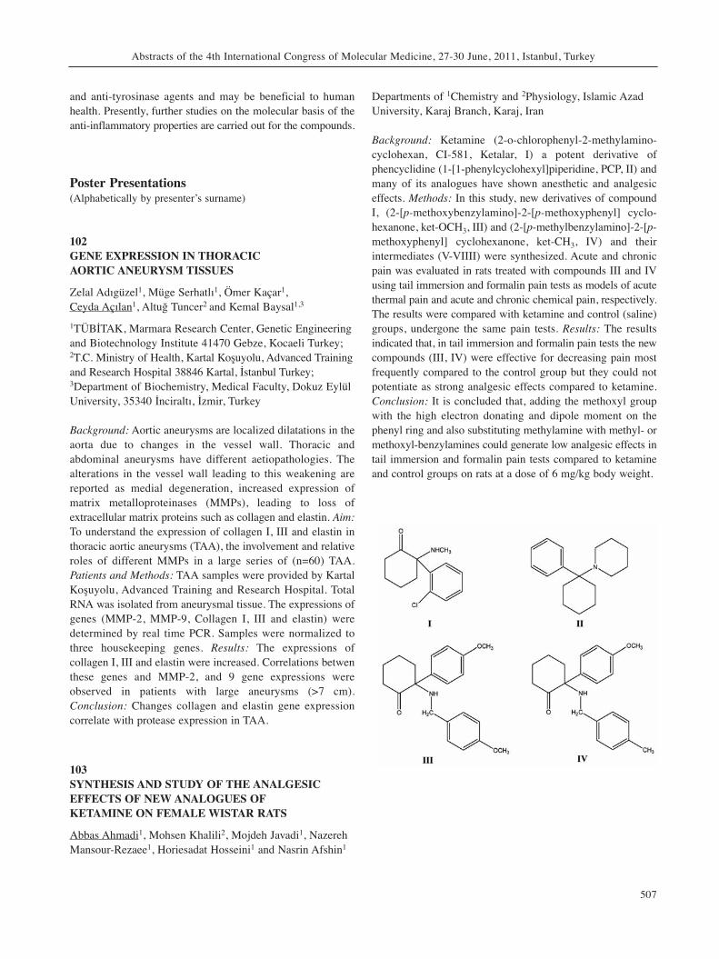
and anti-tyrosinase agents and may be beneficial to humanhealth. Presently, further studies on the molecular basis of theanti-inflammatory properties are carried out for the compounds.
Poster Presentations(Alphabetically by presenter’s surname)
102GENE EXPRESSION IN THORACIC AORTIC ANEURYSM TISSUES
Zelal Adıgüzel1, Müge Serhatlı1, Ömer Kaçar1, Ceyda Açılan1, Altuğ Tuncer2 and Kemal Baysal1,3
1TÜBİTAK, Marmara Research Center, Genetic Engineeringand Biotechnology Institute 41470 Gebze, Kocaeli Turkey;2T.C. Ministry of Health, Kartal Koşuyolu, Advanced Trainingand Research Hospital 38846 Kartal, İstanbul Turkey;3Department of Biochemistry, Medical Faculty, Dokuz EylülUniversity, 35340 İnciraltı, İzmir, Turkey
Background: Aortic aneurysms are localized dilatations in theaorta due to changes in the vessel wall. Thoracic andabdominal aneurysms have different aetiopathologies. Thealterations in the vessel wall leading to this weakening arereported as medial degeneration, increased expression ofmatrix metalloproteinases (MMPs), leading to loss ofextracellular matrix proteins such as collagen and elastin. Aim:To understand the expression of collagen I, III and elastin inthoracic aortic aneurysms (TAA), the involvement and relativeroles of different MMPs in a large series of (n=60) TAA.Patients and Methods: TAA samples were provided by KartalKoşuyolu, Advanced Training and Research Hospital. TotalRNA was isolated from aneurysmal tissue. The expressions ofgenes (MMP-2, MMP-9, Collagen I, III and elastin) weredetermined by real time PCR. Samples were normalized tothree housekeeping genes. Results: The expressions ofcollagen I, III and elastin were increased. Correlations betwenthese genes and MMP-2, and 9 gene expressions wereobserved in patients with large aneurysms (>7 cm).Conclusion: Changes collagen and elastin gene expressioncorrelate with protease expression in TAA.
103SYNTHESIS AND STUDY OF THE ANALGESICEFFECTS OF NEW ANALOGUES OF KETAMINE ON FEMALE WISTAR RATS
Abbas Ahmadi1, Mohsen Khalili2, Mojdeh Javadi1, NazerehMansour-Rezaee1, Horiesadat Hosseini1 and Nasrin Afshin1
Departments of 1Chemistry and 2Physiology, Islamic AzadUniversity, Karaj Branch, Karaj, Iran
Background: Ketamine (2-o-chlorophenyl-2-methylamino-cyclohexan, CI-581, Ketalar, I) a potent derivative ofphencyclidine (1-[1-phenylcyclohexyl]piperidine, PCP, II) andmany of its analogues have shown anesthetic and analgesiceffects. Methods: In this study, new derivatives of compoundI, (2-[p-methoxybenzylamino]-2-[p-methoxyphenyl] cyclo-hexanone, ket-OCH3, III) and (2-[p-methylbenzylamino]-2-[p-methoxyphenyl] cyclohexanone, ket-CH3, IV) and theirintermediates (V-VIIII) were synthesized. Acute and chronicpain was evaluated in rats treated with compounds III and IVusing tail immersion and formalin pain tests as models of acutethermal pain and acute and chronic chemical pain, respectively.The results were compared with ketamine and control (saline)groups, undergone the same pain tests. Results: The resultsindicated that, in tail immersion and formalin pain tests the newcompounds (III, IV) were effective for decreasing pain mostfrequently compared to the control group but they could notpotentiate as strong analgesic effects compared to ketamine.Conclusion: It is concluded that, adding the methoxyl groupwith the high electron donating and dipole moment on thephenyl ring and also substituting methylamine with methyl- ormethoxyl-benzylamines could generate low analgesic effects intail immersion and formalin pain tests compared to ketamineand control groups on rats at a dose of 6 mg/kg body weight.
Abstracts of the 4th International Congress of Molecular Medicine, 27-30 June, 2011, Istanbul, Turkey
507
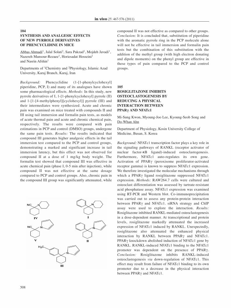
104SYNTHESIS AND ANALGESIC EFFECTS OF NEW PYRROLE DERIVATIVES OF PHENCYCLIDINE IN MICE
Abbas Ahmadi1, Jalal Solati2, Sara Pakzad1, Mojdeh Javadi1,Nazereh Mansour-Rezaee1, Horiesadat Hosseini1
and Nasrin Afshin1
Departments of 1Chemistry and 2Physiology, Islamic AzadUniversity, Karaj Branch, Karaj, Iran
Background: Phencyclidine (1-[1-phenylcyclohexyl]piperidine, PCP, I) and many of its analogues have shownsome pharmacological effects. Methods: In this study, newpyrrole derivatives of I, 1-[1-phenylcyclohexyl] pyrrole (II)and 1-[1-[4-methylphenyl][cyclohexyl]] pyrrole (III) andtheir intermediates were synthesized. Acute and chronicpain was examined on mice treated with compounds II andIII using tail immersion and formalin pain tests, as modelsof acute thermal pain and acute and chronic chemical pain,respectively. The results were compared with painestimations in PCP and control (DMSO) groups, undergonethe same pain tests. Results: The results indicated thatcompound III generates higher analgesic effects in the tailimmersion test compared to the PCP and control groups,demonstrating a marked and significant increase in tailimmersion latency, but this effect was not observed forcompound II at a dose of 1 mg/kg body weight. Theformalin test showed that compound III was effective inacute chemical pain (phase I, 0-5 min after injection), whilecompound II was not effective at the same dosagecompared to PCP and control groups. Also, chronic pain inthe compound III group was significantly attenuated, while
compound II was not effective as compared to other groups.Conclusion: It is concluded that, substitution of piperidinewith the aromatic pyrrole ring in the PCP molecule alonewill not be effective in tail immersion and formalin paintests but the combination of this substitution with theaddition of the methyl group (with high electron donatingand dipole moments) on the phenyl group are effective inthese types of pain compared to the PCP and controlgroups.
105ROSIGLITAZONE INHIBITSOSTEOCLASTOGENESIS BY REDUCING A PHYSICAL INTERACTION BETWEEN PPARγ AND NFATc1
Mi-Sung Kwon, Myoung-Joo Lee, Kyoung-Seob Song andDo-Whan Ahn
Department of Physiology, Kosin University College ofMedicine, Busan, S. Korea
Background: NFATc1 transcription factor plays a key role inthe signaling pathways of RANKL (receptor activator ofnuclear factor-κB ligand)-induced osteoclastogenesis.Furthermore, NFATc1 auto-regulates its own gene.Activation of PPARγ (peroxisome proliferator-activatedreceptor gamma) is known to suppress NFATc1 expression.We therefore investigated the molecular mechanisms throughwhich a PPARγ ligand rosiglitazone suppressed NFATc1expression. Methods: RAW264.7 cells were cultured andosteoclast differentiation was assessed by tartrate-resistantacid phosphatase assay. NFATc1 expression was examinedusing RT-PCR and Western blot. Co-immunoprecipitationwas carried out to assess any protein-protein interactionbetween PPARγ and NFATc1. siRNA strategy and ChIPassay were used to explore the interaction. Results:Rosiglitazone inhibited RANKL-mediated osteoclastogenesisin a dose-dependent manner. At transcriptional and proteinlevels, rosiglitazone markedly attenuated the increasedexpression of NFATc1 induced by RANKL. Unexpectedly,rosiglitazone also attenuated the enhanced physicalinteraction by RANKL between PPARγ and NFATc1.PPARγ knockdown abolished induction of NFATc1 gene byRANKL. RANKL-induced NFATc1 binding to the NFATc1promoter was dependent on the presence of PPARγ.Conclusion: Rosiglitazone inhibits RANKL-inducedosteoclastogenesis via down-regulation of NFATc1. Thiseffect may result from failure of NFATc1 binding to its ownpromoter due to a decrease in the physical interactionbetween PPARγ and NFATc1.
in vivo 25: 467-576 (2011)
508
106URINE CYTOLOGY, URINARY SURVIVIN mRNAEXPRESSION AND NUCLEAR MATRIX PROTEIN 22IN THE DETECTION OF TRANSITIONAL CELLCARCINOMA OF THE BLADDER
May Al-Maghrebi1, Elijah O. Kehinde2, Kusum Kapila3,Fahd Al-Mulla3 and Jehoram T. Anim3
Departments of 1Biochemistry, 2Surgery (Division ofUrology) and 3Pathology, Faculty of Medicine, KuwaitUniversity, Kuwait
Background: The search for an objective and sensitive markerfor the detection of bladder cancer is an active field oftranslational research. Thus, we aimed to assess the sensitivityand specificity of survivin mRNA expression and NMP22BladderCheck (BC) test in comparison to urine cytology (UC)for the detection of transitional cell carcinoma (TCC) of thebladder. Methods: Voided urine samples collected from 41healthy controls and 80 patients diagnosed with TCC of thebladder were subjected to UC, NMP22BC test and reversetranscription-quantitative real time PCR for survivin mRNAexpression. Results: Survivin mRNA expression in healthycontrols was significantly different from patients with TCC ofthe bladder (p<0.0001) with a positive correlation with G2 andG3 grade tumors, but not with low grade G1 tumors. A cut-offvalue of 2 for survivin expression yielded 92% sensitivity and100% specificity. NMP22BC had 61.3% sensitivity and 100%specificity, while UC had 40% sensitivity and 76% specificity.Conclusion: Quantitative urinary survivin mRNA expressionenhances the sensitivity and specificity of UC alone or incombination with the NMP22BC test for the detection of TCCof the bladder.
107GENETIC DETERMINANTS OF L-THYROXINEDOSE REQUIREMENT IN ATHYROTIC PATIENTSWITH DIFFERENTIATED THYROID CANCER ANDTHEIR POTENTIAL ROLE IN ITS ETIOLOGY
Maha M. Al-Rasheed1,2,3, Angela Macadam1, AndrewOverall1, Ali Alzahrani2, Nduna Dzimiri2 and Paul R. Gard1
1School of Pharmacy and Biomolecular Sciences, Universityof Brighton, Brighton, U.K.; 2King Faisal Specialist Hospital and Research Center,Riyadh, Saudi Arabia;3School of Pharmacy, King Saud University, Riyadh, SaudiArabia
Background: Patients with differentiated thyroid cancer (DTC)are usually managed with total thyroidectomy and subsequentradioiodine ablation of the remnant thyroid tissue. L-thyroxine
(L-T4) therapy (about 2 μg/kg) is necessary with a widevariation in patient dose requirements. Therefore, the aims ofthis study were to identify sodium iodide symporter (NIS)single nucleotide polymorphisms (SNPs) involved in thyroidhormone metabolism and to evaluate their association withDTC and L-T4 dose requirement in the Saudi population.Methods: Detection of SNPs was accomplished in 100 DTCpatients and genotyping for 3 selected SNPs (rs4808708,rs4808709 and rs7250346) was performed in 409 DTC casesand 406 controls. Results: A total of 22 SNPs were identifiedin the NIS gene; 7 of them were novel. There was significantassociation of the G allele in rs4808708 and rs7250346variants [(Odds ratio (95% CI), 1.30 (1.05-1.60); p=0.016 and2.30 (1.20-4.39); p=0.012), respectively] and for the AAgenotype of the rs4808708 variant [(Odds ratio (95% CI),1.38(1.05-1.82)] with DTC, independent of age and sex. Nosignificant association was found for these SNPs with L-T4dose requirement. Conclusion: The study identified two NISvariants associated with an increased risk of DTC. In DTCpatients, the variability in the L-T4 dose requirement does notappear to be related with NIS polymorphisms.
108TRANSCRIPTIONAL ACTIVATION OF THE COPPER/ZINC SUPEROXIDE DISMUTASE GENE BY ELK-1
Göksu Alpay and Işil Aksan Kurnaz
Department of Genetics and Bioengineering, University ofYeditepe, Istanbul, Turkey
Background: Elk-1 is activated by the MAPK pathway and itis a member of the ETS domain transcription factor.Copper/Zinc superoxide dismutase SOD1 participates in thecontrol of the intracellular concentration of reactive oxygenintermediates and catalyzes the dismutation of superoxideradicals. According to recent studies, Elk-1 has a bindingdomain in the SOD1 positive regulatory element in thepromoter region and enhances SOD1 transcription. In thisstudy, we aimed to explore the relationship between Elk-1 andSOD1. Methods: We determined the binding motifs of Elk-1in SOD1 human and rat promoter regions. Primers weredesigned according to these regions and these regions werecloned into pgl-3 luciferase vector. In further analyses, we willmake a luciferase assay to find out the effect of Elk-1 onSOD1 transcription. We will also use mutated Elk-1expression plasmids to analyze the functional role of Elk-1.Results: Rat and human promoter regions were successfullycloned to pgl-3 vector. Conclusion: Since it has been shownthat Elk-1 has a positive effect on SOD1 transcription weexpect to see a decrease in activation of SOD1 when Elk-1 isabsent or mutated.
Abstracts of the 4th International Congress of Molecular Medicine, 27-30 June, 2011, Istanbul, Turkey
509
109EFFECTS OF ROSIGLITAZONE OR METFORMINTREATMENT ON LEPTIN AND OXIDATIVE STRESSPARAMETERS IN TYPE 2 DIABETIC PATIENTS
Şura Esra Altinkaynak1, Aymelek Gönenç1, Nilüfer Bayraktar2 and Cüneyt Anil3
1Department of Biochemistry, Faculty of Pharmacy, GaziUniversity, Ankara, Turkey;Departments of 2Biochemistry and 3Endocrinology, BaşkentHospital, Bahçeli, Ankara, Turkey
Background: It is generally accepted that oxidative stress isresponsible for the etiology and complications of diabetes.Methods: Leptin malondialdehyde (MDA), total antioxidantcapacity (TAC), superoxide dismutase (SOD), catalase(CAT), paraoxonase and 8-hydroxy-2’-deoxyguanosine (8-OHdG) were measured in 30 healthy subjects and 50 patientswith diabetes mellitus. MDA and TAC were assayed withspectrophotometric methods. Leptin, SOD, CAT,paraoxonase and 8-OHdG were analyzed using appropriatecommercially available kits. Results: Before treatment, SODand TAC levels were decreased in all patients compared tocontrols (p=0.005, p=0.01, respectively), while MDA levelswere increased (p=0.024). The patients with diabetesmellitus were divided into two groups: the rosiglitazonegroup (n=25) and the metformin group (n=25). Treatmentwas carried out for three months. Before treatment, in therosiglitazone group, TAC levels were diminished (p=0.015),while MDA levels were increased (p=0.019) compared tocontrols. After treatment, decreased MDA and 8-OHdGlevels were found (p=0.008, p=0.000, respectively) in therosiglitazone group compared to the pretreatment values ofthis group. Before treatment in the metformin group, SODlevels were decreased compared to controls (p=0.000). Aftertreatment, SOD and TAC levels were increased (p=0.029,p=0.000) in the metformin group compared to thepretreatment values of this group. Conclusion: Our resultssuggest that oxidative stress seems to decrease withrosiglitazone or metformin treatment.
110SERUM PARAOXONASE-1 ACTIVITIES ANDOXIDATIVE STATUS IN PATIENTS WITH PLAQUE-TYPE PSORIASIS WITH/WITHOUTMETABOLIC SYNDROME
Murat Usta1, Enver Turan2, Hale Aral1, Berrin Bercik Inal1,Mehmet Salih Gurel2 and Guvenc Guvenen1
1Clinical Biochemistry Laboratory and 2Department ofDermatology, Ministry of Health Istanbul Research andTraining Hospital, Istanbul, Turkey
Background: Psoriasis is a chronic immune-mediatedinflammatory skin disease associated with metabolicsyndrome, which is made up of a cluster of disordersincluding obesity, diabetes mellitus, dyslipidaemia, andcardiovascular disease. The aim of the present study was toinvestigate serum paraoxonase-1 activities and oxidative statusparameters in patients with plaque-type psoriasis with orwithout metabolic syndrome. Methods: In this study, psoriaticpatients with (n=25) or without (n=27) metabolic syndromeaccording to the criteria of the International DiabetesFederation (IDF) were matched for age and sex to an equallysized control group (n=25). Results: In patients withoutmetabolic syndrome, serum paraoxonase and arylesteraseactivities showed mean decreases of 29% and 6%,respectively, whereas in patients with metabolic syndrome, themean decreases in the enzymes’ activities were 35% and 11%,respectively, compared with those in the control group. Serumtotal antioxidant capacity and total oxidant status were notstatistically significant in any of the three groups. Conclusion:The significant decrease observed in serum paraoxonaseactivity was independent of the metabolic syndrome inpatients with mild-to-moderate plaque-type psoriasis, whereasthe significant decrease in serum arylesterase activity wasassociated with the metabolic syndrome.
111TURNAROUND TIME OF CARDIAC MARKERS IN EMERGENCY DEPARTMENT
Hale Aral1, Berrin Bercik Inal1, Pinar Bilgi1, Abdurrahim Akgundogdu2 and Guvenc Guvenen1
1Clinical Biochemistry Laboratory, Ministry of HealthIstanbul Research and Training Hospital, Istanbul, Turkey;2Electrical and Electronics Department, Engineering Faculty,Istanbul University, Istanbul, Turkey
Background: We aimed at determining the turn-around-time(TAT) of MB isoenzyme of creatine-kinase (CK-MB) andtroponin I (TnI) in the host Emergency DepartmentLaboratory. Methods: Serum CK-MB levels were assayedusing in Advia 1800 instrument using the immuno-inhibitionmethod, and TnI levels on Centaur CP using thechemiluminescence method (Siemens Healthcare Diagnostics,Germany). TAT values of TnI (n=11,532) and of CK-MB(n=17,399) from June to December 2010 were included in thisstudy. In data analysis, the time outcomes were successfullyclustered (two clusters; 0.86, 0.14) separately by SimpleExpectation Maximisation (EM) class (weka). EM assigns aprobability distribution to each instance which indicates theprobability of it belonging to each of the clusters. EM allowsone to decide how many clusters to create by cross validation,or it is possible to specify a priori how many clusters to
in vivo 25: 467-576 (2011)
510
generate. Results: The time when the specimen was orderedto when the result was available was significantly shorter in86 % of CK-MB (55.84±16.52 minutes, log likelihood: -27.77249) compared with 86 % of TnI (61.10±16.63 minutes,log likelihood: -25.27239). Conclusion: Local factorsincluding clinical (physicians and nurses) and laboratory staff,sample transport, diagnostic equipment should be revisedperiodically in emergency departments in order to improveperformance.
112THE STUDY OF SER892GLY AND GLY972ARGPOLYMORPHISMS OF IRS-1 GENE IN TYPE 2DIABETIC PATIENTS IN KONYA REGION
Hilal Arikoglu1, Hulya Ozdemir2, Melda Aksoy Hepdogru2,M. Sait Gonen3, Dudu Erkoc Kaya1 and S. Ali Kayis4
1Department of Medical Biology, Selcuklu Medical Facultyand Departments of 2Medical Biology and 3Endocrinology,Meram Medical Faculty, Selcuk University, Konya, Turkey;4Biometry-Genetics Unit, Faculty of Agriculture, SelcukUniversity, Konya, Turkey
Background: Type 2 diabetes mellitus (T2DM) ischaracterized by a combination of peripheral insulinresistance and impaired insulin secretory capacity ofpancreatic β-cell. Insulin receptor substrate-1 (IRS-1) is oneof multiple proteins that mediate signal transduction of theactivated insulin reseptor. We aimed at determining theallele profiles and the risk alleles of the SNPs Ser892Glyand Gly972Arg in the IRS-1 gene and to identify theassociations with the disease in patients in the Konya regionof Turkey, in which type 2 diabetes is very common.Methods: Our study included 169 type 2 diabetic patientsand 69 healthy controls. Fasting plasma glucose, fastinginsulin, HbA1c and c-peptide values were measured for allpatients and controls. Oral glucose tolerance test was donein healthy controls. Target SNPs in IRS-1 gene werescreened by polymerase chain reaction-single strandedconformational polymorphism (PCR-SSCP) technique. Chi-Square test was employed in examination of SNP genotypesrelationship in patients and controls. Single point regressionanalysis and Kruskal-Wallis test were employed to obtainSNP genotype - biochemical phenotypes relationship fornormally and non-normally distributed phenotypes,respectively. In all tests p-value <0.05 was consideredstatistically significant. Results: Ser892Gly and Gly972Argpolymorphisms in IRS-1 gene that were screened in ourstudy were not associated with the disease (p>0.05).Conclusion: We found no evidence for an association ofSer892Gly and Gly972Arg polymorphisms of IRS-1 genewith type 2 diabetes (p>0.05).
113GENETIC CHARACTERIZATION OFFLUOROQUINOLONE-RESISTANT KLEBSIELLA PNEUMONIAE ISOLATES IN RUSSIA
Maria Atroshkina, Elena Ilina and Vadim Govorun
Research Institute for Physical-Chemical Medicine ofMinistry of Public Health of Russian Federation, Moscow,Russia
Background: Klebsiella pneumoniae is a frequent cause ofnosocomial pneumonia and fluoroquinolones are potentialantibiotics used for its treatment. In this study, thecontribution of genetic factors in the development of K.pneumoniae resistance to fluoroquinolones was examined.Methods: Susceptibility testing of K. pneumoniae wasperformed by the disc-diffusion method. Genomic DNA waspurified using DNA Express kit (Lytech Ltd, Russia).Detection of single nuclear polymorphisms in Ser83 andAsp87 codons of gyrA and in Ser80 and Glu84 codons ofparC was performed by PCR-primer extension/reactionfollowed by mass spectrometry. Results: In total 96 isolatesof K. pneumoniae were tested. The most susceptible tofluoroquinolones isolates (12/13; 92.3%) carried neithergyrA nor parC mutations. All moderately resistant isolates(7/7, 100%) exhibited mutations only in gyrA, while themajority of resistant isolates (74/79, 93.7%) revealedmutations both in gyrA and parC genes. Additionally, thecapability of direct identification of such mutations in K.pneumoniae genomic DNA purified from urine samples wasshown. Conclusion: Identification of fluoroquinoloneresistance-associated mutations may be useful tobacteriologists; however, only the mutations in gyrA andparC genes led to clinically relevant resistance of K.pneumoniae to fluoroquinolones.
114THE NF-κΒ INHIBITOR ΙκΒα NEGATES COLONCANCER CELL MIGRATION, INVASION,PROLIFERATION AND TUMOR GROWTH
Samir Attoub1,6, Rabah Iratni2,5, Suhail Al-Salam3, Khouloud Arafat1, M.A.H Al Sultan1, Nadia Al Marzouqi1,Eric Bruyneel4, Marc Bracke4, Olivier De Wever4 andChristian Gespach6
1Department of Pharmacology and Therapeutics, and3Department of Pathology, Faculty of Medicine and HealthSciences, UAE University, P. O. Box: 17666, Al Ain, UnitedArab Emirates;2Department of Biology, UAE University, P.O. Box: 17551,Al Ain, United Arab Emirates;
Abstracts of the 4th International Congress of Molecular Medicine, 27-30 June, 2011, Istanbul, Turkey
511
4Laboratory of Experimental Cancer Research, UniversityHospital, De Pintelaan 185, B-9000 Gent, Belgium; 5Institut Albert Bonniot, INSERM U823, University JosephFourier, Grenoble 1, Site Santé, 38042, Grenoble, Cedex 6,France;6INSERM U 673 and U938, Molecular and Clinical Oncologyof Solid Tumors; University Pierre et Marie Curie Paris VI,Hôpital Saint-Antoine, 75571 Paris Cedex 12, France
It is well accepted that the NF-κΒ pathways are involved ininflammatory diseases, cancer development and progressionin human solid tumors. The NF-κΒ signaling element ΙκΒαwas shown to inactivate NF-κΒ activity throughsequestration of this transcription factor in the cytoplasm. Inthe present study, we investigated the impact of the ΙκΒα onthe invasive growth of human colon cancer cells HCT8/S11stably transfected by this endogenous NF-κΒ inhibitor. Wereport that ΙκΒα ectopic expression inhibited NF-κΒpromoter activity induced by the Y527Fsrc oncogene andreduced HCT8/S11 cell migration in wound healing assays.Our data show that ΙκΒα abrogated collagen type I invasioninduced by the trefoil factors TFF1 and TFF3 but wasineffective on the invasive phenotype determined by leptin.Moreover, ΙκΒα reduced HCT8/S11 cell proliferation invitro and the growth of their corresponding tumor xenograftsestablished in the athymic mice. Taken together our datademonstrated that the intrinsic NF-κΒ inhibitor ΙκΒαnegates several transforming functions in human coloncancer cells. Our data provide the rationale for furtherpreclinical and clinical studies based on therapeuticinterventions targeting NF-κΒ pathway.
115IDENTIFICATION OF ALLOSTERIC RESIDUES INTHE ATPase DOMAIN OF HSP70 MOLECULARCHAPERONES UPON SUBSTRATE BINDING
İrem Avcılar, Umut Günsel, Ani Kıçik, Gökhan Gün andGizem Dinler-Doğanay
Molecular Biology and Genetics Department, IstanbulTechnical University, Istanbul, Turkey
Background: Hsp70 chaperones play important roles in cellsincluding protein folding, trafficking, degradation and enablingsurvival under stress conditions. DnaK is an Echerichia coliHsp70 homolog comprising an ATPase and a substrate-bindingdomain. Communication between the domains is essential forchaperone function. Previous studies showed that DnaK(1-392), containing the ATPase domain and the entire linkerregion, can mimic the substrate-stimulated form of full-lenghDnaK showing an ATPase rate similar to that of the substrate-present state for the full-length protein. Using this knowledge,
we aimed to understand the allosteric mechanism underlyingthe substrate binding effects to the ATPase domain bypinpointing the putative residues that are present in this pathusing DnaK(1-392). Methods: We identified sites that can becritical for allostery and applied protein engineering methodsto make replacement mutants. We purified the mutants,measured their ATPase levels and studied their effects onstability using circular dichroism. Results and Conclusion: Wefound that a particular mutation site is critical for the activityand allostery of the ATPase domain of DnaK. This mutationcaused an enhancement in the activity of the ATPase domainand also altered its stability by lowering the first meltingtransition from 50˚C to 48˚C, suggesting that mutation to thatsite may have a role in the regulation of the ATPase dynamicsof the domain.
116EFFECT OF F68 PLURONIC BLOCK COPOLYMERON ELECTROPORATION OF HeLa CELLS
Safa Aydın1,2, Mehmet Emir Yalvaç1, Aysu Yilmaz1, Ferruh Ozcan2 and Fikrettin Şahin1
1Department of Genetics and BioEngineering, College ofEngineering and Architecture, Yeditepe University, Istanbul,Turkey;2Department of Molecular Biology and Genetics, Faculty ofScience, Gebze Institute of Technology, Cayirova, Gebze,Kocaeli, Turkey
Background: Electroporation is an efficient non-viral genedelivery method having some adverse effects on cells; forexample, the phospholipid bilayer of the cell is temporarilydisturbed by electrical pulses and the cell pores may becometoo sizeable or collapse after membrane discharge, causing celldamage. Pluronic F68 is a non-ionic and small foamingsurfactant consisting of a central polypropylene oxide and twopolyethylene-oxide groups. F68 was displayed to interact withmembrane lipid bilayer to stabilize it. In this study, we aimedto increase the transfection efficiency in HeLa cells by addingF68 during and after electroporation. Methods: HeLa cellswere electroporated at various voltages in 0.2-mm cuvvettescontaining RPMI-1640 medium without serum, with orwithout F68 and with 5μg of pEGFP-N2 plasmid DNA. Aftertransfection, the cell viability and transfection efficiency weremeasured by using MTS assay and flow cytometry,respectively. Results: The optimum conditions forelectroporation of HeLa cells were determined to be 140V at500μF capacitance, which resulted in 36 % transfectionefficiency. When the cells were electroporated in the presenceof F68, the efficiency increased by 30%, lowering the celldeath during electroporation. Discussion: The major drawbackof electroporation is that some cells are highly sensitive to the
in vivo 25: 467-576 (2011)
512
electrical stress occurring during electroporation. Our datasuggest that F68 may enable the transfection of genes intosensitive cell lines by lowering cell death and increasing thetransfection efficiency.
117DETECTION OF GENE POLYMORPHISMSASSOCIATED WITH THE LOSS OF BONE MINERAL DENSITY (CASE REPORT)
Belkıs Aydınol1, Kemal Nas2, Sedat Genç1 and BurhanBaykara2
Departments of 1Clinical Chemistry and 2Physical Therapyand Rehabilitation, Medical Faculty, Dicle Üniversity,Diyarbakır, Turkey
Background: Osteoporosis is a common disease which ischaracterised by low bone mass and an increased risk offracture. Candidate genes which have been studied in relationto BMD (bone mineral density) and osteoporotic fracturesinclude Vitamin D receptor, estrogen receptor, Col1 A1 gene,calcitonin receptor. We present here a case of a 39 year oldman whose BMD result was: Z score, -4.2; T score, -4.3.Methods: We used clinical array systems to detectpolymorphisms. This method is based on a low density chipat the bottom of a classical 2 ml tube. DNA was extractedfrom blood using EDTA. DNA amplification,denaturation,hybridization and the other steps were carried out.Biochemical analyses, complete blood analysis were recorded.Results: We analysed Col1A1-SP1, CTR-ALU1, ESR1X-XBAI, ESR1P-PVUII, VDRF-FOKI and VDRB-BSMIpolymorphisms for collagen type1 gene, calcitonin receptorgene, estrogen receptor gene and vitamin D receptor generespectively. The analysis showed that his genotype was Ss,Aa, ,pp, Xx, bb, Ff (normal genes are indicated by capitalletters). Conclusion: Positive associations between thesepolymorphisms and bone density have previously beenreported by several studies. These type of studies will assistin clinical desicion making and support certain therapies,especially for early age fractures and early treatment for boneprevention.
118SOME RARE COMPLEX MUTATIONS OF MEFVGENE IN DIYARBAKIR REGION OF TURKEY
Belkıs Aydınol1, SedatYılmaz1, Sedat Genç1 and M. Mustafa Aydınol2
1Department of Clinical Biochemistry, Medical Faculty,Dicle University, Diyarbakır, Turkey;2Province Health Center, Diyarbakır, Turkey
Background: FMF is an autosomal recessive inflammatorydisorder that predominantly affects Jews, Armenians, Turksand Arabs. It is characterised by recurrent fevers, abdominal,chest, joint pains and erysipelas-like skin disease. Methods: Inthis study, 1,428 patients who attended to Dicle University,Medical Faculty with complaints of fever and joint andabdominal pains were studied for FMF mutation detection byVienna lab. strip assay method. Results: Mutations weredetected in 689 patients, eight of which were interesting sincethey combined complex mutations. The mutations were asfollows: triple heterozygous, double homozygous,homozygous + heterozygous cases. Triple mutations:E148Q/P369S/M694V (2 cases), E148Q/P369S/M694I (1case). Double homosygous: E148Q/E148Q/P369S/P369S (2cases). Homozygous + heterozygous mutations:E148Q/E148Q R761H/R761H M694V/M694V M6 94Vheterozygous (1 case), E148Q heterozygous (1 case) andE148Q heterozygous (1 case). According to our literaturesearch, we considered the influence of these mutations ondisease prognosis. Double homozygous case types were themost common reported in the literature. Conclusion: A highprevalence of FMF mutations was found in this region.Genetic analysis must be performed in all patients withsuspected of FMF. Due to the complexity and interference ofthese mutations, genetic counseling and new-born screeningmust be performed in this region.
119EFFECTS OF THE PPAR-GAMMA ANDAPOLIPOPROTEIN E GENE POLYMORPHISMS ON CLINICAL AND LIPID CHARACTERISTICS IN PATIENTS WITH DIABETIC AND NON-DIABETIC CORONARY HEART DISEASE
Hulya Yilmaz Aydogan1, Ozlem Kurnaz1, Ozlem Kurt2, A. Basak Akadam Teker1, Ozlem Kucukhuseyin1, Atike Tekeli3, Burcu Caykara1 and Turgay Isbir1
1Department of Molecular Medicine, Institute forExperimental Medicine, 2Department of Biochemistry,Faculty of Pharmacy, Istanbul University, Istanbul, Turkey; 3Department of Cardiovascular Surgery, Uskudar StateHospital, Istanbul, Turkey
We investigated whether Pro12Ala and C161T variants in thePPARgamma2 gene are associated with the occurrence oftype-2 coronary heart disease (CHD) in patients prospectivelycharacterized for the presence or absence of diabetes mellitus.PPAR-gammaPro12Ala, PPAR-gamma C161T andapolipoprotein E gene polymorphisms were determined usingPCR-RFLP in 262 patients with CHD (103 T2DM, 159 non-diabetic) and 105 healthy people. Statistical analysis revealedno significant difference both in the PPAR gamma Pro12Ala
Abstracts of the 4th International Congress of Molecular Medicine, 27-30 June, 2011, Istanbul, Turkey
513
and ApoE genotypes and allele frequencies between the studygroups (p>0.05). However, PPARgamma ProAla heterozygotegenotype and 12Ala allele seemed to be protective againsthypertension and left ventricular hypertrophy in males.Statistically significant differences were observed in genotypefrequencies between the healthy and diabetic/total patientgroups in the distribution of PPARgamma C161T (p<0.05).The frequency of CC genotype of PPAR-gamma C161T washigher in the diabetic patients than in the controls. We caninfer from these data that diabetic patients with CC genotypemay be susceptible to the development of CHD and that Tallele may be protective against this disease. The frequency ofT161allele in diabetic and in total CHD patients wassignificantly lower in males than in the females. PPARgamma161T allele was associated with increased total cholesterollevels in total patient group. We observed that 161T allelestrengthened the effect of ApoE4 allele on cholesterol levels.Furthermore, male patients carrying T161 allele had a lowerprevalence of left ventricular hypertrophy than female patients.Our findings suggest that PPARgamma C161T polymorphismmay contribute to the development of CHD, especially in malediabetic patients.
120PROTEIN CARBONYLATION AS A BIOMARKER OF OXIDATIVE STRESS
Burcu Baba1, Aymelek Gönenç1, Sabahattin Aslan2 andAysun Hacışevki1
1Department of Biochemistry, Faculty of Pharmacy, GaziUniversity, Ankara, Turkey;2Department of Surgery, Ankara Oncology Educational andResearch Hospital, Ankara, Turkey
Background: Reactive oxygen species (ROS) can damage allbiomolecules. Oxidative modifications of enzymes andstructural proteins plays a significant role in the aetiologyand/or progression of several human diseases. Protein carbonylcontent is the most widely used marker of oxidativemodification of proteins. Besides, it is known that TNF-αcontributes to formation of ROS. We aimed to investigatechanges of TNF-α and protein carbonyl levels ingastrointestinal cancer patients compared to healthy controlindividuals. Methods: Serum TNF-α and protein carbonyllevels in gastrointestinal cancer patients (n=108) and healthycontrols (n=35) were measured. Patients were divided intothree groups: the first group had gastric cancer, the secondgroup had colorectal cancer and the third group had pancreas,liver and esophagus cancer. TNF-α and protein carbonyl levelswere analyzed by using kits. Results: The levels of TNF-α andprotein carbonyl were statistically higher in cancer patientscompared with the healthy controls (p<0.01). The mean values
for protein carbonyl were found to be significantly higher inthe first and second groups than in controls or the third group(p<0.01). There was a significant difference between the thirdgroup and controls for TNF-α (p<0.05). Conclusion:Enhanced protein carbonyl levels may be a diagnosticbiomarker for oxidative damage in cancer.
121ROLE OF C3435T AND G2677T/A POLYMORPHISMSIN THE DRUG-TRANSPORTER GENE MDR1 INMULTIDRUG-RESISTANT EPILEPSY
Bahadır Batar1,2, Özlem Tun1, Gözde Yeşil2, Mehmet Seven2,Adnan Yüksel2 and Mehmet Güven1
Departments of 1Medical Biology and 2Medical Genetics,Cerrahpasa School of Medicine, Istanbul University,Istanbul, Turkey
Background: Multiple drug resistance is a common problemin the treatment of epilepsy, and approximately 30% ofpatients continue to have seizures despite all therapeuticinterventions. Among various classes of drug transporters,genetic variants of P-glycoprotein (P-gp) encoded by themultidrug resistance 1 (MDR1) gene have been associatedwith drug-resistant epilepsy. Our aim was to investigate theeffect of the C3435T and G2677T/A polymorphisms ofMDR1 on drug resistance in Turkish patients with epilepsy.Methods: MDR1 C3435T and G2677T/A were genotyped in103 patients with epilepsy, classified as drug-resistant in 46and drug-responsive in 57, and 150 control subjects withoutepilepsy. Genotypes of the C3435T and G2677T/Apolymorphisms were determined by polymerase chain reactionfollowed by restriction fragment length polymorphism.Results: There was no statistically significant differencebetween genotype and allele frequencies of drug-resistant anddrug-responsive epilepsy patients. The frequencies in the drug-resistant and drug-responsive groups did not differsignificantly from control subjects. Conclusion: MDR1polymorphisms investigated in this study are not associatedwith drug resistance in Turkish epileptic patients.
122ASSOCIATION BETWEEN GENETIC VARIATION ONCHROMOSOME 9p21 AND HYPERTENSION
Burcu Bayoglu1, Mujgan Cengiz1, Huseyin Altug Cakmak2,Omer Uysal3 and Husniye Yuksel2
Departments of 1Medical Biology and 2Cardiology,Cerrahpasa School of Medicine, Istanbul University,Istanbul, Turkey;3Department of Biostatistics and Medical Informatics,
in vivo 25: 467-576 (2011)
514
School of Medicine, Bezmialem Vakif University, Istanbul,Turkey
Background: Hypertension is a complex multifactorial diseasecaused by interactions between genetic and environmentalfactors. It is an independent determinant of cardiovascular risk.Recent genome-wide association studies (GWAS) revealedthat some 9p21 variants are associated with cardiovasculardiseases (CVD). The aim of this study was to investigate thepossible influence of the single nucleotide polymorphism(SNP), rs10757274, on chromosome 9p21 on a sample ofTurkish patients with hypertension. Methods: In this study,rs10757274 A/G polymorphism was studied in 170 patientswith hypertension and 109 healthy subjects. Genomic DNAwas extracted from peripheral blood leukocytes. 9p21rs10757274 A/G polymorphism was genotyped by real timepolymerase chain reaction. Results: For rs10757274 A/Gpolymorphism the frequencies of the AA, AG and GGgenotypes were 12.4%, 46.4% and 41.2% in the hypertensionpatients and 24.8%, 53.2% and 22% in the healthy controls,respectively. An extremely significant relationship wasobserved in genotype distribution between the hypertensionpatients and the healthy controls for rs10757274polymorphism (p=0.001). No significant relationship wasobserved when genotype frequencies were compared withbody mass index values in the hypertension patients (F=2.365,p=0.097). Conclusion: We suggest that 9p21 rs10757274 A/Gpolymorphism may play an important role in the pathogenesisof hypertension.
123ASSOCIATION OF A COMMON VARIANT ONCHROMOSOME 9p21 IN TURKISH PATIENTS WITH METABOLIC SYNDROME
Burcu Bayoglu1, Mujgan Cengiz1, Huseyin Altug Cakmak2,Omer Uysal3 and Husniye Yuksel2
Departments of 1Medical Biology and 2Cardiology,Cerrahpasa School of Medicine, Istanbul University,Istanbul, Turkey;3Department of Biostatistics and Medical Informatics,School of Medicine, Bezmialem Vakif University, Istanbul,Turkey
Background: The metabolic syndrome is a complicatedsyndrome causing insulin resistance, hyperinsulinemia, centralobesity, dyslipidemia, atherosclerosis and inflammation. It isoften accompanied by different types of cardiovascular disease(CVD). Recent genome-wide association studies (GWAS)revealed that some 9p21 variants are associated with type-2diabetes and CVD. In this study, we examined whether thesingle nucleotide polymorphism (SNP), rs1333049, on
chromosome 9p21 is associated with the metabolic syndrome.Methods: A sample of 66 unrelated Turkish patients withmetabolic syndrome and 122 healthy controls wereinvestigated in this study. Genomic DNA extraction wasperformed from peripheral blood leukocytes. 9p21 rs1333049C/G polymorphism was genotyped by RT-PCR. Results: Thefrequencies of the CC, CG and GG genotypes were 25.7%,47% and 27.3% in the metabolic syndrome patients and 41%,43.4% and 15.6% in the healthy controls, respectively. Astatistically borderline significance was observed in thegenotype distribution between the metabolic syndromepatients and the control group for the rs1333049polymorphism (p=0.053). No significant relationship wasobserved when genotype frequencies were compared withbody mass index values in metabolic syndrome patients(F=0.66, p=0.51). Conclusion: We suggest that the 9p21rs1333049 polymorphism may play an important role in thepathogenesis of the metabolic syndrome. However, furtherstudies are necessary to confirm these findings.
124THE POLYMORPHISM OF CODON D727E OF TSHR(THYROID-STIMULATING HORMONE RECEPTOR)GENE IN A TURKISH POPULATION WITHMULTINODULAR GOITER
Banu Bayram1, Reşit Sönmez2, Harun Önlü1, Sedat Bozarı1,Züleyha Türkoğlu1 and Fezan Şahin3
1Department of Biology, Faculty of Science and Arts, MuşAlparslan University, Muş, Turkey;2Department of General Surgery, Private Muş Şifa Hospital,Muş, Turkey;3Department of Biostatistics, Faculty of Medicine, EskişehirOsmangazi University, Eskişehir, Turkey
Background: Multinodular goiter involves more than onenodules, is the most common endocrine pathology in theworld and is still a problem in the clinic. Genetic variation isan important factor in the development of the disease. Thisstudy was performed to determine the allele frequencies anddistribution of genotypes of codon D727E polymorphism ofthe TSHR (thyroid-stimulating hormone receptor) gene in anendemic Turkish multinodular goiter population. Methods:Genomic DNA was extracted from 172 persons (118 patientswith multinodular goiter and 54 healthy controls). Thepolymorphism of codon D727E of the TSHR gene wasidentified by polymerase chain reaction-based restrictionanalysis. Results: There was a statistically significantdifference between the groups with respect to genotypedistribution (p<0.05). The C and G allele frequencies were92% and 8% in the patients, respectively, while the respectivefrequencies in the control group were 100% and 0%.
Abstracts of the 4th International Congress of Molecular Medicine, 27-30 June, 2011, Istanbul, Turkey
515
Conclusion: This study suggests that the polymorphism ofcodon D727E of the TSHR gene may be a marker foridentifying multinodular goiter. However, there is no directrelationship between this gene polymorphism and the disease.
125THE ASSOCIATION OF PARAOXONASE IPHENOTYPES WITH ASYMMETRICDIMETHYLARGININE IN MYOCARDIAL INFARCTION
Seldag Bekpinar1, Yesim Unlücerci1, Akar Yilmaz2, Ayten Can1 and Figen Gürdöl1
Departments of 1Biochemistry and 2Cardiology, IstanbulFaculty of Medicine, Istanbul University, Istanbul, Turkey
A genetic change affecting the primary sequence at glutamine192 on paraoxonase-1 (PON-1) protein gives rise to twoallozymes in human serum. The polymorphic BB type of PON-1 is believed to be associated with cardiovascular diseasesbecause it is being less effective at protecting LDL fromoxidation. Asymmetric dimethylarginine (ADMA), anendogenous nitric oxide synthase (NOS) inhibitor, is a majordeterminant of NO bioavailability in the vascular system andthere is evidence for a causal relationship between increasedADMA levels and several cardiovascular risk factors. In thisstudy, we investigated whether serum PON-1 activity andADMA concentration were altered in myocardial infarction(MI). We also investigated the relationship between theallozymes of PON-1 and the arginine/ADMA ratio. SerumPON-1 activity was measured in 46 MI patients and the PON-1 allozyme phenotypes were determined. ADMA and argininelevels were measured using HPLC. No change in PON-1activity was observed within 6 h of MI. In the study group,HDL levels were correlated with PON-1 activity. Controlsubjects with less effective BB phenotype had higher arginineand arginine/ADMA ratio than those bearing AA phenotype;the difference in the latter was significant (209 vs. 114,p<0.05). This phenotype-related difference was not observedin MI patients. It is concluded that the less effective BBphenotype of PON-1 may not be associated with cardiovascularheart disease, if the bioavailability of NO is sufficient to protectthe vascular system from oxidative damage.
126AUTOSOMAL DOMINANT POLYCYSTIC KIDNEYDISEASE GENE ‘PKD1’ SEQUENCE VARIATIONS INTURKISH PATIENTS AND HEALTHY INDIVIDUALS
Afig Berdeli1, Sinem Nalbantoglu1, Betül Sözeri2, İlkayDemirel1, Pervin Toparlak1 and Sevgi Mir3
1Molecular Medicine Laboratory, 2Division of PediatricRheumatology and 3Division of Pediatric Nephrology, EgeUniversity, 35100, Bornova, Izmir, Turkey
Background: Autosomal dominant polycystic kidney disease(ADPKD) is the most common form of inherited kidneydiseases which results in renal failure. One of the genesassociated with the disease is PKD1 located on chromosome16p13.3 located (the other gene is PKD2), which producespolycystin-1 protein, interacting polycystin-2, whose normalfunction is not well understood. The aim of the present studywas to document the sequence variations found in the PKD1gene. Methods: Bidirectional DNA cycle sequencing analysisof entire coding exons and adjacent intronic segments ofPKD1 gene was performed for ten patients with ADPKDclinical diagnosis and six healthy individuals. Results:Distributed between exons 5 and 15, a total of 25 differentlikely pathogenic germline sequence changes (1 nonsense, 24missense mutations), 15 different SNPs and 2 intronic changeswere identified in the patient group. Twenty one out of 25likely pathogenic sequence changes are unique for the Turkishpopulation. In the control group, 14 different intronic changes,81 different missense mutations; 2 different nonsensemutations and 43 different SNPs were detected. Conclusion:The establishment of mutations and genotype - phenotypecorrelation in ADPKD patients will improve diagnosis andclinical prognosis of ADPKD patients.
127DNA SEQUENCE ANALYSIS OF MEFV GENE IN 3101 TURKISH PATIENTS CLINICALLYDIAGNOSED WITH FAMILIAL MEDITERRANEAN FEVER
Afig Berdeli, Sinem Nalbantoglu, Merve Atan, AybenPasolar, Pervin Toparlak, Demet Tıglı andf Ilkay Demırel
Faculty of Medicine, Child Hospital Molecular MedicineLaboratory, Ege University 35100, Bornova, Izmir, Turkey
Background: Familial Mediterranean Fever (FMF,MIM249100) is a Mendelian periodic fever syndrome withoutevidence of high-titer autoantibodies or antigen-specific Tcells. The causative disease gene is 16p13.3 chromosomallylocated MEFV (Mediterranean Fever) gene encoding the 781amino acid protein, pyrin. Here we aimed to document thesequence variants found along the MEFV gene exons, intronicand promoter segments in Turkish patients with FMF clinicaldiagnosis. Methods: Detailed DNA sequencing analysis wasperformed for 3101 patients and families. Results: 11 novelmutations and 1 novel SNP in exons 2, 3, 5, 9, and 10including R151S, S166L, G340R, P350R, G456A, Y471X,S503C, I506V, A511V, K695N, L709R, and P588P, G219G,
in vivo 25: 467-576 (2011)
516
were characterized and registered in INFEVERS (database ofhereditary autoinflammatory disorders mutations). Throughthe missense mutations; 115 were homozygous, 258 werecompound and complex heterozygous, and 775 wereheterozygous for one mutation. Allelic frequencies of M694V,E148Q, V726A, and M680I accounted for 38.1; 21; 11; and10.8%, respectively; followed by P369S, R408Q, K695R,M694I, R761H accounting for 4,3; 3,5; 2,8; and 1,6%.Conclusion: DNA sequencing method is recommended, inparticular for finding the hidden sequence variants along theentire MEFV gene in asymptomatic patients and individuals.
128STUDIES ON INTERLEUKIN 10 GENE -2849PROMOTER AND ANGIOTENSIN CONVERTING ENZYME INSERTION/DELETION POLYMORPHISMS IN TURKISH PREECLAMPSIA PATIENTS
Ceyhun Bereketoglu1, Mulkiye Kasap1, Ayfer Pazarbasi1,Fatma Tuncay Ozgunen2, Tayfun Bagis3, Mehmet BertanYilmaz1 and Mehmet Ali Erkoc1
Departments of 1Medical Biology and 2Obstetrics andGynecology, Cukurova University, Adana, Turkey;3Department of Obstetrics and Gynecology, BaskentUniversity Adana Medical and Research Center, Adana,Turkey
Background: The aim of the study was to investigate whetherthere is an association between interleukin (IL-10) -2849 andangiotensin converting enzyme (ACE) insertion/deletion (I/D)polymorphisms and PE. Methods: A total of 126 preeclampticand 116 normotensive pregnant women were genotyped forIL-10 -2849 promoter and ACE I/D polymorphisms and thedistribution of genotype and allele frequencies belonging tothese polymorphisms in preeclampsia and controls wasevaluated. Codominant, dominant and recessive models wereapplied in ACE gene I/D polymorphism. Results: For the IL-10gene, the AA genotype was found significantly morefrequently in preeclampsia patients than normotensive controls(p=0.030). The A allele frequency was 14.3% in preeclampsiawhile it was 12.5% in normotensive controls (p=0.564). ForACE gene I/D polymorphism, in the codominant model theDD genotype was found to be significantly higher inpreeclampsia patients than in normotensive controls(p=0.016). In the dominant model (DD frequency versusDI+II frequency) there was a significant relationship betweenDD genotype and preeclampsia (p=0.006). D allele frequencywas 64.6% in preeclampsia patients while it was 56.1% innormotensive controls (p=0.062). Conclusion: There was asignificant difference in terms of genotype distributionbetween preeclampsia patients and normotensive controls,
while no difference was found in allele frequency both for IL-10 -2849 and ACE I/D polymorphisms.
129ANALYSES OF TUMOR NECROSIS FACTOR ALPHA -G308A GENOTYPES IN SCHIZOPHRENIA PATIENTS
Omer Ates1, Nihan Bozkurt1, Birgul Elbozan Cumurcu2,Suheyla Unal2 and Zeynep Elyas Kaya3
1Department of Medical Biology, Medical Faculty,Gaziosmanpasa University, Tokat, Turkey;2Department of Psychiatry, Medical Faculty, InonuUniversity, Turgut Ozal Medical Center, Malatya, Turkey;3Department of 2. Psychiatry, Ankara Numune Educationand Research Hospital, Ankara, Turkey
Background: Schizophrenia is a genetically complexneuropsychiatric disorder characterized by profounddisturbances of perception, thought, cognition, emotion andsocial functioning and affects approximately 1% of thepopulation worldwide. The pathogenesis of schizophrenia isunclear but various cytokines seem to be particularly involved.Tumor necrosis factor-alpha (TNF-α) is a proinflammatorycytokine with functions in nerve cell growth, differentiation,and apoptosis. The level of TNF-α production is thought to beinfluenced by a -308G/A promoter polymorphism. Methods:In this study, our aim was to investigate the associationbetween TNF-308G/A polymorphism and schizophreniapatients. DNA was extracted from 100 patients withschizophrenia and 298 controls. TNF-308G/A polymorphismwas analyzed using amplification refractory mutation system-polymerase chain reaction (ARMS–PCR). Results: The -308G/A genotypic and -308A allelic frequency was higher inpatients with schizophrenia than healthy controls and asignificant association was found between TNF-308G/Apolymorphism and schizophrenia in Turkish patients(p<0.020, OR: 1.78, 95% CI: 1.07–2.91). Conclusion: Ourstudy suggests that TNF-α -308G/A polymorphism may playa role in determining susceptibility to schizophrenia.
130PHOX2B GENE MUTATIONS IN NEUROBLASTOMAS
Hasan Onur Çağlar1, Sait Tumer1, Ozkan Bagci1, Nur Olgun2 and Oguz Altungoz1
Departments of 1Medical Biology and Genetics, and2Pediatric Oncology, Dokuz Eylul University, Izmir, Turkey
Background: Neuroblastoma (NB) is characterized by genomicimbalances in tumor cells. Germline mutations of PHOX2B
Abstracts of the 4th International Congress of Molecular Medicine, 27-30 June, 2011, Istanbul, Turkey
517
were detected in Ondine syndrome and some of these casesdeveloped NB. Acquired mutations of PHOX2B havepreviously been detected in rare cases of sporadic NB. In thisstudy, we aimed to detect somatic PHOX2B mutations in 128additional neuroblastic tumors. Methods: DNA sequenceanalyses of all PHOX2B exons were performed in 114 NB, 7ganglioneuroblastoma and 7 ganglioneuroma samples.Sequence data were analyzed by “Mutation Surveyor”software. Results: Mutation analyses revealed a novel mutationof c.96G>A (D32N) in exon 1 in a NB case. In addition,c.1101_1118het-del18 and c.1098_1136het-del39 deletions inexon 3 that encodes the polyalanine tract were detected in aNB and a ganglioneuroma, respectively. Both of these deletionswere previously undefined in terms of their size and effect onreading frame. Two different single nucleotide polymorphisms,c.552C>T and c.762A>C, were present in five NB samples.Conclusion: PHOX2B mutations are rare in NBs and can beobserved in fully differentiated histological subtypes. Thepresence of PHOX2B mutation in a tumor with maturationsuggests that it is prognostically insignificant. Since deletionsof polyadenine tract changes the three-dimensional structure ofPHOX2B and, thus, affects its function, inactivating PHOX2Bmutations may contribute to the development of NB.
131THE EFFECTS OF RESUSCITATION WITHHYDROXYETHYL STARCH ON LIVER TISSUEAFTER ACUTE HEMORRHAGE IN RATS
Mehmet Caglar1, Nuran Ekerbicer1, Sevinc Inan2, Emine Esra Nal2, Figen Tarakci1 and Mustafa Ozbek1
Departments of 1Physiology, and 2Histology andEmbryology, Faculty of Medicine, Celal Bayar University,Manisa, Turkey
Background: An important aspect in the resuscitationtreatment of hypovolemia is the re-establishment of normaltissue hemodynamics. This study was designed to investigatethe histo-hemodynamical effects of resuscitation infusion ofhydroxyethyl starch (HES) solutions on liver tissue, in anacute hemorrhage rat model. Methods: Male Wistar albino rats(n=12) weighing 270-340 g were randomly divided into threegroups, namely the control group, the hypovolemia group andthe isovolemic resuscitation group with 6% HES (130/0.4).The tissue samples were fixed in 10% formalin and preparedusing a routine paraffin procedure. Sections were stained withhematoxylin and eosin. Anti-vascular endothelial growth factor(VEGF), anti-endothelial nitric oxide synthase (eNOS) andanti-inducible nitric oxide synthase (iNOS) primary antibodieswere used for immunohistochemical examination. Results:Hypovolemia decreased both the mean arterial pressure andheart rate, and induced strong immunoreactivities of VEGF,
eNOS and iNOS. In contrast, mild/moderate and moderateimmunoreactivities were seen in the control and isovolemicresuscitation groups, respectively. Conclusion: Ischemia andangiogenesis caused by hypovolemia increased iNOS, eNOS,and VEGF immunoreactivities. The use of HES solutions,even in the acute period of hypovolemia, seems to be effectivein the prevention of pathophysiologic changes of liver tissue.
132IDENTIFICATION OF PHOSPHORYLATION SITES ON PEA3 TRANSCRIPTION FACTOR AND THEIR EFFECTS ON CANCER CELLS
Berrak Çağlayan, Elif Kon and Işil Aksan Kurnaz
Department of Molecular Medicine, Istanbul University,Istanbul, Turkey
Background: The PEA3 group of transcription factors (Pea3,Erm and Er81) possess an ETS-DNA binding region and areknown to play an important role in oncogenic mechanisms.Overexpression of the Pea3 gene was noted to increasemetastasis in various cancer types, including breast, prostateand colon cancer. Although phosphorylation-dependentactivation mechanisms were demonstrated for Er81 and Erm,such mechanisms for Pea3 are unknown. In this study, we arepredicting the phosphorylation sites on Pea3 and identifyingthe effects of these sites on cancer cells. Methods:Phosphorylation motifs of Pea3 protein were predicted usingonline bioinformatics tools. Silencing and enhancing mutantswere constructed from wild-type Pea3 using PCR-based site-directed mutagenesis. In order to identify whether these motifswere the correct sites, luciferase reporter assays weredesigned. Results: From the results obtained in bioinformaticsstudies, we identified the sites having a strong possibility ofphosphorylation by different kinases, including p38, MAPK,CDC2 or CDK5. Conclusion: Pea3 protein is associated withtumorigenesis and aggressiveness of cancer cells. We suggestthat the identification of phosphorylation-dependent activationor repression mechanisms of Pea3 may be crucial in findingan appropriate therapy.
133OPTIMISATION AND VALIDATION OF DNASEQUENCING FOR KIT MUTATION TESTING INGASTROINTESTINAL STROMAL TUMOURS
Gizem Çalıbaşı1, Yasemin Baskın1, İlhan Öztop2, ÖzgülSağol3, Koray Atila4, Hülya Ellidokuz5 and Uğur Yılmaz2
Departments of 1Basic Oncology, 2Clinical Oncology,3Pathology, 4Surgery and 5Preventive Oncology, Dokuz EylulUniversity, Institute of Oncology, Izmir, Turkey
in vivo 25: 467-576 (2011)
518
Background: Mutations of KIT gene exons 9, 11, 13 and 17are associated with prognosis and benefit from targetedtherapy in gastrointestinal stromal tumours. DNA sequencingis the gold standard in determination of mutations but it is adifficult labour- and time-consuming process. For thesereasons, there is an urgent need for standardized sequencingprocedures and validated results to provide accurate testing ofKIT mutation status. Methods: Our pharmacogeneticlaboratory KIT gene exon 9 (κ1), 11 (κ2), 13(κ3) and 17 (κ4)dideoxy-Sanger sequencing results were compared with thosefrom an expert sequencing center which applies dideoxysequencing and has a large sample volume (accepted as goldstandard). Results: The inter-rate reliabilities were found to beoutstanding (κ1=1.00, Mc Nemar p: 1.00), outstanding(κ2=1.00, Mc Nemar p: 1.00), moderate (κ3=0.588, Mc Nemarp: 1.00; 95%CI, -0.092 – 1.268) and outstanding (κ4=1.00, McNemar p: 1.00), respectively. Conclusion: The reliability ofour method is outstanding (exon9, 11 and 17) and moderate(exon 13), according to expert sequencing. Accurate detectionof KIT gene mutation status, the validity of standardizedtesting procedures and methods are required. Our resultsclearly show that this method may give a reliable clinicaloutcome, compared with expert laboratory results.
134PYROSEQUENCING ANALYSIS FOR DETECTIONOF KRAS MUTATIONS IN COLORECTAL CANCER
Yasemin Baskın1, Gizem Çalıbaşı1, Emre Canda2, Sülen Sarıoğlu3 and Uğur Yılmaz4
Departments of 1Basic Oncology, 2Surgery, 3Pathology and4Clinical Oncology, Dokuz Eylul University, Izmir, Turkey
Background: According to the NCCN and ASCO’s colorectalcancer guidelines, KRAS oncogene mutation status has beenreported to be a predictive marker of tumour response toepidermal growth factor receptor inhibitors (such as cetuximaband panitumumab). In this evaluation we compared thepyrosequencing method with the conventional dideoxy DNAsequencing (as a gold standard), microarray based genotypingtechnology and allele-specific polymerase chain reaction(ARMS-PCR). The usefulness of the methods was investigatedfor KRAS mutation analysis. Methods: The DNA wasextracted from 10 μm thick sections of 37 colorectal tumourtissues taken from formalin fixed paraffin embedded (FFPE)tissue blocks (QIAamp DNA FFPE Tissue Kit, ZymoResearch). KRAS mutation was analyzed by pyrosequencing,dideoxy sequencing, microarray technology and ARMS-PCR.Results: When pyrosequencing was compared with dideoxyDNA sequencing (κ1), ARMS-PCR (κ2) and microarraytechnology (κ3), the inter-rate reliabilities were found to besubstantial (κ1=0.706 (p<0.001); 95% CI, 0.490-0.922),
substantial (κ2=0.625 (p<0.001); 95% CI, 0.355-0.895) andoutstanding (κ3=0.889 (p<0.001); 95% CI, 0.679-1,099),respectively. Conclusion: Pyrosequencing reliability issubstantial according to DNA sequencing. It can also detectmore mutations than ARMS-PCR and microarray technology.
135THE ROLE OF DNA SEQUENCING INPHARMACOGENETIC APPLICATIONS
Gizem Çalıbaşı1,Yasemin Baskın1, İlhan Öztop2 and Uğur Yılmaz2
Departments of 1Basic Oncology and 2Clinical Oncology,Dokuz Eylul University, Izmir, Turkey
Background: Inter-individual genetic differences are observedin both drug response and toxicity associated with anticancertherapy. Pharmacogenetic/genomic studies have successfullyidentified these genetic differences which are significant in themanagement of cancer. In particular, gene amplification andsomatic mutations in the tumour genome may be predictiveand prognostic for chemotherapy outcome. Therefore, tumourgenetic material based mutation analysis may be a first step inpersonalised therapy selection. Methods: Classical dideoxyDNA sequencing is the gold standard in mutation analysisbecause it allows determination of all mutation possibilities.Genomic DNA was extracted from 10 μm thick sections andamplified with specifically designed primers of KIT geneexons 9, 11, 13 and 17. After post-PCR purification, pureamplicons were subject to sequencing PCR and DNAfragments were obtained for mutation analysis. Results: TheDNA sequence was analysed from DNA fragments. GeneratedDNA sequences were compared with a reference sequence andmutations were determined. Conclusion: With thismethodology all possible mutations and variants can bedetermined and resequencing studies elucidate new populationvariants. Therefore new variant and new drug combinationscan be developed.
136UNUSUAL HIGH FREQUENCY OF KRASMUTATIONS IN FEMALE COLORECTAL CANCER PATIENTS
Yasemin Baskın1, Gizem Çalıbaşı1, Emre Canda2, SülenSarıoğlu3 and Uğur Yılmaz4
Departments of 1Basic Oncology, 2Surgery, 3Pathology and4Clinical Oncology, Dokuz Eylul University, Izmir, Turkey
Background: KRAS mutations frequently occur in differenttypes of cancers. Recent studies have shown that the presence
Abstracts of the 4th International Congress of Molecular Medicine, 27-30 June, 2011, Istanbul, Turkey
519
of activating KRAS mutations has been identified as a potentpredictor of resistance to EGFR-directed antibodies such ascetuximab or panitumumab. Methods: Genomic DNA wasextracted from formalin fixed paraffin embedded tissuesections and mutation analysis was performed by real-timepolymerase chain reaction (PCR). Results: In this study,KRAS gene codon 12 and 13 mutation frequency was 38%and, conversely, 50% were wild-type. The most frequentmutations were glycine to valine on codon 12 (p.G12V, 27%),glycine to aspartic acid on codon 12 (p.G12D, 22%) andglycine to cysteine on codon 12 (p.G12C, 17%). Mutationfrequency in females was higher (49%) than in males (30%).Conversely to this difference, the most common mutationtypes were similar in males and females (p.Gly12Val,p.Gly12Asp). Conclusion: In our experience from predictivetesting for KRAS mutations, the mutation rate (44%) is higherthan that published in the literature (35-40%) and CosmicDatabase (33%, n=28,884). Our study suggests that KRASmutations reveal a high mutation frequency (49%) incolorectal cancers from females and this group is the leastlikely to respond to anti-EGFR therapies.
137CORRELATION BETWEEN HETEROGENEOUSEXPRESSION OF SIALYLTRANSFERASES AND MUC16 IN OVARIAN TUMOR TISSUES
Henda Chahed1, Houda Bouanene1, Leila Ben Fatma2,Halima Ben Limem1, Salima Ferchichi1, Anour Chaieb3,Slim Ben Ahmed2 and Abdelhedi Miled1
1Biochemistry Laboratory, 2Department of Oncology, 3Department of Gynecology, University Hospital FarhatHached, Sousse, Tunisia
Background: A number of glycoproteins, such as CA125, areabnormally glycosylated in ovarian cancer. Most aberrantglycosylations are associated altered sialyltransferase (ST)expression. The aim of this study was to evaluate theexpression of six sialyltransferases and MUC16, as well ascorrelations in ovarian benign and malignant tissues. Methods:mRNA expression of six sialyltransferases and MUC16 wasassessed in 16 human ovarian tumors (7 benign and 9malignant tumors) by real time PCR. Results: mRNA ofST6GAL I and ST3GAL I were not significantly up-regulatedin ovarian cancer tissues, while ST6GAL II and ST3GAL IVwere not significantly increased in benign tumors. There wasno change between ST3GAL III, ST3GALVI expression indifferent tumour sub-types. MUC16 was significantlyincreased in carcinoma tissue. A significant correlation wasfound between ST3GAL III and ST3GAL IV. MUC16correlated with ST3GALVI and ST6GALI. ST6GAL I waswell correlated with ST3GALVI. ST6GAL II was significantly
correlated with ST3GALIII and ST3GALIV. Conclusion: Theexpression level of the examined sialyltransferases andMUC16 may be heterogeneous as a consequence of oncogenictransformation of the ovary.
138COMBINATION OF FUSION GENES, CYTOSINEDEAMINASE AND INTERFERON-BETA, INTHERAPEUTIC STEM CELLS RESULTED IN ANINHIBITION OF HUMAN HEPATOCARCINOMACELL GROWTH VIA THEIR MIGRATORY ABILITY
Nam-Hee Kang and Kyung-Chul Choi
Laboratory of Veterinary Biochemistry and Immunology,College of Veterinary Medicine, Chungbuk NationalUniversity, Cheongju, Chungbuk, 361-763 Republic ofKorea
Background: Stem cells have received a great deal of attentionfor their clinical and therapeutic potential in treating humandiseases and disorders. Recent studies have shown that it ispossible for genetically engineered stem cells (GESTECs) toproduce suicide enzymes that convert non-toxic prodrugs totoxic metabolites, selectively migrate toward tumor sites andto reduce tumor growth. In the present study, we evaluatedwhether these GESTECs are capable of migrating tohepatocarcinoma cells and examined the potential therapeuticefficacy of gene-directed enzyme pro-drug therapy againstliver cancer cells in vitro. Methods: The expression of cytosinedeaminase (CD) and human interferon-beta (IFN-b) genes inengineered stem cells was examined in GESTECs followinggene introduction. In addition, chemo-attractant molecules,such as SCF, CXCR4, c-kit, VEGF, and VEGFR2, wereconfirmed in liver hepatocarcinoma cells, Hep3B, formigratory capability. To determine the migratory ability ofengineered stem cells, we performed a modified transwellassay. When treated with 5-fluorocytosine (5-FC) in thepresence of engineered stem cells, the viability of liver cancercells was measured by MTT assay. Results: GESTECs, i.e.,HB1.F3.CD or HB1.F3.CD.interferon-β (IFN-β) cells,engineered to express a suicide gene, CD, selectively migratedtoward liver cancer cells. Treatment of Hep3B human livercancer cells with the prodrug 5-FC in the presence ofHB1.F3.CD or HB1.F3.CD.IFN-β cells resulted in theinhibition of Hep3B cell growth. A high inhibitory effect onHep3B cell growth was induced by HB1.F3.CD.IFN-β in thepresence of 5-FC than by HB1.F3.CD alone. Conclusion:Based on the data presented herein, we suggest that GESTECsexpressing CD may have a potent advantage for selectivelytreating human hepatocarcinoma. Furthermore, GESTECsexpressing a fusion gene encoding CD and IFN-β may exert asynergic antitumor effect on this type of tumor.
in vivo 25: 467-576 (2011)
520
139THERAPEUTIC ANTITUMOR EFFECTS OFGENETICALLY ENGINEERED STEM CELLSEXPRESSING CYTOSINE DEAMINASE ANDHUMAN INTERFERON-BETA TO SELECTIVELYTARGET HUMAN NON-SMALL CELL LUNG CARCINOMA CELLS
Kyung-A Hwang, Eui-Bae Jeung and Kyung-Chul Choi
Laboratory of Veterinary Biochemistry and Immunology,College of Veterinary Medicine, Chungbuk NationalUniversity, Cheongju, Chungbuk, 361-763 Republic ofKorea
Background: Recent studies have shown that geneticallyengineered stem cells (GESTECs) produce suicide enzymesthat convert non-toxic pro-drugs to toxic metabolites whichselectively migrate toward tumor sites and reduce tumorgrowth. GESTECs were engineered to express bacterialcytosine deaminase (CD) and human interferon-beta (IFN-b)genes [i.e., HB1.F3.CD or HB1.F3.CD.IFN-b]. The CD geneplays a role in converting non-toxic prodrug 5-fluorocytosin(5-FC) to the toxic agent, 5-fluorouracil (5-FU) andadditionally the IFN-b gene has an antitumor effect. In thepresent study, we evaluated whether these GESTECs arecapable of migrating to human non-small cell lung carcinomacells and examined the potential therapeutic efficacy of gene-directed enzyme pro-drug therapy against lung cancer cells invitro. Methods: Using RT-PCR, we examined the expressionof the CD and IFN-b genes in engineered stem cells. Also,chemo-attractant molecules, such as SCF, CXCR4, c-kit,VEGF, and VEGFR2, were confirmed in lung cancer cells. Todetermine the migratory ability of engineered stem cells, weperformed a modified transwell assay. When treated with 5-FC in the presence of engineered stem cells, the viability oflung cancer cells was measured by MTT assay. Results: In thisstudy, we confirmed the expressions of CD and IFN-b genes inGESTECs and several chemo-attractant molecules in lungadenocarcinoma cells. A modified transwell migration assaywas performed to determine the migratory capacity ofGESTECs to lung cancer cells. GESTECs cells engineered toexpress a suicide gene, CD or/and IFN-b, selectively migratedtoward lung cancer cells. Treatment of a human non-small celllung carcinoma cell line (A549, a lung carcinoma derivedfrom human lung epithelial cells) with the pro-drug 5-FC inthe presence of HB1.F3.CD or HB1.F3.CD.IFN-b cellsresulted in the inhibition of lung cancer cell growth.Conclusion: The results of this study show that GESTECsexpressing CD or CD.IFN-b genes may selectively migratetoward lung cancer cells. Moreover, this GEPT systemresulted in an anti-proliferative effect on lung cancer cells,suggesting that GESTECs expressing a suicide gene combinedwith the application of a pro-drug may have therapeutic
potential for selectively targeting lung cancers. Furthermore,GESTECs expressing the fusion of CD and IFN-b genes mayhave a synergic anti-tumor effect compared to GESTECsexpressing CD alone.
140INVESTIGATION OF FETUIN-A SINGLENUCLEOTIDE GENE POLYMORPHISM INURINARY STONE DISEASE PATIENTS
Ajda Çoker1, Serdar Arısan2, Deniz Coşkun1, Abdülmecit Yavuz2, Elif Damla Arısan1, N. Cem Sönmez2 and Narçin Palavan Ünsal1
1Molecular Biology and Genetics Department, IstanbulKultur University, Atakoy Campus, 34156, Istanbul, Turkey;2Sisli Etfal Research and Training Hospital, 1st UrologyClinics, Sisli-Istanbul, Turkey
Background: Urinary stone disease is a complex andmultifactorial disorder which has high incidence ratio in theTurkish population. Various molecules and proteins are likelyinvolved in the progression of calcium oxalate saturation andformation of crystals. One of the proteins suspected to belinked with the calcification mechanism is Fetuin-a. Singlenucleotide gene polymorphisms at 766 C/G of the Fetuin-agene were shown to be linked with disease progression. In thisstudy our aim was to investigate the association between 766C/G polymorhisms and urinary stone disease progression.Results: We found that there were no significant differencesin genotype distribution of 766 alleles compared to controlsubjects (p=0.328). Moreover, we did not find any statisticallysignificant difference in Fetuin A 766 G/T allele frequencies inurinary stone disease patients compared to healthy individuals(p=0.299). Conclusion: In preliminary results, 766 C/G genepolymorphism for Fetuin-a gene was not a significant factorfor urinary stone disease.
141APOPTOTIC ACTIVITY OF QUERCETIN ANDTAMOXIFEN IN CACO-2 CELLS
Didem Turgut Cosan1, Ahu Soyocak1, Ayşe Basaran1, IrfanDegirmenci1, Hasan Veysi Gunes1 and Cengiz Bal2
Departments of 1Medical Biology and 2Biostatistics, MedicalFaculty, Eskisehir Osmangazi University, Eskisehir, Turkey
Background: Polyphenolic compounds such as quercetin maybe used in the treatment of cancer. Tamoxifen is a non-steroidal anti-estrogen drug widely used in the treatment ofpatients with estrogen receptor-positive breast cancer.Additionally, the efficacy of tamoxifen in CaCo-2 cells
Abstracts of the 4th International Congress of Molecular Medicine, 27-30 June, 2011, Istanbul, Turkey
521
suggests its potential effectiveness in colon carcinoma aswell as non-estrogen sensitive tumors. The aim of the studywas to investigate the apoptotic effects of quercetin andtamoxifen in CaCo-2 cells. Methods: CaCo-2 cells werecultured with quercetin and tamoxifen at concentrations of25, 50 and 100 μM. The effects of quercetin and tamoxifenon apoptotic index was determined at 24th, 48 th and 72ndhours. Results: Apoptotic index was increased in cellsexposed to quercetin and tamoxifen when compared tocontrol cells in a dose-dependent manner. Conclusion: Theinduction of apoptosis by quercetin and tamoxifen in colonadenocarcioma may play an important role in cancertreatment.
142THE FREQUENCY AND SUBTYPES OFBLASTOCYSTIS IN PATIENTS WITHGASTROINTESTINAL SYMPTOMS
Hande Dagci1, Özgür Kurt2, Mete Demirel3, Aliye Mandiracioglu4, Şöhret Aydemir5, Ulaş Saz6, Aldert Bart7 and Tom van Gool7
Departments of 1Parasitology, 4Public Health, 5Microbiologyand 6Pediatrics, School of Medicine, Ege University, Izmir,Turkey;2Department of Biology, Faculty of Arts and Sciences, CelalBayar University, Muradiye-Manisa, Turkey;3International Pediatric Hospital, Ministry of Health’sManisa Moris Sinasi, Manisa, Turkey;7Department of Medical Microbiology, Center for Infectionand Immunity, Academic Medical Center, Amsterdam, TheNetherlands
Background: Blastocystis is a common parasite of humans andthere are controversial reports about its pathogenicity. It hasmany subtypes; among them subtype 3 is both common andreported to be pathogenic for patients. The aim of this studywas to identify the Blastocystis subtypes in a cohort ofsymptomatic patients from İzmir and Manisa provinces.Methods: A total of 617 stool samples from patients withgastrointestinal symptoms were examined with routine O&Pexamination and inoculated in Jones medium. Positive sampleswere further assessed to identify coinfections with otherparasites (Entamoeba sp., Cryptosporidium sp.), bacteria(Salmonella, Shigella and Eschericia coli) and viruses(Adenovirus, Rotavirus) by culture and enzyme immunoassay(EIA) tests. DNA was isolated from each stool and culturesample, followed by PCR and sequencing. Results: Eighty-three (13.5%) stool and 81 culture samples were included inthe study; 68 (11.0%) of them were only Blastocystis-positive.Subtype 3 was by far the most common subtype (n=38;45.8%). Subtype 6 and 7 were firstly isolated from
symptomatic patients from Izmir and Manisa. Conclusion:Subtype 3, which was reported to have pathogenic potential,was the most common subtype in our cohort. Patients withsubtypes 6 and 7 need further assessment concerning thezoonotic potential of Blastocystis.
143EFFECT OF MONOCYTE CHEMOATTRACTANTPROTEIN-1 (MCP-1) GENE VARIANTS ONSCHIZOPHRENIA: RESULTS FROM A CASE-CONTROL STUDY IN TURKISH POPULATION
Selcuk Dasdemir, Cem Ismail Kucukali, Zeynep BirsuCincin, Elif Ozkok, Bedia Cakmakoglu and Makbule Aydin
İ.Ü. DETAE, Istanbul, Turkey
Background: Many infectious agents have been linked to anincreased risk of schizophrenia. However, it has never beenclear which infectious agents are the most important, or howthey might cause psychosis and other symptoms ofschizophrenia. The CC chemokine monocyte-chemoattractantprotein-1 (MCP-1) represents a potent stimulus for monocyterecruitment into inflammatory sites. Since there is acorrelation between inflammation and schizophrenia, MCP-1may be associated with an increased risk of schizophrenia. Weinvestigated the relationship between MCP-1 A2518Gpolymorphism and schizophrenia in the Turkish population.Methods: The present analyses are based on 50 case subjects(29 men, 21 women) with schizophrenia and 129 non-casesubjects (55 men, 74 women). Genotyping of the MCP-1A2518G polymorphism was accomplished by PCR-RFLPmethod. Results: We detected a positive correlation betweenMCP-1 A2518G polymorphism and schizophrenia in this case-control study (p=0.041 X2=6.40). Frequencies of MCP-1A2518G genotypes were 69.8% AA, 2.3% GG and 27.9% AGin the control group and 52% AA, 8% GG and 40% AG in thepatient group. Conclusion: It is likely that MCP-1 variantsmay affect the susceptibility to schizophrenia, however, alarger patient group should be studied to draw a firmconclusion.
144EFFECTS OF CAFFEINE ON OXIDANT–ANTIOXIDANT MECHANISMS IN THE RAT LIVER
Canan Demirtas1, Ebru Ofluoglu2, Ahmet Hussein1 andHatice Pasaoglu1
1Department of Medical Biochemistry, Faculty of Medicine,Gazi University, Ankara, Turkey;2Zonguldak Vocational School of Health Services,Zonguldak Karaelmas University, Zonguldak, Turkey
in vivo 25: 467-576 (2011)
522
Background: Caffeine (1,3,7-trimethylxanthine) is a purinealkaloid which exists in a variety of foods and drinks. Themain aim of our study was to compare the potentialantioxidant effects of caffeine on rat liver at two differentdoses of oral caffeine intake. We measured MDA levels, SODand catalase activity in the liver. Methods: Thirty femaleWister rats (mean weight, 250 g) were used. Rats were equallydivided into three groups. Group 1 was the control group.Group 2 received 30 mg/kg of caffeine and group 3 received100 mg/kg caffeine (non-toxic high dose) for 14 days (shorttime period) orally. Results and Conclusion: The 14-day low-dose (30 mg/kg) and non-toxic high-dose (100 mg/kg) caffeineusage decreased lipid peroxidation in the liver. Antioxidantenzyme (SOD and catalase) activities in the rat liver showedstatistically significant increase with caffeine intake. Tissuecatalase activity showed a strong positive correlation.Decreased lipid peroxidation and increased antioxidantenzyme activities which improve oxidative stress showed thatthe tested doses of caffeine may have an antioxidant activity.Determining the antioxidant mechanisms and the antioxidanteffect of caffeine at a suitable dose requires advanced animaland human studies.
145PROTECTIVE EFFECT OF HUMAN TOOTH GERM CELLS ON H2O2-INDUCED TOXICITY IN MOUSE LEYDIG CELLS
Ayşegül Doğan, Mehmet Emir Yalvaç, Aysu Yilmaz andFikrettin Şahin
Department of Genetics and BioEngineering, College ofEngineering and Architecture, Yeditepe University, Istanbul,Turkey
Background: Human tooth germ stem cells (HTGSCs) isolatedfrom human tooth germs have the characteristics ofmesenchymal stem cells (MSCs) with high differentiationcapacity. Leydig cells produce testosterone in mammalian testismaintaining steroidogenic functions under the control of theluteinizing hormone (LH). Reactive oxygen species, producedas a byproduct of steroidogenesis, were reported to be toxic onsteroidogenic pathways in correlation with aging. In this studywe tested the protective effect of MSCs on Leydig cells,exposed to H2O2-induced oxidative stress. Methods: Fully-characterized MSCs were used in this study by collecting theirconditioned medium and applying them on TM3 Leydig cellswhich were treated with 200 μM of H2O2 for 24h. Aftertreatment, cell viability was measured by the MTS assay. P53gene expression was determined with real time PCR. Results:30% and 40% conditioned medium exerted protective effectson TM3 cells by lowering the levels of p53 expression.Conclusion: HTGSCs have a protective effect on TM3 cells
exposed to H2O2. This effect may be due to factors secreted byHTGSCs to the culture medium. The data support the evidencethat MSCs may provide a potential treatment against reactiveoxygen species-related problems in the reproductive system.
146CYTOKINE PROFILE IN INDUSTRIAL WORKERS
Oleg Dolgikh, Nina Zaitseva, Dina Dianova and Tatyana Lykhina
Federal Scientific Center for Medical and Preventive HealthRisk Management Technologies, Perm, Russia
Background: Cytokines regulate inflammatory and immunereactions. This study was designed to investigate changes ofthe cytokine profile in workers of the chemical industry.Methods: A total of 111 individuals were examined. The studygroup comprised 72 workers exposed to hydroxylbenzene, asthe main occupational hazard factor, while the control groupcomprised 39 non-exposed individuals. Cytokines levels(IL1β, IL4, IL6, IL8, IL10, IFNγ and TNFα) were measuredby ELISA and expressed in pg/ml. Results: The study groupdemonstrated a statistically significant (р<0.05) decrease inIL1β (1.14±0.21), TNFα (1.03±0.09), IFNγ (4.03±0.34), IL10(2.58±0.21) levels compared to the control group levels(1.66±0.25; 1.56±0.15; 5.64±0.56; 4.49±0.39; respectively).IL1β, IFNγ and TNFα increased cell sensitivity to apoptosis.IL6 concentrations (2.79±0.35) in the workers were reliably(р<0.05) higher than the control levels (1.75±0.14). DecreasedIFNγ levels, IL4 (0.85±0.06) levels corresponding to thecontrol levels (0.80±0.06), elevated IL6 levels and reducedexpression of IL10 indicated a predominant Th2 immuneresponse indirectly suggesting immunopathological changesdetermining a shift in T-helper cell differentiation. Conclusion:The modification of T-lymphocyte apoptosis caused by theimpact of technogenic factors led to the shift of the cytokinebalance in favor of Th2 response. The deficiency ofproapoptotic IL1β, IFNγ, TNFα cytokines induced a decreasein apoptosis in the workers.
147MOLECULAR MARKERS OF APOPTOSIS IN INDUSTRIAL WORKERS
Oleg Dolgikh, Nina Zaitseva, Dina Dianova and AlexanderKrivtsov
Federal Scientific Center for Medical and Preventive HealthRisk Management Technologies, Perm, Russia
Background: Apoptosis, a genetically regulated processinvolved into the differentiation, morphogenesis and
Abstracts of the 4th International Congress of Molecular Medicine, 27-30 June, 2011, Istanbul, Turkey
523
maintenance of cell homeostasis, is important for humanmechanisms of adaptation to the occupational environment.This study aimed to identify the features of molecularactivation markers in workers of the chemical industryexposed to hydroxylbenzene. Methods: A total of 111individuals, including 72 workers in the study group and 39non-exposed individuals in the control group, were examined.Both groups were comparable in age, sex and somaticdiseases. CD25+ and CD95+ lymphocyte levels were measuredby membrane immunofluorescence using a FACSCalibur flowcytometer (Becton Dickinson). Lymphocyte apoptosis levelswere determined by staining with annexin V-FITC andpropidium iodide (PI). Results: The study group demonstrateda statistically significant (р<0.05) increase in CD25+
(13.84±0.68%) and CD95+ (42.21±1.21%) levels compared tothe control group (9.21±0.63%; 0.69±0.03, respectively).Reliably (р<0.05) decreased annexin V-FITC+PI– cell levels(2.72±0.45%) were observed in the workers compared to thecontrol group (4.77±0.42%). However, annexin V-FITC+PI+
levels (necrotic cells) in the study group (13.06±1.17) werewithin the range of the control levels (11.12±1.44).Conclusion: The presence of industrial chemical componentsin the occupational environment leads to the inhibition ofapoptosis. Decreased apoptosis of effector cells together withhapten loading causes autoimmune, allergic andblastomogenic processes.
148INVESTIGATION OF CELLULAR LOCALIZATIONOF THE R25W BEST1 MUTANT
Veselina Moskova-Doumanova1, Abhay Krishna2, Emeline Nandrot3, Paloma Dominguez Gimenez2, Albena Yordanova4, Zdravko Lalchev5, Shomi Bhattacharya2,6 and Jordan Doumanov5
Departments of 1Cytology, Histology and Embryology, and5Biochemistry, St. Kliment Ohridski University of Sofia,Sofia, Bulgaria;2Andalusian Center of Molecular Biology and RegenerativeMedicine-CABIMER, Sevilla, Spain;3Centre de Recherche Institut de la Vision, Université Pierreet Marie Curie-Paris 6, INSERM UMR_S 968, CNRSUMR_7210, Paris, France;4Department of Lipid-Protein Interections, Institute ofBiophysics, Bulgarian Academy of Science, Sofia, Bulgaria;6Institute of Ophthalmology, University College London,London, U.K.
Background: Bestrophin-1 (Best1) is a transmembrane protein,predominantly expressed in retinal pigment epithelial (RPE)cells and localized in their basolateral plasma membrane.Best1 is thought to be a Ca2+-activated Cl– channel or a
regulator of ion transport or both. Mutations in Best1 lead toretinal pathologies including the Best vitelliform maculardystrophy (BVMD). In this study, we investigated the cellularlocalization of the R25W Best1 mutant, transiently expressedin polarized Madin-Darby canine kidney (MDCK) cells.Methods: R25W Best1 mutant constructs were generatedusing site-directed mutagenesis and transfected in MDCKcells. For protein sorting experiments, confocal microscopystudies and statistical methods for quantification ofmislocalization were used. Results: R25W mutants showedaltered basolateral expression compared to wild-type Best1 inpolarized MDCK cells. They were partly relocalized to theapical surface of the cells. Conclusion: The R25W mutationhas been found in several BVMD patients. Alteration of itscellular localization may affect the ion equilibrium and overallfunction of RPE cells. These data represent an interestinginsight into the underlying pathogenic mechanism of BVMD.
149HEMODYNAMICAL ANDIMMUNOHISTOCHEMICAL EFFECTS OF DEXAMETHASONE TREATMENT ON ACUTE PARAQUAT INTOXICATION IN RAT BRAIN
Nuran Ekerbicer1, Tugba Gurpinar2, Figen Tarakci1, ElginTurkoz3 and Sevinc Inan3
Departments of 1Physiology, 2Pharmacology, and 3Histology and Embryology, Faculty of Medicine, Celal Bayar University, Manisa, Turkey
Background: Paraquat (PQ) toxicity leads to inflammation,oxidative stress and damage in tissues. Dexamethasone (Dexa)has been successfully used in PQ poisoning due to its anti-inflammatory effects. The aim of this study was to investigatethe hemodynamical and immunohistochemical effects of Dexatreatment in acute PQ poisoning on brain tissue. Methods: Atotal of 16 rats were divided into the following four groupswith four animals each: control, Dexa (100 mg/kg i.p.), PQ(25 mg/kg i.p.) and PQ+Dexa. Mean arterial pressure (MAP)and heart rate (HR) were recorded. Brain samples were fixedin formalin and prepared using a routine paraffin procedure.Anti-cyclooxygenase-1 (COX1), anti-cyclooxygenase-2(COX2), anti-angiotensin converting enzyme (ACE), anti-aquaporin-1 (AQU-1), anti-vascular cell adhesion molecule(VCAM) primary antibodies were used for immunohisto-chemical examination. Results: MAP values were differentbetween Dexa, PQ and PQ+Dexa groups at 60 min. HRincreased in all groups compared with the control group at 120min. Immunoreactivities were observed as moderate in thecontrol group, minimal in the Dexa group and moderate in thePQ group except for COX2. Immunoreactivities were
in vivo 25: 467-576 (2011)
524
observed as mild in the PQ+Dexa group, whereas VCAMimmunoreactivity of this group was closer to that of thecontrol group. Conclusion: Hemodynamical and tissue effectsof PQ were reversed by Dexa in early stage and high-doseDexa suppressed immunoreactivity in the brain. Dexatreatment may be useful in the acute process of PQintoxication.
150ADMINISTRATION OF LIPOIC ACID AND ZINCPROTECTS AGAINST IONIZING IRRADIATION-INDUCED INTESTINAL TOXICITY
Burak Erdemci1 and Sare Altaş2
Departments of 1Radiation Oncology and 2Pathology,Ataturk University, Erzurum, Turkey
Background: Radiation therapy of carcinomas of abdominalorgans is accompanied by enteritis, a significant side-effectof radiation. Radiotherapy-induced enteritis also contributesto the morbidity of chemoradiotherapy of metastaticmalignancies and also limits dose escalation protocols dueto dehydration, intestinal ulceration and the requirement fortreatment breaks. This study evaluated intestinalradioprotection by the lipoic acid (LA) and zinc (Zn)antioxidant agents and their combined use. Methods: Thirtyfive Sprague-Dawley rats were randomized into five groups:control; irradiation alone (IR); LA alone; Zn alone; and LA-Zn plus IR. Rats received oral LA (100 mg/kg/day) and/orZn (10 mg/kg/day) five days before 10 Gy lower-bodyirradiation. Results: Microscopic examinations of irradiatedintestinal tissues revealed the presence of decreased villusheight and mucosal thickness. LA, Zn and LA+Zn treatmentssignificantly increased the villus/cript ratio and mucosalthickness compared to irradiated controls at 24 h (p<0.05 forboth). Pretreatment with LA and Zn markedly preventedvillus atrophy. Combined use of LA and ZN provided thebest protection for the irradiated intestinal mucosa.Conclusion: This study suggests that the combinedpretreatment with LA and Zn may contribute to theprevention of radiation-induced acute intestinal toxicity incancer treatment.
151COMBINED USE OF ZINC AND LIPOIC ACID DECREASES APOPTOSIS IN IRRADIATED LIVER TISSUE
Burak Erdemci1 and Sare Altaş2
Departments of 1Radiation Oncology and 2Pathology,Ataturk University, Erzurum, Turkey
Background: Radiotherapy is an important modality in thetreatment of cancer. One of the major limiting factors incancer therapy is normal tissue toxicity. For this reason,chemical protection with antioxidant agents is an importantarea of interest for radioprotection. In the present study, weinvestigated the effects of zinc (Zn) and lipoic acid (LA) inrats using a radiation-induced liver injury model. Methods: Atotal of 35 adult Sprague Downey rats were randomly dividedinto 5 groups of 7 animals each. Group 1 was the controlgroup. Group 2 received 10 Gy of lower-body irradiation as asingle dose (RT). Groups 3 and 4 received the same irradiationas Group 2 plus 10 mg/kg/day Zn (RT+ Zn) or 100 mg/kg/dayLA (RT+ LA) orally, respectively. Group 5 received the sameirradiation and Zn plus LA orally as groups 3 and 4. Ratsreceived oral LA or Zn five days before irradiation. Apoptosiswas determined by the TUNEL assay at 168 h. Results:Irradiation increased apoptosis when compared with thecontrol group (p<0.05). The rates of apoptosis of the rats ingroups 3 and 4 were lower than in the RT group. We haveclearly showed that the combined usage of LA and Znsignificantly reduced radiation-induced apoptosis in livertissue (p<0.05). Conclusion: The combined use of LA and Znmay prevent the liver from radiation-induced damage.
152EXAMINATION OF PLASMA MYELOPEROXIDASELEVELS IN MYOCARDIAL INFARCTION
H. Arzu Ergen1, Özlem Timirci Kahraman1, Atike Tekeli2,A.Nazli Turan1, Yunus Emre Ekşi1, Mustafa Adıgüzel1,Selim Isbir2 and Turgay Isbir3
1Department of Molecular Medicine, Institute ofExperimental Medicine, Istanbul University, Istanbul, Turkey2Department of Cardiovascular Surgery, Faculty ofMedicine, Marmara University, Istanbul, Turkey;3Department of Medical Biology, Faculty of Medicine,Yeditepe University, Istanbul, Turkey
Background: Free radicals and, as a result of them, primarily,LDL-cholesterol and the damages of the endothelial wallcause to atherosclerotic formations. Myeloperoxidase (MPO),a lysosomal enzyme in polymorphonuclear leukocytes is oneof the factors involved in the inflammatory response. Previousstudies have shown that MPO is synthesized in atheroscleroticlesions and joins proatherogenic lipoprotein oxidation. Thisstudy aimed at investigating the levels of MPO polymorphismin patients with myocardial infarction in the Turkishpopulation. Methods: A total of 100 myocardial infarctionpatients and 100 healthy controls were included in the study.Plasma MPO levels were determined by ELISA. Results:Increased levels of MPO in patients with myocardial infarctionwere not statistically different from those in the controls.
Abstracts of the 4th International Congress of Molecular Medicine, 27-30 June, 2011, Istanbul, Turkey
525
However, elevated LDL-cholesterol and total plasmacholesterol levels and smoking were among the factors whichcontributed to increased levels of MPO. Conclusion: Follow-up studies are needed to achieve a better understanding of therole of MPO in the development of myocardial infarction.
153DIFFERENT ANTIOXIDANT PATHWAYS OFHOSPITALIZED PEDIATRIC PATIENTS WITHPANDEMIC INFLUENZA A (H1N1)
Osman Evliyaoğlu1, Selvi Kelekçi2, Hülya Başçık2, NuriyeMete1 and Fuat Gürkan2
Departments of 1Medical Biochemistry and 2Pediatry,Medical Faculty of Dicle University, Diyarbakır, Turkey
Background: This study evaluated the relationship of totalantioxidant capacity (TAC) and Coenzyme Q (Q10) levelswith clinical outcome of hospitalized children with pandemicinfluenza. Serum copper (Cu) and zinc (Zn) levels were alsodetermined to evaluate the antioxidative enzyme activitychanges depending on their cofactor concentrations.Methods: A total of 66 patients, age range 48 days to 14years, with suspected H1N1 virus infection were hospitalizedand nasal swabs were sent to the National InfluenzaReference Laboratory for confirmation of pandemicinfluenza A virus infection (H1N1) by rRT-PCR assay.Results: A total of 28 patients had H1N1 and 38 patients hadseasonal influenza. Clinical and/or radiological pneumoniawere detected in 24 of H1N1 patients and 4 patients wereexitus. TAC, Q10 and Zn levels were decreased significantlyin H1N1 patients compared to those with seasonal influenza.There was no relationship between mortality andbiochemical parameters. Serum Cu levels, WBC and theother routine laboratory tests were not changed significantlyin patients with H1N1. Conclusion: Our data showed thatpathogenesis of pandemic influenza infection had additionaldifferent mechanisms of oxidative stress from that ofseasonal influenza. Antioxidants which use the lipid pathwayor Zn may be the main oxidative factors that take part in thepathophysiology of H1N1.
154ANGIOTENSIN-CONVERTING ENZYME (ACE) GENE POLYMORPHISM AND RISK OF HYPERTONIC DISEASE IN THE UKRAINIAN POPULATION
Liliya Fishchuk1 and Natalia Gorovenko2
1DNA Diagnostics Laboratory, Department of GeneticDiagnostics, State Institute of Genetic and Regenerative
Medicine, Academy of Medical Sciences of Ukraine, Kiev,Ukraine;2Department of Medical and Laboratory of Genetics,P.L.Shupik National Medical Academy of Post-GraduateEducation, Kiev, Ukraine
Background: Angiotensin-converting enzyme (ACE) is a keycomponent of the renin-angiotensin system, the mostimportant humoral regulator of blood pressure. The aim of thisstudy was to evaluate the contribution of the ACE genepolymorphism in the risk of developing hypertension diseasein the Ukrainian population. Methods: Patients withhypertonic disease (n=229) and healthy controls (n=284) wereincluded in the study. The two groups consisted of age-matched individuals of both sexes. Polymerase chain reactionwas used to detect the I/D alleles of the ACE gene. Results:The frequencies of II, ID and DD genotypes among thehypertensive group were 27.1%, 50.2% and 22.7%,respectively, whereas, in the control group, they were foundto be 38.0%, 44.0% and 18.0%, respectively. The II genotypewas significantly lower in the hypertensive group than in thecontrol group (odds ratio: 0.61, 95% CI: 0.41-0.88, χ2=6.87,p<0.05). The D allele was also more frequent in thehypertensive group than in the control group (odds ratio: 1.38,95% CI: 1.07-1.76, χ2=6.36, p<0.05). Conclusion: The Dallele of ACE I/D polymorphism may be a potential risk allelefor hypertonic disease in the Ukrainian population.
155SACCHAROMYCES CEREVISIAE – A PROMISINGTOOL FOR STUDYING HUMAN BRAIN TUMORS
Milena Georgieva, Ekaterina Peycheva and George Miloshev
Molecular Genetics Lab, R. Tsanev Institute of MolecularBiology, Bulgarian Academy of Sciences, Sofia, Bulgaria
Background: Glioblastoma multiforme is the most aggressiveamong the human gliomas with exclusively bad prognosis.Recently, we have started researching some epigeneticcharacteristics of these tumor cells. Our attention is focusedon a very specific linker histone subtype - H1 zero,characteristic in highly differentiated cells. We have noted thatits quantity is reduced to completely missing in human glialcells. Therefore, we started developing Saccharomycescerevisiae as a model for studying this cancer type. Methods:The biochemical methods for the isolation of specific linkerhistones allowed quantification of H1 zero in normal andtumor brain cells. Gene cloning techniques were applied foryeast linker histone knock-out and subsequent cloning ofhuman H1 zero in yeast cells. Standard methods formonitoring cell growth were further used. Chromatin structurewas assessed by Chromatin Comet Assay and AFM. Results:
in vivo 25: 467-576 (2011)
526
Successful cloning of human H1 zero in S. cerevisiae cellswas proved unambiguously. The growth potential of cellstransformed with H1 zero conveyed interesting characteristicsthat will be discussed in detail. Conclusion: Saccharomycescerevisiae may be used in studying linker histones andchromatin structure as important epigenetic phenomena intumor development.
156THE RELATION BETWEEN APE1 ASP148GLUPOLYMORPHISM AND BIPOLAR DISORDER:RESULTS FROM A CASE-CONTROL STUDY INTURKISH POPULATION
Hatice Humeyra Gokce, Cem Ismail Kucukali, SelcukDasdemir, Zeynep Birsu Cincin, Elif Ozkok, BediaCakmakoglu and Makbule Aydin
İ.Ü. DETAE, Istanbul, Turkey
Background: Bipolar disorder (BD) is one of the most severeforms of mental illness. Studies have consistently reportedincreased lipid peroxidation and changes in the majorantioxidant enzymes in individuals with BD, suggesting thatoxidative stress may play a role in its pathophysiology. Theexcessive generation of reactive oxygen species can lead tolipid oxidation and DNA damage. The polymorphisms onDNA repair genes may have important effects on the increasedrisk of BD. We investigated the relationship between APE1Asp148Glu polymorphism and BP in this study. Methods: Thepresent analyses are based on 50 case subjects with BD and92 non-case subjects. Genotyping was performed bypolymerase chain reaction and restriction fragment lengthpolymorphism. Results: There was no correlation betweenAPE1 Asp148Glu polymorphism and BD (p=0.182).Frequencies of APE1 Asp148Glu genotypes were 54.3% AA,15.2% GG and 30.4% AG in the control group and 42% AA,12% GG and 46% AG in the patient group. Conclusion: It isunlikely that APE1 Asp148Glu polymorphism is associatedwith BD. However, the study group size should be increasedto support a final conclusion.
157ULTRASTRUCTURE OF THE SEMINIFEROUSEPITHELIUM AND INTERTUBULAR TISSUE OFTHE INFERTILE HUMAN WITH AZOOSPERMIA
Pelin Bıyıksız1, Süheyla Gonca1, Seyhun Solakoğlu2, Serdar Filiz1, Melih Çulha3 and Eray Çalışkan4
Departments of 1Histology and Embryology, 3Urology and4Obstetrics and Gynecology, Kocaeli University MedicalFaculty, Kocaeli, Turkey;
2Department of Histology and Embryology, IstanbulUniversity Medical Faculty, Istanbul, Turkey
Background: The aim of the present study was to investigateabnormal morphology in the testes of azoospermic patients.Methods: Testicular biopsy specimens taken from patientswith azoospermia were studied by electron microscopy.Biopsies were performed in azoospermic patients in order todistinguish between sperm positive and sperm negative group.Results: In the sperm positive group we observed Sertoli cellswhich were supported by several successive generations ofgerm cells. The tails of spermotozoan were clearly seen.However, in the sperm negative group, complete germ cell losswas characterized in the tubulus seminiferus. Large andirregular Sertoli cells contained abundant intracytoplasmicorganelles and a prominent nucleus. Peritubular wall presentedwith increased collagen fibers. The intertubular tissue of thehuman testis is composed of loose connective tissuecontaining blood vessels, macrophages, mast cells, and Leydigcells which occur either as single cells or form small clusters.Conclusion: From the testicular biopsies obtained fromazospermic patients in the ICSI procedure it is observed thatsperm negative patients show significant morphologicaldamage to testicular germ cells compared to sperm positivepatients. Therefore it is concluded that this low spermatogenicefficiency in sperm negative patients significantly impairs theirfertility.
158RELATIONSHIP BETWEEN OXIDATIVE STRESSAND ESSENTIAL HYPERTENSION
Aymelek Gönenç1, Aysun Hacişevki1, Yusuf Tavil2, Atiye Çengel2 and Meral Torun1
1Department of Biochemistry, Faculty of Pharmacy, GaziUniversity, Ankara, Turkey;2Department of Cardiology, Faculty of Medicine, Beşevler,Ankara, Turkey
Background: Recent evidence suggests that increasedvascular oxidative stress contributes to the pathophysiologyof hypertension. Methods: In the present study, the levels ofMDA, coenzyme Q10 and TAC were measured to comparethe extent of oxidative stress in 38 patients recentlydiagnosed with hypertension and 22 healthy controls. MDAwas measured as thiobarbituric acid (TAC) reactivesubstances. TAC was assayed with a spectrophotometricmethod. Coenyzme Q10 was quantitated by HPLCtechnique. Results: In the total patient group, significantdifferences were found in MDA and TAC compared with thecontrol group (p<0.05, p<0.01, respectively). There was nosignificant difference between the two groups according to
Abstracts of the 4th International Congress of Molecular Medicine, 27-30 June, 2011, Istanbul, Turkey
527
coenzyme Q10. The hypertensive patients and healthysubjects were divided into two groups; dipper or non-dipper.Decreased TAC levels were found in the normotensive non-dipper group compared with the hypertensive non-dippergroup (p<0.01). MDA levels were decreased in thenormotensive dipper group compared with the hypertensivedipper group (p<0.05). The MDA and CoQ10 levels in thenormotensive non-dipper group were lower than in thehypertensive dipper group (p<0.01, p<0.05, respectively).Conclusion: This study revealed that there is a consistentstatistically significant difference between essentialhypertensives and controls with respect to the measuredparameters. This study showed that oxidative stress isincreased in patients with hypertension.
159MGMT PROMOTER METHYLATION INBULGARIAN PATIENTS WITH GLIAL TUMORS
Teodora Goranova1, Gergana Stancheva1,2, Atanaska Mitkova1,2, Radka Kaneva1,2, George Poptodorov3,Margarita Kamenova4, Nikolay Velinov3, Maria Laleva3,Vanio Mitev1,2 and Nikolay Gabrovsky3
1Molecular Medicine Center and 2Department of MedicalChemistry and Biochemistry, Medical University, Sofia,Bulgaria;Departments of 3Neurosurgery and 4Pathology, UniversityMultiprofile Hospital for Active Treatment and EmergencyMedicine “N.I. Pirogov”, Sofia, Bulgaria
Background: MGMT is a repair protein that removes alkylgroups from the O6 position of guanine in DNA, thusprotecting the cell from alkylating agents. Promoterhypermethylation of the gene MGMT has been extensivelystudied in glioma patients because of its importance forprognostic and treatment purposes. Methylation of MGMT hasbeen associated with prolonged survival and might be suitablefor use as a predictor of response to treatment with alkylatingdrugs. Methods: To determine the prognostic value of MGMTin Bulgarian patients with glial tumors, we assessed itspromoter methylation in glial tumor tissues from 50 patientsby methyl-specific PCR. Results: MGMT was found to bemethylated in 12 patients (24%). No statistically significantcorrelation was observed with mutations in IDH1 and noprognostic significance was found for MGMT in the examinedgroup. Although MGMT was not shown to be a prognosticfactor in patients who underwent only surgery, in the group ofpatients treated with radio- and/ or chemotherapy MGMTmethylation status was associated with the overall survival(p=0.031). Conclusion: Promoter hypermethylation of thegene MGMT may be applied as a prognostic factor inBulgarian patients with gliomas.
160MMP2 GENE POLYMORPHISMS AND MMP2MRNA LEVELS IN PATIENTS WITH SUPERFICIALVARICES OF LOWER EXTREMITIES
Uzay Görmüş1, Özlem Timirci Kahraman2, Selim Isbir3,Atike Tekeli4 and Turgay Isbir5
1Department of Biochemistry, Faculty of Medicine, IstanbulBilim University, Istanbul, Turkey;2Department of Molecular Medicine, Institute forExperimental Medicine, Istanbul University, Istanbul, Turkey; 3Department of Cardiovascular Surgery, Faculty ofMedicine, Marmara University, Istanbul, Turkey;4Department of Cardiovascular Surgery, Uskudar StateHospital, Istanbul, Turkey; 5Depatment of Medical Biology, Faculty of Medicine,Yeditepe University, Istanbul, Turkey
Background: Although superficial varices of the lowerextremities are common and with high morbidity, their etiologyhas not been elucidated yet. Previously, it was thought thatvenous hypertension was responsible for such cases by causingvalvular insufficiency. However, recent findings indicate thatthe changes in the venous wall structure may be the maininitiating factors of this condition. Matrix metalloproteinaseenzyme-2 (MMP2) is one of the enzymes known to have a rolein remodelling the extracellular matrix, mainly in vascularstructures. Materials and Methods: We studied two functionalgene polymorphisms in -735 and -1306 regions of the MMP2gene and their effects on mRNA expression of MMP2. Weused a previously defined method for polymorphism analyses,namely PCR-RFLP. Results: CC genotype and C allele forMMP2 -735 gene region were more common in the controlgroup, while there was no significant difference between thegroups for MMP2 -1306 gene polymorphisms. MMP2 mRNAlevels were higher in the group that had both varices andcoronary artery disease (CAD). Conclusion: There was nosignificant effect of MMP2 polymorphisms on mRNAexpression. As MMP2 mRNA levels were higher in CADpatients with varices compared to the CAD only and varicesonly groups, it is necessary to perform further studies toelucidate the relationship between CAD and varices.
161LIPID PEROXIDATION AND PARATHYROIDHORMONE ARE THE STATISTICALDETERMINANTS OF CALCIUM IN THEERYTHROCYTES OF PERITONEAL DIALYSIS PATIENTS
Saadet Gümüşlü1, Emel Şahin1, Ayşe Yeşim Göçmen2,Mehmet Şahin3, Hüseyin Koçak4 and Murat Tuncer5
in vivo 25: 467-576 (2011)
528
1Department of Biochemistry, 3Health Sciences ResearchCentre, 4Department of Nephrology, Faculty of Medicine,Akdeniz University, Antalya, Turkey;2Department of Biochemistry, Faculty of Medicine, BozokUniversity, Yozgat, Turkey;5Organ Transplantation Unit, Medical Park HospitalComplex, Antalya, Turkey
Background: The aim of this study was to examine thealterations in calcium and lipid peroxidation in red blood cells(RBCs) and serum samples of continuous ambulatoryperitoneal dialysis (CAPD) patients. We also investigated therelationship between parathyroid hormone (PTH) and calciumhomeostasis. Methods: Routine blood counts and bloodanalysis were performed using standard laboratory proceduresin serum samples. The concentration of thiobarbituric acid-reactive substances (TBARS) was measured in erythrocytesand serum samples. RBC calcium was measured by Fura-2AM in a spectrofluorometer. Results: In CAPD patients,hemoglobin, albumin and high-density lipoprotein cholesterollevels were lower, while glucose, very-low density lipoproteincholesterol, triglyceride, magnesium, PTH, sensitive C-reactive protein and uric acid levels were higher than in thecontrols. TBARS levels in RBCs and serum samples andcytosolic calcium in RBCs were all found to be significantlyincreased in CAPD patients compared to the control subjects.Multiple regression analysis showed that RBC TBARS andserum PTH were the independent predictors of RBC calciumin our study. Conclusion: Our results confirmed that oxidativestress is an important risk factor for CAPD. The results ofmultiple regression analysis suggest that RBC calcium isaffected by the increased levels of both TBARS and PTH.
162POLYMORPHISMS OF THE -765 G→C AND -1195A→G ON CYCLOOXYGENASE-2 GENE AFFECTTHE RISK OF PREECLAMPSIA
Figen Gürdöl1, Bedia Ağaçhan2, Selçuk Daşdemir2, Elif Işbilen1, Seldağ Bekpinar1 and Turgay Isbir3
1Department of Biochemistry, Istanbul Faculty of Medicine,2Department of Molecular Medicine, Institute forExperimental Medicine, Istanbul University, Istanbul,Turkey;3Department of Medical Biology, Faculty of Medicine,Yeditepe University, Istanbul, Turkey
Cyclooxygenase-2 (COX-2, prostaglandin synthase 2), aninducible enzyme in the generation of prostaglandins, playsan important role in inflammatory processes. Several singlenucleotide polymorphisms (SNPs) in the promoter region ofCOX-2 gene, which are associated with altered
transcriptional activity, have been described. Enhancedproduction of vasoconstrictor metabolites leading tohypertension is the main characteristic of preeclampsia. Ithas been shown that neutrophils from preeclamptic womenexpress significantly more COX-2 than those from healthypregnant women. This study aimed to investigate thefrequency of two sSNPs in the promoter region of the COX-2 gene (-765 G→C and -1195A→G) in women withpreeclampsia. COX-2 gene polymorphisms were performedby polymerase chain reaction and restriction fragmentlength polymorphism in 128 controls and 74 patients.Genotype distribution and allelic frequencies for -765G→Cpolymorphism of the COX-2 gene were significantlydifferent between patients and controls (p=0.000 andp=0.042, respectively). The odds ratio for preeclampsia riskassociated to the -765G allelic variant was 4.07 (95% CI:0.89-18.56). The AA genotype of the -1195 SNP waspresent at a significantly higher frequency among all pre-eclamptic subjects (p=0.000, χ2: 13.4, OR: 3.44, 95% CI:1.74-6.77). Over 80% of the preeclamptic individuals were-1195A homozygotes compared with 55% of the controlsubjects. These findings suggest that SNPs on the promoterregion of the COX-2 gene may reduce the risk forpreeclampsia.
163ALTERATION IN SERUM OF TOTAL ANTIOXIDANT CAPACITY AND LIPIDHYDROPEROXIDE IN RENAL FAILURE PATIENTS
Aysun Hacişevki1, Aymelek Gönenç1, Şebnem Karakan2,Bolkan Şimşek1 and Meral Torun1
1Department of Biochemistry, Faculty of Pharmacy, GaziUniversity, Ankara, Turkey;2Department of Nephrology, Faculty of Medicine, BaşkentUniversity, Ankara, Turkey
Background: Free-radical production from a number ofsources is likely to be increased in chronic renal failure(CRF). These highly reactive free radicals can cause damagethrough several pathways. End-stage renal disease isassociated with numerous complications, which may partlyresult from excessive amounts of reactive oxygen speciesand/or decreased antioxidant activity. The natural antioxidantsystem consists of antioxidant enzymes and numerousantioxidant compounds and protects the functional andstructural molecules against reactive oxygen species-mediated cytotoxicity and tissue damage. Methods: The aimof this study was to investigate the changes in the levels oftotal antioxidant capacity and lipid hydroperoxide in patientswith renal failure. A total of 64 CRF patients and 22 healthycontrols were enrolled in the study. Total antioxidant
Abstracts of the 4th International Congress of Molecular Medicine, 27-30 June, 2011, Istanbul, Turkey
529
capacity and lipid hydroperoxide levels were measured byspectrophotometric methods. Results: Serum levels of totalantioxidant capacity were significantly increased in thecontrols compared to the CRF patients (p<0.01). Althoughthe lipid hydroperoxide levels of the patient group seemed tobe higher than those of the control group, no significantdifference was observed between the two groups.Conclusion: The lowering of the total antioxidant activity inCRF patients on hemodialysis may contribute to theincreased oxidative damage and the development of renalcomplications.
164Y CHROMOSOME MICRODELETION ANALYSIS, THROMBORISK FACTORS AND CYTOGENETIC ANALYSIS IN PATIENTS WITH UNDESCENDED TESTIS
Kuyas Hekimler1, Asuman Ozgoz1, Fadime Mutlu Icduygu1,Salih Cetinkursun2, Adnan Narci2, Evrim Akin2
and Necat Imirzalioglu1
Departments of 1Medical Genetics and 2Pediatric Surgery, Afyon Kocatepe University,Afyonkarahisar, Turkey
Background: Undescended testis is the failure of one orboth testes to descend into the dartos layer of scrotum.Detecting AZF deletions in adult infertiles withundescended testis history is important in order todetermine patient prognosis. Chromosomal abnormalities inpatients with undescended testis were reported to indicate aprevalence of 3-4%. It is reported that the anti-mullerianhormone (AMH) plays a role in the first phase of testisdescendence and MTHFRC677T polymorphism increasesAMH concentration. Methods: The study included 40patients with undescended testis and 40 healthy children. Ychromosome microdeletion analysis was performed andthromborisk factors were estimated by PCR-RFLP and real-time PCR, respectively. Also, chromosome analysis wasperformed for both study groups. Results: There wereneither chromosomal abnormalities nor Y chromosomemicrodeletions in either study group. When the groups wereevaluated for thromborisk factors, there was still nosignificant difference between them. Conclusion:MTHFRC677T heterozygous polymorphism was morefrequent in the patient than in the control group althoughthis difference was not statistically significant. Ychromosome microdeletions and chromosome abnormalitieswere not detected in either group. These results may bepositive prognostic factors for the patients withundescended testis.
165THE INTRONIC POLYMORPHISM OFANGIOTENSIN I CONVERTING ENZYME AMONG TUNISIAN PATIENTS WITH CORONARY HEART DISEASE
Henda Chahed1, Chalghoum Abdelkader1, Noichri Yousri1,Dandana Azza1, Ben Limam Halima1, Jeridi Guider2,Ferchichi Salima1 and Miled Abdelhedi1
1Biochemestry Laboratory, 2Department of Cardiology,CHU Farhat Hached , Sousse, Tunisia
Background: The acute coronary syndromes (ACS) areclassified among the major causes of mortality in theindustrialized countries. The increased angiotensin Iconverting enzyme (ACEI) activity characterizes a hereditarypredisposition to ACS. The aim of this study was to evaluatethe ACEI activity in Tunisian patients with coronary heartdisease, and investigate the association between this activityand an intronic deletion (D) of 287pb on the intron 16 of theACEI gene. Methods: The study population included 72coronary patients (mean age 59.72±12.3 years) and 34 healthycontrol subjects (mean age 53.3±9.6 years). ACEI activity wasmeasured by kinetic method. The intronic deletion (D) wasidentified by PCR technique. Results: An increased activity ofACEI was observed in patients compared to control subjects(84.38±33.83UI/L vs. 59.06±18.2UI/L, p=10(-5)). A raisedrelative frequency of the D/D genotype (51.4%) was observed,whereas among the controls, the I/I genotype prevailed (62%).D/D genotype was always associated with highest ACEIactivity for the patients and the control subjects. Conclusion:This deletion (homozygous or heterozygous) predispose tocardiovascular diseases.
166FIBRONECTIN IMPROVES OSTEOBLAST-LIKECELLS INTERACTION WITH NEW APATITE-NANODIAMOND COATINGS
Kamelia Hristova1, Emilia Pecheva2, Lilyana Pramatarova2
and Georgi Altankov1,3
1Institute of Biophysics and Biomedical Engineering,Bulgarian Academy of Sciences, Sofia, Bulgaria; 2Institute of Solid State Physics, Bulgarian Academy ofSciences, Sofia, Bulgaria; 3Institute of Bioengineering of Cataluna, Barcelona, Spain
New apatite (AP)/nanodiamond (ND) coating has beendeveloped to improve the physical and biological properties ofstainless steel (SS) versus single AP coating. Homogeneouslyelectrodeposited AP-ND layers demonstrate increasedmechanical strength, interlayer cohesion and ductility.
in vivo 25: 467-576 (2011)
530
Fibronectin (FN) is one of the earliest proteins to be laid downin the extracellular matrix and its accumulation in the areas ofskeletogenesis suggests its involvement in the early stages ofbone formation. FN is important for the initial cellularinteraction with biomaterial. In the absence of serum,osteoblast-like MG63 cells attach well but spread poorly oneither AP or AP-ND substrates. Pre-adsorption with serum orFN improves cellular interaction, an effect better pronouncedon AP-ND coating. In a single protein adsorption study, FITC-labeled FN showed enhanced deposition on AP-ND layers,consistent with the significantly improved cell adhesion,spreading and focal adhesion formation (in comparison to SSand AP), particularly at low FN adsorption concentrations (1μg/ml). Higher FN concentrations (20 μg/ml) abolished thisdifference, suggesting that the promoted cellular interaction ofserum (where FN is low) is caused by the greater affinity forFN. Moreover, it was found that MG63 cells tend to rearrangeboth adsorbed and secreted FN on the AP-ND layer,suggesting a facilitated FN matrix formation.
167EVALUATING MEFV GENE MUTATIONS INPATIENTS WITH BEHCET’S DISEASE
Ayşenur Cetışlı1, Mustafa Kulaç1, Fadime Mutlu İçduygu2,Asuman Özgöz2, Kuyaş Hekımler2 and Necat İmirzalioğlu2
Departments of 1Dermatology and 2Medical Genetics,School of Medicine, Afyon Kocatepe University,Afyonkarahisar, Turkey
Background: Behcet’s disease is characterized by repetitiveaphthous ulcers, genital ulcers, uveitis, skin lesions andarticular, neurologic and vascular gastrointestinal involvement.Encouraging results have been reported in some MEFV genestudies for Behcet’s disease. E148Q, M694I, M680I, M694V,V726A MEFV gene mutations are reported to cause somesymptoms in Behcet’s disease. This study investigated whetherthere is a relation between MEFV gene mutations andBehcet’s disease. Methods: Forty Behcet’s disease patientswithout FMF diagnosis and symptoms in themselves or theirfamily and twenty healthy controls were included in the study.Following DNA isolation E148Q, M694I, M680I, M694V,V726A mutations were carried out by real time PCR. Results:According to MEFV mutation scanning, eight patients and onehealthy control were heterozygous for M694V; two patientsand one healthy control were heterozygous for M680I; twopatients were heterozygous for V726A; four patients wereheterozygous, one healthy control was homozygous, twohealthy controls were heterozygous for E148Q. No M694Imutation was observed in either group. Conclusion: There wasnot a statistically significant difference in MEFV mutationfrequencies between Behcet’s disease patients and the healthy
controls. Furthermore, total mutation frequency was found tobe 35% and M694V showed the highest frequency comparedto the other mutations.
168ANTI-ANGIOGENIC EFFECTS OF METOPROLOLAND α-LACTALBUMIN ON PRIMARY ANDMETASTATIC COLON CANCER CELL LINES
Sevinc Inan, H. Seda Vatansever, Derya Gozuacik and Fatma Karaca
Department of Histology and Embryology, Faculty ofMedicine, Celal Bayar University, Manisa, Turkey
Background: Angiogenic factors, such as vascular endothelialgrowth factor (VEGF), its receptors, matrix metalloproteases(MMPs) and nitric oxide synthase (NOS), are involved in theprogression of many carcinomas. The aim of this study was toinvestigate the anti-angiogenic effects of metoprolol succinate,a selective β1 receptor blocker, and α-lactalbumin on primaryand metastatic human colon cancer cell lines by using indirectimmunohistochemical methods. Methods: Primary (Colo-320)and metastatic (Colo-741) human colon cells were cultured inRPMI-1640 medium, containing 10% fetal bovine serum, 1%L-glutamine and 1% penicillin/streptomycin solution in ahumidity incubator at 37˚C, containing 5% CO2. Cells weregrown on coverslips and incubated with metoprolol and/or α-lactalbumin for 48 h. Cells were fixed and immunostained withanti-VEGF, anti-flk-1, anti-eNOS, anti-iNOS, anti-MMP-2 andanti-MMP-9 primary antibodies using the avidin-biotin-peroxidase method. Staining intensities were measured using asemi-quantitative method. ANOVA statistical tests were usedto compare the measurements. Results: Primary and metastaticcolon cancer cells had moderate/strong VEGF and iNOSimmunoreactivities; moderate/mild flk-1 and eNOSimmunoreactivities; mild MMP-2 and MMP-9 immuno-reactivities, respectively. Decreased immunoreactivities weredetected on colon cancer cells in metoprolol and α-lactalbumintreated groups (p<0.05). Conclusion: Strong/ moderate VEGFand NOS immunoreactivities on colon cancer cells maysuggest an increase of cell invasion or metastasis. Due to theiranti-angiogenic effects, metoprolol and α-lactalbumin may beused as additional drugs for cancer therapy.
169RELATIONSHIP BETWEEN PON1 GENE Q192R POLYMORPHISM AND OXIDATIVE STRESS OF REPERFUSION
Olcay Isbir1, Ozlem Timirci Kahraman2, Uzay Gormus3, A. Burak Dalan4 and H. Arzu Ergen2
Abstracts of the 4th International Congress of Molecular Medicine, 27-30 June, 2011, Istanbul, Turkey
531
1Department of Anesthesiology and Reanimation, Faculty ofMedicine, Acibadem University, Istanbul, Turkey;2Department of Molecular Medicine, Institute ofExperimental Medicine, Istanbul University, Istanbul,Turkey;3Department of Biochemistry, Faculty of Medicine, IstanbulBilim University, Istanbul, Turkey;4Yeditepe University Hospital, Istanbul, Turkey
Background: The HDL-associated enzyme paraoxonase 1(PON1) plays a preventive role against oxidative stress. Thepurpose of this study was to investigate a possible associationof the PON1 Q192R polymorphism with the risk ofdeveloping oxidative stress after reperfusion. Patients andMethods: Knee replacement surgery is an ischemia/reperfusion model by usage of tourniquet applied on the kneearea to restrict the blood flow during the operation. The studyconstituted of 51 patients undergoing elective arthroscopicknee surgery and 50 healthy individuals. PON 1 gene Q192Rpolymorphism was performed by polymerase chain reactionand restriction fragment length polymorphism. Statisticalanalyses were performed using SPSS for Windows version11.0. Results: Distribution of genotypes of the PON1 Q192Rpolymorphism was approximately: 80.4% (QQ), 15.7% (QR)and 3.9% (RR) in the patient group and 30% (QQ), 4% (QR)and 66% (RR) in the healthy controls. The frequency of theQQ genotype was higher in patients compared to controls(p<0.05, χ2=0.507, OR=9.567 95% CI: 3,818-23,970).Conclusion: Our data suggest that the PON1 192 wild-type(QQ) genotype may be associated with the risk of oxidativestress after reperfusion in Turkish population.
170KRAS IMMUNOHISTOCHEMICAL ANALYSIS INHELICOBACTER PYLORI-ASSOCIATED CHRONICGASTRITIS AND GASTRIC CANCER
Hairuszah Ithnin1, Razana Mohd Ali1, Nurulhafizah Samsudin1 and Arni Talib2
1Department of Pathology, Faculty of Medicine and HealthSciences, Universiti Putra Malaysia, Malaysia; 2Department of Pathology, Kuala Lumpur Hospital, Malaysia
Background: KRAS is a proto-oncogene and its proteinupregulation is believed to play a role in promoting cancer.In this study, we investigated KRAS expression in gastriccancer and Helicobacter pylori-associated chronic gastritis(HPCG), a known precursor for gastric cancer. We aimed atdetermining a possible protein marker for early developmentof gastric cancer. Methods: The expression of KRAS in 62cases of HPCG and 31 cases of gastric cancer wasinvestigated immunohistochemically on archived formalin-
fixed, paraffin-embedded specimens. Slides were scored usingfour-step scoring system (0, 1+, 2+, and 3+) and wereanalyzed with Wilcoxon Signed-rank test and Mann WhitneyU-test, SPSS version 17.0. We considered p<0.05 to besignificant. Results: The expression of KRAS in gastriccancer was found to be significantly higher than in HPCG(p=0.02). We found 20 (68%) of 31 gastric cancer cases withmoderate to strong KRAS expression and 15 (24%) of the 62HPCG cases had moderate immunoreactivity. In bothconditions, the KRAS expression was significantly higherthan in its adjacent normal areas (p=0.00). Conclusion:Overexpression of KRAS was significantly higher in gastriccancer compared to HPCG. Further studies are necessary toascertain the role of KRAS in the development of HPCG togastric cancer.
171DIFFERENTIAL EXPRESSION AND REGULATIONOF CALCIUM ION TRANSPORT-RELATED GENES,TRPV6, PMCA1, NCKX3 AND CABP-28K, IN HUMAN PLACENTAL BEWO CELLS
Eui-Bae Jeung, Hyun Yang and Kyung-Chul Choi
Laboratory of Vet Biochemistry and Molecular Biology,College of Veterinary Medicine, Chungbuk NationalUniversity, Cheongju, Chungbuk, Republic of Korea
Background: Preeclampsia is a pregnancy-specific diseasecharacterized by the de novo development of concurrenthypertension, proteinuria and oxidative stress in the placenta.During last trimester of gestation, the Ca2+ transport frommother to fetus increases in response to the accelerateddemand for Ca2+ caused by bone mineralization in the fetus.The calcium transporters TRPV5 and 6 are cytosolic diffusionof Ca2+ bound to calcium binding proteins (CaBP-9k/-28k)and basolateral extrusion of Ca2+ through plasma membraneCa2+-ATPase 1(PMCA1) and to a lesser extent by Na+/K+/Ca2+
exchanger (NCKX3). Methods: Cell membrane and cytosoliccalcium transporters, i.e., TRPV6, PMCA1, NCKX3 andCaBP-28k, were investigated by RT-PCR and Western blotanalysis at induced oxidative stress in human placental BeWocells. Results: In hypoxia, the expression of TRPV6 mRNAand protein level was not altered; however NCKX3 and CaBP-28k were induced by hypoxia in BeWo cells compared with acontrol (normoxia). In addition, the expression of PMCA1mRNA and protein was decreased at hypoxic BeWo cells.Conclusion: These results indicate that calcium transporters,TRPV6, PMCA1, NCKX3 and CaBP-28k, are distinctlyexpressed by induced oxidative stress, suggesting thatalterations of these calcium transporters may be a determinantfactor affecting calcium transfer by hypoxic stress in BeWocell model.
in vivo 25: 467-576 (2011)
532
172DISTINCT EXPRESSION OF DIVERSE CALCIUMTRANSPORT GENES IN THE PLACENTA OFCALBINDIN-D9K AND -28K KNOCKOUT MICE
Eui-Bae Jeung
Laboratory of Veterinary Biochemistry and MolecularBiology, College of Veterinary Medicine, Chungbuk NationalUniversity, Cheongju, Chungbuk, Republic of Korea
Background: Calbindins (CaBPs) have a high affinity forCa2+ ions. Many types of calcium channel, intracellularcalcium binding proteins, NA+/Ca2+ exchangers (NCX),transient receptor potential cation channels (TRPV) arefound in the placenta. In this study, the calcium channel inmaternal–fetal Ca2+ transport was investigated using thephenotypes of wild-type, CaBP-9k, CaBP-28k and CaBP-9k/28k knockout (KO) mouse models. Methods: Expressionof calcium transport genes in three dissected sections ofplacenta (MP: maternal-placenta, CP: central-placenta, FP:fetal-placenta) were examined at gestational day 19 in thesemice. Results: The expression of TRPV6 was highest inCaBP-28k KO mice at MP and CP, or CaBP-9k KO mice atFP. The level of CaBP-9k was significantly induced in CaBP-28k KO mice at MP and CP, but not altered at FP. Inaddition, the expression of CaBP-28k was more reduced inCaBP-9k KO mice than WT in all sections of placenta.Conclusion: These results indicate that TRPV6 participatesin transferring calcium ions between maternal and fetalcompartments and alteration of CaBP-9k/28k is involved inintracellular Ca2+ buffering system among KO mice. Takentogether, the TRPV6 and CaBP-9k genes may play a role asa key element in controlling calcium transport in the placentaduring pregnancy.
173DISTINCT EXPRESSION OF PLACENTAL CALCIUMTRANSPORT PROTEINS, TRPV6, PMCA1, NCXK3,AND CABP-28K, IN PREECLAMPTIC PREGNANTWOMEN
Eui-Bae Jeung and Hyun Yang
Laboratory of Veterinary Biochemistry and MolecularBiology, College of Veterinary Medicine, Chungbuk NationalUniversity, Cheongju, Chungbuk, Republic of Korea
Background: The ionic form of Ca2+ serves as a universalintracellular messenger to modulate many processes such asneurotransmission, hormone secretion, and programmed celldeath. The calcium transporters TRPV5 and 6 are cytosolicdiffusion of Ca2+ bound to calcium binding proteins (CaBP-9k/-28k) and basolateral extrusion of Ca2+ through plasma
membrane Ca2+-ATPase 1 (PMCA1) and to a lesser extent byNa+/K+/Ca2+ exchanger (NCKX3). Methods and Results: Wedemonstrated the expression of cell membrane and cytosolicCa2+ transporters in dissected three sections (fetal-, central-,maternal-) of healthy or pre-eclamptic human placenta.During human preterm labor, placental expression of calciumtransporters (TRPV6, PMCA1, NCKX3) mRNA and proteinwere levels increased in pre-eclamptic placenta (PEP) in fetaland maternal sections, however, the expression of CaBP-28kmRNA and protein level decreased in PEP in fetal andmaternal sections compared with normal placenta (NP).Conclusion: Taken together, these results indicate thatplacental calcium transporters have potential roles in differentsections of placenta between NP and PEP, suggesting thatinduced calcium transporters of PEP may be involved andthat pre-eclamptic stress in human placenta is a determinantfactor affecting calcium transfer in pre-eclamptic placentaltissues.
174ESTROGEN AND PROGESTERONE REGULATEAPOPTOSIS- AND ENDOPLASMIC RETICULUMSTRESS-RELATED GENES IN THE UTERI OFCALBINDIN-D9K AND -D28K KNOCKOUT MICE
Eui-Bae Jeung, Eui-Man Jung and Kyung-Chul Choi
Laboratory of Veterinary Biochemistry and MolecularBiology, College of Veterinary Medicine, Chungbuk NationalUniversity, Cheongju, Chungbuk, Republic of Korea
Background: Calcium (Ca2+) is a regulator of apoptoticsignaling. Calbindins (CaBPs) have a high affinity for Ca2+
ions. Uterine CaBPs appear to be involved in the regulation ofmyometrial activity by intracellular Ca2+. The aim of thepresent study was to evaluate the regulation of apoptosis in theuteri of CaBP-9k, CaBP-28k, and CaBP-9k/28k knockout(KO) mice. Methods: The expressions of Bax, bcl-2, caspasesand endoplasmic reticulum (ER) stress genes were examinedby Western blot analysis in the uterus of KO mice. Results:Our findings indicated that Bax was enhanced in the uteri ofCaBP-28k and CaBP-9k/28k KO mice. The expressions ofcaspase 3, 6, and 7 were higher in both CaBP-28k and CaBP-9k/28k KO mice. CHOP and Bip levels were elevated inCaBP-28k KO mice. When immature mice were treated with17β-estradiol (E2) or progesterone (P4) for three days, theexpressions of Bax and caspase 3 were increased by E2treatment in WT and CaBP-9k KO mice, and by P4 treatmentin CaBP-28k KO mice. Conclusion: These results indicate thatCaBP-28k blocks the up-regulation of apoptosis-related genesand ER stress genes, implying that CaBP-28k may decreasethe expression of genes involved in apoptosis and ER stress inmurine uterine tissue.
Abstracts of the 4th International Congress of Molecular Medicine, 27-30 June, 2011, Istanbul, Turkey
533
175IN VITRO ANALYSES OF GASRAL ASPIRATESFROM PREMATURELY BORN INFANTS WITHNEONATAL RESPIRATORY DISTRESS SYNDROME
Albena Jordanova1, Asya Tsanova2, Georgi As. Georgiev3,Decho Chakarov4, Emilia Christova5, Cristian Vassilieff6
and Zdravko Lalchev3
1Institute of Biophysics and Biomedical Engineering,Bulgarian Academy of Sciences, Sofia, Bulgaria;Faculties of 2Medicine, 3Biology and 6Chemistry, St. Kl.Ohridski University of Sofia, Sofia, Bulgaria;4Sheinovo Hospital, Sofia, Bulgaria;5Faculty of Public Health, Medical University of Sofia,Sofia, Bulgaria
Background: The absence of a “mature” alveolar surfactant inthe lungs is the main reason for the development of neonatalrespiratory distress syndrome (NRDS) that often has a lethaloutcome. In order to determine the infants’ lung maturity andthe necessity of surfactant therapy, we investigated the surfaceproperties of gastral aspirates from infants with NRDS andfrom normally-born infants. Methods: The pendant dropmethod allows the determination of the surface characteristicsof small aliquots of gastral aspirate: equilibrium surfacetension in static conditions and maximal and minimal surfacetension during dynamic compression-decompression cycles.Results: The gastral aspirates of infants with NRDS had higherequilibrium and maximal and minimal surface tension valuescompared to the healthy infants. Conclusion: Our resultsshowed significant differences in the surface behavior ofgastral aspirates between prematurely- and normally-borninfants. The method used may be useful for clinical diagnosticapplications.
176INTERACTION OF VIPOXIN AND ITSCOMPONENTS WITH LIPID MONOLAYERS
Albena Jordanova1, Vasil Atanasov2 and Svetla Petrova3
1Institute of Biophysics and Biomedical Engineering,Bulgarian Academy of Sciences, Sofia, Bulgaria;Faculties of 2Chemistry and 3Biology, St. Kl. OhridskiUniversity of Sofia, Sofia, Bulgaria
Background: The neurotoxin vipoxin, isolated from the Viperaammodites meridionalis venom, is a heterodimeric complex,composed of two structurally related subunits, a toxicphospholipase A2 (PLA2), and a non-toxic and catalyticallyinactive one. PLA2 (ЕС 3.1.1.4) plays a crucial role innumerous physiological processes including remodeling ofphospholipids, biosynthesis of prostaglandins, cell
proliferation and signal transduction. Venom PLA2 enzymesexhibit a variety of pharmacological effects such asneurotoxicity, myotoxicity, platelet aggregation, hemolyticeffect and inflammatory processes. Methods: The Wilhelmymethod was used, which allows the determination of thesurface parameters of the formed monolayer, namely theequilibrium and the maximal and minimal surface tension andshape of the hysteresis curve. Results: We investigated PLA2activity against the following lipid monolayers: DPPC, DPPS,POPC, POPG, POPS and DOPE. PLA2 displayed strongaffinity to the phospholipids with unsaturated fatty acid at thesn-2 position. Conclusion: The obtained results providedinformation about the structure-function relationship of theinvestigated PLA2 and its impact on the differentpharmacological effects of PLA2.
177EXPRESSION OF GENES AND PROTEINS INSMOOTH MUSCLE CELLS ISOLATED FROMTHORACIC AORTIC ANEURYSMS
Müge Serhatlı1, Zelal Adıgüzel1, Ömer Kaçar1, Altuğ Tuncer2 and Kemal Baysal1,3
1TÜBİTAK, Marmara Research Center, Genetic Engineeringand Biotechnology Institute 41470 Gebze, Kocaeli Turkey;2T.C. Ministry of Health, Kartal Koşuyolu, AdvancedTraining and Research Hospital 38846 Kartal, İstanbulTurkey;3Department of Biochemistry, Medical Faculty, Dokuz EylülUniversity, 35340 İnciraltı, İzmir Turkey
Aortic aneurysms are formed by an irreversible localizeddilatation of the aorta. The alterations in the vessel wall aremedial degeneration, loss of smooth muscle cells (SMC) andincreased expression of matrix metalloproteinases, leading toloss of extracellular matrix proteins. Aim: To understand thechanges in SMC specific genes and proteins in aneurysms ofthe thoracic aorta (TAA) and the effect of different cultureconditions on SMC phenotype. Patients and Methods: Twentyfive TAA samples were provided by Kartal Koşuyolu,Advanced Training and Research Hospital. SMC were isolatedfrom the samples using the explant technique andimmunohistochemistry was performed to characterize SMCmarker proteins. Gene expression in SMC was quantified byreal time PCR. Results: There are significant differencesbetween the expression of SMC contractile genes and proteinswhich form the contractile apparatus, such as actin, myosinheavy chain and calponin and markers of a contractile-synthetic switch such as h-caldesmon and collagen I in cells ofTAA patients. Conclusion: Our results indicate that SMC inTAA tissues present with a phenotypic diversity, whichpersists in SMC isolated from tissue.
in vivo 25: 467-576 (2011)
534
178MATRIX METALLOPROTEINASE-2 AND -9ACTIVITY IN THORACIC AORTIC ANEURYSMS
Ömer Kaçar1, Müge Serhatlı1, Zelal Adıgüzel1, Emel Akgün1, Altuğ Tuncer2 and Kemal Baysal1,3
1TÜBİTAK, Marmara Research Center, Genetic Engineeringand Biotechnology Institute, 41470 Gebze, Kocaeli, Turkey;2T.C. Ministry of Health, Kartal Koşuyolu, AdvancedTraining and Research Hospital, 38846 Kartal, İstanbul,Turkey;3Department of Biochemistry, Medical Faculty, Dokuz EylülUniversity, 35340 İnciraltı, İZMİR Turkey
Background: Aortic aneurysms are formed by remodeling ofa certain part of the aorta, leading to localized dilatation andweakening of the vessel wall. The alterations in the vessel wallleading to this remodeling are: medial degeneration, increasedexpression of matrix metalloproteinases (MMPs), leading todegradation of extracellular matrix proteins such as collagenand elastin. Aim: To understand the role of MMP-2 and -9activation in thoracic aortic aneurysms (TAA) and thecorrelation of ELISA, gel zymography and in situzymography. Patients and Methods: Aortic aneurysmalsamples and serum were obtained from 20 ascending TAApatients during surgery by Kartal Koşuyolu, AdvancedTraining and Research Hospital. MMP-2 and MMP-9 serumconcentrations were measured by ELISA. Active and totalMMP-2 and -9 levels were determined by semi-quantitativegel zymography. These data were correlated with in situzymography for elastase activity following cryosectioning ofthe samples. The expression of certain genes in aneurysmaltissue were determined by real time PCR. Results: There weresignificant differences between the serum levels andexpression of MMP-2 and MMP-9 genes and their activitiesmeasured by zymography in tissue of TAA patients.
179THE CYTOTOXICITY OF PACLITAXEL IN LONG-TERM CULTURED CELLS AND THE POSSIBLETHERAPEUTIC EFFECTS OF AMLODIPINE ONPACLITAXEL CYTOTOXICITY
Elvan Özbek1, Saltuk Buğra Karahan1, Abdulgani Tatar2 andAhmet Hacimüftüoğlu3
Departments of 1Histology and Embryology, and 2Geneticsand 3Pharmacology, Faculty of Medicine, Atatürk University,Erzurum, Turkey
Background: We aimed at evaluating the toxic effects ofpaclitaxel, an antineoplastic drug, on cultured amnion cellsand the recovering effects of amlodipine, a calcium canal
blocker, on the cytotoxicity of paclitaxel. Methods: Surpluscell cultures of amniosynthesis samples were used. After the14-day incubation, amlodipine in different concentrations(10–5, 10–6, 10–7 M) was added, one dose daily, for four daysinto the cultured cells within the microplates. Then, the 10–7-M and 10–8-M solutions of paclitaxel were applied only onceto some of the amlodipine-added wells or to the wells notincluding amlodipine. There were 12 experimental groups:Group 1 (control), Group 2 (10–7 M paclitaxel), Group 3 (10–8
M paclitaxel), Group 4 (10–7 M amlodipine + 10–8 Mpaclitaxel), Group 5 (10–7 M amlodipine + 10–7 M paclitaxel),Group 6 (10–5 M amlodipine), Group 7 (10–6 M amlodipine),Group 8 (10–5 M amlodipine + 10–7 M paclitaxel), Group 9(10–6 M amlodipine + 10–7 M paclitaxel), Group 10 (10–5 Mamlodipine + 10–8 M paclitaxel), Group 11 (10–6 Mamlodipine + 10–8 M paclitaxel), Group 12 (10–7 Mamlodipine). After the experiment, the viabilities of the cellswere measured using a mQuant spectrophotometer. The datawere statistically analyzed. Results: Paclitaxel in bothconcentrations caused significant cytotoxicity (p<0.05). Therewere no statistically significant differences (p>0.05) betweenthe control group and the groups receiving a drug combination(Groups 4, 5, 8-11). Conclusion: The cytotoxic effects ofpaclitaxel on healthy cells may be decreased by amlodipine.
180TESTING THE PRESENCE OF TICK-BORNEENCEPHALITIS VIRUS (TBEV) IN IXODID TICKS (ACARI: IXODIDAE) FROM TOKAT AND ORDU VICINITY
Tunay Karan, Saban Tekın, Adem Keskın and Ahmet Bursalı
Department of Biology, Gaziosmanpasa University, Tokat,Turkey
Background: Tick-borne encephalitis virus (TBEV) is amember of the genus Flavivirus in the family of Flaviviridae.Tick-borne encephalitis (TBE) is an endemic disease fromEurope to Asia. TBE is an infectious disease involving thecentral nervous system, which may result in death. There arethree major forms of the disease: Central European, FarEastern and Siberian. TBEV is transmitted to humans byixodid tick species such as Ixodes ricinius, Ixodes persulcatusand Haemphysalis concinna. Methods: In the present study,the presence of TBEV in hard ticks from the Tokat, Ordu andFatsa provinces of Turkey were tested using reversetranscriptase-polymerase chain reaction (RT-PCR). Results:According to the RT-PCR results, no TBEV was detected inticks collected in this study. Conclusion: These resultsindicated that hard ticks of the regions tested have notharbored TBEV and have no potential for transmission ofTBEV.
Abstracts of the 4th International Congress of Molecular Medicine, 27-30 June, 2011, Istanbul, Turkey
535
181IDENTIFICATION OF BAG-1 INTERACTINGPARTNERS FROM MCF-7 HUMAN BREAST CANCER CELL LINES
Seyma Katrinli1, Pelin Ozfiliz1, E.Damla Arısan2
and Gizem Dinler-Doganay1
1Istanbul Technical University, Molecular Biology andGenetics Department, Maslak, Istanbul, 34469, Turkey; 2Kultur University, Molecular Biology and GeneticsDepartment, Bakırkoy, Istanbul, 34156, Turkey
Background: Bag-1 (Bcl-2 associated athonogene-1) belongsto an anti-apoptotic Bag family that acts as an adaptor proteinto regulate a wide variety of cellular processes, includingproliferation, cell survival, transcription, apoptosis andmotility. Bag-1 has four functionally distinct isoforms that caninteract with various molecular targets such as Hsp70/Hsc70molecular chaperones, components of the ubiquitylation/proteasome machinery, Bcl-2, Raf-1 kinase, nuclear hormonereceptors and DNA. In human tumors, the expression andinteracting partners of Bag-1 isoforms are frequently altered,thus these may serve as powerful prognostic/predictivecarcinogenic markers. This study aims at delineating theinteracting partners of Bag-1 isoforms in Mcf-7 breast cancercells. Methods: We first cloned the Bag-1L gene to a cloningvector carrying a tag for affinity purification of complexes,transfected Mcf-7 breast cancer cells with the designed vectorand obtained stable cell lines overexpressing our clone. Afterextensive purification steps, we obtained complexes fromhuman cell lines. SDS-PAGE, Western-blot analysis andtandem mass spectrometry were applied for the identificationof the interacting partners of Bag-1. Results and Conclusion:Our stable cell lines expressing Bag-1 isoforms confirmedalready known interacting partners. Current studies areunderway to determine novel partners of Bag-1 isoforms. Webelieve that once the complete sets of Bag-1 complexes areobtained and the interacting partners are determined for Mcf-7 cell lines, the role of each Bag-1 isoform in the mentionedpathways can be understood better. This can further provideroutes to study tumor development.
182BCL3 GENE POLYMORPHISMS AND NON-SYNDROMIC CLEFT LIP WITH OR WITHOUT CLEFT PALATE
Inga Kempa1,2, Olga Martinkevica1, Janis Klovins2, Ilze Akota3, Biruta Barkane3, Astrida Krumina2 and Baiba Lace2
1Department of Biology and Microbiology and 3Institute ofStomatology, Riga Stradins University, Riga, Latvia;
2Latvian Biomedical Research and Study Centre, Riga,Latvia
Background: Orofacial clefts form as a result of interaction ofenvironmental and genetic factors. It has been reported thatgene BCL3 (B-cell leukemia/lymphoma-3) on chromosome19q13.1-13.2 may play a role in the etiology of non-syndromic cleft lip with or without cleft palate (NSCLP) basedon linkage and association studies in several populations. Theaim of the study was to investigate the possible contributionof BCL3 gene in the development of non-syndromic orofacialclefts. Patients and Methods: Five single nucleotidepolymorphisms (SNPs), rs7257231, rs10401176, rs8103315,rs1979377 and rs2927456, in the BCL3 gene were analyzedusing MALDI-TOF in two distinct sample cohorts.Transmission distortion was performed in 102 trios and case-control analyses were performed in 132 patients and 335healthy, unrelated and randomly selected individuals fromLatvia. The data were analyzed with FBAT and PLINKsoftware after data cleaning. Isolated cleft cases were excludedfrom the analysis because of their small sample size. Results:An association between the SNP rs7257231 in BCL3 and thenon-syndromic cleft lip with or without cleft palate was found(p=0.0098). Haplotype analyses in Latvian families andindividuals also showed significant associations with the non-syndromic cleft lip with or without cleft palate. Noassociations were found in the case-control comparisons.Conclusion: Our results in the Latvian population continue tosupport a role for BCL3 in the non-syndromic cleft lip with orwithout cleft palate in humans. These results showed evidenceof the possible involvement of BCL3 gene in the etiology ofNSCLP; however, further studies are necessary to clarify thisfinding.
183A NOVEL MITF-TRANSCRIPTION INHIBITOR AND ITS EFFECT ONMELANOGENESIS IN MELAN-A CELLS
Eun-Ki Kim1, Ji-Min Um1, Manki-Son1, Hyang-Bok Lee1,Jeong-Hyun Shin2 and Kyoung Tai No3
1Department of Biological Engineering, National Lab ofSkin-bioactive Material and 2Department of Dermatology,College of Medicine, Inha University, 402-751, Incheon,Korea;3Department of Biotechnology, Bioinformatics & MolecularDesign Research Center, Yonsei University, Korea
Background: Microphthalmia associated transcription factor(Mitf) is a key regulatory transcriptional factor ofpigmentation-related genes including tyrosinase. Inhibition of
in vivo 25: 467-576 (2011)
536
tyrosinase transcription by blocking the binding of Mitf withits promoter E-box DNA can control the pigmentation.However, no such chemicals were reported so far. Objective:To discover and evaluate the small molecule inhibitors of Mitf-E-box DNA. Methods: Candidate chemicals were screened byvirtual screening from pharmacophore data followed by MitfE-box DNA protein chip. After selecting the best chemical, itsinhibitory activity on binding interaction between Mitf and E-box DNA, electrophoretic mobility shift assay (EMSA) wasperformed. To evaluate the depigmenting activity of Compound#17 {1-[2-(4-Chloro-phenoxy)-ethyl]-1H-benzoimidazol-2-ylsulfanyl}-acetic acid, cellular melanin assay, and Western blotwere performed in melan-a cells. Results: Among 27 chemicalsselected from a pharmacophore data by virtual screening,Compound #17, which showed the most potent inhibitoryactivity against Mitf-E-box DNA binding in protein chip, wasscreened. EMSA results confirmed the specific inhibition ofCompound#17 on Mitf-E-box DNA binding. In melan-a cells,Compound #17 reduced tyrosinase expression and melaninsynthesis (62.5% with 25 μM). Conclusion: The results showthat Compound #17 is the first small molecule inhibitor ofMitf-E-box DNA binding with depigmenting activity.
184COMPARISON OF MULTIPLEX REAL-TIME PCR WITH BLOOD CULTURE FOR IDENTIFICATION OF BLOODSTREAM PATHOGENS
Hyun Soo Kim1, Dong Hee Whang2 and Kyu Man Lee1
1Department of Laboratory Medicine, Hallym UniversityCollege of Medicine, Seoul, Korea; 2Department of Laboratory Medicine, Inje University SeoulPaik Hospital, Seoul, Korea
Background: Rapid identification of pathogens and earlyantibiotic treatment are critical for reducing mortality inpatients with severe infection. We evaluated a recently-developed multiplex real-time PCR test that can detect 90clinically important bacteria and fungi in whole blood andcompared its results to those of a conventional blood culture(BC). Methods: Real-time PCR (Magicplex Sepsis Test,Seegene, Korea) was compared with blood culture for itsability to detect bacteria and fungi in blood samples from181 patients in whom sepsis was suspected. Results:Magicplex gave a positive rate of 24.3% (44/181), whereasthe positive rate with BC was 16.0% (29/181). Theperformance of Magicplex compared with BC was asfollows: overall percent agreement 84.5% (153/181), positivepercent agreement (sensitivity) 79.3% (23/29), and negativepercent agreement (specificity) 86.1% (131/152). The total
procedure from DNA extraction to identification ofpathogens was finished within six hours. Analyticalsensitivity of 30 CFU/ml was obtained for C. albicans, S.aureus, S. pneumonia, and P. aeruginosa. Conclusion:Magicplex Sepsis Test is a rapid and sensitive method fordetection of pathogens compared with conventional bloodculture. Our results suggest that this assay could be avaluable complementary tool in the diagnosis andmanagement of sepsis.
185ANALYSIS OF DIFFERENT MEFV TRANSCRIPTS EXPRESSION IN FAMILIAL MEDITERRANEAN FEVER
Asli K. Kirectepe1, Gokce Celikyapi Erdem1, Gulen Hatemi2, Huri Ozdogan2, Nil Arisoy3, Ozgur Kasapcopur3 and Eda Tahir Turanli1,4
1Institute of Science and Technology, and 4Department ofMolecular Biology and Genetics, Istanbul TechnicalUniversity, Istanbul, Turkey; Departments of 2Rheumatology and 3PediatricRheumatology, Cerrahpasa Medical Faculty, IstanbulUniversity, Istanbul, Turkey
Introduction: Pyrin/Marenostrin, which are the products ofMEFV, have a regulatory role in inflammation. Severalalternatively spliced MEFV transcripts were previouslyidentified. Some transcripts (FL, 2a, 2Δ, 8ext, 9ext) canproduce different pyrin isoforms, although other transcriptsare degraded by nonsense-mediated-decay (NMD). Weaimed to compare different MEFV transcripts levels betweenfamilial Mediterranean fever (FMF)-patients and healthy-controls. Methods: Total RNA was isolated from venousblood of 42 FMF-patients and 20 healthy-controls.Expression of alternatively spliced MEFV transcripts wasquantified using SYBR-Green based LightCycler 480 real-time PCR system. Three primers were used which span thejunction of exon2-3, exon1-3 and exon4-5. The expressionlevels were quantified using the β2M gene as an internalcontrol. Results: Two MEFV transcripts levels, which wereanalyzed using primers specific to exon2-3 and exon4-5,were found to be significantly decreased compared tocontrols (Exon2-3 p=0.017 and exon4-5 p=0.0052).However, the MEFV transcript (lacking the second exon)levels (analyzed by primers spanning exon1-3) were foundto be significantly higher in FMF patients compared tohealthy controls (p=0.026). Conclusion: As shownpreviously, FMF-patients had decreased MEFV expressionlevels compared to healthy-controls in total. Finding anincrease in exon2 spliced form in FMF patients mightindicate its role in disease pathology.
Abstracts of the 4th International Congress of Molecular Medicine, 27-30 June, 2011, Istanbul, Turkey
537
186BRADYKININ TYPE-2 RECEPTOR EXPRESSION AND BIOAVAILABILITY OF NO
Oktay Kocgirli1, Tatsiana Suvorava1, Sandra Valcaccia1,Farbod Khosravani1, Thao-Vi Dao1, Vera Balz2, Axel Gödecke3, Murat Bas4 and Georg Kojda1
1Institute of Pharmacology and Clinical Pharmacology,2Institute for Transplantation Diagnostics and CellTherapeutics, 3Institute of Physiology, Heinrich HeineUniversity, Duesseldorf, Germany; 4Hals-, Nasen- und Ohrenklinik, Klinikum rechts der Isar,Technische Universität München, Munich, Germany
Background: It has been reported that endothelium-dependent coronary vasodilation induced by bradykinin isenhanced in eNOS–/–-mice. Methods: BKR-2 protein levelswere evaluated in aorta, heart and lung of two differenteNOS–/– mouse strains (Shesely and Goedecke), mice withendothelial-specific overexpression of eNOS (eNOS++), micewhich express eNOS only in the endothelium(eNOS–/–/eNOS+) and in C57Bl/6 mice treated with differentdoses of NO-donor PETN (0, 6 and 60 mg PETN/kgBW/day) and with the NOS inhibitor L-NA (100mg/kg/BW). Finally, experiments using endothelial andsmooth muscle cells incubated with NO-donors and L-NAwere performed. Results: Lung BKR-2 protein showed asignificant 1.5-3 fold upregulation in both eNOS–/– strains,while myocardial and aortic BKR-2 were significantly up-regulated in the Shesely-strain only. In eNOS–/–/eNOS+,BKR-2 was similar to C57Bl/6, suggesting a causal role foreNOS-deficiency. In striking contrast, BKR-2 protein levelwas not different in organs of eNOS++ mice as compared toeNOSn. Neither treatment with different doses of PETN norL-NA resulted in changes of BKR-2 in C57Bl/6. Likewise,endothelial and smooth muscle cells showed no BKR-2-response to NO- donors and L-NA. Conclusion: These datasuggest that upregulation BKR-2 in eNOS–/– is largelycompensatory and independent of NO.
187THERMODYNAMIC BEHAVIOUR OF BLOOD PLASMA FROM PATIENTS WITH COLORECTAL CANCER
Sashka Krumova1, Svetla Todinova1, Panayot Kurtev2,Valentin Dimitrov2, Zlate Dudunkov2 and Stefka Taneva1,3
1Institute of Biophysics and Biomedical Engineering,Bulgarian Academy of Sciences, Sofia 1113, Bulgaria;2National Oncology Hospital, Sofia 1756, Bulgaria;3Unidad de Biofísica (CSIC/UPV) y Departamento de
Bioquímica y Biología Molecular, Universidad del PaísVasco IKERBASQUE, Basque Foundation for Science,48011, Bilbao, Spain
Background: Differential scanning calorimetry (DSC) wasrecently recognized as a novel approach for diagnosis ofdiseases. A typical blood plasma melting profile was foundin healthy individuals, whereas strongly modified DSCprofiles were observed in patients. Therefore, DSC isexpected to prove useful for biomedical studies. Methods:DSC is a highly sensitive technique that measures preciselythe thermally-induced conformational transitions ofbiomolecules and determines the thermodynamic parametersof protein denaturation. Since DSC monitors global heatchanges upon denaturation, all interactions contributing to thestability of the protein conformational states can be probed.Results: DSC scans of plasma from patients with colorectalcancer (CRC), the majority at T1-4N0M0 and T3-4N1M0stages, i.e. all without distal metastasis but some with lymphnode involvement were recorded. Three groups of profiles canbe distinguished based on the variations in the main thermaltransitions of the DSC profiles. Typically CRC thermogramspossess featureless high temperature region compared to thatof healthy individuals, possibly a characteristic feature formalignancy.
188EFFECTS OF MANGANESE SUPEROXIDEDISMUTASE (MnSOD) ALA-9VAL POLYMORPHISM ON BLADDER CANCER
Canan Küçükgergin1, Öner Sanli2, Ismet Nane2
and Şule Seçkin1
Departments of 1Biochemistry and 2Urology, IstanbulUniversity, Istanbul, Turkey
Aim: The aim of this study was to investigate the associationbetween Manganese superoxide dismutase (MnSOD) Ala-9Val polymorphism and bladder cancer. Patients andMethods: The study included 157 patients (mean age=63.2years) with histopathologically confirmed transitional cellcarcinoma of bladder and 224 controls (mean age=61.7years). For the determination of MnSOD Ala-9Val genepolymorphism, PCR was used. Statistical analysis forgenotype distribution was performed by using the chi-squaretest. Results: There were not significant differences in theage and body mass index between patients and controls. Nostatistically significant association between MnSOD Ala-9Val polymorphism and the risk of bladder cancer wasfound among the controls and the bladder cancer patients(p>0.05). Conclusion: According to our findings; the
in vivo 25: 467-576 (2011)
538
distribution of Val/Ala genotype in MnSOD polymorphismis frequent in the Turkish population. However, nostatistically significant association between MnSOD Ala-9Val polymorphism and the risk of bladder cancer wasfound among the controls and the bladder cancer patients.It is, therefore, suggested that MnSOD Ala-9Valpolymorphism is not a risk factor for bladder cancer in theTurkish population.
189THE EFFECTS OF PPAR-γ PRO12ALA AND LOX-1 K167N VARIANTS ON TURKISH CORONARY ARTERY DISEASE PATIENTS
Özlem Kurnaz1, Hülya Yilmaz Aydoğan1, Selim Isbir2 andTurgay Isbir3
1Department of Molecular Medicine, Institute ofExperimental Medicine, Istanbul University, Istanbul,Turkey; 2Department of Cardiovascular Surgery, School ofMedicine, Marmara University, Istanbul, Turkey;3Department of Medical Biology, Faculty of Medicine,Yeditepe University, Istanbul, Turkey
Background: Peroxisome proliferator-activated receptor-γ(PPAR-γ) is expressed in activated monocytes and foam cellsof atherosclerotic lesions. Lectin-like oxidized low-densitylipoprotein receptor-1 (LOX-1) is dramatically up-regulatedin adipocytes upon treatment with PPAR-γ ligands. This is adirect effect of PPARγ and has identified the molecular basisfor PPARγ repression and activation of this gene. The aimof this study was to investigate the effect of LOX–1 K167Nand PPARγ Pro12Ala gene polymorphisms together inTurkish coronary artery disease (CAD) patients. Methods:LOX-1 K167N and PPAR-γ Pro12Ala polymorphisms werestudied by the PCR-RFLP method in 76 patients with CADand 55 healthy people. Results: The frequencies of KKgenotype, K allele (p<0.05) and KK and ProPro genotypestogether (p=0.004) were higher in the patients than in thecontrols, while the frequencies of NN genotype (p<0.05) andAla and N alleles together (p=0.004) were higher in thecontrols than in the patients. Ala and K alleles together werehigher in the patients than in the controls (p=0.02). It wasobserved that the decreasing CAD risk in patients who hadAla alleles increased with the K allele (OR: 0.865→3.483),smoking (OR: 0.865→3.242) and male gender (OR:0.865→2.993). Conclusion: We suggest that male gender,smoking and K allele decrease the protective effects of Alaallele. Finally, LOX-1 K167N and PPAR Pro12Ala variantsare effective on CAD by gene-environment and gene-geneinteractions.
190ASSOCIATION BETWEEN BETA3-ADRENERGICRECEPTOR GENE POLYMORPHISM WITH BODYMASS INDEX AND BONE MINERAL DENSITY INTURKISH POSTMENOPAUSAL WOMEN
Ozlem Kurt1, Hulya Yilmaz Aydogan2, Mehmet Uyar4, M. Fatih Seyhan2, Ayse Can1 and Turgay Isbir3
1Department of Biochemistry, Faculty of Pharmacy, and2Department of Molecular Medicine, The Institute ofExperimental Medicine, Istanbul University, Istanbul, Turkey; 3Department of Medical Biology, Faculty of Medicine,Yeditepe University, Istanbul, Turkey; 4Department of Physical Medicine and Rehabilitation,Uskudar State Hospital, Istanbul, Turkey
Background: Previous studies have suggested that beta3-adrenergic receptor (ADRB3) gene is associated with thebody mass index (BMI), which is an important predictor ofbone mineral density (BMD). The present study investigatedthe relationship between ADRB3 Trp64Arg polymorphism,BMI and BMD in Turkish postmenopausal women. Methods:A total of 53 healthy control, 112 osteopenic and 81osteoporotic postmenopausal women were recruited. For thedetection of ADRB3 Trp64Arg polymorphism, polymerasechain reaction-restriction fragment length polymorphismtechniques were used. BMD was measured at the lumbarspine and hip by dual-energy X-ray absorptiometry. Results:The distribution of ADRB3 Trp64Arg genotypes was similarin the three groups (p>0.05). “Arg/Arg” genotype was notobserved. In the osteoporotic group, “Trp/Trp” genotype wasassociated with low BMI values compared to “Trp64Arg”genotype (p=0.052) and “Arg” allele (p=0.049).Furthermore, subjects with “Trp/Trp” genotype had lowerBMD values at the femoral neck (p=0.044) and total hip(p=0.043) than those with “Trp/Arg” genotype. Nosignificant effects of the ADRB3 Trp64Arg genotypes onBMI and BMD values were found at any site in theosteopenic and healthy groups. Conclusion: Our data suggestthat the ADRB3 Trp64Arg polymorphism may contribute tothe determination of BMI and BMD in the Turkishpostmenopausal women.
191OXIDATIVE STRESS INDUCED BY FREE RADICALS AND ITS CORRECTION BY USING ANTIOXIDANTS IN VIVO
Liga Larmane1, Alise Silova1, Georgijs Moisejevs1, Nina Rusakova1, Andrejs Skesters1 and Tija Zvagule2
1Laboratory of Biochemistry, Riga Stradins University, Riga,Latvia;
Abstracts of the 4th International Congress of Molecular Medicine, 27-30 June, 2011, Istanbul, Turkey
539

2The Center of Occupational and Radiological Medicine,PaulsStradins Clinical University Hospital, Riga, Latvia
Background: The aim of our study was to investigate ifadditional therapy of antioxidants (e.g. Se, α-tocopherol)may affect the reduction-oxidation (red-ox) state of theChernobyl clean-up workers (ChCW) from Latvia. ChCW incomparison to people of the same age, gender and othercriteria, suffer more chronic and oncological diseases andthey are subjected to many chronic inflammatory processesthat, in their turn, are related to free radical production andaccordingly the red-ox state fluctuations. Methods: Men ofage 40 to 65 years (ChCW and healthy volunteers) wereinvolved in the study. Each group received differentcombinations of antioxidants or placebo. Reducedglutathione (GSH), total antioxidant status, glutathioneperoxidase (GSH-Px), Cu, Zn-superoxide dismutase (SOD)etc. and several lipid peroxidation (LPO) markers –malondialdehyde etc. were detected in blood, plasma anderythrocytes. Results: The results indicate that there is atendency for LPO intensity to decrease during and afterantioxidant therapy; in some patient groups, however,antioxidative defense improved by increase of Se and GSH,GSH-Px, SOD accordingly. Conclusion: We cannot confirmthat additional antioxidant therapy guarantees animprovement in all cases related to higher free radicalproduction, although our results demonstrate it may probablyhelp to keep the red-ox state in balance.
192GENOTYPE AND ALLELE FREQUENCY OF MDR1, CYP2D6 AND CYP2C9 IN THE UKRAINIAN POPULATION
Natalia Levkovich1 and Natalia Gorovenko2
1DNA Diagnostics Laboratory, Department of GeneticDiagnostics, State Institute of Genetic and RegenerativeMedicine, Academy of Medical Sciences of Ukraine, Kiev,Ukraine;2Department of Medical and Laboratory of Genetics, P.L.Shupik National Medical Academy of Post-GraduateEducation, Kiev, Ukraine
Background: The frequency of functionally relevant allelesand polymorphisms of genes encoding xenobioticmetabolizing enzymes has wide ethnic variation.Pharmacogenetic studies of the frequency of genotypes andallelic variants of CYP2D6*4, CYP2C9*2, *3 and C3435Tpolymorphism of MDR1 have not yet been conducted inUkraine. Methods: Polymerase chain reaction and restrictionfragment length polymorphism were used to determine the
genotypes of CYP2D6, CYP2C9 and MDR1 in 920 healthymen and women of Ukrainian origin. Results: Thedistribution of C3435C, C3435T and T3435T genotypes ofMDR1 gene were 26.75%, 50.88% and 22.37%, respectively.The frequency of the C and T alleles was 0.48 and 0.52,respectively. The frequency of the non-functional CYP2D6*4allele was 0.19, while that of the wild-type allele was 0.81.The distribution of *1*1, *1*4 and *4*4 genotypes ofCYP2D6 gene were 64.79%, 31.35% and 3.86%, respectively.CYP2C9*2 and *3 alleles were found with frequencies of0.86 and 0.12, respectively. Conclusion: The assessment ofthe distribution of alleles of important xenobioticmetabolizing enzymes among the Ukrainian populationshowed similarities to other Caucasians. Examination of thefrequencies of these genes may be useful before prescribingpharmacotherapy.
193THROMBOPHILIC MUTATIONS IN CHILDHOODCARDIAC AND GREAT VESSEL THROMBOSIS
Baris Malbora1, Hasibe Verdi2, Yaprak Yilmaz2, Zekai Avci1,Nimet Cindik1, Bulent Alioglu1, Birgul Varan1, Namik Ozbek1 and Fatma Belgin Atac2
Departments of 1Pediatrics and 2Medical Biology, BaskentUniversity, School of Medicine, Ankara, Turkey
A dramatic increase in childhood venous thromboembolismhas been reported in the last decade. Children with congenitalheart disease (CHD) are the largest pediatric patient group,accounting for one third of children suffering from venousthromboembolism. Both the acquired and the inheritedprothrombotic risk factors have a major impact in thedevelopment of thrombosis. This study was conducted toanalyze the incidence of Factor V (G1691A, A1090G, andA1299G), prothrombin G20210A, methylenetetrahydrofolatereductase (C677T, A1298C, T1317C) mutations and PAI-14G/5G polymorphism in children who had cardiac thrombosis(CT) diagnosed in our institution from January 1997 to June2009. The data strongly suggest that underlying disorders suchas CHD, malignancy, and cardiomyopathy and clinical riskfactors such as cardiac surgery, central venous catheters, andsystemic infections are important contributors for thedevelopment of CT. The type of disorder usually determinesthe site of thrombosis. This study also shows that to ensureearly diagnosis, routine screening for CT should be performedin patients who have undergone an invasive procedure. Ourresults strongly suggest that the FVG1691A and PTG20210Amutations may be predisposing molecular risk factors for CTin these patients and more aggressive prophylaxis is suggestedin such a situation.
in vivo 25: 467-576 (2011)
540
194HPV INFECTION AND TP53 CODON 72POLYMORPHISM IN CERVICAL CARCINOMA PATIENTS FROM SERBIA
Emina Malisic, Radmila Jankovic, Ksenija Jakovljevic andMilena Cavic
Laboratory for Molecular Genetics, Institute for Oncologyand Radiology of Serbia, Belgrade, Serbia
Background: Genetic susceptibility to HPV infection and itsimpact on the course of cervical carcinomas is unclear. Theaim of this study was to examine the correlation of HPVinfection with demographic, clinicopathological and patientcharacteristics, as well as TP53 codon 72 polymorphism.Methods: DNA was isolated by the salting out method fromthe carcinoma tissue of 48 patients. Presence of HPV,HPV16 and HPV18 was detected through PCRamplification of L1, E7 and E1 viral genes respectively.TP53 codon 72 polymorphism was assessed by restrictionfragment length polymorphism. Results: HPV was presentin 62.5% carcinomas; 58.3% were HPV16; 16.7% HPV18and 14.6% HPV16 plus HPV18. HPV infection wascommon in well and moderately differentiated (52.9% and75.0%) tumors, in contrast to poorly differentiated tumors(40.0%). The median age of diagnosis of disease variedfrom 48 years for patients without HPV infection to 52years for patients with multiple HPV infection. Thefrequencies of TP53 genotypes Arg/Arg, Arg/Pro andPro/Pro in HPV-positive tumors were 66.7%, 30.0% and3.3% in comparison with 61.1%, 38.9% and 0% for HPV-negative tumors. Multiple infection was present only withinthe Arg/Arg genotype. Conclusion: Absence of HPVinfection correlates with a more aggressive phenotype andan earlier occurrence of cervical carcinomas. Arg/Arggenotype of TP53 gene is more prone to multiple infection.
195A COMPARATIVE STUDY OF CELL-FREE EPSTEIN-BARR VIRUS DNA AND MICRORNAS INNASOPHARYNGEAL CARCINOMA SCREENING
On-Ying Man, Thian-Sze Wong, Yu-Wai Chan and William Ignace Wei
Department of Surgery, the University of Hong Kong, HongKong SAR, China
Background: Nasopharyngeal carcinoma (NPC) is amalignant squamous cell carcinoma in the head and neckregion. Epstein-Barr virus (EBV) is associated withundifferentiated NPC. Cell-free EBV DNA is routinely usedas a serological marker. Circulating nucleic acids were
recently suggested as non-invasive diagnosis markers. Thisstudy aims to examine the use of microRNAs (miRNAs) as asupplement to existing screening methods. Methods: Theexpression levels of hsa-miR-21, hsa-miR-1301, ebv-miR-BART7 and ebv-miR-BART22 were investigated in tenpaired tumor-normal tissues, plasmas of sixty other NPCpatients and twenty-five cohorts. Plasmas from thirteen NPCpatients were evaluated for both EBV and miRNAs levelsbetween pre-operative and post-operative (three months afterradiotherapy) patients. MiRNA and EBV DNA quantitationwere performed by quantitative real-time PCR and EBVquant kit, respectively. Results: MiR-BART7 showed higherexpression in tumor tissues (p=0.038) and in NPC plasmasamples with sensitivity 71.15% and specificity 76.47%.Among patients who had already received radiotherapy, 12of 13 patients showed elevated miR-BART7 expressionwhereas 4 of 13 patients showed elevated EBV DNA, 3 of13 patients had increased and 5 of 13 undetermined.Conclusion: MiR-BART7 showed significantly higherexpression in NPC with good sensitivity and specificity.Further studies will be undertaken on the functional role ofmiR-BART7.
196BIOLOGICAL ACTIVITY OF NOVEL PEPTIDES AS NOCICEPTIN-RECEPTOR LIGANDS
Polina Mateeva1, Rositsa Zamfirova1, Petar Todorov2 andEmilia Naydenova2
1Institute of Neurobiology, BAS, Sofia, Bulgaria;2University of Chemical Technology and Metallurgy, Sofia,Bulgaria
Background: The aim of this study was the biological screeningof newly synthesized analogs of G-protein-coupled nociceptinreceptor. Methods: On electrically stimulated vas-deferenssmooth-muscle preparations, cumulative dose-response curveswere created for nociceptin and its derivatives, applied aloneor after blockade of opioid or nociceptin receptors. Results:The novel analogs are based on nociceptin(1-13)NH2 as achemical template. In three of the derivatives, Phe1 wassubstituted by 1-[(methoxyphosphono)methylamino]cyclo-alkanecarboxylic acid with 5,- 7-, or 8-membered rings. Theyinhibited the smooth muscle contractions only in the highestconcentration used. In other three peptides, the substituent waslinked to Phe1. These peptide analogs showed good agonistactivity and specificity to nociceptin receptors. Conclusion:The removal of Phe1 in the peptide molecule reduced thepotency, efficacy and selectivity of the new compounds towardsnociceptin receptors. The enlargement of the peptide carbonring did not significantly modify the activity of the newanalogs.
Abstracts of the 4th International Congress of Molecular Medicine, 27-30 June, 2011, Istanbul, Turkey
541
197PLURONIC F68 INCREASES THE EFFICIENCY OF GROWTH FACTORS IN REDUCING KETAMINE-INDUCED NEUROTOXICITY
Dilek Mercan1, Mehmet Emir Yalvac1, Albert Rizvanov1,2
and Fikrettin Şahin1
1Department of Genetics and BioEngineering, College ofEngineering and Architecture, Yeditepe University, Istanbul,Turkey;2Department of Genetics, Faculty of Biology and SoilSciences, Kazan State University, Kazan, Russia
Background: Ketamine is a non-competitive N-methyl-D-aspartate (NMDA) receptor antagonist which is used as aninjectable anesthetic. It was shown that ketamine causesneurotoxicity and cell death. It was found that long-termketamine administration increases hyperphosphorylated tau-positive cells, which is a hallmark of Alzheimer’s disease. Inthis study, pluronic F68, a drug delivery reagent, combined withgrowth factors, fibroblast growth factor-2 (FGF-2), insulin-likegrowth factor (IGF), brain-derived neurotrophic factor (BDNF),nerve growth factor (NGF) and vascular endothelial growthfactor (VEGF), were used to eliminate ketamine-inducedneurotoxicity. Methods: Firstly, F68 was applied on SH-SY5Ycells and cell viability was measured by using MTS test. Non-toxic concentration (80μM) of F68 was mixed with the studiedgrowth factors and applied on SH-SY5Y cells exposed toneurotoxicity induced by ketamine. After 24 h, the cell viabilitywas measured by using MTS test. Results: FGF-2, VEGF, IGF,NGF and BDNF reduced the ketamine-induced toxicity and F68significantly increased the efficiency of all growth factors inreducing the neurotoxicity. Conclusion: These data suggest thatF68 increases the efficiency of growth factors in amelioratingthe side effects of ketamine. F68 may be used as a medicinalagent to reduce the adverse effects of ketamine.
198STUDY OF THE HIGHER ORDER CHROMATIN STRUCTURE IN FRAGILE X MENTAL RETARDATION GENE (FMR1) KNOCK-OUT DROSOPHILA MUTANTS
Milena Georgieva1, Ginka Genova2 and George Miloshev1
1Laboratory of Molecular Genetics, R. Tsanev Institute ofMolecular Biology, Bulgarian Academy of Sciences, Sofia,Bulgaria;2Department of Genetics, St. Cl. Ohridsky University ofSofia, Sofia, Bulgaria
Background: Mutations in the fragile X mental retardationgene (FMR1) are responsible for the most common form of
hereditary mental retardation known as the fragile Xsyndrome. FMRP is a RNA binding protein, which regulatestranslation and mRNA trafficking in neurons. In Drosophila,dFmr1 has also been shown to participate in heterochromatin-mediated gene silencing. In order to understand the molecularmechanisms by which dFMRP fulfills its function we usedDrosophila melanogaster as a model organism. Our aim wasto study the possible involvement of dFMRP in changes of thechromatin structure and dynamics in Drosophila. Methods:The method of Comet assay is widely used for the detection ofcuts in the DNA molecule. By combining this method with theactivity of several nucleases we developed a method calledchromatin Comet assay (ChCA). This method may revealchanges which appear in the chromatin structure of chromatin-dependent mutant cells. Results: Using the method of ChCA,we obtained results showing differences in the higher-levelorganization of chromatin structure in the dfmr1 knock-outmutants. These differences involve the size and the way oforganization of the chromatin loops. Conclusion: A hypothesisconcerning the molecular mechanisms, which may lead to theobserved phenotype differences in chromatin will be discussedin detail.
199GENETIC FACTORS IN VENOUS THROMBOSIS AND PULMONARY EMBOLISM
Yergali Miyerbekov1, Zhanibek Artykbaev1, GulnaraSvyatova2 and Vladimir Mutagirov1
1National Scientific Centre of Surgery, Almaty, Kazakhstan;2Scientific Research Institute of Obstetrics, Gynecology andPerinatology, Almaty, Kazakhstan
Aim: To define the role of genetic infringements in venousthrombosis and thromboembolic complication occurrence aftersurgery. Methods: After local ethic committee approval andinformed consent, 86 patients were studied who underwentgeneral surgery. For venous thrombosis and pulmonaryembolism verification we used ultrasound Doppler,echocardiography and angiography. All patients had molecular-genetic tests to reveal mutations of II and V clothing factors.Results: Twelve of 86 patients had deep vein thrombosis(13.9%). In five of them pulmonary embolism occurred in thepost operative period. In 7% of all patients we revealedheterozygote mutation of II and V factors of coagulation. Thefrequency of such mutations in patients without thromboticcomplication was 3.4%, but in patients with thromboticcomplication it was 33.3% (p<0.05). In patients withthrombotic complications we observed mutation of both II andV factors in 16.6%, the frequency of single mutation of factorII or V was 8.3%. Discussion: Mutation of factor II results in aten-fold increase in risk of thrombotic complication, mutation
in vivo 25: 467-576 (2011)
542
of V factor results in a 2.4-fold increased risk and mutation ofboth factors results in a 16.3-fold increased risk of thromboticcomplication. Anticoagulant therapy is required in patients withmutation of II and/or V factor of clothing.
200IMPORTANCE OF THE TNFα GENEPOLYMORPHISM FOR INTENSIVE CARESTRATEGY IN PATIENTS WITH SEPTICCOMPLICATIONS AFTER SURGERY
Yergali Miyerbekov1, Gulnara Svyatova2, Marat Pashimov1
and Vladimir Mutagirov1
1National Scientific Centre of Surgery, Almaty, Kazakhstan;2Scientific Research Institute of Obstetrics, Gynecology andPerinatology, Almaty, Kazakhstan
Aim: To study role of the polymorphism of the tumor necrosisfactor (TNFα) gene for prognosis of septic complications inpatients after urgent surgery. Methods: We studied thefrequency of septic complications after urgent abdominalsurgery in 152 patients. For all patients we used genotypeexamination to reveal polymorphism of gene (308G>A)TNFα. Results: A total of 49 patients had septic complicationsafter surgery (group 1) and 103 patients had no suchcomplications (group 2). The pathological genotype AA ofTNFα gene was found in 68.6±6.62% patients of group 1 andin 9.9±3.36% patients of group 2 (p<0.05). AG genotype waspresent in 19.7±9.45% patients of group 1 and in 38.5±6.7%patients of group 2 (p<0.05). Genotype GG was present in13.5±9.05% patients of group 1 and 53.9±6.93% patients ofgroup 2 (p<0.05). Discussion: Prevalence of genotype AA ofTNFα gene in patients of group 1 let us to suggest its role indevelopment of septic complications after surgery. GenotypeAA TNFα can be settled as early prognostic criteria of riskfor development of septic complications in surgical patientsand may substantiate early prevented forced treatment ofseptic complications to decrease morbidity and mortality.
201THE RELATIONSHIP BETWEEN TNF-ALPHA GENEPOLYMORPHISMS AND SUSCEPTIBILITY TOBEHCET’S DISEASE IN NORTH-WEST IRAN
Tamouchin Moharrami, Zohre Jahan Afruz, Morteza Jabbarpur Bonyadi, Saeed Dastgiri, Mohsen Esmaeeli, Susan Kolahi, Alireza Khabbazi, AliAsghar Ebrahimi, Mehrzad Haj Ali Lu and Mahdieh Taghizadeh
Department of Medical Genetics, Faculty of Medicine,University of Medical Sciences, Tabriz, Iran
Background: Behçet’s disease (BD) is a multisystematicdisease which includes recurring mouth and genital ulcersalong with inflammation inside the eye and skin problems.BD patients have higher serum levels of tumor necrosis factor(TNF). The positive response of BD patients to antiTNFtherapy suggests a pivotal role for TNF-alpha in this disease.Aim: The aim of this study was to investigate the possiblerelation between TNF-alpha-1031T/C and TNF-alpha-308G/A polymorphisms and the susceptibility to BD in thepopulation of North-West Iran. Methods: The distribution oftwo polymorphisms within the TNF gene promoter regionwas compared in 53 BD patients and 79 healthy controlsusing PCR-RFLP. Results: A significant difference wasobserved with respect to the allele frequency of TNF-alpha-1031C which was higher in BD patients compared to thecontrols; while the allele frequency of TNF alpha-308Arevealed no difference between the two groups. Conclusion:The frequency of CG haplotype was significantly high in BDpatients while the frequency of TA haplotype wassignificantly low in these patients. This result reveals that inthe population of North-West Iran, TNF alpha gene is linkedto BD susceptibility.
202REARRANGEMENT OF THE MLL GENE IN THERAPY-RELATED MYELODYSPLASTIC SYNDROME (MDS) IN PATIENTS PREVIOUSLY TREATED WITH AGENTS TARGETING DNA- TOPOISOMERASE II
Eman Mosad1, Madleen Abdou2 and Amen Zaki3
1Clinical Pathology Department, and 3Medical Oncology Department, South Egypt Cancer Institute, and2Clinical Pathology Department, Faculty of Medicine, Assiut University, Assiut, Egypt
Background: Therapy-related acute myeloid leukemias (t-AML), with balanced translocations affecting 11q23 point inMLL gene, are one of the most serious complications oftreatments with topoisomerase II inhibitors. However, thereare few reports of t-AML. We aimed at studing if thesetranslocations are cumulative dose dependent, their frequencyin therapy-related MDS and the relationship between theirpresence and the response criteria. Methods: The studyincluded 60 patients with various malignancies (NHL 94.4%and 5.6% neuroblastoma) in remission treated bytopoisomerase 2 inhibitors; 36 patients with and 24 patientswithout therapy related myelodysplasia features. All bonemarrow samples of the patients were evaluated by
Abstracts of the 4th International Congress of Molecular Medicine, 27-30 June, 2011, Istanbul, Turkey
543
fluorescence in situ hybridization (FISH) for 11q23 pointbreakage in MLL gene. Results: MLL gene rearrangementfrequency was 38.9 % in dysplastic versus 8.3% in nondysplastic groups; p<0.001. It was associated with bad course(r 0.5, p<0.0001). It was associated with a worse overallsurvival (mean 13±2 versus 39±3 months, log rank p value<0.0001). It was dose dependent with a cut-off value of 290mg/kg of topoisomerase II inhibitors, as assessed by ROCcurve (area under the curve 0.84±0.05, p<0.0001).Conclusion: MLL gene may be etiopathogenetically involvedin hematological neoplasias and survival.
203MOLECULAR MECHANISMS OF HIGH RESISTANCE TO ANTITUMOR DRUGS IN HUMAN MELANOMA
Irina Kondrasheva1,2, Elizaveta Moskaleva1,2, Olga Vysotskaja1, Alexander Glukhov1,3, Sergei Severin1,3 and Eugenie Severin2,3
1Department of Health Care, Moscow Research Institute of Medical Ecology, Moscow, Russia;2Russian Research Center of Molecular Diagnostic and Treatment, Moscow, Russia;3I.M. Sechenov First Moscow State Medical University,Moscow, Russia
Background: Melanoma is a highly aggressive tumorcharacterized by a strong resistance to antitumor drugs.Several biochemical mechanisms of melanoma resistancewere investigated in this study. Methods: Melanoma celllines were established from melanoma patients. The tumorcell survival was measured 72 h after drug treatment. Theexpressions of melanoma antigen MAGE-3, proangiogenicvascular endothelial growth factor (VEGF) and apoptosisinhibitor survivin were measured by RT-PCR. The activityof a group of proteins called the ABC transporters whichmediate the active efflux of chemotherapeutic drugs fromcancer cells was measured on the velocity of therhodamine-123 efflux by flow cytometry. Results: Allinvestigated melanoma cell lines expressed MAGE-3 andsplicing variants of survivin and VEGF. The levels ofsurvivin expression correlated with the levels of VEGF, butno correlation with melanoma sensitivity to antitumordrugs was found. The rhodamine efflux was seen in alllines; the highest efflux velocity was seen in ABCB1+
melanoma cell line. This line was the most resistant todoxorubicin. Conclusion: The intrinsic melanomaresistance involves multiple biochemical mechanismswhich are important for the development of newtherapeutic methods.
204TARGETED DELIVERY OF ANTICANCER DRUGSUSING rhAFPfr-LINKED PLGA-NANOPARTICLESBY RECEPTOR-MEDIATED ENDOCYTOSIS
Nadejda Gukasova1, Artem Godovanniy4, Aleksandra Naidenova1, Galina Posypanova1,3,Natalia Pozdniakova1, Nikita Yabbarov2, Ekaterina Vasilenko2, Elizaveta Moskaleva1,2, Sergei Severin1,3 and Eugenie Severin2,3
1Department of Health Care, Moscow Research Institute ofMedical Ecology, Moscow, Russia;2Russian Research Center of Molecular Diagnostic andTreatment, Moscow, Russia;3I.M. Sechenov First Moscow State Medical University,Moscow, Russia;4Institute of Gene Biology, Russian Academy of Sciences,Moscow, Russia
Background: Polymeric-based delivery systems linked to tumor-specific ligands are a promising approach for targetinganticancer drugs into tumor. Our previous studies showed thatα-fetoprotein- (AFP-) specific receptor is the universal tumor-specific antigen which is abundantly expressed on the surfaceof tumor cells and that AFP and a recombinant C-terminalhuman AFP fragment (rhAFPfr) effectively deliver anticancerdrugs in the tumor cells. A biodegradable polymer, thecopolymer of poly(lactic-co-glycolide) (PLGA), is beingextensively used in biomedical applications because of its abilityto encapsulate various drug molecules. Methods: rhAFPfr wasbound to PLGA-nanoparticles (PLGA-NPs) with encapsulatedcytotoxic drugs. The accumulation and antiproliferative activityof NPs was studied in human breast adenocarcinoma cell linesMCF-7 and MCF-7Adr. Results: rhAFPfr-linked PLGA-NPsbound specifically to the AFP receptor on the tumor cell surface.An enhanced cytotoxicity of these NPs was demonstrated.rhAFPfr-linked PLGA-NPs allows the effective reversal ofmultidrug resistance of tumor cells due to overexpression of themdr1 gene. Conclusion: rhAFPfr can be used as a protein vectorfor the targeted delivery of PLGA-NPs with encapsulatedcytotoxic drugs to tumor cells.
205GAMMA-GLUTAMYLTRANSPEPTIDASE ACTIVITY IN NORMAL AND TUMOR HUMAN LUNG CELL LINES
Veselina Moskova-Doumanova1, Elica Filipova1, Tanya Topouzova-Hristova1, Ivaylo Ivanov2, Elena Stephanova1 and Mashenka Dimitrova3
Departments of 1Cytology, Histology and Embryology, and2Hydrobiology, Faculty of Biology, St. Kl. Ohridski
in vivo 25: 467-576 (2011)
544
University of Sofia, Sofia, Bulgaria;3Institute of Experimental Morphology, Pathology andAnthropology, Bulgarian Academy of Sciences, Sofia,Bulgaria
Background: Gamma-glutamyl transpeptidase (EC 2.3.2.2,GGT) is a cell-surface protease, hydrolyzing γ-glutamylresidues from glutathione. Its activity is low in normal cellsbut increases with tumor progression. The dynamics of GGTsurface activity in the normal human lung cell line P wascompared with those in two tumor lung cell lines, A549 andSK-MES-1. Methods: The dynamics of GGT activity wasdetermined by γ-glutamyl-para-nitroanilide as a substrate. Thevelocity of reaction was measured with and without pre-incubation with glycine, glycyl-glycine or alanine. Results: Pand SK-MES-1 cell lines showed a linear reaction course, witha speed of 2.5 and 1.64 μM p-NA/min/106 cells, respectively.A549 cells showed a biphasic reaction pattern with the twophases having a speed of 2.56 and 4.19 μM p-NA/min/106.Pre-incubation decreased the relative velocity for tumor linesin comparison with cells pre-incubated only in buffer.Conclusion: Normal cells showed intermediate GGT-activityin comparison with tumor cells. Surfactant-producing A549cells had higher activity, probably due to the aggressive natureof lung adenocarcinomas. Amino acids and peptides did notactivate the enzyme in either of the tumor lines. GGT can bestudied as a diagnostic marker for non-small cell lungcarcinomas.
206INVESTIGATION OF EPHX1 GENEPOLYMORPHISMS IN COLORECTAL CANCER PATIENTS
Yasemin Musteri-Oltulu1, Ender Coskunpinar1, Soykan Arikan2, Zelal Erkisi1, Canan Cacina1, Ayse Eren1, Nergiz Akkaya1, Gokhan Ozkan1, Ilhan Yaylim-Eraltan1 and Turgay Isbir3
1Molecular Medicine Department, DETAE, IstanbulUniversity, Istanbul, Turkey;2Thoracic Surgery Department, Istanbul Training andResearch Hospital, Thoracic Surgery Department, Istanbul,Turkey;3Department of Medical Biology, Medical Faculty, YeditepeUniversity, Istanbul, Turkey
Background: Metabolism of chemical carcinogens, includingtheir activation and detoxification, plays a key role incarcinogenesis. Microsomal epoxide hydrolase (EPHX1) playsan important role in the metabolism of polyaromatichydrocarbons (PAHs) and detoxification of procarcinogens.The aim of this study was to investigate the association
between the development of colorectal cancer and EPHX1gene polymorphisms. Methods: We investigated thepolymorphisms in exon 3 (T>C, Tyr113His) and exon 4 (A>G,His139Arg) of the EPHX1 gene in 101 colorectal cancerpatients and 118 controls by polymerase chain reaction-restriction fragment length polymorphism. Results: Thefrequencies of the TT, TC and CC for EPHX1 exon 3 were38.1%, 55.1% and 6.8% in the controls and 38.6%, 43.6% and17.8% in the patients, respectively. The frequencies of EPHX1exon 4 genotypes were 79.2% AA, 18.8% AG and 2% GG inthe control group and 62.2% AA, 37% AG and 0.8% GG inthe patient group. Individuals carrying EPHX1 exon 3 CCgenotype had a 2.6-fold increased risk (p=0.012), while thosecarrying the EPHX1 exon 4 AG genotype had decreased riskof colorectal cancer compared to the controls (p=0.003).Conclusion: Our results suggest that the exon 3 Tyr113His andexon 4 His139Arg polymorphisms of EPHXI may beassociated with an increased risk of colorectal cancer.
207KINETIC ANALYSIS OF THE AMINO TERMINALEND OF ACTIVE SITE LOOP OF LACTATEDEHYDROGENASE FROM PLASMODIUM VIVAX
Ozal Mutlu1 and Dilek Turgut-Balik2
1Department of Biology, Marmara University, Istanbul,Turkey; 2Department of Bioengineering, Yildiz Technical University,Istanbul, Turkey
Background: Malaria is one of the most deadly and widespreaddiseases caused by Plasmodium parasites. Development ofresistance to common drugs leads to the determination of noveltargets. Because Plasmodial lactate dehydrogenase is differentfrom its mammalian counterparts, it has been determined as adrug target. In this study, kinetic parameters were analysed onthe previously mutated P. vivax LDHs to mimick Toxoplasmagondii I LDH (LMITg), Toxoplasma gondii II LDH (LMIITg),Eimeria acervulina LDH (LMEa) and Eimeria tenellaLDH(LMEt). Methods: Mutant LDH genes were amplified byPCR, subcloned into the plasmid vector and overproduced inE. coli. Enzymes were purified by using Ni-NTA agarosematrix and kinetic properties analyzed. Results: The Kcat valueof LMITg, LMEa, LMEt and LMIITg mutant enzymes weresimilar to wild-type P. vivax LDH. However Km value ofmutant proteins was found to be higher indicating decreasingsubstrate binding affinity to the active site loop. Conclusion:Decrease in the enzymatic activity indicates that the aminoterminal end of active site loop is sensitive to changes,supporting the suggestion that this site should be evaluated asan ideal target in drug design studies for both Plasmodium andApicomplexans.
Abstracts of the 4th International Congress of Molecular Medicine, 27-30 June, 2011, Istanbul, Turkey
545
208APOPTOTIC SPECK LIKE PROTEIN mRNAQUANTITATION IN DIFFERENT MEFV MUTATION PROFILES OF FAMILIALMEDITERRANEAN FEVER PATIENTS
Sinem Nalbantoglu1, Bahattin Tanyolac2 and Afig Berdeli1
1Children’s Hospital Ege and 2Department ofBioengineering, Ege University, 35100, Bornova, Izmir,Turkey
Background: Familial Mediterranean Fever (FMF) is ahereditary autoinflammatory disorder characterized byepisodes of inflammation in the absence of high-titerautoantibodies or antigen-specific T cells. For theinflammatory pathways, apoptotic speck-like proteincontaining a caspase recruitment domain (ASC) is theessential adaptor protein for caspase 1 mediated interleukin(IL)-1β and IL-18 processing in inflammasomes. This studyaimed to indicate a possible relation between ASC proteinexpression and different combinations of MEFV mutationsin FMF patients. Methods: Reverse Transcriptase real timePCR analysis was performed to document the comperativeASC gene expression quantities in MEFV gene mutation (+)groups of compound heterozygous (20), heterozygous (20),homozygous (20), and SNP groups (20). Results: Nosignificant fold-change was obtained between compoundheterozygous and heterozygous; compound heterozygous andhomozygous; compound heterozygous and SNP group;heterozygous and homozygous; heterozygous and SNP;homozygous and SNP groups (p>0.05). However, there wasa slight trend toward higher ASC gene expression incompound heterozygous in comparison with theheterozygous, homozygous, and SNP groups. Conclusion:The slightly increased gene expression is thought tocontribute to encompassing the typical FMF phenotypes; butit is not considered as a modifier gene in diseasepathogenesis.
209MOLECULAR ANALYSIS IN THE IDENTIFICATIONOF THE NOTCH3 GENE SEQUENCE VARIANTS ANDGENETIC DIAGNOSIS
Sinem Nalbantoglu, Demet Tıglı, Merve Atan Afig Berdeli
Molecular Medicine Laboratory, School of Medicine,Children’s Hospital, Ege University, 35100, Bornova, Izmir,Turkey
Background: Autosomal dominant disorder CADASIL(cerebral autosomal dominant arteriopathy with subcorticalinfarcts and leukoencephalopathy) is an adult-onset neurologic
disorder characterized by recurrent strokes and dementia. Thisstudy aimed to indicate CADASIL related mutations in thecausative disease gene, NOTCH3, and perform genotype-phenotype correlations. Methods: Bidirectional DNA cyclesequencing analysis of entire coding exons, introns andpromoter of NOTCH3 gene was performed for 5 patients withCADASIL clinical diagnosis and 15 healthy individuals.Results: Genetic testing confirmed the following NOTCH3sequence variations: p.Gln151Glu; p. Arg169Cys;p.Ala2223Val; Ala202Ala, Cys846Cys, Pro914Pro, andPro1521Pro; IVS7 +16A>G, IVS17 +23G>A, IVS21 +49C>T,IVS23 +21T>A, IVS26 +24G>C, IVS26 -21G>T, IVS31+37G>A; IVS29 +4 T>C; IVS32 – 8 T>C; IVS32 +28 T>G;IVS32 +45T>C; IVS32 +55 A>G; 3’UTR+402 A>C. WhileGln151Glu and Arg169Cys mutations are known CADASILrelated pathogenic mutations, the pathogenic effect ofAla2223Val remains unknown. None of the sequence changes(except SNPs) of the NOTCH3 gene were identified in any 30chromosomes of healthy individuals. Conclusion: SinceCADASIL disease is often misdiagnosed with early initialsymptoms because of its frequency, clinical heterogeneity andmode of inheritance and clinical presentation, accuratediagnostic confirmation can be obtained only on a moleculargenetic basis.
210CELL CYCLE MARKERS SHOW DIFFERENTEXPRESSION AND LOCALIZATION PATTERNS IN NEURON-LIKE PC12 CELLS AND PRIMARY HIPPOCAMPAL NEURONS
Yesim Negis, Aysegul Yildiz Unal, Sirin Korulu and Arzu Karabay
Department of Molecular Biology and Genetics, IstanbulTechnical University, Istanbul, Turkey
Background: Neuron-like PC12 cells are extensively used inplace of neurons in published studies. The aim of the presentstudy was to compare mRNA and protein expressions of cellcycle proteins in terminally differentiated post-mitotic primaryhippocampal neurons, mitotically active PC12 cells and NGF-differentiated post-mitotic PC12 cells. Methods: The mRNAexpression levels of cell-cycle related genes were analyzedwith Real-Time quantitative PCR, protein expression levels byWestern-blotting and sub-cellular localization byimmunocytochemistry. Results: In hippocampal neurons, thepresence of cell cycle proteins was detected only at the mRNAlevel except for cyclinA, cyclinE and Cdk4, which weredetectable also at the protein level. In NGF-treated PC12 cells,cyclinD and Cdk4 were localized in the nucleus, while inneurons cyclinD expression was not detectable, and Cdk4localized in the cytoplasm. CyclinA and cyclinE were also
in vivo 25: 467-576 (2011)
546

localized differently in both cell types. Conclusion: Theseresults suggest that PC12 cells and primary neurons aredifferent in terms of cell cycle protein expression andlocalization. Thus, it may not be very appropriate to use thesecells as neuronal model system in order to understandneuronal physiological activities, upstream of where there maylie cell cycle activation triggered events.
211VITAMIN E IS ACTIVATED BY PHOSPHORYLATIONTO α-TOCOPHERYL PHOSPHATE
Yesim Negis1,2, Nesrin Kartal Özer1 and Angelo Azzi2
1Faculty of Medicine, Department of Biochemistry, MarmaraUniversity, Istanbul, Turkey; 2Vascular Biology Laboratory, JM USDA-HNRCA at TuftsUniversity, Boston, MA 02111, U.S.A.
Background: α-Tocopherol, traditionally described as anantioxidant, appears to possess specific cellular functionsindependent of its radical scavenging properties. Recently, aderivative of α-tocopherol, tocopheryl phosphate (α-TP),phosphorylated at the OH group of the chromane ring, hasbeen found to be present in biological tissues and foodstuffs,indicating that this form is a natural derivative of α-tocopherol. Methods: The effects of α-TP on cell proliferationand CD36 mRNA expression were investigated and comparedwith those produced by α-tocopherol in vitro and in vivo.Results: While it is similar to α-tocopherol, α-TP appears tobe more potent than α-tocopherol in inhibiting cellproliferation, down regulating CD36 transcription, inhibitingatherosclerotic plaque formation. In cells and animals α-TPdoes not act by liberating α-tocopherol; rather, the intactmolecule appears to be more potent than α-tocopherol itself.Conclusion: The finding that α-TP is more potent than α-tocopherol, that it can be synthesized in cells, tissues andanimals supports the hypothesis that α-tocopherol produces anovel, more potent molecular species with cell signalingproperties.
212GENOME-WIDE ANALYSIS OF FORMALIN-FIXEDPARAFFIN-EMBEDDED PULMONARY METASTATICTUMOR SAMPLES OF OSTEOSARCOMA PATIENTS
Zelha Nil1, Mükerrem Safali2, Fikret Arpaci3
and Ufuk Gündüz1
1Department of Biological Sciences, Middle East TechnicalUniversity, Ankara, Turkey;Departments of 2Pathology and 3Medical Oncology, GulhaneMilitary Medical Academy, Ankara, Turkey
Background: Osteosarcoma is the primary malignant tumorof bone with an incidence rate of 19% among all cancertypes. The vast majority of patients have pulmonarymetastases at the time they are diagnosed and about half ofthem develop lung disease later. Pulmonary metastatic tumorslead to poor prognosis and increased death rate. Therefore, itis essential to investigate the cellular and molecularmechanisms underlying the pulmonary metastasis ofosteosarcoma. In this study, a genome-wide scan for loss ofheterozygosity (LOH) and copy number changes wasconducted using formalin-fixed paraffin-embedded (FFPE)samples of metastatic osteosarcoma patients in order toidentify molecular markers in osteosarcoma metastasis.Methods: DNA isolation from paired normal and tumor FFPEsamples was performed with a modified protocol of QiagenDNA isolation kit. Affymetrix 250K Sty-Mapping SNP array,containing approximately 250,000 SNPs, was used toinvestigate genome-wide LOH and copy number changes ofchromosomal regions. Results: Chromosomal regions 1q41,2q14.3, 5q14.3, 8q23.3, 14q12 and 15q11.2 were identifiedas regions with significant copy number amplification, while1q12, 2p11.2, 6q22.31 and 16p11.2 were identified as regionswith significant copy number loss. LOH events were detectedin all samples. Conclusion: Regions of LOH and copynumber changes identified in this analysis may provideinsights into the underlying genes and processes involved inosteosarcoma metastasis. The present study also demonstrateda feasible approach to use archival FFPE samples for theassessment of LOH and copy number changes.
213THE ANALYSIS OF POLYMORPHISM OFGENES ACE, ENOS3 AND TNFα IN PATIENT WITH RHEUMATOID ARTHRITIS OF KAZAKH POPULATION
Zh.E. Omarbekova1, D.N. Salimbaeva2, G.M. Berezina2, G.S. Svyatova2 and R.L. Ivanova1
1Medical Center of State Medical University, Semey City,Kazakhstan;2National Center of Obstetrics, Gynecology andPerinatology, Medical Genetic Laboratory, Almaty City,Kazakhstan
Aim: To assess the role of allele polymorphism of I/D 287 bpАСЕ, 4, 5 times repeating 27 bp in intron 4 (4b, 4a) eNOS3and G308A TNFα genes in rheumatoid arthritis (RA) in theKazakh population. Patients and Methods: A total of 66patients of Kazakh nationality with RA were surveyed. Thecontrol group was comprised of 100 healthy Kazakhs ofcomparable gender and age. Genotyping was performed bythe restriction fragment length polymorphism method.
Abstracts of the 4th International Congress of Molecular Medicine, 27-30 June, 2011, Istanbul, Turkey
547
Results: There was no significant difference in the frequencyof homozygous for the mutant allele, genotype DD: 24% ofthe mutant allele D and 56% in the intervention group incontrast to the data of the control group 22.8, 48.6, χ2=0.02(p=0.88) and χ2=0.58 (p=0.44), respectively. The mutantgenotype 4aa was not defined in patients with RA. The AAgenotype occurred more frequently in RA patients (53% and4.0%, accordingly, р<0.00001, χ2=50.5) and rarely GGgenotype (7.6% and 37.0%, accordingly, р=0.00001,χ2=16.7), than in control. The A allele of the TNFα genesignificantly increased the risk of RA (OR=5.3). Conclusion:Polymorphism of the TNF gene is associated with apredisposition to the development of RA in the Kazakhpopulation.
214DETECTION OF PORPHYROMONASENDODONTALIS IN ACUTE PERIRADICULAR ABSCESS BY REAL-TIME PCR
Ahmet Özbek1, M. Selçuk Özbek2, Osman Aktaş1 andMehmet B. Özkülekçi1
Departments of 1Medical Microbiology and 2Endodontics,Atatürk University, Erzurum, Turkey
Background: The escape of microorganisms and theirproducts from the root canal system into the periradiculartissue can cause tissue damage and precipitate periradicularinflammation. An acute periradicular abscess can develop asa result of the excessive and sudden microbial invasion ofperiradicular tissues. The microbiota involved is mixed anddominated by anaerobic bacteria. P. endodontalis, a black-pigmented anaerobe, is a highly oxygen sensitive andfastidious microorganism. Therefore, it cannot be easilydetected in periradicular abscess by conventionalmicrobiological methods. Owing to difficulties in isolatingand identifying P. endodontalis by using conventionalmethods the present study was undertaken to determine thepresence of P. endodontalis in microbiology samples takenfrom patients with acute periradicular abscesses using asensitive molecular method, real-time PCR. Methods:Microbial samples were collected by aspiration from 17patients diagnosed as acute periradicular abscess. DNA wasextracted from the samples by using a QIAamp DNA mini-kit and analyzed with real-time PCR. Results: Real-timePCR used in this study allowed the detection of P.endodontalis in 6 of 17 (35.3%) cases diagnosed as acuteperiradicular abscess. Conclusion: Our findings suggest thatP. endodontalis may participate in the pathogenesis of acuteperiradicular abscesses.
215DETECTION OF TANNERELLA FORSYTHIA INACUTE APICAL PERIODONTITIS AND CHRONICAPICAL PERIODONTITIS BY REAL-TIME PCR
Ahmet Özbek1, M. Selçuk Özbek2, Elvan Özbek3 andOsman Aktaş1
Departments of 1Medical Microbiology, 2Endodontics, and3Histology and Embryology, Atatürk University, Erzurum,Turkey
Background: Apical periodontitis, an inflammatory disorder ofperiradicular tissues, can be classified as acute or chronicperiodontitis. Acute apical periodontitis (AAP) presents withacute clinical symptoms but no periapical bone resorption.Chronic apical periodontitis (CAP) is characterized by theabsence of clinical symptoms and the presence of a periapicalradiolucency. Tannerella forsythia is a slow growing,fastidious microorganism that is difficult to culture. In recentyears, molecular genetic methods have been used to detectmicroorganisms that are impossible or difficult to culture. Theaim of the present study was to investigate the prevalence of T.forsythia in AAP and CAP by using a sensitive molecularmethod, real-time PCR. Methods: Microbial samples werecollected from 16 single-rooted teeth with necrotic pulps.Teeth were categorized by diagnosis as having acute apicalperiodontitis (n=9) or chronic apical periodontitis (n=7). DNAwas extracted from the samples by using a QIAamp DNAmini-kit and analyzed with real-time PCR. Results: T.forsythia was detected in five of nine samples diagnosed asAAP (55.5%), and three of seven samples with CAP (42.8%).In general T. forsythia was found in eight of sixteen cases(50%). Conclusion: Our findings suggest that T. forsythia mayparticipate in the pathogenesis of different forms ofperiradicular lesions.
216AN INTERESTING STIMULATION OF ADRENAL MACROPHAGES IN CHRONIC MILD STRESS-EXPOSED RATS
Elvan Özbek1, Semin Gedikli1 and Ahmet Özbek2
Departments of 1Histology and Embryology, and2Microbiology, Atatürk University, Faculty of Medicine,Erzurum, Turkey
Background: Macrophages are supposed to play a role in theimmune-adrenocortical interaction within adrenals. We aimedto examine changes of adrenal macrophages in the stressmodel. Methods: A total of 24 Sprague-Dawley rats (12 males,12 females) were divided into four groups of 6 males or 6females each. Two groups were controls of the females and
in vivo 25: 467-576 (2011)
548
males. The other two groups, the stress groups, received achronic-mild-stress procedure for two weeks. Histologicalslides of the removed adrenal glands were prepared, andstained with Haematoxylin-Eosin (HE) and Periodic acid-Schiff (PAS), and examined under a light microscope. Formorphometrical evaluation, adreno-cortical thicknesses weremeasured using ocular-meter, and compared betweenexperimental groups. Results: Adreno-cortical thickness wassignificantly increased in the stress groups (p<0.05),suggesting an augmentation of cortical function due to stress.Excessive adrenocortical macrophages with yellowish-browncytoplasm containing lipofuscin granules were observed inHE-slides of stress groups. These abundant cells were alsoPAS-positive indicating richness in lysosomes. Conclusion:Chronic-mild stress can alter adreno-cortical structure tostimulate steroidogenesis. Macrophages not only expressphagocytic activity but also secrete cytokines stimulatinghypothalamic-pituitary-adrenal axis. Thus, secretion of adreno-cortical glucocorticoids can also be induced by adrenalmacrophages during stress. Additionally, these macrophageskeep digesting cellular debris resulting from increased adreno-cortical metabolism in the stress groups.
217DETECTION OF HUMAN CYTOMEGALOVIRUS(HCMV) AND EPSTEIN-BARR VIRUS (EBV) IN PERIAPICAL LESIONS
Ahmet Özbek1, M. Selçuk Özbek2, Elvan Özbek3
and M. Selim Yavuz4
Departments of 1Microbiology, 2Endodontics, 3Histology andEmbryology, and 4Oral and Maxillofacial Surgery, AtatürkUniversity, Faculty of Medicine, Erzurum, Turkey
Background: The recent use of molecular methods to detectHerpesviridae in periapical lesions has suggested that someHerpesviridae participate in the etiology of periapical lesions.Herpesviridae may cause disease as a direct result of viralinfection and replication, or because of virally inducedimpairment of the host defence. As the type and prevalence ofHerpesviridae and microorganisms in endodontic infectionsdiffer among geographic locations, the aim of this study wasto investigate the presence of HCMV and EBV in Turkishpatients with periapical lesions. Methods: Microbial sampleswere collected from nine symptomatic and seven asymptomaticpatients with periapical lesions at the time of apicoectomy. Toobtain the DNA from the samples, Qiagen® DNeasy Blood andTissue Kit was used according to the manufacturer’sinstructions. To detect CMV and EBV-DNA, Fluorion® real-time PCR amplification kits for CMV and EBV were used.Results: HCMV-DNA was detected in four of the ninesymptomatic and two of the seven asymptomatic periapical
lesions. EBV-DNA was observed in two symptomatic and oneasymptomatic periapical lesion. Conclusion: The present datasuggest that HCMV or EBV infections may participate in thepathogenesis of periapical lesions.
218FREE OXYGEN RADICALS ASSOCIATED WITH GROWTH IN COELIAC DISEASE
Mustafa Ozcetin1, Muzaffer Katar2, Resul Yilmaz1,Fikret Ozugurlu2 and Erhan Karaaslan1
Departments of 1Pediatrics and 2Biochemistry,Gaziosmanpasa Univesity, Tokat, Turkey
Background: Coeliac disease (CD) is an immune-mediateddisease of gluten-sensitive individuals. Oxidative stress hasbeen reported to play an important role in the pathogenesisof CD. The aim of this study was to investigate the frequencyof the superoxide dismutase (SOD) and glutathioneperoxidase (GSH-Px) polymorphisms and whether theirpossible association with the mutations DQA1*0501,DQB1*0201, DRB1*04 are frequent in CD. Patients andMethods: The study involved 56 CD patients, 35 female(62.5%) and 21 male (37.5%), with a mean age 6.66±4.18years. Height and weight measurements of the patients wereobtained to evaluate their growth and development. Thecorrelation between the SOD and GSH-PX polymorphismsand the investigated mutations was also investigated. Results:SOD and GSH-PX polymorphisms were found inhomozygote, heterozygote and wild-type patients. At leastone of the mutations DQA1*0501, DQB1*0201 andDRB1*04 were found in 41 patients. Conclusion: Althoughthe etiology of CD is not entirely clear, many mechanismshave been suggested. The retardation of growth anddevelopment in CD patients may be associated with oxidativestress and decreased antioxidant capacity.
219ASSOCIATION OF THE SNP-19 IN THE CALPAIN-10 GENE WITH TYPE 2 DIABETESPATIENTS IN TURKISH POPULATION
Hulya Ozdemir1, A. Bulent Turhan1, Hilal Arikoglu2, M. Sait Gonen3 and S. Ali Kayis4
1Department of Medical Biology, Meram Medical Facultyand Departments of 2Medical Biology and 3Endocrinology,Meram Medical Faculty, Selcuk University, Konya, Turkey; 4Biometry-Genetics Unit, Faculty of Agriculture, SelcukUniversity, Konya, Turkey
Abstracts of the 4th International Congress of Molecular Medicine, 27-30 June, 2011, Istanbul, Turkey
549
Background: Calpain-10 (CAPN10) has been identified as asusceptibility gene in type 2 diabetes mellitus (T2DM) andmay be implicated in insulin resistance and impaired insulinsecretion. SNP-19, an insertion/deletion polymorphismconsisting of two (2R) or three (3R) repeats of a 32-bpsequence, is a noncoding regions of CAPN10 and is believedto alter risk by affecting the transcriptional regulation ofCAPN10. The aim of this study was to identify the effect ofSNP-19 on the development of the T2DM in Turkishpopulation. Methods: 169 patients with type 2 diabetes and69 healthy controls were included. Fasting plasma glucose(FPG), fasting insulin, HbA1c and c-peptide values wereobtained from patients and controls. Oral glucose tolerancetest was done in healthy controls. Polymerase chain reaction-single stranded conformational polymorphism technique wasused in genotyping. Chi-Square test was employed inexamination of SNP genotypes relationship with patient andcontrol and Hardy-Weinberg (HW) equilibrium withinpatients and control. One-Way ANOVA and Kruskal-Wallistests were employed to obtain SNP genotype effect onbiochemical phenotypes for normally and non-normallydistributed phenotypes, respectively. Results: SNP-19genotype frequencies for 2R/2R, 2R/3R, and 3R/3R were27.5%, 43% and 29.5% in patients and 28.6%, 35.7% and35.7% in controls, respectively. Both patients and controlgroups were in HW equilibrium. Genotype distribution wasindependent of patients and control groups. No genotypeeffect was observed on HbA1c, Homo-IR, FPG andprevalence of diabetes. Conclusion: No association wasobserved between T2DM and SNP-19 of CAPN10 in Turkishpopulation.
220ASSOCIATION BETWEEN THE CCR5 32-BP DELETION ALLELE AND OBSESSIVE-COMPULSIVE DISORDER (OCD)
Abdullah Ozdemircan, Cem Ismail Kucukali, SelcukDasdemir, Zeynep Birsu Cincin, Elif Ozkok, BediaCakmakoglu and Makbule Aydin
İ.Ü. DETAE, Istanbul, Turkey
Background: Obsessive-compulsive disorder (OCD) affectschildren and adults. As in most psychiatric disorders,genetic and environmental factors play an important role inthe development of OCD. Chemokines and their receptorsare involved in the regulation of a variety of normalfunctions in the brain, including those of neurodevelopment,intercellular communication and neuronal survival. Thechemokine receptor 5 (CCR5) binds a number of
chemokines and may play a role in the development ofOCD. We prospectively investigated whether CCR5 delta 32Insertion/deletion polymorphism account for an increasedrisk of obsessive-compulsive disorder. Methods: The presentanalyses are based on 48 case subjects with obsessive-compulsive disorder and 100 non-case subjects. Genotypingof the CCR5 32-nucleotide deletion polymorphism wasaccomplished by enzymatic amplification of genomic DNAand agarose gel electrophoresis of amplified fragments.Results: CCR5-delta32 genotype distribution of casesdiffered significantly from that of controls (p=0.043).Frequencies of CCR5 delta 32 insertion/deletion genotypeswere 81% wt/wt, 7% deletion/deletion, 12% wt/deletion inthe control group and 95.8% wt/wt, 0% deletion/deletionand 2% wt/deletion in the patient group. Conclusion: It islikely that CCR5 delta 32 insertion/deletion variants affectsusceptibility to OCD.
221‘NANOBACTERIA’ IN THE CALCIFIED BREAST TUMOR
Pınar Özkal-Baydın1, J. Sedef Göçmen2, Esra Erdemli1, İ. Gündüz Tunç3, H. Basri Şener3, Berrak Gümüşkaya3 andAsuman Sunguroğlu1
1Faculty of Medicine, Ankara University, Ankara, Turkey;2Faculty of Medicine, Başkent University, Ankara,Turkey;3Dış Kapı Education and Research Hospital, Ankara, Turkey
Introduction: The formation of organized inorganic crystallinestructures within macromolecular extracellular matrices isreferred to as ‘biomineralization’. The molecular basis ofmineralization remains largely unknown. Recently, it has beenshown that bacteria play a major role in the biogeochemicalcycles for mineral formation. Biomineralizied calcificationsthat are formed by calcium deposits are also detected bymammogram examination. Some of the detectedmicrocalcifications may be related to malignancy. Takentogether, ‘nanobacteria’ or ‘calcified nano-particles’ arethought to be a source of malignant calcifications in breastcancers. The aim of this study was to investigate the presenceof nanobacteria in breast tumor tissue. Materials and Methods:Initially, only medium and fetal bovine serum (FBS) werecultured to obtain and screen calcified nano-particles. Thencultures were prepared from calcified breast tumor tissue takenfrom 15 breast cancer patients and 15 cultures only withmedium supplemented with γ-radiated FBS as a control.Presence of particles was investigated with transmissionelectron microscopy, microbiologic and spectrophotometricmethods. Results: No nanobacteria-like particles were foundin the samples.
in vivo 25: 467-576 (2011)
550
222LIPID PROFILES, LIPOPROTEIN(a), OXIDIZED LDLAND ADIPONECTIN LEVELS IN PATIENTS USINGCARBAMAZEPIN AND VALPROIC ACID
Neslihan Eşkut1, Feriha Özkaya2, Gamze Tuna3, Yaşar Zorlu1
and Güldal Kirkali3
1Clinic of Neurology, Tepecik Training and ResearchHospital, Izmir, Turkey;Departments of 2Molecular Medicine and 3MedicalBiochemistry, Dokuz Eylul University, Izmir, Turkey
Background: Epilepsy is a common chronic disorder requiringlong-term therapy. Recent evidence indicates that prolongeduse of antiepileptic drugs (AEDs) may modify vascular riskfactors; however, the influence of AED therapy onatherosclerosis is not well-known. The aim of this study wasto evaluate the effect of the use of carbamazepin or valproicacid on the risk for atherosclerosis. Methods: A total of 64patients receiving valproic acid and 44 patients receivingcarbamazepin were included in the study as case groups, while48 healthy subjects were included as a control group.Cholesterol, triglyceride, LDL, HDL, lipoprotein(a), oxidized-LDL, and adiponectin levels were studied both in the case andcontrol groups. Lipoprotein(a), oxidized-LDL and adiponectinlevels were measured by ELISA. Other data includingcholesterol, triglyceride, LDL and HDL levels were obtainedfrom patient files. All statistical analyses were carried outusing SPSS 11.0. Results: Cholesterol and LDL levels weresignificantly lower in the valproic acid group than in the othergroups. HDL levels in the carbamazepin group weresignificantly higher than in the other groups. Adiponectinlevels were significantly decreased in the valproic acid group.There was no significant difference between the three groupsregarding triglyceride, oxidized-LDL and lipoprotein(a) levels.Conclusion: In this study no evidence indicating an increasedrisk for arteriosclerosis due to AED use was found.
223EFFECTS OF HUMAN TOOTH GERM DERIVEDMESENCHYMAL STEM CELLS ON CANCER CELLSTREATED WITH ANTI-CANCER DRUGS
Cevriye Pamukcu1, Mehmet Emir Yalvaç1, Albert Rizvanov1,2 and Fikrettin Şahin1
1Department of Genetics and BioEngineering, College ofEngineering and Architecture, Yeditepe University, 26Ağustos Campus, Kayisdagi cad., Kayisdagi, TR-34755Istanbul, Turkey;2Department of Genetics, Faculty of Biology and SoilSciences, Kazan State University, ul. Kremlevskaya 18, R-420008 Kazan, Russia
Background: Tumor cells interact with their microenvironmentduring their development and metastasis. Mesenchymal stemcells (MSCs) are important components of the tumormicroenvironment, modulating tumor growth and developmentin many ways. In this study, we investigated the effects ofhuman tooth germ (HTG) derived MSC-conditioned medium(CM) on MCF-7 and SHSY5Y cells treated with anticancerdrugs, doxorubicine and paclitaxel. Methods: Effective dosesof doxorubucine and paclitaxel on MCF-7 and SHSY5Y cellswere determined by MTS (cell viability assay). CM of HTGstem cells was collected when the cells reached 70%confluency. The effect of this CM on doxorubucin andpaclitaxel treated MCF-7 cells was measured by MTS assayand real time PCR(analysis of apoptotic markers caspase3 andp53). Results: The results showed that CM of HTG stem cellsincreased the survival of doxorubicin and paclitaxel treatedMCF-7 and SHSY5Y cells by around 30%. It was also shownthat CM reduced doxorubicin and paclitaxel inducedapoptosis. Discussion: Our findings demonsrate that CM ofMSCs has a protective effect on MCF7 and SHSY5Y cellstreated with doxorubucin and paclitaxel, reducing the effect ofanticancer drugs. These results provide supporting evidencethat MSCs play a role in the growth of tumor cells bysecreting cytokines or chemokines, which increase the cellsurvival of tumors during anticancer therapy.
224INHIBITORY EFFECTS OF PAEONIASUFFRUTICOSA ON ALLERGIC REACTIONS BYINHIBITING THE NF-KAPPAB/I KAPPAB-ALPHASIGNALING PATHWAY AND BY SUPPRESSING THEPHOSPHORYLATION OF ERK IN AN ANIMALMODEL AND HUMAN MAST CELLS
Jeong-Su Park1, MiKyung Jeong2, GyungJun Kim2 andJong-Hyeong Park2
1Center for Clinical Research and Genomics, KyungHeeUniversity College of Oriental Medicine, Seoul, Republic ofKorea;2College of oriental Medicine, KyungWon University,Republic of Korea
Background: The root cortex of Paeonia suffruticosaAndrews (PSA), also known as Moutan Cortex, is known tohave anti-allergic and anti-inflammatory properties. Thisstudy investigates the effect and mechanism of PSA by invivo and in vitro methods. Methods: To confirm the effectsof PSA extract on degranulation and histamine release,peritoneal mast cell were prepared from orally administeredPSA from rat 1h prior to antigen challenge. Mast cells wereisolated from peritoneal cells. The amount of histaminereleased, TNF-α and IL-6 were measured by ELISA assay
Abstracts of the 4th International Congress of Molecular Medicine, 27-30 June, 2011, Istanbul, Turkey
551
using mast cells. Also, an in vivo model of the anti-DNP IgE-mediated PCA reaction was investigated from dorsal skinsites. Results: Treatment with the root cortex of PSA (up to0.4 mg/ml) showed no cytotoxicity in human mast cells. Theethanol extract of PSA (200 mg/kg) significantly inhibitedthe passive cutaneous anaphylaxis reaction in vivo andsuppressed the release of histamine from rat peritoneal mastcells induced by compound 48/80. It was also found thatPSA decreased the expressions of TNF-alpha and IL-6 inPMA- and A23187-stimulated HMC-1 cells. PSA alsoinduced the inactivation of I kappaB-alpha and NF-kappaB,as well as the suppression of the phosphorylation ofextracellular signal-regulated kinase (ERK). Conclusion: Ourfindings therefore suggest that PSA may be a promisingcompound for anti-allergic inflammation by inhibiting theNF-kappaB/I kappaB-alpha signaling pathway and bysuppressing the phosphorylation of ERK.
225EXOCRINOPATHY MEDIATED BYAUTOANTIBODIES ISOLATED FROM SJOGREN SYNDROME PATIENTS’ SERA
MeiHong Jin, Sung-Min Hwang, Boyoul Kim, Yong Hwan Shin, Jun-Seok Bae, Seul Ki Choi, Eun Namkoong and Kyungpyo Park
Department of Physiology, School of Dentistry, SeoulNational University and Dental Research Institute, Seoul110-749, Korea
SS(Sjögren’s syndrome) autoantibodies have been proposedas pathogenic. However, its inhibitory mechanism on theM3R(muscarinic type 3 receptor) function remainsunknown. Purified IgGs were obtained from the SS patients’sera(SS IgG) and tested for their inhibitory effects on theM3R function. Preincubation of the cells with SS IgGsignificantly decreased CICT(carbachol-induced [Ca2+]itransient). In contrast, normal IgG had no effect on theCICT. The inhibitory effects of SS IgG on the CICT wereanalogous to the SS IgG incubation periods. We thenexamined whether SS IgG induces internalization of M3R.Incubation of the cells with SS IgG significantly decreasedM3R expression at the membrane with a simultaneousincrease in M3R detected within the cytosol. The amount ofmembrane clathrin, but not Gβ, was also altered after SSIgG incubation. Immunofluorescence staining furtherdemonstrated the co-localization and subsequentinternalization of M3R with clathrin following SS IgGtreatment, which was prevented by pretreatment with thelysosomal inhibitor. In conclusion, these result may providea potential mechanism for the exocrinopathy commonlyobserved in SS patients.
226THE EFFECT OF LYSOPHOSPHATIDIC ACID ON THE TOXICITY OF CHEMOTHERAPEUTICAGENTS IN PROSTATE CANCER PC-3 CELLS
Gizem Esra Parlak1, Saadet Gümüşlü1, Mehmet Sahin2 andEmel Sahin3
1Department of Biochemistry, 2Health Sciences ResearchCentre, 3Department of Central Laboratory, ClinicalBiochemistry Unit, Faculty of Medicine, Akdeniz University,Antalya, Turkey
Background: Docetaxel, mitoxantrone and estramustine,which have apoptotic and cell killing activity, are commonly-used agents in prostate cancer treatment. Lysophosphatidicacid (LPA) is a bioactive phospholipid that is involved invarious cellular events, including cell proliferation. The aimof this study was to investigate the effect of LPA on thetoxicity of docetaxel-, mitoxantrone- and estramustine-treatedPC-3 cells. Methods: PC-3 cells were treated with differentconcentrations of LPA (1-15μM) in order to find the optimumdose. After the determination of the optimum LPA dose, PC-3 cells were treated with docetaxel (10 mM), mitoxantrone(0.2 μM) and estramustine (10 μM) with or without 10 μMLPA. In each group cell proliferation was determined by acommercial cell proliferation assay kit. Results: It was foundthat 10 μM LPA was the optimum dose and LPA treatmentincreased cell proliferation in docetaxel-, estramustine- andmitoxantrone-treated cells. Conclusion: We found that the cellkilling activity of docetaxel, mitoxantrone and estramustinewas strongly antagonized by LPA. This study suggests thatthe blocking of LPA secretion, which is highly expressed incancer cells, may enhance the cell killing activity ofdocetaxel, estramustine and mitoxantrone in prostate cancertreatment.
227ATYPICAL VKORC1 HAPLOTYPES IN THE OMANIPOPULATION: IMPLICATIONS FOR DOSEVARIABILITY OF VITAMIN K ANTAGONIST
A.V. Pathare1, S. Al Zadjali1, R. Misquith1, S. Alkindi1,2,N.A. Pathare3, C. Lapoumeroulie4, A. Paldi5 and R. Krishnamoorthy4
1Department of Hematology, Sultan Qaboos UniversityHospital, Oman;2College of Medicine and Health Sciences, Muscat, Oman;3Department of Natural Sciences, Oman Medical College,Muscat, Oman;4INSERM, U763, Hopital Robert Debre, Paris, France;5GENETHON, 1bis rue de l’Internationale, BP60, 91002Evry, France
in vivo 25: 467-576 (2011)
552
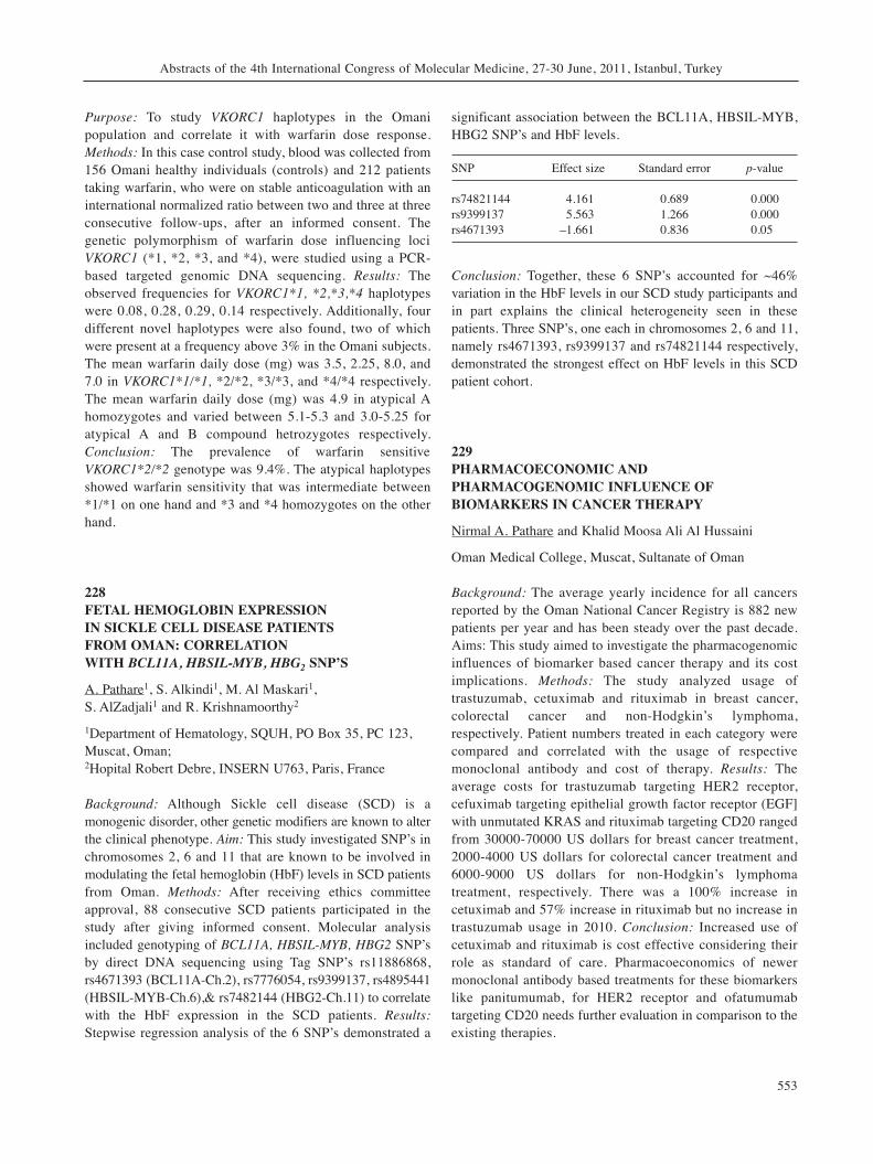
Purpose: To study VKORC1 haplotypes in the Omanipopulation and correlate it with warfarin dose response.Methods: In this case control study, blood was collected from156 Omani healthy individuals (controls) and 212 patientstaking warfarin, who were on stable anticoagulation with aninternational normalized ratio between two and three at threeconsecutive follow-ups, after an informed consent. Thegenetic polymorphism of warfarin dose influencing lociVKORC1 (*1, *2, *3, and *4), were studied using a PCR-based targeted genomic DNA sequencing. Results: Theobserved frequencies for VKORC1*1, *2,*3,*4 haplotypeswere 0.08, 0.28, 0.29, 0.14 respectively. Additionally, fourdifferent novel haplotypes were also found, two of whichwere present at a frequency above 3% in the Omani subjects.The mean warfarin daily dose (mg) was 3.5, 2.25, 8.0, and7.0 in VKORC1*1/*1, *2/*2, *3/*3, and *4/*4 respectively.The mean warfarin daily dose (mg) was 4.9 in atypical Ahomozygotes and varied between 5.1-5.3 and 3.0-5.25 foratypical A and B compound hetrozygotes respectively.Conclusion: The prevalence of warfarin sensitiveVKORC1*2/*2 genotype was 9.4%. The atypical haplotypesshowed warfarin sensitivity that was intermediate between*1/*1 on one hand and *3 and *4 homozygotes on the otherhand.
228FETAL HEMOGLOBIN EXPRESSION IN SICKLE CELL DISEASE PATIENTS FROM OMAN: CORRELATION WITH BCL11A, HBSIL-MYB, HBG2 SNP’S
A. Pathare1, S. Alkindi1, M. Al Maskari1, S. AlZadjali1 and R. Krishnamoorthy2
1Department of Hematology, SQUH, PO Box 35, PC 123,Muscat, Oman; 2Hopital Robert Debre, INSERN U763, Paris, France
Background: Although Sickle cell disease (SCD) is amonogenic disorder, other genetic modifiers are known to alterthe clinical phenotype. Aim: This study investigated SNP’s inchromosomes 2, 6 and 11 that are known to be involved inmodulating the fetal hemoglobin (HbF) levels in SCD patientsfrom Oman. Methods: After receiving ethics committeeapproval, 88 consecutive SCD patients participated in thestudy after giving informed consent. Molecular analysisincluded genotyping of BCL11A, HBSIL-MYB, HBG2 SNP’sby direct DNA sequencing using Tag SNP’s rs11886868,rs4671393 (BCL11A-Ch.2), rs7776054, rs9399137, rs4895441(HBSIL-MYB-Ch.6),& rs7482144 (HBG2-Ch.11) to correlatewith the HbF expression in the SCD patients. Results:Stepwise regression analysis of the 6 SNP’s demonstrated a
significant association between the BCL11A, HBSIL-MYB,HBG2 SNP’s and HbF levels.
SNP Effect size Standard error p-value
rs74821144 4.161 0.689 0.000rs9399137 5.563 1.266 0.000rs4671393 –1.661 0.836 0.05
Conclusion: Together, these 6 SNP’s accounted for ~46%variation in the HbF levels in our SCD study participants andin part explains the clinical heterogeneity seen in thesepatients. Three SNP’s, one each in chromosomes 2, 6 and 11,namely rs4671393, rs9399137 and rs74821144 respectively,demonstrated the strongest effect on HbF levels in this SCDpatient cohort.
229PHARMACOECONOMIC ANDPHARMACOGENOMIC INFLUENCE OFBIOMARKERS IN CANCER THERAPY
Nirmal A. Pathare and Khalid Moosa Ali Al Hussaini
Oman Medical College, Muscat, Sultanate of Oman
Background: The average yearly incidence for all cancersreported by the Oman National Cancer Registry is 882 newpatients per year and has been steady over the past decade.Aims: This study aimed to investigate the pharmacogenomicinfluences of biomarker based cancer therapy and its costimplications. Methods: The study analyzed usage oftrastuzumab, cetuximab and rituximab in breast cancer,colorectal cancer and non-Hodgkin’s lymphoma,respectively. Patient numbers treated in each category werecompared and correlated with the usage of respectivemonoclonal antibody and cost of therapy. Results: Theaverage costs for trastuzumab targeting HER2 receptor,cefuximab targeting epithelial growth factor receptor (EGF]with unmutated KRAS and rituximab targeting CD20 rangedfrom 30000-70000 US dollars for breast cancer treatment,2000-4000 US dollars for colorectal cancer treatment and6000-9000 US dollars for non-Hodgkin’s lymphomatreatment, respectively. There was a 100% increase incetuximab and 57% increase in rituximab but no increase intrastuzumab usage in 2010. Conclusion: Increased use ofcetuximab and rituximab is cost effective considering theirrole as standard of care. Pharmacoeconomics of newermonoclonal antibody based treatments for these biomarkerslike panitumumab, for HER2 receptor and ofatumumabtargeting CD20 needs further evaluation in comparison to theexisting therapies.
Abstracts of the 4th International Congress of Molecular Medicine, 27-30 June, 2011, Istanbul, Turkey
553
230DETECTION OF TYPE 1 COLLAGEN ALPHA 1GENE -1997 G/T POLYMORPHISM INPOSTMENOPAUSAL OSTEOPOROTIC TURKISH WOMEN
Sabriye Kocaturk-Sel1, Halil Kasap1, Ayfer Pazarbasi1,M.Bertan Yılmaz1, Ali Irfan Guzel2, Tunay Sarpel3, Sibel Basaran3, Lutfiye Ozpak1, Mehmet Ali Erkoc1, Gamze Comertpay1 and Inayet Nur Uslu1
Departments of 1Medical Biology and 3Physical Therapy,Cukurova University, Adana, Turkey; 2Department of Medical Biology, Rize University, Rize,Turkey
Background: Osteoporosis is a disease characterised byreduced bone mass, which leads to microarchitecturaldeterioration of bone tissue and increased fracture risk.Genetic factors play an important role in the pathogenesis ofosteoporosis. Type I collagen is the most abundant protein ofthe bone matrix and the collagen type I alpha 1(COLIA1)gene has been considered one of the most important candidategenes for osteoporosis. Methods: In this study we analyzed therelationship between -1997 G/T single nucleotidepolymorphisms at the COL1A1 regulator region. We analyzedthe DNA of 79 osteoporotic postmenopausal patients and 43healthy postmenopausal Turkish women. All the subjects weregenotyped by using polymerase chain reaction–restrictionfragment length polymorphism. Results: Patient genotypeswere: TT 26%; GT 61% and GG, 12%, respectively, whereascontrols were TT 40%, GT 44% and GG 16%, respectively.The TT genotype of the -1997 G/T polymorphism in theCOLIA1 gene was significantly associated with the risk ofdeveloping osteoporosis in our cases. Conclusion: Our resultssuggest an important role of the COLIA1 gene in osteoporosis.Mutations in the regulatory region within a gene may lead todifferent levels of gene expression and thus the -1997 G/Tpolymorphism may be a valuable genetic marker.
231GENETIC POLYMORPHISMS IN THE ESTROGENRECEPTOR ALPHA GENE IN TURKISH PATIENTSWITH FAMILIAL PROSTATE CARCINOMA
Ayfer Pazarbaşi1, Davut Alptekin1, Ümit Lüleyap1, Zühtü Tansuğ2, Müzeyyen Izmirli3, M. Bertan Yilmaz1, Dilge Onatoğlu-Arikan1, Ceyhun Bereketoğlu1, Sabriye Kocatürk-Sel1, Lütfiye Özpak1, Nilgün Tanriverdi1,Mehmet Ali Erkoç1, Ali Irfan Güzel4, Erdal Tunç1, Özgür Turgut1 and Osman Demirhan1
Departments of 1Medical Biology and 2Urology, ÇukurovaUniversity, Adana, Turkey;
3Department of Medical Biology, Vakıf Gureba University,Istanbul, Turkey;4Department of Medical Biology, Rize University, Rize,Turkey
Background: Polymorphisms of estrogen receptor (ER) alphagene in the estrogen metabolism pathway may be involved inthe risk of developing prostate carcinoma. We evaluated theassociation between genetic polymorphisms in the ER alphagene and the risk of developing familial prostate carcinoma.Methods: A total of 34 cases with prostate carcinoma whosefirst-degree relatives had prostate carcinoma and 30 healthyage-matched male controls were enrolled. The genotype of ERalpha gene was analyzed. Results: Among the controls, the ERalpha PvuII genotypes of C/C, C/T and T/T were observed in37%, 26%, and 37%, respectively, whereas the respectivepercentages among the patients were 18%, 41%, and 41%.Among the controls, the ER alpha XbaI genotypes of G/G,G/A and A/A were observed in 33%, 37%, and 33%,respectively, whereas the respective percentages among thepatients were 12%, 47% and 41%. The C/C genotype of thePvuII site and G/G genotype of the XbaI site in the ER alphagene were associated significantly with the risk of developingprostate carcinoma. Conclusion: Polymorphisms of the ERalpha gene in the estrogen metabolism pathway are associatedsignificantly with the risk of familial prostate carcinoma.
232EVALUATION OF MOLECULAR ASPECTS OF MALEINFERTILITY IN THE LATVIAN POPULATION
Agrita Puzuka1, Natalija Pronina2, Juris Erenpreiss3,Voldemars Lejins4, Liana Pliss5, Viesturs Baumanis5 andAstrida Krumina1
Laboratories of 1Molecular Genetics and 3Andrology, RigaStradins University, Riga, Latvia;2Medical Genetics Clinic, Children University Hospital,Riga, Latvia;4EGV Clinics, Riga, Latvia;5Latvian Biomedical Research and Study Centre, Riga,Latvia
Background: Some aspects of male infertility may beexplained analysing Y-chromosomal and autosomal genes thatare involved in the process of spermatogenesis. The aim of thisstudy was to investigate molecular-genetic aspects ofidiopathic male infertility in the Latvian population. The studyincluded 100 idiopathic infertile men (infertile group) and 153healthy individuals (control group). Methods: Y-chromosomemicrodeletions were analyzed by two multiplex PCR. Ychromosome haplogroups (Y-Hg) were detected usingappropriate biallelic markers. CFTR gene mutation, delF508,
in vivo 25: 467-576 (2011)
554
R117H and IVS8 polymorphism analysis was performed byPCR and followed by RFLP and sequencing. Results: Y-chromosome microdeletions were detected in 5% (5 cases of100: 3 cases AZFc; 2 cases AZFa+b+c). Y-Hg analysis showedthat Hg N3a1 and Hg R1a1 were less frequent in the infertilegroup compared to the control group, however Hg K* waspredominantly found in the infertile group (p<0.001). Analysisof CFTR gene mutation, delF508, R117H and IVS8polymorphism did not confirm association with infertility.Conclusion: The frequency of Y-chromosome microdeletionsin males with idiopathic infertility was 5%. Y chromosome HgK* may be associated with male infertility. CFTR genemutations, delF508, R117H and IVS8 polymorphisms do notaffect the process of spermatogenesis directly.
233ROLE OF C-KIT EXON 11 MUTATIONS AND HLA-G POLYMORPHISM IN LEUKEMIA IN NORTHERN INDIA
Syed Tasleem Raza1, Syed Rizwan Hussain1, Hena Naqvi1,Sunil G. Babu2, Abbas Ali Mahdi3 and Farzana Mahdi1
1Department of Biochemistry, ERA’S Lucknow MedicalCollege, Lucknow, India; 2Department of Biotechnology, BBA University, Lucknow,India;3Department of Biochemistry, CSM Medical University,Lucknow, India
Background: C-Kit gene is a receptor tyrosine kinase class IIIthat is expressed by early hematopoietic progenitor cells andplays an important role in hematopoietic stem cellproliferation, differentiation and survival. The 14bp insertion /deletion of the HLA-G gene has been shown to play animportant role in various types of neoplasia. Methods: PCR-SSCP followed by direct DNA sequencing. Results: Of 31leukemia patients, 18 were male and 13 were female with agesranging from 2 to 65 years. The mean age of patients was 32.3years and SD±1.03. A total of nineteen mutations weredetected in six patients that include Lys550Asn, Tyr568Ser,Ile571Leu, Thr574Pro, Gln575His, Tyr578Pro, Asp579His,His580Gln, Trp582Ser, Arg586Thr, Asn587Asp andArg588Met and novel point mutations at codons Ile563Lys,Val569Leu, Tyr570Ser, Ile571Thr and Pro577Ser. Ile571Leuand Trp582Ser substitution was found in two independentcases. The frequency of I/I, I/D and D/D genotype in patientswas 19.37%, 48.38% and 32.25% and in controls 24%, 46%and 30% respectively. Conclusion: These observations suggestthat mutations in exon 11 of the c-kit gene might representuseful molecular genetic markers for leukemia while there isno significant association of HLA-G polymorphism inleukemia.
234RESPONSE TO ATHEROGENIC DIET IN SYRIAN HAMSTERS
Albert A. Rizvanov1,2,3,4, Margarita N. Katina3, Leisan R. Kasimova3, Alsu R. Abdulkhakova3, Sayar R. Abdulkhakov3, Andrey P. Kiassov3
and Raushania F. Gaifullina3
1Kazan (Volga Region) Federal University, Kazan, Russia; 2Yeditepe University, Istanbul, Turkey; 3Kazan State Medical University, Kazan, Russia; 4Republic Clinical Hospital, Kazan, Russia
Background: Cost-effective small animal models ofatherosclerosis and data on the use of atherogenic diets in non-transgenic animals is important for testing anti-atherogenictherapies. Methods: We used Syrian hamsters (Mesocricetusauratus) on high cholesterol/high dietary fat atherogenic diet(the Paigen diets’ modification) as a model for atherosclerosis.Animals were sacrificed after 14 weeks’ diet and histologicalexamination of their organs and tissues was performed.Results: The body mass was significantly lower in the testgroup compared with the control. A severe hepatomegaly wasdetected in all hamsters in the test group. The maximum livermass was up to 25% of the whole body mass. Histologicalfeatures of severe hepatosis were observed. Concentricmyocardial hypertrophy was found in the test group.Conclusion: Syrian hamsters strongly responded toatherogenic diet and can be used as a model for testing anti-atherogenic therapies.
235PREPARATION AND CHARACTERIZATION OF MONOCLONAL ANTIBODIES TO ADIPONECTIN FOR ITS MEASUREMENT IN HUMAN SERUM
Alla Rodina1,2, Vladimir Sologub1, Irina Koromyslova1, Nila Gorokhovets1, Vladimir Makarov1, Denis Andreev2,Nikolai Ermakov1 and Sergei Severin1,2
1Department of Health Care, Moscow Research Institute ofMedical Ecology, Moscow, Russia;2I.M. Sechenov First Moscow State Medical University,Russia
Background: Low levels of adiponectin are associated withincreased prevalence of cardiovascular disorders, includingischemic heart disease. Methods: Monoclonal antibodieswere prepared against purified recombinant humanadiponectin. The reactivity of these monoclonal antibodieson Western blot analysis was restricted to a monomer and
Abstracts of the 4th International Congress of Molecular Medicine, 27-30 June, 2011, Istanbul, Turkey
555
trimer form of adiponectin. With these monoclonalantibodies, we developed a novel ELISA system. Thespecificity of this system was verified by analysis of serumfractions separated by gel-filtration chromatography. Results:The developed ELISA system was specific to high-molecular-weight (HMW) multimer adiponectin. The HMWadiponectin concentrations measured with this novel ELISAsystem were: 11.01±2.58 mkg/ml (mean±SD) in healthywomen and 8.42±1.59 mkg/ml in healthy men. The serumHMW adiponectin concentration was lower in patients withischemic heart disease than in healthy controls; 6.01±2.73mkg/ml vs. 8.42±1.59 mkg/ml (р=0.015) in men and5.79±2.98 mkg/ml vs. 11.01±2.58 mkg/ml (р=0.0003) inwomen, respectively. Conclusion: The developed, novelELISA system, specific for the HMW form of adiponectinmay be used for diagnostic clinical examinations in patientswith ischemic heart disease.
236REGULATION OF ADIPOSE TISSUE DERIVED STEM CELLS DIFFERENTIATION BY RECOMBINANT ADIPONECTIN
Alla Rodina1,2, Elizaveta Moskaleva1,2, Nila Gorokhovets1,Vladimir Makarov1, Olga Popova1, Elizaveta Nekrasova3,Sergei Severin1,3 and Eugenie Severin2
1Department of Health Care, Moscow Research Institute ofMedical Ecology, Moscow, Russia;2Russian Research Center of Molecular Diagnostic andTreatment, Moscow, Russia;3I.M. Sechenov First Moscow State Medical University,Moscow, Russia
Background: Adipose-derived stromal/stem cells (ASCs) canbe obtained from the lipoaspirate of individual donors forregenerative medicine applications. Previously we haveshown that adiponectin levels are lowered in some diseases.The aim of this study was to investigate the ASCdifferentiation in the presence of the adipogenic mediumwith insulin and in the presence of human recombinantadiponectin. Methods: ASC were isolated from humansubcutaneous lipoaspirate. Flow cytometry andimmunocytochemical staining were used for ASCcharacterization. Results: The ASC population was CD34–,CD44+, CD90+, CD105+ and HLA-DRlow. In 1% of ASCs thealpha-fetoprotein receptor (embryonic cell marker) wasfound and 7% ASCs possessed SP phenotype. When ASCswere cultured in standard medium, 1% of ASCs werespontaneously differentiated into adipocytes. After threeweeks culturing in adipogenic medium, 87.5% ASCs
acquired the typical morphology of lipid-laden cellscontaining intracellular lipid droplets; however, in thepresence of adiponectin, only 4.3% of ASCs differentiated inadipocytes. When ASCs were cultured in the presence ofadiponectin without adipogenic medium, the cells did notdifferentiate into adipocytes. Conclusion: The lower levelsof adiponectin in patients with some diseases may alter ASCdifferentiation after transplantation.
237IS THERE ANY CORRELATION BETWEEN IL-10 POLYMORPHISMS AND BIPOLAR DISORDER?
Omer Ates1, Aydin Rustemoglu1, Birgul Elbozan Cumurcu2
and Suheyla Unal2
1Department of Medical Biology, Medical Faculty,Gaziosmanpasa University, 60100, Tokat, Turkey;2Turgut Ozal Medical Center, Department of Psychiatry,Medical Faculty, Inonu University, 44315, Malatya, Turkey
Background: The neurobiology of bipolar disorder is notcompletely understood. Cytokines have received increasingattention as potential mediators of the interaction withimmune and neuroendocrine system and specific pathwaysinvolved in mood, energy and activity control. Previousreports have suggested the association of mania and bipolardepression with a pro-inflammatory state. Genes encodingcytokines are highly polymorphic and single nucleotidepolymorphisms, associated with increased or reducedcytokine production, have been described. The aim of thisstudy was to define the genetic immunological scenarioassociated with bipolar disorder. Methods: Eighty-two bipolarTurkish outpatients affected by bipolar disorder, and 298healthy controls were enrolled into the study. We analyzedallele and genotype distribution of -592C/A, -819C/T and -1082G/A IL-10 promoter polymorphisms by PolymeraseChain Reaction Sequence Specific Primers technique.Results: There were no significant differences ingenotype/allele distribution of IL10 -1082G/A, -819C/T, -592C/A between patients with bipolar disorder and controls(p≥0.05). We observed different genotype frequencies of IL-10 polymorphisms in the bipolar patients and controls. Inparticular, bipolar patients were characterized by a highpercentage of the GA genotype for -1082G/A locus(p=0.029), and the GCC/ACC genotype included all threepolymorphic loci (p=0.006). On other hand, the ACC/ATAgenotype was detected lower in bipolar patients than controls(p=0.028). Conclusion: Our results showed that IL-10promoter polymorphisms might be associated with bipolardisorder in Turkish patients.
in vivo 25: 467-576 (2011)
556
238INTERSECTINS AND DISEASES: CLUES FROMTHEIR FUNCTIONAL CHARACTERISTICS
Alla Rynditch, Liudmyla Tsyba, Inessa Skrypkina, Oleksii Nikolaienko, Olga Novokhatska, Sergii Kropyvko,Mykola Dergai, Oleksandr Dergai, Tetyana Gryaznova and Dmytro Morderer
Institute of Molecular Biology and Genetics, Kyiv, Ukraine
Background: Adaptors of intersectin (ITSN) family serve asscaffolds for multiprotein complex assembly during clathrin-mediated endocytosis. Abnormalities of ITSN1 expressionhave been associated with endocytic brain anomalies reportedin Down syndrome and at the early stages of Alzheimer’sdesease. ITSN2 was proposed to be a predictive marker forbreast cancer. Methods: Immunoprecipitations, pull-downexperiments, determination of subcellular localization bydirect and indirect immunofluorescence, mass-spectrometryand RT-PCR were used in this study. Results: We identified 17alternative splicing events affecting ITSN1 pre-mRNA andfound an alternative transcription initiation site in the fifthintron of ITSN1 gene. ITSN1 isoforms differed in theirdomain organization, interaction with protein partners,localization in different tissues and stages of development.Using mass spectrometry analysis and in silico prediction, weidentified 11 novel protein partners of ITSN1 and ITSN2.These proteins are involved in signal transduction, actin andtubulin cytoskeleton formation, endocytosis, cell adhesion andmigration. Using a Xenopus animal model, we demonstratedthe role of ITSN2 in the coordinated changes of actincytoskeleton during early embryonic development.Conclusion: The results demonstrated the complex regulationof ITSN adaptor proteins that provide interfaces for theinteraction between basic endocytic machinery and differentcell processes such as signalling, sorting and cytoskeletonrearrangements.
239PRODUCTION OF MEASLES TRUNCATEDHEMAGGLUTININ PROTEIN IN PROKARYOTICCELLS FOR PREPARING RECOMBINANT VACCINE
Zohreh Azita Sadigh1, Abbas Shafyi1, Bijan Bambai2 andFatemeh Esnaashari1
1Razi Vaccine and Serum Research Institute, Karaj, Iran;2National Institute of Genetic Engineering andBiotechnology, Karaj, Iran;
Background: The Measles virus, a member ofParamyxoviridae family with a non-segmented negative
strand RNA, has two surface glycoproteins: H and F. H isresponsible for the cell tropism, receptor binding, andhemagglutinating activity. It has several conformationalepitopes for induction of immune system to produceneutralizing antibody. Recent studies confirmed the protectiveactivity of some linear epitopes. In this respect, we chose aspecific sequential hemagglutinin epitope for cloning andexpression in prokaryotic cells Methods: Measles Virusgenomic material was extracted from infected Vero cells. Twothird of 5’ end of the H gene was amplified by designedprimers containing secreted signal for production of truncatedprotein during RT-PCR reaction and inserted into the cloningand expression vector respectively. Finally the BL21 strainsof E. coli were transformed with expression vector in orderto produce recombinant protein. Results: The cloned fragmentof Measles virus H gene was confirmed by sequencing. SDS-PAGE and Western-blot using goat polyclonal antibodyagainst Measles Virus indicated the presence of properamount of recombinant H protein. Conclusion: According toour study, after neutralizing assay these clones may be furtherused in the development of a recombinant vaccine against theMeasles virus.
240ANALYSIS OF THE INFLUENCE OF BRAF V600E MUTATION ON THE METHYLATION STATUS OF MLH1 AND MGMT ANDMICROSATELLITE INSTABILITY IN PATIENTS WITH THYROID CANCER
Juliana Carvalho Santos1, André Uchimura Bastos2, Janete Maria Cerutti2 and Marcelo Lima Ribeiro1
1Sao Francisco University Medical School ClinicalPharmacology and Gastroenterolgy Unit Laboratory ofMicrobiology and Molecular Biology, Bragança Paulista, SP, Brazil;2Universidade Federal de São Paulo Department ofMorphology and Genetics, São Paulo, SP, Brazil
Background: Thyroid cancer is the most common cancer ofthe endocrine system and its incidence has increased veryrapidly in recent years. The BRAF mutation is the mostcommonly occurring known genetic alteration in thyroidcancer. This mutation can cause hypermethylation of somerepair genes, such as MLH1 and MGMT, and consequentmicrosatellite instability (MSI). The aim of the present studywas to evaluate the effects of BRAF V600E mutation onmethylation status of MGMT and MLH1 and MSI. Methods:Tumor tissue samples were taken from 85 patients harboringthe BRAF V600E mutation. Genomic DNA was amplifiedby polymerase chain reaction (PCR) using seven markers.
Abstracts of the 4th International Congress of Molecular Medicine, 27-30 June, 2011, Istanbul, Turkey
557
Methylation analysis of MGMT and MLH1 was done bymeans of methylation specific (MSP)-PCR. Results:Amongst the patients, 70% showed MSI. We observed thatMGMT and MLH1 promoter hypermethylation was afrequent occurrence in MSI (66%). Conclusion: These datasuggest that this silencing mechanism plays a major role inthe inactivation of these genes and thus contributes to thedevelopment of thyroid cancer. A significant correlation ofMLH1 and MGMT hypermethylation with MSI was foundin these tumors.
241EFFECTS OF CAFFEIC ACID PHENETHYL ESTER (CAPE) ON SOME BIOCHEMICAL ANDHISTOPATHOLOGICAL PARAMETERS INSELECTED TISSUES OF DIABETIC RATS
Sefa Celik1, Suat Erdogan2, Mehmet Tuzcu3 and Serkan Sen1
1Department of Biochemistry, Faculty of Medicine, AfyonKocatepe University, Afyonkarahisar, Turkey 2Department of Biochemistry, Faculty of VeterinaryMedicine, Mustafa Kemal University, Hatay, Turkey 3Adana Veteriner Kontrol Araştırma Enstitüsü, Adana,Turkey
Background: Phenolic compounds are commonly used asfood additives and form the active ingredients of folkmedicines due to their antioxidant, anti-inflammatory andanti-carcinogenic effects. In this study, the effects of CAPEon diabetes-damaged liver function and integrity of liver,pancreas, kidney, testis and heart were investigated. Methods:Diabetes was induced by administrating 45mg/kgstreptozotocin (i.p.) to the rats. CAPE treatments wereperformed for 60 days and blood and various tissues wereanalysed. Histopathological changes were graded as absent(0), mild (1), medium (2), and severe (3), according to theseverity of alterations. Results: CAPE treatments considerablylowered plasma glucose concentraion in CAPEI and CAPEIIgroups. Insulin levels of all CAPE treated groups were 2-foldhigher than those in the diabetic group. CAPE administrationsignificantly reduced the cholesterol and triglycerideconcentrations elevated by diabetes. Diabetes-increasedplasma ALT activity was decreased to the control level by allthree CAPE treatments. In comparison with control,histopathological alterations in liver, pancreas, kidney, testisand heart tissues of diabetic rats were normalized in variousdegrees with CAPE treatments. Conclusion: CAPE treatmentwas able to significantly lower hyperglycemia and reversesome damage in tissues of diabetic rats. CAPE applicationsmight be considered in treating hyperglycemia and tissuecomplications in diabetic patients.
242THE EFFECT OF ACETYLYSALICYLIC ACID (ASA) ON PLASMA ARGINO-VASOPRESSIN (AVP) LEVELS IN TYPE II DIABETICS
Serkan Sen1, Sefa Celik1 and Omer Hazman2
1Department of Biochemistry, Faculty of Medicine, AfyonKocatepe University, Afyonkarahisar, Turkey;2Department of Biochemistry, Faculty of VeterinaryMedicine, Afyon Kocatepe University, Afyonkarahisar,Turkey
Background: Acetylsalicylic acid (ASA) irreversibly blocksthe platelet cyclo-oxygenase enzyme system, preventingformation of thromboxane A2 and inhibiting plateletaggregation for the life of the affected platelet. AVP is apleiotropic peptide which affects a wide range of peripheraland centrally regulated functions in order to conserve waterin the kidney. This study aimed at investigating theimportance of ASA on osmoregulation and glycemic controlin diabetic rats. Methods: Twenty four rats were randomlydivided into four groups: Control(I), ASA Control(II),Diabetic(III) and ASA Diabetic(IV). Diabetes was induced bySTZ treatment (30 mg/kg, two injections) in obese rats.Plasma AVP levels were analyzed by ELISA. Serumelectrolytes (Na, K, Cl), creatinine, albumin and total proteinlevels were analyzed by an auto-analyzer. Results: At the endof the study ASA treatments increased the serum AVP levels(2-fold) in diabetic rats. Also, ASA treatments decreased thefasting blood glucose levels in diabetic rats. Nevertheless,sodium, creatinine, albumin and total protein levels in plasmawere also decreased by ASA. Conclusion: The hypoglycemiceffect of ASA can be attributed to increase in blood volumeby AVP levels. This may be a new approach in the efficacyof ASA in reducing the fasting blood glucose levels indiabetics.
243ANTIBIOTIC RESISTANCE AND PHYLOGENETIC PATTERNS IN UROPATHOGENIC ESCHERICHIA COLI
Kavous Solhjoo1, Sara Asadi2, Abbas Ali Rezaiean2 andSaeid Sobhanian1
1Jahrom University of Medical Sciences, Faculty ofMedicine, Jahrom, Iran;2Azad University of Jahrom Branch, Jahrom, Iran
Background: Escherichia coli is the most frequent agent ofurinary tract infections (UTIs). The aim of this study was toassess whether Escherichia coli phylogenetic groups were
in vivo 25: 467-576 (2011)
558
associated with the level of antibiotic resistance incommunity-acquired bacteriuria. Methods: Between Juneand September 2010, urine specimens were collected from1000 patients, all with clinically diagnosed UTIs, attendingMottahri and Pymanieh Hospitals in Jahrom, Iran.Specimens were examined for different uropathogens usingstandard microbiological procedures. A total of 60uropathogenic E. coli were isolated and characterized.Triplex PCR was used to classify the phylogenetic groups,and susceptibility testing was performed by the discdiffusion method. Data were assessed by statistical analysis.Results: Overall, 70% of the sixty E. coli isolates belongedto phylogenic group D, 23.3% to A, and 6.7% to B1. Thehighest sensitivity and resistance to antibiotics wereobserved with nitrofurantoin (97.6%) and trimethoprim-sulfamethoxazole (48.3%), respectively. Antibioticresistance to one and more than three antibiotics,respectively, was most frequent in group A (35.7%/42.9%),followed by D (35.7%/38.1%), and B1 (50.0%/0%).Conclusion: Phylogenetic group D was predominant in E.coli community-acquired bacteriuria and statistical analysisshowed that phylogenetic groups were not associated withthe level of antibiotic resistance (p>0.05).
244ASSOCIATION BETWEEN G/A455 FIBRINOGENPOLYMORPHISM AND PREMATURE CORONARYARTERY DISEASE IN IRANIAN PATIENTS
Mohammad Shojaee, Kavous Solhjoo, Davar Aledavood andMaedeh Motavas
Jahrom University of Medical Sciences, Jahrom, Iran
Introduction and Objective: Several studies have shown anassociation between plasma fibrinogen levels, coronary heartdisease premature myocardial infarction (PMI) and stroke.Despite intensive investigations, there is no clear evidence foran effect of beta-fibrinogen gene polymorphism on plasmafibrinogen levels and myocardial infarction. The aim of thisstudy was to determine the association of the G/A455polymorphism of beta-fibrinogen gene with plasmafibrinogen levels and myocardial infarction. Patients andMethods: We conducted a case-control study consisted of 100patients with premature ischemic heart disease (IHD) withage below 50 years and 100 volunteer subjects withoutevidence of IHD as control group. The two groups werematched according to age, gender and smoking status. Plasmafibrinogen levels were measured by Clauss clotting timemethod and G/A455 mutations were identified by PCRfollowed by HaeIII restriction enzyme digestion of amplifiedDNA and electrophoresis. Results: Plasma fibrinogen levelswere higher in the patients with premature myocardial
infarction than in control subjects (p=0.001, odds ratio=2.12).In the control group, the plasma fibrinogen levels weresignificantly higher in smokers than non-smokers (p=0.032).However, there was no association between 455G/Apolymorphism, plasma fibrinogen levels (p=0.65) andincidence of PMI (p=0.663). Conclusion: Increased plasmafibrinogen level was associated with cigarette smoking andrisk of PMI but no association was found with G/A455polymorphism.
245PEPTODOGLYCAN FROM STAPHYLOCOCCUSAUREUS INCREASES MUC5AC GENE EXPRESSION VIA THE RSK1-CREB PATHWAY IN HUMAN AIRWAY EPITHELIAL CELLS
Myoung Joo Lee, MiSung Kwon, Do Whan Ahn andKyoung Seob Song
Department of Physiology, Kosin University College ofMedicine, Busan, Korea
Background: Respiratory tract exposure to viruses, airpollutants or bacteria pathogens can lead to pulmonarydiseases. The knowledge on the molecular mechanisms ofmucous overproduction caused by these pathogens isnecessary for the development of new therapeutic strategies.It is well established from in vitro data that theoverexpression of MUC5AC is induced by peptidoglycan(PGN) derived from Staphylococcus aureus. However, themechanisms by which PGN activates MUC5AC geneexpression in the airway remain unclear. The aim of thisstudy was to identify the mechanisms of PGN-inducedMUC5AC gene expression. Methods and Results: We foundthat PGN could induce MUC5AC and MUC8 gene expressionin a time- and dose-dependent manner. Moreover, activationof ERK1/2 and JNK increased after treatment of cells withPGN, whereas phosporylation of p38 was undetected. Ofthese MAPKs, pharmacological inhibition of ERK1/2decreased PGN-induced MUC5AC gene expression. Inaddition, we examined the activation of p90 ribosomal S6kinase 1 (RSK1) as a downstream signaling target of ERK1/2in PGN signaling. The activation of RSK1 was prevented bypretreatment with PD98059. We also found that RSK1mediated the PGN-induced phosphorylation of cAMPresponse element-binding protein (CREB) and thetranscription of MUC5AC. Furthermore, the cAMP-responseelement (CRE) in the MUC5AC promoter appeared to beimportant for PGN-induced MUC5AC gene expression inNCI-H292 cells. Conclusion: These results give additionalinformation about the intracellular signal transductionpathway involved in mucin production during inflammation.
Abstracts of the 4th International Congress of Molecular Medicine, 27-30 June, 2011, Istanbul, Turkey
559
246PROMOTER HYPERMETHYLATION OF CDKN2AGENE IN BULGARIAN PATIENTS WITHLARYNGEAL SQUAMOUS CELL CARCINOMA
Gergana Stancheva1,2, Reni Tzveova1, Stanislav Jordanov3,Atanaska Mitkova1,2, Ivan Chalakov3, Radka Kaneva1,2,Vanio Mitev1,2 and Teodora Goranova1
1Molecular Medicine Center, 2Department of MedicalChemistry and Biochemistry, Medical University, Sofia,Bulgaria; 3Department of ENT, Clinic of ENT, Tsaritsa Yoanna – ISULUMHAT, Sofia, Bulgaria
Background: An important mechanism for gene inactivation isthe de novo methylation of normally unmethylated CpGislands within the promoter region of carcinoma-associatedgenes. Such a gene involved in laryngeal carcinogenesis isCDKN2A, localized on chromosome 9p21. It is a tumor-suppressor gene which encodes a cyclin-dependent kinaseinhibitor with an important role in the regulation of the G1/Scell-cycle checkpoint. Hypermethylation of CDKN2Apromoter is reported in 9-47% of laryngeal tumors. Methods:Genomic DNA was extracted from 50 laryngeal fresh-frozentumor tissues and was bisulfate-converted. Promoterhypermethylation of CDKN2A gene was analyzed bymethylation-specific polymerase chain reaction (MSP) usingprimer sets specific for unmethylated and methylatedsequences. Results: MSP analysis demonstrated thehypermethylation of CDKN2A gene in 20 (40%) patients withlaryngeal cancer. A significant increase of CDKN2A promoterhypermethylation was observed in patients with an early stagecancer (T1-T2) (p=0.017). Epigenetic changes were morefrequent in specimens from younger patients (≤60 years ofage) compared to older ones, but the differences were notstatistically significant. Conclusion: Our results indicate thatepigenetic modifications of CDKN2A gene are implicated ina significant proportion of cases with laryngeal carcinoma,especially at the early stages of its development.
247TRANSCRIPTION PROFILING OF HEAD AND NECK SQUAMOUS CELL CARCINOMA
Hynek Strnad1, Michal Kolar1, Jana Sachova1, LukasLacina2, Jan Paces1, Marketa Urbanova3, Miluse Hroudova1,Vaclav Paces1, Jan Betka3, Karel Smetana4, Jan Plzak3,Martin Chovanec3, Zdenek Cada2 and Cestmir Vlcek1
1Institute of Molecular Genetics, Academy of Sciences of the Czech Republic, Videnska 1083, 140 00 Prague 4, Czech Republic;2Institute of Anatomy and Department of
Dermatovenereology, 3Department of Otorhinolaryngology,Head and Neck Surgery and 4Institute of Anatomy, CharlesUniversity in Prague, Czech Republic
Background: Head and neck squamous cell carcinoma(HNSCC) is one of the most prevalent tumour typescharacterized by a high mortality. The poor prognosis resultsfrom a lack of markers of early stages of the disease. Here wepresent results of a whole genome transcription profiling of101 patients with HNSCC. Methods: Three snap-frozensamples were collected from each patient, a sample of tumour,peritumoural tissue and normal bucal tissue. The patient-matched RNA samples analyzed by microarray allowed us toextract individual related variations of the transcriptome.Results: Clustering analysis of the data revealed a strongcontrast between normal and tumour tissues together with sub-classification of peritumoural tissues. We further distinguishedkeratinizing and non-keratinizing tumours as two separateclusters. We detected 1500 differentially expressed transcriptsbetween tumours and normal tissues. We observed genesrelated to extracellular matrix or its remodelling, specificchemokines and cytokines. We detected deregulated KEGGpathways associated with DNA replication, cell cycle,cytokine interaction, immune response and several signallingpathways. The overall metabolism was down-regulated in thetumours. Conclusion: The presented data of large-cohortwhole-genome transcription profiling reveal several markergenes distinguishing tumour and normal tissues. The resultsare interpreted in the broad scale of metabolic, regulatory andsignalling pathways.
248THE MOLECULAR-GENETIC DETERMINANTS OF CARDIORENAL COMPLICATIONS IN PATIENTS WITH ARTERIAL HYPERTENSION IN THE KAZAKH POPULATION
Gulnara Svyatova1, Gulnara Junusbekova2 and Meyramgul Tundybaeva3
1Scientific Centre of Obstetrics, Gynecology andPerinatology, Almaty, Kazakhstan; 2Scientific-Research Institute of Cardiology and InternalDiseases, Almaty, Kazakhstan; 3JCS “Astana Medical University”, Astana, Kazakhstan
Aim: To define the involvement of polymorphisms C344T ofgene CYP11B2, Leu10Pro of gene TGF-β1 and С677Т of geneMTHFR in the development of cardiorenal complications inpatients with arterial hypertension (AH) in the Kazakhpopulation. Patients and Methods: A total of 142 patients withАH were surveyed, with mean age 51.8±4,7 years, bloodpressure average 165.1±1.58/104.6±1.02 mm Hg, duration of
in vivo 25: 467-576 (2011)
560
АH 10.4±3.5 years. The control group included 119 healthyKazakhs of comparable age and gender. The genotyping wasperformed by the restriction fragment length polymorphismmethod. Results: It was established that the CC-genotype of theCYP11B2 gene is associated with concentric type remodelingand disturbance of diastolic function of the left ventricle (LV).The genotype Pro/Pro of the TGF-β1 gene was associated withthe development of concentric hypertrophy of the LV and withaugmentation of thickness, a complex of intim/media of thecommon carotid artery. An association of genotype Pro/Pro ofgene TGF-β1 with depression of rate of a glomerular filtrationless than 60 ml/min/1.73m2 was found. The frequency ofT677T genotypes of the MTHFR gene was significantly higheramong AH patients with microalbuminuria. Conclusion: Theessential influence of polymorphisms C344T of CYP11B2,Leu10Pro of TGF-β1 and С677Т of MTHFR genes onremodeling of cardiovascular system and kidneys in patientswith АH is established in the Kazakh population.
249CURCUMIN INHIBITS CANCER CELL MIGRATION AND INVASION IN SQUAMOUS CELL CARCINOMA OF THE TONGUE THROUGH DOWN-REGULATION OF MMP10
Wing-Yan Tang, Thian-Sze Wong, Jimmy Yu-Wai Chan andWilliam Ignace Wei
Department of Surgery, The University of Hong Kong, HongKong SAR, China
Background: Curcumin is a natural polyphenol. Its anticancereffect has been σηοςν in various cancer types, including headand neck squamous cell carcinoma. Our study hypothesizedthat curcumin may suppress the invasiveness of tonguesquamous cell carcinoma (TSCC) through the down-regulationof matrix metalloproteinase 10 (MMP10). Methods: mRNA-microarray analysis was performed on HN21B, in response tocurcumin. Immunohistochemical staining of MMP10 wasperformed on a tissue array. MMP10 expression levels wereverified by qRT-PCR, Western blotting and immunohisto-chemical staining on TSCC cell lines. The effect of curcuminon TSCC cell lines was assessed by wound healing, adhesion,migration and invasion assays. Results: Microarray analysisshowed that MMP10 was down-regulated by 2.36-fold inHN21B in response to curcumin. In 13 out of 20 cases (65%),the TSCC tissue showed higher MMP10 expression than theadjacent normal tissue. Curcumin treatment of TSCC cell linesresulted in down-regulation of MMP10 gene and proteinexpression level in a dose-dependent manner. Curcumin alsosuppressed adhesion, migration and invasion of TSCC celllines. Conclusion: Curcumin treatment reduced MMP10expression level in TSCC cell lines. This correlated with the
reduction of invasiveness of TSCC cells. Application ofcurcumin as a chemotherapeutic agent warrants furtherinvestigation.
250CHARACTERISTICS OF CARBONYL DERIVATES OF BLOOD PROTEINS IN PATIENTS WITH CHRONIC PYELONEPHRITIS
Neila Tankibayeva, Dmitriy Kluev, Larisa Muravleva andVilen Molotov-Lushanskyi
Karaganda State Medical University, Karaganda, Republic ofKazakhstan
Early studies have demonstrated the activation of lipidperoxidation in the erythrocytes and have suggested thedevelopment of carbonyl stress of proteins in the plasma anderythrocytes in chronic pyelonephritis patients. This studyexamined the carbonyl derivative content in blood plasma anderythrocytes in patients with chronic pyelonephritis. A total of33 patients with verified acute chronic pyelonephritis and 20healthy donors were included in the study. The carbonylderivatives of proteins were examined by using a reaction testwith 2,4-dinitrophenyl hydrazine. A significant accumulationof carbonyl derivatives of proteins was shown in blood plasma.There was an increase of ketone- and aldehyde-dinitrophenylhydrazones of basic classes by 4 and 4.8 times,respectively, in patients compared to controls (p<0.001). Thecontent of ketone- and aldehyde-dinitrophenylhydrazones ofneutral classes were higher by 3.86 and 6 times, respectively,in patients compared to controls (p<0.001). Persistency ofoxidative modified proteins in the patients’ blood induced abroad spectrum of molecular damage depending on the typeof proteins involved in the carbonylation process. Carbonylstress, developing at the same time with other metabolicdisorders, aggravated the influence of alternative mechanismsin renal chronic inflammatory pathology.
251DETERMINATION OF EXTRACELLULAR NUCLEIC ACIDS CONTENT IN BLOOD AND URINE OF PATIENTS WITH TUBULOPATHY AND GLOMERULOPATHY
Neila Tankibayeva, Dmitriy Kluev, Larisa Muravleva andVilen Molotov-Lushanskyi
Karaganda State Medical University, Karaganda, Republic ofKazakhstan
Research during the last 10-15 years has established theinterrelation of the amount and changes of the contents of
Abstracts of the 4th International Congress of Molecular Medicine, 27-30 June, 2011, Istanbul, Turkey
561
extracellular circulating nucleic acids (cirNA) with thedevelopment of some pathological processes. This studyexamined the content of cirNA in the blood plasma,erythrocytes and urine in 33 patients with chronicpyelonephritis, 17 patients with glomerulonephritis, 20 patientswith interstitial nephritis and 32 healthy donors. Plasma RNAconcentration in pyelonephritis patients was decreased 7.5-foldcompared to the controls, whereas RNA in erythrocytes andurine was increased. DNA concentration in the erythrocytesand urine of pyelonephritis patients was decreased. The levelof DNA decreased in the blood of glomerulonephritis patients.In these patients, DNA and RNA urine concentrations werehigher 1.45- and 6.62-fold, respectively compared to thecontrols. The content of RNA and DNA in the erythrocytes,blood plasma and urine were increased in the patients withinterstitial nephritis. Our results demonstrated the differentpattern of cirNA content changes in blood and urine of patientswith tubulopathy and glomerulopathy. This may be caused bythe diverse range of cell sources of cirNA, the efficientelimination of acids and the nephron functional condition.
252DOES RESTRICTION OF AMINO ACIDS OTHERTHAN METHIONINE AFFECT TELOMERE LENGTHAND TELOMERASE ACTIVITY IN AGING RATS?
Sevda Tanrikulu-Kucuk1 and Evin Ademoglu2
1Department of Biochemistry, Istanbul Bilim University,Istanbul, Turkey;2Department of Biochemistry Istanbul University,Istanbul,Turkey
According to the replicative theory, aging has been attributed tothe shortening of telomeres with each cell cycle. Calorierestriction is the most powerful experimental manner to slowdown aging. Recently, it was suggested that instead of calorierestriction, variations in the proportions of some dietarycomponents might also affect longevity. The present study wasundertaken to determine the influence of amino acids other thanmethionine on aging. The telomere length and telomeraseactivity were evaluated in the liver of the aging male Wistar ratsthat were fed either with normal diet (ND) or a 40% proteinrestricted diet, except for methionine (PREMD), for 4 months.Although liver telomerase activities of both 8- and 16- monthold rats receiving either ND or PREMD seemed to have somevariations, the differences were not significant. Comparison ofthe telomere length of 8- and 16-month old rats receiving NDshowed that 16-month old rats have 36% shorter telomeres(p<0.05). 16-month old rats receiving PREMD did not exhibitsignificantly different telomere length than the 8-month old ratsreceiving the same diet. Our results indicate that the restrictionin dietary levels of the amino acids other than methionine may
have some preventive contribution to the telomere length but,do not have any effect on telomerase activity.
253EFFECT OF MELATONIN AND LACTOFERRIN ONODONTOGENIC DIFFERENTIATION OF HUMANTOOTH GERM STEM CELLS
P. Neslihan Taşlı, Mehmet Emir Yalvaç and Fikrettin Şahin
Department of Genetics and BioEngineering, College ofEngineering and Architecture, Yeditepe University, 26Ağustos Campus, Kayisdagi cad., Kayisdagi, TR-34755Istanbul, Turkey
Background: Melatonin is known to regulate a variety ofphysiological processes including the control of circadianrhythms, regulation of seasonal reproductive function andregulation of body temperature. Lactoferrin is an iron-bindingglycoprotein endowed with multiple functions, including non-specific immune-defence against pathogens, immuno-modulatory activity and regulation of cell growth. It wassuggested that melatonin and lactoferrin may have aninfluence on tooth formation and growth. In this study, wetested melatonin and Lactoferrin on odontogenicdifferentiation of human tooth germ stem cells (HTGSCs).Method: HTGSCs with mesenchymal stem cells were seededin 6 well and 24 well plates followed by addition ofodontogenic differentiation medium, with and withoutmelatonin 50 nM and lactoferrin 50 μg/ml. Differentiation wasevaluated by measuring the level of alkaline phosphatase(ALP) activity and dentin-sialophosphoprotein (DSPP)expression. Results: The data demonstrated that melatonin andlactoferrin increase odontogenic differentiation of HTGSCssignificantly, which was confirmed by ALP assay andimmunocytohemistry analysis. When lactoferrin and melatoninwere added to the cells in combination, they increased thedifferentiation to a lesser extent than that if they were usedalone. Discussion: Pulpa or dentin regeneration is achallenging task after root canal treatment. Many tooth fillingmaterials show toxic effects on dental pulp cells, reducing thelevel of regeneration. Our results suggest that both lactoferrinand melatonin might have therapeutic potential in pulparegeneration and recovery of dentin tissue.
254COMBINED EFFECTS OF APOLIPOPROTEIN E AND PCSK9 R496W VARIANTS IN CORONARY HEART DISEASE
A. Basak Akadam Teker1, Hulya Yilmaz Aydogan1, Akif Arat1, C.Selim İsbir2 and Turgay Isbir1
in vivo 25: 467-576 (2011)
562
1Department of Molecular Medicine, Institute forExperimental Medicine, Istanbul University, Istanbul,Turkey;2Department of Cardiovascular Surgery, Marmara UniversitySchool of Medicine, Istanbul, Turkey
Background: Proprotein convertase subtilisin-like kexin type9 (PCSK9) plays a key role in plasma cholesterol metabolismby mediating the expression of low-density lipoprotein (LDL)receptors. ApolipoproteinE encoded by apoE gene serves as aligand for LDL receptor and functions in the removal ofremnants of chylomicron and VLDL in plasma. Methods: Weinvestigated the effects of the PCSK9 R496W and ApoEpolymorphisms in 62 patients with coronary heart disease(CHD) and 51 healthy controls by PCR-RFLP method.Results: ApoE4 and ApoE2 allele frequencies were higher inthe patients than in the controls, but not significantly. T496frequency was higher in the controls (p=0.001, OR:0.464,95% CI:0.279-0.772). ApoE2-C496 (p=0.010, OR:2.122, 95%CI:1.171-3.848) and ApoE4-C496 (p>0.05) alleles togetherwere higher in the patients, while ApoE2-T496 (p=0.001,OR:0.441, 95% CI:0.258-0.754) and ApoE4-T496 (p=0.006,OR:0.465, 95% CI:0.259-0.834) alleles together were lower inthe patients. Conclusion: These results suggest that thecombined presence of the PCSK9 C and non-Apo E3 alleles(E2 and E4) present an increased risk for the development ofCHD. The protective effect of PCSK9 T496 allele wasconserved and the detrimental effect of PCSK9 C496 allelewas strengthened even in the presence of mutant ApoE allelesin CHD. It can be speculated that there might be gene-geneinteractions among PCSK9 and ApoE polymorphisms.
255EVALUATION OF THE RELATIONSHIP BETWEENDEGREE OF VALVE DAMAGES CAUSED BYRHEUMATIC HEART DISEASE WITH INTERFERONGAMMA 874 T/A POLYMORPHISM
Erhan Teker1, A. Basak Akadam Teker2, Hulya Yilmaz Aydogan2, Oğuz Öztürk2 and Zehra Buğra1
Departments of 1Cardiology and 2Molecular Medicine,Istanbul University, Istanbul, Turkey
Background: IFN-γ may be responsible for the increasedvalvular fibrosis and calcification in the pathogenesis ofrheumatic heart disease (RHD). However the role of IFN-γgenetic variant in RHD has not been studied. The aim of thisstudy was to investigate the possible relationship between theIFN-γ gene polymorphisms and RHD in the Turkishpopulation. Methods: IFN-γ 874 polymorphisms weredetermined with Amplification Refractory Mutation SystemPCR in 152 RHD patients and 151 healthy controls. Patients
were classified into two groups; the first group consisted ofpatients with severe valve damage (SVD) and were treatedwith valve replacement or balloon valvuloplasty and thesecond group consisted of mild valvular damage (MVL).Results: IFN-γ 874T allele and TT genotype were significantlyincreased in patients compared to healthy controls (for T allelp=0.002,OR=2.40 and for TT genotype p=0.018, OR=2.14).Patients with SVR showed increased frequencies of -874 TT(p=0.009, OR=3.66) and patients with a history ofvalvuloplasty showed increased frequencies of -874TT(p=0.012, OR=2.75). Moreover, decreased frequencies of INFgama 874 AA genotype (p=0.01, OR=3.06) were found inpatients with a history of valve replacement. Conclusion: Thedata demonstrated that RHD, especially severe valve damage,is associated with IFN-γ 874TA polymorphism in the Turkishpopulation.
256EFFECTS OF THAI MEDICINAL HERBALEXTRACTS CONTAINING ANTI-PSORIATICACTIVITY ON THE EXPRESSION ON NF-ΚB SIGNALING BIOMARKERS IN HACAT KERATINOCYTES
Chanachai Saelee1, Visa Thongrakard1 and Tewin Tencomnao2
1Graduate Program in Clinical Biochemistry and MolecularMedicine, Department of Clinical Chemistry, Faculty ofAllied Health Sciences, Chulalongkorn University, Bangkok,Thailand; 2Department of Clinical Chemistry, Faculty of Allied HealthSciences, Chulalongkorn University, Bangkok, Thailand
Background: Psoriasis is a chronic inflammatory skin disordercharacterized by rapid proliferation of keratinocytes andincomplete keratinization. Discovery of safer and moreeffective anti-psoriatic drugs is an area of active research.Using a HaCaT keratinocyte cell line as an in vitro model, wepreviously found that ethanolic extracts from three Thaimedicinal herbs, Alpinia galanga, Curcuma longa and Annonasquamosa, contain anti-psoriatic activity. In the current study,we aimed at investigating if these Thai medicinal herbalextracts play a molecular role in suppressing psoriasis viaregulation of NF-κB signaling biomarkers. Methods: Usingsemi-quantitative RT-PCR and a report gene assay, weanalyzed the effects of these potential herbal extracts on tendifferent genes of the NF-κB signaling network in HaCaTcells. Results: In accordance with our hypothesis, we foundthat the extract derived from Alpinia galanga significantlyincreased the expression of TNFAIP3 and significantlyreduced the expression of CSF-1 and NF-κB2. Curcuma longaextract significantly decreased the expression of CSF-1, IL-8,
Abstracts of the 4th International Congress of Molecular Medicine, 27-30 June, 2011, Istanbul, Turkey
563
NF-κB2, NF-κB1 and RelA. Annona squamosa extractsignificantly lowered the expression of CD40 and NF-κB1.Conclusion: These herbal extracts function against psoriasisin vitro by controlling the NF-κB signaling biomarkers,suggesting that further in vivo research should be conductedin studying their beneficial therapeutic effects on psoriasis.
257EVALUATION OF ANTI-HEAT SHOCK PROTEIN 60 (HSP60) ANTIBODIES AND INTERLEUKINSCONCENTRATIONS IN PERIPHERAL BLOOD OFNEONATES AFTER AUTOLOGOUS UMBILICALCORD BLOOD TRANSFUSION (AUCBT) DURING OPEN HEART CARDIAC SURGERY
Y. Tkachenko1, V. Zhovnir2, K. Chasovskyi1, O. Fedevich1,G. Vorobyova1, L. Yakovenko3 and L. Sidoryk3
1Department of Bio-tissue and Reconstructive Surgery,2Directorate, Ukrainian Children’s Cardiac Center, Kyiv,Ukraine;3Laboratory of Molecular Mechanisms of AutoimmuneProcesses, Institute of Molecular Biology and Genetics,Kyiv, Ukraine
Background: In this study we investigated the influence ofautologous umbilical cord blood (AUCBT) on theinflammatory response and the level of anti-Hsp 60antibodies in neonates after total repair of complexcongenital heart defects (CHD), to assess its safety.Methods: Between September 2009 and December 2010, 39neonates were studied. The first group consisted of 8neonates who received AUCBT only during surgery andtheir stay in the intensive care unit. The second groupincluded 11 neonates who underwent surgical repair usinghomologous donor blood. As a control group, we chose 20healthy newborns without any cardiac malformation. Weinvestigated serum levels of antibodies to heat shock protein60 (Hsp 60) and concentrations of cytokines (IL8, TNF α)pre- and post-operatively. Results: The serum concentrationof IL-8 preoperatively was significantly higher in bothgroups of patients compared to the control group. On thefirst post-op day, the concentrations of serum TNFα and IL-8 increased significantly in both groups when compared tothe preoperative concentrations. On the eighth day aftersurgery, in 40% of patients from the second group, the levelsof anti-Hsp 60 antibodies were increased significantly(p<0.05). Also on the eight day after surgery we found acorrelation between the serum levels of anti-Hsp 60antibodies and the concentration of TNFα in the secondgroup (r=–0.76, p<0.05). In both groups, on the eighth dayafter surgery, the serum concentrations of IL-8 and TNFαwere significantly higher in patients with lower levels of
anti-Hsp60 antibodies. Conclusion: The use of AUCBT inneonatal cardiac surgery is safer in comparison to allogeneicdonor blood transfusion.
258BEHAVIOR OF TWO MICROTUBULE-ASSOCIATED PROTEINS AFTER INFECTION OF HeLa CELLS WITHENTEROHAEMORRHAGIC E. COLI(EHEC) O157:H-
Tanya Topouzova-Hristova1, Radka Ivanova2, Krasimira Vasileva2, Veselina Levakova2, Elena Stephanova1 and Stoyanka Stoitsova2
1Department of Cytology, Histology and Embryology,Faculty of Biology, Sofia University, Sofia, Bulgaria; 2Institute of Microbiology, Sofia, Bulgaria
Background: Escherichia coli O157 (EHEC) is a foodbornepathogen causing both diarrheal and systemic disease.Bacterial invasion is characterized by rearrangement of actincytoskeleton, known as ‘attaching and effacing’ histopathologyand is caused by Type III secretion proteins. The aim of thepresent study was to determine alterations of microtubule-associated intracellular traffic in HeLa cells after infectionwith EHEC. Methods: E. coli O157 was cultivated for 18 h inTSB at 37˚C. HeLa cells were cultured on sterile coverslidesin standard conditions. The HeLa monolayers were inoculatedwith bacterial cells in final concentration of 106 cells/ml andincubated for 1 h at 37˚C. Slides were fixed and doublelabeled with anti-VDP (FITC-conjugated secondary antibody)and anti-dynein (TRITC-conjugated secondary antibody).Results: We observed increased VDP clustering under theplasma membrane after EHEC inoculation. The VDP signalwas stronger and covered a larger area in infected cells.Concomitantly, dynein decreased and concentrated in thecentral part of the cells. Conclusion: EHEC infectioninfluences microtubule-associated motor proteins in HeLacells. Bacteria most likely exploit signal transduction pathwaysrelated to the cytoskeletal components of the host cells topromote their invasion.
259STUDY OF THE INTERACTION BETWEEN SYNTHETIC METHIONINE-ENKEPHALINS AND MODEL LIPID MEMBRANES
Asya Tsanova1, Albena Jordanova2, Georgi As. Georgiev3,Tamara Pajpanova4, Evgeny Golovinsky4 and Zdravko Lalchev3
in vivo 25: 467-576 (2011)
564
Faculties of 1Medicine and 3Biology, St. Kliment OhridskiUniversity of Sofia, Sofia, Bulgaria;2Institute of Biophysics and Biomedical Engineering,4Institute of Molecular Biology, Bulgarian Academy ofSciences, Sofia, Bulgaria
Background: Enkephalins are pentapeptides (Tyr-Gly-Gly-Phe-Met/Leu) with a proven antinociceptive action. It is believedthat their interaction with the lipids composing the membranesis important their bioactivation. Methods: The interaction of thesynthetic methionine-enkephalin (Met-enk) and its amidatedderivative (Met-enk-NH2) with dimyristoylphosphatidylcholine(DMPC) and dimyristoylphosphatidylglycerol (DMPG) wasevaluated using thin liquid films (TLF) and the method ofScheludko and Exerowa. Results: The study of the TLFsformed by DMPC and DMPG and by lipids-Met-enk/Met-enk-NH2 mixtures showed differences in the leakage kinetics of thefilms, which was characterized by a different time for the TLFformation and its different thickness in equilibrium.Conclusion: The comparison of the parameters studied showedthat the enkephalin-lipid interaction is predominantly due tohydrophobic interactions. However, the amidation of Met-enkresulted in converting the zwitterionic Met-enk into a cation,which led to a change in the mixed lipid-Met-enk-NH2 TLFbehavior. Most probably, these effects were due to theelectrostatic forces between the amidated enkephalin and thephospholipids heads.
260DETECTION OF MULTIPLE GENETIC CHANGES IN NEUROBLASTOMA BY MLPA
Sait Tumer1, Ozkan Bagci1, Nur Olgun2 and Oguz Altungoz1
Departments of 1Medical Biology and Genetics, and2Pediatric Oncology, Dokuz Eylul University, Izmir, Turkey
Background: The biological hallmark of neuroblastoma isacquired genomic imbalances in tumor cells. MLPA issensitive enough to detect a single copy number change atmultiple loci effectively. In this study, we aimed at detectinggenomic aberrations in a series of 146 sporadicneuroblastoma cases by using MLPA. Methods: MLPAanalyses of multiple loci at chromosomes 1, 2, 3, 4, 7, 9, 12,14 and 17 were performed and the associations betweengenetic parameters were evaluated. Results: Majoranomalies were MYCN amplification (MNA) (17.7%), 1p(19.4%), 3p (15.3%) and 11q (21.0%) deletions and 17q(70.2%) gain. Significant associations were determinedbetween MNA and 1p deletion and 11q and 3p deletions(p=0.000 for each pair). Conclusion: Genetic analysis ofmultiple loci by MLPA revealed the following threedifferent tumor groups: (i) tumors with MNA and 1p
deletion or at least one of them, (ii) tumors with 11q and 3pdeletions or at least one of them and (iii) tumors withanomalies other than 1p, 3p, 11q deletions or MNA. Inconclusion, our results showed that MLPA can confidentlyand effectively be utilized to detect multiple genomicimbalances at a time and these changes may be classifiedinto genetic subtypes of neuroblastoma.
261INVESTIGATION OF THE EPITHELIAL GROWTH FACTOR RECEPTOR GENE VARIATIONS IN BLADDER CANCER PATIENTS BY INFINITI™ ANALYZER
Nagehan Ersoy Tunali1, Özlem Kurnaz1, N. Ozan Tiryakioğlu1 and Ö. Onur Çakir2
1Department of Molecular Biology and Genetics, Faculty ofArts and Sciences, Haliç University, Istanbul, Turkey;2Bağcılar Training and Research Hospital, T.R. Ministry ofHealth, İstanbul, Turkey
Background: The epidermal growth factor receptor (EGFR) isa tyrosine kinase transmembrane receptor, which activatessignal transduction pathways upon ligand binding. It regulatesessential processes in carcinogenesis and EGFR inhibitorswere recently used in the treatment of epithelial cancers.Although the EGFR gene is frequently mutated in a subset ofcarcinomas, no genetic alterations have been reported inbladder tumors to date. Only two EGFR single nucleotidepolymorphisms have been found to be associated withincreased bladder cancer risk. Methods: DNA samplesextracted from leukocytes of 48 bladder cancer patients wereanalyzed for the 50 known mutations in EGFR exons 18-21using the INFINITI® EGFR Assay. The assay is automated bythe INFINITI™ Analyzer, which uses a film-based microarraytechnology. Results: None of the samples was found to bepositive for the mutations within the tyrosine kinase domainof EGFR. Conclusion: Mutations in the kinase domain ofEGFR are rare events in bladder cancer and large-scalemolecular analyses should be performed prior to consideringtyrosine kinase inhibitors for the treatment of bladder tumors.
262ANALYSIS OF GENE ASSOCIATION OF THE ENDOTHELIN-1 SYSTEM WITHCARDIOCEREBRAL COMPLICATIONS AMONGPATIENTS WITH ARTERIAL HYPERTENSION IN THE KAZAKH POPULATION
Meyramgul Tundybaeva1, Gulnara Junusbekova2 andGulnara Svyatova3
Abstracts of the 4th International Congress of Molecular Medicine, 27-30 June, 2011, Istanbul, Turkey
565
1JCS “Astana Medical University”, Astana, Kazakhstan; 2Scientific-research Institute of Cardiology and InternalDiseases, Almaty, Kazakhstan; 3Scientific Centre of Obstetrics, Gynecology andPerinatology, Almaty, Kazakhstan
Aim: To estimate the genetic input of polymorphisms G5665Tof ррЕDN1 and C1363T of EDNRA genes in the developmentof cardiocerebral complications among patients with arterialhypertension (AH) in the Kazakh population. Patients andMethods: A total of 130 patients of Kazakh nationality withAH of the IInd-IIItd degrees (mean age 54.8±4.7) weresurveyed. The control group was comprised of 74 healthyKazakhs of comparable gender and age. Complexechocardiographic investigations, ultrasonic investigation ofthe main arteries of the brain and a computer tomography scanof the brain were performed. The genotyping was performedby the restriction fragment length polymorphism method.Results: The AH patients with T1363T genotypes of theEDNRA gene had a significantly higher level of endothelin-1- 10.42±0.56 pg/ml (χ2=4.37). The carriage of T5665Tgenotype of the ррEDN1 gene increased the risk ofneurovisual signs of ‘leukoaraiosis’ three-fold (χ2=4.91). Thefrequency of the T1363T genotype of the EDNRA geneamong AH patients with lacunar ischemic impairment wassignificantly higher (χ2=4.5). The T1363T and C1363Tgenotypes of the EDNRA gene significantly increased theresistance index in the middle cerebral artery (OR=4.8) anddecreased the pulsation index (OR=2.8). Conclusion: Thecarriage of T1363T genotype of the EDNRA gene and theT1363T genotype of the EDNRA gene significantly increasedthe risk of cardiocerebral complications among patients withAH in the Kazakh population.
2635-FLUOROURACIL AND GEMCITABINE EFFECTSON MCF-7 VIA THE JAK/STAT PATHWAY
Elgin Türköz Uluer, Işil Aydemir, Sevinç Inan, Kemal Özbilgin and H. Seda Vatansever
Department of Histology Embryology, Faculty of Medicine,Celal Bayar University, Manisa, Turkey
Background: 5-Fluorouracil (5-FU) and gemcitabine areimportant agents for the treatment of several solid tumors,including breast carcinoma. The Janus kinase (JAK)/signaltransducer and activator of transcription (STAT) pathwaytransmits a wide range of cytoplasmic signals from cytokines,growth factors and hormones that bind to specific cell-surfacereceptors. Thus, aberrant activation of the JAK/STAT pathwaymay predispose to malignancy due to deregulation ofproliferation, differentiation or apoptosis. In this study we
aimed to investigate the effects of 5-FU and gemcitabine onMCF-7 cells in the JAK/STAT pathway. Methods: MCF-7breast cancer cells were cultured in RPMI containing 10%FCS, 1% L-glutamine and 1% penicillin/streptomycine. Whenthe cells became confluent, they were incubated withgemcitabine and 5-FU for 24 h. Distribution of JAK1,2,3 andSTAT2,3,4,5 were evaluated on treated and non-treated MCF-7 cells by indirect immunohistochemistry method. Results:After treatment with 5-FU, JAK1 and STAT2, expressionswere decreased in MCF-7 cells, compared with gemcitabine-treated or non-treated groups. Conclusion: According toimmunohistochemical analyses, the JAK/STAT pathway maybe affected after 5-FU treatment rather than gemcitabinetreatment.
264NAT2 GENE POLYMORPHISM DISTRIBUTIONS IN HEALTHY TURKISH INDIVIDUALS FROM THE MERSIN REGION
Nil Doğruer Ünal, Hatice Yildirim Yaroğlu, Lokman Ayazand Lülüfer Tamer Gümüş
Department of Biochemistry, Mersin University, Mersin,Turkey
Human arylamine N-acetyltransferase 2, phase-II drug-metabolizing enzyme (NAT2; EC 2.3.1.5) encoded by theNAT2 gene plays a key role in the bioactivation of aromatic andheterocyclic amines. Its involvement in drug metabolism anddisease susceptibility remains a central theme forpharmacogenetic research, because of its genetic variabilityamong human populations and this variation may affect the rateof activation or detoxification of genotoxic compounds. Somesingle nucleotide polymorphisms in the NAT2 gene maychange protein structure and/or stability and segregate humanphenotypes into rapid, intermediate and slow acetylationphenotypes. The knowledge of acetylator status may help toindividualize selection and drug dosing, to predict toxicity andto improve the clinical outcome of therapy. In this study weaimed at determining the distribution of NAT2 genotypes inunrelated Turkish individuals from the Mersin region. GenomicDNA was extracted from blood samples of 104 healthysubjects using High Pure PCR Template Preparation Kit(Roche Applied Science, Germany). NAT2 polymorphismsG→A 191 NAT2*14A, C→T 481 NAT2*5A, G→A 590NAT2*6A, G→A 857 NAT2*7A/B were analyzed with a NAT2Mutation Detection Kit (Roche Applied Science) in LightCycler Real Time PCR. The distributions of NAT2*5A,NAT2*6A, NAT2*7A/B and NAT2*14A wild-type were:39.4%, 57.7%, 57.7% and 54.8%, respectively; heterozygousgenotypes were 53.8%, 38.5%, 39.4% and 44.2%, respectively;mutants were: 6.7%, 3.8%, 2.9% and 1.0%, respectively.
in vivo 25: 467-576 (2011)
566
265REVERSAL OF MULTIDRUG RESISTANCE INZOLEDRONIC ACID RESISTANT MCF7 CELL LINE
Çağrı Urfalı and Ufuk Gündüz
Department of Biological Sciences, Middle East TechnicalUniversity, 06531 Ankara, Turkey
Background: Multidrug resistance (MDR) is developed intumor cells against various cytotoxic agents. Zoledronic acidis a bisphosphonate which interferes with cell-to-bone matrixattachment and prevents bone fractures in breast cancertreatment. Development of resistance to zoledronic acid waspreviously examined in our laboratory. Overexpression ofbreast cancer resistance protein could correlate withzoledronic acid resistance in MCF7 cell line. In the currentstudy, the effect of isoflavonoid class inhibitor Biochanin-Awas investigated in a zoledronic acid resistant MCF7(MCF7/8000nMZol) cell line to reverse BCRP-mediatedMDR. Methods: The MCF7/8000nMZol cell line wasdeveloped by stepwise selection in drug concentrationincrements and its resistance was confirmed by XTT cellproliferation assay. The expression of BCRP, MDR1, MRP1and β-actin were analyzed by real-time qPCR (RT-qPCR)after Biochanin-A treatment at different concentrations. Theeffect of Biochanin-A on proliferation of MCF-7/8000nMZolcells was investigated using XTT cell proliferation assay.Results: The MCF7/8000nMZol cell line showedapproximately 3.5 fold resistance against zoledronic acidcompared to MCF7/S. RT-qPCR results indicated increasedBCRP expression level in MCF7/8000nMZol cells. MDR1and MRP1 expressions were not detected in both cell lines.Biochanin-A did not show toxic effects at lowconcentrations. Conclusion: These results indicate thatoverexpression of BCRP correlates with zoledronic acidresistance. Low cytotoxicity of Biochanin-A may imply thatBiochanin-A could be a potential candidate for reversal ofBCRP-mediated MDR.
266WOUND HEALING PROPERTIES OF MODIFIED SILVER NANOPARTICLES AFTER TOPICAL APPLICATION
Ünal Uslu1, Alev Cumbul1, Seda Demir2, Milas Ugur3,Ertugrul Kiliç3, Süleyman Akman4 and Mustafa Culha2
Departments of 1Histology and Embryology, and3Physiology, Faculty of Medicine, 2Department of Geneticsand Bioengineering, Faculty of Engineering andArchitecture, Yeditepe University, Istanbul, Turkey; 4Department of Chemistry, Faculty of Science and Art,Istanbul Technical University, Istanbul, Turkey
Background: Silver has been used for centuries due to itsantimicrobial and wound healing properties. In this study,citrate-reduced and oligonucleotide-modified silvernanoparticles (AgNPs) were tested for their wound healingproperty in an animal model of ulcer. Methods: A 12-baseoligonucleotide was used to coat the citrate-reduced AgNPs.The healing process was monitored daily during five days bymeasuring the ulcer diameter. The tissue samples obtainedfrom the healed ulcer regions were analyzed for epithelialdamage, congestion, inflammatory cell infiltration, fibroblastproliferation and collagen re-construction. Results: Thehistological analysis revealed that the AgNPs and AgNP-oligonucleotide signaled congestion, inflammatory cellinfiltration, fibroblast proliferation and new collagensynthesis. Although the fibroblast proliferation seemed to bethe same for both AgNPs and AgNP-oligonucleotide, thecollagen synthesis was further improved with the AgNP-oligonucleotide. Conclusion: The results of this studysuggest that both citrate-reduced and oligonucleotide-coatedAgNPs can be used to speed up the healing process.
267EFFECT OF SOME INHIBITORS ON MARIGOLDPOLYPHENOL OXIDASE ACTIVITY
Gulnur Arabaci and Ayşe Usluoğlu
Department of Chemistry, Faculty of Arts and Science,Sakarya University, Sakarya, Turkey
Background: Polyphenoloxidase (PPO), [EC 1.14.18.1] is acopper containing enzyme that catalyzes o-hydroxylation ofmonophenols to o-diphenols and oxidation of the o-diphenolsto o-quinones in the presence of molecular oxygen. Thepolymerization of these quinones leads to the formation of aheterogeneous group of melanins, and browning pigments thatcause organoleptic and nutritional modifications in plants.PPO is widely distributed among plants, bacteria and animals.In this study, the effects of some compounds on PPO activityin marigold (Calendula officinalis) were investigated Methods:Marigolds were homogenized in 0.1 M of phosphate buffer(pH 7.0) containing 1 mM ascorbic acid and 0.5%polyvinylpyrrolidone. The homogenate was filtered andcentrifuged and PPO activity was determined at 420 nm.Sodium azide, ascorbic acid, thiourea and L-cysteine wereused as PPO inhibitors and PPO activity was determined byusing 4-methylcatechol as a substrate. IC50 values werecalculated from the plots of inhibitor concentration versuspercentage inhibition of PPO. Results: The IC50 values were1.5 , 20, 3.6 and 15 mM for thiourea, sodium azide, ascorbicacid and L-cysteine, respectively. Conclusion: Among theseinhibitors, thiourea and ascorbic acid were the most effectiveinhibitors for marigold (Callendula officinalis) PPO.
Abstracts of the 4th International Congress of Molecular Medicine, 27-30 June, 2011, Istanbul, Turkey
567
268PROTECTIVE EFFECT OF NOOTROPIC DRUG NOOPEPT AGAINST Aβ-INDUCEDNEUROTOXICITY IN DIFFERENTIATED PC12 CELLS
Julia Vakhitova1, Milyausha Salimgareeva1, Ulyana Fatkullina1, Liana Zainullina1, Rita Ostrovskaya2 and Sergey Seredenin2
1Institute of Biochemistry and Genetics, Ufa ScientificCentre RAS, Ufa, Russia;2Zakusov Institute of Pharmacology RAMS, Moscow, Russia
Background: Noopept (N-phenyl-acetyl-L-prolylglycine ethylester) demonstrates wide spectrum cognition restoring activityin several animal models of Alzheimer’s disease (AD). Theneuron-like cultures exposed to fragments of β-Amyloidpeptide (Aβ) are known to constitute a reliable experimental invitro model of AD. This study was conducted to find outwhether Noopept is able to protect PC12 cells from Aβ25-35-induced cellular toxicity and to clarify the underlyingmechanisms. Methods: To elucidate the ability of Noopept (10μM, 72 h before Аβ treatment 5 μM, 24 h) to affects thesensitivity of cells to Аβ, cell viability, apoptotic rate,intracellular reactive oxidative species (ROS) accumulation andneurite outgrouth were measured. Results: It was shown thatcultures treated with Noopept + Аβ had higher cell viability, alower apoptosis rate and an attenuated overproduction ofintracellular ROS compared with cells exposed to Аβ alone.Scoring of neurite morphological features revealed thatNoopept treatment led to elongation of neurite length, anincrease in total neurite numbers and neurit branching pointsat the same conditions. Conclusion: The present studydemonstrates the ability of Noopept to ameliorate the cellularneurotoxicity features induced by Аβ and provide evidence forNoopept potency to prevent the progression of AD.
269CELL DEATH BY THE α-LACTALBUMIN ANDMETAPROLOL IN PRIMARY AND METASTATICHUMAN COLON CANCER CELL LINES ISCONTROLED VIA MITOCHONDRIAL PATHWAY
H. Seda Vatansever1, Sevinc Inan1, Derya Gozuacik1,Fatma Karaca1 and Gulsah Kurkcu2
1Department of Histology and Embryology, Faculty ofMedicine, Celal Bayar University, Manisa, Turkey; 2Department of Bioengineering, Ege University, Izmir,Turkey
Background: The prevalence of colorectal cancer is commonfor both women and men. HAMLET (human α-lactalbumin)
consists of Ca+2 binding protein α-lactalbumin which attacheson the surface of tumor cells. Adrenoceptors modulate diverseintracellular processes, such as DNA synthesis throughactivation of mitogen-activated protein kinases. This studyaimed at investigating the effects of the α-lactalbumin and β1-adrenoceptor antagonist metaprolol in human colon cancer celllines. Methods: Colon primary (Colo-320) and metastatic(Colo-741) cancer lines were cultured in RPMI-1640 mediumcontaining 10% fetal bovine serum, 1% L-glutamine and 1%penicillin-streptomycin at 37˚C and 5% CO2 in air. Each cellline was divided in four groups. The first group was thecontrol group and it was not treated with any drugs. Thesecond group was treated with α-lactalbumin, the third withmetoprolol and the fourth group with α-lactalbumin andmetoprolol for 48 h. The distribution of apoptotic cells wasdetermined using a TUNEL assay, while the distribution ofcytochrome-c, Bax, Bcl-xL and caspase-3 was determinedusing indirect immunuperoxidase assays. Results: The TUNELpositive cells were more detectable in Colo-320 after thecombination treatment (α-lactalbumin and metoprolol). Whileimmunoreactivities were observed in all groups, cytochrome-c immunoreactivites were statistically increased in the groupof combination treatment. Conclusion: The combinationtreatment (α-lactalbumin and metoprolol) affected Colo-320rather than Colo741 induction of apoptosis and activation ofcytochrome-c.
270GLY 71 ARG MUTATION OF UDP-GLUCURONOSYLTRANSFERASE IN CORD BLOOD
A. Vitrinel, F. Bakar, M. Berber and G. Erdoğ
Department of Pediatrics Yeditepe University, Istanbul,Turkey
Variant promoter polymorphism of A(TA)7TAA is rare in theAsian population. These mutations are missense mutations thatare in the coding region of UGT1A1. The most common ofthese mutations is at nucleotide 211, which has guanineadenine change (G-A). Glycine at position is replaced by 71arginine. Variations in the coding region of UGT1A1 result indysfunction of the abnormal UGT1A1 enzyme. The aim ofthis study was to determine and investigate the genotypedistributions of the GLY 71 ARG mutation in cord blood.Twenty newborn infants were included in this study. Thedistribution of GLY71 ARG mutation of the UDP-Glucuronosyl transferase I A1 gene was determined bypolymerase chain-based restriction fragment lengthpolymorphism. We found no significant difference in thedistribution of the GG AG AA allelic and genotypicfrequencies. Further candidate gene analysis can be expectedto elucidate the genetic background of this mutation.
in vivo 25: 467-576 (2011)
568
271PREVALENCE OF ANTI-HBC AND OCCULTHEPATITIS B VIRUS INFECTIONS IN KOREAN BLOOD DONORS
Dong Hee Whang1, Hyun Soo Kim2 and Dong Hee Seo3
1Department of Laboratory Medicine, Inje UniversityCollege of Medicine, Seoulpaik Hospital, Seoul, Korea; 2Department of Laboratory Medicine, Hallym UniversityCollege of Medicine, Seoul, Korea; 3Labgenomics Clinical Laboratories, Seoul, Korea
Background: This study was performed to determine theprevalence of anti-hepatitis B core antibodies among Koreanblood donors and the frequencies of Hepatitis B virus (HBV)DNA and anti-HBs in anti-HBc-positive donors. Methods: Atotal of 12,461 consenting blood donors were consecutivelyenrolled from the Korean Red Cross Blood Services fromApril to October 2008. All donors were screened for anti-HBcwith an electrochemiluminescence immunoassay. Repeatreactive anti-HBc-positive donors were assayed for anti-HBsand for HBV-DNA using the Cobas TaqScreen multiplex teston individual donation. Results: Of the 12,461 donors, 1,682(13.5%) were reactive for anti-HBc. Among the different agegroups, there was a steady increase in the anti-HBc positiverate, ranging from 2.0% in the age group of <20 years to80.0% in the age group of ≥60 years (p<0.0001). Of the anti-HBc-positive donors, 1,523 (90.5%) were anti-HBs-positive.HBV DNA was detected in two donors who were anti-HBc-positive and HBsAg-negative. The prevalence of occult HBVinfection was 0.016% and the HBV nucleic acid test yield was1 in 838 (0.12%). Conclusion: This study helps to determinethe current status of hepatitis B infection and the prevalenceof occult HBV infection in the blood donor population inKorea.
272OPTIMIZATION OF DNA EXTRACTION FROM FORMALIN-FIXED PARAFFIN-EMBEDDEDTISSUE: AN ANALYSIS OF THYROID AND LIVER NEEDLE BIOPSY SAMPLES
Serap Yalçın1 and Önder Öngürü2
1Ahi Evran University, Kaman Vocational High School,Kırşehir, Turkey;2Department of Pathology, Gülhane Military MedicalAcademy, Ankara, Turkey
Background: Formalin-fixed paraffin-embedded (FFPE)tissues represent the largest source of archival biologicalmaterial available for genomic studies associated with thepathogenesis of cancer and other diseases. However, DNA
degradation induced by formalin and the obstruction ofparaffin to DNA extraction make it difficult to recover high-quality DNA from formalin fixed and paraffin embeddedtissues. The small size of specimen (such as in a liver biopsy)is another problem in PCR analyses. Methods: In this study,different methods for the extraction of genomic DNA fromFFPE samples were compared: extraction with proteinase K-phenol/chloroform and commercial kits. The examinedmethods were optimized via several modifications in liverneedle biopsy and thyroid samples. Then, the obtained DNAwas used for the amplification of a 224 bp fragment of BRAFgene and a 303 bp fragment of interleukin-6 gene in PCR.Amplicons were analyzed by electrophoresis in a 2% agarosegel, visualized by ethidium bromide staining. Results: Thehigh efficiency of commercial kits in the DNA extractioncompared to the proteinase K-phenol/chloroform method wereestablished as statistically significant (p<0.05).Conclusion:The determination of high quality DNA, especially fromsmall-sized FFPE, samples can contribute worthwhileknowledge to future studies.
273ROLE OF INTERLEUKIN 6 -174G/C PROMOTER POLYMORPHISMS IN PATIENTS INFECTED BY HEPATITIS B VIRUS
Serap Yalçın1, Şeref Demirbaş2 and Önder Öngürü3
1Ahi Evran University, Kaman Vocational High School,Kırşehir, Turkey;Departments of 2Pathology and 3Internal Medicine, GülhaneMilitary Medical Academy, Ankara, Turkey
Background: Hepatitis B (HBV) is one of the most importanthealth problems in the world. IL-6 has been shown to be amajor inflammatory cytokine, inducing cell proliferation andexpression of acute response genes, such as fibrinogen and C-reactive protein in hepatocytes. IL-6 levels are elevated inpatients acutely infected with HBV and have been associatedwith progression of infection to chronic hepatitis. Methods: Inthe present study, the role of IL-6-174G/C polymorphism wasinvestigated on individuals which are diagnosed as hepatitis Bvirus carrier (n=19), chronic hepatitis (n=10) and cirrhosis(n=13), and non-infected individuals with HBV (n=8). In orderto determine IL-6 polymorphism, the polymerase chainreaction-restriction fragment length polymorphism (PCR-RFLP) method was applied using paraffin embedded liverneedle biopsy specimens. Results: The genotype frequenciesin 50 samples were observed as 76% homozygote typical(GG), 22% heterozygote (GC) and 2% homozygote atypical(CC). In this study, a significant statistical association wasestablished between IL-6 polymorphism (GG, GC and CCgenotype) and chronic hepatitis and cirrhosis compared to
Abstracts of the 4th International Congress of Molecular Medicine, 27-30 June, 2011, Istanbul, Turkey
569
hepatitis B virus carriers and non-infected individuals withHBV (p=0.033). Conclusion: IL-6-174G/C polymorphism isan imperative risk factor for the progression of the infectionsrelated to chronic hepatitis B.
274HUMAN TOOTH GERM STEM CELLS PROTECTSH-SY5Y CELLS FROM AMYLOID-β (Aβ)1−42 -INDUCED NEUROTOXICITY
Mehmet Emir Yalvac1, Dilek Mercan1, Safa Aydin1, Albert Rizvanov1,2, András Palotás3 and Fikrettin Şahin1
1Department of Genetics and BioEngineering, College ofEngineering and Architecture, Yeditepe University, Istanbul,Turkey;2Department of Genetics, Faculty of Biology and SoilSciences, Kazan State University, Kazan, Russia;3Asklepios-Med, Szeged, Hungary
Background: The deposition of extracellular amyloid-β(Aβ)peptide, which is generated from the cleavage of amyloidprecursor protein, is considered to be the main hallmark ofAlzheimer’s disease. The accumulation of the main aggregateamyloid-β(Aβ)1-42 has been linked to progressive neuronaldeath observed in the hippocampal and cerebral cortexes. Stemcells are thought to be promising in terms of treatingAlzheimer’s disease, since they protect neuronal cells fromneurotoxicity. Human tooth germ stem cells (HTGSCs) havethe characteristics of mesenchymal stem cells and are easilyobtained from waste dental materials. In this study, we testedthe protective effect of HTGSC-conditioned medium (CM)against Aβ1-42-induced neurotoxicity in SH-SY5Y cells.Methods: CM was collected when the cells reached 70%confluency and applied on the SH-SY5Y cells, which wereexposed to Aβ1-42 (5μM). After 24 h, the cell viability wasmeasured by using the MTS test. Results: CM of HTGSCssignificantly increased the cell viability of SH-SY5Y in thepresence of Aβ1-42. Conclusion: There is evidence of achemical crosstalk between stem cells and neurons affectingthe survival of neurons in case of neurodegenerative diseases.These data suggest that HTGSCs may secrete solubleprotective cytokines or chemokines which antagonize Aβ1-42neurotoxicity in neurons.
275THE DISTRIBUTION OF CYP2C9 AND CYP2C19 GENE POLYMORPHISMS IN THE MERSIN POPULATION
Hatice Yildirim Yaroğlu, Nil Ünal, Lokman Ayaz and Lülüfer Tamer Gümüş
Department of Medical Biochemistry, Faculty of Medicine,Mersin University, Mersin, Turkey
CYP2C9 and CYP2C19 are xenobiotic-metabolizing enzymesthat metabolize foreign compounds, such as clinically-useddrugs and other xenobiotics. More than 12 variants of CYP2C9and CYP2C19 are known, some of which can be linked toaltered drug metabolism. CYP2C9*2 (430C→T), CYP2C9*3(1075A→C), CYP2C19*2 (681G→A), CYP2C19*3(636G→A) are the most common alleles of CYP2C9 andCYP2C19. The nucleotide changes in the CYP2C9*2 and*3alleles lead to changes in the amino acid sequence (R144C forCYP2C9*2 and I359L for CYP2C9*3) and, thus, to decreasedenzyme activity. In the case of CYP2C19*2, *3, the nucleotidechanges lead to a splicing defect, stop codon and GTGinitiation codon. The aim of this study was to determine thedistribution of CYP2C9 and CYP2C19 gene polymorphisms inthe Mersin population. A total of 117 healthy subjects wereselected among the people in the Mersin region. Blood wascollected in EDTA-containing tubes and DNA was extractedfrom the leucocytes. CYP2C9*2, CYP2C9*3 CYP2C19*2 andCYP2C19*3 alleles were determined by CYP2C9 andCYP2C19 Mutation Detection Kits by Light Cycler Real TimePCR. No mutant genotype was observed. The distribution ofCYP2C9*2, CYP2C9*3 and CYP2C19*2 heterozygousgenotypes was found to be 16.2%, 28.2% and 17%,respectively. CYP2C19*3 was found to be wild genotype in allsubjects.
276GROWTH OF ENDOMETRIAL CANCER CELLSWAS SYNERGISTICALLY INHIBITED BYTHERAPEUTIC STEM CELLS WITH CYTOSINEDEAMINASE AND INTERFERON-BETA
Bo-Rim Yi, Eui-Bae Jeung and Kyung-Chul Choi
Laboratory of Veterinary Biochemistry and Immunology,College of Veterinary Medicine, Chungbuk NationalUniversity, Cheongju, Chungbuk, 361-763 Republic ofKorea
Background: Recently, much attention has been given togene therapy, especially gene-directed enzyme/prodrugtherapies (GEPT) which are advantageous over radiotherapyor/and chemotherapy. The cytosine deaminase (CD)/5-fluorocytosine (5-FC) system is well-known as one of theGEPT that induces metabolic suicide followingadministration of prodrug 5-FC. The present study usedengineered human neural stem cell lines to confirm thetherapeutic efficacy of this system. Parental stem cellHB1.F3 was modified by E. coli CD and human interferon-beta (IFN-b) gene to produce HB1.F3.CD and
in vivo 25: 467-576 (2011)
570
HB1.F3.CD.IFN-b cells, respectively. Methods: Endometrialcancer and engineered stem cells were cultured in 10% FBScontained DMEM. After evaluation of CD and IFN-b geneexpression in the stem cells using RT-PCR, we investigatedthe expressions of chemoattractant molecules, such as SCF,CXCR4, c-kit, VEGF, and VEGFR2, in endometrial cancercells. To determine migration ability of these engineeredstem cells compared to primary cells, we performed amodified transwell assay. Using co-culture system and MTTassay, we examined the herapeutic efficacy of engineeredstem cells with a prodrug 5-FC to selectively targetendometrial cancer cells in vitro. Results: In this study, weconfirmed the migratory capacity of these engineered stemcells to endometrial Ishikawa cancer cells. Importantchemoattractant factors of tumor tropic ability in these stemcells appear to be attributing to growth factors, theirreceptors and chemokines secreted by cancer cells. Weconfirmed that several factors such as SCF, VEGF/VEGFR2,CXCR4, and c-kit were expressed in endometrial cancercells. Also using co-culture system and MTT assay,decreased viability of endometrial cancer cells was identifiedin the presence of HB1.F3.CD and HB1.F3.CD.IFN-b cells.Endometrial cancer cell viability was more decreased whenco-culture with HB1.F3.CD.IFN-b rather than HB1.F3.CD.Conclusion: These results suggest that engineered stem cellsexpressing CD and/or IFN-b may have a therapeutic potentialagainst endometrial cancer cells in vitro,via their tumortropism. In addition, genetically modified stem cell-basedgene therapy can be applied to potential therapy forendometrial cancer via their tumor-tropic capacity.
277MIGRATORY AND THERAPEUTIC EFFECTS OFENGINEERED STEM CELLS EXPRESSING ASUICIDE ENZYME AND INTERFERON-BETAAGAINST BREAST CANCER CELLS
Bo-Rim Yi, Seung U. Kim and Kyung-Chul Choi
Laboratory of Veterinary Biochemistry and Immunology,College of Veterinary Medicine, Chungbuk NationalUniversity, Cheongju, Chungbuk, 361-763 Republic ofKorea
Background: Engineered stem cells may be advantageous ingene therapy for potential of treatments to various humancancers including brain, hepatocarcinoma, ovary, andprostate. E. coli cytosine deaminase (CD) as a suicide genethat can convert 5-fluorocytosine (5-FC), a non-toxicprodrug, to its active form, 5-fluorouracil (5-FU), whichinduces tumor killing effect and apoptosis as inhibition ofDNA synthesis. Also, human interferon-beta (IFN-b) isknown as a cytokine with antitumor effects. In this study,
human neural stem cells were engineered by E. coli CD andhuman IFN-b genes for cancer gene therapy, the selectiveantitumor effects of genetically engineered stem cells wereconfirmed. Methods: Breast cancer cells (MDA-MB-231 andMCF-7) and genetically engineered stem cells (HB1.F3,HB1.F3.CD, and HB1.F3.CD.IFN-b) were cultured in RPMIand DMEM containing 10% FBS. Expression of transductedCD and IFN-b genes, and chemoattractant ligands andreceptors were confirmed by RT-PCR. To evaluate themigratory ability of these stem cells, we performed amodified migration assay in vitro. In addition, using MTTassay, we tested the therapeutic efficacy of engineered stemcells after treatment with 5-FC in the presence of breastcancer cells. Results: We confirmed that engineered stemcells expressed CD in HB1.F3.CD and CD plus IFN-b inHB1.F3.CD.IFN-b, and both HB1.F3.CD andHB1.F3.CD.IFN-b cells were selectively migrated towardthese breast cancer cells, MCF-7 and MDA-MB-231. Theviability of breast cancer cells was significantly reduced byco-culture with HB1.F3.CD and HB1.F3.CD.IFN-b in thepresence of a prodrug, 5-FC. More strong inhibition of breastcancer cell growth was observed by HB1.F3.CD.IFN-bcompared to HB1.F3.CD. In addition, chemoattractantmolecules, such as SCF/c-kit, VEGF/VEGFR2, and SDF-1/CXCR4, were identified in these breast cancer cells,indicating that these molecules play a role in the tumor tropiceffect. Conclusion: These results suggest that engineeredstem cells expressing CD and/or IFN-b may have atherapeutic potential against breast cancer cells in vitro viatheir tumor tropism. A further study is warranted to prove invivo efficacy by applying this in a xenograft animal modelfor gene therapy to human breast cancer therapy. In addition,immuno-deficient and transgenic or knockout mouse modelsshould be employed to elucidate the therapeutic usefulnessof these engineered stem cells expressing specific targetgenes.
278MATERNAL-FETAL PROINFLAMMATORYCYTOKINE GENE POLYMORPHISM ANDPRETERM BIRTH
Yaprak Yilmaz1, Hasibe Verdi1, Ayşe Taneri1, A. Canan Yazıcı2, Ali Haberal3, Aylin Tarcan4, Namık Özbek4 and F. Belgin Atac1
Departments of 1Medical Biology, 2Biostatistics, 3Obstetricsand Gynecology, and 4Pediatrics, Baskent University, Schoolof Medicine, Ankara, Turkey
The association between maternal-fetal proinflammatorycytokine genotype and preterm birth was studied. Isolatedgenomic DNA from maternal and cord blood samples of 100
Abstracts of the 4th International Congress of Molecular Medicine, 27-30 June, 2011, Istanbul, Turkey
571
preterm and 101 term labors were used for TNFα (-238G/A,-308G/A), IL-1α (4845G/T) and IL-1β (-511C/T) genotyping.TNFα -238GA genotype in term neonates was significantlyhigher than in the premature neonates (p<0.05). Maternal-fetal TNFα -238 heterozygocity was associated with termlabor (p<0.05). TNFα -308GA and AA genotype wasassociated with term labor (mothers and neonates: p<0.05and p<0.001, respectively). The incidence of term labor wassignificantly increased in patients with the TNFα -308GAgenotype. When the -308GA carrier has a fetus with GGgenotype the incidence of preterm labor increases (p<0.01).The 4845T allele was significantly more frequent in pretermmothers and neonates (p<0.001, p<0.001). The effect ofmaternal-fetal genotype for the pregnancy outcome revealsthat the presence of maternal 4845GG and GT genotypesincrease term labor incidence, while fetal 4845TT genotypewas a significant independent risk factor for preterm birth(p<0.01). The IL-1β -511TT genotype was significantly morefrequent in preterm neonates. The preterm labor risk wassignificantly increased in maternal -511TT genotype or fetalCT genotypes, while if maternal -511CT or TT genotypes hada -511TT fetus, the incidence of term pregnancy outcomeincreases (p<0.01).
279LIPID PROFILE, CENTRAL OBESITY, OBESITYAND GLN223ARG LEPTIN-RECEPTOR GENEPOLYMORPHISM IN TURKISH MEN WITHOBSTRUCTIVE SLEEP APNEA SYNDROME
Sebnem Yosunkaya1, Serkan Kuccukturk2, Hacer KuzuOkur3 and Seyit Ali Kayis4
Departments of 1Chest Disease, 2Medical Biology and4Animal Science, Selcuk University, Konya, Turkey;3Department of Chest Disease and Thoracic Surgery,Sureyyapasa Reserch and Teaching Hospital, Istanbul,Turkey
Background: The relationship between Gln223Argpolymorphism of the leptin receptor gene (LEPR) and lipidprofile, body mass index (BMI) and central obesity wasinvestigated in Turkish men with obstructive sleep apneasyndrome (OSAS). Methods: DNA was obtained fromleukocytes of 88 patients and 32 controls. All subjectsunderwent polysomnography to confirm the OSAS diagnosis.Polymerase chain reaction-restriction fragment lengthpolymorphism was employed to obtain polymorphism of theLEPR gene. Results: There were no significant differences inallelic frequencies and genotype distributions of the examinedpolymorphism of the LEPR gene between OSAS patients andcontrol subjects (p>0.05). The effects of OSAS, genotype andtheir combination onto continuous variables were analyzed
using a linear method. There was no impact of LEPR genepolymorphisms on waist and neck circumference in men withOSAS. We observed a statistically significant impact ofgenotype (p=0.038) and genotype+OSAS (p=0.019) on serumLDL levels in a co-dominant model in lean patients. OSASpatients carrying the R allele had significantly lower BMI thanthose not carrying the R allele (p=0.035). Conclusion:Gln223Arg SNP in the LEPR gene is not associated withOSAS, whereas it is associated with BMI and serum LDLlevels in Turkish men with OSAS.
280Q223R POLYMORPHISM AND LEPTINLEVELS IN PATIENTS WITH OBSTRUCTIVE SLEEP APNEA SYNDROME
Sebnem Yosunkaya1, Serkan Kuccukturk2, Hacer Kuzu Okur3 and Seyit Ali Kayis4
Departments of 1Chest Disease, 2Medical Biology and4Animal Science, Selcuk University, Konya, Turkey;3Department of Chest Disease and Thoracic Surgery,Sureyyapasa Research and Teaching Hospital, Istanbul,Turkey
Background: Recent studies show that Gln223Argpolymorphism of the leptin receptor gene (LEPR) isassociated with circulating leptin levels. The aim of this studywas (i) to characterize Gln223Arg polymorphism of LEPRand the serum leptin levels, which may have implications onthe cardiovascular and metabolic risk associated withobstructive sleep apnea-hypopnea (OSA) and (ii) toinvestigate the relationship between leptin level of serum andGln223Arg polymorphism in Turkish men. Methods: A totalof 28 untreated patients with OSAS (apnea-hypopnea index:AHI≥15) and 16 non-OSAS (AHI<5) were included in thestudy. To confirm the diagnosis, all patients underwentstandard polysomnography (PSG). Serum samples were takenin the morning after overnight fasting. Polymerase chainreaction-restriction fragment length polymorphism (PCR-RFLP) technique was employed to obtain genotypes of theLEPR gene. Leptin levels were measured by a commercialkit. Results: Higher serum leptin levels were not found inOSAS patients compared to controls (p>0.05). There were nosignificant differences in allelic frequencies and genotypedistributions of the examined polymorphism of the LEPRgene between OSAS patients and control subjects (p>0.05).There was no relationship between serum leptin levels andgenotypes according to fitted models (co-dominant: QQ, QR,RR (p>0.05) and dominant model: QQ and QR/RR(p>0.05)). Conclusion: OSAS has no association with serumleptin levels and Gln223Arg polymorphism of the leptinreceptor gene.
in vivo 25: 467-576 (2011)
572
281CHARACTERIZATION OF FIBRINOLYTIC ENZYME FROM BACILLUS CEREUS 13BN
Zarita Zakaria, Madihah Mohammad Salleh, Noor AiniAbdul Rashid, Haryati Jamaluddin and Chan Geik Far
University of Technology Malaysia, Faculty of Biosciencesand Bioengineering, Johor, Malaysia
There are more than twenty enzymes in the body that assistin clotting of the blood, but only one endogenouslyfibrinolytic enzyme, called plasmin, is able to break the clotdown (Sumi et al., 1987). Recently, microbial fibrinolyticenzymes, such as nattokinase and streptokinase have beenused commercially as alternatives to chemical therapy.Because of the medical importance, the study of fibrinolyticenzyme production and properties from locally isolatedbacteria Bacillus cereus 13BN, which is found inMalaysian fermented food, belacan, has been undertaken.From the enzyme assay through fibrin plate methods andcaseinolytic test, B.cereus 13BN has higher fibrinolyticenzyme activity, 343 FUml–1, compared to reported B.vallismortis Ace02 (248 FUml–1) and B. subtilis KCTC1027 (188 FUml–1). Various kinetics factors were studiedincluding incubation times, initial enzyme concentration,pH and temperature to investigate the optimal properties ofthe enzyme. Additionally, the enzyme named subtilisin13BN not only strongly hydrolyzes thrombi in vivo, butalso converts plasminogen to plasmin. Though clinicalresults are necessary for thrombolytic application, thisnewly isolated subtilisin 13BN has a potential as afibrinolytic enzyme.
282CLONING AND EXPRESSION OF A NOVELFIBRINOLYTIC ENZYME FROM NEWLY ISOLATED BACILLUS CEREUS 13BN
Zarita Zakaria, Noor Aini Abdul Rashid, MadihahMohammad Salleh, Chan Geik Far and Razauden Zulkifli
University of Technology Malaysia, Faculty of Biosciencesand Bioengineering, Johor, Malaysia
Since chemical thrombolytic agents are expensive and haveundesirable side effects, natural sources of fibrinolyticenzyme have now attracted much more attention than moreconventional agents. Thus, a potential fibrinolytic enzymeproducer from locally isolated bacteria, Bacillus cereus13BN and some of its properties were studied. Theextracellular protease enzyme was isolated from Malaysianfermented food, belacan (shrimp paste) and cloned intopET22b(+) expression vector before been introduced into
Escherichia coli BL21(DE3). The nucleotide sequence ofcloned fibrinolytic enzyme gene revealed a single openreading frame of 4221 bp coding 1407 amino acids. Theenzyme was actively expressed by transformant E. coli,containing constructed recombinant plasmid name aspET22b316. Its strong fibrinolytic activity can be seenthrough the fibrin clot degradation pattern on SDS-page gel.Meanwhile, the specific activity for recombinant enzyme issmaller (0.929U/ml) than endogenous sp. (0.934U/ml). Eventhough both the endogenous and recombinant enzyme havethe same biochemical characteristics i.e. molecular weight(~70 kDa) and a serine protease, their optimal properties ontemperature and pH are slightly different (pH 7.8, 50˚C andpH 7, 40˚C respectively). These two enzymes not onlystrongly hydrolyze thrombi in vivo, but also convertplasminogen to plasmin. It may thus be a potent effectivenatural oral thrombolytic agent replacing ordinary chemicaltherapy.
283SYNTHESIS AND BIOLOGICAL ACTIVITY OF NOVEL HEXAPEPTIDES WITH AMINOPHOSPHONATES MOIETY AS NOCICEPTIN RECEPTOR LIGANDS
Rositsa Zamfirova1, Petar Todorov2, Polina Mateeva1, Nikola Pavlov2 and Emilia Naydenova2
1Institute of Neurobiology, BAS, Sofia, Bulgaria;2University of Chemical Technology and Metallurgy, Sofia,Bulgaria
Background: The study aimed at synthesis and biologicalscreening of new analogs of Ac-RYYRWK-NH2 (ahexapeptide with high affinity to the nociceptin receptor),modified at the N-terminal with 1[(methoxyphosphono)methylamino]cycloalkanecarboxylic acid. Methods: Thecompounds were tested for agonistic activity in vitro onelectrically stimulated vas deferens smooth-musclepreparations. The selectivity was tested after blockade ofthe opioid and nociceptin receptors. Results: Thereplacement of the acetyl group by aminophosphonatesmoiety reduced the potency of the peptide, but not thespecificity to the nociceptin receptor. When Arg at position1 was substituted with cyclic aminophosphonates, theagonistic activity significantly decreased. The enlargementof the carbon cycle in the substituent (5-8 C-atoms)additionally diminished both the activity and the selectivityfor the nociceptin-receptor. Conclusion: The incorporationof aminophosphonates in position 1 of hexapeptidesdecreased the nociceptin-like activity of the newlysynthesized analogs.
Abstracts of the 4th International Congress of Molecular Medicine, 27-30 June, 2011, Istanbul, Turkey
573
284MUTATION SPECTRUM OF WILSON DISEASE IN LATVIA
Agnese Zarina1, Linda Piekuse1, Madara Kreile1, AstridaKrumina1, Baiba Lace1, Jazeps Keiss2, Valentina Sondore2
and Gunta Cernevska3
1Riga Stradins University, Riga, Latvia;2Infectology Center of Latvia, Riga, Latvia;3Children Clinical University Hospital, Riga, Latvia
Introduction: Wilson disease (WD) is an inherited copper-metabolism disorder with an average prevalence of 1:30,000,caused by mutations in the gene ATP7B, which is expressedpredominantly in the liver and the central nervous system.There are over 300 mutations, with the most prevalentmutation among individuals of Northern European originbeing H1069Q. Aim: To investigate the frequency of H1069Qmutation in the ATP7B gene in Latvian WD patients and toperform mutation detection in seven exons of the gene ATP7Bin WD patients. Patients and Methods: Mutation analysis wasperformed in the DNA of 58 WD patients whose symptomsand laboratory tests gathered three or more points by the WDscoring system. Mutation H1069Q testing was performed byPCR Bi-PASA. Seven exons of the gene ATP7B (2, 7, 8, 14,16, 17 and 18) were sequenced. Results: Five mutations wereidentified: two previously described (H1069Q,2298_2299insC), and three novel mutations (V1036I, V748Mand D1267G), thus genetically confirming WD diagnosis in26 patients; for two patients this was done pre-symptomatically. Conclusion: In Latvian WD patients fivedifferent mutations were found and the H1069Q mutation waspresent at 50% of the disease alleles.
285VITAMIN D RECEPTOR GENE POLYMORPHISMSAND ORAL CANCER RISK
Katarina Zeljic1,2, Gordana Supic2, Ruzica Kozomara3,Nebojsa Jovic3 and Zvonko Magic2
1Faculty of Biology, University of Belgrade, Belgrade,Serbia;2Institute for Medical Research and 3Clinic for MaxillofacialSurgery, Military Medical Academy, Belgrade, Serbia
Background: A number of studies have suggested thatgenetic background may play an important role in theetiology of oral squamous cell carcinoma (OSCC). VitaminD receptor gene (VDR) is involved in a wide range ofcellular processes and may influence susceptibility towardOSCC. The aim of this study was to determine the role ofVDR gene polymorphisms in the development and clinical
parameters of OSCC. Methods: The study was conducted in105 oral cancer patients and a control group of 120individuals. Genetic polymorphisms in VDR gene (EcoRV,Apa, Taq) were determined using the PCR-RFLP method.Results: The homozygous variant genotype EE wassignificantly associated with decreased OSCC risk [oddsratio (OR)=0.351, 95% confidence interval (CI)=0.159-0.773, p=0.008] compared to the common ee genotype, aswell as the AA genotype in comparison to the aa genotype[OR=0.519, CI=0.259-1.038, p=0.046]. No association withreduced oral cancer risk was observed for Taqpolymorphism [OR=0.915, CI=0.364-2.302, p=0.522].EcoRV polymorphism was significantly associated withnodal status (p=0.04). Conclusion: Our data suggest thatEcoRV and Apa polymorphisms may be associated with adecreased oral cancer risk, while the observed associationof EcoRV polymorphism with the presence of lymph nodemetastases may indicate a role of VDR gene in tumordissemination.
286ANTIOXIDANT AND ANTI-TYROSINASEACTIVITIES OF PIPER OFFICINARIUM LEAVE EXTRACTS
Muhammad Helmi Nadri1, Farediah Ahmad2, Nuzul HakimiWan Salleh2 and Razauden Mohamed Zulkifli1
1Faculty of Biosciences and Bioengineering and 2Departmentof Chemistry, Universiti Teknologi Malaysia, Johor Malaysia
Piper officinarium, which belongs to the Piperaceae family, isused in local traditional medicine in Indonesia and Malaysia.This is the first evaluation of antioxidant and anti-tyrosinaseactivities on Piper officinarium. Different extracting solvents,including methanol (POLM), ethyl acetate (POLE) and hexane(POLH), were used. Crude extracts were evaluated for theirantioxidant activities by DPPH and FRAP assay. Totalphenolic content (TPC) was determined according to theFolin-Ciocalteu method. Piper officinarium was also evaluatedfor anti-tyrosinase activities. POLH had the highestantioxidant activity follow by POLM, POLE and POLH withtheir IC50, 1274 μg/ml, 99.83 μg/ml and 59.34 μg/mlrespectively when compared to ascorbic acid with only 13.00μg/ml. TPC showed close correlation with the DPPH radicalscavenging activities, with POLE being the highest followedby POLM and with no activity from POLH. POLM is the onlyextract that is positive for anti-tyrosinase activity with 26.43%inhibition. Research is underway for the isolation of purecompounds.POLE: Piper officinarium leaves Ethyl acetatePOLM: Piper officinarium leaves MethanolPOLH: Piper officinarium leaves Hexane
in vivo 25: 467-576 (2011)
574
Abstracts of the 4th International Congress of Molecular Medicine, 27-30 June, 2011, Istanbul, Turkey
575
Authors Index (Figures indicate abstract numbers)
Açılan C, 1, 102Ahmadi A, 103, 104Ahn D-W, 105Akar N, 2Alimohammadian MH, 3Al-Maghrebi M, 106Alpay G, 108Al-Rasheed MM, 107Altinkaynak ŞE, 109Aral H, 110, 111Arikoglu H, 112Asli DE, 4Atroshkina M, 113Attoub S, 114Avsar T, 5Aydın S, 116Aydınol B, 117, 118Aydogan HY, 119Azzi A, 6Baalash AA, 7, 8Baba B, 120Bagheri M, 9, 10Ball G, 11Banerjea AC, 12Batar B, 121Baykal AT, 13Bayoglu B, 122, 123Bayram B, 124Bekpinar S, 125Benarous K, 14, 15Benjeddou M, 16Berdeli A, 126, 127Bereketoglu C, 128Bhouri W, 17Bouchard M, 18Bouhlel I, 19Bozkurt N, 129Butterworth PJ, 20Cacına C, 21Çağlar HO, 130Çağlayan B, 132Çalıbaşı G, 133, 134, 135, 136Chahed H, 137, 165Choi C, 22Choi K-C, 23, 138, 139Çoker A, 140Cosan DT, 141Coskunpinar E, 24
Cubeddu L, 25Dagci H, 142Dasdemir S, 143De Soto JA, 28, 29Demircan B, 27Demirtas C, 144Dimas K, 30Dinler-Doganay G, 115, 181Doğan A, 145Dolgikh O, 146, 147Doumanov J, 148Doxsey S, 31Eghdami A, 32Ekerbicer N, 131, 149Eraltan EH, 33Erdemci B, 150, 151Ergen HA, 152Evliyaoğlu O, 153Fakurazi S, 34Fishchuk L, 154Gamsjaeger R, 36Geng Y-J, 37Georgieva M, 155Gokce HH, 156Gonca S, 157Gönenç A, 158Goranova T, 159Görmüş U, 160Gounaris A, 38Gozuacik D, 39Gümüs LT, 40Gümüşlü S, 161Gündüz U, 41Güney Aİ, 42Gürdöl F, 162Guven A, 43Hacişevki A, 163Hadad BK, 44Haris PI, 45Hekim N, 46Hekimler K, 164Hristova K, 166İmirzalioğlu N, 167Inan S, 168Irmak-Yazicioglu MB, 47Isbir O, 169Ithnin H, 170Jamialahmadi K, 48
Jeung E-B, 171, 172, 173, 174Jordanova A, 175, 176Kaçar Ö, 177, 178Kahraman OT, 49Kaplan E, 50Karabay A, 51, 52Karahan SB, 179Karan T, 180Kars MD, 53, 54Karteris E, 55Kaspar EÇ, 56Kavsan V, 57Kempa I, 182Kenar L, 58Kesli R, 59, 60Khaleque A, 61Kim HS, 184Kim E-K, 183Kip S, 35Kiraç D, 62Kirectepe AK, 185Kocgirli O, 186Koopman P, 63Korkmaz KS, 26Koşar PA, 64, 65Krumova S, 187Küçükgergin C, 188Küçükhüseyin Ö, 66Kumar S, 67Kurnaz I, 68Kurnaz Ö, 189Kurt O, 190Kurtel H, 69Larmane L, 191Levkovich N, 192Linder S, 70Mahdi AA, 71Mahdi F, 72Mahley RW, 73Malbora B, 193Malisic E, 194Malki A, 74Man O-Y, 195Mateeva P, 196Mercan D, 197Miled A, 137Miloshev G, 75, 198Miyerbekov Y, 199, 200
continued

Moharrami T, 201Mosad E, 202Moskaleva E, 203, 204Moskova-Doumanova V, 205Musteri-Oltulu Y, 206Mutlu O, 207Nalbantoglu S, 208, 209Negis Y, 210, 211Nil Z, 212Omarbekova ZhE, 213Özbek A, 214, 215, 216, 217Ozcetin M, 218Ozdemir H, 219Ozdemircan A, 220Özkal-Baydın P, 221Özkaya F, 222Öztürk LK, 76Öztürk M, 77Pamukcu C, 223Park J, 224Park K, 225Parlak GE, 226Pathare NA, 229Pathare AV, 227Pathare A, 228Pazarbasi A, 230, 231Puzuka A, 232Raza ST, 233Ribeiro ML, 78, 79Rizvanov AA, 80, 234Rodina A, 235, 236
Roy B, 81, 82Rustemoglu A, 237Rynditch A, 238Sadigh ZA, 239Santos JC, 240Saridaki Z, 83Seker H, 84Sen PC, 85Sen S, 241, 242Senturk A, 86Sersa G, 87Shahin M, 88Solhjoo K, 243, 244Song KS, 245Stancheva G, 246Strnad H, 247Svyatova G, 248Taghizadeh M, 89Tang W-Y, 249Tankibayeva N, 250, 251Tanrikulu-Kucuk S, 252Taşlı PN, 253Teker ABA, 254Teker E, 255Tencomnao T, 256Theuring F, 90Tkachenko Y, 257Topouzova-Hristova T, 258Toptaş B, 91Tsangaris GT, 92Tsanova A, 259
Tumer S, 260Tunali NE, 261Tundybaeva M, 262Turkcigdem S, 93Tyukhtina A, 94Ulucan K, 95Uluer ET, 263Ulukaya E, 96Ünal ND, 264Urfalı Ç, 265Uslu Ü, 266Usluoğlu A, 267Vakhitova J, 268Vatanoğlu E, 97Vatansever HS, 269Vitrinel A, 270Wakil SJ, 98Whang DH, 271Wheatley M, 99Yalçın S, 272, 273Yalvac ME, 274Yaroğlu HY, 275Yi B-R, 276, 228Yilmaz Y, 278Yosunkaya S, 279, 280Zakaria Z, 281, 282Zamfirova R, 283Zarina A, 284Zeljic K, 285Zoras O, 100Zulkifli RM, 101, 286
in vivo 25: 467-576 (2011)
576