a tour of the cell
TRANSCRIPT
CAMPBELL BIOLOGY IN FOCUS
© 2016 Pearson Education, Inc.
URRY • CAIN • WASSERMAN • MINORSKY • REECE
Lecture Presentations by
Kathleen Fitzpatrick and
Nicole Tunbridge,
Simon Fraser University
SECOND EDITION
4A Tour of
the Cell

Overview: The Fundamental Units of Life
All organisms are made of cells
The cell is the simplest collection of matter
that can be alive
All cells are related by their descent from earlier
cells
Though cells can differ substantially from one
another, they share common features
© 2016 Pearson Education, Inc.
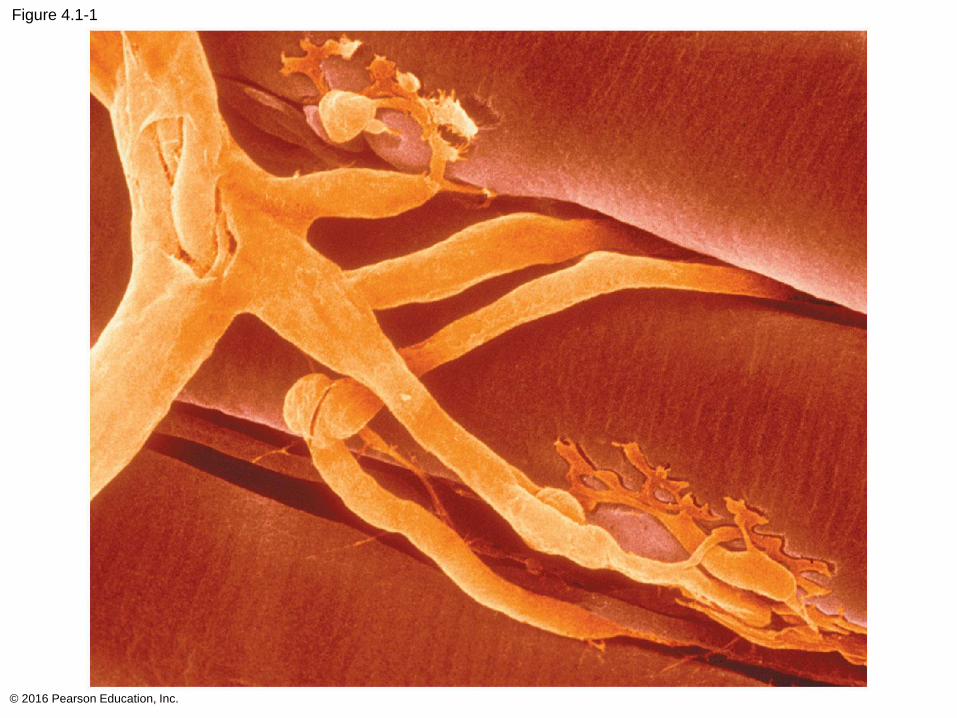
Figure 4.1-1
© 2016 Pearson Education, Inc.
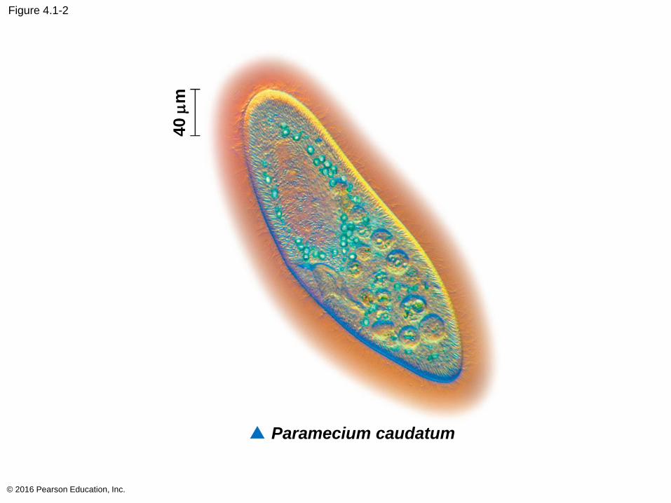
Figure 4.1-2
© 2016 Pearson Education, Inc.
▲ Paramecium caudatum
40 m
m
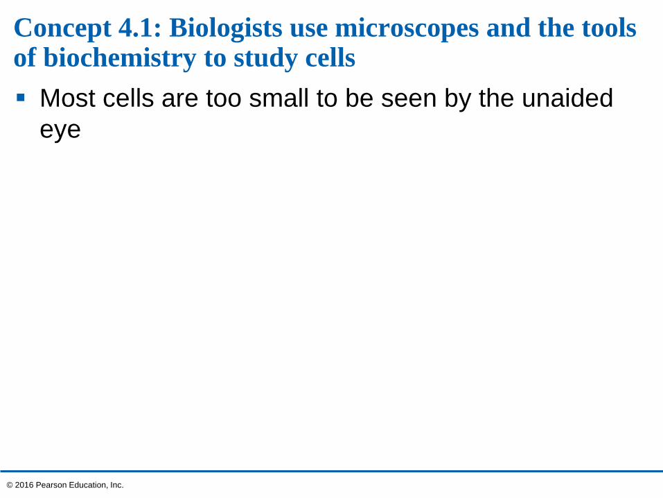
Concept 4.1: Biologists use microscopes and the tools of biochemistry to study cells
Most cells are too small to be seen by the unaided
eye
© 2016 Pearson Education, Inc.
Microscopy
Scientists use microscopes to observe cells too
small to be seen with the naked eye
In a light microscope (LM), visible light is passed
through a specimen and then through glass lenses
Lenses refract (bend) the light, so that the image is
magnified
© 2016 Pearson Education, Inc.
Three important parameters of microscopy
Magnification, the ratio of an object’s image size to its
real size
Resolution, the measure of the clarity of the image,
or the minimum distance between two distinguishable
points
Contrast, visible differences in parts of the sample
© 2016 Pearson Education, Inc.
LMs can magnify effectively to about 1,000 times
the size of the actual specimen
Various techniques enhance contrast and enable
cell components to be stained or labeled
Most subcellular structures, including organelles
(membrane-enclosed compartments), are too small
to be resolved by light microscopy
© 2016 Pearson Education, Inc.
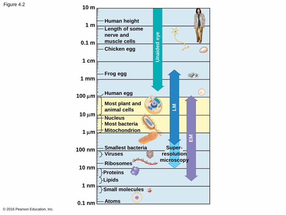
Figure 4.2
© 2016 Pearson Education, Inc.
Human height
Length of some
nerve and
muscle cells
Chicken egg
Frog egg
Human egg
Most plant and
animal cells
NucleusMost bacteria
Mitochondrion
Smallest bacteria
Viruses
Ribosomes
Super-
resolution
microscopy
Proteins
Lipids
Small molecules
Atoms0.1 nm
1 nm
10 nm
100 nm
1 mm
10 mm
100 mm
1 mm
1 cm
0.1 m
1 m
10 m
Un
aid
ed
eye
LM
EM
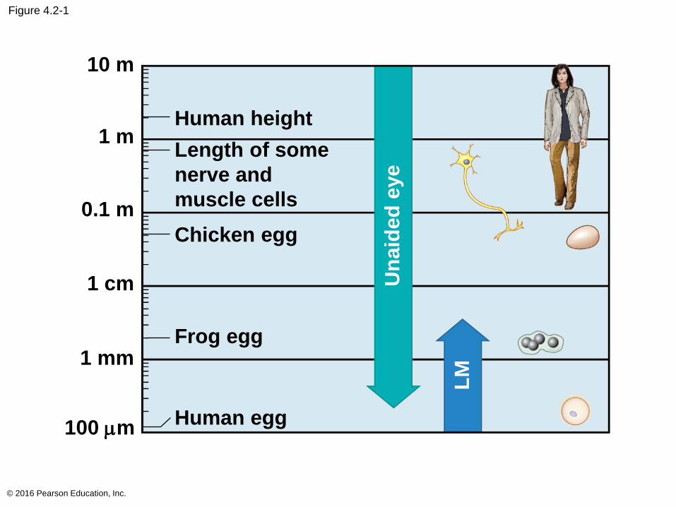
Figure 4.2-1
© 2016 Pearson Education, Inc.
Human height
Length of some
nerve and
muscle cells
Chicken egg
Frog egg
Human egg100 mm
1 mm
1 cm
0.1 m
1 m
10 m
Un
aid
ed
eye
LM
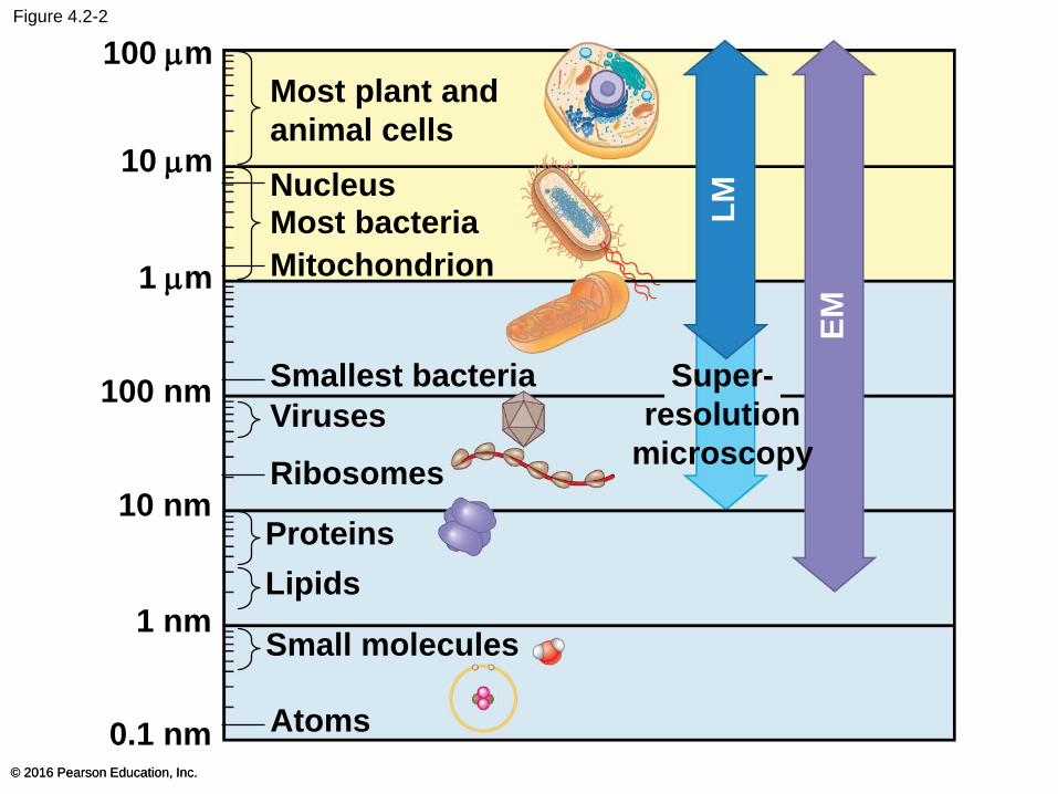
Figure 4.2-2
© 2016 Pearson Education, Inc.© 2016 Pearson Education, Inc.
Most plant and
animal cells
NucleusMost bacteria
Mitochondrion
Smallest bacteria
Viruses
Ribosomes
Super-
resolution
microscopy
Proteins
Lipids
Small molecules
Atoms0.1 nm
1 nm
10 nm
100 nm
1 mm
10 mm
LM
EM
100 mm

Figure 4.2
© 2016 Pearson Education, Inc.
Human height
Length of some
nerve and
muscle cells
Chicken egg
Frog egg
Human egg
Most plant and
animal cells
NucleusMost bacteria
Mitochondrion
Smallest bacteria
Viruses
Ribosomes
Super-
resolution
microscopy
Proteins
Lipids
Small molecules
Atoms0.1 nm
1 nm
10 nm
100 nm
1 mm
10 mm
100 mm
1 mm
1 cm
0.1 m
1 m
10 m
Un
aid
ed
eye
LM
EM
Two basic types of electron microscopes (EMs)
are used to study subcellular structures
Scanning electron microscopes (SEMs) focus a
beam of electrons onto the surface of a specimen,
producing images that look three-dimensional
Transmission electron microscopes (TEMs)
focus a beam of electrons through a specimen
TEM is used mainly to study the internal structure
of cells
© 2016 Pearson Education, Inc.
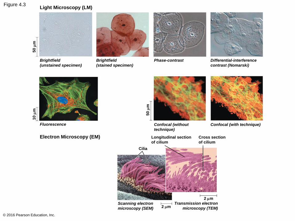
Figure 4.3
© 2016 Pearson Education, Inc.
Light Microscopy (LM)
Electron Microscopy (EM)
Brightfield
(unstained specimen)
Brightfield
(stained specimen)
Phase-contrast Differential-interference
contrast (Nomarski)
Confocal (without
technique)
Confocal (with technique)
Longitudinal section
of cilium
Cross section
of cilium
Cilia
Scanning electron
microscopy (SEM) 2 mm
2 mm
Transmission electron
microscopy (TEM)
Fluorescence
10 m
m50 m
m
50 m
m
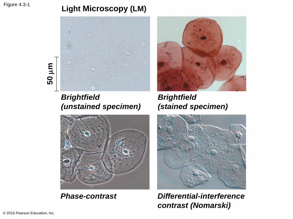
Figure 4.3-1
© 2016 Pearson Education, Inc.
Light Microscopy (LM)
Brightfield
(unstained specimen)
Brightfield
(stained specimen)
Phase-contrast Differential-interference
contrast (Nomarski)
50
mm
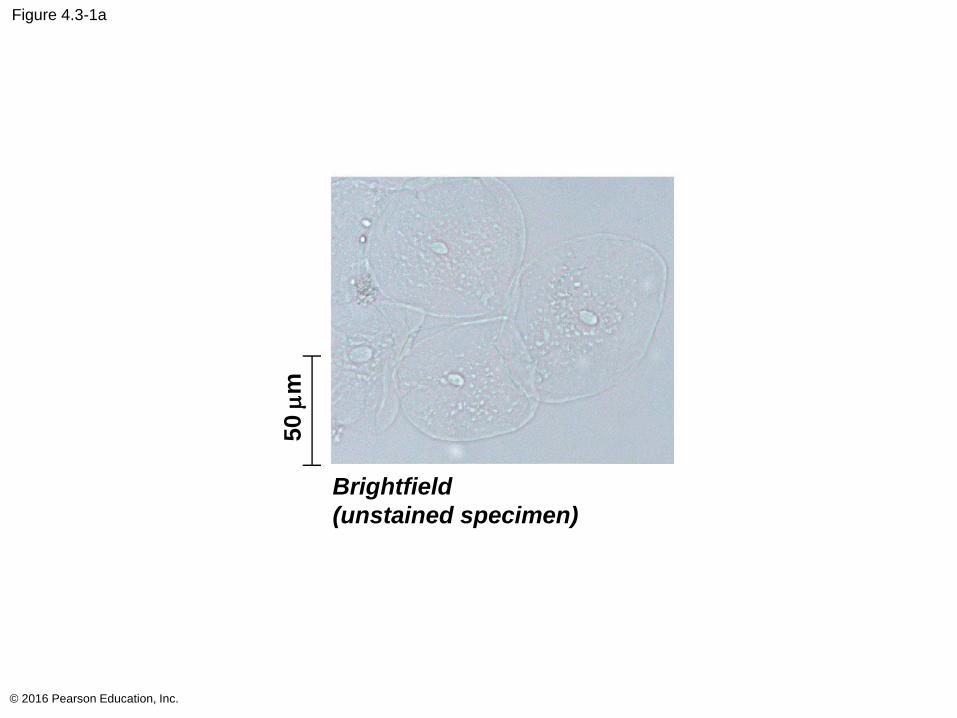
Figure 4.3-1a
© 2016 Pearson Education, Inc.
Brightfield
(unstained specimen)
50 m
m
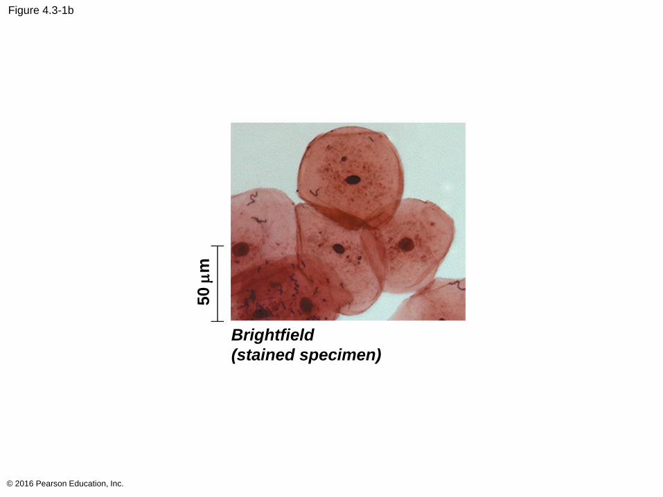
Figure 4.3-1b
© 2016 Pearson Education, Inc.
Brightfield
(stained specimen)
50 m
m
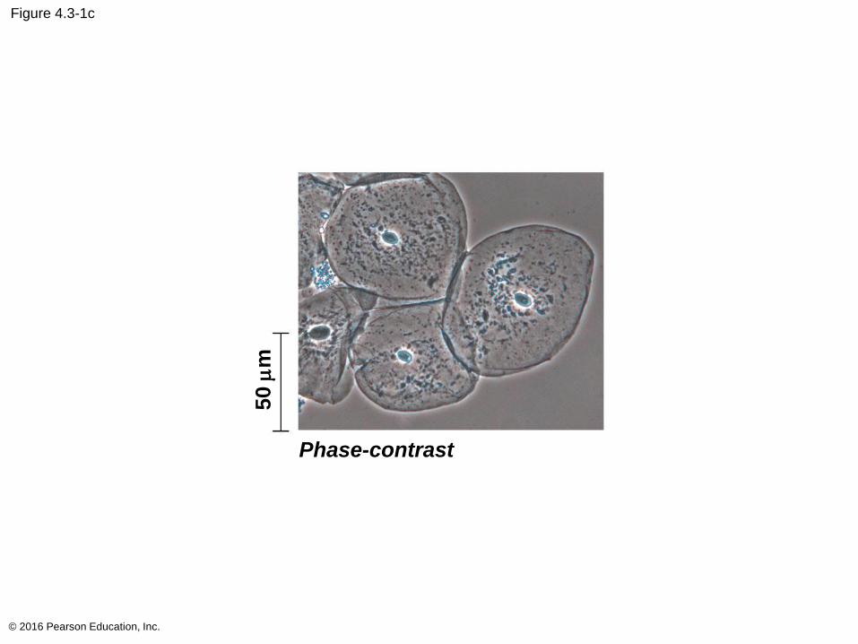
Figure 4.3-1c
© 2016 Pearson Education, Inc.
Phase-contrast
50 m
m
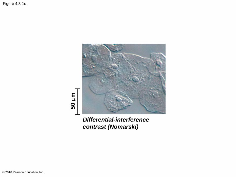
Figure 4.3-1d
© 2016 Pearson Education, Inc.
Differential-interference
contrast (Nomarski)
50 m
m
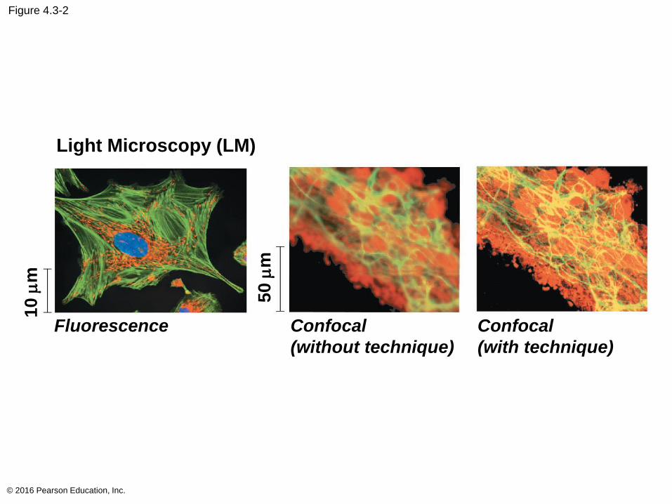
Figure 4.3-2
© 2016 Pearson Education, Inc.
Light Microscopy (LM)
Confocal
(with technique)
Fluorescence
10
mm
50
mm
Confocal
(without technique)
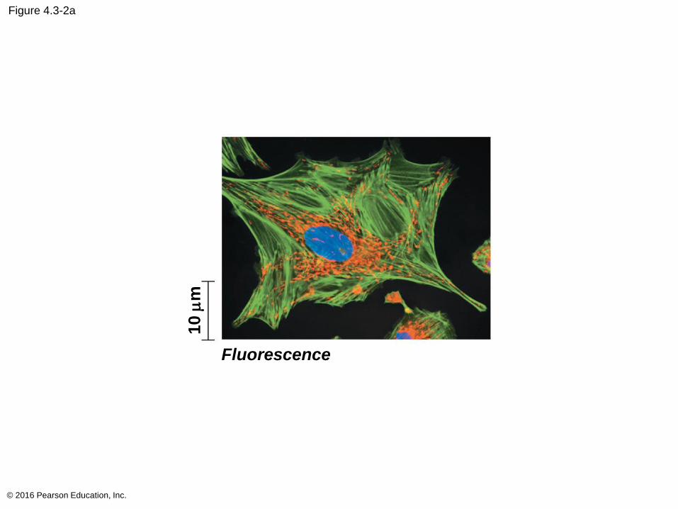
Figure 4.3-2a
© 2016 Pearson Education, Inc.
Fluorescence
10 m
m
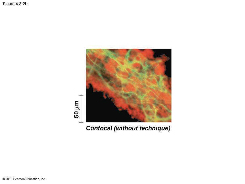
Figure 4.3-2b
© 2016 Pearson Education, Inc.
50 m
m
Confocal (without technique)
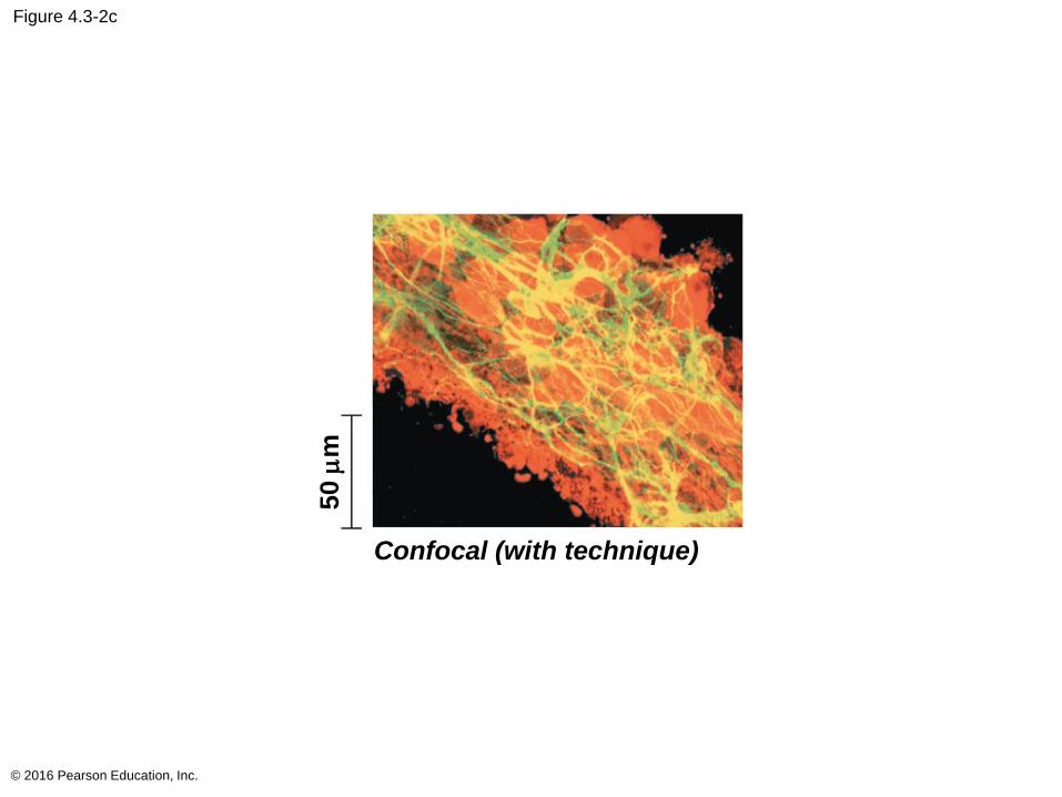
Figure 4.3-2c
© 2016 Pearson Education, Inc.
Confocal (with technique)
50 m
m
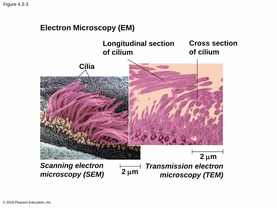
Figure 4.3-3
© 2016 Pearson Education, Inc.
Longitudinal section
of cilium
Cross section
of cilium
Cilia
Scanning electron
microscopy (SEM) 2 mm
2 mm
Transmission electron
microscopy (TEM)
Electron Microscopy (EM)
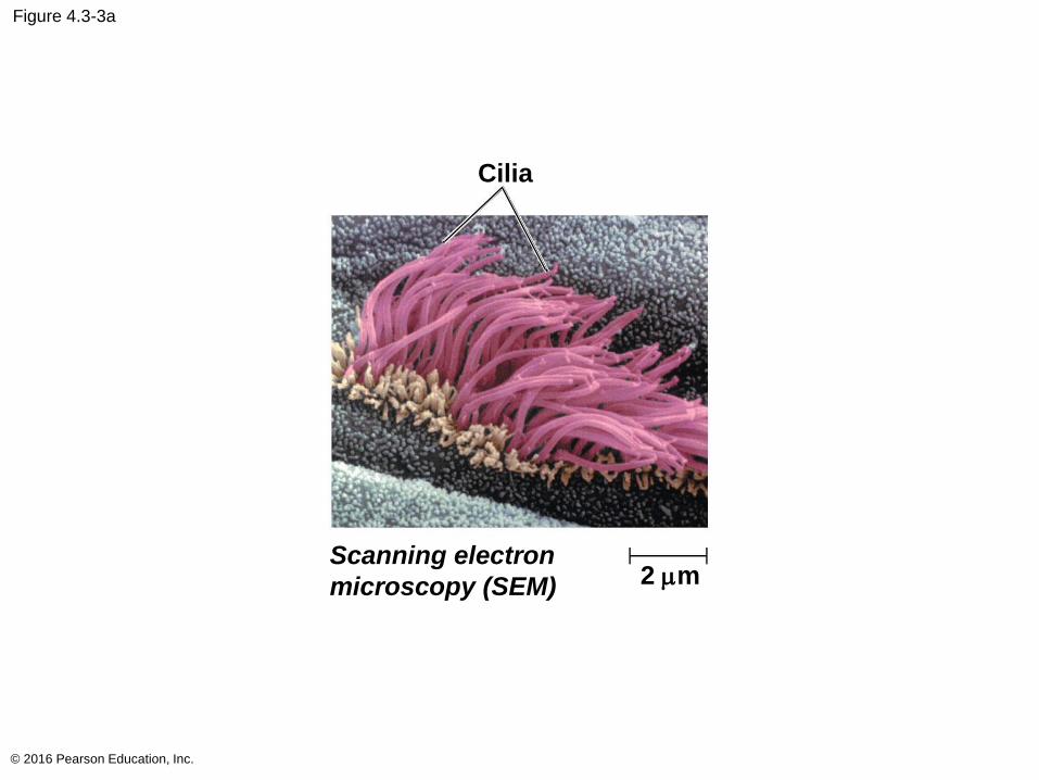
Figure 4.3-3a
© 2016 Pearson Education, Inc.
Cilia
Scanning electron
microscopy (SEM) 2 mm
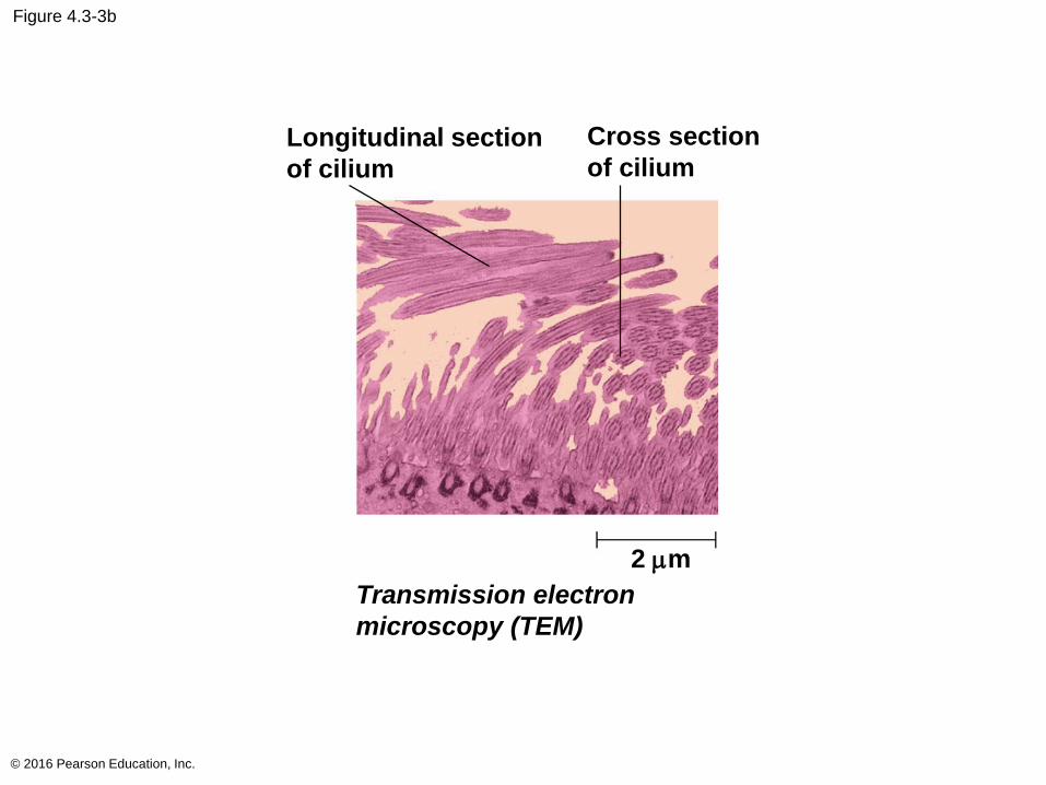
Figure 4.3-3b
© 2016 Pearson Education, Inc.
Longitudinal section
of cilium
Cross section
of cilium
2 mm
Transmission electron
microscopy (TEM)
Recent advances in light microscopy
Labeling molecules or structures with fluorescent
markers improves visualization of details
Confocal and other types of microscopy have
sharpened images of tissues and cells
New techniques and labeling have improved
resolution so that structures as small as 10–20 mm
can be distinguished
© 2016 Pearson Education, Inc.
Cell Fractionation
Cell fractionation breaks up cells and separates
the components, using centrifugation
Cell components separate based on their
relative size
Cell fractionation enables scientists to determine the
functions of organelles
Biochemistry and cytology help correlate cell
function with structure
© 2016 Pearson Education, Inc.
Concept 4.2: Eukaryotic cells have internal membranes that compartmentalize their functions
The basic structural and functional unit of every
organism is one of two types of cells: prokaryotic or
eukaryotic
Organisms of the domains Bacteria and Archaea
consist of prokaryotic cells
Protists, fungi, animals, and plants all consist of
eukaryotic cells
© 2016 Pearson Education, Inc.
Comparing Prokaryotic and Eukaryotic Cells
Basic features of all cells
Plasma membrane
Semifluid substance called cytosol
Chromosomes (carry genes)
Ribosomes (make proteins)
© 2016 Pearson Education, Inc.
In a eukaryotic cell most of the DNA is in the
nucleus, an organelle that is bounded by a double
membrane
Prokaryotic cells are characterized by having
No nucleus
DNA in an unbound region called the nucleoid
No membrane-bound organelles
Both types of cells contain cytoplasm bound by the
plasma membrane
© 2016 Pearson Education, Inc.
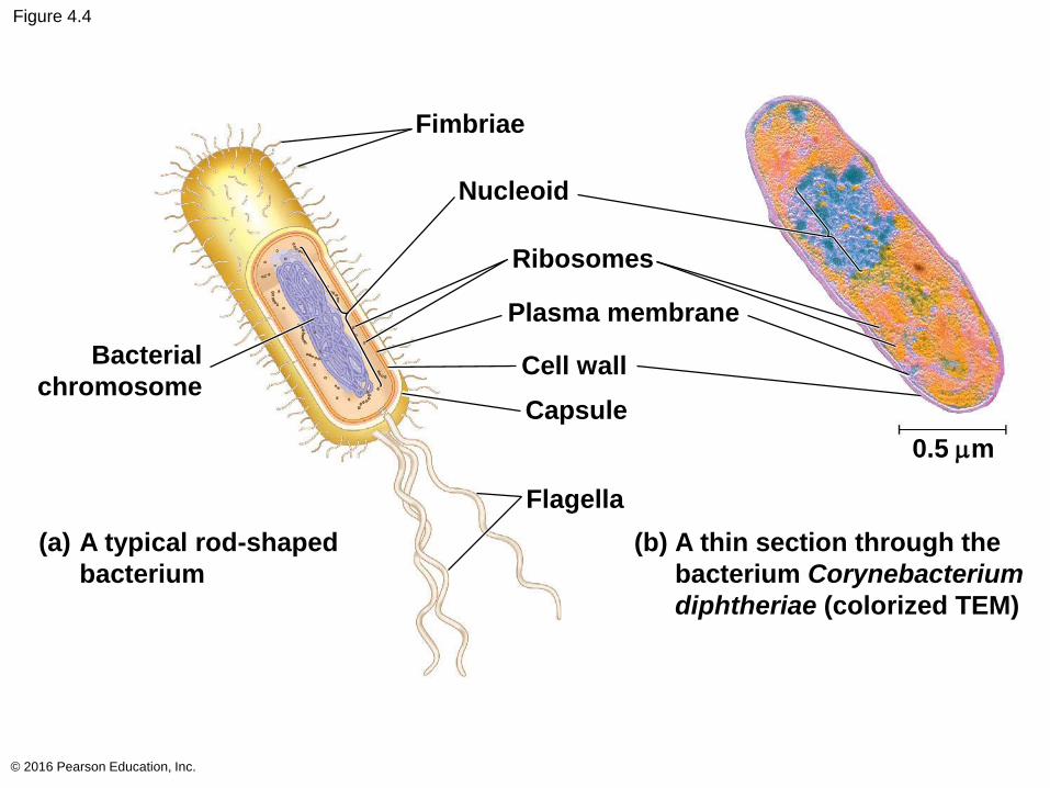
Figure 4.4
© 2016 Pearson Education, Inc.
Fimbriae
Nucleoid
Ribosomes
Plasma membrane
Cell wall
Capsule
Flagella
Bacterial
chromosome
A typical rod-shaped
bacterium
(a) A thin section through the
bacterium Corynebacterium
diphtheriae (colorized TEM)
(b)
0.5 mm
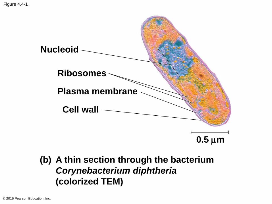
Figure 4.4-1
© 2016 Pearson Education, Inc.
Nucleoid
Ribosomes
Plasma membrane
Cell wall
A thin section through the bacterium
Corynebacterium diphtheria
(colorized TEM)
(b)
0.5 mm
Eukaryotic cells are generally much larger than
prokaryotic cells
Typical bacteria are 1–5 mm in diameter
Eukaryotic cells are typically 10–100 mm in diameter
© 2016 Pearson Education, Inc.
The plasma membrane is a selective barrier that
allows sufficient passage of oxygen, nutrients, and
waste to service the volume of every cell
The general structure of a biological membrane is a
double layer of phospholipids
© 2016 Pearson Education, Inc.
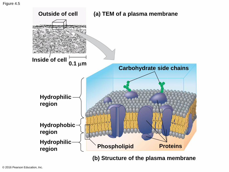
Figure 4.5
© 2016 Pearson Education, Inc.
Outside of cell (a) TEM of a plasma membrane
Inside of cell0.1 mm
Carbohydrate side chains
Hydrophilic
region
Hydrophobic
region
Hydrophilic
region Phospholipid Proteins
(b) Structure of the plasma membrane
Figure 4.5-1
© 2016 Pearson Education, Inc.
Outside of cell
Inside of cell0.1 mm
(a) TEM of a plasma
membrane
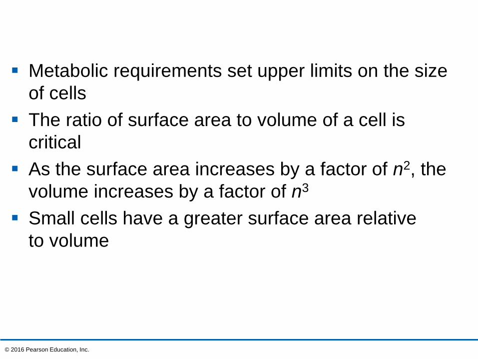
Metabolic requirements set upper limits on the size
of cells
The ratio of surface area to volume of a cell is
critical
As the surface area increases by a factor of n2, the
volume increases by a factor of n3
Small cells have a greater surface area relative
to volume
© 2016 Pearson Education, Inc.
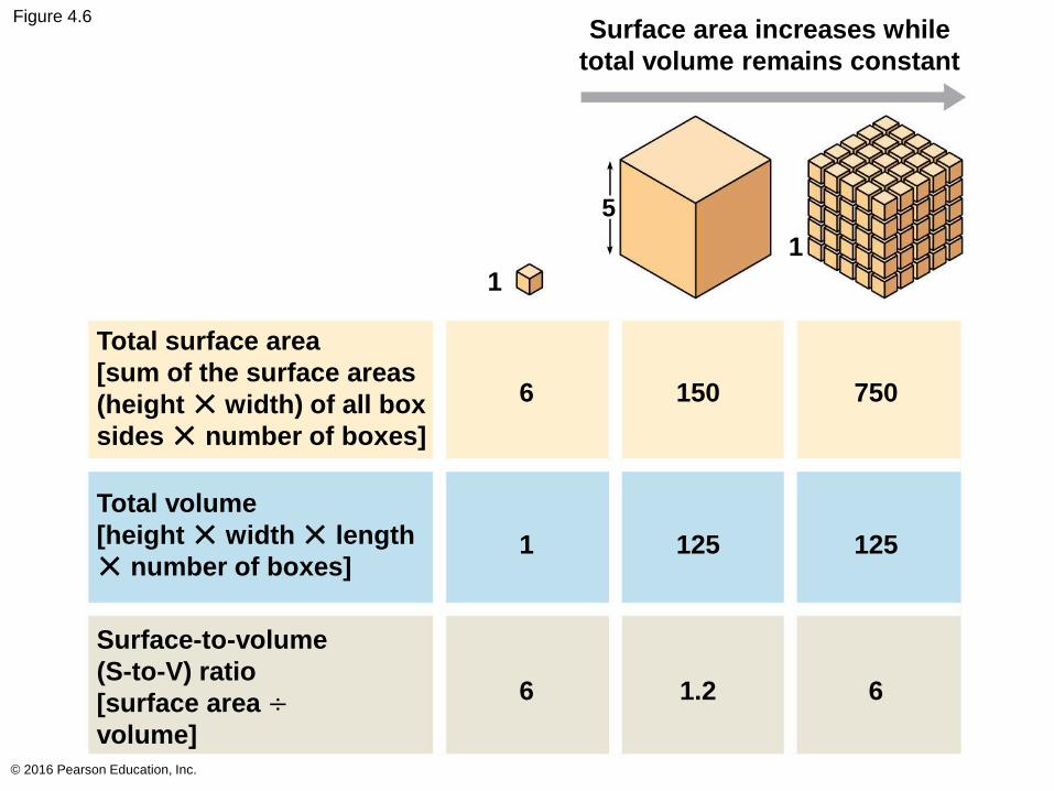
Figure 4.6
© 2016 Pearson Education, Inc.
Surface area increases while
total volume remains constant
1
1
5
6 150 750
Total surface area
[sum of the surface areas
(height ✕ width) of all box
sides ✕ number of boxes]
Total volume
[height ✕ width ✕ length
✕ number of boxes]
Surface-to-volume
(S-to-V) ratio
[surface area ÷volume]
1 125 125
6 1.2 6
A Panoramic View of the Eukaryotic Cell
A eukaryotic cell has internal membranes that divide
the cell into compartments—organelles
The plasma membrane and organelle membranes
participate directly in the cell’s metabolism
© 2016 Pearson Education, Inc.
Animation: Tour of an Animal Cell
© 2016 Pearson Education, Inc.
Animation: Tour of a Plant Cell
© 2016 Pearson Education, Inc.
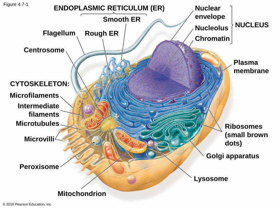
Figure 4.7-1
© 2016 Pearson Education, Inc.
ENDOPLASMIC RETICULUM (ER)
Smooth ER
Flagellum
Centrosome
CYTOSKELETON:
Microfilaments
Intermediate
filaments
Microtubules
Microvilli
Peroxisome
Mitochondrion
Lysosome
Golgi apparatus
Ribosomes
(small brown
dots)
Plasma
membrane
NUCLEUS
Nuclear
envelope
Nucleolus
ChromatinRough ER
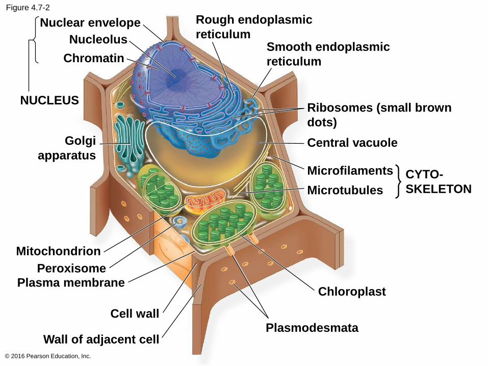
Figure 4.7-2
© 2016 Pearson Education, Inc.
Nuclear envelope
Nucleolus
Chromatin
NUCLEUS
Golgi
apparatus
Rough endoplasmic
reticulumSmooth endoplasmic
reticulum
Central vacuole
Microfilaments
Microtubules
CYTO-
SKELETON
Mitochondrion
PeroxisomePlasma membrane
Cell wall
Wall of adjacent cellPlasmodesmata
Chloroplast
Ribosomes (small brown
dots)
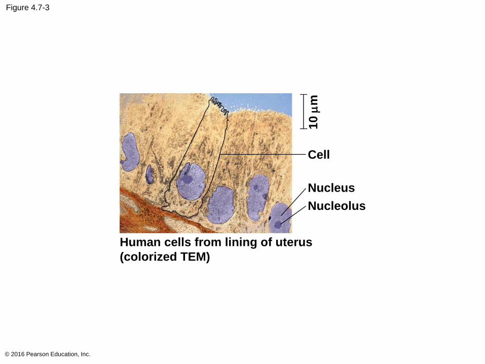
Figure 4.7-3
© 2016 Pearson Education, Inc.
Cell
Nucleus
Nucleolus
10 m
m
Human cells from lining of uterus
(colorized TEM)
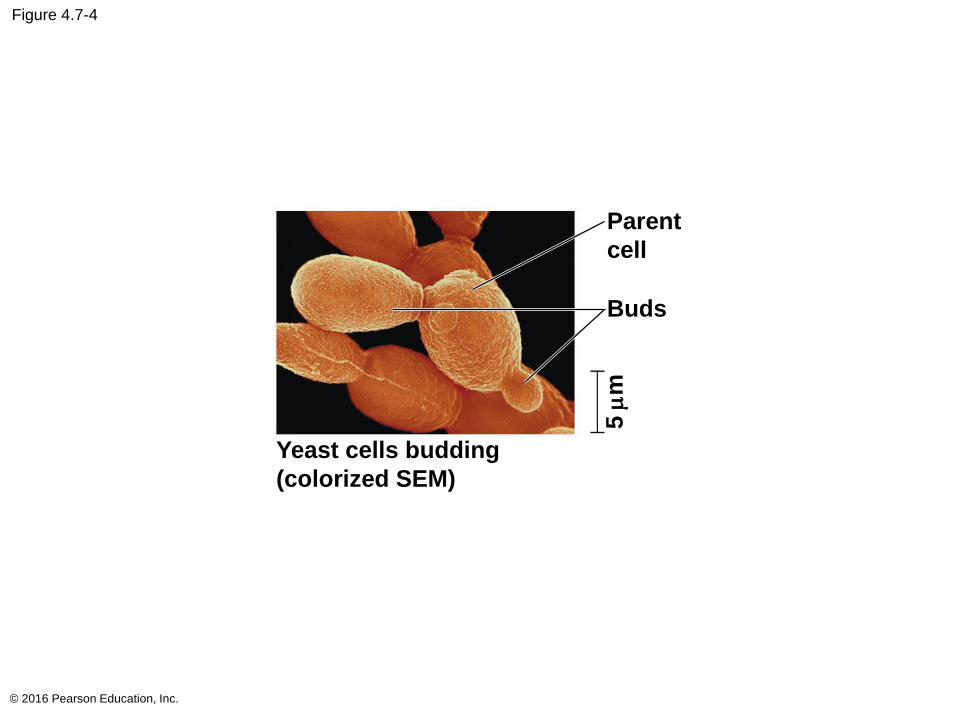
Figure 4.7-4
© 2016 Pearson Education, Inc.
5m
m
Parent
cell
Buds
Yeast cells budding
(colorized SEM)
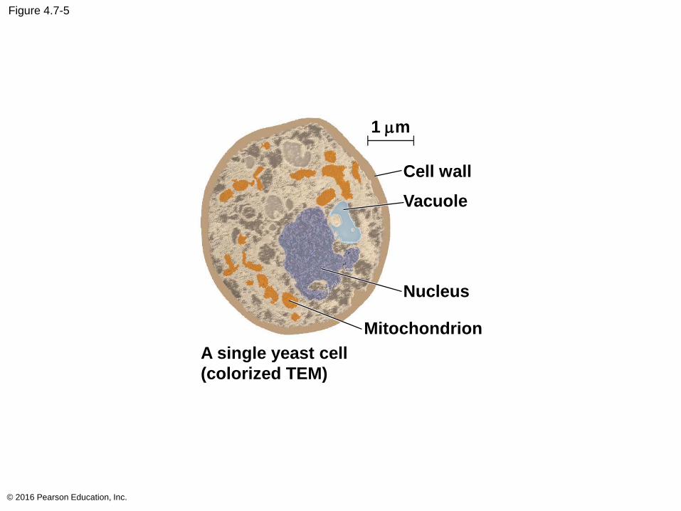
Figure 4.7-5
© 2016 Pearson Education, Inc.
1 mm
Cell wall
Vacuole
Nucleus
Mitochondrion
A single yeast cell
(colorized TEM)
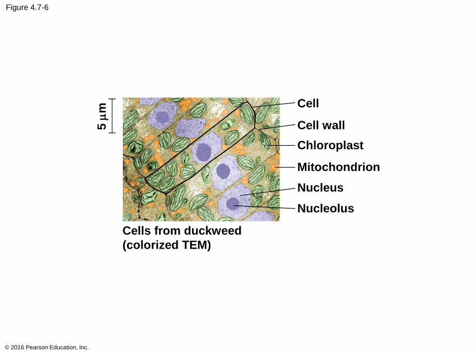
Figure 4.7-6
© 2016 Pearson Education, Inc.
Cell
Cell wall
Chloroplast
Mitochondrion
Nucleus
Nucleolus
Cells from duckweed
(colorized TEM)
5 m
m
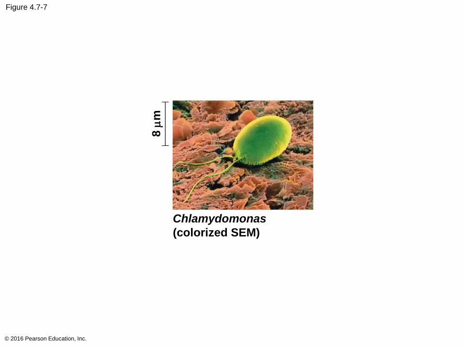
Figure 4.7-7
© 2016 Pearson Education, Inc.
Chlamydomonas
(colorized SEM)
8m
m
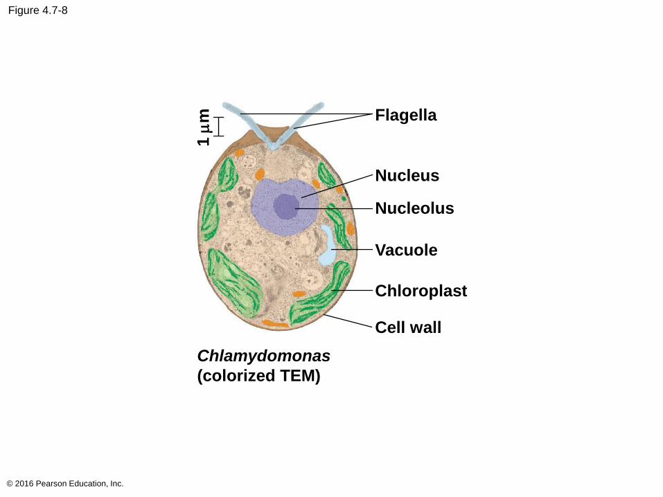
Figure 4.7-8
© 2016 Pearson Education, Inc.
Chlamydomonas
(colorized TEM)
1m
m Flagella
Nucleus
Nucleolus
Vacuole
Chloroplast
Cell wall
Concept 4.3: The eukaryotic cell’s genetic instructions are housed in the nucleus and carried out by the ribosomes
The nucleus contains most of the DNA in a
eukaryotic cell
Ribosomes use the information from the DNA to
make proteins
© 2016 Pearson Education, Inc.
The Nucleus: Information Central
The nucleus contains most of the cell’s genes and
is usually the most conspicuous organelle
The nuclear envelope encloses the nucleus,
separating it from the cytoplasm
The nuclear membrane is a double membrane;
each membrane consists of a lipid bilayer
© 2016 Pearson Education, Inc.
Nuclear pores regulate the entry and exit of
molecules
The shape of the nucleus is maintained by the
nuclear lamina, which is composed of protein
© 2016 Pearson Education, Inc.
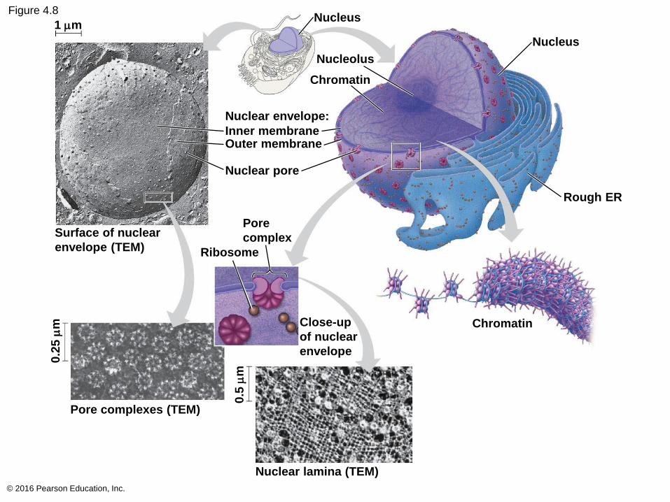
Figure 4.8
© 2016 Pearson Education, Inc.
Nucleus
Nucleolus
Chromatin
Nuclear envelope:
Inner membraneOuter membrane
Nuclear pore
1 mm
Surface of nuclear
envelope (TEM)
Pore
complex
Ribosome
Close-up
of nuclear
envelope
0.2
5 m
m
Pore complexes (TEM)
0.5
mm
Nuclear lamina (TEM)
Chromatin
Rough ER
Nucleus
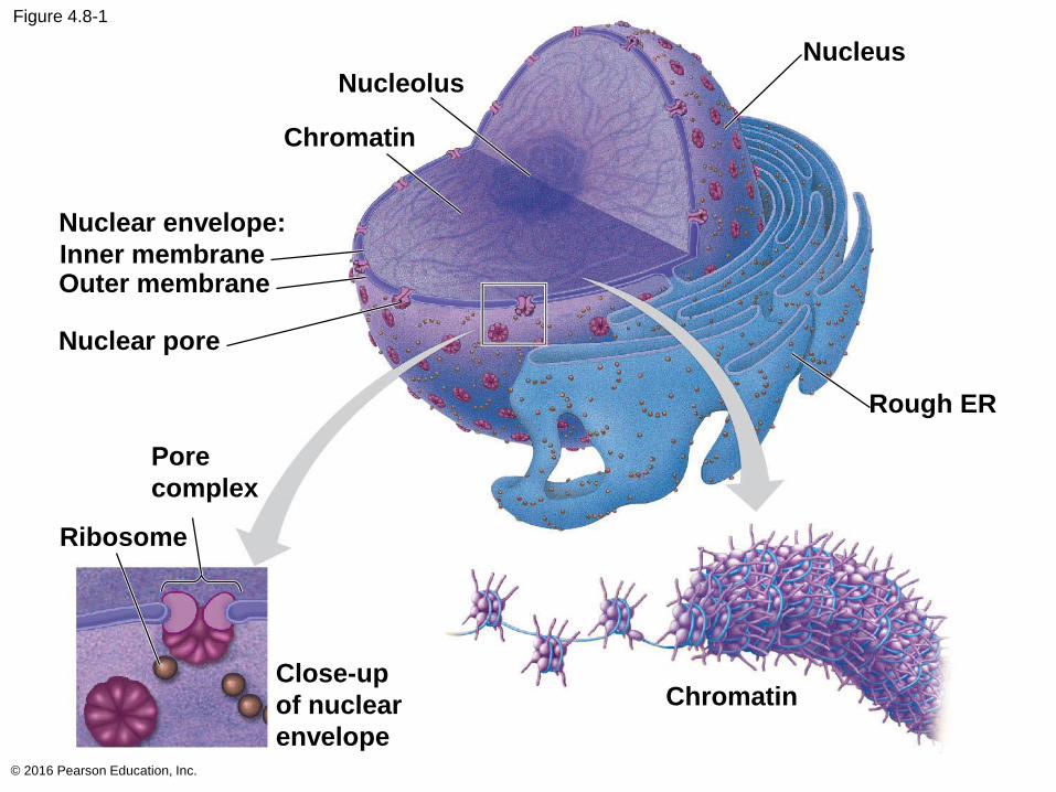
Figure 4.8-1
© 2016 Pearson Education, Inc.
Nucleolus
Chromatin
Nuclear envelope:
Inner membraneOuter membrane
Nuclear pore
Pore
complex
Close-up
of nuclear
envelope
Chromatin
Rough ER
Nucleus
Ribosome
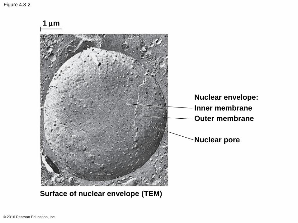
Figure 4.8-2
© 2016 Pearson Education, Inc.
Nuclear envelope:
Inner membrane
Outer membrane
Nuclear pore
1 mm
Surface of nuclear envelope (TEM)
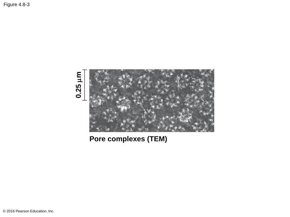
Figure 4.8-3
© 2016 Pearson Education, Inc.
0.2
5 m
m
Pore complexes (TEM)
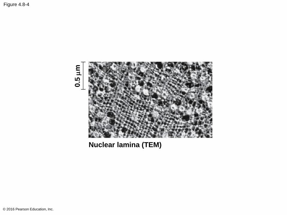
Figure 4.8-4
© 2016 Pearson Education, Inc.
0.5
mm
Nuclear lamina (TEM)
In the nucleus, DNA is organized into discrete units
called chromosomes
Each chromosome is one long DNA molecule
associated with proteins
The DNA and proteins of chromosomes together
are called chromatin
Chromatin condenses to form discrete
chromosomes as a cell prepares to divide
The nucleolus is located within the nucleus and is
the site of ribosomal RNA (rRNA) synthesis
© 2016 Pearson Education, Inc.
Ribosomes: Protein Factories
Ribosomes are complexes of ribosomal RNA and
protein
Ribosomes carry out protein synthesis in two
locations
In the cytosol (free ribosomes)
On the outside of the endoplasmic reticulum or the
nuclear envelope (bound ribosomes)
© 2016 Pearson Education, Inc.
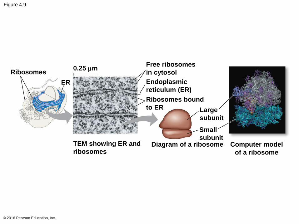
Figure 4.9
© 2016 Pearson Education, Inc.
Ribosomes0.25 mm
ER
Free ribosomes
in cytosol
Endoplasmic
reticulum (ER)
Ribosomes bound
to ER Large
subunit
Small
subunitTEM showing ER and
ribosomesDiagram of a ribosome Computer model
of a ribosome
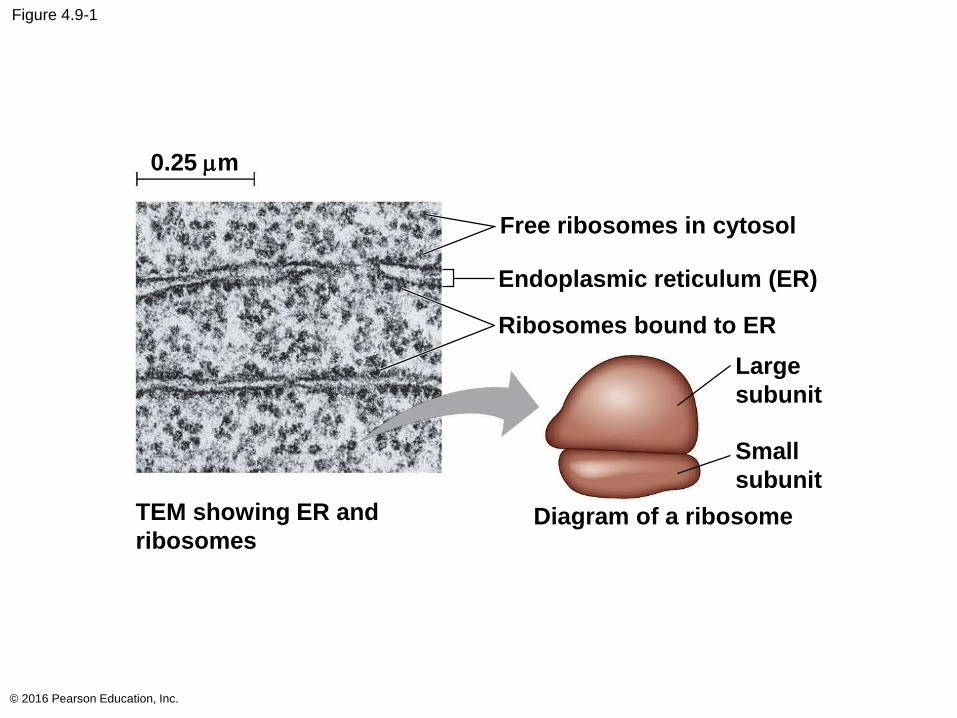
Figure 4.9-1
© 2016 Pearson Education, Inc.
0.25 mm
Free ribosomes in cytosol
Endoplasmic reticulum (ER)
Large
subunit
Small
subunit
TEM showing ER and
ribosomesDiagram of a ribosome
Ribosomes bound to ER
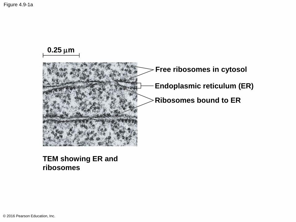
Figure 4.9-1a
© 2016 Pearson Education, Inc.
0.25 mm
Free ribosomes in cytosol
Endoplasmic reticulum (ER)
TEM showing ER and
ribosomes
Ribosomes bound to ER
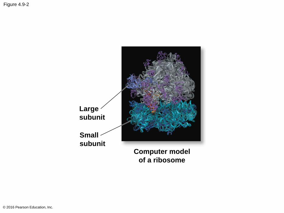
Figure 4.9-2
© 2016 Pearson Education, Inc.
Large
subunit
Small
subunitComputer model
of a ribosome
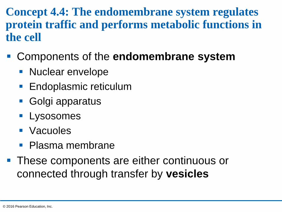
Concept 4.4: The endomembrane system regulates protein traffic and performs metabolic functions in the cell
Components of the endomembrane system
Nuclear envelope
Endoplasmic reticulum
Golgi apparatus
Lysosomes
Vacuoles
Plasma membrane
These components are either continuous or
connected through transfer by vesicles
© 2016 Pearson Education, Inc.

The Endoplasmic Reticulum: Biosynthetic Factory
The endoplasmic reticulum (ER) accounts for
more than half of the total membrane in many
eukaryotic cells
The ER membrane is continuous with the nuclear
envelope
There are two distinct regions of ER
Smooth ER: lacks ribosomes
Rough ER: surface is studded with ribosomes
© 2016 Pearson Education, Inc.
Video: Endoplasmic Reticulum
© 2016 Pearson Education, Inc.

Video: ER and Mitochondria
© 2016 Pearson Education, Inc.
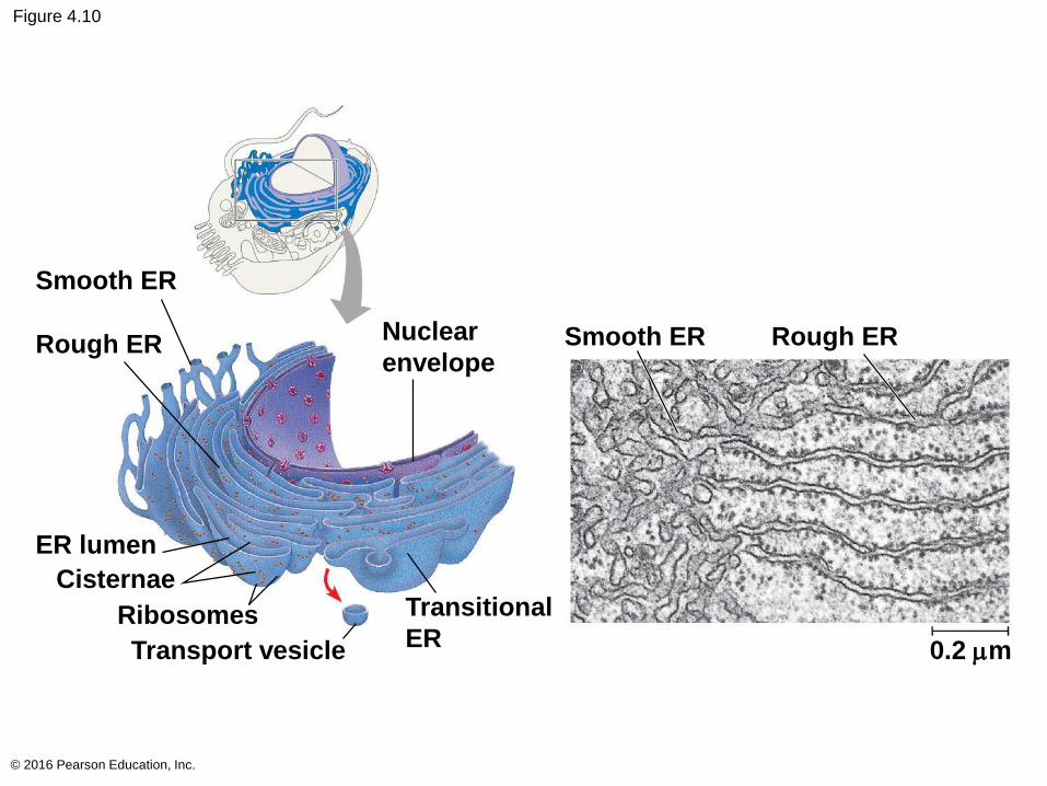
Figure 4.10
© 2016 Pearson Education, Inc.
Smooth ER
Rough ER
ER lumen
Cisternae
Ribosomes
Transport vesicle
Transitional
ER
Nuclear
envelopeSmooth ER Rough ER
0.2 mm
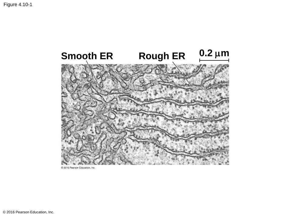
Figure 4.10-1
© 2016 Pearson Education, Inc.
Smooth ER Rough ER 0.2 mm
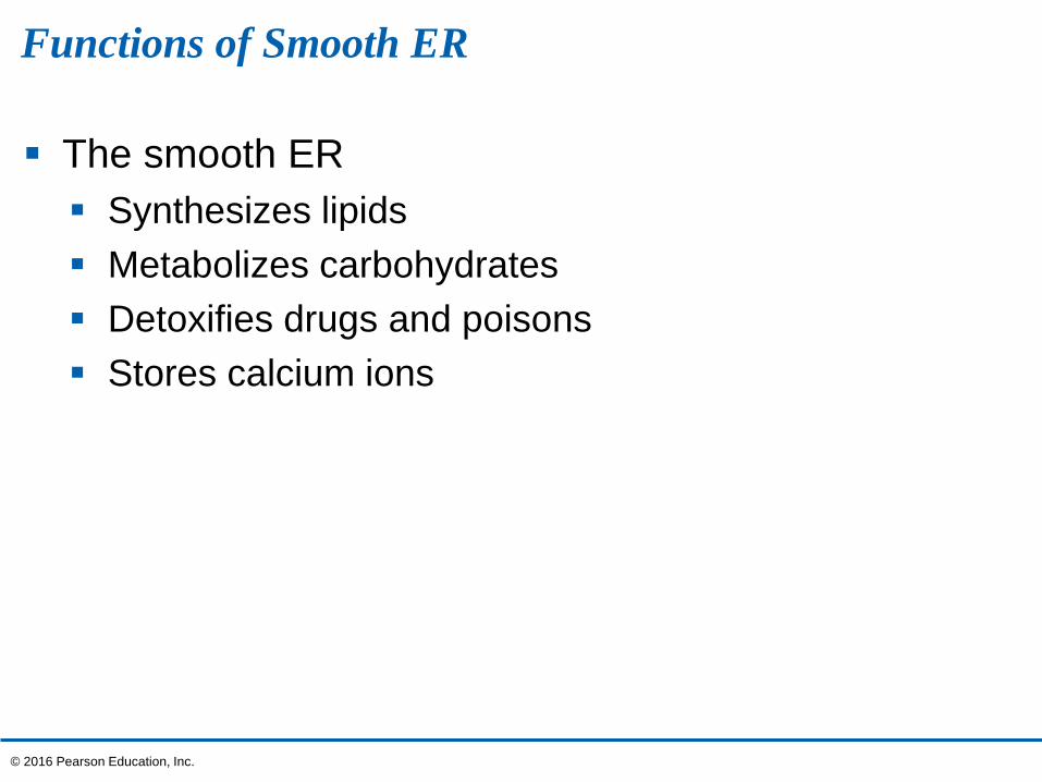
Functions of Smooth ER
The smooth ER
Synthesizes lipids
Metabolizes carbohydrates
Detoxifies drugs and poisons
Stores calcium ions
© 2016 Pearson Education, Inc.
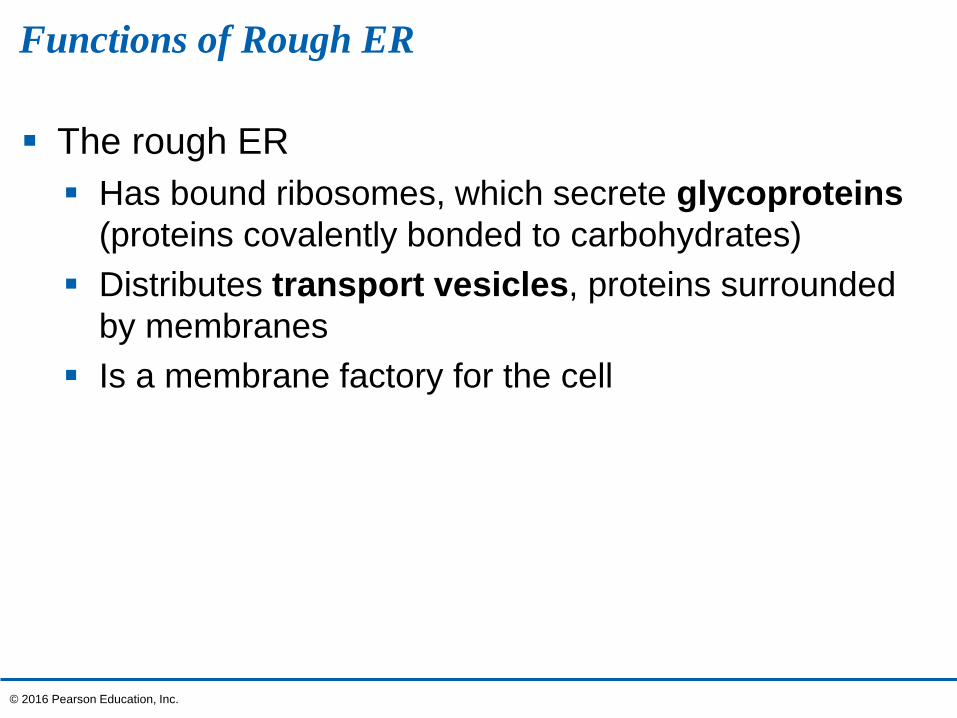
Functions of Rough ER
The rough ER
Has bound ribosomes, which secrete glycoproteins
(proteins covalently bonded to carbohydrates)
Distributes transport vesicles, proteins surrounded
by membranes
Is a membrane factory for the cell
© 2016 Pearson Education, Inc.
The Golgi Apparatus: Shipping and Receiving Center
The Golgi apparatus consists of flattened
membranous sacs called cisternae
Functions of the Golgi apparatus
Modifies products of the ER
Manufactures certain macromolecules
Sorts and packages materials into transport vesicles
© 2016 Pearson Education, Inc.
Video: ER to Golgi Traffic
© 2016 Pearson Education, Inc.
Video: Golgi 3-D
© 2016 Pearson Education, Inc.
Video: Golgi Secretion
© 2016 Pearson Education, Inc.
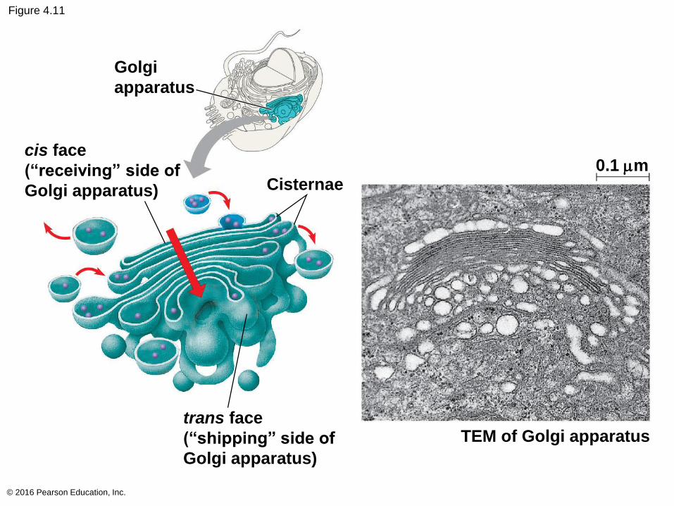
Figure 4.11
© 2016 Pearson Education, Inc.
Golgi
apparatus
cis face
(“receiving” side of
Golgi apparatus) Cisternae
trans face
(“shipping” side of
Golgi apparatus)
TEM of Golgi apparatus
0.1 mm
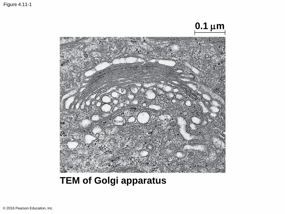
Figure 4.11-1
© 2016 Pearson Education, Inc.
TEM of Golgi apparatus
0.1 mm
Lysosomes: Digestive Compartments
A lysosome is a membranous sac of hydrolytic
enzymes that can digest macromolecules
Lysosomal enzymes work best in the acidic
environment inside the lysosome
The three-dimensional shape of lysosomal proteins
protects them from digestion by lysosomal enzymes
© 2016 Pearson Education, Inc.
Some types of cell can engulf another cell by
phagocytosis; this forms a food vacuole
A lysosome fuses with the food vacuole and digests
the molecules
Lysosomes also use enzymes to recycle the cell’s
own organelles and macromolecules, a process
called autophagy
© 2016 Pearson Education, Inc.
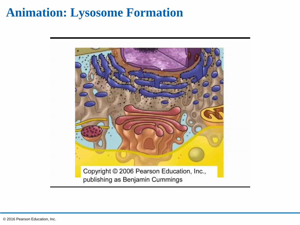
Animation: Lysosome Formation
© 2016 Pearson Education, Inc.
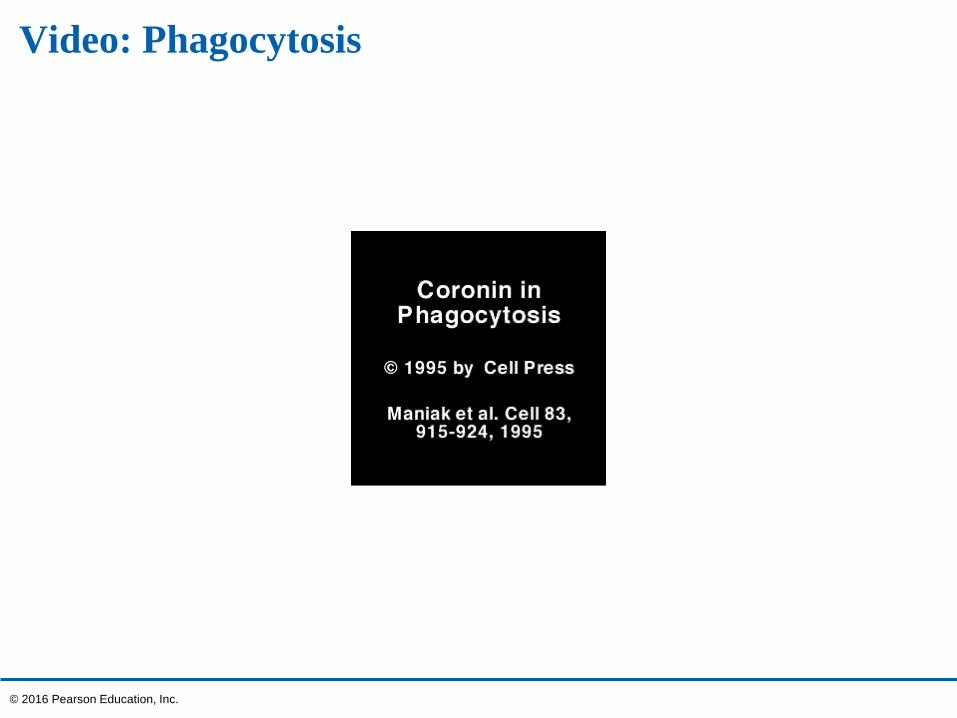
Video: Phagocytosis
© 2016 Pearson Education, Inc.
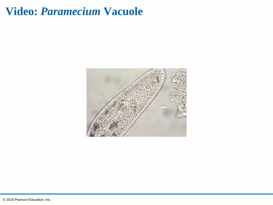
Video: Paramecium Vacuole
© 2016 Pearson Education, Inc.
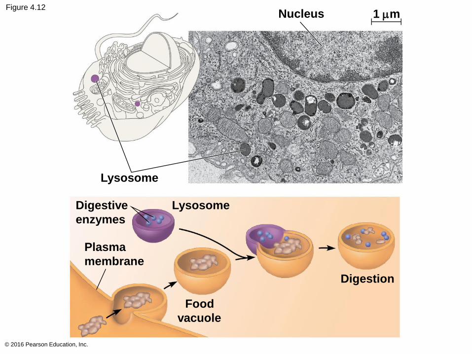
Figure 4.12
© 2016 Pearson Education, Inc.
Nucleus 1 mm
Lysosome
Digestive
enzymes
Lysosome
Plasma
membrane
Digestion
Food
vacuole
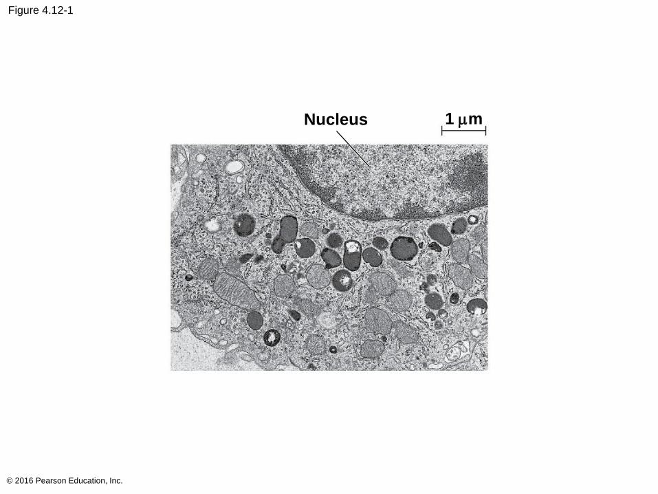
Figure 4.12-1
© 2016 Pearson Education, Inc.
Nucleus 1 mm
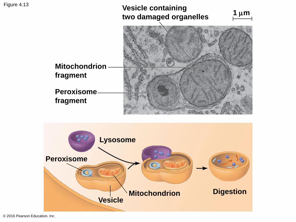
Figure 4.13
© 2016 Pearson Education, Inc.
Vesicle containing
two damaged organelles1 mm
Mitochondrion
fragment
Peroxisome
fragment
Lysosome
Peroxisome
Vesicle Mitochondrion Digestion
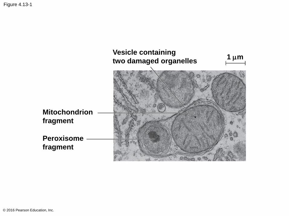
Figure 4.13-1
© 2016 Pearson Education, Inc.
Vesicle containing
two damaged organelles1 mm
Mitochondrion
fragment
Peroxisome
fragment
Vacuoles: Diverse Maintenance Compartments
Vacuoles are large vesicles derived from the
endoplasmic reticulum and Golgi apparatus
The solution inside a vacuole differs in composition
from the cytosol
© 2016 Pearson Education, Inc.
Food vacuoles are formed by phagocytosis
Contractile vacuoles, found in many freshwater
protists, pump excess water out of cells
Central vacuoles, found in many mature plant
cells, hold organic compounds and water
Certain vacuoles in plants and fungi carry out
enzymatic hydrolysis like lysosomes
© 2016 Pearson Education, Inc.
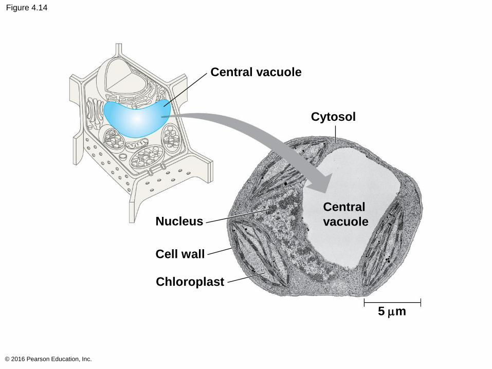
Figure 4.14
© 2016 Pearson Education, Inc.
Central vacuole
Cytosol
Nucleus
Cell wall
Chloroplast
Central
vacuole
5 mm
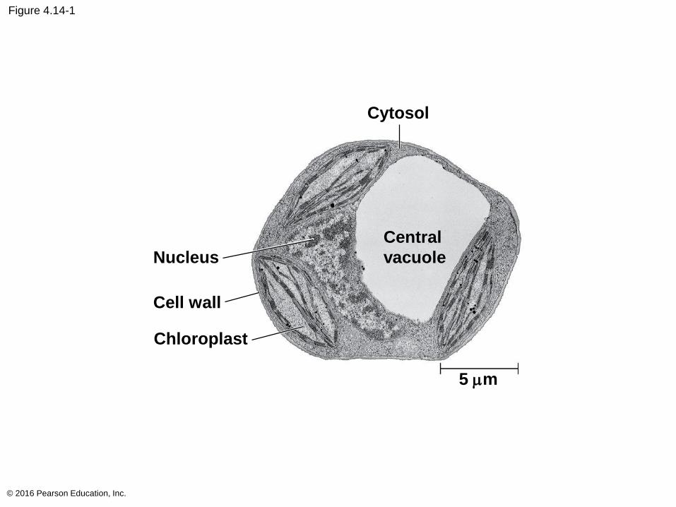
Figure 4.14-1
© 2016 Pearson Education, Inc.
Cytosol
Nucleus
Cell wall
Chloroplast
Central
vacuole
5 mm
The Endomembrane System: A Review
The endomembrane system is a complex and
dynamic player in the cell’s compartmental
organization
© 2016 Pearson Education, Inc.
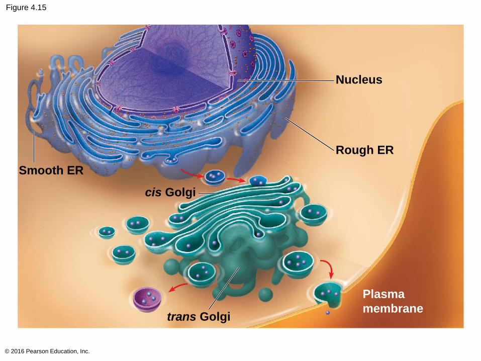
Figure 4.15
© 2016 Pearson Education, Inc.
Smooth ER
cis Golgi
trans Golgi
Rough ER
Nucleus
Plasma
membrane
Concept 4.5: Mitochondria and chloroplasts change energy from one form to another
Mitochondria are the sites of cellular respiration, a
metabolic process that uses oxygen to generate
ATP
Chloroplasts, found in plants and algae, are the
sites of photosynthesis
Peroxisomes are oxidative organelles
© 2016 Pearson Education, Inc.
The Evolutionary Origins of Mitochondria and Chloroplasts
Mitochondria and chloroplasts display similarities
with bacteria
Enveloped by a double membrane
Contain ribosomes and multiple circular DNA
molecules
Grow and reproduce somewhat independently in
cells
© 2016 Pearson Education, Inc.
The endosymbiont theory
An early ancestor of eukaryotic cells engulfed a
nonphotosynthetic prokaryotic cell, which formed an
endosymbiont relationship with its host
The host cell and endosymbiont merged into a single
organism, a eukaryotic cell with a mitochondrion
At least one of these cells may have taken up a
photosynthetic prokaryote, becoming the ancestor of
cells that contain chloroplasts
© 2016 Pearson Education, Inc.
Video: ER and Mitochondria
© 2016 Pearson Education, Inc.
Video: Mitochondria 3-D
© 2016 Pearson Education, Inc.
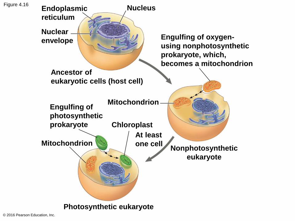
Figure 4.16
© 2016 Pearson Education, Inc.
Endoplasmic
reticulum
Nuclear
envelope
Ancestor of
eukaryotic cells (host cell)
Engulfing of
photosynthetic
prokaryote
Mitochondrion
Chloroplast
At least
one cellMitochondrionNonphotosynthetic
eukaryote
Engulfing of oxygen-
using nonphotosynthetic
prokaryote, which,
becomes a mitochondrion
Nucleus
Photosynthetic eukaryote
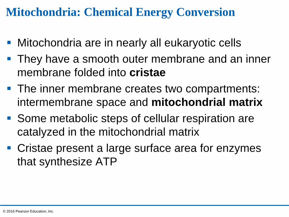
Mitochondria: Chemical Energy Conversion
Mitochondria are in nearly all eukaryotic cells
They have a smooth outer membrane and an inner
membrane folded into cristae
The inner membrane creates two compartments:
intermembrane space and mitochondrial matrix
Some metabolic steps of cellular respiration are
catalyzed in the mitochondrial matrix
Cristae present a large surface area for enzymes
that synthesize ATP
© 2016 Pearson Education, Inc.
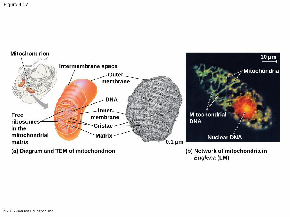
Figure 4.17
© 2016 Pearson Education, Inc.
Mitochondrion
Intermembrane space
Outer
membrane
DNA
Inner
membrane
Cristae
Matrix
Free
ribosomes
in the
mitochondrial
matrix
(a) Diagram and TEM of mitochondrion
0.1 mm
(b) Network of mitochondria in
Euglena (LM)
Mitochondrial
DNA
10 mm
Mitochondria
Nuclear DNA
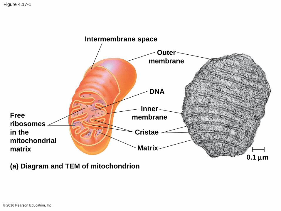
Figure 4.17-1
© 2016 Pearson Education, Inc.
Intermembrane space
Outer
membrane
DNA
Inner
membrane
Cristae
Matrix
Free
ribosomes
in the
mitochondrial
matrix
(a) Diagram and TEM of mitochondrion
0.1 mm
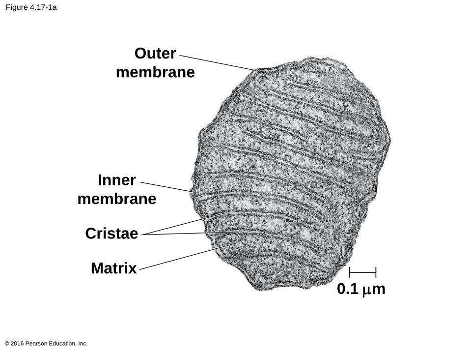
Figure 4.17-1a
© 2016 Pearson Education, Inc.
Outer
membrane
Inner
membrane
Cristae
Matrix
0.1 mm
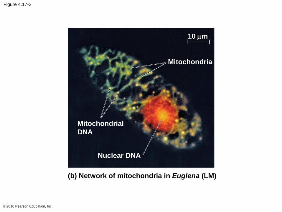
Figure 4.17-2
© 2016 Pearson Education, Inc.
(b) Network of mitochondria in Euglena (LM)
Mitochondrial
DNA
10 mm
Mitochondria
Nuclear DNA
Chloroplasts: Capture of Light Energy
Chloroplasts contain the green pigment chlorophyll,
as well as enzymes and other molecules that
function in photosynthesis
Chloroplasts are found in leaves and other green
organs of plants and in algae
© 2016 Pearson Education, Inc.
Chloroplast structure includes
Thylakoids, membranous sacs, stacked to form
a granum
Stroma, the internal fluid
The chloroplast is one of a group of plant organelles
called plastids
© 2016 Pearson Education, Inc.
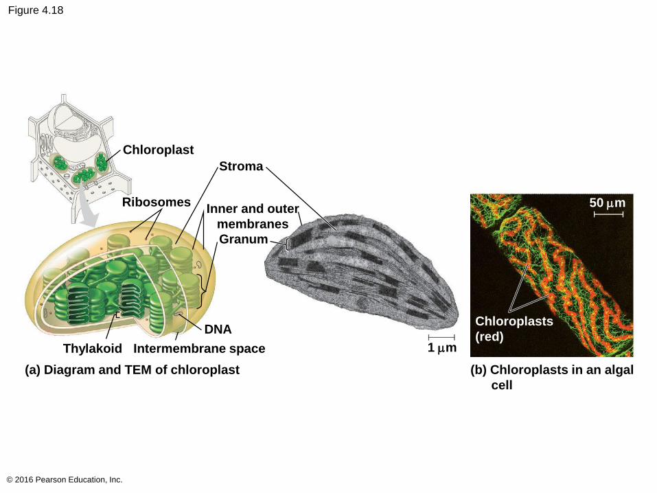
Figure 4.18
© 2016 Pearson Education, Inc.
Chloroplast
Stroma
RibosomesInner and outer
membranes
Granum
DNA
Thylakoid Intermembrane space
(a) Diagram and TEM of chloroplast
1 mm
(b) Chloroplasts in an algal
cell
Chloroplasts
(red)
50 mm
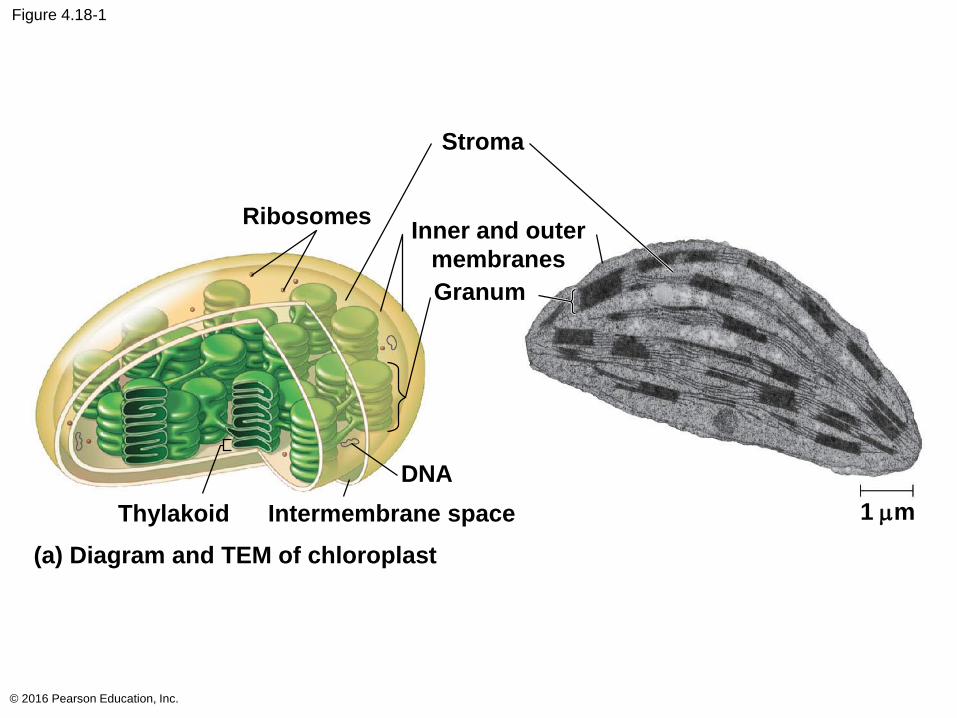
Figure 4.18-1
© 2016 Pearson Education, Inc.
Stroma
RibosomesInner and outer
membranes
Granum
DNA
Thylakoid Intermembrane space
(a) Diagram and TEM of chloroplast
1 mm
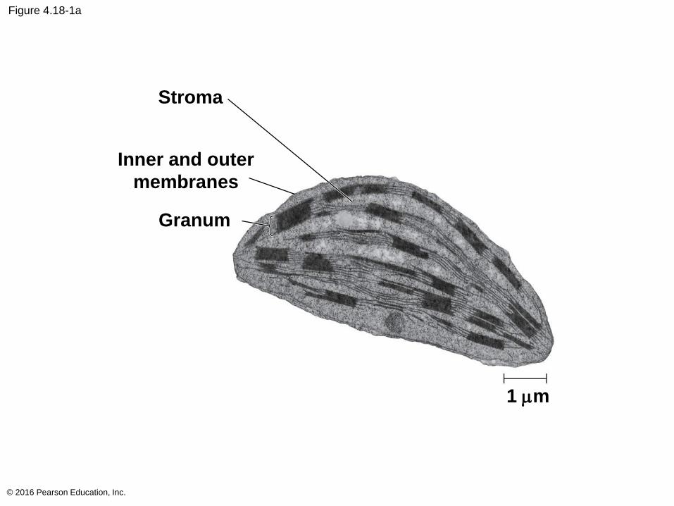
Figure 4.18-1a
© 2016 Pearson Education, Inc.
Stroma
Inner and outer
membranes
Granum
1 mm
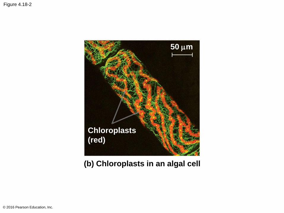
Figure 4.18-2
© 2016 Pearson Education, Inc.
(b) Chloroplasts in an algal cell
Chloroplasts
(red)
50 mm
Peroxisomes: Oxidation
Peroxisomes are specialized metabolic
compartments bounded by a single membrane
Peroxisomes produce hydrogen peroxide and then
convert it to water
Peroxisomes perform reactions with many different
functions
© 2016 Pearson Education, Inc.
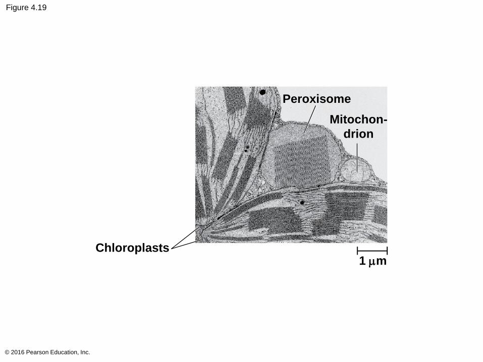
Figure 4.19
© 2016 Pearson Education, Inc.
Peroxisome
Mitochon-
drion
Chloroplasts1 mm
Concept 4.6: The cytoskeleton is a network of fibers that organizes structures and activities in the cell
The cytoskeleton is a network of fibers extending
throughout the cytoplasm
It organizes the cell’s structures and activities
© 2016 Pearson Education, Inc.
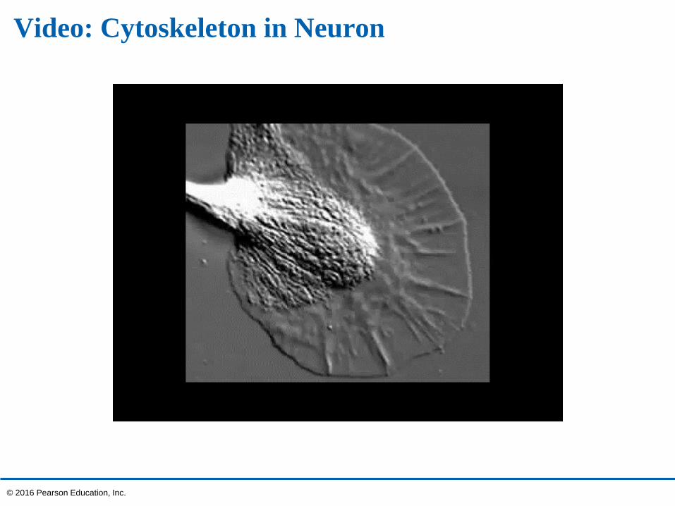
Video: Cytoskeleton in Neuron
© 2016 Pearson Education, Inc.
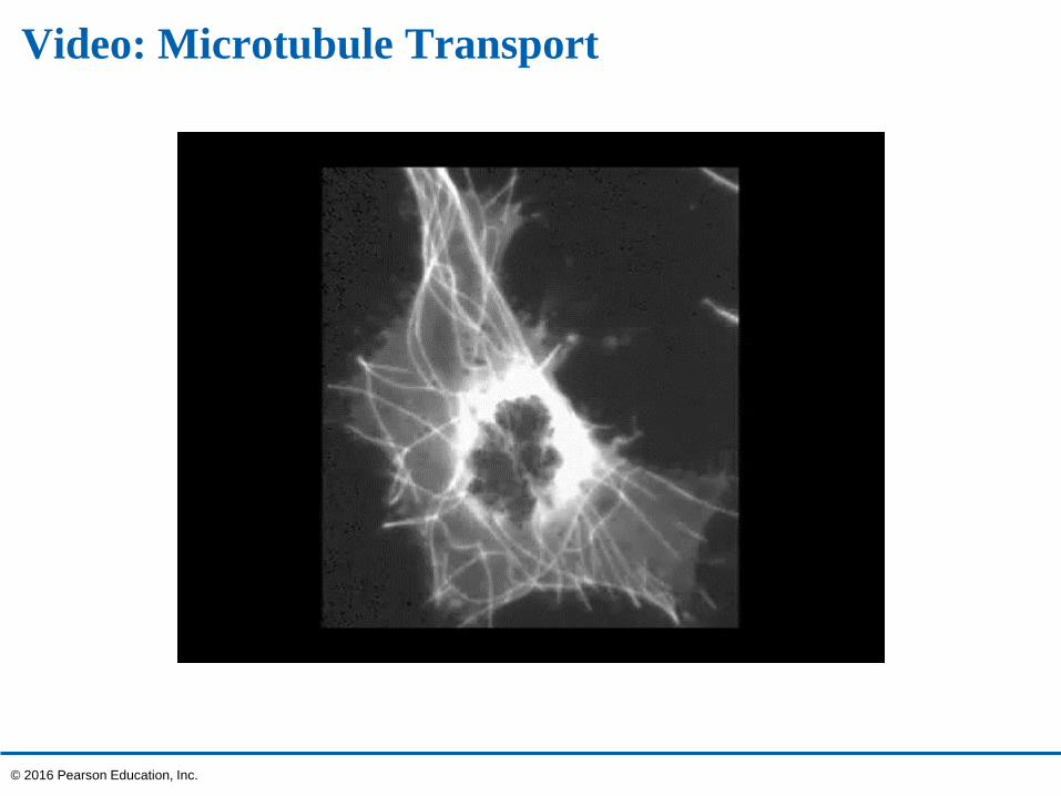
Video: Microtubule Transport
© 2016 Pearson Education, Inc.
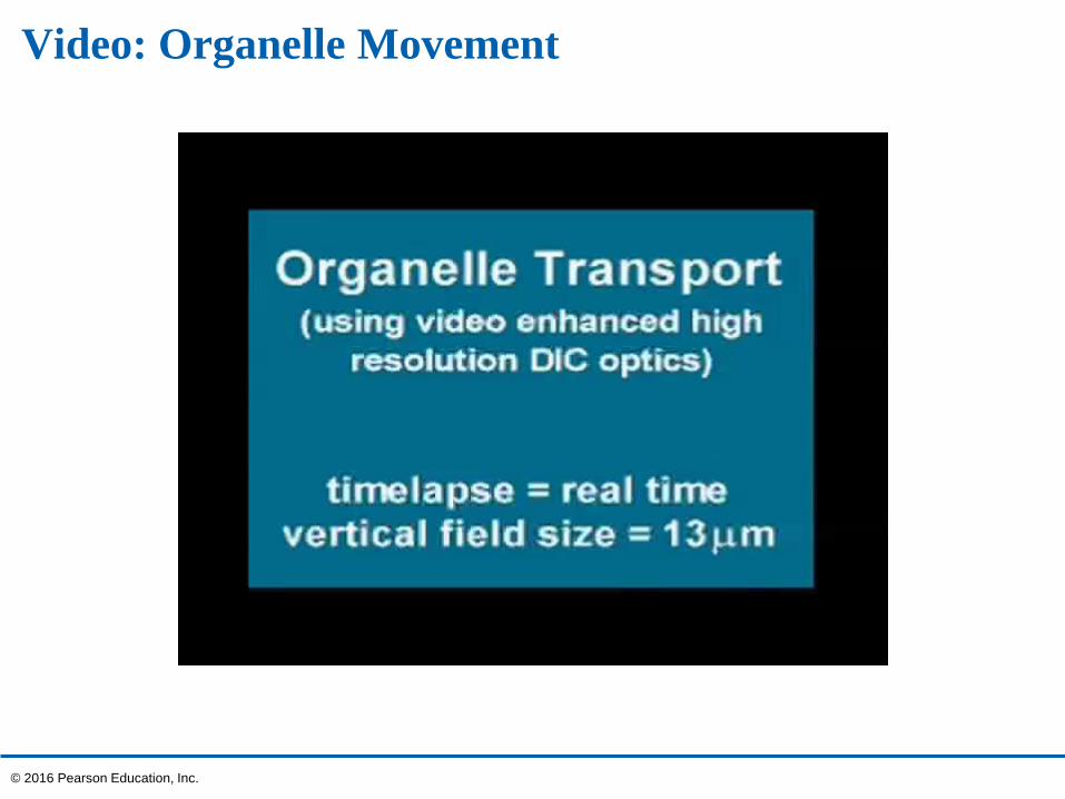
Video: Organelle Movement
© 2016 Pearson Education, Inc.
Video: Organelle Transport
© 2016 Pearson Education, Inc.
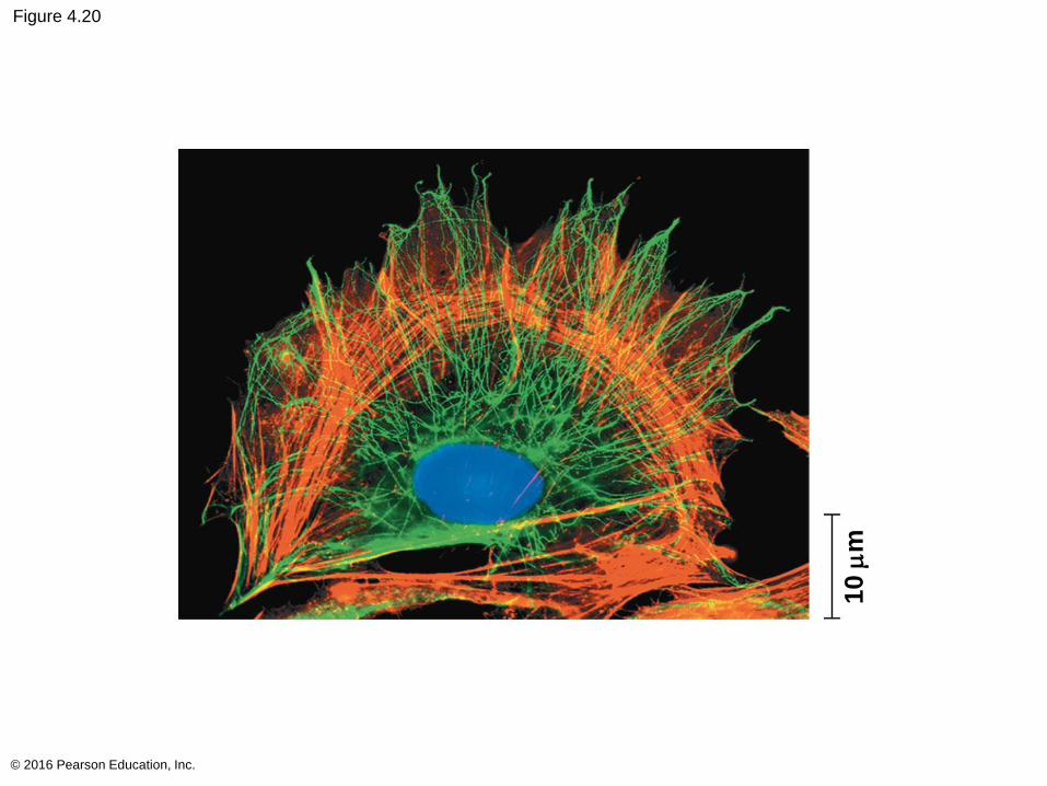
Figure 4.20
© 2016 Pearson Education, Inc.
10 m
m
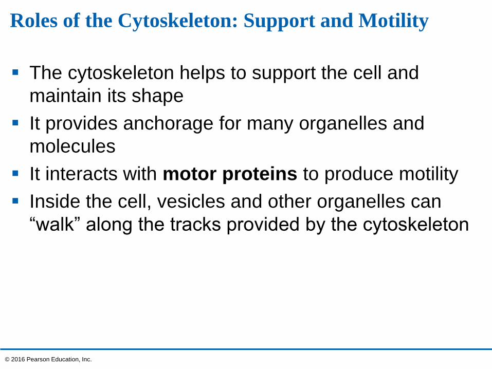
Roles of the Cytoskeleton: Support and Motility
The cytoskeleton helps to support the cell and
maintain its shape
It provides anchorage for many organelles and
molecules
It interacts with motor proteins to produce motility
Inside the cell, vesicles and other organelles can
“walk” along the tracks provided by the cytoskeleton
© 2016 Pearson Education, Inc.
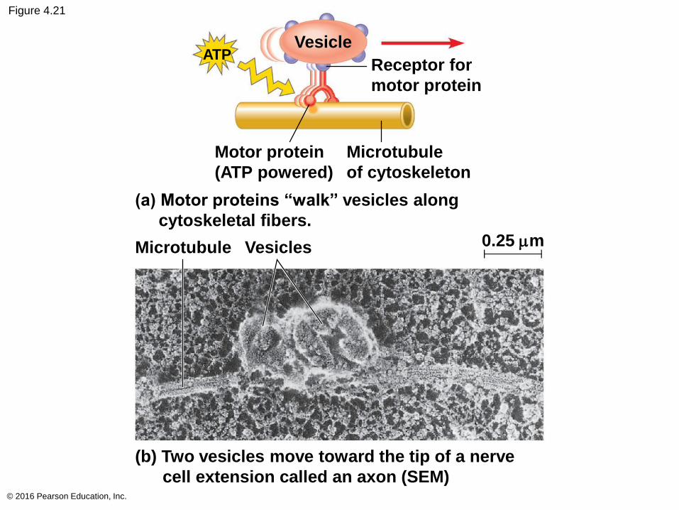
Figure 4.21
© 2016 Pearson Education, Inc.
(a) Motor proteins “walkˮ vesicles along
cytoskeletal fibers.
(b) Two vesicles move toward the tip of a nerve
cell extension called an axon (SEM)
Microtubule Vesicles 0.25 mm
Microtubule
of cytoskeleton
Motor protein
(ATP powered)
Receptor for
motor protein
VesicleATP
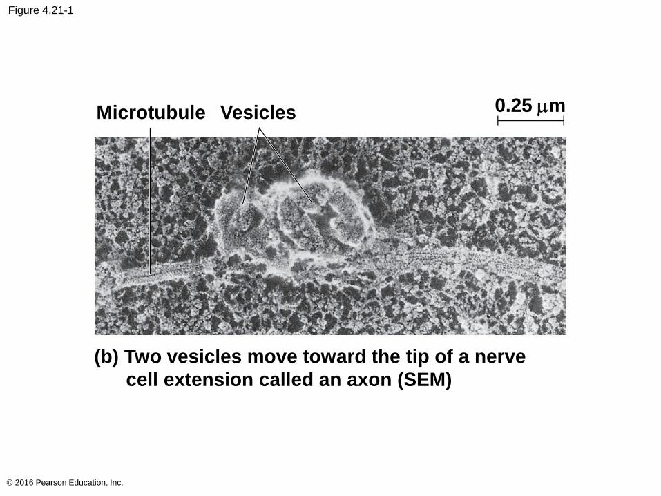
Figure 4.21-1
© 2016 Pearson Education, Inc.
(b) Two vesicles move toward the tip of a nerve
cell extension called an axon (SEM)
Microtubule Vesicles 0.25 mm
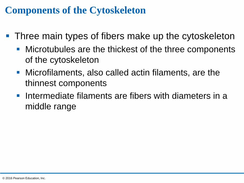
Components of the Cytoskeleton
Three main types of fibers make up the cytoskeleton
Microtubules are the thickest of the three components
of the cytoskeleton
Microfilaments, also called actin filaments, are the
thinnest components
Intermediate filaments are fibers with diameters in a
middle range
© 2016 Pearson Education, Inc.
Video: Actin Cytoskeleton
© 2016 Pearson Education, Inc.
Video: Actin in Crawling Cell
© 2016 Pearson Education, Inc.
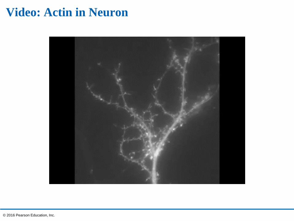
Video: Actin in Neuron
© 2016 Pearson Education, Inc.
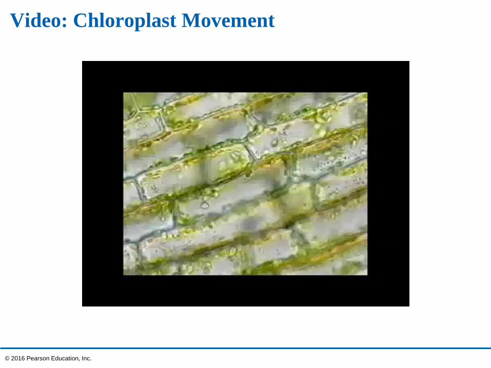
Video: Chloroplast Movement
© 2016 Pearson Education, Inc.
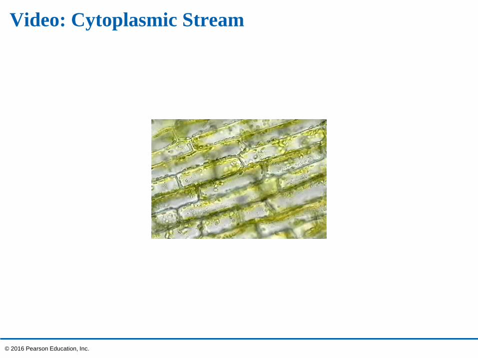
Video: Cytoplasmic Stream
© 2016 Pearson Education, Inc.
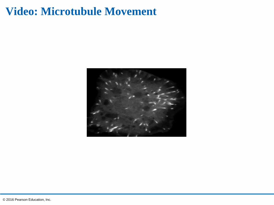
Video: Microtubule Movement
© 2016 Pearson Education, Inc.
Video: Microtubules
© 2016 Pearson Education, Inc.
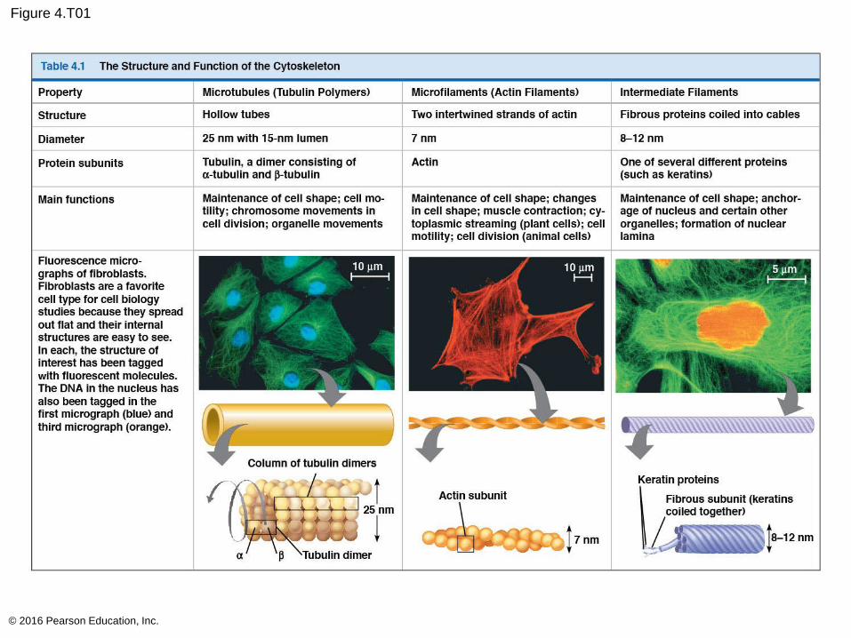
Figure 4.T01
© 2016 Pearson Education, Inc.
Figure 4.T01-1
© 2016 Pearson Education, Inc.
Microtubules (Tubulin Polymers)
Hollow tubes
25 nm with 15-nm lumen
Tubulin, a dimer consisting of
a-tubulin and b-tubulin
Maintenance of cell shape; cell mo-
tility; chromosome movements in
cell division; organelle movements
10 mm
Column of tubulin dimers
Tubulin dimera b
25 nm
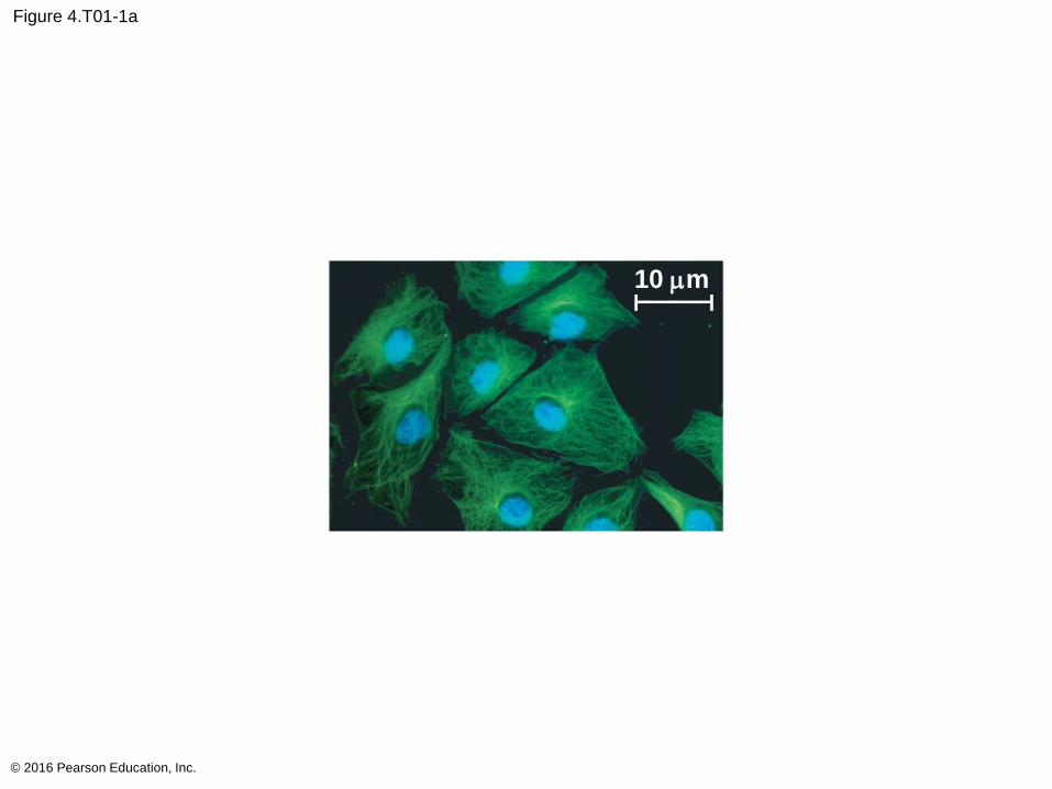
Figure 4.T01-1a
© 2016 Pearson Education, Inc.
10 mm
10 mm
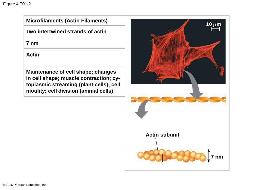
Figure 4.T01-2
© 2016 Pearson Education, Inc.
Microfilaments (Actin Filaments)
Two intertwined strands of actin
7 nm
Actin
Maintenance of cell shape; changes
in cell shape; muscle contraction; cy-
toplasmic streaming (plant cells); cell
motility; cell division (animal cells)
Actin subunit
7 nm
10 mm
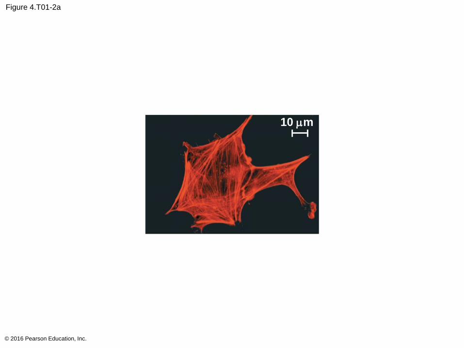
Figure 4.T01-2a
© 2016 Pearson Education, Inc.
10 mm
10 mm
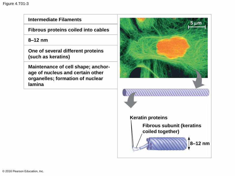
Figure 4.T01-3
© 2016 Pearson Education, Inc.
Intermediate Filaments
Fibrous proteins coiled into cables
8–12 nm
One of several different proteins
(such as keratins)
Maintenance of cell shape; anchor-
age of nucleus and certain other
organelles; formation of nuclear
lamina
Keratin proteins
Fibrous subunit (keratins
coiled together)
8–12 nm
5 mm
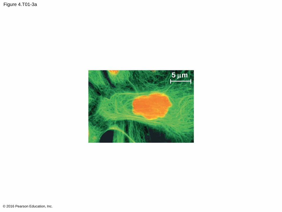
Figure 4.T01-3a
© 2016 Pearson Education, Inc.
5 mm
5 mm
Microtubules
Microtubules are hollow rods constructed from
globular protein dimers called tubulin
Functions of microtubules
Shape and support the cell
Guide movement of organelles
Separate chromosomes during cell division
© 2016 Pearson Education, Inc.
Centrosomes and Centrioles
In animal cells, microtubules grow out from a
centrosome near the nucleus
The centrosome is a “microtubule-organizing
center”
The centrosome has a pair of centrioles, each with
nine triplets of microtubules arranged in a ring
© 2016 Pearson Education, Inc.
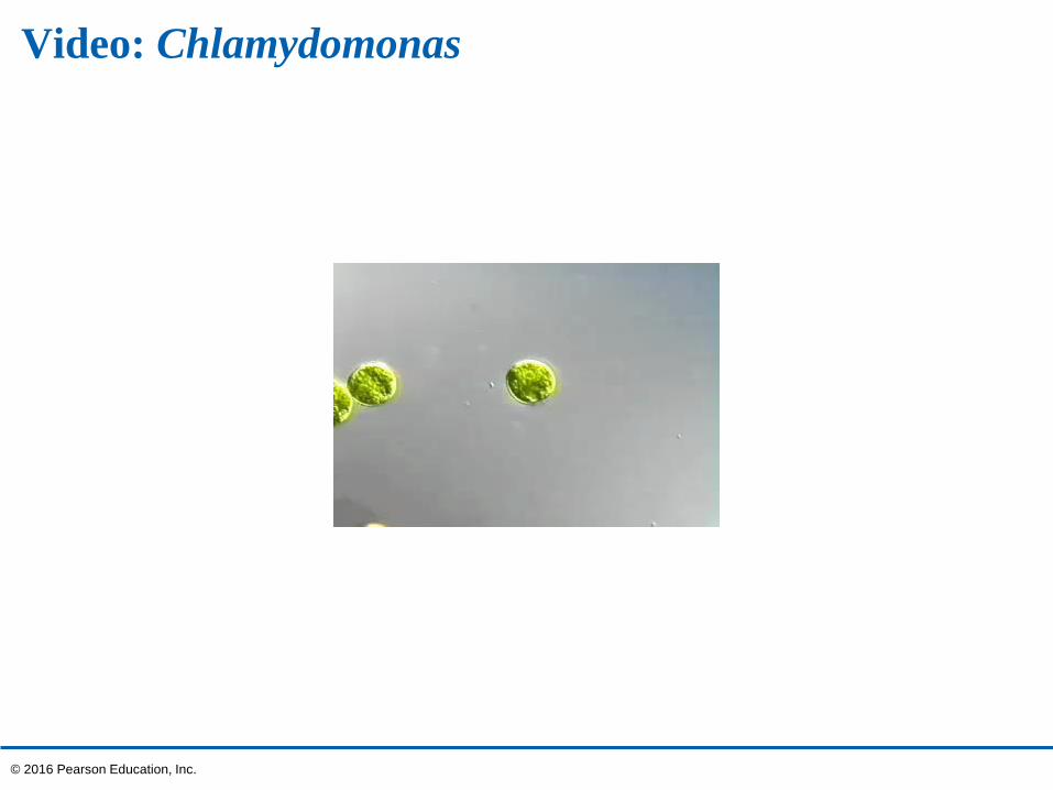
Video: Chlamydomonas
© 2016 Pearson Education, Inc.
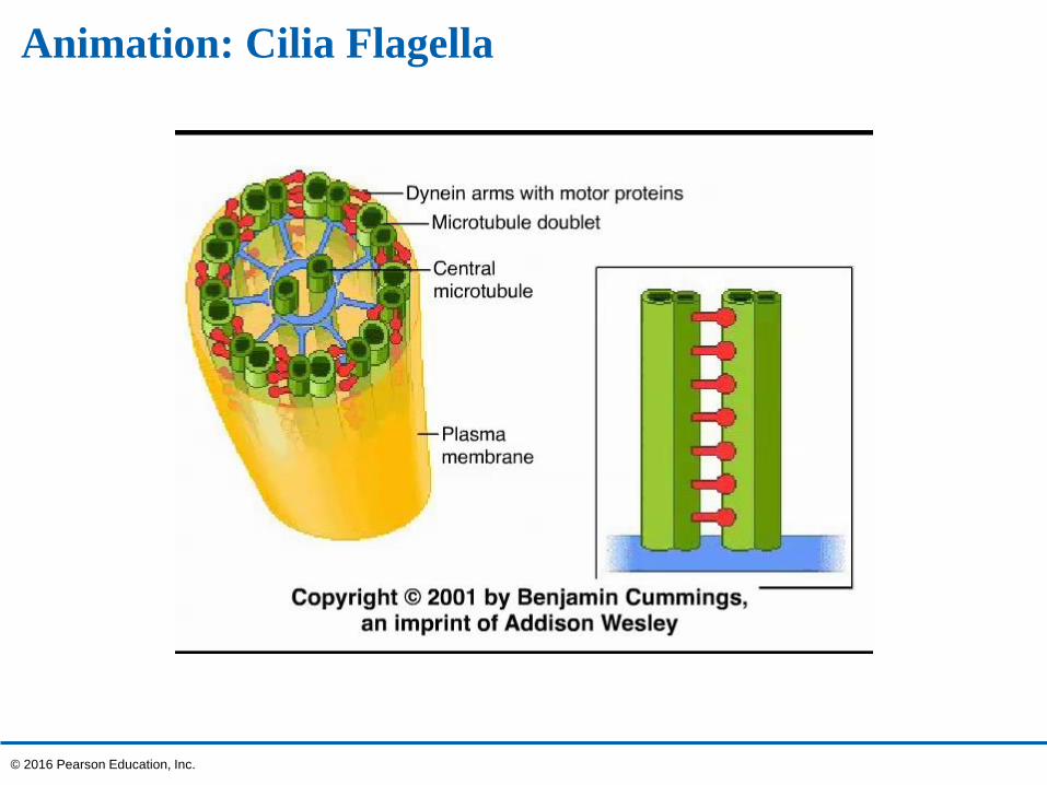
Animation: Cilia Flagella
© 2016 Pearson Education, Inc.
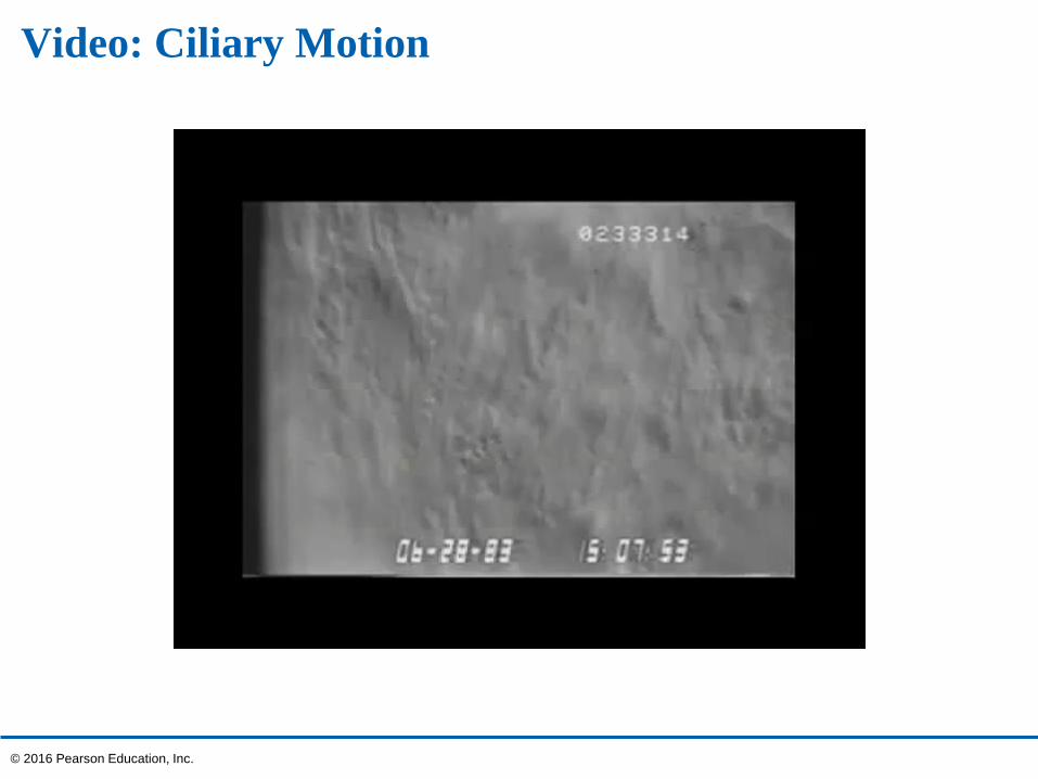
Video: Ciliary Motion
© 2016 Pearson Education, Inc.
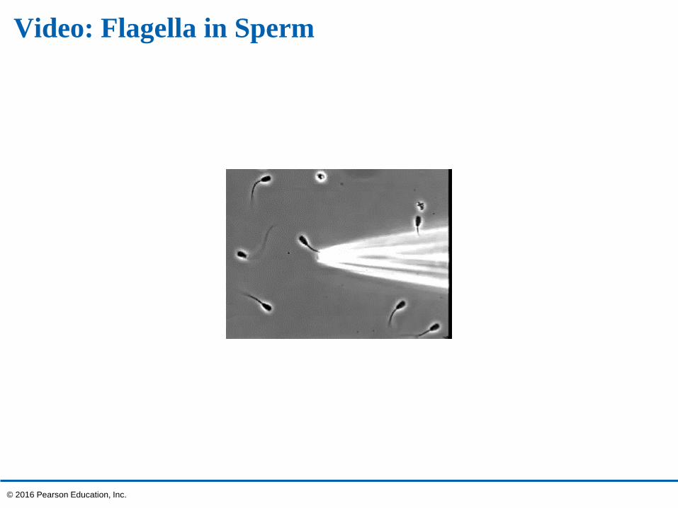
Video: Flagella in Sperm
© 2016 Pearson Education, Inc.
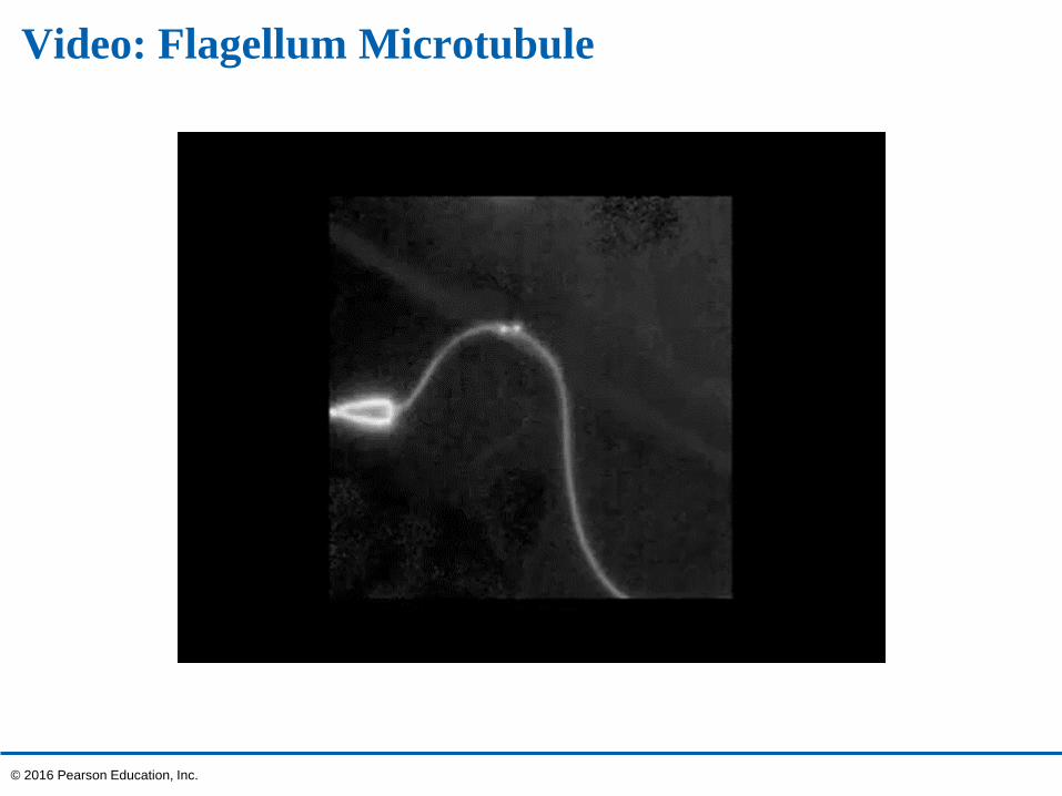
Video: Flagellum Microtubule
© 2016 Pearson Education, Inc.
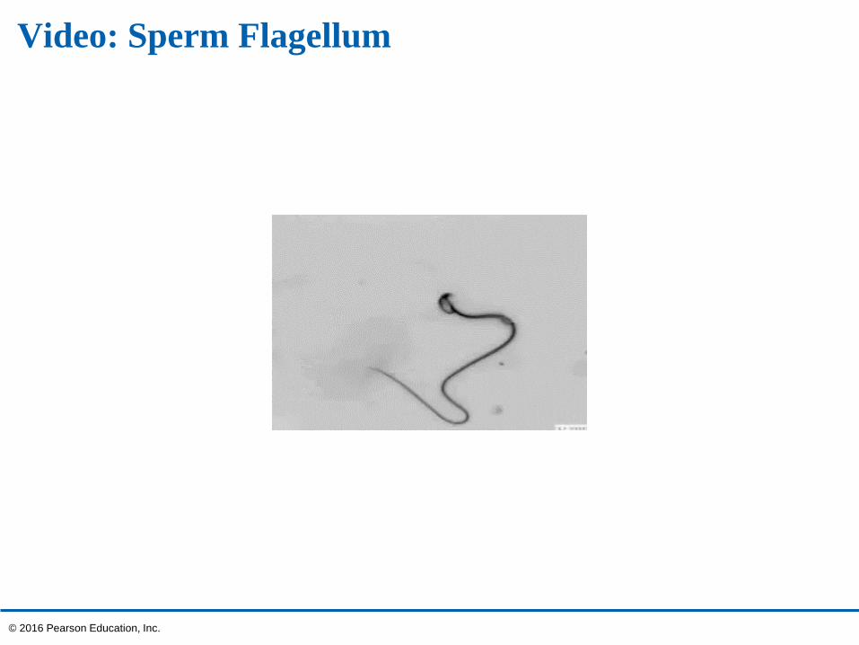
Video: Sperm Flagellum
© 2016 Pearson Education, Inc.
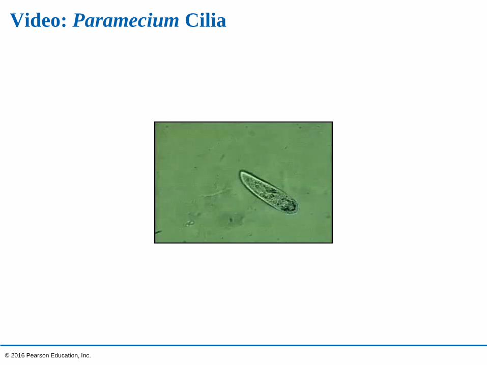
Video: Paramecium Cilia
© 2016 Pearson Education, Inc.
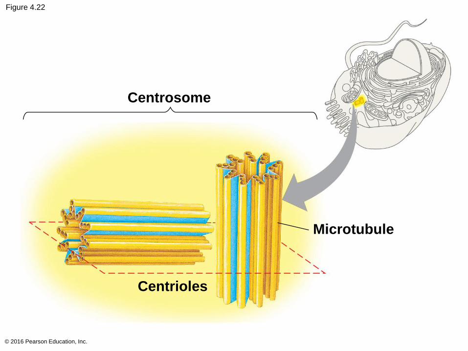
Figure 4.22
© 2016 Pearson Education, Inc.
Centrosome
Centrioles
Microtubule
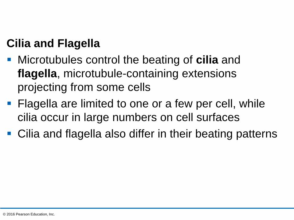
Cilia and Flagella
Microtubules control the beating of cilia and
flagella, microtubule-containing extensions
projecting from some cells
Flagella are limited to one or a few per cell, while
cilia occur in large numbers on cell surfaces
Cilia and flagella also differ in their beating patterns
© 2016 Pearson Education, Inc.
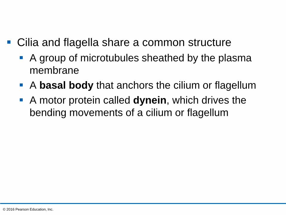
Cilia and flagella share a common structure
A group of microtubules sheathed by the plasma
membrane
A basal body that anchors the cilium or flagellum
A motor protein called dynein, which drives the
bending movements of a cilium or flagellum
© 2016 Pearson Education, Inc.
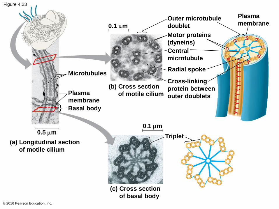
Figure 4.23
© 2016 Pearson Education, Inc.
0.1 mm
Outer microtubule
doublet
Motor proteins
(dyneins)
Central
microtubule
Radial spoke
Cross-linking
protein between
outer doublets
(b) Cross section
of motile cilium
(c) Cross section
of basal body
Microtubules
Plasma
membrane
Basal body
0.5 mm
(a) Longitudinal section
of motile cilium
0.1 mm
Triplet
Plasma
membrane
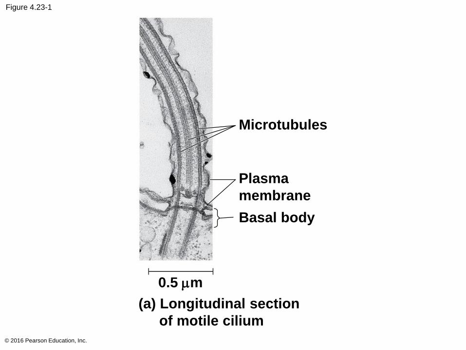
Figure 4.23-1
© 2016 Pearson Education, Inc.
Microtubules
Plasma
membrane
0.5 mm
(a) Longitudinal section
of motile cilium
Basal body
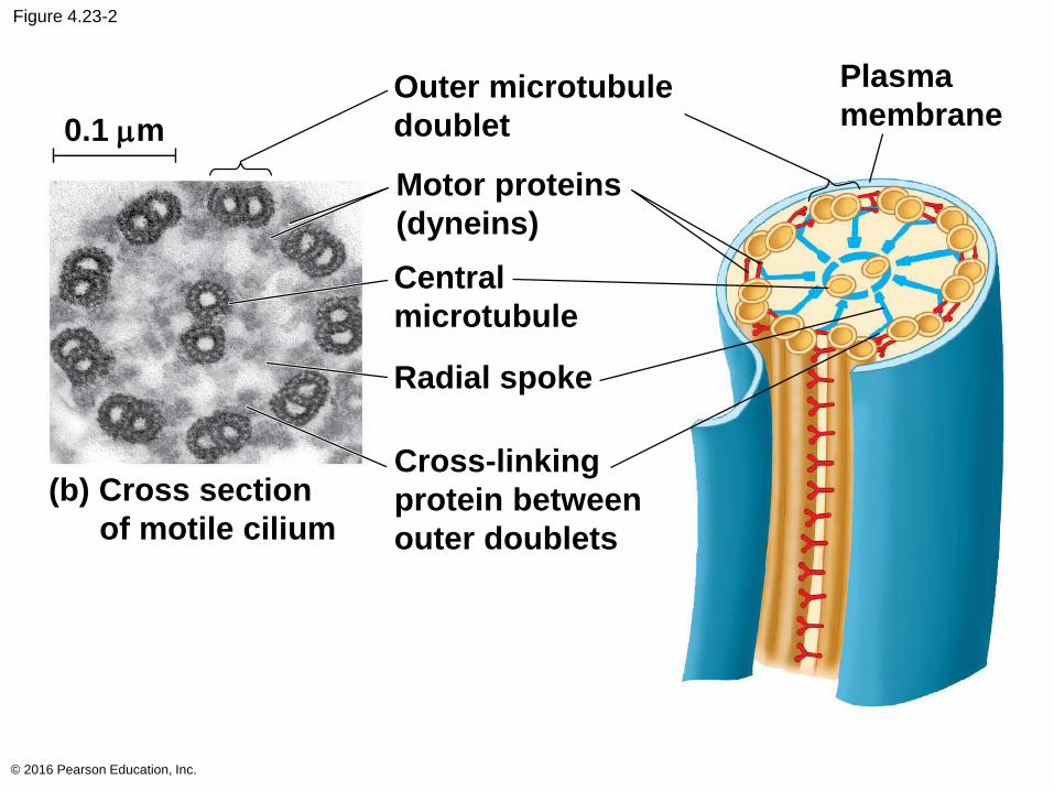
Figure 4.23-2
© 2016 Pearson Education, Inc.
0.1 mm
Outer microtubule
doublet
Motor proteins
(dyneins)
Central
microtubule
Radial spoke
Cross-linking
protein between
outer doublets
(b) Cross section
of motile cilium
Plasma
membrane
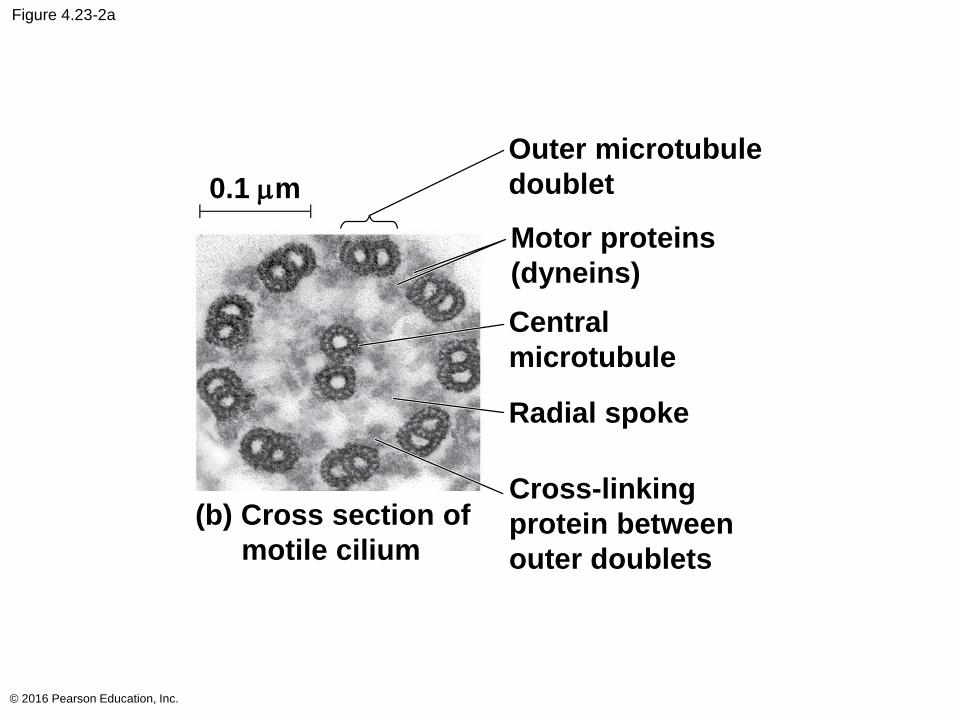
Figure 4.23-2a
© 2016 Pearson Education, Inc.
0.1 mm
Outer microtubule
doublet
Motor proteins
(dyneins)
Central
microtubule
Radial spoke
Cross-linking
protein between
outer doublets
(b) Cross section of
motile cilium
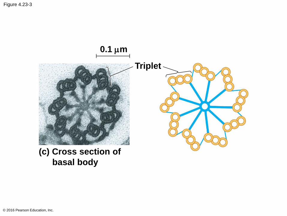
Figure 4.23-3
© 2016 Pearson Education, Inc.
(c) Cross section of
basal body
0.1 mm
Triplet
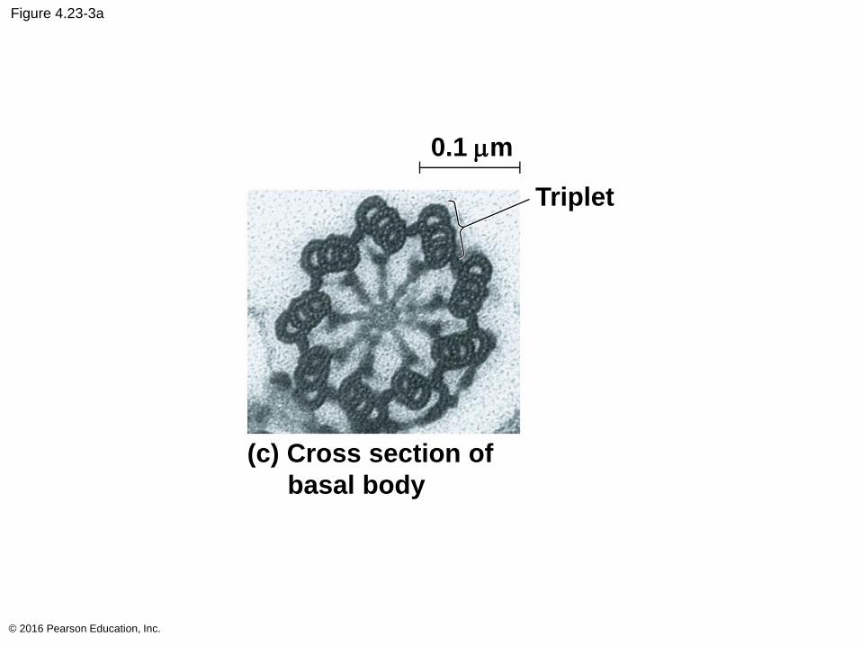
Figure 4.23-3a
© 2016 Pearson Education, Inc.
(c) Cross section of
basal body
0.1 mm
Triplet
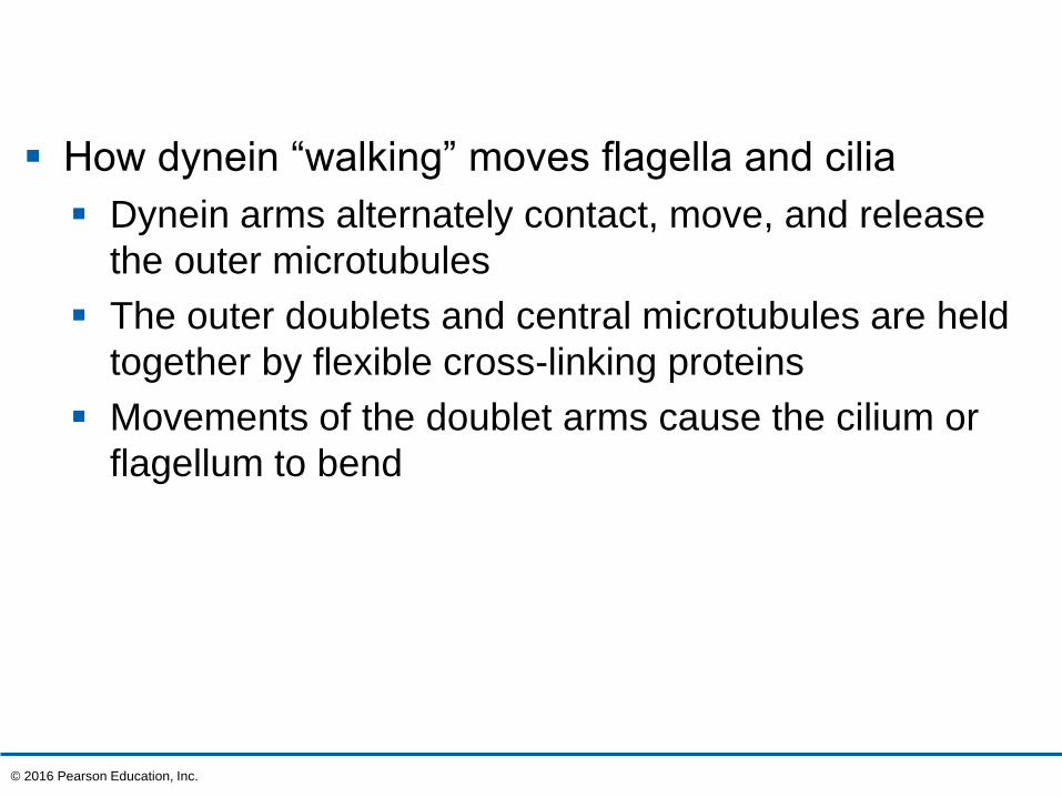
How dynein “walking” moves flagella and cilia
Dynein arms alternately contact, move, and release
the outer microtubules
The outer doublets and central microtubules are held
together by flexible cross-linking proteins
Movements of the doublet arms cause the cilium or
flagellum to bend
© 2016 Pearson Education, Inc.
Microfilaments (Actin Filaments)
Microfilaments are thin solid rods, built from
molecules of globular actin subunits
The structural role of microfilaments is to bear
tension, resisting pulling forces within the cell
Bundles of microfilaments make up the core of
microvilli of intestinal cells
© 2016 Pearson Education, Inc.
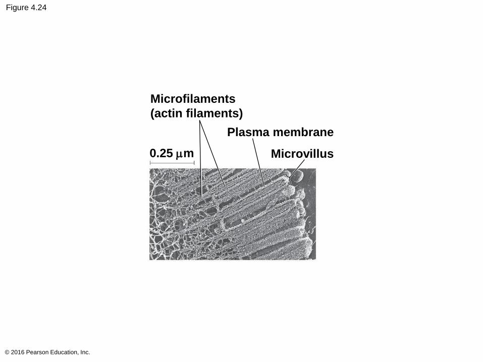
Figure 4.24
© 2016 Pearson Education, Inc.
Microfilaments
(actin filaments)
0.25 mm
Plasma membrane
Microvillus
Microfilaments that function in cellular motility
interact with the motor protein myosin
For example, actin and myosin interact to cause
muscle contraction, amoeboid movement of white
blood cells, and cytoplasmic streaming in plant cells
© 2016 Pearson Education, Inc.
Intermediate Filaments
Intermediate filaments are larger than
microfilaments but smaller than microtubules
Intermediate filaments are only found in the cells of
some animals, including vertebrates
They support cell shape and fix organelles in place
Intermediate filaments are more permanent
cytoskeleton elements than the other two classes
© 2016 Pearson Education, Inc.
Concept 4.7: Extracellular components and connections between cells help coordinate cellular activities
Most cells synthesize and secrete materials that are
external to the plasma membrane
These extracellular materials are involved in many
cellular functions
© 2016 Pearson Education, Inc.

Cell Walls of Plants
The cell wall is an extracellular structure that
distinguishes plant cells from animal cells
The cell wall protects the plant cell, maintains its
shape, and prevents excessive uptake of water
Plant cell walls are made of cellulose fibers
embedded in other polysaccharides and protein
© 2016 Pearson Education, Inc.
Plant cell walls may have multiple layers
Primary cell wall: relatively thin and flexible
Middle lamella: thin layer between primary walls of
adjacent cells
Secondary cell wall (in some cells): added between
the plasma membrane and the primary cell wall
Plasmodesmata are channels between adjacent
plant cells
© 2016 Pearson Education, Inc.
Video: Collagen Model
© 2016 Pearson Education, Inc.
Video: Extracellular Matrix
© 2016 Pearson Education, Inc.
Video: Fibronectin
© 2016 Pearson Education, Inc.
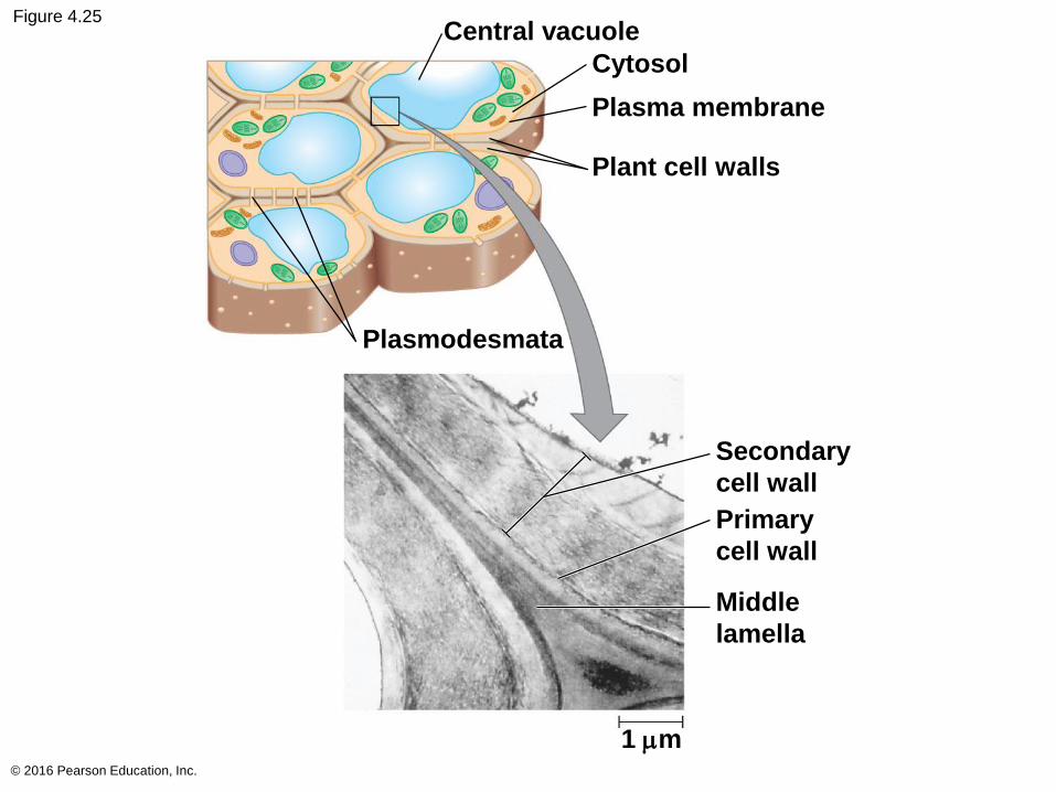
Figure 4.25
© 2016 Pearson Education, Inc.
Central vacuole
Cytosol
Plasma membrane
Plant cell walls
Plasmodesmata
Secondary
cell wall
Primary
cell wall
Middle
lamella
1 mm
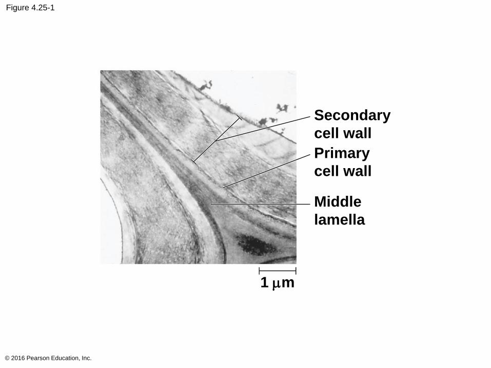
Figure 4.25-1
© 2016 Pearson Education, Inc.
Secondary
cell wall
Primary
cell wall
Middle
lamella
1 mm
The Extracellular Matrix (ECM) of Animal Cells
Animal cells lack cell walls but are covered by an
elaborate extracellular matrix (ECM)
The ECM is made up of glycoproteins such as
collagen, proteoglycans, and fibronectin
ECM proteins bind to receptor proteins in the
plasma membrane called integrins
© 2016 Pearson Education, Inc.
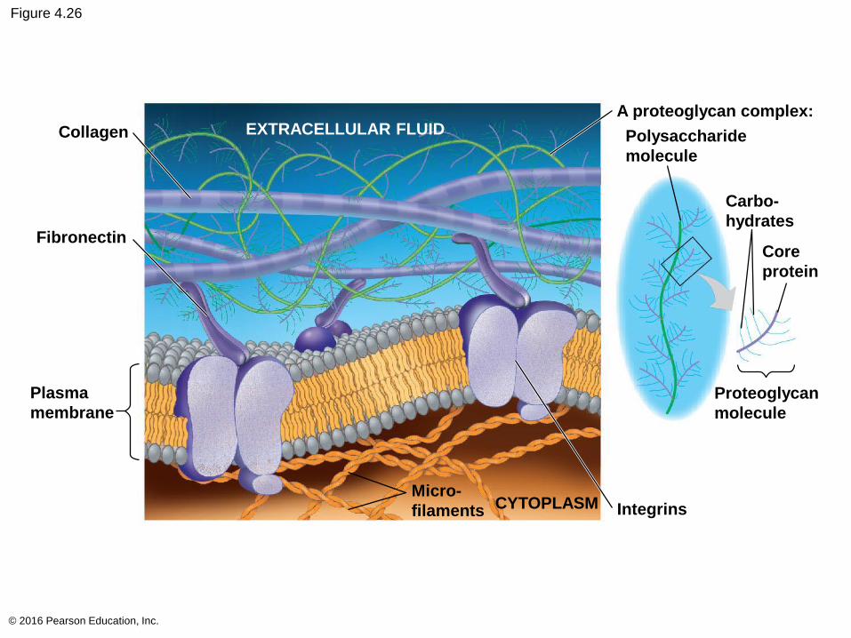
Figure 4.26
© 2016 Pearson Education, Inc.
Collagen
Fibronectin
Plasma
membrane
Micro-
filaments CYTOPLASM Integrins
Proteoglycan
molecule
Core
protein
Carbo-
hydrates
Polysaccharide
molecule
A proteoglycan complex:EXTRACELLULAR FLUID
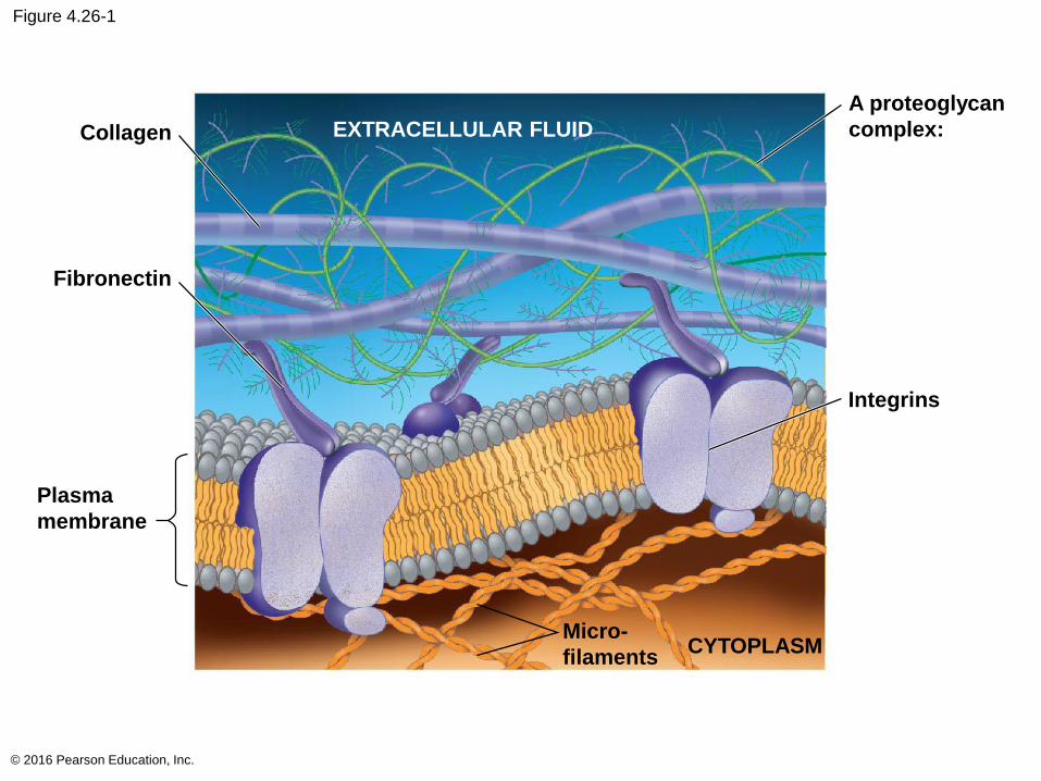
Figure 4.26-1
© 2016 Pearson Education, Inc.
Collagen
Fibronectin
Plasma
membrane
Micro-
filamentsCYTOPLASM
Integrins
A proteoglycan
complex:EXTRACELLULAR FLUID
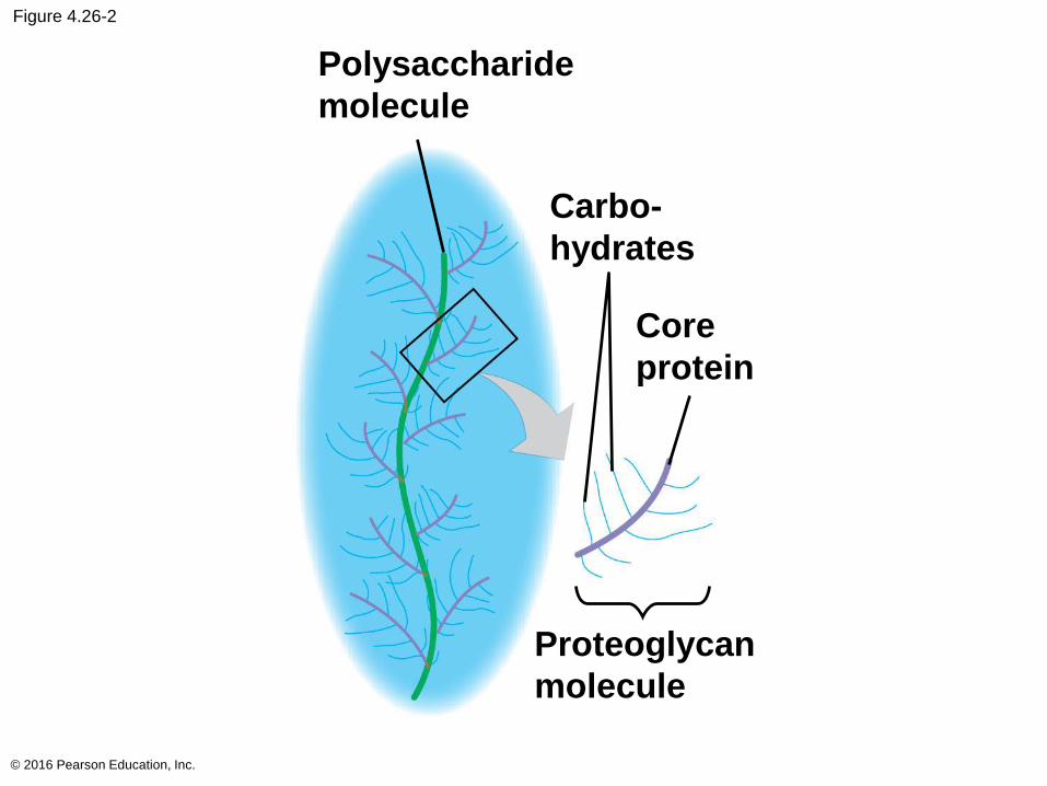
Figure 4.26-2
© 2016 Pearson Education, Inc.
Proteoglycan
molecule
Core
protein
Carbo-
hydrates
Polysaccharide
molecule
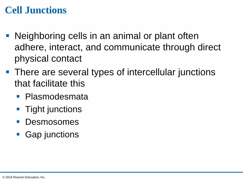
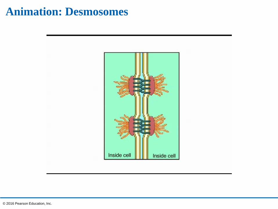
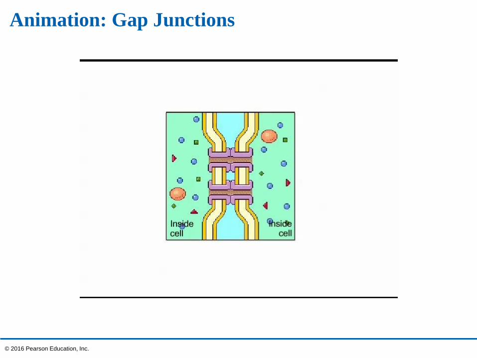
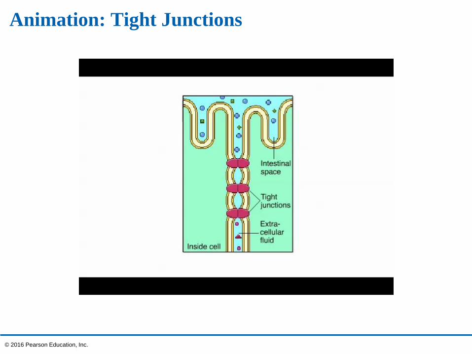
Cell Junctions
Neighboring cells in an animal or plant often
adhere, interact, and communicate through direct
physical contact
There are several types of intercellular junctions
that facilitate this
Plasmodesmata
Tight junctions
Desmosomes
Gap junctions
© 2016 Pearson Education, Inc.
Plasmodesmata in Plant Cells
Plasmodesmata are channels that perforate plant
cell walls
Through plasmodesmata, water and small solutes
(and sometimes proteins and RNA) can pass from
cell to cell
© 2016 Pearson Education, Inc.
Tight Junctions, Desmosomes, and Gap Junctions in Animal Cells
Animal cells have three main types of cell junctions
Tight junctions
Desmosomes
Gap junctions
All are especially common in epithelial tissue
© 2016 Pearson Education, Inc.
Animation: Desmosomes
© 2016 Pearson Education, Inc.
Animation: Gap Junctions
© 2016 Pearson Education, Inc.
Animation: Tight Junctions
© 2016 Pearson Education, Inc.
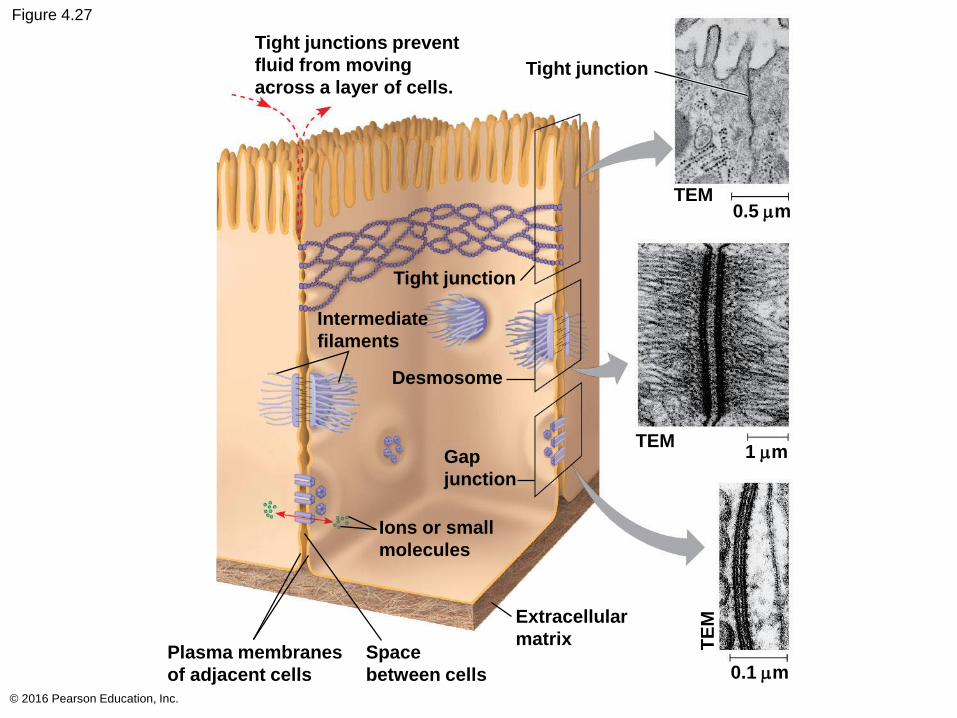
Figure 4.27
© 2016 Pearson Education, Inc.
Tight junctions prevent
fluid from moving
across a layer of cells.Tight junction
TEM0.5 mm
TEM1 mm
Tight junction
Intermediate
filaments
Desmosome
Gap
junction
Ions or small
molecules
Plasma membranes
of adjacent cells
Space
between cells
Extracellular
matrix
0.1 mm
TE
M
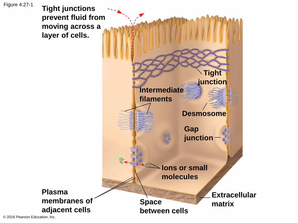
Figure 4.27-1
© 2016 Pearson Education, Inc.
Tight junctions
prevent fluid from
moving across a
layer of cells.
Tight
junctionIntermediate
filaments
Desmosome
Gap
junction
Ions or small
molecules
Plasma
membranes of
adjacent cellsSpace
between cells
Extracellular
matrix
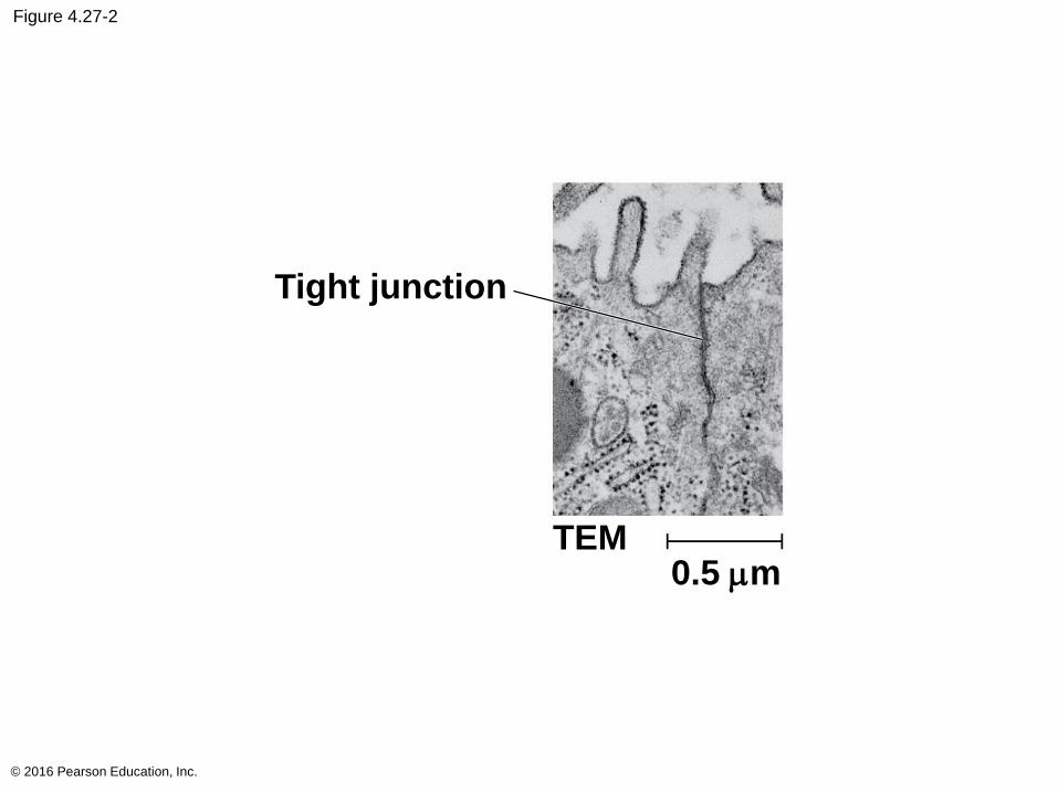
Figure 4.27-2
© 2016 Pearson Education, Inc.
Tight junction
TEM0.5 mm
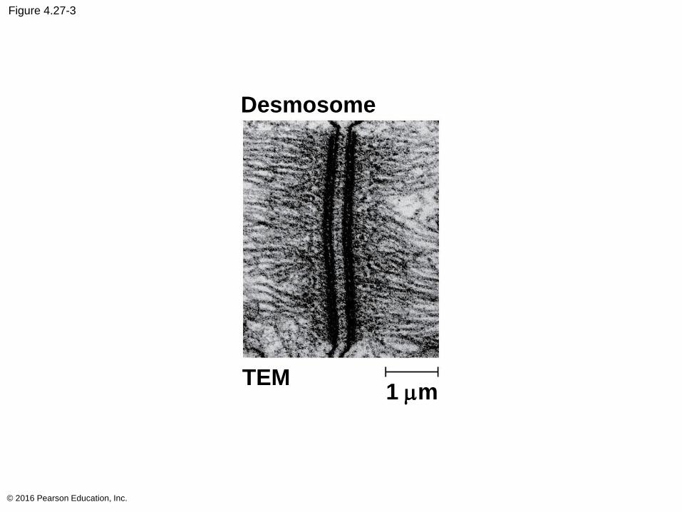
Figure 4.27-3
© 2016 Pearson Education, Inc.
TEM1 mm
Desmosome
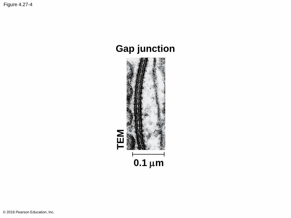
Figure 4.27-4
© 2016 Pearson Education, Inc.
Gap junction
0.1 mm
TE
M
The Cell: A Living Unit Greater Than the Sum of Its Parts
None of the components of a cell work alone
For example, a macrophage’s ability to destroy
bacteria involves the whole cell, coordinating
components such as the cytoskeleton, lysosomes,
and plasma membrane
Cellular functions arise from cellular order
© 2016 Pearson Education, Inc.
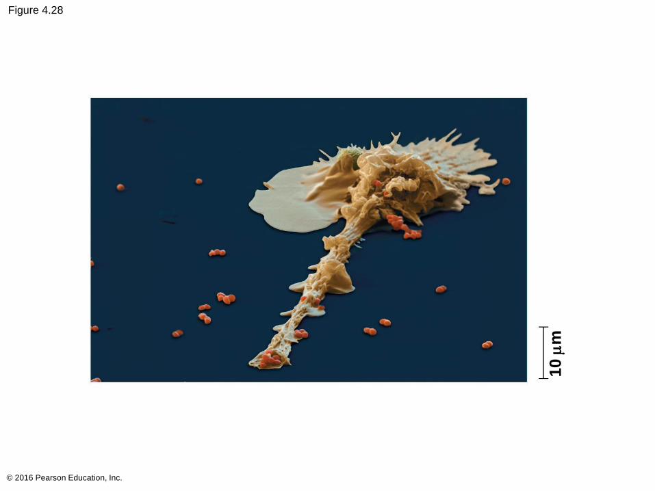
Figure 4.28
© 2016 Pearson Education, Inc.
10 m
m
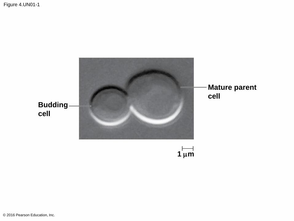
Figure 4.UN01-1
© 2016 Pearson Education, Inc.
Budding
cell
Mature parent
cell
1 mm
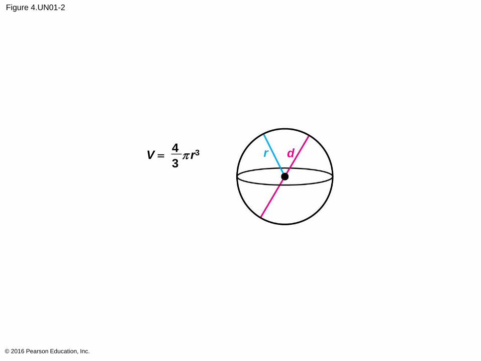
Figure 4.UN01-2
© 2016 Pearson Education, Inc.
V = p r34
3r d
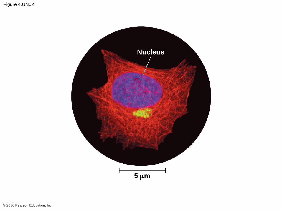
Figure 4.UN02
© 2016 Pearson Education, Inc.
5 mm
Nucleus
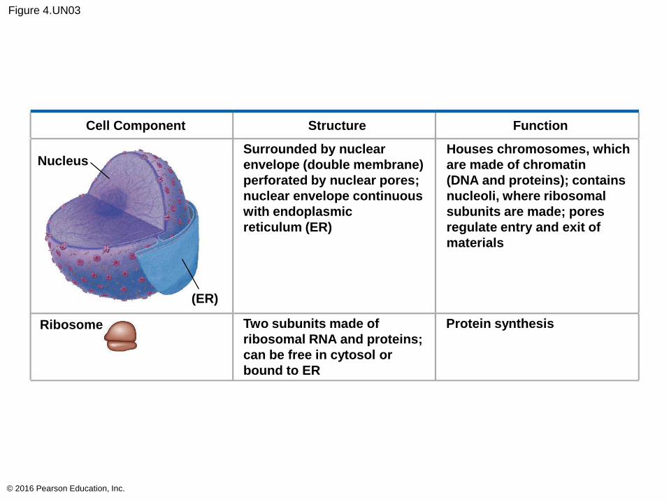
Figure 4.UN03
© 2016 Pearson Education, Inc.
Cell Component Structure Function
Nucleus
Ribosome
(ER)
Two subunits made of
ribosomal RNA and proteins;
can be free in cytosol or
bound to ER
Surrounded by nuclear
envelope (double membrane)
perforated by nuclear pores;
nuclear envelope continuous
with endoplasmic
reticulum (ER)
Houses chromosomes, which
are made of chromatin
(DNA and proteins); contains
nucleoli, where ribosomal
subunits are made; pores
regulate entry and exit of
materials
Protein synthesis
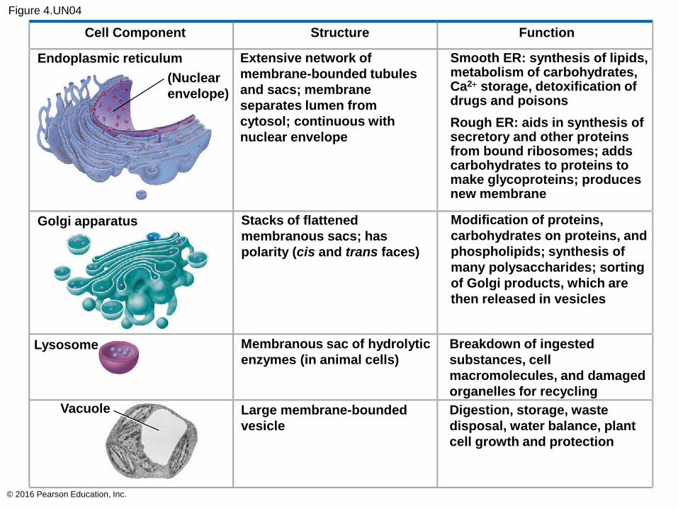
Figure 4.UN04
© 2016 Pearson Education, Inc.
Cell Component Structure Function
Endoplasmic reticulum
(Nuclear
envelope)
Extensive network of
membrane-bounded tubules
and sacs; membrane
separates lumen from
cytosol; continuous with
nuclear envelopeRough ER: aids in synthesis ofsecretory and other proteinsfrom bound ribosomes; addscarbohydrates to proteins tomake glycoproteins; producesnew membrane
Golgi apparatus Stacks of flattened
membranous sacs; has
polarity (cis and trans faces)
Modification of proteins,
carbohydrates on proteins, and
phospholipids; synthesis of
many polysaccharides; sorting
of Golgi products, which are
then released in vesicles
Lysosome
Vacuole
Membranous sac of hydrolytic
enzymes (in animal cells)
Large membrane-bounded
vesicle
Breakdown of ingested
substances, cell
macromolecules, and damaged
organelles for recycling
Digestion, storage, waste
disposal, water balance, plant
cell growth and protection
Smooth ER: synthesis of lipids,metabolism of carbohydrates,Ca2+ storage, detoxification ofdrugs and poisons
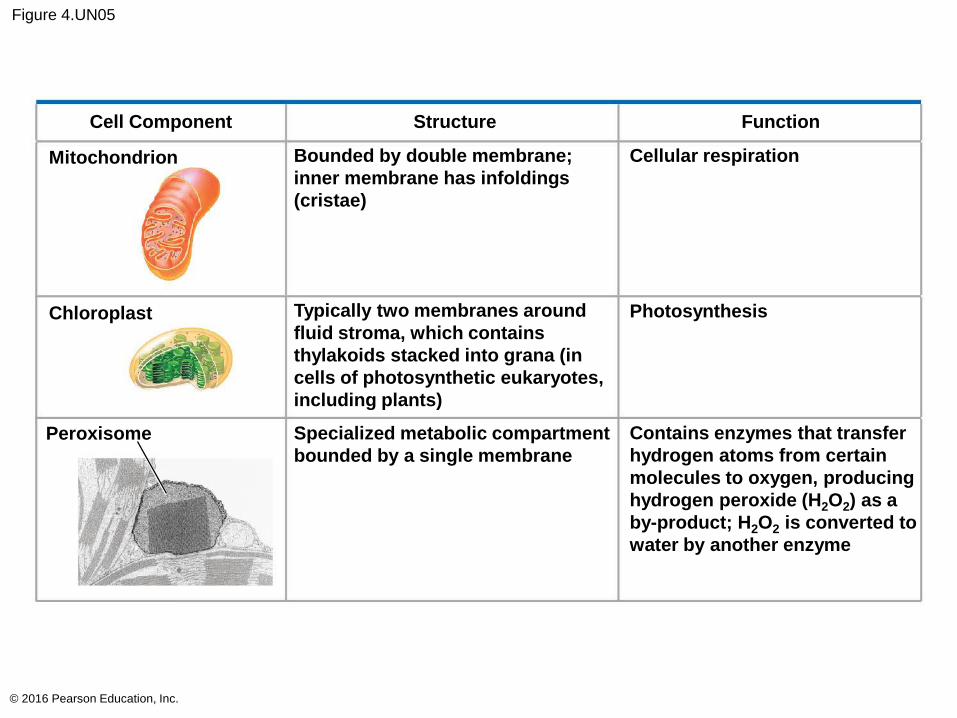
Figure 4.UN05
© 2016 Pearson Education, Inc.
Cell Component Structure Function
Mitochondrion
Chloroplast
Peroxisome
Bounded by double membrane;
inner membrane has infoldings
(cristae)
Typically two membranes around
fluid stroma, which contains
thylakoids stacked into grana (in
cells of photosynthetic eukaryotes,
including plants)
Specialized metabolic compartment
bounded by a single membrane
Cellular respiration
Photosynthesis
Contains enzymes that transfer
hydrogen atoms from certain
molecules to oxygen, producing
hydrogen peroxide (H2O2) as a
by-product; H2O2 is converted to
water by another enzyme
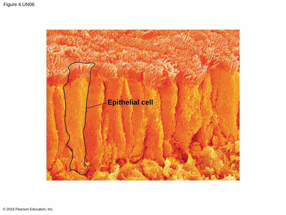
Figure 4.UN06
© 2016 Pearson Education, Inc.
Epithelial cell