a simple hydrothermal preparation of tio2 nanomaterials using concentrated hydrochloric acid
TRANSCRIPT
ARTICLE IN PRESS
Journal of Crystal Growth 312 (2009) 79–85
Contents lists available at ScienceDirect
Journal of Crystal Growth
0022-02
doi:10.1
� Corr
E-m
journal homepage: www.elsevier.com/locate/jcrysgro
A simple hydrothermal preparation of TiO2 nanomaterials using concentratedhydrochloric acid
Thuy-Duong Nguyen Phan, Hai-Dinh Pham, Tran Viet Cuong, Eui Jung Kim,Sunwook Kim, Eun Woo Shin �
School of Chemical Engineering and Bioengineering, University of Ulsan Mugeo-dong, Nam-gu, Ulsan 680-749, South Korea
a r t i c l e i n f o
Article history:
Received 19 August 2009
Received in revised form
17 September 2009
Accepted 24 September 2009
Communicated by K. Nakajimaassembly of nanostructured sub-units including nanocubes, nanoprisms, and nanorods. The crystalline
Available online 2 October 2009
PACS:
61.46.Hk
68.37.Hk
68.37.Lp
81.07.Bc
Keywords:
A1. Crystalline nanostructure
A1. Morphology control
A2. Hydrothermal method
B1. TiO2
B1. Hydrochloric acid
48/$ - see front matter & 2009 Elsevier B.V. A
016/j.jcrysgro.2009.09.032
esponding author. Tel.: +82 52 259 2253; fax
ail address: [email protected] (E. Woo
a b s t r a c t
A TiO2 nanostructure was synthesized via a simple method using only concentrated hydrochloric acid as
the morphological/crystallographic controlling agent. Microscopy images showed that the texture of the
TiO2 powder could be easily engineered and tuned by tailoring the HCl volume, creating cuboid, flower,
cauliflower, and ball-shaped particles. Three-dimensional TiO2 microparticles resulted from the self-
anatase and rutile phases were also identified depending on the acidic medium. HCl played a key role in
orchestrating the structures and morphologies of the TiO2 nanoscale materials. The phase transforma-
tion and morphological changes were strongly related to the crystal growth mechanism of the TiO2
nanostructure.
& 2009 Elsevier B.V. All rights reserved.
1. Introduction
Titanium dioxide (TiO2) is one of the most commonly usedoxide semiconductor materials due to its wide band gap (3.2 eV),low cost, non-toxic nature, strong oxidizing power, high resistanceto chemical or photo-induced corrosion, and maximum lightscattering with virtually no absorption. It has been widely usedin photocatalysis, photovoltaics, solar energy conversion, sensors,textiles, paints, cosmetics, etc. [1]. Numerous studies have revealedthat the physical and chemical characteristics, as well as theperformance, of nanostructured TiO2 are strongly dependent on itscrystalline structure, morphology, and dimension [1–3]. TiO2 hasthree distinct crystalline polymorphs (anatase, rutile, and brookite),and each phase displays different characteristics appropriate forspecific applications. Texture engineering of TiO2 nanomaterialsis therefore essential and has attracted considerable attention overthe past two decades. The texture design of Ti-based materials hasbeen intensively studied and developed using several different
ll rights reserved.
: +82 52 259 1689.
Shin).
strategies, including sol–gel, hydrothermal, micelle and inversemicelle usage, solvothermal, direct oxidation, electrodeposition,chemical/physical vapor deposition, emulsion or hydrolysis pre-cipitation, ultrasonic, and microwave approaches [1]. Up to now, avariety of titanate and titania-based materials have been success-fully synthesized, including nanorods, nanotubes, nanofibers,nanosheets, nanowires, nanobelts, nanoflowers, nanoleaves, nano-spheres, and nanoneedles [3–10]. Hydrothermal processing hasbeen widely applied in the preparation of TiO2 nanomaterials; thus,their textures have been controlled by several variables, forexample, precursors, pH, reaction temperature, aging duration,water pressure, and solvent characteristics [2,11]. This approachis complicated due to a long progression of preparation steps andthe use of several reactants and additives, which influence the timeand cost.
The goal of this study was to easily tune the crystalline phases,morphologies, and structural properties of TiO2 nanostructuresusing concentrated hydrochloric acid via a simple hydrothermalprocess in which only titanium n-butoxide and hydrochloric acid(38%) were used. All samples were further characterized by X-raydiffraction patterns, scanning electron microscopy, and transmis-sion electron microscopy.
ARTICLE IN PRESS
10 20 30 40 50 60 70 80 90
DTC-15
Inte
nsity
(a.u
.)
2θ (degrees)
Anatase
DTC-9.0
DTC-6.0
DTC-5.0
DTC-2.15
Rutile
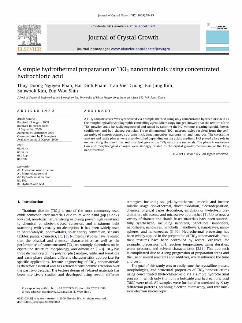
Fig. 1. Broad-scan XRD patterns of TiO2 nanomaterials using various Ti/HCl ratios.
T.-D. Nguyen Phan et al. / Journal of Crystal Growth 312 (2009) 79–8580
2. Experimental section
Titanium dioxide nanostructures were synthesized usingconcentrated HCl for morphology control and no additionalchemicals were used. In a simple process, the desired volumesof HCl (38%, Junsei) were added dropwise to 15 ml of titaniumn-butoxide (TBO, Ti(OBu)4, 98%, Acros) in a 100 ml Teflon-linedstainless steel autoclave. After conducting a hydrothermal processat 180 1C for 36 h and allowing the reaction to cool naturallyto room temperature, the white products were harvested bycentrifugation at 10,000 rpm and washed several times with amixture of ethanol and deionized water. After drying in air at105 1C for 12 h, the powders were generally designated as ‘‘DTC-i’’,where ‘‘i’’ corresponded to acid volume (see in Table 1).
The crystallographic phases of the TiO2 nanostructures wereidentified by X-ray diffraction (XRD) patterns using a Rigaku RAD-3C Diffractometer (Japan) with Cu-Ka radiation (l=1.5418 A) at ascan rate of 21 (2y)/min, operated at 35 kV and 20 mA. Thecrystallite size was evaluated from a certain diffraction line by theScherrer formula, and the weight percentages of the rutile andbrookite phases in the samples were estimated from the literature[12]. The structures, morphologies, and dimensions of TiO2
nanomaterials were characterized by field-emission scanningelectron microscopy (FE-SEM, JEOL, JSM-600F, 10 kV) and trans-mission electron microscopy (TEM, Hitachi, H-8100, Japan) at anaccelerating voltage of 200 kV. The samples for TEM wereprepared by ultrasonically dispersing the powders in ethanoland further deposited on carbon copper grid. The elementalanalysis was recorded by an energy-dispersive X-ray spectrometer(EDX) attached to FE-SEM system.
3. Results and discussion
3.1. Morphologies and structures of the TiO2 nanomaterials at
different volumes of concentrated HCL
The preparation conditions and related structural parametersof the TiO2 nanostructures are shown in Table 1. Wide-angle PXRDpatterns providing information on the crystalline nature of theTiO2 are presented in Fig. 1. The series of strong peaks in DTC-2.15were indexed to body-centered tetragonal TiO2 with the I41/amd
structure (space group 141 in the International Tables for X-rayCrystallography). All diffraction lines assigned to the (0 0 4),(2 0 0), (12 5), and main (10 1) reflections at 2y=25.251 areindicative of the nanocrystalline anatase phase (JCPDS 21-1272).As seen in Table 1, the average anatase crystalline domainevaluated using the Scherrer equation was about 7.77 nm. Phasetransformation from anatase to rutile appeared in sample DTC-5.0
Table 1Structural parameters of TiO2 nanostructures derived from XRD patterns.
Sample Ti/HCl ratio
(v/v)
Crystalline phase XR (%)a LA (nm)b LR (nm)b
DTC-2.15 7.0 Anatase – 7.77 –
DTC-5.0 3.0 Anatase+Rutile 94.8 9.26 24.37
DTC-6.0 2.5 Anatase+Rutile 73.6 16.40 26.56
DTC-9.0 1.67 Anatase+Rutile 33.6 20.84 25.96
DTC-15 1.0 Rutile 100 – 35.01
a Weight percentages of the rutile phase, as estimated from the following
equation XR=AR/(0.886AA+AR), where AA and AR are the integrated intensities of the
anatase peak and rutile peak, respectively.b Crystallite size, as evaluated using the Scherrer formula, L=kl/b cos y, in
which k=0.9 is the shape factor, y is the Bragg angle, and b is the full-width at half-
maximum (FWHM) of the (10 1) diffraction for the anatase phase and the (110)
diffraction for the rutile phase.
with the presence of the main (110) line at 2y of 27.51, whichis consistent with the value reported in the literature (JCPDS21-1276). The rutile phase was predominant at approximately94.8% of the sample in the form of tetragonal TiO2 with symmetrygroup P42/mnm, whereas anatase was found to make up 5.2% ofthe sample. The crystallite sizes based on the diffraction linewidths were 9.26 and 24.37 nm for the anatase and rutile phases,respectively. Slightly increasing the HCl content to 6 ml resultedin a mixture of anatase and rutile phases with a higher fractionof anatase (approximately 26%). The crystallite sizes weresignificantly enlarged at LA=16.4 nm and LR=26.56 nm. Other-wise, the DTC-9.0 sample expressed a higher fraction of crystallineanatase (approximately 66%), and the consequent decrease in therutile phase along with an increase of both anatase and rutilesizes. More sharpness and higher strength in both the (10 1) and(110) reflections resulted in smaller FWHM values, leadingto larger values of LA and LR, with average sizes of 20.84 and25.96 nm, respectively. This implies an increase in crystallinityand, correspondingly, the nanocrystallite growth of TiO2
when using a larger acid amount. As the volume ratio of thetitanium precursor and acid reached one, the phase-pure rutileconstructed TiO2 nanomaterials with grains that were 35.01 nmin size. It can be concluded that varying the HCl volume resultedin a crystalline phase transformation of the TiO2 film with acorresponding anatase/rutile ratio.
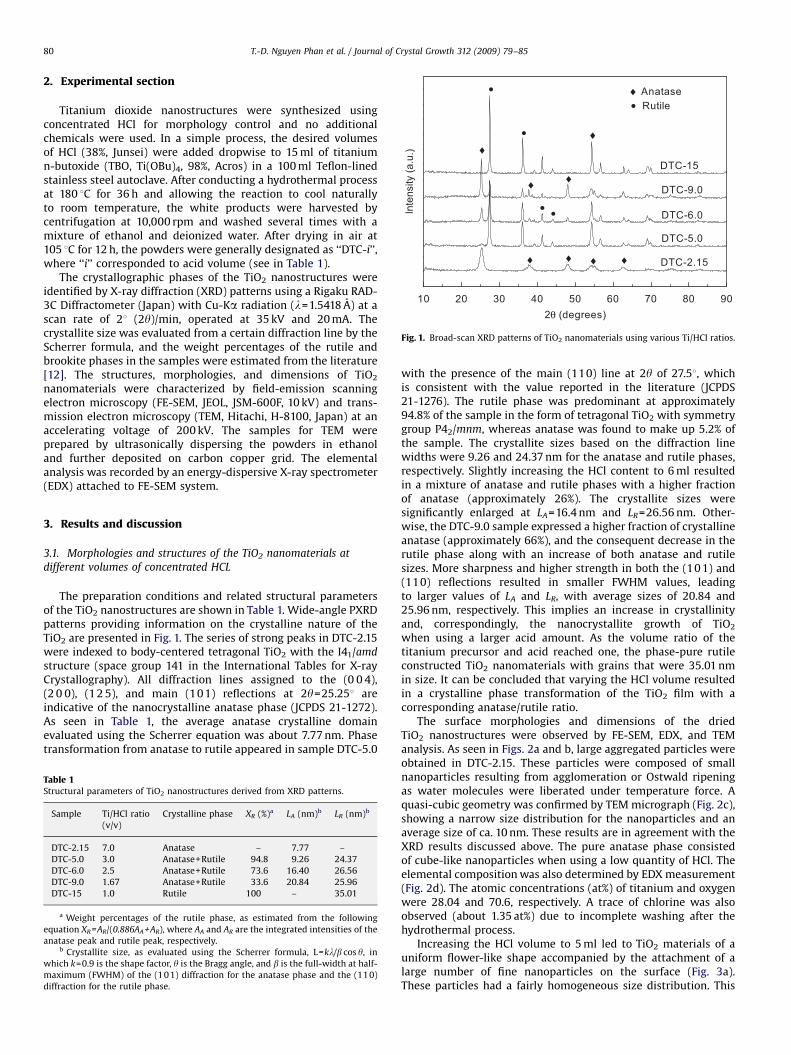
The surface morphologies and dimensions of the driedTiO2 nanostructures were observed by FE-SEM, EDX, and TEManalysis. As seen in Figs. 2a and b, large aggregated particles wereobtained in DTC-2.15. These particles were composed of smallnanoparticles resulting from agglomeration or Ostwald ripeningas water molecules were liberated under temperature force. Aquasi-cubic geometry was confirmed by TEM micrograph (Fig. 2c),showing a narrow size distribution for the nanoparticles and anaverage size of ca. 10 nm. These results are in agreement with theXRD results discussed above. The pure anatase phase consistedof cube-like nanoparticles when using a low quantity of HCl. Theelemental composition was also determined by EDX measurement(Fig. 2d). The atomic concentrations (at%) of titanium and oxygenwere 28.04 and 70.6, respectively. A trace of chlorine was alsoobserved (about 1.35 at%) due to incomplete washing after thehydrothermal process.
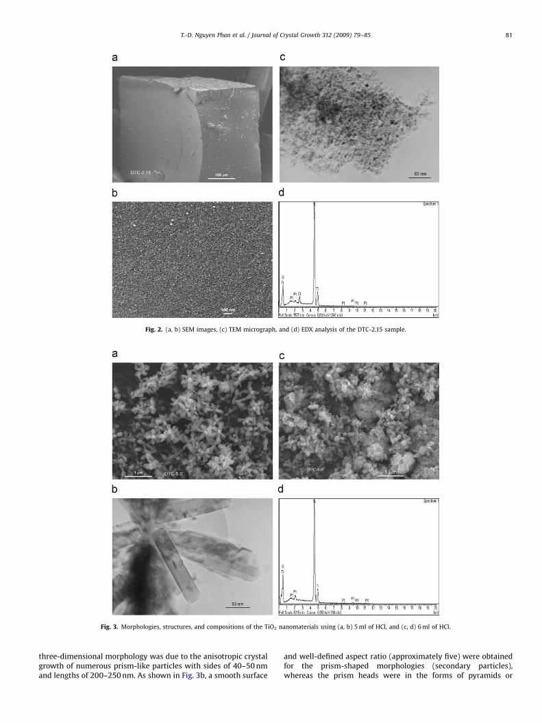
Increasing the HCl volume to 5 ml led to TiO2 materials of auniform flower-like shape accompanied by the attachment of alarge number of fine nanoparticles on the surface (Fig. 3a).These particles had a fairly homogeneous size distribution. This
ARTICLE IN PRESS
Fig. 2. (a, b) SEM images, (c) TEM micrograph, and (d) EDX analysis of the DTC-2.15 sample.
Fig. 3. Morphologies, structures, and compositions of the TiO2 nanomaterials using (a, b) 5 ml of HCl, and (c, d) 6 ml of HCl.
T.-D. Nguyen Phan et al. / Journal of Crystal Growth 312 (2009) 79–85 81
three-dimensional morphology was due to the anisotropic crystalgrowth of numerous prism-like particles with sides of 40–50 nmand lengths of 200–250 nm. As shown in Fig. 3b, a smooth surface
and well-defined aspect ratio (approximately five) were obtainedfor the prism-shaped morphologies (secondary particles),whereas the prism heads were in the forms of pyramids or
ARTICLE IN PRESS
T.-D. Nguyen Phan et al. / Journal of Crystal Growth 312 (2009) 79–8582
rectangles. Either adhesion or the outstretched distributionof quasi-cubic and granular primary nanocrystallites with sizes15–25 nm led to the observation of a flower surface in raspberry-like form because of the initial crystal growth of the titaniumprecursor in the slightly strong acidic medium. When correlatedwith the aforementioned XRD pattern, the dominance of therutile phase at about 95% of the sample strongly contributedto the secondary prism crystallites. The observation of bothnanostructures indicates that the formation of prisms and theflower-like morphology possibly stemmed from the crystalgrowth of the primary nanoparticles and the further epitaxialprocess of the prisms, which will be discussed later. This type of3D morphology was maintained as the HCl volume was increasedto 6 ml, at which the prism-shaped particles had sides of 30 nmand lengths of 150–200 nm, as shown in Fig. 3c. However, anunperfected cauliflower-like morphology was concomitant withthe flower-type morphology in this case and had outer diametersof 800 nm to 1.5mm due to the assembly of pyramidal nanoprismswith sides of 80 nm lengths of 400–600 nm. Furthermore,the adhesion of smaller nanoparticles (10 nm) to the flowersurface was more common than in the former case. This impliesthe beginning of a shape transformation of the TiO2
nanostructure. EDX results recorded over an area of the samplein Fig. 3d showed that DTC-6.0 was composed of either Ti or O, at24.38 and 75.62 at%, respectively.
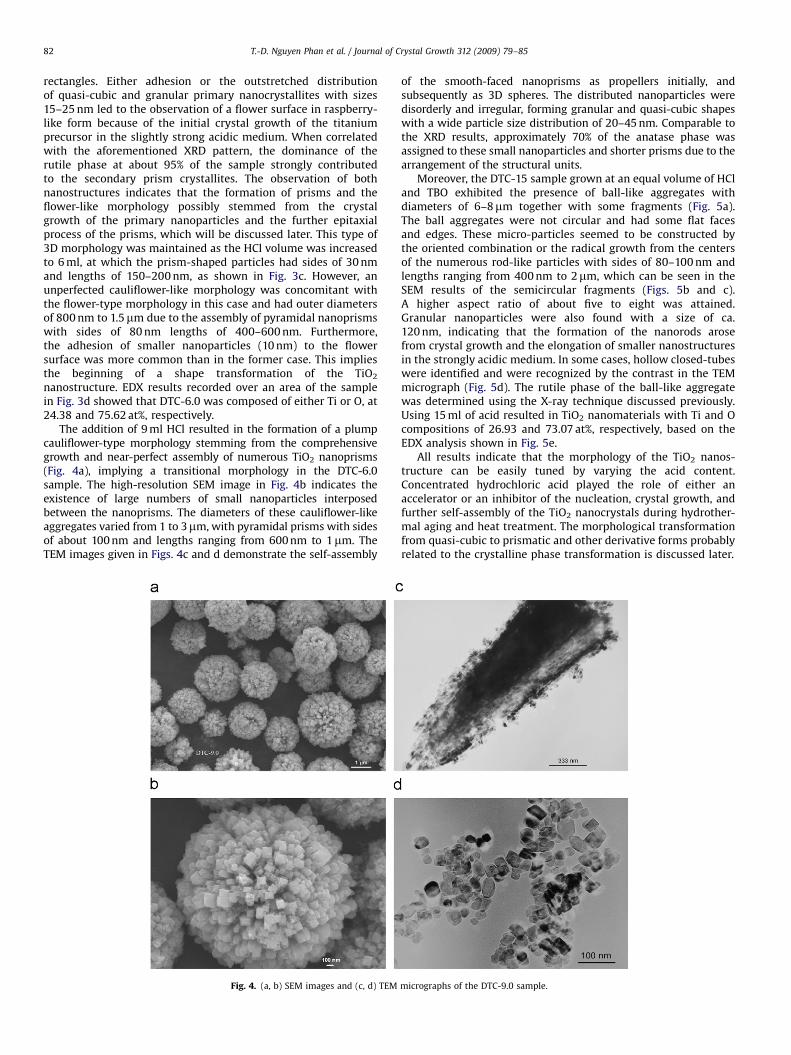
The addition of 9 ml HCl resulted in the formation of a plumpcauliflower-type morphology stemming from the comprehensivegrowth and near-perfect assembly of numerous TiO2 nanoprisms(Fig. 4a), implying a transitional morphology in the DTC-6.0sample. The high-resolution SEM image in Fig. 4b indicates theexistence of large numbers of small nanoparticles interposedbetween the nanoprisms. The diameters of these cauliflower-likeaggregates varied from 1 to 3mm, with pyramidal prisms with sidesof about 100 nm and lengths ranging from 600 nm to 1mm. TheTEM images given in Figs. 4c and d demonstrate the self-assembly
Fig. 4. (a, b) SEM images and (c, d) TEM
of the smooth-faced nanoprisms as propellers initially, andsubsequently as 3D spheres. The distributed nanoparticles weredisorderly and irregular, forming granular and quasi-cubic shapeswith a wide particle size distribution of 20–45 nm. Comparable tothe XRD results, approximately 70% of the anatase phase wasassigned to these small nanoparticles and shorter prisms due to thearrangement of the structural units.
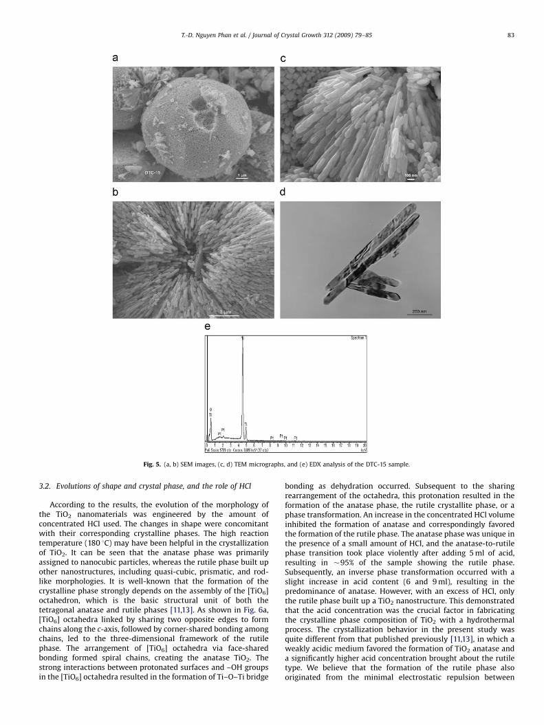
Moreover, the DTC-15 sample grown at an equal volume of HCland TBO exhibited the presence of ball-like aggregates withdiameters of 6–8mm together with some fragments (Fig. 5a).The ball aggregates were not circular and had some flat facesand edges. These micro-particles seemed to be constructed bythe oriented combination or the radical growth from the centersof the numerous rod-like particles with sides of 80–100 nm andlengths ranging from 400 nm to 2mm, which can be seen in theSEM results of the semicircular fragments (Figs. 5b and c).A higher aspect ratio of about five to eight was attained.Granular nanoparticles were also found with a size of ca.120 nm, indicating that the formation of the nanorods arosefrom crystal growth and the elongation of smaller nanostructuresin the strongly acidic medium. In some cases, hollow closed-tubeswere identified and were recognized by the contrast in the TEMmicrograph (Fig. 5d). The rutile phase of the ball-like aggregatewas determined using the X-ray technique discussed previously.Using 15 ml of acid resulted in TiO2 nanomaterials with Ti and Ocompositions of 26.93 and 73.07 at%, respectively, based on theEDX analysis shown in Fig. 5e.
All results indicate that the morphology of the TiO2 nanos-tructure can be easily tuned by varying the acid content.Concentrated hydrochloric acid played the role of either anaccelerator or an inhibitor of the nucleation, crystal growth, andfurther self-assembly of the TiO2 nanocrystals during hydrother-mal aging and heat treatment. The morphological transformationfrom quasi-cubic to prismatic and other derivative forms probablyrelated to the crystalline phase transformation is discussed later.
micrographs of the DTC-9.0 sample.
ARTICLE IN PRESS
Fig. 5. (a, b) SEM images, (c, d) TEM micrographs, and (e) EDX analysis of the DTC-15 sample.
T.-D. Nguyen Phan et al. / Journal of Crystal Growth 312 (2009) 79–85 83
3.2. Evolutions of shape and crystal phase, and the role of HCl
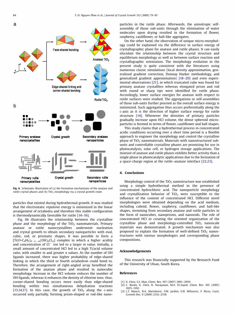
According to the results, the evolution of the morphology ofthe TiO2 nanomaterials was engineered by the amount ofconcentrated HCl used. The changes in shape were concomitantwith their corresponding crystalline phases. The high reactiontemperature (180 1C) may have been helpful in the crystallizationof TiO2. It can be seen that the anatase phase was primarilyassigned to nanocubic particles, whereas the rutile phase built upother nanostructures, including quasi-cubic, prismatic, and rod-like morphologies. It is well-known that the formation of thecrystalline phase strongly depends on the assembly of the [TiO6]octahedron, which is the basic structural unit of both thetetragonal anatase and rutile phases [11,13]. As shown in Fig. 6a,[TiO6] octahedra linked by sharing two opposite edges to formchains along the c-axis, followed by corner-shared bonding amongchains, led to the three-dimensional framework of the rutilephase. The arrangement of [TiO6] octahedra via face-sharedbonding formed spiral chains, creating the anatase TiO2. Thestrong interactions between protonated surfaces and –OH groupsin the [TiO6] octahedra resulted in the formation of Ti–O–Ti bridge
bonding as dehydration occurred. Subsequent to the sharingrearrangement of the octahedra, this protonation resulted in theformation of the anatase phase, the rutile crystallite phase, or aphase transformation. An increase in the concentrated HCl volumeinhibited the formation of anatase and correspondingly favoredthe formation of the rutile phase. The anatase phase was unique inthe presence of a small amount of HCl, and the anatase-to-rutilephase transition took place violently after adding 5 ml of acid,resulting in �95% of the sample showing the rutile phase.Subsequently, an inverse phase transformation occurred with aslight increase in acid content (6 and 9 ml), resulting in thepredominance of anatase. However, with an excess of HCl, onlythe rutile phase built up a TiO2 nanostructure. This demonstratedthat the acid concentration was the crucial factor in fabricatingthe crystalline phase composition of TiO2 with a hydrothermalprocess. The crystallization behavior in the present study wasquite different from that published previously [11,13], in which aweakly acidic medium favored the formation of TiO2 anatase anda significantly higher acid concentration brought about the rutiletype. We believe that the formation of the rutile phase alsooriginated from the minimal electrostatic repulsion between
ARTICLE IN PRESS
Fig. 6. Schematic illustration of (a) the formation mechanisms of the anatase and
rutile crystal phases and (b) TiO2 morphology via a crystal growth route.
T.-D. Nguyen Phan et al. / Journal of Crystal Growth 312 (2009) 79–8584
particles that existed during hydrothermal growth. It was studiedthat the electrostatic repulsive energy is minimized in the lineararrangement of octahedra and therefore this stable configurationis thermodynamically favorable for rutile [14–16].
Fig. 6b illustrates the relationship between the crystallinephase and the morphology of the TiO2 nanomaterials. Primaryanatase or rutile nanocrystallites underwent nucleationand crystal growth to obtain secondary nanoparticles with oval,cubic, rod, or prismatic shapes. It was possible to form a[Ti(O-C4H9)4�n�m(OH)nClm] complex in which a higher acidityand concentration of Cl� ion led to a larger m value. Initially, asmall amount of concentrated HCl led to a high Ti/acid volumeratio, with smaller m and greater n values. As the number of OHligands increased, there was higher probability of edge-sharedlinking in which the third or fourth octahedron could bond to.Therefore, the arrangement of right-angled array benefited theformation of the anatase phase and resulted in nanocubicmorphology. Increase in the HCl volume reduces the number ofOH ligands, whereas it enhances the density of chlorine ions; thus,corner-shared bonding occurs more easily than edge-sharedbonding within two simultaneous dehydration reactions[13,14,17]. In this case, the growth of TiO2 along the c-axisoccurred only partially, forming prism-shaped or rod-like nano-
particles in the rutile phase. Afterwards, the anisotropic self-assembly of those sub-units through the elimination of watermolecules upon drying resulted in the formation of flower,raspberry, cauliflower, or ball-like aggregates.
On the other hand, the observation of unique micro-morphol-ogy could be explained via the difference in surface energy ofcrystallographic plane for anatase and rutile phases. It can easilyelucidate the relationship between the crystal structure andequilibrium morphology as well as between surface reaction andcrystallographic orientation. The morphology evolution in thepresent study is quite consistent with the literatures usingnumerous classic simulations (local density approximation, gen-eralized gradient correction, Donnay Harker methodology, andgeneralized gradient approximation) [18–20] and even experi-mental observations [21], in which truncated cube was found forprimary anatase crystallites whereas elongated prism and rodwith round or sharp tips were identified for rutile phase.Accordingly, lower surface energies for anatase with respect torutile surfaces were studied. The aggregations or self-assembliesof those sub-units further proceed as the overall surface energy isminimized. Such aggregation thus occurs preferentially along thec-axis as it is the direction of higher surface energy for rutilestructure [14]. Whenever the densities of primary particlesgradually increase upon HCl volume, the dense spheroid micro-particles is formed in terms of flower, cauliflower and ball shapes.
This study claims that a hydrothermal process in concentratedacidic conditions occurring over a short time period is a flexibleapproach to engineer the morphology and control the crystallinephase of TiO2 nanomaterials. Materials with nanostructured sub-units and controllable crystalline phases are promising for use inphotocatalysis, solar cell, or hydrogen storage applications. Themixture of anatase and rutile phases exhibits better activity than asingle phase in photocatalytic applications due to the formation ofa space-charge region at the rutile–anatase interface [22,23].
4. Conclusions
Morphology control of the TiO2 nanostructure was establishedusing a simple hydrothermal method in the presence ofconcentrated hydrochloric acid. The nanoparticle morphologyand crystallization behavior of TiO2 were susceptible to theinfluence of the content of concentrated HCl. Different novelmorphologies were obtained depending on the acid medium,including cuboid, flower, raspberry, cauliflower, and ball-likeshapes, stemming from secondary anatase and rutile particles inthe form of nanocubes, nanoprisms, and nanorods. The role ofconcentrated HCl in creating the oriented organization of thecrystalline phase and morphology-controlled TiO2 nanoscalematerials was demonstrated. A growth mechanism was alsoproposed to explain the formation of well-defined TiO2 nanos-tructures with various morphologies and corresponding phasecompositions.
Acknowledgements
This research was financially supported by the Research Fundof the University of Ulsan, South Korea.
References
[1] X. Chen, S.S. Mao, Chem. Rev. 107 (2007) 2891–2959.[2] C. Burda, X. Chen, R. Narayanan, M.A. El-Sayed, Chem. Rev. 105 (2005)
1025–1102.[3] L.T. Mancic, B.A. Marinkovic, P.M. Jardim, O.B. Milosevic, F. Rizzo, Cryst.
Growth Des. 9 (2009) 2152–2158.
ARTICLE IN PRESS
T.-D. Nguyen Phan et al. / Journal of Crystal Growth 312 (2009) 79–85 85
[4] T. Kasuga, M. Hiramatsu, A. Hoson, T. Sekino, K. Niihara, Langmuir 14 (1998)3160–3163.
[5] R. Zhang, L. Gao, Mater. Res. Bull. 36 (2001) 1957–1965.[6] K. Fukuda, T. Sasaki, M. Watanabe, I. Nakai, K. Inaba, K. Omote, Cryst. Growth
Des. 3 (2003) 281–283.[7] Y.V. Kolen’ko, K.A. Kovnir, A.I. Gavrilov, A.V. Garshev, J. Frantti, O.I. Lebedev,
B.R. Churagulov, G. Van Tendeloo, M. Yoshimura, J. Phys. Chem. B 110 (2006)4030–4038.
[8] M. Hernandez-Velez, Thin Solid Films 495 (2006) 51–63.[9] G. Wu, J. Wang, D.F. Thomas, A. Chen, Langmuir 24 (2008) 3503–3509.
[10] S.-J. Liu, X.-X. Wu, B. Hu, J.-Y. Gong, S.-H. Yu, Cryst. Growth Des. 9 (2009)1511–1518.
[11] H. Cheng, J. Ma, Z. Zhao, L. Qi, Chem. Mater. 7 (1995) 663–671.[12] R.A. Spurr, H. Myers, Anal. Chem. 29 (1957) 760–762.[13] F. Izumi, Bull. Chem. Soc. Jpn. 51 (1978) 1771–1776.
[14] M. Gopal, W.J. Moberly Chan, L.C. De Jonghe, J. Mater. Sci. 32 (1997) 6001–6008.[15] S. Watson, D. Beydoun, J. Scott, R. Amal, J. Nanopart. Res. 6 (2004) 193–207.[16] J.-P. Nikkaken, T. Kanerva, T. Mantyla, J. Cryst. Growth 304 (2007) 179–183.[17] Y. Wang, L. Zhang, K. Deng, X. Chen, Z. Zou, J. Phys. Chem. C 111 (2007)
2709–2714.[18] J. Goniakowski, J.M. Holender, L.N. Kantorovich, M.J. Gillan, Phys. Rev. B 53
(1996) 957–960.[19] P.M. Oliver, G.W. Watson, E.T. Kelsey, S.C. Parker, J. Mater. Chem. 7 (1997)
563–568.[20] A. Vittadini, A. Selloni, F.P. Rotzinger, M. Gratzel, Phys. Rev. Lett. 81 (1998)
2954–2957.[21] R.L. Penn, J.F. Banfield, Geochim. Cosmochim. Acta 63 (1999) 1549–1557.[22] A. Fujishima, K. Hashimoto, T. Watanabe, Photocatalysis Fundamentals and
Applications, first ed., BKC, Tokyo, 1999.[23] K. Woan, G. Pyrgiotakis, W. Sigmund, Adv. Mater. 21 (2009) 2233–2239.