a role for snare complex dimerization during neurosecretion
TRANSCRIPT
1
A role for SNARE complex dimerization during
neurosecretion
Elena Fdez*, Thomas A. Jowitt†, Ming-Chuan Wang†, Manisha
Rajebhosale†, Keith Foster#, Jordi Bella†, Clair Baldock†, Philip G.
Woodman† and Sabine Hilfiker*‡
*Institute of Parasitology and Biomedicine “López-Neyra”, CSIC (Spanish
National Research Council), Granada, Spain; †Faculty of Life Sciences, The
University of Manchester, UK; #Syntaxin Ltd, Abingdon, Oxon, UK.
EF and TJ contributed equally.
‡Address correspondence: Sabine Hilfiker, Institute of Parasitology and Biomedicine
“López-Neyra”, CSIC (Spanish National Research Council), 18100 Granada, Spain, Tel:
(34) 958-181654, email: [email protected]
Running head: SNARE complex dimerization
Abbreviations: SNARE: soluble N-ethylmaleimide-sensitive factor attachment protein
receptor (SNARE); syb2: synaptobrevin 2; MALLS: multi-angle laser light scattering;
SAXS: small angle X-ray scattering; TEM: transmission electron microscopy.
http://www.molbiolcell.org/content/suppl/2008/05/28/E08-01-0010.DC1.htmlSupplemental Material can be found at:
2
Abstract
The interactions underlying the cooperativity of soluble N-ethylmaleimide-
sensitive factor attachment protein receptor (SNARE) complexes during
neurotransmission are not known. Here, we provide a molecular characterization of a
dimer formed between the cytoplasmic portions of neuronal SNARE complexes.
Dimerization generates a two-winged structure in which the C-termini of cytosolic
SNARE complexes are in close apposition, and involves residues from the vesicle-
associated (v)-SNARE synaptobrevin 2 that lie close to the cytosol-membrane interface
within the full-length protein. Mutation of these residues reduces stability of dimers
formed between SNARE complexes, without affecting the stability of each individual
SNARE complex. These mutations also cause a corresponding decrease in the ability of
botulinum toxin-resistant synaptobrevin 2 to rescue regulated exocytosis in toxin-treated
neuroendocrine cells. Moreover, such synaptobrevin 2 mutants give rise to a dominant-
negative inhibition of exocytosis. These data are consistent with an important role for
SNARE complex dimers in neurosecretion.
3
Introduction
Neurotransmitter release occurs when synaptic vesicles fuse with the plasma
membrane. A crucial step in this process involves the assembly of a SNARE complex, a
highly stable, parallel 4-helix bundle formed between the synaptic vesicle SNARE
synaptobrevin 2 (syb2) and the plasma membrane SNAREs syntaxin 1 and SNAP-25
(Söllner et al., 1993; Hanson et al., 1997; Sutton et al., 1998; Südhof, 2004; Jahn and
Scheller, 2006; Rizo et al., 2006; Wojcik and Brose, 2007). Current data suggest that
SNARE complex formation proceeds in a vectorial fashion from the N-terminal,
membrane-distal region towards the C-terminal, membrane-proximal end, which may
draw the opposing membranes close enough together for fusion to proceed (Fiebig et al.,
1999; Pobbati et al., 2006; Sorensen et al., 2006). Consistent with this, in reconstituted
assay systems, SNAREs on their own can support membrane fusion ( Weber et al., 1998;
Hu et al., 2003; Giraudo et al., 2006; Pobbati et al., 2006).
In intact cells, evoked membrane fusion involves the cooperative action of
multiple SNARE complexes (Cull-Candy et al., 1976; Bevan and Wendon, 1984; Stewart
et al., 2000). The exact number of complexes required is currently unknown, and
estimates vary from 3 (Hua and Scheller, 2001) to 5-8 (Han et al., 2004) to 10-15 (Keller
and Neale, 2001; Keller et al., 2004; Montecucco et al., 2005). Such differences may
reflect the distinct experimental paradigms employed and/or the types of secretory
organelles studied. Thus, higher-order multimers of SNARE complexes may be required
for fast exocytosis of synaptic vesicles, whilst lower-order multimers may be sufficient
for the slower exocytosis of large dense-core granules from chromaffin and
4
neuroendocrine PC12 cells (Montecucco et al., 2005).
The mechanism(s) responsible for SNARE complex multimerization remains
controversial. Initial studies suggested that multimerization of synaptic SNARE
complexes could be achieved via domain swapping, whereby one of the two SNAP-25
helices could be substituted by the equivalent helix from a neighboring complex (Kweon
et al., 2002). Alternative models proposed the involvement of accessory proteins, such as
synaptotagmin (Littleton et al., 2001) or complexin (Tokumaru et al., 2001), or the
transmembrane domains of syb2 and syntaxin 1A (Laage et al., 2000), in synaptic
SNARE complex multimerization. However, SNARE complexes assembled from
recombinant coils and lacking transmembrane domains are able to associate with each
other (Fasshauer et al., 1997; Fasshauer et al., 1998; Margittai et al., 2001; Ernst and
Brunger, 2003), arguing that at least some of the interactions that support multimerization
may require neither accessory proteins nor transmembrane domains. The precise
multimeric nature and configuration of these recombinant cytosolic SNARE complexes is
ambiguous. Conflicting results have been reported, ranging from dimers involving C-
terminal residues of at least one of the two monomers, to mixtures of monomers/trimers
in solution (Fasshauer et al., 1997; Fasshauer et al., 1998; Margittai et al., 2001; Ernst
and Brunger, 2003). In addition, no investigations have addressed whether such
interactions between SNARE complexes might contribute to their biological action.
To test whether multimerization of SNARE complexes mediated by their
cytosolic domains is an important step during neurotransmitter release, we first
performed a detailed characterization of how such multimers are configured. We
5
identified amino acids within synaptobrevin 2 that contribute to a cytosolic SNARE
complex dimer formed with micromolar affinity. Functional analysis of synaptobrevin 2
mutants in which these residues are replaced provides evidence for an important role for
SNARE complex dimers during exocytosis.
6
Materials and Methods
Plasmids and protein purification
Constructs encoding sequences for the ‘coils’ that form the ‘minimal’ SNARE
complex of rat syntaxin 1a (191-262), synaptobrevin 2 (syb2) (1-96), SNAP-25 B (1-83),
and SNAP-25 B (120-206), constructs encoding full-length non-tagged syb2 and GFP-
tagged syb2 and FRET probes were generated using standard PCR and cloning
procedures (for details see Supplementary Methods). Recombinant proteins were
expressed as N-terminally tagged GST fusion proteins and purified using standard
protocols (Söllner et al., 1993) (see also Supplementary Methods). Protein concentrations
were estimated by the Bradford assay, and ranged from 0.3 – 1 mg/ml. SNARE
complexes were formed by overnight assembly of equimolar concentrations of purified
components in standard buffer and concentrated to approximately 2 mg/ml. Complex
formation was verified by SDS-PAGE. Determination of synaptotagmin 1 and complexin
binding to SNARE complexes was performed as described in Supplementary Methods.
Multi-angle light scattering
SNARE complexes were purified from recombinant coils on a Superdex-200
24/30 gel filtration column (GE, Amersham, UK) run in 5 mM Tris-HCl pH 7.4, 50 mM
NaCl at 0.71 ml/min using a Dionex BioLC HPLC (Camberley, UK). The SNARE
complex dimer peak resolved clearly from the mixture. For SNARE complex mutants all
molecular weight analysis refers to that of material within the dimer peak, though some
SNARE complex monomer could also be found (data not shown). Protein passed through
7
a Wyatt EOS 18-angle laser photometer (Wyatt Technology, Santa Barbara, CA) with the
13th detector replaced with a QELS detector (Wyatt Technology) for the simultaneous
measurement of hydrodynamic radius. This was coupled to a Optilab rEX refractive
index detector (Wyatt Technology), and the hydrodynamic radius, molecular weight
moments and concentration of peaks were analysed using Astra 4.98 (Wyatt
Technology).
Analytical ultracentrifugation
SNARE complex dimers (approximately 8 μM) were purified by gel filtration. All
experiments were performed in 5 mM Tris-HCl, pH 7.4 containing the indicated
concentrations of NaCl, and using a XL-A ultracentrifuge (Beckman Coulter, Fullertin,
CA) with an An50Ti 8-hole rotor fitted either with the standard two-sector open-filled
centerpieces for sedimentation velocity, or with six-sector epon-filled centrepieces for
equilibrium studies, with quartz glass windows. Velocity sedimentation analysis was
performed at 40,000 rpm at 20°C, with the sedimenting boundary monitored at 230 nm
every 9 min. Frictional ratios for the monomer and dimer were calculated from the
sedimentation coefficient. For data interpretation and solution bead modelling, see
Supplementary Methods.
Equilibrium sedimentation was performed at 4°C, using rotor speeds of 10, 15
and 21,000 rpm and scanning at 230 nm every 4 hr until equilibrium was reached. For
molecular weight analysis, data was analysed with the fitting program HeteroAnalysis
using a single ideal model for a distribution of the mean molecular weight. Data
8
was expressed as the average MW from this approximation (MWapp) relative to the
theoretical MW of the monomer. Association constants were investigated using
concentrations of 1, 2.5 and 5 µM protein at the same three rotor speeds in 5 mM Tris-
HCl and 0.3 M NaCl. Global analysis using Sedphat (developed by Peter Schuck, NIH,
Bethesda, MA) of a monomer-dimer association produced the best fit.
Small Angle X-ray Scattering Data Collection
Small angle X-ray solution scattering was carried out using gel filtration-purified
SNARE complex dimers (8 μM) on ID02 at the European Synchrotron Radiation
Facility, Grenoble, France, using 1 m and 5 m sample to detector distances. During data
collection the sample was maintained at 10°C. The corresponding profiles were merged
so as to cover the momentum transfer interval 0.0038 Å-1 < q < 0.53Å-1. The modulus of
the momentum transfer is defined as q = 4πsinθ/λ, where 2θ is the scattering angle, and λ
is the wavelength. With a 1 metre camera distance the maximum scattering angle
corresponds to a Bragg resolution of 11.8 Å. SAXS patterns were recorded using an
image intensified CCD detector having single photon sensitivity and 14bit dynamic
range. The wavelength of X-rays used was 0.1 nm. For further details about data
collection and analysis see Supplementary Methods.
TEM Single Particle Image Analysis
Gel filtration-purified SNARE complex dimers were concentrated 3-fold (24
9
μM), and 5 µl of sample was allowed to adsorb for 30 sec onto a glow discharged (25
mA for 30 seconds) carbon-coated 400 mesh copper grid. The grid was washed 3 times
with milliQ water and then negatively stained with 2% (w/v) uranyl acetate pH 4.7 for 20
seconds. Grids were observed using a FEI Tecnai Twin TEM equipped with a LaB6
filament operating at 120keV. Images were recorded under low dose conditions at -600
nm defocus on a 2048 x 2048 CCD camera with a pixel size of 2Å. The level of defocus
of each individual image was checked by inspecting the position of the Thon rings in the
power spectra and comparing this to the calculated contrast transfer function (CTF). For
details on image processing, see Supplementary Methods.
Fluorescence spectroscopy
SNARE complexes (approximately 5 µM) were purified by gel filtration
chromatography, and tryptophan fluorescence was measured in a 100 µl, 1 cm path-
length cell using a FP750 spectrofluorometer (Jasco, Tokyo, Japan) by excitation at 295
nm and monitoring of emission fluorescence between 300 and 450 nm.
CD spectroscopy
The CD spectra of SNARE complexes (2-5 µM) purified by gel filtration
chromatography were monitored from 260 to 190 nm in 0.2 nm steps (with 10 averages
assayed) in a 0.05 cm path-length cuvette using a J810 spectrapolarometer (Jasco).
10
Differential Scanning Calorimetry
The stability of SNARE complexes was investigated using a VP-DSC
microcalorimeter (Microcal Inc.) with a 0.52 ml total loading volume (see also
Supplementary Methods).
FRET measurements
GFP-tagged SNARE complexes (see Supplementary Methods for further details)
were purified by gel filtration chromatography (4 μM) and molecular mass was analysed
by MALLS as described above, with a determined mass of 144 +/- 4 kDa, close to the
predicted mass of 146 kDa for GFP-tagged SNARE complex dimers. Purified C-C or N-
C-tagged SNARE complex dimers were analysed using a J750 spectrometer (Jasco) and a
100 μl, 1 cm pathlength cell. The excitation wavelength was 433 nm and emission
spectra were measured between 450 and 650 nm in 1 nm steps.
Determination of localization and expression levels of syb2 and of SDS-resistant
SNARE complex stability
The overexpression levels of non-tagged syb2 and mutants thereof, the
localization of GFP-tagged syb2 and mutants thereof, and the determination of GFP-
tagged SDS-resistant SNARE complex stabilities, were determined as described in
Supplementary Methods.
11
Secretion assays
Confluent PC12 cells were plated onto collagen-coated 6-well dishes at 80%
confluency, and transfected the following day with 3 μg DNA using 10 μl
LipofectAMINE 2000 (Invitrogen). Cells were re-plated into 6-well dishes at a ratio of
1:2 the next day, and secretion assays performed two days after replating, with all test
and control conditions carried out on the same pool of transfected cells. Controls were
treated with 0.6 ml of physiological saline solution (PSS; 145 mM NaCl, 5.6 mM KCl,
2.2 mM CaCl2, 0.5 mM MgCl2, 5.6 mM glucose, 15 mM Hepes-NaOH, pH 7.4), and
evoked neurosecretion was triggered by a 5 min incubation with high K+ saline solution
(PSS containing 95 mM NaCl and 56 mM KCl). The amount of hGH in the medium and
in cells was determined by an enzyme-linked immunosorbent assay, and the total amount
of hGH and the percentage hGH secreted calculated against an hGH standard curve.
Statistical analyses were performed with the paired Student’s t test.
Toxin rescue assays
Release of hGH from toxin-treated, permeabilized PC12 cells transfected with
botB-resistant syb2 (Q76V,F77W) was measured essentially as described (Quetglas et
al., 2002) in the presence of recombinantly expressed endopeptidase light-chain of
botulinum neurotoxin serotype B (botB/LC). BotB/LC was purified as described before
(Sutton et al., 2005), was of high homogeneity as assessed by SDS-PAGE, and showed
12
endopeptidase activity comparable to native botulinum neurotoxin using an in vitro
VAMP peptide cleavage assay (Sutton et al., 2005). Determination of the cleavage of
endogenous syb2 upon toxin treatment, and of the levels of expressed toxin-insensitive
syb2 and syb2 mutants are described in Supplementary Methods.
13
Results
Global solution structure of synaptic SNARE complex multimers
As an essential step towards testing whether multimerization of SNARE
complexes facilitated by interactions between their cytosolic domains is important for
neurotransmitter release, we first ascertained whether cytosolic portions of SNARE
complexes would form multimers of defined stoichiometry and configuration in solution.
SNARE complexes encompassing the cytoplasmic coiled coil-forming motifs of syb2,
syntaxin 1 and SNAP-25 (Supplementary Figure 1) were purified by size exclusion
chromatography to yield a single major peak containing assembled SNAREs
(Supplementary Figure 2a; data not shown). When re-chromatographed and analyzed by
Multi-angle Laser Light Scattering (MALLS), they yielded a molecular weight of 89,500
Da (Supplementary Figure 2b; Table Ia), twice that of a monomeric SNARE complex.
Treatment with 1M NaCl (Antonin et al., 2000) reduced the molecular weight to 44,600
Da (Supplementary Figure 2b). Hence, cytosolic neuronal SNARE complexes are
quantitatively incorporated into salt-sensitive dimers.
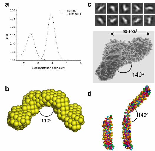
To explore how this dimer is configured, we first examined its shape in solution.
Initially, purified SNARE complexes were subjected to velocity sedimentation in low or
high salt. Surprisingly, the sedimentation coefficients of the dimer and monomer were
relatively close, whereas the frictional ratio was appreciably different and the
hydrodynamic radius of the dimer was much larger than that of the monomer (Figure 1a,
Table Ia). These data suggest that the dimer is more elongated than the monomer, and
argue against it forming by extensive alignment of monomers.
14
The solution shape of the dimer was determined more precisely by using small
angle X-ray solution scattering (SAXS). The experimental scattering profiles were
modelled using an ab initio procedure (Svergun, 1999; Svergun et al., 2001)
(Supplementary Figure 3), and the averaged filtered structure resolved as a two-winged
particle with each wing approximately 9.5 nm long and tilted at about 110° relative to
each other (Figure 1b). Using transmission electron microscopy to determine the shape
directly revealed a similar structure, with each wing 9-10 nm long and set at 140° to each
other (Figure 1c).
To confirm that these shapes are consistent with the observed hydrodynamic
properties, bead models were generated from the atomic coordinates within SNARE
complexes (Sutton et al., 1998) using the program SOMO (Spotorno et al., 1997), then
aligned in hypothetical configurations and modelled for their hydrodynamic properties.
End-to-end dimers set at an angle of 130-140° generated hydrodynamic properties close
to those observed experimentally (Figure 1d and Table Ib). Thus, three methods of shape
analysis suggest that cytosolic SNARE complexes form uniformly configured dimers, in
which the dimerization interface lies close to one end of each SNARE complex and
generates an open, two-winged structure.
Orientation of the SNARE complex dimer
Fusion of primed vesicles requires conversion of partially assembled trans-
SNARE complexes into fully assembled, fusion-competent SNARE bundles (Fiebig et
15
al., 1999; Pobbati et al., 2006; Sorensen et al., 2006). Therefore, a functionally relevant
dimer should involve C-terminal SNARE complex regions that lie close to the point of
membrane insertion. To address whether the C-termini of SNARE complexes are close to
each other within a dimer, we measured FRET in SNARE complexes assembled using a
mixture of syb2-CFP and syb2-Venus, providing complexes containing either FRET
donor or FRET acceptor at the C-terminus (Figure 2a). For comparison, SNARE
complexes were assembled using syb2-Venus and CFP-syb2. All complexes formed with
similar efficiency, and were equally stable, as compared to those containing wt syb2 (data
not shown and Supplementary Figure 4). However, only when both FRET partners were
located at the C-termini of syb2 molecules was a significant FRET signal observed that
was sensitive to disruption of the dimer by high salt (Figure 2b,c). Hence, the C-termini
of both individual, soluble SNARE complex monomers are adjacent to each other in the
dimer. This would suggest that dimers could form in vivo only when SNARE complex
assembly is virtually complete.
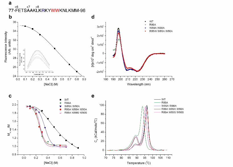
Residues involved in forming the dimer interface
To test the functional significance of this dimer, we first identified amino acids
that contribute to dimerization. We focussed initially on neighboring tryptophan residues
(W89 and W90) within syb2 that are adjacent to the cytosol-membrane interface and
hence likely to be close to the point of dimerization (Figure 3a). Since these are the only
tryptophans within the cytosolic SNARE complex, any changes in intrinsic tryptophan
fluorescence upon conversion of dimers to monomers would indicate that these
16
residues form part of the dimer interface. Indeed, the tryptophan fluorescence decreased
with increasing [NaCl] (Figure 3b), closely matching the monomerization of SNARE
complex dimers determined independently by equilibrium sedimentation (Figure 3c). The
peak fluorescence emission wavelength remained unchanged (~355 nm) at all [NaCl]
(Figure 3b, inset), suggesting that the tryptophan residues in both SNARE complex
dimers and monomers occupy hydrophilic environments. Thus, the fluorescence decrease
upon monomerization most likely results from release of rotational constraints imposed
upon at least one tryptophan residue within the dimer, and suggests that one or both
tryptophans participate in dimerization.
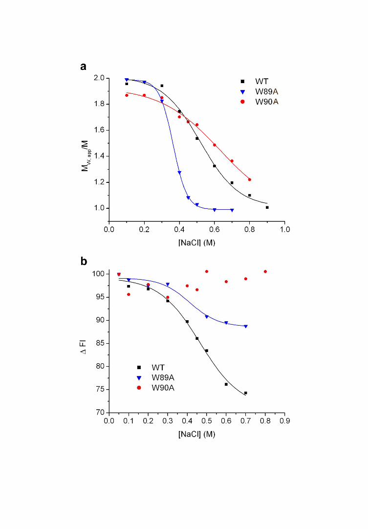
Mutational analysis provided direct evidence for this. Equilibrium sedimentation
of SNARE complex dimers containing syb2(W89A,W90A) showed that they were more
sensitive to increasing [NaCl] than wild-type dimers (Figure 3c, Table IIa). Analysis of
single mutants revealed that dimers containing syb2(W89A) displayed NaCl sensitivity
similar to those containing syb2(W89A,W90A), whilst those containing syb2(W90A)
were slightly more stable than wildtype (Figure 4a). Hence, W89 makes the greater
contribution to dimer stability. However, both tryptophans seem to lie at the dimer
interface, as the tryptophan fluorescence from W90 in SNARE complex dimers formed
using syb2(W89A) decreased in intensity at NaCl concentrations which caused dimer
disassembly (Figure 4b). This would indicate that it is W90 that is subject to rotational
constraints within the dimer.
We reasoned that residues close to W89-W90 may also contribute to the dimer
interface. To this end, we tested effects of mutating the conserved residue R86 within
17
syb2 on dimer stability. Indeed, dimers formed using syb2(R86A), and
syb2(R86A,W89A,W90A) were substantially more sensitive to increased [NaCl] (Figure
3c, Table IIa). To compare affinities using sedimentation analysis, SNARE complexes
were analysed after dilution in 0.3 M NaCl, a salt concentration at which wildtype and
mutant SNARE complexes are all at equilibrium between monomers and dimers (Figure
3c). The association constant for each SNARE dimer species was obtained by global
analysis of concentration-dependent dimerization at equilibrium (Supplementary Figure
5). The association constant was 5.43 µM for wild-type SNARE complex dimers,
compared to 11.97 µM and 46.23 µM for those containing syb2(W89A,W90A) and
syb2(R86A,W89A,W90A), respectively. Analysis of sedimentation experiments
performed using wildtype SNARE dimers at varying salt concentrations confirmed that
the association constant was linear with respect to [NaCl] (data not shown), yielding an
estimated association constant of 1.23 μM at 150 mM NaCl, close to physiological ionic
strength.
The mutations that we have identified substantially reduce the affinity of SNARE
complex dimerization. Importantly, however, they do not affect the stability of each
SNARE complex monomer significantly. These formed with identical helicity to wild-
type SNARE complexes, assessed by the CD spectroscopy peak at 220 nm (Figure 3d).
Differential scanning calorimetry was used to provide a precise estimate of the energy
associated with SNARE complex formation, and showed that the very high stability of
the SNARE complex coiled-coil helix was essentially unaffected by the syb2
dimerization mutations; the melting temperature for all SNARE complexes was very
18
similar and the change in enthalpy associated with this transition was not altered
significantly (Figure 3e and Table IIb). Incorporation of syb2(R86A) displayed a slight
effect on the stability of individual SNARE complexes (Figure 3e) but importantly, this
was not observed when syb2(R86A,W89A,W90A) was employed. In summary, our data
indicate that three residues of syb2 (R86, W89 and W90) form part of the interaction
interface of an open, wing-shaped SNARE complex dimer as observed in solution. In
addition, since the two tryptophan residues are fully solvent-exposed, the salt-sensitivity
of the dimer is likely due to salt-dependent changes in its structure, including disruption
of salt bridge(s) involving R86.
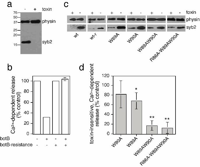
Syb2 dimerization mutants are unable to support secretion
We next aimed to determine whether residues in syb2 found at the SNARE
complex dimer interface in vitro may be important for supporting neurosecretion in vivo.
For this purpose, we transfected neuroendocrine PC12 cells with a plasmid containing
both syb2 and human growth hormone (hGH) (Sugita et al., 1999). Cells were then
permeabilized and treated with recombinant botulinum neurotoxin type B light-chain
(botB/LC) (Sutton et al., 2005), which cleaves and inactivates syb2 (Figure 5a). As
expected, Ca2+-dependent secretion of hGH was decreased (to 32% of control) by the
presence of botB/LC in cells transfected with a plasmid containing wildtype syb2 (Figure
5b). Such profound, but incomplete inhibition of release has been described previously
(Chilcote et al., 1995; Quetglas et al., 2002). The expression of botB/LC-resistant syb2
(Q76V, F77W) restored secretion (104% of control) in the presence of toxin
19
(Figure 5b). This rescue assay allowed us to measure the ability of syb2 dimerization
mutants to support exocytosis in the absence of endogenous protein. Toxin-insensitive
wildtype and mutant syb2 were all expressed to similar degrees (Figure 5c). However,
toxin-insensitive syb2(W89A,W90A) was severely impaired in its ability to rescue
secretion (17.5% of control) (Figure 5d), similar to what has been previously described
(Quetglas et al., 2002). Using this assay system, no further additive effects could be
observed using syb2(R86A,W89A,W90A), which was equally deficient in supporting
release. Interestingly, syb2(W90A) displayed a slight deficit in its ability to rescue
secretion, with syb2(W89A) displaying more pronounced effects (Figure 5d). Hence, the
ability of syb2 mutants to support release in vivo reflects the contribution of each
corresponding amino acid to the SNARE complex dimer interface. Previous studies have
implicated W90 in calmodulin-dependent regulation of exocytosis (Quetglas et al., 2000;
Quetglas et al., 2002). However, since W90A would disrupt the calmodulin binding motif
within syb2 (Rhoads and Friedberg, 1997; Chin and Means, 2000), the ability of this
mutant to largely support secretion would suggest that such an interaction does not play a
major role in vivo.
Dominant-negative secretory effects of syb2 dimerization mutants
To further test the importance of SNARE complex dimers during regulated
exocytosis, we used a dominant-negative approach. Since PC12 cells display Ca2+-
evoked secretion which requires cooperativity between 3 or more SNARE complexes
(Hua and Scheller, 2001), expression of full-length syb2 mutants that are
20
incorporated into SNARE complexes of normal stability but impaired in dimer formation
should generate a dominant-negative phenotype, revealing a potential role for SNARE
complex dimerization in membrane fusion.
PC12 cells were transfected with syb2 and hGH as before, and intact cells were
assayed directly to measure constitutive and evoked exocytosis. Exogenous syb2 was
expressed at approximately 2-3 times over endogenous levels, and all mutants analysed
were expressed to similar degrees (Figure 6a). Expression of wild-type syb2 did not
affect basal or evoked secretion of hGH (data not shown). In contrast, expression of syb2
dimerization mutants reduced evoked secretion without affecting basal secretion or levels
of hGH expression (Figure 6b). This inhibition was reproducible, statistically significant,
and greatest for syb2(R86A,W89A,W90A), in line with the more pronounced effect of
this mutant on SNARE complex dimer stability (Figure 6c). All syb2 constructs analysed
in this study localized to neuritic appendages and were efficiently incorporated with
endogenous SNARE proteins into SNARE complex monomers of essentially the same
stability, assessed by heating in SDS (Figure 6e-h, Supplementary Figure 6,
Supplementary Figure 7).
Complexins and synaptotagmin 1 bind to SNARE complexes with distinct
outcomes for membrane fusion reactions (Tang et al., 2006). As such interactions may be
mutually exclusive with SNARE complex dimerization, we formed SNARE complexes
in the presence of complexin 1 or synaptotagmin 1, respectively, and measured the extent
of dimer formation by size exclusion chromatography. SNARE complex dimerization
was not prevented by an excess of complexin 1, which co-eluted with SNARE complex
21
dimers (Supplementary Figure 8a,b). Binding was essentially stoichiometric, as judged
by the substantial change in apparent molecular weight of dimeric SNARE complexes in
the presence of complexin 1. Complexin 1 bound to wildtype and
syb2(R86A,W89A,W90A)-containing SNARE complexes with similar efficiency,
consistent with complexin binding to a site within the SNARE complex distinct from the
dimerization interface. On the other hand, the C2AB domain of synaptotagmin 1 could
not be detected to bind to SNARE complexes in solution (Supplementary Figure 8c).
Whilst it remains formally possible that the lack of synaptotagmin 1 binding is a
consequence of SNARE complex dimerization, this is unlikely, as no synaptotagmin 1
binding could be detected, even on over-exposed blots, in fractions migrating slightly
behind the SNARE complex dimer peak and containing a minor population of SNARE
complex monomers (data not shown). Such lack of co-migration of soluble
synaptotagmin 1 with SNARE complexes during size-exclusion chromatography has
been previously observed (Bowen et al., 2005), indicative of a low-affinity interaction in
solution. In either case, various structural and mutational data (Chen et al., 2002;
Rickman et al., 2006; Lynch et al., 2007) further indicate that residues distinct from those
involved in dimerization seem to be responsible for complexin and synaptotagmin
binding to SNARE complexes.
W89 and W90 of syb2 have been implicated in binding to phospholipids
(Quetglas et al., 2000; Quetglas et al., 2002; de Haro et al., 2004). In fact, it has been
suggested that the reversible insertion of these residues of syb2 into the synaptic vesicle
membrane may decrease the availability of syb2 and hence the probability of SNARE
22
complex formation (Hu et al., 2002; Kweon et al., 2003). We therefore also examined
SNARE complex dimers formed using a syb2 mutant in which the tryptophans were
replaced with hydrophilic serine residues known to support rapid SNARE complex
formation (Kweon et al., 2003). Expression of syb2(R86A,W89S,W90S) inhibited
evoked secretion to approximately the same level as syb2(R86A,W89A,W90A) (Figure
6c). The stability of dimers formed using this mutant was virtually identical to that
formed using syb2(R86A,W89A,W90A) (Figure 3c), and the stability of monomers was
essentially the same as wild-type (Figure 3d,e and Table IIb). Analogous to results
obtained using the toxin rescue assay, syb2(W89A) displayed a dominant-negative effect
on secretion, whilst syb2(W90A) was without effect (Figure 6d). Hence, the inhibitory
effects of the mutants analysed here are likely not due to interfering with calmodulin
and/or phospholipid binding of syb2, as previously suggested (Quetglas et al., 2000;
Quetglas et al., 2002; de Haro et al., 2004), but are consistent with a mechanism
involving impaired SNARE complex multimerization.
23
Discussion
A better understanding of the mechanistic aspects of vesicle exocytosis depends
on a quantitative characterization of the elements driving this process. Functional studies
have clearly indicated that multiple SNARE complexes cooperate to bring about an
individual vesicular fusion event (Hua and Scheller, 2001; Keller and Neale, 2001; Han
et al., 2004; Keller et al., 2004; Montecucco et al., 2005), with the number of
participating complexes possibly affecting the speed of opening, or the diameter of the
fusion pore (Han et al., 2004). Whilst oligomerization may be an inherent feature of
SNARE complexes, a detailed description of how such interactions take place and their
relevance for membrane fusion has been lacking. In this study, we describe and
characterize a defined SNARE complex dimer that forms with micromolar affinity in
solution in vitro, and provide evidence for its role in neurosecretion in vivo.
The soluble part of SNARE complexes was found to form dimers with the C-
termini of both monomers interacting at an obtuse angle. This novel and surprising
structure is quite distinct from all lattice interactions between neuronal SNARE complex
monomers displayed in the crystal structure (Sutton et al., 1998) (PDB entry 1SFC).
Whilst one such crystallographic dimer is formed towards the C-termini of both SNARE
complex monomers, with W89 of syb2 part of the interaction interface (Supplementary
Figure 9), the alignment of monomers along their entire length make this ‘closed’ crystal
form distinct from the open, wing-shaped structure of dimers as identified in solution.
Hydrodynamic bead modeling of this crystallographic dimer confirms that its
hydrodynamic properties are very different from those observed experimentally. In
24
addition, our biochemical and biophysical data strongly indicate the existence of a
homogeneous population of dimers. Thus, interactions favored in crystal lattices may not
be observed in other contexts (Vestergaard et al., 2005).
Whilst dimerization between cytosolic domains is likely to play an important role
in the multimerization of SNARE complexes, the overall oligomeric status and shape of
SNARE complexes in vivo may be further influenced by the juxtaposition of the
membrane, the presence of the transmembrane domains, and accessory factors. Indeed,
previous studies have shown that native SNARE complexes purified from brain, or
SNARE complexes assembled from recombinant full-length SNAREs containing
transmembrane domains, assemble into star-shaped particles mostly containing 3 or 4
bundles emanating from their center (Rickman et al., 2005). Such structure may be
obtained if two dimers associate with each other through their transmembrane domains
(Hohl et al., 1998; Laage et al., 2000; Bowen et al., 2002; Roy et al., 2004).
Unfortunately, when only one of the two SNAREs carried a transmembrane domain,
reassembly experiments led to the generation of large irregular aggregates (Rickman et
al., 2005). This makes it difficult to determine the exact contributions of the
transmembrane domains and/or membrane-proximal regions of syb2 to the formation of
oligomeric structures obtained with full-length SNAREs. Similarly, the need for
detergent solubilization precludes analysis of whether identified SNARE complex
multimers are present in their trans or cis forms, and additional evidence for the existence
and importance of SNARE complex oligomers may be best obtained using in vivo
approaches.
25
Earlier studies using syb2(W89A,W90A) had suggested that these residues
mediate the binding of syb2 to calmodulin (Quetglas et al., 2002; de Haro et al., 2004).
However, no single mutational analysis was performed to corroborate these results. The
consensus motif for Ca2+-dependent calmodulin binding involves select hydrophobic
residues at positions 1-5-8-14, and an overall net electrostatic charge of +2 to +6 (Rhoads
and Friedberg, 1997). As such, W90 is at position 14 of this motif, and a mutation to
alanine at this position would not be tolerated (Rhoads and Friedberg, 1997). We find that
syb2(W90A) has no effect on secretion either in the presence or absence of endogenous
syb2. Similarly, the overall net charge of the calmodulin binding motif within syb2 is +3,
and the secretory effect of another mutant (syb2(K83A,K87V)) was suggested to be due
to eliminating those charge requirements (Quetglas et al., 2002). However, since K83 is
part of the SNARE motif, the effects of this mutant may have been due to altered SNARE
complex stability, which was not assessed in sufficient detail. Finally, mutating W89 and
W90 to hydrophilic serines does not enhance secretion, as would be expected if the
availability of syb2 would be restricted due to its interacting with the hydrophobic part of
the phospholipid bilayer (Kweon et al., 2003). Our analysis shows that the ability of syb2
mutants to reduce SNARE complex dimer stability in vitro parallels their inhibition of
secretion in vivo. Thus, whilst we cannot fully exclude compound effects, our data
indicate that the secretory effects observed with the syb2 mutants are most likely due to
interfering with SNARE complex dimer stability in vivo.
The dimerization of neuronal SNARE complexes may generate an important
intermediate during evoked secretion. This intermediate may be transient in vivo, formed
26
after trans SNARE complex assembly and close to the point of fusion. Whilst detailed
electrophysiological experiments will be needed to determine the precise role of SNARE
complex dimers in membrane fusion events, one can speculate from their solution
structure how they might contribute towards formation of an oligomeric complex around
a fusion pore, consistent with the energies required to bring about membrane fusion (Li et
al., 2007). It is interesting to note that the syb2(W89A,W90A) mutant does not seem to
be defective in mediating liposomal fusion events (Siddiqui et al., 2007), whilst
profoundly inhibiting Ca2+-dependent secretion. Thus, secretory defects using the syb2
dimerization mutants only seem to be evident in cell systems requiring cooperativity
between SNARE complexes. Such SNARE complex dimerization may improve the
efficiency of vesicle exocytosis by contributing to the cooperative relationship between
calcium and synaptic transmission.
Acknowledgements
This work was supported by the UK MRC (Grants G9722026/G9901377) and the
Spanish MEC (BFU2004-02969 and BFU2007-63635). The laboratory of S.H. is member
of the Network for Cooperative Research on Membrane Transport Proteins (REIT), co-
funded by the MEC and the European Regional Development Fund (ERDF) (BFU2007-
30688-E/BFI). S.H. is supported by a Ramón y Cajal Fellowship. E.F. is supported by a
fellowship (FPI) from the Spanish MEC. We thank J. Zimmerberg, R. Fernandez-Chacón
and D. Thornton for critical comments. We thank S. High for the full-length syb2
construct, G. Schiavo for the SNAP-25 construct, M. Carrington for the Venus construct,
27
H. McMahon for the complexin constructs, A.F. Parlow for the anti-hGH antibody, and
the ESRF and T. Narayanan for assistance with SAXS measurements.
28
References
Antonin, W., Holroyd, C., Fasshauer, D., Pabst, S., Fischer von Mollard, G. and Jahn, R.
(2000). A SNARE complex mediating fusion of late endosomes defines
conserved properties of SNARE structure and function. EMBO J. 19, 6453-6464.
Bevan, S. and Wendon, L.M. (1984). A study of the action of tetanus toxin at rat soleus
neuromuscular junctions. J. Physiol. (Lond) 348, 1-17.
Bowen, M.E., Engelman, D.M. and Brunger, A.T. (2002). Mutational analysis of
synaptobrevin transmembrane domain oligomerization. Biochemistry 41, 15861-
15866.
Bowen, M.E., Weninger, K., Ernst, J., Chu, S. and Brunger, A.T. (2005). Single-molecule
studies of synaptotagmin and complexin binding to the SNARE complex.
Biophys. J. 89, 690-702.
Chen, X., Tomchick, D.R., Kovrigin, E., Arac, D., Machius, M., Südhof, T.C. and Rizo,
J. (2002). Three-dimensional structure of the complexin/SNARE complex.
Neuron 33, 397-409.
Chilcote, T.J., Galli, T., Mundigl, O., Edelmann, L., McPherson, P.S., Takei, K. and De
Camilli, P. (1995). Cellubrevin and synaptobrevins: similar subcellular
localization and biochemical properties in PC12 cells. J Cell Biol. 129, 219-231.
Chin, D. and Means, A.R. (2000). Calmodulin: a prototypical calcium sensor. Trends in
Cell Biology 10, 322.
Cull-Candy, S.G., Lundh, H. and Thesleff, S. (1976). Effects of botulinum toxin on
neuromuscular transmission in the rat. J. Physiol. (Lond) 260, 177-203.
29
de Haro, L., Ferracci, G., Opi, S., Iborra, C., Quetglas, S., Miquelis, R., Leveque, C. and
Seagar, M. (2004). Ca2+/calmodulin transfers the membrane-proximal lipid-
binding domain of the v-SNARE synaptobrevin from cis to trans bilayers. Proc.
Natl. Acad. Sci. USA 101, 1578-1583.
Ernst, J.A. and Brünger, A.T. (2003). High resolution structure, stability, and
synaptotagmin binding of a truncated neuronal SNARE complex. J. Biol. Chem.
278, 8630-8636.
Fasshauer, D., Eliason, W.K., Brünger, A.T. and Jahn, R. (1998). Identification of a
minimal core of the synaptic SNARE complex sufficient for reversible assembly
and disassembly. Biochemistry 37, 10354-10362.
Fasshauer, D., Otto, H., Eliason, W.K., Jahn, R. and Brünger, A.T. (1997). Structural
changes are associated with soluble N-ethylmaleimide-sensitive fusion protein
attachment protein receptor complex formation. J. Biol. Chem. 272, 28036-28041.
Fiebig, K.M., Rice, L.M., Pollock, E. and Brünger, A.T. (1999). Folding intermediates of
SNARE complex assembly. Nature Struct. Biol. 6, 117-123.
Giraudo, C.G., Eng, W.S., Melia, T.J. and Rothman, J.E. (2006). A Clamping Mechanism
Involved in SNARE-Dependent Exocytosis. Science 313, 676-680.
Han, X., Wang, C.T., Bai, J., Chapman, E.R. and Jackson, M.B. (2004). Transmembrane
segments of syntaxin line the fusion pore of Ca2+-triggered exocytosis. Science
304, 289-292.
Hanson, P.I., Roth, R., Morisaki, R., Jahn, R. and Heuser, J.E. (1997). Structure and
conformational changes in NSF and its membrane receptor complexes visualized
by quick-freeze/deep-etch electron microscopy. Cell 90, 523-536.
30
Hohl, T.M., Parlati, F., Wimmer, C., Rothman, J.E., Söllner, T.H. and Engelhardt, H.
(1998). Arrangement of subunits in 20S particles consisting of NSF, SNAPs, and
SNARE complexes. Mol. Cell 2, 539-548.
Hu, C., Ahmed, M., Melia, T.J., Söllner, T., Mayer, T. and Rothman, J.E. (2003). Fusion
of cells by flipped SNAREs. Science 300, 1745-1749.
Hu, K., Carroll, J., Fedorovich, S., Rickman, C., Sukhodub, A. and Davletov, B. (2002).
Vesicular restriction of synaptobrevin suggests a role for calcium in membrane
fusion. Nature 415, 646-650.
Hua, Y. and Scheller, R.H. (2001). Three SNARE complexes cooperate to mediate
membrane fusion. Proc. Natl. Acad. Sci. USA 98, 8065-8070.
Jahn, R. and Scheller, R.H. (2006). SNAREs-engines for membrane fusion. Nat. Rev.
Mol. Cell. Biol. 7, 631-643.
Keller, J.E., Cai, F. and Neale, E.A. (2004). Uptake of botulinum neurotoxin into cultured
neurons. Biochemistry 43, 526-532.
Keller, J.E. and Neale, E.A. (2001). The role of the synaptic protein SNAP-25 in the
potency of botulinum neurotoxin type A. J. Biol. Chem. 276, 13476-13482.
Kweon, D.-H., Kim, C.S. and Shin, Y.-K. (2003). Regulation of neuronal SNARE
assembly by the membrane. Nature Struct. Biol. 10, 440-447.
Kweon, D.H., Chen, Y., Zhang, F., Poirier, M., Kim, C.S. and Shin, Y.K. (2002). Probing
domain swapping for the neuronal SNARE complex with electron paramagnetic
resonance. Biochemistry 41, 5449-5452.
Laage, R., Rohde, J., Brosig, B. and Langosch, D. (2000). A conserved membrane-
spanning amino acid motif drives homomeric and supports heteromeric assembly
31
of presynaptic SNARE proteins. J. Biol. Chem. 275, 17481-17487.
Li, F., Pincet, F., Perez, E., Eng, W.S., Melia, T.J., Rothman, J.E. and Tareste, D. (2007).
Energetics and dynamics of SNAREpin folding across lipid bilayers. Nat. Struct.
Mol. Biol. 14, 890-896.
Littleton, J.T., Bai, J., Vyas, B., Desai, R., Baltus, A.E., Garment, M.B., Carlson, S.D.,
Ganetzky, B. and Chapman, E.R. (2001). Synaptotagmin mutants reveal essential
functions for the C2B domain in Ca2+-triggered fusion and recycling of synaptic
vesicles in vivo. J. Neurosci. 21, 1421-1433.
Lynch, K.L., Gerona, R.R.L., Larsen, E.C., Marcia, R.F., Mitchell, J.C. and Martin, T.F.J.
(2007) Synaptotagmin C2A loop 2 mediates Ca2+-dependent SNARE interactions
essential for Ca2+-triggered vesicle exocytosis. Mol. Biol. Cell 18, 4957-4968.
Margittai, M., Fasshauer, D., Pabst, S., Jahn, R. and Langen, R. (2001). Homo- and
heterooligomeric SNARE complexes studied by site-directed spin labeling. J.
Biol. Chem. 276, 13169-13177.
Montecucco, C., Schiavo, G. and Pantano, S. (2005). SNARE complexes and
neuroexocytosis: how many, how close? Trends in Biochemical Sciences 30, 367.
Pobbati, A.V., Stein, A. and Fasshauer, D. (2006). N- to C-Terminal SNARE Complex
Assembly Promotes Rapid Membrane Fusion. Science 313, 673-676.
Quetglas, S., Iborra, C., Sasakawa, N., De Haro, L., Kumakura, K., Sato, K., Leveque, C.
and Seagar, M. (2002). Calmodulin and lipid binding to synaptobrevin regulates
calcium-dependent exocytosis. EMBO J. 21, 3970-3979.
Quetglas, S., Leveque, C., Miquelis, R., Sato, K. and Seagar, M. (2000). Ca2+-dependent
32
regulation of synaptic SNARE complex assembly via a calmodulin- and
phospholipid-binding domain of synaptobrevin. Proc. Natl. Acad. Sci. USA 97,
9695-9700.
Rhoads, A.R. and Friedberg, F. (1997). Sequence motifs for calmodulin recognition.
Faseb J 11, 331-340.
Rickman, C., Hu, K., Carroll, J. and Davletov, B. (2005). Self-assembly of SNARE
fusion proteins into star-shaped oligomers. Biochem. J. 388, 75-79.
Rickman, C., Jiménez, J.L., Graham, M.E., Archer, D.A., Soloviev, M., Burgoyne, R.D.
and Davletov, B. (2006) Conserved prefusion protein assembly in regulated
exocytosis. Mol. Biol. Cell 17, 283-294).
Rizo, J., Chen, X. and Arac, D. (2006). Unraveling the mechanisms of synaptotagmin and
SNARE function in neurotransmitter release. Trends in Cell Biology 16, 339.
Roy, R., Laage, R. and Langosch, D. (2004). Synaptobrevin transmembrane domain
dimerization-revisited. Biochemistry 43, 4964-4970.
Siddiqui, T.J., Vites, O., Stein, A., Heintzmann, R., Jahn, R. and Fasshauer, D. (2007).
Determinants of Synaptobrevin Regulation in Membranes. Mol. Biol. Cell 18,
2037-2046.
Sorensen, J.B., Wiederhold, K., Muller, E.M., Milosevic, I., Nagy, G., de Groot, B.L.,
Grubmuller, H. and Fasshauer, D. (2006). Sequential N- to C-terminal SNARE
complex assembly drives priming and fusion of secretory vesicles. EMBO J. 25,
955-966.
Spotorno, B., Piccinini, L., Tassara, G., Ruggiero, C., Nardini, M., Molina, F. and Rocco,
33
M. (1997). BEAMS (BEAds Modelling System): a set of computer programs for
the generation, the visualization and the computation of the hydrodynamic and
conformational properties of bead models of proteins. European Biophysics
Journal 25, 373-384.
Stewart, B.A., Mohtashami, M., Trimble, W.S. and Boulianne, G.L. (2000). SNARE
proteins contribute to calcium cooperativity of synaptic transmission. Proc. Natl.
Acad. Sci. USA 97, 13955-13960.
Südhof, T.C. (2004). The Synaptic Vesicle Cycle. Annual Review of Neuroscience 27,
509-547.
Sugita, S., Janz, R. and Südhof, T.C. (1999). Synaptogyrins regulate Ca2+-dependent
exocytosis in PC12 cells. J. Biol. Chem. 274, 18893-18901.
Sutton, J.M., Wayne, J., Scott-Tucker, A., O'Brien, S.M., Marks, P.M., Alexander, F.C.,
Shone, C.C. and Chaddock, J.A. (2005). Preparation of specifically activatable
endopeptidase derivatives of Clostridium botulinum toxins type A, B, and C and
their applications. Protein Expr. Purif. 40, 31-41.
Sutton, R.B., Fasshauer, D., Jahn, R. and Brünger, A.T. (1998). Crystal structure of a
SNARE complex involved in synaptic exocytosis at 2.4Å resolution. Nature 395,
347-353.
Svergun, D.I. (1999). Restoring Low Resolution Structure of Biological Macromolecules
from Solution Scattering Using Simulated Annealing. Biophys. J. 76, 2879-2886.
Svergun, D.I., Petoukhov, M.V. and Koch, M.H. (2001). Determination of domain
structure of proteins from X-ray solution scattering. Biophys. J. 80, 2946-2953.
Söllner, T., Bennett, M.K., Whiteheart, S.W., Scheller, R.H. and Rothman, J.E. (1993). A
34
protein assembly-disassembly pathway in vitro that may correspond to sequential
steps of synaptic vesicle docking, activation, and fusion. Cell 75, 409-418.
Tang, J., Maximov, A., Shin, O., Dai, H., Rizo, J. and Südhof, T.C. (2006). A
complexin/synaptotagmin 1 switch controls fast synaptic vesicle exocytosis. Cell
126, 1175-1187.
Tokumaru, H., Umayahara, K., Pellegrini, L., Ishizuka, T., Saisu, H., Betz, H.,
Augustine, G. and Abe, T. (2001). SNARE complex oligomerization by
synaphin/complexin is essential for synaptic vesicle exocytosis. Cell 104, 421-
432.
Vestergaard, B., Sanyal, S., Roessle, M., Mora, L., Buckingham, R.H., Kastrup, J.S.,
Gajhede, M., Svergun, D.I. and Ehrenberg, M. (2005). The SAXS solution
structure of RF1 differs from its crystal structure and is similar to its ribosome
bound cryo-EM structure. Mol. Cell 20, 929-938.
Weber, T., Zemelman, B.V., McNew, J.A., Westermann, B., Gmachl, M., Parlati, F.,
Söllner, T.H. and Rothman, J.E. (1998). SNAREpins: minimal machinery for
membrane fusion. Cell 92, 759-772.
Wojcik, S.M. and Brose, N. (2007). Regulation of membrane fusion in synaptic
excitation-secretion coupling: speed and accuracy matter. Neuron 55, 11-24.
35
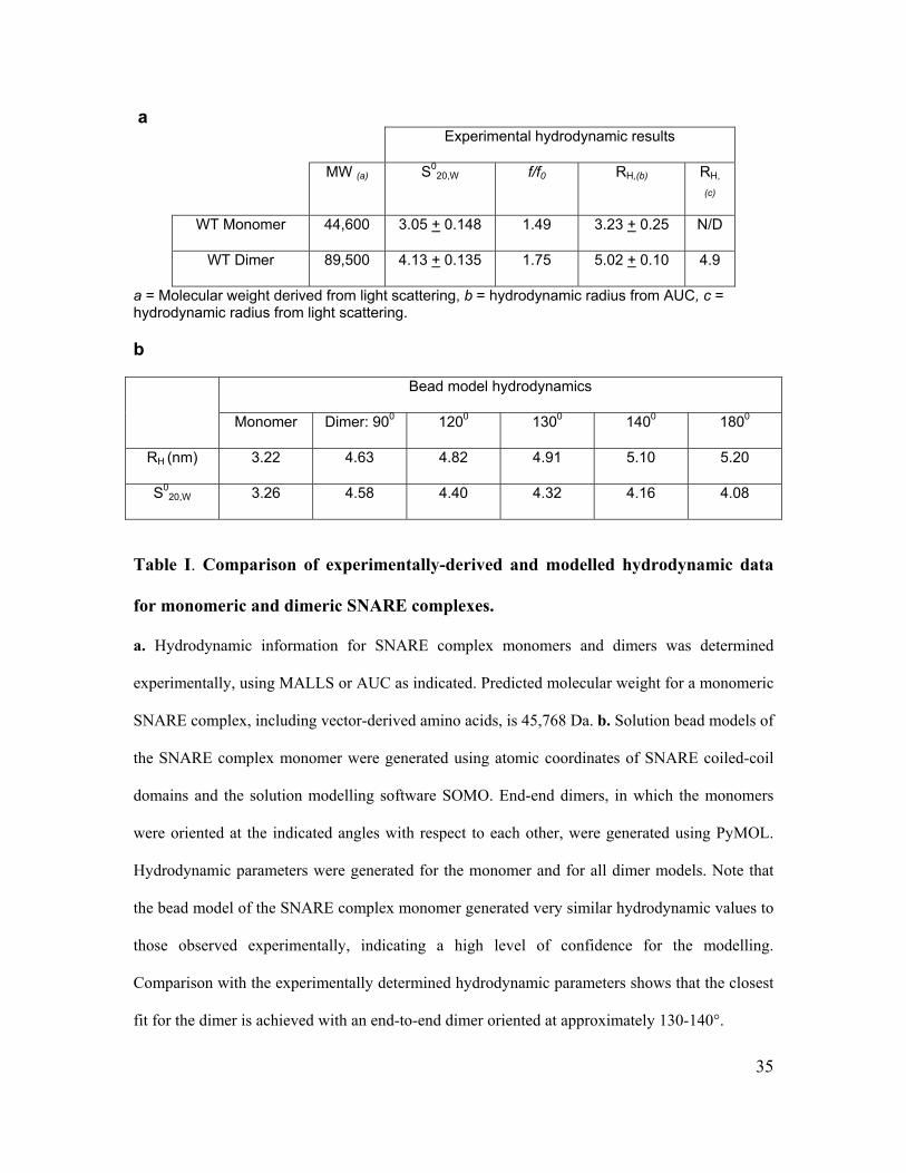
a Experimental hydrodynamic results
MW (a) S020,W f/f0 RH,(b) RH,
(c)
WT Monomer 44,600 3.05 + 0.148 1.49 3.23 + 0.25 N/D
WT Dimer 89,500 4.13 + 0.135 1.75 5.02 + 0.10 4.9
a = Molecular weight derived from light scattering, b = hydrodynamic radius from AUC, c = hydrodynamic radius from light scattering.
b
Bead model hydrodynamics
Monomer Dimer: 900 1200 1300 1400 1800
RH (nm) 3.22 4.63 4.82 4.91 5.10 5.20
S020,W 3.26 4.58 4.40 4.32 4.16 4.08
Table I. Comparison of experimentally-derived and modelled hydrodynamic data
for monomeric and dimeric SNARE complexes.
a. Hydrodynamic information for SNARE complex monomers and dimers was determined
experimentally, using MALLS or AUC as indicated. Predicted molecular weight for a monomeric
SNARE complex, including vector-derived amino acids, is 45,768 Da. b. Solution bead models of
the SNARE complex monomer were generated using atomic coordinates of SNARE coiled-coil
domains and the solution modelling software SOMO. End-end dimers, in which the monomers
were oriented at the indicated angles with respect to each other, were generated using PyMOL.
Hydrodynamic parameters were generated for the monomer and for all dimer models. Note that
the bead model of the SNARE complex monomer generated very similar hydrodynamic values to
those observed experimentally, indicating a high level of confidence for the modelling.
Comparison with the experimentally determined hydrodynamic parameters shows that the closest
fit for the dimer is achieved with an end-to-end dimer oriented at approximately 130-140°.
36
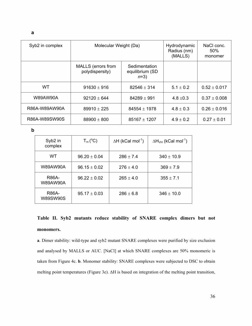
a
Syb2 in complex Molecular Weight (Da) Hydrodynamic Radius (nm)
(MALLS)
NaCl conc. 50%
monomer
MALLS (errors from polydispersity)
Sedimentation equilibrium (SD
n=3)
WT 91630 ± 916 82546 ± 314 5.1 ± 0.2 0.52 ± 0.017
W89AW90A 92120 ± 644 84289 ± 991 4.8 ±0.3 0.37 ± 0.008
R86A-W89AW90A 89910 ± 225 84554 ± 1978 4.8 ± 0.3 0.26 ± 0.016
R86A-W89SW90S 88900 ± 800 85167 ± 1207 4.9 ± 0.2 0.27 ± 0.01
b
Syb2 in complex
Tm (oC) ΔH (kCal mol-1) ΔHVH (kCal mol-1)
WT 96.20 ± 0.04 286 ± 7.4 340 ± 10.9
W89AW90A 96.15 ± 0.02 276 ± 4.0 369 ± 7.9
R86A-W89AW90A
96.22 ± 0.02 265 ± 4.0 355 ± 7.1
R86A-W89SW90S
95.17 ± 0.03 286 ± 6.8 346 ± 10.0
Table II. Syb2 mutants reduce stability of SNARE complex dimers but not
monomers.
a. Dimer stability: wild-type and syb2 mutant SNARE complexes were purified by size exclusion
and analysed by MALLS or AUC. [NaCl] at which SNARE complexes are 50% monomeric is
taken from Figure 4c. b. Monomer stability: SNARE complexes were subjected to DSC to obtain
melting point temperatures (Figure 3e). ΔH is based on integration of the melting point transition,

37
whilst ΔHVH is based on peak width and is independent of protein concentration.
38
Figure Legends
Figure 1. Neuronal SNARE complexes form a wing-shaped, end-to-end dimer.
Purified SNARE complex dimers were analysed by: (a) sedimentation in 50 mM (dashed
line) or 1M (solid line) NaCl; (b) SAXS, with shapes simulated ab initio and the “most
probable” shape represented as an arrays of beads; (c) TEM, with the ten most common
shape classes shown above and an average refined structure below; (d) aligning SNARE
complex monomer bead models (left) to generate a shape matching the observed
hydrodynamic properties (right).
Figure 2. The C-termini of both SNARE complex monomers are adjacent to each other
in the dimer.
(a) Schematic diagram of C-terminally tagged FRET syb2 proteins, uncomplexed or
incorporated into SNARE complex dimers. (b) Emission spectra of purified dimers
tagged at the C-terminus with Venus as FRET acceptor, and either at the C-terminus
(black) or N-terminus (grey) with CFP as FRET donor. Spectra are shown in the absence
(thick lines) or presence (thin lines) of 1 M NaCl, and were normalized to their values at
481 nm (CFP emission peak). (c) Left: FRET ratios (emission ratio 527/481 nm) from
experiments depicted in (b), where -/+ refers to the absence or presence of 1M NaCl.
Right: The ratio of FRET ratios (-NaCl/+NaCl) obtained with SNARE complex dimers
tagged at both C-termini (CC) or at the C-terminus and N-terminus (NC), or obtained
with tagged syb2 proteins only (coils). Values are mean ± S.E.M (n=5).
39
Figure 3. Three residues from syb2 form part of the dimer interface.
(a) Sequence of membrane-proximal region of syb2, with amino acids important for
SNARE complex dimer stability colored and the position of hydrophobic SNARE motif
layers indicated above. (b) Peak intrinsic fluorescence of SNARE complex dimers at
increasing [NaCl] (inset; full emission spectra). Fluorescence intensity of free tryptophan
remains unchanged with increasing [NaCl] (not shown). (c) Dimers containing wild-type
(black); W89A,W90A (blue); R86A (green); R86A,W89A,W90A (red);
R86A,W89S,W90S (magenta) syb2 were measured for their sensitivity to [NaCl] using
sedimentation equilibrium. (d) α-helical content using CD spectroscopy
(R86A,W89S,W90S mutant not determined). (e) SNARE complex melting temperature
using differential scanning calorimetry.
Figure 4. Relative contribution of individual tryptophan residues within syb2 to dimer
interface.
(a) Dimers containing wild-type (black); W89A (blue) and W90A (red) syb2 were
measured for their sensitivity to [NaCl] using sedimentation equilibrium. (b) Peak
intrinsic fluorescence of SNARE complex dimers containing wild-type (black), W89A
(blue) and W90A (red) syb2 at increasing [NaCl]. Fluorescence intensity is expressed
relative to the emission maximum of each protein complex in 50 mM NaCl.
Figure 5. Toxin-insensitive, mutant syb2 is unable to rescue secretion from PC12 cells.
(a) PC12 cells were permeabilized with 10 μM digitonin and incubated in the presence or
absence of botulinum neurotoxin B light-chain (BotB/LC), followed by detection of
40
intact syb2 coil by Western blotting. Blots were reprobed for synaptophysin (physin) to
determine equal amounts of protein loading. (b) PC12 cells, transfected with a plasmid
encoding for hGH as well as wildtype or botulinum toxin-resistant syb2 (Q76V,F77W)
were permeabilized with 10 μM digitonin and incubated in the presence or absence of
BotB/LC. Ca2+-dependent hGH release was evoked by 10 μM Ca2+ for 10 min, and
compared to basal release (0 μM Ca2+). The amount of hGH in the medium and in the
cells was determined by an enzyme-linked immunosorbent assay, and the percentage of
secreted hGH, and the total amount of hGH were calculated against an hGH standard
curve. The graph is a representative of two independent experiments with duplicate data
points. Error bars are only shown if larger than bar columns. (c) Cells were transfected
with wildtype or toxin-resistant wildtype syb2 (wt-r), or toxin-resistant mutant syb2 as
indicated, and treated with BotB/HnLC upon permeabilization to compare expression
levels. (d) To standardize results from repeated experiments, secretion observed in the
presence of toxin-insensitive, wildtype syb2 was set to 100%, and the relative lack of
rescue of secretion of test plasmids in the presence of toxin normalized to this control.
Values are means + S.E.M. (n=3-5). The statistical significance of differences from
wildtype were analysed by a Student’s t-test (*, p < 0.05; **, p < 0.01).
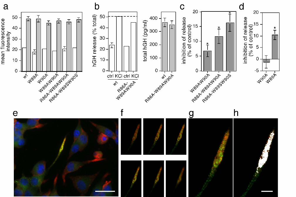
Figure 6. Syb2 mutants display dominant-negative secretory effects. (a) Expression of
syb2, or syb2 mutants analyzed, relative to endogenous levels. (b) Example of hGH
secretion experiment in cells transfected with wildtype or R86A-W89AW90A mutant
syb2. Cells were stimulated for 5 min with physiological saline (ctrl) or high K+ (KCl).
The amount of hGH in the medium and in the cells was determined by an enzyme-linked
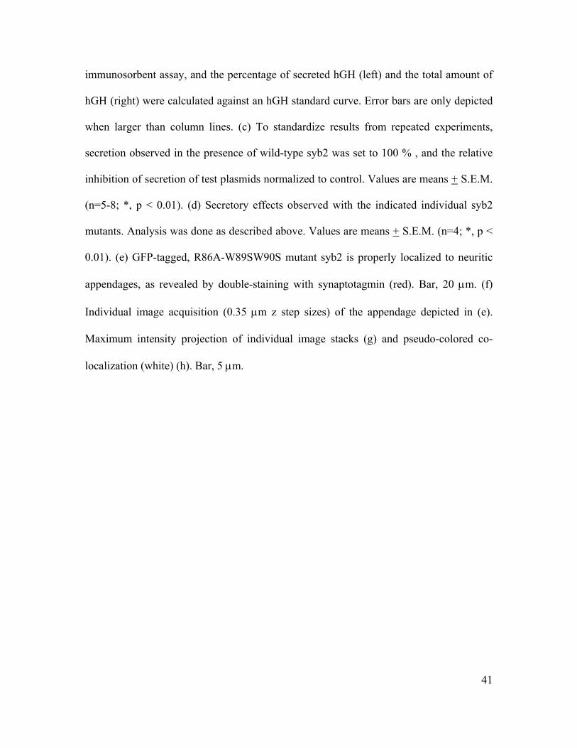
41
immunosorbent assay, and the percentage of secreted hGH (left) and the total amount of
hGH (right) were calculated against an hGH standard curve. Error bars are only depicted
when larger than column lines. (c) To standardize results from repeated experiments,
secretion observed in the presence of wild-type syb2 was set to 100 % , and the relative
inhibition of secretion of test plasmids normalized to control. Values are means + S.E.M.
(n=5-8; *, p < 0.01). (d) Secretory effects observed with the indicated individual syb2
mutants. Analysis was done as described above. Values are means + S.E.M. (n=4; *, p <
0.01). (e) GFP-tagged, R86A-W89SW90S mutant syb2 is properly localized to neuritic
appendages, as revealed by double-staining with synaptotagmin (red). Bar, 20 μm. (f)
Individual image acquisition (0.35 μm z step sizes) of the appendage depicted in (e).
Maximum intensity projection of individual image stacks (g) and pseudo-colored co-
localization (white) (h). Bar, 5 μm.