a putative sugar‐binding transcriptional regulator in a novel gene locus in enterococcus faecalis...
TRANSCRIPT

420 • JID 2004:189 (1 February) • Hufnagel et al.
M A J O R A R T I C L E
A Putative Sugar-Binding Transcriptional Regulatorin a Novel Gene Locus in Enterococcus faecalisContributes to Production of Biofilm and ProlongedBacteremia in Mice
Markus Hufnagel,1,3,a Stefanie Koch,1,a Roberta Creti,2 Lucilla Baldassarri,2 and Johannes Huebner1
1Channing Laboratory, Brigham and Women’s Hospital, Harvard Medical School, Boston, Massachusetts; 2Istituto Superiore di Sanita, Rome, Italy;3University Children’s Hospital Kiel, Kiel, Germany
A biofilm-negative transposon mutant was created from an Enterococcus faecalis strain that produces a lot ofbiofilm. The transposon had been inserted in the second gene of a locus consisting of 4 open-reading frames,designated bop (biofilm on plastic surfaces). A nonpolar deletion of this gene and of parts of the 2 flankinggenes was created; production of biofilm by this deletion mutant was significantly enhanced, compared withthat by the wild-type strain. Expression of a downstream gene was significantly lower in the transposon mutantthan in the wild-type strain and the biofilm-enhanced deletion mutant. Transformation of this gene into thetransposon mutant partially restored production of biofilm. Mice challenged by intravenous injection withthe biofilm-negative mutant strain showed significantly reduced numbers of colony-forming units in the blood,compared with mice challenged with the biofilm-enhanced deletion mutant and the wild-type. These resultsindicate that bop is involved in production of biofilm and probably regulates expression of biofilm in the E.faecalis strain tested.
Enterococci are one of the leading causes of infections
in hospitalized patients and the third-most-common
cause of nosocomial bloodstream infections [1]. Be-
cause of their intrinsic resistance to many clinically
available antibiotics, these pathogens are associated
with significant morbidity and mortality, especially
among immunocompromised patients [2]. Production
of biofilm is recognized as a virulence factor in many
Received 5 March 2003; accepted 22 July 2003; electronically published 20 January2004.
Presented in part: 103rd general meeting of the American Society for Microbiology,Washington, DC, 18–23 May 2003 (abstract B-077).
Financial support: National Institute of Allergy and Infectious Diseases, NationalInstitutes of Health (grant AI50667 to J.H.); Walter Marget Foundation (support toM.H.).
a M.H. and S.K. contributed equally to this work.Reprints or correspondence: Dr. Johannes Huebner, Channing Laboratory, Brig-
ham and Women’s Hospital, 181 Longwood Ave., Boston, MA 02115 ([email protected]).
The Journal of Infectious Diseases 2004; 189:420–30� 2004 by the Infectious Diseases Society of America. All rights reserved.0022-1899/2004/18903-0009$15.00
pathogens [3], and several authors have described the
ability of enterococci to produce biofilm [4–8].
Native-valve endocarditis is a well documented bio-
film process, and biofilms formed on heart valves are
described by the medical community as vegetations [9].
Biofilms also play important roles in enterococcal in-
fections of dental root canals [10], in the obstruction
or blocking of urethral catheters and ureteral stents [11,
12], and in ocular infections [13].
Relatively little is known about the molecular mech-
anisms that control production and maintenance of
biofilm in enterococci [3]. Baldassarri et al. [5] noted
that production of biofilm by enterococci is influenced
by either the presence of additional carbohydrates or
the depletion of iron in growth media. Toledo-Arana
et al. [4] have shown that the gene encoding entero-
coccal surface protein (Esp), esp, is involved in the pri-
mary attachment of enterococci to abiotic surfaces.
However, their finding that the inactivation of esp has
no effect on the production of biofilm by the E. faecalis
strain analyzed that produced the most biofilm indi-

Biofilm in E. faecalis • JID 2004:189 (1 February) • 421
Table 1. Enterococcus faecalis strains and plasmids.
Strain or plasmid Characterization Reference
Strain
E. faecalis type 9 Strong biofilm-producing strain [17]
E. faecalis 12030 Strong biofilm-producing strain [29]
E. faecalis 10D5 Biofilm-negative transposonmutant of E. faecalis type 9
This study
E. faecalis 10D5-pEU327 E. faecalis 10D5 containinggram-positive expressionvector pEU327 without insert
This study
E. faecalis 10D5-pSBR E. faecalis 10D5 containingpEU327 with insertion ofbopD
This study
E. faecalis T9-TDM E. faecalis type 9 with deletionof part of bopA, completedeletion of bopB, and partialdeletion of bopC
This study
Escherichia coli DH5a
and TOP 10Gram-negative hosts for cloning
Staphylococcus aureusATCC 35556
Strong biofilm-producing S.aureus strain
[28]
Plasmid
pTV1-OK Transposon-mutagenesis vectorcontaining Tn917
[16]
pTEX4577 Suicide vector for targetedmutagenesis
[20]
pEU327 Gram-positive expression vector [22]
pCRII-TOPO Cloning vector Invitrogen
pTDM pTEX4577 with insert carryingdeletion in 3 ORFs
This study
pSBR pEU327 with insertion of thebopD gene
This study
NOTE. ORFs, open-reading frames.
cates that additional mechanisms might be involved. The pre-
sent study was performed to gain a better understanding of the
molecular mechanisms involved in the establishment of enter-
ococcal biofilms and to clarify their role in pathogenicity.
MATERIALS AND METHODS
Bacterial strains, plasmids, and media. The bacterial strains
and plasmids used in the present study are shown in table 1.
Enterococci were grown without agitation at 37�C in Todd-
Hewitt broth (THB; Becton Dickinson) or tryptic soy broth
(TSB; Becton Dickinson), with the addition of 1% glucose as
indicated (THBG or TSBG, respectively). Escherichia coli strains
DH5a and TOP 10 were cultured aerobically in Luria-Bertani
broth on a rotor rack at 37�C. Erythromycin (10 mg/mL) was
added for E. faecalis, kanamycin was added for enterococci
(2000 mg/mL) and E. coli (50 mg/mL), and spectinomycin was
added for enterococci (500 mg/mL) and E. coli (250 mg/mL)
(all from Sigma Chemical).
Biofilm plate assay. Enterococci were tested for produc-
tion of biofilm according to the protocol described by Baldas-
sarri et al. [5]. In brief, bacteria were grown in TSBG overnight
at 37�C. Polystyrene 96-well tissue-culture plates (Costar) were
filled with 180 mL of fresh TSBG, and 20 mL of the culture
grown overnight was added to each well. The plates were in-
cubated for either 8 or 18 h at 37�C and read in an ELISA
reader (BIO-TEK Instruments) at an optical density at 595 nm,
to assure homogeneous growth in all wells. The culture medium
was discarded, and the wells were washed carefully 3 times with
200 mL of PBS without disturbing the biofilm on the bottom
of the wells. The plates were dried for 1 h at 60�C and were
stained with 2% Hucker’s crystal violet for 2 min. Excess stain
was removed by rinsing the plates under tap water, and plates
were dried for 10 min at 60�C. The optical density at 595 nm
was determined in an ELISA reader. Each assay was performed
at least in triplicate and repeated at least twice. The optical
density values were analyzed by analysis of variance (ANOVA)
with Newman-Keuls multiple comparisons test. Bartlett’s test
for equal variances was done to ensure that variances between
groups did not differ significantly (PRISM software, version 3;
GraphPad).
DNA manipulations. Chromosomal DNA from entero-
cocci was prepared by use of the Qiagen DNeasy Tissue Kit,
according to the manufacturer’s instructions. Plasmid DNA was
prepared from enterococci and E. coli by use of the Wizard
Plus Mini or Midipreps kit (Promega). DNA was purified from
agarose gels and from polymerase chain reactions (PCRs) by
use of the QIAquick Gel Extraction Kit or the PCR Purification
Kit (Qiagen), according to the manufacturer’s instructions.
Restriction enzymes and modifying enzymes were obtained
from Invitrogen or New England Biolabs. Custom primers were
ordered from Invitrogen. Electrocompetent enterococci were
prepared according to the method of Fiedler and Wirth [14],
and electroporation was performed in a Bio-Rad Gene Pulser
II by use of the parameters given by Fiedler and Wirth [14].
Southern hybridization was performed by use of the ECL direct
nucleic acid labeling and detection system (Amersham). All
other methods (DNA ligations, electrophoresis, and transfor-
mation of competent E. coli) were performed by use of standard
techniques [15].
Transposon mutagenesis. Plasmid pTV1-OK carrying
transposon Tn917 was used for transposon mutagenesis [16].
The plasmid was electroporated into E. faecalis type 9 [17], and
the bacteria were grown with kanamycin and erythromycin at
30�C to maintain the plasmid. After a temperature shift to the
nonpermissive temperature (42�C), the plasmid was cured, and
the transposon was forced to integrate into the chromosomal
DNA. Bacteria were replica-plated onto erythromycin-contain-
ing Todd-Hewitt agar (THA) plates with or without kanamycin.
422 • JID 2004:189 (1 February) • Hufnagel et al.
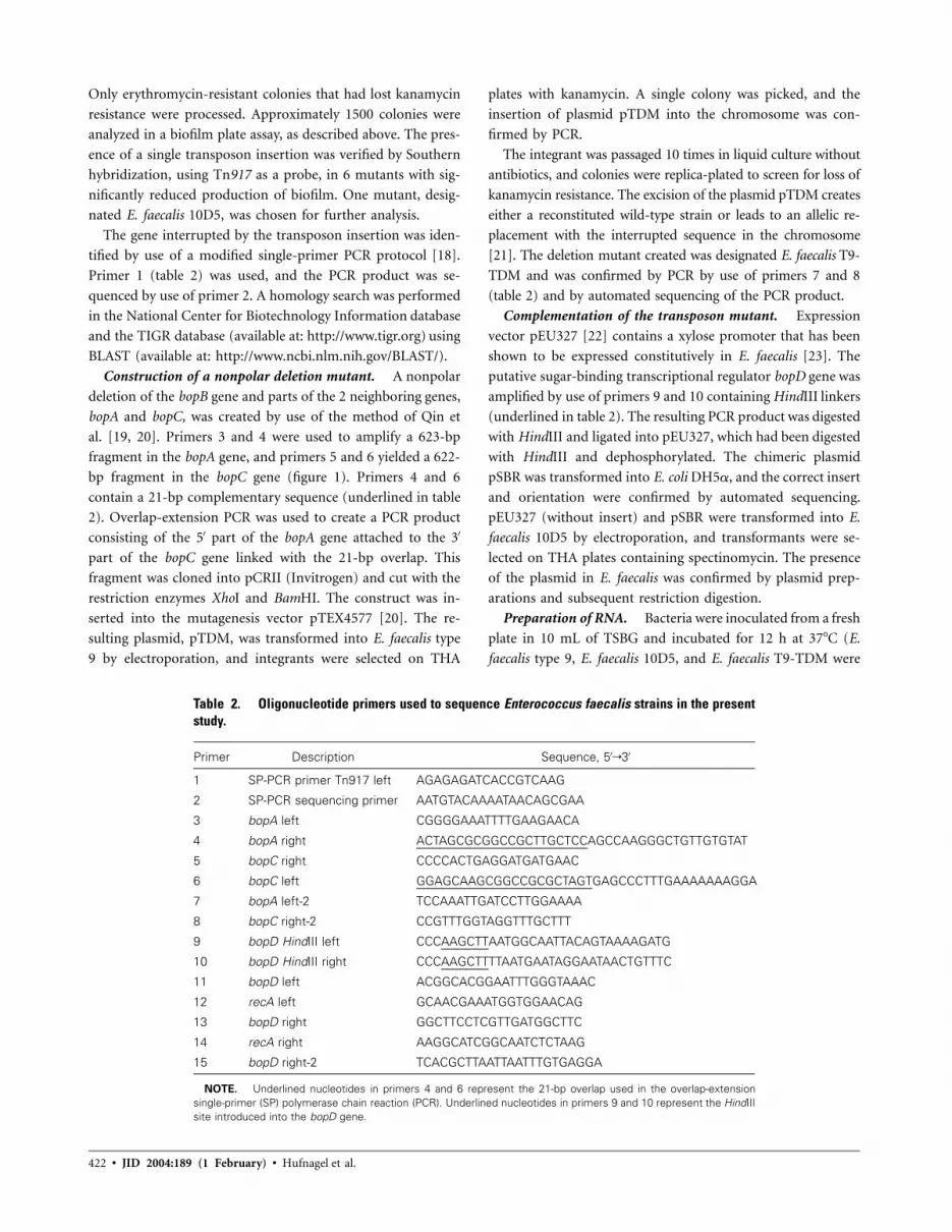
Table 2. Oligonucleotide primers used to sequence Enterococcus faecalis strains in the presentstudy.
Primer Description Sequence, 5′r3′
1 SP-PCR primer Tn917 left AGAGAGATCACCGTCAAG
2 SP-PCR sequencing primer AATGTACAAAATAACAGCGAA
3 bopA left CGGGGAAATTTTGAAGAACA
4 bopA right ACTAGCGCGGCCGCTTGCTCCAGCCAAGGGCTGTTGTGTAT
5 bopC right CCCCACTGAGGATGATGAAC
6 bopC left GGAGCAAGCGGCCGCGCTAGTGAGCCCTTTGAAAAAAAGGA
7 bopA left-2 TCCAAATTGATCCTTGGAAAA
8 bopC right-2 CCGTTTGGTAGGTTTGCTTT
9 bopD HindIII left CCCAAGCTTAATGGCAATTACAGTAAAAGATG
10 bopD HindIII right CCCAAGCTTTTAATGAATAGGAATAACTGTTTC
11 bopD left ACGGCACGGAATTTGGGTAAAC
12 recA left GCAACGAAATGGTGGAACAG
13 bopD right GGCTTCCTCGTTGATGGCTTC
14 recA right AAGGCATCGGCAATCTCTAAG
15 bopD right-2 TCACGCTTAATTAATTTGTGAGGA
NOTE. Underlined nucleotides in primers 4 and 6 represent the 21-bp overlap used in the overlap-extensionsingle-primer (SP) polymerase chain reaction (PCR). Underlined nucleotides in primers 9 and 10 represent the HindIIIsite introduced into the bopD gene.
Only erythromycin-resistant colonies that had lost kanamycin
resistance were processed. Approximately 1500 colonies were
analyzed in a biofilm plate assay, as described above. The pres-
ence of a single transposon insertion was verified by Southern
hybridization, using Tn917 as a probe, in 6 mutants with sig-
nificantly reduced production of biofilm. One mutant, desig-
nated E. faecalis 10D5, was chosen for further analysis.
The gene interrupted by the transposon insertion was iden-
tified by use of a modified single-primer PCR protocol [18].
Primer 1 (table 2) was used, and the PCR product was se-
quenced by use of primer 2. A homology search was performed
in the National Center for Biotechnology Information database
and the TIGR database (available at: http://www.tigr.org) using
BLAST (available at: http://www.ncbi.nlm.nih.gov/BLAST/).
Construction of a nonpolar deletion mutant. A nonpolar
deletion of the bopB gene and parts of the 2 neighboring genes,
bopA and bopC, was created by use of the method of Qin et
al. [19, 20]. Primers 3 and 4 were used to amplify a 623-bp
fragment in the bopA gene, and primers 5 and 6 yielded a 622-
bp fragment in the bopC gene (figure 1). Primers 4 and 6
contain a 21-bp complementary sequence (underlined in table
2). Overlap-extension PCR was used to create a PCR product
consisting of the 5′ part of the bopA gene attached to the 3′
part of the bopC gene linked with the 21-bp overlap. This
fragment was cloned into pCRII (Invitrogen) and cut with the
restriction enzymes XhoI and BamHI. The construct was in-
serted into the mutagenesis vector pTEX4577 [20]. The re-
sulting plasmid, pTDM, was transformed into E. faecalis type
9 by electroporation, and integrants were selected on THA
plates with kanamycin. A single colony was picked, and the
insertion of plasmid pTDM into the chromosome was con-
firmed by PCR.
The integrant was passaged 10 times in liquid culture without
antibiotics, and colonies were replica-plated to screen for loss of
kanamycin resistance. The excision of the plasmid pTDM creates
either a reconstituted wild-type strain or leads to an allelic re-
placement with the interrupted sequence in the chromosome
[21]. The deletion mutant created was designated E. faecalis T9-
TDM and was confirmed by PCR by use of primers 7 and 8
(table 2) and by automated sequencing of the PCR product.
Complementation of the transposon mutant. Expression
vector pEU327 [22] contains a xylose promoter that has been
shown to be expressed constitutively in E. faecalis [23]. The
putative sugar-binding transcriptional regulator bopD gene was
amplified by use of primers 9 and 10 containing HindIII linkers
(underlined in table 2). The resulting PCR product was digested
with HindIII and ligated into pEU327, which had been digested
with HindIII and dephosphorylated. The chimeric plasmid
pSBR was transformed into E. coli DH5a, and the correct insert
and orientation were confirmed by automated sequencing.
pEU327 (without insert) and pSBR were transformed into E.
faecalis 10D5 by electroporation, and transformants were se-
lected on THA plates containing spectinomycin. The presence
of the plasmid in E. faecalis was confirmed by plasmid prep-
arations and subsequent restriction digestion.
Preparation of RNA. Bacteria were inoculated from a fresh
plate in 10 mL of TSBG and incubated for 12 h at 37�C (E.
faecalis type 9, E. faecalis 10D5, and E. faecalis T9-TDM were

Biofilm in E. faecalis • JID 2004:189 (1 February) • 423
Figure 1. Schematic representation of the gene locus in Enterococcus faecalis involved in production of biofilm. All 4 genes are expressed on a singlemRNA transcript. The insertion of the transposon in E. faecalis 10D5 is indicated by a triangle, and the gene deletion of E. faecalis T9-TDM is shown bythe bracket. Arrows, primers; nos. correspond to the nos. used in table 2. Primers 1, 2, 12, and 14 are not shown.
grown in TSBG without antibiotics; E. faecalis 10D5-pEU and
E. faecalis 10D5-pSBR were grown with 500 mg/mL spectino-
mycin). A volume of 400 mL of this culture was used to in-
oculate 20 mL of TSBG with or without the above-mentioned
antibiotics and was incubated for 3 h at 37�C without aeration.
Cells ( ; calculated after measurement of optical density95 � 10
at 650 nm) were mixed with a 2� volume of RNAprotect
Bacteria reagent (Qiagen), and RNA was isolated by use of the
RNeasy Midi Kit (Qiagen), according to the manufacturer’s
recommendations, which included digestion with DNase I on
the spin column. The concentration of RNA was calculated after
measurement of optical density at 260 nm, and equal amounts
were used as starting material in subsequent experiments. To
remove any remaining DNA contaminations, a second DNase I
treatment (Invitrogen) of each RNA sample was performed ac-
cording to the manufacturer’s instructions. The enzyme was in-
activated at 65�C in the presence of EDTA. To verify the absence
of residual genomic DNA in each sample, the RNA was amplified
in a PCR with Taq DNA polymerase and the respective primers
in the presence and absence of SuperScript II Reverse Transciptase
(Invitrogen). RNA samples were used for cDNA synthesis by use
of the iScriptcDNA synthesis kit (Biorad), according to the man-
ufacturer’s recommendations.
Quantitative real-time PCR. Quantitative real-time PCR
was performed in an iCycler iQ (Bio-Rad) by use of the
QuantiTect SYBR green PCR kit (Qiagen), according to the
manufacturer’s recommendations. The bopD transcripts were
amplified with the primers 11 and 13 (table 2). The consti-
tutively expressed housekeeping gene recA was amplified with
the primers 12 and 14 (table 2) as an internal control to nor-
malize RNA concentrations. To monitor the specificity of the
reactions, final PCR products were analyzed by melting curves
showing that all specific primers resulted in only 1 specific
product. The standard curves of dilutions of starting material,
amplification efficiencies, and expression ratios between bopD
and recA were calculated by use of Microsoft Excel. The values
used for comparison of gene expression between the different
strains were the numbers of PCR cycles required to reach the
threshold cycle (Ct), which was set at 10� the SD of the fluo-
rescence of the first 10 cycles. The amount of bopD transcript
relative to the control gene recA was calculated by use of the
standard-curve method, as described elsewhere [24, 25], and
was expressed as the percentage of the amount of transcripts
of the wild-type E. faecalis type 9.
Reverse-transcription (RT) PCR. RT-PCR was performed
with the RNA samples, to detect cotranscription of all 4 bop
genes by use of the SuperScript One-Step RT-PCR with Plat-
inum Taq Kit (Invitrogen), according to the manufacturer’s
recommendations, by use of the primers 7 and 15 (table 2).
Scanning electron microscopy. Bacteria were grown in TSB
or TSBG in 24-well plates containing polystyrene segments.
After a 12-h incubation at 37�C, polystyrene segments were
rinsed twice in PBS and once in 0.1 mol/L cacodylate buffer
and fixed as described elsewhere [5], to preserve extracellular
polysaccharide. In brief, cells were first fixed for 20 min at
room temperature with 0.1 mol/L cacodylate-buffered 2.5%
glutaraldehyde containing 0.075% (wt/vol) ruthenium red
(Glut-RR; Merck) and 75 mmol/L lysine. They were then fixed
with Glut-RR without lysine for 2 h and finally fixed with 1%
OsO4 plus ruthenium red for an additional 1 h. Samples were

424 • JID 2004:189 (1 February) • Hufnagel et al.
dehydrated by exposure to a graded series of ethanol, critical-
point dried, gold sputtered, and examined with a Cambridge
SE360 scanning electron microscope.
Catheter adherence assay. The ability of bacteria to adhere
to polyethylene catheters was tested by use of the method de-
scribed by Muller et al. [26] with modifications. In brief, bac-
teria were inoculated in THB and incubated at 37�C until an
OD650 of 0.4 was reached and diluted to ∼ cfu/mL. Sec-74 � 10
tions of polyethylene catheter tubing (Intramedic PE tubing,
diameter 0.61 mm; Becton Dickinson) were gas sterilized. Sec-
tions of 20 mm were dipped into the broth with the bacterial
inoculum for 30 min at 37�C. The catheter pieces were sub-
sequently washed 3 times for 15 seconds in PBS and vigorously
rolled over tryptic soy agar (TSA) plates. The plates were in-
cubated at 37�C overnight, and colonies were counted.
Mouse sepsis model. The animal studies described in the
present study were reviewed and approved by the Institutional
Animal Care and Use Committee at Harvard University. Fe-
male BALB/c mice were inoculated intravenously in the tail
vein with cfu of either wild-type E. faecalis type 9, bio-85 � 10
film-negative transposon mutant E. faecalis 10D5, or the bio-
film-enhanced triple-gene deletion mutant E. faecalis T9-TDM.
Bacteria were grown in THB overnight, centrifuged, and re-
suspended in sterile saline. Aliquots were shock-frozen and
stored at �80�C. The concentration of the stock was verified
by dilutions and viable counts on TSA, and these numbers were
used to calculate the appropriate dilutions for the desired in-
oculum. For the experiments, aliquots were thawed, diluted in
sterile saline, and injected into the tail vein; the actual diluted
inoculum was once more verified by viable counts. After 3 days,
the mice were killed, blood was obtained by cardiac puncture,
and 100 mL was plated on TSA.
Statistical analysis. Statistical analysis was done by
ANOVA with Newman-Keuls multiple comparison tests, and
Bartlett’s test for equal variances was used to assure that var-
iances did not differ significantly between groups (PRISM, ver-
sion 3; GraphPad Software).
RESULTS
Clinical E. faecalis strains and laboratory isolates were tested
for production of biofilm [27]. One strain, E. faecalis type 9
[17], showed a very high degree of production of biofilm (OD595
of 12.5) after an 18-h incubation period. This value corre-
sponded to the results obtained with a strain of Staphylococcus
aureus (ATCC 35556) that produces a lot of biofilm [28].
Transposon mutagenesis of E. faecalis type 9 was performed
by use of plasmid pTV1-OK [16]. After 3 subpassages at the
nonpermissive temperature, ∼1500 erythromycin-resistant col-
onies were picked with sterile toothpicks and transferred to 96-
well tissue-culture plates. A biofilm plate assay was performed,
and, after 18 h, 1 strain (E. faecalis 10D5) showed a level of
production of biofilm that was 16% of that of the wild-type
strain. This strain was selected for further studies. The presence
of a single insertion of the transposon was confirmed by South-
ern blot, with Tn917 used as a probe (data not shown). A
comparison of the growth curves over a 7-h period until the
stationary phase showed no difference between the wild-type
and mutant 10D5 strains (data not shown). Scanning electron
microscopy of E. faecalis type 9 and E. faecalis 10D5 showed
that the wild-type strain grows in multiple layers on polysty-
rene, whereas the mutant strain grows in only 1 plane, without
the piling up observed with the wild-type strain (figure 2).
The DNA sequence interrupted by the transposon was am-
plified and sequenced according to a modified single-primer
PCR protocol [18]. The transposon insertion was mapped to
an open-reading frame (ORF) by a homology search within the
TIGR database by use of the sequence data for E. faecalis V583.
The function putatively assigned to this gene by TIGR is a b-
phosphoglucomutase. This ORF was designated as bopB. It is
surrounded upstream by a putative glycosyltransferase (bopA)
and downstream by a putative aldose-1-epimerase (bopC) and
a sugar-binding transcriptional regulator (bopD). All 4 ORFs
are predicted to be transcribed in the same direction, and some
of the genes overlap (figure 1). The presence of all 4 ORFs on
a single mRNA transcript in E. faecalis type 9 wild-type strain
was confirmed by RT-PCR with primers 7 and 15 (figure 1).
A putative r-independent transcriptional terminator is located
downstream from bopD, and the next 6 ORFs are predicted to
be transcribed in the opposite direction.
A nonpolar deletion mutant was constructed by removing
part of the 3′ end of the bopA, all of bopB, and part of the 5′
end of bopC (figure 1). This deletion mutant was created by
targeted mutagenesis with the suicide plasmid pTEX4577. The
deletion was verified by performing PCR of the region with
primers outside of the overlap-extension construct. Sequencing
of the respective loci was performed to confirm the deletion.
Production of biofilm of the deletion mutant E. faecalis T9-
TDM was compared with that of the wild-type strain (figure
3), showing significantly enhanced production of biofilm
(122% that of the wild-type strain; ). For this experi-P ! .05
ment, a shorter incubation period (8 h) was used, because 18-
h readings for the deletion mutant exceeded the maximum
values obtained in the photometer.
Insertional inactivation of the bopD gene was unsuccessful
in E. faecalis type 9 and in a different biofilm-producing E.
faecalis strain, E. faecalis 12030 [29], which possesses the bopD
gene, suggesting that the bopD gene is essential for production
of biofilm under the growth conditions used. Using the same
conditions, we were able to create deletion mutants in 2 dif-
ferent genes.
To test the hypothesis that the biofilm-negative phenotype

Figure 2. Scanning electron micrograph of wild-type Enterococcus faecalis type 9 and the mutant strain E. faecalis 10D5. A, The wild-type strain showsa multilayered growth pattern, whereas the biofilm-negative mutant grows mostly in a single layer (B).

426 • JID 2004:189 (1 February) • Hufnagel et al.
Figure 3. Comparison of the biofilm production in wild-type Enterococcusfaecalis (EFS) type 9, biofilm-negative transposon mutant EFS 10D5, trans-poson mutant reconstituted with plasmid pEU327 without insert, transposonmutant reconstituted with the bopD gene, and the biofilm-enhanced triple-deletion mutations EFS T9-TDM. The biofilm-negative transposon mutant10D5 produces significantly less biofilm than does the wild-type strain andthe biofilm-enhanced mutant EFS T9-TDM ( ). Plasmid pEU327 doesP ! .001not affect the production of biofilm by the mutant. However, expressionof the bopD gene by reconstitution of the gene in trans restores productionof biofilm in the transposon mutant strain to 65% of the wild-type levels( , EFS 10D5 vs. EFS 10D5-pSBR). The triple-gene deletion mutantP ! .05EFS T9-TDM produces significantly more biofilm (122%) than does thewild-type EFS type 9 ( ). NS, not significant.P ! .05
Table 3. Threshold cycles (Ct) for Entero-coccus faecalis bopD transcripts, comparingthe wild-type strain, the biofilm-negativetransposon mutant, the biofilm-enhanced de-letion mutant, and the reconstituted strain.
E. faecalis strain Ct, mean (95% CI)
Type 9 15.93 (15.68–16.18)
10D5 19.48 (19.33–19.62)
T9-TDM 15.09 (15.02–15.16)
10D5-pEU327 18.6 (18.27–18.29)
10D5-pSBR 14.28 (13.68–14.88)
NOTE. CI, confidence interval.
of the transposon mutant 10D5 was due to a polar effect of
the transposon insertion on a gene downstream of the bopB
gene, we cloned the bopD gene into the gram-positive expres-
sion vector pEU327 [22]. The construct pSBR was electropo-
rated into the transposon mutant E. faecalis 10D5. The vector
pEU327 without insert was used as a control. After 8 h, the
amount of biofilm produced by E. faecalis 10D5 with pEU327
but without the bopD gene was identical to that produced by
the transposon mutant. After an 8-h incubation period, E. fae-
calis 10D5 displayed 36.8% of the biofilm produced by the
wild-type E. faecalis type 9, compared with 16% after a 18-h
incubation period. However, E. faecalis 10D5 with the substi-
tution of the bopD gene produced 176% of that produced by
the biofilm-negative transposon mutant E. faecalis 10D5 (P !
) and 65% of that produced by the wild-type strain E. faecalis.05
type 9 (figure 3). Overexpression of the other ORFs (i.e., bopA–
bopC) did not result in increased production of biofilm, com-
pared with the transposon mutant carrying the plasmid without
insert (data not shown). These results indicate that the insertion
of this gene leads to at least a partial reconstitution of the
biofilm-producing phenotype in the transposon mutant E.
faecalis 10D5.
We used quantitative analysis of mRNA transcripts by real-
time PCR to investigate whether the decrease in biofilm pro-
duction observed in the transposon mutant 10D5 was indeed
caused by a polar effect of the transposon on the transcription
of the downstream bopD gene. Isolation of total RNA was per-
formed under 2 different conditions: the wild-type, the trans-
poson mutant, and the triple-deletion mutant were grown with-
out antibiotics; and the transposon mutant supplemented with
the expression vector pEU327—with and without the insertion
of the bopD gene—was grown in the presence of the selective
antibiotic spectinomycin. The samples were DNase treated, re-
verse transcribed, and amplified with primers inside the bopD
gene (primers 11 and 13; table 2 and figure 1) using quantitative
real-time PCR. The levels of expression of bopD were nor-
malized to the expression of the constitutively expressed house-
keeping gene recA, which showed similar levels for all strains
(Ct range, 16.43–17.1), using the standard-curve method [24,
25]. The calculated amounts of transcripts of the sugar-binding
transcriptional regulator bopD showed that expression of bopD
mRNA in the transposon mutant and the transposon mutant
with pEU327 was significantly reduced, to 8.5% and 11.2%,
respectively, compared with the expression in E. faecalis type
9, which was assumed to be 100%. The amount of bopD mRNA
transcripts expressed by the biofilm-enhanced deletion mutant
E. faecalis T9-TDM was 161%; the transposon mutant recon-
stituted with the bopD gene showed an expression of 237.7%,
compared with that of the wild-type strain (table 3).
The ability of E. faecalis type 9 to adhere to plastic catheters
was compared with that of the biofilm-negative transposon mu-
tant. After a 30-min incubation period, no difference in adher-
ence could be observed between the wild-type and the transposon
mutant ( , Mann-Whitney U test; figure 4).P p .4082
We studied the role of biofilm as a pathogenicity factor in
a mouse bacteremia model. BALB/c mice were inoculated in-
travenously in the tail vein with cfu of either the wild-85 � 10
type strain ( ), the biofilm-enhanced triple-gene deletionn p 10
mutant E. faecalis T9-TDM ( ), or the biofilm-negativen p 8
transposon mutant strain E. faecalis 10D5 ( ). In mice thatn p 8
received the wild-type strain E. faecalis type 9, significantly less
Biofilm in E. faecalis • JID 2004:189 (1 February) • 427
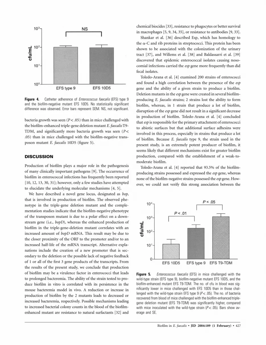
Figure 4. Catheter adherence of Enterococcus faecalis (EFS) type 9and the biofilm-negative mutant EFS 10D5. No statistically significantdifference was observed. Error bars represent SEM. NS, not significant.
Figure 5. Enterococcus faecalis (EFS) in mice challenged with thewild-type strain (EFS type 9), biofilm-negative mutant EFS 10D5, and thebiofilm-enhanced mutant EFS T9-TDM. The no. of cfu in blood was sig-nificantly lower in mice challenged with EFS 10D5 than in those chal-lenged with the wild-type strain EFS type 9 ( ). The no. of bacteriaP ! .05recovered from blood of mice challenged with the biofilm-enhanced triple-gene deletion mutant (EFS T9-TDM) was significantly higher, comparedwith mice inoculated with the wild-type strain ( ). Bars show av-P ! .05erage and SE.
bacteria growth was seen ( ) than in mice challenged withP ! .05
the biofilm-enhanced triple-gene deletion mutant E. faecalis T9-
TDM, and significantly more bacteria growth was seen (P !
) than in mice challenged with the biofilm-negative trans-.05
poson mutant E. faecalis 10D5 (figure 5).
DISCUSSION
Production of biofilm plays a major role in the pathogenesis
of many clinically important pathogens [9]. The occurrence of
biofilm in enterococcal infections has frequently been reported
[10, 12, 13, 30, 31]; however, only a few studies have attempted
to elucidate the underlying molecular mechanisms [4, 5].
We have described a novel gene locus, designated as bop,
that is involved in production of biofilm. The observed phe-
notype in the triple-gene deletion mutant and the comple-
mentation studies indicate that the biofilm-negative phenotype
of the transposon mutant is due to a polar effect on a down-
stream gene (i.e., bopD), whereas the enhanced production of
biofilm in the triple-gene-deletion mutant correlates with an
increased amount of bopD mRNA. This result may be due to
the closer proximity of the ORF to the promoter and/or to an
increased half-life of the mRNA transcript. Alternative expla-
nations include the creation of a new promoter that is sec-
ondary to the deletion or the possible lack of negative feedback
of 1 or all of the first 3 gene products of the transcripts. From
the results of the present study, we conclude that production
of biofilm may be a virulence factor in enterococci that leads
to prolonged bacteremia. The ability of the strain tested to pro-
duce biofilm in vitro is correlated with its persistence in the
mouse bacteremia model in vivo. A reduction or increase in
production of biofilm by the 2 mutants leads to decreased or
increased bacteremia, respectively. Possible mechanisms leading
to increased bacterial colony counts in the blood of the biofilm-
enhanced mutant are resistance to natural surfactants [32] and
chemical biocides [33], resistance to phagocytes or better survival
in macrophages [5, 9, 34, 35], or resistance to antibodies [9, 33].
Shankar et al. [36] described Esp, which has homology to
the a-C and rib proteins in streptococci. This protein has been
shown to be associated with the colonization of the urinary
tract [37], and Willems et al. [38] and Baldassarri et al. [39]
discovered that epidemic enterococcal isolates causing noso-
comial infections carried the esp gene more frequently than did
fecal isolates.
Toledo-Arana et al. [4] examined 200 strains of enterococci
and found a high correlation between the presence of the esp
gene and the ability of a given strain to produce a biofilm.
Deletion mutants in the esp gene were created in several biofilm-
producing E. faecalis strains; 2 strains lost the ability to form
biofilm, whereas, in 1 strain that produce a lot of biofilm,
disruption of the esp gene did not result in a significant decrease
in production of biofilm. Toledo-Arana et al. [4] concluded
that esp is responsible for the primary attachment of enterococci
to abiotic surfaces but that additional surface adhesins were
involved in this process, especially in strains that produce a lot
of biofilm. Because E. faecalis type 9, the strain used in the
present study, is an extremely potent producer of biofilm, it
seems likely that different mechanisms exist for greater biofilm
production, compared with the establishment of a weak-to-
moderate biofilm.
Toledo-Arana et al. [4] reported that 93.5% of the biofilm-
producing strains possessed and expressed the esp gene, whereas
none of the biofilm-negative strains possessed the esp gene. How-
ever, we could not verify this strong association between the
428 • JID 2004:189 (1 February) • Hufnagel et al.
production of biofilm and the presence of esp in the clinical E.
faecalis isolates tested [5]. Thirty-five (44%) of 79 strains tested
negative for the presence of esp by PCR, but were nevertheless
able to produce a lot of biofilm (authors’ unpublished data).
Although this phenomenon may be related to different methods
to test production of biofilm, it may also represent different
characteristics in the strain collections analyzed. Toledo-Arana et
al. [4] used an assay adapted by O’Toole et al. [40] for the
study of production of biofilm by Pseudomonas fluorescens. In
that assay, the crystal-violet is dissolved in ethanol-acetone be-
fore optical density is measured, whereas the method used in
the present study [41] uses readings of the dried microtiter
plates. In the present study, the ethanol-acetone method pro-
duced less-consistent results than did the method used by
Christensen et al. [41]. Because the procedure proposed by
Christensen et al. [41] was developed for gram-positive bacteria
(i.e., Staphylococcus epidermidis) and has been used successfully
by many researchers to study the production of biofilm, we
feel confident that this method is also applicable to enterococci.
Differences between the biofilm assays exist also with regard
to the concentration of glucose in the medium used (0.25–
0.5% used by Toledo-Arana et al. [4] vs. 1% used in the present
study). No systematic comparison that may explain the con-
flicting results between the 2 different methods has been
performed.
The development of biofilm is a multistep procedure [42],
with phase 1 involving the primary attachment of planktonic
microorganisms to biotic and abiotic surfaces. Toledo-Arana et
al. [4] speculate that Esp is one of the mediators of primary
attachment in enterococci. Phase 2 of production of biofilm is
the molecule-mediated secondary attachment of microorgan-
isms to surfaces. Adhesins, such as the polysaccharide inter-
cellular adhesin in staphylococci [43], mediate the interaction
between the microorganisms and the surface and between in-
dividual bacterial cells. This phase is characterized by the for-
mation of microcolonies [33]. Phase 3 involves the maturation
of the biofilm. The microorganisms change their metabolic
state, produce different extracellular products (such as exo-
polysaccharides) to form a glycocalyx, and divide within the
biofilm. Phase 4 is marked by the detachment of planctonic
cells from the biofilm to colonize further surfaces. On the basis
of findings from scanning electron microscopy studies, we hy-
pothesize that our transposon mutant is defective in phase 2
of production of biofilm. The transposon mutant grew in a
monolayer on polystyrene segments, whereas the wild-type
strain and the biofilm-enhanced triple deletion mutant grew in
a multilayer, with a 3-dimensional pile-up and the formation
of microcolonies, which were noticeably absent in the trans-
poson mutant. The monolayer represents the primary attach-
ment, whereas the formation of a multilayer with microcolonies
requires the machinery of the secondary attachment. Com-
parison of the initial attachment in a catheter adherence model
[26] showed no difference in the attachment to a polyethylene
catheter of the transposon mutant and the wild-type strain. We
therefore assume that the secondary attachment, and not the
primary attachment, is impaired in our transposon mutant. On
the other hand, Toledo-Arana et al. [4] speculate that esp is
involved in the initial adherence of the bacteria to abiotic sur-
faces. This may explain the results by Toleda-Arana et al. [4]
that strains that produce a lot of biofilm were not affected in
their adherence by the inactivation of esp. Another possibility
might be that bopD up-regulates expression of esp and possibly
other virulence factors involved in adhesion of the bacteria to
surfaces. The residual production of biofilm by our transposon
mutant may also be related to the intact primary attachment
of the bacteria mediated by esp that is not affected by the
reduction of bopD expression.
We are aware that the creation of a deletion mutant in the
bopD gene would be the definitive proof that the bopD gene
causes the observed phenotype in the transposon mutant. De-
spite numerous attempts with different suicide vectors, flanking
regions, and bacterial strains (data not shown), we were unable
to delete the bopD gene by targeted mutagenesis. One possible
explanation is that bopD is essential in the biofilm-positive
strains tested. We screened a collection of 18 unrelated E. fae-
calis strains by use of PCR for the presence of the bopD gene
and found that all strains carried the bopD gene, supporting
the hypothesis that the gene is essential (data not shown). Fur-
thermore, this hypothesis is supported by the fact that the
transposon insertion did not completely inactivate bopD and
that the mutant strain still expresses a residual amount of bopD
mRNA (8.5% of the wild type). Depending on the incubation
period, the amount of biofilm produced by the mutant is 16%–
36% of that produced by the wild-type, suggesting a close re-
lationship between the expression of bopD and production of
biofilm.
Baldassarri et al. [5] found that, of 73 strains of clinical E.
faecalis isolates analyzed, 66% were strong producers of biofilm,
14% were weak producers, and 20% did not produce any bio-
film. The formation of biofilm in these strains was strongly
affected by the presence of additional carbohydrates in the
growth medium.
The dependence of production of biofilm on the presence
of specific carbohydrate sources in the growth medium has
been described by a number of investigators [3–5, 9]. The
attributed function (sugar-binding transcriptional regulator) of
the bopD-encoded protein responsible for the biofilm-negative
phenotype in the present study might explain this observation.
This gene shows significant sequence homology (29% identity
and 50% similarity) with the ccpA gene of E. faecalis [44], which
is involved in carbohydrate metabolism. The gene also shows
significant homology with the ccpA gene of Listeria innocua

Biofilm in E. faecalis • JID 2004:189 (1 February) • 429
(30% identity and 55% similarity), Listeria monocytogenes (30%
identity and 54% similarity), Lactococcus lactis (30% identity
and 54% similarity), Bacillus subtilis (29% identity and 54%
similarity), Streptococcus pyogenes (28% identity and 51% sim-
ilarity), Streptococcus pneumoniae (29% identity and 52% sim-
ilarity), and with a putative maltose operon transcriptional re-
pressor in S. pyogenes (45% identity and 65% similarity) and
S. aureus (31% identity and 53% similarity).
O’Toole et al. [45] found that a gene with a similar function
to bopD (i.e., carbon catabolite regulation), the global carbon
metabolism regulator crc, is part of a signal transduction path-
way required for production of biofilm by Pseudomonas aeru-
ginosa. A crc mutant created by O’Toole et al. [45] showed, in
scanning electron microscopy, a morphologic pattern similar
to that of our mutant, growing only as a dispersed monolayer.
The phenotype of their strains was associated with a defective
type IV pilus–mediated twitching motility caused by decreased
pilA transcription.
Carbon catabolite repression proteins, such as CcpA, are
known to regulate transcription of hundreds of promoters [46]
and might therefore be involved in the regulation of different
genes probably associated with production of biofilm, such as
esp, aggregation substance, and others. Further studies are un-
der way to identify proteins that are expressed under the control
of the bopD gene in our prototype strain.
Acknowledgments
We thank P. Crowley, L. Hancock, D. Garsin, K. Puopolo,
K. Jefferson, M. Coyne, L. Comstock, L. Madoff, M. Cieslewicz,
and G. Pier, for strains, plasmids, and helpful discussions; N.
Voynow, for editorial assistance; and N. Reiniger and A. Ross,
for help with the real-time polymerase chain reaction.
References
1. Edmond MB, Wallace SE, McClish DK, Pfaller MA, Jones RN, WenzelRP. Nosocomial bloodstream infections in United States hospitals: athree-year analysis. Clin Infect Dis 1999; 29:239–44.
2. Murray BE. Vancomycin-resistant enterococcal infections. N Engl JMed 2000; 342:710–21.
3. O’Toole G, Kaplan HB, Kolter R. Biofilm formation as microbial de-velopment. Annu Rev Microbiol 2000; 54:49–79.
4. Toledo-Arana A, Valle J, Solano C, et al. The enterococcal surfaceprotein, Esp, is involved in Enterococcus faecalis biofilm formation. ApplEnviron Microbiol 2001; 67:4538–45.
5. Baldassarri L, Cecchini R, Bertuccini L, et al. Enterococcus spp. producesslime and survives in rat peritoneal macrophages. Med Microbiol Im-munol (Berl) 2001; 190:113–20.
6. Su SH, Eaton JW, Venezia RA, Tang L. Interactions of vancomycinresistant enterococci with biomaterial surfaces. Asaio J 1998; 44:770–5.
7. Joyanes P, Pascual A, Martinez-Martinez L, Hevia A, Perea EJ. In vitroadherence of Enterococcus faecalis and Enterococcus faecium to plasticbiomaterials. Clin Microbiol Infect 1999; 5:382–6.
8. Joyanes P, Pascual A, Martinez-Martinez L, Hevia A, Perea EJ. In vitro
adherence of Enterococcus faecalis and Enterococcus faecium to urinarycatheters. Eur J Clin Microbiol Infect Dis 2000; 19:124–7.
9. Donlan RM, Costerton JW. Biofilms: survival mechanisms of clinicallyrelevant microorganisms. Clin Microbiol Rev 2002; 15:167–93.
10. Distel JW, Hatton JF, Gillespie MJ. Biofilm formation in medicatedroot canals. J Endod 2002; 28:689–93.
11. Tunney MM, Gorman SP. Evaluation of a poly(vinyl pyrollidone)–coated biomaterial for urological use. Biomaterials 2002; 23:4601–8.
12. Sabbuba N, Hughes G, Stickler DJ. The migration of Proteus mirabilisand other urinary tract pathogens over Foley catheters. BJU Int 2002;89:55–60.
13. Zegans ME, Becker HI, Budzik J, O’Toole G. The role of bacterialbiofilms in ocular infections. DNA Cell Biol 2002; 21:415–20.
14. Fiedler S, Wirth R. Transformation of Enterococcus faecalis and En-terococcus faecium by electroporation. In: Dunny GM, Cleary PP,McKay LL, eds. Genetics and molecular biology of streptococci, lac-tococci, and enterococci. Washington, DC: American Society for Mi-crobiology, 1991:301.
15. Sambrook J, Russell DW. Molecular cloning: a laboratory manual. 3rded. Cold Spring Harbor, NY: Cold Spring Harbor Laboratory Press,2001.
16. Gutierrez JA, Crowley PJ, Brown DP, Hillman JD, Youngman P, BleiweisAS. Insertional mutagenesis and recovery of interrupted genes of Strep-tococcus mutans by using transposon Tn917: preliminary characteri-zation of mutants displaying acid sensitivity and nutritional require-ments. J Bacteriol 1996; 178:4166–75.
17. Maekawa S, Yoshioka M, Kumamoto Y. Proposal of a new scheme forthe serological typing of Enterococcus faecalis strains. Microbiol Immu-nol 1992; 36:671–81.
18. Karlyshev AV, Pallen MJ, Wren BW. Single-primer PCR procedure forrapid identification of transposon insertion sites. Biotechniques 2000;28:1078, 1080, 1082.
19. Qin X, Teng F, Xu Y, Singh KV, Weinstock GM, Murray BE. Targetedmutagenesis of enterococcal genes. Methods Cell Sci 1998; 20:21–33.
20. Qin X, Singh KV, Weinstock GM, Murray BE. Characterization of fsr,a regulator controlling expression of gelatinase and serine protease inEnterococcus faecalis OG1RF. J Bacteriol 2001; 183:3372–82.
21. Cieslewicz MJ, Kasper DL, Wang Y, Wessels MR. Functional analysisin type Ia group B Streptococcus of a cluster of genes involved inextracellular polysaccharide production by diverse species of strepto-cocci. J Biol Chem 2001; 276:139–46.
22. Eichenbaum Z, Federle MJ, Marra D, et al. Use of the lactococcal nisApromoter to regulate gene expression in gram-positive bacteria: com-parison of induction level and promoter strength. Appl Environ Mi-crobiol 1998; 64:2763–9.
23. Hancock LE, Gilmore MS. The capsular polysaccharide of Enterococcusfaecalis and its relationship to other polysaccharides in the cell wall.Proc Natl Acad Sci USA 2002; 99:1574–9.
24. Winer J, Jung CK, Shackel I, Williams PM. Development and validationof real-time quantitative reverse transcriptase-polymerase chain reac-tion for monitoring gene expression in cardiac myocytes in vitro. AnalBiochem 1999; 270:41–9.
25. Johnson MR, Wang K, Smith JB, Heslin MJ, Diasio RB. Quantitation ofdihydropyrimidine dehydrogenase expression by real-time reverse tran-scription polymerase chain reaction. Anal Biochem 2000; 278:175–84.
26. Muller E, Takeda S, Goldmann DA, Pier GB. Blood proteins do notpromote adherence of coagulase-negative staphylococci to biomaterials.Infect Immun 1991; 59:3323–6.
27. Dicuonzo G, Gherardi G, Lorino G, et al. Antibiotic resistance andgenotypic characterization by PFGE of clinical and environmental iso-lates of enterococci. FEMS Microbiol Lett 2001; 201:205–11.
28. Cramton SE, Gerke C, Schnell NF, Nichols WW, Gotz F. The inter-cellular adhesion (ica) locus is present in Staphylococcus aureus and isrequired for biofilm formation. Infect Immun 1999; 67:5427–33.
29. Huebner J, Wang Y, Krueger WA, et al. Isolation and chemical char-acterization of a capsular polysaccharide antigen shared by clinical

430 • JID 2004:189 (1 February) • Hufnagel et al.
isolates of Enterococcus faecalis and vancomycin-resistant Enterococcusfaecium. Infect Immun 1999; 67:1213–9.
30. Leung JW, Liu YL, Desta TD, Libby ED, Inciardi JF, Lam K. In vitroevaluation of antibiotic prophylaxis in the prevention of biliary stentblockage. Gastrointest Endosc 2000; 51:296–303.
31. Dautle MP, Ulrich RL, Hughes TA. Typing and subtyping of 83 clinicalisolates purified from surgically implanted silicone feeding tubes byrandom amplified polymorphic DNA amplification. J Clin Microbiol2002; 40:414–21.
32. Anwar H, Strap JL, Costerton JW. Susceptibility of biofilm cells ofPseudomonas aeruginosa to bactericidal actions of whole blood andserum. FEMS Microbiol Lett 1992; 71:235–41.
33. Costerton JW, Stewart PS, Greenberg EP. Bacterial biofilms: a commoncause of persistent infections. Science 1999; 284:1318–22.
34. Jensen ET, Kharazmi A, Lam K, Costerton JW, Hoiby N. Human poly-morphonuclear leukocyte response to Pseudomonas aeruginosa grownin biofilms. Infect Immun 1990; 58:2383–5.
35. Gentry-Weeks CR, Karkhoff-Schweizer R, Pikis A, Estay M, Keith JM.Survival of Enterococcus faecalis in mouse peritoneal macrophages. In-fect Immun 1999; 67:2160–5.
36. Shankar V, Baghdayan AS, Huycke MM, Lindahl G, Gilmore MS.Infection-derived Enterococcus faecalis strains are enriched in esp, agene encoding a novel surface protein. Infect Immun 1999; 67:193–200.
37. Shankar N, Lockatell CV, Baghdayan AS, Drachenberg C, Gilmore MS,Johnson DE. Role of Enterococcus faecalis surface protein Esp in thepathogenesis of ascending urinary tract infection. Infect Immun 2001;69:4366–72.
38. Willems RJ, Homan W, Top J, et al. Variant esp gene as a marker of
a distinct genetic lineage of vancomycin-resistant Enterococcus faeciumspreading in hospitals. Lancet 2001; 357:853–5.
39. Baldassarri L, Bertuccini L, Ammendolia MG, Gherardi G, Creti R.Variant esp gene in vancomycin–sensitive Enterococcus faecium. Lancet2001; 357:1802.
40. O’Toole GA, Kolter R. Initiation of biofilm formation in Pseudomonasfluorescens WCS365 proceeds via multiple, convergent signalling path-ways: a genetic analysis. Mol Microbiol 1998; 28:449–61.
41. Christensen GD, Simpson WA, Younger JJ, et al. Adherence of coag-ulase-negative staphylococci to plastic tissue culture plates: a quanti-tative model for the adherence of staphylococci to medical devices. JClin Microbiol 1985; 22:996–1006.
42. Dunne WM Jr. Bacterial adhesion: seen any good biofilms lately? ClinMicrobiol Rev 2002; 15:155–66.
43. Mack D, Fischer W, Krokotsch A, et al. The intercellular adhesin in-volved in biofilm accumulation of Staphylococcus epidermidis is a linearb-1,6-linked glucosaminoglycan: purification and structural analysis. JBacteriol 1996; 178:175–83.
44. Leboeuf C, Leblanc L, Auffray Y, Hartke A. Characterization of theccpA gene of Enterococcus faecalis: identification of starvation-inducibleproteins regulated by ccpA. J Bacteriol 2000; 182:5799–806.
45. O’Toole GA, Gibbs KA, Hager PW, Phibbs PV Jr, Kolter R. The globalcarbon metabolism regulator Crc is a component of a signal trans-duction pathway required for biofilm development by Pseudomonasaeruginosa. J Bacteriol 2000; 182:425–31.
46. Bruckner R, Titgemeyer F. Carbon catabolite repression in bacteria:choice of the carbon source and autoregulatory limitation of sugarutilization. FEMS Microbiol Lett 2002; 209:141–8.