a generic approach for the purification of signaling complexes that specifically interact with the...
TRANSCRIPT
A Generic Approach for the Purification ofSignaling Complexes That Specifically Interactwith the Carboxyl-terminal Domain ofG Protein-coupled Receptors*□S
Pascal Maurice‡§, Avais M. Daulat‡§¶, Cedric Broussard§�, Julien Mozo**,Guilhem Clary§�, Francoise Hotellier§�, Philippe Chafey§�, Jean-Luc Guillaume‡§,Gilles Ferry**, Jean A. Boutin**, Philippe Delagrange**, Luc Camoin§�,and Ralf Jockers‡§‡‡
G protein-coupled receptors (GPCRs) constitute the larg-est family of membrane receptors and are major drugtargets. Recent progress has shown that GPCRs are partof large protein complexes that regulate their activity. Wepresent here a generic approach for identification of thesecomplexes that is based on the use of receptor subdo-mains and that overcomes the limitations of currentlyused genetics and proteomics approaches. Our approachconsists of a carefully balanced combination of chemi-cally synthesized His6-tagged baits, immobilized metalaffinity chromatography, one- and two-dimensional gelelectrophoresis separation and mass spectrometric iden-tification. The carboxyl-terminal tails (C-tails) of the hu-man MT1 and MT2 melatonin receptors, two class AGPCRs, were used as models to purify protein complexesfrom mouse brain lysates. We identified 32 proteins thatinteracted with the C-tail of MT1, 14 proteins that inter-acted with the C-tail of MT2, and eight proteins thatinteracted with both C-tails. Several randomly selectedproteins were validated by Western blotting, and thefunctional relevance of our data was further confirmedby showing the interaction between the full-length MT1
and the regulator of G protein signaling Z1 in trans-fected HEK 293 cells and native tissue. Taken together,we have established an integrated and generic purifica-tion strategy for the identification of high quality andfunctionally relevant GPCR-associated protein com-plexes that significantly widens the repertoire of avail-able techniques. Molecular & Cellular Proteomics 7:1556–1569, 2008.
G protein-coupled receptors (GPCRs)1 constitute the larg-est family of membrane receptors with more than 800 mem-bers (1, 2). By binding to a great variety of ligands (photons,odorants, amino acids, nucleotides, peptides, proteins, andlipids), GPCRs are key receptors of numerous physiologicalprocesses such as neurotransmission, cell metabolism, se-cretion, cell differentiation, and growth and are targeted byabout half of the drugs prescribed for human diseases (3). It isnow well established that GPCRs do not only couple to het-erotrimeric G proteins but can also physically associate withother less well known intracellular proteins regulating receptortrafficking, subcellular localization, signaling, and desensitiza-tion (4, 5). Intracellular proteins can interact directly orindirectly, via adaptor proteins, with intracellular receptor do-mains. Among these domains, the carboxyl-terminal tail (C-tail) is considered a key domain able to recruit intracellularproteins in large submembrane signaling networks (6, 7).
Several approaches have been described in the literature toidentify proteins that interact with GPCRs. The yeast two-hybrid assay has been used to screen for proteins that bind tocytosolic domains of GPCRs (8–10). However, this system,which is very sensitive for the detection of in vivo protein-protein interactions, has shown several limitations includingthe generation of many false positives and negatives, thedetection of only binary interactions, and the non-physiolog-ical relevance of the identified interactions (e.g. occurring inthe yeast nucleus). Moreover this approach is not designed to
From the ‡Department of Cell Biology and �Laboratoire de Pro-teomique, Institut Cochin, Universite Paris Descartes, CNRS (UMR8104), Paris F-75014, France, §INSERM U567, Paris F-75014,France, and **Institut de Recherches SERVIER, Suresnes F-92150, France
Received, September 12, 2007, and in revised form, April 28, 2008Published, MCP Papers in Press, April 29, 2008, DOI 10.1074/
mcp.M700435-MCP200
1 The abbreviations used are: GPCR, G protein-coupled receptor;C-tail, carboxyl-terminal tail; 1D, one-dimensional; 2D, two-dimen-sional; Ni-NTA, nickel-nitrilotriacetic acid; MT, melatonin receptor;PDZ, PSD-95/Disc-large/ZO-1; ERK, extracellular signal-regulated ki-nase; CK2, casein kinase II; GRK, G protein-coupled receptor kinase;HEK, human embryonic kidney; 5-HT, 5-hydroxytryptamine; RGSZ1,regulator of G protein signaling Z1; ABC, ammonium bicarbonate;nNOS, neuronal NO synthase; PKC, protein kinase C; HA, hemagglu-tinin; GTP�S, guanosine 5�-3-O-(thio)triphosphate; CHO, Chinesehamster ovary; RGS, regulator of G protein signaling; YFP, yellowfluorescent protein; PSD, postsynaptic density; TAP, tandem affinitypurification.
Research
© 2008 by The American Society for Biochemistry and Molecular Biology, Inc.1556 Molecular & Cellular Proteomics 7.8This paper is available on line at http://www.mcponline.org
identify protein complexes. To overcome these limitations,proteomics approaches were developed based on peptideaffinity chromatography coupled to protein identification bymass spectrometry. The first generation used the entire re-ceptor C-tail expressed as a GST fusion protein in Escherichiacoli to purify the interactome of the C-tail from brain lysates(11). Although some interacting proteins have been identifiedfor the 5-HT2C receptor, several limitations prevented a moregeneral application of this approach. Indeed the amount ofnonspecifically retained proteins is high because of the pres-ence of contaminating bacterial proteins, full-length and trun-cated GST fusion proteins, and proteins that nonspecificallybind to the GST carrier. In addition, the complex sample hasto be separated by two-dimensional (2D) electrophoresis be-fore mass spectrometry analysis of the recruited proteins,thus minimizing the number of potential candidates becauseof the limitations inherent to 2D electrophoresis for hydropho-bic, basic, and large proteins. The second generation of pep-tide affinity chromatography is based on the use of shortsynthetic peptides encompassing a specific interaction motifthat recruit only the proteins that interact with this specificmotif. The amount of nonspecific proteins is indeed largelyreduced in this approach, and the pattern of specific interac-tion partners is much less complex. Consequently this ap-proach has been successfully used for several GPCRs and isexpected to be applied to multiple GPCR interaction motifs(12, 13). However, this evolution of peptide affinity chroma-tography does not respond to the initial task, the identificationof the interactome of the entire C-tail, without prior knowledgeof specific interaction motifs.
In the present study, we present an improved peptide af-finity chromatography that has the potential to become thefirst generic approach for the purification of GPCR C-tail-associated protein complexes. We combined the use ofchemically synthesized His6-tagged peptides encompassingthe entire receptor C-tail combined with metal affinity immo-bilization on a Ni-NTA matrix to recover protein complexesfrom mouse brain lysates. The C-tails of the MT1 and MT2
melatonin receptors, typical class A GPCRs, were used asmodel receptors. Major features of this approach are the lownonspecific binding, the high integrity of recovered com-plexes, and the compatibility with 1D and 2D electrophoresis.We report the identification of 40 and 22 proteins that specif-ically associate with the C-tails of MT1 and MT2, respectively.To demonstrate the functional relevance of the identified pro-tein complexes, we selected the interaction between MT1 andthe regulator of G protein signaling Z1 (RGSZ1) for furthercharacterization in transfected HEK 293 cells and nativetissue.
EXPERIMENTAL PROCEDURES
Peptide Affinity Chromatography—Peptides encompassing the C-tails of the MT1 and MT2 receptors were chemically synthesized byNeoMPS (Strasbourg, France) with a His6 tag at the amino terminus.
The synthetic peptides (MT1, last 61 amino acids, 7.16 kDa, 88.5%purity; MT2, last 58 amino acids, 6.80 kDa, 90% purity) were coupledvia the His6 tag to Ni-NTA-agarose beads (Qiagen).
Brains of C57/Bl6 mice were crushed in 20 ml of buffer containing20 mM NaH2PO4, 2 mM Na3VO4, 10 mM NaF, protease inhibitormixture EDTA-free (Roche Applied Science), pH 8.0, using an Ultra-Turrax T25 (Janke-Kunkel). CHAPS (10 mM final concentration) andNaCl (150 mM final concentration) were added, and the homogenateswere incubated for 3 h at 4 °C under gentle end-over-end mixing.After centrifugation (10,000 � g for 1 h at 4 °C), the supernatants werecollected, and the protein concentration was determined by BCAassay (Pierce).
Supernatants (10 mg of solubilized brain proteins) were incubatedovernight at 4 °C with 20 �l of Ni-NTA-agarose beads (300–350 �g ofimmobilized peptide) in the presence of 20 mM imidazole to reducenonspecific binding. The beads were washed five times in washingbuffer (20 mM NaH2PO4, 2 mM Na3VO4, 10 mM NaF, protease inhibitormixture EDTA-free, 10 mM CHAPS, 150 mM NaCl, 20 mM imidazole,pH 8), and proteins retained by affinity were eluted with either 50 �l of2% SDS in PBS (95 °C for 10 min) for BCA measurements, 1Delectrophoresis, and immunoblotting or 200 �l of rehydration buffer (8M urea, 2 M thiourea, 4% CHAPS) for 45 min at room temperature for2D electrophoresis.
1D and 2D Electrophoresis—For 1D electrophoresis, eluted pro-teins with 2% SDS in PBS were denatured in SDS-PAGE loadingbuffer (62.5 mM Tris/HCl, pH 6.8, 2% SDS, 10% glycerol, 0.5%bromphenol blue) for 5 min at 95 °C. Samples were subjected toSDS-PAGE on a 10 or 5–9% gradient polyacrylamide gel, and pro-teins were silver-stained according to Rabilloud and Charmont (14).
For 2D electrophoresis, eluted proteins in rehydration buffer werefirst separated according to their isoelectric point along a nonlinearIPG strip (pH 3–11 nonlinear, 11 cm long) using the IPGphor appara-tus (GE Healthcare). DeStreack Reagent (15 mg/ml of rehydrationsolution; GE Healthcare) and ampholytes (1% IPG buffer; GE Health-care) were added to the sample, and then sample loading for the firstdimension was performed by passive in-gel rehydration. Focalizationwas stopped when 27,000 V-h were reached. After the first dimen-sion, the IPG strips were equilibrated for 15 min in buffer A (6 M urea,2% SDS, 30% glycerol, 50 mM Tris/HCl, pH 8.6) containing 1% DTTand then for 15 min in buffer A containing 4.7% iodoacetamide. Forthe second dimension, the strips were loaded on 8–18% SDS-poly-acrylamide gels. The gels were then silver-stained according to Ra-billoud and Charmont (14).
In-gel Trypsin Digestion—Spots of interest were excised from thegel, washed with 200 mM ammonium bicarbonate (ABC) and imme-diately destained according to Gharahdaghi et al. (15). In-gel trypsindigestion was carried out as described in a protocol based on theZipTip Plate (Millipore) with minor modifications. After destaining,spots were rinsed three times with water and shrunk with 50 mM ABC,50% ACN for 20 min at room temperature. Gel pieces were driedusing 100% ACN for 15 min and then incubated in 50 mM ABCcontaining 10 mM DTT for 1 h at 56 °C. The solution was thenreplaced by 55 mM iodoacetamide in 50 mM ABC for 30 min in thedark at room temperature. The gel pieces were washed twice with 50mM ABC, finally shrunk with 25 mM ABC, 50% ACN for 30 min, anddried using 100% ACN for 10 min. Gel pieces were rehydrated in 20�l of 40 mM ABC, 10% ACN, pH 8.0, containing 12.5 �g/ml sequenc-ing grade modified trypsin (Promega). Proteins were digested over-night at 37 °C. After digestion, the gel pieces were shrunk with 100%ACN, and peptides were extracted with 0.2% TFA. Peptides werethen desalted using C18 phase on a ZipPlate. Two elutions wereperformed successively to recover products from the C18 phase, firstwith 50% ACN, 0.1% TFA and then using 90% ACN, 0.1% TFA.Pooled elutions were concentrated using a vacuum centrifuge (Ep-
Purification of GPCR-associated Protein Complexes
Molecular & Cellular Proteomics 7.8 1557
pendorf), and generated peptides were redissolved in 3 �l of 1%formic acid before mass spectrometry analysis.
Protein Identification by MALDI-TOF Mass Spectrometry—Di-gested samples were spotted directly onto a 96 � 2 MALDI plate(Applied Biosystems) using the dried droplet method (0.5 �l of thesample mixed on the plate with 0.5 �l of �-cyano-4-hydroxycinnamicacid matrix in ACN/water/TFA (50:50:0.1%). Droplets were allowed todry at room temperature. Mass spectrometry analysis were per-formed on a MALDI-TOF Voyager DE-PRO mass spectrometer(Applied Biosystems) and acquired in positive ion reflector mode.Spectra were obtained in a reflectron-delayed extraction in sourceover a mass range of 500–3500 daltons. Raw spectra were treatedautomatically according to the Mascot wizard algorithm (version1.1.2.0, Matrix Science) using the default parameters with the ex-ception of the maximum peak count reduced to a value correspond-ing to the expected number of tryptic peptides according to theexperimental molecular weight of the protein. The protein databasewas restricted to the rodent entries of the comprehensive nonredun-dant protein sequence database (NCBInr database; National Centerfor Biotechnology Information) (from version October 2005, 118,719entries to version October 2007, 217,820 entries). When non-signifi-cant scoring was obtained, a manual treatment was performed ac-cording to the following explanations. First raw spectra were treatedusing a noise filter algorithm (correlation factor, 0.7) and the defaultadvanced base-line correction. Then monoisotopic masses weregenerated on a fully detected spectrum after internal calibration usingautodigestion tryptic peptides or external calibration using a mixtureof five external standards (PepMix 1, LaserBio Labs, Sophia Antipolis,France). Mains peaks were selected according to local background,molecular weight of the protein, and known peak contamination fromtrypsin and keratin using PeakErazor software (version 1.49, Light-house Data, Odense, Denmark). Searches were carried out using fourdifferent algorithms for protein identification: Mascot (version 2.1,Matrix Science), ProFound (version 2005.02.14, Proteometrics), MS-Fit (version 3.2.1, ProteinProspector), and Aldente (version2006.09.11, ExPASy) softwares on the rodent entries of the NCBInrdatabase. A combination of results from different algorithms offersthe option to cross-validate and consolidate the identification throughthe complementary use of several packages. Allowed variable mod-ifications were oxidation of methionine, acrylamide-modified cys-teine, and carbamidomethylation of cysteine. Up to one missed tryp-tic cleavage was considered, and a mass accuracy in the range of25–50 ppm was used for all tryptic mass searches. Identification wasconsidered positive if the protein was identified with the most ele-vated score using at least two algorithms. For a protein with a scoreclose to the threshold value, the identification was rejected. Identifiedproteins are listed in Tables I and II. If peptides matched to multiplemembers of a protein family, the protein with the highest number ofmatched peptides and experimental molecular weight and isoelectricpoint close to theoretical parameters was indicated in the table, andNCBI accession numbers of the other isoforms are mentioned at thebottom of the table.
Antibodies—Monoclonal anti-14-3-3 � and anti-PP2A (catalyticsubunit, clone 1D6) and polyclonal anti-Gi3�, anti-neuronal NO syn-thase (nNOS), anti-panPKCs, anti-PKC �, anti-14-3-3 �, anti-PP2A-A�/� (regulatory subunit), and anti-c-Myc antibodies were purchasedfrom Santa Cruz Biotechnology. Monoclonal anti-postsynaptic den-sity (PSD)-95 antibodies (clone K28/86.2) were from Upstate Biotech-nology. Polyclonal anti-FLAG and monoclonal anti-tubulin � (cloneDM 1A), anti-tubulin � (clone TUB 2.1), anti-tubulin � (clone GTU-88),and anti-FLAG M2 antibodies were from Sigma. Monoclonal anti-HA(clone 12CA5) and anti-G protein-coupled receptor kinase (GRK) 2/3(clone C5/1.1) antibodies were purchased from Roche Diagnosticsand Millipore, respectively. CyTM3-conjugated goat anti-mouse anti-
bodies were from Jackson ImmunoResearch Laboratories, andhorseradish peroxidase-conjugated secondary antibody was fromPierce. Polyclonal anti-otubain 1 and anti-MUPP1 antibodies were akind gift from Dr. M. Y. Balakirev (Grenoble, France) and Dr. R. T.Javier (Houston, TX), respectively. Polyclonal anti-RGSZ1 antibodieswere kindly provided by Dr. J. Garzon (Madrid, Spain).
Plasmids—The cDNA encoding HA-RGSZ1 was purchased fromthe University of Missouri Rolla cDNA Resource Center (Rolla, MO),and pGEX-4T-1 vector was from Amersham Biosciences. The cDNAencoding Myc-RGS10 was a kind gift from Dr. P. J. Casey (Durham,NC). The FLAG-MT1 protein construct has been described elsewhere(16). The FLAG-MT2 construct was obtained by a PCR-based ap-proach using a Myc-MT2 cDNA (16) and the Phusion High-FidelityDNA Polymerase (Finnzymes). Restriction sites for EcoRI and XbaIwere introduced immediately adjacent to the initiation and terminationcodons of MT2 by PCR. After digestion by the respective restrictionenzymes, the resulting insert was ligated into a pcDNA3.1 vectorencoding FLAG. All constructs were confirmed by sequencing.
Cell Culture and Transfection—HEK 293 cells were grown in Dul-becco’s modified Eagle’s medium supplemented with 10% fetal calfserum, 100 units/ml penicillin, 0.1 mg/ml streptomycin, 0.02 M HEPESand transfected at 40–50% confluence using FuGENE 6 (RocheApplied Science).
Immunoprecipitation—HEK 293 cells grown in 100-mm culturedishes were transiently transfected by 4 �g of either FLAG-MT1 orFLAG-MT2 cDNAs together with 4 �g of HA-RGSZ1 or Myc-RGS10cDNAs. 48 post-transfection, cells were stimulated, or not, by 10�7 M
melatonin for 15 min at 37 °C, washed three times in PBS, and lysedin 1.5 ml of cold lysis buffer (75 mM Tris, 2 mM EDTA, 12 mM MgCl2,10 mM CHAPS, pH 7.4). After sonication, receptors were solubilizedfor 3 h at 4 °C under gentle end-over-end mixing, and lysates werecentrifuged at 12,000 � g for 1 h at 4 °C. Receptor immunoprecipi-tation was performed on 1 ml of supernatant using 4 �g of polyclonalanti-FLAG antibodies preadsorbed on protein G-Sepharose beads(Sigma). Immunoprecipitated proteins were eluted with 50 �l of SDS-PAGE sample buffer (62.5 mM Tris/HCl, pH 6.8, 2% SDS, 10% glyc-erol, 100 mM DTT, 0.5% bromphenol blue) for 10 min at 95 °C andsubjected to SDS-PAGE and immunoblotting.
Immunoblotting—Proteins resolved by 1D electrophoresis weretransferred onto nitrocellulose membrane (Whatman GmbH). Afterincubation in PBS supplemented with 5% skimmed milk and 0.05%Tween 20 for 1 h at room temperature, membranes were incubated inprimary antibody solution in PBS supplemented with 5% skimmedmilk and 0.05% Tween 20 for an additional 1 h at room temperature.After washes in PBS supplemented with 0.05% Tween 20, immuno-reactivity was revealed using a horseradish peroxidase-conjugatedsecondary antibody (1:10,000) and the ECL chemiluminescent rea-gent (Perbio).
Immunofluorescence Microscopy—HEK 293 cells transiently trans-fected by 2 �g of FLAG-MT1 together with 1 �g of RGSZ1-YFP cDNAwere grown on sterile coverslips. 48 post-transfection, cells werestimulated, or not, by 10�7 M melatonin for 15 min at 37 °C, washedthree times in PBS, and fixed with 4% paraformaldehyde in PBS for15 min. After blocking with 3% BSA in PBS for 30 min, cells wereincubated with monoclonal anti-FLAG M2 (1:500) antibodies in PBScontaining 0.3% BSA for 1 h at room temperature. Coverslips werethen washed three times with PBS and incubated with Cy3-conju-gated goat anti-mouse antibodies (1:800) in PBS containing 0.3%BSA for 40 min at room temperature. Coverslips were mounted andanalyzed by confocal laser microscopy (Leica TCS SP2 AOBS).
Production and Purification of HA-RGSZ1—To express HA-RGSZ1as a GST fusion protein, restriction sites for BamHI and XhoI wereintroduced immediately adjacent to the initiation and terminationcodons by PCR of the plasmid encoding HA-RGSZ1 using the Phu-
Purification of GPCR-associated Protein Complexes
1558 Molecular & Cellular Proteomics 7.8
sion High-Fidelity DNA Polymerase. After digestion by BamHI andXhoI, the resulting insert was ligated into pGEX-4T-1 vector. DNAsequences were confirmed by sequencing. The resulting plasmid wastransformed into BL21(DE3) cells (Invitrogen), and cells were grown inLB medium containing 50 �g/ml ampicillin. Expression of GST-HA-RGSZ1 protein was induced by adding 1 mM isopropylthiogalactoside(Sigma) to midlog cultures. Cultures were harvested after 7 h at 37 °C.Cells were centrifuged at 6000 � g for 15 min, and the pellet wasstored at �20 °C. For purification of HA-RGSZ1, the pellet was re-suspended in cold PBS containing protease inhibitor mixture EDTA-free, crushed with an Ultra-Turrax T25, and sonicated. After adding1% Triton X-100 (Sigma), the cell suspension was centrifuged at10,000 � g for 30 min, and the supernatant was collected and appliedto glutathione-agarose beads (Sigma) that had been equilibrated with1% Triton X-100 in PBS. After extensive washes, HA-RGSZ1 proteinswere eluted by 0.04 unit/�l thrombin (Amersham Biosciences) in 200�l of PBS (2 h at 22 °C). Eluates were applied to new, freshly preparedglutathione-agarose beads to eliminate residual contaminant GSTand bacterial proteins. The purity of HA-RGSZ1 was assessed bySDS-PAGE after silver staining.
[35S]GTP�S Binding—[35S]GTP�S binding was determined in 100�l of reaction mixture containing 20 mM HEPES, 100 mM NaCl, 20�g/ml saponin, 3 �M GDP, 3 mM MgCl2, protease inhibitor mixtureEDTA-free, pH 7.4. The reaction mixture also contained CHO cellmembranes stably expressing the MT1 receptor (5 fmol), 0.2 nM
[35S]GTP�S, and purified RGSZ1 with or without 1 �M melatonin. Thereaction was started at 37 °C by adding [35S]GTP�S and stopped at60 min by filtration. Filters were washed with 50 mM Tris/HCl, pH 7.4,and radioactivity was measured.
Radioligand Binding—Ovine pituitary pars tuberalis was collected,and crude membranes were prepared essentially as described previ-ously (17). Radioligand binding was performed with pars tuberalis mem-
branes (1.5–2 mg of protein) in 1 ml of TEM buffer (75 mM Tris, 12 mM
MgCl2, 2 mM EDTA, protease inhibitor mixture EDTA-free, pH 7.4)containing 400 pM 2-[125I]melatonin (PerkinElmer Life Sciences) for 90min at 37 °C. After membrane solubilization with 1% digitonin (overnightat 4 °C) and centrifugation (90 min at 18,000 � g at 4 °C), RGSZ1 wasimmunoprecipitated from solubilized proteins using 10 �l of polyclonalanti-RGSZ1 antibodies preadsorbed on protein G-Sepharose beads.Beads were washed, and radioactivity was measured after rapid filtra-tion through GF/F glass fiber filters (Whatman).
RESULTS
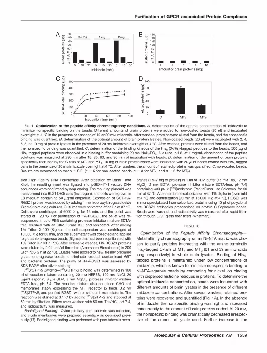
Optimization of the Peptide Affinity Chromatography—Metal affinity chromatography on an Ni-NTA matrix was cho-sen to purify proteins interacting with the amino-terminallyHis6-tagged C-tails of MT1 and MT2 (61 and 59 amino acidslong, respectively) in whole brain lysates. Binding of His6-tagged proteins is maintained under low concentrations ofimidazole, which is known to minimize nonspecific binding toNi-NTA-agarose beads by competing for nickel ion bindingwith dispersed histidine residues in proteins. To determine theoptimal imidazole concentration, beads were incubated withdifferent amounts of brain lysates in the presence of differentimidazole concentrations. After several washes, retained pro-teins were recovered and quantified (Fig. 1A). In the absenceof imidazole, the nonspecific binding was high and increasedconcurrently to the amount of brain proteins added. At 20 mM,the nonspecific binding was dramatically decreased irrespec-tive of the amount of lysate used. Further increase in the
B
2 4 6 8 10amount of brain proteins (mg)
elut
ed p
rote
ins
(µg)
2
160140120100806040200
4 6 8 10
A
imidazole (mM)
0.5 mg 1 mg 2 mg
elut
ed p
rote
ins
(µg)
160140120100806040200
0 10 20 0 10 20 0 10 20
D
C + MT1 + MT2
elut
ed p
rote
ins
(µg)
100
80
60
40
20
0
C
incubation time (min)
500
0
400
100
300
200
20 40 60 80 1000
6xH
is-C
-tail
bind
ing
(µg)
FIG. 1. Optimization of the peptide affinity chromatography conditions. A, determination of the optimal concentration of imidazole tominimize nonspecific binding on the beads. Different amounts of brain proteins were added to non-coated beads (20 �l) and incubatedovernight at 4 °C in the presence or absence of 10 or 20 mM imidazole. After washes, proteins were eluted from the beads, and the nonspecificbinding was quantified. B, determination of the optimal amount of brain protein lysates. Non-coated beads (20 �l) were incubated with 2, 4,6, 8, or 10 mg of protein lysates in the presence of 20 mM imidazole overnight at 4 °C. After washes, proteins were eluted from the beads, andthe nonspecific binding was quantified. C, determination of the binding kinetics of the His6 (6xHis)-tagged peptides to the beads. 500 �g ofHis6-tagged peptides were dissolved in a binding buffer containing 20 mM NaH2PO4, 6 M urea, pH 8, at 1 mg/ml. Absorbance of the peptidesolutions was measured at 280 nm after 15, 30, 60, and 90 min of incubation with beads. D, determination of the amount of brain proteinsspecifically recruited by the C-tails of MT1 and MT2. 10 mg of brain protein lysate were incubated with 20 �l of beads coated with His6-taggedbaits in the presence of 20 mM imidazole overnight at 4 °C. After washes, the amount of retained proteins was quantified. C, non-coated beads.Results are expressed as mean � S.E. (n � 5 for non-coated beads, n � 3 for MT1, and n � 6 for MT2).
Purification of GPCR-associated Protein Complexes
Molecular & Cellular Proteomics 7.8 1559
amount of brain lysate (up to 10 mg of protein) did not changethe level of nonspecific binding (10–12 �g) (Fig. 1B). We thusincubated Ni-NTA beads with 10 mg of brain lysate in thepresence of 20 mM imidazole in all further experiments.
We then determined the binding kinetics of the His6-taggedpeptides to the Ni-NTA beads in phosphate buffer at pH 8, theoptimal pH for binding of His6-tagged peptides (according tothe manufacturer) and for the preservation of protein complexes(18). Binding of peptides was rapid and reached a plateau after30 min. After 90 min, 295 � 21 �g (n � 4) and 351 � 16 �g (n �
4) of His6 MT1 and MT2 C-tails, respectively, were immobilizedon Ni-NTA beads (Fig. 1C). Using these optimized experimentalsettings, the C-tails of MT1 and MT2 typically recruited 80.7 �
4.3 �g (n � 3) and 26.4 � 2.7 �g (n � 6) of proteins, respec-tively. Under our conditions, nonspecific binding in the absenceof peptide was 11.9 � 1.4 �g (n � 6).
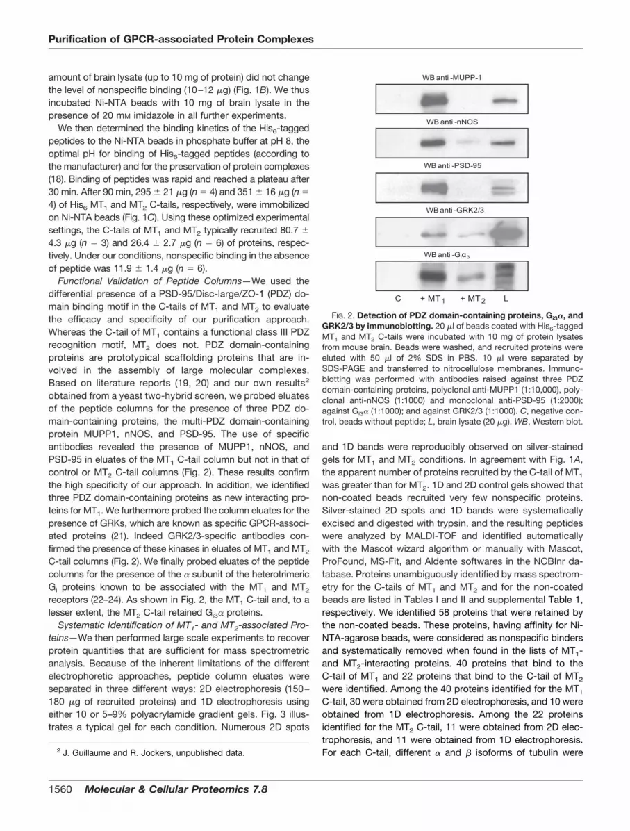
Functional Validation of Peptide Columns—We used thedifferential presence of a PSD-95/Disc-large/ZO-1 (PDZ) do-main binding motif in the C-tails of MT1 and MT2 to evaluatethe efficacy and specificity of our purification approach.Whereas the C-tail of MT1 contains a functional class III PDZrecognition motif, MT2 does not. PDZ domain-containingproteins are prototypical scaffolding proteins that are in-volved in the assembly of large molecular complexes.Based on literature reports (19, 20) and our own results2
obtained from a yeast two-hybrid screen, we probed eluatesof the peptide columns for the presence of three PDZ do-main-containing proteins, the multi-PDZ domain-containingprotein MUPP1, nNOS, and PSD-95. The use of specificantibodies revealed the presence of MUPP1, nNOS, andPSD-95 in eluates of the MT1 C-tail column but not in that ofcontrol or MT2 C-tail columns (Fig. 2). These results confirmthe high specificity of our approach. In addition, we identifiedthree PDZ domain-containing proteins as new interacting pro-teins for MT1. We furthermore probed the column eluates for thepresence of GRKs, which are known as specific GPCR-associ-ated proteins (21). Indeed GRK2/3-specific antibodies con-firmed the presence of these kinases in eluates of MT1 and MT2
C-tail columns (Fig. 2). We finally probed eluates of the peptidecolumns for the presence of the � subunit of the heterotrimericGi proteins known to be associated with the MT1 and MT2
receptors (22–24). As shown in Fig. 2, the MT1 C-tail and, to alesser extent, the MT2 C-tail retained Gi3� proteins.
Systematic Identification of MT1- and MT2-associated Pro-teins—We then performed large scale experiments to recoverprotein quantities that are sufficient for mass spectrometricanalysis. Because of the inherent limitations of the differentelectrophoretic approaches, peptide column eluates wereseparated in three different ways: 2D electrophoresis (150–180 �g of recruited proteins) and 1D electrophoresis usingeither 10 or 5–9% polyacrylamide gradient gels. Fig. 3 illus-trates a typical gel for each condition. Numerous 2D spots
and 1D bands were reproducibly observed on silver-stainedgels for MT1 and MT2 conditions. In agreement with Fig. 1A,the apparent number of proteins recruited by the C-tail of MT1
was greater than for MT2. 1D and 2D control gels showed thatnon-coated beads recruited very few nonspecific proteins.Silver-stained 2D spots and 1D bands were systematicallyexcised and digested with trypsin, and the resulting peptideswere analyzed by MALDI-TOF and identified automaticallywith the Mascot wizard algorithm or manually with Mascot,ProFound, MS-Fit, and Aldente softwares in the NCBInr da-tabase. Proteins unambiguously identified by mass spectrom-etry for the C-tails of MT1 and MT2 and for the non-coatedbeads are listed in Tables I and II and supplemental Table 1,respectively. We identified 58 proteins that were retained bythe non-coated beads. These proteins, having affinity for Ni-NTA-agarose beads, were considered as nonspecific bindersand systematically removed when found in the lists of MT1-and MT2-interacting proteins. 40 proteins that bind to theC-tail of MT1 and 22 proteins that bind to the C-tail of MT2
were identified. Among the 40 proteins identified for the MT1
C-tail, 30 were obtained from 2D electrophoresis, and 10 wereobtained from 1D electrophoresis. Among the 22 proteinsidentified for the MT2 C-tail, 11 were obtained from 2D elec-trophoresis, and 11 were obtained from 1D electrophoresis.For each C-tail, different � and � isoforms of tubulin were2 J. Guillaume and R. Jockers, unpublished data.
WB anti -MUPP-1
WB anti -nNOS
WB anti -PSD-95
C + MT1 + MT2 L
WB anti -GRK2/3
WB anti -Giα3
FIG. 2. Detection of PDZ domain-containing proteins, Gi3�, andGRK2/3 by immunoblotting. 20 �l of beads coated with His6-taggedMT1 and MT2 C-tails were incubated with 10 mg of protein lysatesfrom mouse brain. Beads were washed, and recruited proteins wereeluted with 50 �l of 2% SDS in PBS. 10 �l were separated bySDS-PAGE and transferred to nitrocellulose membranes. Immuno-blotting was performed with antibodies raised against three PDZdomain-containing proteins, polyclonal anti-MUPP1 (1:10,000), poly-clonal anti-nNOS (1:1000) and monoclonal anti-PSD-95 (1:2000);against Gi3� (1:1000); and against GRK2/3 (1:1000). C, negative con-trol, beads without peptide; L, brain lysate (20 �g). WB, Western blot.
Purification of GPCR-associated Protein Complexes
1560 Molecular & Cellular Proteomics 7.8

identified from 1D electrophoresis (10 and 5–9% gradientgels), but the corresponding spots, although easily noticeablefrom 2D gels, were not excised for identification. If � and �
isoforms of tubulin are not taken into account, separation ofsamples by 1D electrophoresis allowed the identification ofeight and nine additional high molecular mass proteins (in-cluding seven proteins of molecular mass �150 kDa) for MT1
and MT2, respectively. This significant increase in the numberof identified interacting proteins (20% of the MT1 and 41% ofthe MT2 interactomes) clearly illustrates that separation ofcolumn eluates by 1D electrophoresis is complementary to 2Delectrophoresis. Identified proteins showed localization to dif-ferent subcellular compartments (plasma membrane, cytosol,and cytoskeleton) and could be divided into five distinctgroups: “membrane proteins,” “signaling proteins,” “cytoskel-eton proteins,” “chaperones and stress response proteins,”and a last group, classified as “others,” containing the remain-ing proteins for which the function is not well described.
Validation of MT1- and MT2-associated Proteins by Immu-noblotting Screen—Using specific antibodies, we next per-formed an immunoblotting screen to estimate the overall re-liability of our mass spectrometry data and to quantify therelative amount of proteins that are recruited by both C-tails.Seven different antibodies were tested and confirmed thepresence of the interacting protein identified by MALDI-TOF,demonstrating the high quality of our mass spectrometric
data (Fig. 4). These experiments validated common interac-tion partners for both receptors, such as the tubulin � and �
isoforms, the catalytic subunit of the protein phosphatasePP2A, and the PKC �2, and showed higher recruitment withthe C-tail of MT1 compared with MT2. The immunoblottingscreen also confirmed MT1-specific interacting partners, suchas 14-3-3 �; the ubiquitin thiolesterase otubain 1 (OTUB1),which is known to express different alternatively spliced forms(25); and an RGSZ1 splice variant migrating at 36 kDa asreported previously in bovine brain (26). In addition, validationof 14-3-3 �, otubain 1, and the catalytic subunit of PP2Ademonstrates the reliability of MALDI-TOF identifications from1D gels. Using other antibodies, we also demonstrated thepresence of the regulatory subunit A�/� associated with thecatalytic subunit of PP2A and another isoform of 14-3-3 (14-3-3 �) in the MT1 C-tail eluates (supplemental Fig. 1). BothC-tails also recruited the � isoform of tubulin.
Validation of the Interaction between the MT1 Receptor andRGSZ1 in Living Cells—RGS proteins are signaling modula-tors of heterotrimeric G� proteins that increase their rate ofGTP hydrolysis. Recent observations indicate that RGS pro-teins regulate G protein-dependent signaling not only in a Gprotein-specific manner but also in a receptor-specific man-ner (27–30). The molecular basis for receptor specificity is stillpoorly understood. However, the recruitment of RGS proteinsinto receptor-associated complexes, as suggested by our
3 11
Non-coated beads + MT1 + MT2
25015010075
50
37
252015
10pI
AkDa
25015010075
50
37
252015
10
kDa25015010075
5037
252015
10
kDa
3 11pI 3 11pI
2121581169866564335
27
20
kDa
212
158116986656
kDa
C + MT1 + MT2
B C
C + MT1 + MT2
FIG. 3. 2D and 1D electrophoresis separation of the MT1 and MT2 C-tail-associated protein complexes. Mouse brain protein complexesrecruited by the Ni-NTA-immobilized C-tail of MT1 and MT2 receptors were separated by 2D (A) or 1D electrophoresis on a 10% (B) or 5–9%gradient polyacrylamide gel (C) and silver-stained. A typical gel for each condition is shown. C, negative control, beads without peptide. Thearrows indicate the position of RGSZ1.
Purification of GPCR-associated Protein Complexes
Molecular & Cellular Proteomics 7.8 1561
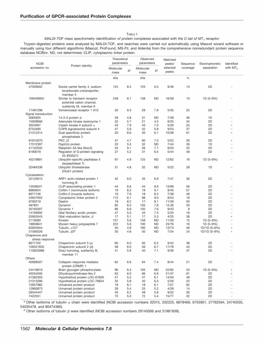
TABLE IMALDI-TOF mass spectrometry identification of protein complexes associated with the C-tail of MT1 receptor
Trypsin-digested proteins were analyzed by MALDI-TOF, and searches were carried out automatically using Mascot wizard software ormanually using four different algorithms (Mascot, ProFound, MS-Fit, and Aldente) from the comprehensive nonredundant protein sequencedatabase NCBInr. ND, not determined; CLIP, cytoplasmic linker protein.
NCBIaccession no.
Protein identity
Theoreticalparameters
Observedparameters
Matchedpeaks/
selectedpeaks
Sequencecoverage
Electrophoreticseparation
Identifiedwith MT2Molecular
masspI
Molecularmass
pI
kDa kDa %
Membrane protein47059002 Solute carrier family 4, sodium
bicarbonate cotransporter,member 5
124 8.4 125 6.5 9/46 13 2D �
109459952 Similar to transient receptorpotential cation channel,subfamily M, member 6
248 8.7 158 ND 16/58 10 1D (5–9%) �
17481296 Vomeronasal receptor 1 A12 35 9.3 28 7.8 5/35 25 2D �Signal transduction
3065925 14-3-3 protein � 28 4.8 31 ND 7/39 36 1D �15928666 Adenylate kinase isoenzyme 1 22 5.7 21 4.5 6/25 34 2D �3024067 Casein kinase II subunit � 45 7.9 43 7.5 5/26 25 2D �6753490 COP9 signalosome subunit 4 47 5.6 42 5.8 9/54 37 2D �21312314 Dual specificity protein
phosphatase 320 6.6 20 6.1 10/38 61 2D �
81912670 PKC �2 47 4.7 60 7.5 5/22 26 2D �17512397 Ppp2cb protein 32 5.5 32 ND 7/44 28 1D �41152502 Rabphilin 3A-like (Noc2) 34 9.1 26 7.7 9/53 25 2D �6180019 Regulator of G-protein signaling
20 (RGSZ1)27 5.2 31 6.5 5/44 48 2D �
45219891 Ubiquitin-specific peptidase 5(isopeptidase T)
97 4.9 125 ND 12/62 16 1D (5–9%) �
32484336 Ubiquitin thiolesterase(Otub1 protein)
31 4.8 32 ND 5/22 28 1D �
Cytoskeleton22122615 ARP1 actin-related protein 1
homolog B42 6.0 45 6.8 7/47 30 2D �
13508541 CLIP-associating protein 1 44 6.6 44 8.6 13/68 56 2D �6680924 Cofilin-1 (nonmuscle isoform) 19 8.2 18 9.1 9/45 57 2D �6671746 Cofilin-2 (muscle isoform) 19 7.6 18 8.2 7/38 48 2D �24657655 Cytoplasmic linker protein 2 112 6.1 88 9.0 8/53 18 2D �9790219 Destrin 19 8.2 17 9.1 11/26 50 2D �487851 Dynamin 85 6.0 105 7.8 13.36 20 2D �35193307 Dynamin 1 96 6.6 105 7.6 9/43 8 2D �14193692 Glial fibrillary acidic protein 47 5.0 43 7.5 5/25 16 2D �25955543 Glial maturation factor, � 17 5.1 17 5.3 4/25 38 2D �2119280 Kinesin 101 5.6 158 ND 11/58 15 1D (5–9%) �18859641 Myosin heavy polypeptide 7 222 5.6 100 ND 23/76 16 1D (5–9%) �62825944 Tubulin, �1Ca 50 4.9 160 ND 13/73 46 1D/1D (5–9%) �21595026 Tubulin, �3b 50 4.8 50 ND 7/34 24 1D/1D (5–9%) �
Chaperone andstress response
6671702 Chaperonin subunit 5 (�) 60 6.0 65 6.2 9/42 38 2D �126521835 Chaperonin subunit 2 (�) 58 6.0 56 6.7 17/78 43 2D �110625998 DnaJ homolog, subfamily B,
member 1141 5.9 40 7.1 9/36 37 2D �
Others40068507 Collapsin response mediator
protein (CRMP) 162 6.6 64 7.4 8/44 21 2D �
24418919 Brain glycogen phosphorylase 96 6.3 105 ND 22/82 33 1D (5–9%) �40254595 Dihydropyrimidinase-like 2 63 6.0 66 6.9 21/47 61 2D �21362303 Hypothetical protein LOC-67826 47 5.5 47 6.1 14/50 48 2D �21313286 Hypothetical protein LOC-76824 32 5.8 40 6.3 5/26 22 2D �12857982 Unnamed protein product 18 6.1 18 6.1 7/27 62 2D �12860873 Unnamed protein product 29 5.4 25 5.2 4/28 14 2D �26344447 Unnamed protein product 40 6.2 48 5.8 9/52 30 2D �7422551 Unnamed protein product 72 5.0 72 5.4 15/77 32 2D �
a Other isoforms of tubulin � chain were identified (NCBI accession numbers 32015, 202225, 6678469, 6755901, 27762594, 34740335,54035478, and 80474380).
b Other isoforms of tubulin � were identified (NCBI accession numbers 29145006 and 31981939).
Purification of GPCR-associated Protein Complexes
1562 Molecular & Cellular Proteomics 7.8

data, represents one possible explanation. To confirm thephysiological association of RGSZ1 with the full-length MT1
receptor, we co-expressed either FLAG-MT1 or FLAG-MT2
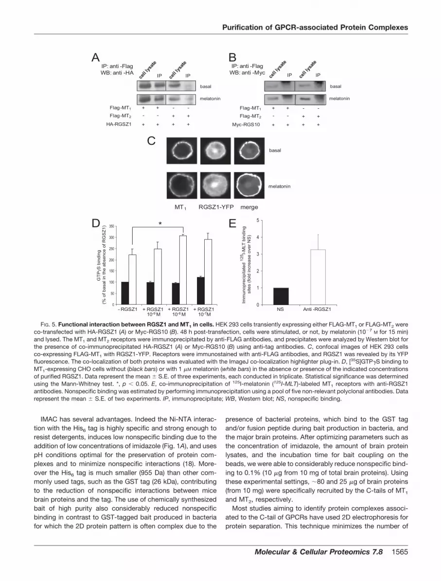
constructs together with HA-tagged RGSZ1 in HEK 293 cells.Co-immunoprecipitation experiments performed with anti-FLAG antibodies revealed that only MT1 co-immunoprecipi-tated RGSZ1 in both resting and melatonin-activated cells(Fig. 5A). The specificity of the interaction was further vali-dated using RGS10 as a negative control. As shown in Fig.5B, immunoprecipitation of FLAG-MT1 or FLAG-MT2 did notco-immunoprecipitate RGS10. As an additional control, wefurther performed immunofluorescence experiments in HEK293 cells co-expressing FLAG-MT1 together with RGSZ1-YFP(Fig. 5C). Consistent with our biochemical experiments, adistinct co-localization at the plasma membrane was ob-served in these cells irrespective of the activation state of thereceptor.
To evaluate the functional relevance of RGSZ1 couplingto the MT1 receptor, we determined the effect of increasingconcentrations of purified RGSZ1 on [35S]GTP�S binding inresponse to 1 �M melatonin using CHO cell membranesstably expressing high levels of MT1 (Fig. 5D). As expected,the agonist melatonin stimulated [35S]GTP�S binding (to221 � 27%), indicating efficient MT1 protein coupling in theabsence of RGSZ1. When purified RGSZ1 was added, anincrease in the [35S]GTP�S binding was observed in re-sponse to the agonist as reported previously for RGS4 (31).This assay is designed to promote [35S]GTP�S binding inthe presence of RGS proteins as GTP hydrolysis becomesrate-limiting due to saturation of GDP-GTP exchange athigh concentrations of receptor (31). This results in localdepletion of inactive heterotrimeric G-GDP that is reversedby RGS GTPase-activating protein activity. Indeed purifiedRGSZ1 accelerated melatonin-promoted [35S]GTP�S bind-
TABLE IIMALDI-TOF mass spectrometry identification of protein complexes associated with the C-tail of MT2 receptor
Trypsin-digested proteins were analyzed by MALDI-TOF, and searches were carried out automatically using Mascot wizard software ormanually using four different algorithms (Mascot, ProFound, MS-Fit, and Aldente) from the comprehensive nonredundant protein sequencedatabase NCBInr. ND, not determined.
NCBIaccession no. Protein identity
Proteinparameters
Observedparameters
Matchedpeaks/
selectedpeaks
Sequencecoverage
Electrophoreticseparation
Identifiedwith MT1Molecular
mass pI Molecularmass pI
kDa kDa %
Membrane protein6978547 Na�/K�-ATPase �3 subunit 113 5.3 105 ND 12/59 16 1D (5–9%) �26251984 Na�/K�-ATPase 3 (fragment) 33 5.2 33 8.5 4/27 30 2D �62660468 Similar to WD repeat
membrane protein150 5.9 125 ND 12/66 10 1D (5–9%) �
Signal transduction1262300 Casein kinase II � subunit 45 7.9 38 9.5 5/46 23 2D �400704 Catenin �-1 (p120-catenin) 102 6.5 110 ND 10/55 16 1D (5–9%) �56205529 Clathrin heavy polypeptide 194 5.5 180 ND 16/39 13 1D (5–9%) �21312314 Dual specificity phosphatase 3 21 6.1 21 6.1 7/58 50 2D �33438248 MKIAA0034 protein (clathrin
heavy chain)192 5.4 190 ND 22/67 17 1D (5–9%) �
31158356 PKC �2 47 4.7 41 9.0 6/44 28 2D �45477181 Tyrosine-protein phosphatase,
nonreceptor type 13272 5.9 250 ND 7/20 5 1D (5–9%) �
Cytoskeleton11993948 Glial maturation factor � 17 5.3 18 4.6 5/24 38 2D �547890 Microtubule-associated
protein 2203 4.8 250 ND 8/34 7 1D (5–9%) �
6678469 Tubulin, �1Ca 50 4.9 55 ND 9/49 30 1D/1D (5–9%) �12963615 Tubulin �3b 50 4.8 60 ND 13/46 44 1D/1D (5–9%) �
Chaperone andstress response
6754256 Heat shock protein 9 74 5.9 75 5.9 12/61 23 2D �97536358 Heat shock protein, 105 kDa
(HSP-E7I)96 5.4 65 4.5 6/41 13 2D �
Others24418919 Brain glycogen phosphorylase 97 6.3 100 ND 14/51 21 1D (5–9%) �7710012 Crystallin, � 34 5.4 35 5.3 4/40 23 2D �40254595 Dihydropyrimidinase-like 2 63 6.0 70 7.0 13/64 31 2D �66912155 mUp76 (Calpain 3) 77 5.6 60 7.7 8/38 25 2D �1334146 Unnamed protein product 30 5.3 29 4.2 5/36 32 2D �26326811 Unnamed protein product 120 8.8 158 ND 10/66 16 1D (5–9%) �
a Other isoform of tubulin � chain was identified (NCBI accession number 32015).b Other isoforms of tubulin � chain were identified (NCBI accession numbers 13542680 and 62663465).
Purification of GPCR-associated Protein Complexes
Molecular & Cellular Proteomics 7.8 1563

ing, reaching maximal values at 10�8 M RGSZ1 (305 � 8%,p � 0.02).
To confirm that the presence of RGSZ1 in MT1-associatedprotein complexes is physiologically relevant, we performedimmunoprecipitation studies with ovine pituitary pars tuberalistissue samples known to express significant amounts of en-dogenous MT1 receptors (32, 33) and RGSZ1 (data notshown). In the absence of high affinity anti-MT1 antibodies,receptors were labeled with the specific 2-[125I]iodomelatoninradioligand, and protein complexes were solubilized and im-munoprecipitated with anti-RGSZ1 antibodies. As shown inFig. 5E, significant amounts of radiolabeled MT1 were precip-itated in the presence of anti-RGSZ1 antibodies, demonstrat-ing the existence of MT1-RGSZ1 complexes in native tissue.
DISCUSSION
In this study, we present an improved proteomics approachcoupling peptide affinity chromatography with 1D and 2D elec-trophoresis separation and mass spectrometry for the identifi-cation of proteins interacting with the C-tail of GPCRs. To date,this is the first study proposing the use of an adapted andoptimized IMAC for the identification of protein complexes.Using this approach, we identified 40 proteins that bind to theC-tail of MT1 and 22 proteins that bind to the C-tail of MT2.
Interactions between GPCRs and associated proteins areknown to involve the intracellular loops, the transmembrane,and the C-tail of GPCRs. The most abundant and the best
studied are proteins interacting with the C-tail of GPCRs. ThisC-tail, recently nicknamed the “magic” tail by Bockaert et al.(7), serves as a scaffold for the formation of a signalosomecontaining diverse effectors and clustering proteins. The na-ture of the interaction proteins that bind to the C-tail of GPCRscan determine not only its targeting to a specific cellularcompartment but also its association with other signaling orstructural proteins and the fine tuning of its signal transduc-tion such as desensitization and resensitization (34). There-fore, the identification of the protein complexes associatedwith GPCRs constitutes an important step toward the devel-opment of new drugs. Indeed these compounds could beused to disrupt or strengthen specific interactions betweenGPCRs and their associated proteins. Different proteomicsapproaches coupling peptide affinity chromatography andmass spectrometry have been developed to identify proteinsinteracting with the C-tail of GPCRs and have proven theirefficacy in identifying protein complexes associated with theC-tail of GPCRs (7, 35). However, these approaches, whichused either short peptides encompassing a specific interac-tion motif such as the PDZ ligand of the 5-HT2A and 5-HT2C
receptors (12, 13) or the entire C-tail expressed as a GSTfusion protein such as for the 5-HT2C receptor (11), haveshown limitations. They were either too restrictive to someparticular interaction proteins (PDZ domain-containing pro-teins) or used GST fusion proteins as “bait” leading to in-creased background.
WB anti -tubulin alpha
43
56
66
97116
43
56
66
97116
WB anti -tubulin beta
kDa
kDa
C + MT1 + MT2 L
WB anti -PKC zeta
47
kDa
32,5WB anti -14-3-3 beta kDa
27
35
WB anti -PP2A subunit ckDa
WB anti -otubain 1
49
kDa
34C + MT1 + MT2 L
C + MT1 + MT2 L
C + MT1 + MT2 L
C + MT1 + MT2 L
C + MT1 + MT2 L
WB anti -RGSZ1
36
C + MT1 + MT2 L
kDa
FIG. 4. Validation of MALDI-TOF-identified binding proteins by immu-noblotting. Immobilized His6-taggedMT1 and MT2 C-tails were incubatedwith 10 mg of protein lysates frommouse brain. Recruited proteins wereeluted with 50 �l of 2% SDS in PBS. 10�l were separated by SDS-PAGE andtransferred to nitrocellulose mem-branes. Immunoblotting was performedwith monoclonal anti-tubulin � (1:2000),monoclonal anti-tubulin � (1:2000),monoclonal anti-PP2A catalytic subunit(1:1000), polyclonal anti-PKC � (1:1000),monoclonal anti-14-3-3 � (1:1000), poly-clonal anti-otubain 1 (1:2000), and poly-clonal anti-murine RGSZ1 (1:500) anti-bodies. C, negative control, non-coatedbeads; L, brain lysate (20 �g). WB, West-ern blot.
Purification of GPCR-associated Protein Complexes
1564 Molecular & Cellular Proteomics 7.8
IMAC has several advantages. Indeed the Ni-NTA interac-tion with the His6 tag is highly specific and strong enough toresist detergents, induces low nonspecific binding due to theaddition of low concentrations of imidazole (Fig. 1A), and usespH conditions optimal for the preservation of protein com-plexes and to minimize nonspecific interactions (18). More-over the His6 tag is much smaller (955 Da) than other com-monly used tags, such as the GST tag (26 kDa), contributingto the reduction of nonspecific interactions between micebrain proteins and the tag. The use of chemically synthesizedbait of high purity also considerably reduced nonspecificbinding in contrast to GST-tagged bait produced in bacteriafor which the 2D protein pattern is often complex due to the
presence of bacterial proteins, which bind to the GST tagand/or fusion peptide during bait production in bacteria, andthe major brain proteins. After optimizing parameters such asthe concentration of imidazole, the amount of brain proteinlysates, and the incubation time for bait coupling on thebeads, we were able to considerably reduce nonspecific bind-ing to 0.1% (10 �g from 10 mg of total brain proteins). Usingthese experimental settings, �80 and 25 �g of brain proteins(from 10 mg) were specifically recruited by the C-tails of MT1
and MT2, respectively.Most studies aiming to identify protein complexes associ-
ated to the C-tail of GPCRs have used 2D electrophoresis forprotein separation. This technique minimizes the number of
A B
++ ---- ++
++++
Flag-MT1
Flag-MT2
HA-RGSZ1
IP: anti -FlagWB: anti -HA
basal
melatonin
IP IP
++ ---- ++
++++
Flag-MT1
Flag-MT2
Myc-RGS10
IP: anti -FlagWB: anti -Myc
basal
melatonin
IP IP
ED
0
50
100
150
200
250
300
350
- RGSZ1 + RGSZ1 + RGSZ1 + RGSZ110-9 M 10-8 M 10-7M
*
0
1
2
3
4
5
NS Anti -RGSZ1
GTP
γS b
indi
ng
(% o
f bas
al in
the
abse
nce
of R
GS
Z1)
Imm
unop
reci
pita
ted
125 I-M
LT b
indi
ng
site
s (fo
ld in
crea
se o
ver N
S)
MT1 RGSZ1-YFP merge
basal
melatonin
C
FIG. 5. Functional interaction between RGSZ1 and MT1 in cells. HEK 293 cells transiently expressing either FLAG-MT1 or FLAG-MT2 wereco-transfected with HA-RGSZ1 (A) or Myc-RGS10 (B). 48 h post-transfection, cells were stimulated, or not, by melatonin (10�7 M for 15 min)and lysed. The MT1 and MT2 receptors were immunoprecipitated by anti-FLAG antibodies, and precipitates were analyzed by Western blot forthe presence of co-immunoprecipitated HA-RGSZ1 (A) or Myc-RGS10 (B) using anti-tag antibodies. C, confocal images of HEK 293 cellsco-expressing FLAG-MT1 with RGSZ1-YFP. Receptors were immunostained with anti-FLAG antibodies, and RGSZ1 was revealed by its YFPfluorescence. The co-localization of both proteins was evaluated with the ImageJ co-localization highlighter plug-in. D, [35S]GTP�S binding toMT1-expressing CHO cells without (black bars) or with 1 �M melatonin (white bars) in the absence or presence of the indicated concentrationsof purified RGSZ1. Data represent the mean � S.E. of three experiments, each conducted in triplicate. Statistical significance was determinedusing the Mann-Whitney test. *, p � 0.05. E, co-immunoprecipitation of 125I-melatonin (125I-MLT)-labeled MT1 receptors with anti-RGSZ1antibodies. Nonspecific binding was estimated by performing immunoprecipitation using a pool of five non-relevant polyclonal antibodies. Datarepresent the mean � S.E. of two experiments. IP, immunoprecipitate; WB, Western blot; NS, nonspecific binding.
Purification of GPCR-associated Protein Complexes
Molecular & Cellular Proteomics 7.8 1565
potential candidates as it is not adapted for selection ofhydrophobic, basic, and large proteins. To enlarge the num-ber of interacting proteins and obtain a more detailed inter-actome, we performed 2D electrophoresis and 1D electro-phoresis using 10 and 5–9% gradient gels. The utilization of1D electrophoresis was possible because of the small size ofthe His6-tagged baits (7.16 kDa for the MT1 and 6.80 kDa forthe MT2 C-tails) in contrast to the larger GST baits whose sizemakes the use of 1D electrophoresis difficult notably for theidentification of proteins with molecular weights similar to thatof the bait. Among the 40 proteins identified for the MT1 C-tail,30 were obtained from 2D electrophoresis, and 10 were ob-tained from 1D electrophoresis. Among the 22 proteins iden-tified for the MT2 C-tail, 11 were obtained from 2D electro-phoresis, and 11 were obtained from 1D electrophoresis.These results demonstrated that the combination of 1D and2D electrophoresis greatly increased the number of proteinsidentified and that the use of only 2D electrophoresis led to anunderestimation of multiprotein complexes associated to theC-tail of GPCRs. Identification of proteins by MALDI-TOFmass spectrometry, which only provides peptide mass finger-prints but no sequence information, is known to require priorhigh quality purification. However, our study has shown thatseveral proteins separated by 1D electrophoresis could beunambiguously identified by MALDI-TOF because of the sen-sitivity of the apparatus and the stringency of our experimen-tal conditions that considerably reduce the nonspecific bind-ing and consequently the complexity of the proteinelectrophoretic pattern for MS identification.
Several previously described GPCR-associated proteinswere identified. In this study, we showed that the C-tail of MT1
recruited 14-3-3 � and destrin, proteins shown to be associ-ated with mGluR5 (36), and dynamin 1, a protein known tointeract with 5-HT2C (11) and mGluR5 (36). Some proteinsfound in MT2 C-tail eluates were also already described asGPCR-binding proteins, such as the clathrin heavy chain andNa�/K�-ATPase �3 subunit for mGluR5 (36) and microtubule-associated protein 2 (MAP2) for mGluR5 (36) and 5-HT4a (13).Ten common members of MT1 and MT2 C-tail-associatedcomplexes were identified, including well known GPCR-asso-ciated proteins such as Gi� proteins and GRK2/3 and otherproteins such as dual specificity phosphatase 3 (DSP3), aprotein able to dephosphorylate protein substrates containingboth phosphotyrosine and phosphoserine or phosphothreo-nine residues, with specificity for the mitogen-activated pro-tein kinases (MAPKs) Erk2 and c-Jun NH2-terminal kinase(JNK) (37). The serine/threonine protein kinase casein kinase II(CK2), recently shown to be involved in phosphorylation of theM3 muscarinic receptor (38), was also found as a commoninteraction member of MT1 and MT2 C-tails. Interestingly arole for CK2 consensus sites within GPCR C-tails has beenproposed in targeting GPCRs to the �-arrestin-dependentpathway (39). PKC �2 and tubulins � and � were also recruitedby both MT1 and MT2 C-tails and confirmed by immunoblot-
ting. PKC �2 is specifically expressed in the mouse brain (40)and is a member of the atypical PKC subfamily. Tubulins �
and � constitute the basic building block of microtubules, themajor components of the cytoskeleton involved in many es-sential processes (41). Tubulins � and � have been shown tointeract with the C-tails of mGluR7a and mGluR1a, respec-tively (42–44), and we recently demonstrated a modulation ofmelatonin receptors and G protein function by microtubules(45).
Among the proteins involved in signal transduction thatwere specifically recruited by the C-tail of MT1, we identifiedtwo proteins involved in the deubiquitination pathway andmembers of the ubiquitin-specific processing proteases,otubain 1 (confirmed by Western blot) and ubiquitin-specificpeptidase 5, also known as isopeptidase T. Subunit 4 ofCOP9, a multiprotein complex of the ubiquitin-proteasomepathway, was also identified. The � isoform of the catalyticsubunit of protein phosphatase 2A (PPP2cb) was also found.PP2A is a highly conserved serine/threonine phosphatase thatplays pivotal roles in diverse cellular functions (46). A directinteraction between this subunit and the C-tail of the iono-tropic N-methyl-D-aspartate receptor has already been dem-onstrated by yeast two-hybrid and immunoprecipitationexperiments (47). In addition, PP2A has been shown to co-immunoprecipitate with mGluR5 and regulates mGluR5-de-pendent mitogen-activated protein kinase/extracellular sig-nal-regulated kinase kinase (MEK)/ERK phosphorylation inneurons (48). Finally three PDZ domain-containing proteinswere also identified as specific MT1-associated proteins byimmunoblot: MUPP1, nNOS, and PSD-95. We failed to iden-tify these three proteins by MS probably due to their lowabundance within the gels. MUPP1 has been characterizedpreviously as a direct interaction partner of MT1 that promotesGi protein coupling of this receptor (49). Neuronal NO syn-thase has been previously proposed as a potential interactingcandidate of the MT1 receptor (20). However, nothing isknown about the functional consequences of this particularinteraction. PSD-95 is a prototypical scaffolding protein highlyenriched in the PSD that belongs to the membrane-associ-ated guanylate kinase family (51). PSD-95 interacts with ahost of proteins of diverse functions, most notably N-methyl-D-aspartate receptors, and regulates the functional integrationof these receptors into signaling complexes in PSD (51). In-teraction between PSD-95 and GPCRs has already been de-scribed as with the somatostatin receptors SSTR4 and SSTR1(52) and dopamine D1 receptors (53). The coupling of PSD-95with the C-tail of D1 receptors has been shown to regulate thesurface expression and the intracellular trafficking of the re-ceptors and to inhibit D1 receptor-mediated signaling inmammalian cells (53). Gi3� proteins were also detected byimmunoblot but again failed to be identified by MS. In con-trast, Gi� was easily detectable by MS using the tandemaffinity purification (TAP) approach (24) suggesting that theC-tail of MT1 and MT2 is involved in Gi� binding but that
Purification of GPCR-associated Protein Complexes
1566 Molecular & Cellular Proteomics 7.8
further molecular determinants provided by the entire recep-tor are needed for efficient Gi� recruitment.
By our approach, we were also able to identify membraneproteins, which are often undetectable when using 2D elec-trophoresis. The majority of these membrane proteins wereidentified following 1D electrophoresis. Surprisingly, however,some of them were identified by 2D electrophoresis, such asthe vomeronasal type-1 receptor A12. Vomeronasal receptorsare GPCRs that bind pheromones and are responsible forvarious behavioral and neuroendocrine responses betweenindividuals (54). Heterodimerization between this vomeronasalreceptor and the melatonin MT1 receptor remains to be dem-onstrated. The number of identified proteins interacting withthe MT2 C-tail was lesser than that of MT1. However, severalinteresting proteins were identified, including catenin �-1, alsoknown as p120-catenin. This protein is a regulator of cadherinstability and an important modulator of Rho GTPase activities(55). �-Catenin was first identified through its interaction withpresenilin-1, the molecule most frequently mutated in familialAlzheimer disease (56), and shown to interact with mGluR1receptors by co-immunoprecipitation experiments using ratbrain homogenates and dissociate from mGluR1 upon acti-vation by L-glutamate (57).
Approaches based on isolated GPCR subdomains, such asthe C-tail, have to be compared with the recently describedTAP approach that uses the entire receptor as bait to pulldown GPCR-associated protein complexes (24). A carefulanalysis of the advantages and drawbacks shows that bothapproaches are rather complementary than mutually exclu-sive. Whereas the entire receptor allows the purification ofprotein complexes formed in intact cells, the C-tail approachis more easily accessible for comparing the interactomes ofdifferent tissues and under different experimental conditions(different pathophysiological models and pharmacologicaltreatments). A complete list of advantages and drawbacks ofboth approaches is presented in supplemental Table 2. Incontrast to the TAP approach, which is, by itself, a co-immu-noprecipitation performed from living cells, interactions be-tween the bait and the proteins identified by approachesusing GPCR subdomains need to be confirmed in intactcells with the full-length receptor ideally expressed endog-enously. In this study, we validated the interaction betweenthe MT1 receptor and RGSZ1, a brain-specific protein thatbelongs to the RGS family (26, 58, 59). RGS proteins havebeen described as signaling modulators of heterotrimericG� proteins that increase their rate of GTP hydrolysis andterminate signaling (60–62) and more recently as proteinsable to directly interact with GPCRs (27, 63–65). We suc-ceeded in confirming the interaction between the MT1 re-ceptors and RGSZ1 in cells from the ovine pars tuberalis,which expresses both proteins endogenously, and in trans-fected HEK 293 cells. The interaction was functional, con-stitutive, occurred at the plasma membrane, and was onlydetected with MT1 but not with MT2. The constitutive nature
of the interaction suggests precoupling of RGSZ1 to MT1 atthe basal state and molecular rearrangement within thispre-existing complex upon agonist stimulation. This modelis in good agreement with results described for other RGSproteins (27, 50, 63).
In conclusion, we present here a generic strategy that rep-resents a major methodological advance for the identificationof components of GPCR C-tail-associated complexes andovercomes the limitations of currently used genetics and pro-teomics approaches. Moreover this approach appears to becomplementary to recently developed proteomics ap-proaches based on the use of the entire GPCR as bait. Fea-tures of this new strategy are the use of chemically synthe-sized His6-tagged baits and IMAC that resulted in lownonspecific binding, pH conditions that preserved the integ-rity of associated protein complexes, and the association of1D and 2D gel electrophoresis for protein separation thatincreases the number of identified interaction partners. These62 candidates will now provide a basis for future cellularstudies. Our approach represents a new opportunity for thestudy of protein complexes associated with GPCRs that canbe used to compare GPCR interactomes from different tis-sues and can be extended to the identification of GPCR-associated complexes under pathological conditions or afterin vivo pharmacological treatments. Several interactionsstrongly depend on reversible post-translational modificationssuch as phosphorylation. In vitro phosphorylation of the ser-ine, threonine, and tyrosine residues of GPCR subdomains orthe replacement of these residues by phosphomimetic aminoacids are two further extensions of our approach to specifi-cally retain those complexes whose interaction depends onreceptor phosphorylation.
Acknowledgments—We thank Drs. Martine Migaud and BenoîtMalpaux (Institut National de la Recherche Agronomique, Nouzilly,France) for providing pars tuberalis tissue samples, Patty Chen forcomments on the manuscript, and Dr. Francois Guillonneau (bothfrom the Institut Cochin, Paris, France) for invaluable advices.
* This work was supported in part by grants from SERVIER, theFondation Recherche Medicale (“Equipe FRM”), Fondation pour laRecherche sur le Cerveau (FRC) Neurodon, INSERM, and CNRS. Thecosts of publication of this article were defrayed in part by the pay-ment of page charges. This article must therefore be hereby marked“advertisement” in accordance with 18 U.S.C. Section 1734 solely toindicate this fact.
□S The on-line version of this article (available at http://www.mcponline.org) contains supplemental material.
¶ Holds a EGID fellowship.‡‡ To whom correspondence should be addressed. Tel.: 331-40-
51-64-34; Fax: 331-40-51-64-30; E-mail: [email protected].
REFERENCES
1. Fredriksson, R., Lagerstrom, M. C., Lundin, L. G., and Schioth, H. B. (2003)The G-protein-coupled receptors in the human genome form five mainfamilies. Phylogenetic analysis, paralogon groups, and fingerprints. Mol.Pharmacol. 63, 1256–1272
2. Vassilatis, D. K., Hohmann, J. G., Zeng, H., Li, F., Ranchalis, J. E., Mortrud,M. T., Brown, A., Rodriguez, S. S., Weller, J. R., Wright, A. C., Bergmann,
Purification of GPCR-associated Protein Complexes
Molecular & Cellular Proteomics 7.8 1567
J. E., and Gaitanaris, G. A. (2003) The G protein-coupled receptor rep-ertoires of human and mouse. Proc. Natl. Acad. Sci. U. S. A. 100,4903–4908
3. Ellis, C., and The Nature Reviews Drug Discovery GPCR QuestionnaireParticipants (2004) The state of GPCR research in 2004. Nat. Rev. DrugDiscov. 3, 575, 577–626
4. Milligan, G., and White, J. H. (2001) Protein-protein interactions at G-protein-coupled receptors. Trends Pharmacol. Sci. 22, 513–518
5. Dev, K. K., Nakanishi, S., and Henley, J. M. (2001) Regulation of mglu7
receptors by proteins that interact with the intracellular C-terminus.Trends Pharmacol. Sci. 22, 355–361
6. El Far, O., and Betz, H. (2002) G-protein-coupled receptors for neurotrans-mitter amino acids: C-terminal tails, crowded signalosomes. Biochem. J.365, 329–336
7. Bockaert, J., Marin, P., Dumuis, A., and Fagni, L. (2003) The ‘magic tail’ ofG protein-coupled receptors: an anchorage for functional protein net-works. FEBS Lett. 546, 65–72
8. Diviani, D., Lattion, A. L., Abuin, L., Staub, O., and Cotecchia, S. (2003) Theadaptor complex 2 directly interacts with the �1b-adrenergic receptorand plays a role in receptor endocytosis. J. Biol. Chem. 278,19331–19340
9. Chen, Z., Hague, C., Hall, R. A., and Minneman, K. P. (2006) Syntrophinsregulate �1D-adrenergic receptors through a PDZ domain-mediatedinteraction. J. Biol. Chem. 281, 12414–12420
10. De Martelaere, K., Lintermans, B., Haegeman, G., and Vanhoenacker, P.(2007) Novel interaction between the human 5-HT7 receptor isoformsand PLAC-24/eIF3k. Cell. Signal. 19, 278–288
11. Becamel, C., Alonso, G., Galeotti, N., Demey, E., Jouin, P., Ullmer, C.,Dumuis, A., Bockaert, J., and Marin, P. (2002) Synaptic multiproteincomplexes associated with 5-HT2C receptors: a proteomic approach.EMBO J. 21, 2332–2342
12. Becamel, C., Gavarini, S., Chanrion, B., Alonso, G., Galeotti, N., Dumuis, A.,Bockaert, J., and Marin, P. (2004) The serotonin 5-HT2A and 5-HT2Creceptors interact with specific sets of PDZ proteins. J. Biol. Chem. 279,20257–20266
13. Joubert, L., Hanson, B., Barthet, G., Sebben, M., Claeysen, S., Hong, W.,Marin, P., Dumuis, A., and Bockaert, J. (2004) New sorting nexin (SNX27)and NHERF specifically interact with the 5-HT4a receptor splice variant:roles in receptor targeting. J. Cell Sci. 117, 5367–5379
14. Rabilloud, T., and Charmont, S. (2000) Detection of proteins on two-dimensional electrophoresis gels, in Proteome Research: Two-dimen-sional Electrophoresis and Identification Methods (Rabilloud, T., ed) pp.107–126, Springer, Berlin
15. Gharahdaghi, F., Weinberg, C. R., Meagher, D. A., Imai, B. S., and Mische,S. M. (1999) Mass spectrometric identification of proteins from silver-stained polyacrylamide gel: a method for the removal of silver ions toenhance sensitivity. Electrophoresis 20, 601–605
16. Levoye, A., Dam, J., Ayoub, M. A., Guillaume, J. L., Couturier, C., Dela-grange, P., and Jockers, R. (2006) The orphan GPR50 receptor specifi-cally inhibits MT1 melatonin receptor function through heterodimeriza-tion. EMBO J. 25, 3012–3023
17. Jockers, R., Da Silva, A., Strosberg, A. D., Bouvier, M., and Marullo, S.(1996) New molecular and structural determinants involved in �2-adre-nergic receptor desensitization and sequestration. Delineation using chi-meric �3/�2-adrenergic receptors. J. Biol. Chem. 271, 9355–9362
18. Husi, H., Ward, M. A., Choudhary, J. S., Blackstock, W. P., and Grant, S. G.(2000) Proteomic analysis of NMDA receptor-adhesion protein signalingcomplexes. Nat. Neurosci. 3, 661–669
19. Becamel, C., Figge, A., Poliak, S., Dumuis, A., Peles, E., Bockaert, J.,Lubbert, H., and Ullmer, C. (2001) Interaction of serotonin 5-hy-droxytryptamine type 2C receptors with PDZ10 of the multi-PDZ domainprotein MUPP1. J. Biol. Chem. 276, 12974–12982
20. Stricker, N. L., Christopherson, K. S., Yi, B. A., Schatz, P. J., Raab, R. W.,Dawes, G., Bassett, D. E., Bredt, D. S., and Li, M. (1997) PDZ domain ofneuronal nitric oxide synthase recognizes novel C-terminal peptide se-quences. Nat. Biotechnol. 15, 336–342
21. Ribas, C., Penela, P., Murga, C., Salcedo, A., Garcıa-Hoz, C., Jurado-Pueyo, M., Aymerich, I., and Mayor, F., Jr. (2007) The G protein-coupledreceptor kinase (GRK) interactome: role of GRKs in GPCR regulation andsignaling. Biochim. Biophys. Acta 1768, 913–922
22. Barrett, P., MacLean, A., and Morgan, P. J. (1994) Evidence for multiple
forms of melatonin receptor-G-protein complexes by solubilization andgel electrophoresis. J. Neuroendocrinol. 6, 509–515
23. Brydon, L., Roka, F., Petit, L., deCoppet, P., Tissot, M., Barrett, P., Morgan,P. J., Nanoff, C., Strosberg, A. D., and Jockers, R. (1999) Dual signalingof human Mel1a melatonin receptors via Gi2, Gi3, and Gq/11 proteins.Mol. Endocrinol. 13, 2025–2038
24. Daulat, A. M., Maurice, P., Froment, C., Guillaume, J. L., Broussard, C.,Monsarrat, B., Delagrange, P., and Jockers, R. (2007) Purification andidentification of G protein-coupled receptor protein complexes undernative conditions. Mol. Cell. Proteomics 6, 835–844
25. Soares, L., Seroogy, C., Skrenta, H., Anandasabapathy, N., Lovelace, P.,Chung, C. D., Engleman, E., and Fathman, C. G. (2004) Two isoforms ofotubain 1 regulate T cell anergy via GRAIL. Nat. Immunol. 5, 45–54
26. Wang, J., Ducret, A., Tu, Y., Kozasa, T., Aebersold, R., and Ross, E. M.(1998) RGSZ1, a Gz-selective RGS protein in brain. Structure, membraneassociation, regulation by G�z phosphorylation, and relationship to a GzGTPase-activating protein subfamily. J. Biol. Chem. 273, 26014–26025
27. Bernstein, L. S., Ramineni, S., Hague, C., Cladman, W., Chidiac, P., Levey,A. I., and Hepler, J. R. (2004) RGS2 binds directly and selectively to theM1 muscarinic acetylcholine receptor third intracellular loop to modulateGq/11� signaling. J. Biol. Chem. 279, 21248–21256
28. Benians, A., Nobles, M., Hosny, S., and Tinker, A. (2005) Regulators ofG-protein signaling form a quaternary complex with the agonist, recep-tor, and G-protein. A novel explanation for the acceleration of signalingactivation kinetics. J. Biol. Chem. 280, 13383–13394
29. Abramow-Newerly, M., Roy, A. A., Nunn, C., and Chidiac, P. (2006) RGSproteins have a signalling complex: interactions between RGS proteinsand GPCRs, effectors, and auxiliary proteins. Cell. Signal. 18, 579–591
30. Neitzel, K. L., and Hepler, J. R. (2006) Cellular mechanisms that determineselective RGS protein regulation of G protein-coupled receptor signaling.Semin. Cell Dev. Biol. 17, 383–389
31. Zhong, H., Wade, S. M., Woolf, P. J., Linderman, J. J., Traynor, J. R., andNeubig, R. R. (2003) A spatial focusing model for G protein signals.Regulator of G protein signaling (RGS) protein-mediated kinetic scaffold-ing. J. Biol. Chem. 278, 7278–7284
32. Barrett, P., MacLean, A., Davidson, G., and Morgan, P. J. (1996) Regulationof the Mel 1a melatonin receptor mRNA and protein levels in the ovinepars tuberalis: evidence for a cyclic adenosine 3�,5�-monophosphate-independent Mel 1a receptor coupling and an autoregulatory mechanismof expression. Mol. Endocrinol. 10, 892–902
33. Morgan, P. J. (2000) The pars tuberalis: the missing link in the photoperi-odic regulation of prolactin secretion? J. Neuroendocrinol. 12, 287–295
34. Bockaert, J., Fagni, L., Dumuis, A., and Marin, P. (2004) GPCR interactingproteins (GIP). Pharmacol. Ther. 103, 203–221
35. Pluder, F., Morl, K., and Beck-Sickinger, A. G. (2006) Proteome analysis tostudy signal transduction of G protein-coupled receptors. Pharmacol.Ther. 112, 1–11
36. Farr, C. D., Gafken, P. R., Norbeck, A. D., Doneanu, C. E., Stapels, M. D.,Barofsky, D. F., Minami, M., and Saugstad, J. A. (2004) Proteomicanalysis of native metabotropic glutamate receptor 5 protein complexesreveals novel molecular constituents. J. Neurochem. 91, 438–450
37. Ducruet, A. P., Vogt, A., Wipf, P., and Lazo, J. S. (2005) Dual specificityprotein phosphatases: therapeutic targets for cancer and Alzheimer’sdisease. Annu. Rev. Pharmacol. Toxicol. 45, 725–750
38. Torrecilla, I., Spragg, E. J., Poulin, B., McWilliams, P. J., Mistry, S. C.,Blaukat, A., and Tobin, A. B. (2007) Phosphorylation and regulation of aG protein-coupled receptor by protein kinase CK2. J. Cell Biol. 177,127–137
39. Hanyaloglu, A. C., Vrecl, M., Kroeger, K. M., Miles, L. E., Qian, H., Thomas,W. G., and Eidne, K. A. (2001) Casein kinase II sites in the intracellularC-terminal domain of the thyrotropin-releasing hormone receptor andchimeric gonadotropin-releasing hormone receptors contribute to�-arrestin-dependent internalization. J. Biol. Chem. 276, 18066–18074
40. Hirai, T., Niino, Y. S., and Chida, K. (2003) PKC�II, a small molecule ofprotein kinase C �, specifically expressed in the mouse brain. Neurosci.Lett. 348, 151–154
41. McKean, P. G., Vaughan, S., and Gull, K. (2001) The extended tubulinsuperfamily. J. Cell Sci. 114, 2723–2733
42. Saugstad, J. A., Yang, S., Pohl, J., Hall, R. A., and Conn, P. J. (2002)Interaction between metabotropic glutamate receptor 7 and alpha tubu-lin. J. Neurochem. 80, 980–988
Purification of GPCR-associated Protein Complexes
1568 Molecular & Cellular Proteomics 7.8
43. Ciruela, F., Robbins, M. J., Willis, A. C., and McIlhinney, R. A. (1999)Interactions of the C terminus of metabotropic glutamate receptor type1alpha with rat brain proteins: evidence for a direct interaction withtubulin. J. Neurochem. 72, 346–354
44. Enz, R. (2007) The trick of the tail: protein-protein interactions of metabo-tropic glutamate receptors. BioEssays 29, 60–73
45. Jarzynka, M. J., Passey, D. K., Ignatius, P. F., Melan, M. A., Radio, N. M.,Jockers, R., Rasenick, M. M., Brydon, L., and Witt-Enderby, P. A. (2006)Modulation of melatonin receptors and G-protein function by microtu-bules. J. Pineal Res. 41, 324–336
46. Janssens, V., and Goris, J. (2001) Protein phosphatase 2A: a highly regu-lated family of serine/threonine phosphatases implicated in cell growthand signalling. Biochem. J. 353, 417–439
47. Chan, S. F., and Sucher, N. J. (2001) An NMDA receptor signaling complexwith protein phosphatase 2A. J. Neurosci. 21, 7985–7992
48. Mao, L., Yang, L., Arora, A., Choe, E. S., Zhang, G., Liu, Z., Fibuch, E. E.,and Wang, J. Q. (2005) Role of protein phosphatase 2A in mGluR5-regulated MEK/ERK phosphorylation in neurons. J. Biol. Chem. 280,12602–12610
49. Guillaume, J. L., Daulat, A. M., Maurice, P., Levoye, A., Migaud, M., Brydon,L., Malpaux, B., Borg-Capra, C., and Jockers, R. (2008) The PDZ proteinMUPP1 promotes Gi coupling and signalling of the MT1 melatonin re-ceptor. J. Biol. Chem. 283, 16762–16771
50. Roy, A. A., Lemberg, K. E., and Chidiac, P. (2003) Recruitment of RGS2 andRGS4 to the plasma membrane by G proteins and receptors reflectsfunctional interactions. Mol. Pharmacol. 64, 587–593
51. Kim, E., and Sheng, M. (2004) PDZ domain proteins of synapses. Nat. Rev.Neurosci. 5, 771–781
52. Christenn, M., Kindler, S., Schulz, S., Buck, F., Richter, D., and Kreienkamp,H. J. (2007) Interaction of brain somatostatin receptors with the PDZdomains of PSD-95. FEBS Lett. 581, 5173–5177
53. Zhang, J., Vinuela, A., Neely, M. H., Hallett, P. J., Grant, S. G., Miller, G. M.,Isacson, O., Caron, M. G., and Yao, W. D. (2007) Inhibition of thedopamine D1 receptor signaling by PSD-95. J. Biol. Chem. 282,15778–15789
54. Rouquier, S., and Giorgi, D. (2006) Olfactory receptor gene repertoires inmammals. Mutat. Res. 616, 95–102
55. Reynolds, A. B. (2007) p120-catenin: past and present. Biochim. Biophys.Acta 1773, 2–7
56. Zhou, J., Liyanage, U., Medina, M., Ho, C., Simmons, A. D., Lovett, M., andKosik, K. S. (1997) Presenilin 1 interaction in the brain with a novelmember of the Armadillo family. Neuroreport 8, 2085–2090
57. Jones, S. B., Lanford, G. W., Chen, Y. H., Morabito, M., Moribito, M., Kim,K., and Lu, Q. (2002) Glutamate-induced �-catenin redistribution anddissociation from postsynaptic receptor complexes. Neuroscience 115,1009–1021
58. Glick, J. L., Meigs, T. E., Miron, A., and Casey, P. J. (1998) RGSZ1, aGz-selective regulator of G protein signaling whose action is sensitive tothe phosphorylation state of Gz�. J. Biol. Chem. 273, 26008–26013
59. Larminie, C., Murdock, P., Walhin, J. P., Duckworth, M., Blumer, K. J.,Scheideler, M. A., and Garnier, M. (2004) Selective expression of regu-lators of G-protein signaling (RGS) in the human central nervous system.Brain Res. Mol. Brain Res. 122, 24–34
60. Ross, E. M., and Wilkie, T. M. (2000) GTPase-activating proteins for het-erotrimeric G proteins: regulators of G protein signaling (RGS) and RGS-like proteins. Annu. Rev. Biochem. 69, 795–827
61. Hollinger, S., and Hepler, J. R. (2000) Cellular regulation of RGS proteins:modulators and integrators of G protein signaling. Pharmacol. Rev. 54,527–559
62. Willars, G. B. (2006) Mammalian RGS proteins: multifunctional regulators ofcellular signalling. Semin. Cell Dev. Biol. 17, 363–376
63. Hague, C., Bernstein, L. S., Ramineni, S., Chen, Z., Minneman, K. P., andHepler, J. R. (2005) Selective inhibition of �1A-adrenergic receptor sig-naling by RGS2 association with the receptor third intracellular loop.J. Biol. Chem. 280, 27289–27295
64. Georgoussi, Z., Leontiadis, L., Mazarakou, G., Merkouris, M., Hyde, K., andHamm, H. (2006) Selective interactions between G protein subunits andRGS4 with the C-terminal domains of the �- and �-opioid receptorsregulate opioid receptor signaling. Cell. Signal. 18, 771–782
65. Snow, B. E., Hall, R. A., Krumins, A. M., Brothers, G. M., Bouchard, D.,Brothers, C. A., Chung, S., Mangion, J., Gilman, A. G., Lefkowitz, R. J.,and Siderovski, D. P. (1998) GTPase activating specificity of RGS12 andbinding specificity of an alternatively spliced PDZ (PSD-95/Dlg/ZO-1)domain. J. Biol. Chem. 273, 17749–17755
Purification of GPCR-associated Protein Complexes
Molecular & Cellular Proteomics 7.8 1569