a focused review - mdpi
TRANSCRIPT
�����������������
Citation: Kamperman, R.G.; van der
Kooi, A.J.; de Visser, M.; Aronica, E.;
Raaphorst, J. Pathophysiological
Mechanisms and Treatment of
Dermatomyositis and Immune
Mediated Necrotizing Myopathies: A
Focused Review. Int. J. Mol. Sci. 2022,
23, 4301. https://doi.org/10.3390/
ijms23084301
Academic Editor: Yasmina Juarranz
Received: 4 March 2022
Accepted: 5 April 2022
Published: 13 April 2022
Publisher’s Note: MDPI stays neutral
with regard to jurisdictional claims in
published maps and institutional affil-
iations.
Copyright: © 2022 by the authors.
Licensee MDPI, Basel, Switzerland.
This article is an open access article
distributed under the terms and
conditions of the Creative Commons
Attribution (CC BY) license (https://
creativecommons.org/licenses/by/
4.0/).
International Journal of
Molecular Sciences
Review
Pathophysiological Mechanisms and Treatment ofDermatomyositis and Immune Mediated NecrotizingMyopathies: A Focused ReviewRenske G. Kamperman 1,*, Anneke J. van der Kooi 1, Marianne de Visser 1, Eleonora Aronica 2
and Joost Raaphorst 1
1 Department of Neurology, Amsterdam UMC, University of Amsterdam, Amsterdam Neuroscience,1100 DD Amsterdam, The Netherlands; [email protected] (A.J.v.d.K.);[email protected] (M.d.V.); [email protected] (J.R.)
2 Department of (Neuro)Pathology, Amsterdam UMC, University of Amsterdam, Meibergdreef 9,1105 AZ Amsterdam, The Netherlands; [email protected]
* Correspondence: [email protected]
Abstract: Idiopathic inflammatory myopathies (IIM), collectively known as myositis, are a compositegroup of rare autoimmune diseases affecting mostly skeletal muscle, although other organs or tissuesmay also be involved. The main clinical feature of myositis is subacute, progressive, symmetricalmuscle weakness in the proximal arms and legs, whereas subtypes of myositis may also presentwith extramuscular features, such as skin involvement, arthritis or interstitial lung disease (ILD).Established subgroups of IIM include dermatomyositis (DM), immune-mediated necrotizing myopa-thy (IMNM), anti-synthetase syndrome (ASyS), overlap myositis (OM) and inclusion body myositis(IBM). Although these subgroups have overlapping clinical features, the widespread variation in theclinical manifestations of IIM suggests different pathophysiological mechanisms. Various componentsof the immune system are known to be important immunopathogenic pathways in IIM, althoughthe exact pathophysiological mechanisms causing the muscle damage remain unknown. Currenttreatment, which consists of glucocorticoids and other immunosuppressive or immunomodulatingagents, often fails to achieve a sustained beneficial response and is associated with various adverseeffects. New therapeutic targets have been identified that may improve outcomes in patients withIIM. A better understanding of the overlapping and diverging pathophysiological mechanisms of themajor subgroups of myositis is needed to optimize treatment. The aim of this review is to report onrecent advancements regarding DM and IMNM.
Keywords: myositis; pathophysiological mechanisms; interferon pathway; treatment; DM; IMNM
1. Introduction
Idiopathic inflammatory myopathies (IIM), collectively known as myositis, are a com-posite group of rare autoimmune diseases affecting mostly skeletal muscle, although otherorgans or tissues may also be involved [1]. The current classification includes dermato-myositis (DM), immune-mediated necrotizing myopathy (IMNM), anti-synthetase syn-drome (ASyS), overlap myositis (OM) and inclusion body myositis (IBM) [1–3]. Polymyosi-tis is a contested entity within the spectrum and is considered a diagnosis by exclusion [4–6].Clinical features may vary between and within subgroups. DM is characterized by skininvolvement; other extramuscular features are frequently encountered, such as interstitiallung disease (ILD) [2]. IMNM and IBM are the most ‘muscle predominant’ phenotypeswithin the IIM spectrum [7,8]. In ASyS, multisystem involvement is frequent, i.e., ILD andarthritis are often the presenting symptoms and (peri)myocarditis may occur [9]. Overlapmyositis is often associated with connective tissue disorders [10,11].
Int. J. Mol. Sci. 2022, 23, 4301. https://doi.org/10.3390/ijms23084301 https://www.mdpi.com/journal/ijms
Int. J. Mol. Sci. 2022, 23, 4301 2 of 25
In recent decades, both adaptive and innate immune mechanisms have been shownto contribute to the pathogenesis of IIM. The characterization of T and B cell populationsin inflammatory infiltrates, the discovery of myositis-specific autoantibodies (MSA) andmyositis-associated autoantibodies (MAA) with corresponding clinical phenotypes, andnew insights into the role of derangements of the complement system and the importantcontribution of interferonopathy have led to increased understanding and new targetsfor therapy.
A better understanding of the overlapping and diverging pathophysiological mecha-nisms of the major subgroups of myositis is needed to optimize individual treatments foradult myositis patients.
This review aims to describe current knowledge on pathophysiological mechanismsin myositis, in relation to current and future pharmacological treatments.
We focus on two of the most prevalent subtypes of IIM in adults: dermatomyositis(DM) and immune-mediated necrotizing myopathy (IMNM). Other subtypes show a con-siderable overlap with rheumatic disorders (Table 1); for these subtypes we refer to focusedreviews [12,13]. IBM usually presents with a gradual onset and is slowly progressive overyears, characterized by asymmetric weakness [14,15]. Along with inflammatory findings,degenerative disease mechanisms play an important role in IBM, although the etiology ofinflammation and degeneration is so far unresolved [16,17]. IBM tends to be treatment-refractory. As mentioned above, this review focuses on IIM in adults; a review focusing onjuvenile DM was published in 2017 [18].
Table 1. Overlapping and diverging clinical characteristics of idiopathic inflammatory myopathies.
Characteristic DM IMNM ASyS OM
Muscle weakness ++ +++ ++ ++Malignancies ++ + − −Rapid progression * ++ +++ − −Raynaud’s − − ++ ++Skin involvement +++ + ++ +Interstitial lung disease + − +++ ++Peri/myocarditis ** + + + +
* in subtypes (e.g., IMNM with SRP autoantibodies and DM with MDA-5 antibodies (related to ILD)). ** de-scribed in case series of, predominantly, SRP-positive IMNM patients [19,20]. − = not present, + = rarely present,++ = sometimes present, +++ = (very) often present. DM = dermatomyositis, IMNM = immune-mediated necro-tizing myopathy, ASyS = anti-synthetase syndrome, OM = overlap myositis, SRP = signal recognition particle,MDA-5 = anti–melanoma differentiation gene 5, ILD = interstitial lung disease.
For DM and IMNM, we first describe the phenotype, including findings in musclebiopsies, and subsequently we describe three pathophysiological mechanisms: the humoralimmune response including the role of antibodies, derangements of the complement system,and overexpression of the interferon pathway. In addition, we review treatment optionswith an emphasis on these three pathophysiological mechanisms.
1.1. Clinical Features
Dermatomyositis (DM) is the most common (~30–40%) subtype of IIM [2,3,21]. DM ischaracterized by predominant proximal muscle weakness in combination with a typicalskin rash, in particular Gottron papules/sign or heliotrope erythema (Table 1). Muscleweakness leads to problems with swallowing, running, climbing stairs, rising from thefloor, and/or raising arms [2]. On laboratory testing, serum creatine kinase (CK) activity isoften elevated and, in addition, myositis specific antibodies (MDA-5, NXP-2, Mi-2, TIF1-γ,SAE-1) can be found in ~60% of patients [22]. The association with cancer is an importantfeature of DM: approximately 8% of patients with DM have or develop cancer (standardisedincidence ratio 2.4–6.2) [23,24].
Immune mediated necrotizing myopathy (IMNM) is the second most common subtypeof IIM (~20%) [8,21]. IMNM is characterized by rapid-onset severe proximal muscle weak-ness without clinically significant extramuscular disease activity. Serum CK activity is often
Int. J. Mol. Sci. 2022, 23, 4301 3 of 25
highly elevated. Two autoantibodies are associated with IMNM: anti-signal recognitionparticle (SRP) and anti-3-hydroxy-3-methylglutaryl-CoA reductase (HMGCR) autoantibod-ies. There is also a seronegative subtype in which these MSAs cannot be identified; each ofthese three IMNM subtypes (SRP+, HMGCR+ and seronegative) constitute one-third ofpatients with IMNM. In patients with anti-HMGCR autoantibodies, a history of statin useis found in approximately half of the patients [25]. Seronegative and anti-HMGCR-positiveIMNM are associated with cancer, while anti-SRP-positive IMNM is not [26,27].
1.2. Muscle Biopsy
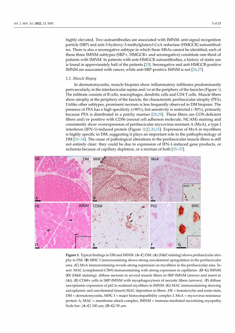
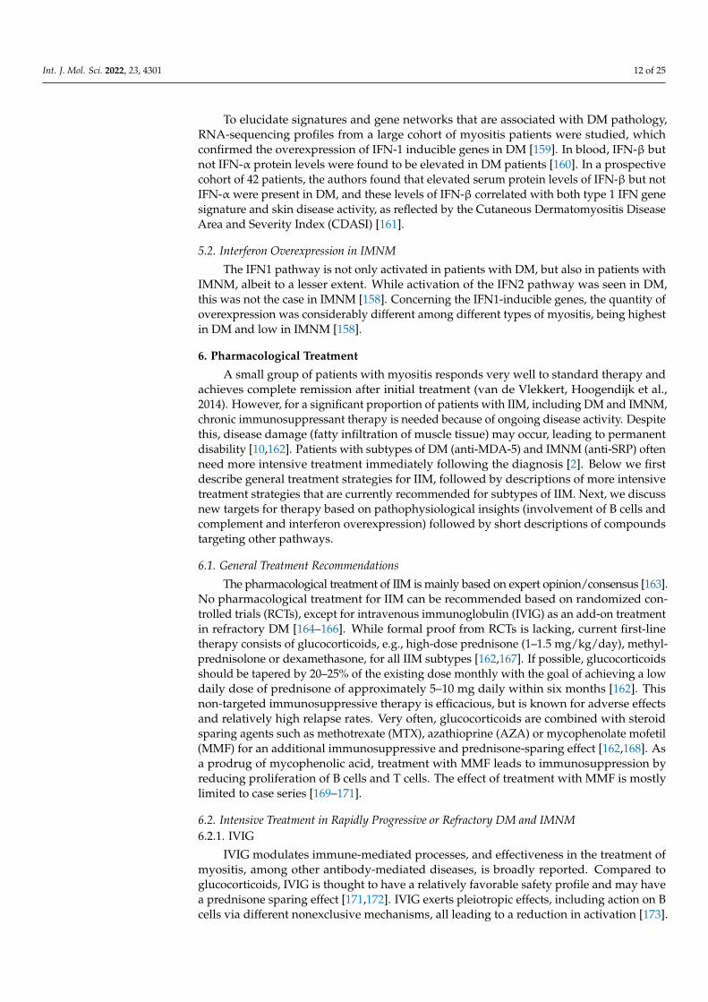
In dermatomyositis, muscle biopsies show inflammatory infiltrates predominantlyperivascularly, in the interfascicular septae and/or at the periphery of the fascicles (Figure 1).The infiltrate consists of B cells, macrophages, dendritic cells and CD4 T cells. Muscle fibersshow atrophy at the periphery of the fascicle, the characteristic perifascicular atrophy (PFA).Unlike other subtypes, prominent necrosis is less frequently observed in DM biopsies. Thepresence of PFA has a high specificity (>90%), but sensitivity is restricted (~50%), primarilybecause PFA is distributed in a patchy manner [28,29]. These fibers are COX-deficientfibers and/or positive with CD56 (neural cell adhesion molecule, NCAM) staining andconsistently show overexpression of perifascicular myxovirus resistant A (MxA), a type Iinterferon (IFN-1)-induced protein (Figure 1) [2,30,31]. Expression of MxA in myofibersis highly specific in DM, suggesting it plays an important role in the pathophysiology ofDM [32–34]. The cause of pathological alterations in the perifascicular muscle fibers is stillnot entirely clear: they could be due to expression of IFN-1-induced gene products, orischemia because of capillary depletion, or a mixture of both [35–37].
Int. J. Mol. Sci. 2022, 23, x FOR PEER REVIEW 4 of 25
Figure 1. Typical findings in DM and IMNM. (A–C) DM. (A) (H&E staining) shows perifascicular
atrophy in DM. (B) MHC I immunostaining shows strong sarcolemmal upregulation in the
perifascicular area. (C) MxA immunostaining reveals strong expression on myofibers in the
perifascicular area. Insert: MAC (complement C5b9) immunostaining with strong expression in
capillaries. (D–G) IMNM. (D) (H&E staining): diffuse necrosis in several muscle fibers in SRP-
IMNM (arrows and insert in (A)). (E) CD68+ cells in SRP-IMNM with myophagocytosis of necrotic
fibers (arrows). (F) diffuse sarcoplasmic expression of p62 in scattered myofibers in IMNM. (G)
MAC immunostaining showing sarcoplasmic and sarcolemmal (insert) MAC deposition in fibers.
HE = hematoxylin and eosin stain, DM = dermatomyositis, MHC I = major histocompatibility com-
plex I, MxA = myxovirus-resistance protein A, MAC = membrane attack complex, IMNM = im-
mune-mediated necrotizing myopathy. Scale bar: (A–C) 100 μm; (D–G) 50 μm.
In IMNM, muscle biopsies show abundant necrotic fibers invaded or surrounded by
macrophages in combination with regeneration of muscle (Figure 1). There is variable ex-
pression of major histocompatibility complex type I (MHC-I) in non-necrotic fibers and
diffuse and fine expression of sarcoplasmic p62 [34,42]. The macrophages in inflammatory
infiltrates in IMNM probably play a role in tissue repair [43,44]. This may explain why, in
muscle biopsies of IMNM, a higher frequency of regenerating fibers compared to necrotic
fibers is seen, when compared to DM biopsies [45].
Although the proportion of necrotic fibers correlates with CD3 and CD8 cells and
with the proportion of MHC-I positive fibers, no signs of overt cytotoxicity are typically
detected in IMNM biopsies. Invasion of non-necrotic muscle fibers by cytotoxic CD8+ T
cells (which is characteristic for inclusion body myositis) was not observed in anti-SRP
positive and anti-HMGCR positive muscles, and the surrounding of muscle fibers by T
cells was a rare event [45].
IMNM muscle biopsies characteristically show sarcolemmal and sarcoplasmic depo-
sition of complement (Membrane Attack Complex (MAC) on necrotic fibers (Figure 1).
Correlation between sarcolemmal deposits of C5b-9 and fiber necrosis has been shown
[45].
2. Pathophysiological Mechanisms
The exact pathophysiological mechanisms causing IIM remain unknown. In general,
and similarly to most other auto-immune disorders, an exogenous trigger (e.g., a viral
infection) is presumed to be required in combination with a genetic predisposition. In
Figure 1. Typical findings in DM and IMNM. (A–C) DM. (A) (H&E staining) shows perifascicular atro-phy in DM. (B) MHC I immunostaining shows strong sarcolemmal upregulation in the perifasciculararea. (C) MxA immunostaining reveals strong expression on myofibers in the perifascicular area. In-sert: MAC (complement C5b9) immunostaining with strong expression in capillaries. (D–G) IMNM.(D) (H&E staining): diffuse necrosis in several muscle fibers in SRP-IMNM (arrows and insert in(A)). (E) CD68+ cells in SRP-IMNM with myophagocytosis of necrotic fibers (arrows). (F) diffusesarcoplasmic expression of p62 in scattered myofibers in IMNM. (G) MAC immunostaining showingsarcoplasmic and sarcolemmal (insert) MAC deposition in fibers. HE = hematoxylin and eosin stain,DM = dermatomyositis, MHC I = major histocompatibility complex I, MxA = myxovirus-resistanceprotein A, MAC = membrane attack complex, IMNM = immune-mediated necrotizing myopathy.Scale bar: (A–C) 100 µm; (D–G) 50 µm.
Int. J. Mol. Sci. 2022, 23, 4301 4 of 25
Histopathological evidence of a microvascular pathogenesis in DM is further sug-gested by capillary abnormalities which were suggested to precede muscle fiber damageand other structural changes: the characteristic finding early in the disease process is thepresence of microtubular inclusions in endothelial cells, often preceding inflammatory cellinfiltrates [38,39]. These inclusions are related to the endoplasmatic reticulum (ER) or tothe outer nuclear membrane, and probably represent membranous specializations withinthe ER during a certain stage of cellular activity [40]. MAC deposition in capillary wallson endomysial capillaries has a high sensitivity and specificity in the diagnosis of DM,differentiating it from other subtypes of IIM [41].
In IMNM, muscle biopsies show abundant necrotic fibers invaded or surrounded bymacrophages in combination with regeneration of muscle (Figure 1). There is variableexpression of major histocompatibility complex type I (MHC-I) in non-necrotic fibers anddiffuse and fine expression of sarcoplasmic p62 [34,42]. The macrophages in inflammatoryinfiltrates in IMNM probably play a role in tissue repair [43,44]. This may explain why, inmuscle biopsies of IMNM, a higher frequency of regenerating fibers compared to necroticfibers is seen, when compared to DM biopsies [45].
Although the proportion of necrotic fibers correlates with CD3 and CD8 cells and withthe proportion of MHC-I positive fibers, no signs of overt cytotoxicity are typically detectedin IMNM biopsies. Invasion of non-necrotic muscle fibers by cytotoxic CD8+ T cells (whichis characteristic for inclusion body myositis) was not observed in anti-SRP positive andanti-HMGCR positive muscles, and the surrounding of muscle fibers by T cells was a rareevent [45].
IMNM muscle biopsies characteristically show sarcolemmal and sarcoplasmic depo-sition of complement (Membrane Attack Complex (MAC) on necrotic fibers (Figure 1).Correlation between sarcolemmal deposits of C5b-9 and fiber necrosis has been shown [45].
2. Pathophysiological Mechanisms
The exact pathophysiological mechanisms causing IIM remain unknown. In general,and similarly to most other auto-immune disorders, an exogenous trigger (e.g., a viralinfection) is presumed to be required in combination with a genetic predisposition. In somepatients, a clear trigger causing IIM can be identified. Anti-HMGCR is, in up to 67% ofcases, associated with prior statin use, particularly in patients older than 50 years of age [8].DM can occur as a paraneoplastic syndrome [46].
Recently, several cases of IIM following SARS-CoV-2 vaccination or COVID-19 infec-tion have been reported, including patients with myositis-specific antibodies [47]. In acase-control study, the autopsy of most patients who died from severe COVID-19 showedsigns of myositis ranging from mild to severe inflammation, with sarcolemmal majorhistocompatibility class 1 and in some cases class 2 staining patterns. In some patients,evidence of capillary interferon overexpression and complement activation was noted,suggesting type-1 interferonopathy. The authors did not demonstrate direct viral infectionof myofibers [48]. This led to the suggestion that SARS-CoV-2 may be associated withpostinfectious myositis.
In IIM, similarly to other autoimmune and immune-mediated diseases, the strongestgenetic risk for disease susceptibility is presumed to localize to specific leucocyte antigen(HLA) alleles [49,50]. The strongest associations have been found by stratifying patientgroups according to antibody status (see section below) [51]. Unique immunogenetic back-grounds were found in Asian IIM patients, when compared to Caucasian populations [52].
Immune-mediated disease mechanisms are thought to encompass both adaptive andinnate disease mechanisms [53]. Previous reports postulated a prominent role for B cells inDM and for cytotoxic CD8+ T-cells in other IIM subtypes [54]. More recent reports indicatethat cellular adaptive immunity, humoral adaptive immunity, and innate immunity arelikely all of importance in the pathogenesis of all subtypes [53]. In short, the pathogenicityof at least some myositis-specific autoantibodies is plausible, as supported by the finding ofrelated auto-antigen overexpression in IIM patients (e.g., TIF1-γ overexpression in endome-
Int. J. Mol. Sci. 2022, 23, 4301 5 of 25
trial cancer cells in patients with cancer-associated DM with anti-TIF1-γ autoantibodies)and experimental passive immunization via auto-antibody transfer (in in vitro and in vivomodels of seropositive IMNM) [26,55–58]. The muscle microenvironment in IIM is enrichedwith both myeloid and plasmacytoid antigen-presenting/dendritic cells, and muscle andmuscle-infiltrating cells overexpress innate immune receptors, including Toll-like receptors,which may lead to nuclear factor kappa B (NF-κB) and pro-inflammatory cytokine andchemokine signaling. This leads to IFN-1 overexpression in dermatomyositis, which maycause a pro-inflammatory state, MHC-class I overexpression, and non-immune mediatedtoxicity via mitochondrial damage and endoplasmatic reticulum stress [30,59].
Non-immune-mediated factors may also contribute to pathogenesis: muscle hypoxia,reactive oxygen species, heat shock response, endoplasmic reticulum stress, and abnormali-ties of cellular autophagy have all been described in IIM [60,61]. In short, these mechanismscreate a positive feedback loop with inflammation, leading to subsequent impaired mus-cle contraction, muscle protein dyshomeostasis, and disease damage with atrophy andirreversible muscle fiber alterations [61,62].
Various disease mechanisms have been postulated in IIM, of which we discuss indepth the role of the humoral immune response (B cells and antibodies), derangementsof the complement system, and overexpression of the interferon pathway, in the mostprevalent subtypes of IIM, dermatomyositis (DM) and immune mediated necrotizingmyopathy (IMNM).
3. Humoral Immunity in DM and IMNM: The Role of B Cells and Antibodies3.1. B Cells
Humoral immunity is a specific immune response mediated by antibodies. Sixty toseventy per cent of IIM patients have myositis-specific (~50%) or myositis-associated (~20%)antibodies [63,64]. These antibodies are produced by plasma cells and, to a lesser extent,plasmablasts. The latter differentiate from antigen-specific B cells, which are present in verylow frequencies in peripheral blood. A crucial role of B cells in the pathogenesis of IIM isevidenced by the relative effectiveness of B cell directed therapies such as rituximab in thetreatment of IIMs [65–67], the endomysial production of B cell activating factor (BAFF), thepresence of B cells and plasma cells in muscle tissue, antibody production, and endomysialB cell-maturation in part within the muscle itself [68,69].
3.1.1. B Cells in DM
DM muscle biopsies are especially known to include infiltrating CD20+ B cells, pre-dominantly in a perivascular distribution within the perimysium [44,70]. The numberof inflammatory cells varies from sparse infiltrates to B-cell rich infiltrates to nodular,follicle-like collections. This so-called ectopic lymphoid neogenesis, which can be seen innonlymphoid tissue in different autoimmune diseases, is thought to be extremely rare inDM [69,71]. Radke et al. [69] demonstrated that IFN-1 signature related gene-expressionlevels paralleled B cell content and architectural organization and linked B cell immunity tothe IFN-1 signature. They hypothesized that in situ B cell differentiation in skeletal musclein DM may dysregulate immunity, leading to excessive IFN response, which probably fuelsan amplification loop [69].
The role of antigen-driven B cell adaptive immune responses within the inflamedmuscle of IIM patients are not fully understood yet. Recent data suggest that variability,diversity, and joining (VDJ) repertoires of B cells from muscle tissues of DM patients deviatefrom normal immunoglobulin heavy chain (IgH) gene repertoires [72]. These VDJ reper-toires of muscle infiltrating B cells of DM patients often undergo clonal diversification andaccumulate high somatic hypermutations, suggesting an antigen-driven response as wellas a possible role of B cells in the pathological mechanisms responsible for this disorder.
Int. J. Mol. Sci. 2022, 23, 4301 6 of 25
3.1.2. B Cells in IMNM
In IMNM muscle biopsies, B cell mediated pathology is not as prominent whencompared to DM biopsies; in both anti-SRP positive and anti-HMGCR positive muscles,CD79+ B cell density was low, as was CD138+ plasma cell density [45]. This in agreementwith others reporting anti-HMGCR subjects showing small numbers of CD20+ cells, whichwere restricted to the endoymysium [44]. On the other hand, an important role of BAFF inthe pathological mechanism of IMNM has been suggested: in refractory patients with anti-SRP positive IMNM, BAFF-receptor (BAFF-R) expression in muscle is upregulated whencompared to non-refractory patients. This finding implicates BAFF as a possible contributorto muscle fiber injury in IMNM [73]. Additionally, autoantibodies in IMNM patients (anti-SRP and anti-HMGCR antibodies) have been reported to be crucial in initiating immuneevents, which are described below in more detail [73,74].
3.2. Autoantibodies
In the last decade over 30 different myositis-specific antibodies (MSA) and myositis-associated antibodies (MAA) have been identified. Out of these, 14 MSAs and 6 MAAshelp discriminate between subgroups and predict a higher chance of malignancy (anti-TIF1-γ, NXP-2, HMGCR) and/or extramuscular manifestations (e.g., ILD—MDA-5), andare therefore of prognostic importance [75,76]. Regarding associations with autoantibodiesand malignancies, no dominating type of malignancy was observed in association with DMor IMNM [77]. One hypothesis regarding the potential mechanisms of cancer-associatedmyositis (CAM) is shared antigen expression [78]. In patients with DM, shared expressionof several antigens is observed between tumor and regenerating muscle cells, indicatingthat an autoimmune response against cancer could cross-react with regenerating muscletissue, leading to auto-immunity [79]. The exact pathophysiological mechanism regardingthe presence of myositis specific autoantibodies and development of cancer remains asyet unknown.
The pathogenic role of the MSAs, if any, is unknown for most of the antibodies. Formost of the MSAs it remains to be determined whether the antigens reside on the surfaceof muscle cells (sarcolemma) in order to be targeted by the antibody, and if not, howantigens are reached intracellularly by the antibodies. Similarly, it is not known for mostMSAs whether they might cross-react with a different, unidentified antigen and whetherthe autoantibodies, other soluble factors, or infiltrating immune cells cause the muscledamage [80]. Below we focus on two MSAs in DM, i.e., anti–transcription intermediaryfactor 1-γ (TIF1-γ) and anti–melanoma differentiation gene 5 (MDA-5); and two MSAsassociated with IMNM, i.e., SRP and HMGCR. For these four MSAs a few lines of evidencehave been proposed which may (partly) explain their role in the pathogenicity of DMand IMNM.
3.2.1. Autoantibodies in DM
DM is associated with five MSAs, i.e., anti-TIF1-γ, anti-MDA-5, anti-SAE-1, anti-Mi-2and anti-NXP-2 (Table 2). The discovery of dermatomyositis-specific antibodies (DMSAs)resulted in valuable insights into the distinct features associated with different antibod-ies [32]. Relative proportions of DMSAs may differ between countries and continents.In a nationwide study on the prevalence of MSAs in the Netherlands (Euroline myositisline-blot assay; Euroimmun), out of 88 patients with an MSA, 49% of MSAs were DMSAs,with relative proportions ranging between 15% (anti-TIF1-γ) and 2% (anti-SAE-1) [81].Different DMSA-associated clinical phenotypes have been defined; in anti-TIF1-γ DM, skinlesions, dysphagia, and malignancy are frequent [82–84], whereas ulcerating skin lesions,interstitial lung disease (ILD) with low CK activity, and less frequent muscle involvementare encountered in anti-MDA-5 DM [85,86].
Int. J. Mol. Sci. 2022, 23, 4301 7 of 25
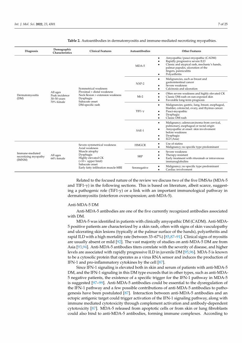
Table 2. Autoantibodies in dermatomyositis and immune-mediated necrotizing myopathies.
Diagnosis DemographicCharacteristics Clinical Features Autoantibodies Other Features
Dermatomyositis(DM)
All agesPeak incidence30–50 years70% female
Symmetrical weaknessProximal > distal weaknessNeck flexion > extension weaknessDysphagiaSubacute onsetDM-specific rash
MDA-5
• Amyopathic/pauci-myopathic (CADM)• Rapidly progressive severe ILD• Classic and atypical rash, mechanic’s hands,
palmar papules, ulceration of thefingers, panniculitis
• Polyarthritis
NXP-2
• Malignancies, such as breast andgastrointestinal cancer
• Severe weakness• Calcinosis and ulceration
Mi-2• Often severe weakness and highly elevated CK• Classic DM-rash on sun-exposed skin• Favorable long-term prognosis
TIF1-γ
• Malignancies; gastric, lung, breast, esophageal,bladder, colorectal, ovary, and thymus cancer.
• Pauci-myopathic• Dysphagia• Classic DM-rash
SAE-1
• Malignancy; adenocarcinoma from cervical,pulmonary, esophageal or rectal origin
• Amyopathic at onset: skin involvementbefore weakness
• Dysphagia• ILD (Asia)
Immune-mediatednecrotizing myopathy(IMNM)
All ages64% female
Severe symmetrical weaknessAxial weaknessMuscle atrophyDysphagiaHighly elevated CK(>10× upper limit)Subacute onsetEarly fatty infiltration muscle-MRI
HMGCR • Use of statins• Malignancy; no specific type predominant
SRP
• Severe weakness• Therapy-resistant• Early treatment with rituximab or intravenous
immunoglobulins
Seronegative • Malignancy; no specific type predominant• Cardiac involvement
Related to the focused nature of the review we discuss two of the five DMSAs (MDA-5and TIF1-γ) in the following sections. This is based on literature, albeit scarce, suggest-ing a pathogenic role (TiF1-y) or a link with an important immunological pathway indermatomyositis (interferon overexpression; anti-MDA-5).
Anti-MDA-5 DM
Anti-MDA-5 antibodies are one of the five currently recognized antibodies associatedwith DM.
MDA-5 was identified in patients with clinically amyopathic DM (CADM). Anti-MDA-5 positive patients are characterized by a skin rash, often with signs of skin vasculopathyand ulcerating skin lesions (typically at the palmar surface of the hands), polyarthritis andrapid ILD with a high mortality rate (between 33–67%) [85,87–91]. Clinical signs of myositisare usually absent or mild [92]. The vast majority of studies on anti-MDA-5 DM are fromAsia [93,94]. Anti-MDA-5 antibodies titers correlate with the severity of disease, and higherlevels are associated with rapidly progressive ILD in juvenile DM [95,96]. MDA-5 is knownto be a cytosolic protein that operates as a virus RNA sensor and induces the production ofIFN-1 and pro-inflammatory cytokines by the cell [87].
Since IFN-1 signaling is elevated both in skin and serum of patients with anti-MDA-5DM, and the IFN-1 signaling in this DM type exceeds that in other types, such as anti-MDA-5 negative patients, the existence of a specific trigger for the IFN-1 pathway in MDA-5is suggested [97–99]. Anti-MDA-5 antibodies could be essential to the dysregulation ofthe IFN-1 pathway and a few possible contributions of anti-MDA-5 antibodies to patho-genesis have been postulated [87]. Interaction between anti-MDA-5 antibodies and anectopic antigenic target could trigger activation of the IFN-1 signaling pathway, along withimmune mediated cytotoxicity through complement activation and antibody-dependentcytotoxicity [87]. MDA-5 released from apoptotic cells or from skin or lung fibroblastscould also bind to anti-MDA-5 antibodies, forming immune complexes. According to
Int. J. Mol. Sci. 2022, 23, 4301 8 of 25
Nombel et al., these immune complexes could potentially deposit in organs, e.g., in dermalor pulmonary vasculature, inducing vascular damage [87].
Finally, anti-MDA-5 antibodies may have the ability to penetrate cells and interactwith cytoplasmic MDA-5, similarly to other antibodies, altering various functional path-ways [87,100–102]. These potential contributions of MDA-5 antibodies to the pathogenesisof DM should be further investigated.
Anti-TIF1-γ DM
Anti-TIF1-γ antibodies are one of the five currently recognized antibodies associatedwith DM. In a murine model, the immune response against TIF-1γ seems to mediatethe induction of myositis [103]. However, the same authors showed that B cells andautoantibodies are not necessary for the development of TIF1-γ-induced murine myositis;therefore, the development of anti-TIF1γ autoantibodies cannot be linked directly to apathogenic role [103].
In adults only, TIF1-y antibodies are strongly associated with an underlying ma-lignancy. Approximately 60–80% of anti-TIF1γ-positive patients have cancer-associatedIIM [76]. Gastric, lung, breast, esophageal, bladder, colorectal, ovary, and thymus tumorsare found. Of interest, anti-TIF1-γ DM is also associated with malignancy in youngerpatients (<40 years) [32].
Anti-TIF1-γ plays a role in tissue differentiation, DNA repair, and tumor suppres-sion [76]. The pathogenicity of anti-TIF1-γ is plausible, as supported by the finding ofTIF1-γ overexpression in endometrial cancer cells in patients with cancer-associated DMwith anti-TIF1-γ autoantibodies [104]. A significantly higher expression of TIF1-γ hasbeen reported in tumors and muscles of anti-TIF1-γ positive DM patients as compared tonon-TIF1-γ associated DM patients [105].
3.2.2. Autoanntibodies in IMNM
IMNM is associated with two specific autoantibodies: anti-SRP and anti-HMGCR [106,107].An increased incidence of cancer is seen in patients with anti-HMGCR antibodies and inseronegative IMNM patients [108]. As described above, in IMNM, the sparse inflammatoryinfiltrates and presence of complement deposits on the sarcolemma of myofibers may pointto an auto-antibody-mediated pathophysiology rather than T cell-dependent cytotoxicity.However, since the expression of the auto-antigens SRP and HMGCR is ubiquitous ratherthan muscle-specific, the pathogenic role of autoantibodies directed against these moleculesin IMNM has remained elusive until the last five years [77,108,109].
Anti-SRP IMNM
In the abovementioned nationwide study on the prevalence of MSAs in the Nether-lands, out of 88 patients with an MSA, 13% of patients had anti-SRP antibodies [81].Compared to other IIMs, anti-SRP myopathy is associated with more pronounced clinicalweakness [45,110,111], highly elevated serum CK levels [45], extensive muscle edema withearly fatty degeneration on muscle MRI [112], and abundant necrotic fibers on biopsy [45].
The course of the disease tends to be rapidly progressive with severe disability withinmonths [113]. In a longitudinal study of IMNM patients, anti-SRP antibody titer correlatedwith serum CK activity and muscle strength [114]. Recent in vitro and in vivo studies haveincreased our understanding of the pathogenic role of anti-SRP autoantibodies in IMNM.
SRP is a ribonucleoprotein (RNP) that mediates nascent protein translocation to theendoplasmic reticulum (ER) [115,116]. Anti-SRP autoantibodies have been shown to inhibitSRP function in vitro [117]. Muscle co-culture with anti-SRP positive serum has beenshown to reduce myoblast viability, more markedly upon addition of complement [118].In another in vitro study, anti-SRP antibodies induced myotubular atrophy, increased pro-inflammatory cytokines, and impaired myoblast fusion due to decreased IL-4/IL-13 [55].
In an in vivo murine model, anti-SRP IgG passive transfer led to myofiber necrosis andmuscle deficit, which was less evident in C3-deficient mice; active immunization with anti-
Int. J. Mol. Sci. 2022, 23, 4301 9 of 25
SRP antibodies elicited muscle weakness [119]. The mechanism by which antibodies mayinteract with their cytoplasmic, ubiquitous cognate antigens and selectively induce musclepathology is as yet unresolved. Antibodies could block SRP function intracellularly; capac-ity for cell penetration has been confirmed for other antibodies, such as anti-DNA [120–123]and anti-RNP [124] antibodies.
As opposed to the intracellular effects of anti-SRP antibodies, sarcolemmal SRP expres-sion and consequential classical complement pathway activation poses another pathogenicpathway. For other ER chaperones, cell surface expression has been observed in variouscells [125] and in specific circumstances, such as macrophage fusion [126]. Indeed, studiesreported sarcolemmal SRP expression through co-labeling with membrane-associated pro-teins (dysferlin and dystrophin). Immunostaining of IMNM muscle biopsies of patientswith SRP antibodies also revealed that SRP positive fibers were partially co-labeling withsarcoplasmic neural cell adhesion molecule (NCAM), a marker of regeneration [45]. Co-localization of complement (C3c) with sarcolemmal and sarcoplasmic SRP expression hasbeen shown [45,118].
In summary, current evidence implies pathogenicity of the SRP-specific immuneresponse, possibly through multiple effector functions including complement-dependentantibody-mediated cytotoxicity. The exact immune-mediated events that lead to reducedmuscle strength, however, remain unclear, complicating targeted disease management.
Anti-HMGCR IMNM
HMGCR is an enzyme located at the membrane of the endoplasmic reticulum, andis the pharmacologic target of statins [26]. Antibodies against HMGCR have been foundin biopsy specimens of IMNM patients and much less frequently in other muscle condi-tions [80,127]. These antibodies are associated with statin exposure in approximately halfof the cases [25]. Statin-unexposed anti-HMGCR patients had higher CK levels and wereyounger [128]. The exact pathogenesis is not known, but a genetic predisposition (class IIHLA allele DRB1*11:01) and increased overexpression of HMG-CoA reductase in muscletissue following exposure to statins both play a role (Mammen 2016). Regenerating musclecells express high levels of HMG-CoA reductase protein [26,129], which is required for nor-mal muscle-cell differentiation [130,131]. Additional genetic risk factors and environmentaltriggers are probably involved but largely unknown [132].
The absence or a low number of infiltrating lymphocytes and the presence of MACon non-necrotic muscle-cell membranes suggest that anti-HMGCR autoantibodies may bepathogenic. This is supported by the observation that, similarly to anti-SRP antibodies,levels of anti-HMGCR antibodies correlate with disease activity (CK levels) and severity(muscle weakness) [27,127,128].
However, HMG-CoA reductase is not known to reside on the sarcolemma, where itcould be targeted by autoantibodies. Alternatively, anti-HMG-CoA reductase autoantibod-ies might cross-react with a different, unidentified antigen. Whether autoantibodies, someother soluble factor, or infiltrating immune cells cause the muscle damage remains to bedetermined (Mammen 2016).
In the abovementioned in vivo study on the pathogenicity of anti-SRP autoantibod-ies, the role of anti-HMGCR autoantibodies was also investigated: similarly to anti-SRPautoantibodies, anti-HMGCR autoantibodies induced myotubular atrophy, increased pro-inflammatory cytokines, and impaired myoblast fusion due to decreased IL-4/IL-13 [55].
In an in vivo study mouse model, C57BL/6, Rag2 deficient (Rag2−/−), and comple-ment component 3 deficient (C3−/−) mice were injected daily with plasma or purifiedantibodies from patients suffering from anti-HMGCR associated IMNM [119]. Muscle defi-ciency, evaluated by muscle strength on electrostimulation and grip test, tended to be lesssevere in mice receiving anti-HMGCR positive IgG compared to anti-SRP positive IgG. Thisseems to be consistent with observations in patients [112]. Histological analysis showed astatistically significant increase in necrotic myofibers after transfer of anti-HMGCR positiveIgG [119].
Int. J. Mol. Sci. 2022, 23, 4301 10 of 25
4. Derangement of the Complement System in DM and IMNM
The complement system is a key part of innate immunity and modulates the adap-tive immunity. There are three activation pathways: the classical, lectin, and alternativepathways. Surface-bound antibodies trigger the classical pathway, and amplification isaccommodated by the alternative pathway, regardless of the initiating route of activa-tion [133]. The classical complement pathway causes a cascade reaction of complementproteins, such as C1 and C3. Subsequently, C3 convertase cleaves C3 to produce C3b, whichforms a complex with C4b and C2a. This complex is a C5 convertase, which cleaves C5 toproduce C5a and C5b [134].
The lectin pathway starts with signal recognition by the oligomeric structures ofmannose-binding lectin (MBL), ficolins and collectins, which activate mannan-bindinglectin serine protease 1 (MASP1) and MASP2, which in their turn moderate production ofC4b. Next, the lectin pathway follows the same steps as the classical pathway [134]. In thealternative pathway, C3 interacts with factor B and factor D, leading to cleavage of furtherC3. As the final step of this pathway, an additional C3b binds to the C3 convertase andforms a C5 convertase, which cleaves C5 [135]. All three pathways generate C5b, whichtriggers the second part of the system: the lytic pathway and formation of the MAC [136].MAC, or C5b-9 complement complex, is the final step in the activation of the complementsystem. Complement deposition is not a unique feature of any IIM, as it may also occur(with divergent qualitative characteristics) in hereditary diseases of skeletal muscle [108].
4.1. Complement in DM
A comprehensive study in patients with DM examined the trigger for complementactivation. Despite the absence of binding of immunoglobulin complexes (IgGs), the patternof complement activation was consistent with activation of the classical pathway [137]. TheMAC reactive capillaries and transverse vessels reacted for C1q, which therefore served asa recognition and regulatory protein for the classical pathway [36].
The description of typical dermatomyositis in patients with a hereditary deficiency ofcomplement factor 2 (C2) illustrates that the classical and lectin pathways are not a conditiosine qua non for the development of DM, and that the alternative pathway may be primaryinvolved in a subset of patients [138].
Plasma levels of activated complement factor 3 (C3a) and C5b-9/MAC in patients withDM with active disease were higher than in inactive DM and healthy controls, suggesting arole of complement in DM [139]. IVIG treatment showed a strong reduction in C3 uptakeand disappearance of C3b and MAC in DM muscle biopsies, which was related to clinicalimprovement [140].
The presence of activated complement in DM is apparent in muscle biopsies. C4dstaining in muscle biopsies of DM patients mirrored C5b-9 staining [141]. Both antibodieslabeled the cytoplasm of degenerating necrotic myofibers. In addition, both antibodiesshowed distinct endomysial capillary labeling in a subset of dermatomyositis musclebiopsies including areas with perifascicular atrophy [141]. C5b-9/MAC deposition oncapillaries is a specific finding in DM biopsies and the extent of C5b-9 deposits seems to beassociated with the clinical course of the disease [36,142,143].
In very early stages of DM or in small muscle biopsies, inflammatory cell infiltrates maybe lacking, while C5b-9 deposits on endomysial capillary vessel walls are present, whichmay precede the development of inflammatory cell infiltrates in DM. C5b-9 deposits arethought to be evidence for DM being caused by a microvasculopathy [144,145]. The patternof capillary C5b-9 deposits is critical; non-DM patients may also show capillary complementdeposits, but in a more diffuse endomysial pattern, as opposed to the perifascicular patternin DM [146]. Although MAC formation is a typical feature in DM, the description oftypical dermatomyositis in patients with hereditary deficiencies of complement factor 9(C9) illustrates that DM may occur without the formation of C5b-9/MAC [147].
Int. J. Mol. Sci. 2022, 23, 4301 11 of 25
Despite uncertainties regarding the events that trigger complement activation and therelative contributions of each activation pathway to DM, there is evidence that DM is in parta complement-mediated microangiopathy, mostly mediated by the classical pathway [36].
4.2. Complement in IMNM
Although the mechanisms underlying myofiber necrosis have not been elucidated,given that IMNM patients have MAC on the sarcolemma of non-necrotic muscle fibers andthat few or no cytotoxic T cells and/or natural killer cells are observed in muscle biopsiesfrom patients with IMNM, myofiber toxicity may occur because of complement-mediatedcell death that is antibody-dependent [27,44,45,107,109,148]. Activation of the classicalcomplement cascade in seropositive IMNM patients has been identified by the presence ofsarcolemmal IgG1 and C1q and by the formation of C5b–9/MAC [45,108].
The contribution of complement mediated muscle injury in IIM has been investigatedin complement deficient and wild-type mice [119]. The authors showed that IgG from pa-tients with seropositive IMNM was markedly less pathogenic in complement (C3)-deficientmice than in wild-type mice. Supplementing recipient mice with Ig-depleted pooled hu-man complement serum augmented the in vivo pathogenicity of IgG from patients withseropositive IMNM [119]. Together, these data suggest an important role of the complementcascade in seropositive IMNM, although the exact contribution of complement pathwaysand factors needs to be further clarified [149].
5. Interferon Pathway5.1. Interferon Overexpression in DM
The interferon (IFN) pathway can be activated by the binding of three different typesof ligands to cell surface receptors: type 1 IFNs (i.e., IFN-α and IFN-β), type 2 IFNs(i.e., IFN-γ), and type 3 IFNs (i.e., IFN-λ) [150]. These proteins bind to their correspondingsurface receptors and subsequently stimulate the expression of IFN-inducible genes viathe Janus kinase (JAK)/signal transducer and activator of transcription (STAT) signalingpathway [151].
Myofibers in regions of PFA consistently express immunostaining of MxA, a IFN-1-induced protein, and MxA was also found in DM in normal-appearing perifascicularfibers [2]. Genes induced by IFN-1 are overexpressed in the muscle tissue of patients withDM. This overexpression is considered to be toxic to perifascicular muscle fibers and adja-cent capillaries [59]. High levels of expression of IFN-1-inducible genes corresponded withdifferent indicators of disease activity in DM, such as muscle weakness and elevated serumCK levels [152,153]. Considering the presence of interferon-inducible gene expressionin patients with DM, Greenberg et al. investigated plasmacytoid dendritic cells (PDCs),known as interferon-producing cells, and effector cells of the innate immune system, inmuscle biopsies (Greenberg, Pinkus et al., 2005) [154]. The finding of abundant PDCsindicated local production of interferon-α/β (IFN-1) [59].
In the innate immune response, the IFN signal activates the JAK-STAT intracellularcascade through interferon-stimulated gene (ISG)-15 conjugation to substrates [155]. ISG15is a ubiquitin-like protein and its conjugation to substrates modifies their expression levelsand activity [155].
A proteomic study showed that ISG15 binds to muscle proteins in DM muscles, suchas MxA, suggesting that ISG15 could play a regulatory role in DM pathology [156].
Along with overexpression of ISG15 in muscle biopsies in DM with PFA, ISG15expression levels alone can be used to accurately quantify the activation of the IFN-1pathway in muscle biopsies in IIM [157,158]. Concerning the IFN-1-inducible genes, thequantity of overexpression was considerably different among different types of myositisand was most consistently shown in DM. Activation of the IFN pathway was associatedwith increased expression of inflammatory cell and muscle regeneration genes. REF(S)
Activation of the IFN-2 pathway was also seen in DM [158].
Int. J. Mol. Sci. 2022, 23, 4301 12 of 25
To elucidate signatures and gene networks that are associated with DM pathology,RNA-sequencing profiles from a large cohort of myositis patients were studied, whichconfirmed the overexpression of IFN-1 inducible genes in DM [159]. In blood, IFN-β butnot IFN-α protein levels were found to be elevated in DM patients [160]. In a prospectivecohort of 42 patients, the authors found that elevated serum protein levels of IFN-β but notIFN-α were present in DM, and these levels of IFN-β correlated with both type 1 IFN genesignature and skin disease activity, as reflected by the Cutaneous Dermatomyositis DiseaseArea and Severity Index (CDASI) [161].
5.2. Interferon Overexpression in IMNM
The IFN1 pathway is not only activated in patients with DM, but also in patients withIMNM, albeit to a lesser extent. While activation of the IFN2 pathway was seen in DM,this was not the case in IMNM [158]. Concerning the IFN1-inducible genes, the quantity ofoverexpression was considerably different among different types of myositis, being highestin DM and low in IMNM [158].
6. Pharmacological Treatment
A small group of patients with myositis responds very well to standard therapy andachieves complete remission after initial treatment (van de Vlekkert, Hoogendijk et al.,2014). However, for a significant proportion of patients with IIM, including DM and IMNM,chronic immunosuppressant therapy is needed because of ongoing disease activity. Despitethis, disease damage (fatty infiltration of muscle tissue) may occur, leading to permanentdisability [10,162]. Patients with subtypes of DM (anti-MDA-5) and IMNM (anti-SRP) oftenneed more intensive treatment immediately following the diagnosis [2]. Below we firstdescribe general treatment strategies for IIM, followed by descriptions of more intensivetreatment strategies that are currently recommended for subtypes of IIM. Next, we discussnew targets for therapy based on pathophysiological insights (involvement of B cells andcomplement and interferon overexpression) followed by short descriptions of compoundstargeting other pathways.
6.1. General Treatment Recommendations
The pharmacological treatment of IIM is mainly based on expert opinion/consensus [163].No pharmacological treatment for IIM can be recommended based on randomized con-trolled trials (RCTs), except for intravenous immunoglobulin (IVIG) as an add-on treatmentin refractory DM [164–166]. While formal proof from RCTs is lacking, current first-linetherapy consists of glucocorticoids, e.g., high-dose prednisone (1–1.5 mg/kg/day), methyl-prednisolone or dexamethasone, for all IIM subtypes [162,167]. If possible, glucocorticoidsshould be tapered by 20–25% of the existing dose monthly with the goal of achieving a lowdaily dose of prednisone of approximately 5–10 mg daily within six months [162]. Thisnon-targeted immunosuppressive therapy is efficacious, but is known for adverse effectsand relatively high relapse rates. Very often, glucocorticoids are combined with steroidsparing agents such as methotrexate (MTX), azathioprine (AZA) or mycophenolate mofetil(MMF) for an additional immunosuppressive and prednisone-sparing effect [162,168]. Asa prodrug of mycophenolic acid, treatment with MMF leads to immunosuppression byreducing proliferation of B cells and T cells. The effect of treatment with MMF is mostlylimited to case series [169–171].
6.2. Intensive Treatment in Rapidly Progressive or Refractory DM and IMNM6.2.1. IVIG
IVIG modulates immune-mediated processes, and effectiveness in the treatment ofmyositis, among other antibody-mediated diseases, is broadly reported. Compared toglucocorticoids, IVIG is thought to have a relatively favorable safety profile and may havea prednisone sparing effect [171,172]. IVIG exerts pleiotropic effects, including action on Bcells via different nonexclusive mechanisms, all leading to a reduction in activation [173].
Int. J. Mol. Sci. 2022, 23, 4301 13 of 25
In refractory DM, IVIG has been proven to be effective as an add-on treatment to glucocor-ticoids and may be added in patients with rapidly progressive and/or severe/refractorydisease [164,165]. IVIG is also considered an effective treatment for refractory CADM basedon two retrospective studies evaluating the clinical response of refractory cutaneous der-matomyositis in patients for whom IVIG was initiated specifically for skin disease [174,175].Although relapses occurred, they were successfully treated with an additional course ofIVIG [175].
Patients with IMNM have been successfully treated with IVIG monotherapy, althoughthe number of reported patients is small [172,176]. As previously mentioned, IMNM ischaracterized by rapid onset and severe proximal muscle weakness, and aggressive earlytreatment should be considered. Early aggressive treatment with at least two immunother-apeutic agents and/or early treatment with IVIG can lead to favorable outcomes [177].
Our recent pilot study in treatment-naive patients with idiopathic inflammatory my-opathies showed clinically relevant improvement in 45% out of 19 patients after six to nineweeks of first-line IVIG monotherapy [178]. Compared to most other immunomodulatingcompounds, IVIG acts more quickly, which could lead to early and sustained suppressionof the inflammatory process when administered together with glucocorticoids. Consideringthe known effects of IVIG in IIM, both as add-on therapy in refractory patients and asmonotherapy in newly diagnosed IIM, double-blind RCTs are needed to investigate theeffect of add-on IVIG in patients with newly diagnosed IIM. Possibly early immunosup-pression via intensive treatment (‘hit early, hit hard’) may induce faster reduction of diseaseactivity and prevent chronic disability due to disease damage in myositis [179].
6.2.2. Plasmapheresis
Plasmapheresis (PE) is used in various refractory autoimmune diseases in order toremove circulating autoantibodies and immune complexes [180]. Since IIM is associatedwith the presence of autoantibodies, a beneficial effect could be assumed, in particular inthose subtypes with autoantibodies with a presumed pathogenic role (in particular anti-SRPand anti-HMGCR) as described above. However, a double-blind, placebo-controlled trialin refractory myositis failed to show efficacy [181]. A Japanese group investigated PE inanti-MDA-5 positive patients refractory to a combined immunosuppressive regimen, in aretrospective, single-center study. Patients who received PE showed a significantly bettersurvival rate than those who did not. These results may suggest that PE is expected to be aneffective adjuvant treatment in anti-MDA-5 positive patients, although larger prospectivestudies are needed [182].
6.2.3. Cyclophosphamide
Cyclophosphamide (CYC), an alkylating agent with strong cytotoxic and immunosup-pressive effects, has been reported to be efficient in severe DM with progressive ILD [183].In a large longitudinal cohort of 204 IIM patients, including 123 DM patients (60%), thetreatment effect of oral CYC in refractory myositis was described [184]. Significant improve-ment in disease activity measures and decrease in daily prednisone dose were reported.Further RCTs are warranted to verify these results [184].
Because standard immunosuppressive, so-called non-targeted, therapies lead to con-siderable numbers of patients with inadequate response to treatment, numerous side effects,and insufficient control of disease, more targeted therapies are warranted [185]. Targeted(biological) therapies, which target immune cells or cytokines directly via an antibody orsmall molecule, may greatly improve outcomes in patients with IIM [171,186]. In terms oflevel of evidence, to the best of our knowledge, one RCT has investigated a biological in IIM,rituximab, which depletes B cells, and is described in more detail below [187].
Int. J. Mol. Sci. 2022, 23, 4301 14 of 25
7. Pharmacological Compounds Targeting B Cells, Interferon Pathway and Complement7.1. B Cell Depletion
B cell-depleting rituximab is the most commonly used biological, and is mainly usedin patients with refractory IIM. The effect of B cell depletion therapies (e.g., rituximab)supports the role of B cells in myositis pathogenesis [65–67,187]. While the previouslymentioned RCT, called the Rituximab in Myositis (RIM) trial, failed to meet the primaryoutcome, 83% of all patients met the predefined definition of improvement [187]. Thepresence of specific MSAs might be associated with differences in treatment response. In asubgroup analysis of the RIM trial, the presence of anti-Mi-2 autoantibodies was a strongpredictor (Hazard Ratio 2.5, p < 0.01) of clinical improvement after B cell depletion withrituximab [187,188].
A retrospective cohort study included 43 IIM patients with inadequate responseto at least two immunosuppressive/immunomodulatory therapies who received twoinfusions of rituximab, one at baseline and one at six months, in order to maintain baselineimmunosuppressive treatment. Clinical and laboratory improvement (defined as >20%improvement in at least three of the six modified IMACS core set measures) was seen in allpatients at six months and the improvement persisted at one year [66].
A recent systematic review including 17 articles concluded that rituximab showedgood effect in the treatment of ILD related to anti-MDA-5 positive DM. Prior to treatmentwith rituximab, the majority (~80%) of patients were treated with medium- or high-doseglucocorticoids and at least one additional immunotherapeutic agent [189]. Therefore,rituximab could be a promising early treatment for anti-MDA-5 DM with ILD.
Belimumab is a humanized monoclonal antibody targeting the cytokine BAFF, whichhas been approved as a treatment for systemic lupus erythematosus (SLE) [190]. A double-blind placebo-controlled trial investigating the efficacy and safety of belimumab in patientswith IIM with a history of inadequate response to three months of glucocorticoids and/orone other immunosuppressive agent is currently ongoing (ClinicalTrials.gov Identifier:NCT02347891).
7.2. Interferon Pathway7.2.1. Anti-IFN-α Antibody
Sifalimumab is an anti-IFN-α monoclonal antibody. In a double-blind, phase 1b clinicaltrial, 51 patients were enrolled, including 26 DM patients. To evaluate the pharmacody-namic effects of sifalimumab in blood and muscle of myositis patients, neutralization of atype I IFN gene signature (IFNGS) was measured. Sifalimumab suppressed the IFNGS inblood, and a similar trend existed in muscle biopsies, although weaker [191]. Patients with15% or greater improvement on MMT scores showed greater neutralization of the IFNGSthan patients with less than 15% improvement in both blood and muscle.
7.2.2. Janus Kinases (JAK) Inhibitors
IFN-1 is suppressed by the JAK inhibitor ruxolitinib, and since the IFN-1 pathwayplays a prominent role in the pathogenesis of refractory DM, its use in future treatmentis of interest. The effect of IFN-1 suppression by ruxolitinib is seen in both muscle andin endothelial cells [192]. The authors reported a clinical improvement after 3 months oftreatment in four patients with refractory DM with reduced serum IFN-1 levels. Results ofa first proof-of-concept, open-label, prospective clinical trial of the JAK inhibitor tofacitinibin 10 patients with refractory DM, who had been treated with a minimum of a 12-weektrial of steroid therapy and at least one other first-line immunosuppressive agent, showedefficacy, as measured by the ACR/EULAR myositis response criteria, and in particular interms of skin changes [193].
Int. J. Mol. Sci. 2022, 23, 4301 15 of 25
7.3. Complement InhibitionAnti-C5 Monoclonal Antibodies
The literature describes one patient with refractory dermatomyositis (with concomitantthrombotic microangiopathy) showing robust improvement of muscle weakness, edema,myalgia and CK levels following treatment with a C5-blocking agent (eculizumab) afterseveral other treatments (steroids, IVIG, and PE) had failed [194]. In a randomized, double-blind, placebo-controlled 8-week pilot study in 13 DM patients, treatment with eculizumabshowed improvement from baseline in terms of Manual Muscle Testing (MMT) (6%),physician global score (9%) and skin disease score (37%) in the treatment arm. The placeboarm showed worsening. No serious adverse effects were noted, and the incidences of minoradverse effects were the same in the treatment and placebo arms [195].
In a phase 2 RCT, 27 IMNM patients with anti-SRP or anti-HMGCR autoantibodiesreceived either daily subcutaneuous zilucoplan, a complement C5 inhibitor, or placebo for8 weeks. Zilucoplan did not yield a significant change in CK levels, which was the primaryoutcome. No unexpected safety findings and no relevant safety differences betweenzilucoplan and placebo were identified (ClinicalTrials.gov identifier: NCT04025632).
A double-blind, randomized, placebo controlled trial with ravulizumab, a humanizedmonoclonal complement C5 inhibitor, is ongoing. In this large trial of 180 adult patientswith DM with inadequate response to two or more standard DM treatments, i.e., glucocor-ticoids or immunosuppressive/immunomodulatory therapies, intravenous ravulizumab isadministered as a loading dose followed by maintenance doses every 8 weeks, with primaryand secondary outcomes measured at 26 and 50 weeks respectively (ClinicalTrials.govIdentifier: NCT04999020).
8. New Pharmacological Compounds Targeting Other Pathways8.1. Tumor Necrosis Factor (TNF)-α
Tumor necrosis factor (TNF)-α, a pro-inflammatory cytokine, is present in elevatedlevels in muscle biopsies in IIM patients [196]. Excessive production of TNF-α may play arole in the pathogenesis of myositis, and therefore may lead to a therapeutic role for TNF-αinhibitors, such as infliximab or etanercept, in the treatment of IIM [197]. A retrospectivestudy in eight patients, six of whom were treated with etanercept, one with infliximab andone with both agents, showed improved motor strength and decreased fatigue [198]. In apilot study, treatment with infliximab in refractory myositis, defined as failure to respondto treatment with high doses of glucocorticoids for a minimum of 6 months, failed to beeffective [199]. An RCT with a cross-over period showed improvement in some patientstreated with infliximab. Sample size was limited (n = 12), and only 4 out of 12 patientsshowed improvement in the second phase of the study at 40 weeks. Future studies withlarger sample sizes are needed [200]. A randomized, double-blind, placebo-controlledtrial of etanercept in DM patients showed that etanercept was well-tolerated and had asignificant steroid-sparing effect [201]. The use of TNF inhibitors in IIM is debated atthe moment, since recent studies report patients developing autoimmune diseases aftertreatment [202,203].
8.2. Interleukine (IL)-Receptor Antagonists
Interleukin-6 (IL-6), a pro-inflammatory cytokine, is expressed in the muscle biopsiesof patients with different subtypes of myositis, including dermatomyositis [204]. Theexact role IL-6 in the pathogenesis of DM needs to be clarified. After approval of the IL-6receptor antagonist tocilizumab for treatment of rheumatoid arthritis, interest in its usefor other autoimmune diseases, such as IIM, has increased. A recent randomized, double-blind controlled trial failed to show the efficacy of tocilizumab in adults with refractorydermatomyositis [205].
Anakinra is a recombinant IL-1 receptor antagonist that has been approved for thetreatment of rheumatoid arthritis. Increased expression of IL-1α and IL-1β is seen in themuscle tissue of IIM patients [206]. Fifteen patients, refractory to high-dose prednisolone
Int. J. Mol. Sci. 2022, 23, 4301 16 of 25
in combination with AZA or MTX, were included in an open-label case study and receiveda daily dose of 100 mg subcutaneous anakinra. Seven patients showed a clinical response,according to the International Myositis Assessment and Clinical Studies Group (IMACS)definition of improvement. Patients with more extramuscular symptoms, such as ILD,responded better. Blood analysis demonstrated that blocking of the IL-1 receptor reducedT cell differentiation [207]. A large, randomized controlled trial is needed to prove theefficacy and safety of anakinra in IIM.
8.3. Inhibition of T-Cell Costimulation
Abatacept prevents T-cell activation by binding to CD80 and CD86 on antigen-presentingcells [208]. A recent randomized open-label trial included 20 patients with myositis refrac-tory to glucocorticoids and/or another immunosuppressant for at least 3 months. Nearlyhalf of the patients showed improvement in muscle strength and health-related qualityof life at 6 months [209]. A large phase III, double-blind, randomized trial evaluating theefficacy of subcutaneous abatacept, in addition to standard treatment, is currently ongoing(ClinicalTrials.gov identifier: NCT02971683).
9. Discussion
In summary, we reviewed recent advancements in the understanding of the patho-physiology and treatment of two of the most prevalent and severe subtypes of myositis,DM and IMNM. Based on new evidence related to the contributions of B cells, pathogenicantibodies, complement pathways and interferon overexpression, new therapeutic targetshave been identified. The promising effects of targeted therapies on laboratory and clinicaloutcomes in small series of myositis patients need to be corroborated in ongoing and futureclinical trials.
Author Contributions: Conceptualization, R.G.K., A.J.v.d.K., J.R.; writing—original draft prepara-tion, R.G.K., A.J.v.d.K. and J.R.; writing—review and editing, R.G.K., A.J.v.d.K., J.R., M.d.V. andE.A.; visualization, R.G.K., E.A., J.R. All authors have read and agreed to the published version ofthe manuscript.
Funding: This research received no external funding.
Institutional Review Board Statement: Not applicable.
Informed Consent Statement: Not applicable.
Data Availability Statement: Not applicable.
Acknowledgments: Several authors of this publication are members of the Netherlands Neuromus-cular Center (NL-NMD) and the European Reference Network for rare neuromuscular diseasesERN-EURO-NMD.
Conflicts of Interest: The authors declare no conflict of interest.
References1. Mariampillai, K.; Granger, B.; Amelin, D.; Guiguet, M.; Hachulla, E.; Maurier, F.; Meyer, A.; Tohme, A.; Charuel, J.L. Development
of a New Classification System for Idiopathic Inflammatory Myopathies Based on Clinical Manifestations and Myositis-SpecificAutoantibodies. JAMA Neurol. 2018, 75, 1528–1537. [CrossRef] [PubMed]
2. Mammen, A.L.; Allenbach, Y.; Stenzel, W.; Benveniste, O.; ENMC 239th Workshop Study Group. 239th ENMC InternationalWorkshop: Classification of dermatomyositis, Amsterdam, the Netherlands, 14–16 December 2018. Neuromuscul. Disord. 2020, 30,70–92. [CrossRef] [PubMed]
3. Schmidt, J. Current Classification and Management of Inflammatory Myopathies. J. Neuromuscul. Dis. 2018, 5, 109–129. [PubMed]4. Tanboon, J.; Uruha, A.; Stenzel, W.; Nishino, I. Where are we moving in the classification of idiopathic inflammatory myopathies?
Curr. Opin. Neurol. 2020, 33, 590–603. [CrossRef]5. van der Meulen, M.F.; Bronner, I.M.; Hoogendijk, J.E.; Burger, H.; van Venrooij, W.J.; Voskuyl, A.E.; Dinant, H.J.; Linssen, W.H.;
Wokke, J.H.; de Visser, M. Polymyositis: An overdiagnosed entity. Neurology 2003, 61, 316–321. [CrossRef]6. Loarce-Martos, J.; Lilleker, J.B.; Parker, M.; McHugh, N.; Chinoy, H. Polymyositis: Is there anything left? A retrospective
diagnostic review from a tertiary myositis centre. Rheumatology 2021, 60, 3398–3403. [CrossRef]
Int. J. Mol. Sci. 2022, 23, 4301 17 of 25
7. Hoogendijk, J.E.; Amato, A.A.; Lecky, B.R.; Choy, E.H.; Lundberg, I.E.; Rose, M.R.; Vencovsky, J.; de Visser, M.; Hughes, R.A.119th ENMC international workshop: Trial design in adult idiopathic inflammatory myopathies, with the exception of inclusionbody myositis, Naarden, The Netherlands, 10–12 October 2003. Neuromuscul. Disord. 2004, 14, 337–345. [CrossRef]
8. Allenbach, Y.; Mammen, A.L.; Benveniste, O.; Stenzel, W.; Immune-Mediated Necrotizing Myopathies Working Group. 224thENMC International Workshop:: Clinico-sero-pathological classification of immune-mediated necrotizing myopathies Zandvoort,The Netherlands, 14–16 October 2016. Neuromuscul. Disord. 2018, 28, 87–99. [CrossRef]
9. Connors, G.R.; Christopher-Stine, L.; Oddis, C.V.; Danoff, S.K. Interstitial lung disease associated with the idiopathic inflammatorymyopathies: What progress has been made in the past 35 years? Chest 2010, 138, 1464–1474. [CrossRef]
10. Van de Vlekkert, J.; Hoogendijk, J.E.; de Visser, M. Long-term follow-up of 62 patients with myositis. J. Neurol. 2014, 261, 992–998.[CrossRef]
11. Nuno-Nuno, L.; Joven, B.E.; Carreira, P.E.; Maldonado-Romero, V.; Larena-Grijalba, C.; Llorente Cubas, I.; Tomero, E.; Barbadillo-Mateos, M.C.; Garcia de la Pena Lefebvre, P.; Ruiz-Gutierrez, L.; et al. Overlap myositis, a distinct entity beyond primaryinflammatory myositis: A retrospective analysis of a large cohort from the REMICAM registry. Int. J. Rheum. Dis. 2019, 22,1393–1401. [PubMed]
12. Fredi, M.; Cavazzana, I.; Franceschini, F. The clinico-serological spectrum of overlap myositis. Curr. Opin. Rheumatol. 2018, 30,637–643. [CrossRef] [PubMed]
13. Zanframundo, G.; Faghihi-Kashani, S.; Scire, C.A.; Bonella, F.; Corte, T.J.; Doyle, T.J.; Fiorentino, D.; Gonzalez-Gay, M.A.; Hudson,M.; Kuwana, M.; et al. Defining anti-synthetase syndrome: A systematic literature review. Clin. Exp. Rheumatol. 2022, 40, 309–319.[CrossRef] [PubMed]
14. Dalakas, M.C. Review: An update on inflammatory and autoimmune myopathies. Neuropathol. Appl. Neurobiol. 2011, 37, 226–242.[CrossRef]
15. Lloyd, T.E.; Mammen, A.L.; Amato, A.A.; Weiss, M.D.; Needham, M.; Greenberg, S.A. Evaluation and construction of diagnosticcriteria for inclusion body myositis. Neurology 2014, 83, 426–433. [CrossRef]
16. Benveniste, O.; Stenzel, W.; Hilton-Jones, D.; Sandri, M.; Boyer, O.; van Engelen, B.G. Amyloid deposits and inflammatoryinfiltrates in sporadic inclusion body myositis: The inflammatory egg comes before the degenerative chicken. Acta Neuropathol.2015, 129, 611–624. [CrossRef]
17. Keller, C.W.; Schmidt, J.; Lunemann, J.D. Immune and myodegenerative pathomechanisms in inclusion body myositis. Ann. Clin.Transl. Neurol. 2017, 4, 422–445. [CrossRef]
18. Bellutti Enders, F.; Bader-Meunier, B.; Baildam, E.; Constantin, T.; Dolezalova, P.; Feldman, B.M.; Lahdenne, P.; Magnusson, B.;Nistala, K.; Ozen, S.; et al. Consensus-based recommendations for the management of juvenile dermatomyositis. Ann. Rheum.Dis. 2017, 76, 329–340. [CrossRef]
19. Allenbach, Y.; Benveniste, O. Peculiar clinicopathological features of immune-mediated necrotizing myopathies. Curr. Opin.Rheumatol. 2018, 30, 655–663. [CrossRef]
20. Kao, A.H.; Lacomis, D.; Lucas, M.; Fertig, N.; Oddis, C.V. Anti-signal recognition particle autoantibody in patients with andpatients without idiopathic inflammatory myopathy. Arthritis Rheum. 2004, 50, 209–215. [CrossRef]
21. Lilleker, J.B.; Vencovsky, J.; Wang, G.; Wedderburn, L.R.; Diederichsen, L.P.; Schmidt, J.; Oakley, P.; Benveniste, O.; Danieli, M.G.;Danko, K.; et al. The EuroMyositis registry: An international collaborative tool to facilitate myositis research. Ann. Rheum. Dis.2018, 77, 30–39. [CrossRef] [PubMed]
22. Lundberg, I.E.; Fujimoto, M.; Vencovsky, J.; Aggarwal, R.; Holmqvist, M.; Christopher-Stine, L.; Mammen, A.L.; Miller, F.W.Idiopathic inflammatory myopathies. Nat. Rev. Dis. Primers 2021, 7, 86. [CrossRef] [PubMed]
23. Chen, Y.J.; Wu, C.Y.; Huang, Y.L.; Wang, C.B.; Shen, J.L.; Chang, Y.T. Cancer risks of dermatomyositis and polymyositis: Anationwide cohort study in Taiwan. Arthritis Res. Ther. 2010, 12, R70. [CrossRef] [PubMed]
24. Hill, C.L.; Zhang, Y.; Sigurgeirsson, B.; Pukkala, E.; Mellemkjaer, L.; Airio, A.; Evans, S.R.; Felson, D.T. Frequency of specificcancer types in dermatomyositis and polymyositis: A population-based study. Lancet 2001, 357, 96–100. [CrossRef]
25. Musset, L.; Allenbach, Y.; Benveniste, O.; Boyer, O.; Bossuyt, X.; Bentow, C.; Phillips, J.; Mammen, A.; Van Damme, P.; Westhovens,R.; et al. Anti-HMGCR antibodies as a biomarker for immune-mediated necrotizing myopathies: A history of statins andexperience from a large international multi-center study. Autoimmun. Rev. 2016, 15, 983–993. [CrossRef] [PubMed]
26. Mammen, A.L.; Chung, T.; Christopher-Stine, L.; Rosen, P.; Rosen, A.; Doering, K.R.; Casciola-Rosen, L.A. Autoantibodies against3-hydroxy-3-methylglutaryl-coenzyme A reductase in patients with statin-associated autoimmune myopathy. Arthritis Rheum.2011, 63, 713–721. [CrossRef] [PubMed]
27. Allenbach, Y.; Drouot, L.; Rigolet, A.; Charuel, J.L.; Jouen, F.; Romero, N.B.; Maisonobe, T.; Dubourg, O.; Behin, A.; Laforet, P.; et al.Anti-HMGCR autoantibodies in European patients with autoimmune necrotizing myopathies: Inconstant exposure to statin.Medicine 2014, 93, 150–157. [CrossRef]
28. Suarez-Calvet, X.; Gallardo, E.; Pinal-Fernandez, I.; De Luna, N.; Lleixa, C.; Diaz-Manera, J.; Rojas-Garcia, R.; Castellvi, I.;Martinez, M.A.; Grau, J.M.; et al. RIG-I expression in perifascicular myofibers is a reliable biomarker of dermatomyositis. ArthritisRes. Ther. 2017, 19, 174. [CrossRef]
29. Pinal-Fernandez, I.; Casciola-Rosen, L.A.; Christopher-Stine, L.; Corse, A.M.; Mammen, A.L. The Prevalence of IndividualHistopathologic Features Varies according to Autoantibody Status in Muscle Biopsies from Patients with Dermatomyositis. J.Rheumatol. 2015, 42, 1448–1454. [CrossRef]
Int. J. Mol. Sci. 2022, 23, 4301 18 of 25
30. Greenberg, S.A. Dermatomyositis and type 1 interferons. Curr. Rheumatol. Rep. 2010, 12, 198–203. [CrossRef]31. Uruha, A.; Allenbach, Y.; Charuel, J.L.; Musset, L.; Aussy, A.; Boyer, O.; Mariampillai, K.; Landon-Cardinal, O.; Rasmussen, C.;
Bolko, L.; et al. Diagnostic potential of sarcoplasmic myxovirus resistance protein A expression in subsets of dermatomyositis.Neuropathol. Appl. Neurobiol. 2019, 45, 513–522. [CrossRef]
32. Tanboon, J.; Inoue, M.; Saito, Y.; Tachimori, H.; Hayashi, S.; Noguchi, S.; Okiyama, N.; Fujimoto, M.; Nishino, I. Dermatomyositis:Muscle Pathology According to Antibody Subtypes. Neurology 2022, 98, e739–e749. [CrossRef]
33. Uruha, A.; Nishikawa, A.; Tsuburaya, R.S.; Hamanaka, K.; Kuwana, M.; Watanabe, Y.; Suzuki, S.; Suzuki, N.; Nishino, I.Sarcoplasmic MxA expression: A valuable marker of dermatomyositis. Neurology 2017, 88, 493–500. [CrossRef]
34. Uruha, A.; Goebel, H.H.; Stenzel, W. Updates on the Immunopathology in Idiopathic Inflammatory Myopathies. Curr. Rheumatol.Rep. 2021, 23, 56. [CrossRef]
35. Franzi, S.; Salajegheh, M.; Nazareno, R.; Greenberg, S.A. Type 1 interferons inhibit myotube formation independently ofupregulation of interferon-stimulated gene 15. PLoS ONE 2013, 8, e65362. [CrossRef]
36. Lahoria, R.; Selcen, D.; Engel, A.G. Microvascular alterations and the role of complement in dermatomyositis. Brain 2016, 139 Pt 7,1891–1903. [CrossRef]
37. De Paepe, B. Vascular changes and perifascicular muscle fiber damage in dermatomyositis: Another question of the chicken orthe egg that is on our mind. Ann. Transl. Med. 2017, 5, 22. [CrossRef]
38. De Visser, M.; Emslie-Smith, A.M.; Engel, A.G. Early ultrastructural alterations in adult dermatomyositis. Capillary abnormalitiesprecede other structural changes in muscle. J. Neurol. Sci. 1989, 94, 181–192. [CrossRef]
39. Bronner, I.M.; Hoogendijk, J.E.; Veldman, H.; Ramkema, M.; van den Bergh Weerman, M.A.; Rozemuller, A.J.; de Visser, M.Tubuloreticular structures in different types of myositis: Implications for pathogenesis. Ultrastruct. Pathol. 2008, 32, 123–126.[CrossRef]
40. Banker, B.Q. Dermatomyositis of childhood: Ultrastructural alterations of muscle and intramuscular blood vessels. J. Neuropathol.Exp. Neurol. 1975, 34, 46–75. [CrossRef]
41. Jain, A.; Sharma, M.C.; Sarkar, C.; Bhatia, R.; Singh, S.; Gulati, S.; Handa, R. Detection of the membrane attack complex as adiagnostic tool in dermatomyositis. Acta Neurol. Scand. 2011, 123, 122–129. [CrossRef]
42. Merlonghi, G.; Antonini, G.; Garibaldi, M. Immune-mediated necrotizing myopathy (IMNM): A myopathological challenge.Autoimmun. Rev. 2022, 21, 102993. [CrossRef]
43. Tidball, J.G.; Villalta, S.A. Regulatory interactions between muscle and the immune system during muscle regeneration. Am. J.Physiol. Regul. Integr. Comp. Physiol. 2010, 298, R1173–R1187. [CrossRef]
44. Chung, T.; Christopher-Stine, L.; Paik, J.J.; Corse, A.; Mammen, A.L. The composition of cellular infiltrates in anti-HMG-CoAreductase-associated myopathy. Muscle Nerve 2015, 52, 189–195. [CrossRef]
45. Allenbach, Y.; Arouche-Delaperche, L.; Preusse, C.; Radbruch, H.; Butler-Browne, G.; Champtiaux, N.; Mariampillai, K.; Rigolet,A.; Hufnagl, P.; Zerbe, N.; et al. Necrosis in anti-SRP(+) and anti-HMGCR(+)myopathies: Role of autoantibodies and complement.Neurology 2018, 90, e507–e517. [CrossRef]
46. Stockton, D.; Doherty, V.R.; Brewster, D.H. Risk of cancer in patients with dermatomyositis or polymyositis, and follow-upimplications: A Scottish population-based cohort study. Br. J. Cancer 2001, 85, 41–45. [CrossRef]
47. Gracia-Ramos, A.E.; Martin-Nares, E.; Hernandez-Molina, G. New Onset of Autoimmune Diseases Following COVID-19Diagnosis. Cells 2021, 10, 3592. [CrossRef]
48. Aschman, T.; Schneider, J.; Greuel, S.; Meinhardt, J.; Streit, S.; Goebel, H.H.; Buttnerova, I.; Elezkurtaj, S.; Scheibe, F.; Radke, J.; et al.Association Between SARS-CoV-2 Infection and Immune-Mediated Myopathy in Patients Who Have Died. JAMA Neurol. 2021,78, 948–960. [CrossRef]
49. Miller, F.W.; Chen, W.; O’Hanlon, T.P.; Cooper, R.G.; Vencovsky, J.; Rider, L.G.; Danko, K.; Wedderburn, L.R.; Lundberg, I.E.;Pachman, L.M.; et al. Genome-wide association study identifies HLA 8.1 ancestral haplotype alleles as major genetic risk factorsfor myositis phenotypes. Genes Immun. 2015, 16, 470–480. [CrossRef]
50. Rothwell, S.; Chinoy, H.; Lamb, J.A. Genetics of idiopathic inflammatory myopathies: Insights into disease pathogenesis. Curr.Opin. Rheumatol. 2019, 31, 611–616. [CrossRef]
51. Rothwell, S.; Chinoy, H.; Lamb, J.A.; Miller, F.W.; Rider, L.G.; Wedderburn, L.R.; McHugh, N.J.; Mammen, A.L.; Betteridge, Z.E.;Tansley, S.L.; et al. Focused HLA analysis in Caucasians with myositis identifies significant associations with autoantibodysubgroups. Ann. Rheum. Dis. 2019, 78, 996–1002. [CrossRef] [PubMed]
52. Kang, E.H.; Go, D.J.; Mimori, T.; Lee, S.J.; Kwon, H.M.; Park, J.W.; Park, M.H.; Song, E.Y.; Ha, Y.J.; Lee, E.Y.; et al. Novelsusceptibility alleles in HLA region for myositis and myositis specific autoantibodies in Korean patients. Semin. Arthritis Rheum.2019, 49, 283–287. [CrossRef] [PubMed]
53. Miller, F.W.; Lamb, J.A.; Schmidt, J.; Nagaraju, K. Risk factors and disease mechanisms in myositis. Nat. Rev. Rheumatol. 2018, 14,255–268. [CrossRef] [PubMed]
54. Goebels, N.; Michaelis, D.; Engelhardt, M.; Huber, S.; Bender, A.; Pongratz, D.; Johnson, M.A.; Wekerle, H.; Tschopp, J.; Jenne, D.;et al. Differential expression of perforin in muscle-infiltrating T cells in polymyositis and dermatomyositis. J. Clin. Investig. 1996,97, 2905–2910. [CrossRef]
Int. J. Mol. Sci. 2022, 23, 4301 19 of 25
55. Arouche-Delaperche, L.; Allenbach, Y.; Amelin, D.; Preusse, C.; Mouly, V.; Mauhin, W.; Tchoupou, G.D.; Drouot, L.; Boyer,O.; Stenzel, W.; et al. Pathogenic role of anti-signal recognition protein and anti-3-Hydroxy-3-methylglutaryl-CoA reductaseantibodies in necrotizing myopathies: Myofiber atrophy and impairment of muscle regeneration in necrotizing autoimmunemyopathies. Ann. Neurol. 2017, 81, 538–548. [CrossRef]
56. Muro, Y.; Sugiura, K.; Hoshino, K.; Akiyama, M. Disappearance of anti-MDA-5 autoantibodies in clinically amyopathicDM/interstitial lung disease during disease remission. Rheumatology 2012, 51, 800–804. [CrossRef]
57. Abe, Y.; Matsushita, M.; Tada, K.; Yamaji, K.; Takasaki, Y.; Tamura, N. Clinical characteristics and change in the antibody titres ofpatients with anti-MDA5 antibody-positive inflammatory myositis. Rheumatology 2017, 56, 1492–1497. [CrossRef]
58. Matsushita, T.; Mizumaki, K.; Kano, M.; Yagi, N.; Tennichi, M.; Takeuchi, A.; Okamoto, Y.; Hamaguchi, Y.; Murakami, A.;Hasegawa, M.; et al. Antimelanoma differentiation-associated protein 5 antibody level is a novel tool for monitoring diseaseactivity in rapidly progressive interstitial lung disease with dermatomyositis. Br. J. Dermatol. 2017, 176, 395–402. [CrossRef]
59. Greenberg, S.A.; Pinkus, J.L.; Pinkus, G.S.; Burleson, T.; Sanoudou, D.; Tawil, R.; Barohn, R.J.; Saperstein, D.S.; Briemberg, H.R.;Ericsson, M.; et al. Interferon-alpha/beta-mediated innate immune mechanisms in dermatomyositis. Ann. Neurol. 2005, 57,664–678. [CrossRef]
60. Fischer, N.; Preusse, C.; Radke, J.; Pehl, D.; Allenbach, Y.; Schneider, U.; Feist, E.; von Casteleyn, V.; Hahn, K.; Ruck, T.; et al.Sequestosome-1 (p62) expression reveals chaperone-assisted selective autophagy in immune-mediated necrotizing myopathies.Brain Pathol. 2020, 30, 261–271. [CrossRef]
61. Ceribelli, A.; De Santis, M.; Isailovic, N.; Gershwin, M.E.; Selmi, C. The Immune Response and the Pathogenesis of IdiopathicInflammatory Myositis: A Critical Review. Clin. Rev. Allergy Immunol. 2017, 52, 58–70. [CrossRef] [PubMed]
62. Dalakas, M.C. Inflammatory Muscle Diseases. N. Engl. J. Med. 2015, 373, 393–394. [CrossRef] [PubMed]63. McHugh, N.J.; Tansley, S.L. Autoantibodies in myositis. Nat. Rev. Rheumatol. 2018, 14, 290–302. [CrossRef] [PubMed]64. Betteridge, Z.; McHugh, N. Myositis-specific autoantibodies: An important tool to support diagnosis of myositis. J. Intern. Med.
2016, 280, 8–23. [CrossRef] [PubMed]65. Aggarwal, R.; Oddis, C.V.; Goudeau, D.; Koontz, D.; Qi, Z.; Reed, A.M.; Ascherman, D.P.; Levesque, M.C. Autoantibody levels
in myositis patients correlate with clinical response during B cell depletion with rituximab. Rheumatology 2016, 55, 991–999.[CrossRef] [PubMed]
66. de Souza, F.H.C.; Miossi, R.; de Moraes, J.C.B.; Bonfa, E.; Shinjo, S.K. Favorable rituximab response in patients with refractoryidiopathic inflammatory myopathies. Adv. Rheumatol. 2018, 58, 31. [CrossRef] [PubMed]
67. Leclair, V.; Galindo-Feria, A.S.; Dastmalchi, M.; Holmqvist, M.; Lundberg, I.E. Efficacy and safety of rituximab in anti-synthetaseantibody positive and negative subjects with idiopathic inflammatory myopathy: A registry-based study. Rheumatology 2019, 58,1214–1220. [CrossRef]
68. Greenberg, S.A.; Bradshaw, E.M.; Pinkus, J.L.; Pinkus, G.S.; Burleson, T.; Due, B.; Bregoli, L.; O’Connor, K.C.; Amato, A.A. Plasmacells in muscle in inclusion body myositis and polymyositis. Neurology 2005, 65, 1782–1787. [CrossRef]
69. Radke, J.; Koll, R.; Preusse, C.; Pehl, D.; Todorova, K.; Schonemann, C.; Allenbach, Y.; Aronica, E.; de Visser, M.; Heppner, F.L.; et al.Architectural B-cell organization in skeletal muscle identifies subtypes of dermatomyositis. Neurol. Neuroimmunol. Neuroinflamm.2018, 5, e451. [CrossRef]
70. Brunn, A.; Hans, V.J.; Vogelgesang, S.; Deckert, M. Inflammatory myopathy with abundant macrophages and dermatomyositis:Two stages of one disorder or two distinct entities? Acta Neuropathol. 2009, 118, 793–801. [CrossRef]
71. De Bleecker, J.L.; Engel, A.G.; Butcher, E.C. Peripheral lymphoid tissue-like adhesion molecule expression in nodular infiltrates ininflammatory myopathies. Neuromuscul. Disord. 1996, 6, 255–260. [CrossRef]
72. McIntyre, D.; Zuckerman, N.S.; Field, M.; Mehr, R.; Stott, D.I. The VH repertoire and clonal diversification of B cells ininflammatory myopathies. Eur. J. Immunol. 2014, 44, 585–596. [CrossRef] [PubMed]
73. Zhao, Y.; Zhang, W.; Liu, Y.; Wang, Z.; Yuan, Y. Factors associated with refractory autoimmune necrotizing myopathy withanti-signal recognition particle antibodies. Orphanet J. Rare Dis. 2020, 15, 181. [CrossRef] [PubMed]
74. Suzuki, S.; Nishikawa, A.; Kuwana, M.; Nishimura, H.; Watanabe, Y.; Nakahara, J.; Hayashi, Y.K.; Suzuki, N.; Nishino, I.Inflammatory myopathy with anti-signal recognition particle antibodies: Case series of 100 patients. Orphanet J. Rare Dis. 2015,10, 61. [CrossRef] [PubMed]
75. Rietveld, A.; Lim, J.; de Visser, M.; van Engelen, B.; Pruijn, G.; Benveniste, O.; van der Kooi, A.; Saris, C. Autoantibody testing inidiopathic inflammatory myopathies. Pract. Neurol. 2019, 19, 284–294. [CrossRef]
76. Aussy, A.; Boyer, O.; Cordel, N. Dermatomyositis and Immune-Mediated Necrotizing Myopathies: A Window on Autoimmunityand Cancer. Front. Immunol. 2017, 8, 992. [CrossRef]
77. Allenbach, Y.; Keraen, J.; Bouvier, A.M.; Jooste, V.; Champtiaux, N.; Hervier, B.; Schoindre, Y.; Rigolet, A.; Gilardin, L.;Musset, L.; et al. High risk of cancer in autoimmune necrotizing myopathies: Usefulness of myositis specific antibody. Brain 2016,139, 2131–2135. [CrossRef]
78. Selva-O’Callaghan, A.; Ros, J.; Gil-Vila, A.; Vila-Pijoan, G.; Trallero-Araguas, E.; Pinal-Fernandez, I. Malignancy and myositis,from molecular mimicry to tumor infiltrating lymphocytes. Neuromuscul. Disord. 2019, 29, 819–825. [CrossRef]
79. Casciola-Rosen, L.; Nagaraju, K.; Plotz, P.; Wang, K.; Levine, S.; Gabrielson, E.; Corse, A.; Rosen, A. Enhanced autoantigenexpression in regenerating muscle cells in idiopathic inflammatory myopathy. J. Exp. Med. 2005, 201, 591–601. [CrossRef]
80. Mammen, A.L. Statin-Associated Autoimmune Myopathy. N. Engl. J. Med. 2016, 374, 664–669. [CrossRef]
Int. J. Mol. Sci. 2022, 23, 4301 20 of 25
81. Platteel, A.C.M.; Wevers, B.A.; Lim, J.; Bakker, J.A.; Bontkes, H.J.; Curvers, J.; Damoiseaux, J.; Heron, M.; de Kort, G.; Limper, M.;et al. Frequencies and clinical associations of myositis-related antibodies in The Netherlands: A one-year survey of all Dutchpatients. J. Transl. Autoimmun. 2019, 2, 100013. [CrossRef] [PubMed]
82. Betteridge, Z.; Tansley, S.; Shaddick, G.; Chinoy, H.; Cooper, R.G.; New, R.P.; Lilleker, J.B.; Vencovsky, J.; Chazarain, L.;Danko, K.; et al. Frequency, mutual exclusivity and clinical associations of myositis autoantibodies in a combined Europeancohort of idiopathic inflammatory myopathy patients. J. Autoimmun. 2019, 101, 48–55. [CrossRef] [PubMed]
83. Hida, A.; Yamashita, T.; Hosono, Y.; Inoue, M.; Kaida, K.; Kadoya, M.; Miwa, Y.; Yajima, N.; Maezawa, R.; Arai, S.; et al. Anti-TIF1-gamma antibody and cancer-associated myositis: A clinicohistopathologic study. Neurology 2016, 87, 299–308. [CrossRef]
84. Mugii, N.; Hasegawa, M.; Matsushita, T.; Hamaguchi, Y.; Oohata, S.; Okita, H.; Yahata, T.; Someya, F.; Inoue, K.; Murono, S.; et al.Oropharyngeal Dysphagia in Dermatomyositis: Associations with Clinical and Laboratory Features Including Autoantibodies.PLoS ONE 2016, 11, e0154746. [CrossRef] [PubMed]
85. Allenbach, Y.; Uzunhan, Y.; Toquet, S.; Leroux, G.; Gallay, L.; Marquet, A.; Meyer, A.; Guillaud, C.; Limal, N.; Gagnadoux, F.; et al.Different phenotypes in dermatomyositis associated with anti-MDA5 antibody: Study of 121 cases. Neurology 2020, 95, e70–e78.[CrossRef]
86. Mamyrova, G.; Kishi, T.; Shi, M.; Targoff, I.N.; Huber, A.M.; Curiel, R.V.; Miller, F.W.; Rider, L.G.; Childhood Myositis Hetero-geneity Collaborative Study Group. Anti-MDA5 autoantibodies associated with juvenile dermatomyositis constitute a distinctphenotype in North America. Rheumatology 2021, 60, 1839–1849. [CrossRef]
87. Nombel, A.; Fabien, N.; Coutant, F. Dermatomyositis With Anti-MDA5 Antibodies: Bioclinical Features, Pathogenesis andEmerging Therapies. Front. Immunol. 2021, 12, 773352. [CrossRef]
88. Fiorentino, D.; Chung, L.; Zwerner, J.; Rosen, A.; Casciola-Rosen, L. The mucocutaneous and systemic phenotype of der-matomyositis patients with antibodies to MDA5 (CADM-140): A retrospective study. J. Am. Acad. Dermatol. 2011, 65, 25–34.[CrossRef]
89. Hall, J.C.; Casciola-Rosen, L.; Samedy, L.A.; Werner, J.; Owoyemi, K.; Danoff, S.K.; Christopher-Stine, L. Anti-melanomadifferentiation-associated protein 5-associated dermatomyositis: Expanding the clinical spectrum. Arthritis Care Res. 2013, 65,1307–1315. [CrossRef]
90. Tsuji, H.; Nakashima, R.; Hosono, Y.; Imura, Y.; Yagita, M.; Yoshifuji, H.; Hirata, S.; Nojima, T.; Sugiyama, E.; Hatta, K.;et al. Multicenter Prospective Study of the Efficacy and Safety of Combined Immunosuppressive Therapy with High-DoseGlucocorticoid, Tacrolimus, and Cyclophosphamide in Interstitial Lung Diseases Accompanied by Anti-Melanoma Differentiation-Associated Gene 5-Positive Dermatomyositis. Arthritis Rheumatol. 2020, 72, 488–498.
91. Wu, W.; Guo, L.; Fu, Y.; Wang, K.; Zhang, D.; Xu, W.; Chen, Z.; Ye, S. Interstitial Lung Disease in Anti-MDA5 PositiveDermatomyositis. Clin. Rev. Allergy Immunol. 2021, 60, 293–304. [CrossRef] [PubMed]
92. Hamaguchi, Y.; Kuwana, M.; Hoshino, K.; Hasegawa, M.; Kaji, K.; Matsushita, T.; Komura, K.; Nakamura, M.; Kodera, M.; Suga,N.; et al. Clinical correlations with dermatomyositis-specific autoantibodies in adult Japanese patients with dermatomyositis: Amulticenter cross-sectional study. Arch. Dermatol. 2011, 147, 391–398. [CrossRef] [PubMed]
93. McPherson, M.; Economidou, S.; Liampas, A.; Zis, P.; Parperis, K. Management of MDA-5 antibody positive clinically amyopathicdermatomyositis associated interstitial lung disease: A systematic review. Semin. Arthritis Rheum. 2022, 53, 151959. [CrossRef][PubMed]
94. Chen, Z.; Hu, W.; Wang, Y.; Guo, Z.; Sun, L.; Kuwana, M. Distinct profiles of myositis-specific autoantibodies in Chinese andJapanese patients with polymyositis/dermatomyositis. Clin. Rheumatol. 2015, 34, 1627–1631. [CrossRef] [PubMed]
95. Kobayashi, I.; Okura, Y.; Yamada, M.; Kawamura, N.; Kuwana, M.; Ariga, T. Anti-melanoma differentiation-associated gene 5antibody is a diagnostic and predictive marker for interstitial lung diseases associated with juvenile dermatomyositis. J. Pediatr.2011, 158, 675–677. [CrossRef] [PubMed]
96. Ueki, M.; Kobayashi, I.; Takezaki, S.; Tozawa, Y.; Okura, Y.; Yamada, M.; Kuwana, M.; Ariga, T. Myositis-specific autoantibodiesin Japanese patients with juvenile idiopathic inflammatory myopathies. Mod. Rheumatol. 2019, 29, 351–356. [CrossRef]
97. Zhang, S.H.; Zhao, Y.; Xie, Q.B.; Jiang, Y.; Wu, Y.K.; Yan, B. Aberrant activation of the type I interferon system may contributeto the pathogenesis of anti-melanoma differentiation-associated gene 5 dermatomyositis. Br. J. Dermatol. 2019, 180, 1090–1098.[CrossRef]
98. Horai, Y.; Koga, T.; Fujikawa, K.; Takatani, A.; Nishino, A.; Nakashima, Y.; Suzuki, T.; Kawashiri, S.Y.; Iwamoto, N.; Ichinose, K.;et al. Serum interferon-alpha is a useful biomarker in patients with anti-melanoma differentiation-associated gene 5 (MDA5)antibody-positive dermatomyositis. Mod. Rheumatol. 2015, 25, 85–89. [CrossRef]
99. Cassius, C.; Amode, R.; Delord, M.; Battistella, M.; Poirot, J.; How-Kit, A.; Lepelletier, C.; Jachiet, M.; de Masson, A.; Frumholtz, L.; et al.MDA5(+) Dermatomyositis Is Associated with Stronger Skin Type I Interferon Transcriptomic Signature with Upregulation ofIFN-kappa Transcript. J. Investig. Dermatol. 2020, 140, 1276–1279.e7. [CrossRef]
100. Deng, S.X.; Hanson, E.; Sanz, I. In vivo cell penetration and intracellular transport of anti-Sm and anti-La autoantibodies. Int.Immunol. 2000, 12, 415–423. [CrossRef]
101. Vlahakos, D.; Foster, M.H.; Ucci, A.A.; Barrett, K.J.; Datta, S.K.; Madaio, M.P. Murine monoclonal anti-DNA antibodies penetratecells, bind to nuclei, and induce glomerular proliferation and proteinuria in vivo. J. Am. Soc. Nephrol. 1992, 2, 1345–1354.[CrossRef] [PubMed]
Int. J. Mol. Sci. 2022, 23, 4301 21 of 25
102. Douglas, J.N.; Gardner, L.; Levin, M. Antibodies to an intracellular antigen penetrate neuronal cells and cause deleterious effects.J. Clin. Cell Immunol. 2013, 4, 134. [CrossRef]
103. Okiyama, N.; Ichimura, Y.; Shobo, M.; Tanaka, R.; Kubota, N.; Saito, A.; Ishitsuka, Y.; Watanabe, R.; Fujisawa, Y.;Nakamura, Y.; et al. Immune response to dermatomyositis-specific autoantigen, transcriptional intermediary factor 1gamma canresult in experimental myositis. Ann. Rheum. Dis. 2021, 80, 1201–1208. [CrossRef] [PubMed]
104. Kasuya, A.; Hamaguchi, Y.; Fujimoto, M.; Tokura, Y. TIF1gamma-overexpressing, highly progressive endometrial carcinoma in apatient with dermato-myositis positive for malignancy-associated anti-p155/140 autoantibody. Acta Derm. Venereol. 2013, 93,715–716. [CrossRef]
105. Fujimoto, M.; Murakami, A.; Kurei, S.; Okiyama, N.; Kawakami, A.; Mishima, M.; Sato, S.; Seishima, M.; Suda, T.; Mimori, T.; et al. Enzyme-linked immunosorbent assays for detection of anti-transcriptional intermediary factor-1 gamma and anti-Mi-2 autoantibodies indermatomyositis. J. Dermatol. Sci. 2016, 84, 272–281. [CrossRef]
106. Targoff, I.N.; Johnson, A.E.; Miller, F.W. Antibody to signal recognition particle in polymyositis. Arthritis Rheum. 1990, 33,1361–1370. [CrossRef]
107. Christopher-Stine, L.; Casciola-Rosen, L.A.; Hong, G.; Chung, T.; Corse, A.M.; Mammen, A.L. A novel autoantibody recognizing200-kd and 100-kd proteins is associated with an immune-mediated necrotizing myopathy. Arthritis Rheum. 2010, 62, 2757–2766.[CrossRef]
108. Allenbach, Y.; Benveniste, O.; Stenzel, W.; Boyer, O. Immune-mediated necrotizing myopathy: Clinical features and pathogenesis.Nat. Rev. Rheumatol. 2020, 16, 689–701. [CrossRef]
109. Pinal-Fernandez, I.; Casal-Dominguez, M.; Mammen, A.L. Immune-Mediated Necrotizing Myopathy. Curr. Rheumatol. Rep. 2018,20, 21. [CrossRef]
110. Watanabe, Y.; Uruha, A.; Suzuki, S.; Nakahara, J.; Hamanaka, K.; Takayama, K.; Suzuki, N.; Nishino, I. Clinical features andprognosis in anti-SRP and anti-HMGCR necrotising myopathy. J. Neurol. Neurosurg. Psychiatry 2016, 87, 1038–1044. [CrossRef]
111. Pinal-Fernandez, I.; Parks, C.; Werner, J.L.; Albayda, J.; Paik, J.; Danoff, S.K.; Casciola-Rosen, L.; Christopher-Stine, L.; Mammen,A.L. Longitudinal Course of Disease in a Large Cohort of Myositis Patients With Autoantibodies Recognizing the SignalRecognition Particle. Arthritis Care Res. 2017, 69, 263–270. [CrossRef] [PubMed]
112. Pinal-Fernandez, I.; Casal-Dominguez, M.; Carrino, J.A.; Lahouti, A.H.; Basharat, P.; Albayda, J.; Paik, J.J.; Ahlawat, S.;Danoff, S.K.; Lloyd, T.E.; et al. Thigh muscle MRI in immune-mediated necrotising myopathy: Extensive oedema, earlymuscle damage and role of anti-SRP autoantibodies as a marker of severity. Ann. Rheum. Dis. 2017, 76, 681–687. [CrossRef]
113. Hengstman, G.J.; ter Laak, H.J.; Vree Egberts, W.T.; Lundberg, I.E.; Moutsopoulos, H.M.; Vencovsky, J.; Doria, A.; Mosca, M.; vanVenrooij, W.J.; van Engelen, B.G. Anti-signal recognition particle autoantibodies: Marker of a necrotising myopathy. Ann. Rheum.Dis. 2006, 65, 1635–1638. [CrossRef] [PubMed]
114. Benveniste, O.; Drouot, L.; Jouen, F.; Charuel, J.L.; Bloch-Queyrat, C.; Behin, A.; Amoura, Z.; Marie, I.; Guiguet, M.; Eymard, B.;et al. Correlation of anti-signal recognition particle autoantibody levels with creatine kinase activity in patients with necrotizingmyopathy. Arthritis Rheum. 2011, 63, 1961–1971. [CrossRef] [PubMed]
115. Okada, N.; Mimori, T.; Mukai, R.; Kashiwagi, H.; Hardin, J.A. Characterization of human autoantibodies that selectivelyprecipitate the 7SL RNA component of the signal recognition particle. J. Immunol. 1987, 138, 3219–3223.
116. Reeves, W.H.; Nigam, S.K.; Blobel, G. Human autoantibodies reactive with the signal-recognition particle. Proc. Natl. Acad. Sci.USA 1986, 83, 9507–9511. [CrossRef] [PubMed]
117. Romisch, K.; Miller, F.W.; Dobberstein, B.; High, S. Human autoantibodies against the 54 kDa protein of the signal recognitionparticle block function at multiple stages. Arthritis Res. Ther. 2006, 8, R39. [CrossRef]
118. Rojana-udomsart, A.; Mitrpant, C.; Bundell, C.; Price, L.; Luo, Y.B.; Fabian, V.; Wilton, S.D.; Hollingsworth, P.; Mastaglia, F.L.Complement-mediated muscle cell lysis: A possible mechanism of myonecrosis in anti-SRP associated necrotizing myopathy(ASANM). J. Neuroimmunol. 2013, 264, 65–70. [CrossRef]
119. Bergua, C.; Chiavelli, H.; Allenbach, Y.; Arouche-Delaperche, L.; Arnoult, C.; Bourdenet, G.; Jean, L.; Zoubairi, R.; Guerout, N.;Mahler, M.; et al. In vivo pathogenicity of IgG from patients with anti-SRP or anti-HMGCR autoantibodies in immune-mediatednecrotising myopathy. Ann. Rheum. Dis. 2019, 78, 131–139. [CrossRef]
120. Yanase, K.; Madaio, M.P. Nuclear localizing anti-DNA antibodies enter cells via caveoli and modulate expression of caveolin andp53. J. Autoimmun. 2005, 24, 145–151. [CrossRef]
121. Yanase, K.; Smith, R.M.; Puccetti, A.; Jarett, L.; Madaio, M.P. Receptor-mediated cellular entry of nuclear localizing anti-DNAantibodies via myosin 1. J. Clin. Investig. 1997, 100, 25–31. [CrossRef] [PubMed]
122. Jang, E.J.; Nahm, D.H.; Jang, Y.J. Mouse monoclonal autoantibodies penetrate mouse macrophage cells and stimulate NF-kappaBactivation and TNF-alpha release. Immunol. Lett. 2009, 124, 70–76. [CrossRef] [PubMed]
123. Song, Y.C.; Sun, G.H.; Lee, T.P.; Huang, J.C.; Yu, C.L.; Chen, C.H.; Tang, S.J.; Sun, K.H. Arginines in the CDR of anti-dsDNAautoantibodies facilitate cell internalization via electrostatic interactions. Eur. J. Immunol. 2008, 38, 3178–3190. [CrossRef][PubMed]
124. Alarcon-Segovia, D.; Ruiz-Arguelles, A.; Fishbein, E. Antibody to nuclear ribonucleoprotein penetrates live human mononuclearcells through Fc receptors. Nature 1978, 271, 67–69. [CrossRef]
125. Okazaki, Y.; Ohno, H.; Takase, K.; Ochiai, T.; Saito, T. Cell surface expression of calnexin, a molecular chaperone in the endoplasmicreticulum. J. Biol. Chem. 2000, 275, 35751–35758. [CrossRef]
Int. J. Mol. Sci. 2022, 23, 4301 22 of 25
126. McNally, A.K.; Anderson, J.M. Multinucleated giant cell formation exhibits features of phagocytosis with participation of theendoplasmic reticulum. Exp. Mol. Pathol. 2005, 79, 126–135. [CrossRef]
127. Alshehri, A.; Choksi, R.; Bucelli, R.; Pestronk, A. Myopathy with anti-HMGCR antibodies: Perimysium and myofiber pathology.Neurol. Neuroimmunol. Neuroinflamm. 2015, 2, e124. [CrossRef]
128. Werner, J.L.; Christopher-Stine, L.; Ghazarian, S.R.; Pak, K.S.; Kus, J.E.; Daya, N.R.; Lloyd, T.E.; Mammen, A.L. Antibodylevels correlate with creatine kinase levels and strength in anti-3-hydroxy-3-methylglutaryl-coenzyme A reductase-associatedautoimmune myopathy. Arthritis Rheum. 2012, 64, 4087–4093. [CrossRef]
129. Watanabe, Y.; Suzuki, S.; Nishimura, H.; Murata, K.Y.; Kurashige, T.; Ikawa, M.; Asahi, M.; Konishi, H.; Mitsuma, S.; Kawabata, S.;et al. Statins and myotoxic effects associated with anti-3-hydroxy-3-methylglutaryl-coenzyme A reductase autoantibodies: Anobservational study in Japan. Medicine 2015, 94, e416. [CrossRef]
130. Trapani, L.; Segatto, M.; La Rosa, P.; Fanelli, F.; Moreno, S.; Marino, M.; Pallottini, V. 3-hydroxy 3-methylglutaryl coenzyme Areductase inhibition impairs muscle regeneration. J. Cell Biochem. 2012, 113, 2057–2063. [CrossRef]
131. Martini, C.; Trapani, L.; Narciso, L.; Marino, M.; Trentalance, A.; Pallottini, V. 3-hydroxy 3-methylglutaryl coenzyme A reductaseincrease is essential for rat muscle differentiation. J. Cell Physiol. 2009, 220, 524–530. [CrossRef] [PubMed]
132. Group, S.C.; Link, E.; Parish, S.; Armitage, J.; Bowman, L.; Heath, S.; Matsuda, F.; Gut, I.; Lathrop, M.; Collins, R. SLCO1B1variants and statin-induced myopathy–a genomewide study. N. Engl. J. Med. 2008, 359, 789–799.
133. Morgan, B.P.; Harris, C.L. Complement, a target for therapy in inflammatory and degenerative diseases. Nat. Rev. Drug Discov.2015, 14, 857–877. [CrossRef] [PubMed]
134. Dalakas, M.C.; Alexopoulos, H.; Spaeth, P.J. Complement in neurological disorders and emerging complement-targeted therapeu-tics. Nat. Rev. Neurol. 2020, 16, 601–617. [CrossRef]
135. Zwarthoff, S.A.; Berends, E.T.M.; Mol, S.; Ruyken, M.; Aerts, P.C.; Jozsi, M.; de Haas, C.J.C.; Rooijakkers, S.H.M.; Gorham, R.D.,Jr. Functional Characterization of Alternative and Classical Pathway C3/C5 Convertase Activity and Inhibition Using PurifiedModels. Front. Immunol. 2018, 9, 1691. [CrossRef]
136. Bayly-Jones, C.; Bubeck, D.; Dunstone, M.A. The mystery behind membrane insertion: A review of the complement membraneattack complex. Philos. Trans. R. Soc. Lond. B Biol. Sci. 2017, 372, 20160221. [CrossRef]
137. Greenberg, S.A. Proposed immunologic models of the inflammatory myopathies and potential therapeutic implications. Neurology2007, 69, 2008–2019. [CrossRef]
138. Leddy, J.P.; Griggs, R.C.; Klemperer, M.R.; Frank, M.M. Hereditary complement (C2) deficiency with dermatomyositis. Am. J.Med. 1975, 58, 83–91. [CrossRef]
139. Campo, A.; Hausmann, G.; Martí, R.M.; Estrach, T.; Grau, J.M.; Porcel, J.M.; Herrero, C. Complement activation products (C3aand C5b-9) as markers of activity of dermatomyositis. Comparison with usual biochemical parameters. Actas Dermosifiliogr. 2007,98, 403–414. [CrossRef]
140. Basta, M.; Dalakas, M.C. High-dose intravenous immunoglobulin exerts its beneficial effect in patients with dermatomyositis byblocking endomysial deposition of activated complement fragments. J. Clin. Investig. 1994, 94, 1729–1735. [CrossRef]
141. Pytel, P. C4d staining as immunohistochemical marker in inflammatory myopathies. Appl. Immunohistochem. Mol. Morphol. 2014,22, 696–704. [CrossRef]
142. Greenberg, S.A.; Amato, A.A. Uncertainties in the pathogenesis of adult dermatomyositis. Curr. Opin. Neurol. 2004, 17, 359–364.[CrossRef] [PubMed]
143. Emslie-Smith, A.M.; Engel, A.G. Microvascular changes in early and advanced dermatomyositis: A quantitative study. Ann.Neurol. 1990, 27, 343–356. [CrossRef] [PubMed]
144. Kissel, J.T.; Mendell, J.R.; Rammohan, K.W. Microvascular deposition of complement membrane attack complex in dermato-myositis. N. Engl. J. Med. 1986, 314, 329–334. [CrossRef] [PubMed]
145. Whitaker, J.N.; Engel, W.K. Vascular deposits of immunoglobulin and complement in idiopathic inflammatory myopathy. N.Engl. J. Med. 1972, 286, 333–338. [CrossRef]
146. Braczynski, A.K.; Harter, P.N.; Zeiner, P.S.; Drott, U.; Tews, D.S.; Preusse, C.; Penski, C.; Dunst, M.; Weis, J.; Stenzel, W.; et al.C5b-9 deposits on endomysial capillaries in non-dermatomyositis cases. Neuromuscul. Disord. 2016, 26, 283–291. [CrossRef]
147. Ichikawa, E.; Furuta, J.; Kawachi, Y.; Imakado, S.; Otsuka, F. Hereditary complement (C9) deficiency associated with dermato-myositis. Br. J. Dermatol. 2001, 144, 1080–1083. [CrossRef]
148. Knauss, S.; Preusse, C.; Allenbach, Y.; Leonard-Louis, S.; Touat, M.; Fischer, N.; Radbruch, H.; Mothes, R.; Matyash, V.;Bohmerle, W.; et al. PD1 pathway in immune-mediated myopathies: Pathogenesis of dysfunctional T cells revisited. Neurol.Neuroimmunol. Neuroinflamm. 2019, 6, e558. [CrossRef]
149. Albazli, K.; Kaminski, H.J.; Howard, J.F., Jr. Complement Inhibitor Therapy for Myasthenia Gravis. Front. Immunol. 2020, 11, 917.[CrossRef]
150. Ivashkiv, L.B.; Donlin, L.T. Regulation of type I interferon responses. Nat. Rev. Immunol. 2014, 14, 36–49. [CrossRef]151. O’Shea, J.J.; Schwartz, D.M.; Villarino, A.V.; Gadina, M.; McInnes, I.B.; Laurence, A. The JAK-STAT pathway: Impact on human
disease and therapeutic intervention. Annu. Rev. Med. 2015, 66, 311–328. [CrossRef] [PubMed]152. Walsh, R.J.; Kong, S.W.; Yao, Y.; Jallal, B.; Kiener, P.A.; Pinkus, J.L.; Beggs, A.H.; Amato, A.A.; Greenberg, S.A. Type I interferon-
inducible gene expression in blood is present and reflects disease activity in dermatomyositis and polymyositis. Arthritis Rheum.2007, 56, 3784–3792. [CrossRef]
Int. J. Mol. Sci. 2022, 23, 4301 23 of 25
153. Baechler, E.C.; Bauer, J.W.; Slattery, C.A.; Ortmann, W.A.; Espe, K.J.; Novitzke, J.; Ytterberg, S.R.; Gregersen, P.K.; Behrens, T.W.;Reed, A.M. An interferon signature in the peripheral blood of dermatomyositis patients is associated with disease activity. Mol.Med. 2007, 13, 59–68. [CrossRef]
154. Siegal, F.P.; Kadowaki, N.; Shodell, M.; Fitzgerald-Bocarsly, P.A.; Shah, K.; Ho, S.; Antonenko, S.; Liu, Y.J. The nature of theprincipal type 1 interferon-producing cells in human blood. Science 1999, 284, 1835–1837. [CrossRef] [PubMed]
155. Kiefer, K.; Oropallo, M.A.; Cancro, M.P.; Marshak-Rothstein, A. Role of type I interferons in the activation of autoreactive B cells.Immunol. Cell Biol. 2012, 90, 498–504. [CrossRef] [PubMed]
156. Gao, S.; Luo, H.; Zhang, H.; Zuo, X.; Wang, L.; Zhu, H. Using multi-omics methods to understand dermatomyositis/polymyositis.Autoimmun Rev. 2017, 16, 1044–1048. [CrossRef] [PubMed]
157. Salajegheh, M.; Kong, S.W.; Pinkus, J.L.; Walsh, R.J.; Liao, A.; Nazareno, R.; Amato, A.A.; Krastins, B.; Morehouse, C.; Higgs, B.W.; et al.Interferon-stimulated gene 15 (ISG15) conjugates proteins in dermatomyositis muscle with perifascicular atrophy. Ann. Neurol2010, 67, 53–63. [CrossRef]
158. Pinal-Fernandez, I.; Casal-Dominguez, M.; Derfoul, A.; Pak, K.; Plotz, P.; Miller, F.W.; Milisenda, J.C.; Grau-Junyent, J.M.; Selva-O’Callaghan, A.; Paik, J.; et al. Identification of distinctive interferon gene signatures in different types of myositis. Neurology2019, 93, e1193–e1204. [CrossRef]
159. Amici, D.R.; Pinal-Fernandez, I.; Christopher-Stine, L.; Mammen, A.L.; Mendillo, M.L. A network of core and subtype-specificgene expression programs in myositis. Acta Neuropathol. 2021, 142, 887–898. [CrossRef]
160. Liao, A.P.; Salajegheh, M.; Nazareno, R.; Kagan, J.C.; Jubin, R.G.; Greenberg, S.A. Interferon beta is associated with type 1interferon-inducible gene expression in dermatomyositis. Ann. Rheum. Dis. 2011, 70, 831–836. [CrossRef]
161. Huard, C.; Gulla, S.V.; Bennett, D.V.; Coyle, A.J.; Vleugels, R.A.; Greenberg, S.A. Correlation of cutaneous disease activity withtype 1 interferon gene signature and interferon beta in dermatomyositis. Br. J. Dermatol. 2017, 176, 1224–1230. [CrossRef][PubMed]
162. Oddis, C.V.; Aggarwal, R. Treatment in myositis. Nat. Rev. Rheumatol. 2018, 14, 279–289. [CrossRef]163. Gordon, P.A.; Winer, J.B.; Hoogendijk, J.E.; Choy, E.H. Immunosuppressant and immunomodulatory treatment for dermatomyosi-
tis and polymyositis. Cochrane Database Syst. Rev. 2012, 2012, CD003643. [CrossRef] [PubMed]164. Dalakas, M.C.; Illa, I.; Dambrosia, J.M.; Soueidan, S.A.; Stein, D.P.; Otero, C.; Dinsmore, S.T.; McCrosky, S. A controlled trial
of high-dose intravenous immune globulin infusions as treatment for dermatomyositis. N. Engl. J. Med. 1993, 329, 1993–2000.[CrossRef] [PubMed]
165. Aggarwal, R.; Charles-Schoeman, C.; Schessl, J.; Dimachkie, M.M.; Beckmann, I.; Levine, T. Prospective, double-blind, randomized,placebo-controlled phase III study evaluating efficacy and safety of octagam 10% in patients with dermatomyositis (“ProDERMStudy”). Medicine 2021, 100, e23677. [CrossRef]
166. Vermaak, E.; Tansley, S.L.; McHugh, N.J. The evidence for immunotherapy in dermatomyositis and polymyositis: A systematicreview. Clin. Rheumatol. 2015, 34, 2089–2095. [CrossRef]
167. van de Vlekkert, J.; Hoogendijk, J.E.; de Haan, R.J.; Algra, A.; van der Tweel, I.; van der Pol, W.L.; Uijtendaal, E.V.; de Visser,M.; Dexa Myositis, T. Oral dexamethasone pulse therapy versus daily prednisolone in sub-acute onset myositis, a randomisedclinical trial. Neuromuscul. Disord. 2010, 20, 382–389. [CrossRef]
168. Newman, E.D.; Scott, D.W. The Use of Low-dose Oral Methotrexate in the Treatment of Polymyositis and Dermatomyositis. J.Clin. Rheumatol. 1995, 1, 99–102. [CrossRef]
169. Majithia, V.; Harisdangkul, V. Mycophenolate mofetil (CellCept): An alternative therapy for autoimmune inflammatory myopathy.Rheumatology 2005, 44, 386–389. [CrossRef]
170. Pisoni, C.N.; Cuadrado, M.J.; Khamashta, M.A.; Hughes, G.R.; D’Cruz, D.P. Mycophenolate mofetil treatment in resistant myositis.Rheumatology 2007, 46, 516–518. [CrossRef]
171. Danieli, M.G.; Calcabrini, L.; Calabrese, V.; Marchetti, A.; Logullo, F.; Gabrielli, A. Intravenous immunoglobulin as add ontreatment with mycophenolate mofetil in severe myositis. Autoimmun. Rev. 2009, 9, 124–127. [CrossRef]
172. Goswami, R.P.; Haldar, S.N.; Chatterjee, M.; Vij, P.; van der Kooi, A.J.; Lim, J.; Raaphorst, J.; Bhadu, D.; Gelardi, C.; Danieli, M.G.; et al. Efficacyand safety of intravenous and subcutaneous immunoglobulin therapy in idiopathic inflammatory myopathy: A systematicreview and meta-analysis. Autoimmun. Rev. 2022, 21, 102997. [CrossRef] [PubMed]
173. Seite, J.F.; Hillion, S.; Harbonnier, T.; Pers, J.O. Review: Intravenous immunoglobulin and B cells: When the product regulates theproducer. Arthritis Rheumatol. 2015, 67, 595–603. [CrossRef] [PubMed]
174. Bounfour, T.; Bouaziz, J.D.; Bezier, M.; Cordoliani, F.; Saussine, A.; Petit, A.; Juillard, C.; Bagot, M.; Rybojad, M. Clinical efficacy ofintravenous immunoglobulins for the treatment of dermatomyositis skin lesions without muscle disease. J. Eur. Acad. Dermatol.Venereol. 2014, 28, 1150–1157. [CrossRef]
175. Femia, A.N.; Eastham, A.B.; Lam, C.; Merola, J.F.; Qureshi, A.A.; Vleugels, R.A. Intravenous immunoglobulin for refractorycutaneous dermatomyositis: A retrospective analysis from an academic medical center. J. Am. Acad. Dermatol. 2013, 69, 654–657.[CrossRef] [PubMed]
176. Mammen, A.L.; Tiniakou, E. Intravenous Immune Globulin for Statin-Triggered Autoimmune Myopathy. N. Engl. J. Med. 2015,373, 1680–1682. [CrossRef]
177. Kassardjian, C.D.; Lennon, V.A.; Alfugham, N.B.; Mahler, M.; Milone, M. Clinical Features and Treatment Outcomes of NecrotizingAutoimmune Myopathy. JAMA Neurol. 2015, 72, 996–1003. [CrossRef] [PubMed]
Int. J. Mol. Sci. 2022, 23, 4301 24 of 25
178. Lim, J.; Eftimov, F.; Verhamme, C.; Brusse, E.; Hoogendijk, J.E.; Saris, C.G.J.; Raaphorst, J.; De Haan, R.J.; van Schaik, I.N.;Aronica, E.; et al. Intravenous immunoglobulins as first-line treatment in idiopathic inflammatory myopathies: A pilot study.Rheumatology 2021, 60, 1784–1792. [CrossRef]
179. Detert, J.; Bastian, H.; Listing, J.; Weiss, A.; Wassenberg, S.; Liebhaber, A.; Rockwitz, K.; Alten, R.; Kruger, K.; Rau, R.; et al.Induction therapy with adalimumab plus methotrexate for 24 weeks followed by methotrexate monotherapy up to week 48 versusmethotrexate therapy alone for DMARD-naive patients with early rheumatoid arthritis: HIT HARD, an investigator-initiatedstudy. Ann. Rheum. Dis. 2013, 72, 844–850. [CrossRef]
180. Reeves, H.M.; Winters, J.L. The mechanisms of action of plasma exchange. Br. J. Haematol. 2014, 164, 342–351. [CrossRef]181. Miller, F.W.; Leitman, S.F.; Cronin, M.E.; Hicks, J.E.; Leff, R.L.; Wesley, R.; Fraser, D.D.; Dalakas, M.; Plotz, P.H. Controlled trial
of plasma exchange and leukapheresis in polymyositis and dermatomyositis. N. Engl. J. Med. 1992, 326, 1380–1384. [CrossRef][PubMed]
182. Shirakashi, M.; Nakashima, R.; Tsuji, H.; Tanizawa, K.; Handa, T.; Hosono, Y.; Akizuki, S.; Murakami, K.; Hashimoto, M.;Yoshifuji, H.; et al. Efficacy of plasma exchange in anti-MDA5-positive dermatomyositis with interstitial lung disease undercombined immunosuppressive treatment. Rheumatology 2020, 59, 3284–3292. [CrossRef]
183. Hervier, B.; Uzunhan, Y. Inflammatory Myopathy-Related Interstitial Lung Disease: From Pathophysiology to Treatment. Front.Med. 2019, 6, 326. [CrossRef] [PubMed]
184. Bae, S.; Charles-Schoeman, C. Oral cyclophosphamide in treatment of patients with refractory idiopathic inflammatory my-opathies: A retrospective observational study. Clin. Rheumatol. 2018, 37, 2113–2123. [CrossRef] [PubMed]
185. Lundberg, I.E.; Vencovsky, J.; Alexanderson, H. Therapy of myositis: Biological and physical. Curr. Opin. Rheumatol. 2014, 26,704–711. [CrossRef]
186. Moghadam-Kia, S.; Aggarwal, R.; Oddis, C.V. Biologics for idiopathic inflammatory myopathies. Curr. Opin. Rheumatol. 2017, 29,645–651. [CrossRef]
187. Oddis, C.V.; Reed, A.M.; Aggarwal, R.; Rider, L.G.; Ascherman, D.P.; Levesque, M.C.; Barohn, R.J.; Feldman, B.M.; Harris-Love,M.O.; Koontz, D.C.; et al. Rituximab in the treatment of refractory adult and juvenile dermatomyositis and adult polymyositis: Arandomized, placebo-phase trial. Arthritis Rheum. 2013, 65, 314–324. [CrossRef]
188. Aggarwal, R.; Bandos, A.; Reed, A.M.; Ascherman, D.P.; Barohn, R.J.; Feldman, B.M.; Miller, F.W.; Rider, L.G.; Harris-Love, M.O.;Levesque, M.C.; et al. Predictors of clinical improvement in rituximab-treated refractory adult and juvenile dermatomyositis andadult polymyositis. Arthritis Rheumatol. 2014, 66, 740–749. [CrossRef]
189. He, C.; Li, W.; Xie, Q.; Yin, G. Rituximab in the Treatment of Interstitial Lung Diseases Related to Anti-Melanoma Differentiation-Associated Gene 5 Dermatomyositis: A Systematic Review. Front. Immunol. 2021, 12, 820163. [CrossRef]
190. Kaegi, C.; Steiner, U.C.; Wuest, B.; Crowley, C.; Boyman, O. Systematic review of safety and efficacy of belimumab in treatingimmune-mediated disorders. Allergy 2021, 76, 2673–2683. [CrossRef]
191. Higgs, B.W.; Zhu, W.; Morehouse, C.; White, W.I.; Brohawn, P.; Guo, X.; Rebelatto, M.; Le, C.; Amato, A.; Fiorentino, D.; et al. Aphase 1b clinical trial evaluating sifalimumab, an anti-IFN-alpha monoclonal antibody, shows target neutralisation of a type I IFNsignature in blood of dermatomyositis and polymyositis patients. Ann. Rheum. Dis. 2014, 73, 256–262. [CrossRef] [PubMed]
192. Ladislau, L.; Suarez-Calvet, X.; Toquet, S.; Landon-Cardinal, O.; Amelin, D.; Depp, M.; Rodero, M.P.; Hathazi, D.; Duffy, D.;Bondet, V.; et al. JAK inhibitor improves type I interferon induced damage: Proof of concept in dermatomyositis. Brain 2018, 141,1609–1621. [CrossRef]
193. Paik, J.J.; Casciola-Rosen, L.; Shin, J.Y.; Albayda, J.; Tiniakou, E.; Leung, D.G.; Gutierrez-Alamillo, L.; Perin, J.; Florea, L.;Antonescu, C.; et al. Study of Tofacitinib in Refractory Dermatomyositis: An Open-Label Pilot Study of Ten Patients. ArthritisRheumatol. 2021, 73, 858–865. [CrossRef] [PubMed]
194. Faguer, S.; Belliere, J.; Ribes, D. Complement C5-blocking Agent in Refractory Dermatomyositis. J. Rheumatol. 2018, 45, 1710–1711.[CrossRef] [PubMed]
195. Takada, K.; Bookbinder, S.; Furie, R.; Oddis, C.; Mojcik, C.; Bombara, M.; Plotz, P.; Kissel, J. A pilot study of eculizumab in patientswith dermatomyositis. Arthritis Rheum. 2002, 46, S489.
196. Lundberg, I.E.; Dastmalchi, M. Possible pathogenic mechanisms in inflammatory myopathies. Rheum. Dis. Clin. N. Am. 2002, 28,799–822. [CrossRef]
197. Efthimiou, P. Tumor necrosis factor-alpha in inflammatory myopathies: Pathophysiology and therapeutic implications. Semin.Arthritis Rheum. 2006, 36, 168–172. [CrossRef]
198. Efthimiou, P.; Schwartzman, S.; Kagen, L.J. Possible role for tumour necrosis factor inhibitors in the treatment of resistantdermatomyositis and polymyositis: A retrospective study of eight patients. Ann. Rheum. Dis. 2006, 65, 1233–1236. [CrossRef]
199. Dastmalchi, M.; Grundtman, C.; Alexanderson, H.; Mavragani, C.P.; Einarsdottir, H.; Helmers, S.B.; Elvin, K.; Crow, M.K.;Nennesmo, I.; Lundberg, I.E. A high incidence of disease flares in an open pilot study of infliximab in patients with refractoryinflammatory myopathies. Ann. Rheum. Dis. 2008, 67, 1670–1677. [CrossRef]
200. Schiffenbauer, A.; Garg, M.; Castro, C.; Pokrovnichka, A.; Joe, G.; Shrader, J.; Cabalar, I.V.; Faghihi-Kashani, S.; Harris-Love,M.O.; Plotz, P.H.; et al. A randomized, double-blind, placebo-controlled trial of infliximab in refractory polymyositis anddermatomyositis. Semin. Arthritis Rheum. 2018, 47, 858–864. [CrossRef]
201. Muscle Study, G. A randomized, pilot trial of etanercept in dermatomyositis. Ann. Neurol. 2011, 70, 427–436. [CrossRef] [PubMed]
Int. J. Mol. Sci. 2022, 23, 4301 25 of 25
202. Klein, R.; Rosenbach, M.; Kim, E.J.; Kim, B.; Werth, V.P.; Dunham, J. Tumor necrosis factor inhibitor-associated dermatomyositis.Arch. Dermatol. 2010, 146, 780–784. [CrossRef] [PubMed]
203. Riolo, G.; Towheed, T.E. Anti-tumor necrosis factor inhibitor therapy-induced dermatomyositis and fasciitis. J. Rheumatol. 2012,39, 192–194.
204. Lepidi, H.; Frances, V.; Figarella-Branger, D.; Bartoli, C.; Machado-Baeta, A.; Pellissier, J.F. Local expression of cytokines inidiopathic inflammatory myopathies. Neuropathol. Appl. Neurobiol. 1998, 24, 73–79. [CrossRef] [PubMed]
205. Aggarwal, R.; Rockette, H.; Venturupalli, S.; Marder, G.; Dimachkie, M.; Gazeley, D.; Ernste, F.C.; Crofford, L.; Moghadam-Kia, S.;Koontz, D.; et al. Tocilizumab in Myositis: Results of a Phase IIb Double-Blind Randomized Controlled Trial. Arthritis Rheumatol.2020, 72, 958.
206. Grundtman, C.; Salomonsson, S.; Dorph, C.; Bruton, J.; Andersson, U.; Lundberg, I.E. Immunolocalization of interleukin-1receptors in the sarcolemma and nuclei of skeletal muscle in patients with idiopathic inflammatory myopathies. Arthritis Rheum.2007, 56, 674–687. [CrossRef]
207. Zong, M.; Dorph, C.; Dastmalchi, M.; Alexanderson, H.; Pieper, J.; Amoudruz, P.; Barbasso Helmers, S.; Nennesmo, I.; Malmstrom,V.; Lundberg, I.E. Anakinra treatment in patients with refractory inflammatory myopathies and possible predictive responsebiomarkers: A mechanistic study with 12 months follow-up. Ann. Rheum. Dis. 2014, 73, 913–920. [CrossRef]
208. Lorenzetti, R.; Janowska, I.; Smulski, C.R.; Frede, N.; Henneberger, N.; Walter, L.; Schleyer, M.T.; Huppe, J.M.; Staniek, J.; Salzer,U.; et al. Abatacept modulates CD80 and CD86 expression and memory formation in human B-cells. J. Autoimmun. 2019, 101,145–152. [CrossRef]
209. Tjarnlund, A.; Tang, Q.; Wick, C.; Dastmalchi, M.; Mann, H.; Tomasova Studynkova, J.; Chura, R.; Gullick, N.J.; Salerno, R.;Ronnelid, J.; et al. Abatacept in the treatment of adult dermatomyositis and polymyositis: A randomised, phase IIb treatmentdelayed-start trial. Ann. Rheum. Dis. 2018, 77, 55–62. [CrossRef]