a comparative health analysis of the historic african american cemetery population from 1la151,...
TRANSCRIPT
A COMPARATIVE HEALTH ANAYLYSIS OF THE HISTORIC AFRICAN AMERICAN
CEMETERY POPULATION FROM 1LA151, FOSTER CEMETERY, TO THREE
CONTEMPORANEOUS HISTORIC SOUTHEASTERN AFRICAN AMERICAN
CEMETERY POPULATIONS
by
BRANDON SAMUEL THOMPSON
A THESIS
Submitted in partial fulfillment of the requirements
for the degree of Master of Arts
in the Department of Anthropology
in the Graduate School of
The University of Alabama
TUSCALOOSA, ALABAMA
2009
UMI Number: 1468043
INFORMATION TO USERS
The quality of this reproduction is dependent upon the quality of the copy
submitted. Broken or indistinct print, colored or poor quality illustrations and
photographs, print bleed-through, substandard margins, and improper
alignment can adversely affect reproduction.
In the unlikely event that the author did not send a complete manuscript
and there are missing pages, these will be noted. Also, if unauthorized
copyright material had to be removed, a note will indicate the deletion.
______________________________________________________________
UMI Microform 1468043 Copyright 2009 by ProQuest LLC
All rights reserved. This microform edition is protected against unauthorized copying under Title 17, United States Code.
_______________________________________________________________
ProQuest LLC 789 East Eisenhower Parkway
P.O. Box 1346 Ann Arbor, MI 48106-1346

Copyright Brandon Samuel Thompson 2009
ALL RIGHTS RESERVED
ii
ABSTRACT
The focus of this thesis involves the examination of two hypotheses stemming from
field and osteological work at Foster Cemetery (1LA151) located in Lawrence County in
northwest Alabama. The majority of this thesis assesses and compares models of health of
the skeletal populations, using osteological analysis methods, from Foster Cemetery, Elko
Switch (1MA305) located in southern Madison County in north Alabama, Ridley Graveyard
(40WM208) located in Williamson County in central Tennessee, and Cedar Grove Cemetery
(3LA97) located in Lafayette County in southwestern Arkansas. It is hypothesized that the
skeletal population from Foster Cemetery will exhibit a similar level of health, in terms of
demography, diet, growth and development, infection, degenerative joint disease, and
trauma, as the skeletal populations from Elko Switch, Ridley Graveyard, and Cedar Grove
Cemetery.
The second purpose of this project compares the sex/age identifications garnered
from osteological analysis with those of the initial field artifact analysis performed by
Southeastern Anthropological Institute (SAI). By using a transit to plot and map burial
coordinates, SAI created geographical information system (GIS) maps that defined burial
shapes, sizes, and their locations within the cemetery. For the first map, burials were
assigned a sex/age identification based on initial artifact observations in the field. For
instance, a large burial with a shaving razor is identified as an adult male. The age/sex
identifications for the second map are based on osteological analysis. These maps make
iii
possible distinctions easily recognizable and add a visual representation of the field and lab
observations. By doing so any differences between the two maps are clearly shown and
distinguished. It is hypothesized that there will be differences between the sex/age
identifications based on the osteological analysis and those based on the initial field artifact
analysis.
iv
ACKNOWLEDGMENTS
Firstly I would like to thank my advisor Dr. Keith Jacobi for his encouragement and
interest in my work. My committee, Dr. Ian Brown, Dr. Michael Murphy, and Dr. Robert
Clouse, provided support and guidance in completing this work. Additionally, none of this
could have been possible without Hunter Johnson and the crew of SAI. Hunter not only
funded my education and my work, but he also allowed me to approach this project from my
own research design. I would additionally like to acknowledge my family for their never
ending support and encouragement. And lastly thank you to my wife, for everything.
v
CONTENTS
ABSTRACT ................................................................................................ ii
ACKNOWLEDGMENTS ......................................................................... iv
LIST OF TABLES .................................................................................... vii
LIST OF FIGURES ................................................................................... ix
1. INTRODUCTION ...................................................................................1
2. LITERATURE REVIEW AND HEALTH HISTORY .........................17
a. African American Cemeteries ................................................................17
b. Foster Cemetery .....................................................................................19
c. Elko Switch ............................................................................................20
d. Ridley Switch .........................................................................................20
e. Cedar Grove ...........................................................................................21
f. African American Health and Lifeways .................................................22
3. MATERIAL AND METHODS .............................................................34
a. Foster Cemetery .....................................................................................34
b. Elko Switch ............................................................................................38
c. Ridley Graveyard ...................................................................................40
d. Cedar Grove ...........................................................................................40
e. Methods for Osteological Analysis ........................................................41
4. ANALYSIS ............................................................................................47
a. Preservation ............................................................................................49
vi
b. Demography ...........................................................................................49
c. Growth and Development ......................................................................56
d. Infection .................................................................................................60
e. Diet .........................................................................................................64
f. Degenerative Joint Disease .....................................................................66
g. Trauma ...................................................................................................67
h. Map Comparison ....................................................................................68
5. INTERPRETATION..............................................................................73
a. Health Parameters ..................................................................................73
b. Demography ...........................................................................................74
c. Growth and Development ......................................................................76
d. Infection .................................................................................................78
e. Diet .........................................................................................................79
f. Degenerative Joint Disease .....................................................................80
g. Trauma ...................................................................................................81
h. Map Comparison ....................................................................................82
i. Summary .................................................................................................83
6. SUMMARY, CONCLUSIONS, AND RECOMMENDATIONS ........85
REFERENCES ..........................................................................................89
APPENDIX ................................................................................................98
vii
LIST OF TABLES
1. Description of the Foster Cemetery Population in Terms of Sex
Identification ..........................................................................................35
2. Description of the Foster Cemetery Population in Terms of Age .........36
3. Sex and Age Cross Tabulation ...............................................................37
4. Elko Switch Cemetery Age Intervals and Sex Frequency .....................39
5. Ridley Graveyard Age Intervals and Sex Frequency .............................40
6. Cedar Grove Age Intervals and Sex Frequency .....................................41
7. Demographic Composition of Foster Cemetery, Ridley Graveyard,
Cedar Grove and Elko Switch ...............................................................50
8. Age Breakdown by Interval of Each Cemetery Population ...................52
9. Population Percentages per Age Group .................................................54
10. Cemetery Population Stature Estimations ...........................................57
11. Number of Individuals with One or more Linear Enamel
Hypoplasias (LEH) ..............................................................................60
12. Number of Individuals with Tuberculosis and Congenital Syphilis ....63
13. Number of Individuals with Dental Caries ..........................................64
14. Cribra Orbitalia and Porotic Hyperostosis Frequencies and
Percentages ..........................................................................................65
viii
15. Number of Individuals with Degenerative Joint Disease in the
Cemetery Populations ..........................................................................67
16. Number of Individuals with Trauma from the Cemetery
Populations ...........................................................................................68
17. Artifact and Osteological Map Comparisons .......................................72
18. Summary of Health Topics ..................................................................84
ix
LIST OF FIGURES
1. Approximate Cemetery Locations ...........................................................2
2. Foster Cemetery as seen on the 1971 USGS 7.5’ Wheeler Dam
Topographic Quadrangle .........................................................................4
3. General Overview of Foster Cemetery Burial Locations ........................5
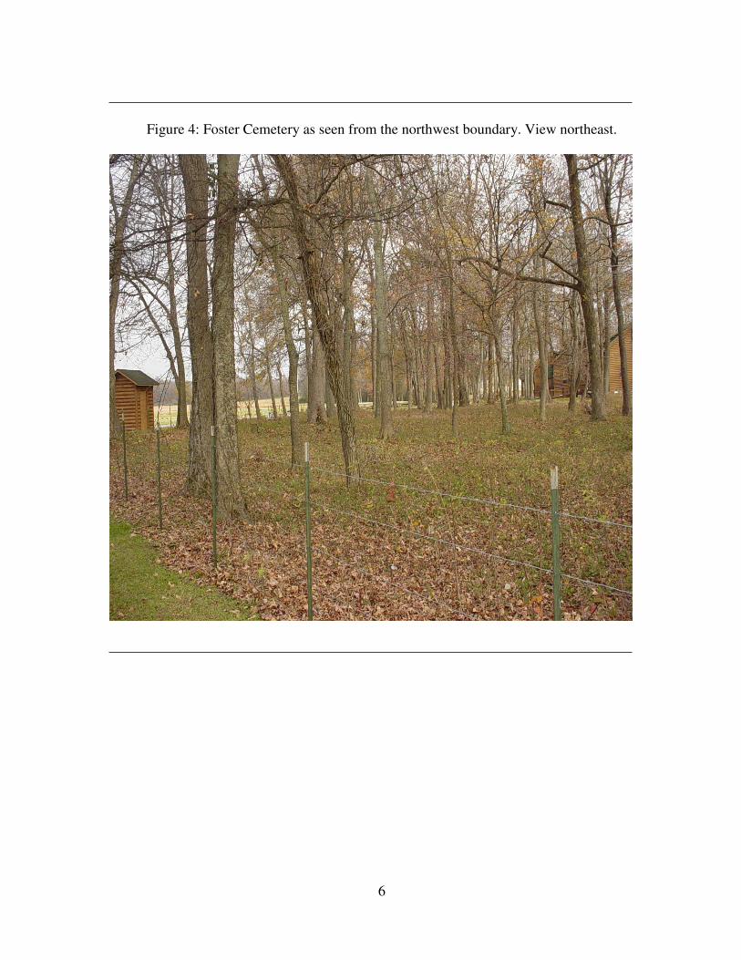
4. Foster Cemetery as seen from the northwest boundary. View
northeast ...................................................................................................6
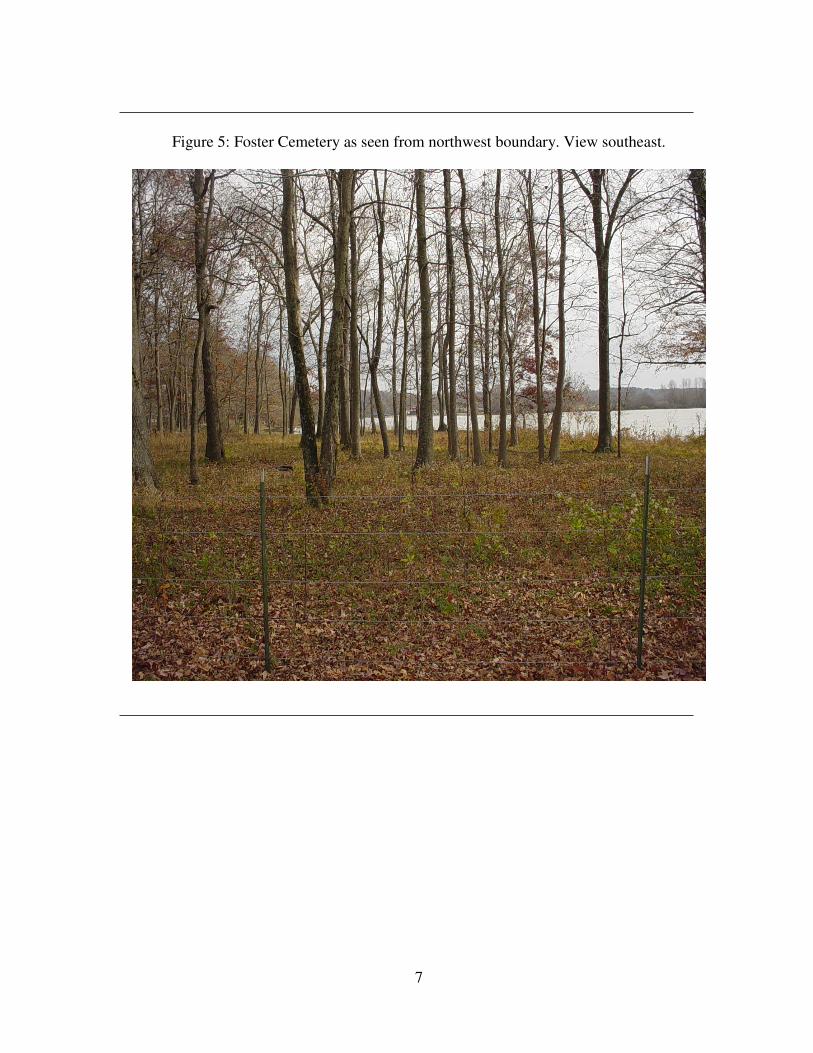
5. Foster Cemetery as seen from the northwest boundary. View
southeast ..................................................................................................7
6. Grave marker found at Foster Cemetery ..................................................8
7. Grave marker found at Foster Cemetery ..................................................9
8. Grave marker found at Foster Cemetery ................................................10
9. Burial Depressions found in the northwest section of Foster
Cemetery ................................................................................................10
10. Age Breakdown by Interval of Each Cemetery Population .................53
11. Cemetery Population Percentages........................................................56
12. Artifact Map of Sex Identification .......................................................70
13. Osteological Map of Sex Identification ...............................................71
14. Burial 3 Cranium with Probable Corrosive Burn ..............................104
15. Burial 3 Cranium with Taphonomic Changes ...................................105
x
16. Burial 3 Dentition with Calculus and Tooth Loss .............................106
17. Burial 4 Maxilla with Tooth Loss and Dental Attrition .....................108
18. Burial 7 Tuberculosis Infection of the Spine .....................................114
19. Burial 7 Tuberculosis Infection of the Eleventh and Twelfth
Thoracic Vertebrae ............................................................................115
20. Burial 7 Tuberculosis Infection of the Pelvis ....................................116
21. Burial 7 Tuberculosis Infection of the Ischium .................................117
22. Burial 7 Tuberculosis Infection with Osseous Mass Located at the
Area of the Pubis ................................................................................118
23. Burial 102 Amelogenesis Imperfecta or Congenital Syphilis-Like
Dentition Pitting and Hypoplastic Activity .......................................174
24. Burial 136 Osteophytes on the Odontoid Process..............................196
25. Burial 137 Arthritic Lipping of the Cervical Vertebrae ....................199
26. Burial 144 Slight Occipital Porotic Hyperostosis ..............................208
27. Burial 200 Periodontal Disease, Tooth Loss, and Calculus ...............242
28. Burial 200 Maxilla, Periodontal Disease, Tooth Loss, and
Calculus .............................................................................................243
1
CHAPTER 1
INTRODUCTION
Anecdotal evidence of skeletal remains eroding and washing away along the banks of
Wilson Lake adjacent to the Tennessee River at the Doublehead Resort in northern Lawrence
County, Alabama, prompted a salvage archaeological operation of Foster Cemetery.
Disturbances, caused by natural and human activities, such as river flooding and industrial
construction, have affected other cemeteries as well. A similar, but more severe case of river
erosion disturbing a cemetery occurred in 1993 in Hardin, Missouri. The Missouri River
flooded and washed away a large portion of the town cemetery prompting reinterment in,
unfortunately, unmarked and mass graves (Wright and Hughes 1996). Another regrettable
aspect of the Hardin, Missouri cemetery flood, was the erosion of the cemetery’s oldest
section, including an acre of historic African American burials that contained little to no
associated historical documentation (Wright and Hughes 1996). Also included in this study
is the Cedar Grove Cemetery, located in Southwest Arkansas. Cedar Grove has a similar
history to the Hardin Cemetery, in that the first indication of its existence was the exposure
of skeletal material eroding from a river bank which prompted the investigation and eventual
salvage of the cemetery (Rose 1985).
In contrast to the Hardin, Missouri cemetery erosion event, the recent salvage
excavations at Foster Cemetery and Cedar Grove provide archaeological and osteological
documentation that has been sorely lacking in the literature on African American cemeteries.
The Southeastern Anthropological Institute (SAI) conducted the salvage excavation of Foster
2
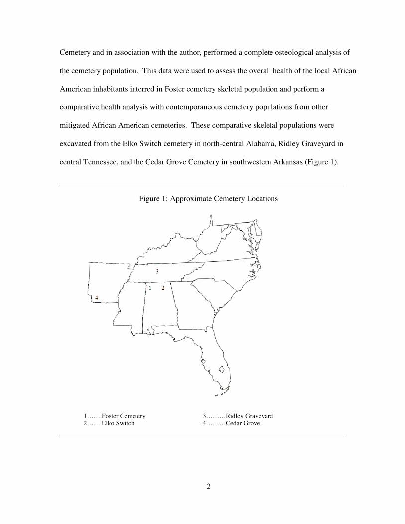
Cemetery and in association with the author, performed a complete osteological analysis of
the cemetery population. This data were used to assess the overall health of the local African
American inhabitants interred in Foster cemetery skeletal population and perform a
comparative health analysis with contemporaneous cemetery populations from other
mitigated African American cemeteries. These comparative skeletal populations were
excavated from the Elko Switch cemetery in north-central Alabama, Ridley Graveyard in
central Tennessee, and the Cedar Grove Cemetery in southwestern Arkansas (Figure 1).
Figure 1: Approximate Cemetery Locations
1…….Foster Cemetery 3………Ridley Graveyard
2…….Elko Switch 4………Cedar Grove
3
Through the cooperation of SAI and the author, an extensive amount of data were
generated. Apart from the osteological data included in this thesis, thousands of artifacts
were analyzed and much historical data were gathered. The historical data included
newspaper articles, death certificates, and grave marker analysis, and this data were then used
to aid in my characterization of the cemetery sample. The total evidence compiled by SAI
and the author, identify Foster Cemetery as an African American cemetery dating
approximately 1870-1960 (Hunter Johnson, personal communication 2007).
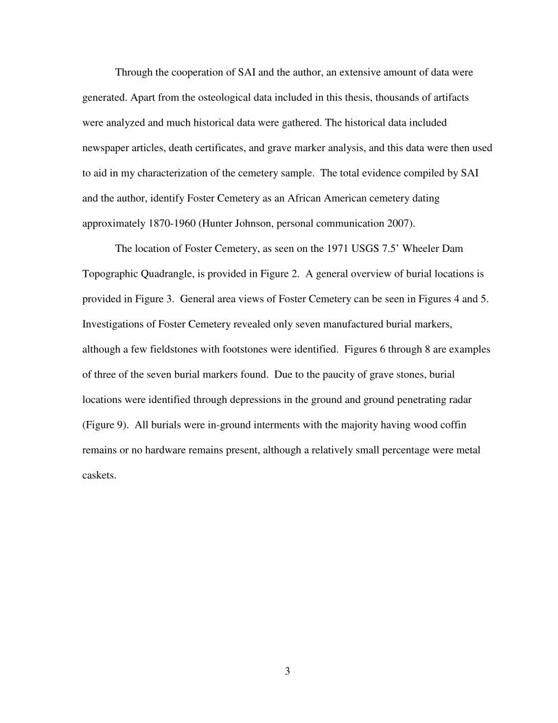
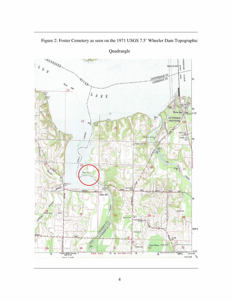
The location of Foster Cemetery, as seen on the 1971 USGS 7.5’ Wheeler Dam
Topographic Quadrangle, is provided in Figure 2. A general overview of burial locations is
provided in Figure 3. General area views of Foster Cemetery can be seen in Figures 4 and 5.
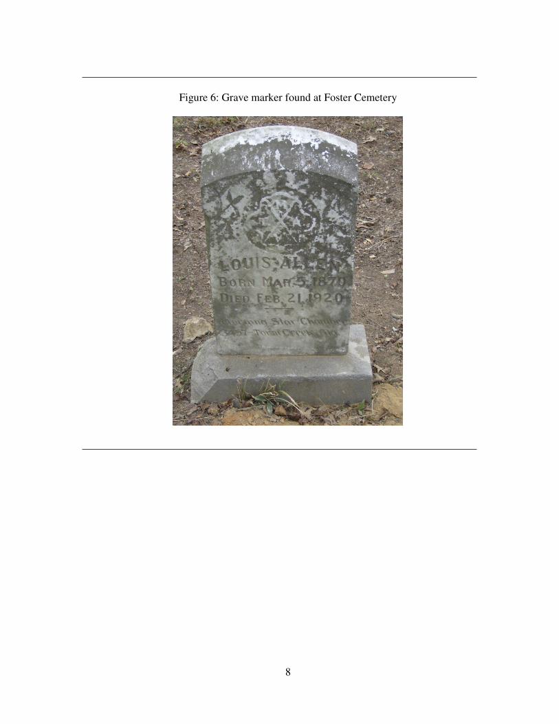
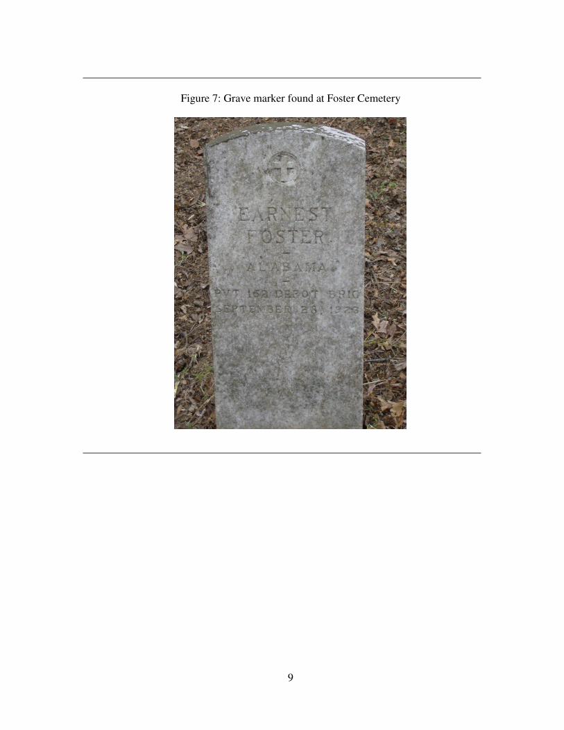
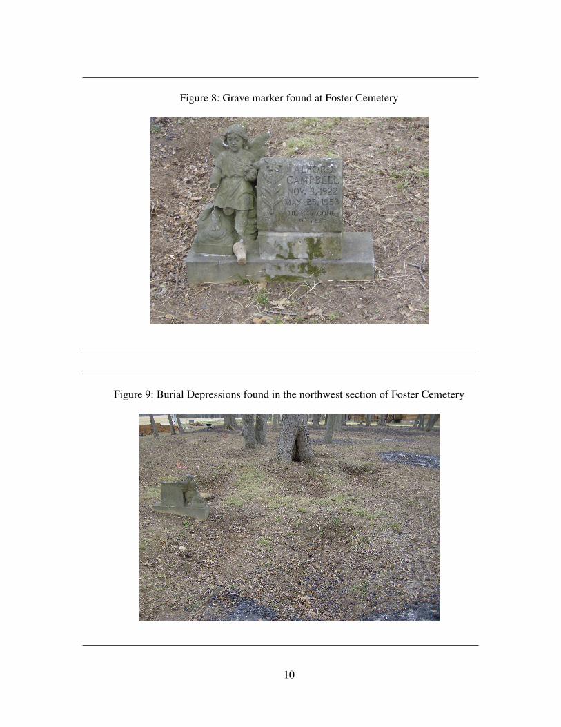
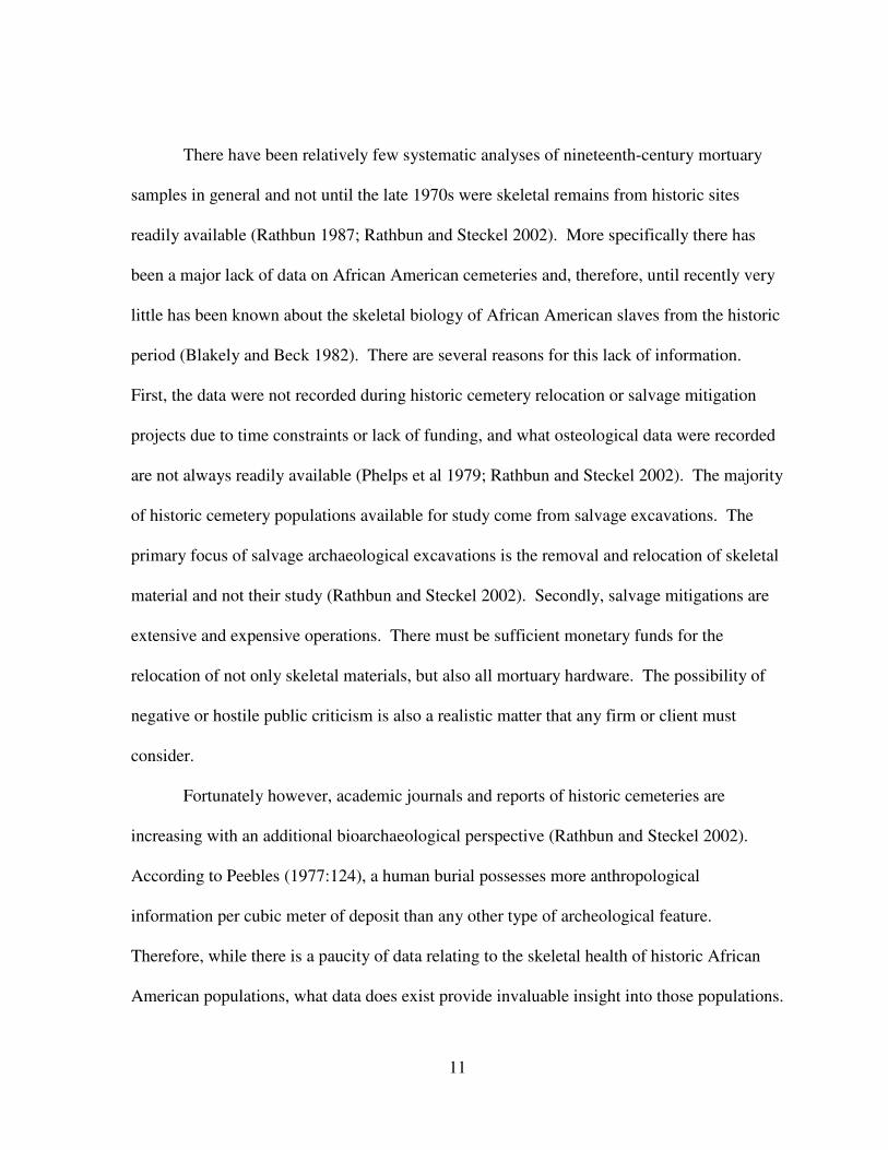
Investigations of Foster Cemetery revealed only seven manufactured burial markers,
although a few fieldstones with footstones were identified. Figures 6 through 8 are examples
of three of the seven burial markers found. Due to the paucity of grave stones, burial
locations were identified through depressions in the ground and ground penetrating radar
(Figure 9). All burials were in-ground interments with the majority having wood coffin
remains or no hardware remains present, although a relatively small percentage were metal
caskets.
4
Figure 2: Foster Cemetery as seen on the 1971 USGS 7.5’ Wheeler Dam Topographic
Quadrangle
5
Figure 3: General Overview of Foster Cemetery Burial Locations
6
Figure 4: Foster Cemetery as seen from the northwest boundary. View northeast.
7
Figure 5: Foster Cemetery as seen from northwest boundary. View southeast.
8
Figure 6: Grave marker found at Foster Cemetery
9
Figure 7: Grave marker found at Foster Cemetery
10
Figure 8: Grave marker found at Foster Cemetery
Figure 9: Burial Depressions found in the northwest section of Foster Cemetery
11
There have been relatively few systematic analyses of nineteenth-century mortuary
samples in general and not until the late 1970s were skeletal remains from historic sites
readily available (Rathbun 1987; Rathbun and Steckel 2002). More specifically there has
been a major lack of data on African American cemeteries and, therefore, until recently very
little has been known about the skeletal biology of African American slaves from the historic
period (Blakely and Beck 1982). There are several reasons for this lack of information.
First, the data were not recorded during historic cemetery relocation or salvage mitigation
projects due to time constraints or lack of funding, and what osteological data were recorded
are not always readily available (Phelps et al 1979; Rathbun and Steckel 2002). The majority
of historic cemetery populations available for study come from salvage excavations. The
primary focus of salvage archaeological excavations is the removal and relocation of skeletal
material and not their study (Rathbun and Steckel 2002). Secondly, salvage mitigations are
extensive and expensive operations. There must be sufficient monetary funds for the
relocation of not only skeletal materials, but also all mortuary hardware. The possibility of
negative or hostile public criticism is also a realistic matter that any firm or client must
consider.
Fortunately however, academic journals and reports of historic cemeteries are
increasing with an additional bioarchaeological perspective (Rathbun and Steckel 2002).
According to Peebles (1977:124), a human burial possesses more anthropological
information per cubic meter of deposit than any other type of archeological feature.
Therefore, while there is a paucity of data relating to the skeletal health of historic African
American populations, what data does exist provide invaluable insight into those populations.
12
The extensive excavation of Foster Cemetery allowed for complete osteological analysis,
which greatly contributes to the knowledge of historic African American health.
The study of human remains and the mitigation of cemeteries are subjects that are
inevitably emotionally charged. Therefore, it is relevant to ask what can be learned from the
study of cemeteries. According to Jacobi (2004), cemeteries are libraries of anthropological
knowledge where religious, economic, social, technological, medical and iconographic
questions can be investigated. The physical integrity of the markers and cemetery grounds is
a reflection of the effort and money spent to maintain the cemetery. This may reflect the
socioeconomic status of the interred populations (Jacobi 2004). An examination of ethnicity
is possible through analysis of language and epitaphs (Jacobi 2004). Demographic questions
pertaining to the number of males, females, and children, how infant mortality has changed
through time, and how age distribution and life expectancy vary through time can be
investigated by the study of cemeteries (Jacobi 2004). Therefore, while the study of human
skeletal remains and the mitigations of cemeteries for salvage or development are subjects of
a sensitive nature, their analysis can yield immeasurable anthropological knowledge.
Because this study examines African American cemeteries, it is important to note
how an African American cemetery can be distinguished from other cemetery types. The
following diagnostic information comes from an undergraduate essay by Annie Donaldson
written during Dr. Ian Brown’s class Marking Graves: Self and Society in Death by (2001).
The study was conducted in Tuscaloosa County, Alabama in association with the University
of Alabama. The purpose of the study was to examine and compare differences in structure
between historic African American and Anglo-American cemeteries. Positive boundaries,
which are defined as any boundary natural or unnatural, subtle or obvious that encloses a
cemetery (i.e. a ridge, road, tree line, or fence) are more common in Anglo American
13
cemeteries and are often absent in African American cemeteries (Donaldson 2001). Unlike
the industrially made Anglo American grave markers, African American cemeteries are
traditionally identified by rudimentary handmade grave markers. Donaldson (2001)
speculates that this difference in grave markers is related to financial status. Additionally an
east/west orientation of burials is more strictly adhered to in African American cemeteries
where organization and alignment of rows of burials seems to be more important in Anglo
American cemeteries.
With the primary data from Foster Cemetery and the reports from Elko Switch,
Ridley Graveyard, and Cedar Grove Cemetery, a model of health was created and used for
the comparative analysis. If the Foster Cemetery population is compared with
contemporaneous historic African American cemetery populations, then it will share similar
patterns of health with the comparative cemetery populations. Although the primary purpose
of this research is the comparative health study of African American cemeteries, there is a
secondary aim. Southeastern Anthropological Institute (SAI) composed a GIS map of the
cemetery identifying age/sex of each burial based on an initial artifact and grave length
assessment. The basic model of this map was used to create an age/sex identification map
based on the osteological analysis and the two were compared. A second hypothesis that I
will test in this thesis is based on this comparison. It is hypothesized that the sex/age
assignments based on coffin size and mortuary hardware will differ from the sex/age
determinations made using osteological analysis.
To articulate these objectives, this study is divided into six additional chapters. An
evaluation and discussion of previous historic African American cemetery excavations, as
well as skeletal health studies is included in chapter two. Cemetery discussions are divided
into geographic areas including the Caribbean, southern United States, and northeastern
14
United States. The purpose of this section of chapter two is to inform the reader of the
relative scarcity of health related osteological analyses of historic African American
populations. Additionally, a brief statement of what cemetery studies contribute is included.
Also in chapter two an overview is given of the histories of the four cemeteries included in
this research; Foster Cemetery, Elko Switch, Ridley Graveyard, and Cedar Grove. The time
period that each cemetery was in use, as well as, the general occupations of the interred
skeletal populations, such as tenant farmers, industrialists, and so forth is included.
Additionally, the locations of the cemeteries are discussed in further detail and the firms and
organizations that conducted the excavations are listed. An examination of the methods used
by each analyst to assess the socioeconomic status of the skeletal populations is presented.
Furthermore, chapter two includes a discussion of African American health, which I
divided into three historical periods: pre-Emancipation, the Tenant Period, and post 1950.
Each period includes a discussion of the health stresses such as diet, trauma, and
occupational stresses. This discussion of health stresses places into context the skeletal
populations being studied and additionally, provides possible causes for the results in the
analysis chapter. After placing the skeletal population in the context of time, these stresses
are then detailed in each of the three time periods. The stresses of health discussed include:
occupation, diet, trauma, pathology, health care, social and political standings, as well as
major changes in each of these categories experienced from one period to the next.
Chapter three discusses the materials and methods used in this study. A brief sample
description of the four skeletal cemetery populations and a brief demographic analysis is
included. In addition to my work on Foster Cemetery, with the three comparative cemeteries
I recorded the number of burials listed in their respective reports, along with a description of
how the skeletal remains were analyzed. Descriptions include osteological analysis, grave
15
length analysis, anecdotal evidence, historical documentation, and artifact analysis.
Additionally, the osteological methods and techniques used to assess and examine the Foster
Cemetery skeletal population follows the sample description found in all four excavation
reports. Tools, charts, and literature references used in examining and recording the
osteological data are evaluated.
Chapter four is the analysis section of this study. Included in this chapter is an
examination of the statistical tests run to compare the health of the Foster Cemetery skeletal
population with the populations of Elko Switch, Ridley Graveyard, and Cedar Grove. The
data from the analyses of demography, growth and development, infection, diet, degenerative
joint disease, and trauma form the basis for the health comparison and comprise this chapter.
The demography section includes a discussion of sex identification and age interval
frequencies and percentages. The growth and development section includes a comparison of
stature estimations and enamel defects for each cemetery population. The infection section
compares the occurrence of tuberculosis and congenital syphilis. Analysis of data that
address diet investigates dental caries, cribra orbitalia, and porotic hyperostosis. The section
on trauma and degenerative joint disease includes an evaluation of healed fractures and the
presence of arthritis. Lastly, a comparison of the sex/age categories identified for the artifact
map created by SAI and the osteology map using the findings from the osteological analysis
is included with each map presented.
In the fifth chapter, an interpretation of the findings from each of the sections in
chapter four is given. In addition to these interpretations, the mapped sex/age categories from
the artifact analysis are compared to osteological identifications, to ascertain and interpret
any differences in these two analyses. The African American history chapter, chapter three, is
referenced and is used to place into context the findings from the analysis chapter. Any
16
similarities and differences between Foster Cemetery and the three comparative cemeteries
are discussed and explained.
The final chapter discusses conclusions drawn from this study and the limitations
experienced in it. Recommendations for future research also are given to improve upon the
work in this analysis. One of the major difficulties in gaining an understanding of African
American cemeteries has been the lack of attention in the literature to the past life ways and
mortuary practices of African Americans in general. In the community of north Alabama that
this excavation was conducted, public awareness and interest in the cemetery greatly aided in
the project. It is hoped that the additional data from Foster Cemetery will be an important
step in bringing these kinds of studies to the forefront.
17
CHAPTER TWO
LITERATURE REVIEW AND HEALTH HISTORY
There is sparse literature available on African American cemeteries and all known
African American excavations in general. A brief history of Foster Cemetery, Elko Switch,
Cedar Grove, and Ridley Graveyard is presented. Additionally in this chapter, an evaluation
of stresses affecting health including diet, occupation, pathology, trauma, and varying access
to health care is discussed. These variables are compared through time to the lifeways of
African Americans from pre-Emancipation, to the Tenant Period, and ultimately, post-1950.
African American Cemeteries
There have been relatively few systematic excavations and investigations of historic
African American cemeteries. The majority of studies that do exist generally involve cultural
resource management mitigation projects whose focus was to salvage cemeteries prior to
disturbance from construction and erosion (Buchner et al 1999). Osteological analysis of
skeletal materials from historic cemeteries began in 1930 by Harry Shapiro who analyzed 20
skeletons from a disturbed cemetery during transit construction in New York City (Rose
1985; Shapiro 1930). Those studying historic cemeteries, using both osteological and artifact
analysis, seek to comprehend social status, customs, precedents, temporal sequences, and the
demographics of interred populations (Buchner et al 1999; Wright and Hughes 1996).
There have been relatively few excavations and examinations of historic African
American cemeteries due to lack of funding, public sensitivity, and time constraints.
18
Additionally, with immediate cemetery relocation and not osteological analysis being the
primary objective, much data were lost or incomplete in early cemetery mitigation projects
such as Hardin, Missouri. In recent decades however, there has been an increase in not only
mortuary hardware analyses, but also osteological examinations of historic African American
cemeteries including those in this comparative analysis (Phelps et al. 1979; Rathbun and
Steckel 2002; Rose 1985). In the Caribbean, slave burials have been examined from the
Virgin Islands and there have been thorough investigations of slave archaeology and skeletal
health on the Newton Plantation in Barbados (Corruccini et al. 1982; Corruccini et al. 1985;
Handler and Lange 1978; Handler et al. 1982; Jacobi et al. 1992).
In the southern United States there have been several excavations and osteological
health investigations including Elko Switch and Foster Cemetery in north Alabama, Ridley
Graveyard in central Tennessee, and Cedar Grove in southwestern Arkansas. Freedman’s
Cemetery (41DL316) is an historic African American cemetery which served the urban
population of Dallas, Texas from 1869 to 1902 (Condon et al. 1998). Approximately 25% of
the cemetery was excavated yielding a total of 1150 burials and has been used in a
comparative health analysis with the skeletal population from Cedar Grove (Davidson et al.
2002). An earlier sample, excavated in 1979 due to industrial construction, from Belleview
Plantation in South Carolina which was owned by Edward Croft from 1738 to 1756, was
composed of a total of nine white adults, five white subadults, and at least two black adult
slaves (Rathbun and Scurry 1991; Rathbun and Steckel 2002).
Another slave skeletal population dating from 1840-1870 South Carolina comes from
the Paul Remley Plantation near Charleston. Following the cemetery’s excavation in 1984,
subsequent health analysis of the skeletons indicated a stressed population that suffered from
anemia, as indicated by cribra orbitalia and porotic hyperostosis, as well as degenerative joint
19
disease (Rathbun 1987; Rathbun and Steckel 2002). An osteological analysis was conducted
on 19 adult African American males from a Union soldier cemetery on Folly Island near
Charleston, South Carolina (Rathbun and Steckel 2002). In 1987, these remains were
uncovered as a result of construction activities and date to 1863 (Legg and Smith 1989;
Rathbun and Steckel 2002).
African American cemetery mitigations also occur in the northeastern United States.
The First African Baptist Church in Philadelphia represents a large urban population. Two
separate cemetery excavations took place here. The first excavation of a cemetery associated
with this church occurred in 1983-1984 and produced 140 burials with 75 adults with skeletal
remains suitable for analysis (Angel et al. 1987; Parrington and Roberts 1984). The second
cemetery mitigation connected to the First African Baptist Church occurred in 1990 and
resulted in the recovery of 89 African American skeletons that had been interred between
1810 and 1822 (Crist et al. 1995).
Foster Cemetery
The original Foster Cemetery, before its relocation, was situated in northwestern
Lawrence County, Alabama approximately seven miles north of the town of Town Creek in
Doublehead Resort, a recreational family vacation resort on the banks of Wilson Lake which
is an extension of the Tennessee River. Foster Cemetery dates approximately from 1870 to
1960 and possibly even as late as the early 1970s; however, landownership records indicate
the possibility that Foster Cemetery originated with slave interments as early as the 1840s
(Hunter Johnson, personal communication 2007). Death certificates, grave marker analysis,
and landownership records were used to identify the ancestry and socioeconomic status of
the Foster Cemetery population. Foster Cemetery is identified as an African American
20
cemetery which served a rural population of freedmen, agriculturalists, tenant farmers and
sharecroppers in the cemetery’s early history, followed by the addition of industrial workers
later in its use. A textile or cotton mill existed in the vicinity of Foster Cemetery for at least
the early part of the cemetery’s use and it is probable that at least some portion of the
cemetery population was employed there (Ibid).
Elko Switch
Elko Switch Cemetery was located approximately four miles west of Huntsville,
Alabama and dated from 1850 to 1920, a total of seventy years of use (Shogren et al. 1989).
Excavations of fifty-six burials from Elko Switch were conducted by The University of
Alabama, Alabama Museum of Natural History, and Office of Archaeological Research
under contract with the State of Alabama Highway Department. The purpose of the
excavation was to relocate the portion of the cemetery affected by encroaching highway
development. No direct historical documentation concerning Elko Switch Cemetery exists;
however, landownership records, anecdotal evidence and artifact and mortuary hardware
analysis were used in determining the age and socioeconomic structure of the Elko Switch
Cemetery population (Shogren et al. 1989). The people associated with this cemetery were
black freedmen and their descendents who lived an agricultural lifestyle as tenant farmers
(Shogren et al. 1989). The likelihood of actual slave interments is possible given the
surmised socioeconomic nature of the cemetery population (Shogren et al. 1989).
Ridley Graveyard
The Ridley Graveyard is located in Williamson County, Tennessee, approximately
eleven km south of Franklin. The cemetery dates from 1885 to 1940, totaling fifty-five years
21
of use (Buchner et al. 1999). A total of forty-seven burials was excavated and relocated by
the Tennessee Department of Transportation through Gresham Smith & Partners, and
Panamerican Consultants, Inc. The abandoned cemetery was relocated due to highway
construction. Although no historical documentation was found specifically mentioning the
cemetery, anecdotal evidence, death certificates, landownership documents, and artifact and
mortuary hardware analysis were used to assess a timeline of use of Ridley Graveyard and
the socioeconomic status of the interred population (Buchner et al. 1999). The population
associated with this cemetery included black freedmen and their tenant farmer descendants,
and based on landownership records, possibly slaves (Buchner et al. 1999). It was proposed
that one reason for the abandonment of the cemetery was the “Great Migration,” a period in
the early to mid twentieth century where African Americans moved north to urban areas for
occupation opportunities and to escape racism (Buchner et al. 1999).
Cedar Grove
Excavations and the subsequent relocation of remains from the Cedar Grove site took
place in 1982 by the Arkansas Archeological Survey under contract with the New Orleans
District of the U.S. Army Corps of Engineers (Rose 1985). The cemetery was located on the
south bank of the Red River in Lafayette County, Arkansas and was relocated due to
revetment construction caused by river erosion. The original report identified 79 burials
being relocated in the 1982 excavations. These 79 burials were dated from 1890 to 1927
(Rose 1985). An additional burial was excavated in 1980 during site testing, and also dated
to the same time period. However, a more recent publication utilizing a new specific artifact
analysis dates the excavated burials between 1900 and 1915 (Davidson et al. 2002). For this
thesis analysis, a combination of the recent publication, as well as the initial cemetery report
22
is used. Landownership records, personal communications, and mortuary hardware analysis
were used to determine the cemetery’s age and the socioeconomic status of the interred
population. Furthermore, it is thought that the overall cemetery population consists of
freedmen and their descendants who became agriculturalists, tenant farmers and
sharecroppers (Rose 1985).
African American Health and Past Lifeways
Historically, constraints on mobility, limited educational opportunities, restricted
political access, and upper bounds on social mobility affected, if not directly then indirectly,
the health of African Americans in the Southeast (Rathbun and Steckel 2002).
Understanding these effects contextualizes and humanizes the cemetery populations being
studied. Additionally, this understanding provides insight into the health results derived from
the osteological analysis. To gain insight into the daily lives of these people, a
comprehensive analysis of the time periods that these cemeteries span is necessary. The
periods of use for the cemeteries are as follows: Elko Switch seventy years (1850-1920),
Cedar Grove fifteen years (1900-1915), Ridley Graveyard fifty-five years (1885-1940), and
Foster Cemetery ninety years (1870-1960). Together the cemeteries span one-hundred and
ten years (1850-1960) (Buchner et al. 1999; Davidson et al. 2002; Johnson, personal
communication 2007; Rose 1985; Shogren et al. 1989). It is necessary here to investigate the
occupations, diet, and healthcare, which contribute to and influence not only health in
general, but more specifically skeletal health, providing historically relevant information
concerning African American populations throughout the one-hundred and ten year period.
Slavery existed in the United States until the end of the Civil War in 1865 and
probable slave and freedmen interments existed at Elko Switch, Ridley Graveyard, Cedar
23
Grove and Foster Cemetery (Buchner et al. 1999; Johnson, personal communication 2007;
Rose 1985; Shogren et al. 1989). Slave work was demanding, brutal, and relentless not only
for the amount of work that was done, but also the type of work. Slaves raised hogs, rolled
tobacco, pressed and cut cane, picked cotton, planted rice, worked on steamships, and were
involved in domestic activities (Diouf 2007). In addition to agricultural occupations,
specialized labor in the forms of blacksmiths, carpenters, stone-cutters, cooks, and
bricklayers also existed (Sellers 1950). These occupations were arduous, repetitive, and
given time, would affect skeletal health. Examples of skeletal bone modification indicative
of these occupations include robust bones with big muscle attachments that roughened the
surface of the bone and indicate the strenuous nature of the work of slaves (Rathbun and
Steckel 2002). Indeed, many recorded afflictions affecting slave populations were
occupational (Postell 1951). Sore fingers from picking cotton, backaches and hernias were
commonly reported (Postell 1951). The most common types of injuries were cuts from axes
or scythes, broken backs, shoulders and thighs, gunshot wounds, and amputations that were
the results of falls, kicks from animals, overturned carts, runaway wagons, and limbs getting
caught in farm machinery (Postell 1951; Savitt 1978). Injuries and afflictions such as these
resulted in degenerative joint disease, fractures, and abnormal bone growths.
In addition to occupational trauma, various forms of infections are identifiable on
skeletal remains (Savitt 1978). There are numerous infectious agents attributing to the
morbidity of enslaved populations. Tuberculosis, rheumatic fever (a joint infection), scarlet
fever, typhoid, diphtheria, and measles are only a few examples of infectious diseases (Savitt
1978). Of the diseases described by Savitt (1978) tuberculosis, rheumatic fever, and typhoid
can produce skeletal lesions (Aufderheide and Martin 1998). Diet and nutrition also affect
skeletal growth, development, and health and there are several infections that could be
24
caused by dietary deficiencies, such as pellagra (Postell 1951). The main food staples of the
slave diet were corn and bacon, although these foods did not constitute the whole diet as
vegetables, milk, fish, and occasionally beef were obtainable (Savitt 1978; Sellers 1950). In
general, while cornmeal was supplied in abundance, shortages of meat did occur even though
the usual ration per week was three pounds per working slave (Sellers 1950). Although
Postell (1951) suspected that slaves received an improperly balanced diet, Savitt (1978)
argues that this was not always the case as there was little financial benefit from a
nutritionally deprived work force. However, with the knowledge that shortages of meat did
occur and that cornmeal lacks adequate amounts of iron and protein, it can be assumed that
iron deficiency anemia occurred with some frequency. Iron deficiency anemia can be seen
on the skeleton in conditions such as porotic hyperostosis and cribra orbitalia which cause
lesions on the frontal, parietal, and occipital bones and the orbital roof (Aufderheide and
Martin 1998). Sickle cell anemia is also worth mentioning here as it can also affect bone and
manifest itself in long bone infarcts, osteoporosis, and necrosis (Aufderheide and Martin
1998). Nutrient deficiencies that result in anemia also can be caused by parasites.
Trichinosis, a parasitic disease caused by the roundworm Trichinella spiralis, almost
certainly occurred relatively frequently given the large quantities of pork consumed by slave
populations (Savitt 1978). Other intestinal parasites that most likely affected slave
populations include Ascaris lumbricoides, a roundworm caused by poor sanitation and
hygiene, Taenia saginata and Taenia solium, tapeworms caused by consuming raw or
undercooked infected beef and pork, and Necator americanus, a hookworm resulting from
contact with soil containing infected human feces (Savitt 1978).
Due to their increased susceptibility to disease, poor diet, and injury, infants and
children deserve attention. In this study age ranges were assigned with infants categorized as
25
zero to four years, and childhood in general extending from zero to 18 years. Mortality of
infants is always high relative to the rest of childhood, whereas mortality is lowest during
later childhood (Danforth 2004; Weiss 1973b). Therefore, a relatively high frequency of
infant burials would be expected while a lower frequency of childhood burials should be
found. Several reasons for this could be a winnowing of the weak during infancy, denying
children meat in their diets due to the belief that it was unsuitable for their systems, an
increase in male deaths due to accidents and violence, and female deaths from childbearing
beginning in young adulthood (Danforth 2004). Infanticide also occurred in slave
populations. Possible causes for infanticide include covering up the shame of an illegitimate
child or refusing to allow one’s child to grow up in bondage (Savitt 1978). Slave children, in
general, had extraordinarily poor health as indicated by stature data (Rathbun and Steckel
2002). Sickle cell disease, hemolytic crisis, severe joint pains, leg ulcers, and intestinal
worms were some afflictions affecting slave children (Savitt 1978).
The severity of conditions affecting slave populations was no doubt influenced by
access to health care. For purely practical financial reasons, owners of slaves were invested
in and responsible for the wellbeing of their slaves in sickness and old age (Sellers 1950).
Successful planters provided a place to care for the sick whether it be a hospital or their own
home (Postell 1951). Often doctors were too far away to be called upon quickly for minor
illnesses; therefore, plantation mistresses, owners, overseers, or slaves assigned to healing
roles cared for the ill (Sellers 1950). Physicians were called upon, however, and
administered a wide variety of treatments and medicines. Visiting physicians played their
most important roles in surgical procedures, acting as a midwife assisting in childbirth, and
prescribing a diversity of drugs such as castor oil, opium, camphor, and quinine (Savitt 1978;
Sellers 1950). Physicians were not without their limitations though. Because no one
26
understood the etiology of diseases, it was hard to effectively treat them (Savitt 1978).
Astute observers could often diagnose and prepare treatment as well as any doctor and given
the expense of physicians, slave owners would often treat the sick themselves (Savitt 1978).
Slaves also chose to treat themselves for several reasons. They often viewed the
prescriptions and practices of physicians as harsh, there was trust in the remedies of friends
and relatives, and it was an act of independence (Savitt 1978). African American medicine
included mixtures of local herbs and plants, as well as conjurers who used spells (Savitt
1978).
Following the Civil War, there is relatively little data concerning the health of African
American populations (Rose 1985). Due to this paucity of specific data, generalizations of
health are made using known occupations, diets, and social constraints and tendencies.
Unfortunately, the modest information pertaining to African American diet, morbidity, and
mortality in the decades following the Civil War is conflicting (Rose 1985). This is probably
the result, at least in part, of poor record and census keeping due to carpetbag rule in the
southern United States (Rose 1985). What is known is that African Americans experienced a
change from enslavement to lives of attempted independence and self-sufficiency. Although
new occupations existed with increased industrialization following the Civil War, farming
and agriculture were very prominent and continued to be the primary occupation and lifestyle
for several reasons. For this study, the period from 1863 to 1950 is called the Tenant Period
and named for the sharecropping or tenant farming labor system established in the South
following the Civil War (Buchner et al. 1999). Because all four cemetery populations in this
study existed during this seventy-five year period and these people prior to death lived an
agricultural life, the farming lifestyle is the primary focus of this section with special
attention to the diet, morbidity, and trauma that relates to the agricultural lifestyle (Buchner

27
et al. 1999; Davidson et al. 2002; Johnson, personal communication 2007; Rose 1985;
Shogren et al. 1989).
Farming, and the wages it brought, was seen as a means in which to build lasting
foundations for black families (Gilbert and Eli 2000). The land provided not only
sustenance, but also the framework for families and something that could be inherited for
future generations (Gilbert and Eli 2000). Therefore, farming for African Americans, was
inextricably linked to being free (Browne 2003). Even though wages were seen as progress,
new wage labor systems based on farming were not always equal. These new labor systems
were often cruel, and this was compounded by the fact that agricultural regions in the South
relied heavily upon black labor (Buchner et al. 1999). Sharecropping, the practice whereby a
tenant lives on and uses the land of the landowner and pays rent with a portion of the crop
produced, was prevalent in the south. Following the Civil War and Reconstruction, demand
for tenants on large, white landholdings was high, and due to a scarcity of other employment
opportunities, and with newly granted independence and aspirations of freedom, a majority
of the African American population adopted this sharecropping lifestyle (Browne 2003;
Gilbert and Eli 2000).
It has been argued that with the adoption of farming and new occupations following
the Civil War, African American populations’ quality of life was high (Stampp 1965).
However, it also has been argued that the quality of life declined during Reconstruction up
into the 1930s based on a lack of food, occurrences of disease, and poor access to medical
care (Farley 1970). In terms of diet, it is known that there is a tendency for individuals to
consume a traditional diet regardless of social or economic changes and, therefore, it can be
inferred that African Americans maintained a similar diet before and after the Civil War
(Rose 1985). Additionally, although they may have attempted to improve upon their diet, the
28
social constraints in place in the late nineteenth and early twentieth centuries continued to
limit access to different food sources (Rose 1985). However, African Americans could now
make use of naturally occurring resources in their areas (e.g. hunting and fishing) and the
communal sharing of butchered hogs and even cattle did occur (Grim 2003; Rose 1985).
Given these observations, it can be assumed that diets were adequate in terms of
nutritional intake, but deficiencies did occur. Dietary related conditions that occurred prior
to the Civil War that may also have occurred during the Tenant Period include pellagra, iron
deficiency anemia causing cribra orbitalia and porotic hyperostosis, and parasitic infestations
caused by consuming undercooked meat such as Trichonosis (Trichinella spiralis), Taenia
saginata and Taenia solium. In addition to anemia, poor diet and parasitic infections also
affect skeletal growth and development can result in an individual becoming short of stature.
As was noted earlier, agricultural labor prior to the Civil War was hard and arduous
causing hernias, degenerative joint disease, fractures, and abnormal bone growths (Savitt
1978). Agricultural labor following the Civil War also was physically demanding and it is
probable that many occupational injuries and afflictions occurring prior to Civil War also
occurred during the Tenant Period. Farm machinery accidents, cuts from tools, amputations,
and fractures from various other activities no doubt occurred and would be reflected in the
skeletal population. Types of degenerative joint disease present in skeletal remains that
occur with rigorous, load bearing and repetitive activity seen in sharecroppers include
osteophytosis, Schmorl’s nodes, and osteoarthritis (Rose 1985).
Farley (1970) noted that following Reconstruction up until the 1930s African
Americans had a lack of medical care in rural areas. It is known that black tenant farmers
had to supply their own equipment often attained at the price of the loan or multiple loans,
which would force them to accumulate enormous debt. There was a resistance to African
29
American social advancement and, thus, there existed a lack of financial support from banks
and insurers (Gilbert and Eli 2000). Given these circumstances, it is possible that what
professional medical resources were available were mostly unaffordable and thus
unobtainable. However, between 1900 and 1950 some African Americans were able to
acquire the luxury of radios (Grim 2003). Not only did radios allow for social and cultural
expression, radios also were used to educate on health, medical care, and eating habits (Grim
2003). Access to improved medical knowledge and technologies, however limited, may have
improved the overall health of those fortunate enough to possess those technologies.
Infectious diseases continued to be a substantial health concern into the late
nineteenth and early to mid twentieth century. Some of the most frequent causes of African
American deaths included tuberculosis, pneumonia, diarrhea, typhoid fever, and malaria
(Farley 1970). Rose (1985) notes that little difference can be found in the disease patterns of
African Americans during slavery and the turn of the century. He also mentions that the
three largest causes of death (pneumonia, tuberculosis, and diarrhea) are diseases whose
incidence and fatality rates can be elevated by malnutrition (Rose 1985). Knowing these
diseases occurred but that there was at least a chance that medical knowledge increased
during the early to mid twentieth century leaves the possibility that there were fewer cases to
be found in the Tenant Period as opposed to prior to the Civil War.
Unlike the pre-Civil War and the Tenant Period, where all the cemetery populations
in this study existed or had possible interments, only Foster Cemetery was in use after 1950
and up into the 1960s (Hunter Johnson, personal communication 2007). Despite the lack of
descriptive health data for African Americans during the 1950s and 1960s, understanding the
social and technological structure of the time can assist in understanding how lifestyle
influenced health.
30
Although described as an agricultural cemetery population, changes in political
movements, laws, and technology may have influenced the agrarian lifestyle of the Foster
Cemetery population. Prior to the 1954 Brown v. Board of Education of Topeka decision,
separate-but-equal facilities and the mistreatment and inequality that segregation brought was
legal as established by the 1896 Plessy v. Ferguson decision (Gilbert and Eli 2000).
However, at least in law, the Brown v. Board of Education of Topeka struck down separate-
but-equal as unconstitutional in school systems. The decision began the process of
permitting African American children access to improved educational opportunities, thus
leading to other employment opportunities (Gilbert and Eli 2000).
Following the Brown decision, the South became an increasingly inhospitable and
even dangerous place for African Americans (Gilbert and Eli 2000). For that reason, violent
injuries such as blunt force trauma, bullet wounds, and broken bones might be present in the
cemetery populations with burials during this time period. Black farmers continued to be
denied loans necessary for the purchase of newer labor saving technologies or new land by
banks and the Farmers Home Administration (Gilbert and Eli 2000). Additionally, by 1960
more than half of America’s crops were picked and harvested by machines. The use of
tractors, mechanical harvesters, and additionally the introduction of chemical weed control
reduced the amount of work for African Americans in the agricultural labor system (Gilbert
and Eli 2000). Due to these limitations, Southern black farmers often encouraged their
children to take advantage of desegregation and attend college; indeed, there were relatively
few Southern black farmers remaining into the 1960s (Gilbert and Eli 2000). With increased
industrialization, even in the South, it is safe to assume that African Americans were
employed in manufacturing and industrial occupations. This is confirmed in the Foster
31
Cemetery population as one individual was recovered who died in a historically documented
industrial accident in 1953 (Johnson, personal communication 2007).
Several assumptions can be made in terms of health when we understand the social
and technological standings of the Post-1950 period. Farming continued to exist as an
occupation and lifestyle, although in decreased frequency. Therefore the occupational
stresses, infectious agents, and dietary deficiencies associated with a rigorous and strenuous
farming lifestyle (e.g. arthritic development, broken bones, tuberculosis, parasites, and
anemia) can be seen. The limited ability of African American farmers to procure farming
equipment such as tractors and mechanical harvesters, possibly kept the levels of
degenerative joint disease and occupational trauma relatively consistent during this period, as
with pre-1950 agricultural populations.
However, considering that there were fewer numbers of farmers and more people
seeking employment in industrial jobs, it is important to have at least a brief overview of
health concerns in manufacturing occupations. A comparative health analysis of African
American cemetery use conducted between Cedar Grove (1881-1927) and Freedman’s
Cemetery (1869-1907) shows some of the major differences between a rural, agriculturally
based community and that of a community with more industrial work, respectively
(Davidson et al. 2002). Davidson et al. (2002) found that degenerative joint disease occurred
more frequently at Freedman’s Cemetery, while healed fractures occurred more frequently at
Cedar Grove. This suggests that urban and industrial work is more repetitive, which leads to
a higher incidence of degenerative joint disease, and that rural, agricultural occupations have
a higher risk of injury and inflicted trauma (Davidson et al 2002). However, as is the case of
the 1953 Foster Cemetery example, inflicted trauma from industrial accidents did occur, in
32
this case from a chemical explosion at a local phosphorus plant (Johnson, personal
communication 2007).
It is suggested that farmers maintained a similar diet to that of pre-1950, with
populations mainly consuming hogs, possibly cattle, corn, and continued utilization of the
natural resources around them through hunting and fishing, due to the social and monetary
constraints and limitations placed on them (Rose 1985). Those fortunate to gain access to
technologies like radios and other new sources of media, had the possibility of increasing
their knowledge of health, medical care, and eating habits (Grim 2003). Thus, it is possible
the dietary health of the Foster Cemetery population from 1950 into the 1960s increased and
was manifest in taller stature of individuals during this time. In addition to increased stature,
there are also fewer and less severe cases of porotic hyperostosis and cribra orbitalia than in
earlier time periods.
This chapter provided a background study on the health of African Americans from
1850 to 1960 and the relationship of health to occupation, diet, trauma, disease, and social
implications. Three periods were discussed, pre-Civil War, the Tenant Period, and post
1950, in an attempt to contextualize and humanize the populations being studied, as well as,
understand the findings of the osteological analysis. What has been shown is that
populations experiencing slavery at the outset and later experiencing oppression and racism,
possessed a relatively poor diet that slowly changed through time with improved
technologies and greater access to differing occupations. Daily life generally consisted of
laborious agricultural work and later industrial professions for those associated with Foster
Cemetery. Throughout the time span of the four cemeteries, infections such as tuberculosis
and pneumonia, for example, and traumas such as degenerative joint disease, broken bones,
and amputations occurred with relative frequencies. Although these conditions and

33
afflictions may not be present in all cemetery populations, they demonstrate the hardships
and lifestyles encountered by those populations in this analysis.
34
CHAPTER THREE
MATERIAL AND METHODS
The main objective of this study is to compare the overall health of the Foster
Cemetery skeletal population with those of Elko Switch, Ridley Graveyard, and Cedar
Grove. To assist in doing so, a brief discussion of the different skeletal cemetery populations
and their demography is included in this chapter, as well as the osteological methods used in
analyzing the Foster Cemetery skeletal remains. The specific tests used to compare the
materials are listed and discussed in the analysis chapter, Chapter Five.
Foster Cemetery
Of the 224 burials identified from Foster Cemetery, only 103 gave an indication of a
sex/age category (male, female, indeterminate, subadult, or adult). A total of 127 burials
have a specific age or age range assigned to them. Twenty-three remains were complete
enough for stature estimations. One-hundred and forty-one burials could be examined for
pathology, trauma, and anomalies. One-hundred and twenty-seven burials had dental remains
present that allowed for the occurrence of dental caries to be recorded. The remains of five
burials, 81, 82, 85, 180, and 223, had disintegrated to a mere trace and were not identifiable
as human. They were not included in the burials with identifiable remains. One burial, 40,
was an animal burial, a dog cranium, and was not included in the comparative health
analysis.
35
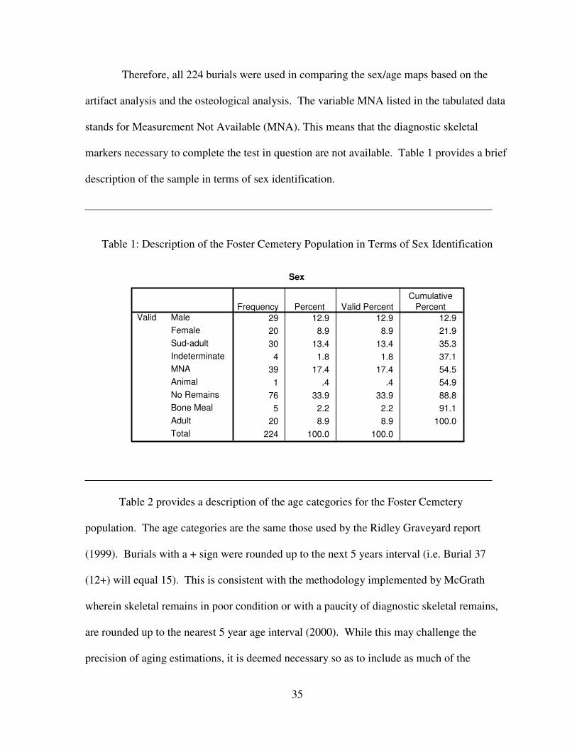
Therefore, all 224 burials were used in comparing the sex/age maps based on the
artifact analysis and the osteological analysis. The variable MNA listed in the tabulated data
stands for Measurement Not Available (MNA). This means that the diagnostic skeletal
markers necessary to complete the test in question are not available. Table 1 provides a brief
description of the sample in terms of sex identification.
Table 1: Description of the Foster Cemetery Population in Terms of Sex Identification
Sex
29 12.9 12.9 12.9
20 8.9 8.9 21.9
30 13.4 13.4 35.3
4 1.8 1.8 37.1
39 17.4 17.4 54.5
1 .4 .4 54.9
76 33.9 33.9 88.8
5 2.2 2.2 91.1
20 8.9 8.9 100.0
224 100.0 100.0
Male
Female
Sud-adult
Indeterminate
MNA
Animal
No Remains
Bone Meal
Adult
Total
ValidFrequency Percent Valid Percent
Cumulative
Percent
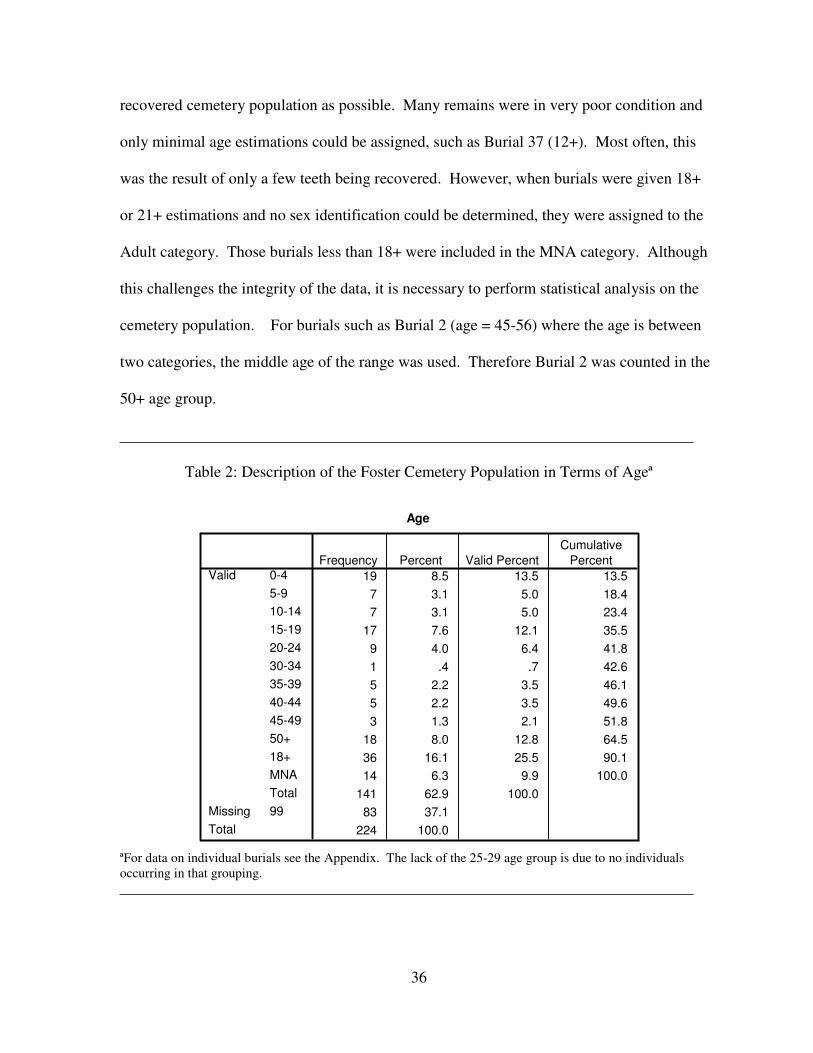
Table 2 provides a description of the age categories for the Foster Cemetery
population. The age categories are the same those used by the Ridley Graveyard report
(1999). Burials with a + sign were rounded up to the next 5 years interval (i.e. Burial 37
(12+) will equal 15). This is consistent with the methodology implemented by McGrath
wherein skeletal remains in poor condition or with a paucity of diagnostic skeletal remains,
are rounded up to the nearest 5 year age interval (2000). While this may challenge the
precision of aging estimations, it is deemed necessary so as to include as much of the
36
recovered cemetery population as possible. Many remains were in very poor condition and
only minimal age estimations could be assigned, such as Burial 37 (12+). Most often, this
was the result of only a few teeth being recovered. However, when burials were given 18+
or 21+ estimations and no sex identification could be determined, they were assigned to the
Adult category. Those burials less than 18+ were included in the MNA category. Although
this challenges the integrity of the data, it is necessary to perform statistical analysis on the
cemetery population. For burials such as Burial 2 (age = 45-56) where the age is between
two categories, the middle age of the range was used. Therefore Burial 2 was counted in the
50+ age group.
Table 2: Description of the Foster Cemetery Population in Terms of Ageª
Age
19 8.5 13.5 13.5
7 3.1 5.0 18.4
7 3.1 5.0 23.4
17 7.6 12.1 35.5
9 4.0 6.4 41.8
1 .4 .7 42.6
5 2.2 3.5 46.1
5 2.2 3.5 49.6
3 1.3 2.1 51.8
18 8.0 12.8 64.5
36 16.1 25.5 90.1
14 6.3 9.9 100.0
141 62.9 100.0
83 37.1
224 100.0
0-4
5-9
10-14
15-19
20-24
30-34
35-39
40-44
45-49
50+
18+
MNA
Total
Valid
99Missing
Total
Frequency Percent Valid Percent
Cumulative
Percent
ªFor data on individual burials see the Appendix. The lack of the 25-29 age group is due to no individuals
occurring in that grouping.
37
The three age categories with the highest frequency and percentage, with the
exception of the 18+ category, are 0-4, 15-19, and 50+. There are several reasons why the
data yielded these results. The high percentage of infant mortality is normal for nearly all
human populations, as is the 15-19 age category, as the latter is an age period that has an
increased frequency of deaths for males due to violence and accidents and increased
frequencies for females due to childbirth (Danforth 2004). The 50+ age category indicates
that once an individual survives through young adulthood, there is a high probability of
reaching old age. Relatively poor preservation of the cemetery population manifests in the
large combined percentage of the 18+ and MNA age categories.
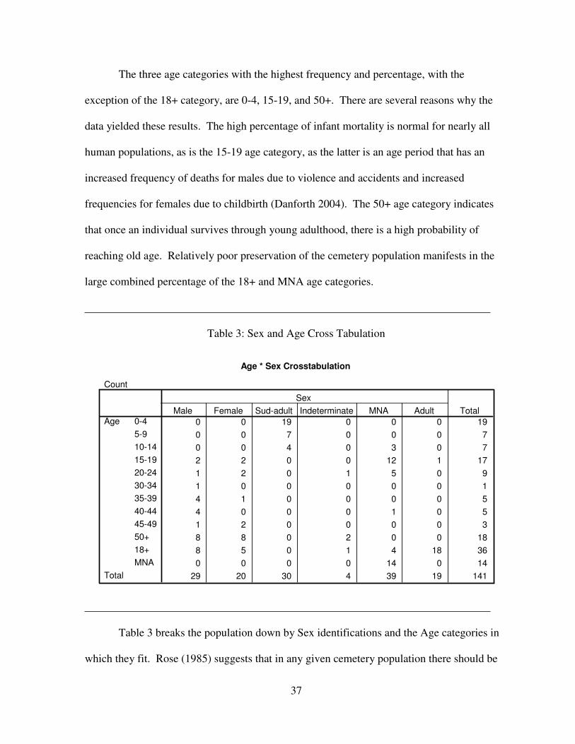
Table 3: Sex and Age Cross Tabulation
Age * Sex Crosstabulation
Count
0 0 19 0 0 0 19
0 0 7 0 0 0 7
0 0 4 0 3 0 7
2 2 0 0 12 1 17
1 2 0 1 5 0 9
1 0 0 0 0 0 1
4 1 0 0 0 0 5
4 0 0 0 1 0 5
1 2 0 0 0 0 3
8 8 0 2 0 0 18
8 5 0 1 4 18 36
0 0 0 0 14 0 14
29 20 30 4 39 19 141
0-4
5-9
10-14
15-19
20-24
30-34
35-39
40-44
45-49
50+
18+
MNA
Age
Total
Male Female Sud-adult Indeterminate MNA Adult
Sex
Total
Table 3 breaks the population down by Sex identifications and the Age categories in
which they fit. Rose (1985) suggests that in any given cemetery population there should be
38
an equal amount of males and females. With Table 3, MNA suggests that while there was no
determinate for sex there was a determinate for age, such as dental development. It has
already been mentioned that there are nine more males than females (29/20). This could be
the result of inaccuracies in the osteological analysis, or the poor preservation of the
cemetery population. There is a possibility that being a rural, agricultural area, there was a
need for males to work the land which would have supported a larger male population. It is
also possible that the women may have had more than one husband having lived longer than
their first (Rose 1985).
Elko Switch
Of the fifty-six burials excavated from Elko Switch Cemetery, only thirty-four had
skeletal remains that allowed for the assessment of age or sex; however, grave dimensions
were used for age-at-death estimations (Shogren et al. 1989). In essence, the smaller a
grave’s dimensions the younger the interred individual is assumed to be. This estimation is
used when no skeletal material is preserved. Similarly to Foster Cemetery, this indicates
very poor preservation of skeletal material. Two burials did not have sufficient skeletal or
mortuary remains for age-at-death estimations and were not included in the demographic
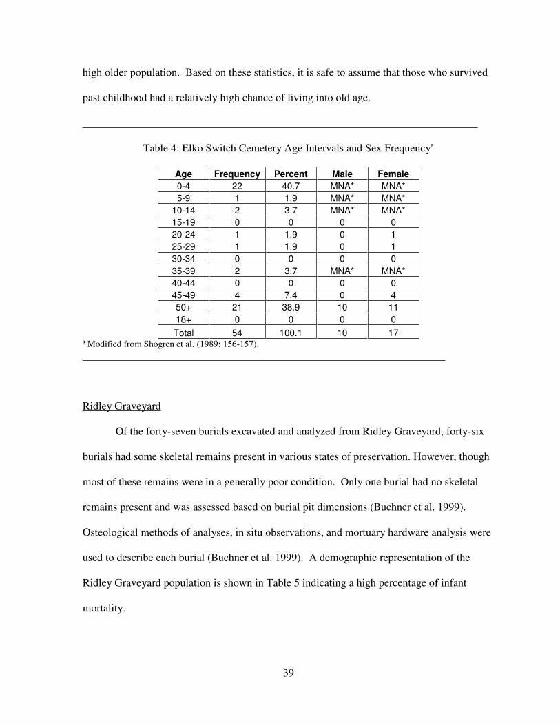
analysis (Shogren et al. 1989). A brief demographic description for the cemetery is shown in
Table 4. Although statistics derived from the small number of excavated burials may not be
completely representative of the rural African American community that Elko Switch
Cemetery served, and grave dimensions for age-at-death may not be as accurate as skeletal
analysis, but they do provide information and give a voice to a marginalized and
undocumented people (Shogren et al. 1989). Table 4 shows a high infant mortality rate and a
39
high older population. Based on these statistics, it is safe to assume that those who survived
past childhood had a relatively high chance of living into old age.
Table 4: Elko Switch Cemetery Age Intervals and Sex Frequencyª
Age Frequency Percent Male Female
0-4 22 40.7 MNA* MNA*
5-9 1 1.9 MNA* MNA*
10-14 2 3.7 MNA* MNA*
15-19 0 0 0 0
20-24 1 1.9 0 1
25-29 1 1.9 0 1
30-34 0 0 0 0
35-39 2 3.7 MNA* MNA*
40-44 0 0 0 0
45-49 4 7.4 0 4
50+ 21 38.9 10 11
18+ 0 0 0 0
Total 54 100.1 10 17 ª Modified from Shogren et al. (1989: 156-157).
Ridley Graveyard
Of the forty-seven burials excavated and analyzed from Ridley Graveyard, forty-six
burials had some skeletal remains present in various states of preservation. However, though
most of these remains were in a generally poor condition. Only one burial had no skeletal
remains present and was assessed based on burial pit dimensions (Buchner et al. 1999).
Osteological methods of analyses, in situ observations, and mortuary hardware analysis were
used to describe each burial (Buchner et al. 1999). A demographic representation of the
Ridley Graveyard population is shown in Table 5 indicating a high percentage of infant
mortality.
40
Table 5: Ridley Graveyard Age Intervals and Sex Frequencyª
Age Frequency Percent Male Female
0-4 18 38.3 MNA* MNA*
5-9 2 4.3 MNA* MNA*
10-14 0 0 MNA* MNA*
15-19 2 4.3 1 0
20-24 0 0 0 0
25-29 4 8.5 0 4
30-34 4 8.5 1 3
35-39 8 17 2 6
40-44 2 4.3 1 1
45-49 1 2.1 1 0
50+ 6 12.8 6 0
18+ 0 0 0 0
Total 47 100.1 12 14 ª Modified from Shogren et al. (1989: 156-157).
Cedar Grove
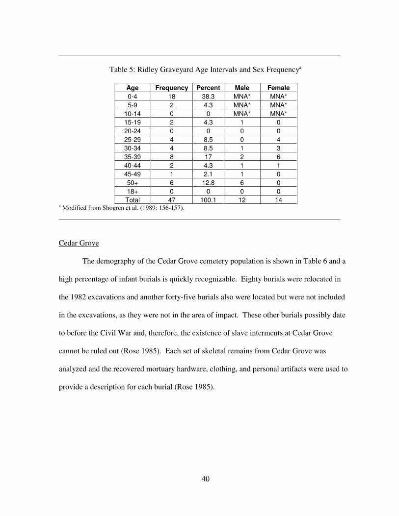
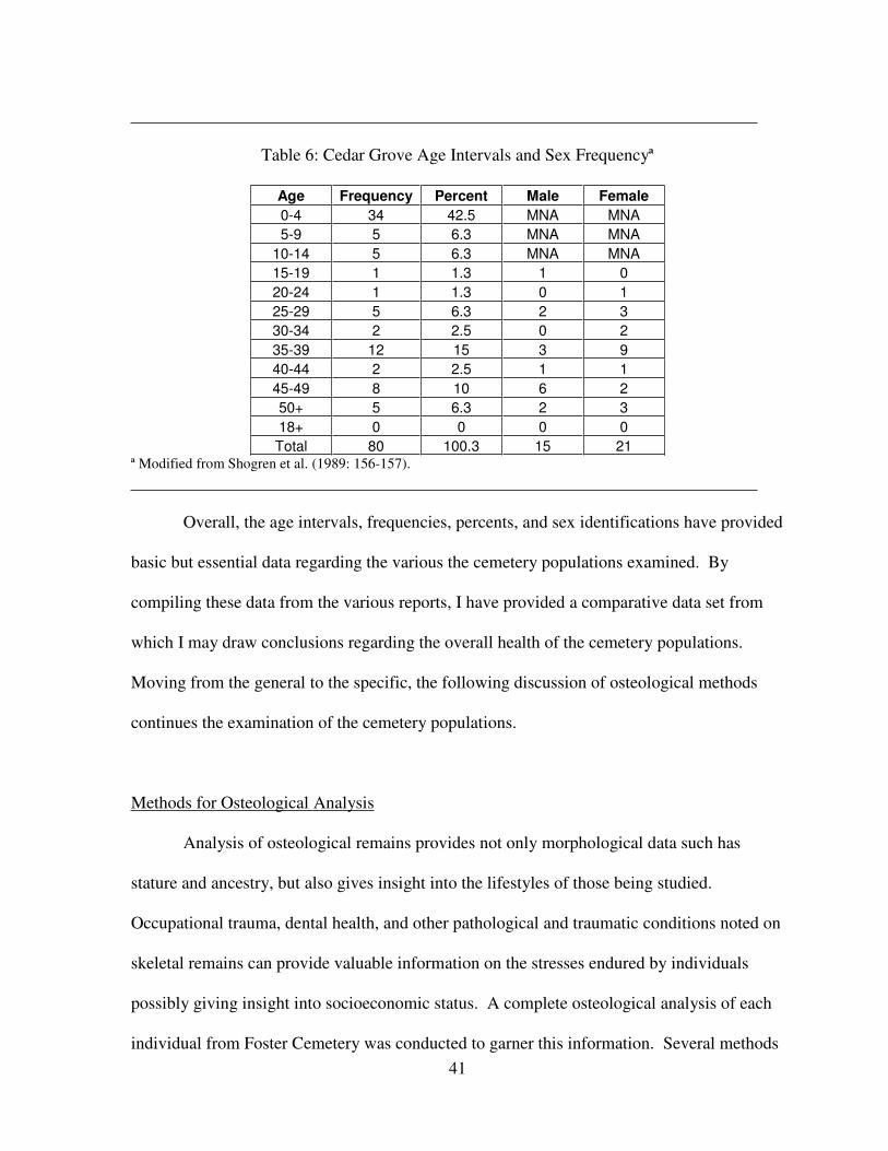
The demography of the Cedar Grove cemetery population is shown in Table 6 and a
high percentage of infant burials is quickly recognizable. Eighty burials were relocated in
the 1982 excavations and another forty-five burials also were located but were not included
in the excavations, as they were not in the area of impact. These other burials possibly date
to before the Civil War and, therefore, the existence of slave interments at Cedar Grove
cannot be ruled out (Rose 1985). Each set of skeletal remains from Cedar Grove was
analyzed and the recovered mortuary hardware, clothing, and personal artifacts were used to
provide a description for each burial (Rose 1985).
41
Table 6: Cedar Grove Age Intervals and Sex Frequencyª
Age Frequency Percent Male Female
0-4 34 42.5 MNA MNA
5-9 5 6.3 MNA MNA
10-14 5 6.3 MNA MNA
15-19 1 1.3 1 0
20-24 1 1.3 0 1
25-29 5 6.3 2 3
30-34 2 2.5 0 2
35-39 12 15 3 9
40-44 2 2.5 1 1
45-49 8 10 6 2
50+ 5 6.3 2 3
18+ 0 0 0 0
Total 80 100.3 15 21 ª Modified from Shogren et al. (1989: 156-157).
Overall, the age intervals, frequencies, percents, and sex identifications have provided
basic but essential data regarding the various the cemetery populations examined. By
compiling these data from the various reports, I have provided a comparative data set from
which I may draw conclusions regarding the overall health of the cemetery populations.
Moving from the general to the specific, the following discussion of osteological methods
continues the examination of the cemetery populations.
Methods for Osteological Analysis
Analysis of osteological remains provides not only morphological data such has
stature and ancestry, but also gives insight into the lifestyles of those being studied.
Occupational trauma, dental health, and other pathological and traumatic conditions noted on
skeletal remains can provide valuable information on the stresses endured by individuals
possibly giving insight into socioeconomic status. A complete osteological analysis of each
individual from Foster Cemetery was conducted to garner this information. Several methods
42
and tools were used to analyze and record the skeletal remains. A general inventory of the
skeletal remains is provided in the Appendix and additional criteria addressing sex, age,
stature, pathology, trauma, and ancestry were noted if present. Morphological markers and
metrics were used to determine and define these criteria. It should be noted, however, that if
the remains were too damaged or deteriorated due to soil conditions, water exposure or
natural decomposition, then some information needed for determining these criteria was not
available. If bone deteriorations were present, it was noted and the condition of the remains
recorded. All skeletal and dental data gathered from this study were recorded on forms
provided by the Alabama Museum of Natural History Laboratory of Human Osteology.
Sex of each set of skeletal remains was determined by analyzing sexually dimorphic
cranial indicators such as the mastoid process, nuchal area, supraorbital ridge, mandible, and
postcranial indicators such as the greater sciatic notch, pubis, preauricular sulcus, head of the
femur, and glenoid fossa (Bass 1995; Buikstra and Ubelaker 1994; Byers 2002; White 1999).
The male mastoid process is large and projecting while the female mastoid process is small
and nonprojecting (Byers 2002). The nuchal area of males is rugged with a hook-like
protuberance while female nuchal areas are smoother and lack the hook-like protuberance
(Buikstra and Ubelaker 1994; Byers 2002). Supraorbital ridges of males are larger and more
robust than those of females which are smoother and less rounded (Buikstra and Ubelaker
1994; Byers 2002). A gonial angle measurement of the mandible less than 124 degrees
suggests a male specimen, whereas a measurement greater than 125 degrees suggests a
female specimen, and will be measured using a mandibulometer (Buikstra and Ubelaker
1994).
The greater sciatic notch is broader in female and narrower in males (Buikstra and
Ubelaker 1994). Pubic shape in males is generally narrow and rectangular while the female
43
pubic shape is broad and square (Byers 2002). It is thought that the preauricular sulcus
occurs more often in females than in males and was scored based on appearance and size for
females, and absent for males (Buikstra and Ubelaker 1994). If the head of the femur
measures greater than 46.5 mm then it indicates a male individual; however, if it measures
less than 43.5 mm it is female (Bass 1995). Measurements between 43.5 mm and 46.5 mm
are indeterminate in terms of sex. The glenoid fossa, found on the scapulae, is larger for
males than females because the head of the humerus for males is typically larger than
females. Generally, measurements greater than 36 mm are male while those less than 34 mm
are female (Bass 1995; Byers 2002). It should be noted that individuals less than 18 years of
age, or those that have not completed puberty, cannot have sex assigned to them as the
osteological indicators are not fully developed (Buikstra and Ubelaker 1994).
Stature can only be estimated on mature adult individuals and was only recorded on
adult skeletons older than eighteen years of age. This is because epiphyseal plates, also
known as the growth plates, do not completely fuse until after adolescence and puberty.
Sliding calipers, spreading calipers, an osteometric board, and metric tape were used to
measure the length and diameter of long bones (Buikstra and Ubelaker 1994). These
measurements were then used in equations to assess stature proposed by Trotter (1970).
Stature formulas have been determined for different ethnicities such as African, European,
Native American, and so forth, and these were used after ancestry was assigned. Foster
Cemetery is an African American cemetery and when ancestry was not identifiable, stature
equations for African ancestral groups were used.
Age was determined through several methods. The completion of endocranial suture
closure, especially those for the coronal, sagittal, and lambdoid sutures, were used to age
individuals up to 42 years (Morse et al. 1983). Ectocranial suture closure scores from
44
Buikstra and Ubelaker (1994) were used and were based upon significance of closure, i.e.
open, minimal, significant and complete obliteration. This ectocranial closures scoring
indicate ages from 20 to 50+ years of age. Pubic symphysis changes were used to age
individuals from 18 to 50 + years of age (Buikstra and Ubelaker 1994; Brooks and Suchey
1990; Todd 1921a, 1921b). Pelvic auricular surface changes were examined to assign an age
in groupings of 20-24, 25-29, 30-34, 35-39, 40-44, 45-49, 50-59, or 60+ (Buikstra and
Ubelaker 1994; Lovejoy et al. 1985). Dental development and eruption was used to age
individuals in a range from birth to 21+ years of age (Buikstra and Ubelaker 1994; Schour
and Massler 1944; Ubelaker 1989). In addition, the Đşcan and Loth (1993) method of
assessing sternal rib ends was used to determine age. Degeneration of sternal rib ends was
group by Đşcan into phases. Various epiphyseal unions and fusions of primary ossification
centers were used to age remains from less than one year to more than 32 years of age
(Buikstra and Ubelaker 1994; Krogman and Đşcan 1986; McKern and Stewart 1957; Redfield
1970, Suchey et al. 1984; Ubelaker 1989). Dental wear and its severity were noted, however,
because dental wear is population specific, exact age estimation was not attempted.
Infant and child burials required special attention as they are morphologically very
distinct from adolescent and mature skeletons; however, they are a good indicator of the
relative health of a population and access to health care (Scheuer and Black 2004). Aging
and identification charts and images for infants and children came from Baker et al. (2005),
Buikstra and Ubelaker (1994), Scheuer and Black (2004), Schour and Massler (1944) and
Ubelaker (1989).
Although research indicates the cemetery population is of African American ancestry
(Hunter Johnson, personal communication 2007), each buried individual was treated as an
unknown ethnicity. Specific measurements outlined in Buikstra and Ubelaker (1994) and
45
Steele and Bramblett (1988), as well as non-metric traits defined by Rhine (1990) and Gill
(1995), were used to ascertain the ethnicity of each specimen. Non-metric traits used in
ancestry determination for the cranium include analysis of dentition, with special attention to
the incisors, and an evaluation of the robustness of the zygomatics bones, curvature of the
palate, the complexity of cranial sutures, the nasal profile, and the presence of wormian
bones (Gill 1995; Rhine 1990).
Skeletal trauma and pathology, both important indicators of health and activity, were
also noted. Physical accidents, as well as cultural activities relating to occupation, can lead
to morphological alterations of the skeleton (Aufderheide and Rodríguez-Martín 2005). For
this study, trauma was defined as any skeletal alteration caused by an outside force such as
occupational related stress, violence inflicted trauma, and accidental trauma. For example,
compression of lumbar disks and osteophytic development is a result of repetitive heavy
lifting indicating a physically taxing lifestyle (Kennedy 1989). For the purposes of this
study, pathology was defined as the physical manifestations of diseases or other infectious
agents upon skeletal remains. These diseases and infectious agents included, but were not
limited to, periodontal disease, tuberculosis, and osteoarthritis (Aufderheide and Rodríguez-
Martín 2005; Barnes 1994). Pathological occurrences were noted, measured if applicable,
causes stated, and manifestations photographed.
Dental health also was given thorough analysis because of the hereditary, congenital,
and developmental information it possesses. The diameter of dental caries was made using
hex keys or Allen wrenches. Diameter of the caries was assessed by determining the largest
diameter of hex key that approximates its size. Ranges for sizes included less than two
millimeters upwards. Additionally, dental caries location was also recorded. Linear enamel
hypoplasias are “deficiencies in enamel thickness which may be caused by three phenomena:
46
systematic metabolic stress, hereditary anomalies, and localized trauma” (Buikstra and
Ubelaker 1994). Linear enamel hypoplasias were measured using a standard dial caliper with
sharpened tips. The caliper was used to determine the distance from the cemento-enamel-
junction (CEJ) to the most distant (occlusal) aspect of the defect (Buikstra and Ubelaker
1994; Hillson 1996). This provides an estimation of the age at which the metabolic stress or
trauma occurred. Other abnormal dental conditions were recorded, such as the presence of
calculus, and placed in the appropriate category, i.e. anomaly, trauma, or pathology.
Finally, it is noted here that many burials contained little or no discernable
osteological information. For burials with no skeletal remains present, it was recorded as No
Remains Present. The abbreviation MNA stands for “Measurement Not Available” and
applies to skeletal remains where sex, stature, age, or ethnicity indicators were not present or
preserved. The use of the classification “Bone Meal” applies to burials that were highly
deteriorated with only trace amounts of skeletal substance remaining. Remains under this
classification have almost no discernable information and can not be identified as human.
Any nonhuman remains were recorded as Animal or to species level if possible. The
following chapter continues to discuss the health of the individuals from the four cemeteries
through an analysis of criteria provided in this chapter.
47
CHAPTER FOUR
ANALYSIS
The health comparison of the skeletal remains from the cemetery populations of
Ridley Graveyard, Elko Switch, Cedar Grove, and Foster Cemetery is based on demography,
growth and development, infection, diet, degenerative joint disease and trauma (Davidson et
al. 2002). Demography consists of sex identifications and age-at-death assessments. Growth
and development discusses adult height and enamel defects (Davidson et al. 2002). Infection
compares the occurrence of specific infectious agents or diseases. The comparison of diet
includes a discussion of the appearance of dental caries, cribra orbitalia, and porotic
hyperostosis. Trauma and degenerative joint disease includes a discussion of arthritic
development and healed fractures. Finally sex identifications created by observing the initial
artifact findings are compared to actual sex identifications using the osteological data follows
the health comparisons.
It should also be understood that only osteological data are being used for the Foster
Cemetery category, which contrasts with the use of grave length, artifacts, and osteological
analysis from the other cemeteries. This can be seen in the 18+ category found in Foster
Cemetery. The 18+ category was created for adult remains that were mostly fragmented.
These remains did not possess any aging or sexing characteristics that could be observed.
Because of the relatively poor preservation of burial remains, the category was included
because it represents a percentage of the population which reached adulthood. Additionally,
for all sections included in this analysis, the original reports from each cemetery excavation
48
as well as more recent publications were referenced. By using both of these data sources a
more precise image of the comparative populations can be created. Also, the sample size
used for the tests in each section is determined by the criteria necessary for each test, the
supplemental data provided by more recent publications, or literature sources, such as death
certificates, used in the other cemetery reports.
For any statistical comparison between the cemetery populations, it must be taken
into consideration the total number of burials recovered from each. Ridley Graveyard
contained 47 excavated burials. A total of 56 burials were excavated from Elko Switch, but
only 54 had either osteological or grave lengths evidence to estimate age. Eighty burials
were excavated from Cedar Grove. Only 127 burials were used from Foster Cemetery.
Although 224 burials were excavated, only 127 were adequately preserved allowing for age
estimation. Due to the differences in the cemetery populations, relative percentages form the
basis for all comparisons.
For the purposes of this study, individuals that comprise the Foster Cemetery
population are thought to be equivalent in health with the individuals from other cemetery
populations when calculated percentages fall between the range of the percentages and
averages of those other cemetery populations. Also, comparable health applies to a
percentage or average within 5% of the outliers of the range. For example, if the calculated
percentage for any test is between 20% and 30%, a percentage of 18% will constitute
comparable health because it is within 5% of the lowest and greatest values of the calculated
range. Anything greater than 5% will be identified as sicker or healthier depending on the
test.
49
Preservation
The cemeteries in this study span some 110 years from 1850 to 1960 with Elko
Switch being used for seventy years (1850-1920), Cedar Grove for fifteen years (1900-1915),
Ridley Graveyard for fifty-five years (1885-1940), and Foster Cemetery for ninety years
(1870-1960) (Buchner et al. 1999; Davidson et al. 2002; Hunter Johnson n.d.; Rose 1985;
Shogren et al. 1989). During these 110 years, the skeletal remains were subject to
taphonomic changes, including exposure to acidic soils, root growth, pressure from grave fill,
and water exposure (Buchner et al. 1999). All of the cemetery populations experienced these
changes, but not with the same effects. The population from Cedar Grove generally had
better preservation than the other three cemetery populations and this better preservation can
be seen in some of the tests in the following sections. When applicable, this disparity was
corrected for by using only the skeletal remains available for the analysis to be done. So,
while the limiting affect of often poor preservation should be noted, there are sufficient and
adequate remains to draw health based conclusions. However, such limitations will be noted
where poor preservation severely limits interpretation.
Demography
For evaluating stress level and adaptive success for a given population, demographic
analysis is an ideal method because regardless of historical documentation, it portrays the
effects of those stresses on the population (Rose 1985). Furthermore, paleodemography has
been shown to be a very productive and informative method (Lallo et al. 1980; Lovejoy et al.
1977; Moore et al. 1975; Weiss 1973a). Demographic data for the four cemeteries are shown
in Table 7, modeled after the tables used by Davidson et al. (2002).
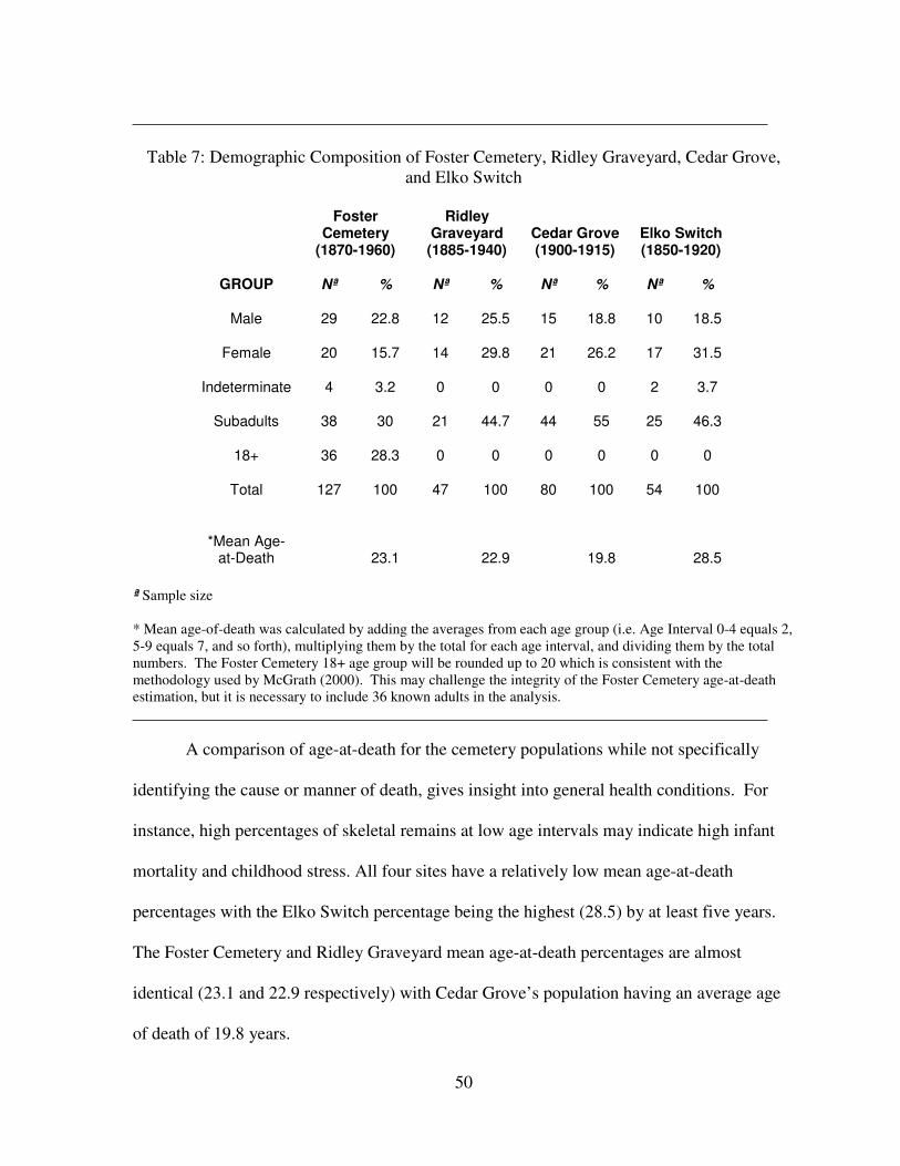
50
Table 7: Demographic Composition of Foster Cemetery, Ridley Graveyard, Cedar Grove,
and Elko Switch
GROUP
Foster Cemetery
(1870-1960)
Ridley Graveyard (1885-1940)
Cedar Grove (1900-1915)
Elko Switch (1850-1920)
Nª % Nª % Nª % Nª %
Male 29 22.8 12 25.5 15 18.8 10 18.5
Female 20 15.7 14 29.8 21 26.2 17 31.5
Indeterminate 4 3.2 0 0 0 0 2 3.7
Subadults 38 30 21 44.7 44 55 25 46.3
18+ 36 28.3 0 0 0 0 0 0
Total 127 100 47 100 80 100 54 100
*Mean Age-
at-Death 23.1 22.9 19.8 28.5
ª Sample size
* Mean age-of-death was calculated by adding the averages from each age group (i.e. Age Interval 0-4 equals 2,
5-9 equals 7, and so forth), multiplying them by the total for each age interval, and dividing them by the total
numbers. The Foster Cemetery 18+ age group will be rounded up to 20 which is consistent with the
methodology used by McGrath (2000). This may challenge the integrity of the Foster Cemetery age-at-death
estimation, but it is necessary to include 36 known adults in the analysis.
A comparison of age-at-death for the cemetery populations while not specifically
identifying the cause or manner of death, gives insight into general health conditions. For
instance, high percentages of skeletal remains at low age intervals may indicate high infant
mortality and childhood stress. All four sites have a relatively low mean age-at-death
percentages with the Elko Switch percentage being the highest (28.5) by at least five years.
The Foster Cemetery and Ridley Graveyard mean age-at-death percentages are almost
identical (23.1 and 22.9 respectively) with Cedar Grove’s population having an average age
of death of 19.8 years.
51
Subadult burials form the largest percentage found at each cemetery although Foster
Cemetery has the lowest percentage by 14.7% in comparison to Ridley Graveyard, 15% in
comparison to Cedar Grove, and 16.3% in comparison to Elko Switch. Cedar Grove and
Elko Switch have nearly identical percentages of male interments (18.8 and 18.5
correspondingly). Though Foster Cemetery and Ridley Graveyard are similar (22.8 and 25.5
in that order), they are slightly higher than the other populations. Foster Cemetery has the
lowest percentage of female burials (15.7%), which is 14.1% lower than Ridley (29.8),
10.5% below Cedar Grove (26.2), and 15.8% lower than Elko Switch (31.5).
Only Foster Cemetery has burials that are in the 18+ category, with 28.3% of its
burials found there. The Foster sample has such a high percentage because this analysis does
not include age or sex estimations based on hardware and artifact analysis, or on
coffin/casket length used by analysts of other cemeteries.
The age groups and designations for Tables 8 and 9, and Figures 2 and 3 come from
the Ridley Graveyard report (Buchner et al. 1999). Table 8 and Figure 2 provide information
on the frequency of interments within each age interval found at the cemeteries involved in
this analysis.
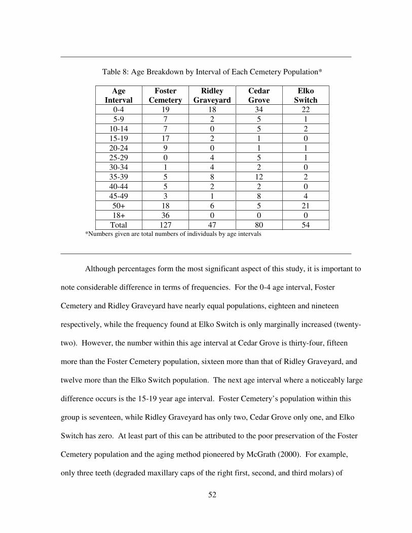
52
Table 8: Age Breakdown by Interval of Each Cemetery Population*
Age
Interval
Foster
Cemetery
Ridley
Graveyard
Cedar
Grove
Elko
Switch
0-4 19 18 34 22
5-9 7 2 5 1
10-14 7 0 5 2
15-19 17 2 1 0
20-24 9 0 1 1
25-29 0 4 5 1
30-34 1 4 2 0
35-39 5 8 12 2
40-44 5 2 2 0
45-49 3 1 8 4
50+ 18 6 5 21
18+ 36 0 0 0
Total 127 47 80 54 *Numbers given are total numbers of individuals by age intervals
Although percentages form the most significant aspect of this study, it is important to
note considerable difference in terms of frequencies. For the 0-4 age interval, Foster
Cemetery and Ridley Graveyard have nearly equal populations, eighteen and nineteen
respectively, while the frequency found at Elko Switch is only marginally increased (twenty-
two). However, the number within this age interval at Cedar Grove is thirty-four, fifteen
more than the Foster Cemetery population, sixteen more than that of Ridley Graveyard, and
twelve more than the Elko Switch population. The next age interval where a noticeably large
difference occurs is the 15-19 year age interval. Foster Cemetery’s population within this
group is seventeen, while Ridley Graveyard has only two, Cedar Grove only one, and Elko
Switch has zero. At least part of this can be attributed to the poor preservation of the Foster
Cemetery population and the aging method pioneered by McGrath (2000). For example,
only three teeth (degraded maxillary caps of the right first, second, and third molars) of
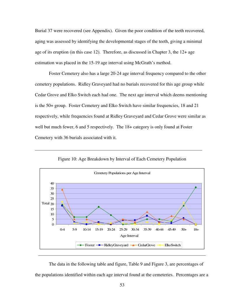
53
Burial 37 were recovered (see Appendix). Given the poor condition of the teeth recovered,
aging was assessed by identifying the developmental stages of the teeth, giving a minimal
age of its eruption (in this case 12). Therefore, as discussed in Chapter 3, the 12+ age
estimation was placed in the 15-19 age interval using McGrath’s method.
Foster Cemetery also has a large 20-24 age interval frequency compared to the other
cemetery populations. Ridley Graveyard had no burials recovered for this age group while
Cedar Grove and Elko Switch each had one. The next age interval which deems mentioning
is the 50+ group. Foster Cemetery and Elko Switch have similar frequencies, 18 and 21
respectively, while frequencies found at Ridley Graveyard and Cedar Grove were similar as
well but much fewer, 6 and 5 respectively. The 18+ category is only found at Foster
Cemetery with 36 burials associated with it.
Figure 10: Age Breakdown by Interval of Each Cemetery Population
Cemetery Populations per Age Interval
0
5
10
15
20
25
30
35
40
0-4 5-9 10-14 15-19 20-24 25-29 30-34 35-39 40-44 45-49 50+ 18+
Age Interval
Total
Foster RidleyGraveyard CedarGrove ElkoSwitch
The data in the following table and figure, Table 9 and Figure 3, are percentages of
the populations identified within each age interval found at the cemeteries. Percentages are a
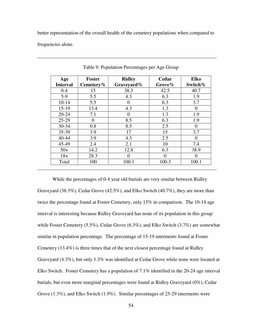
54
better representation of the overall health of the cemetery populations when compared to
frequencies alone.
Table 9: Population Percentages per Age Group
Age
Interval
Foster
Cemetery%
Ridley
Graveyard%
Cedar
Grove%
Elko
Switch%
0-4 15 38.3 42.5 40.7
5-9 5.5 4.3 6.3 1.9
10-14 5.5 0 6.3 3.7
15-19 13.4 4.3 1.3 0
20-24 7.1 0 1.3 1.9
25-29 0 8.5 6.3 1.9
30-34 0.8 8.5 2.5 0
35-39 3.9 17 15 3.7
40-44 3.9 4.3 2.5 0
45-49 2.4 2.1 10 7.4
50+ 14.2 12.8 6.3 38.9
18+ 28.3 0 0 0
Total 100 100.1 100.3 100.1
While the percentages of 0-4 year old burials are very similar between Ridley
Graveyard (38.3%), Cedar Grove (42.5%), and Elko Switch (40.7%), they are more than
twice the percentage found at Foster Cemetery, only 15% in comparison. The 10-14 age
interval is interesting because Ridley Graveyard has none of its population in this group
while Foster Cemetery (5.5%), Cedar Grove (6.3%), and Elko Switch (3.7%) are somewhat
similar in population percentage. The percentage of 15-19 interments found at Foster
Cemetery (13.4%) is three times that of the next closest percentage found at Ridley
Graveyard (4.3%), but only 1.3% was identified at Cedar Grove while none were located at
Elko Switch. Foster Cemetery has a population of 7.1% identified in the 20-24 age interval
burials, but even more marginal percentages were found at Ridley Graveyard (0%), Cedar
Grove (1.3%), and Elko Switch (1.9%). Similar percentages of 25-29 interments were
55
identified at Ridley Graveyard (8.5%) and Cedar Grove (6.3%) with lower percentages, but
also similar, located at Foster Cemetery (0%) and Elko Switch (1.9%).
The 30-34 age interval also has percentages worth mentioning. Foster Cemetery
(0.8%), Cedar Grove (2.5%), and Elko Switch (0%) have small or no percentage of their
populations occurring in this age interval. However, Ridley Graveyard has 8.5% of its
population occurring within the 30-34 age interval. The 35-39 age interval proved interesting
in that the four cemetery populations were grouped in pairs regarding relative percentages.
Foster Cemetery and Elko Switch have low percentages, 3.9% and 3.7% respectively. Ridley
Graveyard and Cedar Grove have similar higher percentages of 17% for Ridley Graveyard
and 15% for Cedar Grove. The frequencies of Foster Cemetery (3.9%), Ridley Graveyard
(4.3%), and Cedar Grove (2.5%) are similar in the 40-44 age interval, however, Elko Switch
has no percentage of its population identified within this age interval.
Foster Cemetery and Ridley Graveyard have similar percentages of their populations
occurring in the 45-49 age interval (2.4% and 2.1% respectively). Cedar Grove and Elko
Switch have higher percentages occurring in this age interval that are somewhat similar (10%
and 7.4% respectively). Similar to the 0-4 age interval, the 50+ age interval has some of the
highest percentages found within this analysis. The Cedar Grove percentage is the lowest of
this age group being 6.3%. The percentage identified in Foster Cemetery is 14.2% and is
very similar to the 12.8% identified at Ridley Graveyard. The highest percentage found in
this age group occurs in Elko Switch with 38.9% of its population found there.
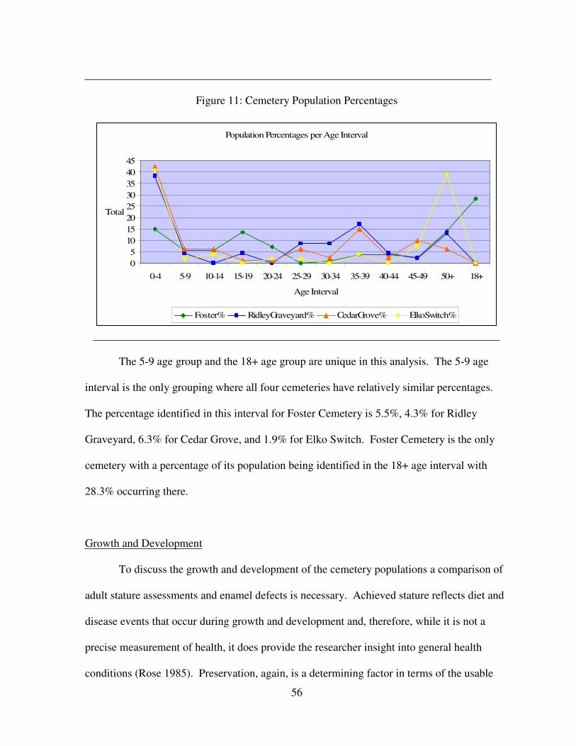
56
Figure 11: Cemetery Population Percentages
Population Percentages per Age Interval
0
5
10
15
20
25
30
35
40
45
0-4 5-9 10-14 15-19 20-24 25-29 30-34 35-39 40-44 45-49 50+ 18+
Age Interval
Total
Foster% RidleyGraveyard% CedarGrove% ElkoSwitch%
The 5-9 age group and the 18+ age group are unique in this analysis. The 5-9 age
interval is the only grouping where all four cemeteries have relatively similar percentages.
The percentage identified in this interval for Foster Cemetery is 5.5%, 4.3% for Ridley
Graveyard, 6.3% for Cedar Grove, and 1.9% for Elko Switch. Foster Cemetery is the only
cemetery with a percentage of its population being identified in the 18+ age interval with
28.3% occurring there.
Growth and Development
To discuss the growth and development of the cemetery populations a comparison of
adult stature assessments and enamel defects is necessary. Achieved stature reflects diet and
disease events that occur during growth and development and, therefore, while it is not a
precise measurement of health, it does provide the researcher insight into general health
conditions (Rose 1985). Preservation, again, is a determining factor in terms of the usable
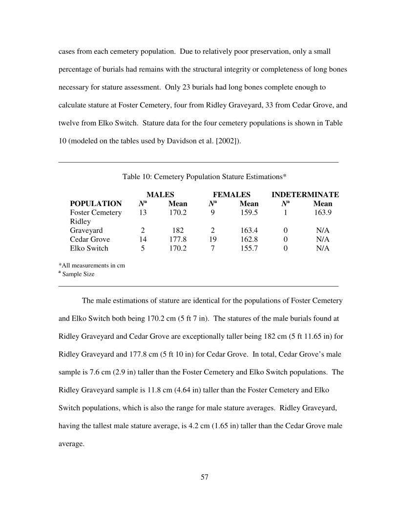
57
cases from each cemetery population. Due to relatively poor preservation, only a small
percentage of burials had remains with the structural integrity or completeness of long bones
necessary for stature assessment. Only 23 burials had long bones complete enough to
calculate stature at Foster Cemetery, four from Ridley Graveyard, 33 from Cedar Grove, and
twelve from Elko Switch. Stature data for the four cemetery populations is shown in Table
10 (modeled on the tables used by Davidson et al. [2002]).
Table 10: Cemetery Population Stature Estimations*
POPULATION
MALES FEMALES INDETERMINATE
Nª Mean Nª Mean Nª Mean
Foster Cemetery 13 170.2 9 159.5 1 163.9
Ridley
Graveyard 2 182 2 163.4 0 N/A
Cedar Grove 14 177.8 19 162.8 0 N/A
Elko Switch 5 170.2 7 155.7 0 N/A
*All measurements in cm
ª Sample Size
The male estimations of stature are identical for the populations of Foster Cemetery
and Elko Switch both being 170.2 cm (5 ft 7 in). The statures of the male burials found at
Ridley Graveyard and Cedar Grove are exceptionally taller being 182 cm (5 ft 11.65 in) for
Ridley Graveyard and 177.8 cm (5 ft 10 in) for Cedar Grove. In total, Cedar Grove’s male
sample is 7.6 cm (2.9 in) taller than the Foster Cemetery and Elko Switch populations. The
Ridley Graveyard sample is 11.8 cm (4.64 in) taller than the Foster Cemetery and Elko
Switch populations, which is also the range for male stature averages. Ridley Graveyard,
having the tallest male stature average, is 4.2 cm (1.65 in) taller than the Cedar Grove male
average.
58
Foster Cemetery’s female stature average, 159.5 cm (5 ft 2.79 in), is larger only than
the Elko Switch female average of 155.7 cm (5 ft 1.29 in), a difference of 3.8 cm (1.49 in).
Like the male stature averages, Ridley Graveyard’s female stature estimate is the largest of
the four cemeteries, 163.4 cm (5 ft 4.33 in). However, the difference between the Foster
Cemetery and Ridley Graveyard averages is much closer being 3.9 cm (1.53 in). The
average of the Cedar Grove population is 162.8 cm (5 ft 4.09 in), a difference of only 0.6 cm
(.23 in) in comparison to the Ridley Graveyard sample. The range of the female averages is
smaller than that of the male averages, 7.7 cm (3.03 in) in comparison to 11.8 cm (4.64 in).
Burial 6 from Foster Cemetery was the only indeterminate burial identified within the stature
analysis and had an estimated stature of 163.9 cm (5 ft 4.52 in).
Linear enamel hypoplasias (LEH) are horizontal grooves found in the enamel of
teeth. Incisors and canines are the most sensitive to the enamel disruptions that result from
non-fatal stresses induced by array of metabolic stresses including weaning, fever, starvation,
diarrhea, and measles (Aufderheide and Rodríguez-Martín 1999; Davidson et al. 2002).
Linear enamel hypoplasias provide a record of disease and stressful events during dental
development, i.e. childhood, and the sensitivity of the tooth to the stress event, as Davidson
et al. note (2002), is determined by nutrition and especially protein. When a child is starving
or not getting adequate nutrition, enamel forming cells called ameloblasts, shut down their
production of enamel. When the child recovers, the ameloblasts recover and produce normal
amounts of enamel (Jacobi, personal communication). Therefore, examining the proportion
and frequency of linear enamel hypoplasias provides insight into possible periods of disease
and malnutrition during childhood.
A few remarks are necessary before an analysis of LEH within the cemetery
populations. Ridley Graveyard requires special attention. In general, an inventory of
59
recovered dentition was not provided for each burial, although hypoplastic activity was
recorded (Buchner et al. 1999). For example, Burial 15 from the Ridley Graveyard report
that the “teeth appear to be large;” however, the actual teeth that are being referred to is not
specified (Buchner et al. 1999). Therefore, for burials where a direct mention of teeth
recovered was not given, those burials are not included in this analysis. However, those
burials with hypoplasias in the dentition that are recorded (see Appendix) are used in this
analysis. Therefore, the main requirements for the burials is that they possess at least one
deciduous or permanent incisor or canine recovered, and furthermore, the recovered teeth
must have at least one or more linear enamel hypoplasias in order to be included in this
analysis. This requirement was used for all cemetery populations.
Although the table used for this section is modeled after the Davidson et al. (2002)
publication, which compares the rural Cedar Grove cemetery population to the urban
Freedman’s Cemetery in Dallas, Texas, it utilized the total number of deciduous incisors and
canines, as well as permanent incisors and canines from each cemetery population to
compare dental health. However, the number of individuals contributing to the available
dentition was not included. Therefore, while the Davidson et al. (2002) publication is more
recent and includes additional data, the original cemetery report by Rose (1985) was
referenced for this section noting the number of burials with hypoplastic activity.
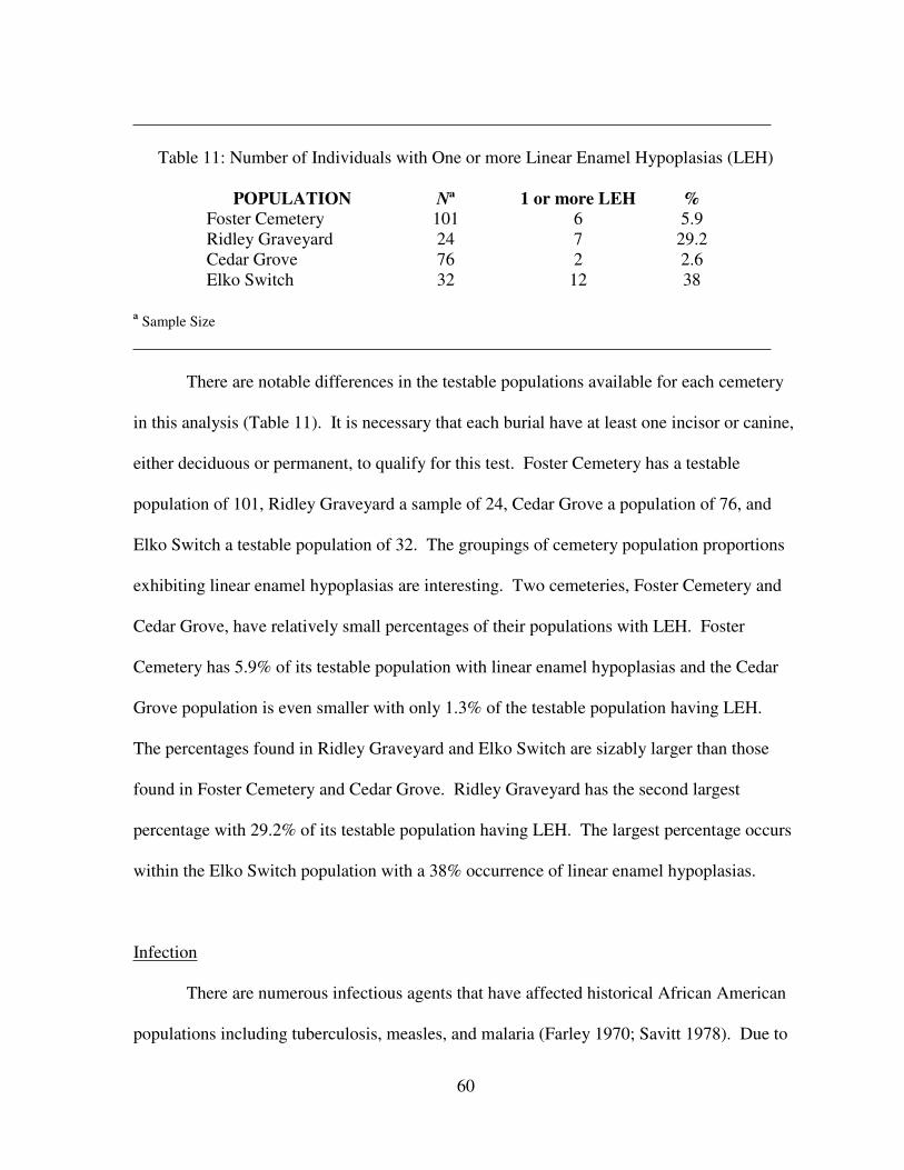
60
Table 11: Number of Individuals with One or more Linear Enamel Hypoplasias (LEH)
POPULATION Nª 1 or more LEH %
Foster Cemetery 101 6 5.9
Ridley Graveyard 24 7 29.2
Cedar Grove 76 2 2.6
Elko Switch 32 12 38
ª Sample Size
There are notable differences in the testable populations available for each cemetery
in this analysis (Table 11). It is necessary that each burial have at least one incisor or canine,
either deciduous or permanent, to qualify for this test. Foster Cemetery has a testable
population of 101, Ridley Graveyard a sample of 24, Cedar Grove a population of 76, and
Elko Switch a testable population of 32. The groupings of cemetery population proportions
exhibiting linear enamel hypoplasias are interesting. Two cemeteries, Foster Cemetery and
Cedar Grove, have relatively small percentages of their populations with LEH. Foster
Cemetery has 5.9% of its testable population with linear enamel hypoplasias and the Cedar
Grove population is even smaller with only 1.3% of the testable population having LEH.
The percentages found in Ridley Graveyard and Elko Switch are sizably larger than those
found in Foster Cemetery and Cedar Grove. Ridley Graveyard has the second largest
percentage with 29.2% of its testable population having LEH. The largest percentage occurs
within the Elko Switch population with a 38% occurrence of linear enamel hypoplasias.
Infection
There are numerous infectious agents that have affected historical African American
populations including tuberculosis, measles, and malaria (Farley 1970; Savitt 1978). Due to
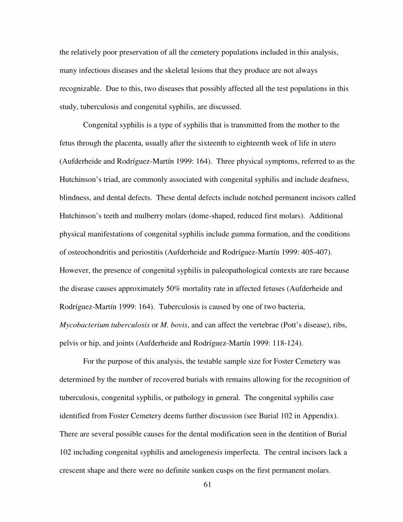
61
the relatively poor preservation of all the cemetery populations included in this analysis,
many infectious diseases and the skeletal lesions that they produce are not always
recognizable. Due to this, two diseases that possibly affected all the test populations in this
study, tuberculosis and congenital syphilis, are discussed.
Congenital syphilis is a type of syphilis that is transmitted from the mother to the
fetus through the placenta, usually after the sixteenth to eighteenth week of life in utero
(Aufderheide and Rodríguez-Martín 1999: 164). Three physical symptoms, referred to as the
Hutchinson’s triad, are commonly associated with congenital syphilis and include deafness,
blindness, and dental defects. These dental defects include notched permanent incisors called
Hutchinson’s teeth and mulberry molars (dome-shaped, reduced first molars). Additional
physical manifestations of congenital syphilis include gumma formation, and the conditions
of osteochondritis and periostitis (Aufderheide and Rodríguez-Martín 1999: 405-407).
However, the presence of congenital syphilis in paleopathological contexts are rare because
the disease causes approximately 50% mortality rate in affected fetuses (Aufderheide and
Rodríguez-Martín 1999: 164). Tuberculosis is caused by one of two bacteria,
Mycobacterium tuberculosis or M. bovis, and can affect the vertebrae (Pott’s disease), ribs,
pelvis or hip, and joints (Aufderheide and Rodríguez-Martín 1999: 118-124).
For the purpose of this analysis, the testable sample size for Foster Cemetery was
determined by the number of recovered burials with remains allowing for the recognition of
tuberculosis, congenital syphilis, or pathology in general. The congenital syphilis case
identified from Foster Cemetery deems further discussion (see Burial 102 in Appendix).
There are several possible causes for the dental modification seen in the dentition of Burial
102 including congenital syphilis and amelogenesis imperfecta. The central incisors lack a
crescent shape and there were no definite sunken cusps on the first permanent molars.
62
Therefore, congenital syphilis cannot be definitely diagnosed. Because of this, amelogenesis
imperfecta is a consideration given the frailty of the teeth and their coloration. As such, this
case should be noted as being congenital syphilis-like and not a definitive identification.
With the comparative cemetery populations, confirmation of cases of tuberculosis and
congenital syphilis was made using various methods, including skeletal analysis and
examination of death certificates. The following discussion is a detailed breakdown of the
methods used to garner this information from each cemetery. The cases of tuberculosis for
Ridley Graveyard were confirmed through death certificate analysis and, therefore, the entire
cemetery population was used. However, the congenital syphilis sample size was determined
through the number of burials where skeletal remains permitted an examination of possible
pathologies. With the Elko Switch cemetery, all burials with skeletal criteria allowing for
age and sex estimations were used. It also should be noted that the five congenital syphilis
cases from the Elko Switch population are possible cases, where other potential sources for
the dental destruction and calcification could be possible (Shogren et al. 1989). For the
Cedar Grove cemetery, Rose (1989) identified the tuberculosis cases through active
periostitis on the pleural surfaces of the ribs. The congenital syphilis cases at Cedar Grove
were discussed in Davidson et al. (2002). They used dental casts to determine the presence
of congenital syphilis and, therefore, I only examined those burials with appropriate
dentition. Results for this test are seen in Table 12.
63
Table 12: Number of Individuals with Tuberculosis and Congenital Syphilis
POPULATION
TUBERCULOSIS
CONGENITAL
SYPHILIS
Nª Frequency % Nª Frequency %
Foster Cemetery 141 1 0.7 141 1* 0.7
Ridley
Graveyard 49 3 6.1 38 1 2.6
Cedar Grove 80 5 6.3 77 4 5.2
Elko Switch 34 1 2.9 34 5* 14.7
ª Sample Size
*Ambiguous cases of dental malformation
The Foster Cemetery population had the lowest percentage of tuberculosis in this
analysis. Less than one percent (0.7%), or only one case, was identified from a testable
sample of 141 cases. Elko Switch has the next lowest percentage of tuberculosis (2.9%) with
only one skeleton exhibiting the skeletal destruction of tuberculosis. Ridley Graveyard had
three documented deaths caused by tuberculosis representing 6.1% of the total cemetery
population of 49. These three deaths were not confirmed through tuberculosis related bone
destruction on actual skeletal remains. Overall, the highest frequency and percentage of
tuberculosis found at the cemeteries was from Cedar Grove, with five cases of tuberculosis in
80 burials (6.3%).
As with the presence of tuberculosis, the Foster Cemetery population has the lowest
percentage and frequency of congenital syphilis. Of the 141 testable cases, only one
exhibited possible manifestations of congenital syphilis (0.7%). The Ridley Graveyard
population has the same frequency as Foster Cemetery (1), but due to a lower testable
population (38), the percentage of cases of present (2.6%) is higher than Foster Cemetery. At
Cedar Grove, four of the 77 burials were identified as having congenital syphilis (5.2%). The
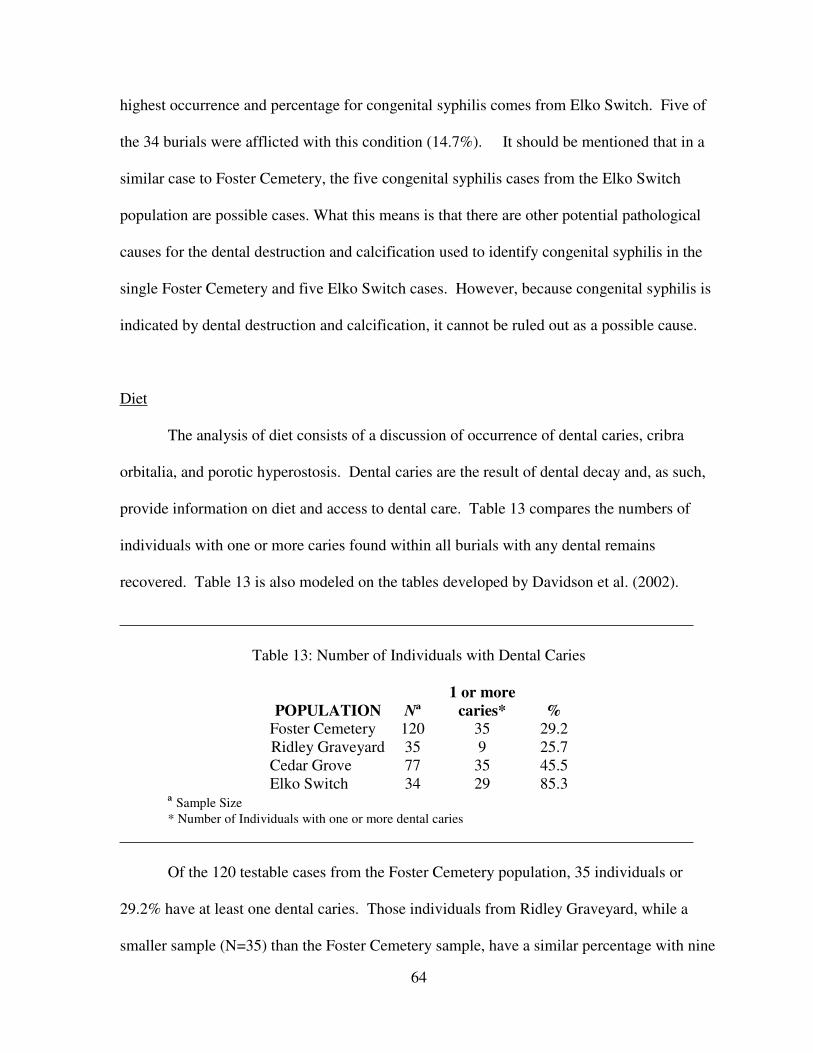
64
highest occurrence and percentage for congenital syphilis comes from Elko Switch. Five of
the 34 burials were afflicted with this condition (14.7%). It should be mentioned that in a
similar case to Foster Cemetery, the five congenital syphilis cases from the Elko Switch
population are possible cases. What this means is that there are other potential pathological
causes for the dental destruction and calcification used to identify congenital syphilis in the
single Foster Cemetery and five Elko Switch cases. However, because congenital syphilis is
indicated by dental destruction and calcification, it cannot be ruled out as a possible cause.
Diet
The analysis of diet consists of a discussion of occurrence of dental caries, cribra
orbitalia, and porotic hyperostosis. Dental caries are the result of dental decay and, as such,
provide information on diet and access to dental care. Table 13 compares the numbers of
individuals with one or more caries found within all burials with any dental remains
recovered. Table 13 is also modeled on the tables developed by Davidson et al. (2002).
Table 13: Number of Individuals with Dental Caries
POPULATION Nª
1 or more
caries* %
Foster Cemetery 120 35 29.2
Ridley Graveyard 35 9 25.7
Cedar Grove 77 35 45.5
Elko Switch 34 29 85.3
ª Sample Size
* Number of Individuals with one or more dental caries
Of the 120 testable cases from the Foster Cemetery population, 35 individuals or
29.2% have at least one dental caries. Those individuals from Ridley Graveyard, while a
smaller sample (N=35) than the Foster Cemetery sample, have a similar percentage with nine
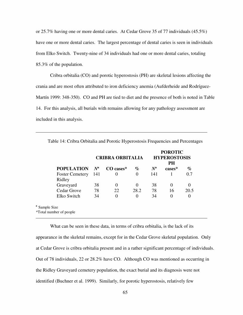
65
or 25.7% having one or more dental caries. At Cedar Grove 35 of 77 individuals (45.5%)
have one or more dental caries. The largest percentage of dental caries is seen in individuals
from Elko Switch. Twenty-nine of 34 individuals had one or more dental caries, totaling
85.3% of the population.
Cribra orbitalia (CO) and porotic hyperostosis (PH) are skeletal lesions affecting the
crania and are most often attributed to iron deficiency anemia (Aufderheide and Rodríguez-
Martín 1999: 348-350). CO and PH are tied to diet and the presence of both is noted in Table
14. For this analysis, all burials with remains allowing for any pathology assessment are
included in this analysis.
Table 14: Cribra Orbitalia and Porotic Hyperostosis Frequencies and Percentages
POPULATION
CRIBRA ORBITALIA
POROTIC
HYPEROSTOSIS
Nª CO cases* % Nª
PH
cases* %
Foster Cemetery 141 0 0 141 1 0.7
Ridley
Graveyard 38 0 0 38 0 0
Cedar Grove 78 22 28.2 78 16 20.5
Elko Switch 34 0 0 34 0 0
ª Sample Size
*Total number of people
What can be seen in these data, in terms of cribra orbitalia, is the lack of its
appearance in the skeletal remains, except for in the Cedar Grove skeletal population. Only
at Cedar Grove is cribra orbitalia present and in a rather significant percentage of individuals.
Out of 78 individuals, 22 or 28.2% have CO. Although CO was mentioned as occurring in
the Ridley Graveyard cemetery population, the exact burial and its diagnosis were not
identified (Buchner et al. 1999). Similarly, for porotic hyperostosis, relatively few
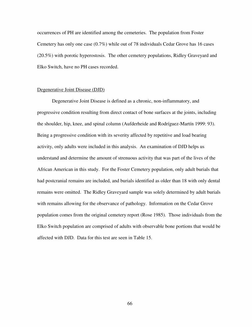
66
occurrences of PH are identified among the cemeteries. The population from Foster
Cemetery has only one case (0.7%) while out of 78 individuals Cedar Grove has 16 cases
(20.5%) with porotic hyperostosis. The other cemetery populations, Ridley Graveyard and
Elko Switch, have no PH cases recorded.
Degenerative Joint Disease (DJD)
Degenerative Joint Disease is defined as a chronic, non-inflammatory, and
progressive condition resulting from direct contact of bone surfaces at the joints, including
the shoulder, hip, knee, and spinal column (Aufderheide and Rodríguez-Martín 1999: 93).
Being a progressive condition with its severity affected by repetitive and load bearing
activity, only adults were included in this analysis. An examination of DJD helps us
understand and determine the amount of strenuous activity that was part of the lives of the
African American in this study. For the Foster Cemetery population, only adult burials that
had postcranial remains are included, and burials identified as older than 18 with only dental
remains were omitted. The Ridley Graveyard sample was solely determined by adult burials
with remains allowing for the observance of pathology. Information on the Cedar Grove
population comes from the original cemetery report (Rose 1985). Those individuals from the
Elko Switch population are comprised of adults with observable bone portions that would be
affected with DJD. Data for this test are seen in Table 15.
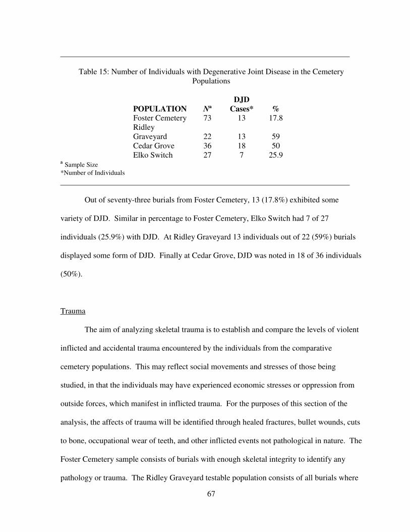
67
Table 15: Number of Individuals with Degenerative Joint Disease in the Cemetery
Populations
POPULATION Nª
DJD
Cases* %
Foster Cemetery 73 13 17.8
Ridley
Graveyard 22 13 59
Cedar Grove 36 18 50
Elko Switch 27 7 25.9
ª Sample Size
*Number of Individuals
Out of seventy-three burials from Foster Cemetery, 13 (17.8%) exhibited some
variety of DJD. Similar in percentage to Foster Cemetery, Elko Switch had 7 of 27
individuals (25.9%) with DJD. At Ridley Graveyard 13 individuals out of 22 (59%) burials
displayed some form of DJD. Finally at Cedar Grove, DJD was noted in 18 of 36 individuals
(50%).
Trauma
The aim of analyzing skeletal trauma is to establish and compare the levels of violent
inflicted and accidental trauma encountered by the individuals from the comparative
cemetery populations. This may reflect social movements and stresses of those being
studied, in that the individuals may have experienced economic stresses or oppression from
outside forces, which manifest in inflicted trauma. For the purposes of this section of the
analysis, the affects of trauma will be identified through healed fractures, bullet wounds, cuts
to bone, occupational wear of teeth, and other inflicted events not pathological in nature. The
Foster Cemetery sample consists of burials with enough skeletal integrity to identify any
pathology or trauma. The Ridley Graveyard testable population consists of all burials where
68
some skeletal material existed. Testable cases from Elko Switch consist of burials with any
skeletal material, as does the Cedar Grove population. Data for this section are shown in
Table 16.
Table 16: Number of Individuals with Trauma from the Cemetery Populations
POPULATIONS Nª
Trauma
Cases* %
Foster Cemetery 141 7 5
Ridley Graveyard 47 0 0
Cedar Grove 80 20 25
Elko Switch 34 4 11.8
ª Sample Size
* Number of individuals
At Foster Cemetery out of 141 burials seven individuals (5%) exhibited trauma while
at Ridley Graveyard no cases of trauma were recorded in the sample of 47 burials. Cedar
Grove has 20 out of 80 individuals (25%) with some type of trauma, making it the highest
percentage in this section. Finally, Elko Switch had four out of thirty-four burials (11.8%)
with some form of trauma.
Map Comparison
Following the salvage excavation of Foster Cemetery, Southeastern Anthropological
Institute (SAI) constructed a preliminary sex identification map based on initial artifact and
grave length analyses (Figure 4). After the completion of the osteological analysis, SAI
constructed another sex identification map based on these data (Figure 5). For the purpose of
this analysis, categories used in the maps are simplified for comparative purposes. These
categories include: Male, Female, Unknown Adult, Subadult, Measurement Not Available
(MNA), Animal, and No Remains. Male and Female comprise adult individuals whose age
69
is above 18 and consists of identified male, female, and possible male and females. The
Unknown Adult category consists of those burials above 18 years of age that do not have a
sex assigned to them. This includes the indeterminate burials from the osteology map
(Figure 5). The subadult category consists of those individuals below 18 years of age, which
includes all child interments. For the most part, sex identification under the age of 16 is not
possible and even at age 18 there can be ambiguity; therefore, all those individuals below age
18 were identified as subadult. The MNA category refers to burials where there is not
adequate data to infer sex or age, which includes the “Bone Meal” category found in the
osteology map (Figure 5). The animal category refers to all burials that are non-human or are
faunal remains. It should be noted that at the time these maps were created, three additional
burials had yet to be excavated, leaving a total of 221 burials. Therefore, the percentages
used in this section are based on 221 total burials. The results and percentages for both the
artifact and osteological maps are found in Table 17.
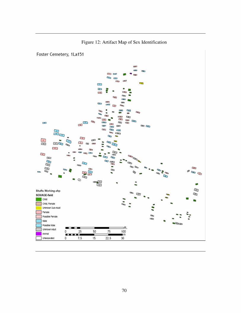
70
Figure 12: Artifact Map of Sex Identification
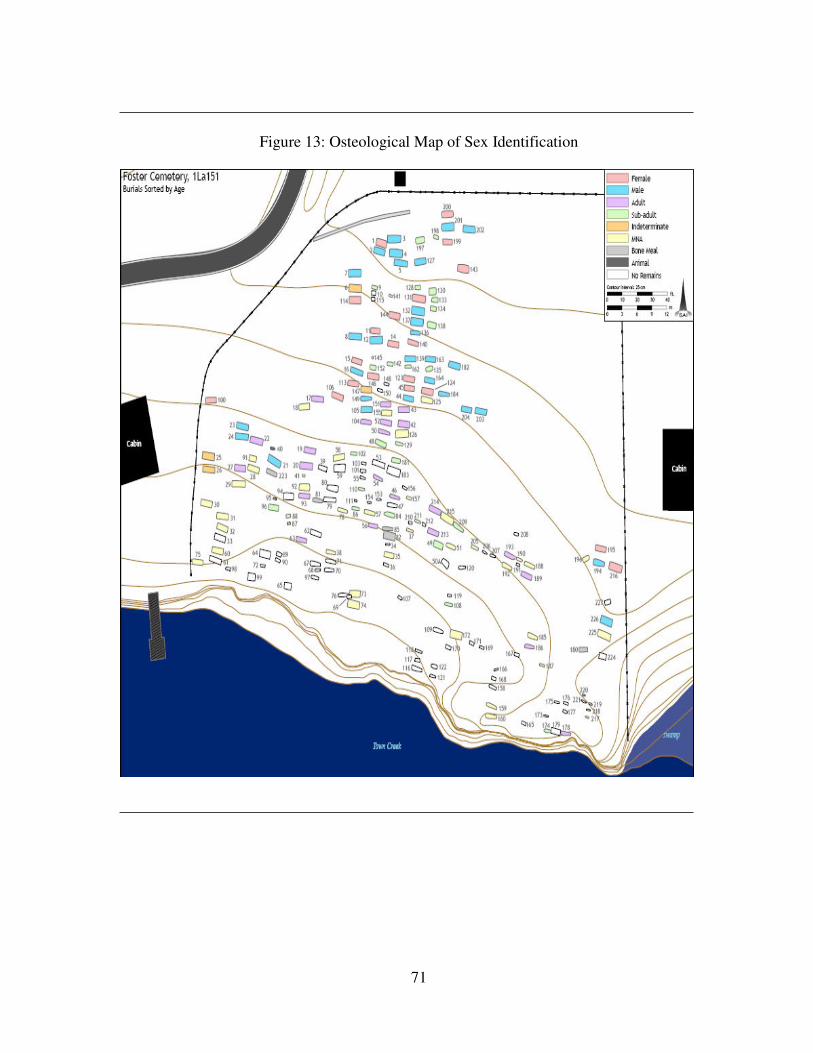
71
Figure 13: Osteological Map of Sex Identification
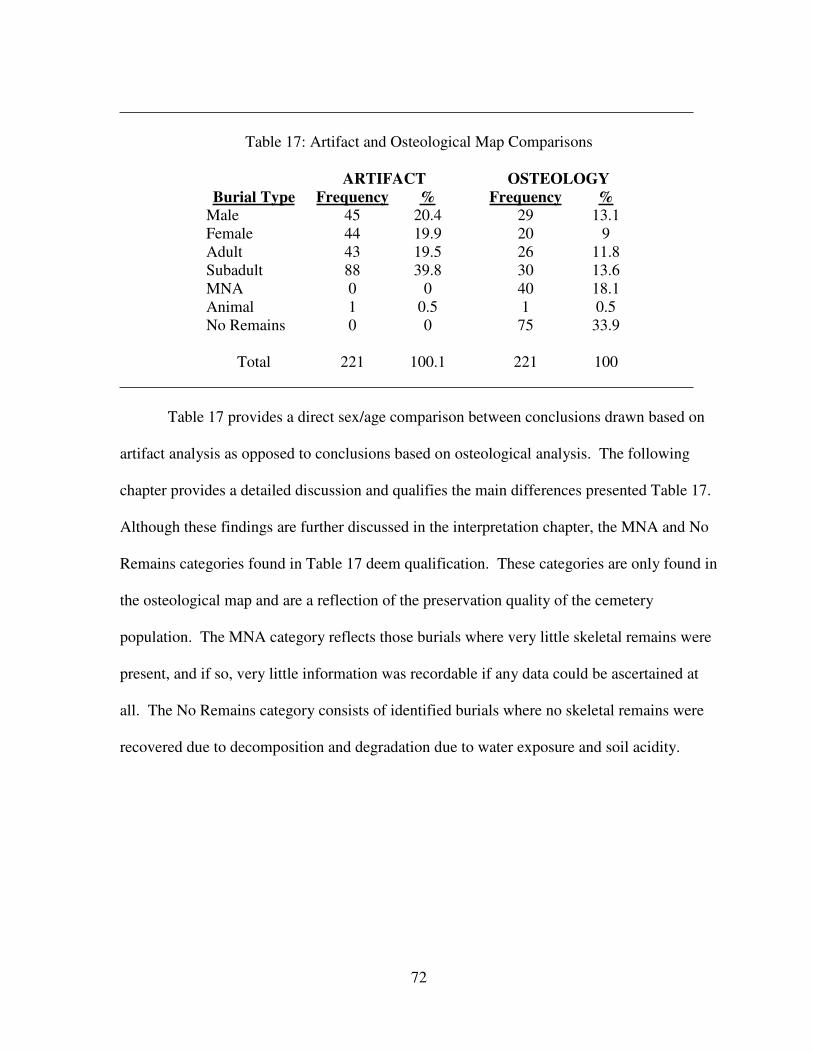
72
Table 17: Artifact and Osteological Map Comparisons
Burial Type
ARTIFACT OSTEOLOGY
Frequency % Frequency %
Male 45 20.4 29 13.1
Female 44 19.9 20 9
Adult 43 19.5 26 11.8
Subadult 88 39.8 30 13.6
MNA 0 0 40 18.1
Animal 1 0.5 1 0.5
No Remains 0 0 75 33.9
Total 221 100.1 221 100
Table 17 provides a direct sex/age comparison between conclusions drawn based on
artifact analysis as opposed to conclusions based on osteological analysis. The following
chapter provides a detailed discussion and qualifies the main differences presented Table 17.
Although these findings are further discussed in the interpretation chapter, the MNA and No
Remains categories found in Table 17 deem qualification. These categories are only found in
the osteological map and are a reflection of the preservation quality of the cemetery
population. The MNA category reflects those burials where very little skeletal remains were
present, and if so, very little information was recordable if any data could be ascertained at
all. The No Remains category consists of identified burials where no skeletal remains were
recovered due to decomposition and degradation due to water exposure and soil acidity.
73
CHAPTER FIVE
INTERPRETATION
The goal of this chapter is to assess the health of the individuals at Foster Cemetery
and compare their health to the health of those individuals buried at Ridley Graveyard, Cedar
Grove, and Elko Switch. This chapter begins with a discussion of the general preservation
from each cemetery, as it qualifies the test results, followed by the parameters by which the
degree of health was determined. Following this discussion is an examination of the results
from each section of the health comparison: demography, growth and development,
infection, diet, degenerative joint disease, and trauma.
Health Parameters
In this study, if the derived percentages and averages pertaining to a health topic
involving the individuals from Foster Cemetery fall between or within 5% of the ranges
created by the other cemetery populations, they are considered equal and normal in health.
For example, if the range for any test is between 20 and 30, a percentage of 18 will constitute
equal health identification. Any variation greater than 5% will be identified as sicker or
healthier depending on the test. Any obviously large or small percentage or average, for any
cemetery population is discussed.
74
Demography
The results of the demographic analysis are divided into a discussion of sex and age
intervals. In terms of sex (see Table 1, p. 35), the numbers of males comprising each
cemetery are similar. The 22.8% of males from Foster Cemetery is less than and is most
similar to the highest percentage found at Ridley Graveyard (25.5%). While not conclusive,
it is interesting to note that Foster and Ridley are the most recently used cemeteries. The
lowest male percentage occurring at Elko Switch (18.5%) and the Cedar Grove male
population (18.8%) are the most similar in this study.
The Foster Cemetery female population is atypical of the other cemetery populations
with a 15.7% occurrence rate. It is 10.5% less than the next population percentage from
Cedar Grove (26.2%). The other percentages rates for females are 29.8% for Ridley
Graveyard and 31.5% for Elko Switch. Rose (1985) notes that economic factors may explain
the shortage of males in the Cedar Grove population in that economic stress may have forced
men away from their families in search of work. It is possible that there were many job
opportunities, such as industrial and textile manufacturing, in the area using Foster Cemetery,
thus encouraging men to migrate to the area. It is also possible that the women of the area
relocated in search of work or remarried after outliving a previous spouse. Similarly, the
greater number of females at Elko Switch may be the result of men outliving their first wife
and remarrying.
The subadult population from Foster Cemetery is also atypical of the comparative
populations. The Foster Cemetery subadult percentage consists of 30% of the total testable
population while the other cemetery populations range from 44.7% to 55%. Therefore, the
Foster Cemetery population is healthier in this respect, as fewer subadult burials are
identified. It is possible that the shortage of subadult burials corresponds to the shortage of
75
adult females from Foster Cemetery. However, 38 subadult deaths is still a considerable
number. The relatively high number of subadult burials found at all cemeteries may be the
result of contagious diseases and inadequate access to health care.
Only Foster Cemetery and Elko Switch have indeterminate burials within them with
3.2% and 3.7% respectively. In this respect, the Foster Cemetery population is of equal
health with the other cemetery populations. The 18+ category only exists at Foster
Cemetery. While this may skew the data, it was deemed necessary to include this percentage
(28.3%) of the population in the analysis, as it represents part of the adult population.
For specific percentages and data concerning age intervals, see Tables 2 and 3 (p. 36
and p. 37 respectively). Foster Cemetery has similar demographic makeup in the 5-9, 10-14,
30-34, 35-39, 40-44, 45-49, and 50+ age intervals. However, the 0-4 and 25-29 age intervals
have fewer individuals, which may indicate a part of the population that was healthier, while
the 15-19, 20-24, and 18+ intervals are more indicative of poor health. Only 15% of the
Foster Cemetery population occurs in the 0-4 age interval, while the other cemetery
population percentages are much higher and range from 38.3% to 42.5%. The relative
shortage of adult females may explain the relatively small percentage of infants. It is also
possible that Foster Cemetery, being the most recently used cemetery by twenty years, may
have had better access to and increased knowledge of healthcare and nutrition. This may also
be the case in the 25-29 age interval, as women in this age group are of childbearing age.
Foster Cemetery is only sicker in the 15-19 and 20-24 age intervals. This in part may
be the result of the aging method used by McGrath (2000), as many burials were only
identifiable to a minimal age (Burial 37, 12+ = 15). Deaths in these age intervals can also be
attributed to violence in males and the beginnings of childbearing in females in young
adulthood (Danforth 2004). Perhaps social factors, such as competition for available work as

76
proposed earlier or involvement in war, led to violent deaths in the male population at Foster
Cemetery. At least one young male serviceman existed at Foster Cemetery and was
identified through the government issued grave marker (Hunter Johnson n.d.). However,
Foster Cemetery was similar in health in 7 of the 12 age intervals, or 58.3%, making it
generally as healthy as the comparative cemetery populations included in this study.
Growth and Development
The discussion of growth and development consists of adult stature estimations and
enamel defects known as linear enamel hypoplasias. For this section, refer to Tables 10 and
11 on pp. 57 and 60 respectively. These growth and development measurements provide
insight into the individual’s access to adequate nutrition and specific disease events.
Through the observance of final adult statures, inferences can be made on levels of protein
that an individual had access to during life. Using stature analyses, researchers can
determine if protein was used to fight numerous infections or make up for calorie
deficiencies during heavy workload periods. Ultimately, if any hardships were faced in life,
the final stature of an individual will appear reduced (Davidson et al. 2002).
For both male and female statures, the Foster Cemetery population’s stature is
comparable to the other cemetery populations. For males, the Foster Cemetery and Elko
Switch populations have identical stature (170.2 cm, 5 ft 7 in). The populations of Ridley
Graveyard and Cedar Grove are considerably taller, 182 cm (5 ft 11.65 in) and 177.8 cm (5 ft
10 in) respectively. Female stature estimates at Foster Cemetery and Elko Switch are the
smallest (159.5 cm [5 ft 2.79 in] and 155.7 cm [5 ft 1.29 in] respectively). Ridley Graveyard
has an average female population stature of 163.4 cm (5 ft 4.33 in) and the female population
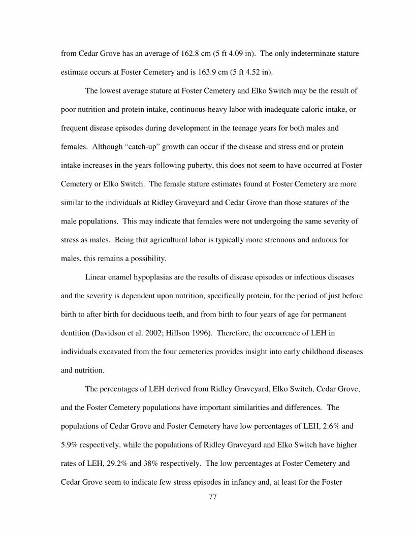
77
from Cedar Grove has an average of 162.8 cm (5 ft 4.09 in). The only indeterminate stature
estimate occurs at Foster Cemetery and is 163.9 cm (5 ft 4.52 in).
The lowest average stature at Foster Cemetery and Elko Switch may be the result of
poor nutrition and protein intake, continuous heavy labor with inadequate caloric intake, or
frequent disease episodes during development in the teenage years for both males and
females. Although “catch-up” growth can occur if the disease and stress end or protein
intake increases in the years following puberty, this does not seem to have occurred at Foster
Cemetery or Elko Switch. The female stature estimates found at Foster Cemetery are more
similar to the individuals at Ridley Graveyard and Cedar Grove than those statures of the
male populations. This may indicate that females were not undergoing the same severity of
stress as males. Being that agricultural labor is typically more strenuous and arduous for
males, this remains a possibility.
Linear enamel hypoplasias are the results of disease episodes or infectious diseases
and the severity is dependent upon nutrition, specifically protein, for the period of just before
birth to after birth for deciduous teeth, and from birth to four years of age for permanent
dentition (Davidson et al. 2002; Hillson 1996). Therefore, the occurrence of LEH in
individuals excavated from the four cemeteries provides insight into early childhood diseases
and nutrition.
The percentages of LEH derived from Ridley Graveyard, Elko Switch, Cedar Grove,
and the Foster Cemetery populations have important similarities and differences. The
populations of Cedar Grove and Foster Cemetery have low percentages of LEH, 2.6% and
5.9% respectively, while the populations of Ridley Graveyard and Elko Switch have higher
rates of LEH, 29.2% and 38% respectively. The low percentages at Foster Cemetery and
Cedar Grove seem to indicate few stress episodes in infancy and, at least for the Foster
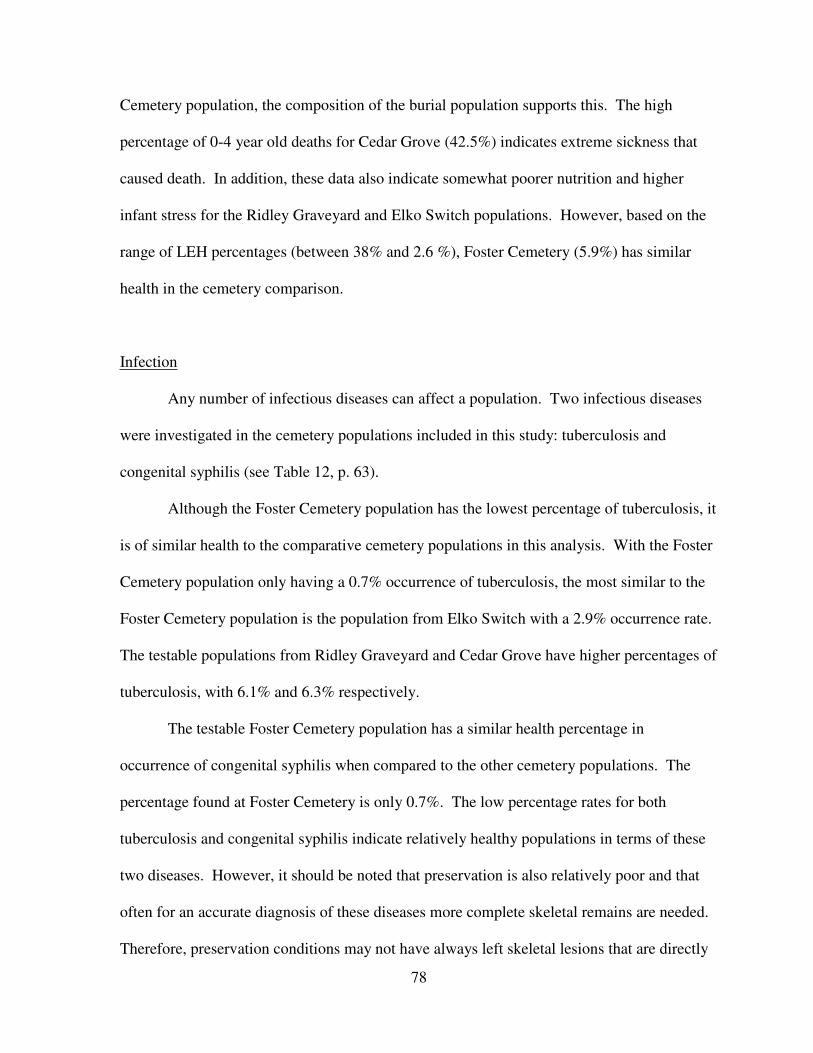
78
Cemetery population, the composition of the burial population supports this. The high
percentage of 0-4 year old deaths for Cedar Grove (42.5%) indicates extreme sickness that
caused death. In addition, these data also indicate somewhat poorer nutrition and higher
infant stress for the Ridley Graveyard and Elko Switch populations. However, based on the
range of LEH percentages (between 38% and 2.6 %), Foster Cemetery (5.9%) has similar
health in the cemetery comparison.
Infection
Any number of infectious diseases can affect a population. Two infectious diseases
were investigated in the cemetery populations included in this study: tuberculosis and
congenital syphilis (see Table 12, p. 63).
Although the Foster Cemetery population has the lowest percentage of tuberculosis, it
is of similar health to the comparative cemetery populations in this analysis. With the Foster
Cemetery population only having a 0.7% occurrence of tuberculosis, the most similar to the
Foster Cemetery population is the population from Elko Switch with a 2.9% occurrence rate.
The testable populations from Ridley Graveyard and Cedar Grove have higher percentages of
tuberculosis, with 6.1% and 6.3% respectively.
The testable Foster Cemetery population has a similar health percentage in
occurrence of congenital syphilis when compared to the other cemetery populations. The
percentage found at Foster Cemetery is only 0.7%. The low percentage rates for both
tuberculosis and congenital syphilis indicate relatively healthy populations in terms of these
two diseases. However, it should be noted that preservation is also relatively poor and that
often for an accurate diagnosis of these diseases more complete skeletal remains are needed.
Therefore, preservation conditions may not have always left skeletal lesions that are directly
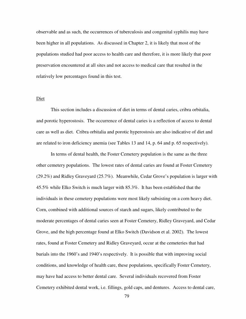
79
observable and as such, the occurrences of tuberculosis and congenital syphilis may have
been higher in all populations. As discussed in Chapter 2, it is likely that most of the
populations studied had poor access to health care and therefore, it is more likely that poor
preservation encountered at all sites and not access to medical care that resulted in the
relatively low percentages found in this test.
Diet
This section includes a discussion of diet in terms of dental caries, cribra orbitalia,
and porotic hyperostosis. The occurrence of dental caries is a reflection of access to dental
care as well as diet. Cribra orbitalia and porotic hyperostosis are also indicative of diet and
are related to iron deficiency anemia (see Tables 13 and 14, p. 64 and p. 65 respectively).
In terms of dental health, the Foster Cemetery population is the same as the three
other cemetery populations. The lowest rates of dental caries are found at Foster Cemetery
(29.2%) and Ridley Graveyard (25.7%). Meanwhile, Cedar Grove’s population is larger with
45.5% while Elko Switch is much larger with 85.3%. It has been established that the
individuals in these cemetery populations were most likely subsisting on a corn heavy diet.
Corn, combined with additional sources of starch and sugars, likely contributed to the
moderate percentages of dental caries seen at Foster Cemetery, Ridley Graveyard, and Cedar
Grove, and the high percentage found at Elko Switch (Davidson et al. 2002). The lowest
rates, found at Foster Cemetery and Ridley Graveyard, occur at the cemeteries that had
burials into the 1960’s and 1940’s respectively. It is possible that with improving social
conditions, and knowledge of health care, these populations, specifically Foster Cemetery,
may have had access to better dental care. Several individuals recovered from Foster
Cemetery exhibited dental work, i.e. fillings, gold caps, and dentures. Access to dental care,
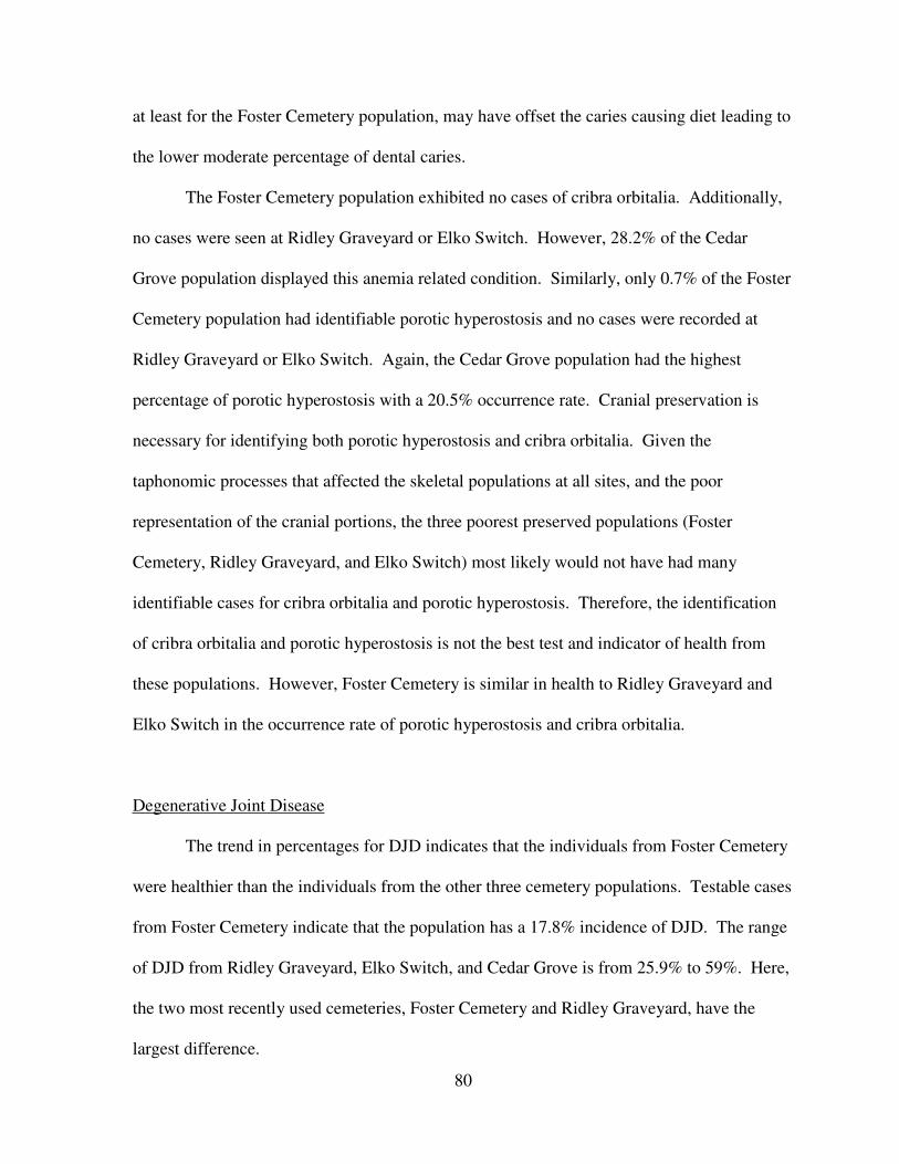
80
at least for the Foster Cemetery population, may have offset the caries causing diet leading to
the lower moderate percentage of dental caries.
The Foster Cemetery population exhibited no cases of cribra orbitalia. Additionally,
no cases were seen at Ridley Graveyard or Elko Switch. However, 28.2% of the Cedar
Grove population displayed this anemia related condition. Similarly, only 0.7% of the Foster
Cemetery population had identifiable porotic hyperostosis and no cases were recorded at
Ridley Graveyard or Elko Switch. Again, the Cedar Grove population had the highest
percentage of porotic hyperostosis with a 20.5% occurrence rate. Cranial preservation is
necessary for identifying both porotic hyperostosis and cribra orbitalia. Given the
taphonomic processes that affected the skeletal populations at all sites, and the poor
representation of the cranial portions, the three poorest preserved populations (Foster
Cemetery, Ridley Graveyard, and Elko Switch) most likely would not have had many
identifiable cases for cribra orbitalia and porotic hyperostosis. Therefore, the identification
of cribra orbitalia and porotic hyperostosis is not the best test and indicator of health from
these populations. However, Foster Cemetery is similar in health to Ridley Graveyard and
Elko Switch in the occurrence rate of porotic hyperostosis and cribra orbitalia.
Degenerative Joint Disease
The trend in percentages for DJD indicates that the individuals from Foster Cemetery
were healthier than the individuals from the other three cemetery populations. Testable cases
from Foster Cemetery indicate that the population has a 17.8% incidence of DJD. The range
of DJD from Ridley Graveyard, Elko Switch, and Cedar Grove is from 25.9% to 59%. Here,
the two most recently used cemeteries, Foster Cemetery and Ridley Graveyard, have the
largest difference.
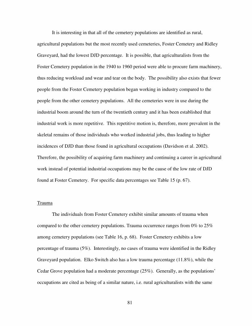
81
It is interesting in that all of the cemetery populations are identified as rural,
agricultural populations but the most recently used cemeteries, Foster Cemetery and Ridley
Graveyard, had the lowest DJD percentage. It is possible, that agriculturalists from the
Foster Cemetery population in the 1940 to 1960 period were able to procure farm machinery,
thus reducing workload and wear and tear on the body. The possibility also exists that fewer
people from the Foster Cemetery population began working in industry compared to the
people from the other cemetery populations. All the cemeteries were in use during the
industrial boom around the turn of the twentieth century and it has been established that
industrial work is more repetitive. This repetitive motion is, therefore, more prevalent in the
skeletal remains of those individuals who worked industrial jobs, thus leading to higher
incidences of DJD than those found in agricultural occupations (Davidson et al. 2002).
Therefore, the possibility of acquiring farm machinery and continuing a career in agricultural
work instead of potential industrial occupations may be the cause of the low rate of DJD
found at Foster Cemetery. For specific data percentages see Table 15 (p. 67).
Trauma
The individuals from Foster Cemetery exhibit similar amounts of trauma when
compared to the other cemetery populations. Trauma occurrence ranges from 0% to 25%
among cemetery populations (see Table 16, p. 68). Foster Cemetery exhibits a low
percentage of trauma (5%). Interestingly, no cases of trauma were identified in the Ridley
Graveyard population. Elko Switch also has a low trauma percentage (11.8%), while the
Cedar Grove population had a moderate percentage (25%). Generally, as the populations’
occupations are cited as being of a similar nature, i.e. rural agriculturalists with the same
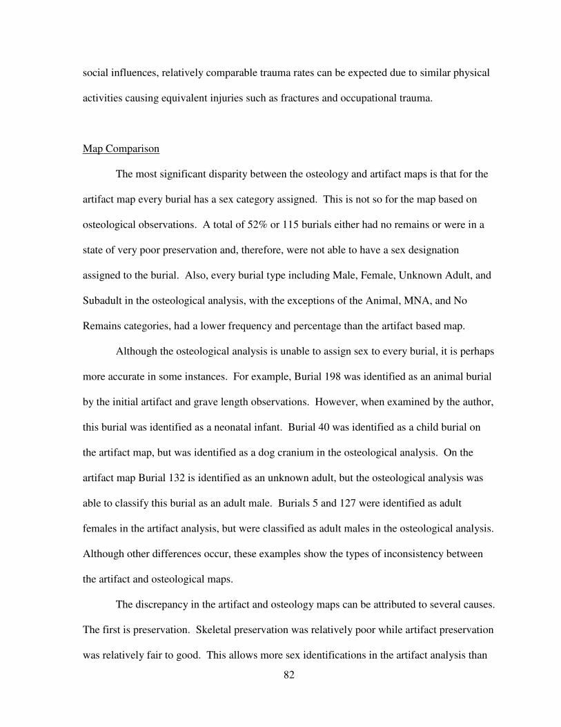
82
social influences, relatively comparable trauma rates can be expected due to similar physical
activities causing equivalent injuries such as fractures and occupational trauma.
Map Comparison
The most significant disparity between the osteology and artifact maps is that for the
artifact map every burial has a sex category assigned. This is not so for the map based on
osteological observations. A total of 52% or 115 burials either had no remains or were in a
state of very poor preservation and, therefore, were not able to have a sex designation
assigned to the burial. Also, every burial type including Male, Female, Unknown Adult, and
Subadult in the osteological analysis, with the exceptions of the Animal, MNA, and No
Remains categories, had a lower frequency and percentage than the artifact based map.
Although the osteological analysis is unable to assign sex to every burial, it is perhaps
more accurate in some instances. For example, Burial 198 was identified as an animal burial
by the initial artifact and grave length observations. However, when examined by the author,
this burial was identified as a neonatal infant. Burial 40 was identified as a child burial on
the artifact map, but was identified as a dog cranium in the osteological analysis. On the
artifact map Burial 132 is identified as an unknown adult, but the osteological analysis was
able to classify this burial as an adult male. Burials 5 and 127 were identified as adult
females in the artifact analysis, but were classified as adult males in the osteological analysis.
Although other differences occur, these examples show the types of inconsistency between
the artifact and osteological maps.
The discrepancy in the artifact and osteology maps can be attributed to several causes.
The first is preservation. Skeletal preservation was relatively poor while artifact preservation
was relatively fair to good. This allows more sex identifications in the artifact analysis than
83
in the osteological analysis. The second cause of these differences can be lack of knowledge
of human osteology in field crews and can be seen in the misidentification burials 40 and
198. A third cause is cultural bias. Artifacts can be classified as male or female, adult or
child, through a lens of cultural identification. For example, a smoking pipe may be more
closely associated as a male item thus leading the burial to be identified as male, however,
the possibility and probability that the pipe belonged to a female also exists. The last cause is
associated with grave size, in that large graves may be identified as male and small graves
may be classified as female. This may be seen in Burial 127, as it as a relatively small grave
that is identified as female in the artifact map but the osteological data identifies it as male.
In summary, using artifact analysis to identify sex/age provides more identifications than
osteological analysis in cases of poor preservation. However, both methods are
recommended as they provide two avenues of data gathering and are ultimately
complementary.
Summary
Table 18 provides a summary of the findings of the tests in this study. The cemetery
populations’ health is ranked 1 through 4 with 1 being the healthiest and 4 being the least
healthy.
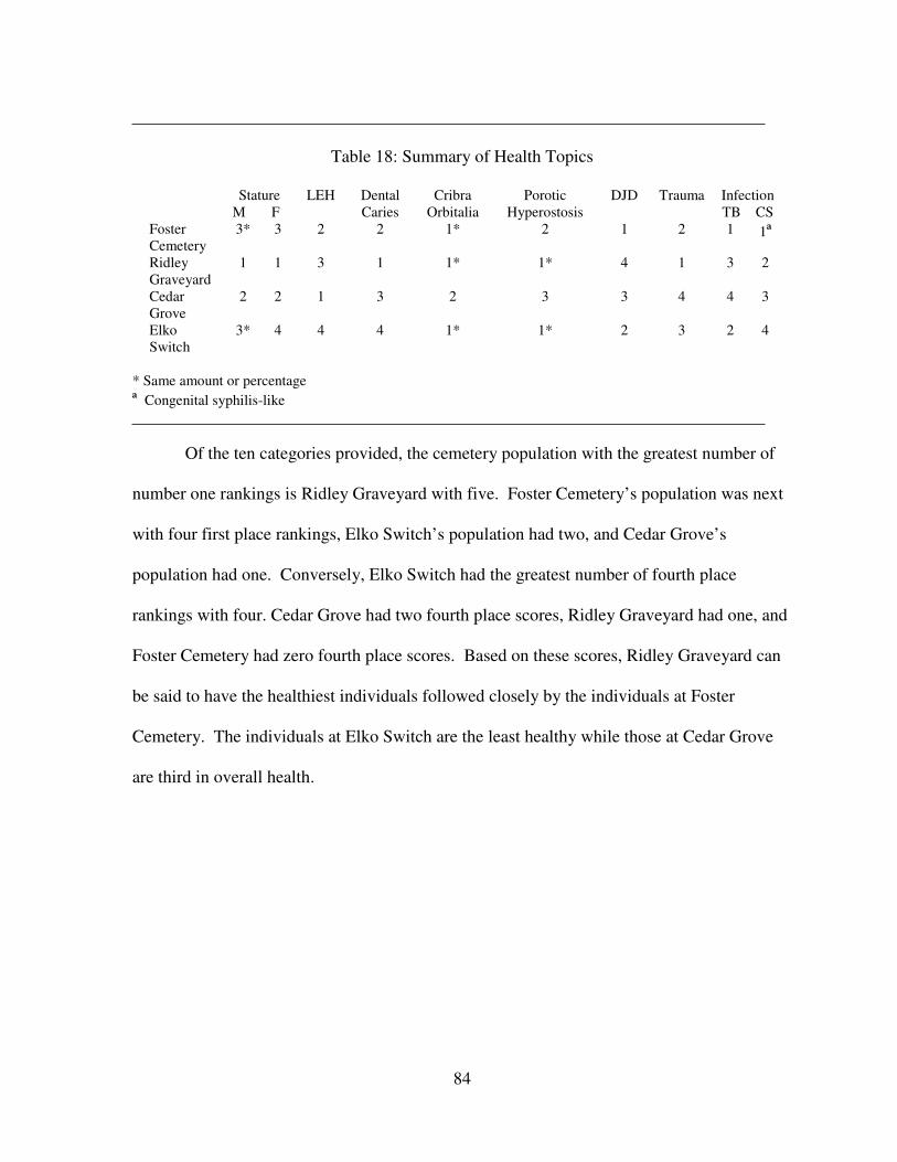
84
Table 18: Summary of Health Topics
Stature
M F
LEH Dental
Caries
Cribra
Orbitalia
Porotic
Hyperostosis
DJD Trauma Infection
TB CS
Foster
Cemetery
3* 3 2 2 1* 2 1 2 1 1ª
Ridley
Graveyard
1 1 3 1 1* 1* 4 1 3 2
Cedar
Grove
2 2 1 3 2 3 3 4 4 3
Elko
Switch
3* 4 4 4 1* 1* 2 3 2 4
* Same amount or percentage
ª Congenital syphilis-like
Of the ten categories provided, the cemetery population with the greatest number of
number one rankings is Ridley Graveyard with five. Foster Cemetery’s population was next
with four first place rankings, Elko Switch’s population had two, and Cedar Grove’s
population had one. Conversely, Elko Switch had the greatest number of fourth place
rankings with four. Cedar Grove had two fourth place scores, Ridley Graveyard had one, and
Foster Cemetery had zero fourth place scores. Based on these scores, Ridley Graveyard can
be said to have the healthiest individuals followed closely by the individuals at Foster
Cemetery. The individuals at Elko Switch are the least healthy while those at Cedar Grove
are third in overall health.
85
CHAPTER SIX
SUMMARY, CONCLUSIONS, AND RECOMMENDATIONS
Foster Cemetery (1La151) is an African American cemetery and consists of 224
burials dating from 1870 to 1960. This study attempted a comparative health analysis of the
Foster Cemetery population with those of three other southeastern African American
populations: Elko Switch, Cedar Grove, and Ridley Graveyard. It was hypothesized that the
Foster Cemetery population would be similar in health status to individuals of other
cemeteries when compared across the following areas: demography, growth and
development, infection, diet, degenerative joint disease, and trauma. Given that at least part
of the period of use for each cemetery was contemporaneous, that each cemetery population
was identified as being rural agriculturalist, and that each population experienced comparable
social influences, the similar health findings are not unexpected.
In terms of demography, the Foster Cemetery population is similar to the other
cemeteries in terms of the number of male interments and the relative health of females and
subadults. However, the Foster Cemetery population is equal in health in seven of the twelve
age intervals or 58.3%. Therefore, in general the Foster Cemetery population is equal in
terms of health in demographic analysis, although exceptions include fewer female and
subadult deaths, and much higher numbers of people dying in the 15-19 and 20-24 age
intervals.
The Foster Cemetery population is similar in growth and development based on
stature estimations and the number of linear enamel hypoplasias. Infection rates of the Foster
86
Cemetery population are also similar to the comparative cemeteries when tuberculosis and
congenital syphilis were examined. The comparison of diet consists of the occurrence of
dental caries, cribra orbitalia, and porotic hyperostosis and the Foster Cemetery population is
equally healthy when compared to all three cemeteries. The low occurrence of degenerative
joint disease indicates that the Foster Cemetery population is healthier than the other
cemetery populations. The presence of trauma for Foster Cemetery is similar to the levels of
trauma in the comparative cemetery populations. Therefore, the Foster Cemetery population
is equal in health in five of the six or 83.3% of the tested categories, and is healthier in only
one (degenerative joint disease). These tests support the hypothesis that, generally, the
individuals at Foster Cemetery would be similar in health to the three other cemetery
populations.
The second hypothesis in this study considered the sex/age identifications for the
preliminary artifact and grave length observations against those of the osteological analysis.
It was hypothesized that there would be differences in terms of the sex/age identifications in
the artifact analysis map and the osteological analysis map. Indeed, while many burials were
similarly identified, many burials had different classifications, thus supporting the
hypothesis. Based on this study, one can conclude that sex/age identifications garnered from
artifact findings can yield more identifications than osteological observations when
confronted with a burial population that is poorly preserved. However, osteological analysis
may be more accurate by providing a specialized focus on the individual remains that artifact
analysis cannot accomplish alone.
Certain limitations were uncovered as well as possible recommendations for future
study. For many of the individuals only dental information was recordable, as teeth were the
only remains present. Unfortunately for aging criteria, dental eruption only provides age
87
estimation up to 21 years of age. The lack of dental attrition aging data for rural African
American populations for the period of time that includes Foster Cemetery population limits
the effectiveness of dental aging. According to the aging method pioneered by McGrath
(2000), rounding age estimated ages up to the nearest five year age interval challenges the
accuracy of aging data. However, by following McGrath’s method, statistical analysis of the
cemetery population is possible.
Perhaps the most significant recommendation is the addition of specific time period
ranges for specific burials, which are based on mortuary hardware analysis. Mortuary
hardware analysis allows for the direct dating of coffin materials including handle and nail
types. At the time of this study, specific time ranges did not exist for each burial but were
being established through this artifact analysis. Knowledge of time period ranges for each
interment will provide a more accurate and thorough understanding of the progression of
usage of Foster Cemetery. This will provide a more precise estimation of health throughout
Foster Cemetery’s use. For example, high frequencies of interments dating to the late 1910s
may be associated with the 1918 flu epidemic. If large quantities of interments were found
during this time period the population would more likely be an industrial one, because the flu
virus would spread more quickly in close-quarter factory work than in an agricultural setting.
This study has shown the Foster Cemetery population was of similar health to
contemporaneous African American populations. The lifestyle of those interred at Foster
Cemetery was physically demanding and susceptible to not only diet restrictions and disease,
but also social oppression. Perhaps the most significant contribution from this study is the
knowledge of African American life in the post-Civil War southern United States where a
paucity of information existed previously. As a joint effort between the author and SAI, this
study has contributed a significant amount of data concerning the health of African American

88
cemetery populations where relatively little information existed before. It is hoped that
anthropologists find this research useful for future research as the basis for further
comparative studies.
89
REFERENCES
Angel, J. L., J. O. Kelley, M. Parrington, and S. Pinter
1987 Life Stresses of the Free Black Community as Represented by the First African
Baptist Church, Philadelphia, 1823-1841. American Journal of Physical
Anthropology 74, No. 2: 213-230.
Aufderheide, A. C., and C. Rodríguez-Martín
1998 The Cambridge Encyclopedia of Human Paleopathology. Cambridge, United
Kingdom: Cambridge University Press.
Baker, B. J., T. L. Dupras, and M. W. Tocheri
2005 The Osteology of Infants and Children. Texas A & M University Anthropology
Series No. 12. Texas A & M University Press.
Barnes, E.
1994 Developmental Defects of the Axial Skeleton in Paleopathology. University Press of
Colorado.
Bass, W. M.
1995 Human Osteology: A Laboratory and Field Manual. Missouri Archaeological
Society No. 2.
Blakely, R. L., and L. A. Beck
1982 Bioarchaeology in the Urban Context. In The Archaeology of Urban America: The
Search for Pattern and Process, edited by Roy S. Dickens, Jr., pp. 175-207.
Toronto: Academic Press.
Brooks, S. T. and J. M. Suchey
1990 Skeletal Age Determination Based on the Os Pubis: A Comparison of the Acsadi-
Nemeskeri and Suchey-Brooks Methods. In Standards for Data Collection from
Human Skeletal Remains, edited by J. E. Buikstra and D. H. Ubelaker, p. 22.
Arkansas Archaeological Survey Research No. 44. Fayetteville, AK: Arkansas
Archaeological Society.
90
Browne, W. P.
2003 Benign Public Policies, Malignant Consequences, and the Demise of African
American Agriculture. In African American Life in the Rural South, 1900-1950,
edited by R. D. Hurt, pp. 129-151. Columbia and London: University of Missouri
Press.
Buchner, A. C., E. Breitburg, C. Williams, and E. A. Williams
1999 At Rest Again: The Ridley Graveyard (40WM208) Archaeological Relocation
Project, Williamson County, Tennessee. Tuscaloosa: Panamerican Consultants, Inc.
Buikstra, J. E. and D. H. Ubelaker, eds.
1994 Standards for Data Collection from Human Skeletal Remains. Arkansas
Archaeological Survey Research No. 44. Fayetteville: Arkansas Archeological
Society.
Byers, S.
2002 Introduction to Forensic Anthropology: A Textbook. Boston: Allyn and Bacon.
Condon, C. G., J. L. Becker, H. J. H. Edgar, J. M. Davidson, J. R. Hoffman, P. Kalima, D.
Kysar, S. Moorhead, V. M. Owens, and K. Condon
1998 Freedman’s Cemetery: Site 41DL316, Dallas, Texas, Assessments of Sex, Age at
Death, Stature and Date of Interment for Excavated Burials. Report No. 9.
Archaeology Studies Program, Environmental Affairs Division. Austin, Texas:
Texas Department of Transportation.
Corruccini, R. S., J. S. Handler, R. J. Mutaw, and F. W. Lange
1982 Osteology of a Slave Burial Population from Barbados, West Indies. American
Journal of Physical Anthropology 59: 443-459.
Corruccini, R. S., K. P. Jacobi, J. S. Handler
1985 Distribution of Enamel Hypoplasias in an Early Caribbean Slave Population.
American Journal of Physical Anthropology 66, No. 2: 158.
Crist, T. A. J., R. M. Pitts, A. Washburn, J. P. McCarthy, and D. G. Roberts
1995 A Distinct Church of the Lord Jesus. The History, Archeology, and Physical
Anthropology of the Tenth Street First African Baptist Church Cemetery,
Philadelphia, Pennsylvania, Site Number 36PH72. Vine Expressway (I-676), L.R.
67045, ER #82-101-0133. Philadelphia: John Miller Associates, Inc.
91
Dailey, R. C.
1974 Osteological Analysis of Human Skeletal Remains from the Virgin Islands.
Typescript, Florida State University, Tallahassee.
Danforth, M. E.
2004 African American Men, Women, and Children in Nineteenth-Century Natchez,
Mississippi: An Analysis of the City Cemetery Sexton’s Records. In Engendering
African American Archaeology, edited by J. E. Galle and A. L. Young, pp. 237-262.
Knoxville, Tennessee: The University of Tennessee Press.
Davidson, J. M., J .C. Rose, M. P. Gutmann, M. R. Haines, K. Condon, and C. Condon
2002 The Quality of African-American Life in the Old Southwest near the Turn of the
Twentieth Century. In The Backbone of History, edited by Richard H. Steckel and
Jerome C. Rose, pp. 226-277. Cambridge, England: Cambridge University Press.
Diouf, S. A.
2007 Dreams of Africa in Alabama. New York: Oxford University Press.
Donaldson, A.
2001 Unpublished essay from Dr. Ian Brown’s seminar Marking Graves: Self and Society
in Death, pp. 10-19. Tuscaloosa: The University of Alabama.
Farley, R.
1970 Growth of the Black Population: A Study of Demographic Trends. Chicago:
Markham Publishing.
Gilbert, C. and Q. Eli
2000 Homecoming: The Story of African-American Farmers. Boston: Beacon Press.
Gill, G. W.
1995 Challenge on the Frontier: Discerning American Indians from Whites
Osteologically. Journal of Forensic Sciences 40:783-788.
Grim, V.
2003 African American Rural Culture, 1900-1950. In African American Life in the Rural
South, 1900-1950, edited by R. D. Hurt, pp. 108-128. Columbia and London:
University of Missouri Press.
92
Handler, J. S. and F. W. Lange
1978 Plantation Slavery in Barbados: An Archeological and Historical Investigation.
Harvard University Press, Cambridge.
Handler, J. S., R. S. Corruccini, and R. J. Mutaw
1982 Tooth Mutilation in the Caribbean: Evidence from a Slave Burial Population in
Barbados. Journal of Human Evolution 11: 297-313.
Đşcan, M. Y. and S. R. Loth
1993 Casts of Age Phases from the Sternal End of the Rib for White Males and Females.
France Casting, Bellvue, Colorado.
Jacobi, K. P.
2003 What’s in a grave? Anthropological Data Snatching in a Cemetery
Jacobi, K. P., D. C. Cook, R. S. Corruccini, and J. S. Handler
1992 Congenital Syphilis in the Past: Slaves at Newton Plantation, Barbados, West
Indies. American Journal of Physical Anthropology 89, No. 2: 145-158.
Hillson, S.
1996 Dental Anthropology. Cambridge: Cambridge University Press.
Kennedy, Kenneth A. R.
1989 Skeletal Markers of Occupational Stress in Reconstruction of Life from the
Skeleton, edited by M. Y. Iscan and K. A. R. Kennedy, pp. 129-160. Alan R. Liss,
New York.
Krogman, W. M., and M. Y. Iscan
1986 The Human Skeleton in Forensic Medicine. In Standards for Data Collection from
Human Skeletal Remains, edited by J. E. Buikstra and D. H. Ubelaker, p. 22.
Arkansas Archaeological Survey Research No. 44. Fayetteville, Arkansas: Arkansas
Archaeological Society.
Lallo, John W., J. C. Rose, G. J. Aremlagos
1980 An Ecological Interpretation of Variation in Mortality Within Three Prehistoric
American Indian Populations from Dickson Mounds. In Early Native Americans,
edited by D. L. Browman, pp. 203-238, Mouton Publishers, the Hague.
93
Legg, J. B., and S. D. Smith
1989 The Best Ever Occupied… Archaeological Investigations of a Civil War
Encampment on Folly Island, South Carolina. Research Manuscript Series 209.
Columbia: South Carolina Institute of Archaeology and Anthropology.
Lovejoy, C. O., R. S. Meindl, T. R. Pryzbeck, T. S. Barton, K. G. Heiple, and D. Knotting
1977 Paleodemography of the Libben Site, Ottawa County, Ohio. Science 198:291-293.
Lovejoy, C. O., R. S. Meindl, I. R. Pryzbeck, and R. P. Mensforth
1985 Chronological Metamorphosis of the Auricular Surface of the Ilium: A New
Method for the Determination of Age at Death. American Journal of Physical
Anthropology 68: 15-28.
McGrath, S. E.
2000 An Ethnohistorical, Archaeological, Osteological, and Mitochondrial DNA
Analysis of a Nineteenth-Century African American Cemetery: A Methodological
Case Study. University of Alabama Press, Tuscaloosa.
McKern, T. and T. D. Stewart
1957 Skeletal Age Changes in Young American Males, Analyzed from the Standpoint of
Identification. Standards for Data Collection from Human Skeletal Remains, edited
by J. E. Buikstra and D. H. Ubelaker, p. 22. Arkansas Archaeological Survey
Research No. 44. Fayetteville, Arkansas: Arkansas Archaeological Society.
Moore, J. A., A. C. Swedlund, and G. J. Aremlagos
1975 The Use of Life Tables in Paleodemography. Society of American Archaeology
Memoir 30:57-70.
Morse, D., Duncan, J., and J. Stoutamire
1983 Handbook of Forensic Archaeology and Anthropology. Tallahassee: Bill’s
Bookstore.
Ortner, D.J. and W.G.J. Putschar
1985 Identification of Pathological Conditions in Human Skeletal Remains. Smithsonian
Press, Washington.
Parrington, M. and D. G. Roberts
1984 The First African Baptist Cemetery. Archaeology 37, No. 6: 26-32.
94
Peebles, C. S.
1977 Biocultural Adaptation in Prehistoric America: An Archaeologist’s Perspective. In
Biocultural Adaptation in Prehistoric America, edited by R. L. Blakely, pp. 115-30.
Proceedings No. 11. Southern Anthropological Society, University of Georgia
Press, Athens.
Phelps, D .S., Green, J. B., and K. C. Hartsell
1979 An Archaeological-Historical Study of the Bryan Cemetery and Site 31CV25,
Simmons-Nott Airport, New Bern, North Carolina. Publication No. 10. Raleigh:
North Carolina Archaeological Council.
Postell, W. D.
1970[1951] The Health of Slaves on Southern Plantations. Gloucester: Louisiana State
University Press.
Rathbun, T. A.
1987 Health and Disease at a South Carolina Plantation: 1840-1870. American Journal of
Physical Anthropology 74:239-253.
Rathbun, T. A. and J. D. Scurry
1991 Status and Health in Colonial South Carolina: Belleview Plantation, 1738-1756. In
What Mean These Bones? Studies in Southeastern Bioarchaeology, edited by J. L.
Powell, P. S. Bridges, and A. M. W. Mires, 148-164. Tuscaloosa: University of
Alabama Press.
Rathbun, T. A. and R. H. Steckel
2002 The Health of Slaves and Free Blacks in the East. In The Backbone of History,
edited by Richard H. Steckel and Jerome C. Rose, pp. 226-277. Cambridge,
England: Cambridge University Press.
Redfield, A.
1970 A New Aid to Aging Immature Skeletons. In Standards for Data Collection from
Human Skeletal Remains, edited by J. E. Buikstra and D. H. Ubelaker, p. 22.
Arkansas Archaeological Survey Research No. 44. Fayetteville, Arkansas: Arkansas
Archaeological Society.
95
Rhine, S.
1990 Non-metric Skull Racing. In Skeletal Attribution of Race: Methods for Forensic
Anthropology, edited by G. W. Gill and S. Rhine, pp. 9-20, Albuquerque, New
Mexico: Maxwell Museum of Anthropology, Anthropological Papers Number 4.
Rose, J. C., ed.
1985 Gone to a Better Land. Arkansas Archeological Survey Research Series No. 25.
Fayetteville, Arkansas: Arkansas Archeological Survey U. S. A.
1989 Biological Consequences of Segregation and Economic Deprivation: A Post-
Slavery Population from Southwest Arkansas. The Journal of Economic History,
Vol. 49, No. 2, The Tasks of Economic History, pp. 351-360.
Savitt, T. L.
1978 Medicine and Slavery: The Diseases and Health Care of Blacks in Antebellum
Virginia. Urbana: University of Illinois Press.
Scheuer, L., and S. Black
2004 The Juvenile Skeleton. Amsterdam: Elsevier Academic Press.
Schour, I., and M. Massler
1944 Development of the Human Dentition. Chicago: American Dental Association.
Sellers, J. B.
1950 Slavery in Alabama. Tuscaloosa: The University of Alabama Press.
Shapiro, H. L.
1930 Old New Yorker’s: A Series of Crania From the Nagel Burying Grounds. American
Journal of Physical Anthropology, No. 14: 379-404.
Shogren, M. G., K. R. Turner, and J. C. Perroni
1989 Elko Switch Cemetery: An Archaeological Perspective. Report of Investigations No.
58. Tuscaloosa, Alabama: The University of Alabama.
Stamp, K. M.
1965 The Era of Reconstruction: 1865-1877. New York: Vintage Books.
96
Steele, D. G. and C. A. Bramblett,
1988 The Anatomy and Biology of the Human Skeleton. College Station: Texas A&M
University Press.
Suchey, J. M., P. A. Owings, D. V. Wiseley, and T. T. Noguchi
1984 Skeletal Aging of Unidentified Persons. In Standards for Data Collection from
Human Skeletal Remains, edited by J. E. Buikstra and D. H. Ubelaker, p. 22.
Arkansas Archaeological Survey Research No. 44. Fayetteville, Arkansas: Arkansas
Archaeological Society.
Todd, T. W.
1921a Changes in the Pubic Bone. I: The Male White Pubis. In Standards for Data
Collection from Human Skeletal Remains, edited by J. E. Buikstra and D. H.
Ubelaker, p. 22. Arkansas Archaeological Survey Research No. 44. Fayetteville,
Arkansas: Arkansas Archaeological Society.
1921b Age Changes in the Pubic Bone. III: The Pubis of the White Female. IV: The pubis
of the female white-negro hybrid. In Standards for Data Collection from Human
Skeletal Remains, edited by J. E. Buikstra and D. H. Ubelaker, p. 22. Arkansas
Archaeological Survey Research No. 44. Fayetteville, Arkansas: Arkansas
Archaeological Society.
Trotter, M.
1970 Estimation of Stature from Intact Long Bones. In Personal Identification in Mass
Disasters, edited by T. D. Stewart, pp. 71-83. Washington, D.C.: Smithsonian
Institution Press.
Ubelaker, D. H.
1989 Human Skeletal Remains. 2nd
ed. Taraxacum Press, Washington D.C. In Standards
for Data Collection from Human Skeletal Remains, edited by J. E. Buikstra and D.
H. Ubelaker, p. 22. Arkansas Archaeological Survey Research No. 44. Fayetteville,
Arkansas: Arkansas Archaeological Society.
Ubelaker, D. H. and J. L. Angel
1976 Analysis of the Hull bay Skeletons, St. Thomas. Journal of the Virgin Island
Archeological Society 3: 7-9.
Weiss, K. M.
1973a Demographic Disturbance and the Use of Life Tables in Anthropology. Society
American Archeology Memoir No. 30:46-56. Society for American Archaeology,
Washington, D.C.
97
1973b Demographic Models for Anthropology. Memoirs of the Society for American
Archaeology No. 27, Washington.
White, T. D.
2000 Human Osteology. San Diego: Academic Press.
Wright, R. V. H. and W. B. Hughes III
1996 Lay Down Body: Living History in African-American Cemeteries. Detroit: Visible
Ink Press.

98
APPENDIX
99
Of the 224 burials identified by Southeastern Anthropological Institute at Foster
Cemetery, osteological analysis revealed 139 burials that yielded the following information.
Burial 1
Demographics
Sex: Female
Age: 55-60
Ancestry: African
Stature: 159.73cm (5 ft 3 in)
Preservation
Condition: Fair
Inventory: The following cranial elements were present: complete frontal, complete
occipital, complete left and right parietal, complete left and right temporal, complete
left and right zygomatic, complete left and right maxilla, incomplete left and right
sphenoid, complete mandible, incomplete facial bones, and 16 teeth.
The following postcranial elements were present: incomplete left and right
scapula, complete left and right clavicle, incomplete left humerus, complete right
humerus, incomplete left radius, complete right radius, incomplete left ulna, complete
right ulna, incomplete left and right innominate, incomplete left and right femur,
incomplete left and right tibia, complete left fibula, incomplete right fibula, 17 ribs,
one carpal, eleven phalanges, incomplete left and right calcaneus, complete left and
right talus, complete right cuboid, incomplete right navicular, five metatarsals, six
cervical vertebrae, twelve thoracic vertebrae, five lumbar vertebrae, and three sacral
vertebrae.
Paleopathology
100
Dental: The maxillary right teeth from the canine to the third molar have lingual
calculus at the cementoenamel junction (CEJ). The maxillary left lateral incisor is
rotated labial-distally. Maxillary left second and third molars lost antemortem with
alveolar bone resorption. The mandibular left first molar has a 1.3cm buccal root
abscess. Mandibular left central incisor to the right third molar lost antemortem
with marked mandibular alveolar bone resorption.
Infectious Disease: Not Applicable
Trauma: The right tibia exhibits a healed fracture of proximal 1/3.
Other: There is osteoarthritic lipping of thoracic vertebrae 1 through 6 may indicate
age related degeneration. Additionally, an osteophyte on the right ilium also may be
the result of arthritis brought on by age.
Burial 2
Demographics
Sex: Male
Age: 45-56
Ancestry: African
Stature: 174.9cm (5 ft 9 in)
Preservation
Condition: Fair
Inventory: The following cranial elements were present: complete frontal, complete
occipital, complete left and right parietal, complete left and right temporal, complete
left and right zygomatic, complete left and right maxilla, complete left and right
sphenoid, incomplete mandible, incomplete facial bones, 27 teeth.

101
The following postcranial elements were present: incomplete left and right
scapula, incomplete left and right clavicle, complete left humerus, incomplete right
humerus, incomplete left and right radius, incomplete left and right ulna, incomplete
left and right innominate, incomplete left and right femur, incomplete left tibia,
complete right tibia, incomplete left fibula, complete right fibula, 17 ribs, two
metacarpals, seven phalanges, incomplete left and right calcaneus, incomplete left
and right talus, incomplete left and right cuboid, incomplete left and right navicular,
incomplete left and right med., int., and lat. cuneiform, ten metatarsals, ten phalanges,
six cervical vertebrae, twelve thoracic vertebrae, five lumbar vertebrae, and four
sacral vertebrae.
Paleopathology
Dental: Maxillary left and right third molars have been lost antemortem. Maxillary
lateral right incisor and central left incisor have gold crowns.
Infectious Disease: Not Applicable
Trauma: Schmorl’s nodes (intervertebral disk herniations) appear from the fifth
thoracic vertebra through the fourth lumbar vertebra. Schmorl’s nodes appear on the
superior and inferior vertebral bodies from the sixth thoracic vertebra through the
third lumbar vertebra. They only appear on the inferior surface of the fifth thoracic
vertebra and the superior vertebral body of the fourth lumbar vertebra. Schmorl’s
nodes are believed to be the result of trauma and also may be degenerative in nature
(Aufderheide and Rodríguez-Martín 2005).
Other: Not Applicable
Burial 3 (Figures 14 through 16)
102
Demographics
Sex: Male
Age: 30-35
Ancestry: European. Although ancestry assessment identified this individual as
European, the cranium was exposed to chemical or heat from a chemical/phosphate
explosion (Hunter Johnson, personal communication). The individual is also known
to have been an African American (Hunter Johnson, personal communication).
Stature: 172.36cm (5 ft 7.6 in)
Preservation
Condition: Fair
Inventory: The following cranial elements were present: complete frontal, occipital,
parietals, temporals, zygomatics, maxilla, sphenoid, and mandible, incomplete hyoid,
complete facial bones, and 28 teeth.
The following postcranial elements were present: incomplete left and right
scapula, complete left and right clavicle, incomplete left and right radius, humerus,
ulna, innominate, and femur, incomplete left patella, complete right patella,
incomplete left tibia, complete right tibia, incomplete left fibula, complete right
fibula, 15 ribs, incomplete left scaphoid, incomplete left and right trapezoid, left and
right capitate, incomplete left hamate, ten metacarpals, 23 upper phalanges,
incomplete left calcaneus, complete right calcaneus, incomplete left talus, complete
right talus, complete left and right cuboid, complete left and right navicular, complete
left and right med. cuneiform, complete left int. cuneiform, incomplete right int.
cuneiform, complete left lat. cuneiform, incomplete right cuneiform, ten metatarsals,
103
ten lower phalanges, seven cervical vertebrae, twelve thoracic vertebrae, five lumbar
vertebrae, and three sacral vertebrae.
Paleopathology
Dental: The maxillary right third molar has an occlusal 2.5mm large caries and
calculus on all sides of the tooth (Figure 16). Maxillary left second molar was lost
antemortem as was the mandibular third molars and mandibular left second molar.
There is a 2.5mm large in size occlusal caries on the maxillary left third molar, as
well as, calculus on all enamel surfaces. The alveolar bone area of the mandibular
second and third molars shows signs of healing and alveolar bone resorption.
Calculus is present in some degree on all mandibular teeth and is particularly heavy
on the lingual surfaces.
Infectious Disease: Not Applicable
Trauma: There is a probable corrosive burn on the lateral left of the frontal bone
(Figure 14). There are no fracture lines endocranially or ectocranially which is
consistent with a burn and not impact trauma. The area of effect measures 3.5cm in
width and 3.3cm in height. There is an opening into the cranial vault in the middle of
the effected area measuring 0.9cm in width and 0.65cm in height. The occipital also
has alterations that may be the result of a burn or exposure to heat (Figure 15). The
alterations are not consistent with other skeletal changes seen in the cemetery
population.
Other: A small Wormian (sutural) bone is present along the left parietal and
occipital along the lambdoidal suture.
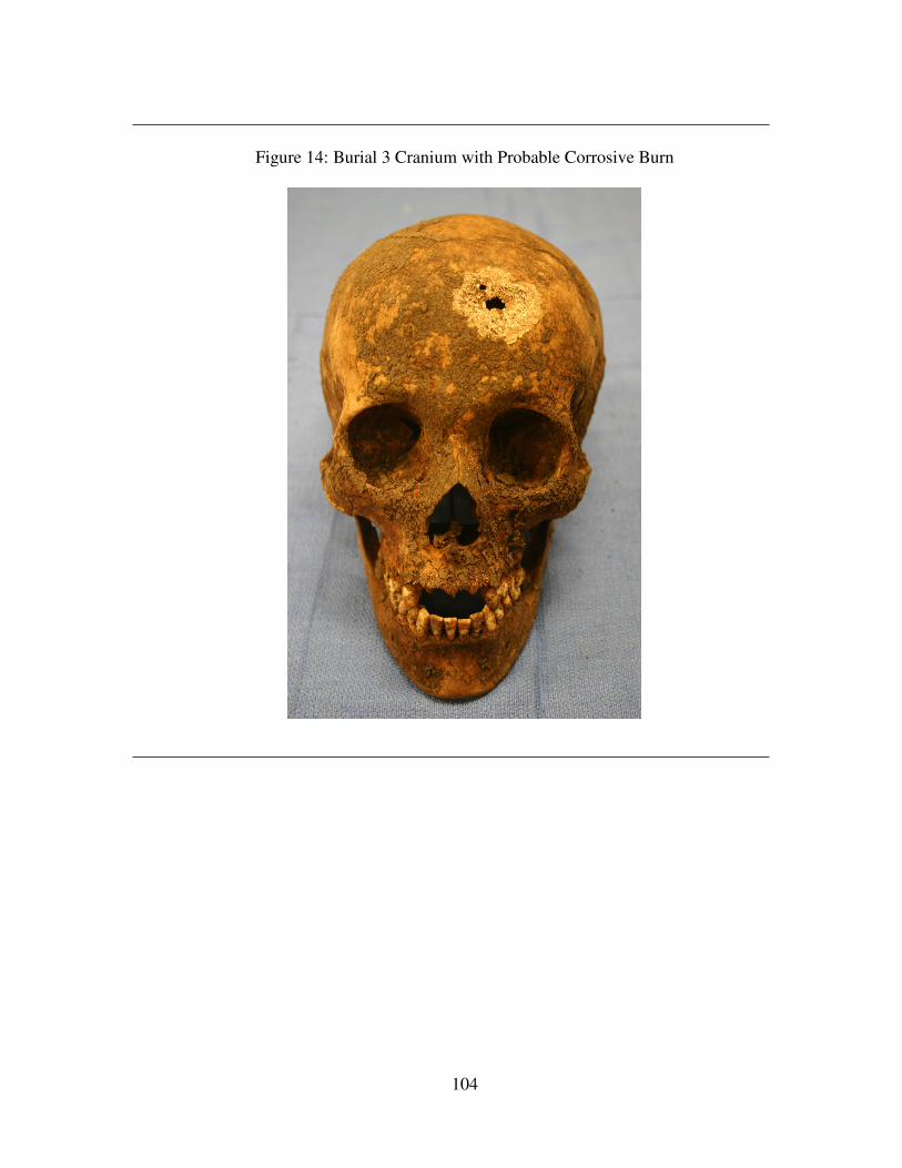
104
Figure 14: Burial 3 Cranium with Probable Corrosive Burn
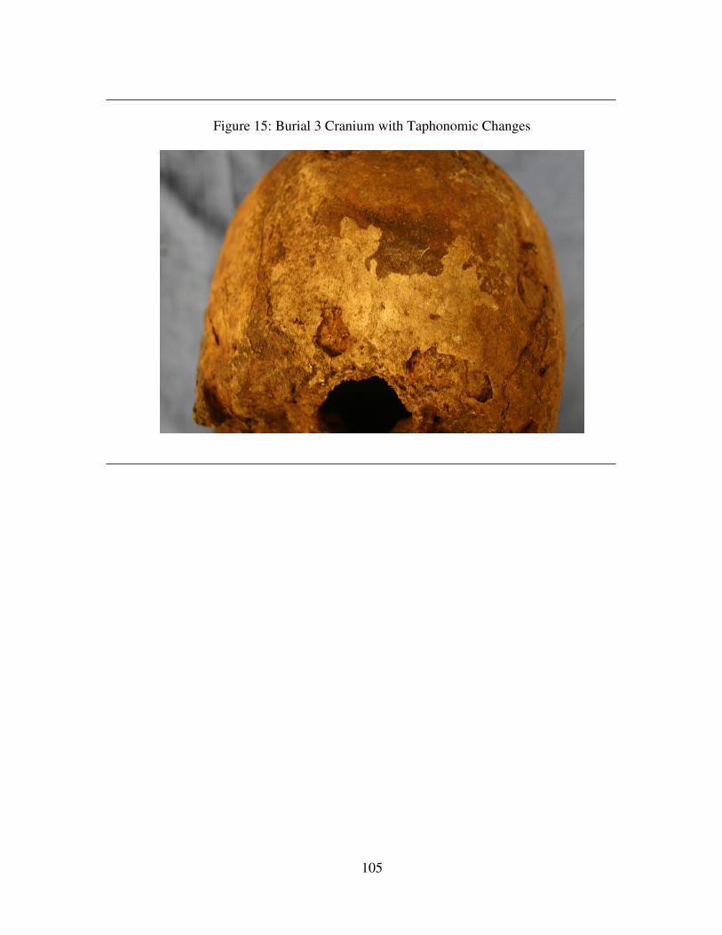
105
Figure 15: Burial 3 Cranium with Taphonomic Changes
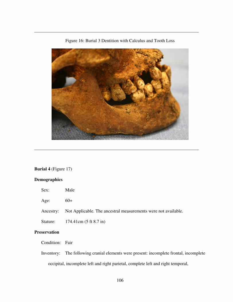
106
Figure 16: Burial 3 Dentition with Calculus and Tooth Loss
Burial 4 (Figure 17)
Demographics
Sex: Male
Age: 60+
Ancestry: Not Applicable. The ancestral measurements were not available.
Stature: 174.41cm (5 ft 8.7 in)
Preservation
Condition: Fair
Inventory: The following cranial elements were present: incomplete frontal, incomplete
occipital, incomplete left and right parietal, complete left and right temporal,
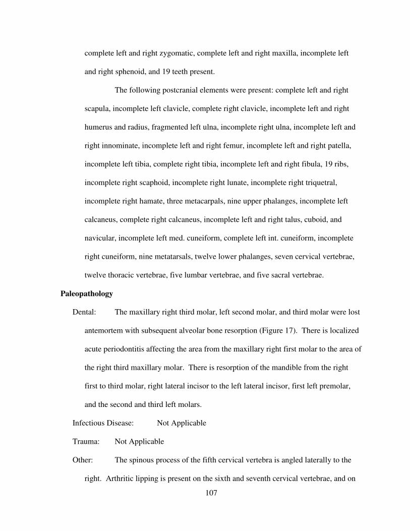
107
complete left and right zygomatic, complete left and right maxilla, incomplete left
and right sphenoid, and 19 teeth present.
The following postcranial elements were present: complete left and right
scapula, incomplete left clavicle, complete right clavicle, incomplete left and right
humerus and radius, fragmented left ulna, incomplete right ulna, incomplete left and
right innominate, incomplete left and right femur, incomplete left and right patella,
incomplete left tibia, complete right tibia, incomplete left and right fibula, 19 ribs,
incomplete right scaphoid, incomplete right lunate, incomplete right triquetral,
incomplete right hamate, three metacarpals, nine upper phalanges, incomplete left
calcaneus, complete right calcaneus, incomplete left and right talus, cuboid, and
navicular, incomplete left med. cuneiform, complete left int. cuneiform, incomplete
right cuneiform, nine metatarsals, twelve lower phalanges, seven cervical vertebrae,
twelve thoracic vertebrae, five lumbar vertebrae, and five sacral vertebrae.
Paleopathology
Dental: The maxillary right third molar, left second molar, and third molar were lost
antemortem with subsequent alveolar bone resorption (Figure 17). There is localized
acute periodontitis affecting the area from the maxillary right first molar to the area of
the right third maxillary molar. There is resorption of the mandible from the right
first to third molar, right lateral incisor to the left lateral incisor, first left premolar,
and the second and third left molars.
Infectious Disease: Not Applicable
Trauma: Not Applicable
Other: The spinous process of the fifth cervical vertebra is angled laterally to the
right. Arthritic lipping is present on the sixth and seventh cervical vertebrae, and on
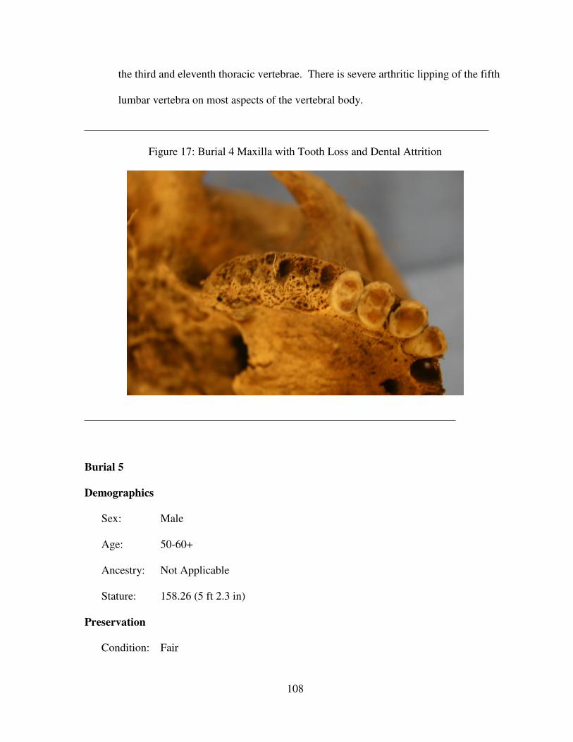
108
the third and eleventh thoracic vertebrae. There is severe arthritic lipping of the fifth
lumbar vertebra on most aspects of the vertebral body.
Figure 17: Burial 4 Maxilla with Tooth Loss and Dental Attrition
Burial 5
Demographics
Sex: Male
Age: 50-60+
Ancestry: Not Applicable
Stature: 158.26 (5 ft 2.3 in)
Preservation
Condition: Fair
109
Inventory: The following cranial elements were present: incomplete frontal, fragmented
occipital, incomplete left and right parietal, temporal, zygomatic, and maxilla,
fragmented left and right sphenoid, incomplete mandible, incomplete hyoid,
incomplete facial bones, and seven teeth.
The following postcranial elements were present: fragmented left and right
scapula, incomplete left and right clavicle, humerus, radius, ulna, innominate, and
femur, fragmented left tibia, incomplete right tibia, fragmented fibula, one
metacarpal, eight upper phalanges, 22 ribs, incomplete left and right calcaneus and
talus, incomplete right cuboid, incomplete right navicular, incomplete right med.
cuneiform, incomplete left int. cuneiform, incomplete left lat. cuneiform, nine
metatarsals, seven cervical vertebrae, twelve thoracic vertebrae, five lumbar
vertebrae, and two sacral vertebrae.
Paleopathology
Dental: There is severe maxillary alveolar bone resorption present from the right
second premolar to the right lateral incisor. Only the roots remain of the maxillary
left first premolar. There is supragingival and subgingival calculus on all of the
maxillary left dentition. Periodontitis affects the maxillary left first premolar through
the left third molar and the maxillary right second premolar through the third molar.
There is a 3mm large distal root caries on the maxillary left first molar, a 2mm large
mesial root caries and a 4mm large distal root caries on the maxillary left second
molar, and a 3mm large mesial root caries on the maxillary left third molar.
Additionally, there is severe mandibular alveolar bone resorption present from of the
right third molar to the right first molar, the right central incisor to the lateral left
incisor, and from the left second premolar to the left third molar.
110
Infectious Disease: Not Applicable
Trauma: The proximal mid-shaft of the right humerus has a degree of robustness and
a medial shaft angulation. This may be the result of a fracture earlier in this
individual’s life.
Other: There is arthritic lipping of the superior and inferior vertebral bodies on
cervical vertebrae two through seven. Cervical vertebrae three through six have
compressed vertebral bodies which are degenerative in nature. Arthritic lipping also
is present on the vertebral bodies of the seventh and eleventh thoracic vertebrae, the
left ulna’s semilunar notch, and right lateral iliac crest.
Burial 6
Demographics
Sex: Indeterminate
Age: 18-21
Ancestry: Not Applicable
Stature: Male 165.3cm (5 ft 5.1 in) Female 162.4cm (5 ft 3.9 in) Both male and
female statures were included because sex could not be determined.
Preservation
Condition: Good
Inventory: The following cranial elements were present: compete frontal, incomplete
occipital, complete left and right parietal, temporal, zygomatic, maxilla, sphenoid,
and mandible, incomplete hyoid, incomplete facial bones, 30 teeth present, and two
teeth in the crpty.
111
The following postcranial elements were present: incomplete left and right
scapula, clavicle, and humerus, incomplete left radius, fragmented right radius,
incomplete left ulna, fragmented right ulna, incomplete left and right innominate,
complete left femur, incomplete right femur, incomplete right patella, incomplete left
and right tibia, incomplete left fibula, incomplete right fibula, fragmented sternum,
incomplete left scaphoid, incomplete left trapezoid, eight upper phalanges, three
metacarpals, 15 ribs, incomplete left and right calcaneus, fragmented right talus,
incomplete right talus, incomplete left and right cuboid, incomplete right navicular,
incomplete med. cuneiform, incomplete right int. cuneiform, incomplete right lat.
cuneiform, eight metatarsals, three lower phalanges, seven cervical vertebrae, twelve
thoracic vertebrae, five lumbar vertebrae, and five sacral vertebrae.
Paleopathology
Dental: The maxillary right third molar measures: mesial/distal .85cm,
buccal/lingual 1.5cm; the maxillary left third molar measures: mesial/distal .95cm,
buccal/lingual 1.5cm. There are mesial/buccal impressions and pitting .5cm apart on
the maxillary right third molar. The mandibular left and right third molars are still in
crypt while the maxillary right and left third molars are lengthened and shortened
anteriorly direction.
Infectious Disease: Not Applicable
Trauma: Not Applicable
Other: Not Applicable
Burial 7 (Figures 18 through 22)
Demographics
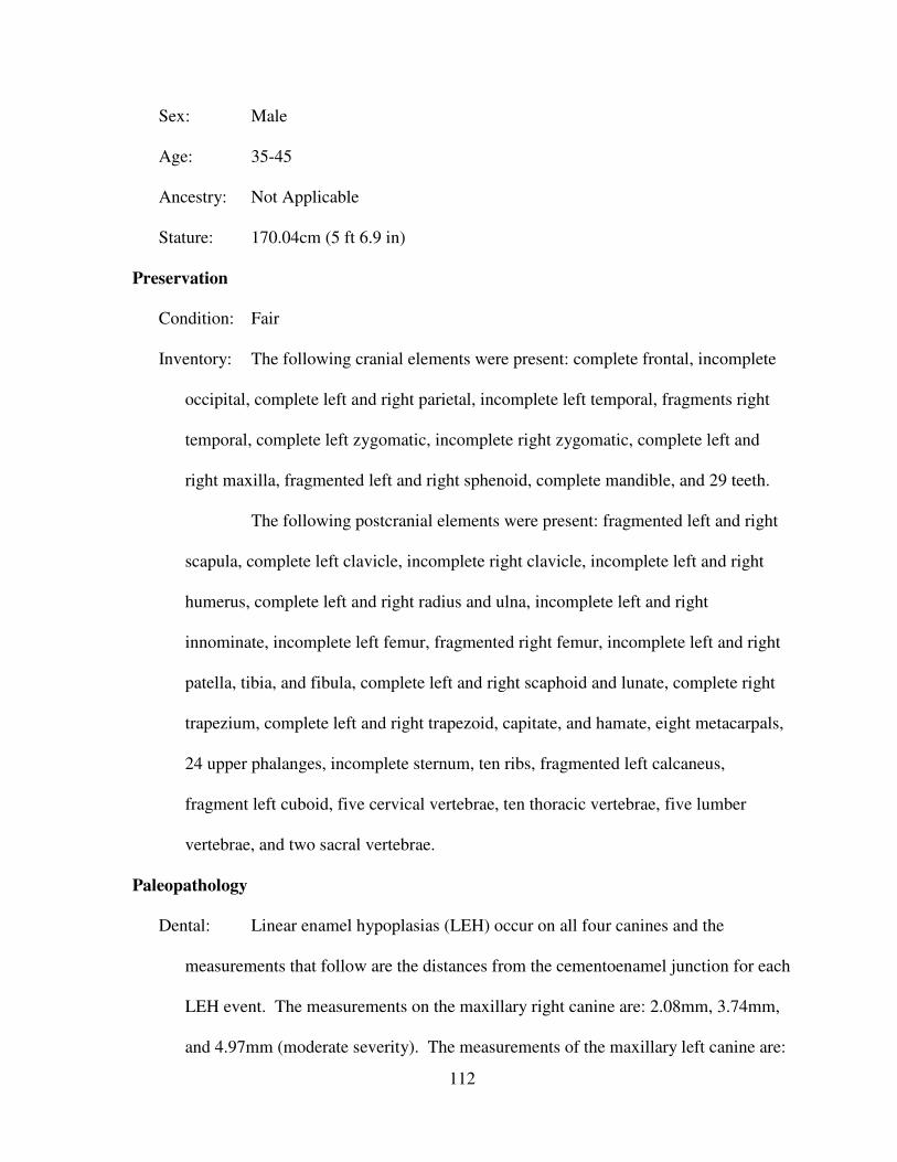
112
Sex: Male
Age: 35-45
Ancestry: Not Applicable
Stature: 170.04cm (5 ft 6.9 in)
Preservation
Condition: Fair
Inventory: The following cranial elements were present: complete frontal, incomplete
occipital, complete left and right parietal, incomplete left temporal, fragments right
temporal, complete left zygomatic, incomplete right zygomatic, complete left and
right maxilla, fragmented left and right sphenoid, complete mandible, and 29 teeth.
The following postcranial elements were present: fragmented left and right
scapula, complete left clavicle, incomplete right clavicle, incomplete left and right
humerus, complete left and right radius and ulna, incomplete left and right
innominate, incomplete left femur, fragmented right femur, incomplete left and right
patella, tibia, and fibula, complete left and right scaphoid and lunate, complete right
trapezium, complete left and right trapezoid, capitate, and hamate, eight metacarpals,
24 upper phalanges, incomplete sternum, ten ribs, fragmented left calcaneus,
fragment left cuboid, five cervical vertebrae, ten thoracic vertebrae, five lumber
vertebrae, and two sacral vertebrae.
Paleopathology
Dental: Linear enamel hypoplasias (LEH) occur on all four canines and the
measurements that follow are the distances from the cementoenamel junction for each
LEH event. The measurements on the maxillary right canine are: 2.08mm, 3.74mm,
and 4.97mm (moderate severity). The measurements of the maxillary left canine are:
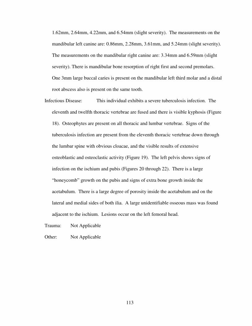
113
1.62mm, 2.64mm, 4.22mm, and 6.54mm (slight severity). The measurements on the
mandibular left canine are: 0.86mm, 2.28mm, 3.61mm, and 5.24mm (slight severity).
The measurements on the mandibular right canine are: 3.34mm and 6.59mm (slight
severity). There is mandibular bone resorption of right first and second premolars.
One 3mm large buccal caries is present on the mandibular left third molar and a distal
root abscess also is present on the same tooth.
Infectious Disease: This individual exhibits a severe tuberculosis infection. The
eleventh and twelfth thoracic vertebrae are fused and there is visible kyphosis (Figure
18). Osteophytes are present on all thoracic and lumbar vertebrae. Signs of the
tuberculosis infection are present from the eleventh thoracic vertebrae down through
the lumbar spine with obvious cloacae, and the visible results of extensive
osteoblastic and osteoclastic activity (Figure 19). The left pelvis shows signs of
infection on the ischium and pubis (Figures 20 through 22). There is a large
“honeycomb” growth on the pubis and signs of extra bone growth inside the
acetabulum. There is a large degree of porosity inside the acetabulum and on the
lateral and medial sides of both ilia. A large unidentifiable osseous mass was found
adjacent to the ischium. Lesions occur on the left femoral head.
Trauma: Not Applicable
Other: Not Applicable
114
Figure 18: Burial 7 Tuberculosis Infection of the Spine
115
Figure 19: Burial 7 Tuberculosis Infection of the Eleventh and Twelfth Thoracic Vertebrae
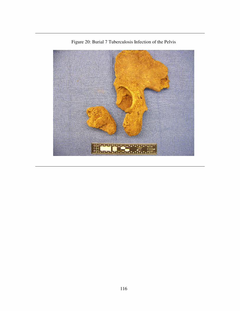
116
Figure 20: Burial 7 Tuberculosis Infection of the Pelvis
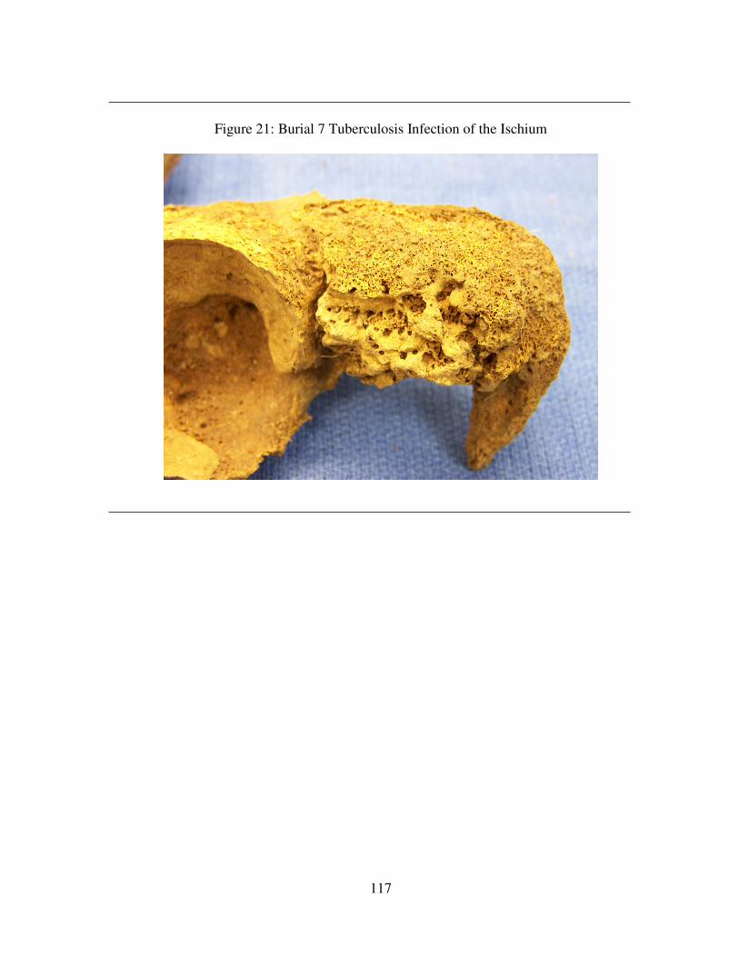
117
Figure 21: Burial 7 Tuberculosis Infection of the Ischium
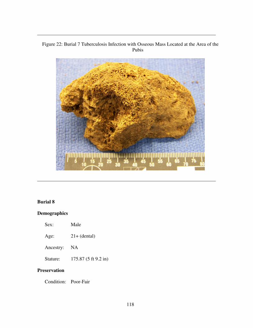
118
Figure 22: Burial 7 Tuberculosis Infection with Osseous Mass Located at the Area of the
Pubis
Burial 8
Demographics
Sex: Male
Age: 21+ (dental)
Ancestry: NA
Stature: 175.87 (5 ft 9.2 in)
Preservation
Condition: Poor-Fair
119
Inventory: The following cranial elements were present: incomplete frontal, incomplete
occipital, incomplete left and right parietal and temporal, incomplete left zygomatic,
fragmented right zygomatic, incomplete left maxilla, fragmented right maxilla,
fragmented left and right sphenoid, incomplete mandible, incomplete facial bones,
and 23 teeth.
The following postcranial elements were present: incomplete left scapula,
fragmented right scapula, fragmented left clavicle, incomplete left and right humerus,
fragmented left radius, incomplete right radius, incomplete left and right ulna,
innominate, femur, patella, and tibia, fragmented fibula, incomplete right fibula, five
upper phalanges, nine ribs, incomplete left and right calcaneus, incomplete right talus,
incomplete right navicular, incomplete right int. cuneiform, incomplete right lat.
cuneiform, eight metatarsals, three cervical vertebrae, and two sacral vertebrae.
Paleopathology
Dental: There is a 5.0mm large distal interproximal contact facet (IPCF) caries on
the maxillary right first molar. There is a 2.5mm large occlusal caries and a 2.0mm
large buccal caries on the mandibular left third molar, a 4.0mm large distal occlusal
caries on the mandibular left second molar, and a 3.5mm large mesial occlusal caries
on the mandibular left first molar. Lingual calculus is present on the mandibular left
canine.
Infectious Disease: Not Applicable
Trauma: Not Applicable
Other: Not Applicable
Burial 9
120
Demographics
Sex: Not Applicable
Age: Neonatal, first four weeks after birth
Ancestry: Not Applicable
Stature: Not Applicable
Preservation
Condition: Poor
Inventory: The following cranial elements were present: incomplete frontal, incomplete
occipital, incomplete left and right sphenoid, and incomplete left and right pars
petrosa.
Paleopathology
Dental: Not Applicable
Infectious Disease: Not Applicable
Trauma: Not Applicable
Other: Not Applicable
Burial 10 No remains were present.
Burial 11
Demographics
Sex: Female
Age: 45-59
Ancestry: Not Applicable
Stature: 158.14cm (5 ft 2.3 in)
121
Preservation
Condition: Poor-Fair
Inventory: The following cranial elements were present: incomplete frontal, incomplete
occipital, incomplete left and right parietal and temporal, fragmented right zygomatic,
fragmented left maxilla, incomplete left and right sphenoid, incomplete mandible,
fragmented facial bones, and 18 teeth.
The following postcranial remains were present: incomplete left scapula,
incomplete left and right clavicle, incomplete left humerus, complete right humerus,
incomplete left and right radius, complete left ulna, incomplete right ulna, incomplete
left and right innominate, incomplete left and right femur, incomplete left tibia,
fragmented right tibia, fragmented left and right tibia, incomplete right lunate,
complete left capitate, incomplete right capitate, nine metacarpals, eleven upper
phalanges, twelve ribs, fragmented right calcaneus, complete left and right talus,
complete left navicular, one metatarsals, one lower phalanges, seven cervical
vertebrae, and four sacral vertebrae.
Paleopathology
Dental: Periodontitis affects the area surrounding the maxillary right second and
third molars. The maxillary right second premolar has a root abscess measuring .4cm
x .9cm. Only roots remain of the maxillary left first premolar. There is a 4mm large
distal IPCF caries at the CEJ on the maxillary right second molar. There is a 3mm
large distal IPCF caries on the maxillary left second molar. There is mandibular
alveolar bone resorption present between the right first molar to the right second
molar.
Infectious Disease: Not Applicable
122
Trauma: Not Applicable
Other: A small Wormian bone is present between the left parietal and occipital.
Burial 12
Demographics
Sex: Male
Age: 50+
Ancestry: Not Applicable
Stature: Not Applicable
Preservation
Condition: Fair
Inventory: The following cranial elements were present: incomplete frontal, incomplete
occipital, incomplete left and right parietal, temporal, zygomatic, and maxilla,
incomplete left sphenoid, fragmented right sphenoid, incomplete mandible,
incomplete hyoid, and 23 teeth.
The following postcranial elements were present: incomplete left scapula,
complete left clavicle, fragmented right clavicle, fragmented left and right humerus,
incomplete right radius, incomplete left and right ulna, incomplete left innominate,
fragmented right innominate, incomplete left and right femur, fragmented left and
right patella, incomplete left fibula, fragmented right fibula, incomplete sternum, two
metacarpals, ten upper phalanges, ten ribs, fragmented left and right calcaneus, left
and right talus, and left and right cuboid, one metatarsal, seven cervical vertebrae,
three thoracic vertebrae, and two sacral vertebrae.
Paleopathology
123
Dental: The maxillary left first and second molars were lost antemortem. There is a
distal and proximal 3mm large IPCF caries on the maxillary right second molar.
Buccal calculus is present at the CEJ of the maxillary right first molar. The maxillary
right canine has a large 5mm caries labial and lingual. The maxillary left first
premolar exhibits a distal IPCF 3mm large caries. On the mandibular left third molar
there is substantial calculus on all sides at the CEJ. Mandibular alveolar bone
resorption is present at the areas between the right second premolar and the right
second molar, and the left second premolar and the left first molar.
Infectious Disease: Not Applicable
Trauma: Not Applicable
Other: Arthritic development is present on the odontoid process of the second
cervical vertebra.
Burial 13 No remains were present.
Burial 14
Demographics
Sex: Female
Age: 45-55
Ancestry: Not Applicable
Stature: 161.33cm (5 ft 3.5 in)
Preservation
Condition: Fair-Good
124
Inventory: The following cranial elements were present: complete frontal, complete
occipital, complete left and right parietal, incomplete left and right temporal,
incomplete left and right zygomatic, complete maxilla, complete mandible, and 28
teeth.
The following postcranial elements were present: incomplete left and right
scapula, incomplete left clavicle, complete left and right humerus, complete left and
right radius, incomplete left ulna, complete right ulna, incomplete left and right
innominate, complete left femur, complete left and right patella, complete left tibia,
incomplete right tibia, incomplete left and right fibula, complete left and right
scaphoid, complete right lunate, incomplete right triquetral, complete right pisiform,
complete left and right trapezium, complete left and right trapezoid, complete left and
right capitate, complete left and right hamate, complete left and right first through
fifth metacarpals, 27 hand phalanges, 17 ribs, incomplete left and right calcaneus,
complete left talus, incomplete right talus, complete left and right navicular, complete
left and right med. cuneiform, incomplete left int. cuneiform, incomplete lat.
cuneiform, incomplete left and right first through fifth metatarsal, 4 foot phalanges,
seven cervical vertebrae, twelve thoracic vertebrae, five lumbar vertebrae, and three
sacral vertebrae.
Paleopathology
Dental: The maxillary right third molar has a 6mm large occlusal caries. Only roots
remain of the maxillary right second molar and the maxillary left second molar and
there is slight periodontitis in the alveolar area around the teeth on both. Only the
roots remain of the maxillary right first premolar and the maxillary left second molar.
The maxillary right first molar has one mesial IPCF 3mm large caries. There is one
125
2.5mm large IPCF distal caries on the maxillary right central incisor. There is one
5mm large occlusal caries on the maxillary left second premolar. There is one 2.5mm
large occlusal caries on the maxillary left third molar. And there is one 4mm large
mesial and occlusal caries on the mandibular left third molar. There is one 4mm large
mesial-occlusal caries on the mandibular right second premolar. Only the roots
remain of the mandibular right second molar. There is one 6mm large occlusal-distal
caries on the mandibular right third molar. Calculus is found on the along the lingual
CEJ from the mandibular left second premolar to the mandibular left second molar.
A root abscess exists between the mandibular right second and third molars.
Infectious Disease: Not Applicable
Trauma: Not Applicable
Other: Not Applicable
Burial 15
Demographics
Sex: Female
Age: 18+
Ancestry: Not Applicable
Stature: Not Applicable
Preservation
Condition: Poor
Inventory: The following cranial elements were present: fragmented frontal,
fragmented occipital, fragment right parietal, fragmented left temporal, incomplete
left zygomatic, and a fragmented mandible.
126
The following postcranial elements were present: incomplete right radius,
incomplete right ulna, incomplete left and right innominate, incomplete left femur,
fragmented right femur, incomplete right patella, fragmented left tibia, incomplete
right tibia, fragmented left and right fibula, two hand phalanges, the first cervical
vertebra, three thoracic fragments, two lumbar fragments, the first sacral vertebra, and
three sacral fragments.
Paleopathology
Dental: Not Applicable
Infectious Disease: Not Applicable
Trauma: Not Applicable
Other: Not Applicable
Burial 16
Demographics
Sex: Male
Age: 21+
Ancestry: Not Applicable
Stature: Not Applicable
Preservation
Condition: Poor
Inventory: The following cranial elements were present: fragmented frontal,
fragmented occipital, fragmented left and right parietals, incomplete left and right
temporal, fragmented left maxilla, incomplete mandible, and 13 teeth.
127
The following postcranial elements were present: fragmented left humerus,
fragmented left and right radius, fragmented left and right ulna, fragmented left
innominate, incomplete left and right femur, incomplete left tibia, fragmented right
tibia, and fragmented left and right fibulae.
Paleopathology
Dental: Mandibular alveolar bone resorption is present at the area of the mandibular
left third molar. There is one 1.5mm large distal IPCF caries on the mandibular left
second molar.
Infectious Disease: Not Applicable
Trauma: Not Applicable
Other: Not Applicable
Burial 17
Demographics
Sex: Not Applicable
Age: 35+
Ancestry: Not Applicable
Stature: Not Applicable
Preservation
Condition: Poor
Inventory: The following cranial elements were present: incomplete frontal, incomplete
occipital, incomplete left and right parietals, incomplete left temporal, fragmented left
and right maxilla, incomplete mandible, and six teeth.
128
The following postcranial elements were present: fragmented left scapula,
fragmented left and right clavicle, fragmented left and right humerus, fragmented left
and right radius, fragmented left and right ulna, fragmented right innominate,
incomplete left and right femur, fragmented left and right tibia, fragmented left fibula,
four ribs, one metacarpal fragment, one metatarsal fragment, the third and fourth
cervical vertebrae, and five cervical fragments.
Paleopathology
Dental: Maxillary alveolar alveolar bone resorption is present at the area from the
right second premolar to the right third molar. There is one 3mm large buccal caries
and one 2mm large lingual caries on the maxillary left first molar. There is one
2.5mm large lingual caries on the maxillary left central incisor. There is one occlusal
caries on the maxillary left canine. There are also two 3mm large occlusal caries on
the maxillary left first premolar.
Infectious Disease: Not Applicable
Trauma: Not Applicable
Other: Not Applicable
Burial 18
Demographics
Sex: Not Applicable
Age: 11+
Ancestry: Not Applicable
Stature: Not Applicable
Preservation
129
Condition: Poor
Inventory: The following cranial elements were present: incomplete frontal, fragmented
occipital, fragmented left and right parietals, fragmented left temporal, incomplete
right temporal, fragmented mandible, and six teeth.
The following postcranial elements were present: incomplete left humerus,
fragmented left radius, fragmented left ulna, fragmented right innominate, incomplete
left and right femur, fragmented left and right tibia, and the first and second cervical
vertebra.
Paleopathology
Dental: There is one 1mm large occlusal caries on the occlusal surface of the
maxillary left canine.
Infectious Disease: Not Applicable
Trauma: Not Applicable
Other: Not Applicable
Burial 19
Demographics
Sex: Not Applicable
Age: 21+
Ancestry: Not Applicable
Stature: Not Applicable
Preservation
Condition: Very Poor
130
Inventory: The only cranial elements present were seven teeth. The following
postcranial elements were presents: fragmented right femur, fragmented left and right
tibia, and fragmented left and right fibula
Paleopathology
Dental: Not Applicable
Infectious Disease: Not Applicable
Trauma: Not Applicable
Other: Not Applicable
Burial 20
Demographics
Sex: Not Applicable
Age: 21+
Ancestry: Not Applicable
Stature: Not Applicable
Preservation
Condition: Very Poor
Inventory: The following cranial elements were present: fragmented frontal,
fragmented right maxilla, and 19 teeth.
The following postcranial elements were present: fragmented and bone meal
remains of the innominate, three ribs, and a sacral fragment.
Paleopathology
131
Dental: There is one 1.5mm large mesial caries on the maxillary left canine. There
is one 1.5 mesial caries on the maxillary left first premolar. There is one 1.5mm large
mesial caries on the maxillary left second premolar.
Infectious Disease: Not Applicable
Trauma: Not Applicable
Other: Not Applicable
Burial 21
Demographics
Sex: Male
Age: 28-44
Ancestry: Not Applicable
Stature: Not Applicable
Preservation
Condition: Poor-Fair
Inventory: The following cranial elements were present: fragmented frontal, incomplete
occipital, incomplete left and right parietals, incomplete left and right temporal,
complete left and right zygomatic, incomplete left and right maxilla, fragmented left
and right sphenoid, complete mandible, and thirteen teeth.
The following postcranial elements were present: incomplete left and right
scapula, incomplete left and right clavicle, incomplete left and right humerus,
incomplete left and right radius, incomplete left and right ulna, incomplete left and
right innominate, incomplete left and right femur, incomplete left and right tibia,
fragmented left and right femur, twelve ribs, incomplete left calcaneus, incomplete
132
left talus, two metatarsal fragments, all seven cervical vertebrae are present but
fragmented, all twelve thoracic vertebrae present but fragmented, and all five lumbar
vertebrae are present but fragmented.
Paleopathology
Dental: There is alveolar bone resorption in the regions of the maxillary left second
and third molars, the mandibular right third molar, and from the mandibular right
canine to the mandibular left central incisor. There is a 2mm large lingual-occlusal
caries on the maxillary right first molar. There is also a 2.5mm large buccal-occlusal
caries on the same tooth. There is lingual calculus at the CEJ on the mandibular right
second premolar, first molar, and the second molar. And there is a 2.5mm large distal
root caries on the mandibular left second premolar, as well as, a 3mm large distal
occlusal-lingual caries on the mandibular left second molar.
Infectious Disease: Not Applicable
Trauma: Not Applicable
Other: There is osteophytic buildup on the cervical vertebral bodies with some
porosity.
Burial 22
Demographics
Sex: Not Applicable
Age: 21+
Ancestry: Not Applicable
Stature: Not Applicable
Preservation
133
Condition: Very Poor
Inventory: Bone meal and four unidentifiable cranial fragments were present.
Additionally, 18 teeth were present. No postcranial elements were present.
Paleopathology
Dental: Not Applicable
Infectious Disease: Not Applicable
Trauma: Not Applicable
Other: Not Applicable
Burial 23
Demographics
Sex: Male
Age: 40+
Ancestry: Not Applicable
Stature: Not Applicable
Preservation
Condition: Poor
Inventory: The following cranial elements were present: incomplete frontal, incomplete
occipital, incomplete left and right temporal, incomplete left and right zygomatic,
incomplete left and right maxilla, incomplete mandible, and 18 teeth.
The following postcranial elements were present: fragmented left scapula,
incomplete right scapula, fragmented left and right clavicle, incomplete left and right
humerus, fragmented left radius, incomplete right radius, incomplete left and right
femur, incomplete right tibia, incomplete right femur, eight ribs, three metacarpal
134
fragments, the second and third cervical fragments, six cervical fragments, and three
lumbar fragments.
Paleopathology
Dental: There is one 8mm large mesial caries on the maxillary right first molar.
There is one 1mm large distal caries on the maxillary left first molar and a 3mm large
mesial caries on the maxillary left third molar. There is a 1mm large distal root caries
on the maxillary right canine. There are two 1mm large caries on the mesial and
buccal surfaces on the maxillary left first premolar. There is a 3mm large mesial
caries and a 4mm large distal caries on the maxillary left second premolar. There is a
4mm large mesial root caries and a 2.5mm large distal root caries on the maxillary
right second premolar. There is mandibular alveolar bone resorption at the left
central and lateral incisors and from the left second premolar to the left molar. There
is a 2.5mm large mesial caries on the mandibular right third molar. There is a 4mm
large mesial root caries on the mandibular right lateral incisor. There is an 8mm large
lingual-distal root caries on the mandibular left canine. There is a 6mm large lingual
root caries and a 5mm large distal root caries on the mandibular right canine. There
is a 4mm large occlusal caries on the mandibular left first premolar. There is a 6mm
large caries on the mesial-distal-lingual surface on the mandibular right first
premolar. There is one 6mm large distal root caries on the maxillary left lateral
incisor. There is a 6mm large mesial root caries on the mandibular right second
premolar. There is a 3mm large mesial interproximal caries.
Infectious Disease: Not Applicable
Trauma: Not Applicable
Other: Not Applicable
135
Burial 24
Demographics
Sex: Male
Age: 21+
Ancestry: Not Applicable
Stature: Not Applicable
Preservation
Condition: Poor
Inventory: The following cranial elements were present: incomplete frontal, incomplete
occipital, complete left and right parietal, complete left temporal, incomplete right
temporal, incomplete left zygomatic, fragmented left and right maxilla, incomplete
mandible, and eight teeth.
The following postcranial elements were present: fragmented right scapula,
incomplete left clavicle, fragmented left and right humerus, fragmented let and right
radius, incomplete left ulna, fragmented right ulna, fragmented left and right
innominate, incomplete left and right femur, incomplete left patella, incomplete left
and right tibia, incomplete left fibula, eight ribs, seven cervical vertebrae, five
thoracic vertebrae, nine thoracic fragments, two lumbar vertebrae, and two sacral
vertebrae.
Paleopathology
Dental: There is a 4.5mm large distal and a 4.5mm large mesial caries on the
maxillary right canine. There is a 4.5mm large mesial-root caries on the mandibular
left central incisor. The maxillary right canine exhibits LEH. There is one LEH
event measuring 5.12mm from the CEJ. Hypoplasias are evident on the middle and
136
occlusal third of the mandibular right lateral incisor and on the middle and occlusal
third of the mandibular left lateral incisor. A dental anomaly is present. It is possible
that the tooth is a premolar that never completed cusp development.
Infectious Disease: Not Applicable
Trauma: Not Applicable
Other: Not Applicable
Burial 25
Demographics
Sex: Indeterminate
Age: 40-60
Ancestry: Not Applicable
Stature: Not Applicable
Preservation
Condition: Very poor
Inventory: The following cranial elements were present: incomplete frontal, incomplete
occipital, incomplete left and right parietal, fragmented left temporal, and two teeth.
The only postcranial elements present were a fragmented left femur, an
incomplete right femur, and one sacral fragment.
Paleopathology
Dental: Maxillary and Mandibular are dentures present and the only natural teeth
that remain are the maxillary right first and second premolars. The maxillary left
central and lateral incisors are gold plated. Mandibular alveolar bone resorption is
present from the mandibular right second premolar to the mandibular right third
137
molar and the mandibular left first molar to the mandibular left third molar. Only the
roots remain of the mandibular left first premolar, left canine, and left central incisor
to the right canine.
Infectious Disease: Not Applicable
Trauma: Not Applicable
Other: Not Applicable
Burial 26
Demographics
Sex: Indeterminate
Age: 21+
Ancestry: Not Applicable
Stature: Not Applicable
Preservation
Condition: Very poor
Inventory: The following cranial elements were present: fragmented left maxilla,
fragmented mandible, and 14 teeth.
The following postcranial elements were present: fragmented right humerus
and 16 bone meal/stains of ribs.
Paleopathology
Dental: There is one 1mm large occlusal caries on the maxillary left second
premolar and one 2mm large occlusal caries on the mandibular left second molar.
Infectious Disease: Not Applicable
Trauma: Not Applicable
138
Other: Not Applicable
Burial 27
Demographics
Sex: Not Applicable
Age: 21+
Ancestry: Not Applicable
Stature: Not Applicable
Preservation
Condition: Very poor
Inventory: The only cranial elements recovered were nine teeth. The only postcranial
elements present were left and right femoral.
Paleopathology
Dental: There is a metal brace joining the maxillary right second premolar to the
maxillary right second molar. The maxillary right first molar was lost antemortem.
Infectious Disease: Not Applicable
Trauma: Not Applicable
Other: Not Applicable
Burial 28
Demographics
Sex: Not Applicable
Age: Not Applicable
Ancestry: Not Applicable
139
Stature: Not Applicable
Preservation
Condition: Very poor
Inventory: One frontal fragment was the only cranial remain present; however, bone
meal was also recovered. The following postcranial elements were present:
fragmented left femur, fragmented left and right tibia, fragmented left and right
fibula, and one sacral fragment. Bone meal was also recovered from the area of the
right femur.
Paleopathology
Dental: Not Applicable
Infectious Disease: Not Applicable
Trauma: Not Applicable
Other: Not Applicable
Burial 29
Demographics
Sex: Not Applicable
Age: 11+
Ancestry: Not Applicable
Stature: Not Applicable
Preservation
Condition: Very poor
140
Inventory: The following cranial remains were present: incomplete frontal, fragmented
occipital, fragmented left and right parietal, fragmented left temporal, incomplete left
zygomatic, and five teeth.
The following postcranial remains were present: incomplete left and right
femur, fragmented left tibia, and incomplete right tibia.
Paleopathology
Dental: There is one 2mm large distal IPCF caries on the mandibular right canine
and one 2.5mm large mesial IPCF caries on the mandibular left second premolar.
Infectious Disease: Not Applicable
Trauma: Not Applicable
Other: The remains of three dogs, indicated by the presence of five scapulae, were
found directly above the casket remains.
Burial 30
Demographics
Sex: Not Applicable
Age: Not Applicable
Ancestry: Not Applicable
Stature: Not Applicable
Preservation
Condition: Very poor
Inventory: The only cranial remains present were frontal bone fragments. The
following postcranial remains were present: fragmented left femur, incomplete right
femur, fragmented right tibia, fragmented right fibula, and one rib fragment.
141
Paleopathology
Dental: Not Applicable
Infectious Disease: Not Applicable
Trauma: Not Applicable
Other: Not Applicable
Burial 31
Demographics
Sex: Not Applicable
Age: 15+
Ancestry: Not Applicable
Stature: Not Applicable
Preservation
Condition: Very poor
Inventory: The only cranial elements present were 16 teeth. The only postcranial
elements present were left and right femoral fragments.
Paleopathology
Dental: The maxillary right central and lateral incisors have gold crowns. There is
also one 2mm large distal IPCF on the right canine. There is one 1.5mm large distal
IPCF caries on the maxillary right second premolar.
Infectious Disease: Not Applicable
Trauma: Not Applicable
Other: Not Applicable
142
Burial 32
Demographics
Sex: Not Applicable
Age: Not Applicable
Ancestry: Not Applicable
Stature: Not Applicable
Preservation
Condition: Not Applicable
Inventory: No cranial elements were recovered for this burial. The following
postcranial elements were present: incomplete right humerus, bone meal from the
innominate area, incomplete right femur, incomplete right tibia, and incomplete right
fibula.
Paleopathology
Dental: Not Applicable
Infectious Disease: Not Applicable
Trauma: Not Applicable
Other: Not Applicable
Burial 33 No remains were present.
Burial 34 No remains were present.
Burial 35
Demographics
143
Sex: Not Applicable
Age: 7+
Ancestry: Not Applicable
Stature: Not Applicable
Preservation
Condition: Very Poor
Inventory: Bone meal and three teeth were recovered from the area of the cranium.
Fragmented right femoral remains were the only solid postcranial remains recovered.
Bone meal was recovered in the following postcranial areas: right radius, right ulna,
right innominate, left femur, left and right tibia, and left and right fibula.
Paleopathology
Dental: Enamel caps were the only portion of the teeth present.
Infectious Disease: Not Applicable
Trauma: Not Applicable
Other: Not Applicable
Burial 36 No remains were present.
Burial 37
Demographics
Sex: Not Applicable
Age: 12+
Ancestry: Not Applicable
Stature: Not Applicable
144
Preservation
Condition: Very poor
Inventory: Three teeth were present.
Paleopathology
Dental: Only the enamel caps of the maxillary right first, second, and third molar
were present.
Infectious Disease: Not Applicable
Trauma: Not Applicable
Other: Not Applicable
Burial 38
Demographics
Sex: Not Applicable
Age: Not Applicable
Ancestry: Not Applicable
Stature: Not Applicable
Preservation
Condition: Very poor
Inventory: Seventeen dental fragments were present.
Paleopathology
Dental: Not Applicable
Infectious Disease: Not Applicable
Trauma: Not Applicable
Other: Not Applicable
145
Burial 39 No remains were present.
Burial 40 This set of remains was actually a dog cranium.
Burial 41
Demographics
Sex: Not Applicable
Age: Not Applicable
Ancestry: Not Applicable
Stature: Not Applicable
Preservation
Condition: Very poor
Inventory: The only skeletal element present was a 1 cm, possibly burned, unidentified
fragment.
Paleopathology
Dental: Not Applicable
Infectious Disease: Not Applicable
Trauma: Not Applicable
Other: Not Applicable
Burial 42
Demographics
Sex: Not Applicable
146
Age: 21+
Ancestry: Not Applicable
Stature: Not Applicable
Preservation
Condition: Very poor
Inventory: The following cranial elements were present: fragmented frontal,
fragmented occipital, incomplete left parietal, fragmented right parietal, fragmented
mandible, and one tooth.
The following postcranial elements were present: fragmented left and right
innominate, incomplete right femur, and incomplete left and right tibiae.
Paleopathology
Dental: Not Applicable
Infectious Disease: Not Applicable
Trauma: Not Applicable
Other: Not Applicable
Burial 43
Demographics
Sex: Not Applicable
Age: 21+
Ancestry: Not Applicable
Stature: Not Applicable
Preservation
Condition: Poor
147
Inventory: The following cranial elements were present: fragmented frontal, incomplete
left and right parietal, fragmented left and right temporal, fragmented left maxilla,
incomplete mandible, and 22 teeth.
The following postcranial remains were present: fragmented right scapula,
fragmented right humerus, fragmented left and right innominate, incomplete left
femur, fragmented right femur, incomplete left tibia, fragmented right tibia, two
fragmented ribs, the first cervical vertebra, and four cervical fragments.
Paleopathology
Dental: The maxillary right third molar has a divided hypocone.
Infectious Disease: Not Applicable
Trauma: There is evidence for occupational dental wear on the maxillary left central
incisor. A small groove is present on the left lateral occlusal surface is present.
Other: Not Applicable
Burial 44
Demographics
Sex: Male
Age: 45+
Ancestry: Not Applicable
Stature: Not Applicable
Preservation
Condition: Poor
148
Inventory: The following cranial elements were present: incomplete frontal, incomplete
occipital, incomplete left and right parietal, incomplete right temporal, fragmented
mandible, and three teeth.
The following postcranial elements were present: fragmented right scapula,
fragmented right clavicle, fragmented right humerus, fragmented right ulna,
fragmented right innominate, fragmented left femur, incomplete right femur,
fragmented left and right tibia, and one fragmented rib.
Paleopathology
Dental: Not Applicable
Infectious Disease: Not Applicable
Trauma: Not Applicable
Other: Not Applicable
Burial 45
Demographics
Sex: Female
Age: 18-21
Ancestry: Not Applicable
Stature: Not Applicable
Preservation
Condition: Fair
Inventory: The following cranial elements were present: incomplete frontal, fragmented
occipital, incomplete left and right parietal, fragmented left temporal, incomplete
right temporal, fragmented left zygomatic, incomplete right zygomatic, incomplete
149
left and right maxilla, fragmented left and right sphenoid, incomplete mandible, and
31 teeth.
The following postcranial elements were present: incomplete left and right
scapula, incomplete left and right clavicle, incomplete left and right humerus,
fragmented left and right radius, incomplete left and right ulna, fragmented left and
right innominate, incomplete left and right femur, fragmented left femur, incomplete
right femur, fragmented left tibia, incomplete right tibia, fragmented left fibula,
incomplete right fibula, 21 ribs, seven cervical vertebrae, twelve thoracic vertebrae,
the first four lumbar vertebrae, and the first two sacral vertebrae.
Paleopathology
Dental: The maxillary left first molar has a 3mm large distal and occlusal lead
filling. The maxillary left third molar is not fully erupted. The mandibular left first
molar has one 4mm large distal and occlusal caries. The mandibular right first molar
has a 4mm large caries on the distal and occlusal surface.
Infectious Disease: Not Applicable
Trauma: Not Applicable
Other: Not Applicable
Burial 46
Demographics
Sex: Not Applicable
Age: 21+
Ancestry: Not Applicable
Stature: Not Applicable
150
Preservation
Condition: Very poor
Inventory: Only five teeth were present with this burial.
Paleopathology
Dental: Not Applicable
Infectious Disease: Not Applicable
Trauma: Not Applicable
Other: Not Applicable
Burial 47 No remains were present.
Burial 48
Demographics
Sex: Not Applicable
Age: 10 years +/- 9 months
Ancestry: Not Applicable
Stature: Not Applicable
Preservation
Condition: Very poor
Inventory: Only 23 teeth were present with this burial.
Paleopathology
Dental: The maxillary right and left first molars have the presence of a Carabelli’s
cusp. The cusps of the left and mandibular right third molars are not completely
formed.
151
Infectious Disease: Not Applicable
Trauma: Not Applicable
Other: Not Applicable
Burial 49
Demographics
Sex: Not Applicable
Age: 6+
Ancestry: Not Applicable
Stature: Not Applicable
Preservation
Condition: Very poor
Inventory: One tooth represents the cranial elements for this burial. The postcranial
elements present were fragmented remains of the left tibia and left fibula.
Paleopathology
Dental: Not Applicable
Infectious Disease: Not Applicable
Trauma: Not Applicable
Other: Not Applicable
Burial 50
Demographics
Sex: Not Applicable
Age: 21+
152
Ancestry: Not Applicable
Stature: Not Applicable
Preservation
Condition: Poor
Inventory: The following cranial elements were present: incomplete occipital,
incomplete left and right parietal, fragmented left and right temporal and 20 teeth.
The following postcranial elements were present: incomplete right ulna,
fragmented left and right femur, fragmented left and right tibia, and three unidentified
metacarpals.
Paleopathology
Dental: The maxillary left first and second premolars are maloccluded lingual-
distally.
Infectious Disease: Not Applicable
Trauma: Not Applicable
Other: Not Applicable
Burial 50a This was mislabeled as a burial but in actuality was a non-culturally related
stain in the soil.
Burial 51
Demographics
Sex: Not Applicable
Age: 21+
Ancestry: Not Applicable
153
Stature: Not Applicable
Preservation
Condition: Very poor
Inventory: Cranial elements present included eleven teeth. Fragments of the left
humerus and right femur were the only postcranial elements present. Bone meal was
present in the areas of the right humerus, innominate, left femur, left tibia, and right
tibia.
Paleopathology
Dental: Not Applicable
Infectious Disease: Not Applicable
Trauma: Not Applicable
Other: Not Applicable
Burial 52
Demographics
Sex: Not Applicable
Age: 21+
Ancestry: Not Applicable
Stature: Not Applicable
Preservation
Condition: Poor
Inventory: The following cranial elements were present: incomplete frontal, incomplete
occipital, incomplete left and right parietal, incomplete left and right temporal,
fragmented left and right maxilla, incomplete mandible, and 27 teeth.
154
The following postcranial elements were present: fragmented right scapula,
fragmented right humerus, incomplete left femur, fragmented right femur, incomplete
left tibia, fragmented right tibia, and fragmented right fibula.
Paleopathology
Dental: Not Applicable
Infectious Disease: Not Applicable
Trauma: Not Applicable
Other: Not Applicable
Burial 53 No remains were present.
Burial 54
Demographics
Sex: Not Applicable
Age: 21+
Ancestry: Not Applicable
Stature: Not Applicable
Preservation
Condition: Very poor
Inventory: The following cranial elements were present: incomplete frontal, fragmented
occipital, fragmented left and right parietal, fragmented right temporal, and ten teeth.
No postcranial elements were present.
Paleopathology
155
Dental: There is one 1.5mm large mesial IPCF caries on the maxillary right second
premolar.
Infectious Disease: Not Applicable
Trauma: Not Applicable
Other: Not Applicable
Burial 55 No remains were present.
Burial 56
Demographics
Sex: Not Applicable
Age: 21+
Ancestry: Not Applicable
Stature: Not Applicable
Preservation
Condition: Very poor
Inventory: Cranial elements present consisted of 18 teeth. The postcranial elements
present consists of bone meal in the areas of the left femur, left and right tibia, and
left and right fibula.
Paleopathology
Dental: There is one 2mm large occlusal caries present on the maxillary right second
molar.
Infectious Disease: Not Applicable
Trauma: Not Applicable

156
Other: Not Applicable
Burial 57
Demographics
Sex: Not Applicable
Age: 15+
Ancestry: Not Applicable
Stature: Not Applicable
Preservation
Condition: Very poor
Inventory: The cranial elements present include four teeth and 19 tooth fragments.
Paleopathology
Dental: Not Applicable
Infectious Disease: Not Applicable
Trauma: Not Applicable
Other: Not Applicable
Burial 58
Demographics
Sex: Not Applicable
Age: Not Applicable
Ancestry: Not Applicable
Stature: Not Applicable
Preservation
157
Condition: Very poor
Inventory: The cranial elements present consist of a fragmented right parietal and
fragmented mandible. The following postcranial elements were present: fragmented
left and right femur, fragmented left tibia, and fragmented right fibula.
Paleopathology
Dental: Not Applicable
Infectious Disease: Not Applicable
Trauma: Not Applicable
Other: Not Applicable
Burial 59 No remains were present.
Burial 60
Demographics
Sex: Not Applicable
Age: Not Applicable
Ancestry: Not Applicable
Stature: Not Applicable
Preservation
Condition: Very poor
Inventory: No cranial elements were present. Postcranial elements present include left
and right femoral fragments.
Paleopathology
Dental: Not Applicable
158
Infectious Disease: Not Applicable
Trauma: Not Applicable
Other: Not Applicable
Burial 61 No remains were present.
Burial 62
Demographics
Sex: Not Applicable
Age: Not Applicable
Ancestry: Not Applicable
Stature: Not Applicable
Preservation
Condition: Very poor
Inventory: Cranial elements present consisted only of unidentifiable bone meal.
Postcranial elements include bone meal in the areas of the left and right femur, left
and right tibia, and sternum.
Paleopathology
Dental: Not Applicable
Infectious Disease: Not Applicable
Trauma: Not Applicable
Other: Not Applicable
Burial 63
159
Demographics
Sex: Not Applicable
Age: 21+
Ancestry: Not Applicable
Stature: Not Applicable
Preservation
Condition: Very poor
Inventory: The cranial elements present include three teeth. No postcranial elements
were present.
Paleopathology
Dental: Not Applicable
Infectious Disease: Not Applicable
Trauma: Not Applicable
Other: Not Applicable
Burial 64 No remains were present.
Burial 65 No remains were present.
Burial 66
Demographics
Sex: Not Applicable
Age: 35+
Ancestry: Not Applicable
160
Stature: Not Applicable
Preservation
Condition: Very poor
Inventory: The following cranial elements were present: incomplete frontal, fragmented
occipital, fragmented left and right parietal, bone meal in the areas of the left and
right temporal and the mandible, and nine teeth. Postcranial elements consist only of
a fragmented left humerus.
Paleopathology
Dental: Not Applicable
Infectious Disease: Not Applicable
Trauma: There is possible occupational trauma to the mandibular right canine. The
distal two-thirds of the occlusal surface are worn in the shape of a groove. This
individual may have been using his teeth as a tool.
Other: Not Applicable
Burial 67 No remains were present.
Burial 68 No remains were present.
Burial 69 No remains were present.
Burial 70 No remains were present.
Burial 71 No remains were present.
Burial 72 No remains were present.
Burial 73
Demographics
161
Sex: Not Applicable
Age: 12+
Ancestry: Not Applicable
Stature: Not Applicable
Preservation
Condition: Very poor
Inventory: Cranial elements present include five teeth present. No postcranial elements
were present.
Paleopathology
Dental: Not Applicable
Infectious Disease: Not Applicable
Trauma: Not Applicable
Other: Not Applicable
Burial 74
Demographics
Sex: Not Applicable
Age: 12+
Ancestry: Not Applicable
Stature: Not Applicable
Preservation
Condition: Very poor
162
Inventory: The cranial elements present were an incomplete left temporal and two
teeth. The following postcranial elements present were fragments of the right
innominate and fragmented of the right femur.
Paleopathology
Dental: Not Applicable
Infectious Disease: Not Applicable
Trauma: Not Applicable
Other: Not Applicable
Burial 75
Demographics
Sex: Not Applicable
Age: Not Applicable
Ancestry: Not Applicable
Stature: Not Applicable
Preservation
Condition: Very poor
Inventory: No cranial elements were present. The following postcranial elements were
present: fragmented left and right humerus, fragmented right radius, fragmented right
ulna, bone meal in the area of the innominate, incomplete left and right femur,
incomplete left and right tibia, fragmented left fibula, incomplete right fibula.
Paleopathology
Dental: Not Applicable
Infectious Disease: Not Applicable
163
Trauma: Not Applicable
Other: Not Applicable
Burial 76 No remains were present.
Burial 77 This burial number was not used.
Burial 78
Demographics
Sex: Not Applicable
Age: 12+
Ancestry: Not Applicable
Stature: Not Applicable
Preservation
Condition: Very poor
Inventory: The cranial element consisted of one tooth. Postcranial elements consisted
of stains in the area of the left and right femur.
Paleopathology
Dental: Not Applicable
Infectious Disease: Not Applicable
Trauma: Not Applicable
Other: Not Applicable
Burial 79 No remains were present.
Burial 80 No remains were present.
164
Burial 81
Demographics
Sex: Not Applicable
Age: Not Applicable
Ancestry: Not Applicable
Stature: Not Applicable
Preservation
Condition: Very poor
Inventory: No cranial elements were present. The postcranial elements present
consisted of bone meal in the areas of the left and right radius, left and right ulna, and
left femur.
Paleopathology
Dental: Not Applicable
Infectious Disease: Not Applicable
Trauma: Not Applicable
Other: Not Applicable
Burial 82
Demographics
Sex: Not Applicable
Age: Not Applicable
Ancestry: Not Applicable
Stature: Not Applicable
165
Preservation
Condition: Very poor
Inventory: Cranial elements consisted only of bone meal. The following postcranial
elements were present: bone meal in the areas of the right clavicle, right humerus, left
and right innominate, left and right femur, left tibia, and the vertebral column.
Paleopathology
Dental: Not Applicable
Infectious Disease: Not Applicable
Trauma: Not Applicable
Other: Not Applicable
Burial 83 No remains were present.
Burial 84
Demographics
Sex: Not Applicable
Age: 12-15
Ancestry: Not Applicable
Stature: Not Applicable
Preservation
Condition: Very poor
Inventory: The cranial elements present consisted of two teeth. The following
postcranial elements were present: bone meal in the areas of the left and right radius,
166
left and right ulna, left and right innominate, left and right tibia, left and right fibula,
and fragments of the left and right femur.
Paleopathology
Dental: Not Applicable
Infectious Disease: Not Applicable
Trauma: Not Applicable
Other: Not Applicable
Burial 85
Demographics
Sex: Not Applicable
Age: Not Applicable
Ancestry: Not Applicable
Stature: Not Applicable
Preservation
Condition: Very poor
Inventory: The cranial elements present consisted of unidentifiable bone meal. The
following postcranial elements were present: bone meal in the areas of the left and
right scapula, left and right humerus, left and radius, left and right ulna, left and right
innominate, left and right femur, left and right tibia, left and right fibula, ribs, and
vertebral column.
Paleopathology
Dental: Not Applicable
Infectious Disease: Not Applicable
167
Trauma: Not Applicable
Other: Not Applicable
Burial 86 (This burial was accidentally assigned two numbers, 86 and 112)
Demographics
Sex: Not Applicable
Age: 7-12
Ancestry: Not Applicable
Stature: Not Applicable
Preservation
Condition: Very poor
Inventory: The following cranial elements were present: fragmented left temporal, 20
permanent teeth, and two deciduous teeth. Postcranial elements present consisted of
bone meal in the areas of the left tibia and left fibula.
Paleopathology
Dental: Not Applicable
Infectious Disease: Not Applicable
Trauma: Not Applicable
Other: Not Applicable
Burial 87 No remains were present.
Burial 88
Demographics
168
Sex: Not Applicable
Age: less than 2 years
Ancestry: Not Applicable
Stature: Not Applicable
Preservation
Condition: Very poor
Inventory: The only skeletal element present is one tooth.
Paleopathology
Dental: Not Applicable
Infectious Disease: Not Applicable
Trauma: Not Applicable
Other: Not Applicable
Burial 89 No remains were present.
Burial 90 No remains were present.
Burial 91
Demographics
Sex: Not Applicable
Age: 10+
Ancestry: Not Applicable
Stature: Not Applicable
Preservation
Condition: Very poor
169
Inventory: The only skeletal elements present were five teeth.
Paleopathology
Dental: Not Applicable
Infectious Disease: Not Applicable
Trauma: Not Applicable
Other: Not Applicable
Burial 92
Demographics
Sex: Not Applicable
Age: Not Applicable
Ancestry: Not Applicable
Stature: Not Applicable
Preservation
Condition: Very poor
Inventory: The following cranial elements were present: incomplete occipital and two
unidentifiable cranial fragments. The following postcranial elements were present:
fragmented right humerus, bone meal in the area of the innominate, and fragmented
left and right femur.
Paleopathology
Dental: Not Applicable
Infectious Disease: Not Applicable
Trauma: Not Applicable
Other: Not Applicable
170
Burial 93
Demographics
Sex: Not Applicable
Age: 21+
Ancestry: Not Applicable
Stature: Not Applicable
Preservation
Condition: Very poor
Inventory: The following cranial elements were present: fragmented mandible, 18
teeth, and unidentifiable bone meal. The following postcranial elements were
present: bone meal in the areas of the left scapula, left clavicle, left and right
humerus, left and right radius, left and right ulna, left and right innominate, left and
right femur, left and right tibia, left and right fibula, and ribs.
Paleopathology
Dental: Not Applicable
Infectious Disease: Not Applicable
Trauma: Not Applicable
Other: Not Applicable
Burial 94 No remains were present.
Burial 95 No remains were present.
171
Burial 96
Demographics
Sex: Not Applicable
Age: 10-12
Ancestry: Not Applicable
Stature: Not Applicable
Preservation
Condition: Very poor
Inventory: The cranial elements present were unidentifiable bone meal and 21 teeth.
The following postcranial elements were present: bone meal in the areas of the right
humerus, right radius, right ulna, left innominate, right femur, left and right tibia, left
and right fibula, and ribs.
Paleopathology
Dental: Not Applicable
Infectious Disease: Not Applicable
Trauma: Not Applicable
Other: Not Applicable
Burial 97 No remains were present.
Burial 98 No remains were present.
Burial 99 No remains were present.
Burial 100
Demographics
172
Sex: Female
Age: 21+
Ancestry: Not Applicable
Stature: Not Applicable
Preservation
Condition: Poor
Inventory: The following cranial elements were present: fragmented frontal,
fragmented occipital, incomplete left and right parietal, incomplete left and right
temporal, incomplete left and right zygomatic, fragmented left and right maxilla,
incomplete mandible, and 17 teeth.
The following postcranial elements were present: fragmented left and right
scapula, fragmented left clavicle, incomplete right clavicle, incomplete left humerus,
fragmented right humerus, incomplete left and right radius, incomplete left and right
ulna, incomplete left and right innominate, incomplete left and right femur,
incomplete left patella, incomplete left and right tibia, incomplete left and right
fibula, eight ribs, the first, second, and third cervical vertebrae, ten thoracic vertebral
fragments, and the lumbar vertebrae fragments.
Paleopathology
Dental: There is one 4mm large occlusal caries on the maxillary right second molar.
Mandibular alveolar bone resorption is present at the area from the left second
premolar to the left third molar.
Infectious Disease: Not Applicable
Trauma: Not Applicable
Other: Not Applicable
173
Burial 101 No remains were present.
Burial 102 (Figure 23)
Demographics
Sex: Not Applicable
Age: 4-5
Ancestry: Not Applicable
Stature: Not Applicable
Preservation
Condition: Very poor
Inventory: The only cranial elements present were thirteen permanent teeth and ten
deciduous teeth. No postcranial elements were present.
Paleopathology
Dental: There are several possible causes for the dental modification seen in the
dentition of Burial 102 including congenital syphilis and amelogenesis imperfecta
(Figure 23). The central incisors lack a crescent shape and there were no definite
sunken cusps on the first permanent molars. Therefore, congenital syphilis cannot be
definitely diagnosed. Because of this, amelogenesis imperfecta is a consideration
given the frailty of the teeth and their coloration. As such, this case should be noted
as being congenital syphilis-like and not a definitive identification.
Infectious Disease: Not Applicable
Trauma: Not Applicable
Other: Not Applicable
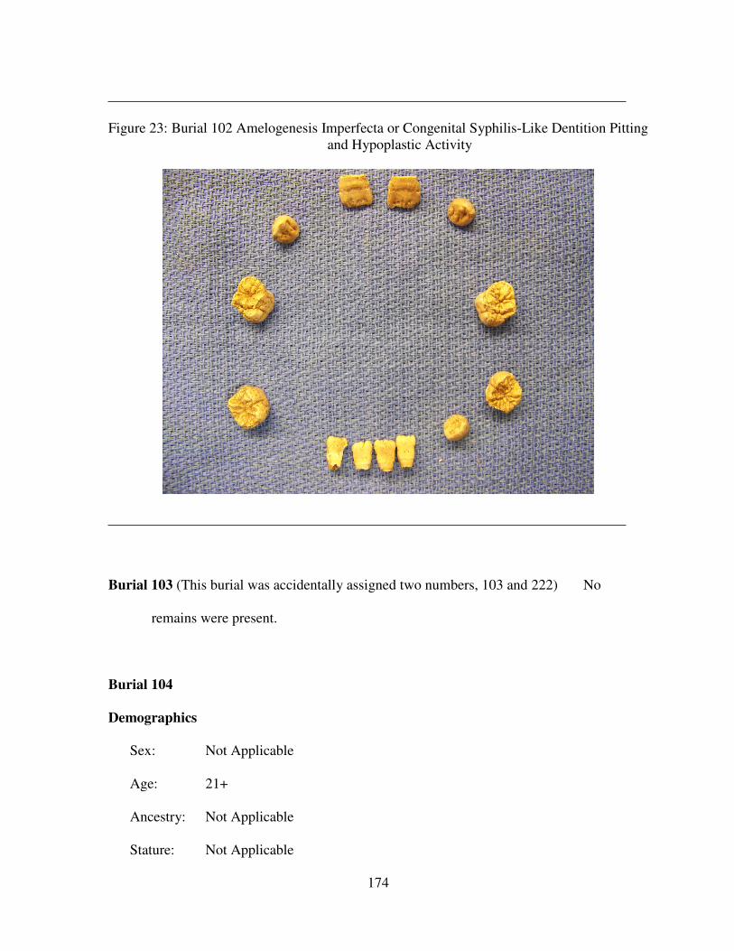
174
Figure 23: Burial 102 Amelogenesis Imperfecta or Congenital Syphilis-Like Dentition Pitting
and Hypoplastic Activity
Burial 103 (This burial was accidentally assigned two numbers, 103 and 222) No
remains were present.
Burial 104
Demographics
Sex: Not Applicable
Age: 21+
Ancestry: Not Applicable
Stature: Not Applicable
175
Preservation
Condition: Poor
Inventory: The following cranial elements were present: incomplete frontal, incomplete
left and right parietal, fragmented left and right temporal, fragmented left and right
maxilla, incomplete mandible, and 19 teeth.
The following postcranial elements were present: fragmented left clavicle,
fragmented left and right humerus, fragmented left and right radius, fragmented left
and right ulna, fragmented left and right innominate, incomplete left and right femur,
and fragmented left and right tibia.
Paleopathology
Dental: Not Applicable
Infectious Disease: Not Applicable
Trauma: Not Applicable
Other: Not Applicable
Burial 105
Demographics
Sex: Male
Age: 21+
Ancestry: Not Applicable
Stature: Not Applicable
Preservation
Condition: Poor-Fair
176
Inventory: The following cranial elements were present: fragmented frontal, incomplete
occipital, incomplete left and right parietal, incomplete left and right temporal,
incomplete mandible, and thirteen teeth.
The following postcranial elements were present: fragmented left and right
scapula, incomplete left and right humerus, fragmented left radius, incomplete right
radius, incomplete left and right ulna, incomplete left and right innominate,
incomplete left and right femur, incomplete left and right tibia, complete right
capitate, incomplete left first, second, and third metacarpal, fragmented left
calcaneus, incomplete right calcaneus, fragmented left and right talus, fragmented left
cuboid, incomplete right cuboid, fragmented left navicular, incomplete left med., int.,
and lat. cuneiform, six thoracic vertebral fragments, two lumbar vertebral thoracic,
and five sacral vertebrae.
Paleopathology
Dental: There is a 2.5mm large occlusal-buccal caries on the maxillary left first
premolar. Mandibular alveolar bone resorption is present at the area from the left
first molar to the left third molar.
Infectious Disease: Not Applicable
Trauma: Not Applicable
Other: Not Applicable
Burial 106
Demographics
Sex: Female
Age: 18
177
Ancestry: Not Applicable
Stature: Not Applicable
Preservation
Condition: Fair
Inventory: The following cranial elements were present: complete ethmoid, complete
nasals, incomplete lacrimals, complete left and right palatine, incomplete frontal,
fragmented occipital, complete left and right parietal, incomplete left and right
temporal, incomplete left and right zygomatic, complete left maxilla, incomplete right
maxilla, fragmented left and right sphenoid, incomplete mandible, and 26 teeth.
The following postcranial elements were present: fragmented left and right
scapula, fragmented left and right clavicle, fragmented left and right humerus,
fragmented left and right radius, fragmented left and right ulna, fragmented left and
right innominate, incomplete left and right femur, incomplete left and right tibia,
fragmented sternum, five ribs, the first through the seventh cervical vertebrae, and
two thoracic vertebral fragments.
Paleopathology
Dental: The mandibular left and maxillary second premolars have not erupted and
there is retention of the mandibular left and maxillary deciduous second molars.
Mandibular crowding has caused the right first and second premolars to be displaced
not mesial and distal, but rather buccal and lingual of one another.
Infectious Disease: Not Applicable
Trauma: Not Applicable
Other: Not Applicable
178
Burial 107 No remains were present.
Burial 108
Demographics
Sex: Not Applicable
Age: 3+
Ancestry: Not Applicable
Stature: Not Applicable
Preservation
Condition: Very poor
Inventory: The only cranial elements present were three adult dental caps.
Paleopathology
Dental: Not Applicable
Infectious Disease: Not Applicable
Trauma: Not Applicable
Other: Not Applicable
Burial 109 No remains were present.
Burial 110
Demographics
Sex: Not Applicable
Age: 3 years +/- 6 months
Ancestry: Not Applicable
179
Stature: Not Applicable
Preservation
Condition: Poor
Inventory: The following cranial elements were present: fragmented frontal,
fragmented occipital, fragmented left and right temporal, fragmented mandible, 18
deciduous teeth, and eleven permanent dental caps.
There were no postcranial elements present.
Paleopathology
Dental: Not Applicable
Infectious Disease: Not Applicable
Trauma: Not Applicable
Other: Not Applicable
Burial 111 No remains were present.
Burial 112 (This burial was accidentally assigned two numbers, 86 and 112)
Burial 113
Demographics
Sex: Female
Age: 21+
Ancestry: Not Applicable
Stature: Not Applicable
Preservation
Condition: Poor
180
Inventory: The following cranial elements were present: incomplete frontal, fragmented
occipital, complete left and right parietal, and 21 teeth.
Paleopathology
Dental: There is one 2.5mm large distal and occlusal caries on the mandibular right
first molar. There is one 2.5mm large occlusal caries on the mandibular left second
molar. There is one 1.5mm large mesial and occlusal caries and one 2mm large distal
and occlusal caries on the mandibular left third molar.
Infectious Disease: Not Applicable
Trauma: Not Applicable
Other: Not Applicable
Burial 114
Demographics
Sex: Female
Age: 30-40
Ancestry: African
Stature: 156.43cm (5 ft 1.6 in)
Preservation
Condition: Good
Inventory: The following cranial elements were present: complete ethmoid, incomplete
nasals, fragmented lacrimals, complete vomer, incomplete left and right inferior nasal
conchae, complete left and right palatine, complete frontal, complete occipital,
complete left and right parietal, complete left and right temporal, complete left and
right zygomatic, incomplete left maxilla, incomplete right maxilla, complete left and
181
right sphenoid, complete mandible, 30 permanent teeth present, and one deciduous
tooth.
The following postcranial elements were present: fragmented left scapula,
incomplete right scapula, incomplete left and right clavicle, incomplete left and right
humerus, incomplete left and right radius, incomplete left and right ulna, incomplete
left and right innominate, incomplete left femur, incomplete left and right tibia,
incomplete left and right fibula, 17 ribs, incomplete second, third, and fourth left
metacarpal, ten hand phalanges, incomplete left and right calcaneus, four foot
phalanges, four metatarsal fragments, seven cervical vertebrae, twelve thoracic
vertebrae, the first through fourth lumbar vertebrae, and the first and second sacral
vertebrae.
Paleopathology
Dental: The maxillary left second premolar is impacted and still in the crypt. The
deciduous maxillary left second molar is still present.
Infectious Disease: Not Applicable
Trauma: Not Applicable
Other: Not Applicable
Burial 115 No remains were present.
Burial 116 No remains were present.
Burial 117 No remains were present.
Burial 118 No remains were present.
Burial 119 No remains were present.
Burial 120 No remains were present.
182
Burial 121 No remains were present.
Burial 122 No remains were present.
Burial 123
Demographics
Sex: Female
Age: 21+
Ancestry: Not Applicable
Stature: Not Applicable
Preservation
Condition: Poor
Inventory: The following cranial elements were present: incomplete frontal, incomplete
occipital, incomplete left and right parietal, incomplete left and right temporal,
fragmented left and right zygomatic, fragmented left and right maxilla, incomplete
mandible, and 28 teeth.
The following postcranial elements were present: fragmented left and right
scapula, incomplete left clavicle, incomplete left humerus, fragmented right humerus,
incomplete left radius, incomplete left and right ulna, incomplete left and right
innominate, incomplete left and right femur, incomplete left tibia, fragmented right
tibia, fragmented left fibula, thirteen ribs, incomplete right third metacarpal, four foot
phalanges, four carpal fragments, the second cervical vertebra, the eleventh and
twelfth thoracic vertebrae, eight thoracic vertebral fragments, the first, second, and
third lumbar vertebrae, and the first sacral vertebra.
Paleopathology
183
Dental: The maxillary right lateral incisor has a gold crown. There is a distal IPCF
gold filling on the maxillary right central incisor and a mesial gold filling on the
maxillary left central incisor. There are 3mm large lead occlusal fillings on the left
and mandibular right second molars.
Infectious Disease: Not Applicable
Trauma: Not Applicable
Other: Not Applicable
Burial 124
Demographics
Sex: Female
Age: 21+
Ancestry: Not Applicable
Stature: Not Applicable
Preservation
Condition: Poor
Inventory: The following cranial elements were present: incomplete frontal, incomplete
occipital, fragmented left and right parietal, incomplete left and right temporal,
fragmented left and right maxilla, fragmented mandible, and ten teeth.
The following postcranial elements were present: incomplete left and right
humerus, fragmented left and right radius, fragmented left and right innominate,
incomplete left femur, fragmented right femur, incomplete right tibia, and two sacral
fragments.
Paleopathology
184
Dental: There is mandibular alveolar bone resorption at the area of the right and left
second molars. There is a 2.5mm large IPCF caries on the mandibular right third
molar. There is also a 2.5mm large distal enamel root caries on the mandibular left
first premolar.
Infectious Disease: Not Applicable
Trauma: Not Applicable
Other: It was noted that the left femoral angle may be larger than average, however,
the remains are in poor Condition: and only a fourth of the head is intact with the
majority of the proximal portion highly deteriorated.
Burial 125
Demographics
Sex: Not Applicable
Age: 12+
Ancestry: Not Applicable
Stature: Not Applicable
Preservation
Condition: Very poor
Inventory: One permanent tooth was the only cranial elements present. A right femoral
fragment was the only postcranial element present.
Paleopathology
Dental: Not Applicable
Infectious Disease: Not Applicable
Trauma: Not Applicable
185
Other: Not Applicable
Burial 126
Demographics
Sex: Not Applicable
Age: Not Applicable
Ancestry: Not Applicable
Stature: Not Applicable
Preservation
Condition: Very poor
Inventory: No cranial elements were present. The following postcranial elements were
present: fragmented left and right femur, and fragmented left and right tibia.
Paleopathology
Dental: Not Applicable
Infectious Disease: Not Applicable
Trauma: Not Applicable
Other: Not Applicable
Burial 127
Demographics
Sex: Male
Age: 50-59
186
Ancestry: This individual may be European in ancestry. However, warping and the
incompleteness of the cranium have most likely skewed and distorted the
measurements thereby resulting in an erroneous European designation.
Stature: 163.45cm (5 ft 3.5 in)
Preservation
Condition: Good
Inventory: The following cranial elements were present: complete frontal, complete
occipital, complete left and right parietal, complete left and right temporal,
incomplete left zygomatic, complete right zygomatic, incomplete left maxilla,
complete right maxilla, complete left and right sphenoid, complete mandible,
incomplete hyoid, and 31 teeth.
The following postcranial elements were present: incomplete left and right
scapula, complete right clavicle, incomplete left and right humerus, complete left and
right radius, complete left and right ulna, incomplete left and right innominate,
incomplete left femur, complete right femur, incomplete left tibia, complete right
tibia, incomplete left fibula, complete right fibula, complete left scaphoid, incomplete
left triquetral, incomplete left hamate, incomplete left first, second, and third
metacarpal, complete right second, third, and fourth metacarpal, incomplete right fifth
metacarpal, four foot phalanges, 22 ribs, incomplete left and right calcaneus,
incomplete left and right talus, complete left and right cuboid, incomplete left and
right navicular, incomplete left med. cuneiform, incomplete left and right first
through fifth metatarsals, the first through seventh cervical vertebrae, first through
twelfth thoracic vertebrae, first through fifth lumbar vertebrae, and the first through
fourth sacral vertebrae.
187
Paleopathology
Dental: Periodontitis is present in the area of the maxillary left first molar to the
third molar. There is a left lateral root abscess present on the maxillary right first and
second molar.
Infectious Disease:
Trauma:
Other: Arthritic lipping is present on the left and right lateral anterior iliac spines.
The left and right auricular surfaces of the pelvis have arthritic lipping and exhibit
some porosity. Minor arthritic lipping is present on the dorsal edge of the glenoid
fossa of the right scapula. Caudal shifting of the sacrum is present with sacralization
of the fifth lumbar vertebra. A septal aperture is present on the left humerus.
Burial 128
Demographics
Sex: Not Applicable
Age: less than 2 weeks
Ancestry: Not Applicable
Stature: Not Applicable
Preservation
Condition: Poor
Inventory: The following cranial elements were present: fragmented frontal,
fragmented left and right temporal, fragmented maxilla, and fragmented mandible.
One right femoral fragmented was the only postcranial element present.
Paleopathology
188
Dental: Not Applicable
Infectious Disease: Not Applicable
Trauma: Not Applicable
Other: Not Applicable
Burial 129
Demographics
Sex: Not Applicable
Age: 6
Ancestry: Not Applicable
Stature: Not Applicable
Preservation
Condition: Poor
Inventory: The following cranial elements were present: fragmented frontal,
fragmented left and right parietal, fragmented left and right temporal, fragmented left
and right maxilla, fragmented mandible, Cranial: frontal, parietals, temporals,
maxilla, and mandible fragmented, 4 permanent teeth, and 15 deciduous teeth.
The following postcranial elements were present: fragmented left and right
scapula, fragmented left clavicle, incomplete left humerus, fragmented right humerus,
fragmented left and right femur, fragmented left and right tibia, six ribs, and two
unidentified vertebral fragments.
Paleopathology
Dental: Not Applicable
Infectious Disease: Not Applicable
189
Trauma: Not Applicable
Other: Not Applicable
Burial 130
Demographics
Sex: Not Applicable
Age: 6 months +/- 2 months
Ancestry: Not Applicable
Stature: Not Applicable
Preservation
Condition: Poor
Inventory: The following cranial elements were present: fragmented frontal,
fragmented left and right temporal, fragmented mandible, and nine deciduous teeth
present.
Paleopathology
Dental: Not Applicable
Infectious Disease: Not Applicable
Trauma: Not Applicable
Other: Not Applicable
Burial 131
Demographics
Sex: Female
Age: 50+
190
Ancestry: Not Applicable
Stature: 155.84cm (5 ft 1.35 in)
Preservation
Condition: Fair
Inventory: The following cranial elements were present: incomplete frontal, incomplete
occipital, incomplete left and right parietal, incomplete left and right temporal,
incomplete left and right zygomatic, incomplete left and right maxilla, fragmented
left and right sphenoid, incomplete mandible, incomplete hyoid, and six teeth.
The following postcranial elements were present: incomplete left and right
scapula, incomplete left clavicle, complete right clavicle, incomplete left and right
humerus, incomplete left radius, fragmented right radius, incomplete left and right
ulna, incomplete left and right innominate, fragmented left femur, incomplete right
femur, incomplete right patella, fragmented left tibia, incomplete right tibia,
fragmented left and right fibula, fragmented sternum, thirteen ribs, complete left
second, third, and fifth metacarpals, complete right first, second, and third
metacarpal, five foot phalanges, five carpal fragments, five metacarpal fragments, six
tarsal fragments, the first through seventh cervical fragments, the first through twelfth
thoracic vertebrae, and the first through fifth lumbar vertebrae.
Paleopathology
Dental: The maxillary right second molar has a gold crown and the maxillary right
central incisor is a partial. Maxillary alveolar bone resorption is present at the area
from the left first molar to the left third molar. There is one 3mm large distal IPCF
caries on the maxillary left first premolar, and one 2.5mm large mesial IPCF caries on
the maxillary left second premolar. Also there is a there is one 4mm large mesial root
191
caries on the maxillary left central incisor. There is complete alveolar bone
resorption in the mandible.
Infectious Disease: Not Applicable
Trauma: Not Applicable
Other: There is arthritic lipping on the anterior portion of the third, fourth, fifth,
sixth, and seventh cervical vertebral bodies. Arthritic lipping is present on both the
left and right lateral sides of the fifth through ninth thoracic vertebral bodies. The
right patella has arthritic lipping on both the anterior and posterior surfaces.
Burial 132
Demographics
Sex: Male
Age: 30-39
Ancestry: Not Applicable
Stature: 163.18cm (5 ft 2.4 in)
Preservation
Condition: Fair
Inventory: The following cranial elements were present: incomplete frontal, incomplete
occipital, incomplete left and right parietal, incomplete left and right temporal,
fragmented left zygomatic, incomplete right zygomatic, left and right maxilla
incomplete, left sphenoid incomplete, mandible incomplete, incomplete hyoid, and 21
teeth.
The following postcranial elements were present: incomplete left and right
scapula, incomplete right clavicle, fragmented left clavicle, incomplete right humerus,
192
fragmented left humerus, incomplete left radius, fragmented right radius, incomplete
right ulna, incomplete left and right innominate, fragmented right femur, incomplete
right patella, fragmented left tibia, incomplete right tibia, fragmented left fibula,
incomplete left fibula, fragmented sternum, incomplete left and right calcaneus,
incomplete left and right talus, fragmented left navicular, incomplete right navicular,
fragmented left med cuneiform, incomplete right med cuneiform, 24 ribs, first
through the seventh cervical vertebrae, seven thoracic vertebral fragments, and the
first through the third sacral vertebrae.
Paleopathology
Dental: There is maxillary alveolar resorption from the right first molar to the right
third molar and the left second premolar to the left second molar. The maxillary right
canine and the mandibular left third molar were lost antemortem. There is one 5mm
large mesial root caries on the mandibular right third molar.
Infectious Disease: Not Applicable
Trauma: Not Applicable
Other: Not Applicable
Burial 133
Demographics
Sex: Not Applicable
Age: 18 months – 2 years
Ancestry: Not Applicable
Stature: Not Applicable
Preservation
193
Condition: Poor
Inventory: The following cranial elements were present: incomplete frontal, incomplete
occipital, incomplete left and right parietal, incomplete left and right temporal,
fragmented left and right maxilla, incomplete left and right sphenoid, incomplete
mandible, and nine deciduous teeth.
The following postcranial elements were present: incomplete right scapula,
fragmented left clavicle, incomplete right clavicle, incomplete right humerus,
fragmented right innominate, incomplete right femur, ten ribs, the second through the
fifth cervical vertebrae, two cervical vertebrae fragments, and seven thoracic
vertebrae fragments.
Paleopathology
Dental: Not Applicable
Infectious Disease: Not Applicable
Trauma: Not Applicable
Other: Not Applicable
Burial 134
Demographics
Sex: Not Applicable
Age: 1 year – 18 months
Ancestry: Not Applicable
Stature: Not Applicable
Preservation
Condition: Poor-Fair
194
Inventory: The following cranial elements were present: incomplete frontal, incomplete
occipital, incomplete left and right parietal, incomplete left and right temporal,
complete left and right zygomatic, incomplete left and right maxilla, fragmented left
sphenoid, incomplete right sphenoid, incomplete mandible, 16 deciduous teeth
erupted, four deciduous teeth in crypt, and the four first permanent molars in crypt.
The following postcranial elements were present: incomplete left scapula,
incomplete left and right clavicle, fragmented left innominate, fragmented left femur,
incomplete right femur, incomplete right tibia, fragmented right fibula, six ribs, and
seven fragmented vertebral fragments.
Paleopathology
Dental: Not Applicable
Infectious Disease: Not Applicable
Trauma: Not Applicable
Other: Not Applicable
Burial 135
Demographics
Sex: Not Applicable
Age: 9 months – 1 year
Ancestry: Not Applicable
Stature: Not Applicable
Preservation
Condition: Very poor
195
Inventory: The following cranial elements were present: fragmented left temporal,
fragmented mandible, and two permanent dental caps. No postcranial elements were
present.
Paleopathology
Dental: Not Applicable
Infectious Disease: Not Applicable
Trauma: Not Applicable
Other: Not Applicable
Burial 136 (Figure 24)
Demographics
Sex: Male
Age: 35-40
Ancestry: Not Applicable
Stature: Not Applicable
Preservation
Condition: Fair
Inventory: The following cranial elements were present: incomplete ethmoid,
incomplete right lacrimal, complete frontal, incomplete occipital, incomplete left and
right parietal, incomplete left and right temporal, incomplete left and right zygomatic,
incomplete left maxilla, fragmented right maxilla, fragmented left and right sphenoid,
incomplete mandible, incomplete hyoid, and 21 teeth.
The following cranial elements were present: fragmented left scapula,
incomplete right clavicle, incomplete left humerus, fragmented left radius,
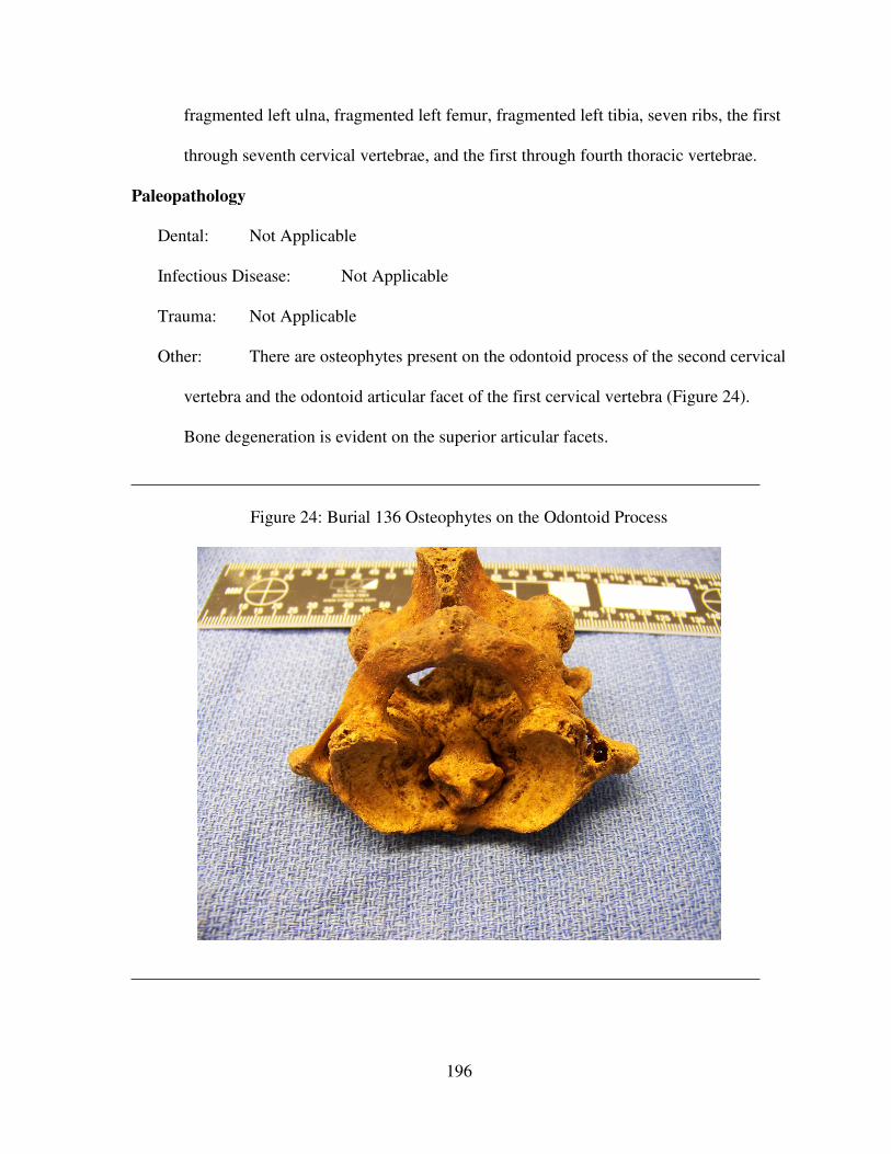
196
fragmented left ulna, fragmented left femur, fragmented left tibia, seven ribs, the first
through seventh cervical vertebrae, and the first through fourth thoracic vertebrae.
Paleopathology
Dental: Not Applicable
Infectious Disease: Not Applicable
Trauma: Not Applicable
Other: There are osteophytes present on the odontoid process of the second cervical
vertebra and the odontoid articular facet of the first cervical vertebra (Figure 24).
Bone degeneration is evident on the superior articular facets.
Figure 24: Burial 136 Osteophytes on the Odontoid Process
197
Burial 137 (Figure 25)
Demographics
Sex: Male
Age: 35-45
Ancestry: Not Applicable
Stature: 165.94cm (5 ft 5.3 in)
Preservation
Condition: Fair
Inventory: The following cranial elements were present: fragmented frontal, incomplete
occipital, fragmented left and right parietal, incomplete left and right temporal,
incomplete right zygomatic, fragmented left and right maxilla, incomplete right
sphenoid, incomplete mandible, and three teeth.
The following postcranial elements were present: fragmented left scapula,
incomplete left clavicle, incomplete left humerus, fragmented right humerus,
incomplete left and right radius, incomplete left and right ulna, incomplete left and
right innominate, incomplete left and right femur, complete right patella, incomplete
left tibia, complete right tibia, incomplete left and right fibula, eight ribs, complete
right scaphoid, fragmented left trapezium, incomplete right trapezium, complete left
and right capitate, complete right hamate, complete left first metacarpal, incomplete
left third through fifth metacarpal, complete right second metacarpal, incomplete right
third through fifth metacarpal, 14 hand phalanges, fragmented left and right
calcaneus, incomplete left talus, complete right talus, incomplete left and right
cuboid, incomplete right navicular, complete left int cuneiform, incomplete left lat
cuneiform, complete right lat cuneiform, incomplete left first through firth
198
metatarsals, complete right first through fifth metatarsals, four foot phalanges, first
through the seventh cervical vertebrae, the first thoracic vertebra, and the second
through the fifth sacral vertebrae.
Paleopathology
Dental: Lingual calculus is present at the CEJ and a 2mm large distal caries on the
maxillary right third molar. There is mandibular alveolar bone resorption at the area
between the left first molar to the left third molar.
Infectious Disease: Not Applicable
Trauma: Not Applicable
Other: Minor to moderate arthritic lipping of the inferior-anterior-superior vertebral
bodies is present on the second through sixth cervical vertebrae (Figure 25).
Additionally, the fifth cervical vertebra shows signs of compression.
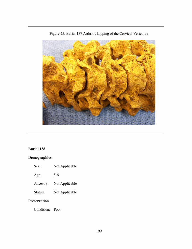
199
Figure 25: Burial 137 Arthritic Lipping of the Cervical Vertebrae
Burial 138
Demographics
Sex: Not Applicable
Age: 5-6
Ancestry: Not Applicable
Stature: Not Applicable
Preservation
Condition: Poor
200
Inventory: The following cranial elements were present: incomplete frontal, incomplete
occipital, incomplete left and right parietal, incomplete left and right temporal,
incomplete left and right maxilla, fragmented left and right sphenoid, incomplete
mandible, 20 deciduous teeth, the first four permanent molars in crypt, and the first
two permanent central incisors erupting.
The following postcranial elements were present: incomplete left and right
clavicle, fragmented left and right radius, fragmented left and right ulna, fragmented
left innominate, incomplete left and right femur, incomplete left tibia, fragmented
right tibia, fragmented left fibula, 18 ribs, and ten vertebral fragments.
Paleopathology
Dental: Not Applicable
Infectious Disease: Not Applicable
Trauma: Not Applicable
Other: Not Applicable
Burial 139
Demographics
Sex: Male
Age: 18
Ancestry: Not Applicable
Stature: 174.8cm (5 ft 8.8 in)
Preservation
Condition: Good
201
Inventory: The following cranial elements were present: complete frontal, fragmented
occipital, incomplete left and right parietal, incomplete left and right temporal,
complete left and right zygomatic, complete left and right maxilla, incomplete left
and right sphenoid, complete mandible, and 28 teeth.
The following postcranial elements were present: incomplete left and right
scapula, incomplete left and right clavicle, incomplete left and right humerus,
complete left radius, incomplete right radius, incomplete left and right ulna,
incomplete left and right innominate, incomplete left and right femur, incomplete left
and right patella, incomplete left and right tibia, incomplete left and right fibula,
fragmented sternum, 24 ribs, complete left and right scaphoid, complete left lunate,
complete left and right triquetral, complete left and right pisiform, complete left
trapezium, complete left trapezoid, complete left capitate, complete left hamate,
incomplete right hamate, incomplete left first through fifth metacarpal, incomplete
right first metacarpal, complete right second through third metacarpal, 14 hand
phalanges, and all cervical, thoracic, lumbar, and sacral vertebrae.
Paleopathology
Dental: There is a 4mm large labial root abscess on the mandibular left lateral
incisor. Linear enamel hypoplasias occur on three mandibular teeth, lateral left
incisor and the left and right canines. The distance of the left canine LEH from the
CEJ is 4.2mm. The distance of the right canine LEH from the CEJ is 4.0mm.
Infectious Disease: Not Applicable
Trauma: Not Applicable
Other: Not Applicable
202
Burial 140
Demographics
Sex: Female
Age: 20-21
Ancestry: Not Applicable
Stature: Not Applicable
Preservation
Condition: Fair
Inventory: The following cranial elements were present: fragmented frontal, incomplete
occipital, fragmented left and right parietal, incomplete left and right temporal,
fragmented left zygomatic, incomplete right zygomatic, fragmented left maxilla,
incomplete right maxilla, fragmented left and right sphenoid, incomplete mandible,
and 28 teeth.
The following postcranial elements were present: fragmented left and right
scapula, fragmented left clavicle, incomplete right clavicle, fragmented left humerus,
incomplete right humerus, fragmented left and right radius, fragmented left ulna,
incomplete right ulna, incomplete left and right innominate, incomplete left and right
femur, incomplete left and right tibia, incomplete right fibula, eleven hand phalanges,
23 ribs, fragmented right calcaneus, incomplete left and right talus, incomplete left
cuboid, fragmented left med cuneiform, incomplete left int cuneiform, incomplete left
first, third, fourth, and fifth metatarsal, incomplete first through fifth right metatarsal,
four foot phalanges, first through seventh cervical vertebrae, the first through sixth
thoracic vertebrae three lumbar vertebral fragments, and five sacral fragments.
Paleopathology
203
Dental: Pitting hypoplasias occur 4.41mm from the CEJ on the maxillary right third
molar and 4.8mm from the CEJ on the maxillary right second molar. There is a
1.1cm wide buccal root abscess on the maxillary left first molar. Pitting hypoplasia is
present 5.18mm from the CEJ on the maxillary left second molar. There is a 1.5mm
large occlusal caries on the maxillary left third molar with pitting hypoplasia
occurring 4.79mm from the CEJ.
Infectious Disease: Not Applicable
Trauma: Not Applicable
Other: Not Applicable
Burial 141
Demographics
Sex: Not Applicable
Age: less than 4 weeks
Ancestry: Not Applicable
Stature: Not Applicable
Preservation
Condition: Poor
Inventory: There were no cranial elements present. The postcranial elements present
included five epiphyses and three long fragments.
Paleopathology
Dental: Not Applicable
Infectious Disease: Not Applicable
Trauma: Not Applicable
204
Other: Not Applicable
Burial 142
Demographics
Sex: Not Applicable
Age: 9 months +/- 2 months
Ancestry: Not Applicable
Stature: Not Applicable
Preservation
Condition: Poor
Inventory: The following cranial elements were present: fragmented frontal,
fragmented occipital, fragmented left and right temporal, fragmented occipital, 19
deciduous teeth in crypt, and 9 permanent dental caps.
The following postcranial elements were present: incomplete left and right
humerus, incomplete left and right femur, incomplete left and right tibia, 17 ribs, and
ten vertebral fragments.
Paleopathology
Dental: Not Applicable
Infectious Disease: Not Applicable
Trauma: Not Applicable
Other: Not Applicable
Burial 143
Demographics
205
Sex: Female
Age: 50-60
Ancestry: Good
Stature: 156.4cm (5 ft 1.6 in)
Preservation
Condition: Good
Inventory: The following cranial elements were present: incomplete frontal, complete
occipital, complete left and right parietal, complete left and right temporal,
incomplete left and right zygomatic, incomplete left and right maxilla, fragmented
left and right sphenoid, complete mandible, incomplete hyoid, and 18 teeth.
The following postcranial elements were present: fragmented left scapula,
incomplete right scapula, incomplete left and right clavicle, compete left humerus,
incomplete right humerus, complete left and right radius, complete left and right ulna,
incomplete left and right innominate, incomplete left and right femur, 17 ribs,
complete right scaphoid, complete right lunate, complete right trapezium, complete
right trapezoid, complete right capitate, complete left and right hamate, complete left
and right first through fifth metacarpals, eleven hand phalanges, complete right first
metatarsal, incomplete second through fifth metatarsal, all cervical, thoracic, and
lumbar vertebrae, and the first through fourth sacral vertebrae.
Paleopathology
Dental: There is mesial-lingual calculus at the CEJ on the maxillary right first molar,
second molar, and third molar. Calculus can also be found at the mesial-lingual
aspect of the CEJ on the maxillary left first and second molars. Alveolar resorption is
present at the areas of the maxillary right first premolar, from the maxillary right
206
central incisor to the maxillary left first premolar, the maxillary left third molar, the
mandibular right first and second molars, the mandibular right canine and first
premolar, the mandibular left first premolar, mandibular left first molar, and the
mandibular left third molar.
There is a 3mm large distal IPCF caries on the maxillary right canine. There
is a 2.5mm large distal IPCF caries on the maxillary left lateral incisor. There is a
2mm large proximal IPCF caries on the maxillary left second premolar. There is a
1.5mm large proximal IPCF mandibular left second molar. There is a 1.5mm large
distal IPCF caries and a 3mm large mesial IPCF on the mandibular left second
premolar. A 3mm large distal IPCF caries and a 2mm large proximal IPCF caries
was formed on the mandibular left canine.
Infectious Disease: Not Applicable
Trauma: Not Applicable
Other: Not Applicable
Burial 144 (Figure 26)
Demographics
Sex: Female
Age: 42+
Ancestry: Not Applicable
Stature: Not Applicable
Preservation
Condition: Poor
207
Inventory: The following cranial elements were present: incomplete frontal, incomplete
occipital, incomplete left and right parietal, incomplete left and right temporal,
incomplete left zygomatic, fragmented left and right maxilla, incomplete mandible,
and thirteen teeth.
The following postcranial elements were present: fragmented right scapula,
fragmented left humerus, incomplete right humerus, fragmented left radius,
incomplete right radius, incomplete left and right ulna, incomplete left and right
innominate, fragmented left femur, incomplete right femur, fragmented left and right
tibia, two ribs, all cervical vertebrae, and the first and second thoracic vertebrae.
Paleopathology
Dental: Alveolar resorption is present at the areas of the maxillary left second
premolar to the maxillary left third molar, the mandibular right first molar to the
mandibular right third molar, and the mandibular left first molar to the mandibular
left third molar. There is a 3mm large distal root caries on the maxillary right second
premolar. There is a 2.5mm large labial caries on the mandibular left canine. There
is calculus below the CEJ on the mandibular right first and second premolar. There is
a 2.5mm large buccal root caries on the mandibular right second molar. There is a
2mm large distal caries, a 3mm large buccal root caries, and a 4mm large abscess on
the mandibular right first premolar.
Infectious Disease: Not Applicable
Trauma: Not Applicable
Other: There is slight occipital porotic hyperostosis around the foramen magnum.
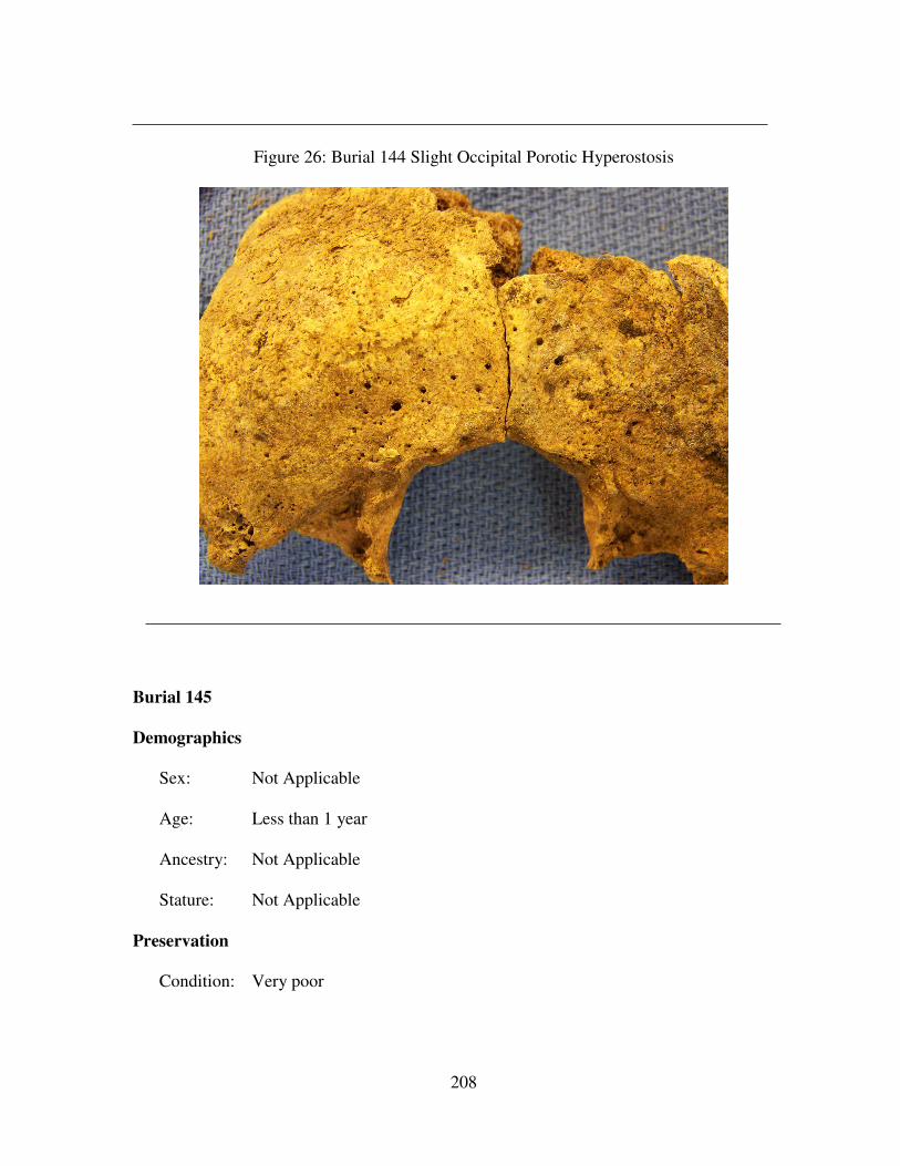
208
Figure 26: Burial 144 Slight Occipital Porotic Hyperostosis
Burial 145
Demographics
Sex: Not Applicable
Age: Less than 1 year
Ancestry: Not Applicable
Stature: Not Applicable
Preservation
Condition: Very poor
209
Inventory: Four unidentifiable fragments were the only cranial elements present. No
postcranial elements were present.
Paleopathology
Dental: Not Applicable
Infectious Disease: Not Applicable
Trauma: Not Applicable
Other: Not Applicable
Burial 146
Demographics
Sex: Female
Age: 18
Ancestry: Not Applicable
Stature: Not Applicable
Preservation
Condition: Poor
Inventory: The following cranial elements were present: incomplete frontal, incomplete
occipital, fragmented left and right parietal, incomplete left and right temporal,
incomplete left zygomatic, and 28 teeth.
The following postcranial elements were present: fragmented left scapula,
incomplete left clavicle, fragmented right clavicle, incomplete left and right humerus,
incomplete left and right radius, incomplete left and right ulna, incomplete left and
right innominate, incomplete left and right femur, incomplete left and right tibia,
fragmented right fibula, 16 ribs, six hand phalanges, four metacarpal fragments, the
210
first through the fourth cervical vertebrae, 19 thoracic vertebral fragments, and one
lumbar vertebral fragment.
Paleopathology
Dental: A deciduous tooth was present between the maxillary left canine and the
maxillary left first molar. However, the maxillary left first molar was lost premortem.
It is possible it was the maxillary left second deciduous molar.
Infectious Disease: Not Applicable
Trauma: Not Applicable
Other: Not Applicable
Burial 147
Demographics
Sex: Indeterminate
Age: 50+
Ancestry: Not Applicable
Stature: Not Applicable
Preservation
Condition: Poor
Inventory: The following cranial elements were present: incomplete frontal, fragmented
occipital, incomplete left parietal, complete right parietal, incomplete left temporal,
complete right temporal, incomplete left and right zygomatic, incomplete left and
right zygomatic, incomplete left and right maxilla, fragmented left and right sphenoid,
incomplete mandible, and 23 teeth.
211
The following postcranial elements were present: fragmented left and right
scapula, incomplete left and right humerus, incomplete right radius, fragmented right
ulna, incomplete left and right innominate, incomplete left and right femur,
incomplete left and right tibia, incomplete left and right fibula, two ribs, one hand
phalange, fragmented left and right calcaneus, fragmented left and right talus,
incomplete right cuboid, the first through seventh cervical vertebrae, three lumbar
vertebral fragments, and the first sacral vertebra.
Paleopathology
Dental: Alveolar bone resorption is present at the maxillary left first and third
molars, the mandibular left first molar, and the mandibular right first molar. The
maxillary left first molar has a gold crown and the maxillary left lateral incisor is a
porcelain partial. The two teeth are linked by the gold cap.
Infectious Disease: Not Applicable
Trauma: Not Applicable
Other: Not Applicable
Burial 148 No remains were present.
Burial 149
Demographics
Sex: Male
Age: 21+
Ancestry: Not Applicable
Stature: Not Applicable
212
Preservation
Condition: Poor
Inventory: The following cranial elements were present: incomplete frontal, incomplete
occipital, incomplete left and right parietal, incomplete left and right temporal,
fragmented right zygomatic, fragmented left and right maxilla, incomplete mandible,
and 18 teeth.
The following cranial elements were present: fragmented right and left
scapula, fragmented left and right clavicle, fragmented left and right humerus,
incomplete left radius, incomplete left ulna, fragmented left and right innominate,
incomplete left femur, fragmented right femur, fragmented left tibia, incomplete right
tibia, five ribs, fragmented right calcaneus, fragmented right talus, and the first and
second cervical vertebrae.
Paleopathology
Dental: The results of periodontal disease was noted in the maxilla from the second
premolar to the third molar. The third molar was lost antemortem and severe porosity
is present. The mandibular left first molar was lost antemortem and alveolar bone
resorption was evident.
Infectious Disease: Not Applicable
Trauma: Not Applicable
Other: Not Applicable
Burial 150 No remains were present.
Burial 151
213
Demographics
Sex: Not Applicable
Age: 21+
Ancestry: Not Applicable
Stature: Not Applicable
Preservation
Condition: Poor
Inventory: The following cranial elements were present: fragmented frontal,
fragmented occipital, fragmented left and right parietal, fragmented left temporal,
incomplete right temporal, fragmented left and right maxilla, fragmented mandible,
and 13 teeth.
The following postcranial elements were present: fragmented left and right
humerus, fragmented left and right innominate, fragmented left and right femur,
fragmented left and right tibia, and four ribs.
Paleopathology
Dental: There is a 4mm large distal caries on the maxillary right second molar.
There is a 4mm large distal caries on the maxillary right third molar. There is a 3mm
large mesial caries on the mandibular left third molar.
Infectious Disease: Not Applicable
Trauma: The mandibular right canine exhibits occupational/use trauma. The distal
half of the occlusal surface is worn mesial-distally. This tooth was probably used as a
tool.
Other: Not Applicable
214
Burial 152
Demographics
Sex: Not Applicable
Age: 3 years +/- 6 months
Ancestry: Not Applicable
Stature: Not Applicable
Preservation
Condition: Poor
Inventory: The following cranial elements were present: fragmented frontal,
fragmented temporal, 14 deciduous teeth, and nine permanent dental caps.
Paleopathology
Dental: Not Applicable
Infectious Disease: Not Applicable
Trauma: Not Applicable
Other: Not Applicable
Burial 153 No remains were present.
Burial 154 No remains were present.
Burial 155
Demographics
Sex: Not Applicable
Age: 10+
Ancestry: Not Applicable
215
Stature: Not Applicable
Preservation
Condition: Poor
Inventory: The following cranial elements were present: fragmented frontal,
fragmented occipital, incomplete left and right parietal, fragmented left and right
temporal, and two teeth.
The following postcranial elements were present: fragmented right humerus,
fragmented right radius, fragmented right ulna, fragmented left and right femur, and
fragmented left and right tibia.
Paleopathology
Dental: There is one 3mm large mesial IPCF caries on the mandibular right first
premolar. There is one 2mm large distal IPCF caries on the on the mandibular right
canine.
Infectious Disease: Not Applicable
Trauma: Not Applicable
Other: Not Applicable
Burial 156 No remains were present.
Burial 157
Demographics
Sex: Not Applicable
Age: Not Applicable
Ancestry: Not Applicable
216
Stature: Not Applicable
Preservation
Condition: Very poor
Inventory: The cranial elements present included fragmented parietal remnants. No
postcranial elements were present.
Paleopathology
Dental: Not Applicable
Infectious Disease: Not Applicable
Trauma: Not Applicable
Other: Not Applicable
Burial 158 No remains were present.
Burial 159
Demographics
Sex: Not Applicable
Age: 12+
Ancestry: Not Applicable
Stature: Not Applicable
Preservation
Condition: Very poor
Inventory: The following cranial elements were present: fragmented frontal,
fragmented occipital, and two teeth.
Paleopathology
217
Dental: Not Applicable
Infectious Disease: Not Applicable
Trauma: Not Applicable
Other: Not Applicable
Burial 160
Demographics
Sex: Not Applicable
Age: 12+
Ancestry: Not Applicable
Stature: Not Applicable
Preservation
Condition: Very poor
Inventory: The cranial elements present consisted of four unidentifiable cranial
fragments and five teeth.
The following postcranial elements were present: fragmented left and right
humerus, fragmented left radius, fragmented left ulna, fragmented left and right
innominate, fragmented left and right femur, fragmented left and right tibia, and
fragmented left and right fibula.
Paleopathology
Dental: Not Applicable
Infectious Disease: Not Applicable
Trauma: Not Applicable
Other: Not Applicable
218
Burial 161 No remains were present.
Burial 162
Demographics
Sex: Not Applicable
Age: 4-5
Ancestry: Not Applicable
Stature: Not Applicable
Preservation
Condition: Poor
Inventory: The following cranial elements were present: complete frontal, incomplete
occipital, complete left and right parietal, incomplete left and right temporal,
incomplete left and right zygomatic, incomplete left and right maxilla, complete
mandible, 14 deciduous teeth, and thirteen permanent teeth in crypt.
The following postcranial elements were present: fragmented left and right
scapula, incomplete left clavicle, incomplete left humerus, fragmented right humerus,
fragmented left and right innominate, incomplete left and right femur, incomplete left
and right tibia, fragmented right fibula, 16 ribs, and the first through seventh cervical
vertebrae.
Paleopathology
Dental: Not Applicable
Infectious Disease: Not Applicable
Trauma: Not Applicable
219
Other: Not Applicable
Burial 163
Demographics
Sex: Male
Age: 18-19
Ancestry: Not Applicable
Stature: 174.8cm (5 ft 8.8 in)
Preservation
Condition: Poor
Inventory: The following cranial elements were present: incomplete left temporal,
incomplete mandible, and 19 teeth.
The following postcranial elements were present: incomplete left and right
scapula, complete left and right clavicle, incomplete left and right humerus, complete
left radius, incomplete right radius, incomplete left and right ulna, incomplete left and
right innominate, incomplete left and right femur, incomplete left and right tibia,
incomplete left and right fibula, incomplete sternum, 23 ribs, complete left third
metacarpal, incomplete left fourth and fifth metacarpal, incomplete right second
through fifth metacarpal, incomplete right calcaneus, incomplete left and right talus,
incomplete right cuboid, incomplete right navicular, incomplete right first through
fourth metatarsal, seven foot phalanges, two unidentified tarsals, all cervical, thoracic,
and lumbar vertebrae, and the first through fourth sacral verterbrae.
Paleopathology
220
Dental: A protostylid (paramolar tubercle) was present on the mandibular left third
molar.
Infectious Disease: Not Applicable
Trauma: Not Applicable
Other: Not Applicable
Burial 164
Demographics
Sex: Male
Age: 20-29
Ancestry: Not Applicable
Stature: 176.27cm (5 ft 9.4 in)
Preservation
Condition: Fair
Inventory: The following cranial elements were present: incomplete frontal, incomplete
occipital, incomplete left and right parietal, incomplete left and right temporal,
incomplete left zygomatic, incomplete left and right maxilla, fragmented left and
right sphenoid, incomplete mandible, and 28 teeth.
The following postcranial elements were present: incomplete left scapula,
fragmented right scapula, incomplete left and right clavicle, incomplete left and right
humerus, incomplete left and right radius, incomplete left and right ulna, incomplete
right femur, incomplete left and right tibia, incomplete left fibula, fragmented right
fibula, 17 ribs, incomplete left second metacarpal, four hand phalanges, four
unidentified carpals, fragmented left calcaneus, incomplete right calcaneus,
221
fragmented left talus, incomplete right talus, incomplete left first metatarsal, all
cervical, thoracic, and lumbar vertebrae, and the first four sacral vertebrae.
Paleopathology
Dental: The maxillary left second premolar was lost antemortem. Only the roots
remain of the mandibular right second and third molars remain. The mandibular left
third molar was lost antemortem and a probable abscess remains at time of death.
Infectious Disease: Not Applicable
Trauma: Not Applicable
Other: Not Applicable
Burial 165 No remains were present.
Burial 166 No remains were present.
Burial 167 No remains were present.
Burial 168 No remains were present.
Burial 169 No remains were present.
Burial 170 No remains were present.
Burial 171 No remains were present.
Burial 172
Demographics
Sex: Not Applicable
Age: 6+
Ancestry: Not Applicable
Stature: Not Applicable
222
Preservation
Condition: Very poor
Inventory: The only cranial element present is the permanent mandibular left first
molar. No postcranial elements were present.
Paleopathology
Dental: Not Applicable
Infectious Disease: Not Applicable
Trauma: Not Applicable
Other: Not Applicable
Burial 173 No remains were present.
Burial 174
Demographics
Sex: Not Applicable
Age: 18 months – 2 years
Ancestry: Not Applicable
Stature: Not Applicable
Preservation
Condition: Very poor
Inventory: The cranial elements present include 14 deciduous teeth, and eleven
permanent dental caps.
Paleopathology
Dental: Not Applicable
223
Infectious Disease: Not Applicable
Trauma: Not Applicable
Other: Not Applicable
Burial 175 No remains were present.
Burial 176 No remains were present.
Burial 177 No remains were present.
Burial 178
Demographics
Sex: Not Applicable
Age: 21+
Ancestry: Not Applicable
Stature: Not Applicable
Preservation
Condition: Poor
Inventory: The following cranial elements were present: fragmented left parietal,
fragmented left temporal, and one tooth. The only postcranial element present was
the fragmented left tibia.
Paleopathology
Dental: Not Applicable
Infectious Disease: Not Applicable
Trauma: Not Applicable
Other: Not Applicable
224
Burial 179 No remains were present.
Burial 180 This burial consisted only of bone meal and no discernable information
could be gathered.
Burial 181
Demographics
Sex: Not Applicable
Age: 3 years +/- 6 months
Ancestry: Not Applicable
Stature: Not Applicable
Preservation
Condition: Poor
Inventory: The following postcranial elements were present: fragmented right temporal,
17 deciduous teeth present, and twelve permanent dental caps.
The following postcranial elements were present: fragmented left radius,
fragmented left and right innominate, and fragmented right femur.
Paleopathology
Dental: Not Applicable
Infectious Disease: Not Applicable
Trauma: Not Applicable
Other: Not Applicable
Burial 182
225
Demographics
Sex: Male
Age: 33-42
Ancestry: Not Applicable
Stature: Not Applicable
Preservation
Condition: Poor-Fair
Inventory: The following cranial elements were present: incomplete frontal, fragmented
occipital, complete left and right parietal, complete left temporal, incomplete right
temporal, incomplete left and right zygomatic, complete left maxilla, incomplete right
maxilla, fragmented left and right sphenoid, incomplete mandible, and 31 teeth.
The following postcranial elements were present: incomplete left and right
scapula, incomplete left and right clavicle, incomplete left and right humerus,
incomplete left and right radius, incomplete left and right ulna, incomplete left and
right innominate, incomplete left and right femur, incomplete left patella, incomplete
left and right tibia, fragmented right fibula, 17 ribs, four hand phalanges, fragmented
left calcaneus, fragmented left talus, one foot phalanges, all cervical and thoracic
vertebrae, and the first through the fourth sacral vertebrae.
Paleopathology
Dental: The maxillary right first molar was lost antemortem.
Infectious Disease: Not Applicable
Trauma: Not Applicable
Other: Not Applicable
226
Burial 183 No remains were present.
Burial 184
Demographics
Sex: Male
Age: 21+
Ancestry: Not Applicable
Stature: Not Applicable
Preservation
Condition: Poor
Inventory: The following cranial elements were present: incomplete occipital,
incomplete left and right parietal, fragmented mandible, and four teeth.
The following postcranial elements were present: fragmented left and right
scapula, incomplete left humerus, fragmented right humerus, fragmented right radius,
incomplete left and right ulna, fragmented left and right innominate, incomplete left
and right femur, incomplete left and right tibia, and fragmented left and right fibula.
Paleopathology
Dental: Alveolar bone resorption is present at the area of the mandibular right third
molar.
Infectious Disease: Not Applicable
Trauma: Not Applicable
Other: Not Applicable
227
Burial 185
Demographics
Sex: Not Applicable
Age: 11+
Ancestry: Not Applicable
Stature: Not Applicable
Preservation
Condition: Very poor
Inventory: The cranial elements present were four teeth. Postcranial elements present
were fragmented left and right femurs. Bone meal was present in the areas of the
cranium and all postcranial areas.
Paleopathology
Dental: Not Applicable
Infectious Disease: Not Applicable
Trauma: Not Applicable
Other: Not Applicable
Burial 186
Demographics
Sex: Not Applicable
Age: 21+
Ancestry: Not Applicable
Stature: Not Applicable
Preservation
228
Condition: Very poor
Inventory: The cranial elements present included 19 teeth. The following postcranial
elements were present: fragmented right humerus, fragmented left femur, and general
concentration of bone meal in the remaining postcranial areas.
Paleopathology
Dental: Not Applicable
Infectious Disease: Not Applicable
Trauma: Not Applicable
Other: Not Applicable
Burial 187
Demographics
Sex: Not Applicable
Age: 9-10
Ancestry: Not Applicable
Stature: Not Applicable
Preservation
Condition: Very poor
Inventory: The following cranial elements were present: fragmented left and right
temporal, four deciduous teeth, and 24 permanent teeth.
The following postcranial elements were present: fragmented left and right
femur, fragmented right tibia, and fragmented left fibula.
Paleopathology
Dental: Not Applicable
229
Infectious Disease: Not Applicable
Trauma: Not Applicable
Other: Not Applicable
Burial 188
Demographics
Sex: Not Applicable
Age: 15+
Ancestry: Not Applicable
Stature: Not Applicable
Preservation
Condition: Very poor
Inventory: The following cranial elements were present: fragmented frontal,
fragmented left and right parietal, fragmented left temporal, and twelve teeth.
The following postcranial elements were present: fragmented right humerus,
fragmented left femur, and fragmented left tibia.
Paleopathology
Dental: Not Applicable
Infectious Disease: Not Applicable
Trauma: Not Applicable
Other: Not Applicable
Burial 189
Demographics
230
Sex: Not Applicable
Age: 21+
Ancestry: Not Applicable
Stature: Not Applicable
Preservation
Condition: Very poor
Inventory: The following cranial elements were present: incomplete right parietal,
fragmented right temporal, fragmented left mandible, and ten teeth.
The following postcranial elements were present: fragmented left and right
femur, and fragmented left and right tibia.
Paleopathology
Dental: Not Applicable
Infectious Disease: Not Applicable
Trauma: Not Applicable
Other: Not Applicable
Burial 190
Demographics
Sex: Not Applicable
Age: Not Applicable
Ancestry: Not Applicable
Stature: Not Applicable
Preservation
Condition: Very poor
231
Inventory: The cranial elements present consisted of unidentifiable cranial fragments.
Paleopathology
Dental: Not Applicable
Infectious Disease: Not Applicable
Trauma: Not Applicable
Other: Not Applicable
Burial 191 No remains were present.
Burial 192
Demographics
Sex: Not Applicable
Age: 15+
Ancestry: Not Applicable
Stature: Not Applicable
Preservation
Condition: Very poor
Inventory: The following cranial elements were present: fragmented right temporal,
one tooth, and twelve unidentifiable cranial fragments.
The following postcranial elements were present: fragmented left femur, and
fragmented left and right tibia.
Paleopathology
Dental: Not Applicable
Infectious Disease: Not Applicable
232
Trauma: Not Applicable
Other: Not Applicable
Burial 193
Demographics
Sex: Not Applicable
Age: 21+
Ancestry: Not Applicable
Stature: Not Applicable
Preservation
Condition: Very poor
Inventory: Fourteen teeth were all the remains present.
Paleopathology
Dental: Not Applicable
Infectious Disease: Not Applicable
Trauma: Not Applicable
Other: Not Applicable
Burial 194
Demographics
Sex: Male
Age: 38+
Ancestry: Not Applicable
Stature: Not Applicable
233
Preservation
Condition: Poor
Inventory: The following cranial elements were present: complete frontal, incomplete
occipital, complete left parietal, incomplete right parietal, complete left temporal,
incomplete right temporal, incomplete left and right zygomatic, incomplete left and
right maxilla, complete left and right sphenoid, complete mandible, and 29 teeth.
The following postcranial elements were present: fragmented left scapula,
incomplete right scapula, incomplete right clavicle, incomplete left and right
humerus, incomplete left and right radius, fragmented left ulna, incomplete right ulna,
incomplete left and right innominate, incomplete left and right femur, incomplete left
patella, incomplete left and right tibia, fragmented left fibula, incomplete right fibula,
ten ribs, incomplete right trapezium, incomplete right capitate, incomplete right
hamate, incomplete left second metacarpal, incomplete right second through fifth
metacarpal, ten hand phalanges, one unidentified metacarpal, incomplete left and
right calcaneus, incomplete left and right talus, fragmented right cuboid, incomplete
left and right navicular, incomplete left first through third metatarsal, fragmented left
fourth metatarsal, fragmented right first metatarsal, fragmented right third and fourth
metatarsal, three tarsal fragments, the first through the fourth cervical vertebrae, the
third through the twelfth thoracic vertebrae, the first through the fifth lumbar
vertebrae, and the first through the third sacral vertebrae.
Paleopathology
Dental: The maxillary left and right third molars were lost antemortem with alveolar
bone resorption occurring. Lingual calculus is present from the maxillary right first
premolar to the maxillary right third molar, the maxillary left first premolar to the
234
maxillary left third molar, and the mandibular left first molar to the mandibular left
third molar. A small amount of lingual and buccal calculus is present on the
maxillary right third molar.
Infectious Disease: Not Applicable
Trauma: Not Applicable
Other: Not Applicable
Burial 195
Demographics
Sex: Female
Age: 50-59
Ancestry: Not Applicable
Stature: Not Applicable
Preservation
Condition: Poor
Inventory: The following cranial elements were present: incomplete frontal, incomplete
occipital, incomplete left and right parietal, incomplete left and right temporal,
fragmented left maxilla, incomplete mandible, and twelve teeth.
The following postcranial elements were present: fragmented left scapula,
incomplete right scapula, incomplete right clavicle, incomplete left and right
humerus, fragmented left radius, incomplete right radius, incomplete left and right
ulna, incomplete left and right innominate, incomplete right femur, incomplete left
and right tibia, fragmented left and right fibula, fragmented sternum, eight ribs,
complete right pisiform, incomplete right trapezium, complete right trapezoid,
235
incomplete left capitate, complete left hamate, incomplete left and right third
metacarpal, six hand phalanges, two carpal fragments, the first and second cervical
vertebrae, seven cervical vertebral fragments, 16 thoracic vertebral fragments, and
one lumbar vertebral fragment.
Paleopathology
Dental: There is a 1.5mm large buccal caries present on the maxillary left second
molar. There are four measurable LEH on the mandibular right canine and their
measurements from the CEJ are the following: 1.36mm, 2.92mm, 5.4mm, and
7.3mm.
Infectious Disease: Not Applicable
Trauma: Not Applicable
Other: Not Applicable
Burial 196
Demographics
Sex: Not Applicable
Age: Not Applicable
Ancestry: Not Applicable
Stature: Not Applicable
Preservation
Condition: Very poor
Inventory: There were no cranial elements present. The only postcranial element
present was a right ulna fragment.
Paleopathology
236
Dental: Not Applicable
Infectious Disease: Not Applicable
Trauma: Not Applicable
Other: Not Applicable
Burial 197
Demographics
Sex: Not Applicable
Age: 5-6
Ancestry: Not Applicable
Stature: Not Applicable
Preservation
Condition: Fair
Inventory: The following cranial elements were present: incomplete frontal, complete
occipital, incomplete left and right parietal, complete left and right temporal,
complete left and right zygomatic, complete left and right maxilla, complete left and
right sphenoid, complete mandible, and eleven teeth.
The following postcranial elements were present: incomplete left scapula,
fragmented right scapula, complete left and right clavicle, incomplete left humerus,
fragmented right humerus, complete left radius, incomplete right radius, complete left
ulna, fragmented right ulna, incomplete left and right innominate, complete left and
right femur, incomplete left and tibia, fragmented left and right fibula, 19 ribs,
complete left first and second metacarpal, three hand phalanges, fragmented right
calcaneus, incomplete left and right talus, incomplete right cuboid, incomplete right
237
first metatarsal, fragmented right second through fourth metatarsal, two foot
phalanges, all cervical vertebrae, all thoracic vertebrae, the first through fourth
lumbar vertebrae, and the first through fourth sacral vertebrae.
Paleopathology
Dental: Not Applicable
Infectious Disease: Not Applicable
Trauma: Not Applicable
Other: This individual exhibits slight porotic hyperostosis occurring on the
occipital near the lambda. Additionally, a small Wormian bone appears between the
right parietal and occipital near the lambda.
Burial 198
Demographics
Sex: Not Applicable
Age: Less than 2 weeks old.
Ancestry: Not Applicable
Stature: Not Applicable
Preservation
Condition: Poor
Inventory: The following cranial elements were present: incomplete frontal, incomplete
left and right temporal, fragmented left maxilla, incomplete mandible, and four
deciduous teeth still in crypt.
The following postcranial elements were present: complete left clavicle,
incomplete left humerus, fragmented right humerus, incomplete left innominate,
238
incomplete left and right femur, incomplete left and right tibia, incomplete right
fibula, and nine vertebral fragments.
Paleopathology
Dental: Not Applicable
Infectious Disease: Not Applicable
Trauma: Not Applicable
Other: Not Applicable
Burial 199
Demographics
Sex: Female
Age: 60+
Ancestry: Not Applicable
Stature: 157.32cm (5 ft 1.9 in)
Preservation
Condition: Fair
Inventory: The following cranial elements were present: incomplete frontal, fragmented
occipital, incomplete left and right parietal, fragmented left temporal, incomplete
right temporal, complete left zygomatic, incomplete left and right maxilla, incomplete
left and right sphenoid, incomplete mandible, complete hyoid, and 21 teeth.
The following postcranial elements were present: incomplete left and right
scapula, incomplete left and right clavicle, incomplete left and right humerus,
incomplete left radius, incomplete left and right ulna, incomplete left and right
innominate, incomplete left and right femur, fragmented right patella, incomplete left
239
and right tibia, incomplete left and right fibula, thirteen ribs, complete right lunate,
incomplete right pisiform, incomplete right trapezium, incomplete right trapezoid,
complete right capitate, complete right hamate, incomplete left first through fifth
metacarpal, incomplete right second and third metacarpal, complete right fourth
metacarpal, nine hand phalanges, incomplete left and right calcaneus, incomplete left
and right talus, incomplete left cuboid, incomplete left navicular, incomplete left and
right med cuneiform, incomplete left and right int cuneiform, incomplete right second
and fourth metatarsal, all cervical vertebrae, the six and seventh thoracic vertebrae,
the third, fourth, and fifth lumbar vertebrae, and the first sacral vertebra.
Paleopathology
Dental: Maxillary alveolar bone resorption occurs at the right second premolar, left
canine, left first premolar, and left first molar. Destruction of bone due to periodontal
disease is present from the maxillary right first molar to the maxillary right third
molar and the maxillary left first molar to the maxillary left third molar Mandibular
alveolar bone resorption is present from the right first molar to the right third molar
and at the left second molar and the third molar. . Periodontal disease has affected
the left and right palatine.
Infectious Disease: Not Applicable
Trauma: Not Applicable
Other: Arthritic lipping is present on the vertebral bodies of the third and fourth
cervical vertebrae. Osteophytic development on the distal superior right clavicle is
present. The glenoid fossa of the left scapula has arthritic lipping on the anterior
surface. Additionally, there is osteophytic development on the articulating surfaces
of the sixth and seventh thoracic vertebrae.
240
Burial 200 (Figures 27 and 28)
Demographics
Sex: Female
Age: 60+
Ancestry: African
Stature: 155.95cm (5 ft 1.4 in)
Preservation
Condition: Good
Inventory: The following cranial elements were present: complete frontal, incomplete
occipital, complete left and right parietal, complete left and right temporal, complete
left and right zygomatic, incomplete left maxilla, complete right maxilla, complete
left and right sphenoid, complete mandible, complete hyoid, and 24 teeth.
The following postcranial elements were present: incomplete left and right
scapula, complete left and right clavicle, complete left humerus, incomplete right
humerus, complete left radius, incomplete right radius, complete left ulna, incomplete
right ulna, incomplete left and right innominate, incomplete left and right femur,
complete left and right patella, incomplete left tibia, incomplete left fibula,
incomplete sternum, 23 ribs, complete left and right scaphoid, complete left and right
lunate, incomplete left triquetral, complete right triquetral, incomplete left pisiform,
complete right pisiform, incomplete left trapezium, complete right trapezium,
complete left and right trapezoid, complete left and right capitate, incomplete left
hamate, complete right hamate, incomplete left second through the fifth metacarpal,
complete right first through the fifth metacarpal, 15 hand phalanges, complete left

241
calcaneus, incomplete right calcaneus, complete left and right talus, complete left
cuboid, incomplete right cuboid, complete left navicular, incomplete right navicular,
complete left med., int., and lat. cuneiform, incomplete right med., int., and lat.
cuneiform, complete left first through fifth metatarsal, incomplete right first through
fifth metatarsal, twelve foot phalanges, the first through fifth and seventh cervical
vertebrae, all thoracic vertebrae, all lumbar vertebrae, and the first through fourth
sacral vertebrae.
Paleopathology
Dental: Generalized periodontitis affects both lingual and labial (buccal) surfaces of
the mandibular and maxillary regions (Figure 27).
Infectious Disease: Not Applicable
Trauma: Not Applicable
Other: Arthritic development is present on the odontoid articular facet of the first
cervical vertebra. Osteoarthritis (osteophytosis) affects all twelve thoracic vertebrae,
and the first, second, fourth, and fifth lumbar vertebrae. Osteophytes were noted on
all these vertebrae. There is fusion of the right lateral transverse process of the first
thoracic vertebra with the head of the adjacent rib. The sternal rib end has fused to
the first rib at the right costal notch. The left and right pelvis have arthritic lipping on
the auricular surface and on the left iliac crest.
The maxillary right second molar and maxillary left second premolar were
lost antemortem (Figure 28). There is agenesis of all four third molars. The
maxillary right first molar has an extreme buildup of calculus on all enamel surfaces.
There is calculus on the distal aspect of the maxillary left central and lateral incisors
and the maxillary left canine. There is buccal calculus on the maxillary left first and
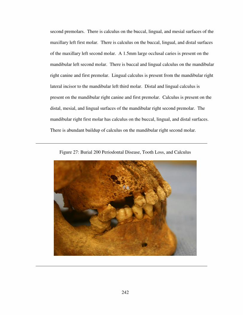
242
second premolars. There is calculus on the buccal, lingual, and mesial surfaces of the
maxillary left first molar. There is calculus on the buccal, lingual, and distal surfaces
of the maxillary left second molar. A 1.5mm large occlusal caries is present on the
mandibular left second molar. There is buccal and lingual calculus on the mandibular
right canine and first premolar. Lingual calculus is present from the mandibular right
lateral incisor to the mandibular left third molar. Distal and lingual calculus is
present on the mandibular right canine and first premolar. Calculus is present on the
distal, mesial, and lingual surfaces of the mandibular right second premolar. The
mandibular right first molar has calculus on the buccal, lingual, and distal surfaces.
There is abundant buildup of calculus on the mandibular right second molar.
Figure 27: Burial 200 Periodontal Disease, Tooth Loss, and Calculus
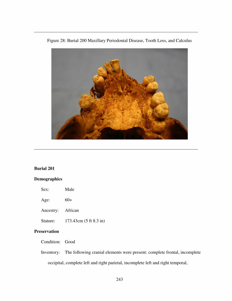
243
Figure 28: Burial 200 Maxillary Periodontal Disease, Tooth Loss, and Calculus
Burial 201
Demographics
Sex: Male
Age: 60+
Ancestry: African
Stature: 173.43cm (5 ft 8.3 in)
Preservation
Condition: Good
Inventory: The following cranial elements were present: complete frontal, incomplete
occipital, complete left and right parietal, incomplete left and right temporal,
244
complete left and right zygomatic, complete left and right maxilla, complete
mandible, and complete hyoid.
The following postcranial elements were present: incomplete left and right
scapula, incomplete left and right clavicle, incomplete left humerus, complete right
humerus, complete left radius, incomplete right radius, complete left ulna, incomplete
right ulna, incomplete left and right innominate, compelte left femur, incomplete right
femur, complete left patella, incomplete left and right tibia, incomplete left and right
fibula, incomplete sternum, 24 ribs, complete left hamate, ten hand phalanges,
incomplete left calcaneus, fragmented right calcaneus, complete left talus, incomplete
right talus, complete left cuboid, complete left navicular, complete left med., int., and
lat. cuneiform, complete left first through fifth metatarsal, incomplete right first
metatarsal, complete right second metatarsal, incomplete right third, fourth, and fifth
metatarsal, seven foot phalanges, all cervical vertebrae, all thoracic vertebrae, all
lumbar vertebrae, and all sacral vertebrae.
Paleopathology
Dental: Complete alveolar alveolar bone resorption
Infectious Disease: Not Applicable
Trauma: Not Applicable
Other: Osteophytes surround the obturator foramen of the right pelvis. There is
severe arthritic lipping of all cervical vertebrae. There are very large osteophytes and
arthritic lipping of all lumbar vertebrae. The fifth and sixth cervical vertebrae are
fused. All the vertebrae are extremely porous. The acetabulum of the right and left
pelvis are very large with deep pits in the acetabular fossa. The mental spines on the
mandible are very prominent
245
Burial 202
Demographics
Sex: Male
Age: 54-64
Ancestry: Not Applicable
Stature: 169.56cm (5 ft 6.6 in)
Preservation
Condition: Fair
Inventory: The following cranial elements were present: complete frontal, fragmented
occipital, complete left and right parietal, incomplete left right temporal, incomplete
left and right zygomatic, incomplete left and right maxilla, incomplete left and right
sphenoid, complete mandible, fragmented hyoid, and 17 teeth.
The following postcranial elements were present: incomplete left scapula,
complete right scapula, complete left clavicle, incomplete right clavicle, incomplete
left humerus, complete right humerus, incomplete left radius, complete right radius,
complete left and right ulna, incomplete left and right innominate, complete left and
right femur, complete left and right patella, complete left and right tibia, incomplete
left and right fibula, incomplete sternum, 24 ribs, incomplete left and right scaphoid,
complete left lunate, incomplete left triquetral, incomplete left pisiform, incomplete
left trapezium, incomplete left trapezoid, incomplete left capitate, complete right
capitate, incomplete left and right hamate, incomplete second through the fifth
metacarpal, incomplete right first through the fifth metacarpal, seven hand phalanges,
incomplete left and right calcaneus, incomplete left talus, complete right talus,
246
complete left and right cuboid, complete left and right navicular, complete left and
right med., int., and lat. cuneiform, incomplete left first through the fifth metatarsal,
complete right first through the fifth metatarsal, eight foot phalanges, all cervical
vertebrae, all thoracic vertebrae, all lumbar vertebrae, and all sacral vertebrae.
Paleopathology
Dental: Maxillary alveolar bone resorption is present at the areas of the maxillary
right first molar, maxillary right central incisor to the maxillary right canine, the
maxillary left second premolar, and the maxillary left first molar. Lingual calculus is
present on the maxillary right third molar and the maxillary left second and third
molars. Lingual calculus occurring at the CEJ is present on the mandibular left lateral
incisor to the mandibular left second molar, the mandibular right central incisor, and
the mandibular left first and second premolars. There is one 6mm large distal root-
enamel caries on the mandibular right first molar.
Infectious Disease: Not Applicable
Trauma: Not Applicable
Other: Not Applicable
Burial 203
Demographics
Sex: Male
Age: 21+
Ancestry: Not Applicable
Stature: Not Applicable
Preservation
247
Condition: Poor
Inventory: The following cranial elements were present: incomplete frontal, fragmented
occipital, fragmented left and right parietal, incomplete left and right temporal,
fragmented left and right maxilla, fragmented left and right sphenoid, incomplete
mandible, and 27 teeth.
The following postcranial elements were present: fragmented left and right
scapula, fragmented left clavicle, incomplete right clavicle, fragmented left humerus,
incomplete right humerus, fragmented left and right radius, fragmented left and right
ulna, fragmented left and right innominate, incomplete left and right femur,
incomplete left and right tibia, fragmented left and right fibula, and two ribs.
Paleopathology
Dental: Mandibular alveolar bone resorption is present at the areas of the right first
molar and the left third molar.
Infectious Disease: Not Applicable
Trauma: Not Applicable
Other: Not Applicable
Burial 204
Demographics
Sex: Male
Age: 21+
Ancestry: Not Applicable
Stature: Not Applicable
Preservation
248
Condition: Very poor
Inventory: The following cranial elements were present: incomplete right temporal and
fragmented mandible. The only postcranial elements present were two right tibia
fragments.
Paleopathology
Dental: A fragment of mandible shows signs of alveolar bone resorption from the
right first molar to the right third molar.
Infectious Disease: Not Applicable
Trauma: Not Applicable
Other: Not Applicable
Burial 205
Demographics
Sex: Not Applicable
Age: 15+
Ancestry: Not Applicable
Stature: Not Applicable
Preservation
Condition: Very poor
Inventory: The following cranial elements were present: incomplete frontal, fragmented
occipital, incomplete left and right parietal, incomplete left temporal, fragmented
right temporal, fragmented left and right maxilla, fragmented left and right sphenoid,
fragmented mandible, incomplete hyoid, and seven teeth.
249
The following postcranial elements were present: fragmented right humerus,
fragmented right innominate, fragmented left femur, incomplete right femur, and
fragmented left and right tibia.
Paleopathology
Dental: Not Applicable
Infectious Disease: Not Applicable
Trauma: Not Applicable
Other: Not Applicable
Burial 206 No remains were present.
Burial 207 No remains were present.
Burial 208 No remains were present.
Burial 209
Demographics
Sex: Not Applicable
Age: 5 years +/- 9 months
Ancestry: Not Applicable
Stature: Not Applicable
Preservation
Condition: Very poor
Inventory: The following cranial elements were present: fragmented frontal,
fragmented left and right temporal, fragmented mandible, 14 deciduous teeth, and 20
permanent dental caps. No postcranial elements were present.
250
Paleopathology
Dental: Not Applicable
Infectious Disease: Not Applicable
Trauma: Not Applicable
Other: Not Applicable
Burial 210 No remains were present.
Burial 211
Demographics
Sex: Not Applicable
Age: 2-3
Ancestry: Not Applicable
Stature: Not Applicable
Preservation
Condition: Very poor
Inventory: The following postcranial elements were present: four deciduous teeth and
thirteen permanent dental caps.
Paleopathology
Dental: Not Applicable
Infectious Disease: Not Applicable
Trauma: Not Applicable
Other: Not Applicable
251
Burial 212 No remains were present.
Burial 213
Demographics
Sex: Not Applicable
Age: 21+
Ancestry: Not Applicable
Stature: Not Applicable
Preservation
Condition: Very poor
Inventory: The only cranial elements present were 21 teeth. No postcranial elements
were recovered.
Paleopathology
Dental: There is one 1.5 mesial IPCF caries on the maxillary left first incisor.
Infectious Disease: Not Applicable
Trauma: Not Applicable
Other: Not Applicable
Burial 214
Demographics
Sex: Not Applicable
Age: 21+
Ancestry: Not Applicable
Stature: Not Applicable
252
Preservation
Condition: Very poor
Inventory: The following cranial elements were present: fragmented frontal,
fragmented occipital, and 17 teeth. Cranial: frontal and occipital fragmented, 17 teeth
present. The postcranial elements present included bone meal and stains in the areas
of the humerus, right radius, and right ulna.
Paleopathology
Dental: Not Applicable
Infectious Disease: Not Applicable
Trauma: Not Applicable
Other: Not Applicable
Burial 215
Demographics
Sex: Not Applicable
Age: Not Applicable
Ancestry: Not Applicable
Stature: Not Applicable
Preservation
Condition: Very poor
Inventory: No cranial elements were present. The postcranial elements present were a
fragmented right scapula and a right femoral fragment.
Paleopathology
Dental: Not Applicable
253
Infectious Disease: Not Applicable
Trauma: Not Applicable
Other: Not Applicable
Burial 216
Demographics
Sex: Female
Age: 40+
Ancestry: Not Applicable
Stature: Not Applicable
Preservation
Condition: Poor
Inventory: The following cranial elements were present: incomplete frontal, complete
occipital, complete left parietal, incomplete right parietal, incomplete left and right
temporal, fragmented left and right maxilla, fragmented left and right sphenoid,
incomplete mandible, and 26 teeth.
The following postcranial elements were present: fragmented left and right
scapula, incomplete left clavicle, incomplete left and right humerus, incomplete left
radius, fragmented right radius, incomplete left ulna, fragmented right ulna,
fragmented left and right innominate, incomplete left and right femur, incomplete left
and right tibia, fragmented left and right fibula, 17 ribs, all cervical vertebrae, and all
thoracic vertebrae.
Paleopathology
254
Dental: Not Applicable
Infectious Disease: Not Applicable
Trauma: Not Applicable
Other: Not Applicable
Burial 217
Demographics
Sex: Not Applicable
Age: 2
Ancestry: Not Applicable
Stature: Not Applicable
Preservation
Condition: Very poor
Inventory: The cranial elements present included one deciduous tooth and two
permanent dental caps.
Paleopathology
Dental: Not Applicable
Infectious Disease: Not Applicable
Trauma: Not Applicable
Other: Not Applicable
Burial 218 No remains were present.
Burial 219 No remains were present.
Burial 220 No remains were present.
255
Burial 221 No remains were present.
Burial 222 (This burial was accidentally assigned two numbers, 103 and 222)
Burial 223
Demographics
Sex: Not Applicable
Age: Not Applicable
Ancestry: Not Applicable
Stature: Not Applicable
Preservation
Condition: Very poor
Inventory: No cranial elements were present. The only postcranial element present was
bone meal present at the area of the left femur.
Paleopathology
Dental: Not Applicable
Infectious Disease: Not Applicable
Trauma: Not Applicable
Other: Not Applicable
Burial 224 No remains were present.
Burial 225
Demographics
Sex: Not Applicable
256
Age: 8+
Ancestry: Not Applicable
Stature: Not Applicable
Preservation
Condition: Very poor
Inventory: The following cranial elements were present: fragmented occipital and five
teeth. The postcranial elements present included fragments of the left and right
femur.
Paleopathology
Dental: Not Applicable
Infectious Disease: Not Applicable
Trauma: Not Applicable
Other: Not Applicable
Burial 226
Demographics
Sex: Male
Age: 40+
Ancestry: Not Applicable
Stature: Not Applicable
Preservation
Condition: Poor
Inventory: The following cranial elements were present: fragmented frontal,
fragmented occipital, incomplete left and right parietal, incomplete left and right
257
temporal, incomplete left and right zygomatic, fragmented left and right maxilla,
fragmented left and right sphenoid, incomplete mandible, and seven teeth.
The following postcranial elements were present: fragmented left and right
scapula, fragmented left and right clavicle, fragmented left and right humerus,
incomplete right radius, fragmented right ulna, fragmented left and right innominate,
incomplete left and right femur, incomplete left and right tibia, fragmented left and
right fibula, four ribs, the first through the fifth cervical vertebrae, ten thoracic
vertebral fragments, the third and fourth lumbar vertebrae, and the third and fourth
sacral vertebrae
Paleopathology
Dental: There is mandibular alveolar bone resorption at the areas from the right first
molar to the right third molar and from the left second premolar to the left molar.
There is one 4mm large mesial IPCF on the maxillary right second premolar.
Infectious Disease: Not Applicable
Trauma: Not Applicable
Other: Not Applicable
Burial 227 No remains were present.