a combined scientometric and critical approach in ... - mdpi
TRANSCRIPT
coatings
Review
A Combined Scientometric and Critical Approach in ReviewingTiZr Implant Alloys and Coating Performances
Radu Nartita 1 , Daniela Ionita 2 and Ioana Demetrescu 1,2,*
�����������������
Citation: Nartita, R.; Ionita, D.;
Demetrescu, I. A Combined
Scientometric and Critical Approach
in Reviewing TiZr Implant Alloys and
Coating Performances. Coatings 2021,
11, 392. https://doi.org/10.3390/
coatings11040392
Academic Editor: Grzegorz Dercz
Received: 13 March 2021
Accepted: 27 March 2021
Published: 30 March 2021
Publisher’s Note: MDPI stays neutral
with regard to jurisdictional claims in
published maps and institutional affil-
iations.
Copyright: © 2021 by the authors.
Licensee MDPI, Basel, Switzerland.
This article is an open access article
distributed under the terms and
conditions of the Creative Commons
Attribution (CC BY) license (https://
creativecommons.org/licenses/by/
4.0/).
1 Department of General Chemistry, Faculty of Applied Chemistry and Material Science,University Polithenica of Bucharest, 313 Splaiul Indepentei, 060042 Bucharest, Romania;[email protected]
2 Academy of Romanian Scientists, Ilfov Street, 3, 050044 Bucharest, Romania; [email protected]* Correspondence: [email protected]
Abstract: This review article was developed based on the scientometric analysis of the evaluatedstudies conducted on titanium−zirconium (TixZr) alloys from 2000 to the present. The scientometricdata obtained helped us to identify the most researched topics and these topics were further analyzedand discussed. An increasing number of researchers are considering TixZr alloys as opposed to thetraditional ones because these alloys present improved mechanical properties and in some casesimproved corrosion resistance and biocompatibility. Due to the natural layer of oxides formed onthese alloys, multiple surface modification methods can be applied to solve some of the challengesfaced in the field of implantable materials. A significant number of studies are now focusing onsurface modifications at the nanometer scale or various coatings for improved corrosion resistanceand biological interactions. Although not yet commercially available, a TiZr alloy with a nanos-tructured surface and embedded biologically active substances, such as antibiotics or coated withhydroxyapatite, may become a future option.
Keywords: TiZr alloy; titanium zirconium alloy; surface modification; nanostructured surfaces;scientometric analysis
1. Introduction
Due to the good biocompatibility of Ti, research on Ti alloys used as biomaterials is ona continuous upward trend [1,2]. A radical increase in research related to zirconium dioxide(ZrO2) has also been observed due to increased mechanical strength, biocompatibility, andaesthetic properties in the case of dental work [1,3]. In this context, the titanium−zirconium(TixZr) alloys have been intensively investigated in the last decade.
The mechanical properties, porosity, surface morphology, and implant design are thedetermining factors for the evolution of osseointegration and longevity of an implant [4,5].
Implantable materials must have mechanical properties such as a modulus of elasticity,yield strength, and ultimate tensile strength that can withstand various biomechanicalforces. Besides, biomaterials must be biocompatible, have low density and increasedresistance to corrosion and wear [6].
Commercially pure titanium (cpTi) was the most attractive alternative for implants [7,8].However, the manufacture of implants with small diameters (≤3.5 mm) from cp-Ti is asso-ciated with an increased risk of fracture due to insufficient mechanical strength. The aim isto develop new alloys and coatings to solve this problem [8–14].
Zr as Ti is situated in Group IV of the periodic table of elements, therefore the twoelements have similar chemical properties and biocompatibilities [15,16]. Thus, TixZr typealloys began to be studied more and more frequently, with the possibility to solve these prob-lems, as both have satisfactory mechanical properties and good biocompatibility [10,17–20].
It is known that a new alloying strategy involving the combination of several principalelements in high concentrations has been in development in the few years. The goal is to
Coatings 2021, 11, 392. https://doi.org/10.3390/coatings11040392 https://www.mdpi.com/journal/coatings
Coatings 2021, 11, 392 2 of 28
elaborate new materials named high entropy alloys with remarkable properties exceedingthose of conventional alloys. Some of them contain Ti and Zr [21–23] and have potentialimplant applications [24].
Compared to other traditional biomaterials, some of the binary TixZr alloys havea higher strength/weight ratio, lower modulus of elasticity and better corrosion resis-tance [25–27]. Moreover, on the surface of alloys containing titanium and zirconium, alayer of apatite is formed that is structurally similar to bone tissue, which improves thebioactivity of these materials [28,29].
The present manuscript has a novel approach being a combined scientometric andcritical one in reviewing the behavior of TiZr alloys based on their composition, surfaceand interface.
2. Methods
A bibliographic search was conducted in the ScienceDirect database using the terms“titanium zirconium implants” and “TiZr implants”, choosing only the “research” and“review” articles published in English from 2000 to the present (5 February 2021). Addi-tionally, to ensure that most of the relevant studies regarding the chosen topic were takeninto consideration, multiple articles were selected manually.
The selected articles were carefully analyzed considering the purpose of this reviewarticle. The inclusion criteria involved studies that examined binary TixZr alloys or thosethat studied relevant characteristics related to the surface of this type of alloy. The aspectsmonitored and evaluated were those related to changes at the micro and nanometric scale,the methods used, mechanical properties, corrosion resistance and biological interactions.
The VOSviewer software (version: 1.6.16) was used for the scientometric analysis. Theevaluated aspects are co-authorship (unit of analysis: authors), co-occurrence of keywordsand terms from titles and abstracts. In all three cases, the maps created from the networkanalysis were based on the selected bibliographic data.
For the authors to be included in the co-authorship analysis, a minimum number of2 articles was selected for the keywords to be included in the co-occurrence analysis, aminimum number of 2 occurrences was selected and for the terms from titles and abstracts,a minimum number of 10 occurrences was selected.
3. Results and Discussions
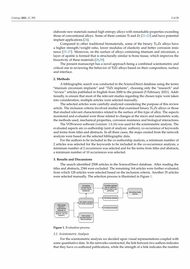
The search identified 2508 articles in the ScienceDirect database. After reading thetitles and abstracts, 2344 were excluded. The remaining 164 articles were further evaluated,from which 128 articles were selected based on the inclusion criteria. Another 35 articleswere selected manually. The selection process is illustrated in Figure 1.
Coatings 2021, 11, x FOR PEER REVIEW 2 of 28
problems, as both have satisfactory mechanical properties and good biocompatibility [10,17–20].
It is known that a new alloying strategy involving the combination of several princi-pal elements in high concentrations has been in development in the few years. The goal is to elaborate new materials named high entropy alloys with remarkable properties exceed-ing those of conventional alloys. Some of them contain Ti and Zr [21–23] and have poten-tial implant applications [24].
Compared to other traditional biomaterials, some of the binary TixZr alloys have a higher strength/weight ratio, lower modulus of elasticity and better corrosion resistance [25–27]. Moreover, on the surface of alloys containing titanium and zirconium, a layer of apatite is formed that is structurally similar to bone tissue, which improves the bioactivity of these materials [28,29].
The present manuscript has a novel approach being a combined scientometric and critical one in reviewing the behavior of TiZr alloys based on their composition, surface and interface.
2. Methods A bibliographic search was conducted in the ScienceDirect database using the terms
“titanium zirconium implants” and “TiZr implants”, choosing only the “research” and “review” articles published in English from 2000 to the present (05 February 2021). Addi-tionally, to ensure that most of the relevant studies regarding the chosen topic were taken into consideration, multiple articles were selected manually.
The selected articles were carefully analyzed considering the purpose of this review article. The inclusion criteria involved studies that examined binary TixZr alloys or those that studied relevant characteristics related to the surface of this type of alloy. The aspects monitored and evaluated were those related to changes at the micro and nanometric scale, the methods used, mechanical properties, corrosion resistance and biological interactions.
The VOSviewer software (version: 1.6.16) was used for the scientometric analysis. The evaluated aspects are co-authorship (unit of analysis: authors), co-occurrence of key-words and terms from titles and abstracts. In all three cases, the maps created from the network analysis were based on the selected bibliographic data.
For the authors to be included in the co-authorship analysis, a minimum number of 2 articles was selected for the keywords to be included in the co-occurrence analysis, a minimum number of 2 occurrences was selected and for the terms from titles and ab-stracts, a minimum number of 10 occurrences was selected.
3. Results and Discussions The search identified 2508 articles in the ScienceDirect database. After reading the
titles and abstracts, 2344 were excluded. The remaining 164 articles were further evalu-ated, from which 128 articles were selected based on the inclusion criteria. Another 35 articles were selected manually. The selection process is illustrated in Figure 1.
Figure 1. Evaluation process.
3.1. Scientometric Analysis
For the scientometric analysis we decided upon visual representations coupled withsome quantitative data. In the networks constructed, the link between two authors indicatesthat they have co-authored publications, while the strength of a link indicates the number
Coatings 2021, 11, 392 3 of 28
of co-authored publications. In the case of terms used (keywords or other terms), the linkindicates that the terms co-occur in the same publication, while the strength of the linkindicates the number of publications in which the terms co-occur.
3.1.1. Co-Authorship
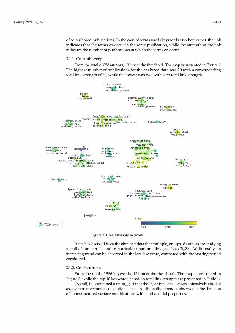
From the total of 858 authors, 100 meet the threshold. The map is presented in Figure 2.The highest number of publications for the analyzed data was 20 with a correspondingtotal link strength of 70, while the lowest was two with zero total link strength.
Coatings 2021, 11, x FOR PEER REVIEW 3 of 28
Figure 1. Evaluation process.
3.1. Scientometric Analysis For the scientometric analysis we decided upon visual representations coupled with
some quantitative data. In the networks constructed, the link between two authors indi-cates that they have co-authored publications, while the strength of a link indicates the number of co-authored publications. In the case of terms used (keywords or other terms), the link indicates that the terms co-occur in the same publication, while the strength of the link indicates the number of publications in which the terms co-occur.
3.1.1. Co-Authorship From the total of 858 authors, 100 meet the threshold. The map is presented in Figure
2. The highest number of publications for the analyzed data was 20 with a corresponding total link strength of 70, while the lowest was two with zero total link strength.
It can be observed from the obtained data that multiple, groups of authors are stud-ying metallic biomaterials and in particular titanium alloys, such as TixZr. Additionally, an increasing trend can be observed in the last few years, compared with the starting pe-riod considered.
Figure 2. Co-authorship network.
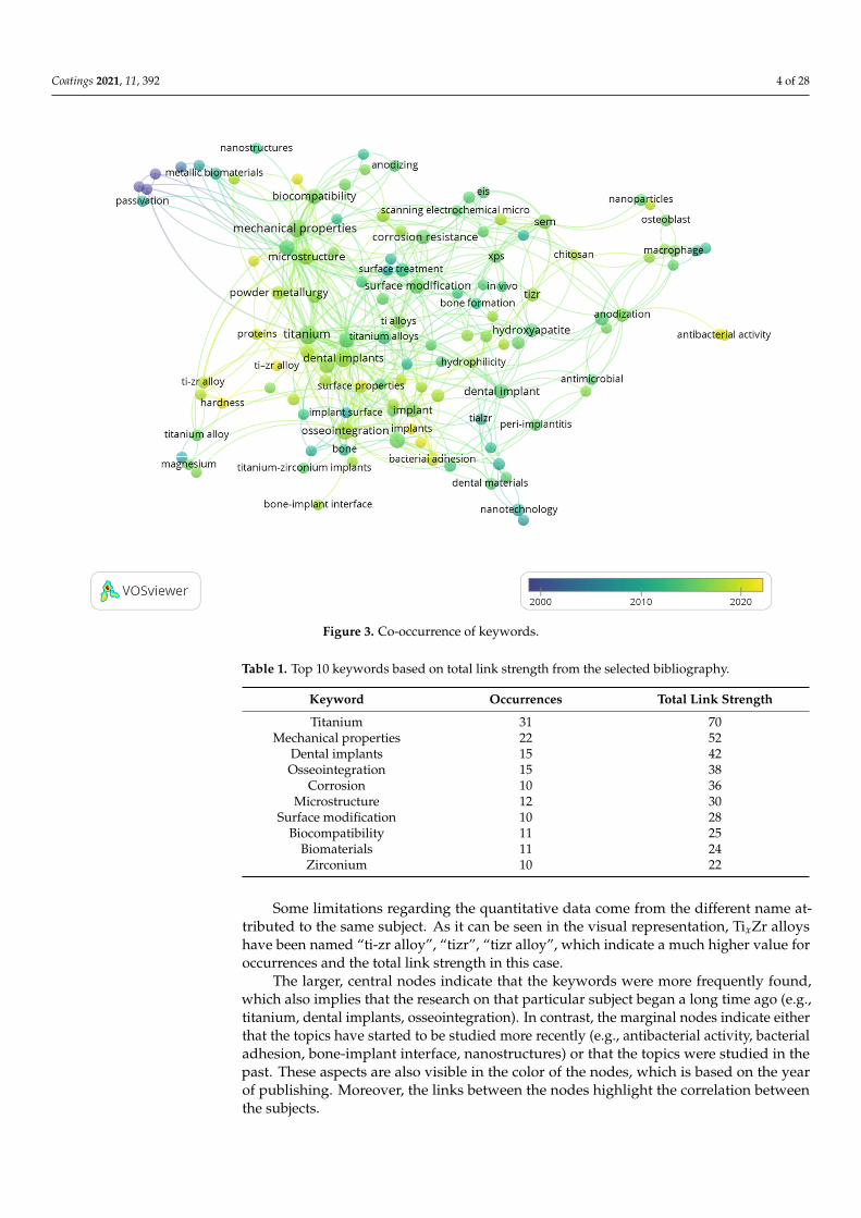
3.1.2. Co-Occurrence From the total of 586 keywords, 121 meet the threshold. The map is presented in Fig-
ure 3, while the top 10 keywords based on total link strength are presented in Table 1.
Figure 2. Co-authorship network.
It can be observed from the obtained data that multiple, groups of authors are studyingmetallic biomaterials and in particular titanium alloys, such as TixZr. Additionally, anincreasing trend can be observed in the last few years, compared with the starting periodconsidered.
3.1.2. Co-Occurrence
From the total of 586 keywords, 121 meet the threshold. The map is presented inFigure 3, while the top 10 keywords based on total link strength are presented in Table 1.
Overall, the combined data suggest that the TixZr type of alloys are intensively studiedas an alternative for the conventional ones. Additionally, a trend is observed in the directionof nanostructured surface modifications with antibacterial properties.
Coatings 2021, 11, 392 4 of 28Coatings 2021, 11, x FOR PEER REVIEW 4 of 28
Figure 3. Co-occurrence of keywords.
Table 1. Top 10 keywords based on total link strength from the selected bibliography.
Keyword Occurrences Total Link Strength Titanium 31 70
Mechanical properties 22 52 Dental implants 15 42 Osseointegration 15 38
Corrosion 10 36 Microstructure 12 30
Surface modification 10 28 Biocompatibility 11 25
Biomaterials 11 24 Zirconium 10 22
Some limitations regarding the quantitative data come from the different name at-tributed to the same subject. As it can be seen in the visual representation, TixZr alloys have been named “ti-zr alloy”, “tizr”, “tizr alloy”, which indicate a much higher value for occurrences and the total link strength in this case.
The larger, central nodes indicate that the keywords were more frequently found, which also implies that the research on that particular subject began a long time ago (e.g., titanium, dental implants, osseointegration). In contrast, the marginal nodes indicate ei-ther that the topics have started to be studied more recently (e.g., antibacterial activity, bacterial adhesion, bone-implant interface, nanostructures) or that the topics were studied in the past. These aspects are also visible in the color of the nodes, which is based on the year of publishing. Moreover, the links between the nodes highlight the correlation be-tween the subjects.
Figure 3. Co-occurrence of keywords.
Table 1. Top 10 keywords based on total link strength from the selected bibliography.
Keyword Occurrences Total Link Strength
Titanium 31 70Mechanical properties 22 52
Dental implants 15 42Osseointegration 15 38
Corrosion 10 36Microstructure 12 30
Surface modification 10 28Biocompatibility 11 25
Biomaterials 11 24Zirconium 10 22
Some limitations regarding the quantitative data come from the different name at-tributed to the same subject. As it can be seen in the visual representation, TixZr alloyshave been named “ti-zr alloy”, “tizr”, “tizr alloy”, which indicate a much higher value foroccurrences and the total link strength in this case.
The larger, central nodes indicate that the keywords were more frequently found,which also implies that the research on that particular subject began a long time ago (e.g.,titanium, dental implants, osseointegration). In contrast, the marginal nodes indicate eitherthat the topics have started to be studied more recently (e.g., antibacterial activity, bacterialadhesion, bone-implant interface, nanostructures) or that the topics were studied in thepast. These aspects are also visible in the color of the nodes, which is based on the yearof publishing. Moreover, the links between the nodes highlight the correlation betweenthe subjects.
Coatings 2021, 11, 392 5 of 28
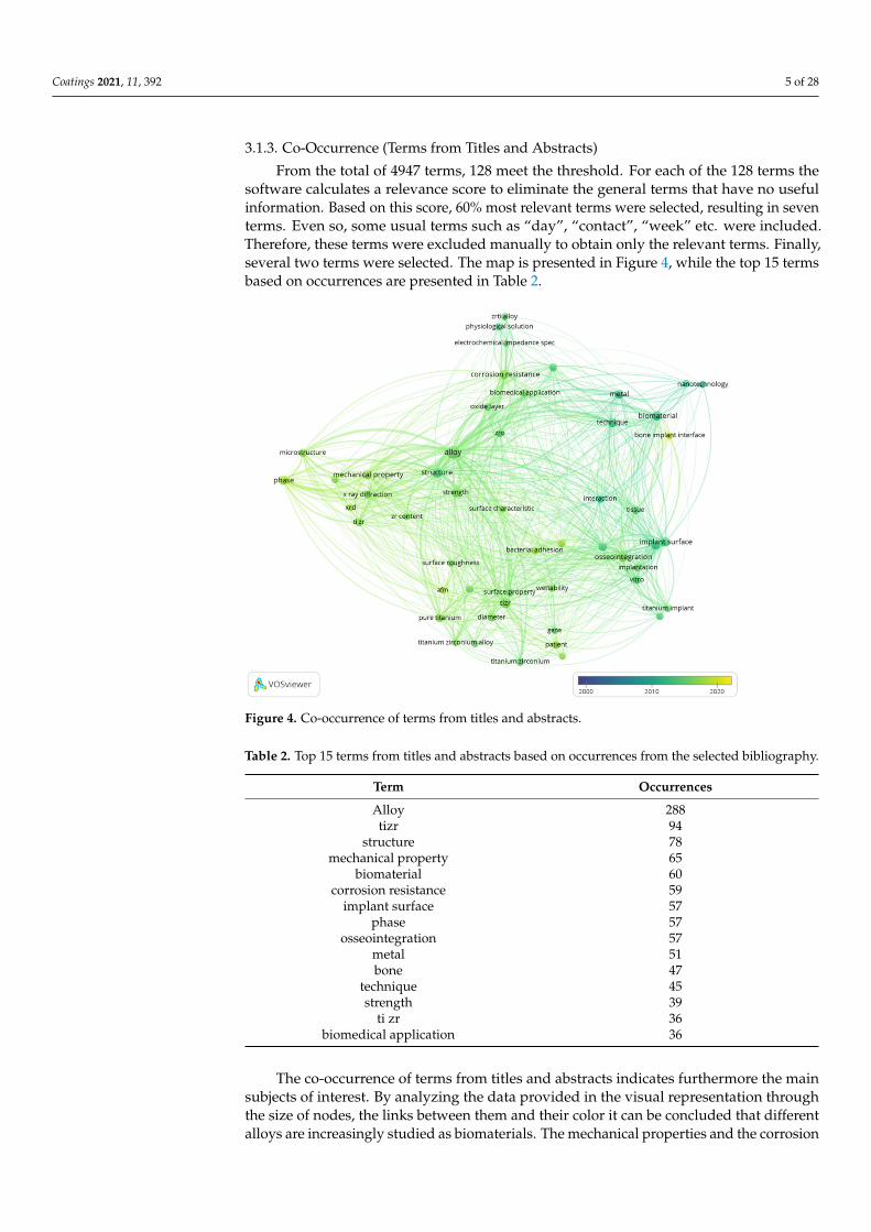
3.1.3. Co-Occurrence (Terms from Titles and Abstracts)
From the total of 4947 terms, 128 meet the threshold. For each of the 128 terms thesoftware calculates a relevance score to eliminate the general terms that have no usefulinformation. Based on this score, 60% most relevant terms were selected, resulting in seventerms. Even so, some usual terms such as “day”, “contact”, “week” etc. were included.Therefore, these terms were excluded manually to obtain only the relevant terms. Finally,several two terms were selected. The map is presented in Figure 4, while the top 15 termsbased on occurrences are presented in Table 2.
Coatings 2021, 11, x FOR PEER REVIEW 5 of 28
Overall, the combined data suggest that the TixZr type of alloys are intensively stud-ied as an alternative for the conventional ones. Additionally, a trend is observed in the direction of nanostructured surface modifications with antibacterial properties.
3.1.3. Co-Occurrence (Terms from Titles and Abstracts) From the total of 4947 terms, 128 meet the threshold. For each of the 128 terms the
software calculates a relevance score to eliminate the general terms that have no useful information. Based on this score, 60% most relevant terms were selected, resulting in seven terms. Even so, some usual terms such as “day”, “contact”, “week” etc were in-cluded. Therefore, these terms were excluded manually to obtain only the relevant terms. Finally, several two terms were selected. The map is presented in Figure 4, while the top 15 terms based on occurrences are presented in Table 2.
Figure 4. Co-occurrence of terms from titles and abstracts.
Table 2. Top 15 terms from titles and abstracts based on occurrences from the selected bibliog-raphy.
Term Occurrences Alloy 288 tizr 94
structure 78 mechanical property 65
biomaterial 60 corrosion resistance 59
implant surface 57 phase 57
osseointegration 57 metal 51 bone 47
technique 45 strength 39
ti zr 36 biomedical application 36
Figure 4. Co-occurrence of terms from titles and abstracts.
Table 2. Top 15 terms from titles and abstracts based on occurrences from the selected bibliography.
Term Occurrences
Alloy 288tizr 94
structure 78mechanical property 65
biomaterial 60corrosion resistance 59
implant surface 57phase 57
osseointegration 57metal 51bone 47
technique 45strength 39
ti zr 36biomedical application 36
The co-occurrence of terms from titles and abstracts indicates furthermore the mainsubjects of interest. By analyzing the data provided in the visual representation throughthe size of nodes, the links between them and their color it can be concluded that differentalloys are increasingly studied as biomaterials. The mechanical properties and the corrosion
Coatings 2021, 11, 392 6 of 28
resistance of these alloys appear to be mentioned more throughout time, but an interestregarding surface modifications in relation to different biological aspects seems to emergein more recent studies.
The top terms combined with the visual representation helped us to establish the moststudied topics that are followed throughout this article such as type of alloy, mechanicalproperties, surface modifications and biocompatibility.
3.2. Properties Related to the Composition of TixZr Alloys
Bone tissue is a type of connective tissue made up of cells and fibers. From theperspective of mechanical properties, two types of bone tissue are differentiated, namelycortical and trabecular. Cortical bone has a higher density and low porosity (10%), having amodulus of elasticity between 4–30 GPa and a compressive strength between 20–193 MPa.The trabecular bone has a low density, being composed in a proportion of 50–90% frompores. This is also reflected in the modulus of elasticity which is between 0.2–2 GPa andthe compressive strength which is between 2–80 MPa [30]. The modulus of elasticity ofbiomaterials should be close to that of the bones. If the biomaterial presents a significantlyhigher modulus of elasticity, a phenomenon known as stress shield may occur, which ischaracterized by a reduction in bone density (osteopenia) [28].
The interstitial elements, carbon (C), oxygen (O), nitrogen (N) and hydrogen (H)have a hardening effect on transition metals due to the high suppression of dislocationmovements and network distortion [31]. Of these, O has the highest solubility in Ti and aconsiderable hardening effect. In TixZr alloys, an increase in hardness has been reportedwith an increase in O content [31,32]. Additionally, increasing the concentration of Zrleads to finer grains, which together with the strengthening of the solid solution leads toincreased hardness, strength and plasticity. At the same time, an increased performanceregarding osteoinduction is obtained due to better adhesion of proteins [10].
Zirconium is considered an isomorphic stabilizer being completely soluble in Ti, itcan exist in both pure α and β phase [33]. In solid solution with another β stabilizer, Zrcan also act as a β stabilizer [34]. The α and α−β mix phases have good strength and creepresistance, but β alloys have a better fatigue strength and lower Young’s modulus [33].The Zr content in the allotropic form α′ (hexagonal martensite) increases the modulusof elasticity [35]. While the α phase has small benefits at the temperature and stressassociated with implants, the β phase is especially beneficial for orthopedic implants andany long-term implant [33,36].
As high entropy alloys (HEAs) have exceptional mechanical and corrosion behaviornew phases derived from the AlCoFeNiSmTiVZr [37] and from Ti−Nb−Ta−Zr after Aladdition have also been investigated [38] establishing the formation of the intermetallicphase after annealing at 600–1200 ◦C.
A study performed on TixZr alloys (x = 10, 20, 30, 40 wt.%) shows that the microhardnessvalues, the bending strengths and the elastic recovery angles increase with the content of Zr,being much higher compared to cpTi [2]. In the case of increasing the Zr content, in ZrxTialloys (x = 10, 20, 30, 40 wt.%) the properties no longer vary constantly with the Ti content.Among the studied alloys, the best values of microhardness and bending modulus werereported for Ti-40Zr wt.% [39]. TixZr binary alloys with high Zr content also present highstrength [40]. The hardness of Ti50Zr alloy (at.%) is 2.5 times higher than pure Ti or Zr [28,41].
Current information shows that TiZr alloys can also be manufactured to have a porousstructure and mechanical properties similar to those of bone tissue [29,42,43]. Wen et al.report the manufacture of TiZr alloy foams with a density of approximately 0.3 and pores of200–500 µm. The Young’s modulus for these is between 78.4 MPa and 15.3 GPa [29]. Porousbulk metallic glass based on Ti-Zr has also been studied as an alternative to conventionalmaterials [44,45].
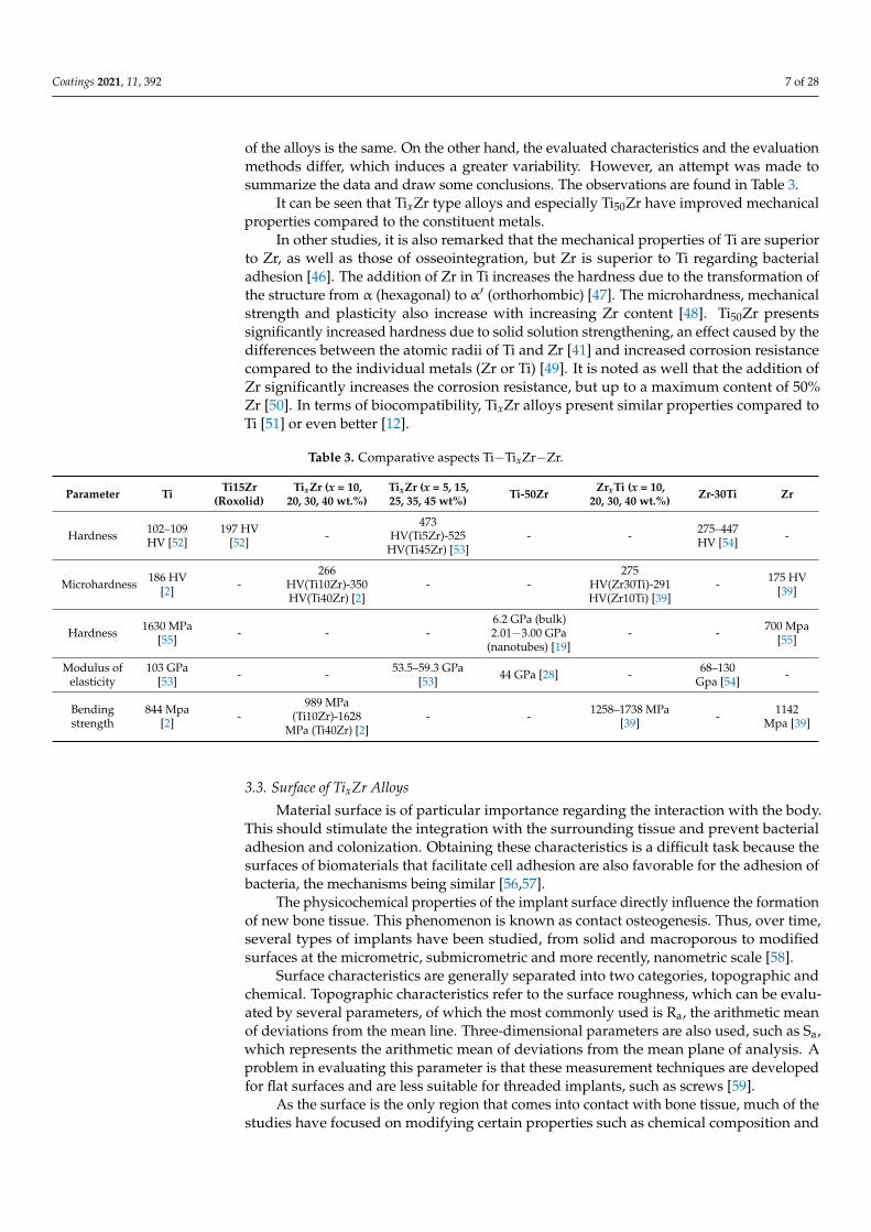
Comparative analysis regarding the mechanical properties between different studieswas difficult to perform for several reasons. On the one hand, different alloy manufacturingtechniques affect their structure and thus their mechanical properties, even if the composition
Coatings 2021, 11, 392 7 of 28
of the alloys is the same. On the other hand, the evaluated characteristics and the evaluationmethods differ, which induces a greater variability. However, an attempt was made tosummarize the data and draw some conclusions. The observations are found in Table 3.
It can be seen that TixZr type alloys and especially Ti50Zr have improved mechanicalproperties compared to the constituent metals.
In other studies, it is also remarked that the mechanical properties of Ti are superiorto Zr, as well as those of osseointegration, but Zr is superior to Ti regarding bacterialadhesion [46]. The addition of Zr in Ti increases the hardness due to the transformation ofthe structure from α (hexagonal) to α′ (orthorhombic) [47]. The microhardness, mechanicalstrength and plasticity also increase with increasing Zr content [48]. Ti50Zr presentssignificantly increased hardness due to solid solution strengthening, an effect caused by thedifferences between the atomic radii of Ti and Zr [41] and increased corrosion resistancecompared to the individual metals (Zr or Ti) [49]. It is noted as well that the addition ofZr significantly increases the corrosion resistance, but up to a maximum content of 50%Zr [50]. In terms of biocompatibility, TixZr alloys present similar properties compared toTi [51] or even better [12].
Table 3. Comparative aspects Ti−TixZr−Zr.
Parameter Ti Ti15Zr(Roxolid)
TixZr (x = 10,20, 30, 40 wt.%)
TixZr (x = 5, 15,25, 35, 45 wt%) Ti-50Zr ZrxTi (x = 10,
20, 30, 40 wt.%) Zr-30Ti Zr
Hardness 102–109HV [52]
197 HV[52] -
473HV(Ti5Zr)-525
HV(Ti45Zr) [53]- - 275–447
HV [54] -
Microhardness 186 HV[2] -
266HV(Ti10Zr)-350HV(Ti40Zr) [2]
- -275
HV(Zr30Ti)-291HV(Zr10Ti) [39]
- 175 HV[39]
Hardness 1630 MPa[55] - - -
6.2 GPa (bulk)2.01−3.00 GPa
(nanotubes) [19]- - 700 Mpa
[55]
Modulus ofelasticity
103 GPa[53] - - 53.5–59.3 GPa
[53] 44 GPa [28] - 68–130Gpa [54] -
Bendingstrength
844 Mpa[2] -
989 MPa(Ti10Zr)-1628
MPa (Ti40Zr) [2]- - 1258–1738 MPa
[39] - 1142Mpa [39]
3.3. Surface of TixZr Alloys
Material surface is of particular importance regarding the interaction with the body.This should stimulate the integration with the surrounding tissue and prevent bacterialadhesion and colonization. Obtaining these characteristics is a difficult task because thesurfaces of biomaterials that facilitate cell adhesion are also favorable for the adhesion ofbacteria, the mechanisms being similar [56,57].
The physicochemical properties of the implant surface directly influence the formationof new bone tissue. This phenomenon is known as contact osteogenesis. Thus, over time,several types of implants have been studied, from solid and macroporous to modifiedsurfaces at the micrometric, submicrometric and more recently, nanometric scale [58].
Surface characteristics are generally separated into two categories, topographic andchemical. Topographic characteristics refer to the surface roughness, which can be evalu-ated by several parameters, of which the most commonly used is Ra, the arithmetic meanof deviations from the mean line. Three-dimensional parameters are also used, such as Sa,which represents the arithmetic mean of deviations from the mean plane of analysis. Aproblem in evaluating this parameter is that these measurement techniques are developedfor flat surfaces and are less suitable for threaded implants, such as screws [59].
As the surface is the only region that comes into contact with bone tissue, much of thestudies have focused on modifying certain properties such as chemical composition and
Coatings 2021, 11, 392 8 of 28
roughness to promote osseointegration and mechanical fixation. The increase in roughnessleads to a larger contact surface for proteins and cells to interact with the material [60]. Incontrast, the metallic biomaterials that are designed to come into direct contact with wholeblood (heart valves, heart aid devices, heart pumps) must have low thrombogenicity [61]and high surface roughness, as well as high surface energy, which is associated with highthrombogenicity [9,62]. Thus, when designing the biomaterial surfaces multiple aspectsmust be considered.
Surface chemistry is relevant through composition, biocompatible metals such asTi and Zr, being valve metals, form a natural layer of oxides that represent a barrier inthe corrosion process, but also through other properties such as surface energy or surfacecharge. It is important to note that changes in topography often induce changes in chemistry,or vice versa [59,63,64]. TixZr type alloys have a better wettability than cpTi, which leads toa different biological behavior [17]. Studies suggest that surfaces with higher hydrophilicitylead to better tissue integration, osseointegration and faster healing [65].
Surface modifications of metallic biomaterials mainly aim at increasing corrosionresistance and improving bioactivity [30,66,67]. Additionaaly, because after surgery thereis an increased risk of developing a bacterial infection and oral antibiotics are generallygiven, a more effective alternative could be to load implants with active substances that actlocally, over time [7,12,68–70].
The techniques used can be mechanical, chemical or physical. Mechanical methodsinclude grinding, polishing and blasting. Chemical methods include treatments with acids,hydrogen peroxide, alkaline solutions, sol-gel techniques, anodic oxidation, micro-arcoxidation (MAO) and chemical vapor deposition. Physical methods include physical vapordeposition, femtosecond laser ablation in solution (FLAS) and several types of thermalspraying methods [71–77].
The naturally formed oxide film on the surface of Ti is composed of several layers,namely TiO (in contact with the metal), Ti2O3 (intermediate layer) and TiO2 (outer layer).Oxidation processes can increase the concentration of TiO2 quite a lot so that the layers ofsuboxides can no longer be detected, but the organization exists [74].
Oxidic compounds formed by the natural phenomenon of passivation may resultfrom the following oxidation reactions [25]:
Ti + H2O→ TiO + 2H+ + 2e− (1)
2TiO + H2O→ Ti2O3 + 2H+ + 2e− (2)
Ti2O3 + H2O→ TiO2 + 2H+ + 2e− (3)
2Zr + H2O→ Zr2O + 2H+ + 2e− (4)
Zr2O + H2O→ 2ZrO + 2H+ + 2e− (5)
2ZrO + H2O→ Zr2O3 + 2H+ + 2e− (6)
Zr2O3 + H2O→ 2ZrO2 + 2H+ + 2e− (7)
The Gibbs free energy for Zr oxidation (−1117.8 kJ/mol) is greater than that for Tioxidation (−890.5 kJ/mol), which suggests that Zr is preferentially oxidized [78]. However,a study performed on the oxide film developed by Ti50Zr alloys (at.%) through anodization(9 V) in different solutions shows that the mobility of Ti4+ ions is higher than that of Zr4+
ions, the outer layer being composed mostly of TiO2. The average reported compositionwas 76% TiO2 and 24% ZrO2 [79]. This aspect is revealed in another study on TixZr alloys(x = 23, 42, 62.5 at.%), showing that the outer layer is composed of TiO2 and that it decreasesin thickness with increasing Zr content. The reported composition of the inner layers was(Ti0.74Zr0.26)O2, (Ti0.56Zr0.44)O2, and (Ti0.365Zr0.635)O2 [80].
The methods used to modify the surfaces of TixZr alloys mentioned in the literature,as well as some relevant aspects are summarized in Table 4.
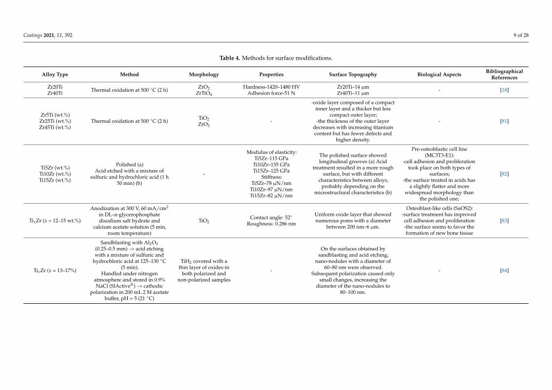
Coatings 2021, 11, 392 9 of 28
Table 4. Methods for surface modifications.
Alloy Type Method Morphology Properties Surface Topography Biological Aspects BibliographicalReferences
Zr20TiZr40Ti Thermal oxidation at 500 ◦C (2 h) ZrO2
ZrTiO4
Hardness-1420–1480 HVAdhesion force-51 N
Zr20Ti–14 µmZr40Ti–11 µm - [18]
Zr5Ti (wt.%)Zr25Ti (wt.%)Zr45Ti (wt.%)
Thermal oxidation at 500 ◦C (2 h) TiO2ZrO2
-
-oxide layer composed of a compactinner layer and a thicker but less
compact outer layer;-the thickness of the outer layer
decreases with increasing titaniumcontent but has fewer defects and
higher density.
- [81]
Ti5Zr (wt.%)Ti10Zr (wt.%)Ti15Zr (wt.%)
Polished (a)Acid etched with a mixture of
sulfuric and hydrochloric acid (1 h50 min) (b)
-
Modulus of elasticity:Ti5Zr–115 GPaTi10Zr–135 GPaTi15Zr–125 GPa
Stiffness:Ti5Zr–78 µN/nm
Ti10Zr–97 µN/nmTi15Zr–82 µN/nm
The polished surface showedlongitudinal grooves (a) Acid
treatment resulted in a more roughsurface, but with different
characteristics between alloys,probably depending on the
microstructural characteristics (b)
Pre-osteoblastic cell line(MC3T3-E1):
-cell adhesion and proliferationtook place on both types of
surfaces;-the surface treated in acids has
a slightly flatter and morewidespread morphology than
the polished one;
[82]
TixZr (x = 12–15 wt.%)
Anodization at 300 V, 60 mA/cm2
in DL-α-glycerophosphatedisodium salt hydrate and
calcium acetate solution (5 min,room temperature)
TiO2Contact angle: 52◦
Roughness: 0.286 nm
Uniform oxide layer that showednumerous pores with a diameter
between 200 nm–6 µm.
Osteoblast-like cells (SaOS2):-surface treatment has improvedcell adhesion and proliferation-the surface seems to favor theformation of new bone tissue
[83]
TixZr (x = 13–17%)
Sandblasting with Al2O3(0.25–0.5 mm)→ acid etchingwith a mixture of sulfuric and
hydrochloric acid at 125–130 ◦C(5 min).
Handled under nitrogenatmosphere and stored in 0.9%NaCl (SlActive®)→ cathodic
polarization in 200 mL 2 M acetatebuffer, pH = 5 (21 ◦C)
TiH2 covered with athin layer of oxides-in
both polarized andnon-polarized samples
-
On the surfaces obtained bysandblasting and acid etching,
nano-nodules with a diameter of60–80 nm were observed.
Subsequent polarization caused onlysmall changes, increasing the
diameter of the nano-nodules to80–100 nm.
- [84]
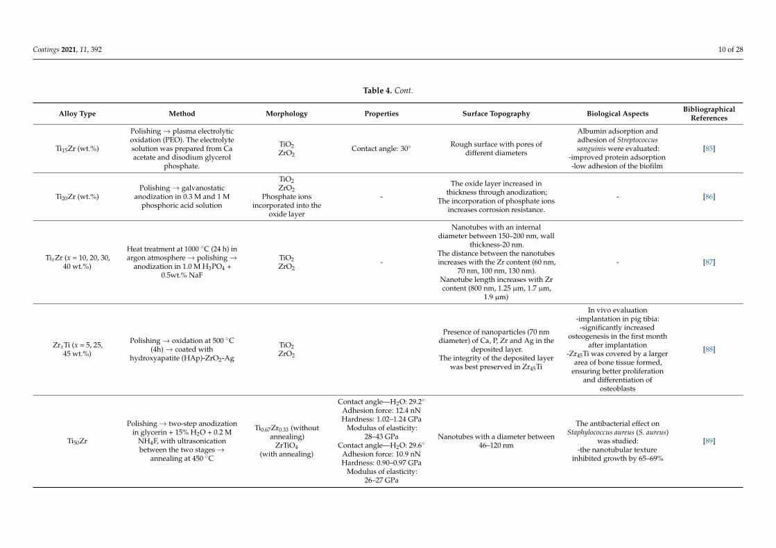
Coatings 2021, 11, 392 10 of 28
Table 4. Cont.
Alloy Type Method Morphology Properties Surface Topography Biological Aspects BibliographicalReferences
Ti15Zr (wt.%)
Polishing→ plasma electrolyticoxidation (PEO). The electrolytesolution was prepared from Caacetate and disodium glycerol
phosphate.
TiO2ZrO2
Contact angle: 30◦ Rough surface with pores ofdifferent diameters
Albumin adsorption andadhesion of Streptococcussanguinis were evaluated:
-improved protein adsorption-low adhesion of the biofilm
[85]
Ti20Zr (wt.%)Polishing→ galvanostatic
anodization in 0.3 M and 1 Mphosphoric acid solution
TiO2ZrO2
Phosphate ionsincorporated into the
oxide layer
-
The oxide layer increased inthickness through anodization;
The incorporation of phosphate ionsincreases corrosion resistance.
- [86]
TixZr (x = 10, 20, 30,40 wt.%)
Heat treatment at 1000 ◦C (24 h) inargon atmosphere→ polishing→
anodization in 1.0 M H3PO4 +0.5wt.% NaF
TiO2ZrO2
-
Nanotubes with an internaldiameter between 150–200 nm, wall
thickness-20 nm.The distance between the nanotubesincreases with the Zr content (60 nm,
70 nm, 100 nm, 130 nm).Nanotube length increases with Zrcontent (800 nm, 1.25 µm, 1.7 µm,
1.9 µm)
- [87]
ZrxTi (x = 5, 25,45 wt.%)
Polishing→ oxidation at 500 ◦C(4h)→ coated with
hydroxyapatite (HAp)-ZrO2-Ag
TiO2ZrO2
Presence of nanoparticles (70 nmdiameter) of Ca, P, Zr and Ag in the
deposited layer.The integrity of the deposited layer
was best preserved in Zr45Ti
In vivo evaluation-implantation in pig tibia:-significantly increased
osteogenesis in the first monthafter implantation
-Zr45Ti was covered by a largerarea of bone tissue formed,
ensuring better proliferationand differentiation of
osteoblasts
[88]
Ti50Zr
Polishing→ two-step anodizationin glycerin + 15% H2O + 0.2 M
NH4F, with ultrasonicationbetween the two stages→
annealing at 450 ◦C
Ti0.67Zr0.33 (withoutannealing)
ZrTiO4(with annealing)
Contact angle—H2O: 29.2◦
Adhesion force: 12.4 nNHardness: 1.02–1.24 GPa
Modulus of elasticity:28–43 GPa
Contact angle—H2O: 29.6◦
Adhesion force: 10.9 nNHardness: 0.90–0.97 GPa
Modulus of elasticity:26–27 GPa
Nanotubes with a diameter between46–120 nm
The antibacterial effect onStaphylococcus aureus (S. aureus)
was studied:-the nanotubular texture
inhibited growth by 65–69%
[89]
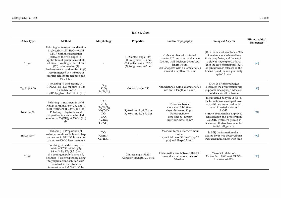
Coatings 2021, 11, 392 11 of 28
Table 4. Cont.
Alloy Type Method Morphology Properties Surface Topography Biological Aspects BibliographicalReferences
Ti50Zr
Polishing→ two-step anodizationin glycerin + 15% H2O + 0.2 M
NH4F, with ultrasonicationbetween the two stages→
application of gentamicin sulfatesolution→ coating with chitosan
(CS) by immersion (1)Surfaces treated as described in (1)
were immersed in a mixture ofsulfuric acid-hydrogen peroxide
for 2 h (2).
-
(1) Contact angle: 30◦
(1) Roughness: 319 nm(2) Contact angle: 52.5◦
(2) Roughness: 440 nm
(1) Nanotubes with internaldiameter 120 nm, external diameter230 nm, wall thickness 30 nm and
length 10 µm(2) Nanopores with a diameter of 70
nm and a depth of 100 nm.
(1) In the case of nanotubes, 68%of gentamicin is released in a
first stage, faster, and the rest ina slower stage up to 21 days.
(2) In the case of nanopores, 82%of gentamicin is released in thefirst 60 h, and the rest gradually
up to 10 days.
[68]
Ti50Zr (wt.%)
Polishing→ acid etching inHNO3: HF: H2O mixture (3:1:2)
→ anodization inK2HPO4/glycerol at 180 ◦C (2 h)
TiO2ZrO2
(ZrxTiyOz)Contact angle: 15◦ Nanochannels with a diameter of 35
nm and a length of 3.2 µm
RAW 264.7 macrophages:-decreases the proliferation rate-supports macrophage adhesion
but does not allow fusion
[90]
Ti50Zr (at.%)
Polishing→ treatment in 10 MNaOH solution at 60◦ C (24 h)→heat treatment at 600◦ C (1 h) (a)
Polishing→ hydrothermaldeposition in a supersaturated
solution of Ca(OH)2 at 200 ◦C (8 h)(b)
TiO2ZrO2
Na2Ti5O11Na2ZrO3
TiO2ZrO2
CaTiO3CaZrO3
Ra-0.62 µm; Rs-3.02 µmRa-0.60 µm; Rs-2.70 µm
Porous network-pore size: 0.4–1.0 um
-layer thickness: 12 µmPorous network
-pore size: 50–100 nm-layer thickness: 45 nm
In simulated body fluid (SBF),the formation of a compact layerof apatite was observed in the
case of treated surfaces.SaOS2:
-surface treatment has improvedcell adhesion and proliferation-Ca(OH)2 treatment proved to
be a more effective treatment forinitial cell growth
[91]
Ti50Zr (at.%)
Polishing→ Preparation ofcolloidal solutions TiO2 and HAp→ heating to 80 ◦C (2 h)→ spincoating→ 600 ◦C heat treatment
TiO2CaTiO3
Ca2Ti2O5
-
Dense, uniform surface, withoutcracks.
Layer thickness: 50 µm (TiO2 (25µm) and HAp (25 µm))
In SBF, the formation of anapatite layer was observed thatincreased in thickness with time.
[92]
Ti50Zr
Polishing→ acid etching in amixture 3:7 30 wt.% H2O2:98 wt.% H2SO4 (1.5 h)→
dip-coating in poly(lactic acid)solution→ electrospinning usingpolycaprolactone solution with
dissolved silver nitrate→immersion in 1 M NaOH (2 h)
- Contact angle: 52.45◦
Adhesion strength: 2.7 MPa
Fibers with a size between 180–700nm and silver nanoparticles of
30–40 nm
Microbial inhibition:Escherichia coli (E. coli): 74.27%
S. aureus: 66.02%[93]
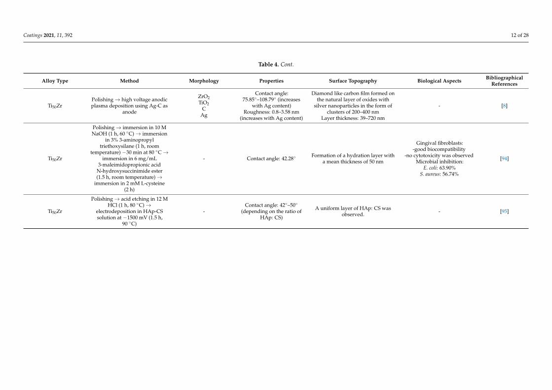
Coatings 2021, 11, 392 12 of 28
Table 4. Cont.
Alloy Type Method Morphology Properties Surface Topography Biological Aspects BibliographicalReferences
Ti50ZrPolishing→ high voltage anodicplasma deposition using Ag-C as
anode
ZrO2TiO2
CAg
Contact angle:75.85◦–108.79◦ (increases
with Ag content)Roughness: 0.8–3.58 nm
(increases with Ag content)
Diamond like carbon film formed onthe natural layer of oxides with
silver nanoparticles in the form ofclusters of 200–400 nm
Layer thickness: 39–720 nm
- [8]
Ti50Zr
Polishing→ immersion in 10 MNaOH (1 h, 60 ◦C)→ immersion
in 3% 3-aminopropyltriethoxysilane (1 h, room
temperature) −30 min at 80 ◦C→immersion in 6 mg/mL
3-maleimidopropionic acidN-hydroxysuccinimide ester(1.5 h, room temperature)→
immersion in 2 mM L-cysteine(2 h)
- Contact angle: 42.28◦ Formation of a hydration layer witha mean thickness of 50 nm
Gingival fibroblasts:-good biocompatibility
-no cytotoxicity was observedMicrobial inhibition:
E. coli: 63.90%S. aureus: 56.74%
[94]
Ti50Zr
Polishing→ acid etching in 12 MHCl (1 h, 80 ◦C)→
electrodeposition in HAp-CSsolution at −1500 mV (1.5 h,
90 ◦C)
-Contact angle: 42◦–50◦
(depending on the ratio ofHAp: CS)
A uniform layer of HAp: CS wasobserved. - [95]
Coatings 2021, 11, 392 13 of 28
3.3.1. Surface Chemistry
Even biomaterials such as TixZr, considered “inert”, degrade to some extent overtime, resulting in increased concentrations of metal ions. The release of metal ions fromthe implant takes place through the dissolution of the passive layer, mechanical wearand electrochemical corrosion. Metal ions can exist as such, but also in organometalliccomplexes, metal oxides or even nanoparticles [96]. Zirconium is considered an anodicalloying element for Ti, which reduces anodic activity [97].
Implant corrosion leads to a decrease in their resistance and the release of metal ionsin the surrounding tissues [98]. Valve metals, which develop a layer of oxide on the surfacethrough exposure to air, have good corrosion resistance [30,99]. If the oxide layer is affected,the metal ions are released continuously until repassivation takes place [30].
Despite the fact that the passive layer functions as a physical barrier responsible forcorrosion resistance that significantly hinders the release of metal ions into the surroundingtissues, small amounts of metal ions are still released from the metallic material. This pro-cess is a part of transient breakdown and reforming events of this film and to metal debriswith acidification as a detriment to the stability of the passive regime. The breakdownof the titanium oxide layer by nucleation of corrosion pits occurs in bioliquids such asRinger’s solution at electrode potentials, well below the pitting potential. The frequency ofbreakdown increases significantly with pH value decrease, with temperature increase andin particular in the presence of chloride anions [100–103].
A study evaluating the corrosion resistance of Ti, TixZr (x = 30, 50, 70%) and Zr ina lactic acid + NaCl solution and artificial saliva solution shows that the addition of Zrsignificantly increases the corrosion resistance, but up to a maximum content of 50% [50].
Although the oxide layer forms naturally on the surface of TixZr alloys, it is thin andweak. To improve Zr20Ti and Zr40Ti implants, oxidation at temperature (500 ◦C, 2 h) wasproposed, which led to the formation of an oxide layer of ZrO2 and ZrTiO4 with a thicknessof 11–14 µm. The oxide layer formed had both increased corrosion resistance and betterwear resistance, the wear rate being almost 10 times lower in the case of Zr20Ti and almost20 times in the case of Zr40Ti [18].
Regarding the electrochemical stability of TixZr implants, it was observed that thepresence of fluoride anions can have a negative impact, favoring corrosion. Given thatthese implants are frequently used as dental implants and that fluoride is introducedinto toothpaste and mouthwash to prevent cavities, solutions must be found. It has beenobserved that the oxide film reacts with these anions, resulting in titanium fluoride orsodium titanium fluoride. The thermal oxidation of TixZr alloys in air at 500 ◦C for twohours appears to significantly reduce the effect of fluoride [104] (pp. 7–8). Another studyregarding the corrosion resistance of Zr−Ti alloys shows too that the thermal oxidation inair at 500 ◦C does improve the corrosion resistance and suggests that prior to the use ofsuch alloys as implant materials, a treatment consisting of thermal oxidation in air followedby exposure to a physiological solution should be applied [105].
The fluoride effect was tested on Zr5Ti, Zr25Ti and Zr45Ti alloys in acidic artificialsaliva, Ph = 3 with a NaF concentration of 0.2 wt.%, 0.5 wt.% and 1 wt.%. The alloyswere tested as such and after thermal oxidation in air at 500 ◦C for two hours. The resultsshowed that although acidic artificial saliva with fluoride is a very aggressive mediumfor ZrxTi alloys, thermal oxidation is an effective method of surface treatment. The oxidelayers formed by TiO2 and ZrO2 provided a good corrosion resistance [106].
Another study performed on Zr5Ti, Zr25Ti and Zr45Ti in artificial saliva solution, inthe presence of NaF (0.05 wt.%, 0.1 wt.% and 0.2 wt.%) and albumin (0.6 wt.%) showedthat Zr45Ti has better corrosion resistance [107]. The same alloys were tested in Ringer’ssolution (pH = 6.8) [108] and physiologically acidified Ringer’s solution at pH = 3, alsoevidencing that Zr45Ti has the highest corrosion resistance. Moreover, it was observed thatafter thermal oxidation at 500 ◦C, the oxide layers became thicker and more stable [81]. Thehigher Ti content (45%) seems to lead to the suppression of pitting corrosion [109].
Coatings 2021, 11, 392 14 of 28
The evaluation of the oxide layer and its corrosion resistance on Ti20Zr alloy showedthat the passivation takes place more easily and that the oxide layer has better corrosionresistance in Ringer’s solution at acidic, neutral and alkaline pH compared to Ti. It was alsoobserved that at acidic pH, calcium and phosphate ions were deposited on the surface, andat neutral and alkaline pH a protective layer of hydroxyapatite (HAp) was formed [110].
The surface degradation of commercial Ti, Zr, and ZrO2 caused by bacterial adhesion(in particular, the Streptococcus species) has been discussed in several papers in the litera-ture [111–113] concluding more recently that dental implant surfaces of TiZr and ZrO2 arenot more susceptible to colonization than commercially pure Ti implants [114,115].
In addition to surface composition, the internal characteristics of metallic biomaterials,such as allotropic phase and defects, also influence the initiation and propagation ofcorrosion [116].
3.3.2. Surface Topography
Comparing the results reported on the surface roughness of biomaterials is difficultto achieve because there are variations regarding the analytical techniques and how theresults are reported. Moreover, the terminology used is not harmonized and the sameterms may refer to different procedures. However, it is generally accepted that surfaceswith higher roughness favor osseointegration compared to smoother ones [117,118].
In addition to the modification techniques, which determine the properties of the cre-ated surfaces, the methods of investigating them are also of major importance. Techniquessuch as nanoindenting and scratching techniques are used to characterize submicrometricsurfaces. Nanoindentation allows for the evaluation of hardness and Young’s modulus.The scratch test allows for the evaluation of coating adhesion to the substrate [19,66].
Rough surfaces can be differentiated into two categories: modified surfaces by coating(e.g., HAp) or without coating (e.g., sandblasting, acid etching). Electrochemical changes,such as anodic oxidation or NaOH treatment lead to the formation of biomimetic surfacesby promoting the formation of apatite [119].
Obtaining structured surfaces on a micrometric scale is generally carried out by acidetching, sandblasting (SB) or oxidation. Sandblasting involves the use of abrasive particles(Al2O3, TiO2, SiO2) at high pressure. This technique can lead to the incorporation of theparticles used in the material and thus to the modification of chemical properties andreduced corrosion resistance. To solve this problem, a subsequent acid etching step isgenerally used to dissolve at least some of the projected particles. Acid etching producessimilar results to sandblasting. The results obtained vary depending on the exposure time,type of acids used and temperature [117].
There is a TixZr alloy marketed under the name Roxolid-SLA® (Straumann, Basel,Switzerland) [11,13,120–123]. This alloy contains 13–17% zirconium and is beginning tobe considered the first choice in medical procedures [124]. The smaller grains of 1–2 µm,compared to 20–30 µm in cpTi lead to increased overall strength [125]. Another variantalso produced by Straumann is RXD-SLActive® this implant is hydrophilic and has ananostructured surface compared to Roxolid-SLA® [126].
The surface named SLA® is obtained by sandblasting with corundum (particle size250–500 µm) followed by etching with a concentrated mixture of hydrochloric acid andsulfuric acid. The implant is then cleaned with nitric acid, rinsed with deionized water anddried in an ambient atmosphere. For the SLActive® surface, the difference lies in rinsingwith an NaCl solution in a nitrogen atmosphere and storage in 0.15 M saline solution. Thischange, apparently minor, leads to a hydrophilic surface with better properties in termsof osseointegration. The formation of nanostructures was also observed on the SLActive®
surface [127].To highlight the differences between mechanical processing and acid etching, the
properties of TixZr alloys (x = 5, 10, 15 wt.%) with these two types of surface treatmentswere evaluated. The mechanically prepared samples were polished with an automaticpolisher, and the etched ones were immersed in a mixture of sulfuric and hydrochloric acid
Coatings 2021, 11, 392 15 of 28
for 1 h and 50 min. Acid etching treatment produced a rougher surface and better overallproperties [82].
The comparative study of the surface Ti, TixZr after sandblasting and acid etchingrespectively, showed that in the case of TixZr, the hydrogen concentration is 1.9 timeshigher. The formation of hydrides favored by the presence of Zr increases the roughness bythe appearance of nanostructures, which in the case of the Ti surface was not observed. Itwas also observed that the hydride layer is below the oxide layer, which can be explainedin the case of sandblasting-acid etching (SBAE) by the fact that the oxide layer is formedafter the formation of hydride, but also by the fact that oxygen could replace hydrogen inthe outer layer [128].
3.4. Coatings with TiZr
To obtain the advantages of TixZr alloys, several studies have been developed thataimed to cover other substrates with these metals.
Through the plasma surface alloying technology, Zr ions were implanted in thesurface of a Ti material, thus creating a TixZr alloy only in the surface layer. The alloys thuscreated had a Zr content of 1.09 at.%, 1.77 at.% and 3.68 at.%. The surface hardness andcorrosion resistance proved to be better than that of cpTi. Good biocompatibility was alsoobserved [17].
A TixZr surface was also obtained on Ti alloy by ion-assisted arc-plasma deposition invacuum. The deposition was made by using two cathodes, one of Ti and one of Zr, whichwere arc evaporated in Ar atmosphere, at 0.2 Pa. The thickness of the obtained layer was5 µm, with a Zr content of 11 wt.% and 22 wt.%. The material thus obtained showed a lowmodulus of elasticity, of 77–98 GPa, compared with the Ti substrate which had 110 GPa.Moreover, nanoindentation showed an increase in resistance to plastic deformation withincreasing Zr content from 11 wt.% to 22 wt.% [129].
The use of a layer of TixZr deposited on a steel material, SS304, also led to a significantimprovement in corrosion resistance [130].
The explosive spraying of Ti−Zr coating could help as well by reducing the Young’smodulus of the materials used as substrate (cpTi or Ti6Al4V), therefore reducing stressshielding [131].
Ti−Zr films were also deposited on Ti substrates by MAO, obtaining a porous crys-talline layer of titanium and zirconium oxides. It has been observed that this approachleads to increased biocompatibility and that cell development increases proportionallywith Zr content [132].
The incorporation of zirconium oxide into the oxide layer of Ti by plasma electrolyticoxidation (PEO) is another possible approach. A rougher surface was thus obtained, whichby immersion in simulated body fluid (SBF) led to the formation of an apatite layer [133].It was also shown that through this approach, the oxide layer formed favored osteoblastadhesion and could even decrease the bacterial adhesion [134]. Besides this, it was shownthat nano-ZnO have antibacterial properties [135].
A possible coating that could lead to improved properties would be that with ZrTiO4by the sol-gel method. The synthesis method has been developed but has not yet beenapplied to biomaterials [136].
3.5. Nanostructured Coatings
The design of nanometer-scale biomaterial surfaces is a subject of continuous re-search [137–140]. Nanostructured surfaces influence the chemical reactivity of materialsand implicitly the biomolecular interactions [141,142]. It has been observed that nanoscalechanges promote osteoinduction and biomaterial–tissue interaction and that implants withnanotubular surfaces show a significant improvement in bone creation and gene expressioncompared to implants without nanostructured surfaces [143–147].
Coatings 2021, 11, 392 16 of 28
3.5.1. Nanotubes
Anodizing may lead to self-ordered nanotubes, with certain dimensions (diameter,length) by varying the anodizing conditions. Nanostructures created in this way may havea partially crystalline structure. Their morphology influences electrochemical stability,wettability and biocompatibility [124].
The formation of nanotubes on TiZr substrate can be accomplished by a two-stepanodizing procedure. They can be annealed in air or reduced in the atmosphere by Ar/H2.By annealing, the mixed oxide tubes are converted to zirconium titanate (ZrTiO4), and byreduction to (Zr0.333Ti0.666)O2 [124].
Nanotubes were reported to be obtained by two-step anodizing using an electrolytesolution with glycerin, 15% water and 0.2 M NH4F. The first anodization was performed at55 V for 4 h, and then the samples were ultrasonicated to remove the formed structures.The second anodization was performed at 75 V for 1 h. The formation of nanotubes is theresult of a competition between the electrochemical formation of oxides and the chemicaldissolution of oxides by the fluoride anion. Reactions can be described as follows [68]:
Me + 2H2O → MeO2 + 4H+ + 4e− (8)
MeO2 + 4H+ + 6F− → [MeF6]2− + 2H2O (9)
In the case of nanotubes formed on TiZr type alloys, the diameter of the nanotubesdecreases with increasing Zr content. Nanotubes with a diameter of approximately 50 nmand a length of 17 nm were obtained. Regarding the heat treatment, fluorine can be removedat 300 ◦C, and at 800 ◦C crystallization occurs, but also the collapse of nanotubes [148].
The formation of TiO2 nanotubes by anodization at 10 V in 1.0 M phosphoric acid and0.5% NaF for 2 h was also studied on TixZr alloys (x = 10, 20, 30, 40%). The nanotubes hadan average diameter of 150–200 nm and a wall thickness of 20 nm. The distance betweenthe nanotubes increased with increasing Zr content of the alloy, being 60, 70, 100 and130 nm [87].
Nanotubes were also obtained on Ti50Zr substrate by two-step anodizing at differentvoltages (15, 30, 45 V) in glycol with 15% H2O and 0.2 M NH4F. The layer formed in thefirst anodization was removed by ultrasonic treatment, thus obtaining a prepared surfaceso that in the second stage of anodization nanotubular structures with a high degree oforganization were obtained [149].
The zirconium titanate (ZrTiO4) nanotubes formed on such alloys have increasedstructural flexibility compared to pure TiO2 nanotubes. The diameter and length of theformed tubes can vary depending on the anodizing potential used without damaging thestructural configuration, at higher potentials the amorphous structure predominates overthe crystalline one [145,150]. Another important aspect, less studied, is that nanotubesformed in this way can have an antibacterial role [150].
3.5.2. Other Nanostructured Surfaces
It was observed that nanopores were obtained on Ti50Zr by anodizing at 5 V and 10 Vin a solution of 1 M (NH4)2SO4 and 0.5 wt% NH4F. The diameter of the nanopores wasincreasing with the applied potential. The material was subsequently annealed at 500 ◦Cfor three hours. The surfaces thus formed had an antibacterial effect, even if smaller thanin the case of nanotubes [150].
The formation of nanopores was also observed by immersing an alloy of Ti50Zr withnanotubular surface obtained by two-step anodizing in a solution of H2SO4 (37 N)/H2O2(30%) for two hours at room temperature [68] and when using spark anodization in asolution of glycerophosphate and calcium acetate on TixZr (x = 12–15 wt.%) [83].
The surface of a Ti20Zr alloy was galvanostatically anodized in phosphoric acidsolution to increase the corrosion resistance. The current density was 10 mA/cm2, theconcentration of phosphoric acid was 1 M, and the anodizing time was 45 min. It has beenobserved that this approach results in a nanometric layer consisting mostly of crystalline
Coatings 2021, 11, 392 17 of 28
TiO2 with incorporated phosphorus ions. The corrosion resistance was 10 times higher inthe case of the anodized sample [86].
Anodization in a hot solution of glycerol-phosphate of a Ti50Zr alloy led to one-dimensional nanostructures in the form of channels [90,151]. As a result of this treatment,better corrosion resistance and higher hydrophilicity were obtained. Additionally, in vitrostudies on RAW 264.7 macrophages showed a reduced reaction in the inflammatory re-sponse [90].
In a study on the cathodic polarization of some TiZr materials (13–17% Zr) with asurface previously processed by sandblasting with aluminum oxide particles (0.25–0.5 mm),the acid etching treatment with a mixture of hydrochloric acid and sulfuric acid at 125–130 ◦Cfor 5 min produced nano-nodules [84].
Another type of nanostructured coating was made with two biopolymers, poly(lacticacid) and polycaprolactone. The methods used were dip coating and electrospinning,respectively, thus obtaining nanofibers [93].
3.6. Biomimetic Coatings
On a Ti15Zr alloy, obtained by mechanical processing (Straumann AG), a biofunctionalcoating was formed by PEO. As the control, the same unmodified alloy was used, andsome were SLA®-treated (sandblasting and acid etching). The samples were used as ananode, while the cathode was a steel tank equipped with a cooling system. The electrolytesolution contained calcium acetate and disodium glycerol phosphate. The samples wereimmersed in 500 mL of solution for 10 min, using a voltage of 290 V and a frequency of250 Hz. During the electrochemical treatment, pores appeared in the places where themicro-discharge took place, thus obtaining molten oxides which were quickly cooled inthe presence of electrolytes when the spark was extinguished. This phenomenon led tothe incorporation of Ca and P, resulting in a Ca/P ratio close to that of HAp. This aspectcombined with the increased surface roughness significantly influenced the biologicalproperties of the implant. In terms of stability, the PEO-treated alloy showed the highestcorrosion resistance compared to control ones [85].
Two other ways to modify the surface of a Ti50Zr at.% alloy have been proposed. Oneof the methods involved immersing the sample in 10.0 M NaOH at 60 ◦C for 24 h. Thesamples were dried at room temperature in air for another 24 h. They were then heattreated at 600 ◦C for one hour in an electric high vacuum furnace. The other methodinvolved the hydrothermal storage in a supersaturated solution of 0.2 M calcium hydroxidein an autoclave at 200 ◦C for eight hours. In the first case, a 12 µm oxide layer composed ofTiO2, ZrO2, Na2Ti5O11 and Na2ZrO3, and in the second case a 45 nm layer composed ofTiO2, ZrO2, CaTiO3 and CaZrO3 was formed. When immersed in SBF, a layer of apatitewas observed in the case of both treated surfaces, but not in the untreated ones [91].
In the case of alkaline treatment, the native layer partially dissolves in the alkalinesolution, forming HTiO3-H2O anions, which combine with the cations to form a hydrogellayer. During the heat treatment, this layer is dehydrated resulting in an amorphous orcrystalline form of alkali titanate [152].
The deposition of a HAp/TiO2 layer on TiZr alloy has been reported by the sol-gelmethod at 3000 rpm for 15 s, followed by heat treatment at 600 ◦C for 20 min. Subsequentheat treatment at 600 ◦C for another 20 min resulted in HAp crystallization. The obtainedlayer had a total thickness of 50 µm, being formed in equal proportion of TiO2/HAp [92].Regardless of the sol-gel method used, cracks may occur due to contractions caused bythe evaporation of a large volume of solvent [153]. In the case of TiO2 coating, the formedcracks were subsequently coated with HAp. The mechanism of apatite formation in SBFcan be explained by the release of Ca2+, Na+, K+, ions from the HAp deposited layer andtheir exchange with hydronium ions from the SBF solution, forming Ti−OH, Zr−OHgroups. These functional groups can react with water molecules in the environment byinducing nucleation. Once formed, apatite nuclei can grow spontaneously by consumingions from the SBF solution [92].
Coatings 2021, 11, 392 18 of 28
The coating of TiZr with HAp was also performed in combination with chitosan (CS),obtaining coatings with different porosities and contact angles depending on the HAp/CSratio used [20,95,154].
Another type of biomimetic coating used on TixZr type alloys (Ti5Zr, Ti25Zr and Ti45Zr)can be the coating with HAp-ZrO2-Ag, which favors the formation of bone tissue [88,155].Silver nanoparticles (AgNPs) are effective, in very low concentrations (0.5–1.0%), in prevent-ing the formation of bacterial biofilm. They interact with proteoglycans on the membrane ofbacterial cells and inside them. Silver ions can also interact with sulfuryl groups preventingthe replication of bacterial DNA [156–158].
Moreover, the development of bioinductive surfaces could increase the healing capac-ity of bone tissue and is a solution for patients with risk factors [70,159]. The implantationof a biologically active molecule (parathyroid hormone fragment 1−34) in the natural oxidelayer was performed on a TiZr implant (Straumann AG) [70].
3.7. Biological Aspects
The interactions between cells and biomaterial are determined by the surface proper-ties [160,161]. Cells do not interact directly with the surface of the biomaterial, but withthe protein layer adsorbed on the surface. The transcriptomic and proteomic technologiesused to create gene and protein expression profiling can be used for a more thoroughunderstanding and to predict the biocompatibility of the researched materials [162–165].
The interaction between human gingival fibroblasts and TixZr materials with surfacestreated in different ways shows that cell adhesion and differentiation are influenced bysurface properties [166,167]. Another study that looked at the expression of several genesinvolved in the process of cell adhesion shows that high surface energy positively influencesthe adhesion of osteoblasts [168].
Microbial infections and in particular infections with multidrug resistant microbes area major problem that must be considered in the development of all biomaterials [169–171].Recent research focuses on the use of nanotechnology in solving this problem, nanometri-cally structured materials having characteristics far superior to conventional ones (bulkform). Additionally, the reactive oxygen species produced by the use of nanocompositeoxide metals or nanoparticles cause the inhibition of bacterial growth [170].
Bacterial adhesion can lead initially to mucositis and later to periimplantitis, which cancause implant loss. Thus this aspect is approached and studied in many studies [172–174].Surface properties, such as roughness, free energy (wettability) and chemical compositionare the determining factors in terms of cell adhesion and microbial colonization [173,175,176].
Surface roughness, chemical composition, wettability and surface charge also influencethe structure and size of the biofilm. Although it is a subject often studied, the resultsobtained in vitro may differ from those obtained in vivo. The differences appear becauseit is difficult to imitate the biologically complex environment. In vivo studies show thatbacterial adhesion increases with increasing roughness and hydrophobic surfaces promotethe accumulation of proteins, which serve as a binding site for bacteria [177].
In a time with more aggressive bacteria and viruses, for the success of dental implants,besides the incorporation of efficient antibacterial agents inside implants, loading of the anti-inflammatory drugs to reduce inflammation and bone forming proteins/peptides/growthfactors (bone morphogenetic protein-2/BMP-2, parathyroid hormone, Zn/Ag/Sr/Mgions/nanoparticles) to enhance new bone formation at the implant–bone interface havebeen applied [178–180]. More recently, creating composite structures with graphene andnanotubes on the implant surface proved to have efficient dual antibacterial and osteogenicproperties [181].
Another important aspect is related to the tribocorrosion of metallic materials. Thereleased metal nanoparticles (metal wear debris) can migrate to any part of the body (exceptthe brain), depositing in the lymph nodes, the liver, spleen and bone marrow. They alsohave a genotoxic potential, which can affect the genetic material, which could lead to canceror birth defects [182].
Coatings 2021, 11, 392 19 of 28
The dissolution of the oxide layers is relatively low in vitro because the potentialchanges are small, but in the body, this process can be accelerated by the presence ofamino acids and proteins. Another process that can accelerate this phenomenon is thegeneration of reactive oxygen species by macrophages. Superoxide dismutase catalyzesO2− producing H2O2 that hyperoxidize the surface of the material. The process thattakes place simultaneously with the dissolution is the reprecipitation, the two being inequilibrium. Phosphate ions are adsorbed on the outer oxide layers of Ti alloys, andsubsequently, calcium ions are adsorbed by them forming an apatite layer. In addition tothese, proteins, sulfites and sulfates have also been observed [183,184].
3.7.1. The Influence of Surfaces In Vitro
TixZr alloys (x = 5, 10, 15, 20 wt.%) were studied, proving that they do not showcytotoxicity [185]. Moreover, compared to other commercial Ti-based alloys, cells onTiZr implants expressed a decrease in pro-inflammatory markers and an increase in anti-inflammatory markers, especially in the case of nanostructured alloy [126].
The formation of nanotubes, mechanical properties and inhibition of bacterial growthof S. aureus was studied on pure Ti and Zr alloys, as well as on several TixZr binary alloys(x = 10, 30, 50, 70 at.%), noting that nanotubular formations inhibit bacterial growth [89]. Inthe case of TiO2 nanotubes, it has been observed that annealing can increase the inhibitoryeffect on S. aureus and Pseudomonas aeruginosa and even that it increases depending on thetemperature used [186].
The properties of nanotubes formed on Ti50Zr by two-step anodizing were also evalu-ated, in vitro, on E. coli culture. It was observed that the antibacterial efficacy depends onthe diameter of the tubes formed, a size that can be controlled by the potential used foranodizing. Tubes with a diameter of less than 20–30 nm demonstrated a better bacteriostaticand bactericidal effect than those with a larger diameter of 50–70 nm [187].
Nanotubes can also be loaded with therapeutic agents, either as such or embeddedin a polymer, by electrospray, lyophilization, immersion or vacuum impregnation tech-niques [188]. On a Ti50Zr alloy two types of nanostructures were formed, nanopores andnanotubes, that were loaded with gentamicin and coated with a layer of CS. It was ob-served that gentamicin was released from nanopores in 10 days, and from nanotubes in21 days [68].
Another coating with a zwitterionic cysteine drug was investigated recently, reportingbetter performance in terms of stability, biocompatibility and antibacterial effect. The ob-tained structure evidenced large bands due to hydroxylic groups which formed a hydrationlayer and determined the increase in hydrophilic character. This layer was responsiblefor an antibacterial effect as well. The electrochemical tests performed in NaCl 0.9% asbioliquid confirmed the improved stability of the coated sample. The cell behavior inthe presence of cysteine coating was determined with gingival fibroblasts, by measuringlactate dehydrogenase activity, concentrations of nitric oxide and the level of reactiveoxygen species. The results obtained indicated that the coating is biocompatible, and nocytotoxicity was evidenced [94].
The cell morphology of MG63 cells (osteoblast-like cells) on Ti50Zr with a nanostruc-tured surface also showed good biocompatibility [149], while the studies performed onRAW 264.7 macrophages showed a reduced reaction in inflammatory response in the caseof unidimensional nanochannels [90].
The incorporation of Ag into Ti50Zr showed strong antibacterial activity on S. au-reus [189] and coating a Ti50Zr alloy with poly (lactic acid), polycaprolactone and silvernanoparticles showed good inhibitory activity in E. coli and S. aureus [93].
Amorphous biomaterials such as Zr46Ti40Ag14 have been studied too as an alternativeto conventional crystalline ones [189]. They do not have structural deficiencies characteristicof crystalline structures (dislocation, vacancy, twinning, grain boundary), which can leadto better mechanical properties and increased corrosion resistance, as well as antibacterialproperties due to the Ag [190].
Coatings 2021, 11, 392 20 of 28
In the case of surfaces treated in alkaline solution, and subsequently, heat treated,better cell adhesion and proliferation were observed for SaOS2 [91]. Coating a Ti-basedalloy with CaTiZr3(PO4)6 also showed that the coating promoted cell proliferation andbone formation [191].
3.7.2. The Influence of Surfaces In Vivo
TixZr implants (x = 13–17%) prepared by sandblasting and acid etching evaluated for12 weeks in a study in rabbits show biocompatibility as good as in the case of cpTi [192]. Inanother study on titanium−zirconium (SLActive surface treatment) implants in rabbits, anincreased quality was shown regarding bone formation in the case of TiZr compared withTi. The evaluation was carried out through the removal of torque values [193].
Additonally, a better response in the case of SLActive® (nanostructured surface)highlighted in a study on human bone mesenchymal stem cells (hBMSCs) [194] is confirmedby a retrospective evaluation in another study of 154 Roxolid implants with SLActive® in107 patients that showed high survival rates [122]. TiZr implant alloys, in general, provedpositive outcomes in numerous studies [120,121,123,195–197]. Despite the success rateof dental TiZr implants, avoiding the acute surgical site infections in some patients isstill challenging [198] due to several unsuccessful events [199] and researchers suggestedcoating therapeutic drugs on the biomaterials surface to locally release antibiotics in acontrolled manner [200].
By using a cathodic polarization setup, a doxycycline loading of a TiZr alloy with aZr content of 13–17% was performed. The method of preparation of the sample was thatused for the preparation of Roxolid SLActive®, namely grit blasted and etching in sulfuricand hydrochloric acid. Samples were handled in a nitrogen atmosphere and stored in0.9% NaCl. The coating was made by connecting the samples to the cathode, the anodebeing Pt. A constant current of 0.65 mA was used for each sample. The electrolyte was a2 M acetate buffer solution in which doxycycline was dissolved to obtain 1 mg/mL. Theprocess took 75 min and was performed at room temperature. The samples were furtherdried and stored in a nitrogen atmosphere. The coated surface evaluated in a rabbit tibialmodel showed increased markers related to bone formation [201].
Following the idea that coating implantable metal alloys with active substances couldbe a solution for controlling inflammation and post-interventional infections, the useof a doxycycline-coated TixZr alloy compared to the same doxycycline-free alloy wasstudied as well in dogs and rabbits. The coating did not produce different histological andhistomorphometrical results compared to the control group [200].
Three binary TiZr alloys (Ti5Zr, Ti25Zr and Ti45Zr) coated with a layer of HAp-ZrO2-Ag were tested after implantation in pig tibia. It was observed that osseointegration wasinfluenced both by the interaction between the tissue and the coating and by the chemicalcomposition of the bulk alloy. The Ti45Zr alloy showed a better organizational structureof newly formed bone tissue, as well as increased proliferation and differentiation ofosteoblasts [88].
Another in vivo study in mice shows that Ti materials with a nanostructured coatingof TiO2 or ZrO2 have a positive influence on cell differentiation [202], while another studyperformed on sheep shows that the anodizing of both Ti and TixZr (x = 12–15 wt.%)improves osseointegration [203].
4. Conclusions
The critical approach decided upon in this review has led to the clear conclusion thatTixZr alloys have all the characteristics of a good biomaterial. Not only do they exhibitgreat biocompatibility, but the mechanical properties are improved up to 2.5 times inTi50Zr compared to Ti or Zr. Moreover, the surface modification methods described in thereviewed studies suggest that with the right combination of chemical and instrumentaltechniques, many characteristics and properties can be molded.
Coatings 2021, 11, 392 21 of 28
The alloys are naturally protected against corrosion through a layer of oxides, that canbe easily further improved through thermal oxidation. More complex methods such asanodizing or sol-gel coatings can be used to obtain different nanostructures that improvecellular interaction, being similar in size. Of course, the risk of bacterial biofilm formationincreases too, but besides the antibacterial effect of some of the nanostructures, antibioticsubstances or other nanoparticles that inhibit bacterial growth may be used in the coatingas well. Furthermore, coatings with hydroxyapatite can improve cell differentiation andproliferation. Additionally, all the studies performed in vivo, that we know of, showedpromising results.
Taking into consideration all the above-mentioned aspects, we believe that TiZr maybecome one of the most used metallic biomaterials in the future.
Author Contributions: Conceptualization, D.I. and I.D.; investigation, R.N., D.I., and I.D.; datacuration, R.N., D.I., and I.D.; writing—original draft preparation, R.N., D.I., and I.D.; writing—reviewand editing, D.I., and I.D.; visualization, R.N.; supervision, I.D. All authors have read and agreed tothe published version of the manuscript.
Funding: This research received no external funding.
Institutional Review Board Statement: Not applicable.
Informed Consent Statement: Not applicable.
Data Availability Statement: Data sharing not available.
Conflicts of Interest: The authors declare no conflict of interest
References1. Iftikhar, S.; Jahanzeb, N.; Saleem, M.; Rehman, S.; Matinlinna, J.P.; Khan, A.S. The trends of dental biomaterials research and
future directions: A mapping review. Saudi Dent. J. 2021. [CrossRef]2. Ho, W.-F.; Chen, W.-K.; Wu, S.-C.; Hsu, H.-C. Structure, mechanical properties, and grindability of dental Ti–Zr alloys. J. Mater.
Sci. Mater. Med. 2008, 19, 3179–3186. [CrossRef]3. He, X.; Reichl, F.X.; Milz, S.; Michalke, B.; Wu, X.; Sprecher, C.M.; Yang, Y.; Gahlert, M.; Röhling, S.; Kniha, H.; et al. Titanium
and zirconium release from titanium-and zirconia implants in mini pig maxillae and their toxicity in vitro. Dent. Mater. 2020, 36,402–412. [CrossRef] [PubMed]
4. Li, J.; Jansen, J.A.; Walboomers, X.F.; van den Beucken, J.J. Mechanical aspects of dental implants and osseointegration: Anarrative review. J. Mech. Behav. Biomed. Mater. 2020, 103, 103574. [CrossRef]
5. Osman, R.B.; Swain, M.V. A critical review of dental implant materials with an emphasis on titanium versus zirconia. Materials2015, 8, 932–958. [CrossRef]
6. Shekhawat, D.; Singh, A.; Banerjee, M.K.; Singh, T.; Patnaik, A. Bioceramic composites for orthopaedic applications: A compre-hensive review of mechanical, biological, and microstructural properties. Ceram. Int. 2021, 47, 3013–3030. [CrossRef]
7. Ionita, D.; Bajenaru-Georgescu, D.; Totea, G.; Mazare, A.; Schmuki, P.; Demetrescu, I. Activity of vancomycin release frombioinspired coatings of hydroxyapatite or TiO2 nanotubes. Int. J. Pharm. 2017, 517, 296–302. [CrossRef]
8. Stoian, A.B.; Surdu-Bob, C.; Anghel, A.; Ionita, D.; Demetrescu, I. Investigation of High Voltage Anodic Plasma (HVAP) Ag-DLCCoatings on Ti50Zr with Different Ag Amounts. Coatings 2019, 9, 792. [CrossRef]
9. Mani, G.; Feldman, M.D.; Patel, D.; Agrawal, C.M. Coronary stents: A materials perspective. Biomaterials 2007, 28, 1689–1710.[CrossRef] [PubMed]
10. Jiang, J.; Zhou, C.; Zhao, Y.; He, F.; Wang, X. Development and properties of dental Ti–Zr binary alloys. J. Mech. Behav. Biomed.Mater. 2020, 112, 104048. [CrossRef]
11. Altuna, P.; Lucas-Taulé, E.; Gargallo-Albiol, J.; Figueras-Álvarez, O.; Hernández-Alfaro, F.; Nart, J. Clinical evidence on titanium-zirconium dental implants: A systematic review and meta-analysis. Int. J. Oral Maxillofac. Surg. 2016, 45, 842–850. [CrossRef]
12. Ionita, D.; Pirvu, C.; Stoian, A.B.; Demetrescu, I. The Trends of TiZr Alloy Research as a Viable Alternative for Ti and Ti16 ZrRoxolid Dental Implants. Coatings 2020, 10, 422. [CrossRef]
13. Sharma, A.; Waddell, J.N.; Li, K.C.; Sharma, L.A.; Prior, D.J.; Duncan, W.J. Is titanium–zirconium alloy a better alternative to puretitanium for oral implant? Composition, mechanical properties, and microstructure analysis. Saudi Dent. J. 2020, 4–11. [CrossRef]
14. Michelle Grandin, H.; Berner, S.; Dard, M. A review of Titanium Zirconium (TiZr) alloys for use in endosseous dental implants.Materials 2012, 5, 1348–1360. [CrossRef]
15. Kondoh, K.; Fukuo, M.; Kariya, S.; Shitara, K.; Li, S.; Alhazaa, A.; Umeda, J. Quantitative strengthening evaluation of powdermetallurgy Ti–Zr binary alloys with high strength and ductility. J. Alloys Compd. 2021, 852, 156954. [CrossRef]
Coatings 2021, 11, 392 22 of 28
16. Möller, B.; Terheyden, H.; Ail, Y.; Purcz, N.M.; Hertrampf, K.; Tabakov, A.; Behrens, E.; Wiltfang, J. A comparison of biocompati-bility and osseointegration of ceramic and titanium implants: An in vivo and in vitro study. Int. J. Oral Maxillofac. Surg. 2012, 41,638–645. [CrossRef] [PubMed]
17. Yan, B.; Tan, J.; Wang, D.; Qiu, J.; Liu, X. Surface alloyed Ti–Zr layer constructed on titanium by Zr ion implantation for improvingphysicochemical and osteogenic properties. Prog. Nat. Sci. Mater. Int. 2020, 30, 635–641. [CrossRef]
18. Cui, W.F.; Shao, C.J. The improved corrosion resistance and anti-wear performance of Zr-xTi alloys by thermal oxidation treatment.Surf. Coat. Technol. 2015, 283, 101–107. [CrossRef]
19. Vardaki, M.; Pantazi, A.; Demetrescu, I.; Enachescu, M. Assessing the Functional Properties of TiZr Nanotubular Structures forBiomedical Applications, through Nano-Scratch Tests and Adhesion Force Maps. Molecules 2021, 26, 900. [CrossRef]
20. Ionita, D.; Vardaki, M.; Stan, M.S.; Dinischiotu, A.; Demetrescu, I. Enhance Stability and in vitro Cell Response to a BioinspiredCoating on Zr Alloy with Increasing Chitosan Content. J. Bionic Eng. 2017, 14, 459–467. [CrossRef]
21. George, E.P.; Raabe, D.; Ritchie, R.O. High-entropy alloys. Nat. Rev. Mater. 2019, 4, 515–534. [CrossRef]22. Wen, J.; Chu, X.; Cao, Y.; Li, N. Effects of Al on Precipitation Behavior of Ti-Nb-Ta-Zr Refractory High Entropy Alloys. Metals
2021, 11, 514. [CrossRef]23. Soni, V.; Gwalani, B.; Alam, T.; Dasari, S.; Zheng, Y.; Senkov, O.N.; Miracle, D.; Banerjee, R. Phase inversion in a two-phase,
BCC+B2, refractory high entropy alloy. Acta Mater. 2020, 185, 89–97. [CrossRef]24. Todai, M.; Nagase, T.; Hori, T.; Matsugaki, A.; Sekita, A.; Nakano, T. Novel TiNbTaZrMo high-entropy alloys for metallic
biomaterials. Scr. Mater. 2017, 129, 65–68. [CrossRef]25. Ji, P.F.; Li, B.; Chen, B.H.; Wang, F.; Ma, W.; Zhang, X.Y.; Ma, M.Z.; Liu, R.P. Effect of Nb addition on the stability and biological
corrosion resistance of Ti-Zr alloy passivation films. Corros. Sci. 2020, 170. [CrossRef]26. Pitchi, C.S.; Priyadarshini, A.; Sana, G.; Narala, S.K.R. A review on alloy composition and synthesis of β-Titanium alloys for
biomedical applications. Mater. Today Proc. 2020, 26, 3297–3304. [CrossRef]27. Pintaõ, C.A.F.; Correa, D.R.N.; Grandini, C.R. Torsion modulus as a tool to evaluate the role of thermo-mechanical treatment and
composition of dental Ti-Zr alloys. J. Mater. Res. Technol. 2019, 8, 4631–4641. [CrossRef]28. Ji, P.; Chen, B.; Li, B.; Tang, Y.; Zhang, G.; Zhang, X.; Ma, M.; Liu, R. Influence of Nb addition on microstructural evolution and
compression mechanical properties of Ti-Zr alloys. J. Mater. Sci. Technol. 2021, 69, 7–14. [CrossRef]29. Wen, C.E.; Yamada, Y.; Hodgson, P.D. Fabrication of novel TiZr alloy foams for biomedical applications. Mater. Sci. Eng. C 2006,
26, 1439–1444. [CrossRef]30. Kaur, M.; Singh, K. Review on titanium and titanium based alloys as biomaterials for orthopaedic applications. Mater. Sci. Eng. C
2019, 102, 844–862. [CrossRef]31. Liu, Y.; Tang, H.; Huang, Q.; Zhao, D.; He, J.; Cao, Y.; Song, M.; Liu, B.; Ouyang, S. Strong-yet-ductile Ti−Zr alloys through high
concentration of oxygen strengthening. Trans. Nonferrous Met. Soc. China 2020, 30, 2449–2458. [CrossRef]32. Vicente, F.B.; Correa, D.R.N.; Donato, T.A.G.; Arana-Chavez, V.E.; Buzalaf, M.A.R.; Grandini, C.R. The influence of small
quantities of oxygen in the structure, microstructure, hardness, elasticity modulus and cytocompatibility of Ti-Zr alloys for dentalapplications. Materials 2014, 7, 542–553. [CrossRef] [PubMed]
33. Biesiekierski, A.; Wang, J.; Abdel-Hady Gepreel, M.; Wen, C. A new look at biomedical Ti-based shape memory alloys. ActaBiomater. 2012, 8, 1661–1669. [CrossRef] [PubMed]
34. Correa, D.R.N.; Kuroda, P.A.B.; Lourenço, M.L.; Buzalaf, M.A.R.; Mendoza, M.E.; Archanjo, B.S.; Achete, C.A.; Rocha, L.A.;Grandini, C.R. Microstructure and selected mechanical properties of aged Ti-15Zr-based alloys for biomedical applications. Mater.Sci. Eng. C 2018, 91, 762–771. [CrossRef]
35. Correa, D.R.N.; Vicente, F.B.; Donato, T.A.G.; Arana-Chavez, V.E.; Buzalaf, M.A.R.; Grandini, C.R. The effect of the solute on thestructure, selected mechanical properties, and biocompatibility of Ti-Zr system alloys for dental applications. Mater. Sci. Eng. C2014, 34, 354–359. [CrossRef]
36. Sngfeng, E.; Shi, L.; Guo, Z.G.; Liu, W.M. The recent progress of tribological biomaterials. Biosurface Biotribol. 2015, 1, 81–97.[CrossRef]
37. Sarswat, P.K.; Sarkar, S.; Murali, A.; Huang, W.; Tan, W.; Free, M.L. Additive manufactured new hybrid high entropy alloysderived from the AlCoFeNiSmTiVZr system. Appl. Surf. Sci. 2019, 476, 242–258. [CrossRef]
38. Cao, Y.; Liu, Y.; Liu, B.; Zhang, W. Precipitation behavior during hot deformation of powder metallurgy Ti-Nb-Ta-Zr-Al highentropy alloys. Intermetallics 2018, 100, 95–103. [CrossRef]
39. Hsu, H.C.; Wu, S.C.; Sung, Y.C.; Ho, W.F. The structure and mechanical properties of as-cast Zr-Ti alloys. J. Alloys Compd. 2009,488, 279–283. [CrossRef]
40. Niinomi, M.; Nakai, M.; Hieda, J. Development of new metallic alloys for biomedical applications. Acta Biomater. 2012, 8,3888–3903. [CrossRef]
41. Rodríguez-Hernández, M.G.; Jiménez, O.; Alvarado-Hernández, F.; Flores, M.; Andrade, E.; Canto, C.E.; Ávila, C.; Espinoza-Beltrán, F. The effect of C content on the mechanical properties of Ti-Zr coatings. J. Mech. Behav. Biomed. Mater. 2015, 49, 269–276.[CrossRef]
42. Matuła, I.; Dercz, G.; Zubko, M.; Maszybrocka, J.; Jurek-Suliga, J.; Golba, S.; Jendrzejewska, I. Microstructure and porosityevolution of the ti–35zr biomedical alloy produced by elemental powder metallurgy. Materials 2020, 13, 4539. [CrossRef]
Coatings 2021, 11, 392 23 of 28
43. Annur, D.; Kartika, I.; Supriadi, S.; Suharno, B. Titanium and titanium based alloy prepared by spark plasma sintering method forbiomedical implant applications—A review. Mater. Res. Express 2021, 8. [CrossRef]
44. Nguyen, V.T.; Wong, X.P.C.; Song, S.M.; Tsai, P.H.; Jang, J.S.C.; Tsao, I.Y.; Lin, C.H.; Nguyen, V.C. Open-cell tizr-based bulkmetallic glass scaffolds with excellent biocompatibility and suitable mechanical properties for biomedical application. J. Funct.Biomater. 2020, 11, 28. [CrossRef] [PubMed]
45. Rajan, S.T.; Thampi, V.V.A.; Terada-Nakaishi, M.; Chen, P.; Hanawa, T.; Nandakumar, A.K.; Subramanian, B. Zirconium-basedmetallic glass and zirconia coatings to inhibit bone formation on titanium. Biomed. Mater. 2020, 15. [CrossRef]
46. Hanawa, T. Zirconia versus titanium in dentistry: A review. Dent. Mater. J. 2020, 39, 24–36. [CrossRef] [PubMed]47. Teffo, M.L.; Nyakane, N.E.; Seerane, M.; Shongwe, M.B.; Machaka, R. The effect of alloying elements on densification and
mechanical behaviour of titanium based alloy. Mater. Today Proc. 2021, 38, 1203–1208. [CrossRef]48. Jiang, J.; Zhao, Y.; Wang, X. Effects of Zr addition on the deformation behavior, microstructure and mechanical properties of
dental Ti alloys. Mater. Sci. Eng. A 2020, 794, 139808. [CrossRef]49. Ali, F.; Mehmood, M.; Qasim, A.M.; Ahmad, J.; Naeem-ur-Rehman; Iqbal, M.; Qureshi, A.H. Comparative study of the structure
and corrosion behavior of Zr-20%Cr and Zr-20%Ti alloy films deposited by multi-arc ion plating technique. Thin Solid Films 2014,564, 277–283. [CrossRef]
50. Akimoto, T.; Ueno, T.; Tsutsumi, Y.; Doi, H.; Hanawa, T.; Wakabayashi, N. Evaluation of corrosion resistance of implant-use Ti-Zrbinary alloys with a range of compositions. J. Biomed. Mater. Res. Part B Appl. Biomater. 2018, 106, 73–79. [CrossRef] [PubMed]
51. Atalay, B.; Aybar, B.; Ergüven, M.; Emes, Y.; Bultan, Ö.; Akça, K.; Yalçin, S.; Baysal, U.; Issever, H.; Çehreli, M.C.; et al. The effectsof pulsed electromagnetic field (PEMF) on osteoblast-like cells cultured on titanium and titanium-zirconium surfaces. J. Craniofac.Surg. 2013, 24, 2127–2134. [CrossRef]
52. Pérez, R.A.; Gargallo, J.; Altuna, P.; Herrero-Climent, M.; Gil, F.J. Fatigue of narrow dental implants: Influence of the hardeningmethod. Materials 2020, 13, 1429. [CrossRef] [PubMed]
53. Wang, B.; Ruan, W.; Liu, J.; Zhang, T.; Yang, H.; Ruan, J. Microstructure, mechanical properties, and preliminary biocompatibilityevaluation of binary Ti–Zr alloys for dental application. J. Biomater. Appl. 2019, 33, 766–775. [CrossRef]
54. Hsu, H.C.; Wu, S.C.; Hsu, S.K.; Sung, Y.C.; Ho, W.F. Effects of heat treatments on the structure and mechanical properties ofZr-30Ti alloys. Mater. Charact. 2011, 62, 157–163. [CrossRef]
55. Byeli, A.V.; Kukareko, V.A.; Kononov, A.G. Titanium and zirconium based alloys modified by intensive plastic deformation andnitrogen ion implantation for biocompatible implants. J. Mech. Behav. Biomed. Mater. 2012, 6, 89–94. [CrossRef]
56. Jones, F.H. Teeth and bones: Applications of surface science to dental materials and related biomaterials. Surf. Sci. Rep. 2001, 42,75–205. [CrossRef]
57. Filipovic, U.; Dahmane, R.G.; Ghannouchi, S.; Zore, A.; Bohinc, K. Bacterial adhesion on orthopedic implants. Adv. ColloidInterface Sci. 2020, 283, 1–12. [CrossRef]
58. Shah, F.A.; Thomsen, P.; Palmquist, A. Osseointegration and current interpretations of the bone-implant interface. Acta Biomater.2019, 84, 1–15. [CrossRef]
59. Puleo, D.A.; Thomas, M.V. Implant Surfaces. Dent. Clin. N. Am. 2006, 50, 323–338. [CrossRef]60. Agarwal, R.; García, A.J. Biomaterial strategies for engineering implants for enhanced osseointegration and bone repair. Adv.
Drug Deliv. Rev. 2015, 94, 53–62. [CrossRef] [PubMed]61. Hong, J.; Azens, A.; Ekdahl, K.N.; Granqvist, C.G.; Nilsson, B. Material-specific thrombin generation following contact between
metal surfaces and whole blood. Biomaterials 2005, 26, 1397–1403. [CrossRef] [PubMed]62. O’Brien, B.; Carroll, W. The evolution of cardiovascular stent materials and surfaces in response to clinical drivers: A review. Acta
Biomater. 2009, 5, 945–958. [CrossRef]63. Kurup, A.; Dhatrak, P.; Khasnis, N. Surface modification techniques of titanium and titanium alloys for biomedical dental
applications: A review. Mater. Today Proc. 2020, 39, 84–90. [CrossRef]64. Chang, Y.R.; Xu, F.F.; Li, J.; You, Y.H.; Liu, C.; Yin, L.H. Surface morphology and surface properties of Ti and TiZr implant
materials. Zhonghua Kou Qiang Yi Xue Za Zhi 2019, 54, 118–123. [CrossRef] [PubMed]65. Gittens, R.A.; Scheideler, L.; Rupp, F.; Hyzy, S.L.; Geis-Gerstorfer, J.; Schwartz, Z.; Boyan, B.D. A review on the wettability of
dental implant surfaces II: Biological and clinical aspects. Acta Biomater. 2014, 10, 2907–2918. [CrossRef] [PubMed]66. Ben-Nissan, B.; Choi, A.H.; Bendavid, A. Mechanical properties of inorganic biomedical thin films and their corresponding
testing methods. Surf. Coat. Technol. 2013, 233, 39–48. [CrossRef]67. Hanawa, T. Biofunctionalization of titanium for dental implant. Jpn. Dent. Sci. Rev. 2010, 46, 93–101. [CrossRef]68. Stoian, A.B.; Demetrescu, I.; Ionita, D. Nanotubes and nano pores with chitosan construct on TiZr serving as drug reservoir.
Colloids Surfaces B Biointerfaces 2020, 185, 110535. [CrossRef]69. Ionita, D.; Grecu, M.; Ungureanu, C.; Demetrescu, I. Antimicrobial activity of the surface coatings on TiAlZr implant biomaterial.
J. Biosci. Bioeng. 2011, 112, 630–634. [CrossRef]70. Micksch, T.; Herrmann, E.; Scharnweber, D.; Schwenzer, B. A modular peptide-based immobilization system for ZrO2, TiZr and
TiO2 surfaces. Acta Biomater. 2015, 12, 290–297. [CrossRef]71. Wang, Y.; Yu, H.; Chen, C.; Zhao, Z. Review of the biocompatibility of micro-arc oxidation coated titanium alloys. Mater. Des.
2015, 85, 640–652. [CrossRef]72. Mahapatro, A. Bio-functional nano-coatings on metallic biomaterials. Mater. Sci. Eng. C 2015, 55, 227–251. [CrossRef]
Coatings 2021, 11, 392 24 of 28
73. Tan, D.; Zhou, S.; Qiu, J.; Khusro, N. Preparation of functional nanomaterials with femtosecond laser ablation in solution.J. Photochem. Photobiol. C Photochem. Rev. 2013, 17, 50–68. [CrossRef]
74. Jaworek, A.; Sobczyk, A.T. Electrospraying route to nanotechnology: An overview. J. Electrostat. 2008, 66, 197–219. [CrossRef]75. Jaworek, A. Micro-and nanoparticle production by electrospraying. Powder Technol. 2007, 176, 18–35. [CrossRef]76. Izquierdo, J.; Bolat, G.; Cimpoesu, N.; Trinca, L.C.; Mareci, D.; Souto, R.M. Electrochemical characterization of pulsed layer
deposited hydroxyapatite-zirconia layers on Ti-21Nb-15Ta-6Zr alloy for biomedical application. Appl. Surf. Sci. 2016, 385, 368–378.[CrossRef]
77. Leite, D.M.C.; de Alencar, M.C.; Mucsi, C.S.; Araujo, J.V.S.; Tavares, L.A.; Berbel, L.O.; Aranha, L.C.; Rossi, J.L. Modifications oftitanium and zirconium alloy surfaces for use as dental implants. Mater. Sci. Forum 2020, 1012, 477–482. [CrossRef]
78. Wang, W.; Li, Z.; Zhang, Z.; Dong, S.; Liu, Y. Atomic oxygen treatment effects on magnetron sputtered Zr-Ti binary films.Appl. Surf. Sci. 2015, 324, 669–676. [CrossRef]
79. Oliveira, N.T.C.; Biaggio, S.R.; Piazza, S.; Sunseri, C.; Di Quarto, F. Photo-electrochemical and impedance investigation of passivelayers grown anodically on titanium alloys. Electrochim. Acta 2004, 49, 4563–4576. [CrossRef]
80. Habazaki, H.; Uozumi, M.; Konno, H.; Shimizu, K.; Nagata, S.; Asami, K.; Matsumoto, K.; Takayama, K.; Oda, Y.; Skeldon, P.; et al.Influences of structure and composition on growth of anodic oxide films on Ti-Zr alloys. Electrochim. Acta 2003, 48, 3257–3266.[CrossRef]
81. Bolat, G.; Izquierdo, J.; Mareci, D.; Sutiman, D.; Souto, R.M. Electrochemical characterization of ZrTi alloys for biomedicalapplications. Part 2: The effect of thermal oxidation. Electrochim. Acta 2013, 106, 432–439. [CrossRef]
82. Cordeiro, J.M.; Faverani, L.P.; Grandini, C.R.; Rangel, E.C.; da Cruz, N.C.; Nociti Junior, F.H.; Almeida, A.B.; Vicente, F.B.; Morais,B.R.G.; Barão, V.A.R.; et al. Characterization of chemically treated Ti-Zr system alloys for dental implant application. Mater. Sci.Eng. C 2018, 92, 849–861. [CrossRef] [PubMed]
83. Sharma, A.; McQuillan, A.J.; Sharma, L.A.; Waddell, J.N.; Shibata, Y.; Duncan, W.J. Spark anodization of titanium–zirconiumalloy: Surface characterization and bioactivity assessment. J. Mater. Sci. Mater. Med. 2015, 26. [CrossRef] [PubMed]
84. Frank, M.J.; Walter, M.S.; Bucko, M.M.; Pamula, E.; Lyngstadaas, S.P.; Haugen, H.J. Polarization of modified titanium andtitanium-zirconium creates nano-structures while hydride formation is modulated. Appl. Surf. Sci. 2013, 282, 7–16. [CrossRef]
85. Cordeiro, J.M.; Pantaroto, H.N.; Paschoaleto, E.M.; Rangel, E.C.; da Cruz, N.C.; Sukotjo, C.; Barão, V.A.R. Synthesis of biofunctionalcoating for a TiZr alloy: Surface, electrochemical, and biological characterizations. Appl. Surf. Sci. 2018, 452, 268–278. [CrossRef]
86. Calderon Moreno, J.M.; Osiceanu, P.; Vasilescu, C.; Anastasescu, M.; Drob, S.I.; Popa, M. Obtaining, structural and corrosioncharacterization of anodized nanolayers on Ti-20Zr alloy surface. Surf. Coat. Technol. 2013, 235, 792–802. [CrossRef]
87. Kim, W.G.; Choe, H.C.; Ko, Y.M.; Brantley, W.A. Nanotube morphology changes for Ti-Zr alloys as Zr content increases. Thin SolidFilms 2009, 517, 5033–5037. [CrossRef]
88. Trincă, L.C.; Mareci, D.; Souto, R.M.; Lozano-Gorrín, A.D.; Izquierdo, J.; Burtan, L.; Motrescu, I.; Vulpe, V.; Pavel, G.; Strungaru,S.; et al. Osseointegration evaluation of ZrTi alloys with hydroxyapatite-zirconia-silver layer in pig’s tibiae. Appl. Surf. Sci. 2019,487, 127–137. [CrossRef]
89. Pantazi, A.; Vardaki, M.; Mihai, G.; Ionita, D.; Stoian, A.B.; Enachescu, M.; Demetrescu, I. Understanding surface and interfaceproperties of modified Ti50Zr with nanotubes. Appl. Surf. Sci. 2020, 506, 144661. [CrossRef]
90. Ion, R.; Stoian, A.B.; Dumitriu, C.; Grigorescu, S.; Mazare, A.; Cimpean, A.; Demetrescu, I.; Schmuki, P. Nanochannels formed onTiZr alloy improve biological response. Acta Biomater. 2015, 24, 370–377. [CrossRef] [PubMed]
91. Chen, X.; Li, Y.; Hodgson, P.D.; Wen, C. In vitro behavior of human osteoblast-like cells (SaOS2) cultured on surface modifiedtitanium and titanium-zirconium alloy. Mater. Sci. Eng. C 2011, 31, 1545–1552. [CrossRef]
92. Wen, C.E.; Xu, W.; Hu, W.Y.; Hodgson, P.D. Hydroxyapatite/titania sol-gel coatings on titanium-zirconium alloy for biomedicalapplications. Acta Biomater. 2007, 3, 403–410. [CrossRef] [PubMed]
93. Prodana, M.; Nistor, C.E.; Stoian, A.B.; Ionita, D.; Burnei, C. Dual nanofibrous bioactive coatings on TiZr implants. Coatings 2020,10, 526. [CrossRef]
94. Demetrescu, I.; Dumitriu, C.; Totea, G.; Nica, C.I.; Dinischiotu, A.; Ionita, D. Zwitterionic cysteine drug coating influence infunctionalization of implantable Ti50Zr alloy for antibacterial, biocompatibility and stability properties. Pharmaceutics 2018, 10,220. [CrossRef] [PubMed]
95. Vardaki, M.; Ionita, D.; Stoian, A.B.; Demetrescu, I. Increasing corrosion resistance of a ZrTi alloy with a bioinspired coating withlow porosity. Mater. Corros. 2017, 68, 988–994. [CrossRef]
96. Matusiewicz, H. Potential release of in vivo trace metals from metallic medical implants in the human body: From ions tonanoparticles—A systematic analytical review. Acta Biomater. 2014, 10, 2379–2403. [CrossRef]
97. Cordeiro, J.M.; Beline, T.; Ribeiro, A.L.R.; Rangel, E.C.; da Cruz, N.C.; Landers, R.; Faverani, L.P.; Vaz, L.G.; Fais, L.M.G.; Vicente,F.B.; et al. Development of binary and ternary titanium alloys for dental implants. Dent. Mater. 2017, 33, 1244–1257. [CrossRef][PubMed]
98. Chen, Q.; Thouas, G.A. Metallic implant biomaterials. Mater. Sci. Eng. R Reports 2015, 87, 1–57. [CrossRef]99. Vasilescu, C.; Drob, S.I.; Neacsu, E.I.; Mirza Rosca, J.C. Surface analysis and corrosion resistance of a new titanium base alloy in
simulated body fluids. Corros. Sci. 2012, 65, 431–440. [CrossRef]100. Souto, R.M.; Burstein, G.T. A preliminary investigation into the microscopic depassivation of passive titanium implant materials
in vitro. J. Mater. Sci. Mater. Med. 1996, 7, 337–343. [CrossRef]
Coatings 2021, 11, 392 25 of 28
101. Burstein, G.T.; Liu, C.; Souto, R.M.; Vines, S.P. Origins of pitting corrosion. Corros. Eng. Sci. Technol. 2004, 39, 25–30. [CrossRef]102. Burstein, G.T.; Souto, R.M. Observations of localised instability of passive titanium in chloride solution. Electrochim. Acta 1995, 40,
1881–1888. [CrossRef]103. Burstein, G.T.; Liu, C.; Souto, R.M. The effect of temperature on the nucleation of corrosion pits on titanium in Ringer’s
physiological solution. Biomaterials 2005, 26, 245–256. [CrossRef] [PubMed]104. Revathi, A.; Borrás, A.D.; Muñoz, A.I.; Richard, C.; Manivasagam, G. Degradation mechanisms and future challenges of titanium
and its alloys for dental implant applications in oral environment. Mater. Sci. Eng. C. Mater. Biol. Appl. 2017, 76, 1354–1368.[CrossRef]
105. Izquierdo, J.; Mareci, D.; Bolat, G.; Santana, J.J.; Rodríguez-Raposo, R.; Fernández-Mérida, L.C.; Burtan, L.; Trincă, L.C.; Souto,R.M. Improvement of the corrosion resistance of biomedical zr-ti alloys using a thermal oxidation treatment. Metals 2020, 10.[CrossRef]
106. Mareci, D.; Bolat, G.; Cailean, A.; Santana, J.J.; Izquierdo, J.; Souto, R.M. Effect of acidic fluoride solution on the corrosionresistance of ZrTi alloys for dental implant application. Corros. Sci. 2014, 87, 334–343. [CrossRef]
107. Mareci, D.; Sutiman, D.; Chelariu, R.; Leon, F.; Curteanu, S. Evaluation of the corrosion resistance of new ZrTi alloys by experimentand simulation with an adaptive instance-based regression model. Corros. Sci. 2013, 73, 106–122. [CrossRef]
108. Bolat, G.; Izquierdo, J.; Santana, J.J.; Mareci, D.; Souto, R.M. Electrochemical characterization of ZrTi alloys for biomedicalapplications. Electrochim. Acta 2013, 88, 447–456. [CrossRef]
109. Izquierdo, J.; Bolat, G.; Mareci, D.; Munteanu, C.; González, S.; Souto, R.M. Electrochemical behaviour of ZrTi alloys in artificialphysiological solution simulating in vitro inflammatory conditions. Appl. Surf. Sci. 2014, 313, 259–266. [CrossRef]
110. Calderon Moreno, J.M.; Vasilescu, E.; Drob, P.; Osiceanu, P.; Vasilescu, C.; Drob, S.I.; Popa, M. Surface analysis and electrochemicalbehavior of Ti-20Zr alloy in simulated physiological fluids. Mater. Sci. Eng. B Solid State Mater. Adv. Technol. 2013, 178, 1195–1204.[CrossRef]
111. Narendrakumar, K.; Kulkarni, M.; Addison, O.; Mazare, A.; Junkar, I.; Schmuki, P.; Sammons, R.; Iglic, A. Adherence of oralstreptococci to nanostructured titanium surfaces. Dent. Mater. 2015, 31, 1460–1468. [CrossRef]
112. Elias, C.N.; Oshida, Y.; Lima, J.H.C.; Muller, C.A. Relationship between surface properties (roughness, wettability and morphol-ogy) of titanium and dental implant removal torque. J. Mech. Behav. Biomed. Mater. 2008, 1, 234–242. [CrossRef]
113. Roehling, S.; Astasov-Frauenhoffer, M.; Hauser-Gerspach, I.; Braissant, O.; Woelfler, H.; Waltimo, T.; Kniha, H.; Gahlert, M. InVitro Biofilm Formation on Titanium and Zirconia Implant Surfaces. J. Periodontol. 2017, 88, 298–307. [CrossRef] [PubMed]
114. Siddiqui, D.A.; Guida, L.; Sridhar, S.; Valderrama, P.; Wilson, T.G.; Rodrigues, D.C. Evaluation of oral microbial corrosion on thesurface degradation of dental implant materials. J. Periodontol. 2019, 90, 72–81. [CrossRef] [PubMed]
115. Chopra, D.; Gulati, K.; Ivanovski, S. Understanding and Optimizing the Antibacterial Functions of Anodized Nano-EngineeredTitanium Implants. Acta Biomater. 2021. [CrossRef] [PubMed]
116. Antunes, R.A.; De Oliveira, M.C.L. Corrosion fatigue of biomedical metallic alloys: Mechanisms and mitigation. Acta Biomater.2012, 8, 937–962. [CrossRef] [PubMed]
117. Tejero, R.; Anitua, E.; Orive, G. Toward the biomimetic implant surface: Biopolymers on titanium-based implants for boneregeneration. Prog. Polym. Sci. 2014, 39, 1406–1447. [CrossRef]
118. Kaluderovic, M.R.; Krajnovic, T.; Maksimovic-Ivanic, D.; Graf, H.L.; Mijatovic, S. Ti-SLActive and TiZr-SLActive dental implantsurfaces promote fast osteoblast differentiation. Coatings 2017, 7, 102. [CrossRef]
119. Marco, F.; Milena, F.; Gianluca, G.; Vittoria, O. Peri-implant osteogenesis in health and osteoporosis. Micron 2005, 36, 630–644.[CrossRef]
120. Cabrera-Domínguez, J.J.; Castellanos-Cosano, L.; Torres-Lagares, D.; Pérez-Fierro, M.; Machuca-Portillo, G. Clinical performanceof titanium-zirconium implants with a hydrophilic surface in patients with controlled type 2 diabetes mellitus: 2-year resultsfrom a prospective case-control clinical study. Clin. Oral Investig. 2020, 24, 2477–2486. [CrossRef]
121. El-Sheikh, A.M.; Shihabuddin, O.F. Clinical and radiographic evaluation of narrow-diameter titanium-zirconium implants inunilateral atrophic mandibular distal extensions: A 1-year pilot study. J. Contemp. Dent. Pract. 2015, 15, 417–422. [CrossRef]
122. Herrmann, J.; Hentschel, A.; Glauche, I.; Vollmer, A.; Schlegel, K.A.; Lutz, R. Implant survival and patient satisfaction of reduceddiameter implants made from a titanium-zirconium alloy: A retrospective cohort study with 550 implants in 311 patients. J. CranioMaxillofac. Surg. 2016, 44, 1940–1944. [CrossRef]
123. Altinci, P.; Can, G.; Gunes, O.; Ozturk, C.; Eren, H. Stability and Marginal Bone Level Changes of SLActive Titanium-ZirconiumImplants Placed with Flapless Surgery: A Prospective Pilot Study. Clin. Implant Dent. Relat. Res. 2016, 18, 1193–1199. [CrossRef]
124. Vardaki, M.; Mohajernia, S.; Pantazi, A.; Nica, I.C.; Enachescu, M.; Mazare, A.; Demetrescu, I.; Schmuki, P. Post treatments effecton TiZr nanostructures fabricated via anodizing. J. Mater. Res. Technol. 2019, 8, 5802–5812. [CrossRef]
125. Medvedev, A.E.; Molotnikov, A.; Lapovok, R.; Zeller, R.; Berner, S.; Habersetzer, P.; Dalla Torre, F. Microstructure and mechanicalproperties of Ti-15Zr alloy used as dental implant material. J. Mech. Behav. Biomed. Mater. 2016, 62, 384–398. [CrossRef]
126. Hotchkiss, K.M.; Sowers, K.T.; Olivares-Navarrete, R. Novel in vitro comparative model of osteogenic and inflammatory cellresponse to dental implants. Dent. Mater. 2019, 35, 176–184. [CrossRef]
127. Murphy, M.; Walczak, M.S.; Thomas, A.G.; Silikas, N.; Berner, S.; Lindsay, R. Toward optimizing dental implant performance:Surface characterization of Ti and TiZr implant materials. Dent. Mater. 2017, 33, 43–53. [CrossRef] [PubMed]
Coatings 2021, 11, 392 26 of 28
128. Frank, M.J.; Walter, M.S.; Lyngstadaas, S.P.; Wintermantel, E.; Haugen, H.J. Hydrogen content in titanium and a titanium-zirconium alloy after acid etching. Mater. Sci. Eng. C 2013, 33, 1282–1288. [CrossRef] [PubMed]
129. Ivanova, A.A.; Surmeneva, M.A.; Shugurov, V.V.; Koval, N.N.; Shulepov, I.A.; Surmenev, R.A. Physico-mechanical properties ofTi-Zr coatings fabricated via ion-assisted arc-plasma deposition. Vacuum 2018, 149, 129–133. [CrossRef]
130. Lin, M.T.; Wan, C.H.; Wu, W. Characterization and corrosion resistance of TiZr coating on SS304 stainless steel using cathodic arcevaporation techniques. Surf. Coat. Technol. 2017, 320, 217–225. [CrossRef]
131. Romanov, D.A.; Sosnin, K.V.; Filyakov, A.D.; Nevskii, S.A.; Moskovskii, S. V The effect of bioinert electroexplosive coatings onstress distribution near the dental implant-bone interface. Mater. Res. Express 2021, 8. [CrossRef]
132. Tsai, M.T.; Chang, Y.Y.; Huang, H.L.; Wu, Y.H.; Shieh, T.M. Micro-arc oxidation treatment enhanced the biological performance ofhuman osteosarcoma cell line and human skin fibroblasts cultured on titanium–zirconium films. Surf. Coat. Technol. 2016, 303,268–276. [CrossRef]
133. Shin, K.R.; Ko, Y.G.; Shin, D.H. Surface characteristics of ZrO2-containing oxide layer in titanium by plasma electrolytic oxidationin K 4P 2O 7 electrolyte. J. Alloys Compd. 2012, 536, S226–S230. [CrossRef]
134. Oleshko, O.; Deineka, V.; Husak, Y.; Korniienko, V.; Dryhval, B.; Dudko, J.; Solodovnyk, O.; Simka, W.; Michalska, J.; Mishchenko,O.; et al. Plasma Electrolytic Oxidation of TiZr Alloy in ZnONPs-Contained Solution: Structural and Biological Assessment. InSpringer Proceedings in Physics; Springer: Berlin/Heidelberg, Germany, 2020; Volume 244, pp. 75–82, ISBN 9789811539954.
135. Wang, X.; Fan, H.; Zhang, F.; Zhao, S.; Liu, Y.; Xu, Y.; Wu, R.; Li, D.; Yang, Y.; Liao, L.; et al. Antibacterial Properties of BilayerBiomimetic Nano-ZnO for Dental Implants. ACS Biomater. Sci. Eng. 2020, 6, 1880–1886. [CrossRef] [PubMed]
136. Simonenko, E.P.; Simonenko, N.P.; Kopitsa, G.P.; Mokrushin, A.S.; Khamova, T.V.; Sizova, S.V.; Khaddazh, M.; Tsvigun, N.V.;Pipich, V.; Gorshkova, Y.E.; et al. A sol-gel synthesis and gas-sensing properties of finely dispersed ZrTiO4. Mater. Chem. Phys.2019, 225, 347–357. [CrossRef]
137. Souza, J.C.M.; Sordi, M.B.; Kanazawa, M.; Ravindran, S.; Henriques, B.; Silva, F.S.; Aparicio, C.; Cooper, L.F. Nano-scalemodification of titanium implant surfaces to enhance osseointegration. Acta Biomater. 2019, 94, 112–131. [CrossRef]
138. Viswanathan, V.; Laha, T.; Balani, K.; Agarwal, A.; Seal, S. Challenges and advances in nanocomposite processing techniques.Mater. Sci. Eng. R Rep. 2006, 54, 121–285. [CrossRef]
139. Byrappa, K.; Adschiri, T. Hydrothermal technology for nanotechnology. Prog. Cryst. Growth Charact. Mater. 2007, 53, 117–166.[CrossRef]
140. Kumar, A.P.; Depan, D.; Singh Tomer, N.; Singh, R.P. Nanoscale particles for polymer degradation and stabilization-Trends andfuture perspectives. Prog. Polym. Sci. 2009, 34, 479–515. [CrossRef]
141. Bauer, S.; Schmuki, P.; von der Mark, K.; Park, J. Engineering biocompatible implant surfaces: Part I: Materials and surfaces.Prog. Mater. Sci. 2013, 58, 261–326. [CrossRef]
142. Manole, C.C.; Dinischiotu, A.; Nica, C.; Demetrescu, I.; Pirvu, C. Influence of electrospun TiO2 nanowires on corrosion resistanceand cell response of Ti50Zr alloy. Mater. Corros. 2018, 69, 1609–1619. [CrossRef]
143. Mendonça, G.; Mendonça, D.B.S.; Aragão, F.J.L.; Cooper, L.F. Advancing dental implant surface technology—From micron-tonanotopography. Biomaterials 2008, 29, 3822–3835. [CrossRef] [PubMed]
144. Geetha, M.; Singh, A.K.; Asokamani, R.; Gogia, A.K. Ti based biomaterials, the ultimate choice for orthopaedic implants—Areview. Prog. Mater. Sci. 2009, 54, 397–425. [CrossRef]
145. Sarraf, M.; Nasiri-Tabrizi, B.; Yeong, C.H.; Madaah Hosseini, H.R.; Saber-Samandari, S.; Basirun, W.J.; Tsuzuki, T. Mixed oxidenanotubes in nanomedicine: A dead-end or a bridge to the future? Ceram. Int. 2021, 47, 2917–2948. [CrossRef] [PubMed]
146. Frandsen, C.J.; Brammer, K.S.; Noh, K.; Connelly, L.S.; Oh, S.; Chen, L.H.; Jin, S. Zirconium oxide nanotube surface promptsincreased osteoblast functionality and mineralization. Mater. Sci. Eng. C 2011, 31, 1716–1722. [CrossRef]
147. Neacsu, P.; Mazare, A.; Cimpean, A.; Park, J.; Costache, M.; Schmuki, P.; Demetrescu, I. Reduced inflammatory activity of RAW264.7 macrophages on titania nanotube modified Ti surface. Int. J. Biochem. Cell Biol. 2014, 55, 187–195. [CrossRef]
148. Minagar, S.; Berndt, C.C.; Wang, J.; Ivanova, E.; Wen, C. A review of the application of anodization for the fabrication of nanotubeson metal implant surfaces. Acta Biomater. 2012, 8, 2875–2888. [CrossRef]
149. Grigorescu, S.; Pruna, V.; Titorencu, I.; Jinga, V.V.; Mazare, A.; Schmuki, P.; Demetrescu, I. The two step nanotube formation onTiZr as scaffolds for cell growth. Bioelectrochemistry 2014, 98, 39–45. [CrossRef] [PubMed]
150. Stoian, A.B.; Vardaki, M.; Ionita, D.; Enachescu, M.; Prodana, M.; Brancoveanu, O.; Demetrescu, I. Nanopores and nanotubesceramic oxides elaborated on titanium alloy with zirconium by changing anodization potentials. Ceram. Int. 2018, 44, 7026–7033.[CrossRef]
151. Ion, R.; Mazare, A.; Dumitriu, C.; Pirvu, C.; Schmuki, P.; Cimpean, A. Nanochannelar topography positively modulates osteoblastdifferentiation and inhibits osteoclastogenesis. Coatings 2018, 8, 294. [CrossRef]
152. Liu, X.; Chu, P.K.; Ding, C. Surface nano-functionalization of biomaterials. Mater. Sci. Eng. R Rep. 2010, 70, 275–302. [CrossRef]153. Wang, D.; Bierwagen, G.P. Sol-gel coatings on metals for corrosion protection. Prog. Org. Coat. 2009, 64, 327–338. [CrossRef]154. Vardaki, M.; Romonti, D.C.; Ionita, D.; Demetrescu, I. Polymeric hybrid coatings on Ti50%Zr alloy. Mater. Plast. 2016, 53, 276–279.155. Mareci, D.; Trincă, L.C.; Căilean, D.; Souto, R.M. Corrosion resistance of ZrTi alloys with hydroxyapatite-zirconia-silver layer in
simulated physiological solution containing proteins for biomaterial applications. Appl. Surf. Sci. 2016, 389, 1069–1075. [CrossRef]156. Padovani, G.C.; Feitosa, V.P.; Sauro, S.; Tay, F.R.; Durán, G.; Paula, A.J.; Durán, N. Advances in Dental Materials through
Nanotechnology: Facts, Perspectives and Toxicological Aspects. Trends Biotechnol. 2015, 33, 621–636. [CrossRef]
Coatings 2021, 11, 392 27 of 28
157. Rai, M.; Yadav, A.; Gade, A. Silver nanoparticles as a new generation of antimicrobials. Biotechnol. Adv. 2009, 27, 76–83. [CrossRef][PubMed]
158. Ionita, D.; Grecu, M.; Ungureanu, C.; Demetrescu, I. Modifying the TiAlZr biomaterial surface with coating, for a betteranticorrosive and antibacterial performance. Appl. Surf. Sci. 2011, 257, 9164–9168. [CrossRef]
159. Micksch, T.; Liebelt, N.; Scharnweber, D.; Schwenzer, B. Investigation of the peptide adsorption on ZrO2, TiZr, and TiO2 surfacesas a method for surface modification. ACS Appl. Mater. Interfaces 2014, 6, 7408–7416. [CrossRef] [PubMed]
160. Zhao, B.; Van Der Mei, H.C.; Subbiahdoss, G.; De Vries, J.; Rustema-Abbing, M.; Kuijer, R.; Busscher, H.J.; Ren, Y. Soft tissueintegration versus early biofilm formation on different dental implant materials. Dent. Mater. 2014, 30, 716–727. [CrossRef]
161. Kunrath, M.F.; Monteiro, M.S.G.; Gupta, S.; Hubler, R.; de Oliveira, S.D. Influence of titanium and zirconia modified surfaces forrapid healing on adhesion and biofilm formation of Staphylococcus epidermidis. Arch. Oral Biol. 2020, 117. [CrossRef]
162. Galli, S.; Jimbo, R.; Naito, Y.; Berner, S.; Dard, M.; Wennerberg, A. Chemically modified titanium–zirconium implants incomparison with commercially pure titanium controls stimulate the early molecular pathways of bone healing. Clin. Oral ImplantsRes. 2017, 28, 1234–1240. [CrossRef]
163. Gallagher, W.M.; Lynch, I.; Allen, L.T.; Miller, I.; Penney, S.C.; O’Connor, D.P.; Pennington, S.; Keenan, A.K.; Dawson, K.A.Molecular basis of cell-biomaterial interaction: Insights gained from transcriptomic and proteomic studies. Biomaterials 2006, 27,5871–5882. [CrossRef]
164. Souza, J.G.S.; Bertolini, M.; Costa, R.C.; Lima, C.V.; Barão, V.A.R. Proteomic profile of the saliva and plasma protein layer adsorbedon Ti–Zr alloy: The effect of sandblasted and acid-etched surface treatment. Biofouling 2020, 36, 428–441. [CrossRef]
165. Chen, S.; Tsoi, J.K.H.; Tsang, P.C.S.; Park, Y.-J.; Song, H.-J.; Matinlinna, J.P. Candida albicans aspects of binary titanium alloys forbiomedical applications. Regen. Biomater. 2020, 7, 213–220. [CrossRef] [PubMed]
166. Gómez-Florit, M.; Xing, R.; Ramis, J.M.; Taxt-Lamolle, S.; Haugen, H.J.; Lyngstadaas, S.P.; Monjo, M. Human gingival fibroblastsfunction is stimulated on machined hydrided titanium zirconium dental implants. J. Dent. 2014, 42, 30–38. [CrossRef] [PubMed]
167. Piattelli, A.; Farias Pontes, A.E.; Degidi, M.; Iezzi, G. Histologic studies on osseointegration: Soft tissues response to implantsurfaces and components. A review. Dent. Mater. 2011, 27, 53–60. [CrossRef] [PubMed]
168. Sista, S.; Wen, C.; Hodgson, P.D.; Pande, G. Expression of cell adhesion and differentiation related genes in MC3T3 osteoblastsplated on titanium alloys: Role of surface properties. Mater. Sci. Eng. C 2013, 33, 1573–1582. [CrossRef] [PubMed]
169. Bordin, D.; Cavalcanti, I.M.G.; Jardim Pimentel, M.; Fortulan, C.A.; Sotto-Maior, B.S.; Del Bel Cury, A.A.; da Silva, W.J. Biofilmand saliva affect the biomechanical behavior of dental implants. J. Biomech. 2015, 48, 997–1002. [CrossRef]
170. Pachaiappan, R.; Rajendran, S.; Show, P.L.; Manavalan, K.; Naushad, M. Metal/metal oxide nanocomposites for bactericidaleffect: A review. Chemosphere 2020, 128607. [CrossRef]
171. Huang, H.L.; Chang, Y.Y.; Liu, J.X.; Tsai, M.T.; Lai, C.H. Antibacterial activity and cell compatibility of TiZrN, TiZrCN, andTiZr-amorphous carbon coatings. Thin Solid Films 2015, 596, 111–117. [CrossRef]
172. Sánchez, M.C.; Llama-Palacios, A.; Fernández, E.; Figuero, E.; Marín, M.J.; León, R.; Blanc, V.; Herrera, D.; Sanz, M. An in vitrobiofilm model associated to dental implants: Structural and quantitative analysis of in vitro biofilm formation on different dentalimplant surfaces. Dent. Mater. 2014, 30, 1161–1171. [CrossRef] [PubMed]
173. Han, A.; Tsoi, J.K.H.; Rodrigues, F.P.; Leprince, J.G.; Palin, W.M. Bacterial adhesion mechanisms on dental implant surfaces andthe influencing factors. Int. J. Adhes. Adhes. 2016, 69, 58–71. [CrossRef]
174. Myshin, H.L.; Wiens, J.P. Factors affecting soft tissue around dental implants: A review of the literature. J. Prosthet. Dent. 2005, 94,440–444. [CrossRef] [PubMed]
175. Ungureanu, C.; Pirvu, C.; Mindroiu, M.; Demetrescu, I. Antibacterial polymeric coating based on polypyrrole and polyethyleneglycol on a new alloy TiAlZr. Prog. Org. Coat. 2012, 75, 349–355. [CrossRef]
176. Al-Radha, A.S.D.; Dymock, D.; Younes, C.; O’Sullivan, D. Surface properties of titanium and zirconia dental implant materialsand their effect on bacterial adhesion. J. Dent. 2012, 40, 146–153. [CrossRef] [PubMed]
177. Al-Ahmad, A.; Wiedmann-Al-Ahmad, M.; Fackler, A.; Follo, M.; Hellwig, E.; Bächle, M.; Hannig, C.; Han, J.S.; Wolkewitz, M.;Kohal, R. In vivo study of the initial bacterial adhesion on different implant materials. Arch. Oral Biol. 2013, 58, 1139–1147.[CrossRef] [PubMed]
178. Gulati, K.; Hamlet, S.M.; Ivanovski, S. Tailoring the immuno-responsiveness of anodized nano-engineered titanium implants. J.Mater. Chem. B 2018, 6, 2677–2689. [CrossRef]
179. Yang, Y.; Liu, L.; Luo, H.; Zhang, D.; Lei, S.; Zhou, K. Dual-Purpose Magnesium-Incorporated Titanium Nanotubes for CombatingBacterial Infection and Ameliorating Osteolysis to Realize Better Osseointegration. ACS Biomater. Sci. Eng. 2019, 5, 5368–5383.[CrossRef]
180. Huo, K.; Zhang, X.; Wang, H.; Zhao, L.; Liu, X.; Chu, P.K. Osteogenic activity and antibacterial effects on titanium surfacesmodified with Zn-incorporated nanotube arrays. Biomaterials 2013, 34, 3467–3478. [CrossRef]
181. Rahnamaee, S.Y.; Bagheri, R.; Heidarpour, H.; Vossoughi, M.; Golizadeh, M.; Samadikuchaksaraei, A. Nanofibrillated chitosancoated highly ordered titania nanotubes array/graphene nanocomposite with improved biological characters. Carbohydr. Polym.2021, 254, 117465. [CrossRef]
182. Prokopovich, P. Interactions between mammalian cells and nano-or micro-sized wear particles: Physico-chemical views againstbiological approaches. Adv. Colloid Interface Sci. 2014, 213, 36–47. [CrossRef]
183. Hanawa, T. Metal ion release from metal implants. Mater. Sci. Eng. C 2004, 24, 745–752. [CrossRef]
Coatings 2021, 11, 392 28 of 28
184. Hanawa, T. Evaluation techniques of metallic biomaterials in vitro. Sci. Technol. Adv. Mater. 2002, 3, 289–295. [CrossRef]185. Park, Y.J.; Song, Y.H.; An, J.H.; Song, H.J.; Anusavice, K.J. Cytocompatibility of pure metals and experimental binary titanium
alloys for implant materials. J. Dent. 2013, 41, 1251–1258. [CrossRef]186. Mazare, A.; Totea, G.; Burnei, C.; Schmuki, P.; Demetrescu, I.; Ionita, D. Corrosion, antibacterial activity and haemocompatibility
of TiO2 nanotubes as a function of their annealing temperature. Corros. Sci. 2016, 103, 215–222. [CrossRef]187. Grigorescu, S.; Ungureanu, C.; Kirchgeorg, R.; Schmuki, P.; Demetrescu, I. Various sized nanotubes on TiZr for antibacterial
surfaces. Appl. Surf. Sci. 2013, 270, 190–196. [CrossRef]188. Lyndon, J.A.; Boyd, B.J.; Birbilis, N. Metallic implant drug/device combinations for controlled drug release in orthopaedic
applications. J. Control. Release 2014, 179, 63–75. [CrossRef]189. Jabed, A.; Khan, M.M.; Camiller, J.; Greenlee-Wacker, M.; Haider, W.; Shabib, I. Property optimization of Zr-Ti-X (X = Ag, Al)
metallic glass via combinatorial development aimed at prospective biomedical application. Surf. Coat. Technol. 2019, 372, 278–287.[CrossRef]
190. Huang, C.H.; Huang, Y.S.; Lin, Y.S.; Lin, C.H.; Huang, J.C.; Chen, C.H.; Li, J.B.; Chen, Y.H.; Jang, J.S.C. Electrochemical andbiocompatibility response of newly developed TiZr-based metallic glasses. Mater. Sci. Eng. C 2014, 43, 343–349. [CrossRef]
191. Heimann, R.B. Thermal spraying of biomaterials. Surf. Coat. Technol. 2006, 201, 2012–2019. [CrossRef]192. Jimbo, R.; Naito, Y.; Galli, S.; Berner, S.; Dard, M.; Wennerberg, A. Biomechanical and Histomorphometrical Evaluation of TiZr
Alloy Implants: An in vivo Study in the Rabbit. Clin. Implant Dent. Relat. Res. 2015, 17, e670–e678. [CrossRef] [PubMed]193. Wen, B.; Zhu, F.; Li, Z.; Zhang, P.; Lin, X.; Dard, M. The osseointegration behavior of titanium-zirconium implants in ovariec-
tomized rabbits. Clin. Oral Implants Res. 2014, 25, 819–825. [CrossRef]194. Yin, L.; Chang, Y.; You, Y.; Liu, C.; Li, J.; Lai, H. Biological responses of human bone mesenchymal stem cells to Ti and TiZr
implant materials. Clin. Implant Dent. Relat. Res. 2019, 21, 550–564. [CrossRef]195. Ghazal, S.; Huynh-Ba, G.; Aghaloo, T.; Dibart, S.; Froum, S.; O’Neal, R.; Cochran, D. Randomized Controlled Multicenter Clinical
Study Evaluating Crestal Bone Level Change of Narrow-Diameter Versus Standard-Diameter Ti-Zr Implants for Single ToothReplacement in Anterior and Premolar Region. Int. J. Oral Maxillofac. Implants 2019, 34, 708–718. [CrossRef]
196. Quirynen, M.; Al-Nawas, B.; Meijer, H.J.A.; Razavi, A.; Reichert, T.E.; Schimmel, M.; Storelli, S.; Romeo, E.; Bräuer, I.; Castro-Laza,J.; et al. Small-diameter titanium Grade IV and titanium-zirconium implants in edentulous mandibles: Three-year results from adouble-blind, randomized controlled trial. Clin. Oral Implants Res. 2015, 26, 831–840. [CrossRef]
197. Müller, F.; Al-Nawas, B.; Storelli, S.; Quirynen, M.; Hicklin, S.; Castro-Laza, J.; Bassetti, R.; Schimmel, M. Small-diametertitanium grade IV and titanium-zirconium implants in edentulous mandibles: Five-year results from a double-blind, randomizedcontrolled trial. BMC Oral Health 2015, 15, 1–10. [CrossRef]
198. Zheng, X.; Mo, A. Immune System-Related Diseases and Dental Implant Treatment. In Dental Implant Treatment in MedicallyCompromised Patients; Springer International Publishing: Cham, Switzerland, 2020; pp. 129–147, ISBN 9783030285562.
199. Karl, M.; Krafft, T.; Kelly, J. Fracture of a Narrow-Diameter Roxolid Implant: Clinical and Fractographic Considerations. Int. J.Oral Maxillofac. Implants 2014, 29, 1193–1196. [CrossRef]
200. Rahmati, M.; Lyngstadaas, S.P.; Reseland, J.E.; Andersbakken, I.; Haugland, H.S.; López-Peña, M.; Cantalapiedra, A.G.; GuzonMuñoz, F.M.; Haugen, H.J. Coating doxycycline on titanium-based implants: Two in vivo studies. Bioact. Mater. 2020, 5, 787–797.[CrossRef]
201. Walter, M.S.; Frank, M.J.; Satué, M.; Monjo, M.; Rønold, H.J.; Lyngstadaas, S.P.; Haugen, H.J. Bioactive implant surface withelectrochemically bound doxycycline promotes bone formation markers in vitro and in vivo. Dent. Mater. 2014, 30, 200–214.[CrossRef]
202. Mendonça, G.; Mendonça, D.B.S.; Simões, L.G.P.; Araújo, A.L.; Leite, E.R.; Golin, A.L.; Aragão, F.J.L.; Cooper, L.F. Nanostructuredimplant surface effect on osteoblast gene expression and bone-to-implant contact in vivo. Mater. Sci. Eng. C 2011, 31, 1809–1818.[CrossRef]
203. Sharma, A.; McQuillan, A.J.; Shibata, Y.; Sharma, L.A.; Waddell, J.N.; Duncan, W.J. Histomorphometric and histologic evaluationof titanium–zirconium (aTiZr) implants with anodized surfaces. J. Mater. Sci. Mater. Med. 2016, 27. [CrossRef] [PubMed]