a clinical and digital subtraction radiographic study
TRANSCRIPT
I. Introduction
Tooth mobility is defined as movement of a toothin a horizontal or vertical direction. All teeth havesome degree of mobility which is related to thewidth of the periodontal ligament, root attachmentarea, elasticity of the alveolar process and functionof the tooth,1 but pathologic tooth mobility can becaused by periodontal disease, occlusal trauma,orthodontic movement, hyperfunction such asprosthodontic overloading,2 and specificallyadvanced periodontal disease results in progressivetooth mobility, pathologic migration, and extrusiondue to reduction in height of the supporting tissues.3
Excessive tooth mobility might be severe impair-ment to function and comfort of some patients, andmight inhibit repair during periodontal therapy.3 Asa treatment to do decrease mobility, increased toothmobility due to widening of the periodontal liga-ment could be treated by occlusal adjustment,increased tooth mobility due to reduced height ofthe supporting structures could be treated by splint-ing, and tooth mobility resulting from combinationof a widened periodontal ligament and reducedheight of the supporting structures, could be treated
by occlusal adjustment first, and if unsatisfactorysplinting therapy is added.4 Objectives of splintingfor resolution of tooth mobility resulting fromreduced height of the supporting structures are, torest the affected structures by limiting the forces towhich they can be subjected, to alter the direction ofsupplied forces, to stabilize proximal contacts, andto prevent supraeruption of teeth.2
Since their development in 1895, radiographshave become indispensable diagnostic tools in den-tal field. Radiographs are the most common non-invasive method to diagnose caries, periapicallesions, and to detect changes in alveolar bone,5 andin periodontology radiographs serve as a permanentrecord of osseous morphology and can be used toassess bone loss resulting from periodontal disease.Radiographs are unique in that they not only allowfor linear measurement of bone loss, but also mayprovide area and volume measurement of theosseous topography associated with the periodontallesion.6 Radiography is limited because it is arestricted 2-dimensional representation of 3-dimen-sional anatomy. As a result, many features of theanatomy are not apparent to the examiner duringvisual examination of the radiograph. This is due to
207
The Effect of Splinting with Concomitant Root Planing : A Clinical and Digital Subtraction Radiographic Study
Ji-Young Lee, Seung-Bum Kye, Won-Kyoung Kim, Yong-Moo Lee, Young Ku, In-Chul Ryu, Sang-Mook Choi, Chong-Pyoung Chung, Soo-Boo Han
Department of Periodontology, College of Dentistry, Seoul National University
대한치주과학회지 : Vol. 31, No. 1, 2001
limitations imposed by the physics and geometry ofradiography, as well as the examiner's perception ofthe radiographic image. The perception of the radi-ographic image may be the rate-limiting factor inconventional radiography, in that 30% to 60% of themineral content of the bone must be lost in order tovisualize changes on a radiographic image,7 andmild destructive lesions in bone do not cause suffi-cent alteration in density to be detected.8-10 Further-more, when active periodontal destruction occursduring disease activity, the earliest phases of resorp-tive changes in a periodontal defect are obscured bya still-existing cortical plate,11-16 therefore radi-ographic images tend to show less severe destruc-tion than is actually present.17
It is difficult to standardise the alignment of films,subject and X-ray source, and even when methodsto standardise are used, monitoring disease progres-sion by examining pairs of intra-oral images with thenaked eye may only reveal gross changes in alveo-lar bone.18
Recently computer aided analysis is becomingused to resolve the problems above mentioned andto detect early changes in mineralized tissue, andone of the most widely used methods is digital sub-traction radiography. This method was introducedto dental diagnosis by Rüttiman et al, Webber et al,and Gröndahl et al, and has shown potential valuein the diagnosis and monitoring of alveolar boneloss in periodontal diseases and in evaluating treat-ment.19-21
The rationale of digital subtraction radiography isbased on the fact that unchanged anatomical struc-tures cancel in the subtraction image, resulting in aless-complex background pattern, against whichdiagnostically-interesting tissue changes can be seenmore easily.22 Subtraction radiography greatlyincreases detection sensitivity by cancelling struc-tured noise,23 and Ortman et al demonstrated that
this method could detect a loss of bone mineral perunit area of 5%.24 However, several problems arisethat are unique to subtraction radiography. Theseinclude standardization of geometric, densitometricand registration procedures.25 The interpretation of adigitized subtraction image is limited by the extentand character of structured noise in the image.26,21 Afactor which can contribute to structured noise in asubtraction image is a difference in film contrast anddensity. Rüttiman et al. showed that, within certainlimits, it is possible to correct differences in film den-sity and contrast by gamma-correcting algorithm.27
Structured noise can be produced by inadequatealignment of radiographs with corre- sponding pro-jection geometry also. To minimize occurrence ofserial radiographs with discrepant geometries, fixa-tion between x-ray source, object, and film is neces-sary. This can be achieved using a custom preparedstent and cephalostat,27-30 and since 1980s, geometricreconstruction algorithms that use reference pointsas a basis for correction of geometric discrepancieshave been introduced.31,32
Dunn et al.5,33 have shown that mathematicaltechnique using 4 reference points can be applied todigital images of radiographs to establish correspon-dence between pairs of images taken at differentprojection angles. Emagoⓡ software(The Oral Diag-nostic System, Amsterdam, The Netherlands) isrecently developed by them for mathematical cor-rection of angulation differences.
Digital images may be acquired either indirectlyby digitization of conventional radiographic filmusing a videocamera,22,34 or directly by using a CCDdetector(Charge-Coupled Device),35 and recently theDigoraⓡ system(Soredex, Orion Corporation Ltd.,Helsinki, Finland) has become available, which usesimaging plates to produce direct digital images by aprocess known as Photo Stimulable Phosphor Lumi-nescence(PSPL).36 In this study, Digoraⓡ system was
208
used for digital image acquisition and Windows-based Emago / Advanced version 3.2 software wasused for image processing and radiographic assess-ment.
This study was performed to investigate the effi-cacy of splint therapy as an adjunct to root planingusing a digital subtraction radiography.
II. Materials and Methods
1. Study Design
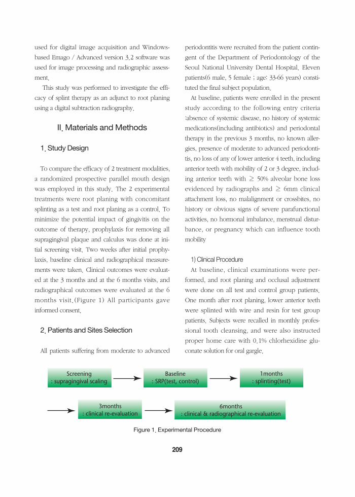
To compare the efficacy of 2 treatment modalities,a randomized prospective parallel mouth designwas employed in this study. The 2 experimentaltreatments were root planing with concomitantsplinting as a test and root planing as a control. Tominimize the potential impact of gingivitis on theoutcome of therapy, prophylaxis for removing allsupragingival plaque and calculus was done at ini-tial screening visit. Two weeks after initial prophy-laxis, baseline clinical and radiographical measure-ments were taken. Clinical outcomes were evaluat-ed at the 3 months and at the 6 months visits, andradiographical outcomes were evaluated at the 6months visit.(Figure 1) All participants gaveinformed consent.
2. Patients and Sites Selection
All patients suffering from moderate to advanced
periodontitis were recruited from the patient contin-gent of the Department of Periodontology of theSeoul National University Dental Hospital. Elevenpatients(6 male, 5 female ; age: 33-66 years) consti-tuted the final subject population.
At baseline, patients were enrolled in the presentstudy according to the following entry criteria:absence of systemic disease, no history of systemicmedications(including antibiotics) and periodontaltherapy in the previous 3 months, no known aller-gies, presence of moderate to advanced periodonti-tis, no loss of any of lower anterior 4 teeth, includinganterior teeth with mobility of 2 or 3 degree, includ-ing anterior teeth with ≥ 50% alveolar bone lossevidenced by radiographs and ≥ 6mm clinicalattachment loss, no malalignment or crossbites, nohistory or obvious signs of severe parafunctionalactivities, no hormonal imbalance, menstrual distur-bance, or pregnancy which can influence toothmobility
1) Clinical ProcedureAt baseline, clinical examinations were per-
formed, and root planing and occlusal adjustmentwere done on all test and control group patients.One month after root planing, lower anterior teethwere splinted with wire and resin for test grouppatients. Subjects were recalled in monthly profes-sional tooth cleansing, and were also instructedproper home care with 0.1% chlorhexidine glu-conate solution for oral gargle.
209
Figure 1. Experimental Procedure
2) Clinical AssessmentAt baseline, 3 and 6 months after initial treatment,
assessment of periodontal status was performed, byone examiner, with the following sequence : gingi-val condition(gingival index, GI, Löe & Silness,1963)37; oral hygiene status(plaque index, PlI, Silness& Löe, 1964)38; position of gingival margin reces-sion(REC, was measured as the distance from thecemento-enamel junction or the margin of a fillingto the free gingival margin and was measured to thenearest millimeter using calibrated periodontalprobe at 6 sites per tooth.); probing pocketdepth(PPD, was measured from the free gingivalmargin to the base of the periodontal pocket using apressure sensitive electronic Florida probe at 6 sitesper tooth.); clinical attachment level(CAL, valuesfrom REC + PPD); clinical attachment gain(CAG, dif-ferences between baseline, 3 and 6 months clinicalattachment level values); bleeding on probing(BOP,assessed at a force of 0.3 N with Florida probe,recorded as presence(1) or absence(0) within 30seconds.); tooth mobility(measured with Periotestⓡ
(Siemens AG, Bensteim, Germany) and measuredby method according to Lindhe 39)
The tooth with the largest CAL value at baselinewas used for further comparison and statisticalanalysis.
3) Radiographic Procedure
CCoonnvveennttiioonnaall rraaddiiooggrraapphhiicc iimmaaggee aaccqquuiissiittiioonnRadiographic examination was carried out at
baseline and 6 months after initial treatment. Peri-apical radiographs were taken using paralleled tech-nique and occlusal bite record(Impregum, ESPE,Germany) attached to the bite-block. The biteblockswere saved and re-used for the postoperative radi-ographic examination 6 month later.
No. 2 Kodak Ektaspeed films(Eastman Kodak Co,Rochester, NY) were used and Heliodent X-rayunit(Siemens Co., Germany) operating at 70kVpand 0.13 sec. was used for radiographic exposure.The radiographs were processed in a PERIOMATautomatic processor.(D?RR TECHNIK, Germany).The radiographs were scanned using Adobe Photo-shop version 5.0 program.
DDiiggiittaall rraaddiiooggrraapphhiicc iimmaaggee aaccqquuiissiittiioonn aanndd ddiiggiittaalliimmaaggee pprroocceessssiinngg
Digital images were taken using Digoraⓡ imagingsystem(Soredex Co., Finland). Heliodent X-rayunit(Siemens Co., Germany) operating at 70kVpand 0.13 sec was used for radiographic exposureand impression was made on a separate bite blockusing an impression material(Impregum, ESPE, Ger-many).
Obtained images were processed on the Emagoⓡ
Advanced version 3.2 software(The Oral DiagnosticSystem, Amsterdam, The Netherlands).
210
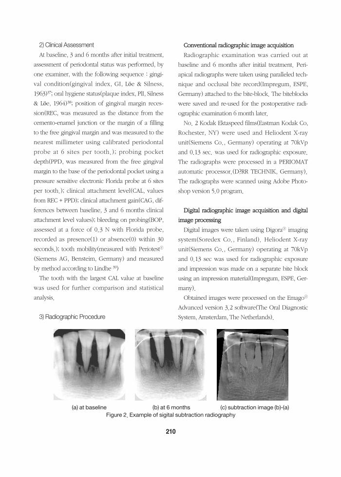
(a) at baseline (b) at 6 months (c) subtraction image (b)-(a)Figure 2. Example of sigital subtraction radiography
The preoperative images of each tooth was usedas the reference image. Four points(2 CEJ's of targettooth and adjacent tooth, 2 apices of target toothand adjacent tooth) were marked with a mouse inthe reference image. Each second image was recon-structed via the same four points and subtractedaccording to its reference image by the geometricstandardization software(Figure 2).
4) Radiographic AssessmentCCoonnvveennttiioonnaall rraaddiiooggrraapphhiicc aasssseessssmmeennttThe tooth length and marginal bone level were
measured on the scanned radiographs. ximo-incisalangle was used as reference point for the measure-ments of tooth length while the bone levels weremeasured from the most coronally positioned levelof the bone subadjacent to the tooth surface to aline through the apex of the tooth and perpendicu-lar to its longitudinal axis. The measurement oftooth length was to assess the reproducibility withregard to tooth enlargement between the preopera-tive and postoperative radiographs. Using measur-ing device, the tooth length and the bone level weremeasured. The bone gain or loss at 6 months post-operatively was
calculated mathematically. The measurementswere repeated 3 times and the means were used.
DDiiggiittaall rraaddiiooggrraapphhiicc aasssseessssmmeenntt After reconstruction, if bone gain or loss was
observed, area of bone formation or resorption wasestimated and expressed in pixels. All areas weremeasured 3 times, and the means were used. Themeasurements were converted to mm's on the basisof the size of a pixel.
5) Statistical AnalysisThe results were analyzed with SPSS version 7.5
software. Baseline values in the 2 treatment groups
were compared using the Mann-Whitney test for allclinical measurements. The Wilcoxon signed ranktest was applied in order to evaluate clinical andradiographic changes between the 2 treatmentgroups as well as within the groups. P values≤0.05were considered significant.
The non-parametric Mann-Whitney and Wilcoxontests were preferred to their parametricequivalents(the unpaired and paired t-tests, respec-tively) because the small size of the samples made itdifficult to check the assumption of normal distribu-tions.
Kendall's correlation analysis was used to evaluatethe relationship between the clinical and two radi-ographic measurements at the 6-months examina-tion.
III. Results
1. Clinical Results
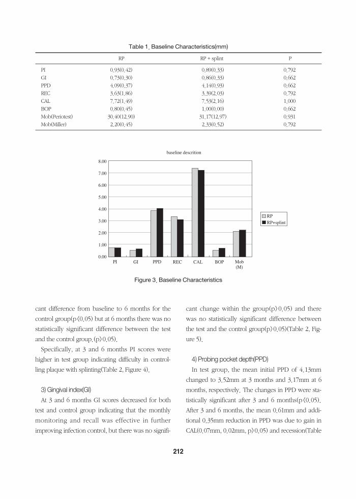
1) Baseline description Baseline characteristics of test and control tooth
are shown in Table 1. The selected tooth presentedwith clinical attachment levels of 7.53±2.16mm inthe test group and 7.72±1.49mm in the controlgroup.
No significant difference in baseline characteristicswas observed comparing the test with the controlgroup(Table 1, Figure 3).
2) Plaque index(PI)The mean clinical recordings at baseline, 3 and 6
months are presented in Table 2.At 3 and 6 months PI scores remained low or
improved with respect to the values detected atbaseline, indicating that the monthly monitoring andrecall was effective in further improving patientcompliance and plaque control. There was signifi-
211
cant difference from baseline to 6 months for thecontrol group(p<0.05) but at 6 months there was nostatistically significant difference between the testand the control group.(p>0.05).
Specifically, at 3 and 6 months PI scores werehigher in test group indicating difficulty in control-ling plaque with splinting(Table 2, Figure 4).
3) Gingival index(GI)At 3 and 6 months GI scores decreased for both
test and control group indicating that the monthlymonitoring and recall was effective in furtherimproving infection control, but there was no signifi-
cant change within the group(p>0.05) and therewas no statistically significant difference betweenthe test and the control group(p>0.05)(Table 2, Fig-ure 5).
4) Probing pocket depth(PPD)In test group, the mean initial PPD of 4.13mm
changed to 3.52mm at 3 months and 3.17mm at 6months, respectively. The changes in PPD were sta-tistically significant after 3 and 6 months(p<0.05).After 3 and 6 months, the mean 0.61mm and addi-tional 0.35mm reduction in PPD was due to gain inCAL(0.07mm, 0.02mm, p>0.05) and recession(Table
212
Table 1. Baseline Characteristics(mm)
RP RP + splint P
PI 0.93(0.42) 0.89(0.33) 0.792GI 0.73(0.30) 0.86(0.33) 0.662PPD 4.09(0.37) 4.14(0.93) 0.662REC 3.63(1.86) 3.39(2.03) 0.792CAL 7.72(1.49) 7.53(2.16) 1.000BOP 0.80(0.45) 1.00(0.00) 0.662Mob(Periotest) 30.40(12.90) 31.17(12.97) 0.931Mob(Miller) 2.20(0.45) 2.33(0.52) 0.792
8.00
7.00
6.00
5.00
4.00
3.00
2.00
1.00
0.00
baseline descrition
PI GI PPD REC CAL BOP Mob(M)
RPRP+splint
Figure 3. Baseline Characteristics
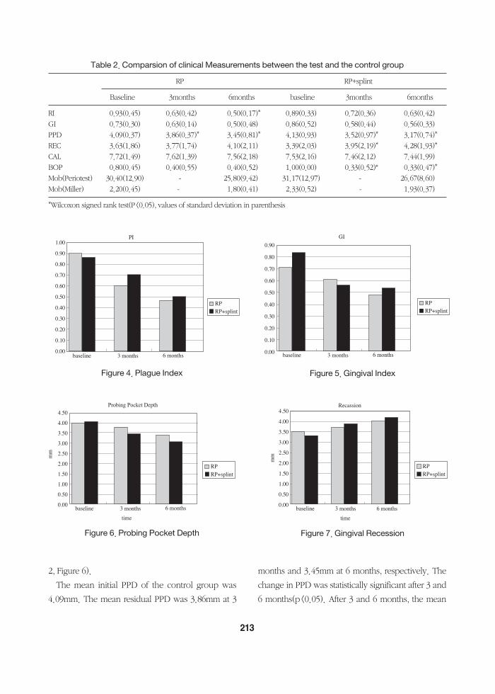
2, Figure 6).The mean initial PPD of the control group was
4.09mm. The mean residual PPD was 3.86mm at 3
months and 3.45mm at 6 months, respectively. Thechange in PPD was statistically significant after 3 and6 months(p<0.05). After 3 and 6 months, the mean
213
Table 2. Comparsion of clinical Measurements between the test and the control group
RP RP+splint
Baseline 3months 6months baseline 3months 6months
RI 0.93(0.45) 0.63(0.42) 0.50(0.17)* 0.89(0.33) 0.72(0.36) 0.63(0.42)GI 0.73(0.30) 0.63(0.14) 0.50(0.48) 0.86(0.52) 0.58(0.44) 0.56(0.33)PPD 4.09(0.37) 3.86(0.37)* 3.45(0.81)* 4.13(0.93) 3.52(0.97)* 3.17(0.74)*REC 3.63(1.86) 3.77(1.74) 4.10(2.11) 3.39(2.03) 3.95(2.19)* 4.28(1.93)*CAL 7.72(1.49) 7.62(1.39) 7.56(2.18) 7.53(2.16) 7.46(2.12) 7.44(1.99)BOP 0.80(0.45) 0.40(0.55) 0.40(0.52) 1.00(0.00) 0.33(0.52)* 0.33(0.47)*Mob(Periotest) 30.40(12.90) - 25.80(9.42) 31.17(12.97) - 26.67(8.60)Mob(Miller) 2.20(0.45) - 1.80(0.41) 2.33(0.52) - 1.93(0.37)
*Wilcoxon signed rank test(P<0.05), values of standard deviation in parenthesis
RPRP+splint
1.00
0.90
0.80
0.70
0.60
0.50
0.40
0.30
0.20
0.10
0.00baseline 3 months 6 months
PI
RPRP+splint
0.90
0.80
0.70
0.60
0.50
0.40
0.30
0.20
0.10
0.00 baseline 3 months 6 months
GI
Figure 4. Plague Index Figure 5. Gingival Index
RPRP+splint
4.50
4.00
3.50
3.00
2.50
2.00
1.50
1.00
0.50
0.00baseline 3 months 6 months
time
Probing Pocket Depth
mm
RPRP+splint
4.50
4.00
3.50
3.00
2.50
2.00
1.50
1.00
0.50
0.00baseline 3 months 6 months
time
Recassion
mm
Figure 6. Probing Pocket Depth Figure 7. Gingival Recession
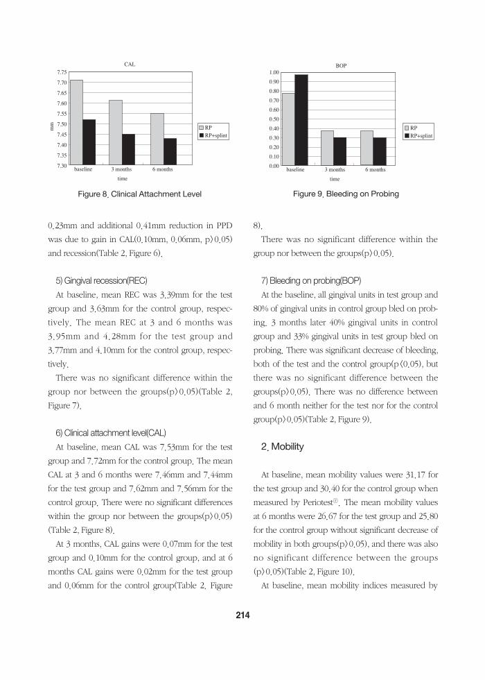
0.23mm and additional 0.41mm reduction in PPDwas due to gain in CAL(0.10mm, 0.06mm, p>0.05)and recession(Table 2, Figure 6).
5) Gingival recession(REC)At baseline, mean REC was 3.39mm for the test
group and 3.63mm for the control group, respec-tively. The mean REC at 3 and 6 months was3.95mm and 4.28mm for the test group and3.77mm and 4.10mm for the control group, respec-tively.
There was no significant difference within thegroup nor between the groups(p>0.05)(Table 2,Figure 7).
6) Clinical attachment level(CAL)At baseline, mean CAL was 7.53mm for the test
group and 7.72mm for the control group. The meanCAL at 3 and 6 months were 7.46mm and 7.44mmfor the test group and 7.62mm and 7.56mm for thecontrol group. There were no significant differenceswithin the group nor between the groups(p>0.05)(Table 2, Figure 8).
At 3 months, CAL gains were 0.07mm for the testgroup and 0.10mm for the control group, and at 6months CAL gains were 0.02mm for the test groupand 0.06mm for the control group(Table 2. Figure
8).There was no significant difference within the
group nor between the groups(p>0.05).
7) Bleeding on probing(BOP)At the baseline, all gingival units in test group and
80% of gingival units in control group bled on prob-ing. 3 months later 40% gingival units in controlgroup and 33% gingival units in test group bled onprobing. There was significant decrease of bleeding,both of the test and the control group(p<0.05), butthere was no significant difference between thegroups(p>0.05). There was no difference betweenand 6 month neither for the test nor for the controlgroup(p>0.05)(Table 2, Figure 9).
2. Mobility
At baseline, mean mobility values were 31.17 forthe test group and 30.40 for the control group whenmeasured by Periotestⓡ. The mean mobility valuesat 6 months were 26.67 for the test group and 25.80for the control group without significant decrease ofmobility in both groups(p>0.05), and there was alsono significant difference between the groups(p>0.05)(Table 2, Figure 10).
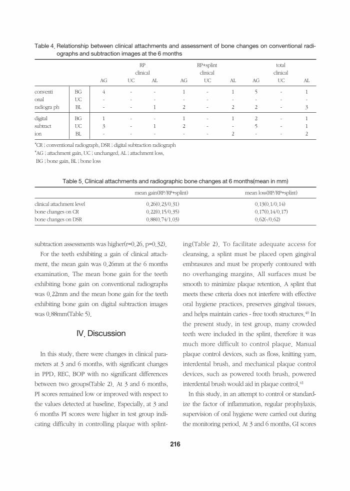
At baseline, mean mobility indices measured by
214
RPRP+splint
7.75
7.70
7.65
7.60
7.55
7.50
7.45
7.40
7.35
7.30 baseline 3 months 6 months
time
CAL
mm
RPRP+splint
1.00
0.90
0.80
0.70
0.60
0.50
0.40
0.30
0.20
0.10
0.00 baseline 3 months 6 months
time
BOP
Figure 8. Clinical Attachment Level Figure 9. Bleeding on Probing
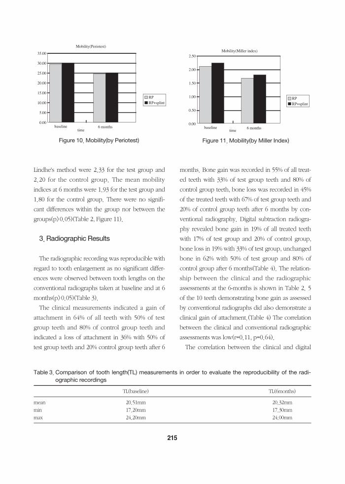
Lindhe's method were 2.33 for the test group and2.20 for the control group. The mean mobilityindices at 6 months were 1.93 for the test group and1.80 for the control group. There were no signifi-cant differences within the group nor between thegroups(p>0.05)(Table 2, Figure 11).
3. Radiographic Results
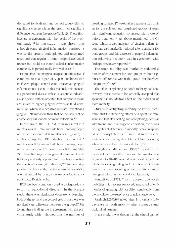
The radiographic recording was reproducible withregard to tooth enlargement as no significant differ-ences were observed between tooth lengths on theconventional radiographs taken at baseline and at 6months(p>0.05)(Table 3).
The clinical measurements indicated a gain ofattachment in 64% of all teeth with 50% of testgroup teeth and 80% of control group teeth andindicated a loss of attachment in 36% with 50% oftest group teeth and 20% control group teeth after 6
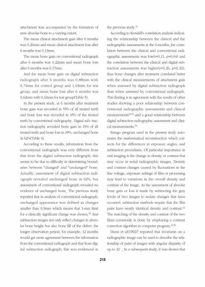
months. Bone gain was recorded in 55% of all treat-ed teeth with 33% of test group teeth and 80% ofcontrol group teeth, bone loss was recorded in 45%of the treated teeth with 67% of test group teeth and20% of control group teeth after 6 months by con-ventional radiography. Digital subtraction radiogra-phy revealed bone gain in 19% of all treated teethwith 17% of test group and 20% of control group,bone loss in 19% with 33% of test group, unchangedbone in 62% with 50% of test group and 80% ofcontrol group after 6 months(Table 4). The relation-ship between the clinical and the radiographicassessments at the 6-months is shown in Table 2. 5of the 10 teeth demonstrating bone gain as assessedby conventional radiographs did also demonstrate aclinical gain of attachment.(Table 4) The correlationbetween the clinical and conventional radiographicassessments was low(r=0.11, p=0.64).
The correlation between the clinical and digital
215
35.00
30.00
25.00
20.00
15.00
10.00
5.00
0.00baseline 6 months
time
RPRP+splint
Mobility(Periotest)
2.50
2.00
1.50
1.00
0.50
0.00baseline 6 months
time
RPRP+splint
Mobility(Miller index)
Figure 10. Mobility(by Periotest) Figure 11. Mobility(by Miller Index)
Table 3. Comparison of tooth length(TL) measurements in order to evaluate the reproducibility of the radi-ographic recordings
TL(baseline) TL(6months)
mean 20.51mm 20.32mmmin 17.20mm 17.30mmmax 24.20mm 24.00mm
subtraction assessments was higher(r=0.26, p=0.32). For the teeth exhibiting a gain of clinical attach-
ment, the mean gain was 0.26mm at the 6 monthsexamination. The mean bone gain for the teethexhibiting bone gain on conventional radiographswas 0.22mm and the mean bone gain for the teethexhibiting bone gain on digital subtraction imageswas 0.88mm(Table 5).
IV. Discussion
In this study, there were changes in clinical para-meters at 3 and 6 months, with significant changesin PPD, REC, BOP with no significant differencesbetween two groups(Table 2). At 3 and 6 months,PI scores remained low or improved with respect tothe values detected at baseline. Especially, at 3 and6 months PI scores were higher in test group indi-cating difficulty in controlling plaque with splint-
ing(Table 2). To facilitate adequate access forcleansing, a splint must be placed open gingivalembrasures and must be properly contoured withno overhanging margins. All surfaces must besmooth to minimize plaque retention. A splint thatmeets these criteria does not interfere with effectiveoral hygiene practices, preserves gingival tissues,and helps maintain caries - free tooth structures.40 Inthe present study, in test group, many crowdedteeth were included in the splint, therefore it wasmuch more difficult to control plaque. Manualplaque control devices, such as floss, knitting yarn,interdental brush, and mechanical plaque controldevices, such as powered tooth brush, poweredinterdental brush would aid in plaque control.41
In this study, in an attempt to control or standard-ize the factor of inflammation, regular prophylaxis,supervision of oral hygiene were carried out duringthe monitoring period. At 3 and 6 months, GI scores
216
Table 4. Relationship between clinical attachments and assessment of bone changes on conventional radi-ographs and subtraction images at the 6 months
RP RP+splint totalclinical clinical clinical
AG UC AL AG UC AL AG UC AL
conventi BG 4 - - 1 - 1 5 - 1onal UC - - - - - - - - -radiogra ph BL - - 1 2 - 2 2 - 3
digital BG 1 - - 1 - 1 2 - 1subtract UC 3 - 1 2 - - 5 - 1ion BL - - - - - 2 - - 2
*CR ; conventional radiograph, DSR ; digital subtraction radiograph*AG ; attachment gain, UC ; unchanged, AL ; attachment loss,BG ; bone gain, BL ; bone loss
Table 5. Clinical attachments and radiographic bone changes at 6 months(mean in mm)
mean gain(RP/RP+splint) mean loss(RP/RP+splint)
clinical attachment level 0.26(0.23/0.31) 0.13(0.1/0.14)bone changes on CR 0.22(0.15/0.35) 0.17(0.14/0.17)bone changes on DSR 0.88(0.74/1.03) 0.62(-/0.62)
decreased for both test and control group with nosignificant change within the group nor significantdifference between the groups(Table 2). These find-ings are in agreement with the results of the previ-ous study.42 In that study, it was shown thatalthough some gingival inflammation persisted, itwas similar around both splinted and unsplintedteeth and that regular 1-month prophylaxes couldreduce but could not control sulcular inflammationcompletely in periodontally involved cases.42
It's possible that marginal adaptation difficulties ofcomposite resin as a part of A splint combined withineffective plaque control could exacerbate gingivalinflammation adjacent to this material, thus increas-ing periodontal disease risk in susceptible individu-als and some authors reported that composite resinsare linked to higher gingival crevicular fluid accu-mulation which is a sensitive indicator quantifyinggingival inflammation than that found adjacent toenamel or glass ionomer cement restoration.41,42
In test group, the PPD reduction measured at 3months was 0.51mm and additional probing depthreduction measured at 6 months was 0.35mm. Incontrol group, the PPD reduction measured at 3months was 0.23mm and additional probing depthreduction measured 6 months was 0.41mm(Table2). These findings are in general agreement withfindings previously reported from studies evaluatingthe effects of non-surgical therapy.43,44 In measuringprobing pocket depth, the intraexaminer variabilitywas minimized by using a pressure-calibrated(con-stant force) Florida probe.
BOP has been commonly used as a diagnostic cri-terion for periodontal disease.45 In the presentstudy, there was significant decrease of bleeding,both of the test and the control group, but there wasno significant difference between the groups(Table2) and these findings are in agreement with the pre-vious study which showed that the number of
bleeding surfaces 17 weeks after treatment was simi-lar for the splinted and unsplinted groups of teethwith significant reduction compared with those ofbefore treatment42. As above mentioned, the GIscore which is also indicator of gingival inflamma-tion was also markedly reduced after treatment forboth groups, and this decrease in gingival inflamma-tion following treatment was in agreement withfindings previously reported.42
The tooth mobility was markedly reduced 6months after treatment for both groups without sig-nificant differences within the group nor betweenthe groups(p>0.05).
The effect of splinting on tooth mobility has con-troversy, but it seems to be generally accepted thatsplinting has no additive effect on the reduction oftooth mobility.
Studies investigating mobility posterior teethfound that the stabilizing effects of a splint are tran-sient and that after scaling and root planing, occlusaladjustment, and oral hygiene education, there wasno significant difference in mobility between splint-ed and nonsplinted teeth, and that more mobileteeth received no significant benefit from splintingwhen compared with less mobile teeth.46,47
Renggli and Mühlemann(1970)48 reported thatincreased tooth mobility in occlusal trauma decreas-es greatly to 18-28% soon after removal of occlusalinterferences by grinding and there is only little evi-dence that mere splinting of teeth exerts a similarbiological effect on the periodontal ligament.
Renggli et al(1971)49 also reported that toothmobilities with splints removed, measured after 6months of splinting, did not differ significantly fromthe mobilities measured prior to splint placement.
Rateitschak(1963)50 noted after 24 months a 14%decrease in tooth mobility after curettage andocclusal adjustment.
In this study, it was shown that the clinical gain of
217
attachment was accompanied by the formation ofnew alveolar bone to a varying extent.
The mean clinical attachment gain after 6 monthswas 0.26mm and mean clinical attachment loss after6 months was 0.13mm.
The mean bone gain on conventional radiographafter 6 months was 0.22mm and mean bone lossafter 6 months was 0.17mm.
And the mean bone gain on digital subtractionradiograph after 6 months was 0.88mm with0.74mm for control group and 1.03mm for testgroup, and mean bone loss after 6 months was0.62mm with 0.62mm for test group(Table 5).
In the present study, at 6 months after treatmentbone gain was recorded in 55% of all treated teethand bone loss was recorded in 45% of the treatedteeth by conventional radiography. Digital sub- trac-tion radiography revealed bone gain in 19% of alltreated teeth and bone loss in 19%, unchanged bonein 62%(Table 4).
According to these results, information from theconventional radiograph was very different fromthat from the digital subtraction radiograph, thisseems to be due to difficulty in determining bound-aries between "changed" and "unchanged" bone.Actually, assessment of digital subtraction radi-ograph revealed unchanged bone in 62%, butassessment of conventional radiograph revealed noevidence of unchanged bone. The previous studyreported that in analysis of conventional radiograph,unchanged appearance was defined as changessmaller than 0.9mm which means that 1-mm limitfor a clinically significant change was chosen.55 Andsubtraction images not only reflect changes in alveo-lar bone height but also bone fill of the defect, thelonger observation period, for example, 12 monthswould get more agreement between the informationfrom the conventional radiograph and that from dig-ital subtraction radiograph, this was evidenced in
the previous study.51
According to Kendall's correlation analysis indicat-ing the relationship between the clinical and theradiographic assessments at the 6-months, the corre-lation between the clinical and conventional radi-ographic assessments was low(r=0.11, p=0.64) andthe correlation between the clinical and digital sub-traction assessments was higher(r=0.26, p=0.32),thus bone changes after treatment correlated betterwith the clinical measurements of attachment gainwhen assessed by digital subtraction radiographthan when assessed by conventional radiograph.This finding is in agreement with the results of otherstudies showing a poor relationship between con-ventional radiographic assessments and clinicalmeasurements52-54 and a good relationship betweendigital subtraction radiographic assessment and clini-cal measurements.55
Emago program used in the present study auto-mates the mathematical reconstruction which cor-rects for the differences in exposure angles, andsubtraction procedures. Of particular importance inoral imaging is the change in density or contrast thatmay occur in serial radiographic images. Densityand contrast changes caused by fluctuations in theline voltage, exposure settings of film or processingmay lead to variations in the overall density andcontrast of the image. As the assessment of alveolarbone gain or loss is made by subtracting the graylevels of two images to isolate changes that haveoccurred, subtraction methods require that the filmpairs have nearly identical density and contrast.22
The matching of the density and contrast of the twofilms commonly is done by employing a contrastcorrection algorithm in computer program.21,56
Dunn et al(1992)5 reported that invariants on aradiographic image can be used to describe the rela-tionship of pairs of images with angular disparity ofup to 32˚. In a subsequent study, it was shown that
218
this registration procedure could be used to estab-lish correspondence between pairs of clinicalimages taken at different projection angles usingfour featuring points.33
Validation research for subtraction radiographyalso examined how small a lesion could be detectedwith a high degree of diagnostic accuracy. The min-imal thickness of bone that may be detected underoptimal conditions(no geometric or contrast distor-tion) was found to be 0.12mm.57 This study alsoexamined the effect of variations in projectiongeometry. When the angulation was misaligned by3˚, the minimal˚thickness of cortical bone thatcould be detected was 0.35mm to 0.42mm.
The mandibular anterior teeth used in the presentstudy are the longest surviving teeth of the peri-odontium.58,59 Thus if teeth present with 50% to 70%bone loss, one can be confident that with properstabilization including splinting therapy and peri-odontal maintenance, survival of the teeth is possi-ble.
Nyman et al demonstrated long-term stability andmaintenance of splinted dentitions that had greaterthan 50% attachment loss of each abutment tooth.Although Ante's law was not satisfied, in theabsence of inflammation, severely periodontallycompromised dentitions could be maintained forextended periods of time, in some cases more than20 years.60,61
Although the effect of splinting as controversy, theuse of the temporary splinting is indicated in the fol-lowing circumstances : where mobility of the teethexists, so that physiologic rest can be effected,where mobility exists to such a degree that effectiveperiodontal treatment and procedures cannot other-wise be executed properly, as a diagnostic aid toevaluate the prognosis before instituting extensivepermanent splinting, to improve the psychologicmorale of the patient with mobile teeth.62,63
Besides the above mentioned indications, splint-ing has some advantages, for example, facilitation ofocclusal adjustment, prevention of food impactionby stabilization of proximal contacts, facilitation ofhealing of diseased supporting tissue, enhancementof postsurgical healing, and so on.64
The term "A-splint" used in the present study wasapparently popularized by Berliner and Kessler65
and has evolved into dental terminology as an easyway to describe the wire-reinforced acrylic resin-amalgam splint. More recently the term "A-splint"has come to include any splint that ties teeth togeth-er with acid-etched composite materials; usuallythere is wire reinforcing included.64
Ideally an "A-splint" should have the followingcharacteristics: provide adequate stabilization for themobile teeth ; have adequate retention ; requireremoval of as little tooth structures possible ; be ableto be completed in a comparatively short time ; notinterfere with the patient's ability to practice goodoral hygiene, be esthetic.65
However, "A-splint" has some common problems,and these are overcontouring in an effort to makethe splint strong enough to resist fracture, estheticproblem, wire stability, arch stability, caries due tothe possible percolation of acrylic resin.64,65 Over-contouring can be avoided by finishing "A-splint"with the same principles as cast restorations andplacement of an interproximal brush should beallowed. To improve wire stability double strands ofwire, twisted strands of wire, cast bars, modifiedmatrix bands, and prefabricated bars were suggest-ed and the most versatile option is the 13- or 15-gauge half-round wire. To get arch stability, onemust gain support in two or more planes.64,66 If theA-splint includes a curve, then all the splinted teethwill appear less mobile. This usually requires includ-ing the canines in a posterior splint. Includingcrossarch splinting increases the multiplane curve
219
and thus creates even more stabilization, i.e.reduced mobility. To reduce caries problem, stan-nous fluoride can be applied to the preparations,and caries incidence maybe related to the retentionof the acrylic to the teeth, thus the vertical and hori-zontal wires for increasing retention may decreasethe caries susceptibility.65
Recently innovative technique employing a bond-able, ribbon-splinting material for reinforcing dentalresins or fiber-reinforced composite resin has devel-oped.73 By combining the chemical adhesive andesthetic characteristics of composite resin with thestrength enhancement of a plasma-treated, high-modulus, splints can resist the load -bearing forcesof occlusion and mastication. These fracture-resis-tant restorations are more durable than most adhe-sive-composite resin alternative splinting materials ofthe past.
Although the effect of splinting has controversy,the general trend seems to be that unless final splint-ing is indicated, temporary splinting during peri-odontal treatment should be avoided and that adecision to splint, for reasons of mobility, shouldusually be reserved until initial therapy has beencompleted but that there are many other factorssuch as patient discomfort from loose or missingteeth which may, of course, dictate otherwise.67,68
In the present study, it can be surmised that thesplinting has no additive effect on the peridontaltreatment , but a more definite, well-controlled studywith the larger sample size is needed to clarify thisfinding.
V. Conclusion
The aim of this study was to compare the effectsof root planing only with those of root planing withconcomitant splinting clinically and radiographically,to compare information from digital subtraction and
from conventional radiography with clinical record-ings for the assessment of bone changes, finally toinvestigate the efficacy of splinting therapy and itcan be concluded that,
1. There were changes in clinical parameters at 3months, with significant changes in PPD, REC,BOP(p<0.05) with no significant differencesbetween two groups(p>0.05).
2. There were also changes in clinical parametersat 6 months, with significant changes in PPD,REC, BOP, PI,(p<0.05) with no significant dif-ferences between two groups(p>0.05).
3. Kendall's correlation analysis shows that thecorrelation between the clinical and the CRassessments low and did not differ significantlyfrom zero(r=0.110, p=0.639) and that therewas higher correlation between the clinical andthe DSR assessments(r=0.257, p=0.315) indicat-ing that bone changes following periodontaltreatment correlated better with the clinicalmeasurements of attachment gain whenassessed by DSR than when assessed by CR.
According to these results, we surmised thatsplinting has no additive effect on Root Planing inperiodontal treatment.
VI. References
1. Lemmerman K. Rationale for stabilization. J Peri-odontol 1976; 47: 405-411
2. Ferencz JL. Splinting. Dent Clin North Am 1987;31: 383-393
3. Fleszar TJ, Knowles JW, Morrison EC, BurgettFG, Nissle RR, Ramfjord SP. Tooth mobility andperiodontal therapy. J Clin Periodontol 1980; 7:495-505
4.Nyman S, Lindhe J. Persistent tooth hypermobili-
220
ty following completion of periodontal treat-ment. J Clin Periodontol 1976; 3: 81-93
5.Dunn SM, Van der Stelt PF. Recognizing invari-ant geometric structure in dental radiographs.Dentomaxillo Radiol 1992; 21: 142-147
6.Reddy MS. Radiographic methods in the evalua-tion of periodontal therapy. J Periodontol 1992;63: 1078-1084
7. Proceedings of the workshop on quantitativeevaluation of periodontal diseases by physicalmeasurement techniques. J Dent Res 1979; 58:547-551
8.Ainamo J, Tammisiao E. Comparison of radi-ographic and clinical signs of early periodontaldisease. Scand J Dent Res 1973; 81: 548-552
9.Akiyoshi M, Mori K. Marginal periodontitis : ahistological study of the incipient stage. J Peri-odontol 1967; 38: 42-52
10. Benveniste R, Bixler D, Conneally PM. Peri-odontal diseases in diabetics. J Periodontol 1967;38: 271-279
11. Bender IB, Seltzer S. Roentgenographic anddirect observation of experimental lesions inbone. I. J Am Dent Assoc 1961; 62: 152-160
12. Bender IB, Seltzer S. Roentgenographic anddirect observation of experimental lesions inbone. Ⅱ. J Am Dent Assoc 1961; 62: 708-716
13. Ramadan AE, Mitchell DF. A roentgenographicstudy of experimental bone destruction. OralSurg Oral Med Oral Pathol 1962; 15: 934-942
14. Paul V, Trott JR. A radiological study of experi-mentally produced lesions in bone. Dent Prac-tioner 1966; 16: 254-258
15. Schwartz SF, Foster JK. Roentgenographic inter-pretation of experimentally produced lesions inbone. Oral Surg Oral Med Oral Pathol 1971; 32:606-612
16. Socransky SS, Haffajee AD, Goodson JM, LindheJ. New concepts of destructive periodontal dis-
ease. J Clin Periodontol 1984; 11: 21-3217. Theilaid J. An evaluation of the reliability of radi-
ographs in the measurement of bone loss inperiodontal disease. J Periodontol 1960; 31: 143-153
18. Rawlinson A, Ellwood RP, Davies RM. An in-vitro evaluation of a dental subtraction radiogra-phy system using bone chips on dried humanmandibles. J Clin Periodontol 1999; 26: 138-142
19. Rüttiman UE, Okano T, Gröndahl HG, GröndahlK, Webber RL. Exposure geometry and film con-trast differences as bases for incomplete cancel-lation of interdental structures in dental subtrac-tion radiography. Proc SPIE 1981; 314: 372-377
20. Webber RL, Rüttiman UE, Gröndahl HG. X-rayimage subtraction as a basis for the assessmentof periodontal changes. J Periodontol Res 1982;17: 509-511
21. Webber RL, Gröndahl HG, Gröndahl K. A digitalsubtraction technique for dental radiography.Oral Surg Oral Med Oral Pathol 1983; 55: 96-102
22. Reddy MS, Jeffcoat MK. Digital subtraction radi-ography. Dent Clin North Am 1993; 37: 553-565
23. Bragger U. Digital imaging in periodontal radi-ography. J Clin Periodontol 1988; 15:551-557
24. Ortman LF, Dunford R, McHenry K, HausmannE. Subtraction radiography and computer assist-ed densitometric analyses of standard radi-ographs. A comparison with 125I absorptiometry.J Periodont Res 1985; 20: 644-651
25. Socransky SS, Gordon G, Goodson JM. Devel-opment of automated registration algorhythmsfor subtraction radiography. J Clin Periodontol1994; 21: 540-543
26. Grondahl HG, Grondahl K. Subtraction radiog-raphy for the diagnosis of periodontal lesions.Oral Surg Oral Med Oral Pathol 1983; 55: 208-213
27. Ruttimann UE, Webber R, Schmide. E. A robust
221
digital method for film contract correction insubtraction radiography. J Periodont Res 1985;21: 486-495
28. McHenry K, Hausmann E, Wikesjo, Dunford R,Christersson L. Methodological aspects andquantitative adjuncts to computerized subtrac-tion radiography. J Periodont Res 1987; 22: 125-132
29. Jeffcoat MK, Reddy MS, Webber RL, RuttimannUE. Extraoral control of geometry for digital sub-traction radiography. J Periodont Res 1987; 22:396-402
30. Socransky SS, Duckworth JE, Goodson JM. Amethod for geometric and densitometric stan-dardization of intraoral radiographs. J Periodon-tol 1983; 12: 435-440
31. Ruttimann UE, Webber RL, Groenhuis RAJ.Computer correction of projective distortion indental radiographs. J Dent Res 1984; 63: 1032-1036
32. Wenzel A. Effect of manual compared with ref-erence point superimposition on image qualityin digital subtraction radiography. Dentomaxillo-fac Radio 1989; 18: 145-150
33. Dunn SM, Van der Stelt PF. Ponce A, Fenesy K.A comparison of two registration techniques fordigital subtraction radiography. DentomaxillofacRadio 1993; 22: 77-80
34. Webber RL, Gröndahl HG, Gröndahl K. Influ-ence of variations in projection geometry on thedetectability of periodontal bone lesions. J ClinPeriodontol 1984; 11: 411-420
35. Furkart J, Dove B. Direct digital radiography forthe detection of periodontal bone lesions. OralSurg Oral Med Oral Pathol 1992; 74: 652-660
36. Wenzel A, Gröndahl HG. Direct digital radiogra-phy in the dental office. Int Dent J 1995; 45: 27-34
37. Loe H, Silness J. Periodontal disease in pregnan-
cy Ⅰ. Prevalence and severity. Acta OdontolScand 1963; 21: 533-551
38. Loe H, Silness J. Periodontal disease in pregnan-cy Ⅱ. Correlation between oral hygiene andperiodontal disease. Acta Odontol Scand 1964;22: 121-135
39. Lindhe J. Textbook of Clinical Periodontology.Munksgaard, Copenhagen 1983a; pp 303-304.1983b; p 394
40. Oikarinen K. Tooth splinting : A review of theliterature and consideration of the versability of awire-composite splint. Endod Dent Traumatol1990; 6: 237-250
41. Serio FG, Strassler HE. The effect of polishingpastes on composite resin surfaces; A SEMstudy. J Periodontol 1988; 59: 837-840
42. Sjostrom S, vanDijken JWK. The effect of glassionomer cement and composite resin fillings onmarginal gingiva. J Clin Periodontol 1991; 18:200-203
43. Goodson JM, Hogan PE. Clinical responses fol-lowing periodontal treatment by local delivery. JPeriodontol 1985; 56: 81-87
44. Caton J, Polson A. Maintenance of healed peri-odontal pockets after a single episode of rootplaning. J Periodontol 1982; 53: 420-424
45. Lang NP, Joss A, Orsanic T. Bleeding on probing: A predictor for progression on periodontal dis-ease? J Clin Periodontol 1986; 13: 590-596
46. Kegel W, Selipsky H. The effect of splinting ontooth mobility. I. During initial therapy. J ClinPeriodontol 1979; 6: 45-58
47. Renggli HH, Schweitzer H. Splinting of teethwith removable bridges-biologic effects. J ClinPeriodontol 1974; 1: 43-46
48. Renggli HH, Muhlemann HR. Tooth mobility,marginal periodontitis and malocclusion. Paro-dontologie 1970; 24: 39-48
49. Renggli HH. Splinting of teeth-an objective
222
assessment. Helvetica Odontologica Acta 1970;15: 129-131
50. Rateitschak KH. The therapeutic effect of localtreatment on periodontal disease assessed uponevaluation of different diagnostic criteria. 1.Changes in tooth mobility. J Periodontol 1963;34: 540-544
51. Wenzel A, Warrer K, Karring T. Digital subtrac-tion radiography in assessing bone changes inperiodontal defects following guided tissueregeneration. J Clin Periodontol 1992: 19: 208-213
52. Kelly GH, Cain RJ, Knowles JW, Nissle RP, Bur-gett FG, Ramfjord SP. Radiographs in clinicalperiodontal trials. J Periodontol 1975; 46: 381-386
53. Isidor F. & Karring T. Long term effect of surgi-cal and non-surgical periodontal treatment. A 5-year clinical study. J Periodont Res 1986; 21:462-472
54. Renvert S, Badersten A, Nilveus R, & Egelberg J.Healing after treatment of periodontalintraosseous defects I. Comparative study ofClinical Method. J Clin Periodontol 1981; 8: 387-399
55. Hausmann E, Christersson L, Dunford R. Useful-ness of subtraction radiography in the evaluationof periodontal therapy. J Periodontol 1985; 56:special issue, 4-8
56. Ohki M, Okaono T, Yamada N. A contrast-cor-rection method for digital subtraction radiogra-phy. J Periodont Res 1988; 23: 277-280
57. Rudolph DJ, White SC, Mankovich NJ. Influenceof geometric distortion and exposure parameterson sensitivity of digital subtraction radiography.Oral Surg Oral Med Oral Pathol 1987; 64: 631-637
58. Nyman SR, Lang NP. Tooth mobility and thebiological rationale for splinting teeth. Periodon-tol 2000 1994; 4: 15-22
59. Nyman S, Lindhe J, Lundgren D. The role ofocclusion for the stability of fixed bridges inpatients with reduced periodontal tissue sup-port. J Clin Periodontol 1975; 2: 53-66
60. Hirschfeld L, Wasserman B. A Long term surveyof tooth loss in 600 treated periodontal patients.J Periodontol 1978; 49: 225-237
61. Pollack RP. An analysis of periodontal therapyfor the 65-year-old and older patient. Gerodon-tics 1986; 2: 135-137
62. Marvin S, Jack L, Thaller J. Temporary splintingfor multiple mobile teeth. Amer Dent Assoc1956; 53: 429-433
63. Howard L, Lawrence A, Weinberg. An evalua-tion of periodontal splinting. J Am Dent Assoc1961; 63: 49-54
64. Curtis M. Becker, David A. Kaiser, Wayne B.Kaldahl. The Evolution of Temporary FixedSplints-The A-Splint Int J Periodont Rest Dent1998; 18: 277-285
65. Kessler M. Mattehew K. A Variation of the Asplint. J Periodontol 1970; 41: 268-271
66. Howard E. Strassler, Alireza Haeri, Jerrold P.Gultz. New-generation Bonded ReinforcingMaterials for Anterior Periodotal Tooth Stabiliza-tion and Splinting. Dent Clin North Am 1999; 43:105-126
67. Waerhaug J. Justification for splinting in peri-odontal therapy. J Prosthetic Dent 1969; 22: 201-208
68. Selipsky, H. Osseous surgery-How much needwe compromise? Dent Clin North Am 1976; 20:79-106
223
-국문초록-
치근활택술과스프린트병행처치의효과에관한연구 :디지털공제촬 술을이용한임상적연구
이지 , 계승범, 김원경, 이용무, 구 , 류인철, 정종평, 최상묵, 한수부
서울대학교 치과대학 치주과학 교실
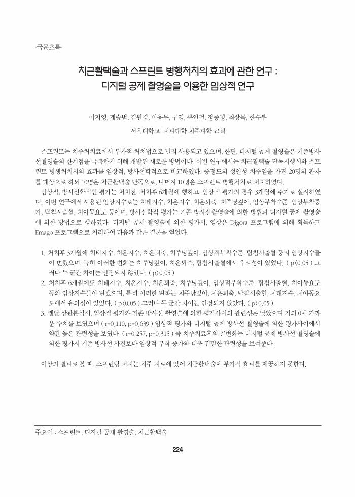
스프린트는 치주처치료에서 부가적 처치법으로 널리 사용되고 있으며, 한편, 디지털 공제 촬 술은 기존방사선촬 술의 한계점을 극복하기 위해 개발된 새로운 방법이다. 이번 연구에서는 치근활택술 단독시행시와 스프린트 병행처치시의 효과를 임상적, 방사선학적으로 비교하 다. 중정도의 성인성 치주염을 가진 20명의 환자를 대상으로 하되 10명은 치근활택술 단독으로, 나머지 10명은 스프린트 병행처치로 처치하 다.
임상적, 방사선학적인 평가는 처치전, 처치후 6개월에 행하고, 임상적 평가의 경우 3개월에 추가로 실시하다. 이번 연구에서 사용된 임상지수로는 치태지수, 치은지수, 치은퇴축, 치주낭깊이, 임상부착수준, 임상부착증가, 탐침시출혈, 치아동요도 등이며, 방사선학적 평가는 기존 방사선촬 술에 의한 방법과 디지털 공제 촬 술에 의한 방법으로 행하 다. 디지털 공제 촬 술에 의한 평가시, 상은 Digora 프로그램에 의해 획득하고Emago 프로그램으로 처리하여 다음과 같은 결론을 얻었다.
1. 처치후 3개월에 치태지수, 치은지수, 치은퇴축, 치주낭깊이, 임상적부착수준, 탐침시출혈 등의 임상지수들이 변했으며, 특히 이러한 변화는 치주낭깊이, 치은퇴축, 탐침시출혈에서 유의성이 있었다. ( p<0.05 ) 그러나 두 군간 차이는 인정되지 않았다. ( p>0.05 )
2. 처치후 6개월에도 치태지수, 치은지수, 치은퇴축, 치주낭깊이, 임상적부착수준, 탐침시출혈, 치아동요도등의 임상지수들이 변했으며, 특히 이러한 변화는 치주낭깊이, 치은퇴축, 탐침시출혈, 치태지수, 치아동요도에서 유의성이 있었다. ( p<0.05 ) 그러나 두 군간 차이는 인정되지 않았다. ( p>0.05 )
3. 켄달 상관분석시, 임상적 평가와 기존 방사선 촬 술에 의한 평가사이의 관련성은 낮았으며 거의 0에 가까운 수치를 보 으며 ( r=0.110, p=0.639 ) 임상적 평가와 디지털 공제 방사선 촬 술에 의한 평가사이에서약간 높은 관련성을 보 다. ( r=0.257, p=0.315 ) 즉 치주치료후의 골변화는 디지털 공제 방사선 촬 술에의한 평가시 기존 방사선 사진보다 임상적 부착 증가와 더욱 긴 한 관련성을 보여준다.
이상의 결과로 볼 때, 스프린팅 처치는 치주 치료에 있어 치근활택술에 부가적 효과를 제공하지 못한다.
주요어 : 스프린트, 디지털 공제 촬 술, 치근활택술
224

225