9781904842781sc.pdf - scion publishing
TRANSCRIPT

Muhammed Akunjee and Nazmul AkunjeeGP Principals, London, UK
Zeshaan Maan,Surgical Trainee, London, UK
and Mina AllyFoundation Doctor, Dartford, UK
© Scion Publishing Ltd, 2012
ISBN 978 1 904842 78 1
First published in 2012
All rights reserved. No part of this book may be reproduced or transmitted, in any form or by any means, without permission.
A CIP catalogue record for this book is available from the British Library.
Scion Publishing LimitedThe Old Hayloft, Vantage Business Park, Bloxham Road, Banbury, Oxfordshire OX16 9UXwww.scionpublishing.com
Important Note from the PublisherThe information contained within this book was obtained by Scion Publishing Limited from sources believed by us to be reliable. However, while every effort has been made to ensure its accuracy, no responsibility for loss or injury whatsoever occasioned to any person acting or refraining from action as a result of information contained herein can be accepted by the authors or publishers.
Although every effort has been made to ensure that all owners of copyright material have been acknowledged in this publication, we would be pleased to acknowledge in subsequent reprints or editions any omissions brought to our attention.
Readers should remember that medicine is a constantly evolving science and while the authors and publishers have ensured that all dosages, applications and practices are based on current indications, there may be specific practices which differ between communities. You should always follow the guidelines laid down by the manufacturers of specific products and the relevant authorities in the country in which you are practising.
Artwork by Nazmul Akunjee, London, UKCover design by amdesign, Banbury, UKTypeset by Manila Typesetting Company, PhilippinesPrinted by MPG Books Group, UK
Contents Foreword vii Preface viii About the authors ix Acknowledgments x Abbreviations xi
Section 1 – History taking skills
1.1 Chest pain 1 1.2 History of palpitations 9 1.3 Shortness of breath 15 1.4 Cough 22 1.5 Abdominal pain 28 1.6 Nausea and vomiting 35 1.7 Dysphagia 42 1.8 Jaundice 48 1.9 Haematuria 54 1.10 Headache 59 1.11 Loss of consciousness 67 1.12 Loss of vision 73 1.13 Red eye 79 1.14 Hearing loss 85 1.15 Depression 91 1.16 Suicide risk assessment 98 1.17 Alcohol 105 1.18 Psychosis 113
Section 2 – Examinations
2.1 Cardiovascular examination 121 2.2 Respiratory examination 135 2.3 Abdomen examination 146 2.4 Inguinal scrotal examination 157 2.5 Rectal examination 165 2.6 Cranial nerves examination 171 2.7 Upper limb motor examination 180 2.8 Lower limb motor examination 188 2.9 Upper limb sensory examination 195 2.10 Lower limb sensory examination 202 2.11 Gait and co-ordination 206 2.12 Eye examination 215 2.13 Ear examination 226 2.14 Peripheral vascular examination: arterial 233 2.15 Peripheral vascular examination: venous 239 2.16 Examination of a lump 245 2.17 Thyroid examination 252 2.18 Lymph nodes examination 259
vi | Contents
Section 3 – Procedures
3.1 Hand washing 267 3.2 Surgical scrubbing 272 3.3 Wound swabbing 276 3.4 Taking a blood pressure 280 3.5 Body mass index 287 3.6 Recording an ECG 293 3.7 Measuring blood glucose 306 3.8 Urine dip testing 313 3.9 Venepuncture 319 3.10 Blood cultures 326 3.11 Cannulation and IV infusion 330 3.12 Blood transfusion 336 3.13 Peak flow meter explanation 342 3.14 Inhaler explanation 347 3.15 Spacer device explanation 352 3.16 Setting up a nebulizer 356 3.17 Arterial blood gas sampling and analysis 360 3.18 Nasogastric intubation 367 3.19 Male catheterization 373
Answers to Common clinical scenarios 379
Index 385
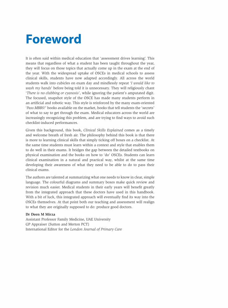
ForewordIt is often said within medical education that ‘assessment drives learning’. This means that regardless of what a student has been taught throughout the year, they will focus on those topics that actually come up in the exam at the end of the year. With the widespread uptake of OSCEs in medical schools to assess clinical skills, students have now adapted accordingly. All across the world students walk into cubicles on exam day and mindlessly repeat ‘I would like to wash my hands’ before being told it is unnecessary. They will religiously chant ‘There is no clubbing or cyanosis’, while ignoring the patient’s amputated digit. The focused, snapshot style of the OSCE has made many students perform in an artificial and robotic way. This style is reinforced by the many exam-oriented ‘Pass MBBS!’ books available on the market, books that tell students the ‘secrets’ of what to say to get through the exam. Medical educators across the world are increasingly recognizing this problem, and are trying to find ways to avoid such checklist-induced performances.
Given this background, this book, Clinical Skills Explained comes as a timely and welcome breath of fresh air. The philosophy behind this book is that there is more to learning clinical skills that simply ticking off boxes on a checklist. At the same time students must learn within a context and style that enables them to do well in their exams. It bridges the gap between the detailed textbooks on physical examination and the books on how to ‘do’ OSCEs. Students can learn clinical examination in a natural and practical way, whilst at the same time developing their awareness of what they need to be able to do to pass their clinical exams.
The authors are talented at summarizing what one needs to know in clear, simple language. The colourful diagrams and summary boxes make quick review and revision much easier. Medical students in their early years will benefit greatly from the integrated approach that these doctors have used in this handbook. With a bit of luck, this integrated approach will eventually find its way into the OSCEs themselves. At that point both our teaching and assessment will realign to what they are originally supposed to do: produce good doctors.
Dr Deen M MirzaAssistant Professor Family Medicine, UAE UniversityGP Appraiser (Sutton and Merton PCT)International Editor for the London Journal of Primary Care
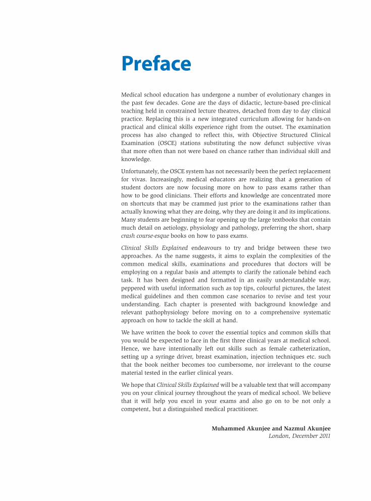
PrefaceMedical school education has undergone a number of evolutionary changes in the past few decades. Gone are the days of didactic, lecture-based pre-clinical teaching held in constrained lecture theatres, detached from day to day clinical practice. Replacing this is a new integrated curriculum allowing for hands-on practical and clinical skills experience right from the outset. The examination process has also changed to reflect this, with Objective Structured Clinical Examination (OSCE) stations substituting the now defunct subjective vivas that more often than not were based on chance rather than individual skill and knowledge.
Unfortunately, the OSCE system has not necessarily been the perfect replacement for vivas. Increasingly, medical educators are realizing that a generation of student doctors are now focusing more on how to pass exams rather than how to be good clinicians. Their efforts and knowledge are concentrated more on shortcuts that may be crammed just prior to the examinations rather than actually knowing what they are doing, why they are doing it and its implications. Many students are beginning to fear opening up the large textbooks that contain much detail on aetiology, physiology and pathology, preferring the short, sharp crash course-esque books on how to pass exams.
Clinical Skills Explained endeavours to try and bridge between these two approaches. As the name suggests, it aims to explain the complexities of the common medical skills, examinations and procedures that doctors will be employing on a regular basis and attempts to clarify the rationale behind each task. It has been designed and formatted in an easily understandable way, peppered with useful information such as top tips, colourful pictures, the latest medical guidelines and then common case scenarios to revise and test your understanding. Each chapter is presented with background knowledge and relevant pathophysiology before moving on to a comprehensive systematic approach on how to tackle the skill at hand.
We have written the book to cover the essential topics and common skills that you would be expected to face in the first three clinical years at medical school. Hence, we have intentionally left out skills such as female catheterization, setting up a syringe driver, breast examination, injection techniques etc. such that the book neither becomes too cumbersome, nor irrelevant to the course material tested in the earlier clinical years.
We hope that Clinical Skills Explained will be a valuable text that will accompany you on your clinical journey throughout the years of medical school. We believe that it will help you excel in your exams and also go on to be not only a competent, but a distinguished medical practitioner.
Muhammed Akunjee and Nazmul AkunjeeLondon, December 2011
About the authorsDr Muhammed Akunjee (MBBS, MRCGP, PGCME, PGCDM) is currently a GP Principal in North London (since 2007) and Clinical Director for the local PCT. He qualified from Guy’s, Kings and St. Thomas’s Medical School in 2002 and completed his MRCGP gaining a distinction in 2006. During this time he was also awarded first prize for the Roche / RCGP Registrar award. As an undergraduate he was awarded the War Memorial Prize and distinction in Psychology / Sociology. He was also awarded a number of bursaries and research awards including the Leukaemia Research Fund Bursary, the Rayne Institute Research Prize, the British Medical & Dental Student’s Fund Bursary, and the King’s College Lightfoot Award. Dr Akunjee is actively involved in medical student teaching and is currently a clinical skills tutor as well as an OSCE examiner at a London university. He has been involved in the publication of a number of papers and medical revision books including the award-winning Easy Guide to OSCE’s series.
Dr Nazmul Akunjee (MBBS, MRCGP) has recently completed his GP vocational training and is currently a GP Principal. He qualified from GKT medical school in 2005 and has published a number of articles related to examination skills in OSCEs in peer-reviewed journals. He completed the London Deanery’s Introduction to Teaching in Primary Care course in June 2011, and is currently teaching medical students at a London university. He is also a course facilitator for the CSAPrep course that helps GP Registrars pass their MRCGP Clinical Skills Assessment examinations. He is also a co-author of the award-winning Easy Guide to OSCE’s series of medical revision books.
Dr Mina Ally (MBBS, BSc) is currently a Year 1 Core Medical Trainee at Whipps Cross University Hospital. She completed an intercalated BSc in Nutrition with Basic Medical Sciences with first class honours at King’s College London in 2007. She qualified with distinction in clinical sciences and clinical practice from King’s College London Medical School in 2009; she had the highest academic ranking on qualification and, was awarded the King’s College London School of Medicine Gold Medal and the Todd Prize in Clinical Medicine, as well as the Haberdasher Company Prize. At medical school she became part of the ‘peer led teaching scheme’, in which she formally taught medical students in the years below in preparation for their clinical examinations. She currently teaches medical students through ward-based activities and structured topic teaching for the OSCE. She has examined mock OSCEs for final year medical students and was an examiner for the MBBS Year 2 Finals OSCE at King’s College London in 2011.
Mr Zeshaan Maan (MBBS, MRCS, MSc) is currently a Year 2 Core Surgical Trainee working at St Andrews Centre for Plastic Surgery and Burns. He was recognised as a National Merit Scholar and AP Scholar with Distinction while studying at Bellarmine College Preparatory in San Jose, California. He qualified from King’s College London School of Medicine in 2008 and has completed a Diploma in Philosophy of Medicine, a surgical MSc at Imperial College London and obtained Membership of the Royal College of Surgeons. At medical school he was involved in organising a number of teaching sessions for students in the years below. During clinical training, he organised teaching programmes for medical students and junior doctors and taught on courses at the RSM and Imperial College. He has also examined the MBBS Year 2 Finals OSCE at King’s College London.
Acknowledgments Reviewers
Dr Asif Ali is a GP Principal at Langley Health Centre and is the Medical Director for Berkshire East Community Health Services, providing a leading role in shaping community health services. He graduated from Guy’s King’s and St Thomas’s (GKT) Medical School, London with a distinction in clinical sciences and was awarded the Cancer Research UK prize (2001) and the Rayne Institute Prize (2001) for research on the effects of war and sanctions on health care systems. He has attained the Diploma of Child Health (2005), Diploma of Royal College of Obstetrics and Gynaecology (2005) and Membership of the Royal College of General Practitioners with distinction (2006). Dr Ali has a great passion for training and education at both undergraduate and postgraduate level. He is an Oxford Deanery accredited GP trainer (Postgraduate Certificate of Medical Education, Oxford Brookes 2009) and has been teaching FY2 doctors and medical students from Oxford University and Guy’s, King’s and St Thomas’s Medical Schools since 2009.
Dr Hafiz Syed is a Consultant in Geriatric and Stroke medicine at the Newham University Hospital. He is also supervisor for the Intermediate Care and Community Stroke Team based in Newham PCT as well as playing a part at the Hyperacute Stroke Unit at the Royal London Hospital. His research interests include post stroke oxygen kinetics (University of East London). He is an avid medical student teacher and trainer and is involved with the Year 2 Medicine in Society Module for the Bart’s and London Medical Schools.
We would also like to extend our thanks to the following people who helped contribute to specific chapters in the writing of this book: Mr Majid Chowdhry, ST3 in Trauma & Orthopaedic Surgery, South Thames Rotation; Dr Mohammed Enayat, Foundation Year 1 doctor at St Thomas’ Hospital; and Zack Ally, Year 4 medical student at Brighton and Sussex Medical School.
We would also like to acknowledge the work and effort made by Dr Matee Ullah, Senior House Officer at the Neurosciences Intensive Care Unit department, John Radcliffe Hospital, Oxford, for his contributions to some of the introductory history chapters.

AbbreviationsA&E Accident and Emergency departmentACE angiotensin converting enzymeAIDS acquired immunodeficiency syndromeAMD age-related macular degenerationAUDIT-C Alcohol Use Disorders Identification TestCOPD chronic obstructive pulmonary diseaseCOX-1 cyclo-oxygenase-1CT computed tomographyCTPA computed tomography pulmonary angiogramDKA diabetic ketoacidosisDVLA Driver & Vehicle Licensing AgencyDVT deep vein thrombosisECG electrocardiogramFAST Fast Alcohol Screening TestGORD gastro-oesophageal reflux diseaseHGV heavy goods vehicleHIV human immunodeficiency virusHNPCC hereditary non-polyposis colorectal carcinomaHRT hormone replacement therapyIBS irritable bowel syndromeICD-10 10th revision of the International Classification of DiseaseLSD lysergic acid diethylamideMDMA 3,4-methylenedioxymethamphetamineMRC Medical Research CouncilMS multiple sclerosisNSAIDs non-steroidal anti-inflammatory drugsOGD oesophago-gastro-duodenoscopyOTC over the counterPE pulmonary embolismSLE systemic lupus erythematosusSSRI selective serotonin re-uptake inhibitorSVT supra-ventricular tachycardiaTB tuberculosisTURP trans-urethral resection of the prostateUTI urinary tract infection

| 121
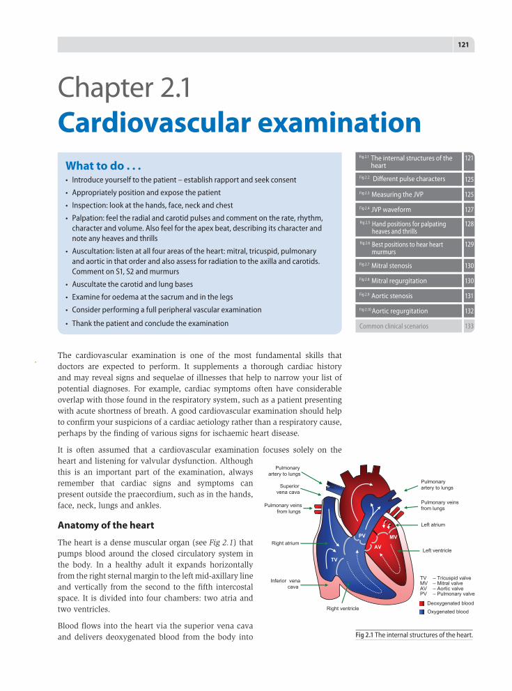
Chapter 2.1Cardiovascular examination
The cardiovascular examination is one of the most fundamental skills that doctors are expected to perform. It supplements a thorough cardiac history and may reveal signs and sequelae of illnesses that help to narrow your list of potential diagnoses. For example, cardiac symptoms often have considerable overlap with those found in the respiratory system, such as a patient presenting with acute shortness of breath. A good cardiovascular examination should help to confirm your suspicions of a cardiac aetiology rather than a respiratory cause, perhaps by the finding of various signs for ischaemic heart disease.
It is often assumed that a cardiovascular examination focuses solely on the heart and listening for valvular dysfunction. Although this is an important part of the examination, always remember that cardiac signs and symptoms can present outside the praecordium, such as in the hands, face, neck, lungs and ankles.
Anatomy of the heart
The heart is a dense muscular organ (see Fig 2.1) that pumps blood around the closed circulatory system in the body. In a healthy adult it expands horizontally from the right sternal margin to the left mid-axillary line and vertically from the second to the fifth intercostal space. It is divided into four chambers: two atria and two ventricles.
Blood flows into the heart via the superior vena cava and delivers deoxygenated blood from the body into
What to do . . .• Introduceyourselftothepatient–establishrapportandseekconsent
• Appropriatelypositionandexposethepatient
• Inspection:lookatthehands,face,neckandchest
• Palpation:feeltheradialandcarotidpulsesandcommentontherate,rhythm,characterandvolume.Alsofeelfortheapexbeat,describingitscharacterandnoteanyheavesandthrills
• Auscultation:listenatallfourareasoftheheart:mitral,tricuspid,pulmonaryandaorticinthatorderandalsoassessforradiationtotheaxillaandcarotids.CommentonS1,S2andmurmurs
• Auscultatethecarotidandlungbases
• Examineforoedemaatthesacrumandinthelegs
• Considerperformingafullperipheralvascularexamination
• Thankthepatientandconcludetheexamination
Fig2.2 Different pulse characters
Fig2.3 Measuring the JVP
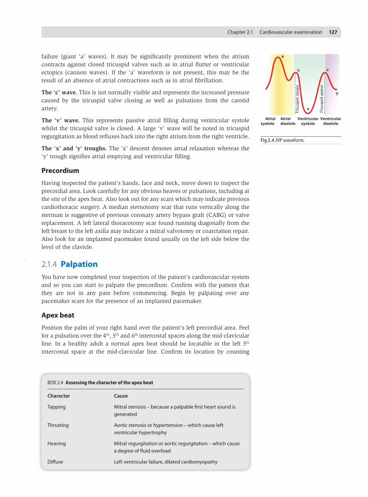
Fig2.4 JVP waveform
125
125
127
Fig2.1 The internal structures of the heart
121
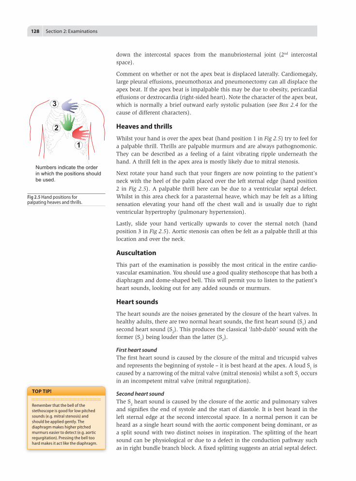
Fig2.5 Handpositionsforpalpatingheavesandthrills
128
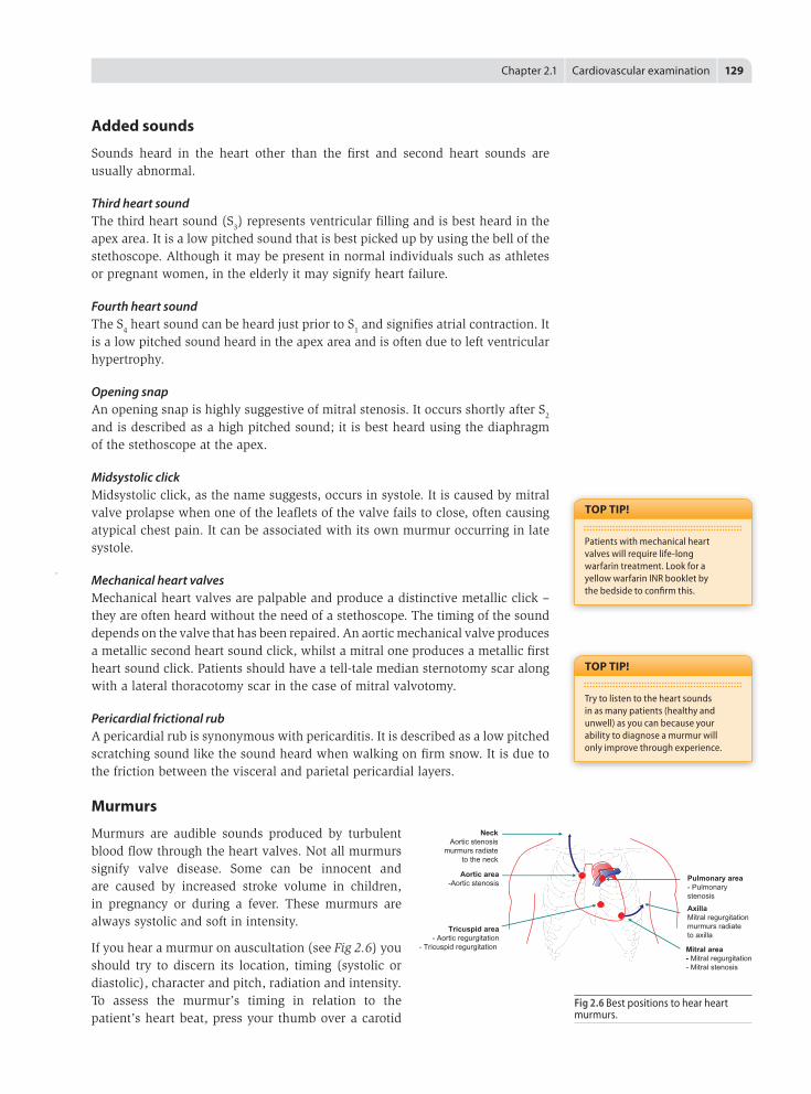
Fig2.6 Bestpositionstohearheartmurmurs
129
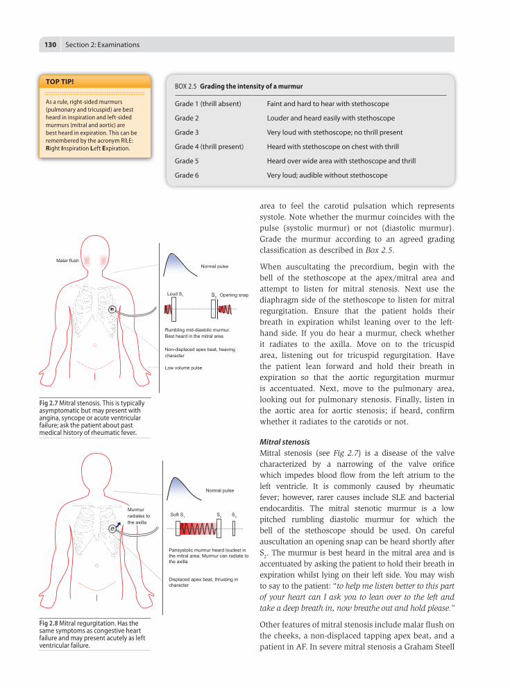
Fig2.7 Mitral stenosis
Fig2.8 Mitral regurgitation
Fig2.9 Aortic stenosis
Fig2.10Aortic regurgitation
130
130
131
132
Commonclinicalscenarios 133
Fig 2.1 The internal structures of the heart.
Right atrium
Pulmonary veins from lungs
TV
PV
AV
MV
Superior vena cava
Inferior vena cava
Left atrium
Pulmonary veins from lungs
Left ventricle
Right ventricle
Pulmonary artery to lungs
Pulmonary artery to lungs
TV – Tricuspid valveMV – Mitral valveAV – Aortic valvePV – Pulmonary valve
Deoxygenated blood
Oxygenated blood
122 | Section 2: Examinations
the right atrium. From here it passes into the right ventricle through the tricuspid valve. As the ventricles contract, blood is pumped from the right ventricle and into the lungs through the pulmonary valve. The left atrium receives oxygenated blood from the lungs from where it is transported into the left ventricle through the mitral (bicuspid) valve. Finally, the left ventricle contracts, forcing blood into the aorta, and out to the rest of the body through the aortic valve.
2.1.1 Beginning the examinationIntroduce yourself to the patient by stating your name and job title. Then ask the patient’s name and age before establishing whether they are in any pain or discomfort.
It is important to explain clearly to the patient what you intend to do when examining the chest; for example, you may wish to say ‘In view of the symptoms you have reported, I need to perform a cardiovascular examination. This will involve me examining and listening to your chest with a stethoscope, as well as looking at your hands, eyes and legs. Before I proceed can I ask if you are in any pain or discomfort?’. Female patients may have to undress to their bra and so they should be offered the option of having a chaperone if they feel uncomfortable.
Check that the patient has understood what you have said and then seek their consent to proceed with the examination.
2.1.2 Patient exposure and positionBefore beginning the examination and asking the patient to undress, ensure that adequate privacy is provided – you should draw curtains around the cubicle or close any doors that are open. Position the patient’s bed or examination couch at a 45 degree angle and ask the patient to lie comfortably with their head resting on a pillow. This will help facilitate your examination of the JVP later on. Expose their chest and ensure that you have a clear view of their lower limbs so that you can assess for any evidence of pedal oedema.
Offer the patient a sheet or blanket to maintain their modesty. Ensure that there is good lighting to observe the patient as this will allow you to perform a proper inspection of the chest. Finally, you should always ensure that you have washed your hands thoroughly before commencing your examination.
2.1.3 InspectionStart your inspection by taking a step back and observing the patient from the end of the bed. Look at the patient and see if they are grimacing with pain or lying comfortably. Look around the patient’s bedside for any evidence of oxygen masks, heart monitors, IV cannula drips or GTN sprays. Observe the patient for any obvious difficulties such as laboured breathing or tachypnoea. Look at the general appearance of the patient, particularly at the face for any typical facies associated with Down, Marfan or Turner syndromes – these syndromes all have an effect on the cardiovascular system (see Box 2.1).
Observe the patient’s skin for the presence of cyanosis (cyanotic congenital heart disease), pallor (anaemia) or bronze pigmentation (haemochromatosis cardiomyopathy). Stand back and listen for any clicking sounds from mechanical heart valves. Move closer to the patient and proceed, in a systematic way, to inspect the hands, face, neck, chest and legs in more detail.
Chapter 2.1 | Cardiovascular examination | 123
Hands
Hand signs are often neglected and overlooked by doctors due to time constraints. However, it is important to note that the hands often reveal vital clues pointing towards underlying pathology. • Observe for any evidence of tar staining indicating a smoker. • Look at the colour of the nail beds; normal nail beds should be pink and
flushed with colour, but blue nail beds represent peripheral cyanosis. • Look at the nails for any evidence of splinter haemorrhages (brown-
coloured thin vertical streaks found under the nail) which are associated with infective endocarditis.
• Check for signs of clubbing which could be due to infective endocarditis (IE), cyanotic congenital heart disease or atrial myxoma.
Next, ask the patient to turn their hands over to expose their palmar surfaces. Look for other features of infective endocarditis such as Osler’s nodes and Janeway lesions. Osler’s nodes are painful nodules found at the fingertips, whilst Janeway lesions are small painless erythematous macular or nodular lesions located on the palms of the hands or the soles of the feet.
Once you have inspected the hands check for temperature – the hands in a normal patient should be warm and well perfused, whereas cold peripheries may indicate inadequate peripheral circulation. Assess the capillary refill time by pressing down and blanching the nail bed for 5 seconds before releasing and watching for the blood to return – in healthy adults this should be less than 2 seconds.
Pulse
Move your attention to the patient’s wrist. Palpate their radial pulse which can be found between the styloid process of the radius and the flexor carpi radialis tendon, just proximal to the wrist joint. Use three fingers to accurately locate and palpate it. You should note the rate, rhythm, volume and character of the pulse. However, be aware that the volume and character of the pulse is better assessed centrally, such as at the carotid artery, because the further you go away from the aorta the more the waveform is distorted.
Next palpate both radial pulses at the same time to assess for any asynchrony (radio-radial delay) which may be a sign of aortic dissection or co-arctation of
BOX 2.1 Typical features of common syndromes associated with cardiovascular dysfunction
Down syndrome Microgenia (abnormally small chin), oblique eye fissures with epicanthic skin folds on the inner corner of the eyes (mongoloid fold), a flat nasal bridge, a single palmar fold, a protruding tongue, macroglossia, short neck, short stature. Cardiac problems include aortic and ventricular septal defects.
Marfan syndrome Arachnodactyly, scoliosis, pectus excavatum (abnormal indentation) or pectus carinatum (abnormal protrusion) of the sternum, a high palate, arm span greater than their height. Cardiac problems include aortic or mitral regurgitation, mitral prolapse, aortic dissection, and cardiac arrhythmias.
Turner syndrome Short stature, low hairline with low set ears and a web neck, broad chest (shield chest) and widely spaced nipples. Cardiac problems include aortic dissection and, to a lesser extent, aortic stenosis.
RheumaticfeverisNOTacardiaccauseofclubbing.
TOP TIP!
124 | Section 2: Examinations
the aorta. Also check for a radio-femoral delay by simultaneously palpating the radial and ipsilateral femoral pulses. A delay between the two may be suggestive of co-arctation of the aorta.
RateAssess the rate of the radial pulse by counting the number of pulsations over 15 seconds, then multiply this value by 4 to get an approximation of the cardiac rate. The normal cardiac rate should be between 60 and 100 beats per minute. Tachycardia is when the rate is more than 100, whereas bradycardia is less than 60.
Rhythm Feel the pulse for its rhythm and regularity: • A normal regular pulse is said to be in sinus rhythm. • A regularly irregular pulse is one whereby the irregularity of the pulse occurs
at regular intervals. The most common cause of this is second degree heart block or sinus arrhythmia (when there is variation of the pulse with breathing – the pulse increases slightly on inspiration and slows on expiration).
• An irregularly irregular pulse, on the other hand, is when the irregularity of the pulse is unpredictable, such as in ectopic beats or atrial fibrillation.
Volume As stated above, volume is best assessed at the carotids. The pulse volume may give some indication of the cardiac output. A weak or low volume pulse may be due to heart failure and aortic stenosis, whereas a strong or large volume pulse may be found in CO
2 retention, thyrotoxicosis or aortic regurgitation.
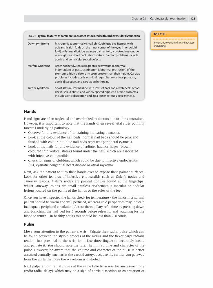
CharacterA normal pulse has a firm upstroke with a slow paced downstroke. Diseases of the aortic valve affect how blood flows through the aorta which alters the character of the pulse (Fig 2.2). In aortic stenosis, with a narrowing of the aorta, blood flow is impeded, producing a slow rising pulse. In aortic regurgitation, the incompetent valve causes pooling of blood in the left ventricle, resulting in an increased stroke volume. This creates a tall initial upstroke with a collapsing downstroke, giving rise to a wide pulse pressure. This is known as collapsing or water hammer pulse and is best assessed by raising the patient’s hand over their head and above the level of their heart whilst palpating their radial artery – an increased tapping sensation will be felt in the fingers held over the radial area.
In mixed aortic valve disease such as aortic stenosis with regurgitation, the pulse may have two upstrokes separated by a short mid-systolic downstroke. This is called the bisferiens pulse and may be felt as a double impulse with each beat of the heart over the radial pulse area.
If the pulse alternates from feeling strong to weak, this is known as pulsus alternans and is synonymous with severe left ventricular failure. Pulsus paradoxus is when there is an exaggeration of the normal physiological response with an increase in pulse volume during expiration – this sign is associated with cardiac tamponade or obstructive lung disease.
Blood pressure
After examining the pulse you should go on to measure the patient’s blood pressure. In an exam setting, the examiner may give you a reading when you indicate that you wish to perform this task.
Co-arctationisacongenitalnarrowingofthedescendingaorta,usuallyjustdistaltotheoriginoftheleftsubclavianartery,leadingtohigherbloodpressureinthearmsthanlegs.Itismorecommoninboys.Mostpatientswiththisconditionhaveusuallybeentreatedsurgicallyandwillbeleftwithaleftthoracotomyscarwithnopulseintheleftarm!
TOP TIP!
Avoidusingthewordsweakorstrongpulseinfrontofthepatientasitmaycauseundueanxiety.
TOP TIP!
Theaorticregurgitationpulsewascoined‘waterhammer’afteraVictorianchild’stoywhichconsistedofaglasstubefilledwitheitherwaterormercurywithinavacuum.Asthetoywasturnedupsidedown,thewaterormercurywouldproduceacharacteristicknockingsoundasitimpactedagainsttheglass.
TOP TIP!
BisferiensorginatesfromLatin:bis–twice,andferio–strike,describingthetwopeaksofthecharacter.
TOP TIP!
Pulsusparadoxus(paradoxicalpulse)isnotreallyaparadox,butanexaggerationofthenorm.
TOP TIP!
Chapter 2.1 | Cardiovascular examination | 125
Face
Move your attention now to the patient’s face, particularly focusing on the sclera, lips and the mouth. The face may give you clues to underlying heart valve problems, cardiovascular risk factors or genetic syndromes that can affect the heart, as follows:
• Take a look at the patient’s sclera, warning them that you will be retracting their eyelid for a clearer view. Look for signs of a pale conjunctiva which may be suggestive of anaemia.
• Next look at the cornea, checking for an opaque grey ring around the iris, known as a corneal arcus. Although this may be a common sight in the elderly (where it is known as a senile arcus), in the younger patient it may signify lipid dysfunction.
• Look around the eyes for other signs of hyperlipidaemia, such as xanthelasma (yellow plaque-like deposits of fat around the peri-orbital area).
• Look at the patient’s cheeks for a dusky pink discoloration known as malar flush that occurs in mitral stenosis.
• Check their lips and tongue for any blue discoloration which may be a sign of central cyanosis. It is often easier to visualize if the patient raises their tongue up against the roof of their palate. When looking in the mouth you should also check for a high arched palate which may be a sign that the patient has Marfan syndrome.
• Inspect the patient’s dentition for poor dental hygiene as this can be associated with infective endocarditis in those with underlying valvular disease.
Neck
Carotid pulseLook across to the patient’s neck and check for any obvious pulsations which may be caused by a carotid pulse. Use your left thumb to assess the carotid pulse for volume and character. The pulse is usually located between the anterior border of the sternocleidomastoid muscle and the larynx around the level of the cricoid cartilage. Move on to examine the other carotid using your right thumb. Never compress both carotids simultaneously.
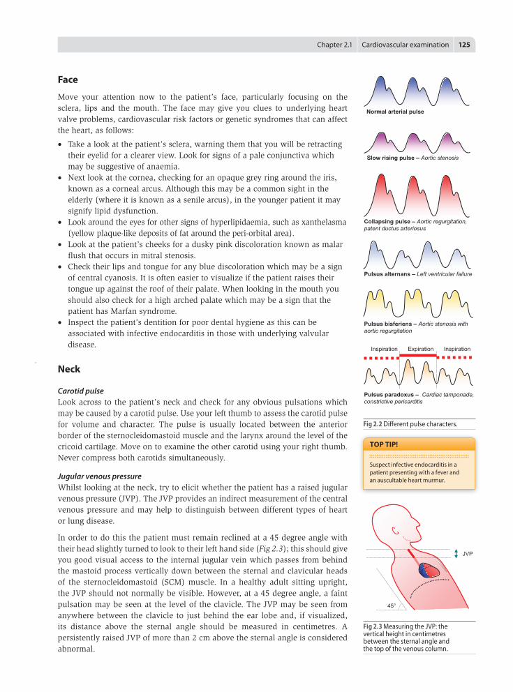
Jugular venous pressureWhilst looking at the neck, try to elicit whether the patient has a raised jugular venous pressure (JVP). The JVP provides an indirect measurement of the central venous pressure and may help to distinguish between different types of heart or lung disease.
In order to do this the patient must remain reclined at a 45 degree angle with their head slightly turned to look to their left hand side (Fig 2.3); this should give you good visual access to the internal jugular vein which passes from behind the mastoid process vertically down between the sternal and clavicular heads of the sternocleidomastoid (SCM) muscle. In a healthy adult sitting upright, the JVP should not normally be visible. However, at a 45 degree angle, a faint pulsation may be seen at the level of the clavicle. The JVP may be seen from anywhere between the clavicle to just behind the ear lobe and, if visualized, its distance above the sternal angle should be measured in centimetres. A persistently raised JVP of more than 2 cm above the sternal angle is considered abnormal.
Suspectinfectiveendocarditisinapatientpresentingwithafeverandanauscultableheartmurmur.
TOP TIP!
Fig 2.3 Measuring the JVP: the vertical height in centimetres between the sternal angle and the top of the venous column.
Fig 2.2 Different pulse characters.
Inspiration Expiration Inspiration
Normal arterial pulse
Collapsing pulse – Aortic regurgitation, patent ductus arteriosus
Pulsus alternans – Left ventricular failure
Pulsus bisferiens – Aortic stenosis with aortic regurgitation
Pulsus paradoxus – Cardiac tamponade, constrictive pericarditis
Slow rising pulse – Aortic stenosis
JVP
45°
126 | Section 2: Examinations
If the JVP cannot be visualized you may wish to perform the hepatojugular reflux to confirm a normal right atrial pressure. Confirm that the patient does not have any pain in the abdomen and then apply firm pressure over the liver for around 10 seconds and observe the supraclavicular area. The pressure should cause a transient rise in the JVP such that it may be visualized above the level of the clavicle. This test may also be used to help distinguish between a carotid and jugular pulsation (see Box 2.2).
A raised pulsating JVP is a sign of right heart failure, whereas a raised non-pulsating JVP is a sign of superior vena cava obstruction, often due to bronchial carcinoma. A paradoxical rise in JVP during inspiration (normally venous return occurs with inspiration and brings the JVP down) indicates constrictive pericarditis and is known as Kussmaul’s sign. See Box 2.3 for further causes of raised JVP.
WaveformWhen looking at the JVP you should try to discern its waveform. Although difficult at first, this skill often improves with experience. The normal waveform (see Fig 2.4) consists of two peaks (a and v) and two troughs (x and y) and a ‘c’ wave lying between the first peak and trough. The ‘a’ and ‘v’ waves can be distinguished by their timing against the carotid pulse: the ‘a’ wave occurs just prior to the pulse whereas the ‘v’ wave will be seen towards the tail end of the arterial pulsation.
The ‘a’ wave. This represents atrial contractions and is elevated if the pulmonary artery pressure is increased such as in pulmonary hypertension or right heart
DonotconfuseKussmaul’ssign(paradoxicalriseofJVPduringinspiration)withKussmaul’sbreathing,whichisdeep,sighingrespirationseeninseveremetabolicacidosis,e.g.diabeticketoacidosisorrenalfailure.
TOP TIP!
BOX 2.3 Causes of raised JVP
Right heart failure
Fluid overload
Tricuspid regurgitation
Pulmonary stenosis, tricuspid stenosis
Pulmonary embolism, pericardial effusion
Constrictive pericarditis
Cardiac tamponade
BOX 2.2 Features to distinguish between the jugular and carotid impulses
Jugular impulse Carotid impulse
Pulse not palpable Palpable pulse
Rapid inward movement Rapid outward movement
Disappears when pressure applied Unaffected when pressure to root of neck applied to root of neck
Two peaks per heartbeat One peak per heartbeat (sinus rhythm)
Varies with position and respiration Unaffected by position or respiration
Transient rise with abdominal pressure No rise with abdominal pressure
AlwayscheckbehindtheearlobeiftheJVPcannotbeseen.
TOP TIP!
Chapter 2.1 | Cardiovascular examination | 127
failure (giant ‘a’ waves). It may be significantly prominent when the atrium contracts against closed tricuspid valves such as in atrial flutter or ventricular ectopics (cannon waves). If the ‘a’ waveform is not present, this may be the result of an absence of atrial contractions such as in atrial fibrillation.
The ‘c’ wave. This is not normally visible and represents the increased pressure caused by the tricuspid valve closing as well as pulsations from the carotid artery.
The ‘v’ wave. This represents passive atrial filling during ventricular systole whilst the tricuspid valve is closed. A large ‘v’ wave will be noted in tricuspid regurgitation as blood refluxes back into the right atrium from the right ventricle.
The ‘x’ and ‘y’ troughs. The ‘x’ descent denotes atrial relaxation whereas the ‘y’ trough signifies atrial emptying and ventricular filling.
Precordium
Having inspected the patient’s hands, face and neck, move down to inspect the precordial area. Look carefully for any obvious heaves or pulsations, including at the site of the apex beat. Also look out for any scars which may indicate previous cardiothoracic surgery. A median sternotomy scar that runs vertically along the sternum is suggestive of previous coronary artery bypass graft (CABG) or valve replacement. A left lateral thoracotomy scar found running diagonally from the left breast to the left axilla may indicate a mitral valvotomy or coarctation repair. Also look for an implanted pacemaker found usually on the left side below the level of the clavicle.
2.1.4 PalpationYou have now completed your inspection of the patient’s cardiovascular system and so you can start to palpate the precordium. Confirm with the patient that they are not in any pain before commencing. Begin by palpating over any pacemaker scars for the presence of an implanted pacemaker.
Apex beat
Position the palm of your right hand over the patient’s left precordial area. Feel for a pulsation over the 4th, 5th and 6th intercostal spaces along the mid-clavicular line. In a healthy adult a normal apex beat should be locatable in the left 5th intercostal space at the mid-clavicular line. Confirm its location by counting
BOX 2.4 Assessing the character of the apex beat
Character Cause
Tapping Mitral stenosis – because a palpable first heart sound is generated
Thrusting Aortic stenosis or hypertension – which cause left ventricular hypertrophy
Heaving Mitral regurgitation or aortic regurgitation – which cause a degree of fluid overload
Diffuse Left ventricular failure, dilated cardiomyopathy
Fig 2.4 JVP waveform.
Ventricularsystole
Ventriculardiastole
Atrialdiastole
Atrialsystole
a
c
x
v
y
Tric
uspi
d cl
oses
Tric
uspi
d op
ens
128 | Section 2: Examinations
down the intercostal spaces from the manubriosternal joint (2nd intercostal space).
Comment on whether or not the apex beat is displaced laterally. Cardiomegaly, large pleural effusions, pneumothorax and pneumonectomy can all displace the apex beat. If the apex beat is impalpable this may be due to obesity, pericardial effusions or dextrocardia (right-sided heart). Note the character of the apex beat, which is normally a brief outward early systolic pulsation (see Box 2.4 for the cause of different characters).
Heaves and thrills
Whilst your hand is over the apex beat (hand position 1 in Fig 2.5) try to feel for a palpable thrill. Thrills are palpable murmurs and are always pathognomonic. They can be described as a feeling of a faint vibrating ripple underneath the hand. A thrill felt in the apex area is mostly likely due to mitral stenosis.
Next rotate your hand such that your fingers are now pointing to the patient’s neck with the heel of the palm placed over the left sternal edge (hand position 2 in Fig 2.5). A palpable thrill here can be due to a ventricular septal defect. Whilst in this area check for a parasternal heave, which may be felt as a lifting sensation elevating your hand off the chest wall and is usually due to right ventricular hypertrophy (pulmonary hypertension).
Lastly, slide your hand vertically upwards to cover the sternal notch (hand position 3 in Fig 2.5). Aortic stenosis can often be felt as a palpable thrill at this location and over the neck.
Auscultation
This part of the examination is possibly the most critical in the entire cardio-vascular examination. You should use a good quality stethoscope that has both a diaphragm and dome-shaped bell. This will permit you to listen to the patient’s heart sounds, looking out for any added sounds or murmurs.
Heart sounds
The heart sounds are the noises generated by the closure of the heart valves. In healthy adults, there are two normal heart sounds, the first heart sound (S1) and second heart sound (S2). This produces the classical ‘lubb-dubb’ sound with the former (S1) being louder than the latter (S2).
First heart soundThe first heart sound is caused by the closure of the mitral and tricuspid valves and represents the beginning of systole – it is best heard at the apex. A loud S1 is caused by a narrowing of the mitral valve (mitral stenosis) whilst a soft S1 occurs in an incompetent mitral valve (mitral regurgitation).
Second heart soundThe S2 heart sound is caused by the closure of the aortic and pulmonary valves and signifies the end of systole and the start of diastole. It is best heard in the left sternal edge at the second intercostal space. In a normal person it can be heard as a single heart sound with the aortic component being dominant, or as a split sound with two distinct noises in inspiration. The splitting of the heart sound can be physiological or due to a defect in the conduction pathway such as in right bundle branch block. A fixed splitting suggests an atrial septal defect.
Rememberthatthebellofthestethoscopeisgoodforlowpitchedsounds(e.g.mitralstenosis)andshouldbeappliedgently.Thediaphragmmakeshigherpitchedmurmurseasiertodetect(e.g.aorticregurgitation).Pressingthebelltoohardmakesitactlikethediaphragm.
TOP TIP!
Fig 2.5 Hand positions for palpating heaves and thrills.
2
1
3
Numbers indicate the order in which the positions should be used.
Chapter 2.1 | Cardiovascular examination | 129
Added sounds
Sounds heard in the heart other than the first and second heart sounds are usually abnormal.
Third heart soundThe third heart sound (S3) represents ventricular filling and is best heard in the apex area. It is a low pitched sound that is best picked up by using the bell of the stethoscope. Although it may be present in normal individuals such as athletes or pregnant women, in the elderly it may signify heart failure.
Fourth heart soundThe S4 heart sound can be heard just prior to S1 and signifies atrial contraction. It is a low pitched sound heard in the apex area and is often due to left ventricular hypertrophy.
Opening snapAn opening snap is highly suggestive of mitral stenosis. It occurs shortly after S2 and is described as a high pitched sound; it is best heard using the diaphragm of the stethoscope at the apex.
Midsystolic clickMidsystolic click, as the name suggests, occurs in systole. It is caused by mitral valve prolapse when one of the leaflets of the valve fails to close, often causing atypical chest pain. It can be associated with its own murmur occurring in late systole.
Mechanical heart valvesMechanical heart valves are palpable and produce a distinctive metallic click – they are often heard without the need of a stethoscope. The timing of the sound depends on the valve that has been repaired. An aortic mechanical valve produces a metallic second heart sound click, whilst a mitral one produces a metallic first heart sound click. Patients should have a tell-tale median sternotomy scar along with a lateral thoracotomy scar in the case of mitral valvotomy.
Pericardial frictional rubA pericardial rub is synonymous with pericarditis. It is described as a low pitched scratching sound like the sound heard when walking on firm snow. It is due to the friction between the visceral and parietal pericardial layers.
Murmurs
Murmurs are audible sounds produced by turbulent blood flow through the heart valves. Not all murmurs signify valve disease. Some can be innocent and are caused by increased stroke volume in children, in pregnancy or during a fever. These murmurs are always systolic and soft in intensity.
If you hear a murmur on auscultation (see Fig 2.6) you should try to discern its location, timing (systolic or diastolic), character and pitch, radiation and intensity. To assess the murmur’s timing in relation to the patient’s heart beat, press your thumb over a carotid
Patientswithmechanicalheartvalveswillrequirelife-longwarfarintreatment.LookforayellowwarfarinINRbookletbythebedsidetoconfirmthis.
TOP TIP!
Trytolistentotheheartsoundsinasmanypatients(healthyandunwell)asyoucanbecauseyourabilitytodiagnoseamurmurwillonlyimprovethroughexperience.
TOP TIP!
Fig 2.6 Best positions to hear heart murmurs.
AxillaMitral regurgitationmurmurs radiate to axilla
Mitral area- Mitral regurgitation- Mitral stenosis
Pulmonary area- Pulmonary stenosis
NeckAortic stenosis
murmurs radiateto the neck
Aortic area-Aortic stenosis
Tricuspid area- Aortic regurgitation
- Tricuspid regurgitation
130 | Section 2: Examinations
area to feel the carotid pulsation which represents systole. Note whether the murmur coincides with the pulse (systolic murmur) or not (diastolic murmur). Grade the murmur according to an agreed grading classification as described in Box 2.5.
When auscultating the precordium, begin with the bell of the stethoscope at the apex/mitral area and attempt to listen for mitral stenosis. Next use the diaphragm side of the stethoscope to listen for mitral regurgitation. Ensure that the patient holds their breath in expiration whilst leaning over to the left-hand side. If you do hear a murmur, check whether it radiates to the axilla. Move on to the tricuspid area, listening out for tricuspid regurgitation. Have the patient lean forward and hold their breath in expiration so that the aortic regurgitation murmur is accentuated. Next, move to the pulmonary area, looking out for pulmonary stenosis. Finally, listen in the aortic area for aortic stenosis; if heard, confirm whether it radiates to the carotids or not.
Mitral stenosisMitral stenosis (see Fig 2.7) is a disease of the valve characterized by a narrowing of the valve orifice which impedes blood flow from the left atrium to the left ventricle. It is commonly caused by rheumatic fever; however, rarer causes include SLE and bacterial endocarditis. The mitral stenotic murmur is a low pitched rumbling diastolic murmur for which the bell of the stethoscope should be used. On careful auscultation an opening snap can be heard shortly after S
2. The murmur is best heard in the mitral area and is accentuated by asking the patient to hold their breath in expiration whilst lying on their left side. You may wish to say to the patient: “to help me listen better to this part of your heart can I ask you to lean over to the left and take a deep breath in, now breathe out and hold please.”
Other features of mitral stenosis include malar flush on the cheeks, a non-displaced tapping apex beat, and a patient in AF. In severe mitral stenosis a Graham Steell
BOX 2.5 Grading the intensity of a murmur
Grade 1 (thrill absent) Faint and hard to hear with stethoscope
Grade 2 Louder and heard easily with stethoscope
Grade 3 Very loud with stethoscope; no thrill present
Grade 4 (thrill present) Heard with stethoscope on chest with thrill
Grade 5 Heard over wide area with stethoscope and thrill
Grade 6 Very loud; audible without stethoscope
Asarule,right-sidedmurmurs(pulmonaryandtricuspid)arebestheardininspirationandleft-sidedmurmurs(mitralandaortic)arebestheardinexpiration.ThiscanberememberedbytheacronymRILE:RightInspirationLeftExpiration.
TOP TIP!
Fig 2.7 Mitral stenosis. This is typically asymptomatic but may present with angina, syncope or acute ventricular failure; ask the patient about past medical history of rheumatic fever.
Loud S1 S2
Low volume pulse
Normal pulse
Rumbling mid-diastolic murmur.Best heard in the mitral area
Non-displaced apex beat, heavingcharacter
Malar flush
Opening snap
Fig 2.8 Mitral regurgitation. Has the same symptoms as congestive heart failure and may present acutely as left ventricular failure.
Soft S1 S2
Normal pulse
Pansystolic murmur heard loudest inthe mitral area. Murmur can radiate tothe axilla
Displaced apex beat, thrusting incharacter
S3
Murmur radiates to the axilla
Chapter 2.1 | Cardiovascular examination | 131
murmur may be found. This is an early diastolic murmur due to pulmonary regurgitation heard loudest in the pulmonary area (second intercostal space, left sternal edge). This occurs because the chronically elevated pressure in the right atrium, which results from the stenosed mitral valve, leads to an increase in pulmonary capillary pressure and pulmonary hypertension, resulting in pulmonary regurgitation.
Mitral regurgitationIn mitral regurgitation (see Fig 2.8) there is an incompetence of the mitral valve leading to incomplete closure during systole. This allows blood to reflux back into the left atrium from the left ventricle. Common causes include mitral valve proplase, ischaemic heart disease, rheumatic fever and Marfan syndrome. Mitral regurgitation may also be a post-MI complication from rupture of the chordae tendinae or papillary muscles. It is best heard in the mitral area of the heart and presents with a soft S
1 and a blowing pansystolic murmur which radiates to the axilla. Additional features may include a displaced thrusting apex beat as well as an S3.
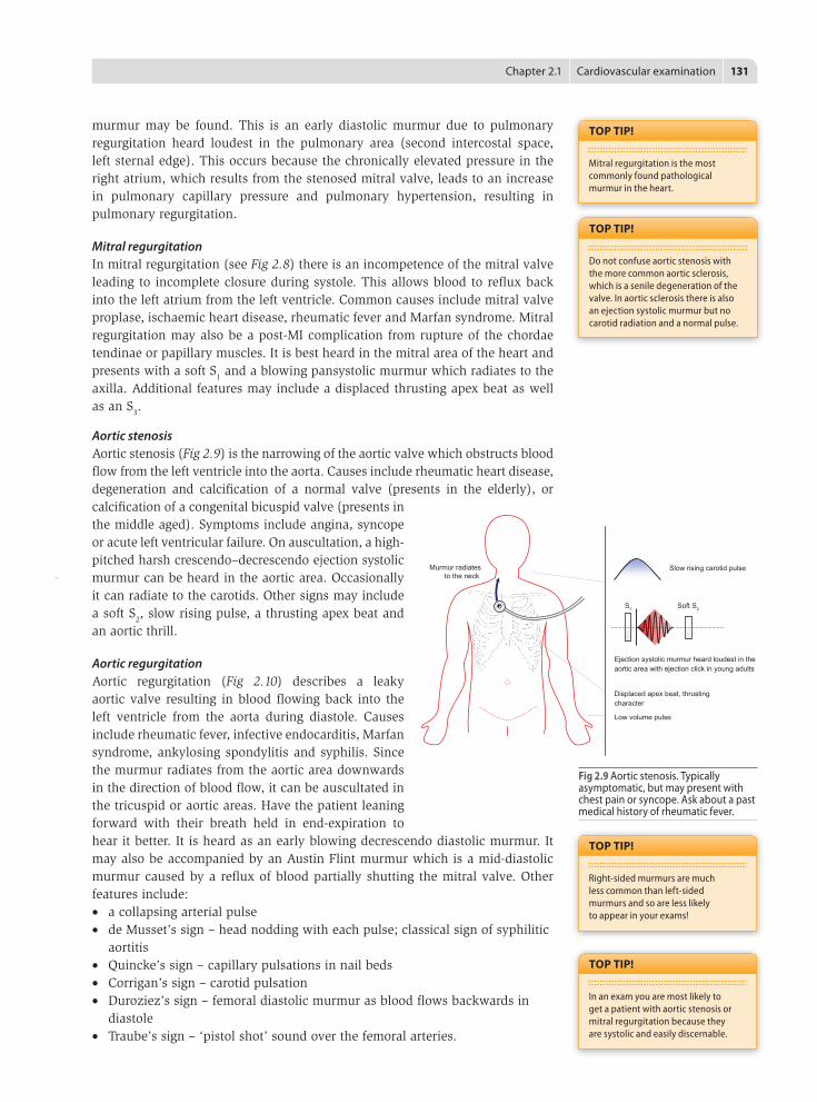
Aortic stenosisAortic stenosis (Fig 2.9) is the narrowing of the aortic valve which obstructs blood flow from the left ventricle into the aorta. Causes include rheumatic heart disease, degeneration and calcification of a normal valve (presents in the elderly), or calcification of a congenital bicuspid valve (presents in the middle aged). Symptoms include angina, syncope or acute left ventricular failure. On auscultation, a high-pitched harsh crescendo–decrescendo ejection systolic murmur can be heard in the aortic area. Occasionally it can radiate to the carotids. Other signs may include a soft S2, slow rising pulse, a thrusting apex beat and an aortic thrill.
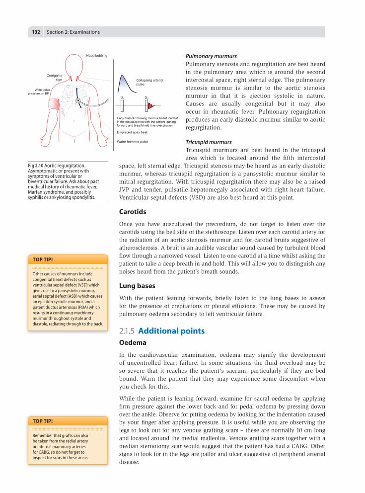
Aortic regurgitationAortic regurgitation (Fig 2.10) describes a leaky aortic valve resulting in blood flowing back into the left ventricle from the aorta during diastole. Causes include rheumatic fever, infective endocarditis, Marfan syndrome, ankylosing spondylitis and syphilis. Since the murmur radiates from the aortic area downwards in the direction of blood flow, it can be auscultated in the tricuspid or aortic areas. Have the patient leaning forward with their breath held in end-expiration to hear it better. It is heard as an early blowing decrescendo diastolic murmur. It may also be accompanied by an Austin Flint murmur which is a mid-diastolic murmur caused by a reflux of blood partially shutting the mitral valve. Other features include: • a collapsing arterial pulse • de Musset’s sign – head nodding with each pulse; classical sign of syphilitic
aortitis • Quincke’s sign – capillary pulsations in nail beds • Corrigan’s sign – carotid pulsation • Duroziez’s sign – femoral diastolic murmur as blood flows backwards in
diastole • Traube’s sign – ‘pistol shot’ sound over the femoral arteries.
Mitralregurgitationisthemostcommonlyfoundpathologicalmurmurintheheart.
TOP TIP!
Donotconfuseaorticstenosiswiththemorecommonaorticsclerosis,whichisaseniledegenerationofthevalve.Inaorticsclerosisthereisalsoanejectionsystolicmurmurbutnocarotidradiationandanormalpulse.
TOP TIP!
Right-sidedmurmursaremuchlesscommonthanleft-sidedmurmursandsoarelesslikelytoappearinyourexams!
TOP TIP!
Inanexamyouaremostlikelytogetapatientwithaorticstenosisormitralregurgitationbecausetheyaresystolicandeasilydiscernable.
TOP TIP!
Fig 2.9 Aortic stenosis. Typically asymptomatic, but may present with chest pain or syncope. Ask about a past medical history of rheumatic fever.
S1 Soft S2
Low volume pulse
Slow rising carotid pulse
Ejection systolic murmur heard loudest in the aortic area with ejection click in young adults
Displaced apex beat, thrusting character
Murmur radiatesto the neck
132 | Section 2: Examinations
Pulmonary murmursPulmonary stenosis and regurgitation are best heard in the pulmonary area which is around the second intercostal space, right sternal edge. The pulmonary stenosis murmur is similar to the aortic stenosis murmur in that it is ejection systolic in nature. Causes are usually congenital but it may also occur in rheumatic fever. Pulmonary regurgitation produces an early diastolic murmur similar to aortic regurgitation.
Tricuspid murmursTricuspid murmurs are best heard in the tricuspid area which is located around the fifth intercostal
space, left sternal edge. Tricuspid stenosis may be heard as an early diastolic murmur, whereas tricuspid regurgitation is a pansystolic murmur similar to mitral regurgitation. With tricuspid regurgitation there may also be a raised JVP and tender, pulsatile hepatomegaly associated with right heart failure. Ventricular septal defects (VSD) are also best heard at this point.
Carotids
Once you have auscultated the precordium, do not forget to listen over the carotids using the bell side of the stethoscope. Listen over each carotid artery for the radiation of an aortic stenosis murmur and for carotid bruits suggestive of atherosclerosis. A bruit is an audible vascular sound caused by turbulent blood flow through a narrowed vessel. Listen to one carotid at a time whilst asking the patient to take a deep breath in and hold. This will allow you to distinguish any noises heard from the patient’s breath sounds.
Lung bases
With the patient leaning forwards, briefly listen to the lung bases to assess for the presence of crepitations or pleural effusions. These may be caused by pulmonary oedema secondary to left ventricular failure.
2.1.5 Additional pointsOedema
In the cardiovascular examination, oedema may signify the development of uncontrolled heart failure. In some situations the fluid overload may be so severe that it reaches the patient’s sacrum, particularly if they are bed bound. Warn the patient that they may experience some discomfort when you check for this.
While the patient is leaning forward, examine for sacral oedema by applying firm pressure against the lower back and for pedal oedema by pressing down over the ankle. Observe for pitting oedema by looking for the indentation caused by your finger after applying pressure. It is useful while you are observing the legs to look out for any venous grafting scars – these are normally 10 cm long and located around the medial malleolus. Venous grafting scars together with a median sternotomy scar would suggest that the patient has had a CABG. Other signs to look for in the legs are pallor and ulcer suggestive of peripheral arterial disease.
Othercausesofmurmursincludecongenitalheartdefectssuchasventricularseptaldefect(VSD)whichgivesrisetoapansystolicmurmur,atrialseptaldefect(ASD)whichcausesanejectionsystolicmurmur,andapatentductusarteriosus(PDA)whichresultsinacontinuousmachinerymurmurthroughoutsystoleanddiastole,radiatingthroughtotheback.
TOP TIP!
RememberthatgraftscanalsobetakenfromtheradialarteryorinternalmammaryarteriesforCABG,sodonotforgettoinspectforscarsintheseareas.
TOP TIP!
Fig 2.10 Aortic regurgitation. Asymptomatic or present with symptoms of ventricular or biventricular failure. Ask about past medical history of rheumatic fever, Marfan syndrome, and possibly syphilis or ankylosing spondylitis.
Water hammer pulse
Collapsing arterialpulse
Early diastolic blowing murmur heard loudestin the tricuspid area with the patient leaningforward and breath held in end-expiration
Displaced apex beat
S2S1
Wide pulsepressure on BP
Head bobbing
Corrigan’ssign
Chapter 2.1 | Cardiovascular examination | 133
Peripheral vascular examination
Your examination of the cardiovascular system will not be complete if you do not examine the rest of the vascular system. Arterial disease in the heart is more than likely to be replicated elsewhere in the body and so you should ask to perform a full peripheral vascular examination (see Chapter 2.14) including the femoral, popliteal, posterior tibial and dorsalis pedis pulses.
2.1.6 Summing upIf you have found a cardiac murmur you should consider referring the patient for an echo cardiogram which will provide detailed structural information about the heart. If you have not already done so, indicate that you would like to measure the blood pressure. If the patient is found to have raised blood pressure you should ask to perform a urine dipstick to look for protein and carry out a fundoscopy to look for evidence of retinopathy.
Also, as with any examination, it is useful to request some bedside tests that may help in the diagnosis, such as an ECG and a chest X-ray of the patient. Indicate that you would also like to have a look at the patient’s oxygen saturations, respiration and temperature chart.
Thank the patient for their co-operation and offer to cover them with a blanket to protect their dignity. Keep the curtain around them drawn and give them appropriate time to dress themselves. Whilst waiting you may wish to collect your thoughts about what information you have gained from the examination before conveying it back to the patient.
2.1.7 Common clinical scenariosThe following are a list of common cardiology cases which you may encounter. For each one, consider: • what the likely diagnosis is • what key features you would look for • what questions you would ask or further investigations you would order to
refine or confirm your diagnosis
You could use role play with a friend, using the histories in the cases below as a framework.
C A S E 1 A 45 year old lady presents with shortness of breath with exercise, which has been getting progressively worse over the last 4 months. Her past medical history is only significant for rheumatic fever at the age of 10. On examination, she is in AF and the apex beat is tapping but not displaced. On auscultation there is a rumbling low-pitched mid-diastolic murmur best heard at the apex, which does not radiate – it is of 3/6 intensity. The lung bases are clear and there is no peripheral oedema.
134 | Section 2: Examinations
C A S E 5A 65 year gentleman attends a new registration check at the GP surgery. He brings along a warfarin diary with INRs currently controlled at 3.5. On examination he is found to have a median sternotomy scar and an audible click coinciding with a second heart sound.
C A S E 3A 70 year old gentleman presents to the GP with episodes of chest pain and fainting while running for the bus. These problems never happened while he was resting. He was given a spray to put underneath his tongue by his GP which helps the pain but the fainting is now becoming more frequent and he is worried. On examination the pulse is 75 beats per minute and regular but is slow rising in nature at the carotids. On auscultation a harsh ejection systolic murmur is heard following an ejection click. The murmur is noted to radiate to the carotids. Otherwise the heart sounds were normal. He did not appear to be in heart failure.
C A S E 4A 47 year old patient with a congenital bicuspid aortic valve was admitted from A&E as he had been feeling unwell for the last 6 weeks. He describes feeling fatigued, sweating at night and sometimes notices blood in his urine. He has unintentionally lost about 5 kg in the last 6 weeks. The only recent history of note is that he had a tooth extraction at the dentist. On examination, he has a temperature of 38.5°C and his nails are boggy with 1 mm red lines in the nail beds. He complains of pain when capillary refill time is assessed and there are a few red nodules present on the palms and finger tips. He was noticed to have a collapsing pulse and the apex beat was thrusting in nature and displaced to the anterior axillary line. Auscultation reveals a loud early diastolic murmur all over the precordium but loudest in the tricuspid region with the patient sitting forward.
C A S E 2A 60 year old gentleman, who is 3 days post MI, has presented with sudden onset of shortness of breath and a cough productive of frothy pink sputum. He is sitting up and finding it very difficult to breathe. On examination, he is in sinus tachycardia with a rate of 115. Auscultation reveals a pansystolic murmur loudest at the apex, which radiates to the axilla. The lung bases show evidence of crepitations and he has pitting oedema in the legs to the level of the knees.