34 introduction to animals 35 sponges and cnidarians 36
TRANSCRIPT
CHAPTERS
34 Introduction to Animals
35 Sponges and Cnidarians
36 Flatworms, Roundworms, and Rotifers
37 Mollusksand Annelids
38 Arthropods
It has taken biologists some 230 years to identify and
describe three quarters of a million insects; if there are
indeed at least thirty million, as Erwin (Terry Erwin, the
Smithsonian Institute) estimates, then, working as they
have in the past, insect taxonomists have ten thousand
years of employment ahead of them.>,
From "Endless Forms Most Beautiful," from The Sixth Extinction: Patterns of Life and the Future of Humankind, by Richard Leakey and Roger Lewin. Copyright © 1995 by Sherma B. V. Reprinted by permission of Doubleday, a division of Bantam Doubleday Dell Publishing Group, Inc.
39 Insects
40 Echinoderms
J3 internetconnect
SC INKS. National Science Teachers Association sc/LINKS Internet resources are located throughout this unit.
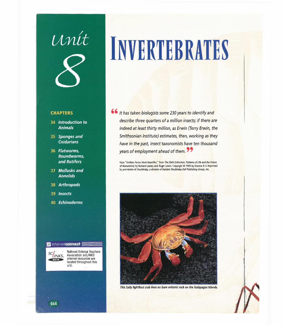
This Sally lightfoot crab lives on bare volcanic rock on the Galápagos Islands.
664
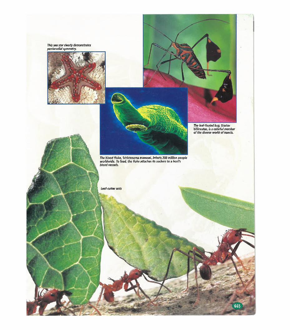
This sea star clearly demonstrates pentaradial symmetry.
The leaf-footed bug. Diactor billineatus, is a colorful member of the diverse world of insects.
The blood fluke. Schistosoma mansoni, infects 200 million people worldwide. To feed, the fluke attaches its suckers to a host's blood vessels.
CHAPTER 34
INTRODUCTION TO ANIMALS
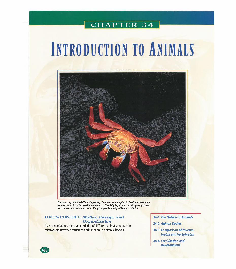
The diversity of animal life is staggering. Animals have adapted to Earth's lushest environments and to its harshest environments. This Sally Lightfoot crab, Grapsus grapsus, lives on the bare volcanic rock of the geologically young Galápagos Islands.
FOCUS CONCEPT: Matter■, Energy, andOrganization
As you read about the characteristics of different animals, notice the relationship between structure and function in animals' bodies.
34-1 The Nature of Animals
34-2 Animal Bodies
34-3 Comparison of Invertebrates and Vertebrates
34-4 Fertilization and Development
SECTION
The Nature oe AnimalsIf you are asked to name an animal, you might respond with
the name of a familiar large-bodied vertebrate—an animal with a backbone—such as a horse, a shark, or an eagle. But the kingdom Animalia is extraordinarily diverse, and most of its members are not vertebrates and do not even live on land. In Unit 8, you will read about invertebrates, animals without a backbone, which account for more than 95 percent of all animal species alive today. In Unit 9, you will read about the vertebrates, which are less numerous but more familiar to us; humans are vertebrates.
CHARACTERISTICSAnimals are multicellular heterotrophic organisms that lack cell walls. Most members of the animal kingdom share other important characteristics, including sexual reproduction and movement.
OBJECTIVES
Define the terms invertebrate and vertebrate.
Identify four important characteristics of animals.
■List two kinds of tissues found only in animals.
♦Explain how the first animals
might have evolved from unicellular organisms.
Multicellular OrganizationThe bodies of animals are multicellular. Some animals contain large numbers of cells. For example, the body of an adult human contains about 50 trillion cells. Unlike the cells of unicellular organisms, the cells of multicellular organisms do not lead independent lives. Each cell depends on the presence and functioning of other cells.
In all but the simplest animal phyla, there is a division of labor among cells. Specialization is the adaptation of a cell for a particular function. Just as a general contractor makes use of carpenters, electricians, and plumbers to build a house, a multicellular organism makes use of specialized cells to perform particular functions, such as digesting food or reproducing. Recall from Chapter 4 that a tissue is a group of similar cells specialized for a specific task. Most animal bodies are composed of combinations of different kinds of tissues. The formation of tissue from many individual cells is made possible by cell junctions, connections between cells that hold the cells together as a unit. The members of most animal phyla have organs, body structures that are composed of more them one type of tissue and that are specialized for a certain function.
Without multicellularity, the enormous variety found in the animal kingdom would not exist. The size of unicellular organisms is limited. Moreover, all of their functions, such as reproduction and
TBSSBBE
cry TOPIC: MulticellularLINKSL organisms
GO TO: www.scilinks.orgKEYWORD: HM667
INTRODUCTION TO ANIMALS
digestion, must be handled within a single cell. Multicellularity and cell specialization have enabled organisms to evolve and adapt to many environments.
HeterotrophyPlants and some unicellular organisms are autotrophic. They make food using simple molecules from their environment and an energy source, such as the sun. Animals, on the other hand, are hetero- trophic. They must obtain complex organic molecules from other sources. Most animals accomplish this by ingestion. During ingestion, an animal takes in organic material, usually in the form of other living things. Digestion then occurs within the animal’s body, and carbohydrates, lipids, amino acids, and other organic molecules are extracted from the material or cells the animal has ingested.
Sexual Reproduction and DevelopmentMost animals can reproduce sexually, and some can also reproduce asexually. Recall from Chapter 8 that in sexual reproduction, two haploid gametes fuse. This diploid zygote, the first cell of a new individual, then undergoes repeated mitotic divisions. Mitotic division of a cell produces two identical offspring cells. How does an adult animal, with its many different organs, tissues, and cell types, arise from a single cell? In the process called development, the enlarging mass of dividing cells undergoes differentiation. During differentiation (dif-uhr-EN-shee-AY-shuhn), cells become different from each other. For example, some cells may become blood cells, while others may become bone cells. The process of differentiation is the path to cell specialization.
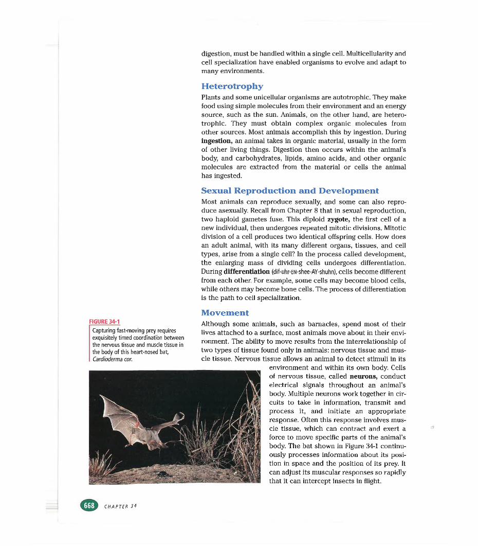
MovementFIGURE 34-1
Capturing fast-moving prey requires exquisitely timed coordination between the nervous tissue and muscle tissue in the body of this heart-nosed bat, Cardioderma cor.
Although some animals, such as barnacles, spend most of their lives attached to a surface, most animals move about in their environment. The ability to move results from the interrelationship of two types of tissue found only in animals: nervous tissue and muscle tissue. Nervous tissue allows an animal to detect stimuli in its
environment and within its own body. Cells of nervous tissue, called neurons, conduct electrical signals throughout an animal’s body. Multiple neurons work together in circuits to take in information, transmit and process it, and initiate an appropriate response. Often this response involves muscle tissue, which can contract and exert a force to move specific parts of the animal’s body. The bat shown in Figure 34-1 continuously processes information about its position in space and the position of its prey. It can adjust its muscular responses so rapidly that it can intercept insects in flight.
CHAPTER 34
ORIGIN AND CLASSIFICATION
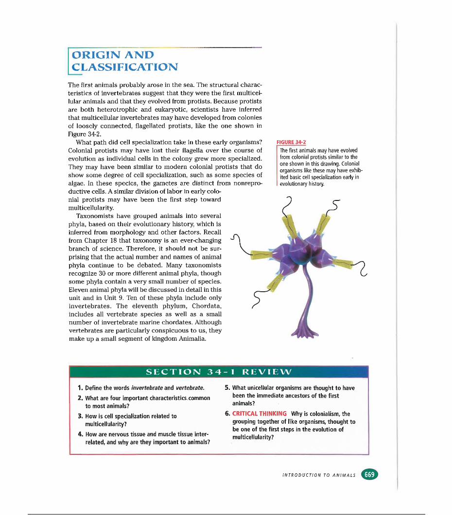
The first animals probably arose in the sea. The structural characteristics of invertebrates suggest that they were the first multicellular animals and that they evolved from protists. Because protists are both heterotrophic and eukaryotic, scientists have inferred that multicellular invertebrates may have developed from colonies of loosely connected, flagellated protists, like the one shown in Figure 34-2.
What path did cell specialization take in these early organisms? Colonial protists may have lost their flagella over the course of evolution as individual cells in the colony grew more specialized. They may have been similar to modern colonial protists that do show some degree of cell specialization, such as some species of algae. In these species, the gametes are distinct from nonrepro- ductive cells. A similar division of labor in early colonial protists may have been the first step toward multicellularity.
Taxonomists have grouped animals into several phyla, based on their evolutionary history, which is inferred from morphology and other factors. Recall from Chapter 18 that taxonomy is an ever-changing branch of science. Therefore, it should not be surprising that the actual number and names of animal phyla continue to be debated. Many taxonomists recognize 30 or more different animal phyla, though some phyla contain a very small number of species.Eleven animal phyla will be discussed in detail in this unit and in Unit 9. Ten of these phyla include only invertebrates. The eleventh phylum, Chordata, includes all vertebrate species as well as a small number of invertebrate marine chordates. Although vertebrates are particularly conspicuous to us, they make up a small segment of kingdom Animalia.
FIGURE 34-2The first animals may have evolved from colonial protists similar to the one shown in this drawing. Colonial organisms like these may have exhibited basic cell specialization early in evolutionary history.
SECTION! 34-1 REVIEW
1. Define the words invertebrate and vertebrate.
2. What are four important characteristics.common to most animals?
3. How is cell specialization related to multicellularity?
4. How are nervous tissue and muscle tissue interrelated, and why are they important to animals?
5. What unicellular organisms are thought to have been the immediate ancestors of the first animals?
6. CRITICAL THINKING Why is colonialism, the grouping together of like organisms, thought to be one of the first steps in the evolution of multicellularity?
INTRODUCTION TO ANIMALS
SECTION
OBJECTIVES
Define the terms dorsal, ventral, anterior, and posterior.
•Describe two types of symmetry
found in animals.
■Name the trait that is strongly
associated with bilateral symmetry.
♦List two functions of the body cavity in animals.
▲
List three structural features that taxonomists use to classify animals.
•List four features
found only in chordates.
Animal BodiesTaxonomic organization is based on phylogenetic
relationships. Today, systematic taxonomists classify animals according to similarities in morphology and other criteria, including the similarity of embryological development and the similarity of certain macromolecules. Recall from Chapter 18 that taxonomists since Linnaeus’s time have used an organism’s morphology to classify it with similar organisms. Morphology, however, is not confined to external appearance. A survey of an animal’s morphology also assesses the internal structure of the body and organization at the level of fundamental tissue types.
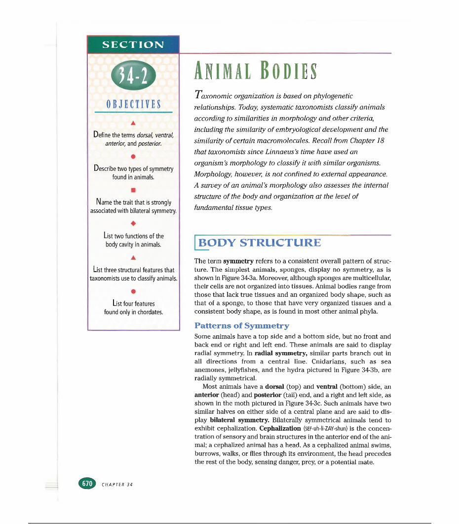
BODY STRUCTUREThe term symmetry refers to a consistent overall pattern of structure. The simplest animals, sponges, display no symmetry, as is shown in Figure 34-3a. Moreover, although sponges are multicellular, their cells are not organized into tissues. Animal bodies range from those that lack true tissues and an organized body shape, such as that of a sponge, to those that have very organized tissues and a consistent body shape, as is found in most other animal phyla.
Patterns of SymmetrySome animals have a top side and a bottom side, but no front and back end or right and left end. These animals are said to display radial symmetry. In radial symmetry, similar parts branch out in all directions from a central line. Cnidarians, such as sea anemones, jellyfishes, and the hydra pictured in Figure 34-3b, are radially symmetrical.
Most animals have a dorsal (top) and ventral (bottom) side, an anterior (head) and posterior (tail) end, and a right and left side, as shown in the moth pictured in Figure 34-3c. Such animals have two similar halves on either side of a central plane and are said to display bilateral symmetry. Bilaterally symmetrical animals tend to exhibit cephalization. Cephalization (SEF-uh-li-ZAY-shun) is the concentration of sensory and brain structures in the anterior end of the animal; a cephalized animal has a head. As a cephalized animal swims, burrows, walks, or flies through its environment, the head precedes the rest of the body, sensing danger, prey, or a potential mate.
CHAPTBR 34
(a) NO SYMMETRY (b) RADIAL SYMMETRY
Germ LayersGerm layers are fundamental tissue types found in the embryos of all animals except sponges, which have no true tissues. The embryos of cnidarians and ctenophores have only two germ layers, but all other animals form three distinct germ layers very early in their development. Every body feature, organ, and tissue—from teeth to toenails—arises from one of these germ layers.
FIGURE 34-3(a) The sponge lacks a consistent pattern of structure, (b) The hydra, an aquatic animal, displays radial symmetry. (c) The moth displays bilateral symmetry and cephalization.
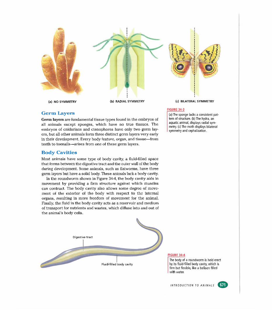
Body CavitiesMost animals have some type of body cavity, a fluid-filled space that forms between the digestive tract and the outer wall of the body during development. Some animals, such as flatworms, have three germ layers but have a solid body. These animals lack a body cavity.
In the roundworm shown in Figure 34-4, the body cavity aids in movement by providing a firm structure against which muscles can contract. The body cavity also allows some degree of movement of the exterior of the body with respect to the internal organs, resulting in more freedom of movement for the animal. Finally, the fluid in the body cavity acts as a reservoir and medium of transport for nutrients and wastes, which diffuse into and out of the animal’s body cells.
FIGURE 34-4
The body of a roundworm Is held erect by its fluid-filled body cavity, which is firm but flexible, like a balloon filled with water.
INTRODUCTION TO ANIMALS
[animal diversity
FIGURE 34-5
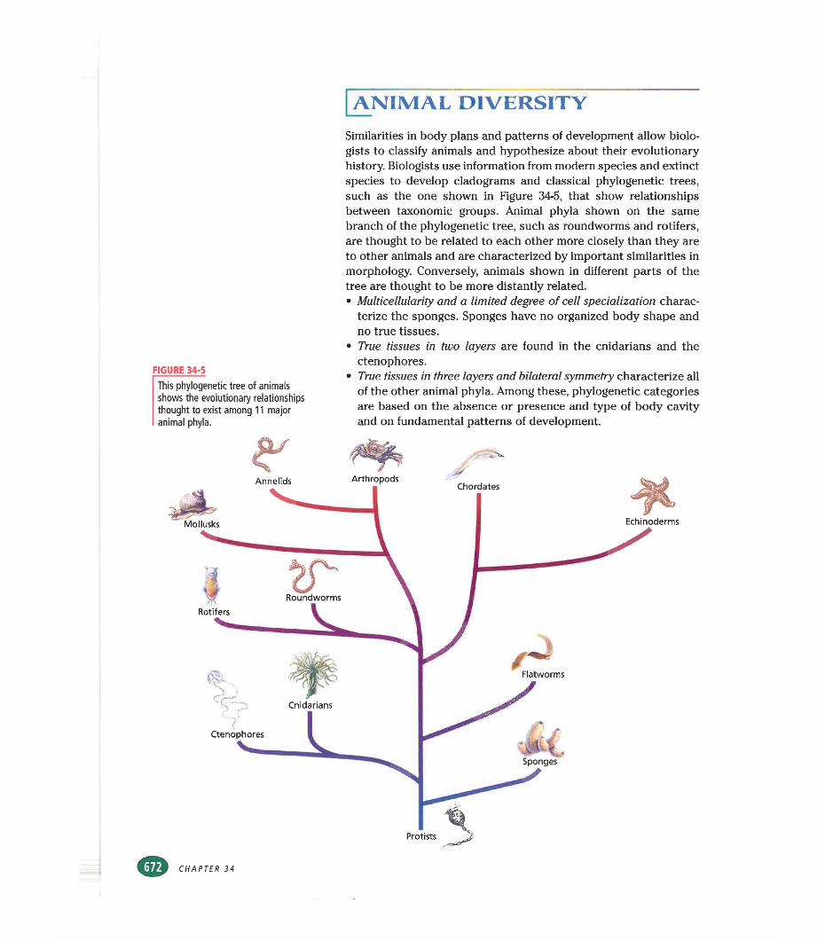
This phylogenetic tree of animals shows the evolutionary relationships thought to exist among 11 major animal phyla.
Similarities in body plans and patterns of development allow biologists to classify animals and hypothesize about their evolutionary history. Biologists use information from modern species and extinct species to develop cladograms and classical phylogenetic trees, such as the one shown in Figure 34-5, that show relationships between taxonomic groups. Animal phyla shown on the same branch of the phylogenetic tree, such as roundworms and rotifers, are thought to be related to each other more closely than they are to other animals and are characterized by important similarities in morphology. Conversely, animals shown in different parts of the tree are thought to be more distantly related.• Multicellularity and a limited degree of cell specialization charac
terize the sponges. Sponges have no organized body shape and no true tissues.
• True tissues in two layers are found in the cnidarians and the ctenophores.
• True tissues in three layers and bilateral symmetry characterize all of the other animal phyla. Among these, phylogenetic categories are based on the absence or presence and type of body cavity and on fundamental patterns of development.
CHAPTER 34
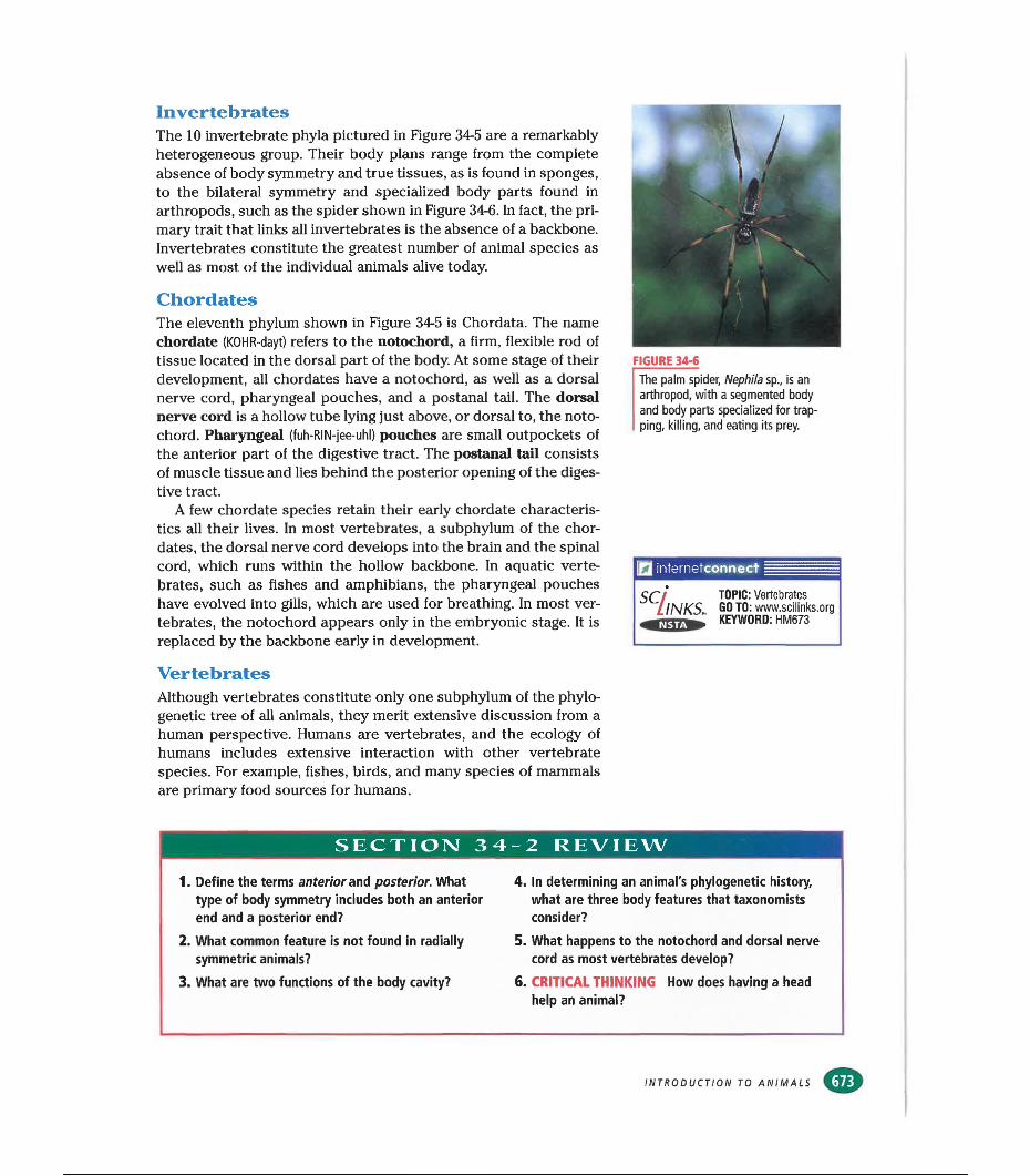
InvertebratesThe 10 invertebrate phyla pictured in Figure 34-5 are a remarkably heterogeneous group. Their body plans range from the complete absence of body symmetry and true tissues, as is found in sponges, to the bilateral symmetry and specialized body parts found in arthropods, such as the spider shown in Figure 34-6. In fact, the primary trait that links all invertebrates is the absence of a backbone. Invertebrates constitute the greatest number of animal species as well as most of the individual animals alive today.
ChordatesThe eleventh phylum shown in Figure 34-5 is Chordata. The name chordate (KOHR-dayt) refers to the notochord, a firm, flexible rod of tissue located in the dorsal part of the body. At some stage of their development, all chordates have a notochord, as well as a dorsal nerve cord, pharyngeal pouches, and a postanal tail. The dorsal nerve cord is a hollow tube lying just above, or dorsal to, the notochord. Pharyngeal (fuh-RIN-jee-uhl) pouches are small outpockets of the anterior part of the digestive tract. The postanal tail consists of muscle tissue and lies behind the posterior opening of the digestive tract.
A few chordate species retain their early chordate characteristics all their lives. In most vertebrates, a subphylum of the chordates, the dorsal nerve cord develops into the brain and the spinal cord, which runs within the hollow backbone. In aquatic vertebrates, such as fishes and amphibians, the pharyngeal pouches have evolved into gills, which are used for breathing. In most vertebrates, the notochord appears only in the embryonic stage. It is replaced by the backbone early in development.
FIGURE 34-6The palm spider, Nephila sp., is an arthropod, with a segmented body and body parts specialized for trapping, killing, and eating its prey.
internetconnect
SW TOPIC: Vertebrates GO TO: www.scilinks.orgKEYWORD: HM673
VertebratesAlthough vertebrates constitute only one subphylum of the phylogenetic tree of all animals, they merit extensive discussion from a human perspective. Humans are vertebrates, and the ecology of humans includes extensive interaction with other vertebrate species. For example, fishes, birds, and many species of mammals are primary food sources for humans.
SECTION 34 -2 REVIEW
1. Define the terms anterior and posterior. What type of body symmetry includes both an anterior end and a posterior end?
4. In determining an animal's phylogenetic history, what are three body features that taxonomists consider?
2. What common feature is not found in radially symmetric animals?
5. What happens to the notochord and dorsal nerve cord as most vertebrates develop?
3. What are two functions of the body cavity? 6. CRITICAL THINKING How does having a head help an animal?
INTRODUCTION TO ANIMALS
SECTION
OBJECTIVES
Compare the body plans and development of Invertebrates
and vertebrates.
•Define the term segmentation,
and name a phylum of segmented animals.
mExplain the difference between an open circulatory system and
a closed circulatory system.
♦Describe the digestive system
found in most invertebrate phyla, and compare It with
vertebrate digestive systems.
FIGURE 34-7
The sea hare, Aplysia californica, is a shell-less mollusk with a simple nervous system.
Comparison oe Invertebrates andVERTEBRATESComparative anatomy, the study of the structure of animal
bodies, is one of the oldest disciplines in biology. Some modern scientists work to establish the relationships between different animals, while others try to establish the relationships between the form and function of morphological features of animals and the role of these features in animal ecology.
INVERTEBRATECHARACTERISTICS
While it may be difficult for us to see many similarities between a clam and an octopus, they are classified in the same phylum. Adult invertebrates show a tremendous amount of morphological diversity.
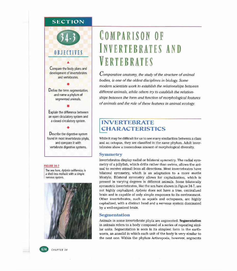
SymmetryInvertebrates display radial or bilateral symmetry. The radial symmetry of a jellyfish, which drifts rather than swims, allows the animal to receive stimuli from all directions. Most invertebrates have bilateral symmetry, which is an adaptation to a more motile lifestyle. Bilateral symmetry allows for cephalization, which is present in varying degrees in different animals. Some bilaterally symmetric invertebrates, like the sea hare shown in Figure 34-7, are not highly cephalized. Aplysia does not have a true, centralized brain and is capable of only simple responses to its environment. Other invertebrates, such as squids and octopuses, are highly cephalized, with a distinct head and a nervous system dominated by a well-organized brain.
SegmentationAnimals in some invertebrate phyla are segmented. Segmentation in animals refers to a body composed of a series of repeating similar units. Segmentation is seen in its simplest form in the earthworm, an annelid in which each unit of the body is very similar to the next one. Within the phylum Arthropoda, however, segments
CHAPTER 34
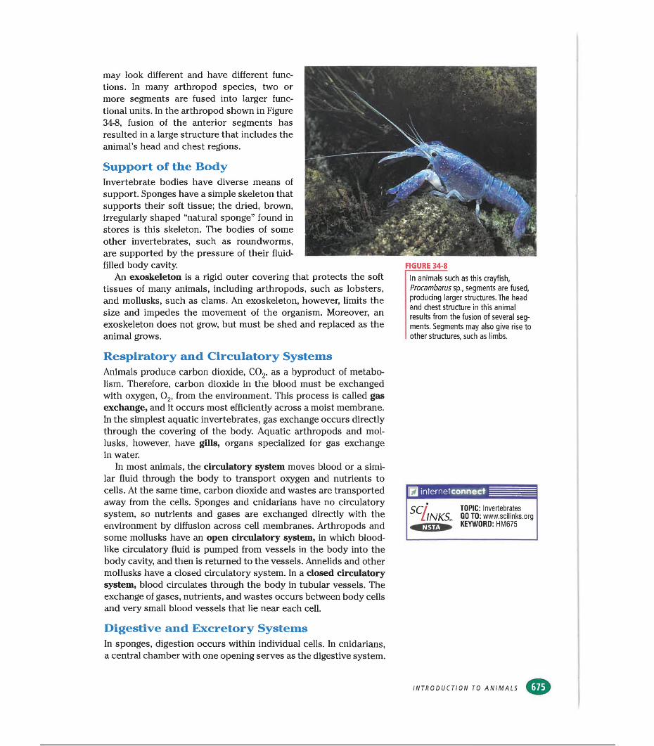
may look different and have different functions. In many arthropod species, two or more segments are fused into larger functional units. In the arthropod shown in Figure 34-8, fusion of the anterior segments has resulted in a large structure that includes the animal’s head and chest regions.
Support of the BodyInvertebrate bodies have diverse means of support. Sponges have a simple skeleton that supports their soft tissue; the dried, brown, irregularly shaped “natural sponge” found in stores is this skeleton. The bodies of some other invertebrates, such as roundworms, are supported by the pressure of their fluid- filled body cavity.
An exoskeleton is a rigid outer covering that protects the soft tissues of many animals, including arthropods, such as lobsters, and mollusks, such as clams. An exoskeleton, however, limits the size and impedes the movement of the organism. Moreover, an exoskeleton does not grow, but must be shed and replaced as the animal grows.
FIGURE 34-8In animals such as this crayfish, Procambarus sp., segments are fused, producing larger structures. The head and chest structure in this animal results from the fusion of several segments. Segments may also give rise to other structures, such as limbs.
Respiratory and Circulatory SystemsAnimals produce carbon dioxide, C02, as a byproduct of metabolism. Therefore, carbon dioxide in the blood must be exchanged with oxygen, 02, from the environment. This process is called gas exchange, and it occurs most efficiently across a moist membrane. In the simplest aquatic invertebrates, gas exchange occurs directly through the covering of the body. Aquatic arthropods and mollusks, however, have gills, organs specialized for gas exchange in water.
In most animals, the circulatory system moves blood or a similar fluid through the body to transport oxygen and nutrients to cells. At the same time, carbon dioxide and wastes are transported away from the cells. Sponges and cnidarians have no circulatory system, so nutrients and gases are exchanged directly with the environment by diffusion across cell membranes. Arthropods and some mollusks have an open circulatory system, in which bloodlike circulatory fluid is pumped from vessels in the body into the body cavity, and then is returned to the vessels. Annelids and other mollusks have a closed circulatory system. In a closed circulatory system, blood circulates through the body in tubular vessels. The exchange of gases, nutrients, and wastes occurs between body cells and very small blood vessels that lie near each cell.
m internetconnect
SCilNKS, TOPIC: Invertebrates GO TO: www.sclllnks.org KEYWORD: HM675
Digestive and Excretory SystemsIn sponges, digestion occurs within individual cells. In cnidarians, a central chamber with one opening serves as the digestive system.
INTRODUCTION TO ANIMALS
Most other invertebrates, however, have a digestive tract, or gut, running through their body. In these animals, food is broken down and nutrients are absorbed by specialized cells that line the gut.
In simple aquatic invertebrates, wastes are excreted as dissolved ammonia, NH3,In terrestrial invertebrates, specialized excretory structures filter ammonia and other wastes from the body cavity. The ammonia is then converted to less toxic substances and water is reabsorbed by the animal before the waste is excreted.
Nervous SystemThe extraordinary degree of diversity among invertebrates is reflected in their nervous systems. Sponges have no neurons, although individual cells can react to environmental stimuli in much the same way protozoa can. Neurons evolved in cnidarians, which have a very simple, loosely connected nervous system. Within a single invertebrate phylum, Mollusca, we can trace a stepwise progression of cephalization and the evolution of the brain. The mollusks have very diverse nervous systems. Recall the sea hare, shown in Figure 34-7. Although its head is not well defined, and its nervous system can perform only simple information processing, the sea hare can learn to contract a part of its body in response to certain stimuli. Contrast this simple behavior with that of a highly cephalized mollusk, such as the octopus. The octopus shows very complex decision-making behavior, and it can build a shelter from debris it finds on the ocean floor.
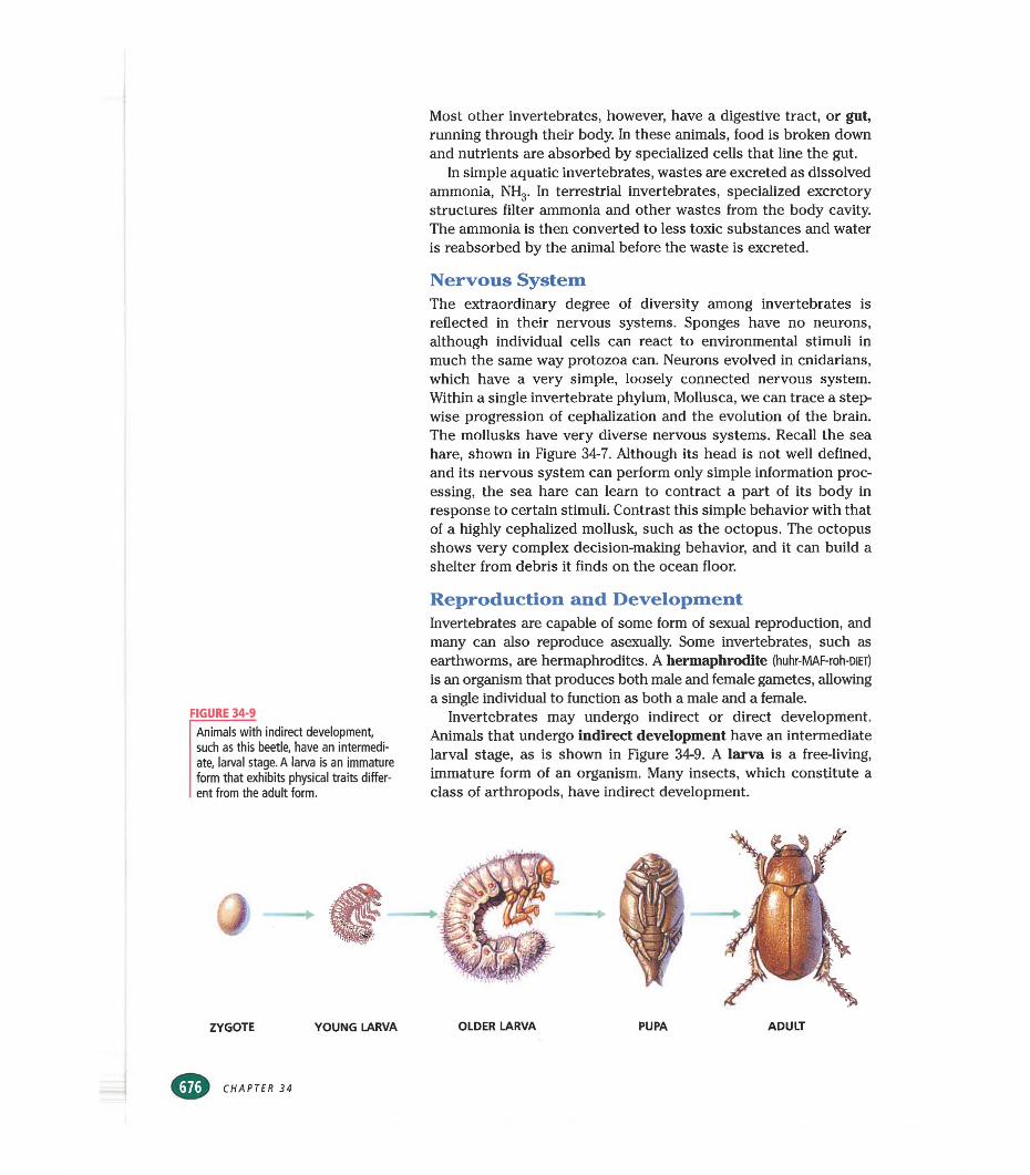
FIGURE 34-9Animals with indirect development, such as this beetle, have an intermediate, larval stage. A larva is an immature form that exhibits physical traits different from the adult form.
Reproduction and DevelopmentInvertebrates are capable of some form of sexual reproduction, and many can also reproduce asexually. Some invertebrates, such as earthworms, are hermaphrodites. A hermaphrodite (huhr-MAF-roh-DiET)
is an organism that produces both male and female gametes, allowing a single individual to function as both a male and a female.
Invertebrates may undergo indirect or direct development. Animals that undergo indirect development have an intermediate larval stage, as is shown in Figure 34-9. A larva is a free-living, immature form of an organism. Many insects, which constitute a class of arthropods, have indirect development.
In contrast, in direct development, the young animal is born or hatched with the same appearance and way of life it will have as an adult; no larval stage occurs. Although most invertebrates undergo indirect development, a few, such as grasshoppers, undergo direct development.
VERTEBRATECHARACTERISTICS
Classes of vertebrates include fishes, amphibians, reptiles, birds, and mammals. All vertebrate classes except fishes spend part or all of their life on land. Many characteristics of terrestrial vertebrates are adaptations to life on land and fall into two broad categories: support of the body and conservation of water.
Support of the BodyIn addition to a backbone, vertebrates have an endoskeleton, an internal skeleton that can support a large, heavy body. The endoskeleton grows as the animal grows.
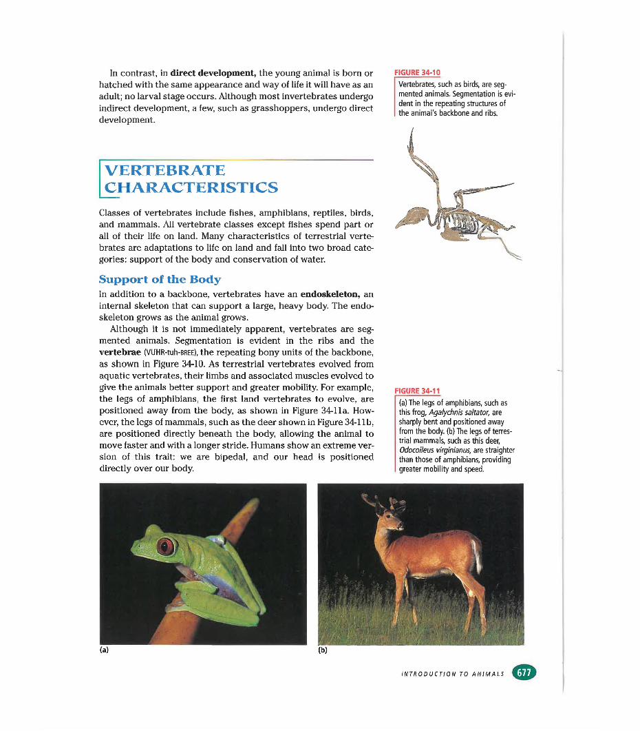
Although it is not immediately apparent, vertebrates are segmented animals. Segmentation is evident in the ribs and the vertebrae (VUHR-tuh-BREE), the repeating bony units of the backbone, as shown in Figure 34-10. As terrestrial vertebrates evolved from aquatic vertebrates, their limbs and associated muscles evolved to give the animals better support and greater mobility. For example, the legs of amphibians, the first land vertebrates to evolve, are positioned away from the body, as shown in Figure 34-1 la. However, the legs of mammals, such as the deer shown in Figure 34-1 lb, are positioned directly beneath the body, allowing the animal to move faster and with a longer stride. Humans show an extreme version of this trait: we are bipedal, and our head is positioned directly over our body.
FIGURE 34-10Vertebrates, such as birds, are segmented animals. Segmentation is evident in the repeating structures of the animal's backbone and ribs.
FIGURE 34-11(a) The legs of amphibians, such as this frog, Agalychnis saltator, are sharply bent and positioned away from the body, (b) The legs of terrestrial mammals, such as this deer, Odocoileus virginianus, are straighter than those of amphibians, providing greater mobility and speed.
INTRODUCTION TO ANIMALS
X
Quick Lab
Identifying Animal Characteristics
Materials BX5 in. note cards (20), 5 pictures of vertebrates,5 pictures of invertebrates
Procedure1. Working in pairs, one partner
will write one different vertebrate characteristic on each of 10 note cards. The other partner will write one different invertebrate characteristic on each of 10 note cards.
2. Designate one partner as the dealer. Place the animal pictures upside down in a stack. The dealer will shuffle and deal all the cards. Turn over one animal picture.
3. The nondealer plays first by laying down as many cards as possible that describe characteristics of the pictured animal. If no card matches, the play is passed to the other player. When neither partner can play, another picture is turned up and play continues.
4. Play ceases when neither student can play or when no pictures are left. The player who is holding the fewest number of cards wins.
Analysis Why are morphological characteristics used to identify organisms? What are the disadvantages of using only morphological characteristics to identify an organism?
Body CoveringsThe outer covering of an animal is called the integument (in-TEG-
yoo-muhnt). While the integuments of fishes and most amphibians are adapted only to moist environments, the integuments of most terrestrial vertebrates are adapted to hold water inside the body. All animal bodies are composed of water-filled cells, and if the water content of the cells is reduced appreciably, the animal will die. The outer covering of terrestrial vertebrates, such as reptiles, birds, and mammals, is largely watertight. Integuments also serve other purposes. The moist skin of an amphibian functions as a respiratory organ for the exchange of gases. The scales of a reptile help protect it from predators. The feathers of birds and the fur of mammals efficiently insulate the body.
Respiratory and Circulatory SystemsGas exchange occurs in the gills of aquatic vertebrates, including fishes and larval amphibians, but these gills do not function out of water. Lungs are organs for gas exchange composed of moist, membranous surfaces deep inside the animal’s body. Lungs evolved in terrestrial vertebrates.
Vertebrates have a closed circulatory system with a multicham- bered, pumping heart. In some vertebrates, the multichambered heart separates oxygenated and deoxygenated blood, improving the efficiency of the circulatory system over that found in other vertebrates and many invertebrates.
Digestive and Excretory Systems Digestion in vertebrates occurs in the gut, which runs from the mouth, at the anterior end, to the anus, at the posterior end. In many vertebrates, the gut is very long with respect to the length of the body, increasing the surface area over which nutrients are absorbed. The human digestive tract is nearly 7 m (23 ft) long. It is folded to fit into a body one-fourth its length.
Vertebrates have the same waste-disposal problems as invertebrates. They must deal with the very toxic ammonia their bodies produce, and most vertebrates must expel wastes while conserving water. Like invertebrates, most vertebrates convert ammonia to less toxic substances. In most vertebrates, organs called kidneys filter wastes from the blood while regulating water levels in the body.
Nervous SystemVertebrates have highly organized brains and the control of specific functions occurs in specific centers in the brain. The structure and function of the nervous system varies among vertebrate orders. For example, within the brain of a fish, much of the tissue processes sensory information. In Figure 34-12, the elongated structure that projects in front of the fish’s eye processes only information about smell. Fishes have limited neural circuitry devoted to decision making. A fish’s responses to stimuli in its environment are rigid, that is, they vary little from situation to situation and from fish to fish.
CHAPTER 34
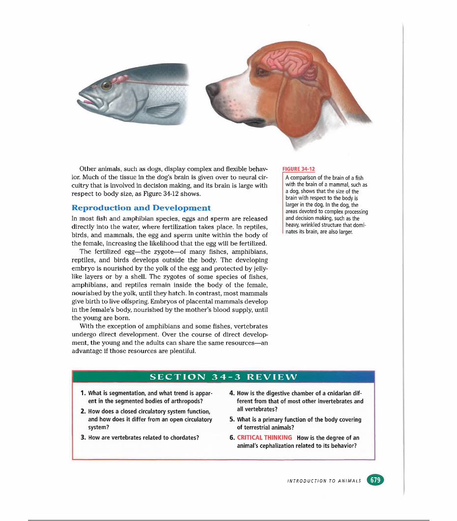
Other animals, such as dogs, display complex and flexible behavior. Much of the tissue in the dog’s brain is given over to neural circuitry that is involved in decision making, and its brain is large with respect to body size, as Figure 34-12 shows.
Reproduction and DevelopmentIn most fish and amphibian species, eggs and sperm are released directly into the water, where fertilization takes place. In reptiles, birds, and mammals, the egg and sperm unite within the body of the female, increasing the likelihood that the egg will be fertilized.
The fertilized egg—the zygote—of many fishes, amphibians, reptiles, and birds develops outside the body. The developing embryo is nourished by the yolk of the egg and protected by jelly- like layers or by a shell. The zygotes of some species of fishes, amphibians, and reptiles remain inside the body of the female, nourished by the yolk, until they hatch. In contrast, most mammals give birth to live offspring. Embryos of placental mammals develop in the female’s body, nourished by the mother’s blood supply, until the young are born.
With the exception of amphibians and some fishes, vertebrates undergo direct development. Over the course of direct development, the young and the adults can share the same resources—an advantage if those resources are plentiful.
FIGURE 34-12
A comparison of the brain of a fish with the brain of a mammal, such as a dog, shows that the size of the brain with respect to the body is larger in the dog. In the dog, the areas devoted to complex processing and decision making, such as the heavy, wrinkled structure that dominates its brain, are also larger.
SECTION 34-3 REVIEW
1. What is segmentation, and what trend is apparent in the segmented bodies of arthropods?
2. How does a closed circulatory system function, and how does it differ from an open circulatory system?
3. How are vertebrates related to chordates?
4. How is the digestive chamber of a cnidarian different from that of most other invertebrates and all vertebrates?
5. What is a primary function of the body covering of terrestrial animals?
6. CRITICAL THINKING How is the degree of an animal's cephaiization related to its behavior?
INTRODUCTION TO ANIMALS
GREAT DISCOVERIES.
The Cell Surface: Embryonic Development
and BeyondHISTORICAL PERSPECTIVE
The mysteries of embryonic growth and development have engaged scientists since ancient times.Until the last century, it was largely unknown how a large, multicellular animal was produced
from a source too small to see. Early scientists debated whether the embryo is pre formed as a miniature individual (a homunculus) or is entirely undifferentiated in form, becoming more
specialized only as it grows and develops. The Reid of developmental biology, which advanced rapidly in the late 1800s and throughout the 1900s, owes much to the pioneering efforts of biologist
Ernest Everett Just. Just demonstrated that the differentiation of the embryonic cells during early development is the result of the interaction between the cytoplasm and the nucleus of the egg.
Just’s later work helped establish that the cell membrane is more than a boundary layer. Rather it plays a critical role as a gatekeeper and conduit for information vital to the cell.
First StepsIn 1899, the German-born American scientist Jacques Loeb, who worked at the University of Chicago and at Woods Hole Marine Biological Laboratory in Cape Cod, Massachusetts, began a study of the early development of eggs. Loeb made the surprising discovery that certain environmental disturbances could initiate development of unfertilized sea urchin eggs. When Loeb pricked the eggs with a needle or changed the salt concentration of the solution surrounding them, the eggs began to divide as if they had been fertilized by sperm. This phenomenon is known as parthenogenesis, and it is now known that it occurs in
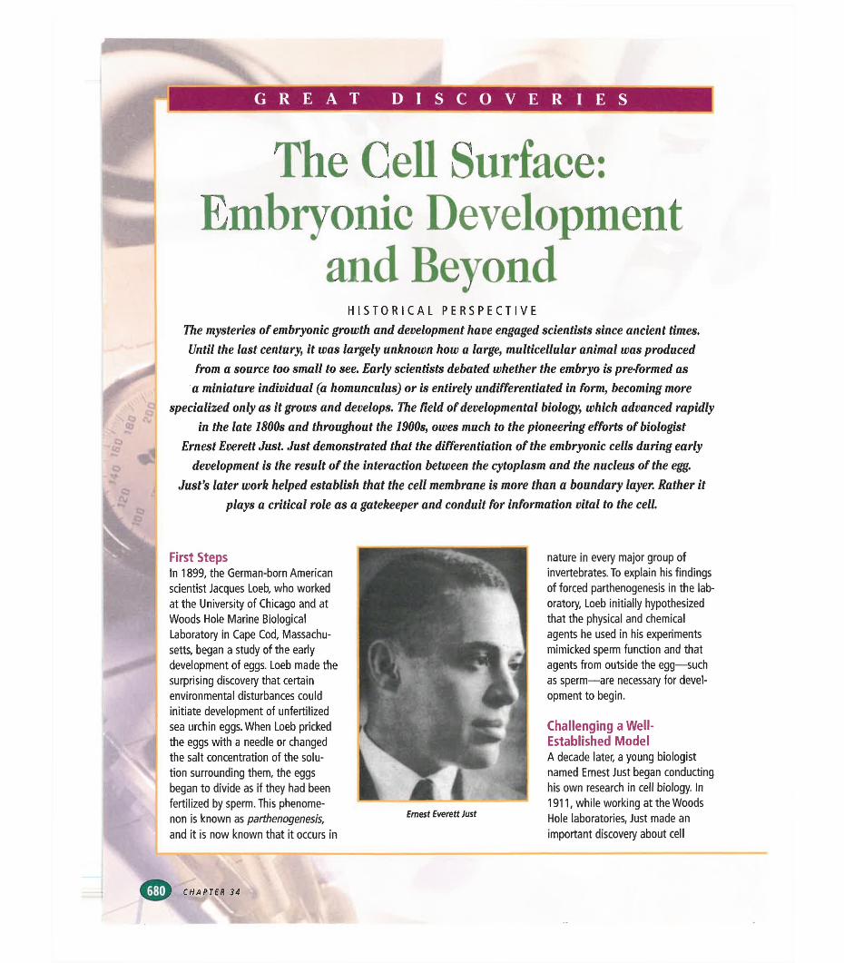
Ernest Everett Just
nature in every major group of invertebrates. To explain his findings of forced parthenogenesis in the laboratory, Loeb initially hypothesized that the physical and chemical agents he used in his experiments mimicked sperm function and that agents from outside the egg—such as sperm—are necessary for development to begin.
Challenging a Well- Established ModelA decade later, a young biologist named Ernest Just began conducting his own research in cell biology. In 1911, while working at the Woods Hole laboratories, Just made an important discovery about cell
CHAPTER 34
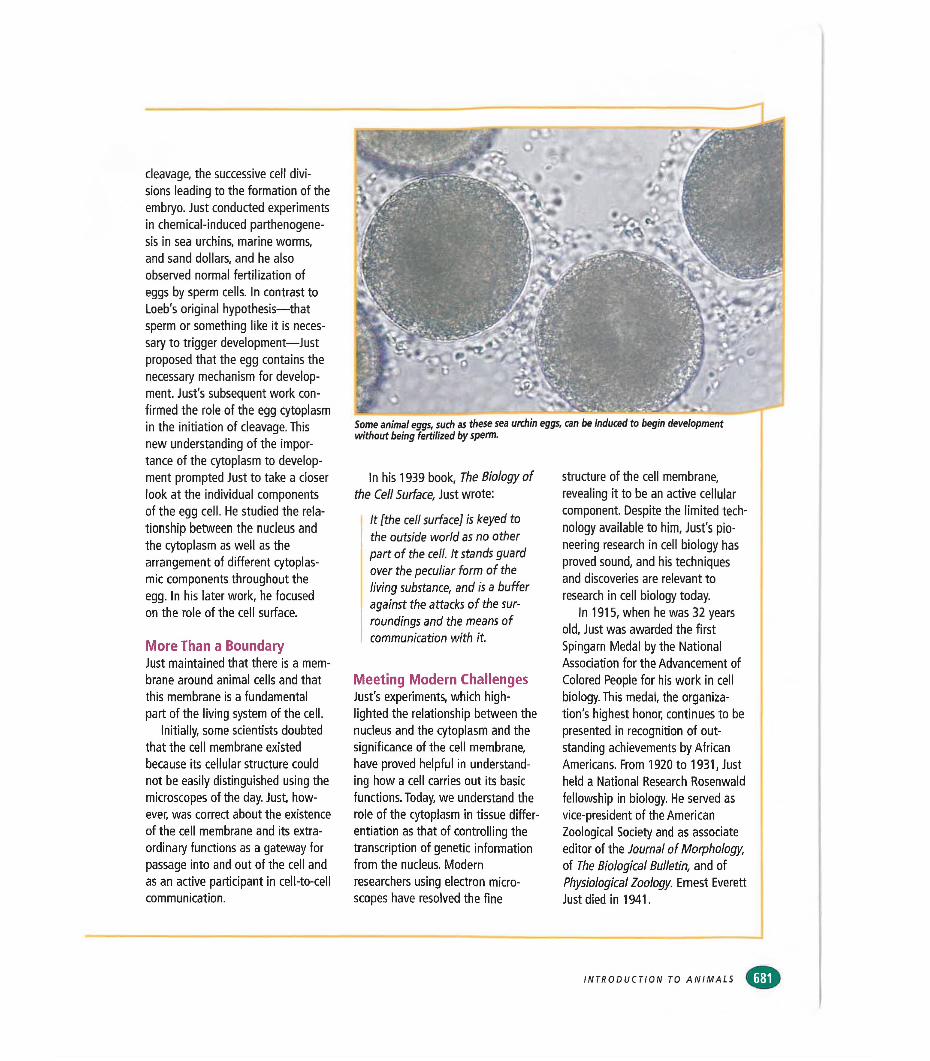
Some animal eggs, such as these sea urchin eggs, can be induced to begin development without being fertilized by sperm.
cleavage, the successive cell divisions leading to the formation of the embryo. Just conducted experiments in chemical-induced parthenogenesis in sea urchins, marine worms, and sand dollars, and he also observed normal fertilization of eggs by sperm cells. In contrast to Loeb's original hypothesis—that sperm or something like it is necessary to trigger development—Just proposed that the egg contains the necessary mechanism for development. Just's subsequent work confirmed the role of the egg cytoplasm in the initiation of cleavage. This new understanding of the importance of the cytoplasm to development prompted Just to take a closer look at the individual components of the egg cell. He studied the relationship between the nucleus and the cytoplasm as well as the arrangement of different cytoplasmic components throughout the egg. In his later work, he focused on the role of the cell surface.
More Than a BoundaryJust maintained that there is a membrane around animal cells and that this membrane is a fundamental part of the living system of the cell.
Initially, some scientists doubted that the cell membrane existed because its cellular structure could not be easily distinguished using the microscopes of the day. Just, however, was correct about the existence of the cell membrane and its extraordinary functions as a gateway for passage into and out of the cell and as an active participant in cell-to-cell communication.
In his 1939 book, The Biology of the Cell Surface, Just wrote:
It [the cell surface] is keyed to the outside world as no other part of the cell. It stands guard over the peculiar form of the living substance, and is a buffer against the attacks of the surroundings and the means of communication with it.
Meeting Modem ChallengesJust's experiments, which highlighted the relationship between the nucleus and the cytoplasm and the significance of the cell membrane, have proved helpful in understanding how a cell carries out its basic functions. Today, we understand the role of the cytoplasm in tissue differentiation as that of controlling the transcription of genetic information from the nucleus. Modern researchers using electron microscopes have resolved the fine
structure of the cell membrane, revealing it to be an active cellular component. Despite the limited technology available to him, Just's pioneering research in cell biology has proved sound, and his techniques and discoveries are relevant to research in cell biology today.
In 1915, when he was 32 years old, Just was awarded the first Spingarn Medal by the National Association for the Advancement of Colored People for his work in cell biology. This medal, the organization's highest honor, continues to be presented in recognition of outstanding achievements by African Americans. From 1920 to 1931, Just held a National Research Rosenwald fellowship in biology. He served as vice-president of the American Zoological Society and as associate editor of the Journal of Morphology, of The Biological Bulletin, and of Physiological Zoology. Ernest Everett Just died in 1941.
INTRODUCTION TO ANIMALS
SECTION
OBJECTIVES
List the steps of fertilization and development through gastrulation.
•Identify the three primary germ layers, and list two body parts formed from each germ layer.
■Define protostome and
deuterostome.
♦Contrast spiral cleavage with
radial cleavage, and name the category of organisms that undergo each type of cleavage.
A
Contrast schizocoely with enterocoely.
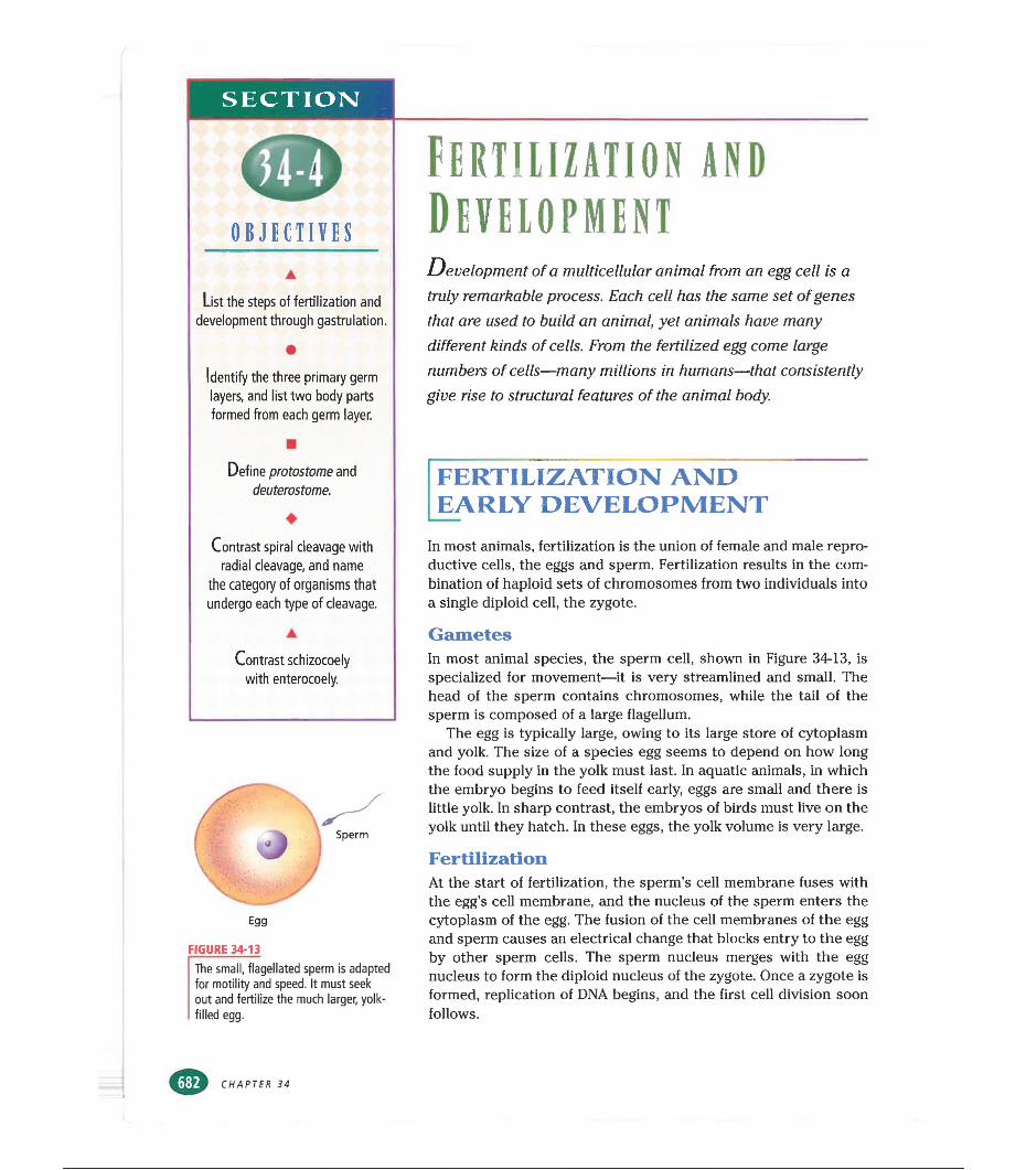
Egg
FIGURE 34-13The small, flagellated sperm is adapted for motility and speed. It must seek out and fertilize the much larger, yolk- filled egg.
fERTIHZATIOR ANDDevelopmentDevelopment of a multicellular animal from an egg cell is a
truly remarkable process. Each cell has the same set of genes that are used to build an animal, yet animals have many different kinds of cells. From the fertilized egg come large numbers of cells—many millions in humans—that consistently give rise to structural features of the animal body.
FERTILIZATION AND EARLY DEVELOPMENT
In most animals, fertilization is the union of female and male reproductive cells, the eggs and sperm. Fertilization results in the combination of haploid sets of chromosomes from two individuals into a single diploid cell, the zygote.
GametesIn most animal species, the sperm cell, shown in Figure 34-13, is specialized for movement—it is very streamlined and small. The head of the sperm contains chromosomes, while the tail of the sperm is composed of a large flagellum.
The egg is typically large, owing to its large store of cytoplasm and yolk. The size of a species egg seems to depend on how long the food supply in the yolk must last. In aquatic animals, in which the embryo begins to feed itself early, eggs are small and there is little yolk. In sharp contrast, the embryos of birds must live on the yolk until they hatch. In these eggs, the yolk volume is very large.
FertilizationAt the start of fertilization, the sperm’s cell membrane fuses with the egg’s cell membrane, and the nucleus of the sperm enters the cytoplasm of the egg. The fusion of the cell membranes of the egg and sperm causes an electrical change that blocks entry to the egg by other sperm cells. The sperm nucleus merges with the egg nucleus to form the diploid nucleus of the zygote. Once a zygote is formed, replication of DNA begins, and the first cell division soon follows.
CHAPTER 34
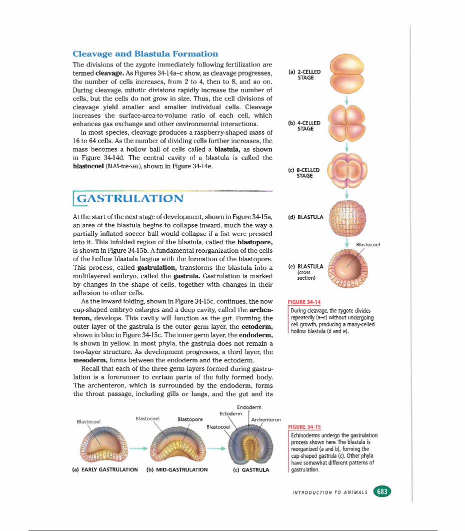
Cleavage and Blastula FormationThe divisions of the zygote immediately following fertilization are termed cleavage. As Figures 34-14a-c show, as cleavage progresses, the number of cells increases, from 2 to 4, then to 8, and so on. During cleavage, mitotic divisions rapidly increase the number of cells, but the cells do not grow in size. Thus, the cell divisions of cleavage yield smaller and smaller individual cells. Cleavage increases the surface-area-to-volume ratio of each cell, which enhances gas exchange and other environmental interactions.
In most species, cleavage produces a raspberry-shaped mass of 16 to 64 cells. As the number of dividing cells further increases, the mass becomes a hollow ball of cells called a blastula, as shown in Figure 34-14d. The central cavity of a blastula is called the blastocoel (BLAS-toe-SEEL), shown in Figure 34-14e.
[gastrulation
At the start of the next stage of development, shown in Figure 34-15a, an area of the blastula begins to collapse inward, much the way a partially inflated soccer ball would collapse if a fist were pressed into it. This infolded region of the blastula, called the blastopore, is shown in Figure 34-15b. A fundamental reorganization of the cells of the hollow blastula begins with the formation of the blastopore. This process, called gastrulation, transforms the blastula into a multilayered embryo, called the gastrula. Gastrulation is marked by changes in the shape of cells, together with changes in their adhesion to other cells.
As the inward folding, shown in Figure 34-15c, continues, the now cup-shaped embryo enlarges and a deep cavity, called the archen- teron, develops. This cavity will function as the gut. Forming the outer layer of the gastrula is the outer germ layer, the ectoderm, shown in blue in Figure 34-15c. The inner germ layer, the endoderm, is shown in yellow. In most phyla, the gastrula does not remain a two-layer structure. As development progresses, a third layer, the mesoderm, forms between the endoderm and the ectoderm.
Recall that each of the three germ layers formed during gastrulation is a forerunner to certain parts of the fully formed body. The archenteron, which is surrounded by the endoderm, forms the throat passage, including gills or lungs, and the gut and its
Endoderm
(a) EARLY GASTRULATION (b) MID-GASTRULATION (c) GASTRULA
(a) 2-CELLED STAGE
(b) 4-CELLED STAGE
(c) 8-CELLED STAGE
(d) BLASTULA
(e) BLASTULA(crosssection)
FIGURE 34-14
During cleavage, the zygote divides repeatedly (a-c) without undergoing cell growth, producing a many-celled hollow blastula (d and e).
FIGURE 34-15
Echinoderms undergo the gastrulation process shown here. The blastula is reorganized (a and b), forming the cup-shaped gastrula (c). Other phyla have somewhat different patterns of gastrulation.
INTRODUCTION TO ANIMALS
associated organs, such as the pancreas and liver. The ectoderm forms the outer layer of the skin, the hair, nails, and the nervous system.
The versatile mesoderm forms a multitude of body parts, including the skeleton, muscles, inner layer of the skin, the circulatory system, and the lining of the body cavity.
_PATTERNS OF DEVELOPMENTA body cavity completely lined by mesoderm is called a coelom. Most phyla have a coelom, and, like patterns of symmetry and number of germ layers, the coelom is a feature which taxonomists use to classify animals with similar phylogenetic origins. The distinct patterns of cleavage and coelom formation found in different animal phyla are additional clues to their phylogenetic history.
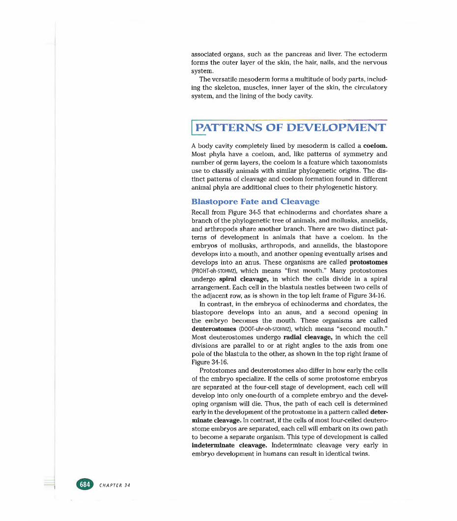
Blastopore Fate and CleavageRecall from Figure 34-5 that echinoderms and chordates share a branch of the phylogenetic tree of animals, and mollusks, annelids, and arthropods share another branch. There are two distinct patterns of development in animals that have a coelom. In the embryos of mollusks, arthropods, and annelids, the blastopore develops into a mouth, and another opening eventually arises and develops into an anus. These organisms are called protostomes (PROHT-oh-STOHMZ), which means “first mouth.” Many protostomes undergo spiral cleavage, in which the cells divide in a spiral arrangement. Each cell in the blástula nestles between two cells of the adjacent row, as is shown in the top left frame of Figure 34-16.
In contrast, in the embryos of echinoderms and chordates, the blastopore develops into an anus, and a second opening in the embryo becomes the mouth. These organisms are called deuterostomes (DOOT-uhr-oh-STOHMZ), which means “second mouth.” Most deuterostomes undergo radial cleavage, in which the cell divisions are parallel to or at right angles to the axis from one pole of the blástula to the other, as shown in the top right frame of Figure 34-16.
Protostomes and deuterostomes also differ in how early the cells of the embryo specialize. If the cells of some protostome embryos are separated at the four-cell stage of development, each cell will develop into only one-fourth of a complete embryo and the developing organism will die. Thus, the path of each cell is determined early in the development of the protostome in a pattern called determinate cleavage. In contrast, if the cells of most four-celled deutero- stome embryos are separated, each cell will embark on its own path to become a separate organism. This type of development is called indeterminate cleavage. Indeterminate cleavage very early in embryo development in humans can result in identical twins.
Protostome Development Deuterostome Development
SPIRAL CLEAVAGE RADIAL CLEAVAGE
SCHIZOCOELY ENTEROCOELY
BlastoporeCoelom
Blastocoel Archenteron gut)
CoelomAnus
BlastoporeCoelom
Anus
BlastocoelArchenteron
CoelomMouth
Endoderm Mesoderm Ectoderm
Coelom FormationThe way in which the coelom forms in many protostomes differs from the way it forms in many deuterostomes. The lower left frame of Figure 34-16 shows coelom formation in protostomes. Cells located at the junction of the endoderm and ectoderm (at the rim of the cup-shaped embryo) split away toward the interior of the gastrula. Rapid division of these cells (shown in pink) in the blastocoel forms the mesoderm. This process of mesoderm formation is called schizocoely (SKIZ-oh-SEEL-ee), or “split body cavity.”
The lower right frame of Figure 34-16 shows coelom formation in deuterostomes. The mesoderm forms when the cells lining the dorsal, or top, part of the archenteron begin dividing rapidly. These rapidly dividing cells (shown in pink) roll outward into the blastocoel, forming the mesoderm. This process of mesoderm formation is called enterocoely (EN-tuhr-oh-SEEL-ee), meaning “gut body cavity.” During both enterocoely and schizocoely, mesodermal cells spread out to completely line the coelom, and the blastocoel disappears. Thus, in both protostomes and deuterostomes, mesoderm lines the interior of the outer body wall and surrounds the gut.
FIGURE 34-16
Many protostomes undergo spiral cleavage during early development, while many deuterostomes undergo radial cleavage. In protostomes, the coelom arises by schizocoely, and the blastopore becomes the mouth. In deuterostomes, the coelom arises by enterocoely, and the blastopore becomes the anus.
INTRODUCTION TO ANIMALS
(a) ACOELOMATE (b) PSEUDOCOELOMATE (c) COELOMATE
FIGURE 34-17
In three-layered acoelomates (a), the endodermic gut is surrounded by a solid layer of mesoderm. In pseudo- coelomates (b), the endodermic gut is suspended in a fluid-filled cavity which is surrounded by mesoderm. In coelo- mates (c), the endodermic gut is surrounded and suspended by mesoderm, which also surrounds the coelom.
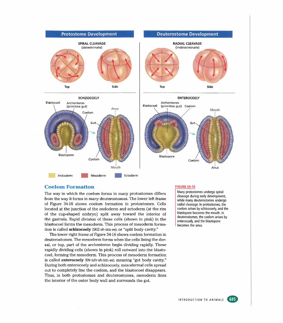
Types of Body CavitiesCompare the cross sections of an acoelomate, a pseudocoelomate, and a coelomate shown in Figure 34-17. In acoelomates (UH-SEE-luh-
mayts), such as flatworms, the body cavity is absent. The interior of the animal is solid, as shown in Figure 34-17a. The endodermic gut, shown in yellow, and the outer covering of the animal, shown in blue, are connected by the solid tissue of the mesoderm.
In some phyla, including rotifers and roundworms, the mesoderm lines the interior of the coelom but does not surround the exterior of the endodermic gut. This type of body cavity, shown in Figure 34-17b, is called a pseudocoelom (SOO-doh-SEE-luhm), which means “false body cavity.” In pseudocoelomates, such as the one shown in Figure 34-17b, mesoderm lines the fluid-filled coelom, and the endodermic gut is suspended in the fluid of the coelom.
In coelomates (SEE-luh-MAYTS), animals with a true coelom, such as the one shown in Figure 34-17c, mesoderm lines the body cavity and surrounds and supports the endodermic gut. The mesoderm also forms the tissues of attachment for the organs located in the coelom, such as the liver and the lungs. Mollusks, annelids, arthropods, chordates, and echinoderms are coelomates.
Look back at the phylogenetic tree in Figure 34-5 and locate the acoelomate, pseudocoelomate, and coelomate phyla. How does their placement on the phylogenetic tree reflect their body type?
SECTION 34-4 REVIEW
1. Beginning with fertilization, list the stepsof development through mesoderm formation.
2. What are the three germ layers formed in all embryos except those of sponges, cnidarians, and ctenophores? Name two body parts that arise from each germ layer.
3. What is a protostome? What is a deuterostome? How does cleavage in these groups differ?
4. Humans and other vertebrates sometimes produce two or more identical offspring. What type of cleavage can result in the formation of identical offspring?
5. How is the mesoderm formed in schizocoely, and how does this process differ from enterocoely?
6. CRITICAL THINKING What adaptive advantage is associated with indeterminate cleavage?
CHAPTER 34
CHAPTER 34 REVIEW
Summary/Vocabulary
Animals are multicellular and hetero- trophic, and their cells lack walls. Most animals reproduce sexually and can move. Animals have cells that are specialized for different functions.Most animals ingest their food and digest it within their bodies.Vocabularycell junction (667) ingestion (668)differentiation (668) invertebrate (667)
Sponges, the simplest animals, have no true tissue and no body symmetry. All other animals have tissue.In the bodies of animals with radial symmetry, similar parts branch out in all directions from a central line. Animals with bilateral symmetry have similar halves. Bilateral symmetry is associated with cephalization, that is, having a head.Vocabularyanterior (670) dorsal (670)bilateral symmetry (670) dorsal nerve cord (673)cephalization (670) germ layer (671 )chordate (673) notochord (673)
Invertebrates have no body symmetry or are radially or bilaterally symmetrical; vertebrates are bilaterally symmetrical.A segmented body is composed of repeating similar units. Some invertebrates and all vertebrates are segmented.Some invertebrates have an exoskeleton.All vertebrates have an endoskeleton.The simplest invertebrates have no circulatory system. Arthropods and some mol- lusks have an open circulatory system. Other mollusks, annelids, and vertebrates have a closed circulatory system.Vocabularycirculatory system (675) closed circulatory system
(675)direct development (677) endoskeleton (677)
exoskeleton (675) gas exchange (675) gill (675) gut (676)hermaphrodite (676)
■ Most animals reproduce sexually, though some also reproduce asexually.
■ Movement and response to the environment are governed by an animal’s nervous tissue and muscle tissue.
■ The first animals may have evolved from colonial protists.
neuron (668) vertebrate (667)specialization (667) zygote (668)
■ Most animals have three germ layers. All body features arise from one of the germ layers.
■ At some stage of their lives, all members of phylum Chordata have a notochord, a dorsal nerve cord, a postanal tail, and pharyngeal pouches.
pharyngeal pouch (673) radial symmetry (670)postanal tail (673) symmetry (670)posterior (670) ventral (670)
■ Sponges digest food within individual cells. Cnidarians digest food in a central chamber. Other invertebrates and all vertebrates have a gut.
■ Some invertebrates have loosely connected circuits of neurons. Some invertebrates and all vertebrates have a well-defined brain.
■ Most invertebrates and vertebrates are capable of some form of sexual reproduction, and some invertebrates can also reproduce asexually.
indirect development (676) integument (678) kidney (678) larva (676) lung (678)
open circulatory system (675)
segmentation (674) vertebra (677)
INTRODUCTION TO ANIMALS
CHAPTER 34 REVIEW
■ During the first cell divisions in the zygote, called cleavage, cells divide repeatedly.
■ The mass of cells produced by cleavage continues to divide, producing the blastula.
■ During gastrula formation, the germ layers —the ectoderm, the endoderm, and in most phyla, the mesoderm—are defined.
■ The endoderm-lined central cavity of the embryo forms the throat passage, gills or lungs, and the gut and its accessory organs.
■ In most protostomes, each cell of the blastula nestles between two cells of adjacent rows; the blastopore develops into the mouth. Separation of cells of the early embryo cause it to die. The mesoderm
forms from the division of cells at the junction of the endoderm and the ectoderm in a process called schizocoely.
■ In most deuterostomes, each cell in the blastula rests directly over the cell of the next row, and the blastopore develops into the anus. Separation of cells of the early embryo results in the development of multiple embryos. The mesoderm forms from cell divisions at the top of the archenteron in a process called enterocoely.
■ Acoelomates have no body cavity. Psuedo- coelomates have a body cavity partially lined with mesoderm, and coelomates have a coelom.
Vocabularyacoelomate (686) archenteron (683) blastocoel (683) blastopore (683) blastula (683) cleavage (683)
coelom (684) coelomate (686) determinate cleavage (684) deuterostome (684) ectoderm (683) endoderm (683)
enterocoely (685) gastrula (683) gastrulation (683) Indeterminate cleavage
(684)mesoderm (683)
protostome (684) pseudocoelom (686) pseudocoelomate (686) radial cleavage (684) schizocoely (685) spiral cleavage (684)
Review
Vocabulary1. Distinguish between cell specialization and
differentiation.2. Identify the structures that allow the forma
tion of tissue from many separate cells.3. Distinguish between radial symmetry and
bilateral symmetry.4. What is the difference between a coelomate
and an acoelomate?5. Compare determinate cleavage with indeter
minate cleavage.
Multiple Choice6. The process that occurs during development
in multicellular organisms and leads to cell specialization is (a) asexual reproduction(b) evolution (c) differentiation (d) fertilization
7. Animals must eat because they are (a) not autotrophic (b) not heterotrophic (c) neither autotrophic nor heterotrophic (d) both autotrophic and heterotrophic.
8. Animals that have no true tissues are the(a) chordates (b) ctenophores (c) cnidarians (d) sponges.
9. Fundamental tissue types in the embryo are called (a) notochords (b) germ layers(c) pharyngeal pouches (d) coeloms.
10. Gas exchange in many aquatic phyla takes place in the (a) lungs (b) gut (c) kidneys(d) gills.
11. The repeating units of the backbone are called the (a) integument (b) exoskeleton(c) notochord (d) vertebrae.
12. A feature in vertebrates that is an adaptation to life on land is (a) lungs (b) gills (c) kidneys(d) a gut.
13. The process that takes place as the zygote begins to divide immediately after fertilization is (a) gastrulation (b) cleavage (c) meio- sis (d) organ formation.
14. Organisms in which the blastopore develops into the anus are called (a) protostomes(b) gastrulae (c) pseudocoelomates (d) deuterostomes.
CHAPTER 34
CHAPTER 34 REVIEW
15. The process of mesoderm formation by division of the cells at the top of the archenteron is called (a) indeterminate cleavage (b) determinate cleavage (c) schizocoely (d) enterocoely.
Short Answer16. Explain how neural tissue and muscle tissue
work together in an animal’s body to allow the animal to respond to its environment.
17. What probable changes did early colonial flagellates undergo as they evolved into the first animals?
18. What can you infer if two phyla are represented on the same branch of a phylogenetic tree?
19. What are four features common to all chor- dates at some time in their life, and what has happened to two of these features in an adult human?
20. How does indirect development differ from direct development, and which type is found in vertebrates?
21. What happened to the position of the body with respect to the legs as vertebrates adapted to life on land?
22. How do the problems of disposal of body waste differ in terrestrial and aquatic animals?
23. How does the body cavity of a pseudocoelo- mate differ from that of a coelomate?
24. Name two body parts formed by each of the following: endoderm, mesoderm, and ectoderm.
25. What structure does the archenteron become in a developing animal?
Critical thinking
1. From the perspective of a single cell, what is one advantage of cell specialization and one disadvantage of cell specialization?
2. Considering that an endoskeleton can support more weight than an exoskeleton, would a large-bodied animal with an exoskeleton be more likely to live in the water or on land? Why?
3. On mammals and birds, the head is positioned higher with respect to the body than it is on amphibians and reptiles. Why might it be helpful to have a head positioned over the body?
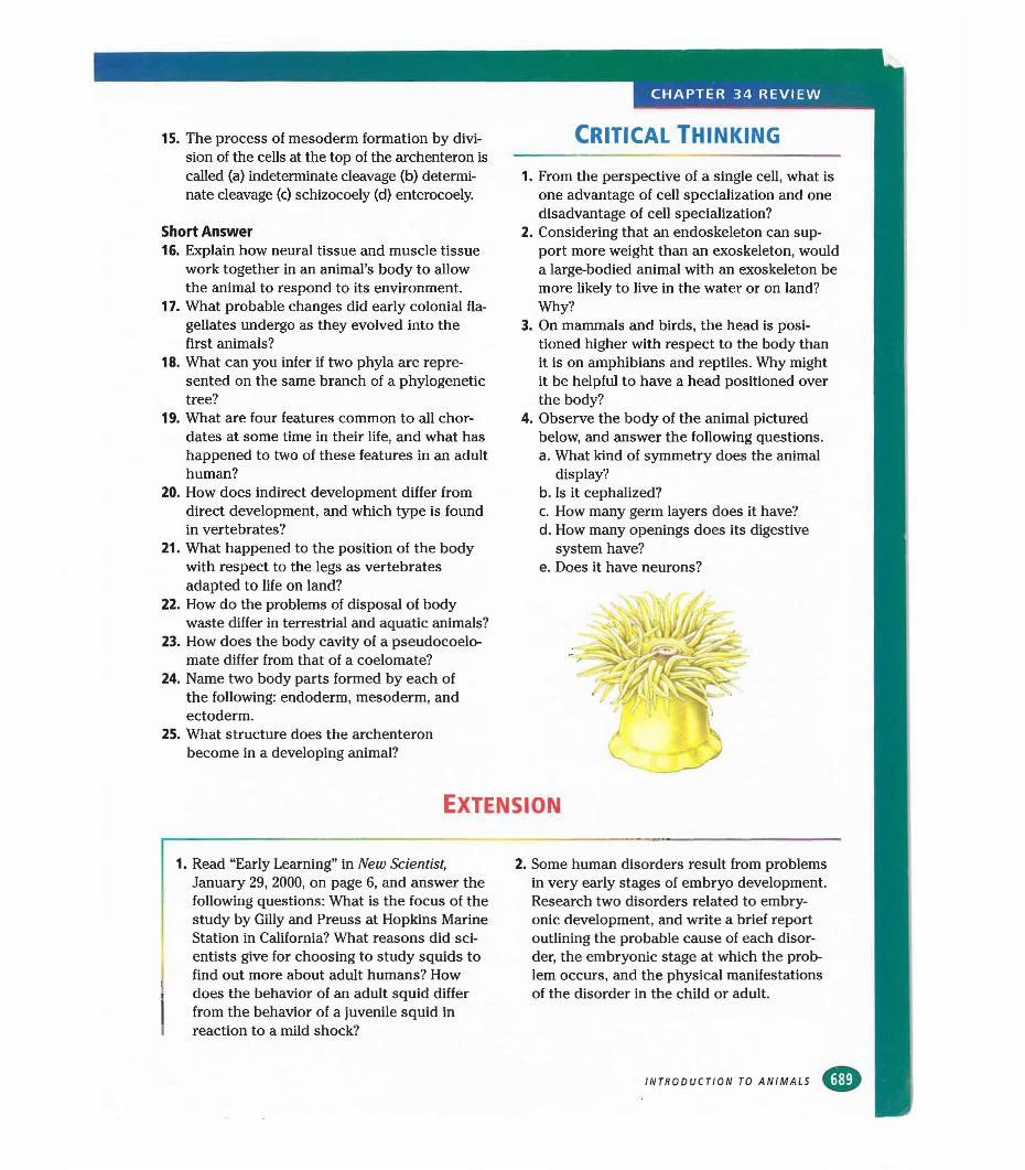
4. Observe the body of the animal pictured below, and answer the following questions.a. What kind of symmetry does the animal
display?b. Is it cephalized?c. How many germ layers does it have?d. How many openings does its digestive
system have?e. Does it have neurons?
Extension
1. Read “Early Learning” in New Scientist, January 29, 2000, on page 6, and answer the following questions: What is the focus of the study by Gilly and Preuss at Hopkins Marine Station in California? What reasons did scientists give for choosing to study squids to find out more about adult humans? How does the behavior of an adult squid differ from the behavior of a juvenile squid in reaction to a mild shock?
2. Some human disorders result from problems in very early stages of embryo development. Research two disorders related to embryonic development, and write a brief report outlining the probable cause of each disorder, the embryonic stage at which the problem occurs, and the physical manifestations of the disorder in the child or adult.
INTRODUCTION TO ANIMALS
CHAPTER 34 INVESTIGATION
Sheep’s Heart DissectionOBJECTIVE!
■ Describe the appearance of the external and internal structures of a sheep's heart.
■ Name the structures and functions of a sheep's heart.
m observing structures■ identifying■ demonstrating
■ sheep's heart■ dissecting tray■ blunt metal probe■ scissors■ scalpel■ tweezers
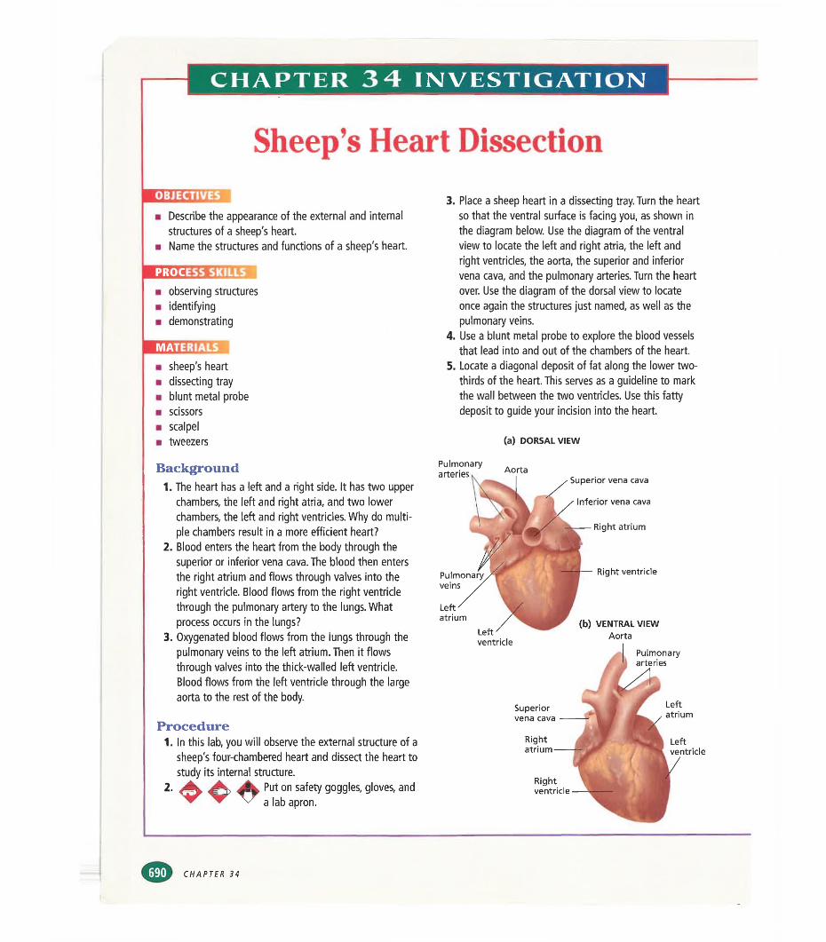
3. Place a sheep heart in a dissecting tray. Turn the heart so that the ventral surface is facing you, as shown in the diagram below. Use the diagram of the ventral view to locate the left and right atria, the left and right ventricles, the aorta, the superior and inferior vena cava, and the pulmonary arteries. Turn the heart over. Use the diagram of the dorsal view to locate once again the structures just named, as well as the pulmonary veins.
4. Use a blunt metal probe to explore the blood vessels that lead into and out of the chambers of the heart.
5. Locate a diagonal deposit of fat along the lower two- thirds of the heart. This serves as a guideline to mark the wall between the two ventricles. Use this fatty deposit to guide your incision into the heart.
(a) DORSAL VIEW
Background1. The heart has a left and a right side. It has two upper
chambers, the left and right atria, and two lower chambers, the left and right ventricles. Why do multiple chambers result in a more efficient heart?
2. Blood enters the heart from the body through the superior or inferior vena cava. The blood then enters the right atrium and flows through valves into the right ventricle. Blood flows from the right ventricle through the pulmonary artery to the lungs. What process occurs in the lungs?
3. Oxygenated blood flows from the lungs through the pulmonary veins to the left atrium. Then it flows through valves into the thick-walled left ventricle. Blood flows from the left ventricle through the large aorta to the rest of the body.
Procedure1. In this lab, you will observe the external structure of a
sheep's four-chambered heart and dissect the heart to study its internal structure.
2. A Put on safety goggles, gloves, and'W ^ \y a |ab apron.
Right atrium
Rightventricle
Pulmonary arteries Aorta
Superior vena cava
Inferior vena cava
veinsRight ventricle
Leftatrium
Left ventricle
Superior vena cava
Leftatrium
Rightatrium
Leftventricle
(b) VENTRAL VIEWAorta
Pulmonaryarteries
CHAPTER 34
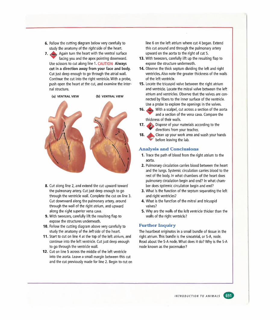
6. Follow the cutting diagram below very carefully to study the anatomy of the right side of the heart.
7. Again turn the heart with the ventral surface ^ facing you and the apex pointing downward.
Use scissors to cut along line 1. CAUTION Always cut in a direction away from your face and body. Cut just deep enough to go through the atrial wall. Continue the cut into the right ventricle. With a probe, push open the heart at the cut, and examine the internal structure.
(a) VENTRAL VIEW (b) VENTRAL VIEW
8. Cut along line 2, and extend the cut upward toward the pulmonary artery. Cut just deep enough to go through the ventricle wall. Complete the cut on line 3. Cut downward along the pulmonary artery, around through the wall of the right atrium, and upward along the right superior vena cava.
9. With tweezers, carefully lift the resulting flap to expose the structures underneath.
10. Follow the cutting diagram above very carefully to study the anatomy of the left side of the heart.
11. Start to cut on line 4 at the top of the left atrium, and continue into the left ventricle. Cut just deep enough to go through the ventricle wall.
12. Cut on line 5 across the middle of the left ventricle into the aorta. Leave a small margin between this cut and the cut previously made for line 2. Begin to cut on
13
line 6 on the left atrium where cut 4 began. Extend this cut around and through the pulmonary artery upward on the aorta to the right of cut 5.With tweezers, carefully lift up the resulting flap to expose the structure underneath.
14. Observe the thick septum dividing the left and right ventricles. Also note the greater thickness of the walls of the left ventricle.
15. Locate the tricuspid valve between the right atrium and ventricle. Locate the mitral valve between the left atrium and ventricles. Observe that the valves are connected by fibers to the inner surface of the ventricle. Use a probe to explore the openings in the valves.
With a scalpel, cut across a section of the aorta ^ and a section of the vena cava. Compare the
thickness of their walls.^ Dispose of your materials according to the ** directions from your teacher.
Clean up your work area and wash your hands V before leaving the lab.
16
17.
18.
Analysis and Conclusions1. Trace the path of blood from the right atrium to the
aorta.2. Pulmonary circulation carries blood between the heart
and the lungs. Systemic circulation carries blood to the rest of the body. In what chambers of the heart does pulmonary circulation begin and end? In what chamber does systemic circulation begin and end?
3. What is the function of the septum separating the left and right ventricles?
4. What is the function of the mitral and tricuspid valves?
5. Why are the walls of the left ventricle thicker than the walls of the right ventricle?
Further InquiryThe heartbeat originates in a small bundle of tissue in the right atrium. This bundle is the sinoatrial, or S-A, node. Read about the S-A node. What does it do? Why is the S-A node known as the pacemaker?
INTRODUCTION TO ANIMALS
CHAPTER 35
Sponges, Cnidarians,AND CTENOPHORES
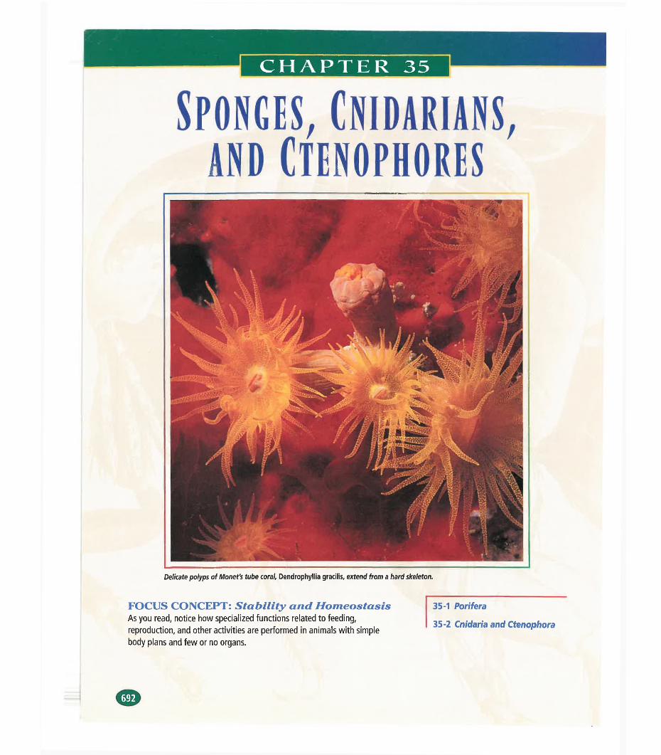
Delicate polyps of Monet's tube coral, Dendrophyllia gracilis, extend from a hard skeleton.
FOCUS CONCEPT: Stability and Homeostasis As you read, notice how specialized functions related to feeding, reproduction, and other activities are performed in animals with simple body plans and few or no organs.
35-1 Pori sera
35-2 Cnidaria and Ctenophora
SECTION
PORIFERAInvertebrates are animals that do not have a backbone. Rather
than being classified according to shared characteristics, invertebrates are an arbitrary classification of extremely diverse animals that share the absence of a characteristic. Invertebrates comprise more than a dozen phyla and more than a million species. About 97 percent of all animal species known are invertebrates, among the simplest of which are sponges.
STRUCTURE AND FUNCTION Of SPONGES
Sponges are aquatic animals that make up the phylum Porifera (pohr- IF-uhr-uh). These simple organisms clearly represent the transition from unicellular to multicellular life. Sponges have no gastrula stage, exhibit less cell specialization than most other animals, and have no true tissues or organs. There are about 10,000 species of sponges. About 150 species live in fresh water, while the rest are marine.
Early biologists thought sponges were plants, and most sponges do resemble plants in some ways. Adult sponges are sessile, which means they attach themselves firmly to a surface and do not move. Sponges grow in many shapes, sizes, and colors, and they often look like mossy mats, cactuses, or blobs of fungus. They can be as small as 1 cm (0.4 in.) in length or as large as 2 m (6.6 ft) in diameter.
(22)OBJECTIVES
Define invertebrates, and explain why they are such
a diverse group.
•Describe the basic body
plan of a sponge.
■Describe the process of filter feeding in sponges.
♦Contrast the processes of
sexual and asexual reproduction in sponges.
Body PlanThe basic body plan of a sponge, as shown in Figure 35-1, suggests many relationships between structure and function. The body wall consists of two layers of cells separated by a jellylike substance. In simple sponges, the body wall surrounds a hollow cylinder that is closed at the bottom and open at the top. The interior of the cylinder is lined with collar cells. By beating their flagella, collar cells draw water into the sponge through numerous pores that penetrate the body wall. In fact, the name Porifera comes from a Latin word meaning “pore-bearer.” The water that is pumped into the interior of the sponge leaves through the osculum (AHS-kyoo-luhm), the opening at the top of the sponge that you can see in Figure 35-1.
A sponge would collapse without some type of supporting structure. In some sponges, support is provided by a simple skeleton made of a network of protein fibers called spongin (SPUHN-jin). Other sponges have skeletons consisting of spicules, tiny, hard particles
SCI TOPIC: SpongesLINKS GO TO: www.scilinks.org
” KEYWORD: HM693
SPONGES, CNIDARIANS, AND CTENOPHORES
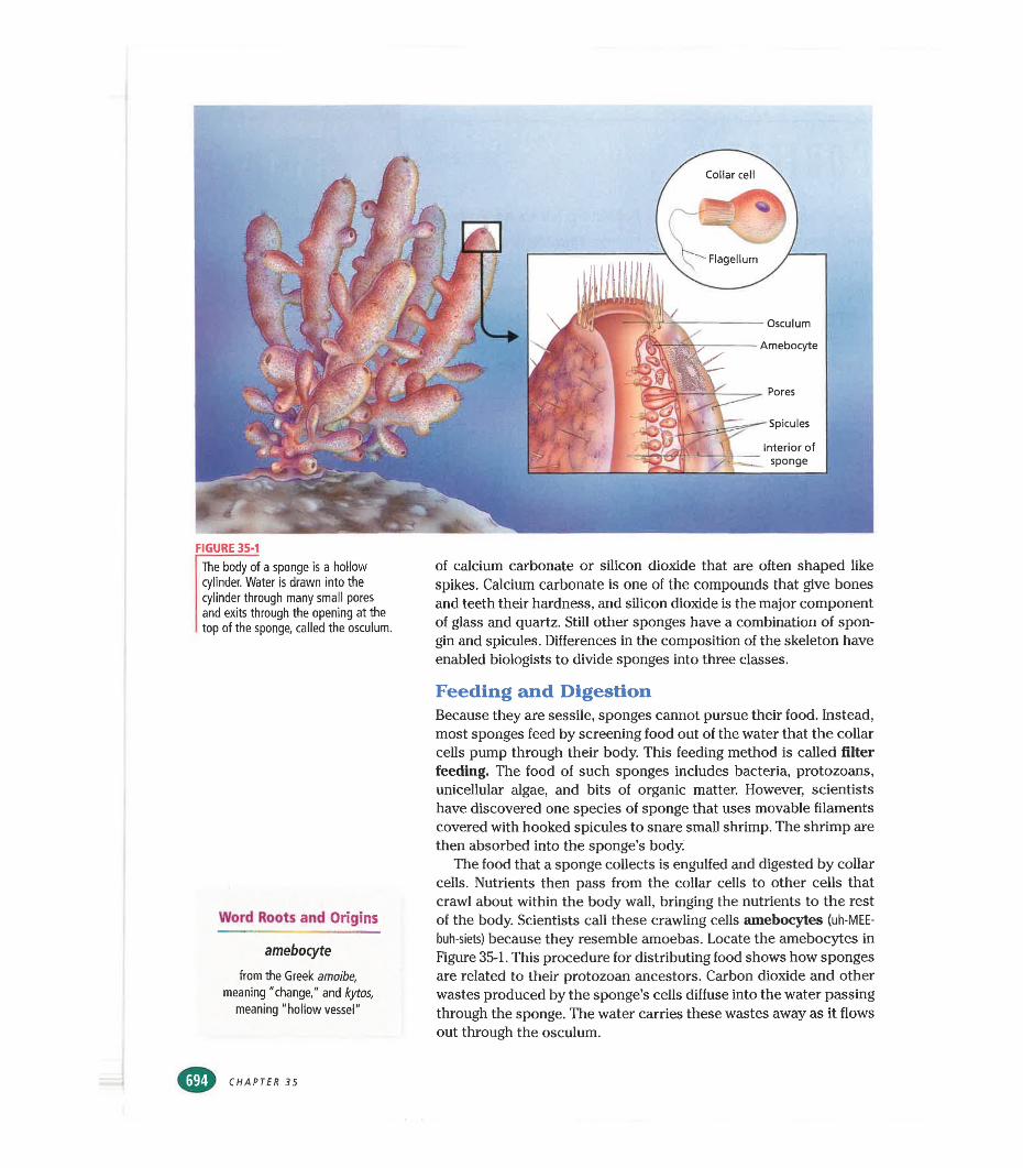
FIGURE 35-1The body of a sponge is a hollow cylinder. Water is drawn into the cylinder through many small pores and exits through the opening at the top of the sponge, called the osculum.
of calcium carbonate or silicon dioxide that are often shaped like spikes. Calcium carbonate is one of the compounds that give bones and teeth their hardness, and silicon dioxide is the major component of glass and quartz. Still other sponges have a combination of spon- gin and spicules. Differences in the composition of the skeleton have enabled biologists to divide sponges into three classes.
Word Roots and Origins
amebocyte
from the Greek amoibe, meaning "change," and kytos,
meaning "hollow vessel"
Feeding and DigestionBecause they are sessile, sponges cannot pursue their food. Instead, most sponges feed by screening food out of the water that the collar cells pump through their body. This feeding method is called filter feeding. The food of such sponges includes bacteria, protozoans, unicellular algae, and bits of organic matter. However, scientists have discovered one species of sponge that uses movable filaments covered with hooked spicules to snare small shrimp. The shrimp cure then absorbed into the sponge’s body.
The food that a sponge collects is engulfed and digested by collar cells. Nutrients then pass from the collar cells to other cells that crawl about within the body wall, bringing the nutrients to the rest of the body. Scientists call these crawling cells amebocytes (uh-MEE-
buh-siets) because they resemble amoebas. Locate the amebocytes in Figure 35-1. This procedure for distributing food shows how sponges are related to their protozoan ancestors. Carbon dioxide and other wastes produced by the sponge’s cells diffuse into the water passing through the sponge. The water carries these wastes away as it flows out through the osculum.
CHAPTER 35
ReproductionSponges can reproduce asexually by forming small buds that break off and live separately. The sponge illustrated in Figure 35-1 has many buds that are still attached. During droughts or cold weather, some freshwater sponges produce internal buds called gemmules (lEM-yoolz). Each gemmule is a food- filled ball of amebocytes surrounded by a protective coat made of organic material and spicules. Gemmules can survive harsh conditions that may kill the adult sponge that formed them. When conditions improve, the sponge cells emerge from the gemmules and grow into new sponges.
Sponges also have remarkable powers of regeneration, the ability to regrow missing parts. In fact, a small piece of a sponge can regenerate a complete new sponge. In some species, even particles small enough to pass through a cloth strainer can regenerate.
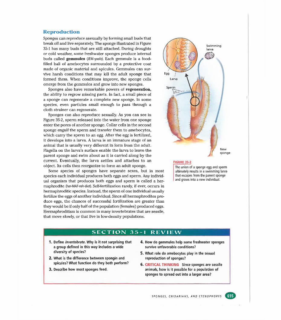
Sponges can also reproduce sexually. As you can see in Figure 35-2, sperm released into the water from one sponge enter the pores of another sponge. Collar cells in the second sponge engulf the sperm and transfer them to amebocytes, which carry the sperm to an egg. After the egg is fertilized, it develops into a larva. A larva is an immature stage of an animal that is usually very different in form from the adult. Flagella on the larva’s surface enable the larva to leave the parent sponge and swim about as it is carried along by the current. Eventually, the larva settles and attaches to an object. Its cells then reorganize to form an adult sponge.
Some species of sponges have separate sexes, but in most species each individual produces both eggs and sperm. Any individual organism that produces both eggs and sperm is called a hermaphrodite (her-MAF-roh-diet). Self-fertilization rarely, if ever, occurs in hermaphroditic species. Instead, the sperm of one individual usually fertilize the eggs of another individual. Since all hermaphrodites produce eggs, the chances of successful fertilization are greater than they would be if only half of the population (females) produced eggs. Hermaphroditism is common in many invertebrates that are sessile, that move slowly, or that live in low-density populations.
Swimminglarva
FIGURE 35-2
The union of a sponge egg and sperm ultimately results in a swimming larva that escapes from the parent sponge and grows into a new individual.
SECTION 3 5-1 REVIEW
1. Define invertebrate. Why is it not surprising that a group defined in this way includes a wide diversity of species?
2. What is the difference between spongin and spicules? What function do they both perform?
3. Describe how most sponges feed.
4. How do gemmules help some freshwater sponges survive unfavorable conditions?
5. What role do amebocytes play in the sexual reproduction of sponges?
6. CRITICAL THINKING Since sponges are sessile animals, how is it possible for a population of sponges to spread out into a larger area?
SPONGES, CNIDARIANS, AND CTENOPHORES
SECTION
Q)OBJECTIVES
Name and describe the two body forms of cnidarians.
•Describe the common
characteristics of cnidarians.
■Identify the three classes of cnidarians, and give an
example of each.
♦Describe the common
characteristics of ctenophores.
CNIDARIA AND CTENOPHORACnidaria (nie-DER-ee-uh) and Ctenophora (tee-NAHF-uhr-uh)
are two phyla of radially symmetrical invertebrates. The animals in these phyla are somewhat more complex than the sponges. Their cells are organized into tissues, and they have a few simple organs. All members of the phyla Cnidaria and Ctenophora are aquatic, and most live in the ocean.
STRUCTURE AND FUNCTION OF CNIDARIANS
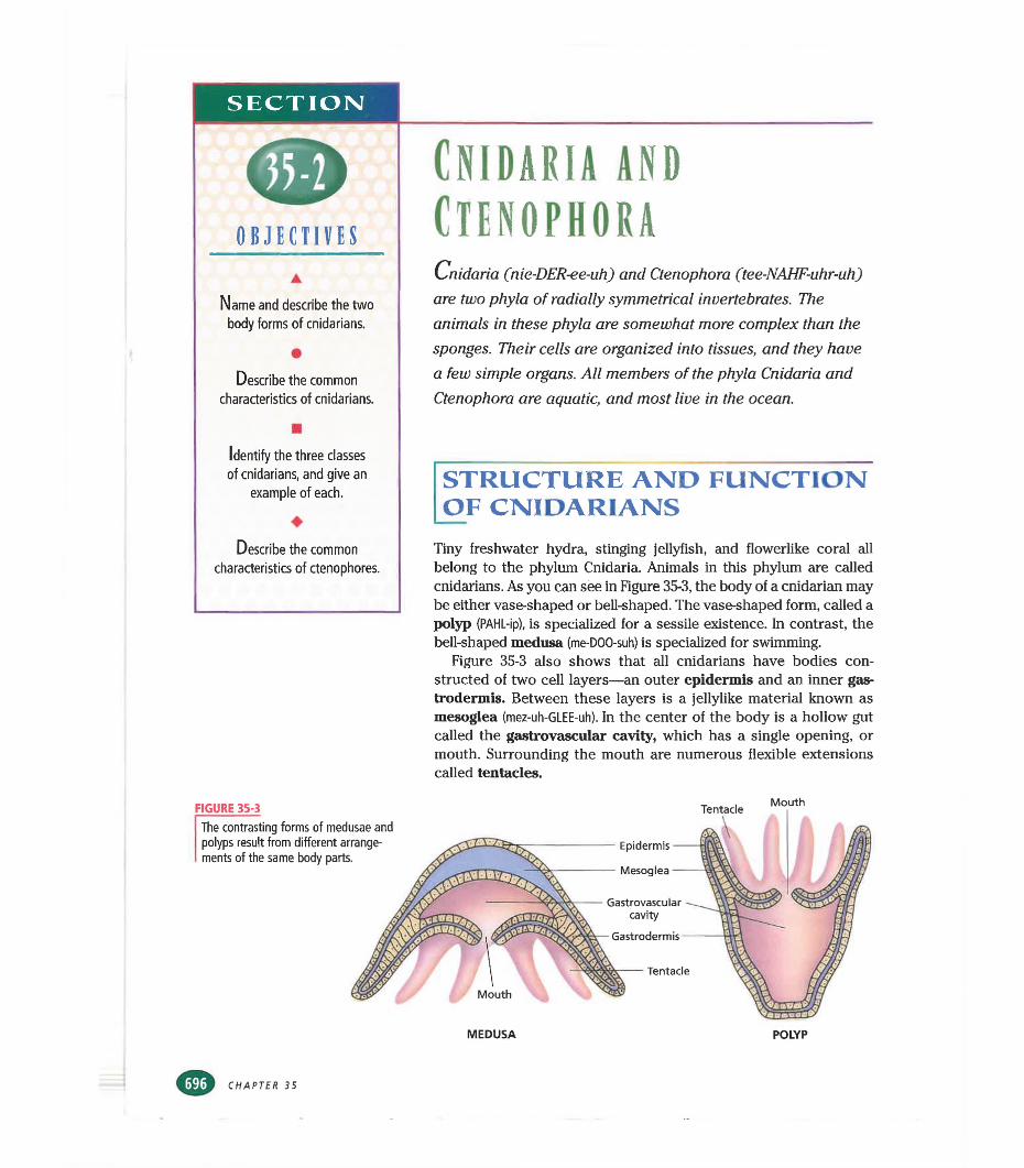
Tiny freshwater hydra, stinging jellyfish, and flowerlike coral all belong to the phylum Cnidaria. Animals in this phylum are called cnidarians. As you can see in Figure 35-3, the body of a cnidarian may be either vase-shaped or bell-shaped. The vase-shaped form, called a polyp (PAHL-ip), is specialized for a sessile existence. In contrast, the bell-shaped medusa (me-DOO-suh) is specialized for swimming.
Figure 35-3 also shows that all cnidarians have bodies constructed of two cell layers—an outer epidermis and an inner gas- trodermis. Between these layers is a jellylike material known as mesoglea (mez-uh-GLEE-uh). In the center of the body is a hollow gut called the gastrovascular cavity, which has a single opening, or mouth. Surrounding the mouth are numerous flexible extensions called tentacles.
FIGURE 35-3 Tentacle Mouth
MEDUSA POLYP
CHAPTER 35
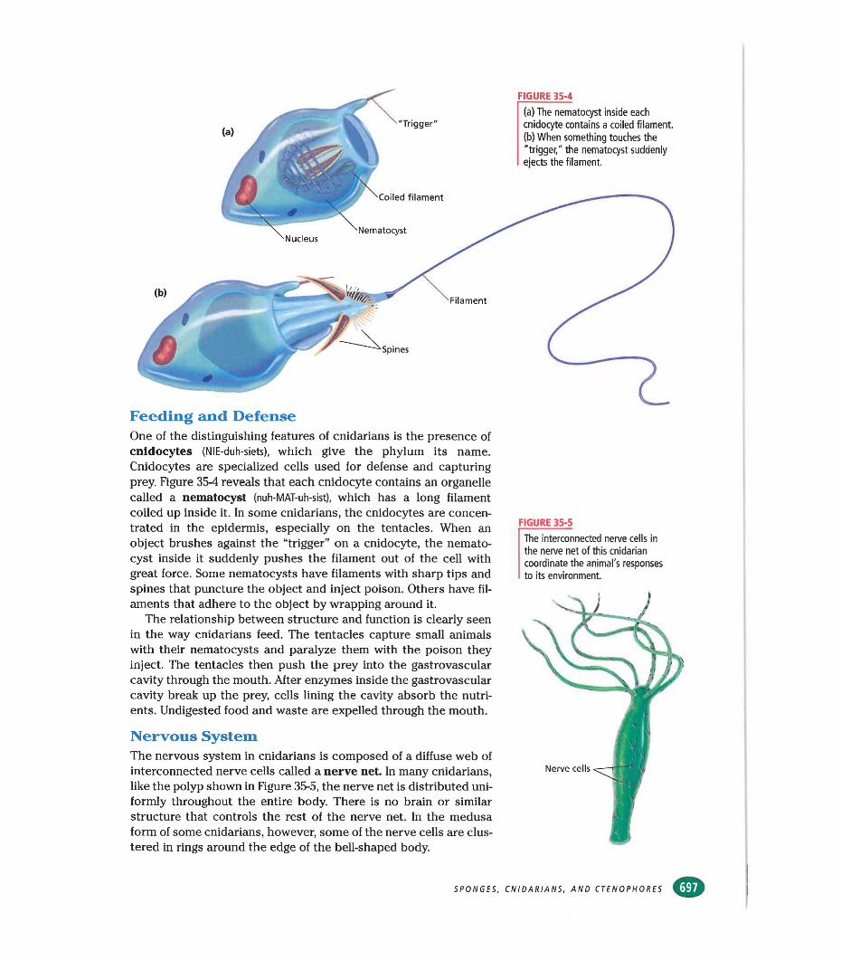
FIGURE 35-4
(a) The nematocyst inside each cnidocyte contains a coiled filament.(b) When something touches the "trigger," the nematocyst suddenly ejects the filament.
"Trigger"
filament
Nematocyst
Feeding and DefenseOne of the distinguishing features of cnidarians is the presence of cnidocytes (NIE-duh-siets), which give the phylum its name. Cnidocytes are specialized cells used for defense and capturing prey. Figure 354 reveals that each cnidocyte contains an organelle called a nematocyst (nuh-MAT-uh-sist), which has a long filament coiled up inside it. In some cnidarians, the cnidocytes are concentrated in the epidermis, especially on the tentacles. When an object brushes against the “trigger” on a cnidocyte, the nematocyst inside it suddenly pushes the filament out of the cell with great force. Some nematocysts have filaments with sharp tips and spines that puncture the object and inject poison. Others have filaments that adhere to the object by wrapping around it.
The relationship between structure and function is clearly seen in the way cnidarians feed. The tentacles capture small animals with their nematocysts and paralyze them with the poison they inject. The tentacles then push the prey into the gastrovascular cavity through the mouth. After enzymes inside the gastrovascular cavity break up the prey, cells lining the cavity absorb the nutrients. Undigested food and waste are expelled through the mouth.
Nervous SystemThe nervous system in cnidarians is composed of a diffuse web of interconnected nerve cells called a nerve net. In many cnidarians, like the polyp shown in Figure 35-5, the nerve net is distributed uniformly throughout the entire body. There is no brain or similar structure that controls the rest of the nerve net. In the medusa form of some cnidarians, however, some of the nerve cells are clustered in rings around the edge of the bell-shaped body.
FIGURE 35-5
The interconnected nerve cells in the nerve net of this cnidarian coordinate the animal's responses to its environment.
SPONGES, CNIDARIANS, AND CTENOPHORES
Quick Lab
Identifying Poriferans, Ctenophorans, and Cnidarians
Materials pencil, paper, a picture of a poriferan, a ctenophoran, or a cnidarian
Procedure1. Prepare a dichotomous key to
differentiate between poriferans, ctenophorans, and cnidarians. (Refer to pp. 354 and 355.)
2. Write paired statements for the phyla Porifera, Ctenophora, and Cnidaria and for the classes Hydrozoa, Scyphozoa, and Anthozoa.
3. Statement (1a) describes cnidarians and leads to the paired statements (3). Statement (1b) will lead to the paired statements (2).
4. Statement (2a) describes the phylum Porifera and (2b) describes the phylum Ctenophora.
5. Statement (3a) describes Hydrozoa and (3b) leads to paired statements (4).
6. Statement (4a) describes Scyphozoa and (4b) describes Anthozoa.
7. Get a picture from your teacher. Identify the animal's phylum and class using your dichotomous key. Exchange keys with another group, and use the new key to identify your animal.
Analysis What do sponges and hydrozoans have in common? How do comb jellies differ from cnidarians? What is the dominant body form in each of the three classes of cnidarians?
FIGURE 35-6
Obelia (a) and Physalia, the Portuguese man-of-war (b), are two examples of colonial hydrozoans. Obelia consists solely of polyps, while Physalia is made of both polyps and medusae. (Obelia: LM 13 X)
The nerve net enables cnidarians to respond to specific stimuli in their environment. For example, when cells in the epidermis are touched, they relay a signal to nerve cells. The nerve cells, in turn, transmit a signal via the nerve net to contractile cells, which can cause the animal to withdraw from the stimulus. In cnidarians with the simplest nerve nets, a stimulus anywhere on the body causes signals to be sent through the nerve net in all directions. These signals bring about a contraction of the entire body.
The nerve net also coordinates the complex activities of the body that are necessary for feeding and traveling through the environment. The movements by which the tentacles bring prey to the mouth and push it into the gastrovascular cavity are controlled by the nerve net, as are the rhythmic contractions of the body that propel swimming medusae through the water.
ClassificationScientists divide cnidarians into three classes—Hydrozoa, Scyphozoa, and Anthozoa—and the members of these classes are known as hydrozoans, scyphozoans, and anthozoans, respectively. Some species of hydrozoans live only as polyps, some live only as medusae, some alternate between these two forms, and some live as mixed colonies of polyps and medusae. Scyphozoans spend most of their lives as medusae, while anthozoans live only as polyps.
CXASS HYDROZOAThe class Hydrozoa includes about 3,700 species, most of which live as colonial organisms in the oceans. One example of a colonial hydrozoan is Obelia. As Figure 35-6a illustrates, Obelia has many polyps attached to branched stalks. Some of the polyps function in gathering food, while others are responsible for reproduction.
Perhaps the most remarkable hydrozoan is the Portuguese man-of-war, Physalia, shown in Figure 35-6b. This hydrozoan exists as a colony of medusae and polyps. Its gas-filled float, which can
(a) (b)
CHAPTER 35
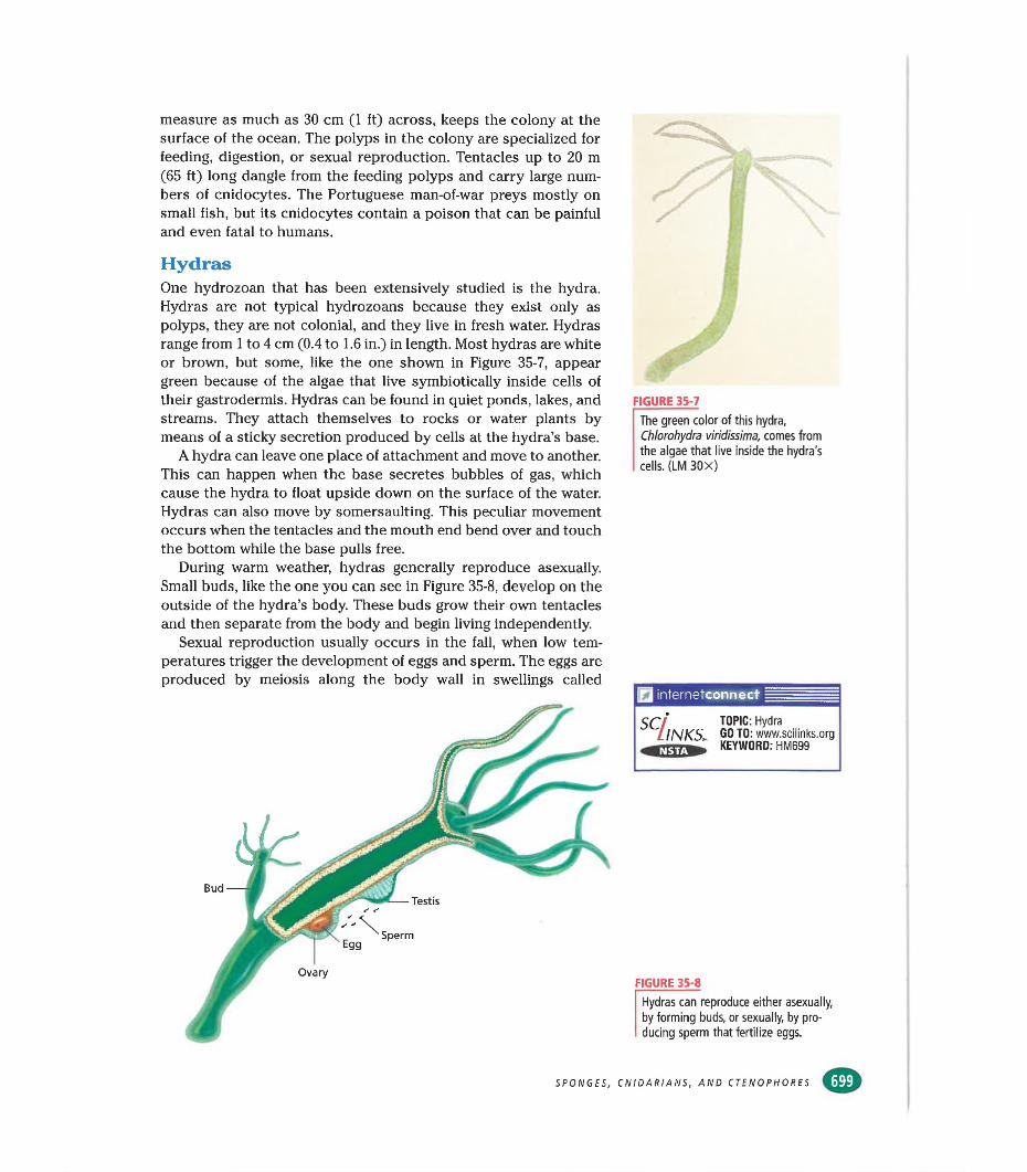
measure as much as 30 cm (1 ft) across, keeps the colony at the surface of the ocean. The polyps in the colony are specialized for feeding, digestion, or sexual reproduction. Tentacles up to 20 m (65 ft) long dangle from the feeding polyps and carry large numbers of cnidocytes. The Portuguese man-of-war preys mostly on small fish, but its cnidocytes contain a poison that can be painful and even fatal to humans.
HydrasOne hydrozoan that has been extensively studied is the hydra. Hydras are not typical hydrozoans because they exist only as polyps, they are not colonial, and they live in fresh water. Hydras range from 1 to 4 cm (0.4 to 1.6 in,) in length. Most hydras are white or brown, but some, like the one shown in Figure 35-7, appear green because of the algae that live symbiotically inside cells of their gastrodermis. Hydras can be found in quiet ponds, lakes, and streams. They attach themselves to rocks or water plants by means of a sticky secretion produced by cells at the hydra’s base.
A hydra can leave one place of attachment and move to another. This can happen when the base secretes bubbles of gas, which cause the hydra to float upside down on the surface of the water. Hydras can also move by somersaulting. This peculiar movement occurs when the tentacles and the mouth end bend over and touch the bottom while the base pulls free.
During warm weather, hydras generally reproduce asexually. Small buds, like the one you can see in Figure 35-8, develop on the outside of the hydra’s body. These buds grow their own tentacles and then separate from the body and begin living independently.
Sexual reproduction usually occurs in the fall, when low temperatures trigger the development of eggs and sperm. The eggs are produced by meiosis along the body wall in swellings called
SPONGES,
FIGURE 35-7
The green color of this hydra, Chlorohydra viridissima, comes from the algae that live inside the hydra's cells. (LM 30X)
internetconnect
SC INKS. TOPIC: HydraGO TO: www.scilinks.orgKEYWORD: HM699
FIGURE 35-8
Hydras can reproduce either asexually, by forming buds, or sexually, by producing sperm that fertilize eggs.
CNIDARIANS, AND CTENOPHORES
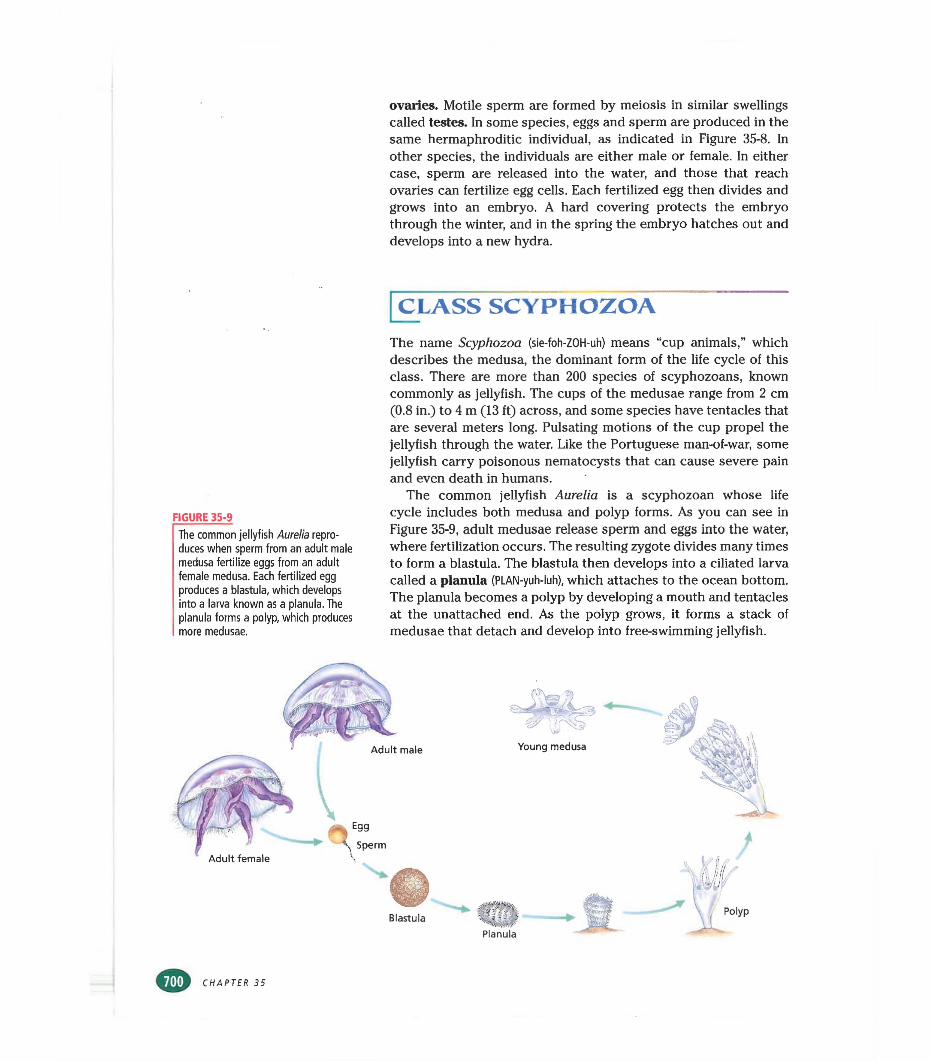
ovaries. Motile sperm are formed by meiosis in similar swellings called testes. In some species, eggs and sperm are produced in the same hermaphroditic individual, as indicated in Figure 35-8. In other species, the individuals are either male or female. In either case, sperm are released into the water, and those that reach ovaries can fertilize egg cells. Each fertilized egg then divides and grows into an embryo. A hard covering protects the embryo through the winter, and in the spring the embryo hatches out and develops into a new hydra.
CLASS SCYPHOZOA
FIGURE 35-9
The common jellyfish Aurelia reproduces when sperm from an adult male medusa fertilize eggs from an adult female medusa. Each fertilized egg produces a blastula, which develops into a larva known as a planula. The planula forms a polyp, which produces more medusae.
The name Scyphozoa (sie-foh-ZOH-uh) means “cup animals,” which describes the medusa, the dominant form of the life cycle of this class. There are more than 200 species of scyphozoans, known commonly as jellyfish. The cups of the medusae range from 2 cm (0.8 in.) to 4 m (13 ft) across, and some species have tentacles that are several meters long. Pulsating motions of the cup propel the jellyfish through the water. Like the Portuguese man-of-war, some jellyfish carry poisonous nematocysts that can cause severe pain and even death in humans.
The common jellyfish Aurelia is a scyphozoan whose life cycle includes both medusa and polyp forms. As you can see in Figure 35-9, adult medusae release sperm and eggs into the water, where fertilization occurs. The resulting zygote divides many times to form a blastula. The blastula then develops into a ciliated larva called a planula (PLAN-yuh-luh), which attaches to the ocean bottom. The planula becomes a polyp by developing a mouth and tentacles at the unattached end. As the polyp grows, it forms a stack of medusae that detach and develop into free-swimming jellyfish.
CHAPTER 35
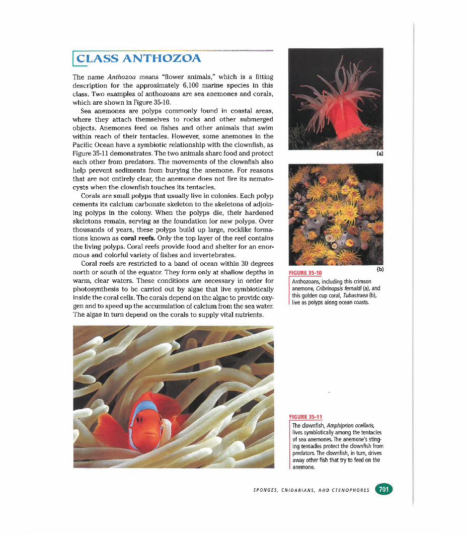
CLASS ANTHOZOAThe name Anthozoa means “flower animals,” which is a fitting description for the approximately 6,100 marine species in this class. Two examples of anthozoans are sea anemones and corals, which are shown in Figure 35-10.
Sea anemones are polyps commonly found in coasted areas, where they attach themselves to rocks and other submerged objects. Anemones feed on fishes and other animals that swim within reach of their tentacles. However, some anemones in the Pacific Ocean have a symbiotic relationship with the clownfish, as Figure 35-11 demonstrates. The two animals share food and protect each other from predators. The movements of the clownfish also help prevent sediments from burying the anemone. For reasons that are not entirely clear, the anemone does not fire its nemato- cysts when the clownfish touches its tentacles.
Corals are small polyps that usually live in colonies. Each polyp cements its calcium carbonate skeleton to the skeletons of adjoining polyps in the colony. When the polyps die, their hardened skeletons remain, serving as the foundation for new polyps. Over thousands of years, these polyps build up large, rocklike formations known as coral reefs. Only the top layer of the reef contains the living polyps. Coral reefs provide food and shelter for an enormous and colorful variety of fishes and invertebrates.
Coral reefs are restricted to a band of ocean within 30 degrees north or south of the equator. They form only at shallow depths in warm, clear waters. These conditions are necessary in order for photosynthesis to be carried out by algae that live symbiotically inside the coral cells. The corals depend on the algae to provide oxygen and to speed up the accumulation of calcium from the sea water. The algae in turn depend on the corals to supply vital nutrients.
Anthozoans, including this crimson anemone, Cribrinopsis fernaldi (a), and this golden cup coral, Tubastraea (b), live as polyps along ocean coasts.
FIGURE 35-11The clownfish, Amphiprion ocellaris, lives symbiotically among the tentacles of sea anemones. The anemone's stinging tentacles protect the clownfish from predators. The clownfish, in turn, drives away other fish that try to feed on the anemone.
SPONGES, CNIDARIANS, AND CTENOPHORES
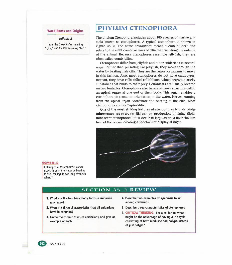
Word Roots and Origins
colloblast
from the Greek kollo, meaning "glue," and blastos, meaning "bud"
FIGURE 35-12
A ctenophore, Pleurobrachia pileus, moves through the water by beating its cilia, trailing Its two long tentacles behind it.
PHYLUM CTENOPHORAThe phylum Ctenophora includes about 100 species of marine animals known as ctenophores. A typical ctenophore is shown in Figure 35-12. The name Ctenophora means “comb holder” and refers to the eight comblike rows of cilia that run along the outside of the animal. Because ctenophores resemble jellyfish, they are often called comb jellies.
Ctenophores differ from jellyfish and other cnidarians in severed ways. Rather than pulsating like jellyfish, they move through the water by beating their cilia. They are the largest organisms to move in this fashion. Also, most ctenophores do not have cnidocytes. Instead, they have cells called colloblasts, which secrete a sticky substance that binds to their prey. Colloblasts are usually located on two tentacles. Ctenophores also have a sensory structure called an apical organ at one end of their body. This organ enables a ctenophore to sense its orientation in the water. Nerves running from the apical organ coordinate the beating of the cilia. Most ctenophores are hermaphroditic.
One of the most striking features of ctenophores is their biolu- minescence (BiE-oh-LOO-muh-NES-ens), or production of light. Biolu- minescent ctenophores often occur in large swarms near the surface of the ocean, creating a spectacular display at night.
SECTION 35-2 REVIEW
1. What are the two basic body forms a cnidarian may have?
2. What are three characteristics that all cnidarians have in common?
3. Name the three classes of cnidarians, and give an example of each.
4. Describe two examples of symbiosis found among cnidarians.
5. Describe three characteristics of ctenophores.6. CRITICAL THINKING For a cnidarian, what
might be the advantage of having a life cycle consisting of both medusae and polyps, instead of just polyps?
CHAPTER 35
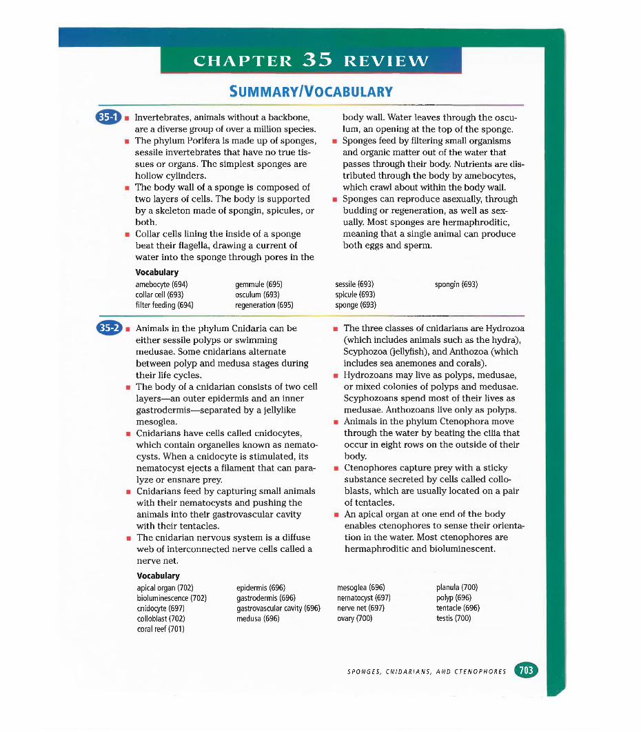
CHAPTER 35 REVIEW
Summary/vocabulary
■ Invertebrates, animals without a backbone, are a diverse group of over a million species.
■ The phylum Porifera is made up of sponges, sessile invertebrates that have no true tissues or organs. The simplest sponges are hollow cylinders.
■ The body wall of a sponge is composed of two layers of cells. The body is supported by a skeleton made of spongin, spicules, or both.
■ Collar cells lining the inside of a sponge beat their flagella, drawing a current of water into the sponge through pores in theVocabularyamebocyte (694) gemmule (695)collar cell (693) osculum (693)filter feeding (694) regeneration (695)
body wall. Water leaves through the osculum, an opening at the top of the sponge.
■ Sponges feed by filtering small organisms and organic matter out of the water that passes through their body. Nutrients are distributed through the body by amebocytes, which crawl about within the body wall.
■ Sponges can reproduce asexually, through budding or regeneration, as well as sexually. Most sponges are hermaphroditic, meaning that a single animal can produce both eggs and sperm.
sessile (693) spongin (693)spicule (693) sponge (693)
■ Animals in the phylum Cnidaria can be either sessile polyps or swimming medusae. Some cnidarians alternate between polyp and medusa stages during their life cycles.
■ The body of a cnidarian consists of two cell layers—an outer epidermis and an inner gastrodermis—separated by a jellylike mesoglea.
■ Cnidarians have cells called cnidocytes, which contain organelles known as nemato- cysts. When a cnidocyte is stimulated, its nematocyst ejects a filament that can paralyze or ensnare prey.
■ Cnidarians feed by capturing small animals with their nematocysts and pushing the animals into their gastrovascular cavity with their tentacles.
■ The cnidarian nervous system is a diffuse web of interconnected nerve cells called a nerve net.Vocabularyapical organ (702) bioluminescence (702) cnidocyte (697) colloblast (702) coral reef (701)
epidermis (696) gastrodermis (696) gastrovascular cavity (696) medusa (696)
■ The three classes of cnidarians cure Hydrozoa (which includes animals such as the hydra), Scyphozoa (jellyfish), and Anthozoa (which includes sea anemones and corals).
■ Hydrozoans may live as polyps, medusae, or mixed colonies of polyps and medusae. Scyphozoans spend most of their lives as medusae. Anthozoans live only as polyps.
■ Animals in the phylum Ctenophora move through the water by beating the cilia that occur in eight rows on the outside of their body.
■ Ctenophores capture prey with a sticky substance secreted by cells called collo- blasts, which are usually located on a pair of tentacles.
■ An apical organ at one end of the body enables ctenophores to sense their orientation in the water. Most ctenophores are hermaphroditic and bioluminescent.
mesoglea (696) nematocyst (697) nerve net (697) ovary (700)
planula (700) polyp (696) tentacle (696) testis (700)
SPONGES, CNIDARIANS, AND CTENOPHORES «U
CHAPTER 35 REVIEW
REVIEW
Vocabulary1. Explain why Porifera is a good name for the
phylum of sponges.2. Explain how sponges feed. What term
describes this method of feeding?3. Describe and name the two body forms of
cnidarians. For each body form, give an example of a cnidarian that has that form.
4. What is the difference between a cnidocyte and a nematocyst?
5. What is the function of a ctenophore’s apical organ?
Multiple Choice6. All invertebrates (a) live in water (b) have
tentacles (c) have no backbone (d) are hermaphrodites.
7. A hydra shows (a) asymmetry (b) spherical symmetry (c) radial symmetry (d) bilateral symmetry.
8. The structures that propel ctenophores through the water are (a) rows of cilia(b) colloblasts (c) apical organs (d) polyps.
9. Spongin and spicules are important to a sponge because they (a) trap food(b) provide support (c) remove wastes (d) produce offspring.
10. Both feeding and sexual reproduction in sponges involve (a) gemmules and collar cells (b) spicules and gemmules (c) amebo- cytes and spongin (d) collar cells and amebocytes.
11. The primary basis for classifying sponges is(a) their shape (b) their color (c) the composition of their skeleton (d) the method by which they reproduce.
12. Characteristics of cnidarians include all of the following except (a) nematocysts(b) collar cells (c) gastrovascular cavity (d) tentacles.
13. The cnidarian body form that is specialized for swimming is called a (a) medusa(b) polyp (c) planula (d) blastula.
14. The movements of cnidarians are coordinated by their (a) mesoglea (b) nematocysts(c) nerve net (d) gastrodermis.
CHAPTER 35
15. Colloblasts are (a) cells that draw water through sponges (b) cells that secrete a sticky substance in ctenophores (c) stages in the life cycle of a jellyfish (d) medusae that live in colonies.
Short Answer16. Describe three ways that sponges represent
the transition from unicellular to multicellular life.
17. How does a cnidarian capture and ingest its prey?
18. Describe the path of water through a sponge, and explain what causes water to flow through a sponge.
19. How is a sponge’s usual method of feeding suited to its sessile lifestyle?
20. Describe the specialization that is found among the individuals that make up a Portuguese man-of-war.
21. Describe the process of sexual reproduction in sponges.
22. What benefit does a sea anemone obtain from its symbiotic relationship with a clown- fish? What benefit does the clownfish obtain from this relationship?
23. Why do coral reefs form only at shallow depths?
24. Describe two ways that ctenophores differ from cnidarians.
25. In the diagram of a hydra shown below, identify the cell layers labeled A and B, the material between them labeled C, and the cell labeled D.
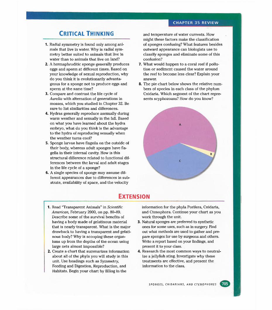
CHAPTER 35 REVIEW
Critical Thinking
1. Radial symmetry is found only among animals that live in water. Why is radial symmetry better suited to animals that live in water than to animals that live on land?
2. A hermaphroditic sponge generally produces eggs and sperm at different times. Based on your knowledge of sexual reproduction, why do you think it is evolutionarily advantageous for a sponge not to produce eggs and sperm at the same time?
3. Compare and contrast the life cycle of Aurelia with alternation of generations in mosses, which you studied in Chapter 32. Be sure to list similarities and differences.
4. Hydras generally reproduce asexually during warm weather and sexually in the fall. Based on what you have learned about the hydra embryo, what do you think is the advantage to the hydra of reproducing sexually when the weather turns cool?
5. Sponge larvae have flagella on the outside of their body, whereas adult sponges have flagella in their internal cavity. How is this structural difference related to functional differences between the larval and adult stages in the life cycle of a sponge?
6. A single species of sponge may assume different appearances due to differences in substrate, availability of space, and the velocity
and temperature of water currents. How might these factors make the classification of sponges confusing? What features besides outward appearance can biologists use to classify sponges and eliminate some of this confusion?
7. What would happen to a coral reef if pollution or sediment caused the water around the reef to become less clear? Explain your answer.
8. The pie chart below shows the relative numbers of species in each class of the phylum Cnidaria. Which segment of the chart represents scyphozoans? How do you know?
A
lB /
Extension
1. Read “Transparent Animals” in Scientific American, February 2000, on pp. 80-89. Describe some of the survival benefits of having a body made of gelatinous material that is nearly transparent. What is the major drawback to having a transparent and gelatinous body? Why is scooping these organisms up from the depths of the ocean using large nets almost impossible?
2. Create a chart that summarizes information about all of the phyla you will study in this unit. Use headings such as Symmetry, Feeding and Digestion, Reproduction, and Habitats. Begin your chart by filling in the
information for the phyla Porifera, Cnidaria, and Ctenophora. Continue your chart as you work through the unit.
3. Natural sponges are preferred to synthetic ones for some uses, such as in surgery. Find out what methods are used to gather and prepare sponges for use by surgeons and others. Write a report based on your findings, and present it to your class.
4. Research the most common ways to neutralize a jellyfish sting. Investigate why these treatments are effective, and present the information to the class..
SPONGES, CNIDARIANS, AND CTENOPHORES
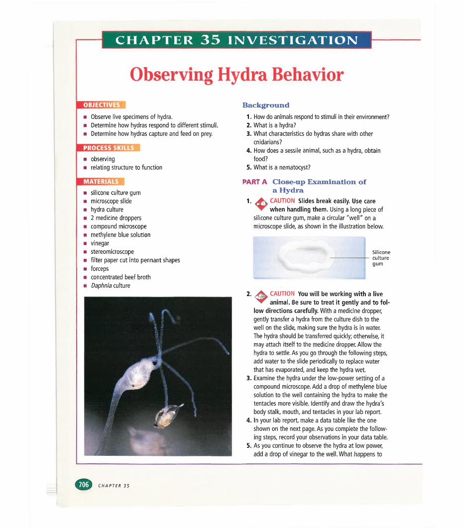
CHAPTER 35 INVESTIGATION
Observing Hydra BehaviorpnHiinu
■ Observe live specimens of hydra.■ Determine how hydras respond to different stimuli.■ Determine how hydras capture and feed on prey.
PROCESS SKI
■ observing■ relating structure to function
Background1. How do animals respond to stimuli in their environment?2. What is a hydra?3. What characteristics do hydras share with other
cnidarians?4. How does a sessile animal, such as a hydra, obtain
food?5. What is a nematocyst?
■mugg■ silicone culture gum■ microscope slide■ hydra culture■ 2 medicine droppers■ compound microscope■ methylene blue solution■ vinegar■ stereomicroscope■ filter paper cut into pennant shapes■ forceps■ concentrated beef broth■ Daphnia culture
PART A Close-up Examination of a Hydra
^ CAUTION Slides break easily. Use care ▼ ^ when handling them. Using a long piece of
silicone culture gum, make a circular "well" on a microscope slide, as shown in the illustration below.
1.
Siliconeculturegum
2. CAUTION You will be working with a live animal. Be sure to treat it gently and to fol
low directions carefully. With a medicine dropper, gently transfer a hydra from the culture dish to the well on the slide, making sure the hydra is in water.The hydra should be transferred quickly; otherwise, it may attach itself to the medicine dropper. Allow the hydra to settle. As you go through the following steps, add water to the slide periodically to replace water that has evaporated, and keep the hydra wet.
3. Examine the hydra under the low-power setting of a compound microscope. Add a drop of methylene blue solution to the well containing the hydra to make the tentacles more visible. Identify and draw the hydra's body stalk, mouth, and tentacles in your lab report.
4. In your lab report, make a data table like the one shown on the next page. As you complete the following steps, record your observations in your data table.
5. As you continue to observe the hydra at low power, add a drop of vinegar to the well. What happens to
CHAPTER 35
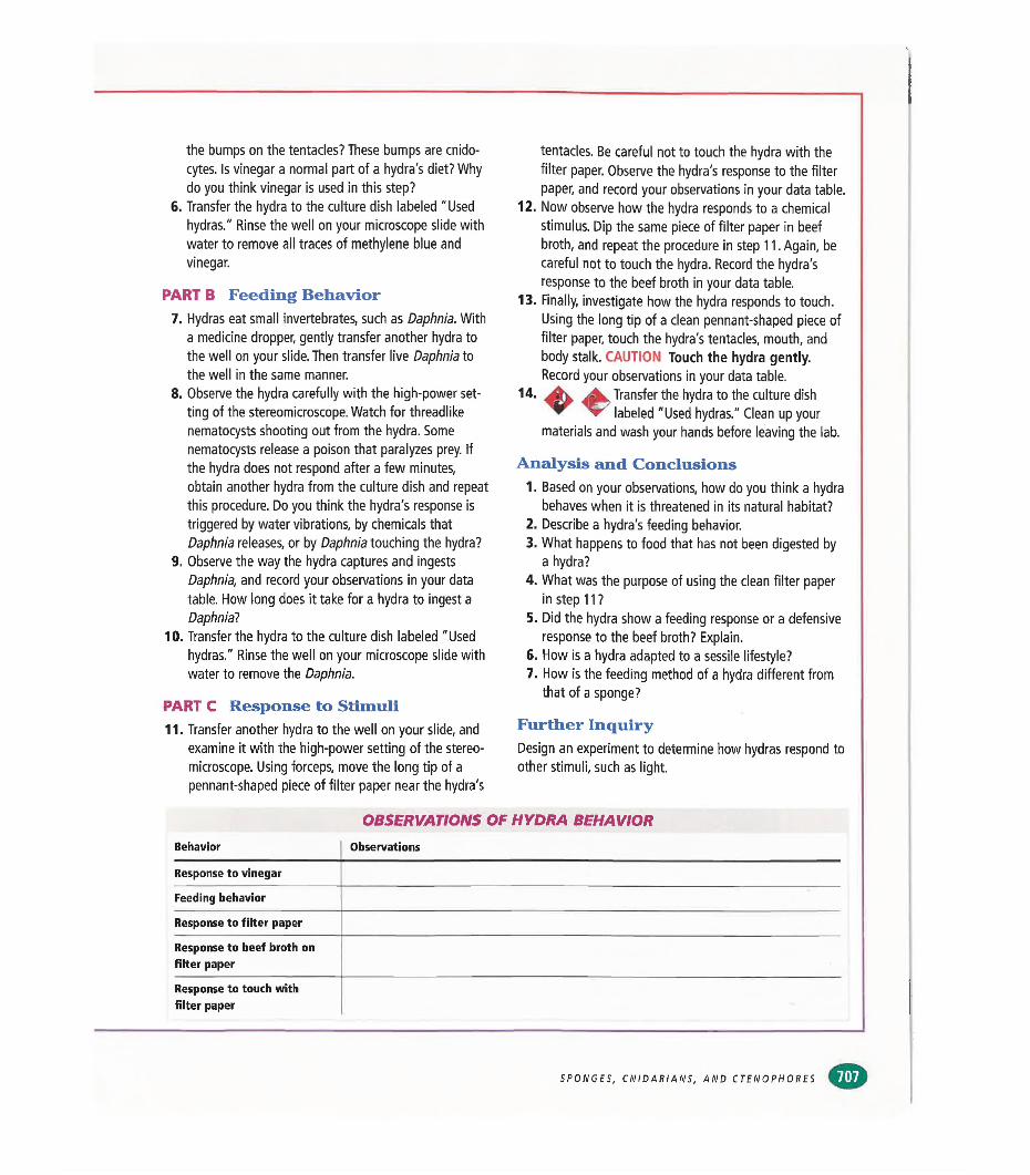
the bumps on the tentacles? These bumps are cnido- cytes. Is vinegar a normal part of a hydra's diet? Why do you think vinegar is used in this step?
6. Transfer the hydra to the culture dish labeled "Used hydras." Rinse the well on your microscope slide with water to remove all traces of methylene blue and vinegar.
PART B Feeding Behavior7. Hydras eat small invertebrates, such as Daphnia. With
a medicine dropper, gently transfer another hydra to the well on your slide. Then transfer live Daphnia to the well in the same manner.
8. Observe the hydra carefully with the high-power setting of the stereomicroscope. Watch for threadlike nematocysts shooting out from the hydra. Some nematocysts release a poison that paralyzes prey. If the hydra does not respond after a few minutes, obtain another hydra from the culture dish and repeat this procedure. Do you think the hydra's response is triggered by water vibrations, by chemicals that Daphnia releases, or by Daphnia touching the hydra?
9. Observe the way the hydra captures and ingests Daphnia, and record your observations in your data table. How long does it take for a hydra to ingest a Daphnia?
10. Transfer the hydra to the culture dish labeled "Used hydras." Rinse the well on your microscope slide with water to remove the Daphnia.
PART C Response to Stimuli11. Transfer another hydra to the well on your slide, and
examine it with the high-power setting of the stereomicroscope. Using forceps, move the long tip of a pennant-shaped piece of filter paper near the hydra's
tentacles. Be careful not to touch the hydra with the filter paper. Observe the hydra's response to the filter paper, and record your observations in your data table.
12. Now observe how the hydra responds to a chemical stimulus. Dip the same piece of filter paper in beef broth, and repeat the procedure in step 11. Again, be careful not to touch the hydra. Record the hydra’s response to the beef broth in your data table.
13. Finally, investigate how the hydra responds to touch. Using the long tip of a clean pennant-shaped piece of filter paper, touch the hydra's tentacles, mouth, and body stalk. CAUTION Touch the hydra gently. Record your observations in your data table.
14. A Transfer the hydra to the culture dishyr' labeled "Used hydras." Clean up your
materials and wash your hands before leaving the lab.
Analysis and Conclusions1. Based on your observations, how do you think a hydra
behaves when it is threatened in its natural habitat?2. Describe a hydra's feeding behavior.3. What happens to food that has not been digested by
a hydra?4. What was the purpose of using the clean filter paper
in step 11 ?5. Did the hydra show a feeding response or a defensive
response to the beef broth? Explain.6. How is a hydra adapted to a sessile lifestyle?7. How is the feeding method of a hydra different from
that of a sponge?
Further InquiryDesign an experiment to determine how hydras respond toother stimuli, such as light.
OBSERVATIONS OF HYDRA BEHAVIORBehavior Observations
Response to vinegar
Feeding behavior
Response to filter paper
Response to beef broth on filter paper
Response to touch with filter paper
SPONGES, CNIDARIANS, AND CTENOPHORES
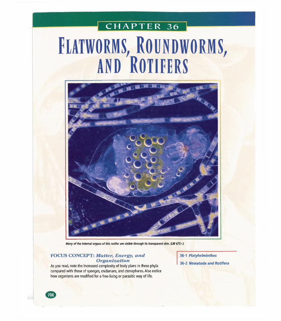
CHAPTER 36
fiATWORMS, Roundworms,and Rotifers
Many of the internal organs of this rotifer are visible through its transparent skin. (LM 67Sx)
FOCUS CONCEPT: Matter, Energy, andOrgan ization
As you read, note the increased complexity of body plans in these phyla compared with those of sponges, cnidarians, and ctenophores. Also notice how organisms are modified for a free-living or parasitic way of life.
36-1 Platyhelminthes
36-2 Nematoda and Rotifera
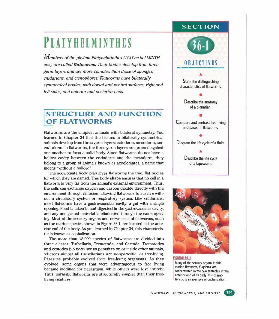
SECTION)
PlATYHELMINlHESMembers of the phylum Platyhelminthes (PLAT-ee-hel-MINTH-
eez) are called flatworms. Their bodies develop from three germ layers and are more complex than those of sponges, cnidarians, and ctenophores. Flatworms have bilaterally symmetrical bodies, with dorsal and ventral surfaces, right and left sides, and anterior and posterior ends.
STRUCTURE AND FUNCTION OF FLATWORMS
Flatworms are the simplest animals with bilateral symmetry. You learned in Chapter 34 that the tissues in bilaterally symmetrical animals develop from three germ layers: ectoderm, mesoderm, and endoderm. In flatworms, the three germ layers are pressed against one another to form a solid body. Since flatworms do not have a hollow cavity between the endoderm and the mesoderm, they belong to a group of animals known as acoelomates, a name that means “without a hollow.”
The acoelomate body plan gives flatworms the thin, flat bodies for which they are named. This body shape ensures that no cell in a flatworm is very far from the animal’s external environment. Thus, the cells can exchange oxygen and carbon dioxide directly with the environment through diffusion, allowing flatworms to survive without a circulatory system or respiratory system. Like cnidarians, most flatworms have a gastrovascular cavity, a gut with a single opening. Food is taken in and digested in the gastrovascular cavity, and any undigested material is eliminated through the same opening. Most of the sensory organs and nerve cells of flatworms, such as the marine species shown in Figure 36-1, are located at the anterior end of the body. As you learned in Chapter 34, this characteristic is known as cephalization.
The more than 18,000 species of flatworms are divided into three classes: Turbellaria, Trematoda, and Cestoda. Trematodes and cestodes (SES-tohdz) live as parasites on or inside other animals, whereas almost all turbellarians are nonparasitic, or free-living. Parasites probably evolved from free-living organisms. As they evolved, some organs that were advantageous to free living became modified for parasitism, while others were lost entirely. Thus, parasitic flatworms are structurally simpler than their free- living relatives.
{¡DOBJECTIVES
▲State the distinguishing
characteristics of flatworms.
•Describe the anatomy
of a planarian.
■Compare and contrast free-living
and parasitic flatworms.
♦ ■Diagram the life cycle of a fluke.
▲
Describe the life cycle
of a tapeworm.
FIGURE 36-1
Many of the sensory organs in this marine flatworm, Euryletta, are concentrated in the two tentacles at the anterior end of its body. This characteristic is an example of cephalization.
FLATWORMS, ROUNDWORMS, AND ROTIFERS mm
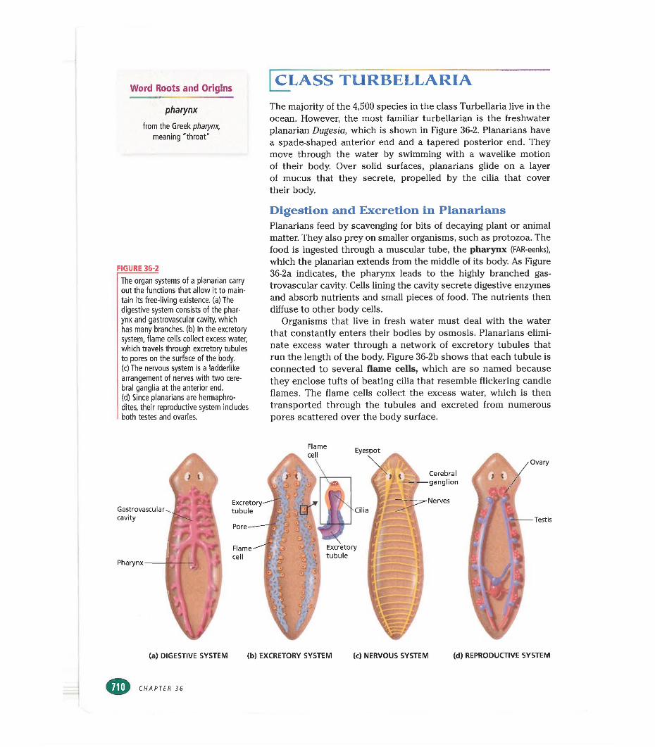
Word Roots and Origins
pharynx
from the Greek pharynx, meaning "throat"
FIGURE 36-2
The organ systems of a planarian carry out the functions that allow it to maintain its free-living existence, (a) The digestive system consists of the pharynx and gastrovascular cavity, which has many branches, (b) In the excretory system, flame cells collect excess water, which travels through excretory tubules to pores on the surface of the body.(c) The nervous system is a ladderlike arrangement of nerves with two cerebral ganglia at the anterior end.(d) Since planarians are hermaphrodites, their reproductive system includes both testes and ovaries.
CLASS TURBELLARIAThe majority of the 4,500 species in the class Turbellaria live in the ocean. However, the most familiar turbellarian is the freshwater planarian Dugesia, which is shown in Figure 36-2. Planarians have a spade-shaped anterior end and a tapered posterior end. They move through the water by swimming with a wavelike motion of their body. Over solid surfaces, planarians glide on a layer of mucus that they secrete, propelled by the cilia that cover their body.
Digestion and Excretion in PlanariansPlanarians feed by scavenging for bits of decaying plant or animal matter. They also prey on smaller organisms, such as protozoa. The food is ingested through a muscular tube, the pharynx (FAR-eenks), which the planarian extends from the middle of its body. As Figure 36-2a indicates, the pharynx leads to the highly branched gastrovascular cavity. Cells lining the cavity secrete digestive enzymes and absorb nutrients and small pieces of food. The nutrients then diffuse to other body cells.
Organisms that live in fresh water must deal with the water that constantly enters their bodies by osmosis. Planarians eliminate excess water through a network of excretory tubules that run the length of the body. Figure 36-2b shows that each tubule is connected to severed flame cells, which are so named because they enclose tufts of beating cilia that resemble flickering candle flames. The flame cells collect the excess water, which is then transported through the tubules and excreted from numerous pores scattered over the body surface.
Gastrovascularcavity
Pharynx
tubule
Flamecell
Cilia
Cerebralganglion
Nerves
Flamecell
Excretorytubule
Ovary
Testis
(a) DIGESTIVE SYSTEM (b) EXCRETORY SYSTEM (c) NERVOUS SYSTEM (d) REPRODUCTIVE SYSTEM
CHAPTER 36
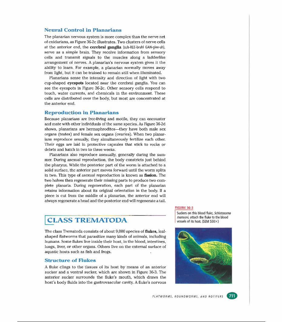
Neural Control in PlanariansThe planarian nervous system is more complex than the nerve net of cnidarians, as Figure 36-2c illustrates. Two clusters of nerve cells at the anterior end, the cerebral ganglia (suh-REE-bruhl GAN-glee-uh),
serve as a simple brain. They receive information from sensory cells and transmit signals to the muscles along a ladderlike arrangement of nerves. A planarian’s nervous system gives it the ability to learn. For example, a planarian normally moves away from light, but it can be trained to remain still when illuminated.
Planarians sense the intensity and direction of light with two cup-shaped eyespots located near the cerebral ganglia. You can see the eyespots in Figure 36-2c. Other sensory cells respond to touch, water currents, and chemicals in the environment. These cells are distributed over the body, but most are concentrated at the anterior end.
Reproduction in PlanariansBecause planarians are free-living and motile, they can encounter and mate with other individuals of the same species. As Figure 36-2d shows, planarians are hermaphrodites—they have both male sex organs (testes) and female sex organs (ovaries). When two planarians reproduce sexually, they simultaneously fertilize each other. Their eggs are laid in protective capsules that stick to rocks or debris and hatch in two to three weeks.
Planarians also reproduce asexually, generally during the summer. During asexual reproduction, the body constricts just behind the pharynx. While the posterior part of the worm is attached to a solid surface, the anterior part moves forward until the worm splits in two. This type of asexual reproduction is known as fission. The two halves then regenerate their missing parts to produce two complete planaria. During regeneration, each part of the planarian retains information about its original orientation in the body. If a piece is cut from the middle of a planarian, the anterior end will always regenerate a head and the posterior end will regenerate a tail.
| CLASS TREMATODAThe class Trematoda consists of about 9,000 species of flukes, leafshaped flatworms that parasitize many kinds of animals, including humans. Some flukes live inside their host, in the blood, intestines, lungs, liver, or other organs. Others live on the external surface of aquatic hosts such as fish and frogs.
Structure of FlukesA fluke clings to the tissues of its host by means of an anterior sucker and a ventral sucker, which are shown in Figure 36-3. The anterior sucker surrounds the fluke’s mouth, which draws the host’s body fluids into the gastrovascular cavity. A fluke’s nervous
FIGURE 36-3
Suckers on this blood fluke, Schistosoma mansoni, attach the fluke to the blood vessels of its host. (SEM 550x)
FLATWORMS, ROUNDWORMS, AND ROTIFERS
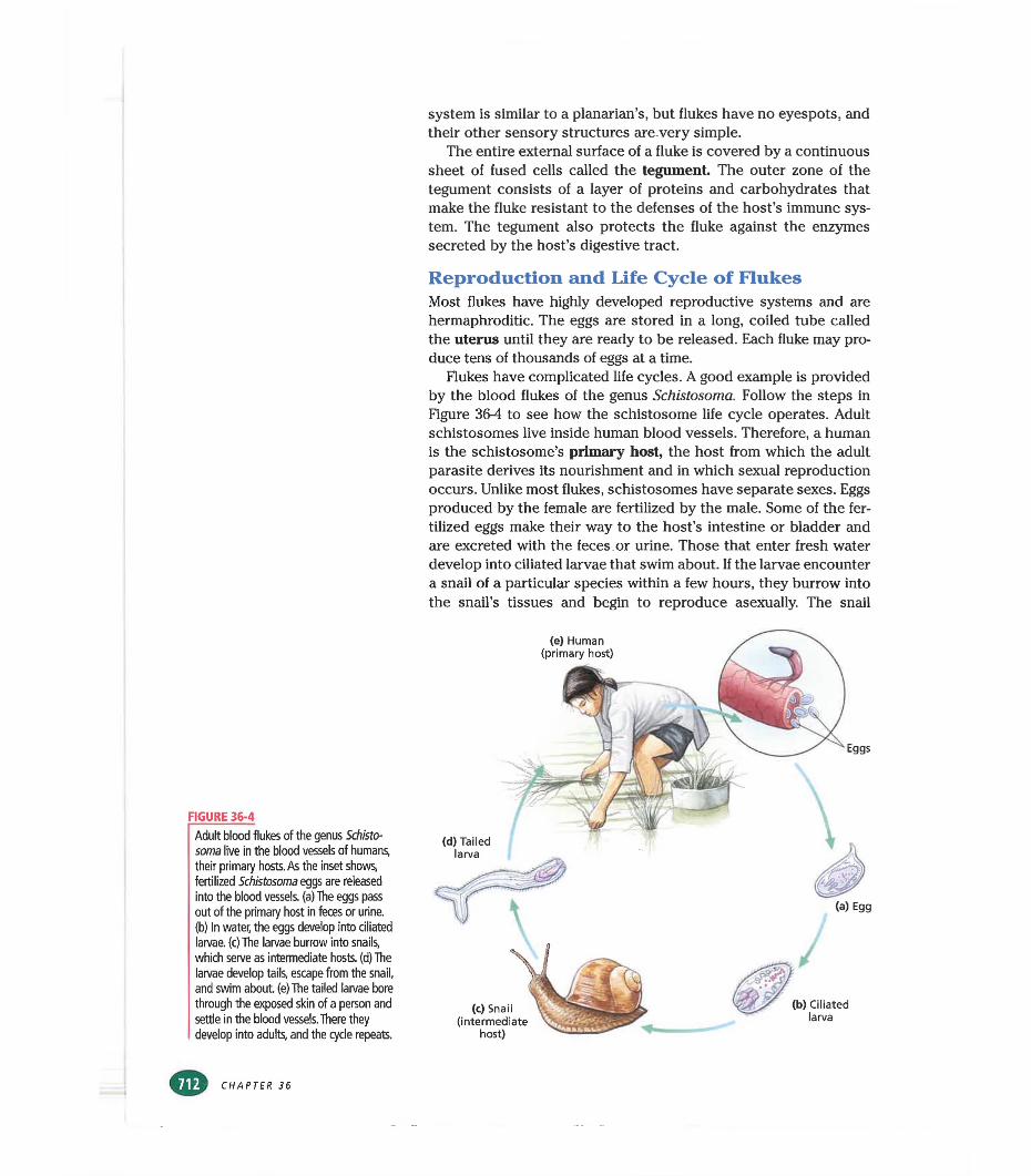
FIGURE 36-4Adult blood flukes of the genus Schistosoma live in the blood vessels of humans, their primary hosts. As the inset shows, fertilized Schistosoma eggs are released into the blood vessels, (a) The eggs pass out of the primary host in feces or urine, (b) In water, the eggs develop into ciliated larvae, (c) The larvae burrow into snails, which serve as intermediate hosts, (d) The larvae develop tails, escape from the snail, and swim about, (e) The tailed larvae bore through the exposed skin of a person and settle in the blood vessels. There they develop into adults, and the cycle repeats.
system is similar to a planarian’s, but flukes have no eyespots, and their other sensory structures are very simple.
The entire external surface of a fluke is covered by a continuous sheet of fused cells called the tegument The outer zone of the tegument consists of a layer of proteins and carbohydrates that make the fluke resistant to the defenses of the host’s immune system. The tegument also protects the fluke against the enzymes secreted by the host’s digestive tract.
Reproduction and Life Cycle of FlukesMost flukes have highly developed reproductive systems and are hermaphroditic. The eggs are stored in a long, coiled tube called the uterus until they are ready to be released. Each fluke may produce tens of thousands of eggs at a time.
Flukes have complicated life cycles. A good example is provided by the blood flukes of the genus Schistosoma. Follow the steps in Figure 36-4 to see how the schistosome life cycle operates. Adult schistosomes live inside human blood vessels. Therefore, a human is the schistosome’s primary host, the host from which the adult parasite derives its nourishment and in which sexual reproduction occurs. Unlike most flukes, schistosomes have separate sexes. Eggs produced by the female are fertilized by the male. Some of the fertilized eggs make their way to the host’s intestine or bladder and are excreted with the feces or urine. Those that enter fresh water develop into ciliated larvae that swim about. If the larvae encounter a snail of a particular species within a few hours, they burrow into the snail’s tissues and begin to reproduce asexually. The snail
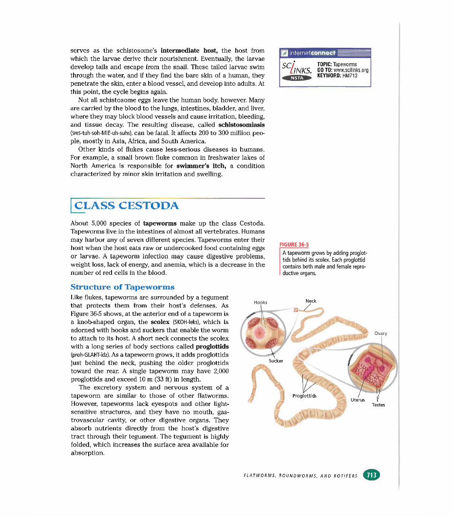
serves as the schistosome’s intermediate host, the host from which the larvae derive their nourishment. Eventually, the larvae develop tails and escape from the snail. These tailed larvae swim through the water, and if they find the bare skin of a human, they penetrate the skin, enter a blood vessel, and develop into adults. At this point, the cycle begins again.
Not all schistosome eggs leave the human body, however. Many are carried by the blood to the lungs, intestines, bladder, and liver, where they may block blood vessels and cause irritation, bleeding, and tissue decay. The resulting disease, called schistosomiasis (SHis-tuh-soh-MIE-uh-suhs), can be fatal. It affects 200 to 300 million people, mostly in Asia, Africa, and South America.
Other kinds of flukes cause less-serious diseases in humans. For example, a small brown fluke common in freshwater lakes of North America is responsible for swimmer’s itch, a condition characterized by minor skin irritation and swelling.
internetconnect
SC/lNKS„ TOPIC: Tapeworms GO TO: www.scilinks.org KEYWORD: HM713
[class cestoda
About 5,000 species of tapeworms make up the class Cestoda. Tapeworms live in the intestines of almost all vertebrates. Humans may harbor any of seven different species. Tapeworms enter their host when the host eats raw or undercooked food containing eggs or larvae. A tapeworm infection may cause digestive problems, weight loss, lack of energy, and anemia, which is a decrease in the number of red cells in the blood.
FIGURE 36-5
A tapeworm grows by adding proglot- tids behind its scolex. Each proglottid contains both male and female reproductive organs.
Structure of TapewormsLike flukes, tapeworms are surrounded by a tegument that protects them from their host’s defenses. As Figure 36-5 shows, at the anterior end of a tapeworm is a knob-shaped organ, the scolex (SKOH-leks), which is adorned with hooks and suckers that enable the worm to attach to its host. A short neck connects the scolex with a long series of body sections called proglottids (proh-GLAHT-idz). As a tapeworm grows, it adds proglottids just behind the neck, pushing the older proglottids toward the rear. A single tapeworm may have 2,000 proglottids and exceed 10 m (33 ft) in length.
The excretory system and nervous system of a tapeworm are similar to those of other flatworms. However, tapeworms lack eyespots and other light- sensitive structures, and they have no mouth, gas- trovascular cavity, or other digestive organs. They absorb nutrients directly from the host’s digestive tract through their tegument. The tegument is highly folded, which increases the surface area available for absorption.
ProglottidsUterus
Testes
FLATWORMS, ROUNDWORMS, AND ROTIFERS
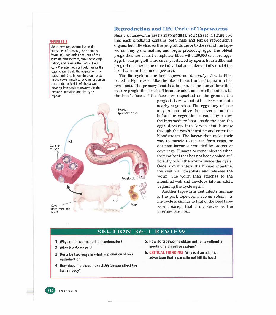
Reproduction and Life Cycle of Tapeworms
FIGURE 36-6
Adult beef tapeworms live in the intestines of humans, their primary hosts, (a) Proglottids pass out of the primary host in feces, crawl onto vegetation, and release their eggs, (b) A cow, the intermediate host, ingests the eggs when it eats the vegetation. The eggs hatch into larvae that form cysts in the cow's muscles, (c) When a person eats undercooked beef, the larvae develop into adult tapeworms in the person's intestine, and the cycle repeats.
Nearly all tapeworms are hermaphrodites. You can see in Figure 36-5 that each proglottid contains both male and female reproductive organs, but little else. As the proglottids move to the rear of the tapeworm, they grow, mature, and begin producing eggs. The oldest proglottids are almost completely filled with 100,000 or more eggs. Eggs in one proglottid are usually fertilized by sperm from a different proglottid, either in the same individual or a different individual if the host has more than one tapeworm.
The life cycle of the beef tapeworm, Taeniarhynchus, is illustrated in Figure 36-6. Like the blood fluke, the beef tapeworm has two hosts. The primary host is a human. In the human intestine, mature proglottids break off from the adult and are eliminated with the host’s feces. If the feces are deposited on the ground, the
proglottids crawl out of the feces and onto nearby vegetation. The eggs they release
(0Cysts in muscle
Cow (intermediate host)
Human (primary host) may remain alive for several months
before the vegetation is eaten by a cow, the intermediate host. Inside the cow, the eggs develop into larvae that burrow through the cow’s intestine and enter the bloodstream. The larvae then make their way to muscle tissue and form cysts, or dormant larvae surrounded by protective coverings. Humans become infected when they eat beef that has not been cooked sufficiently to kill the worms inside the cysts. Once a cyst enters the human intestine, the cyst wall dissolves and releases the worm. The worm then attaches to the intestinal wall and develops into an adult, beginning the cycle again.
Another tapeworm that infects humans is the pork tapeworm, Taenia solium. Its life cycle is similar to that of the beef tapeworm, except that a pig serves as the intermediate host.
SECTION 36-1 REVIEW
1. Why are flatworms called acoelomates?
2. What is a flame cell?3. Describe two ways in which a planarian shows
cephalization.4. How does the blood fluke Schistosoma affect the
human body?
5. How do tapeworms obtain nutrients without a mouth or a digestive system?
6. CRITICAL THINKING Why is it an adaptive advantage that a parasite not kill its host?
CHAPTER 36
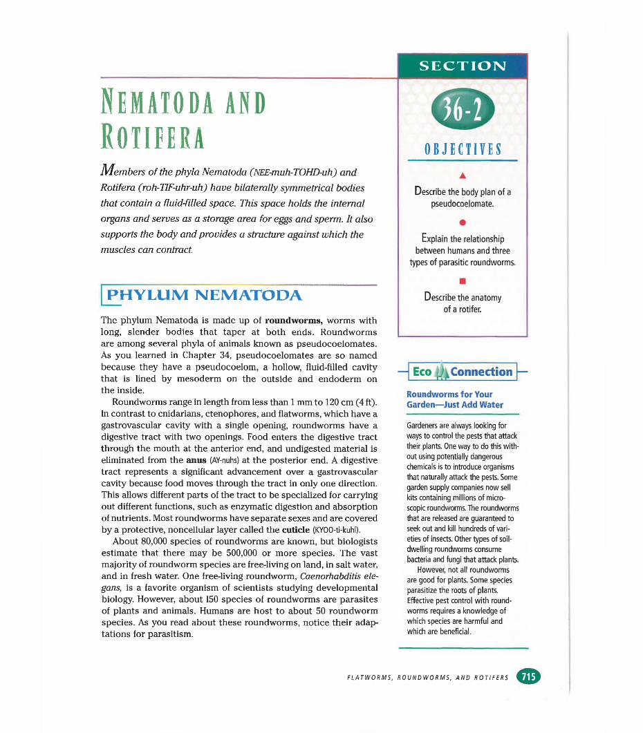
SECTION
Nematoda and RotiferaMembers of the phyla Nematoda (NEE-muh-TOHD-uh) and
Rotifera (roh-TIF-uhr-uh) have bilaterally symmetrical bodies that contain a fluid-filled space. This space holds the internal organs and serves as a storage area for eggs and sperm. It also supports the body and provides a structure against which the muscles can contract.
PHYLUM NEMATODAThe phylum Nematoda is made up of roundworms, worms with long, slender bodies that taper at both ends. Roundworms are among several phyla of animals known as pseudocoelomates. As you learned in Chapter 34, pseudocoelomates are so named because they have a pseudocoelom, a hollow, fluid-filled cavity that is lined by mesoderm on the outside and endoderm on the inside.
Roundworms range in length from less than 1 mm to 120 cm (4 ft). In contrast to cnidarians, ctenophores, and flatworms, which have a gastrovascular cavity with a single opening, roundworms have a digestive tract with two openings. Food enters the digestive tract through the mouth at the anterior end, and undigested material is eliminated from the anus (AY-nuhs) at the posterior end. A digestive tract represents a significant advancement over a gastrovascular cavity because food moves through the tract in only one direction. This allows different parts of the tract to be specialized for carrying out different functions, such as enzymatic digestion and absorption of nutrients. Most roundworms have separate sexes and are covered by a protective, noncellular layer called the cuticle (KYOO-ti-kuhl).
About 80,000 species of roundworms are known, but biologists estimate that there may be 500,000 or more species. The vast majority of roundworm species are free-living on land, in salt water, and in fresh water. One free-living roundworm, Caenorhabditis ele- gans, is a favorite organism of scientists studying developmental biology. However, about 150 species of roundworms are parasites of plants and animals. Humans are host to about 50 roundworm species. As you read about these roundworms, notice their adaptations for parasitism.
OBJECTIVESA
Describe the body plan of a pseudocoelomate.
•Explain the relationship
between humans and three types of parasitic roundworms.
■Describe the anatomy
of a rotifer.
■ Eco ¿Connection
Roundworms for Your Garden—Just Add Water
Gardeners are always looking for ways to control the pests that attack their plants. One way to do this without using potentially dangerous chemicals is to introduce organisms that naturally attack the pests. Some garden supply companies now sell kits containing millions of microscopic roundworms. The roundworms that are released are guaranteed to seek out and kill hundreds of varieties of insects. Other types of soildwelling roundworms consume bacteria and fungi that attack plants.
However, not all roundworms are good for plants. Some species parasitize the roots of plants. Effective pest control with round- worms requires a knowledge of which species are harmful and which are beneficial.
FLATWORMS, ROUNDWORMS, AND ROTIFERS
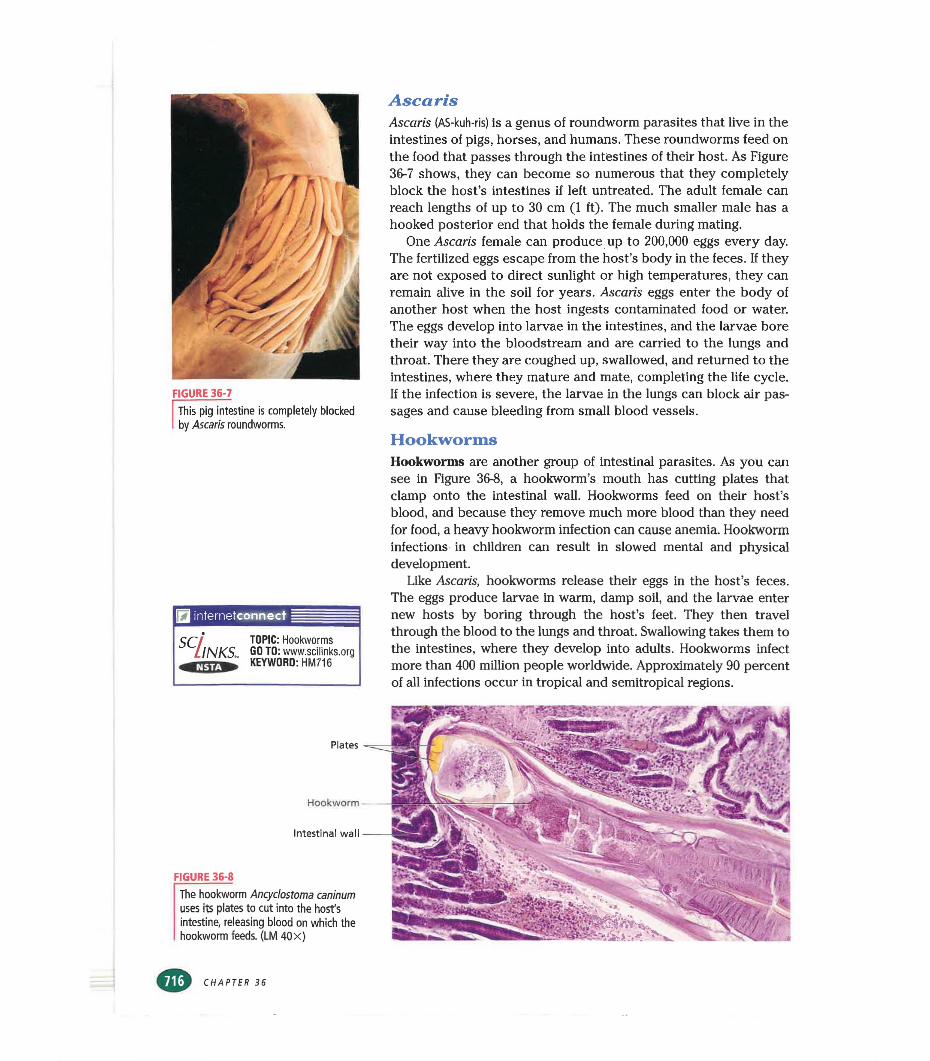
FIGURE 36-7This pig intestine is completely blocked by Ascaris roundworms.
internetconnect
SC/lNKS„ TOPIC: Hookworms GO TO: www.scilinks.orgKEYWORD: HM716
AscarisAscaris (AS-kuh-ris) is a genus of roundworm parasites that live in the intestines of pigs, horses, and humans. These roundworms feed on the food that passes through the intestines of their host. As Figure 36-7 shows, they can become so numerous that they completely block the host’s intestines if left untreated. The adult female can reach lengths of up to 30 cm (1 ft). The much smaller male has a hooked posterior end that holds the female during mating.
One Ascaris female can produce up to 200,000 eggs every day. The fertilized eggs escape from the host’s body in the feces. If they are not exposed to direct sunlight or high temperatures, they can remain alive in the soil for years. Ascaris eggs enter the body of another host when the host ingests contaminated food or water. The eggs develop into larvae in the intestines, and the larvae bore their way into the bloodstream and are carried to the lungs and throat. There they are coughed up, swallowed, and returned to the intestines, where they mature and mate, completing the life cycle. If the infection is severe, the larvae in the lungs can block air passages and cause bleeding from small blood vessels.
HookwormsHookworms are another group of intestinal parasites. As you can see in Figure 36-8, a hookworm’s mouth has cutting plates that clamp onto the intestinal wall. Hookworms feed on their host’s blood, and because they remove much more blood them they need for food, a heavy hookworm infection can cause anemia. Hookworm infections in children can result in slowed mental and physical development.
Like Ascaris, hookworms release their eggs in the host’s feces. The eggs produce larvae in warm, damp soil, and the larvae enter new hosts by boring through the host’s feet. They then travel through the blood to the lungs and throat. Swallowing takes them to the intestines, where they develop into adults. Hookworms infect more than 400 million people worldwide. Approximately 90 percent of all infections occur in tropical and semitropical regions.
Plates
Hookworm
Intestinal wall
FIGURE 36-8
The hookworm Ancyclostoma caninum uses its plates to cut into the host's Intestine, releasing blood on which the hookworm feeds. (LM 40x)
CHAPTER 36
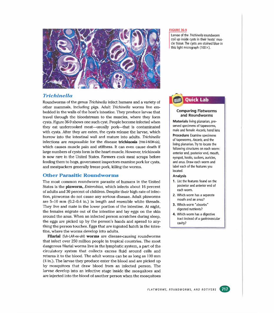
FIGURE 36-9
TrichinellaRoundworms of the genus Trichinella infect humans and a variety of other mammals, including pigs. Adult Trichinella worms live embedded in the walls of the host’s intestine. They produce larvae that travel through the bloodstream to the muscles, where they form cysts. Figure 36-9 shows one such cyst. People become infected when they eat undercooked meat—usually pork—that is contaminated with cysts. After they are eaten, the cysts release the larvae, which burrow into the intestinal wall and mature into adults. Trichinella infections are responsible for the disease trichinosis (TRlK-i-NOH-sis),
which causes muscle pain and stiffness. It can even cause death if large numbers of cysts form in the heart muscle. However, trichinosis is now rare in the United States. Farmers cook meat scraps before feeding them to hogs, government inspectors examine pork for cysts, and meatpackers generally freeze pork, killing the worms.
Other Parasitic RoundwormsThe most common roundworm parasite of humans in the United States is the pinworm, Enterobius, which infects about 16 percent of adults and 30 percent of children. Despite their high rate of infection, pinworms do not cause any serious disease. Adult pinworms are 5-10 mm (0.2-0.4 in.) in length and resemble white threads. They live and mate in the lower portion of the intestine. At night, the females migrate out of the intestine and lay eggs on the skin around the anus. When an infected person scratches during sleep, the eggs are picked up by the person’s hands and spread to anything the person touches. Eggs that are ingested hatch in the intestine, where the worms develop into adults.
Filarial (fuh-LAR-ee-uhl) worms are disease-causing roundworms that infect over 250 million people in tropical countries. The most dangerous filarial worms live in the lymphatic system, a part of the circulatory system that collects excess fluid around cells and returns it to the blood. The adult worms can be as long as 100 mm (4 in.). The larvae they produce enter the blood and are picked up by mosquitoes that draw blood from an infected person. The larvae develop into an infective stage inside the mosquitoes and are injected into the blood of another person when the mosquitoes
Larvae of the Trichinella roundworm coil up inside cysts in their hosts' muscle tissue. The cysts are stained blue in this light micrograph (100x).
gel Quick Lab
Comparing Flatworms and Roundworms
Materials living planarian, preserved specimens of tapeworms, male and female Ascaris, hand lens
Procedure Examine specimens of tapeworms, Ascaris, and the living planarian. Try to locate the following structures on each worm: anterior end, posterior end, mouth, eyespot, hooks, suckers, auricles, and anus. Draw each worm and label each of the features you located.
Analysis1. List the features found on the
posterior and anterior end of each worm.
2. Which worm has a separate mouth and an anus?
3. Which worm "absorbs" digested nutrients?
4. Which worm has a digestive tract instead of a gastrovascular cavity?
FLATWORMS, ROUNDWORMS, AND ROTIFERS
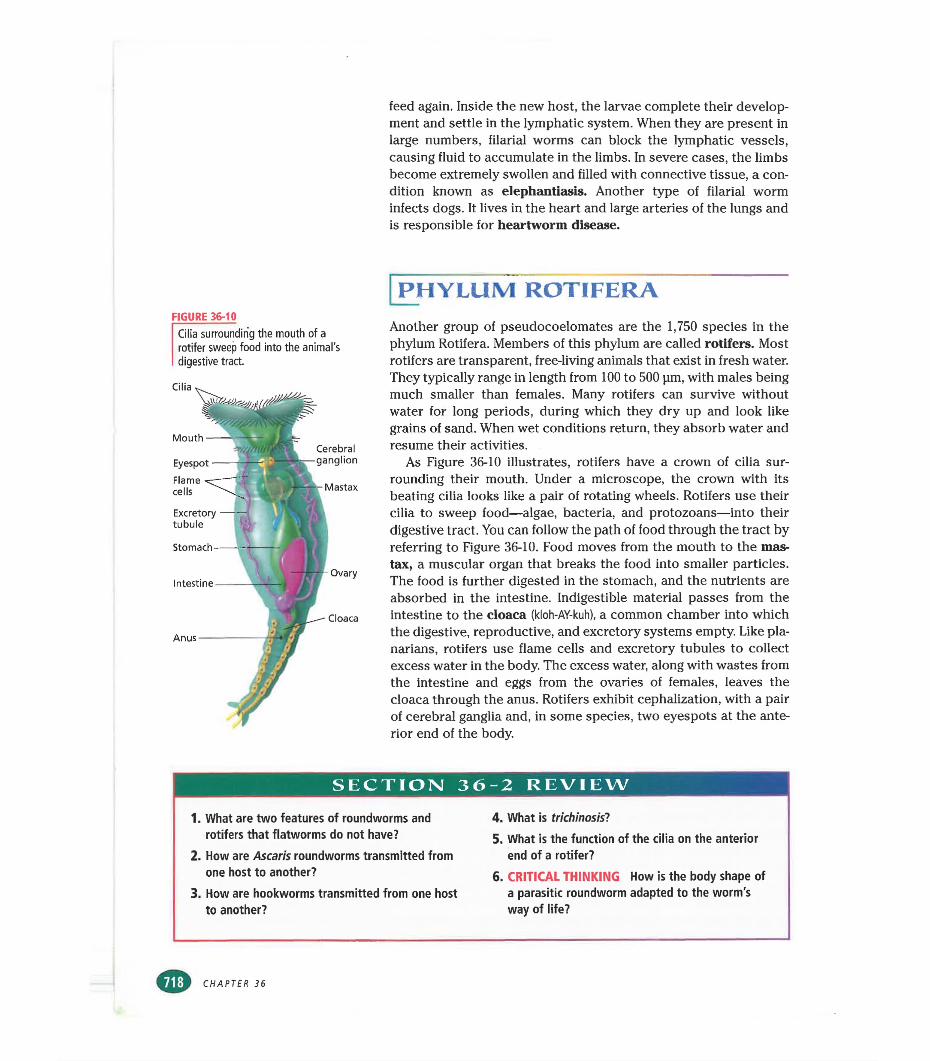
feed again. Inside the new host, the larvae complete their development and settle in the lymphatic system. When they are present in large numbers, filarial worms can block the lymphatic vessels, causing fluid to accumulate in the limbs. In severe cases, the limbs become extremely swollen and filled with connective tissue, a condition known as elephantiasis. Another type of filarial worm infects dogs. It lives in the heart and large arteries of the lungs and is responsible for heartworm disease.
FIGURE 36-10Cilia surrounding the mouth of a rotifer sweep food into the animal's digestive tract.
Cilia
Anus —
Cloaca
Mouth
Eyespot
Flame cells
Excretorytubule
Stomach
IntestineOvary
Cerebral
Mastax
PHYLUM ROTIFERAAnother group of pseudocoelomates are the 1,750 species in the phylum Rotifera. Members of this phylum are called rotifers. Most rotifers are transparent, free-living animals that exist in fresh water. They typically range in length from 100 to 500 pm, with males being much smaller than females. Many rotifers can survive without water for long periods, during which they dry up and look like grains of sand. When wet conditions return, they absorb water and resume their activities.
As Figure 36-10 illustrates, rotifers have a crown of cilia surrounding their mouth. Under a microscope, the crown with its beating cilia looks like a pair of rotating wheels. Rotifers use their cilia to sweep food—algae, bacteria, and protozoans—into their digestive tract. You can follow the path of food through the tract by referring to Figure 36-10. Food moves from the mouth to the mas- tax, a muscular organ that breaks the food into smaller particles. The food is further digested in the stomach, and the nutrients are absorbed in the intestine. Indigestible material passes from the intestine to the cloaca (kloh-AY-kuh), a common chamber into which the digestive, reproductive, and excretory systems empty. Like pla- narians, rotifers use flame cells and excretory tubules to collect excess water in the body. The excess water, along with wastes from the intestine and eggs from the ovaries of females, leaves the cloaca through the anus. Rotifers exhibit cephalization, with a pair of cerebral ganglia and, in some species, two eyespots at the anterior end of the body.
SECTION 36-2 REVIEW
1. What are two features of roundworms and rotifers that flatworms do not have?
2. How are Ascaris roundworms transmitted from one host to another?
3. How are hookworms transmitted from one host to another?
4. What is trichinosis'?5. What is the function of the cilia on the anterior
end of a rotifer?6. CRITICAL THINKING How is the body shape of
a parasitic roundworm adapted to the worm's way of life?
CHAPTER 36
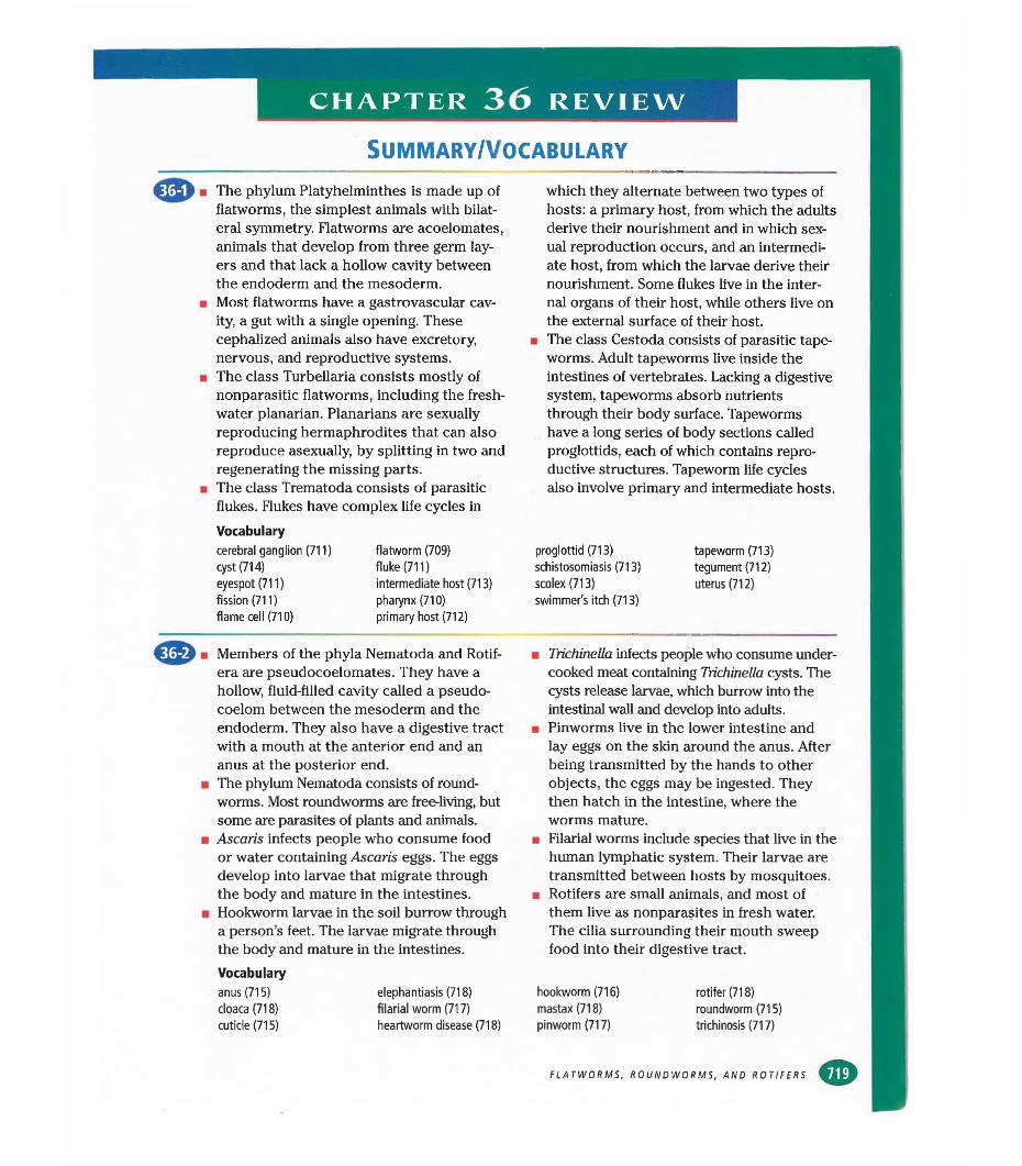
CHAPTER 36 REVIEW
SUMMARY/VOCABULARY
The phylum Platyhelminthes is made up of flatworms, the simplest animals with bilateral symmetry. Flatworms are acoelomates, animals that develop from three germ layers and that lack a hollow cavity between the endoderm and the mesoderm.Most flatworms have a gastrovascular cavity, a gut with a single opening. These cephalized animals also have excretory, nervous, and reproductive systems.The class Turbellaria consists mostly of nonparasitic flatworms, including the freshwater planarian. Planarians are sexually reproducing hermaphrodites that can also reproduce asexually, by splitting in two and regenerating the missing parts.The class Trematoda consists of parasitic flukes. Flukes have complex life cycles inVocabularycerebral ganglion (711) cyst (714) eyespot (711) fission (711) flame cell (710)
Members of the phyla Nematoda and Rotif- era are pseudocoelomates. They have a hollow, fluid-filled cavity called a pseudocoelom between the mesoderm and the endoderm. They also have a digestive tract with a mouth at the anterior end and an anus at the posterior end.The phylum Nematoda consists of round- worms. Most roundworms are free-living, but some are parasites of plants and animals. Ascaris infects people who consume food or water containing Ascaris eggs. The eggs develop into larvae that migrate through the body and mature in the intestines. Hookworm larvae in the soil burrow through a person’s feet. The larvae migrate through the body and mature in the intestines.Vocabularyanus (715) elephantiasis (718)cloaca (718) filarial worm (717)cuticle (715) heartworm disease (718)
which they alternate between two types of hosts: a primary host, from which the adults derive their nourishment and in which sexual reproduction occurs, and an intermediate host, from which the larvae derive their nourishment. Some flukes live in the internal organs of their host, while others live on the external surface of their host,
in The class Cestoda consists of parasitic tapeworms. Adult tapeworms live inside the intestines of vertebrates. Lacking a digestive system, tapeworms absorb nutrients through their body surface. Tapeworms have a long series of body sections called proglottids, each of which contains reproductive structures. Tapeworm life cycles also involve primary and intermediate hosts.
proglottid (713) tapeworm (713)schistosomiasis (713) tegument (712)scolex (713) uterus (712)swimmer's itch (713)
■ Trichinella infects people who consume undercooked meat containing Trichinella cysts. The cysts release larvae, which burrow into the intestinal wall and develop into adults.
■ Pinworms live in the lower intestine and lay eggs on the skin around the anus. After being transmitted by the hands to other objects, the eggs may be ingested. They then hatch in the intestine, where the worms mature.
■ Filarial worms include species that live in the human lymphatic system. Their larvae are transmitted between hosts by mosquitoes.
■ Rotifers are small animals, and most of them live as nonparasites in fresh water. The cilia surrounding their mouth sweep food into their digestive tract.
hookworm (716) rotifer (718)mastax (718) roundworm (715)pinworm (717) trichinosis (717)
flatworm (709) fluke (711)intermediate host (713) pharynx (710) primary host (712)
FLATWORMS, ROUNDWORMS, AND ROTIFERS
CHAPTER 36 REVIEW
REVIEW
Vocabulary1. Distinguish between the terms acoelomate
and pseudocoelomate.2. Choose the term that does not belong in the
following group, and explain why it does not belong: eyespot, flame cell, gastrovascular cavity, proglottid,
3. What is a tegument, and where is it found?4. What is the function of the mastax in a rotifer?5. The word part roti- means “wheel,” and the
suffix -sera means “bearer.” With this information, explain why Rotifera is a good phylum name for the organisms it describes.
Multiple Choice6. A pseudocoelom is located (a) in the endo-
derm (b) in the ectoderm (c) between the mesoderm and the ectoderm (d) between the endoderm and the mesoderm.
7. A planarian uses its pharynx to help it (a) move(b) reproduce (c) feed (d) respond to light.
8. Blood flukes of the genus Schistosoma reproduce sexually (a) in water (b) inside a snail(c) inside a cow’s intestine (d) inside a human’s blood vessels.
9. A tapeworm uses its scolex to (a) attach itself to its host (b) force food into its mouth(c) reproduce (d) eliminate excess water.
10. Most rotifers (a) are parasitic (b) live in the soil (c) feed with the help of cilia (d) have a gastrovascular cavity.
11. The animal shown in the photograph below is a (a) flatworm (b) roundworm (c) tapeworm(d) rotifer.
CHAPTER 36
12. A characteristic that differentiates round- worms from flatworms is (a) the presence of a mesoderm (b) the presence of an anus(c) a bilaterally symmetrical body plan(d) cephalization.
13. Flame cells (a) collect excess water (b) respond to light (c) produce eggs (d) produce sperm.
14. In a fluke, the long, coiled tube that stores eggs until they are ready to be released is the (a) excretory tubule (b) mastax (c) tegument(d) uterus.
15. Humans become infected with tapeworms when they (a) eat undercooked meat containing tapeworm cysts (b) drink water containing tapeworm eggs (c) walk barefoot on contaminated soil (d) wade in contaminated water.
Short Answer16. Explain how a flatworm can survive without
a circulatory system or a respiratory system.17. Describe the nervous system of a planarian.18. How do planarians reproduce?19. Arrange the following steps in the schisto
some life cycle in the correct order, beginning with the first step after sexual reproduction.a. Tailed larvae swim through the water.b. Ciliated larvae swim through the water.c. Larvae penetrate the skin of a human.d. Larvae reproduce asexually in the interme
diate host.e. Larvae burrow into a snail.f. Larvae enter a blood vessel and develop
into adults.g. Fertilized eggs leave the primary host in
the feces or urine.20. What are some adaptations of flukes to a
parasitic way of life?21. How are tapeworms specialized for a para
sitic way of life?22. What are some features shared by round-
worms and rotifers?23. What is the difference between a primary
host and an intermediate host?24. Describe the life cycle of the roundworm
Ascaris.25. How can people avoid becoming infected
with the beef tapeworm?
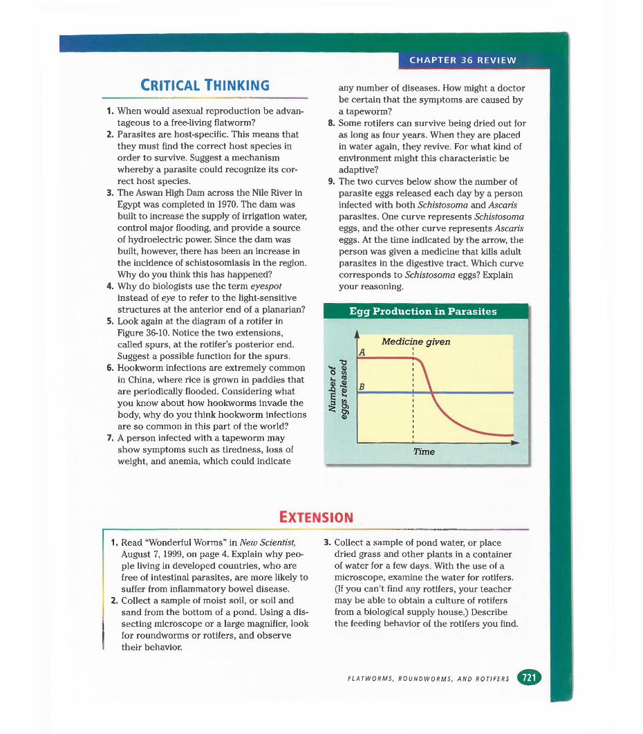
CHAPTER 36 REVIEW
Critical Thinking
1. When would asexual reproduction be advantageous to a free-living flatworm?
2. Parasites are host-specific. This means that they must find the correct host species in order to survive. Suggest a mechanism whereby a parasite could recognize its correct host species.
3. The Aswan High Dam across the Nile River in Egypt was completed in 1970. The dam was built to increase the supply of irrigation water, control major flooding, and provide a source of hydroelectric power. Since the dam was built, however, there has been an increase in the incidence of schistosomiasis in the region. Why do you think this has happened?
4. Why do biologists use the term eyespot instead of eye to refer to the light-sensitive structures at the anterior end of a planarian?
5. Look again at the diagram of a rotifer in Figure 36-10. Notice the two extensions, called spurs, at the rotifer’s posterior end. Suggest a possible function for the spurs.
6. Hookworm infections are extremely common in China, where rice is grown in paddies that are periodically flooded. Considering what you know about how hookworms invade the body, why do you think hookworm infections are so common in this part of the world?
7. A person infected with a tapeworm may show symptoms such as tiredness, loss of weight, and anemia, which could indicate
any number of diseases. How might a doctor be certain that the symptoms are caused by a tapeworm?
8. Some rotifers can survive being dried out for as long as four years. When they are placed in water again, they revive. For what kind of environment might this characteristic be adaptive?
9. The two curves below show the number of parasite eggs released each day by a person infected with both Schistosoma and Ascaris parasites. One curve represents Schistosoma eggs, and the other curve represents Ascaris eggs. At the time indicated by the arrow, the person was given a medicine that kills adult parasites in the digestive tract. Which curve corresponds to Schistosoma eggs? Explain your reasoning.
Egg Production in Parasites
Extension
1. Read “Wonderful Worms” in New Scientist, August 7, 1999, on page 4. Explain why people living in developed countries, who are free of intestinal parasites, are more likely to suffer from inflammatory bowel disease.
2. Collect a sample of moist soil, or soil and sand from the bottom of a pond. Using a dissecting microscope or a large magnifier, look for roundworms or rotifers, and observe their behavior.
3. Collect a sample of pond water, or place dried grass and other plants in a container of water for a few days. With the use of a microscope, examine the water for rotifers. (If you can’t find any rotifers, your teacher may be able to obtain a culture of rotifers from a biological supply house.) Describe the feeding behavior of the rotifers you find
FLATWORMS, ROUNDWORMS, AND ROTIFERS
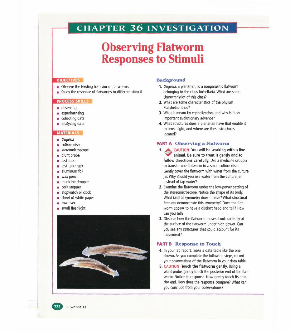
CHAPTER 36 INVESTIGATION
Observing Flatworm Responses to Stimuli
OBJECTIVES■ Observe the feeding behavior of flatworms.■ Study the response of flatworms to different stimuli.
■ observing■ experimenting■ collecting data■ analyzing data
Background1. Dugesia, a planarian, is a nonparasitic flatworm
belonging to the class Turbellaria. What are some characteristics of this class?
2. What are some characteristics of the phylum Platyhelminthes?
3. What is meant by cephalization, and why is it an important evolutionary advance?
4. What structures does a planarian have that enable it to sense light, and where are these structures located?
■ Dugesia
m culture dish■ stereomicroscope■ blunt probe■ test tube■ test-tube rack■ aluminum foil■ wax pencil■ medicine dropper■ cork stopper■ stopwatch or clock■ sheet of white paper■ raw liver■ small flashlight
PART A Observing a Flatworm1. CAUTION You will be working with a live
animal. Be sure to treat it gently and tofollow directions carefully. Use a medicine dropper to transfer one flatworm to a small culture dish.Gently cover the flatworm with water from the culture jar. Why should you use water from the culture jar instead of tap water?
2. Examine the flatworm under the low-power setting of the stereomicroscope. Notice the shape of its body. What kind of symmetry does it have? What structural features demonstrate this symmetry? Does the flat- worm appear to have a distinct head and tail? How can you tell?
3. Observe how the flatworm moves. Look carefully at the surface of the flatworm under high power. Can you see any structures that could account for its movement?
PART B Response to Touch4. In your lab report, make a data table like the one
shown. As you complete the following steps, record your observations of the flatworm in your data table.
5. CAUTION Touch the flatworm gently. Using a blunt probe, gently touch the posterior end of the flat- worm. Notice its response. Now gently touch its anterior end. How does the response compare? What can you conclude from your observations?
CHAPTER 36
PART C Feeding Behavior6. Place a tiny piece of raw liver in the culture dish.
Observe the flatworm for several minutes and describe its feeding response in your data table. Why is it important to use flatworms that have not been recently fed?
7. After several minutes, use the probe to gently turn the flatworm over. What do you observe? If you have time, watch as the flatworm eats the liver. Where do you think the undigested waste will come out?
PART D Response to Gravity8. With a wax pencil, draw a line around the middle of a
test tube.9. Fill the test tube almost full with water from the cul
ture jar. Then use a medicine dropper to transfer one flatworm to the test tube. Seal the test tube with a cork stopper.
10. Hold the test tube horizontally and move it slowly back and forth until the flatworm is centered on the line you drew.
11. To test whether the flatworm can sense gravity, place the test tube vertically in the test-tube rack. Which way should the flatworm move to show a positive response to gravity? Observe the flatworm for several minutes. Use a stopwatch to measure the amount of time the flatworm spends above the line and below the line. Record the times in your data table.
PART E Response to Light12. Check the lighting in the room. The light must be low
and even during this part of the investigation.13. Using a piece of aluminum foil, make a cover that is
big enough to fit over the bottom half of the test tube
you used in Part D. Set the cover aside. Place the test tube horizontally on a white sheet of paper. Make sure the test tube is level. Why is it important for the test tube to be level in this experiment?
14. Position the flashlight so that it will shine directly on the test tube, but do not turn it on.
15. Wait for the flatworm to move to the center line. Then gently place the foil cover over the bottom half of the test tube and turn the flashlight on. Which way should the flatworm move to show a positive response to light? Observe the movements of the flatworm. Use the stopwatch to measure the amount of time the flatworm spends in each half of the test tube. Record the times in your data table.
16. A Return the flatworm to the culture jar.^ ▼ Then clean up your materials and wash
your hands before leaving the lab.
Analysis and Conclusions1. What evidence does the flatworm show of
cephalization?2. The flatworm has an incomplete digestive system and
no circulatory system. How do you think a flatworm's food gets to the cells after it is digested?
3. How does the flatworm respond to gravity? Is the response positive or negative?
4. How does the flatworm respond to light? Is the response positive or negative?
5. Are the flatworm's anterior and posterior ends equally sensitive to light?
Further InquiryDesign an experiment to study the responses of flatwormsto other stimuli, such as vibrations or sound.
OBSERVATIONS OF FLATWORM BEHAVIORBehavior Observations
Response to touch with blunt probe
Feedingbehavior
Response to gravity
Response to light
FLATWORMS, ROUNDWORMS, AND ROTIFERS
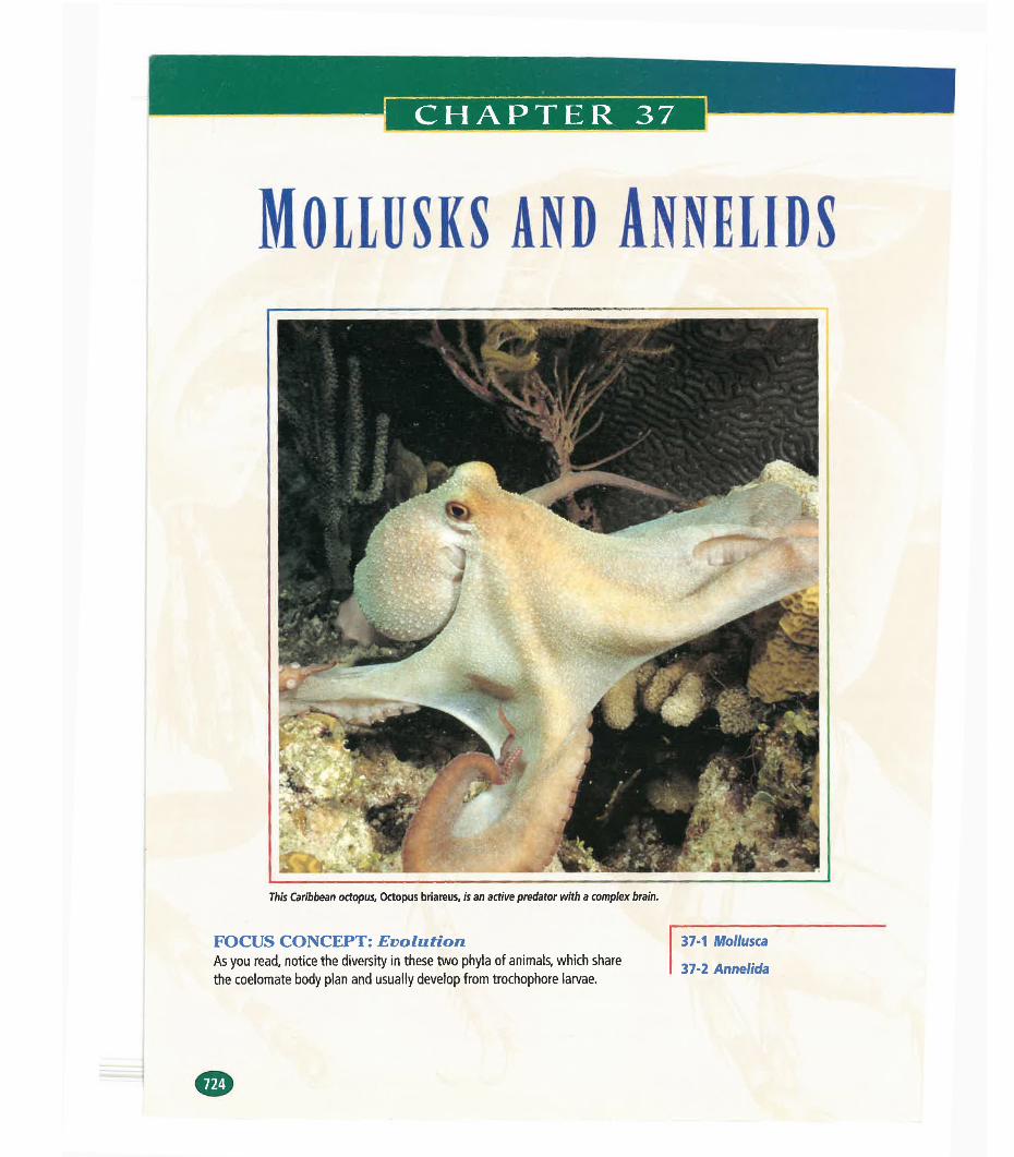
CHAPTER 37
MOLLUSKS AND ANNELIDS
This Caribbean octopus. Octopus briareus, is an active predator with a complex brain.
FOCUS CONCEPT: EvolutionAs you read, notice the diversity in these two phyla of animals, which sharethe coelomate body plan and usually develop from trochophore larvae.
37-1 Mollusca
37-2 Annelida
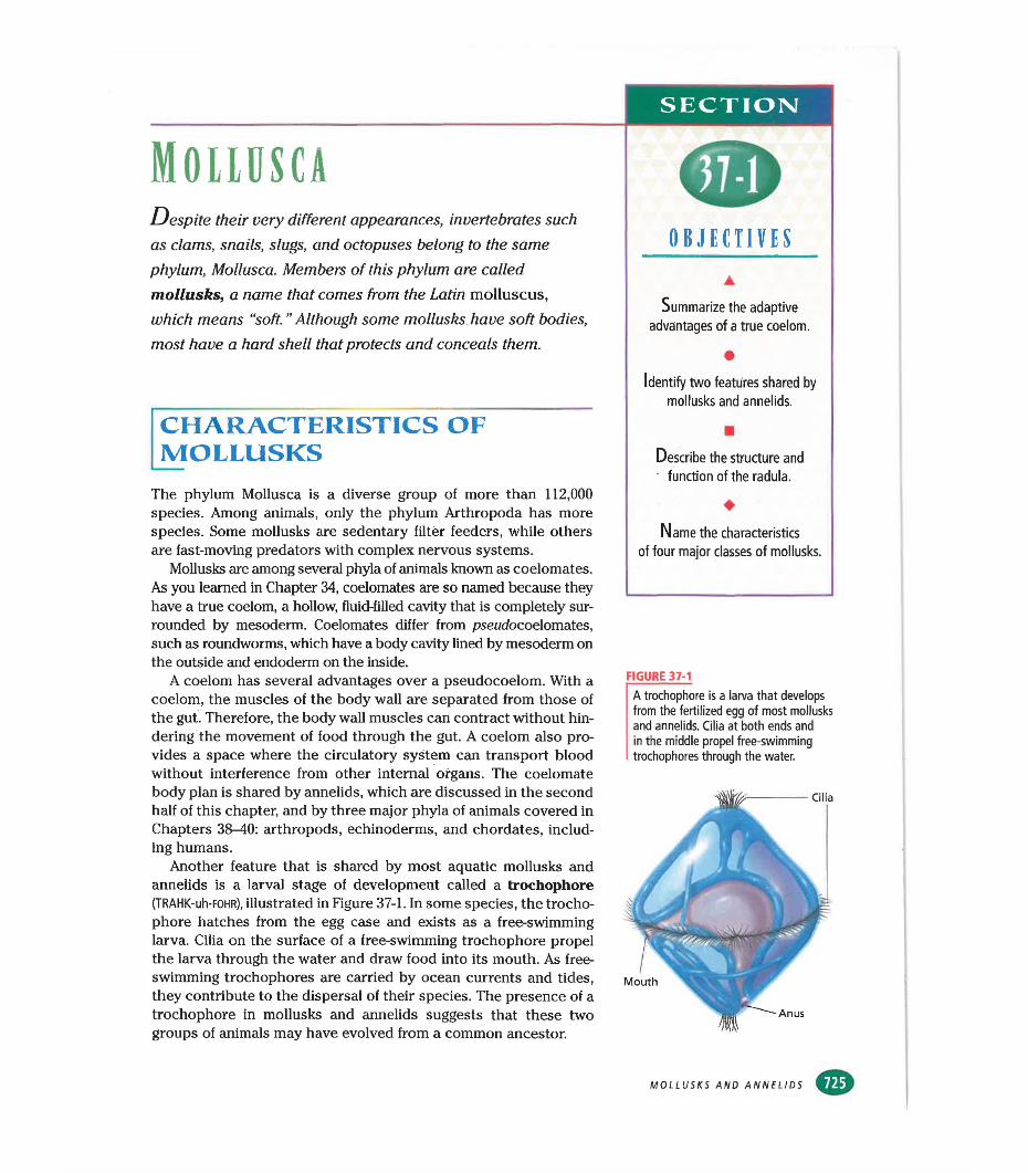
MOLLBSCADespite their very different appearances, invertebrates such
as clams, snails, slugs, and octopuses belong to the same phylum, Mollusca. Members of this phylum are called mollusks, a name that comes from the Latin molluscus, which means “soft. ’’Although some mollusks have soft bodies, most have a hard shell that protects and conceals them.
CHARACTERISTICS OF JVIOLLUSKSThe phylum Mollusca is a diverse group of more than 112,000 species. Among animals, only the phylum Arthropoda has more species. Some mollusks are sedentary filter feeders, while others are fast-moving predators with complex nervous systems.
Mollusks are among several phyla of animals known as coelomates. As you learned in Chapter 34, coelomates are so named because they have a true coelom, a hollow, fluid-filled cavity that is completely surrounded by mesoderm. Coelomates differ from pseudocoelomates, such as roundworms, which have a body cavity lined by mesoderm on the outside and endoderm on the inside.
A coelom has several advantages over a pseudocoelom. With a coelom, the muscles of the body wall are separated from those of the gut. Therefore, the body wall muscles can contract without hindering the movement of food through the gut. A coelom also provides a space where the circulatory system can transport blood without interference from other internal organs. The coelomate body plan is shared by annelids, which are discussed in the second half of this chapter, and by three major phyla of animals covered in Chapters 38-40: arthropods, echinoderms, and chordates, including humans.
Another feature that is shared by most aquatic mollusks and annelids is a larval stage of development called a trochophore (TRAHK-uh-FOHR), illustrated in Figure 37-1. In some species, the trochophore hatches from the egg case and exists as a free-swimming larva. Cilia on the surface of a free-swimming trochophore propel the larva through the water and draw food into its mouth. As free- swimming trochophores are carried by ocean currents and tides, they contribute to the dispersal of their species. The presence of a trochophore in mollusks and annelids suggests that these two groups of animals may have evolved from a common ancestor.
SECTION
OBJECTIVES
Summarize the adaptive advantages of a true coelom.
Identify two features shared by mollusks and annelids.
Describe the structure and function of the radula.
Name the characteristics of four major classes of mollusks.
FIGURE 37-1
A trochophore is a larva that develops from the fertilized egg of most mollusks and annelids. Cilia at both ends and in the middle propel free-swimming trochophores through the water.
Mouth
Anus
Cilia
MOLLUSKS AND ANNELIDS
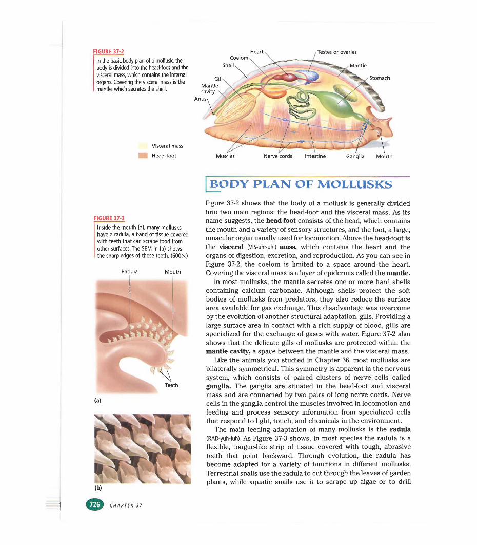
FIGURE 37-2 Heart Testes or ovariesIn the basic body plan of a mollusk, the body is divided Into the head-foot and the visceral mass, which contains the internal organs. Covering the visceral mass is the mantle, which secretes the shell.
Visceral mass
Head-foot
Coelom Shell
GillMantle cavity
Mantle
Stomach
Muscles Nerve cords Intestine Ganglia Mouth
FIGURE 37-3
Inside the mouth (a), many mollusks have a radula, a band of tissue covered with teeth that can scrape food from other surfaces. The SEM in (b) shows the sharp edges of these teeth. (600X)
Radula Mouth
(a)
BODY PLAN OF MOLLUSKSFigure 37-2 shows that the body of a mollusk is generally divided into two main regions: the head-foot and the visceral mass. As its name suggests, the head-foot consists of the head, which contains the mouth and a variety of sensory structures, and the foot, a large, muscular organ usually used for locomotion. Above the head-foot is the visceral (VIS-uhr-uhl) mass, which contains the heart and the organs of digestion, excretion, and reproduction. As you can see in Figure 37-2, the coelom is limited to a space around the heart. Covering the visceral mass is a layer of epidermis called the mantle.
In most mollusks, the mantle secretes one or more hard shells containing calcium carbonate. Although shells protect the soft bodies of mollusks from predators, they also reduce the surface area available for gas exchange. This disadvantage was overcome by the evolution of another structural adaptation, gills. Providing a large surface area in contact with a rich supply of blood, gills are specialized for the exchange of gases with water. Figure 37-2 also shows that the delicate gills of mollusks are protected within the mantle cavity, a space between the mantle and the visceral mass.
Like the animals you studied in Chapter 36, most mollusks are bilaterally symmetrical. This symmetry is apparent in the nervous system, which consists of paired clusters of nerve cells called ganglia. The ganglia are situated in the head-foot and visceral mass and are connected by two pairs of long nerve cords. Nerve cells in the ganglia control the muscles involved in locomotion and feeding and process sensory information from specialized cells that respond to light, touch, and chemicals in the environment.
The main feeding adaptation of many mollusks is the radula (RAD-yuh-luh). As Figure 37-3 shows, in most species the radula is a flexible, tongue-like strip of tissue covered with tough, abrasive teeth that point backward. Through evolution, the radula has become adapted for a variety of functions in different mollusks. Terrestrial snails use the radula to cut through the leaves of garden plants, while aquatic snails use it to scrape up algae or to drill
CHAPTER 37
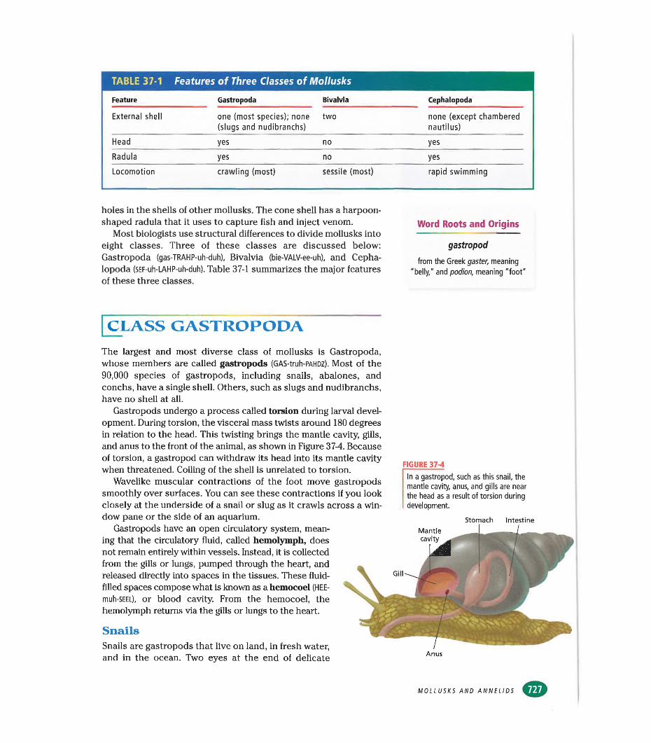
TABLE 37-1 Features of Three Classes of Mollusks
Feature Gastropoda Bivalvia Cephalopoda
External shell one (most species); none two none (except chambered(slugs and nudibranchs) nautilus)
Head yes no yes
Radula yes no yes
Locomotion crawling (most) sessile (most) rapid swimming
holes in the shells of other mollusks. The cone shell has a harpoonshaped radula that it uses to capture fish and inject venom.
Most biologists use structural differences to divide mollusks into eight classes. Three of these classes are discussed below: Gastropoda (gas-TRAHP-uh-duh), Bivalvia (bie-VALV-ee-uh), and Cephalopoda (SEF-uh-LAHP-uh-duh). Table 37-1 summarizes the major features of these three classes.
Word Roots and Origins
gastropod
from the Greek gaster, meaning "belly," and podion, meaning "foot"
[class gastropoda
The largest and most diverse class of mollusks is Gastropoda, whose members are called gastropods (GAS-truh-PAHDZ). Most of the 90,000 species of gastropods, including snails, abalones, and conchs, have a single shell. Others, such as slugs and nudibranchs, have no shell at all.
Gastropods undergo a process called torsion during larval development. During torsion, the visceral mass twists around 180 degrees in relation to the head. This twisting brings the mantle cavity, gills, and anus to the front of the animal, as shown in Figure 37-4. Because of torsion, a gastropod can withdraw its head into its mantle cavity when threatened. Coiling of the shell is unrelated to torsion.
Wavelike muscular contractions of the foot move gastropods smoothly over surfaces. You can see these contractions if you look closely at the underside of a snail or slug as it crawls across a window pane or the side of an aquarium.
Gastropods have an open circulatory system, meaning that the circulatory fluid, called hemolymph, does not remain entirely within vessels. Instead, it is collected from the gills or lungs, pumped through the heart, and released directly into spaces in the tissues. These fluid- filled spaces compose what is known as a hemocoel (HEE- muh-SEEL), or blood cavity. From the hemocoel, the hemolymph returns via the gills or lungs to the heart.
SnailsSnails are gastropods that live on land, in fresh water, and in the ocean. Two eyes at the end of delicate
FIGURE 37-4
In a gastropod, such as this snail, the mantle cavity, anus, and gills are near the head as a result of torsion during development.
Stomach IntestineMantlecavity
Anus
MOLLUSKS AND ANNELIDS
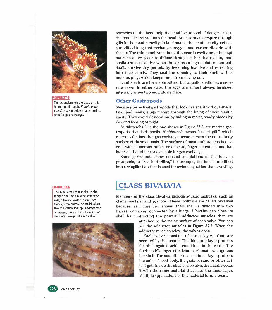
FIGURE 37-5The extensions on the back of this horned nudibranch, Hermissenda crassicornia, provide a large surface area for gas exchange.
tentacles on the head help the snail locate food. If danger arises, the tentacles retract into the head. Aquatic snails respire through gills in the mantle cavity. In land snails, the mantle cavity acts as a modified lung that exchanges oxygen and carbon dioxide with the air. The thin membrane lining the mantle cavity must be kept moist to allow gases to diffuse through it. For this reason, land snails are most active when the air has a high moisture content. Snails survive dry periods by becoming inactive and retreating into their shells. They seal the opening to their shell with a mucous plug, which keeps them from drying out.
Land snails are hermaphrodites, but aquatic snails have separate sexes. In either case, the eggs are almost always fertilized internally when two individuals mate.
Other GastropodsSlugs are terrestrial gastropods that look like snails without shells. Like land snails, slugs respire through the lining of their mantle cavity. They avoid desiccation by hiding in moist, shady places by day and feeding at night.
Nudibranchs, like the one shown in Figure 37-5, are marine gastropods that lack shells. Nudibranch means “naked gill,” which refers to the fact that gas exchange occurs across the entire body surface of these animals. The surface of most nudibranchs is covered with numerous ruffles or delicate, fingerlike extensions that increase the total area available for gas exchange.
Some gastropods show unusual adaptations of the foot. In pteropods, or “sea butterflies,” for example, the foot is modified into a winglike flap that is used for swimming rather than crawling.
FIGURE 37-6The two valves that make up the hinged shell of a bivalve can separate, allowing water to circulate through the animal. Some bivalves, like this calico scallop, Aequipecten viradians, have a row of eyes near the outer margin of each valve.
[class bivalvia
Members of the class Bivalvia include aquatic mollusks, such as clams, oysters, and scallops. These mollusks are called bivalves because, as Figure 37-6 shows, their shell is divided into two halves, or valves, connected by a hinge. A bivalve can close its shell by contracting the powerful adductor muscles that are
attached to the inside surface of each valve. You can see the adductor muscles in Figure 37-7. When the adductor muscles relax, the valves open.
Each valve consists of three layers that are secreted by the mantle. The thin outer layer protects the shell against acidic conditions in the water. The thick middle layer of calcium carbonate strengthens the shell. The smooth, iridescent inner layer protects the animal’s soft body. If a grain of sand or other irritant gets inside the shell of a bivalve, the mantle coats it with the same material that lines the inner layer. Multiple applications of this material form a pearl.
CHAPTER 37
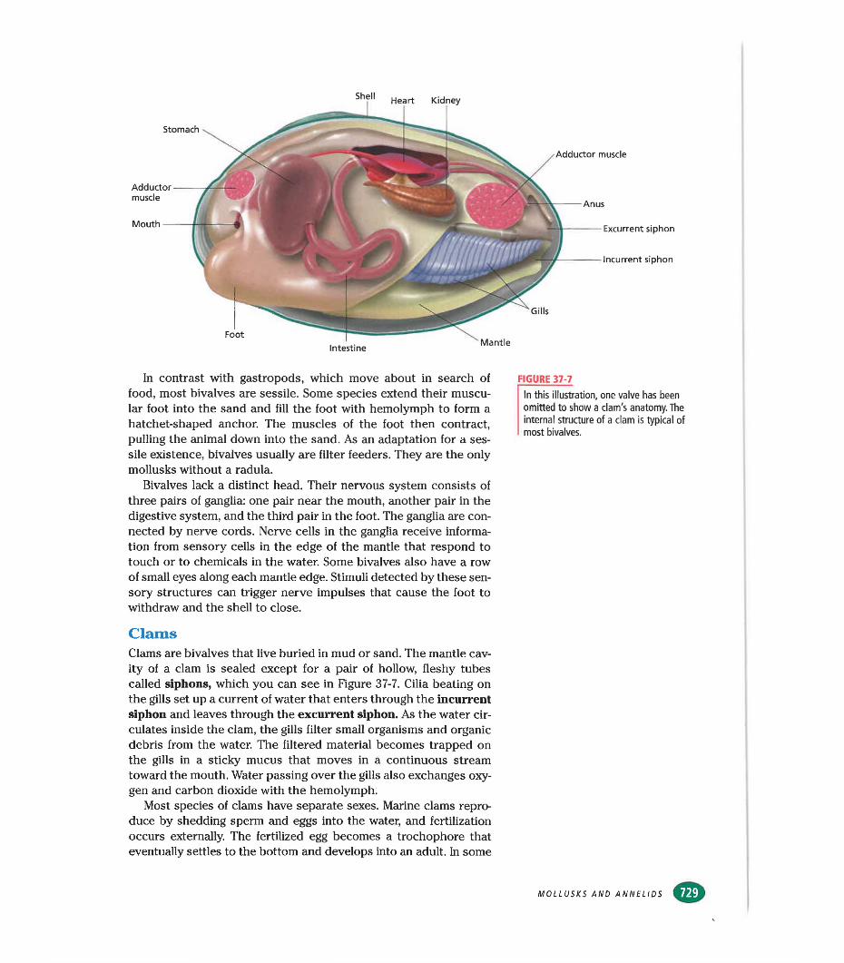
Foot
Adductormuscle
Mouth —
Kidney
Adductor muscle
Gills
Heart
Stomach
Anus
Excurrent siphon
Incurrent siphon
Intestine Mantle
In contrast with gastropods, which move about in search of food, most bivalves are sessile. Some species extend their muscular foot into the sand and fill the foot with hemolymph to form a hatchet-shaped anchor. The muscles of the foot then contract, pulling the animal down into the sand. As an adaptation for a sessile existence, bivalves usually are filter feeders. They are the only mollusks without a radula.
Bivalves lack a distinct head. Their nervous system consists of three pairs of ganglia: one pair near the mouth, another pair in the digestive system, and the third pair in the foot. The ganglia are connected by nerve cords. Nerve cells in the ganglia receive information from sensory cells in the edge of the mantle that respond to touch or to chemicals in the water. Some bivalves also have a row of small eyes along each mantle edge. Stimuli detected by these sensory structures can trigger nerve impulses that cause the foot to withdraw and the shell to close.
FIGURE 37-7
In this illustration, one valve has been omitted to show a clam's anatomy. The internal structure of a clam is typical of most bivalves.
ClamsClams are bivalves that live buried in mud or sand. The mantle cavity of a clam is sealed except for a pair of hollow, fleshy tubes called siphons, which you can see in Figure 37-7. Cilia beating on the gills set up a current of water that enters through the incurrent siphon and leaves through the excurrent siphon. As the water circulates inside the clam, the gills filter small organisms and organic debris from the water. The filtered material becomes trapped on the gills in a sticky mucus that moves in a continuous stream toward the mouth. Water passing over the gills also exchanges oxygen and carbon dioxide with the hemolymph.
Most species of clams have separate sexes. Marine clams reproduce by shedding sperm and eggs into the water, and fertilization occurs externally. The fertilized egg becomes a trochophore that eventually settles to the bottom and develops into an adult. In some
MOLLUSKS AND ANNELIDS
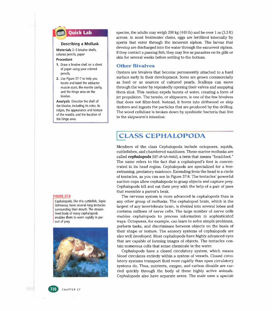
Quick Lab
Describing a Mollusk
Materials 2-3 bivalve shells, colored pencils, paper
Procedure1. Draw a bivalve shell on a sheet
of paper using your colored pencils.
2. Use Figure 37-7 to help you locate and label the adductor muscle scars, the mantle cavity, and the hinge area on the bivalve.
Analysis Describe the shell of the bivalve, including its color, its ridges, the appearance and texture of the mantle, and the location of the hinge area.
species, the adults may weigh 200 kg (440 lb) and be over 1 m (3.3 ft) across. In most freshwater clams, eggs are fertilized internally by sperm that enter through the incurrent siphon. The larvae that develop are discharged into the water through the excurrent siphon. If they contact a passing fish, they may live as parasites on its gills or skin for several weeks before settling to the bottom.
Other BivalvesOysters are bivalves that become permanently attached to a hard surface early in their development. Some are grown commercially as food or as sources of cultured pearls. Scallops can move through the water by repeatedly opening their valves and snapping them shut. This motion expels bursts of water, creating a form of jet propulsion. The teredo, or shipworm, is one of the few bivalves that does not filter-feed. Instead, it bores into driftwood or ship timbers and ingests the particles that are produced by the drilling. The wood cellulose is broken down by symbiotic bacteria that live in the shipworm’s intestine.
CLASS CEPHALOPODA
FIGURE 37-8
Cephalopods, like this cuttlefish, Sepia latimanus, have several long tentacles surrounding their mouth. The streamlined body of many cephalopods enables them to swim rapidly in pursuit of prey.
Members of the class Cephalopoda include octopuses, squids, cuttlefishes, and chambered nautiluses. These marine mollusks are called cephalopods (SEF-uh-luh-PAHDZ), a term that means “head-foot.” The name refers to the fact that a cephalopod’s foot is concentrated in its head region. Cephalopods are specialized for a free- swimming, predatory existence. Extending from the head is a circle of tentacles, as you can see in Figure 37-8. The tentacles’ powerful suction cups allow cephalopods to grasp objects and capture prey. Cephalopods kill and eat their prey with the help of a pair of jaws that resemble a parrot’s beak.
The nervous system is more advanced in cephalopods than in any other group of mollusks. The cephalopod brain, which is the largest of any invertebrate brain, is divided into several lobes and contains millions of nerve cells. The large number of nerve cells enables cephalopods to process information in sophisticated ways. Octopuses, for example, can learn to solve simple problems, perform tasks, and discriminate between objects on the basis of their shape or texture. The sensory systems of cephalopods are also well developed. Most cephalopods have highly advanced eyes that are capable of forming images of objects. The tentacles contain numerous cells that sense chemicals in the water.
Cephalopods have a closed circulatory system, which means blood circulates entirely within a system of vessels. Closed circulatory systems transport fluid more rapidly than open circulatory systems do. Thus, nutrients, oxygen, and carbon dioxide are carried quickly through the body of these highly active animals. Cephalopods also have separate sexes. The male uses a special-
CHAPTER 37
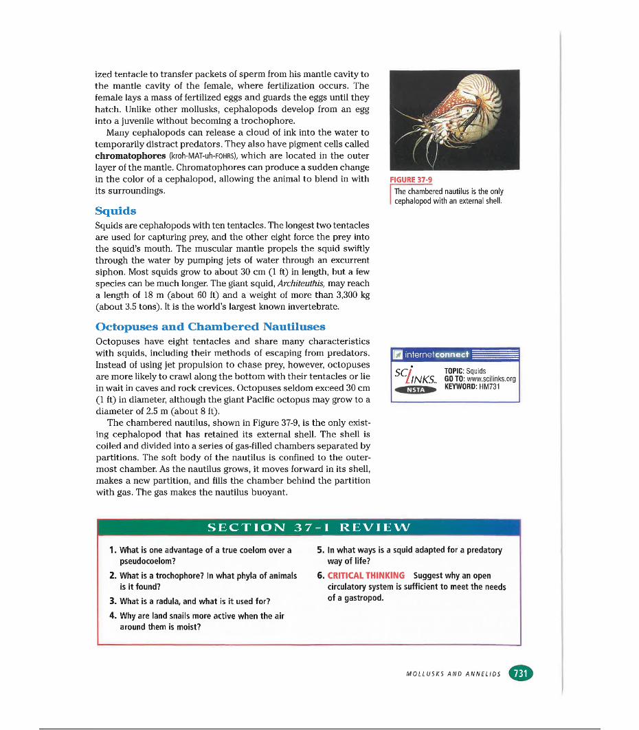
ized tentacle to transfer packets of sperm from his mantle cavity to the mantle cavity of the female, where fertilization occurs. The female lays a mass of fertilized eggs and guards the eggs until they hatch. Unlike other mollusks, cephalopods develop from an egg into a juvenile without becoming a trochophore.
Many cephalopods can release a cloud of ink into the water to temporarily distract predators. They also have pigment cells called chromatophores (kroh-MAT-uh-FOHRS), which are located in the outer layer of the mantle. Chromatophores can produce a sudden change in the color of a cephalopod, allowing the animal to blend in with its surroundings.
SquidsSquids are cephalopods with ten tentacles. The longest two tentacles are used for capturing prey, and the other eight force the prey into the squid’s mouth. The muscular mantle propels the squid swiftly through the water by pumping jets of water through an excurrent siphon. Most squids grow to about 30 cm (1 ft) in length, but a few species can be much longer. The giant squid, Architeuthis, may reach a length of 18 m (about 60 ft) and a weight of more than 3,300 kg (about 3.5 tons). It is the world’s largest known invertebrate.
FIGURE 37-9
The chambered nautilus is the only cephalopod with an external shell.
Octopuses and Chambered NautilusesOctopuses have eight tentacles and share many characteristics with squids, including their methods of escaping from predators. Instead of using jet propulsion to chase prey, however, octopuses are more likely to crawl along the bottom with their tentacles or lie in wait in caves and rock crevices. Octopuses seldom exceed 30 cm (1 ft) in diameter, although the giant Pacific octopus may grow to a diameter of 2.5 m (about 8 ft).
The chambered nautilus, shown in Figure 37-9, is the only existing cephalopod that has retained its external shell. The shell is coiled and divided into a series of gas-filled chambers separated by partitions. The soft body of the nautilus is confined to the outermost chamber. As the nautilus grows, it moves forward in its shell, makes a new partition, and fills the chamber behind the partition with gas. The gas makes the nautilus buoyant.
internetconnect
SCllNKS, TOPIC: Squids GOTO: www.sclllnks.orgKEYWORD: HM731
SECTION 37-1 REVIEW
1. What is one advantage of a true coelom over a pseudocoelom?
2. What is a trochophore? In what phyla of animals is it found?
3. What is a radula, and what is it used for?4. Why are land snails more active when the air
around them is moist?
5. In what ways is a squid adapted for a predatory way of life?
6. CRITICAL THINKING Suggest why an open circulatory system is sufficient to meet the needs of a gastropod.
MOLLUSKS AND ANNELIDS
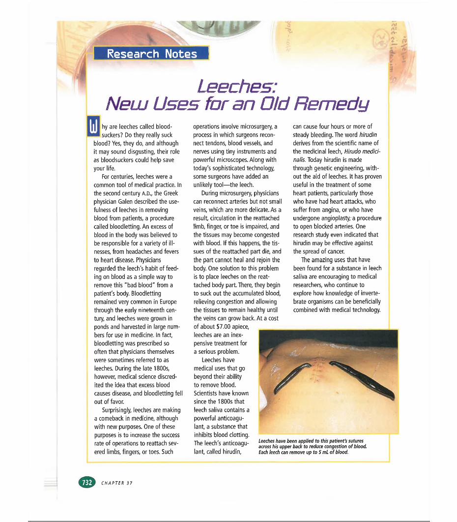
M'li uprResearch Notes
Neuu Uses for an Old Remedyhy are leeches called bloodsuckers? Do they really suck
blood? Yes, they do, and although it may sound disgusting, their role as bloodsuckers could help save your life.
For centuries, leeches were a common tool of medical practice. In the second century A.D., the Greek physician Galen described the usefulness of leeches in removing blood from patients, a procedure called bloodletting. An excess of blood in the body was believed to be responsible for a variety of illnesses, from headaches and fevers to heart disease. Physicians regarded the leech's habit of feeding on blood as a simple way to remove this "bad blood" from a patient's body. Bloodletting remained very common in Europe through the early nineteenth century, and leeches were grown in ponds and harvested in large numbers for use in medicine. In fact, bloodletting was prescribed so often that physicians themselves were sometimes referred to as leeches. During the late 1800s, however, medical science discredited the idea that excess blood causes disease, and bloodletting fell out of favor.
Surprisingly, leeches are making a comeback in medicine, although with new purposes. One of these purposes is to increase the success rate of operations to reattach severed limbs, fingers, or toes. Such
operations involve microsurgery, a process in which surgeons reconnect tendons, blood vessels, and nerves using tiny instruments and powerful microscopes. Along with today’s sophisticated technology, some surgeons have added an unlikely tool—the leech.
During microsurgery, physicians can reconnect arteries but not small veins, which are more delicate. As a result, circulation in the reattached limb, finger, or toe is impaired, and the tissues may become congested with blood. If this happens, the tissues of the reattached part die, and the part cannot heal and rejoin the body. One solution to this problem is to place leeches on the reattached body part. There, they begin to suck out the accumulated blood, relieving congestion and allowing the tissues to remain healthy until the veins can grow back. At a cost of about $7.00 apiece, leeches are an inexpensive treatment for a serious problem.
Leeches have medical uses that go beyond their ability to remove blood.Scientists have known since the 1800s that leech saliva contains a powerful anticoagulant, a substance that inhibits blood clotting.The leech's anticoagulant, called hirudin,
can cause four hours or more of steady bleeding. The word hirudin derives from the scientific name of the medicinal leech, Hirudo medici- nalis. Today hirudin is made through genetic engineering, without the aid of leeches. It has proven useful in the treatment of some heart patients, particularly those who have had heart attacks, who suffer from angina, or who have undergone angioplasty, a procedure to open blocked arteries. One research study even indicated that hirudin may be effective against the spread of cancer.
The amazing uses that have been found for a substance in leech saliva are encouraging to medical researchers, who continue to explore how knowledge of invertebrate organisms can be beneficially combined with medical technology.
Leeches have been applied to this patient's sutures across his upper back to reduce congestion of blood. Each leech can remove up to 5 mL of blood.
CHAPTER 37
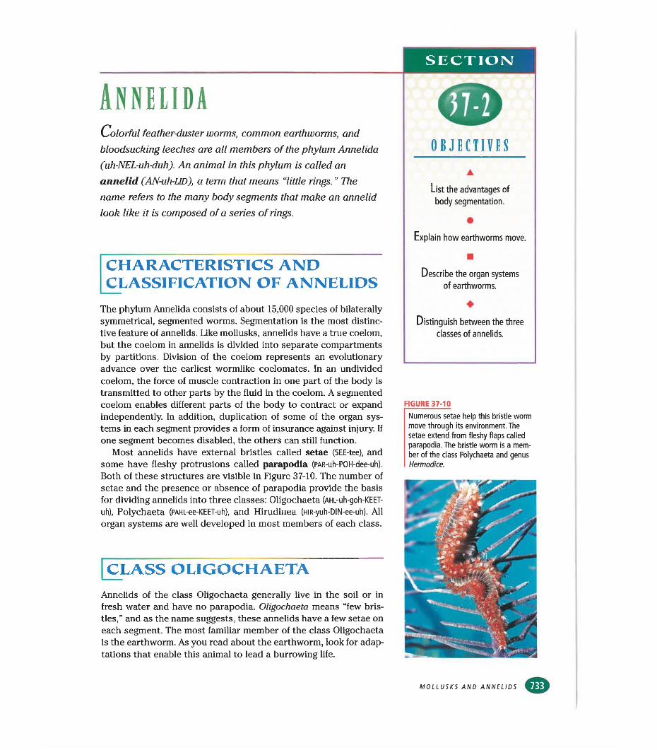
SECTION
AnnelidaColorful feather-duster worms, common earthworms, and
bloodsucking leeches are all members of the phylum Annelida (uh-NEL-uh-duh). An animal in this phylum is called an annelid (AN-uh-LID), a term that means “little rings. ” The name refers to the many body segments that make an annelid look like it is composed of a series of rings.
CHARACTERISTICS AND CLASSIFICATION OF ANNELIDSThe phylum Annelida consists of about 15,000 species of bilaterally symmetrical, segmented worms. Segmentation is the most distinctive feature of annelids. Like mollusks, annelids have a true coelom, but the coelom in annelids is divided into separate compartments by partitions. Division of the coelom represents an evolutionary advance over the earliest wormlike coelomates. In an undivided coelom, the force of muscle contraction in one part of the body is transmitted to other parts by the fluid in the coelom. A segmented coelom enables different parts of the body to contract or expand independently. In addition, duplication of some of the organ systems in each segment provides a form of insurance against injury. If one segment becomes disabled, the others cam still function.
Most annelids have external bristles called setae (SEE-tee), and some have fleshy protrusions called parapodia (PAR-uh-POH-dee-uh).
Both of these structures are visible in Figure 37-10. The number of setae and the presence or absence of parapodia provide the basis for dividing annelids into three classes: Oligochaeta (AHL-uh-goh-KEET-
uh), Polychaeta (PAHL-ee-KEET-uh), and Hirudinea (HlR-yuh-DIN-ee-uh). All organ systems are well developed in most members of each class.
[class oligochaeta
Annelids of the class Oligochaeta generally live in the soil or in fresh water and have no parapodia. Oligochaeta means “few bristles,” and as the name suggests, these annelids have a few setae on each segment. The most familiar member of the class Oligochaeta is the earthworm. As you read about the earthworm, look for adaptations that enable this animal to lead a burrowing life.
OBJECTIVESA
List the advantages of
body segmentation.
•Explain how earthworms move.
■Describe the organ systems
of earthworms.
♦Distinguish between the three
classes of annelids.
FIGURE 37-10
Numerous setae help this bristle worm move through its environment. The setae extend from fleshy flaps called parapodia. The bristle worm is a member of the class Polychaeta and genus Hermodice.
MOLLUSKS AND ANNELIDS €>
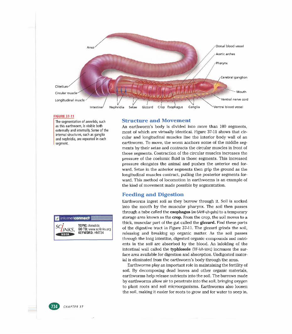
Anus Dorsal blood vessel
Aortic arches
Pharynx
Clltel
Circular muscle
Longitudinal muscle
Cerebral ganglion
Mouth
Ventral nerve cord
Ventral blood vessel
FIGURE 37-11
The segmentation of annelids, such as this earthworm, is visible both externally and internally. Some of the internal structures, such as ganglia and nephridia, are repeated in each segment.
Structure and MovementAn earthworm’s body is divided into more than 100 segments, most of which are virtually identical. Figure 37-11 shows that circular and longitudinal muscles line the interior body wall of an earthworm. To move, the worm anchors some of the middle segments by their setae and contracts the circular muscles in front of those segments. Contraction of the circular muscles increases the pressure of the coelomic fluid in those segments. This increased pressure elongates the animal and pushes the anterior end forward. Setae in the anterior segments then grip the ground as the longitudinal muscles contract, pulling the posterior segments forward. This method of locomotion in earthworms is an example of the kind of movement made possible by segmentation.
inteinetconnect
SCllNKS, TOPIC: Annelids GO TO: www.scilinks.org KEYWORD: HM734
Feeding and DigestionEarthworms ingest soil as they burrow through it. Soil is sucked into the mouth by the muscular pharynx. The soil then passes through a tube called the esophagus (ee-SAHF-uh-guhs) to a temporary storage area known as the crop. From the crop, the soil moves to a thick, muscular part of the gut called the gizzard. Find these parts of the digestive tract in Figure 37-11. The gizzard grinds the soil, releasing and breaking up organic matter. As the soil passes through the long intestine, digested organic compounds and nutrients in the soil are absorbed by the blood. An infolding of the intestinal wall called the typhlosole (TIF-Iuh-SOHL) increases the surface area available for digestion and absorption. Undigested material is eliminated from the earthworm’s body through the anus.
Earthworms play an important role in maintaining the fertility of soil. By decomposing dead leaves and other organic materials, earthworms help release nutrients into the soil. The burrows made by earthworms allow air to penetrate into the soil, bringing oxygen to plant roots and soil microorganisms. Earthworms also loosen the soil, making it easier for roots to grow and for water to seep in.
CHAPTER 37
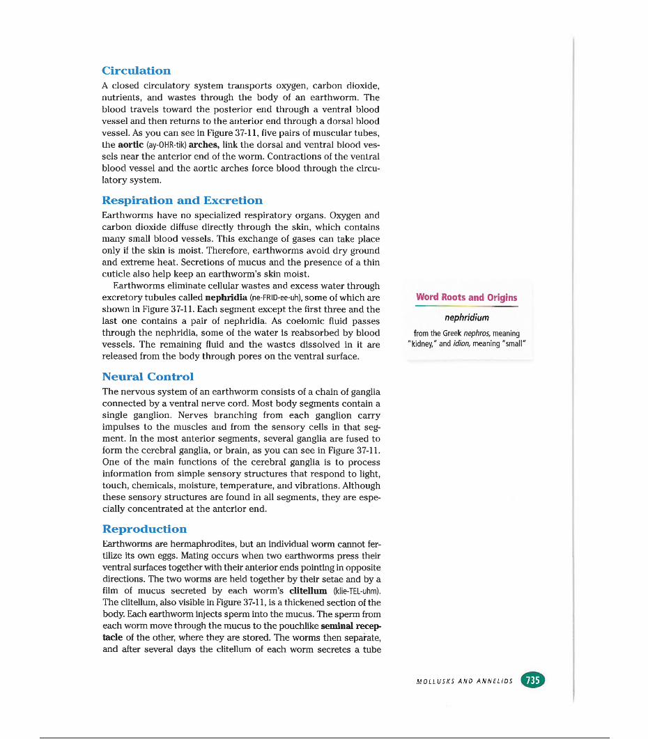
CirculationA closed circulatory system transports oxygen, carbon dioxide, nutrients, and wastes through the body of an earthworm. The blood travels toward the posterior end through a ventral blood vessel and then returns to the anterior end through a dorsal blood vessel. As you can see in Figure 37-11, five pairs of muscular tubes, the aortic (ay-OHR-tik) arches, link the dorsal and ventral blood vessels near the anterior end of the worm. Contractions of the ventral blood vessel and the aortic arches force blood through the circulatory system.
Respiration and ExcretionEarthworms have no specialized respiratory organs. Oxygen and carbon dioxide diffuse directly through the skin, which contains many small blood vessels. This exchange of gases can take place only if the skin is moist. Therefore, earthworms avoid dry ground and extreme heat. Secretions of mucus and the presence of a thin cuticle also help keep an earthworm’s skin moist.
Earthworms eliminate cellular wastes and excess water through excretory tubules called nephridia (ne-FRID-ee-uh), some of which are shown in Figure 37-11. Each segment except the first three and the last one contains a pair of nephridia. As coelomic fluid passes through the nephridia, some of the water is reabsorbed by blood vessels. The remaining fluid and the wastes dissolved in it are released from the body through pores on the ventral surface.
Neural ControlThe nervous system of an earthworm consists of a chain of ganglia connected by a ventral nerve cord. Most body segments contain a single ganglion. Nerves branching from each ganglion carry impulses to the muscles and from the sensory cells in that segment. In the most anterior segments, several ganglia are fused to form the cerebral ganglia, or brain, as you can see in Figure 37-11. One of the main functions of the cerebral ganglia is to process information from simple sensory structures that respond to light, touch, chemicals, moisture, temperature, and vibrations. Although these sensory structures are found in all segments, they are especially concentrated at the anterior end.
ReproductionEarthworms are hermaphrodites, but an individual worm cannot fertilize its own eggs. Mating occurs when two earthworms press their ventral surfaces together with their anterior ends pointing in opposite directions. The two worms are held together by their setae and by a film of mucus secreted by each worm’s clitellum (klie-TEL-uhm).
The clitellum, also visible in Figure 37-11, is a thickened section of the body. Each earthworm injects sperm into the mucus. The sperm from each worm move through the mucus to the pouchlike seminal receptacle of the other, where they are stored. The worms then separate, and after several days the clitellum of each worm secretes a tube
Word Roots and Origins
nephridiumfrom the Greek nephros, meaning
"kidney," and idion, meaning "small'
MOLLUSKS AND ANNELIDS
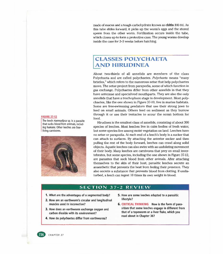
made of mucus and a tough carbohydrate known as chitin (KIE-tin). As this tube slides forward, it picks up the worm’s eggs and the stored sperm from the other worm. Fertilization occurs inside the tube, which closes up to form a protective case. The young worms develop inside the case for 2-3 weeks before hatching.
CLASSES POLYCHAETA AND HIRUDINEA
FIGURE 37-12
The leech Haemadipsa sp. is a parasite that sucks blood from animals, including humans. Other leeches are free- living carnivores.
About two-thirds of all annelids are members of the class Polychaeta and are called polychaetes. Polychaeta means “many bristles,” which refers to the numerous setae that help polychaetes move. The setae project from parapodia, some of which function in gas exchange. Polychaetes differ from other annelids in that they have antennae and specialized mouthparts. They are also the only annelids that have a trochophore stage in development. Most polychaetes, like the one shown in Figure 37-10, live in marine habitats. Some are free-swimming predators that use their strong jaws to feed on small animals. Others feed on sediment as they burrow through it or use their tentacles to scour the ocean bottom for food.
Hirudinea is the smallest class of annelids, consisting of about 300 species of leeches. Most leeches live in calm bodies of fresh water, but some species live among moist vegetation on land. Leeches have no setae or parapodia. At each end of a leech’s body is a sucker that can attach to surfaces. By attaching the anterior sucker and then pulling the rest of the body forward, leeches can crawl along solid objects. Aquatic leeches can also swim with an undulating movement of their body. Many leeches are carnivores that prey on small invertebrates, but some species, including the one shown in Figure 37-12, are parasites that suck blood from other animals. After attaching themselves to the skin of their host, parasitic leeches secrete an anaesthetic that prevents the host from feeling their presence. They also secrete a substance that prevents blood from clotting. If undisturbed, a leech can ingest 10 times its own weight in blood.
SECTION 37-2 REVIEW
1. What are the advantages of a segmented body?2. How are an earthworm's circular and longitudinal
muscles used in locomotion?3. How does an earthworm exchange oxygen and
carbon dioxide with its environment?
4. How do polychaetes differ from earthworms?
5. How are some leeches adapted to a parasitic lifestyle?
6. CRITICAL THINKING How is the form of parasitism that some leeches engage in different from that of a tapeworm or a liver fluke, which you read about in Chapter 36?
CHAPTER 37
CHAPTER 37 REVIEW
S U M M ARY/VOCABU LARY
Mollusks have a true coelom and usually develop from a pear-shaped larva called a trochophore. Their body is divided into the head-foot and the visceral mass, which contains the internal organs.Most mollusks have at least one shell, which is secreted by a layer of epidermis called the mantle. Aquatic mollusks have gills through which they exchange gases with water.The main feeding adaptation of most mollusks is the radula, a tongue-like structure that is modified in different species for scraping, drilling, or harpooning. Gastropods undergo a process called torsion, in which the visceral mass twists during larval development. Snails and most other gastropods have a single shell, while some gastropods, such as slugs and nudibranchs, lack shells. Gastropods move by means ofVocabularyadductor muscle (728) bivalve (728) cephalopod (730) chromatophore (731) excurrent siphon (729)
ganglion (726) gastropod (727) head-foot (726) hemocoel (727) hemolymph (727)
wavelike, muscular contractions of the foot and have an open circulatory system.
■ Bivalves have a shell that is divided into two valves, which they can pull together by contracting powerful adductor muscles. Bivalves lack a distinct head and have no radula. Most are sessile and filter food from the water. In clams, water enters through an incurrent siphon and exits through an excurrent siphon. Food is strained from the water as it passes through the gills.
■ Cephalopods, including octopuses and squids, are free-swimming, predatory mollusks with numerous tentacles. They have an advanced nervous system with a large brain and well-developed sensory organs. Cephalopods have a closed circulatory system and do not pass through a trochophore stage during development.
incurrent siphon (729) mantle (726) mantle cavity (726) mollusk (725) radula (726)
siphon (729) torsion (727) trochophore (725) visceral mass (726)
Annelids have a true coelom and a body that is divided into many segments. Most annelids have external bristles called setae, and some have fleshy protrusions called parapodia.Members of the class Oligochaeta generally live in the soil or in fresh water. They have no parapodia and relatively few setae.The most familiar member of the class Oligochaeta is the earthworm, which feeds on organic matter as it burrows through the soil. Earthworms have a closed circulatory system. They exchange gases through their skin and eliminate cellular wastes andVocabularyannelid (733) aortic arch (735) chitin (736)
ditellum (735) crop (734) esophagus (734)
excess water through excretory tubules called nephridia.
■ Polychaetes have numerous setae that project from parapodia. They also have antennae and specialized mouthparts, and they pass through a trochophore stage during their development. Most polychaetes live in the ocean.
■ Members of the class Hirudinea—leeches— live in fresh water or on land. They have no setae or parapodia. Many leeches are carnivores that prey on small invertebrates, but some are bloodsucking parasites.
gizzard (734) nephridium (735) parapodium (733)
seminal receptacle (735) seta (733) typhlosole (734)
MOLLUSKS AND ANNELIDS
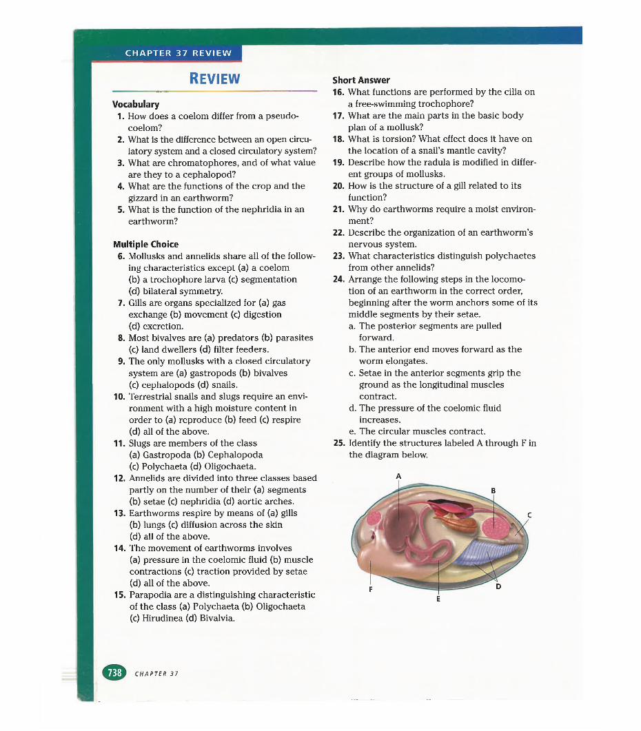
CHAPTER 37 REVIEW
REVIEW
Vocabulary1. How does a coelom differ from a pseudo
coelom?2. What is the difference between an open circu
latory system and a closed circulatory system?3. What are chromatophores, and of what value
are they to a cephalopod?4. What are the functions of the crop and the
gizzard in an earthworm?5. What is the function of the nephridia in an
earthworm?
Multiple Choice6. Mollusks and annelids share all of the follow
ing characteristics except (a) a coelom(b) a trochophore larva (c) segmentation(d) bilateral symmetry.
7. Gills are organs specialized for (a) gas exchange (b) movement (c) digestion (d) excretion.
8. Most bivalves are (a) predators (b) parasites(c) land dwellers (d) filter feeders.
9. The only mollusks with a closed circulatory system are (a) gastropods (b) bivalves(c) cephalopods (d) snails.
10. Terrestrial snails and slugs require an environment with a high moisture content in order to (a) reproduce (b) feed (c) respire(d) all of the above.
11. Slugs are members of the class(a) Gastropoda (b) Cephalopoda(c) Polychaeta (d) Oligochaeta.
12. Annelids are divided into three classes based partly on the number of their (a) segments(b) setae (c) nephridia (d) aortic arches.
13. Earthworms respire by means of (a) gills(b) lungs (c) diffusion across the skin(d) all of the above.
14. The movement of earthworms involves(a) pressure in the coelomic fluid (b) muscle contractions (c) traction provided by setae (d) all of the above.
15. Parapodia are a distinguishing characteristic of the class (a) Polychaeta (b) Oligochaeta(c) Hirudinea (d) Bivalvia.
CHAPTER 37
Short Answer16. What functions are performed by the cilia on
a free-swimming trochophore?17. What are the main parts in the basic body
plan of a mollusk?18. What is torsion? What effect does it have on
the location of a snail’s mantle cavity?19. Describe how the radula is modified in differ
ent groups of mollusks.20. How is the structure of a gill related to its
function?21. Why do earthworms require a moist environ
ment?22. Describe the organization of an earthworm’s
nervous system.23. What characteristics distinguish polychaetes
from other annelids?24. Arrange the following steps in the locomo
tion of an earthworm in the correct order, beginning after the worm anchors some of its middle segments by their setae.a. The posterior segments are pulled
forward.b. The anterior end moves forward as the
worm elongates.c. Setae in the anterior segments grip the
ground as the longitudinal muscles contract.
d. The pressure of the coelomic fluid increases.
e. The circular muscles contract.25. Identify the structures labeled A through F in
the diagram below.
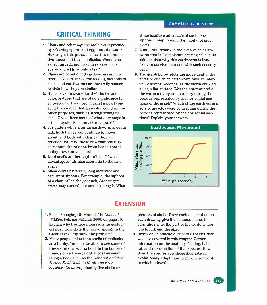
CHAPTER 37 REVIEW
Critical thinking
1. Clams and other aquatic mollusks reproduce by releasing sperm and eggs into the water. How might this process affect the reproductive success of these mollusks? Would you expect aquatic mollusks to release many sperm and eggs or only a few?
2. Clams are aquatic and earthworms are terrestrial. Nevertheless, the feeding methods of clams and earthworms are basically similar. Explain how they are similar.
3. Humans value pearls for their luster and color, features that are of no significance to an oyster. Furthermore, making a pearl consumes resources that an oyster could use for other purposes, such as strengthening its shell. Given these facts, of what advantage is it to an oyster to manufacture a pearl?
4. For quite a while after an earthworm is cut in half, both halves will continue to move about, and both will retract if they are touched. What do these observations suggest about the role the brain has in coordinating these movements?
5. Land snails are hermaphrodites. Of what advantage is this characteristic to the land snail?
6. Many clams have very long incurrent and excurrent siphons. For example, the siphons of a clam called the geoduck, Panope gen- erosa, may exceed one meter in length. What
is the adaptive advantage of such long siphons? Keep in mind the habitat of most clams.
7. A mutation results in the birth of an earthworm that lacks moisture-sensing cells in its skin. Explain why this earthworm is less likely to survive than one with such sensory cells.
8. The graph below plots the movement of the anterior end of an earthworm over an interval of several seconds, as the worm crawled along a flat surface. Was the anterior end of the worm moving or stationary during the periods represented by the horizontal sections of the graph? Which of the earthworm’s sets of muscles were contracting during the periods represented by the horizontal sections? Explain your answers.
Earthworm Movement
Time (in seconds)
Extension
1. Read “Sponging Off Mussels” in National Wildlife, February/March 2000, on page 10. Explain why the zebra mussel is an ecological pest. How does the native sponge in the Great Lakes help solve the problem?
2. Many people collect the shells of mollusks as a hobby. You may be able to see some of these shells in your school, in the homes of friends or relatives, or at a local museum. Using a book such as the National Audubon Society Field Guide to North American Seashore Creatures, identify five shells or
pictures of shells. Draw each one, and under each drawing give the common name, the scientific name, the part of the world where it is found, and the size.
3. Research an annelid or mollusk species that was not covered in this chapter. Gather information on the anatomy, feeding, habitat, and reproduction of that species. How does the species you chose illustrate an evolutionary adaptation to the environment in which it lives?
MOLLUSKS AND ANNELIDS
CHAPTER 37 INVESTIGATION
Observing Earthworm BehaviorOBJECTIVES
■ Observe a live earthworm.■ Test how an earthworm responds to light, moisture, and
ammonia.■ Test the effect of temperature on heart rate.
PROCESS SKILLS■ observing■ hypothesizing■ experimenting■ collecting data■ analyzing data
MMAnAh\*mm safety goggles■ live earthworm■ shallow pan■ paper towels■ medicine dropper■ hand lens■ black paper or piece of cardboard■ fluorescent lamp■ 2 cotton swabs■ 3% aqueous ammonia solution■ 15 cm Petri dish■ thermometer■ stopwatch or clock with second hand■ 2 plastic tubs for water baths■ warm tap water■ icecubes
Background1. How does an earthworm benefit from cephalization?2. Describe how gases enter and exit an earthworm's
body.
PART A Observing an Earthworm1. /K CAUTION You will be working with a live
animal. Be sure to treat it gently and to follow directions carefully. Place a moist paper towel in a pan, and place an earthworm on the paper towel. CAUTION Rinse the earthworm frequently with
water from a medicine dropper to prevent the worm from drying out and becoming lethargic.
2. Observe the behavior of the earthworm for a few minutes. Identify the earthworm's anterior and posterior ends by watching it move in the pan. As the worm crawls around in the pan, it will lead with its anterior end.
3. Locate the earthworm's ditellum. Is the ditellum closer to the anterior end or the posterior end? What is the function of the ditellum?
4. Identify the earthworm's dorsal and ventral surfaces by gently rolling the worm over. The dorsal surface will be on top after the worm rights itself.
5. Pick up the earthworm and feel its skin with your fingers. One surface of the earthworm should feel slightly rougher than the other. The roughness is due to the hairlike setae that project from the earthworm's skin. On which surface are the setae located? Use a hand lens to examine the setae up close.
6. Return the earthworm to the pan, and use the hand lens to find a thick purple line running along the dorsal surface of the worm. This line is the dorsal blood vessel. Does the earthworm have an open or a closed circulatory system?
7. Draw a picture of the earthworm, and label its anterior and posterior ends, dorsal and ventral surfaces, ditellum, setae, and dorsal blood vessel.
PART B Earthworm Responses to Stimuli
8. In this part of the laboratory investigation, you will test the earthworm's responses to three different stimuli. With your lab partners, develop three separate hypotheses that describe an earthworm's responses to light, moisture, and a base. In your lab report, make a data table like the one on the next page.
9. To test the earthworm's response to light, cover half of the pan with black paper or cardboard. Check the lighting in the room.The light must be low and even during this test. Position the fluorescent lamp over the uncovered portion of the pan. Place the earthworm in the center of the pan and observe its movements. Record your observations in your data table.
CHAPTER 37
OBSERVATIONS OF EARTHWORM BEHAVIOR
Behavior Observations
Response to light
Response to moisture
Response to water on a swab
Response to ammonia
10. To test the earthworm's response to moisture, turn off the fluorescent lamp, move it away from the pan, and remove the paper covering half of the pan. Place a piece of dry paper towel on one side of the pan and a piece of wet paper towel on the other side of the pan. Lay the earthworm across the two paper towels. Observe the earthworm's response to the two environments, and record your observations in your data table.
11. To test the earthworm's response to ammonia, make sure the paper towels on both sides of the pan are wet. Moisten a cotton swab with water. Hold the cotton swab first near the earthworm's anterior end and then near its posterior end. Do not touch the earthworm with the swab. Record your observations in your data table.
CAUTION Wear safety goggles at all times ^ during the following procedure. If you get
ammonia on your skin or clothing, wash it off at the sink while calling to your teacher. If you get ammonia in your eyes, immediately flush it out at the eyewash station while calling to your teacher. Moisten a different cotton swab with ammonia solution and repeat step 11. Do not touch the earthworm with the swab or the ammonia solution. Record your observations in your data table.
12.
PART C Effect of Temperature on Heart Rate
13. In this part of the laboratory investigation, you will examine how an earthworm's heart rate changes as its body temperature changes. Add enough tap water to a Petri dish to barely cover the bottom of the dish. Place an earthworm in the dish.
14. Using a hand lens, look for rhythmic contractions of the dorsal blood vessel. Each contraction represents a single heartbeat. Calculate the worm's heart rate by counting the number of contractions that occur in exactly one minute. This is easiest to do if one person counts contractions while another person watches a stopwatch or clock.
15. Place a thermometer next to the worm in the petri dish and measure the temperature. Record the worm's heart rate and the temperature in a table on the chalkboard.
16. Float the Petri dish containing the worm on top of either a warm-water bath or a cold-water bath. Place the thermometer next to the worm in the Petri dish, and watch the temperature until it reaches either 30°C (for the warm-water bath) or 10°C (for the cold-water bath).
17. Remove the Petri dish from the water bath and immediately begin counting heartbeats for exactly one minute. After one minute, measure the temperature in the dish again. Calculate the average temperature to the nearest degree. Record the worm's heart rate and the average temperature in the table on the chalkboard.
18. Using data from the whole class, graph heart rate as a function of temperature. Draw the best-fit curve through the points.
19. A Return the earthworm to the container▼ ‘%• from which you obtained it. Clean up your
materials and wash your hands before leaving the lab.
Analysis and Conclusions1. State whether your hypotheses in Part B were sup
ported by your observations. Explain.2. What is the adaptive advantage of the earthworm's
responses to light and moisture?3. List variables that, if not controlled, might have
affected the results in Part B.4. Describe the relationship between the earthworm’s
heart rate and temperature as shown by your graph.
Further InquiryDesign an experiment to determine which colors of lightan earthworm is sensitive to or which areas on an earthworm are sensitive to light.
MOLLUSKS AND ANNELIDS
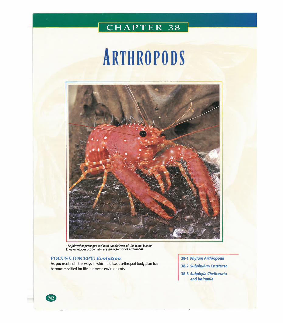
Arthropods
The jointed appendages and hard exoskeleton of this flame lobster,Enoplometopus occidentalis, are characteristic of arthropods.
38-1 Phylum Arthropoda
38-2 Subphylum Crustacea
38-3 Subphyla Chelicerata and Uniramia
FOCUS CONCEPT: EvolutionAs you read, note the ways in which the basic arthropod body plan has become modified for life in diverse environments.
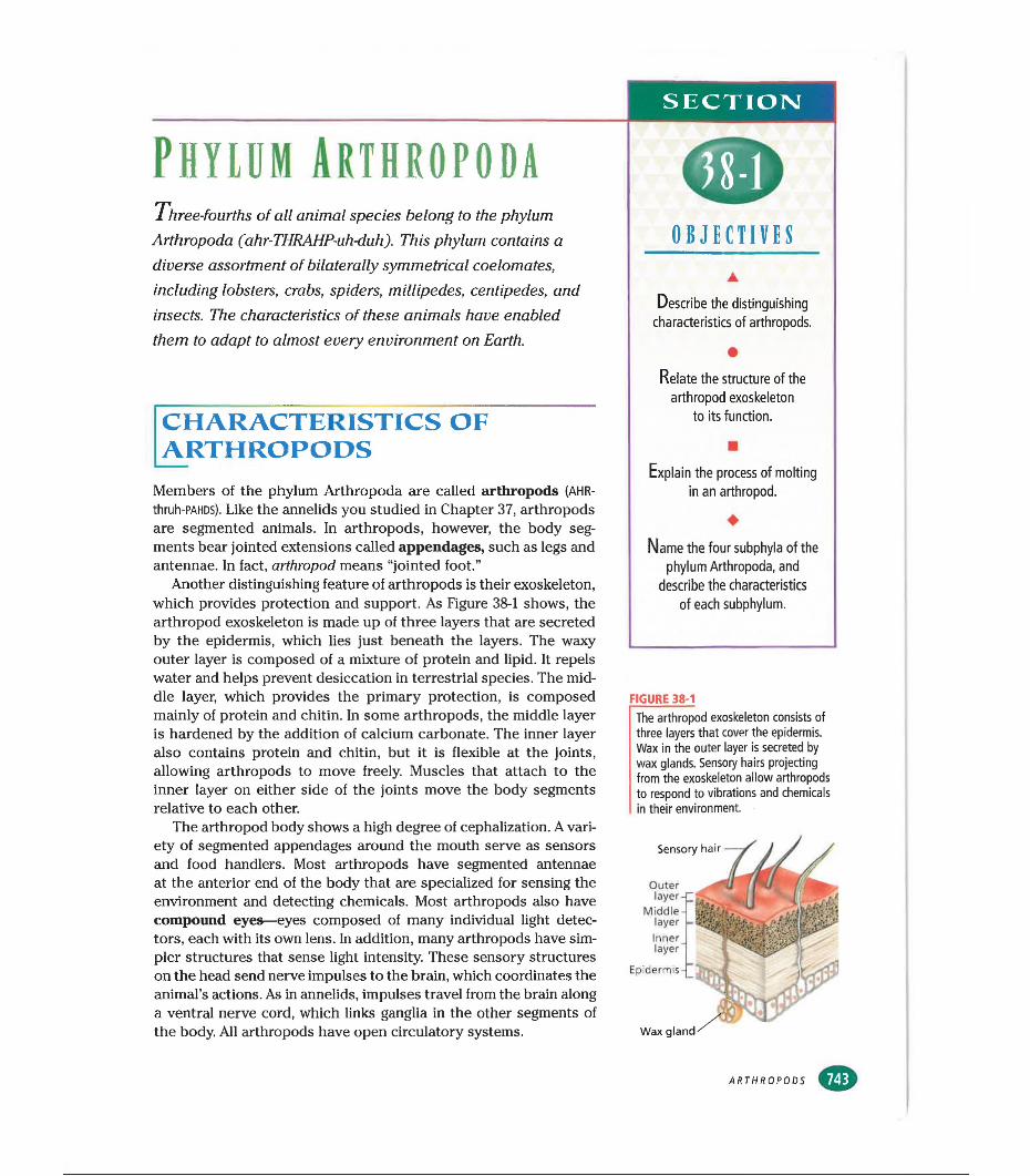
SECTION
PHYLBM arthropodaThree-fourths of all animal species belong to the phylum
Arthropoda (ahr-THRAHP-uh-duh). This phylum contains a diverse assortment of bilaterally symmetrical coelomates, including lobsters, crabs, spiders, millipedes, centipedes, and insects. The characteristics of these animals have enabled them to adapt to almost every environment on Earth.
CHARACTERISTICS OF ARTHROPODS
Members of the phylum Arthropoda are called arthropods (AHR- thruh-PAHDS). Like the annelids you studied in Chapter 37, arthropods are segmented animals. In arthropods, however, the body segments bear jointed extensions called appendages, such as legs and antennae. In fact, arthropod means “jointed foot.”
Another distinguishing feature of arthropods is their exoskeleton, which provides protection and support. As Figure 38-1 shows, the arthropod exoskeleton is made up of three layers that are secreted by the epidermis, which lies just beneath the layers. The waxy outer layer is composed of a mixture of protein and lipid. It repels water and helps prevent desiccation in terrestrial species. The middle layer, which provides the primary protection, is composed mainly of protein and chitin. In some arthropods, the middle layer is hardened by the addition of calcium carbonate. The inner layer also contains protein and chitin, but it is flexible at the joints, allowing arthropods to move freely. Muscles that attach to the inner layer on either side of the joints move the body segments relative to each other.
The arthropod body shows a high degree of cephalization. A variety of segmented appendages around the mouth serve as sensors and food handlers. Most arthropods have segmented antennae at the anterior end of the body that are specialized for sensing the environment and detecting chemicals. Most arthropods also have compound eyes—eyes composed of many individual light detectors, each with its own lens. In addition, many arthropods have simpler structures that sense light intensity. These sensory structures on the head send nerve impulses to the brain, which coordinates the animal’s actions. As in annelids, impulses travel from the brain along a ventral nerve cord, which links ganglia in the other segments of the body. All arthropods have open circulatory systems.
OBJECTIVES
Describe the distinguishing characteristics of arthropods.
•Relate the structure of the
arthropod exoskeleton to its function.
Explain the process of molting in an arthropod.
♦Name the four subphyla of the
phylum Arthropoda, and describe the characteristics
of each subphylum.
FIGURE 38-1The arthropod exoskeleton consists of three layers that cover the epidermis. Wax in the outer layer is secreted by wax glands. Sensory hairs projecting from the exoskeleton allow arthropods to respond to vibrations and chemicals in their environment.
Wax gland
Sensory hair
Outer layer-
MiddlelayerInnerlayer“
Epidermis -
ARTHROPODS
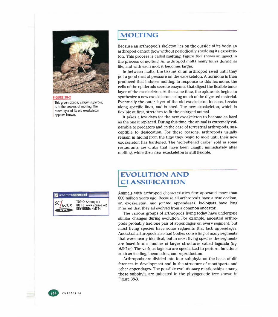
FIGURE 38-2
This green cicada, Tibicen superbus, is in the process of molting. The outer layer of its old exoskeleton appears brown.
MOLTINGBecause an arthropod’s skeleton lies on the outside of its body, an arthropod cannot grow without periodically shedding its exoskeleton. This process is called molting. Figure 38-2 shows an insect in the process of molting. An arthropod molts many times during its life, and with each molt it becomes larger.
In between molts, the tissues of an arthropod swell until they put a good deal of pressure on the exoskeleton. A hormone is then produced that induces molting. In response to this hormone, the cells of the epidermis secrete enzymes that digest the flexible inner layer of the exoskeleton. At the same time, the epidermis begins to synthesize a new exoskeleton, using much of the digested material. Eventually the outer layer of the old exoskeleton loosens, breaks along specific lines, and is shed. The new exoskeleton, which is flexible at first, stretches to fit the enlarged animal.
It takes a few days for the new exoskeleton to become as hard as the one it replaced. During this time, the animal is extremely vulnerable to predators and, in the case of terrestrial arthropods, susceptible to desiccation. For these reasons, arthropods usually remain in hiding from the time they begin to molt until their new exoskeleton has hardened. The “soft-shelled crabs” sold in some restaurants are crabs that have been caught immediately after molting, while their new exoskeleton is still flexible.
EVOLUTION AND CLASSIFICATION
internetconnect
SC INKSl TOPIC: Arthropods GO TO: www.scilinks.org KEYWORD: HM744
Animals with arthropod characteristics first appeared more than 600 million years ago. Because all arthropods have a true coelom, an exoskeleton, and jointed appendages, biologists have long inferred that they all evolved from a common ancestor.
The various groups of arthropods living today have undergone similar changes during evolution. For example, ancestral arthropods probably had one pair of appendages on every segment, but most living species have some segments that lack appendages. Ancestral arthropods also had bodies consisting of many segments that were nearly identical, but in most living species the segments are fused into a number of larger structures called tagmata (tag-
MAHT-uh). The various tagmata are specialized to perform functions such as feeding, locomotion, and reproduction.
Arthropods are divided into four subphyla on the basis of differences in development and in the structure of mouthparts and other appendages. The possible evolutionary relationships among these subphyla are indicated in the phylogenetic tree shown in Figure 38-3.
CHAPTER 38
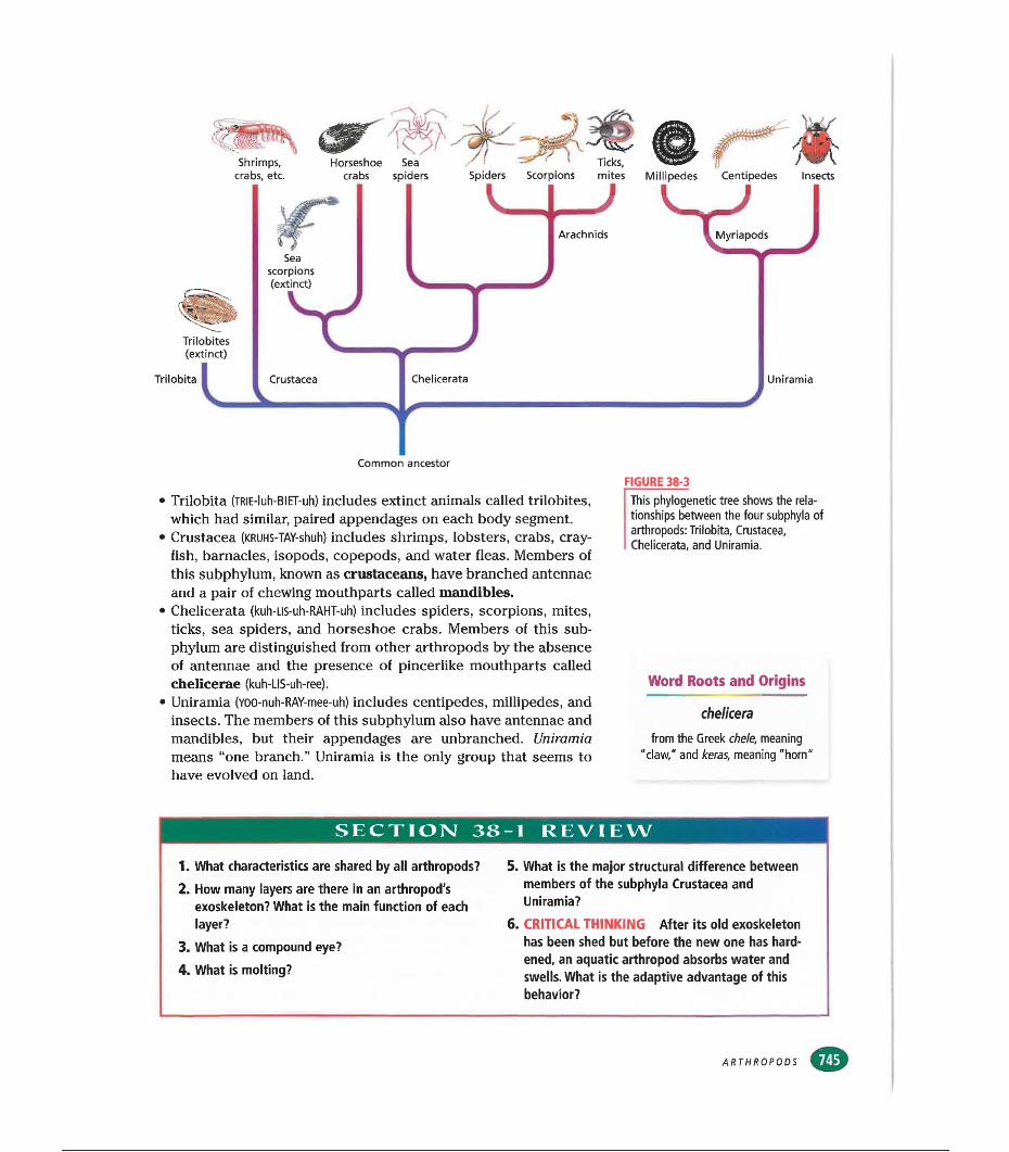
crabs, etc. crabs spiders Spiders Scorpions mites Millipedes Centipedes Insects
• Trilobita (TRlE-luh-BIET-uh) includes extinct animals called trilobites, which had similar, paired appendages on each body segment.
• Crustacea (KRUHS-TAY-shuh) includes shrimps, lobsters, crabs, crayfish, barnacles, isopods, copepods, and water fleas. Members of this subphylum, known as crustaceans, have branched antennae and a pair of chewing mouthparts called mandibles.
• Chelicerata (kuh-Lls-uh-RAHT-uh) includes spiders, scorpions, mites, ticks, sea spiders, and horseshoe crabs. Members of this subphylum are distinguished from other arthropods by the absence of antennae and the presence of pincerlike mouthparts called chelicerae (kuh-LIS-uh-ree).
• Uniramia (yoo-nuh-RAY-mee-uh) includes centipedes, millipedes, and insects. The members of this subphylum also have antennae and mandibles, but their appendages are unbranched. Uniramia means “one branch.” Uniramia is the only group that seems to have evolved on land.
FIGURE 38-3
This phylogenetic tree shows the relationships between the four subphyla of arthropods: Trilobita, Crustacea, Chelicerata, and Uniramia.
Word Roots and Origins
chelicera
from the Greek chele, meaning "daw," and keras, meaning "horn"
SECTION 38-1 REVIEW
1. What characteristics are shared by all arthropods?2. How many layers are there in an arthropod's
exoskeleton? What is the main function of each layer?
3. What is a compound eye?4. What is molting?
5. What is the major structural difference between members of the subphyla Crustacea and Uniramia?
6. CRITICAL THINKING After its old exoskeleton has been shed but before the new one has hardened, an aquatic arthropod absorbs water and swells. What is the adaptive advantage of this behavior?
ARTHROPODS
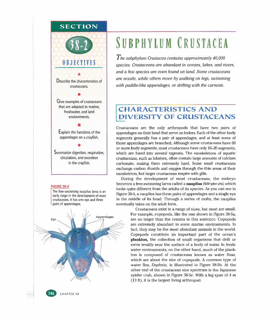
SECTION
OBJECTIVES
Describe the characteristics of crustaceans.
Give examples of crustaceans that are adapted to marine,
freshwater, and land environments.
Explain the functions of the appendages on a crayfish.
♦Summarize digestion, respiration,
circulation, and excretion in the crayfish.
FIGURE 38-4
The free-swimming nauplius larva is an early stage in the development of most crustaceans. It has one eye and three pairs of appendages.
Subphylum CrustaceaTh e subphylum Crustacea contains approximately 40,000
species. Crustaceans are abundant in oceans, lakes, and rivers, and a few species are even found on land. Some crustaceans are sessile, while others move by walking on legs, swimming with paddle-like appendages, or drifting with the currents.
CHARACTERISTICS AND DIVERSITY OF CRUSTACEANS
Crustaceans are the only arthropods that have two pairs of appendages on their head that serve as feelers. Each of the other body segments generally has a pair of appendages, and at least some of those appendages are branched. Although some crustaceans have 60 or more body segments, most crustaceans have only 16-20 segments, which are fused into several tagmata. The exoskeletons of aquatic crustaceans, such as lobsters, often contain large amounts of calcium carbonate, making them extremely hard. Some small crustaceans exchange carbon dioxide and oxygen through the thin areas of their exoskeleton, but larger crustaceans respire with gills.
During the development of most crustaceans, the embryo becomes a free-swimming larva called a nauplius (NAH-plee-uhs), which looks quite different from the adults of its species. As you can see in Figure 38-4, a nauplius has three pairs of appendages and a single eye in the middle of its head. Through a series of molts, the nauplius eventually takes on the adult form.
Crustaceans exist in a range of sizes, but most are small.For example, copepods, like the one shown in Figure 38-5a, are no larger than the comma in this sentence. Copepods are extremely abundant in some marine environments. In fact, they may be the most abundant animals in the world. Copepods constitute an important part of the ocean’s plankton, the collection of small organisms that drift or swim weakly near the surface of a body of water. In freshwater environments, on the other hand, much of the plankton is composed of crustaceans known as water fleas, which are about the size of copepods. A common type of water flea, Daphnia, is illustrated in Figure 38-5b. At the other end of the crustacean size spectrum is the Japanese spider crab, shown in Figure 38-5c. With a leg span of 4 m (13 ft), it is the largest living arthropod.
CHAPTER 38
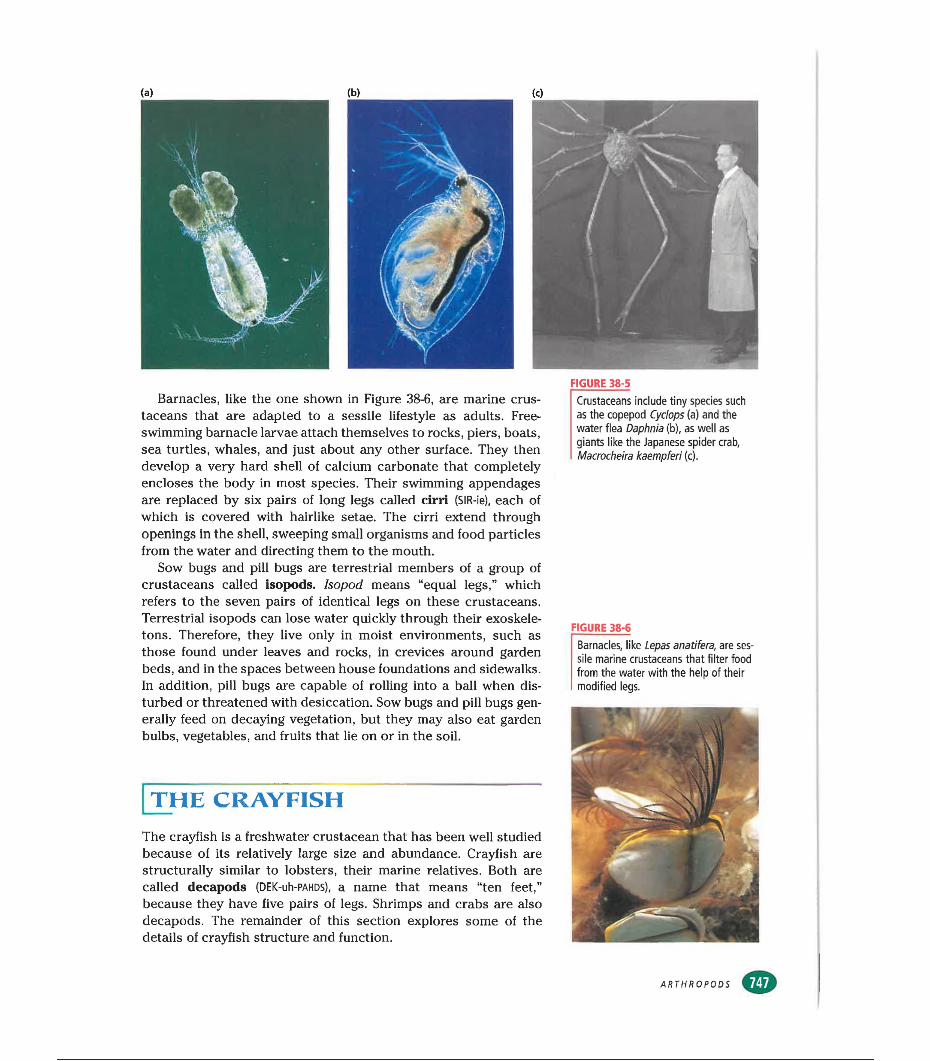
Barnacles, like the one shown in Figure 38-6, are marine crustaceans that are adapted to a sessile lifestyle as adults. Free- swimming barnacle larvae attach themselves to rocks, piers, boats, sea turtles, whales, and just about any other surface. They then develop a very hard shell of calcium carbonate that completely encloses the body in most species. Their swimming appendages are replaced by six pairs of long legs called cirri (SIR-ie), each of which is covered with hairlike setae. The cirri extend through openings in the shell, sweeping small organisms and food particles from the water and directing them to the mouth.
Sow bugs and pill bugs are terrestrial members of a group of crustaceans called isopods. Isopod means “equal legs,” which refers to the seven pairs of identical legs on these crustaceans. Terrestrial isopods can lose water quickly through their exoskeletons. Therefore, they live only in moist environments, such as those found under leaves and rocks, in crevices around garden beds, and in the spaces between house foundations and sidewalks. In addition, pill bugs are capable of rolling into a ball when disturbed or threatened with desiccation. Sow bugs and pill bugs generally feed on decaying vegetation, but they may also eat garden bulbs, vegetables, and fruits that lie on or in the soil.
[the crayfish
The crayfish is a freshwater crustacean that has been well studied because of its relatively large size and abundance. Crayfish are structurally similar to lobsters, their marine relatives. Both are called decapods (DEK-uh-PAHDs), a name that means “ten feet,” because they have five pairs of legs. Shrimps and crabs are also decapods. The remainder of this section explores some of the details of crayfish structure and function.
FIGURE 38-5
Crustaceans include tiny species such as the copepod Cyclops (a) and the water flea Daphnia (b), as well as giants like the Japanese spider crab, Macrocheira kaempferi (c).
FIGURE 38-6
Barnacles, like Lepas anatifera, are sessile marine crustaceans that filter food from the water with the help of their modified legs.
ARTHROPODS
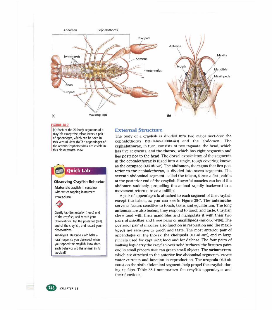
Abdomen Céphalothorax
Maxilla
Mandible
Maxlllipeds
FIGURE 38-7(a) Each of the 20 body segments of a crayfish except the telson bears a pair of appendages, which can be seen in this ventral view, (b) The appendages of the anterior céphalothorax are visible in this closer ventral view.
Quick Lab
Observing Crayfish BehaviorMaterials crayfish in container with water, tapping instrument
Procedure
Gently tap the anterior (head) end of the crayfish, and record your observations. Tap the posterior (tail) end of the crayfish, and record your observations.
Analysis Describe each behavioral response you observed when you tapped the crayfish. How does each behavior aid the animal in its survival?
External StructureThe body of a crayfish is divided into two major sections: the céphalothorax (SEF-uh-luh-THOHR-aks) and the abdomen. The céphalothorax, in turn, consists of two tagmata: the head, which has five segments, and the thorax, which has eight segments and lies posterior to the head. The dorsal exoskeleton of the segments in the céphalothorax is fused into a single, tough covering known as the carapace (KAR-uh-PAYS). The abdomen, the tagma that lies posterior to the céphalothorax, is divided into seven segments. The seventh abdominal segment, called the telson, forms a flat paddle at the posterior end of the crayfish. Powerful muscles can bend the abdomen suddenly, propelling the animal rapidly backward in a movement referred to as a tailflip.
A pair of appendages is attached to each segment of the crayfish except the telson, as you can see in Figure 38-7. The antennules serve as feelers sensitive to touch, taste, and equilibrium. The long antennae are also feelers; they respond to touch and taste. Crayfish chew food with their mandibles and manipulate it with their two pairs of maxillae and three pairs of maxillipeds (mak-SIL-uh-PEDS). The posterior pair of maxillae also function in respiration and the maxillipeds are sensitive to touch and taste. The most anterior pair of appendages on the thorax, the chelipeds (KEE-luh-PEDS), end in large pincers used for capturing food and for defense. The four pairs of walking legs carry the crayfish over solid surfaces; the first two pairs end in small pincers that can grasp small objects. The swimmerets, which are attached to the anterior five abdominal segments, create water currents and function in reproduction. The uropods (YUR-uh-
pahds), on the sixth abdominal segment, help propel the crayfish during tailflips. Table 38-1 summarizes the crayfish appendages and their functions.
CHAPTER 38
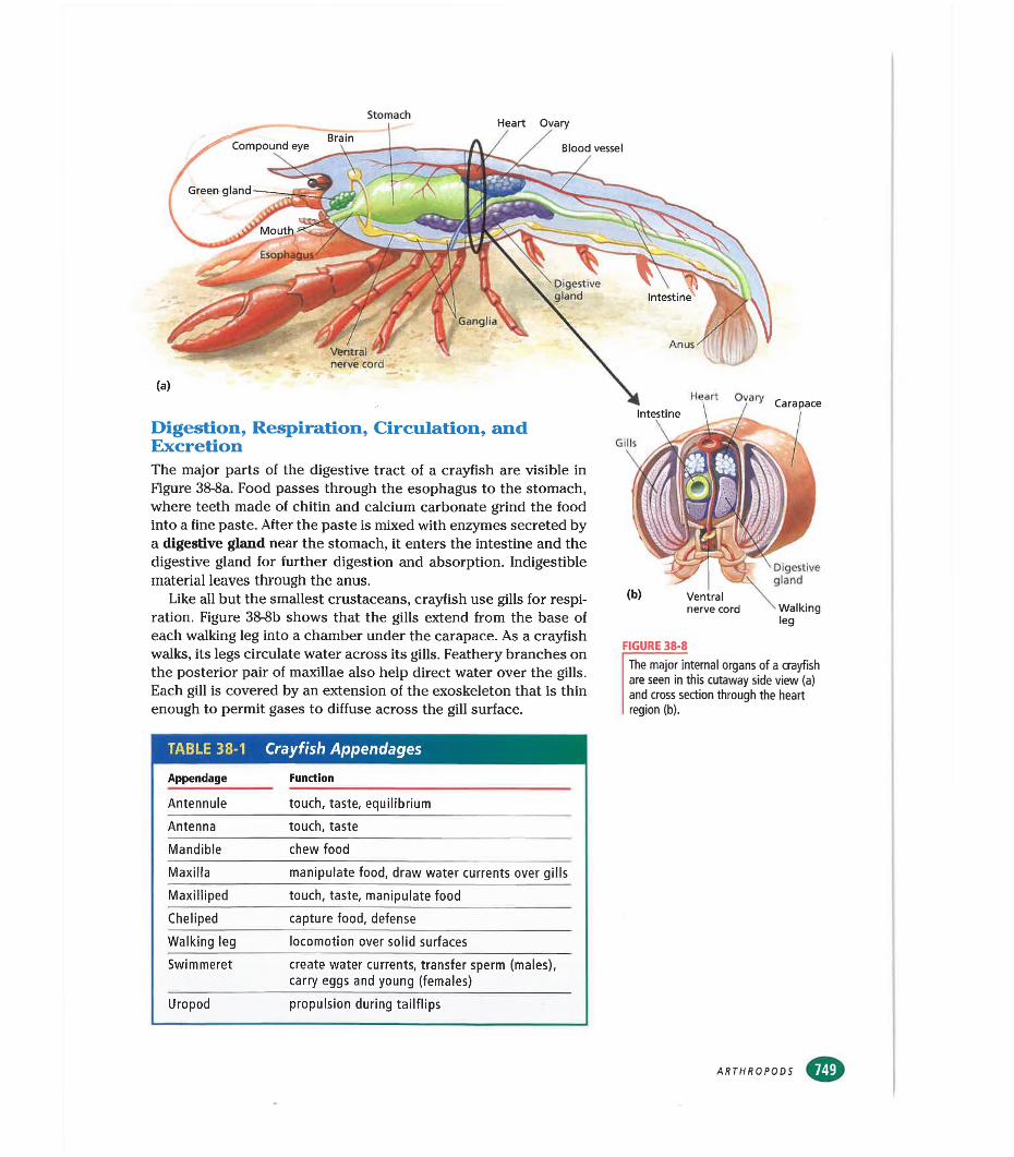
StomachHeart Ovary
BrainCompound eye
Esophagus
Digestivegland Intestine
Ganglia
Anus
Blood vessel
Ventral * nerve cord
(a)
Digestion, Respiration, Circulation, and ExcretionThe major parts of the digestive tract of a crayfish are visible in Figure 38-8a. Food passes through the esophagus to the stomach, where teeth made of chitin and calcium carbonate grind the food into a fine paste. After the paste is mixed with enzymes secreted by a digestive gland near the stomach, it enters the intestine and the digestive gland for further digestion and absorption. Indigestible material leaves through the anus.
Like all but the smallest crustaceans, crayfish use gills for respiration. Figure 38-8b shows that the gills extend from the base of each walking leg into a chamber under the carapace. As a crayfish walks, its legs circulate water across its gills. Feathery branches on the posterior pair of maxillae also help direct water over the gills. Each gill is covered by an extension of the exoskeleton that is thin enough to permit gases to diffuse across the gill surface.
Gills
FIGURE 38-8
The major internal organs of a crayfish are seen in this cutaway side view (a) and cross section through the heart region (b).
Heart Ovary Carapace
Ventral nerve cord
Digestivegland
Walkingleg
TABLE 38-1 Crayfish Appendages
Appendage Function
Antennule touch, taste, equilibrium
Antenna touch, taste
Mandible chew food
Maxilla manipulate food, draw water currents over gills
Maxiiliped touch, taste, manipulate food
Cheliped capture food, defense
Walking leg locomotion over solid surfaces
Swimmeret create water currents, transfer sperm (males), carry eggs and young (females)
Uropod propulsion during tailflips
ARTHROPODS
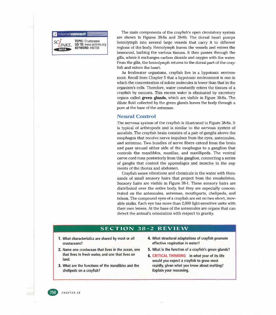
I internetconnect
SCllNKS,. TOPIC: Crustaceans GOTO: www.scilinks.org KEYWORD: HM750
The main components of the crayfish’s open circulatory system are shown in Figures 38-8a and 38-8b. The dorsal heart pumps hemolymph into several large vessels that carry it to different regions of the body. Hemolymph leaves the vessels and enters the hemocoel, bathing the various tissues. It then passes through the gills, where it exchanges carbon dioxide and oxygen with the water. From the gills, the hemolymph returns to the dorsal part of the crayfish and enters the heart.
As freshwater organisms, crayfish live in a hypotonic environment. Recall from Chapter 5 that a hypotonic environment is one in which the concentration of solute molecules is lower than that in the organism’s cells. Therefore, water constantly enters the tissues of a crayfish by osmosis. This excess water is eliminated by excretory organs called green glands, which are visible in Figure 38-8a. The dilute fluid collected by the green glands leaves the body through a pore at the base of the antennae.
Neural ControlThe nervous system of the crayfish is illustrated in Figure 38-8a. It is typical of arthropods and is similar to the nervous system of annelids. The crayfish brain consists of a pair of ganglia above the esophagus that receive nerve impulses from the eyes, antennules, and antennae. Two bundles of nerve fibers extend from the brain and pass around either side of the esophagus to a ganglion that controls the mandibles, maxillae, and maxillipeds. The ventral nerve cord runs posteriorly from this ganglion, connecting a series of ganglia that control the appendages and muscles in the segments of the thorax and abdomen.
Crayfish sense vibrations and chemicals in the water with thousands of small sensory hairs that project from the exoskeleton. Sensory hairs are visible in Figure 38-1. These sensory hairs are distributed over the entire body, but they are especially concentrated on the antennules, antennae, mouthparts, chelipeds, and telson. The compound eyes of a crayfish are set on two short, movable stalks. Each eye has more than 2,000 light-sensitive units with their own lenses. At the base of the antennules are organs that can detect the animal’s orientation with respect to gravity.
SECTION 38-2 REVIEW
1. What characteristics are shared by most or all crustaceans?
2. Name one crustacean that lives in the ocean, one that lives in fresh water, and one that lives on land.
3. What are the functions of the mandibles and the chelipeds on a crayfish?
4. What structural adaptations of crayfish promote effective respiration in water?
5. What is the function of a crayfish's green glands?6. CRITICAL THINKING In what year of its life
would you expect a crayfish to grow most rapidly, given what you know about molting? Explain your reasoning.
CHAPTER 38
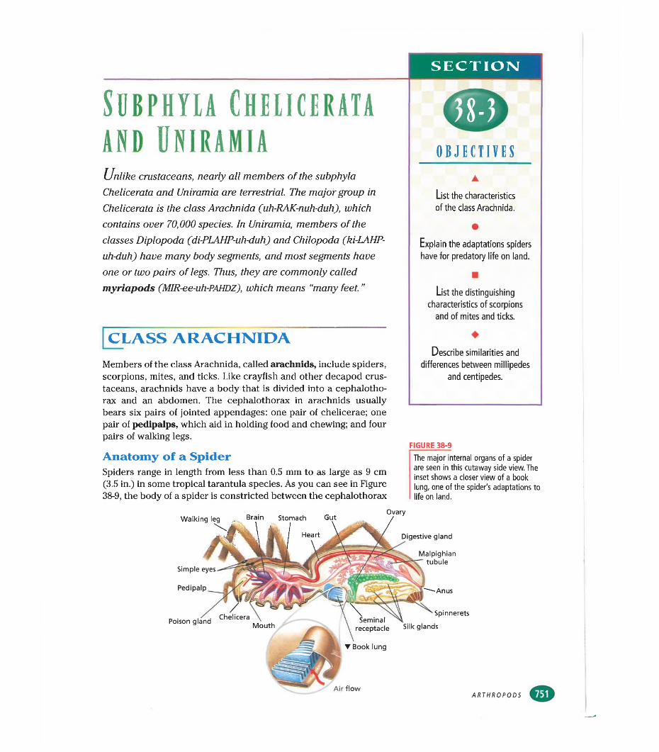
SECTION
SllBPHYLA CHELICERATA AND UNIRAMIA OBJECTIVESUnlike crustaceans, nearly all members of the subphyla
Chelicerata and Uniramia are terrestrial. The major group in Chelicerata is the class Arachnida Çuh-RAK-nuh-duh), which contains over 70,000 species. In Uniramia, members of the classes Diplopoda (di-PLAHP-uh-duh) and Chilopoda (ki-LAHP- uh-duh) have many body segments, and most segments have one or two pairs of legs. Thus, they are commonly called myriapods (MIR-ee-uh-PAHDZ), which means “many feet.”
▲List the characteristics of the class Arachnida.
•Explain the adaptations spiders have for predatory life on land.
■List the distinguishing
characteristics of scorpions and of mites and ticks.
CLASS ARACHNIDAMembers of the class Arachnida, called arachnids, include spiders, scorpions, mites, and ticks. Like crayfish and other decapod crustaceans, arachnids have a body that is divided into a céphalothorax and an abdomen. The céphalothorax in arachnids usually
♦Describe similarities and
differences between millipedes and centipedes.
bears six pairs of jointed appendages: one pair of chelicerae; one pair of pedipalps, which aid in holding food and chewing; and four pairs of walking legs.
FIGURE 38-9Anatomy of a SpiderSpiders range in length from less than 0.5 mm to as large as 9 cm (3.5 in.) in some tropical tarantula species. As you can see in Figure38-9, the body of a spider is constricted between the céphalothorax
The major internal organs of a spider are seen in this cutaway side view. The inset shows a closer view of a book lung, one of the spider's adaptations to life on land.
Walking
Simple
Pedlpalp Anus
Spinnerets
receptacle Silk glands
▼ Book lung
Poison
Digestive gland
Malpighiantubule
Air flowARTHROPODS
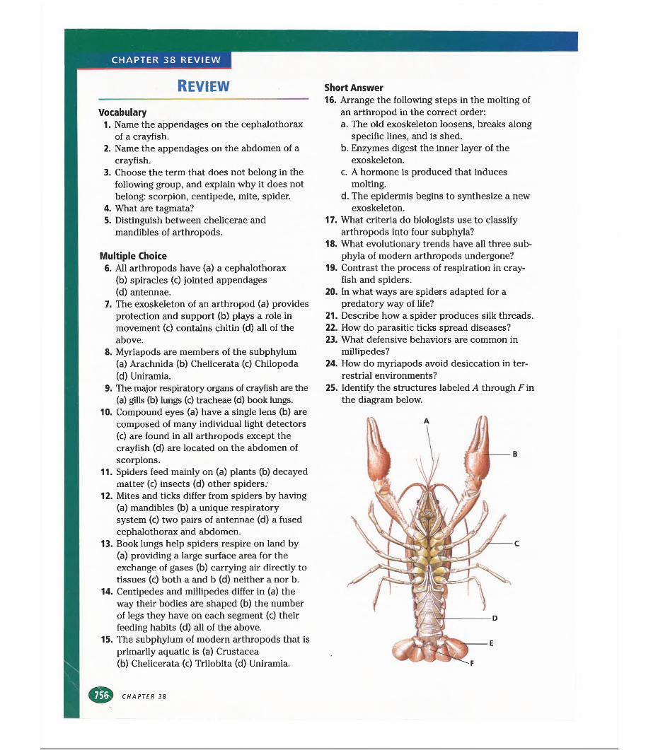
CHAPTER 38 REVIEW
REVIEW
Vocabulary1. Name the appendages on the céphalothorax
of a crayfish.2. Name the appendages on the abdomen of a
crayfish.3. Choose the term that does not belong in the
following group, and explain why it does not belong: scorpion, centipede, mite, spider.
4. What are tagmata?5. Distinguish between chelicerae and
mandibles of arthropods.
Multiple Choice6. All arthropods have (a) a céphalothorax
(b) spiracles (c) jointed appendages(d) antennae.
7. The exoskeleton of an arthropod (a) provides protection and support (b) plays a role in movement (c) contains chitin (d) all of the above.
8. Myriapods are members of the subphylum (a) Arachnida (b) Chelicerata (c) Chilopoda (d) Uniramia.
9. The major respiratory organs of crayfish are the (a) gills (b) lungs (c) tracheae (d) book lungs.
10. Compound eyes (a) have a single lens (b) are composed of many individual light detectors(c) are found in all arthropods except the crayfish (d) are located on the abdomen of scorpions.
11. Spiders feed mainly on (a) plants (b) decayed matter (c) insects (d) other spiders.
12. Mites and ticks differ from spiders by having (a) mandibles (b) a unique respiratory system (c) two pairs of antennae (d) a fused céphalothorax and abdomen.
13. Book lungs help spiders respire on land by(a) providing a large surface area for the exchange of gases (b) carrying air directly to tissues (c) both a and b (d) neither a nor b.
14. Centipedes and millipedes differ in (a) the way their bodies are shaped (b) the number of legs they have on each segment (c) their feeding habits (d) all of the above.
15. The subphylum of modern arthropods that is primarily aquatic is (a) Crustacea(b) Chelicerata (c) Trilobita (d) Uniramia.
Short Answer16. Arrange the following steps in the molting of
an arthropod in the correct order:a. The old exoskeleton loosens, breaks along
specific lines, and is shed.b. Enzymes digest the inner layer of the
exoskeleton.c. A hormone is produced that induces
molting.d. The epidermis begins to synthesize a new
exoskeleton.17. What criteria do biologists use to classify
arthropods into four subphyla?18. What evolutionary trends have all three sub
phyla of modern arthropods undergone?19. Contrast the process of respiration in cray
fish and spiders.20. In what ways are spiders adapted for a
predatory way of life?21. Describe how a spider produces silk threads.22. How do parasitic ticks spread diseases?23. What defensive behaviors are common in
millipedes?24. How do myriapods avoid desiccation in ter
restrial environments?25. Identify the structures labeled A through F in
the diagram below.
CHAPTER 38
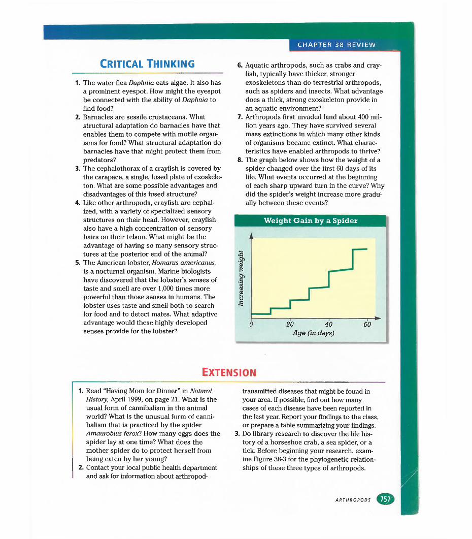
CHAPTER 38 REVIEW
Critical thinking
1. The water flea Daphnia eats algae. It also has a prominent eyespot. How might the eyespot be connected with the ability of Daphnia to find food?
2. Barnacles are sessile crustaceans. What structural adaptation do barnacles have that enables them to compete with motile organisms for food? What structural adaptation do barnacles have that might protect them from predators?
3. The céphalothorax of a crayfish is covered by the carapace, a single, fused plate of exoskeleton. What are some possible advantages and disadvantages of this fused structure?
4. Like other arthropods, crayfish are cephal- ized, with a variety of specialized sensory structures on their head. However, crayfish also have a high concentration of sensory hairs on their telson. What might be the advantage of having so many sensory structures at the posterior end of the animal?
5. The American lobster, Homarus americanus, is a nocturnal organism. Marine biologists have discovered that the lobster’s senses of taste and smell are over 1,000 times more powerful than those senses in humans. The lobster uses taste and smell both to search for food and to detect mates. What adaptive advantage would these highly developed senses provide for the lobster?
6. Aquatic arthropods, such as crabs and crayfish, typically have thicker, stronger exoskeletons than do terrestrial arthropods, such as spiders and insects. What advantage does a thick, strong exoskeleton provide in an aquatic environment?
7. Arthropods first invaded land about 400 million years ago. They have survived several mass extinctions in which many other kinds of organisms became extinct. What characteristics have enabled arthropods to thrive?
8. The graph below shows how the weight of a spider changed over the first 60 days of its life. What events occurred at the beginning of each sharp upward turn in the curve? Why did the spider’s weight increase more gradually between these events?
Weight Gain by a Spider
Extension
1. Read “Having Mom for Dinner” in Natural History, April 1999, on page 21. What is the usual form of cannibalism in the animal world? What is the unusual form of cannibalism that is practiced by the spider Amaurobius feroxl How many eggs does the spider lay at one time? What does the mother spider do to protect herself from being eaten by her young?
2. Contact your local public health department and ask for information about arthropod-
transmitted diseases that might be found in your area. If possible, find out how many cases of each disease have been reported in the last year. Report your findings to the class, or prepare a table summarizing your findings.
3. Do library research to discover the life history of a horseshoe crab, a sea spider, or a tick. Before beginning your research, examine Figure 38-3 for the phylogenetic relationships of these three types of arthropods.
ARTHROPODS
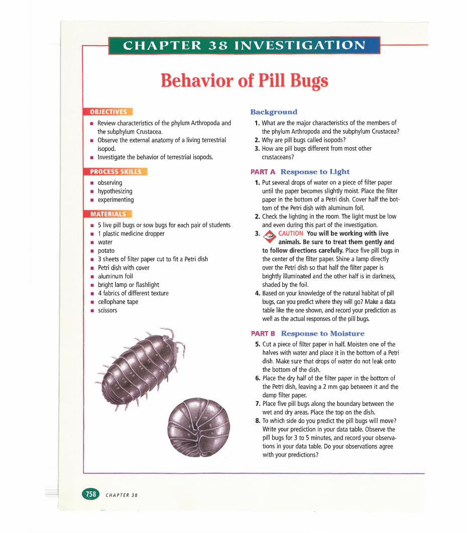
CHAPTER 38 INVESTIGATION
Behavior of Pill BugsOBJECTIVES
■ Review characteristics of the phylum Arthropoda and the subphylum Crustacea.
■ Observe the external anatomy of a living terrestrial isopod.
■ Investigate the behavior of terrestrial isopods.
PROCESS SKILLS
■ observing■ hypothesizing■ experimenting
MATERIALS
■ 5 live pill bugs or sow bugs for each pair of students■ 1 plastic medicine dropper■ water■ potato■ 3 sheets of filter paper cut to fit a Petri dish■ Petri dish with cover■ aluminum foil■ bright lamp or flashlight■ 4 fabrics of different texture■ cellophane tape■ scissors
Background1. What are the major characteristics of the members of
the phylum Arthropoda and the subphylum Crustacea?2. Why are pill bugs called isopods?3. How are pill bugs different from most other
crustaceans?
PART A Response to Light1.
2.
3.
4.
Put several drops of water on a piece of filter paper until the paper becomes slightly moist. Place the filter paper in the bottom of a Petri dish. Cover half the bottom of the Petri dish with aluminum foil.Check the lighting in the room. The light must be low and even during this part of the investigation.
CAUTION You will be working with live animals. Be sure to treat them gently and
to follow directions carefully. Place five pill bugs in the center of the filter paper. Shine a lamp directly over the Petri dish so that half the filter paper is brightly illuminated and the other half is in darkness, shaded by the foil.Based on your knowledge of the natural habitat of pill bugs, can you predict where they will go? Make a data table like the one shown, and record your prediction as well as the actual responses of the pill bugs.
PART B Response to Moisture5. Cut a piece of filter paper in half. Moisten one of the
halves with water and place it in the bottom of a Petri dish. Make sure that drops of water do not leak onto the bottom of the dish.
6. Place the dry half of the filter paper in the bottom of the Petri dish, leaving a 2 mm gap between it and the damp filter paper.
7. Place five pill bugs along the boundary between the wet and dry areas. Place the top on the dish.
8. To which side do you predict the pill bugs will move? Write your prediction in your data table. Observe the pill bugs for 3 to 5 minutes, and record your observations in your data table. Do your observations agree with your predictions?
CHAPTER 38
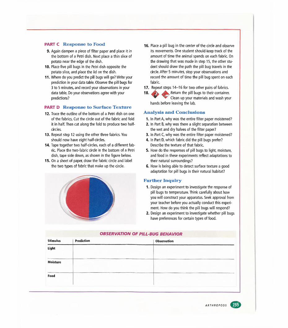
PART C Response to Food9. Again dampen a piece of filter paper and place it in
the bottom of a Petri dish. Next place a thin slice of potato near the edge of the dish.
10. Place five pill bugs in the Petri dish opposite the potato slice, and place the lid on the dish.
11. Where do you predict the pill bugs will go? Write your prediction in your data table. Observe the pill bugs for 3 to 5 minutes, and record your observations in your data table. Do your observations agree with your predictions?
PART D Response to Surface Texture12. Trace the outline of the bottom of a Petri dish on one
of the fabrics. Cut the circle out of the fabric and fold it in half. Then cut along the fold to produce two half- circles.
13. Repeat step 12 using the other three fabrics. You should now have eight half-circles.
14. Tape together two half-circles, each of a different fabric. Place the two-fabric circle in the bottom of a Petri dish, tape side down, as shown in the figure below.
15. On a sheet of paper, draw the fabric circle and label the two types of fabric that make up the circle.
16. Place a pill bug in the center of the circle and observe its movements. One student should keep track of the amount of time the animal spends on each fabric. On the drawing that was made in step 15, the other student should draw the path the pill bug travels in the circle. After 5 minutes, stop your observations and record the amount of time the pill bug spent on each fabric.
17. Repeat steps 14-16 for two other pairs of fabrics.18. 4^ Return the pill bugs to their container.
▼ Clean up your materials and wash your hands before leaving the lab.
Analysis and Conclusions1. In Part A, why was the entire filter paper moistened?2. In Part B, why was there a slight separation between
the wet and dry halves of the filter paper?3. In Part C, why was the entire filter paper moistened?4. In Part D, which fabric did the pill bugs prefer?
Describe the texture of that fabric.5. How do the responses of pill bugs to light, moisture,
and food in these experiments reflect adaptations to their natural surroundings?
6. How is being able to detect surface texture a good adaptation for pill bugs in their natural habitat?
Further Inquiry1. Design an experiment to investigate the response of
pill bugs to temperature. Think carefully about how you will construct your apparatus. Seek approval from your teacher before you actually conduct this experiment. How do you think the pill bugs will respond?
2. Design an experiment to investigate whether pill bugs have preferences for certain types of food.
OBSERVATION OF PILL-BUG BEHAVIORStimulus Prediction Observation
Light
Moisture
Food
ARTHROPODS
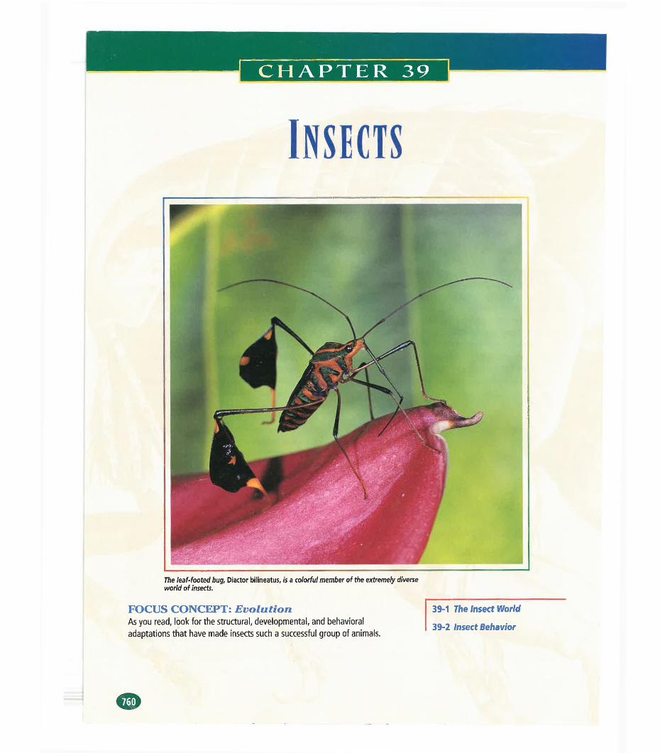
Insects
The leaf-footed bug. Diactor bilineatus, is a colorful member of the extremely diverse world of insects.
FOCUS CONCEPT: EvolutionAs you read, look for the structural, developmental, and behavioral adaptations that have made Insects such a successful group of animals.
39-1 The Insect World
39-2 Insect Behavior
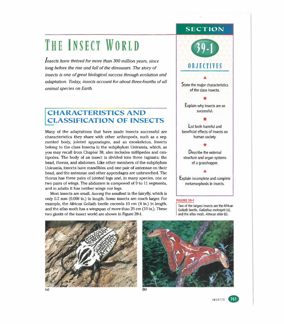
SECTION!
The Insect WorldInsects have thrived for more than 300 million years, since
long before the rise and fall of the dinosaurs. The story of insects is one of great biological success through evolution and adaptation. Today, insects account for about three-fourths of all animal species on Earth.
CHARACTERISTICS AND CLASSIFICATION OF INSECTS
Many of the adaptations that have made insects successful are characteristics they share with other arthropods, such as a segmented body, jointed appendages, and an exoskeleton. Insects belong to the class Insecta in the subphylum Uniramia, which, as you may recall from Chapter 38, also includes millipedes and centipedes. The body of an insect is divided into three tagmata: the head, thorax, and abdomen. Like other members of the subphylum Uniramia, insects have mandibles and one pair of antennae on their head, and the antennae and other appendages are unbranched. The thorax has three pairs of jointed legs and, in many species, one or two pairs of wings. The abdomen is composed of 9 to 11 segments, and in adults it has neither wings nor legs.
Most insects are small. Among the smallest is the fairyfly, which is only 0.2 mm (0.008 in.) in length. Some insects are much larger. For example, the African Goliath beetle exceeds 10 cm (4 in.) in length, and the atlas moth has a wingspan of more than 25 cm (10 in.). These two giants of the insect world are shown in Figure 39-1.
0OBJECTIVES
State the major characteristics of the class Insecta.
•Explain why insects are so
successful.
■List both harmful and
beneficial effects of insects on human society.
♦Describe the external
structure and organ systems of a grasshopper.
▲
Explain incomplete and complete metamorphosis in insects.
FIGURE 39-1
Two of the largest insects are the African Goliath beetle, Goliathus meleagris (a), and the atlas moth, Attacus atlas (b).
INSECTS
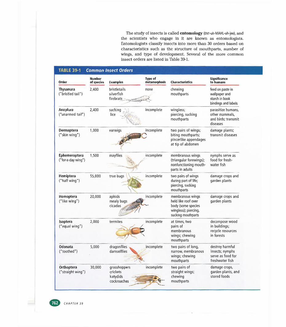
The study of insects is called entomology (ENT-uh-MAHL-uh-jee), and the scientists who engage in it are known as entomologists. Entomologists classify insects into more than 30 orders based on characteristics such as the structure of mouthparts, number of wings, and type of development. Several of the more common insect orders are listed in Table 39-1.
TABLE 39-1 Common Insect Orders
OrderNumber of species Examples
Type of metamorphosis Characteristics
Significance to humans
Thysanura ("bristled tail")
2,400 bristletails nonesilverfishfirebrats.
chewingmouthparts
feed on paste in wallpaper and starch in book bindings and labels
Anoplura("unarmed tail")
2,400 suckinga .Hi •lice
incomplete wingless; piercing, sucking mouthparts
parasitize humans, other mammals, and birds; transmit diseases
Dermaptera ("skin wing")
1,000 earwigs incomplete two pairs of wings; biting mouthparts; pincerlike appendages at tip of abdomen
damage plants; transmit diseases
Ephemeroptera("for-a-day wing")
1,500 mayflies incomplete membranous wings (triangular forewings); nonfunctioning mouthparts in adults
nymphs serve as food for freshwater fish
Hemiptera("half wing")
55,000 true bugs^jj^^^ incomplete two pairs of wings during part of life; piercing, sucking mouthparts
damage crops and garden plants
Homoptera("like wing")
20,000 aphids mealy bugs cicadas
incomplete membranous wings held like roof over body (some species wingless); piercing, sucking mouthparts
damage crops and garden plants
Isoptera("equal wing")
2,000 termites- -,v 4
L ¿¡^
incomplete at times, two pairs of membranous wings; chewing mouthparts
decompose wood in buildings; recycle resources in forests
Odonata("toothed")
5,000 dragonflies damselflies \ .
incomplete two pairs of long, narrow, membranous wings; chewing mouthparts
destroy harmful insects; nymphs serve as food for freshwater fish
Orthoptera("straight wing")
30,000 grasshopperscricketskatydidscockroaches
incomplete
' '
two pairs of straight wings; chewing mouthparts
damage crops, garden plants, and stored foods
CHAPTER 39
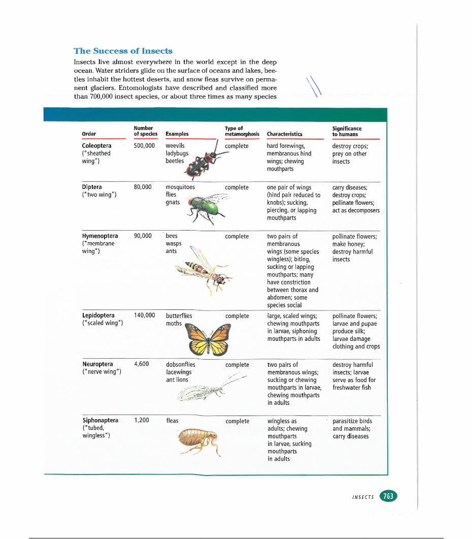
The Success of InsectsInsects live almost everywhere in the world except in the deep ocean. Water striders glide on the surface of oceans and lakes, beetles inhabit the hottest deserts, and snow fleas survive on permanent glaciers. Entomologists have described and classified more than 700,000 insect species, or about three times as many species
Order
Coleóptera("sheathedwing")
Diptera("two wing")
Hymenoptera("membranewing")
Lepidoptera("scaled wing")
Neuroptera("nerve wing")
Siphonaptera("tubed,wingless")
Number of species
500,000
Examples
weevils ladybugs beetles
Type of metamorphosis Characteristics
Significance to humans
hard forewings, membranous hind wings; chewing mouthparts
destroy crops; prey on other insects
80,000 mosquitoes flies gnats
complete one pair of wings (hind pair reduced to knobs); sucking, piercing, or lapping mouthparts
carry diseases; destroy crops; pollinate flowers; act as decomposers
90,000 complete two pairs of membranous wings (some species wingless); biting, sucking or lapping mouthparts; many have constriction between thorax and abdomen; some species social
pollinate flowers; make honey; destroy harmful insects
140,000 butterflies moths
complete large, scaled wings; chewing mouthparts in larvae, siphoning mouthparts in adults
4,600 dobsonflies lacewings ant lions
complete two pairs ofmembranous wings; sucking or chewing mouthparts in larvae, chewing mouthparts in adults
pollinate flowers; larvae and pupae produce silk; larvae damage clothing and crops
destroy harmful insects; larvae serve as food for freshwater fish
1,200 fleas complete wingless as adults; chewing mouthparts in larvae, sucking mouthparts in adults
parasitize birds and mammals; carry diseases
INSECTS
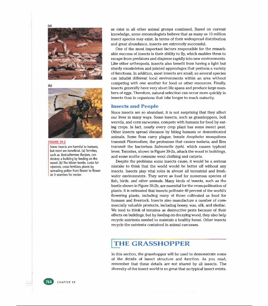
FIGURE 39-2
Some insects are harmful to humans, but most are beneficial, (a) Termites, such as Reticulitermes flavipes, can destroy a building by feeding on the wood, (b) The blister beetle, Lutta ful- vipennis, cross-fertilizes plants by spreading pollen from flower to flower as it searches for nectar.
as exist in all other animal groups combined. Based on current knowledge, some entomologists believe that as many as 10 million insect species may exist. In terms of their widespread distribution and great abundance, insects are extremely successful.
One of the most important factors responsible for the remarkable success of insects is their ability to fly, which enables them to escape from predators and disperse rapidly into new environments. Like other arthropods, insects also benefit from having a light but sturdy exoskeleton and jointed appendages that perform a variety of functions. In addition, most insects are small, so several species can inhabit different local environments within an area without competing with one another for food or other resources. Finally, insects generally have very short life spans and produce large numbers of eggs. Therefore, natural selection can occur more quickly in insects than in organisms that take longer to reach maturity.
Insects and PeopleSince insects are so abundant, it is not surprising that they affect our lives in many ways. Some insects, such as grasshoppers, boll weevils, and corn earworms, compete with humans for food by eating crops. In fact, nearly every crop plant has some insect pest. Other insects spread diseases by biting humans or domesticated animals. Some fleas carry plague; female Anopheles mosquitoes transmit Plasmodium, the protozoan that causes malaria; and flies transmit the bacterium Salmonella typhi, which causes typhoid fever. Termites, shown in Figure 39-2a, attack the wood in buildings, and some moths consume wool clothing and carpets.
Despite the problems some insects cause, it would be a serious mistake to think that the world would be better off without any insects. Insects play vital roles in almost all terrestrial and freshwater environments. They serve as food for numerous species of fish, birds, and other animals. Many kinds of insects, such as the beetle shown in Figure 39-2b, are essential for the cross-pollination of plants. It is estimated that insects pollinate 40 percent of the world’s flowering plants, including many of those cultivated as food for humans and livestock. Insects also manufacture a number of commercially valuable products, including honey, wax, silk, and shellac. We tend to think of termites as destructive pests because of their effects on buildings, but by feeding on decaying wood, they also help recycle nutrients needed to maintain a healthy forest. Other insects recycle the nutrients contained in animal carcasses.
| THE GRASSHOPPER
In this section, the grasshopper will be used to demonstrate some of the details of insect structure and function. As you read, remember that these details are not shared by all insects. The diversity of the insect world is so great that no typical insect exists.
CHAPTER 39
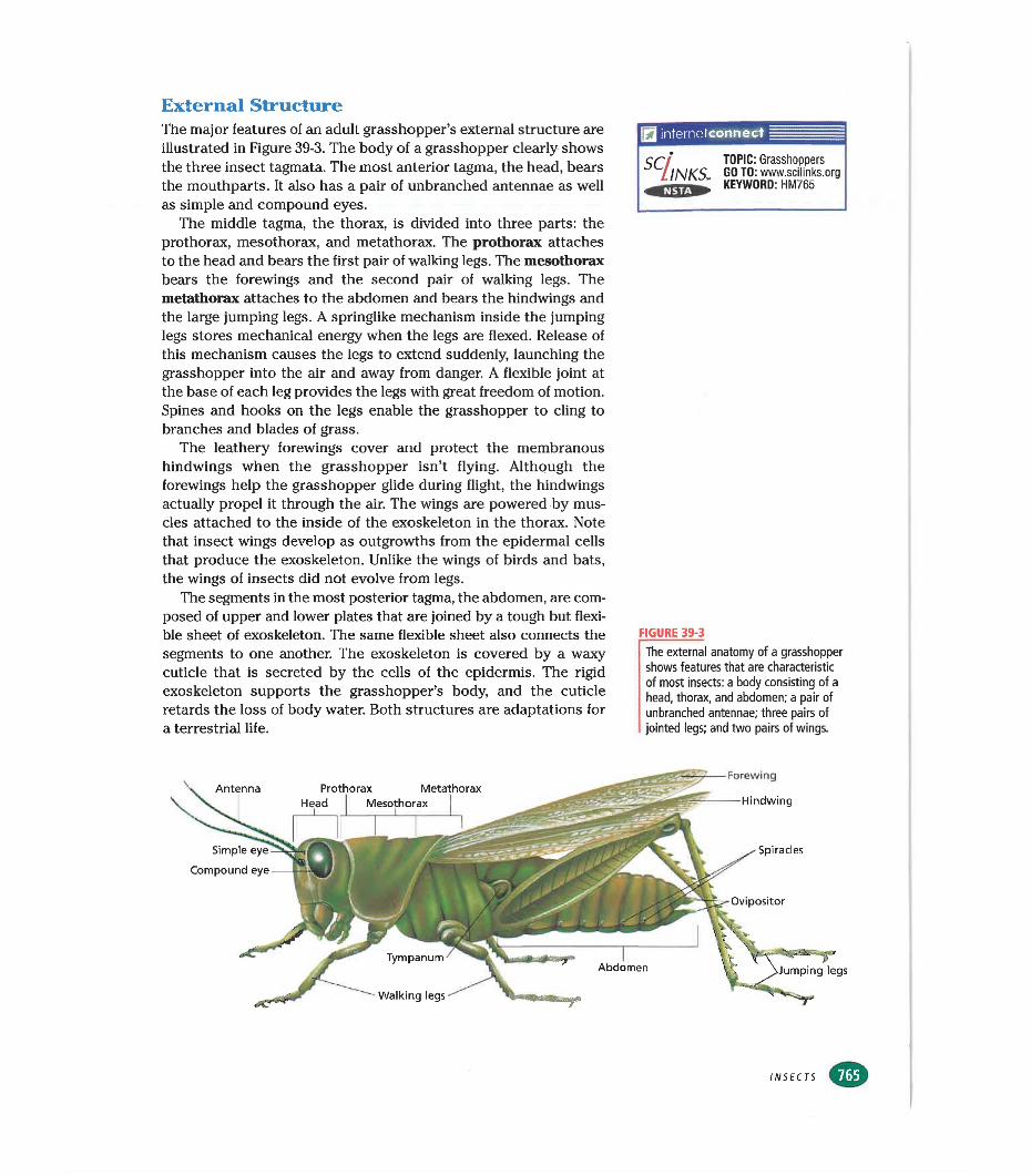
External StructureThe major features of an adult grasshopper’s external structure are illustrated in Figure 39-3. The body of a grasshopper clearly shows the three insect tagmata. The most anterior tagma, the head, bears the mouthparts. It also has a pair of unbranched antennae as well as simple and compound eyes.
The middle tagma, the thorax, is divided into three parts: the prothorax, mesothorax, and metathorax. The prothorax attaches to the head and bears the first pair of walking legs. The mesothorax bears the forewings and the second pair of walking legs. The metathorax attaches to the abdomen and bears the hindwings and the large jumping legs. A springlike mechanism inside the jumping legs stores mechanical energy when the legs are flexed. Release of this mechanism causes the legs to extend suddenly, launching the grasshopper into the air and away from danger. A flexible joint at the base of each leg provides the legs with great freedom of motion. Spines and hooks on the legs enable the grasshopper to cling to branches and blades of grass.
The leathery forewings cover and protect the membranous hindwings when the grasshopper isn’t flying. Although the forewings help the grasshopper glide during flight, the hindwings actually propel it through the air. The wings are powered by muscles attached to the inside of the exoskeleton in the thorax. Note that insect wings develop as outgrowths from the epidermal cells that produce the exoskeleton. Unlike the wings of birds and bats, the wings of insects did not evolve from legs.
The segments in the most posterior tagma, the abdomen, are composed of upper and lower plates that are joined by a tough but flexible sheet of exoskeleton. The same flexible sheet also connects the segments to one another. The exoskeleton is covered by a waxy cuticle that is secreted by the cells of the epidermis. The rigid exoskeleton supports the grasshopper’s body, and the cuticle retards the loss of body water. Both structures are adaptations for a terrestrial life.
m internetconnect
sc/iINKS.TOPIC: Grasshoppers GOTO: www.scilinks.org KEYWORD: HM765
FIGURE 39-3
The external anatomy of a grasshopper shows features that are characteristic of most insects: a body consisting of a head, thorax, and abdomen; a pair of unbranched antennae; three pairs of jointed legs; and two pairs of wings.
Ovipositor
TympanumJumping
Walking legs
-----Forewing
HindwingProthorax MetathoraxAntenna
Head Mesothorax
Simple eye
Compound eye
Spiracles
Abdomen
INSECTS
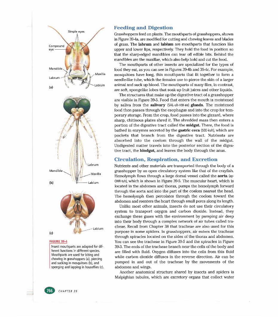
Compound eye----------
Mandible
Maxilla
Labium
FIGURE 39-4
Insect mouthparts are adapted for different functions in différent species. Mouthparts are used for biting and chewing in grasshoppers (a), piercing and sucking in mosquitoes (b), and sponging and lapping in houseflies (c).
Feeding and DigestionGrasshoppers feed on plants. The mouthparts of grasshoppers, shown in Figure 394a, are modified for cutting and chewing leaves and blades of grass. The labrum and labium are mouthparts that function like upper and lower lips, respectively. They hold the food in position so that the sharp-edged mandibles can tear off edible bits. Behind the mandibles are the maxillae, which also help hold and cut the food.
The mouthparts of other insects are specialized for the types of food they eat, as you can see in Figures 394b and 394c. For example, mosquitoes have long, thin mouthparts that fit together to form a needle-like tube, which the females use to pierce the skin of a larger animal and suck up blood. The mouthparts of many flies, in contrast, are soft, spongelike lobes that soak up fruit juices and other liquids.
The structures that make up the digestive tract of a grasshopper are visible in Figure 39-5. Food that enters the mouth is moistened by saliva from the salivary (SAL-uh-VER-ee) glands. The moistened food then passes through the esophagus and into the crop for temporary storage. From the crop, food passes into the gizzard, where sharp, chitinous plates shred it. The shredded mass then enters a portion of the digestive tract called the midgut. There, the food is bathed in enzymes secreted by the gastric ceca (SEE-kuh), which are pockets that branch from the digestive tract. Nutrients are absorbed into the coelom through the wall of the midgut. Undigested matter travels into the posterior section of the digestive tract, the hindgut, and leaves the body through the anus.
Circulation, Respiration, and ExcretionNutrients and other materials are transported through the body of a grasshopper by an open circulatory system like that of the crayfish. Hemolymph flows through a large dorsal vessel called the aorta (ay- OHR-tuh), which is shown in Figure 39-5. The muscular heart, which is located in the abdomen and thorax, pumps the hemolymph forward through the aorta and into the part of the coelom nearest the head. The hemolymph then percolates through the coelom toward the abdomen and reenters the heart through small pores along its length.
Unlike most other animals, insects do not use their circulatory system to transport oxygen and carbon dioxide. Instead, they exchange these gases with the environment by pumping air deep into their body through a complex network of air tubes called tracheae. Recall from Chapter 38 that tracheae are also used for this purpose in some spiders. In grasshoppers, air enters the tracheae through spiracles located on the sides of the thorax and abdomen. You can see the tracheae in Figure 39-5 and the spiracles in Figure 39-3. The ends of the tracheae branch near the cells of the body and are filled with fluid. Oxygen diffuses into the cells from this fluid while carbon dioxide diffuses in the reverse direction. Air can be pumped in and out of the tracheae by the movements of the abdomen and wings.
Another anatomical structure shared by insects and spiders is Malpighian tubules, which are excretory organs that collect water
CHAPTER 39
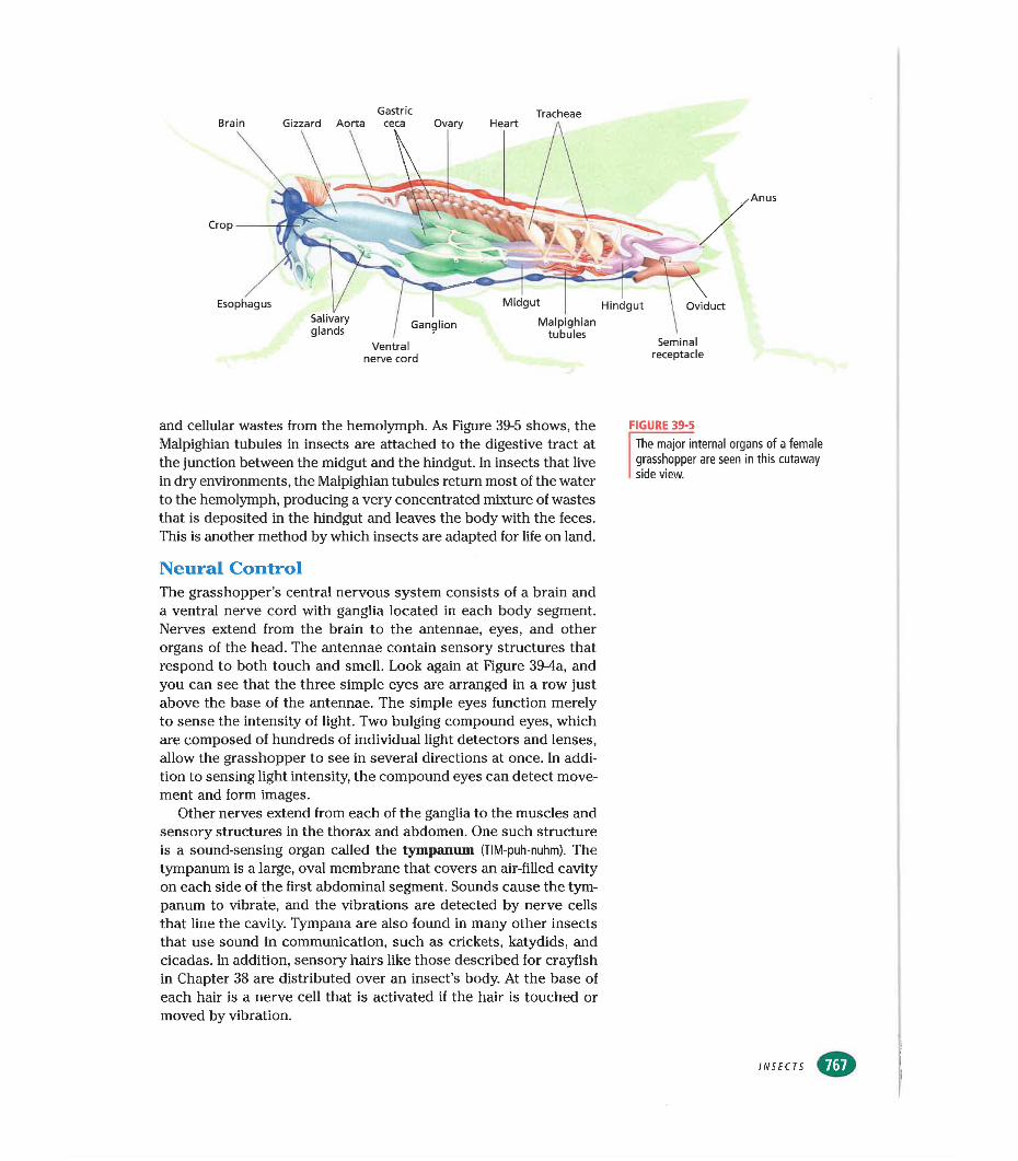
Midgut HindgutMalpighian
tubulesGanglion
BrainGastric Tracheae
Gizzard Aorta ceca Ovary Heart a
Crop
EsophagusSalivaryglands
Ventral nerve cord
Oviduct
Seminalreceptacle
Anus
and cellular wastes from the hemolymph. As Figure 39-5 shows, the Malpighian tubules in insects are attached to the digestive tract at the junction between the midgut and the hindgut. In insects that live in dry environments, the Malpighian tubules return most of the water to the hemolymph, producing a very concentrated mixture of wastes that is deposited in the hindgut and leaves the body with the feces. This is another method by which insects are adapted for life on land.
FIGURE 39-5The major internal organs of a female grasshopper are seen in this cutaway side view.
Neural ControlThe grasshopper’s central nervous system consists of a brain and a ventral nerve cord with ganglia located in each body segment. Nerves extend from the brain to the antennae, eyes, and other organs of the head. The antennae contain sensory structures that respond to both touch and smell. Look again at Figure 39-4a, and you can see that the three simple eyes are arranged in a row just above the base of the antennae. The simple eyes function merely to sense the intensity of light. Two bulging compound eyes, which are composed of hundreds of individual light detectors and lenses, allow the grasshopper to see in several directions at once. In addition to sensing light intensity, the compound eyes can detect movement and form images.
Other nerves extend from each of the ganglia to the muscles and sensory structures in the thorax and abdomen. One such structure is a sound-sensing organ called the tympanum (TIM-puh-nuhm). The tympanum is a large, oval membrane that covers an air-filled cavity on each side of the first abdominal segment. Sounds cause the tympanum to vibrate, and the vibrations are detected by nerve cells that line the cavity. Tympana are also found in many other insects that use sound in communication, such as crickets, katydids, and cicadas. In addition, sensory hairs like those described for crayfish in Chapter 38 are distributed over an insect’s body. At the base of each hair is a nerve cell that is activated if the hair is touched or moved by vibration.
INSECTS
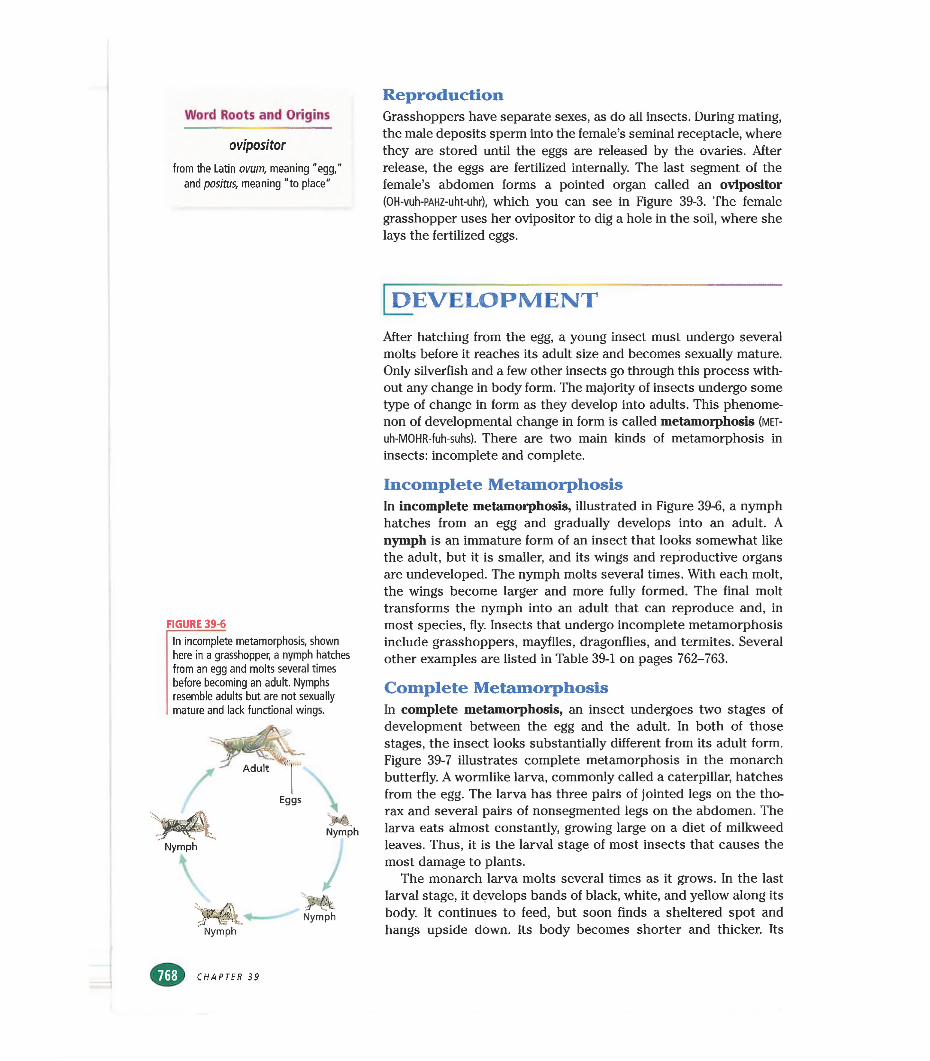
Word Roots and Origins
ovipositorfrom the Latin ovum, meaning "egg,”
and positus, meaning "to place"
ReproductionGrasshoppers have separate sexes, as do all insects. During mating, the male deposits sperm into the female’s seminal receptacle, where they are stored until the eggs are released by the ovaries. After release, the eggs are fertilized internally. The last segment of the female’s abdomen forms a pointed organ called an ovipositor (OH-vuh-PAHZ-uht-uhr), which you can see in Figure 39-3. The female grasshopper uses her ovipositor to dig a hole in the soil, where she lays the fertilized eggs.
FIGURE 39-6
In incomplete metamorphosis, shown here in a grasshopper, a nymph hatches from an egg and molts several times before becoming an adult. Nymphs resemble adults but are not sexually mature and lack functional wings.
NymphNymph
[development
After hatching from the egg, a young insect must undergo several molts before it reaches its adult size and becomes sexually mature. Only silverfish and a few other insects go through this process without any change in body form. The majority of insects undergo some type of change in form as they develop into adults. This phenomenon of developmental change in form is called metamorphosis (met- uh-MOHR-fuh-suhs). There are two main kinds of metamorphosis in insects: incomplete and complete.
Incomplete MetamorphosisIn incomplete metamorphosis, illustrated in Figure 39-6, a nymph hatches from an egg and gradually develops into an adult. A nymph is an immature form of an insect that looks somewhat like the adult, but it is smaller, and its wings and reproductive organs are undeveloped. The nymph molts several times. With each molt, the wings become larger and more fully formed. The final molt transforms the nymph into an adult that can reproduce and, in most species, fly. Insects that undergo incomplete metamorphosis include grasshoppers, mayflies, dragonflies, and termites. Several other examples are listed in Table 39-1 on pages 762-763.
Complete MetamorphosisIn complete metamorphosis, an insect undergoes two stages of development between the egg and the adult. In both of those stages, the insect looks substantially different from its adult form. Figure 39-7 illustrates complete metamorphosis in the monarch butterfly. A wormlike larva, commonly called a caterpillar, hatches from the egg. The larva has three pairs of jointed legs on the thorax and several pairs of nonsegmented legs on the abdomen. The larva eats almost constantly, growing large on a diet of milkweed leaves. Thus, it is the larval stage of most insects that causes the most damage to plants.
The monarch larva molts several times as it grows. In the last larval stage, it develops bands of black, white, and yellow along its body. It continues to feed, but soon finds a sheltered spot and hangs upside down. Its body becomes shorter and thicker. Its
CHAPTER 39
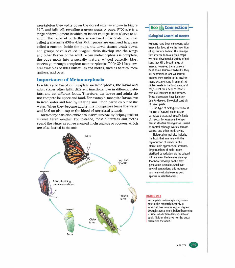
exoskeleton then splits down the dorsal side, as shown in Figure 39-7, and falls off, revealing a green pupa. A pupa (PYOO-puh) is a stage of development in which an insect changes from a larva to an adult. The pupa of butterflies is enclosed in a protective case called a chrysalis (KRIS-uh-luhs). Moth pupae are enclosed in a case called a cocoon. Inside the pupa, the larval tissues break down, and groups of cells called imaginal disks develop into the wings and other tissues of the adult. When metamorphosis is complete, the pupa molts into a sexually mature, winged butterfly. Most insects go through complete metamorphosis. Table 39-1 lists several examples besides butterflies and moths, such as beetles, mosquitoes, and bees.
Importance of MetamorphosisIn a life cycle based on complete metamorphosis, the larval and adult stages often fulfill different functions, live in different habitats, and eat different foods. Therefore, the larvae and adults do not compete for space and food. For example, mosquito larvae live in fresh water and feed by filtering small food particles out of the water. When they become adults, the mosquitoes leave the water and feed on plant sap or the blood of terrestrial animals.
Metamorphosis also enhances insect survival by helping insects survive harsh weather. For instance, most butterflies and moths spend the winter as pupae encased in chrysalises or cocoons, which are often buried in the soil.
- Eco ^Connection
Biological Control of Insects
Humans have been competing with insects for food since the invention of agriculture. To limit the damage that insects do to our food crops, we have developed a variety of poisons that kill a broad range of insects. However, these poisons have some serious drawbacks: they kill beneficial as well as harmful insects; they persist in the environment, accumulating in animals at higher levels in the food web; and they select for strains of insects that are resistant to the poisons. These drawbacks have led scientists to develop biological controls of insect pests.
One type of biological control is the use of natural predators or parasites that attack specific kinds of insects. For example, the bacterium Bacillus thuringiensis is used to control cabbage worms, tomato worms, and other moth larvae.
Biological control also includes methods that interfere with the reproduction of insects. In the sterile-male approach, for instance, large numbers of male insects sterilized by radiation are introduced into an area. The females lay eggs that never develop, so the next generation is smaller. Used over several generations, this technique can nearly eliminate some pest species in selected areas.
FIGURE 39-7
In complete metamorphosis, shown here in the monarch butterfly, a larva hatches from an egg and goes through several molts before becoming a pupa, which then develops into an adult. Neither the larva nor the pupa resembles the adult.
INSECTS
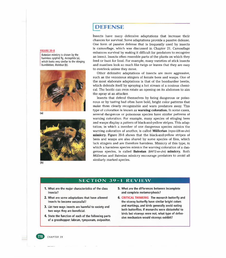
DEFENSE
FIGURE 39-8
Batesian mimicry is shown by the harmless syrphid fly, Arctophila (a), which looks very similar to the stinging bumblebee, Bombus (b).
Insects have many defensive adaptations that increase their chances for survived. Some adaptations provide a passive defense. One form of passive defense that is frequently used by insects is camouflage, which was discussed in Chapter 21. Camouflage enhances survival by making it difficult for predators to recognize an insect. Insects often resemble parts of the plants on which they feed or hunt for food. For example, many varieties of stick insects and mantises look so much like twigs or leaves that they are easy to overlook unless they move.
Other defensive adaptations of insects are more aggressive, such as the venomous stingers of female bees and wasps. One of the most elaborate adaptations is that of the bombardier beetle, which defends itself by spraying a hot stream of a noxious chemical. The beetle can even rotate an opening on its abdomen to aim the spray at an attacker.
Insects that defend themselves by being dangerous or poisonous or by tasting bad often have bold, bright color patterns that make them clearly recognizable and warn predators away. This type of coloration is known as warning coloration. In some cases, several dangerous or poisonous species have similar patterns of warning coloration. For example, many species of stinging bees and wasps display a pattern of black-and-yellow stripes. This adaptation, in which a member of one dangerous species mimics the warning coloration of another, is called Müllerian (myoo-LER-ee-uhn) mimicry. Figure 39-8 shows that the black-and-yellow stripes of bees and wasps are also shared by some species of flies, which lack stingers and are therefore harmless. Mimicry of this type, in which a harmless species mimics the warning coloration of a dangerous species, is called Batesian (BAYTZ-ee-uhn) mimicry. Both Müllerian and Batesian mimicry encourage predators to avoid all similarly marked species.
SECTION 39-1 REVIEW
1. What are the major characteristics of the class Insecta?
2. What are some adaptations that have allowed insects to become successful?
3. List two ways insects are harmful to society and two ways they are beneficial.
4. State the function of each of the following parts of a grasshopper: labrum, tympanum, ovipositor.
5. What are the differences between incomplete and complete metamorphosis?
6. CRITICAL THINKING The monarch butterfly and the viceroy butterfly have similar bright colors and markings, and birds generally avoid eating both butterflies. If monarchs were distasteful to birds but viceroys were not, what type of defensive mechanism would viceroys exhibit?
CHAPTER 39
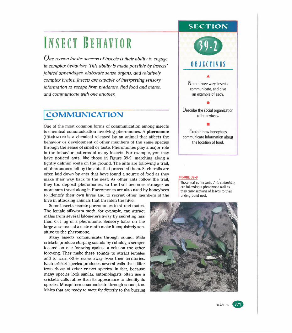
SECTION
Insect behaviorOne reason for the success of insects is their ability to engage
in complex behaviors. This ability is made possible by insects’ OBJECTIVESjointed appendages, elaborate sense organs, and relatively complex brains. Insects are capable of interpreting sensory information to escape from predators, find food and mates,and communicate with one another.
A
Name three ways insects communicate, and give
an example of each.
•Describe the social organization
of honeybees.COMMUNICATIONOne of the most common forms of communication among insects is chemical communication involving pheromones. A pheromone (FER-uh-MOHN) is a chemical released by an animal that affects the behavior or development of other members of the same species through the sense of smell or taste. Pheromones play a major role in the behavior patterns of many insects. For example, you may
■Explain how honeybees
communicate information about the location of food.
have noticed ants, like those in Figure 39-9, marching along a tightly defined route on the ground. The ants are following a trail, of pheromones left by the ants that preceded them. Such trails are often laid down by ants that have found a source of food as they make their way back to the nest. As other ants follow the trail, they too deposit pheromones, so the trail becomes stronger as more ants travel along it. Pheromones are also used by honeybees to identify their own hives and to recruit other members of the hive in attacking animals that threaten the hive.
Some insects secrete pheromones to attract mates.The female silkworm moth, for example, can attract males from several kilometers away by secreting less than 0.01 pg of a pheromone. Sensory hairs on the large antennae of a male moth make it exquisitely sensitive to the pheromone.
Many insects communicate through sound. Male crickets produce chirping sounds by rubbing a scraper located on one forewing against a vein on the other forewing. They make these sounds to attract females and to warn other males away from their territories.Each cricket species produces severed calls that differ from those of other cricket species. In fact, because many species look similar, entomologists often use a cricket’s calls rather them its appearance to identify its species. Mosquitoes communicate through sound, too.Males that are ready to mate fly directly to the buzzing
FIGURE 39-9These leaf-cutter ants, Atta colombica, are following a pheromone trail as they carry sections of leaves to their underground nest.
INSECTS
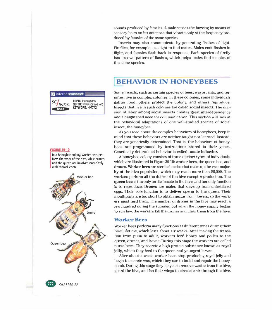
sounds produced by females. A male senses the buzzing by means of sensory hairs on his antennae that vibrate only at the frequency produced by females of the same species.
Insects may also communicate by generating flashes of light. Fireflies, for example, use light to find mates. Males emit flashes in flight, and females flash back in response. Each species of firefly has its own pattern of flashes, which helps males find females of the same species.
BEHAVIOR IN HONEYBEES
internetconnect
SCllNKS. TOPIC: Honeybees GO TO: www.scilinks.org KEYWORD: HM772
FIGURE 39-10
In a honeybee colony, worker bees perform the work of the hive, while drones and the queen are involved exclusively with reproduction.
Worker bee
Drone
Queen bee
Some insects, such as certain species of bees, wasps, ants, and termites, live in complex colonies. In these colonies, some individuals gather food, others protect the colony, and others reproduce. Insects that live in such colonies are called social insects. The division of labor among social insects creates great interdependence and a heightened need for communication. This section will look at the behavioral adaptations of one well-studied species of social insect, the honeybee.
As you read about the complex behaviors of honeybees, keep in mind that these behaviors are neither taught nor learned. Instead, they are genetically determined. That is, the behaviors of honeybees are programmed by instructions stored in their genes. Genetically determined behavior is called innate behavior.
A honeybee colony consists of three distinct types of individuals, which are illustrated in Figure 39-10: worker bees, the queen bee, and drones. Worker bees are sterile females that make up the vast majority of the hive population, which may reach more than 80,000. The workers perform all the duties of the hive except reproduction. The queen bee is the only fertile female in the hive, and her only function is to reproduce. Drones are males that develop from unfertilized eggs. Their sole function is to deliver sperm to the queen. Their mouthparts are too short to obtain nectar from flowers, so the workers must feed them. The number of drones in the hive may reach a few hundred during the summer, but when the honey supply begins to run low, the workers kill the drones and clear them from the hive.
Worker BeesWorker bees perform many functions at different times during their brief lifetime, which lasts about six weeks. After making the transition from pupa to adult, workers feed honey and pollen to the queen, drones, and larvae. During this stage the workers are called nurse bees. They secrete a high-protein substance known as royal jelly, which they feed to the queen and youngest larvae.
After about a week, worker bees stop producing royal jelly and begin to secrete wax, which they use to build and repair the honeycomb. During this stage they may also remove wastes from the hive, guard the hive, and fan their wings to circulate air through the hive.
CHAPTER 39
The workers spend the last weeks of their life gathering nectar and pollen. A number of structural adaptations aid them in this work. Their mouthparts are specialized for lapping up nectar, and their legs have structures that serve as pollen packers, pollen baskets, and pollen combs.
The sterile workers do not use their ovipositors for egg laying. Instead, these structures are modified into barbed stingers that the workers use to protect the hive. When a worker bee stings an animal, the stinger and attached venom sac are left behind in the victim as the bee flies away. The worker, having lost part of its body and much of its hemolymph, dies a day or two later. Wasps also have stingers that are modified ovipositors. Unlike honeybees, they can sting many times because their stinger is not barbed.
The Queen BeeThe queen bee develops from an egg identical to those that develop into the workers. The differences between the queen and the workers result from the continuous diet of royal jelly that the queen is fed throughout her larval development. In addition, the queen herself secretes a pheromone called the queen factor that prevents other female larvae from developing into queens.
The queen’s role is to reproduce. Within a few days after she completes metamorphosis and emerges as an adult, she flies out of the hive and mates in the air with one or more drones. During mating, millions of sperm are deposited in the queen’s seminal receptacle, where they will remain for the five or more years of her life. Although the queen mates only once, she may lay as many as a million eggs each year.
When the hive becomes overcrowded, the queen stops producing the queen factor and leaves the hive. As she leaves, she secretes a swarming pheromone that induces about half of the workers in the hive to follow her and form a swarm. Eventually the swarm finds «mother location for a new hive. Meanwhile, in the old hive, the remaining workers begin feeding royal jelly to other larvae. When a new queen emerges, she produces the queen factor, and in response, the workers destroy the other developing queens. The new queen departs on a mating flight, and the cycle begins again.
The Dances of the BeesWhen honeybees leave the hive and find a source of pollen and nectar, how do they communicate the location of this food source to other workers in the hive? An Austrian biologist, Karl von Frisch (1886-1982), spent 25 years answering this question. His careful experimentation earned him a Nobel Prize in 1973.
To study bees, von Frisch built a glass-walled hive and placed feeding stations stocked with sweetened water near the hive. He noted that “scout bees” returning from the feeding stations would perform a series of dancelike movements in the hive. As shown in Figure 39-1 la, the scout bee would circle first to the right and then to the left, a behavior that von Frisch called the round dance. After
Quick Lab
Interpreting Nonverbal Communication
Materials pencil, paper, wrappedcandy piecesProcedure1. Choose one member of your
group to play the part of the "scout" bee. The others in the group will be the "worker" bees.
2. Your teacher will secretly tell the scout bee where a piece of candy is located. The scout bee will develop a method of nonverbal communication to let the worker bees know where the candy is hidden. The scout bee may not point to the candy. Use Figure 39-11 as a guide to developing your method of communication.
3. When the candy has been located, the scout will hide another piece of candy and select a new scout. The new scout will develop a different way to tell the group where the candy is located. Repeat the procedure until everyone in your group has been a scout.
Analysis1. How effective were each of your
scout's methods for showing the location of the candy?
2. Did the worker bees improve their ability to find the candy after several trials?
3. List and describe some types of nonverbal communication that humans use.
INSECTS
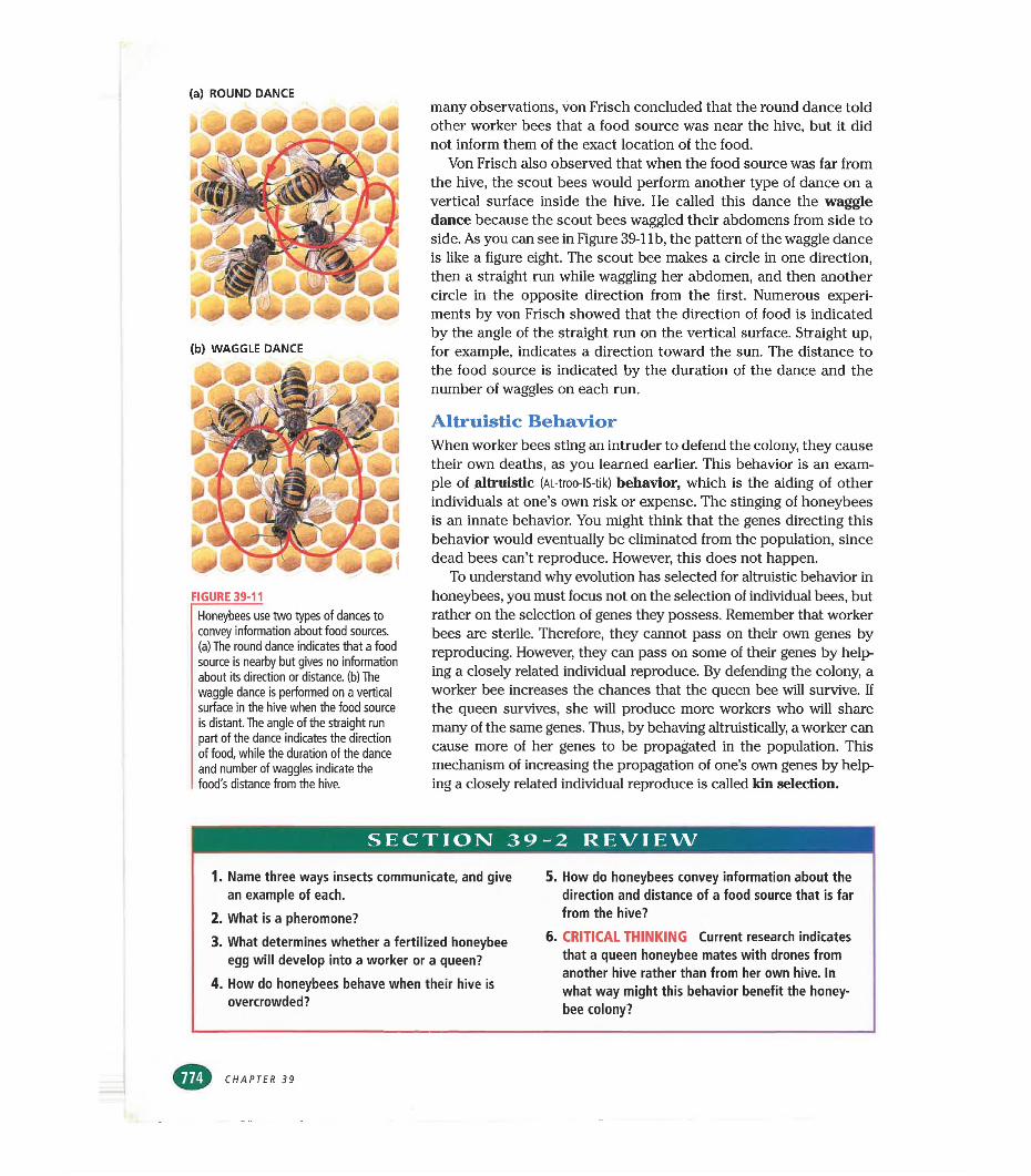
(a) ROUND DANCE
¡0 & *!& 4P
(b) WAGGLE DANCE
FIGURE 39-11
Honeybees use two types of dances to convey information about food sources, (a) The round dance indicates that a food source is nearby but gives no information about its direction or distance, (b) The waggle dance is performed on a vertical surface in the hive when the food source is distant. The angle of the straight run part of the dance indicates the direction of food, while the duration of the dance and number of waggles indicate the food's distance from the hive.
many observations, von Frisch concluded that the round dance told other worker bees that a food source was near the hive, but it did not inform them of the exact location of the food.
Von Frisch also observed that when the food source was far from the hive, the scout bees would perform another type of dance on a vertical surface inside the hive. He called this dance the waggle dance because the scout bees waggled their abdomens from side to side. As you can see in Figure 39-1 lb, the pattern of the waggle dance is like a figure eight. The scout bee makes a circle in one direction, then a straight run while waggling her abdomen, and then another circle in the opposite direction from the first. Numerous experiments by von Frisch showed that the direction of food is indicated by the angle of the straight run on the vertical surface. Straight up, for example, indicates a direction toward the sun. The distance to the food source is indicated by the duration of the dance and the number of waggles on each run.
Altruistic BehaviorWhen worker bees sting an intruder to defend the colony, they cause their own deaths, as you learned earlier. This behavior is an example of altruistic (AL-troo-IS-tik) behavior, which is the eliding of other individuals at one’s own risk or expense. The stinging of honeybees is an innate behavior. You might think that the genes directing this behavior would eventually be eliminated from the population, since dead bees can’t reproduce. However, this does not happen.
To understand why evolution has selected for altruistic behavior in honeybees, you must focus not on the selection of individual bees, but rather on the selection of genes they possess. Remember that worker bees are sterile. Therefore, they cannot pass on their own genes by reproducing. However, they can pass on some of their genes by helping a closely related individual reproduce. By defending the colony, a worker bee increases the chances that the queen bee will survive. If the queen survives, she will produce more workers who will share many of the same genes. Thus, by behaving altruistically, a worker can cause more of her genes to be propagated in the population. This mechanism of increasing the propagation of one’s own genes by helping a closely related individual reproduce is called kin selection.
SECTION 39-2 REVIEW
1. Name three ways insects communicate, and give an example of each.
2. What is a pheromone?3. What determines whether a fertilized honeybee
egg will develop into a worker or a queen?4. How do honeybees behave when their hive is
overcrowded?
5. How do honeybees convey information about the direction and distance of a food source that is far from the hive?
6. CRITICAL THINKING Current research indicates that a queen honeybee mates with drones from another hive rather than from her own hive. In what way might this behavior benefit the honeybee colony?
CHAPTER 39
CHAPTER 39 REVIEW
Summary/Vocabulary
■ The insect body is divided into three tag- mata: the head has mandibles and one pair of unbranched antennae; the thorax has three pairs of jointed legs and, in many species, one or two pairs of wings; the abdomen has 9 to 11 segments but neither wings nor legs in adults.
■ Insects live in almost every terrestrial and freshwater environment. Factors responsible for their success include their ability to fly, exoskeleton, jointed appendages, small size, and short life span.
■ Insects negatively affect humans by competing for food, transmitting diseases, and destroying buildings and other manufactured products. However, insects are also beneficial: they serve as food for other animals, pollinate flowers, make valuable products such as honey, and recycle nutrients.
■ The mouthparts of an insect are specialized for tearing and cutting solid food or for sucking or soaking up liquid food.Insects have an open circulatory system that transports nutrients through the body. Gas exchange occurs by means of air-filled tracheae that reach deep into the body.Vocabularyaorta (766) gastric cecum (766)Batesian mimicry (770) hindgut (766)chrysalis (769) incomplete metamorphosiscocoon (769) (768)complete metamorphosis (768) labium (766) entomology (762) labrum (766)
Malpighian tubules remove cellular wastes from the hemolymph while conserving water.
■ Insect sensory structures include simple and compound eyes, sound-sensing tympana in some species, and sensory hairs on the antennae and other body parts.Most insects go through a metamorphosis. In incomplete metamorphosis, a nymph hatches from an egg and resembles the adult but has undeveloped reproductive organs and no wings. The nymph molts several times to become an adult. In complete metamorphosis, a wormlike larva hatches from an egg and molts several times before becoming a pupa. The pupa molts to produce the adult, which resembles neither the larva nor the pupa.
■ Insects can defend themselves by using camouflage, stinging, or releasing noxious chemicals. Insects that are dangerous or taste bad often have warning coloration that makes them recognizable to predators. The warning coloration of a dangerous species may be mimicked by other species.
mesothorax (765) metamorphosis (768) metathorax (765) midgut (766)Mullerian mimicry (770) nymph (768)
ovipositor (768) prothorax (765) pupa (769) salivary gland (766) tympanum (767) warning coloration (770)
■ Insects communicate by releasing pheromones and by producing sounds and flashes of light.
■ Honeybees live in complex colonies consisting mostly of sterile female workers that perform all duties except reproduction. Each colony also contains one fertile queen bee and a few hundred male drones.Vocabularyaltruistic behavior (774) kin selection (774)drone (772) pheromone (771)innate behavior (772) queen bee (772)
■ Honeybees communicate the direction and distance to food sources by performing dances inside the hive.
■ In defending the colony, worker bees show altruistic behavior toward their close relatives in the colony. By doing so, they increase the propagation of their own genes.
queen factor (773) social insect (772)round dance (773) waggle dance (774)royal jelly (772) worker bee (772)
INSECTS
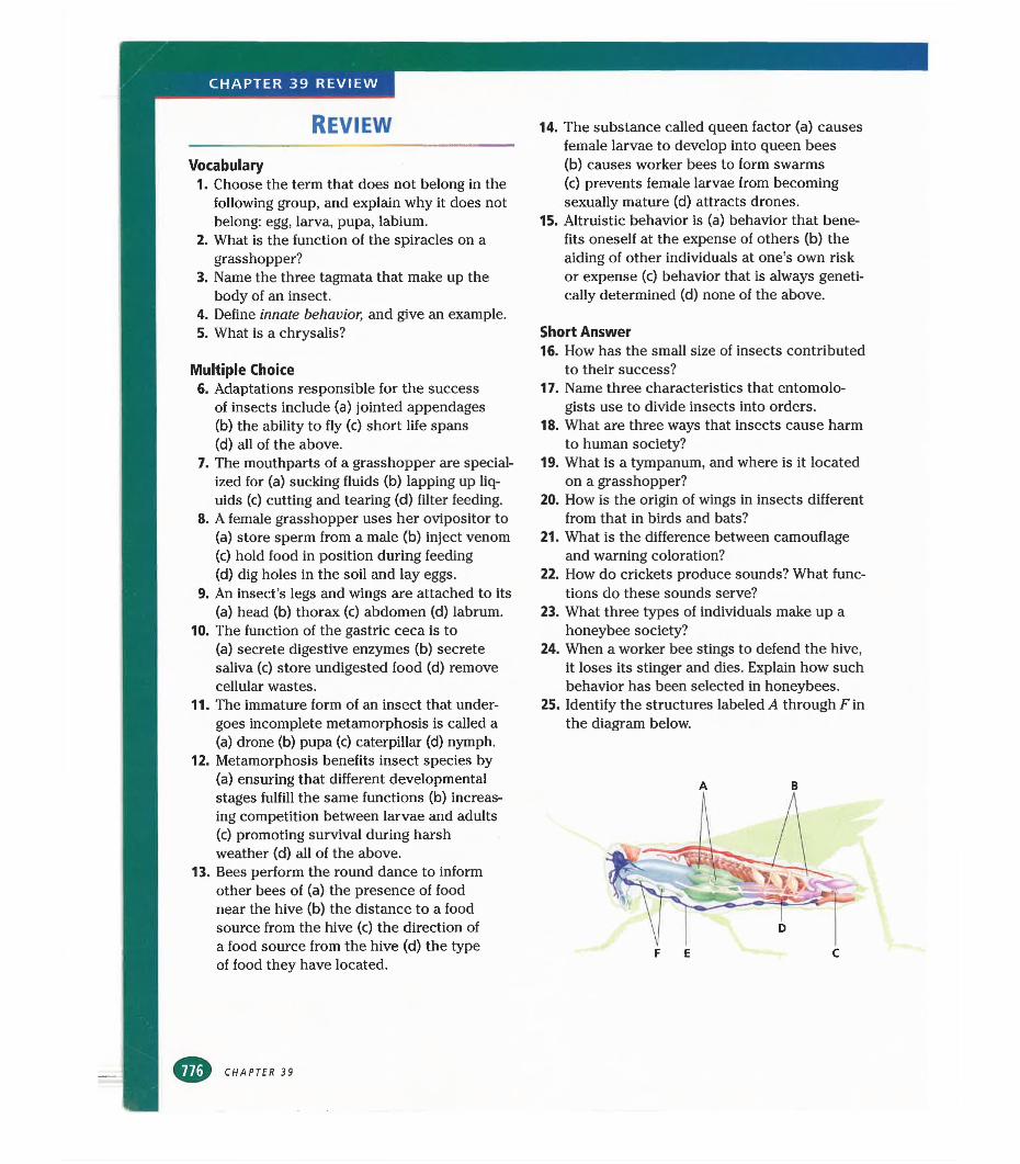
CHAPTER 39 REVIEW
REVIEW
Vocabulary1. Choose the term that does not belong in the
following group, and explain why it does not belong: egg, larva, pupa, labium.
2. What is the function of the spiracles on a grasshopper?
3. Name the three tagmata that make up the body of an insect.
4. Define innate behavior, and give an example.5. What is a chrysalis?
Multiple Choice6. Adaptations responsible for the success
of insects include (a) jointed appendages(b) the ability to fly (c) short life spans(d) all of the above.
7. The mouthparts of a grasshopper are specialized for (a) sucking fluids (b) lapping up liquids (c) cutting and tearing (d) filter feeding.
8. A female grasshopper uses her ovipositor to(a) store sperm from a male (b) inject venom(c) hold food in position during feeding(d) dig holes in the soil and lay eggs.
9. An insect’s legs and wings are attached to its (a) head (b) thorax (c) abdomen (d) labrum.
10. The function of the gastric ceca is to(a) secrete digestive enzymes (b) secrete saliva (c) store undigested food (d) remove cellular wastes.
11. The immature form of an insect that undergoes incomplete metamorphosis is called a (a) drone (b) pupa (c) caterpillar (d) nymph.
12. Metamorphosis benefits insect species by (a) ensuring that different developmental stages fulfill the same functions (b) increasing competition between larvae and adults (c) promoting survival during harsh weather (d) all of the above.
13. Bees perform the round dance to inform other bees of (a) the presence of food near the hive (b) the distance to a food source from the hive (c) the direction of a food source from the hive (d) the type of food they have located.
CHAPTER 39
14. The substance called queen factor (a) causes female larvae to develop into queen bees(b) causes worker bees to form swarms(c) prevents female larvae from becoming sexually mature (d) attracts drones.
15. Altruistic behavior is (a) behavior that benefits oneself at the expense of others (b) the aiding of other individuals at one’s own risk or expense (c) behavior that is always genetically determined (d) none of the above.
Short Answer16. How has the small size of insects contributed
to their success?17. Name three characteristics that entomolo
gists use to divide insects into orders.18. What are three ways that insects cause harm
to human society?19. What is a tympanum, and where is it located
on a grasshopper?20. How is the origin of wings in insects different
from that in birds and bats?21. What is the difference between camouflage
and warning coloration?22. How do crickets produce sounds? What func
tions do these sounds serve?23. What three types of individuals make up a
honeybee society?24. When a worker bee stings to defend the hive,
it loses its stinger and dies. Explain how such behavior has been selected in honeybees.
25. Identify the structures labeled A through Fin the diagram below.
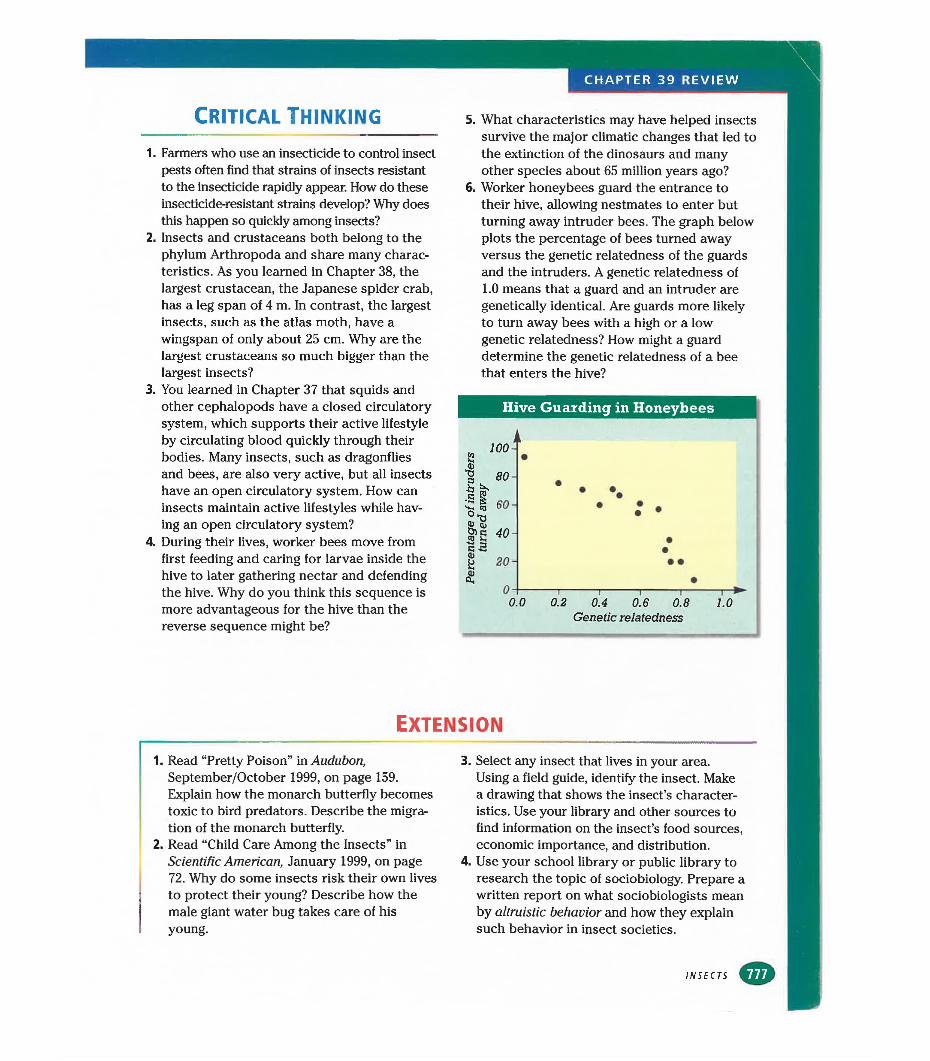
Critical Thinking
1. Farmers who use an insecticide to control insect pests often find that strains of insects resistant to the insecticide rapidly appear. How do these insecticide-resistant strains develop? Why does this happen so quickly among insects?
2. Insects and crustaceans both belong to the phylum Arthropoda and share many characteristics. As you learned in Chapter 38, the largest crustacean, the Japanese spider crab, has a leg span of 4 m. In contrast, the largest insects, such as the atlas moth, have a wingspan of only about 25 cm. Why are the largest crustaceans so much bigger than the largest insects?
3. You learned in Chapter 37 that squids and other cephalopods have a closed circulatory system, which supports their active lifestyle by circulating blood quickly through their bodies. Many insects, such as dragonflies and bees, are also very active, but all insects have an open circulatory system. How can insects maintain active lifestyles while having an open circulatory system?
4. During their lives, worker bees move from first feeding and caring for larvae inside the hive to later gathering nectar and defending the hive. Why do you think this sequence is more advantageous for the hive than the reverse sequence might be?
5. What characteristics may have helped insects survive the major climatic changes that led to the extinction of the dinosaurs and many other species about 65 million years ago?
6. Worker honeybees guard the entrance to their hive, allowing nestmates to enter but turning away intruder bees. The graph below plots the percentage of bees turned away versus the genetic relatedness of the guards and the intruders. A genetic relatedness of 1.0 means that a guard and an intruder are genetically identical. Are guards more likely to turn away bees with a high or a low genetic relatedness? How might a guard determine the genetic relatedness of a bee that enters the hive?
Hive Guarding in Honeybees
T0.2 0.4 0.6 0.8
Genetic relatedness
Extension
1. Read “Pretty Poison” in Audubon, September/October 1999, on page 159. Explain how the monarch butterfly becomes toxic to bird predators. Describe the migration of the monarch butterfly.
2. Read “Child Care Among the Insects” in Scientific American, January 1999, on page 72. Why do some insects risk their own lives to protect their young? Describe how the male giant water bug takes care of his young.
3. Select any insect that lives in your area.Using a field guide, identify the insect. Make a drawing that shows the insect’s characteristics. Use your library and other sources to find information on the insect’s food sources, economic importance, and distribution.
4. Use your school library or public library to research the topic of sociobiology. Prepare a written report on what sociobiologists mean by altruistic behavior and how they explain such behavior in insect societies.
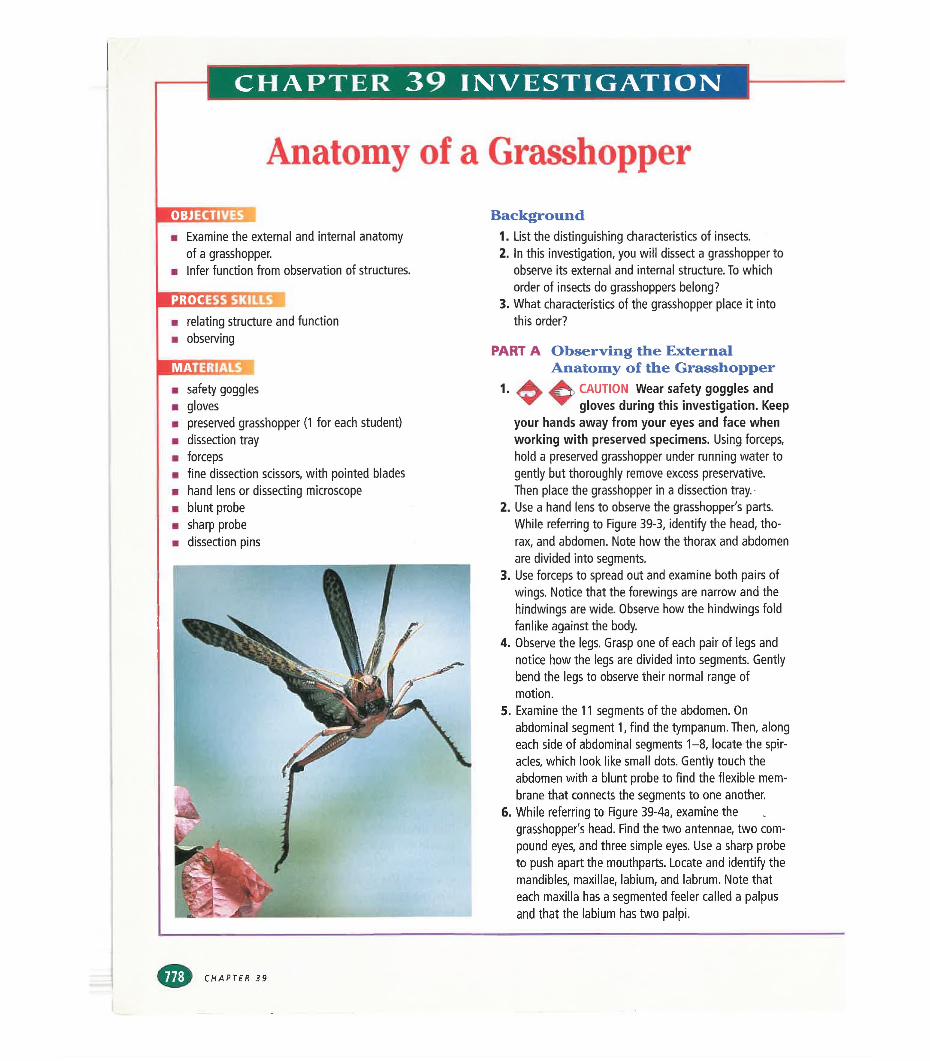
CHAPTER 39 INVESTIGATION
Anatomy of a Grasshopper
a Examine the external and internal anatomy of a grasshopper.
■ Infer function from observation of structures.
PROCESS SKILLS■ relating structure and function■ observing
mmummm safety goggles■ gloves■ preserved grasshopper (1 for each student)■ dissection tray■ forceps■ fine dissection scissors, with pointed blades■ hand lens or dissecting microscope■ blunt probe■ sharp probe■ dissection pins
Background1. List the distinguishing characteristics of insects.2. In this investigation, you will dissect a grasshopper to
observe its external and internal structure. To which order of insects do grasshoppers belong?
3. What characteristics of the grasshopper place it into this order?
PART A Observing the ExternalAnatomy of the Grasshopper
1. CAUTION Wear safety goggles and * ~ gloves during this investigation. Keep
your hands away from your eyes and face when working with preserved specimens. Using forceps, hold a preserved grasshopper under running water to gently but thoroughly remove excess preservative.Then place the grasshopper in a dissection tray.
2. Use a hand lens to observe the grasshopper's parts. While referring to Figure 39-3, identify the head, thorax, and abdomen. Note how the thorax and abdomen are divided into segments.
3. Use forceps to spread out and examine both pairs of wings. Notice that the forewings are narrow and the hindwings are wide. Observe how the hindwings fold fanlike against the body.
4. Observe the legs. Grasp one of each pair of legs and notice how the legs are divided into segments. Gently bend the legs to observe their normal range of motion.
5. Examine the 11 segments of the abdomen. On abdominal segment 1, find the tympanum. Then, along each side of abdominal segments 1-8, locate the spiracles, which look like small dots. Gently touch the abdomen with a blunt probe to find the flexible membrane that connects the segments to one another.
6. While referring to Figure 39-4a, examine the grasshopper's head. Find the two antennae, two compound eyes, and three simple eyes. Use a sharp probe to push apart the mouthparts. Locate and identify the mandibles, maxillae, labium, and labrum. Note that each maxilla has a segmented feeler called a palpus and that the labium has two palpi.
CHAPTER 39
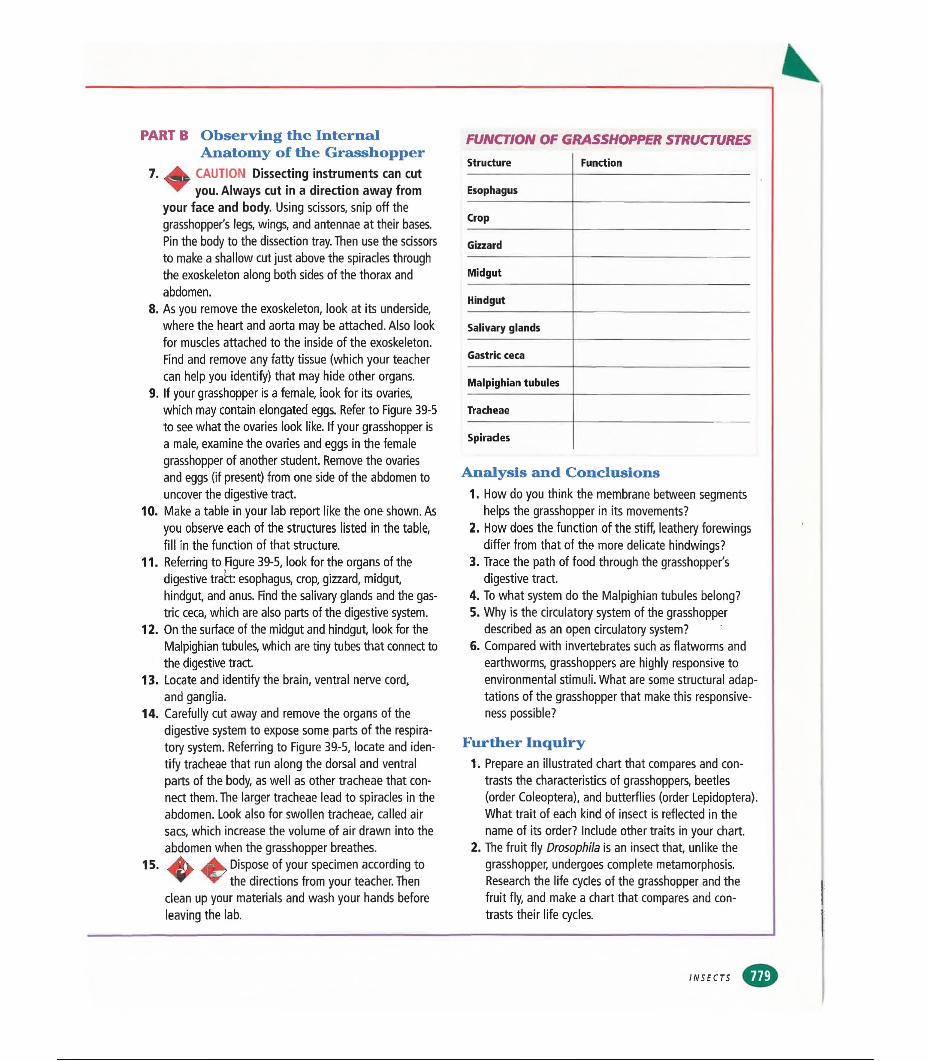
PART B Observing the InternalAnatomy of the Grasshopper
7. CAUTION Dissecting instruments can cut you. Always cut in a direction away from
your face and body. Using scissors, snip off the grasshopper's legs, wings, and antennae at their bases. Pin the body to the dissection tray. Then use the scissors to make a shallow cut just above the spiracles through the exoskeleton along both sides of the thorax and abdomen.
8. As you remove the exoskeleton, look at its underside, where the heart and aorta may be attached. Also look for muscles attached to the inside of the exoskeleton. Find and remove any fatty tissue (which your teacher can help you identify) that may hide other organs.
9. If your grasshopper is a female, look for its ovaries, which may contain elongated eggs. Refer to Figure 39-5 to see what the ovaries look like. If your grasshopper is a male, examine the ovaries and eggs in the female grasshopper of another student. Remove the ovaries and eggs (if present) from one side of the abdomen to uncover the digestive tract.
10. Make a table in your lab report like the one shown. As you observe each of the structures listed in the table, fill in the function of that structure.
11. Referring to Figure 39-5, look for the organs of the digestive tract: esophagus, crop, gizzard, midgut, hindgut, and anus. Find the salivary glands and the gastric ceca, which are also parts of the digestive system.
12. On the surface of the midgut and hindgut, look for the Malpighian tubules, which are tiny tubes that connect to the digestive tract.
13. Locate and identify the brain, ventral nerve cord, and ganglia.
14. Carefully cut away and remove the organs of the digestive system to expose some parts of the respiratory system. Referring to Figure 39-5, locate and identify tracheae that run along the dorsal and ventral parts of the body, as well as other tracheae that connect them. The larger tracheae lead to spiracles in the abdomen. Look also for swollen tracheae, called air sacs, which increase the volume of air drawn into the abdomen when the grasshopper breathes.Jkv, Dispose of your specimen according to
▼ the directions from your teacher. Then clean up your materials and wash your hands before leaving the lab.
15
FUNCTION OF GRASSHOPPER STRUCTURESStructure Function
Esophagus
Crop
Gizzard
Midgut
Hindgut
Salivary glands
Gastric ceca
Malpighian tubules
Tracheae
Spiracles
Analysis and Conclusions1. How do you think the membrane between segments
helps the grasshopper in its movements?2. How does the function of the stiff, leathery forewings
differ from that of the more delicate hindwings?3. Trace the path of food through the grasshopper's
digestive tract.4. To what system do the Malpighian tubules belong?5. Why is the circulatory system of the grasshopper
described as an open circulatory system?6. Compared with invertebrates such as flatworms and
earthworms, grasshoppers are highly responsive to environmental stimuli. What are some structural adaptations of the grasshopper that make this responsiveness possible?
Further Inquiry1. Prepare an illustrated chart that compares and con
trasts the characteristics of grasshoppers, beetles (order Coleóptera), and butterflies (order Lepidoptera). What trait of each kind of insect is reflected in the name of its order? Include other traits in your chart.
2. The fruit fly Drosophila is an insect that, unlike the grasshopper, undergoes complete metamorphosis. Research the life cycles of the grasshopper and the fruit fly, and make a chart that compares and contrasts their life cycles.
INSECTS
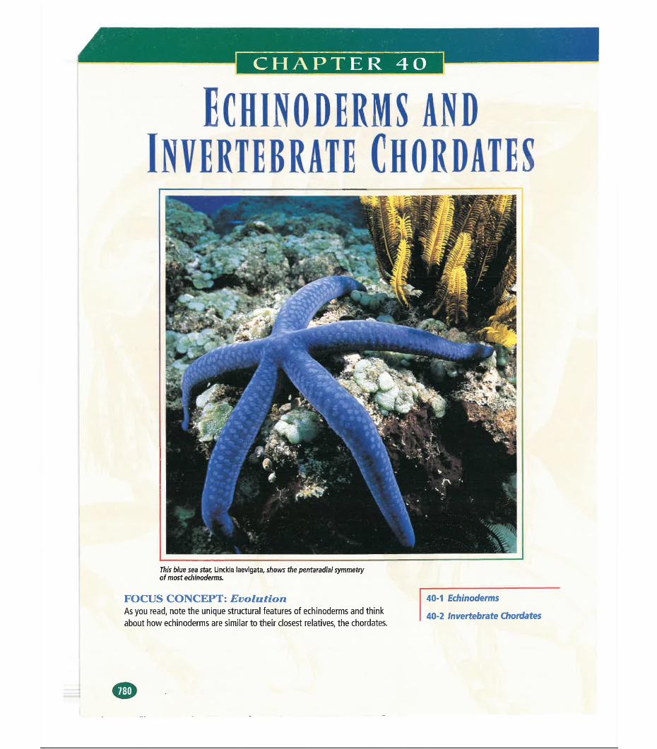
CHAPTER 40
ECH1N0DERMS AND INVERTEBRATE CHORDATES
This blue sea star, Linckia laevigata, shows the pentaradial symmetry of most echinoderms.
FOCUS CONCEPT: EvolutionAs you read, note the unique structural features of echinoderms and think about how echinoderms are similar to their closest relatives, the chordates.
40-1 Echinoderms
40-2 Invertebrate Chordates
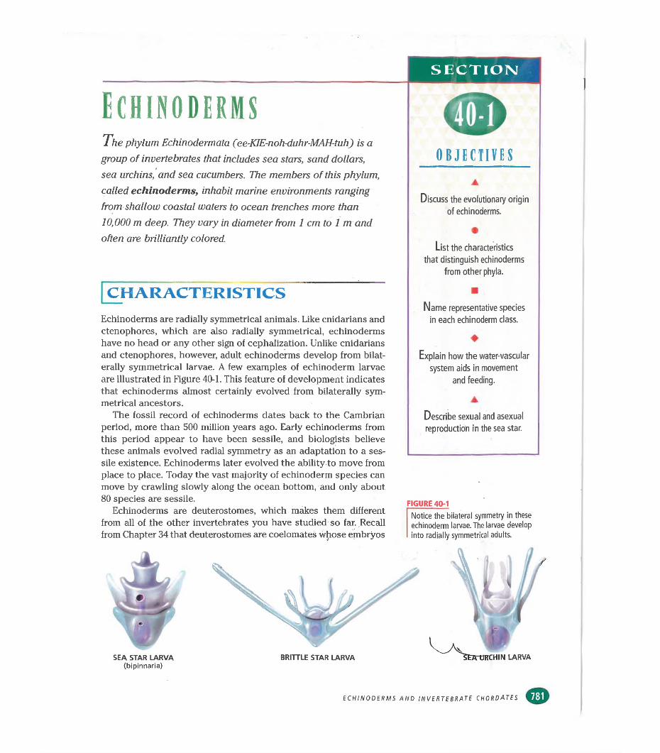
S ECTION
Echinoderms7ft e phylum Echinodermata (ee-KIE-noh-duhr-MAH-tuh) is a group of invertebrates that includes sea stars, sand dollars, sea urchins, and sea cucumbers. The members of this phylum, called echinoderms, inhabit marine environments ranging from shallow coastal waters to ocean trenches more than 10,000 m deep. They vary in diameter from 1 cm to 1 m and often are brilliantly colored.
CHARACTERISTICSEchinoderms are radially symmetrical animals. Like cnidarians and ctenophores, which are also radially symmetrical, echinoderms have no head or any other sign of cephalization. Unlike cnidarians and ctenophores, however, adult echinoderms develop from bilaterally symmetrical larvae. A few examples of echinoderm larvae are illustrated in Figure 40-1. This feature of development indicates that echinoderms almost certainly evolved from bilaterally symmetrical ancestors.
The fossil record of echinoderms dates back to the Cambrian period, more than 500 million years ago. Early echinoderms from this period appear to have been sessile, and biologists believe these animals evolved radial symmetry as an adaptation to a sessile existence. Echinoderms later evolved the ability to move from place to place. Today the vast majority of echinoderm species can move by crawling slowly along the ocean bottom, and only about 80 species are sessile.
Echinoderms are deuterostomes, which makes them different from all of the other invertebrates you have studied so far. Recall from Chapter 34 that deuterostomes are coelomates whose embryos
OBJECTIVES
Discuss the evolutionary origin of echinoderms.
•List the characteristics
that distinguish echinoderms from other phyla.
■Name representative species
in each echinoderm class.
♦Explain how the water-vascular
system aids in movement and feeding.
▲
Describe sexual and asexual reproduction in the sea star.
FIGURE 40-1Notice the bilateral symmetry in these echinoderm larvae. The larvae develop into radially symmetrical adults.
SEA STAR LARVA(bipinnaria)
ECHINODERMS AND INVERTEBRATE CHORDATES
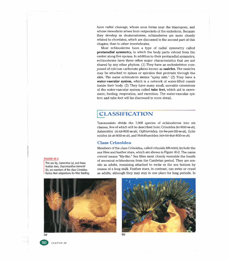
have radial cleavage, whose anus forms near the blastopore, and whose mesoderm arises from outpockets of the endoderm. Because they develop as deuterostomes, echinoderms are more closely related to chordates, which are discussed in the second part of this chapter, than to other invertebrates.
Most echinoderms have a type of radial symmetry called pentaradial symmetry, in which the body parts extend from the center along five spokes. In addition to their pentaradial symmetry, echinoderms have three other major characteristics that are not shared by any other phylum. (1) They have an endoskeleton composed of calcium carbonate plates known as ossicles. The ossicles may be attached to spines or spicules that protrude through the skin. The name echinoderm means “spiny skin.” (2) They have a water-vascular system, which is a network of water-filled canals inside their body. (3) They have many small, movable extensions of the water-vascular system called tube feet, which aid in movement, feeding, respiration, and excretion. The water-vascular system and tube feet will be discussed in more detail.
CLASSIFICATIONTaxonomists divide the 7,000 species of echinoderms into six classes, five of which will be described here: Crinoidea (kri-NOID-ee-uh),
Asteroidea (AS-tuh-ROID-ee-uh), Ophiuroidea (OH-fee-yoor-OID-ee-uh), Echi- noidea (EK-uh-NOID-ee-uh), and Holothuroidea (HOH-loh-thuh-ROID-ee-uh).
FIGURE 40-2
This sea lily, Cenocrinus (a), and these feather stars, Oxycomanthus bennetti (b), are members of the class Crinoidea. Notice their adaptations for filter feeding.
Class CrinoideaMembers of the class Crinoidea, called crinoids (KRI-noids), include the sea lilies and feather stars, which are shown in Figure 40-2. The name crinoid means “lily-like.” Sea lilies most closely resemble the fossils of ancestral echinoderms from the Cambrian period. They are sessile as adults, remaining attached to rocks or the sea bottom by means of a long stalk. Feather stars, in contrast, can swim or crawl as adults, although they may stay in one place for long periods. In
CHAPTER 40
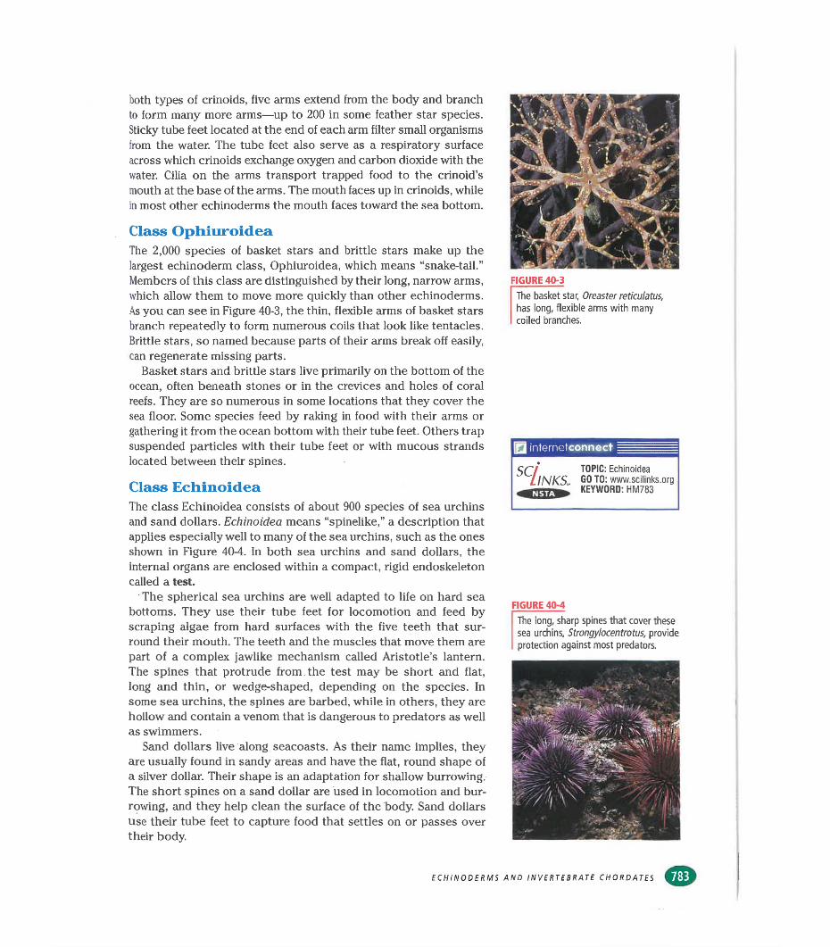
both types of crinoids, five arms extend from the body and branch to form many more arms—up to 200 in some feather star species. Sticky tube feet located at the end of each arm filter small organisms from the water. The tube feet also serve as a respiratory surface across which crinoids exchange oxygen and carbon dioxide with the water. Cilia on the arms transport trapped food to the crinoid’s mouth at the base of the arms. The mouth faces up in crinoids, while in most other echinoderms the mouth faces toward the sea bottom.
Class OphiuroideaThe 2,000 species of basket stars and brittle stars make up the largest echinoderm class, Ophiuroidea, which means “snake-tail.” Members of this class are distinguished by their long, narrow arms, which allow them to move more quickly than other echinoderms. As you can see in Figure 40-3, the thin, flexible arms of basket stars branch repeatedly to form numerous coils that look like tentacles. Brittle stars, so named because parts of their arms break off easily, can regenerate missing parts.
Basket stars and brittle stars live primarily on the bottom of the ocean, often beneath stones or in the crevices and holes of coral reefs. They are so numerous in some locations that they cover the sea floor. Some species feed by raking in food with their arms or gathering it from the ocean bottom with their tube feet. Others trap suspended particles with their tube feet or with mucous strands located between their spines.
Class EchinoideaThe class Echinoidea consists of about 900 species of sea urchins and sand dollars. Echinoidea means “spinelike,” a description that applies especially well to many of the sea urchins, such as the ones shown in Figure 40-4. In both sea urchins and sand dollars, the internal organs are enclosed within a compact, rigid endoskeleton called a test.
The spherical sea urchins are well adapted to life on hard sea bottoms. They use their tube feet for locomotion and feed by scraping algae from hard surfaces with the five teeth that surround their mouth. The teeth and the muscles that move them are part of a complex jawlike mechanism called Aristotle’s lantern. The spines that protrude from the test may be short and flat, long and thin, or wedge-shaped, depending on the species. In some sea urchins, the spines are barbed, while in others, they are hollow and contain a venom that is dangerous to predators as well as swimmers.
Sand dollars live along seacoasts. As their name implies, they are usually found in sandy areas and have the flat, round shape of a silver dollar. Their shape is an adaptation for shallow burrowing. The short spines on a sand dollar are used in locomotion and burrowing, and they help clean the surface of the body. Sand dollars use their tube feet to capture food that settles on or passes over their body.
FIGURE 40-3
The basket star, Oreaster reticulatus, has long, flexible arms with many coiled branches.
SCllNKS, TOPIC: Echinoidea GO TO: www.scilinks.orgKEYWORD: HM783
FIGURE 40-4
The long, sharp spines that cover these sea urchins, Strongylocentrotus, provide protection against most predators.
ECHINODERMS AND INVERTEBRATE CHORDATES
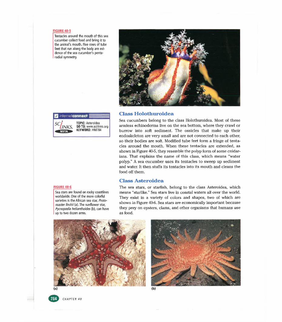
FIGURE 40-5
Tentacles around the mouth of this sea cucumber collect food and bring it to the animal's mouth. Five rows of tube feet that run along the body are evidence of the sea cucumber's penta- radial symmetry.
J internetconnect
5C INKSl TOPIC: Asteroidea GO TO: www.scllinks.org KEYWORD: HM784
Class HolothuroideaSea cucumbers belong to the class Holothuroidea. Most of these armless echinoderms live on the sea bottom, where they crawl or burrow into soft sediment. The ossicles that make up their endoskeleton are very small and are not connected to each other, so their bodies are soft. Modified tube feet form a fringe of tentacles around the mouth. When these tentacles are extended, as shown in Figure 40-5, they resemble the polyp form of some cnidar- ians. That explains the name of this class, which means “water polyp.” A sea cucumber uses its tentacles to sweep up sediment and water. It then stuffs its tentacles into its mouth and cleans the food off them.
FIGURE 40-6
Sea stars are found on rocky coastlines worldwide, One of the more colorful varieties is the African sea star, Protoreaster linckii (a). The sunflower star, Pycnopodia helianthoides (b), can have up to two dozen arms.
Class AsteroideaThe sea stars, or starfish, belong to the class Asteroidea, which means “starlike.” Sea stars live in coastal waters all over the world. They exist in a variety of colors and shapes, two of which are shown in Figure 40-6. Sea stars are economically important because they prey on oysters, clams, and other organisms that humans use as food.
CHAPTER 40
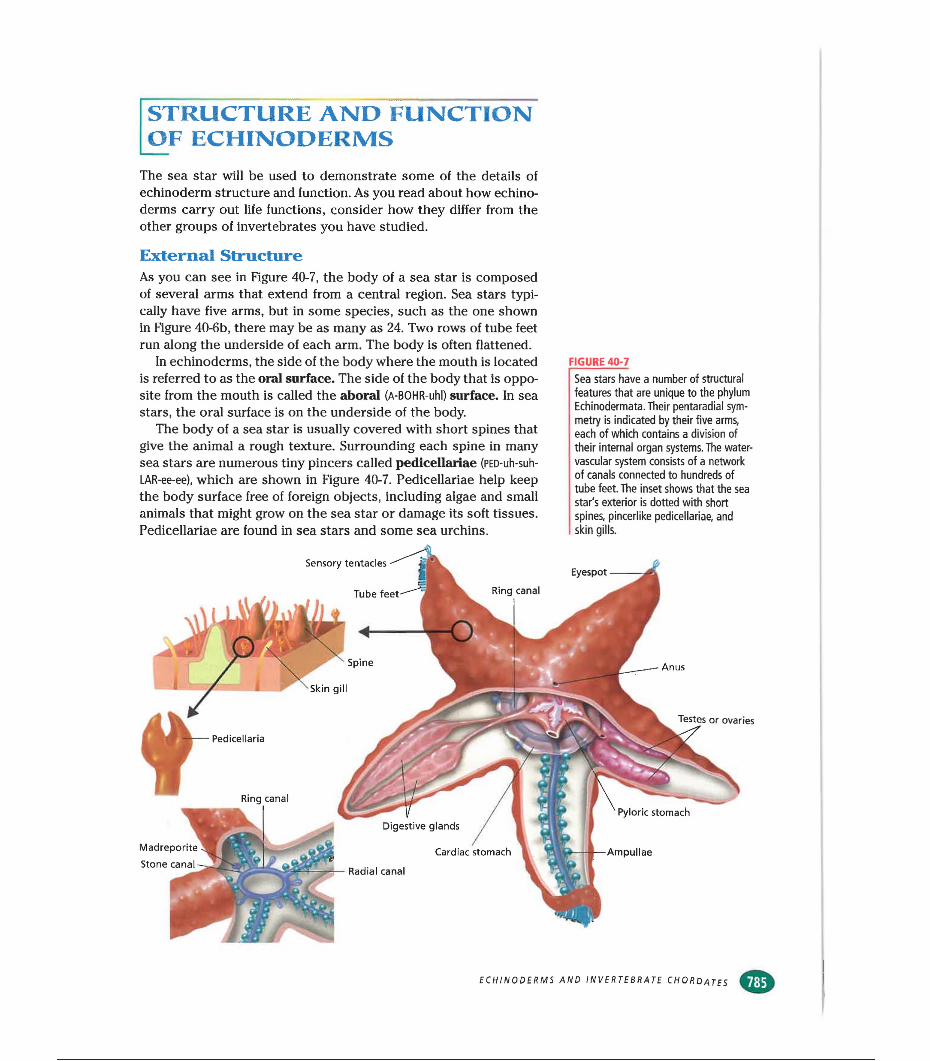
STRUCTURE AND FUNCTION OF ECHINODERMS
The sea star will be used to demonstrate some of the details of echinoderm structure and function. As you read about how echino- derms carry out life functions, consider how they differ from the other groups of invertebrates you have studied.
External StructureAs you can see in Figure 40-7. the body of a sea star is composed of several arms that extend from a central region. Sea stars typically have five arms, but in some species, such as the one shown in Figure 40-6b, there may be as many as 24. Two rows of tube feet run along the underside of each arm. The body is often flattened.
In echinoderms, the side of the body where the mouth is located is referred to as the oral surface. The side of the body that is opposite from the mouth is called the aboral (A-BOHR-uhl) surface. In sea stars, the oral surface is on the underside of the body.
The body of a sea star is usually covered with short spines that give the animal a rough texture. Surrounding each spine in many sea stars are numerous tiny pincers called pedicellariae (PED-uh-suh-
LAR-ee-ee), which are shown in Figure 40-7. Pedicellariae help keep the body surface free of foreign objects, including algae and small animals that might grow on the sea star or damage its soft tissues. Pedicellariae are found in sea stars and some sea urchins.
FIGURE 40-7
Sea stars have a number of structural features that are unique to the phylum Echinodermata. Their pentaradial symmetry is indicated by their five arms, each of which contains a division of their internal organ systems. The water- vascular system consists of a network of canals connected to hundreds of tube feet. The inset shows that the sea star's exterior is dotted with short spines, pincerlike pedicellariae, and skin gills.
Sensory tentaclesEyespot
or ovaries
Pedicellaria
Pyloric stomachDigestive glands
Madreporite Stone canal -
Cardiac stomach Ampullae
ECHINODERMS AND INVERTEBRATE CHORDATES
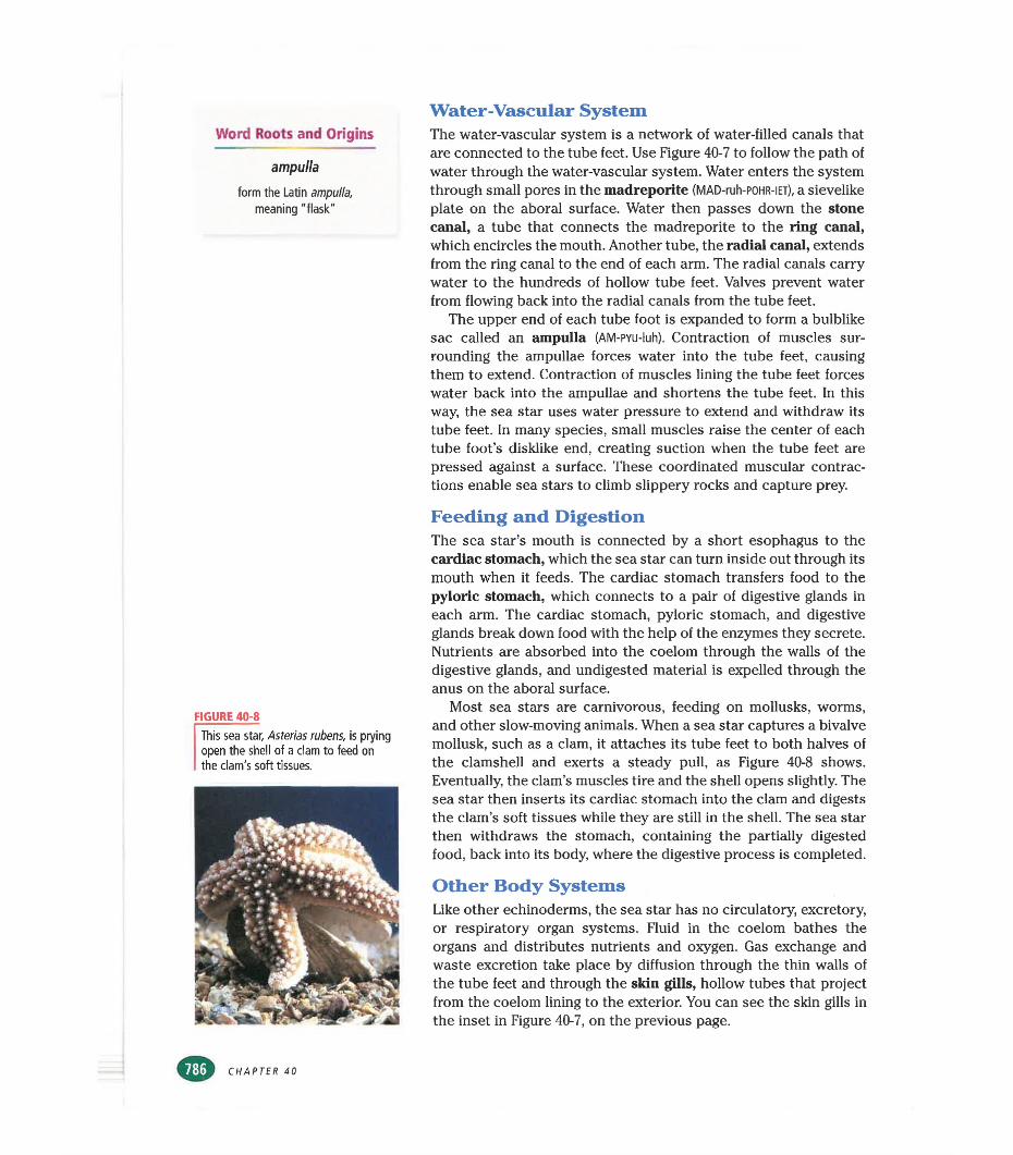
Word Roots and Origins
ampulla
form the Latin ampulla, meaning "flask"
Water-Vascular SystemThe water-vascular system is a network of water-filled canals that are connected to the tube feet. Use Figure 40-7 to follow the path of water through the water-vascular system. Water enters the system through small pores in the madreporite (MAD-ruh-POHR-iET), a sievelike plate on the aboral surface. Water then passes down the stone canal, a tube that connects the madreporite to the ring canal, which encircles the mouth. Another tube, the radial canal, extends from the ring canal to the end of each arm. The radial canals carry water to the hundreds of hollow tube feet. Valves prevent water from flowing back into the radial canals from the tube feet.
The upper end of each tube foot is expanded to form a bulblike sac called an ampulla (AM-PYU-luh). Contraction of muscles surrounding the ampullae forces water into the tube feet, causing them to extend. Contraction of muscles lining the tube feet forces water back into the ampullae and shortens the tube feet. In this way, the sea star uses water pressure to extend and withdraw its tube feet. In many species, small muscles raise the center of each tube foot’s disklike end, creating suction when the tube feet are pressed against a surface. These coordinated muscular contractions enable sea stars to climb slippery rocks and capture prey.
FIGURE 40-8
This sea star, Asterias rubens, is prying open the shell of a clam to feed on the clam's soft tissues.
Feeding and DigestionThe sea star’s mouth is connected by a short esophagus to the cardiac stomach, which the sea star can turn inside out through its mouth when it feeds. The cardiac stomach transfers food to the pyloric stomach, which connects to a pair of digestive glands in each arm. The cardiac stomach, pyloric stomach, and digestive glands break down food with the help of the enzymes they secrete. Nutrients are absorbed into the coelom through the walls of the digestive glands, and undigested material is expelled through the anus on the aboral surface.
Most sea stars are carnivorous, feeding on mollusks, worms, and other slow-moving animals. When a sea star captures a bivalve mollusk, such as a clam, it attaches its tube feet to both halves of the clamshell and exerts a steady pull, as Figure 40-8 shows. Eventually, the clam’s muscles tire and the shell opens slightly. The sea star then inserts its cardiac stomach into the clam and digests the clam’s soft tissues while they are still in the shell. The sea star then withdraws the stomach, containing the partially digested food, back into its body, where the digestive process is completed.
Other Body SystemsLike other echinoderms, the sea star has no circulatory, excretory, or respiratory organ systems. Fluid in the coelom bathes the organs and distributes nutrients and oxygen. Gas exchange and waste excretion take place by diffusion through the thin walls of the tube feet and through the skin gills, hollow tubes that project from the coelom lining to the exterior. You can see the skin gills in the inset in Figure 40-7, on the previous page.
CHAPTER 40
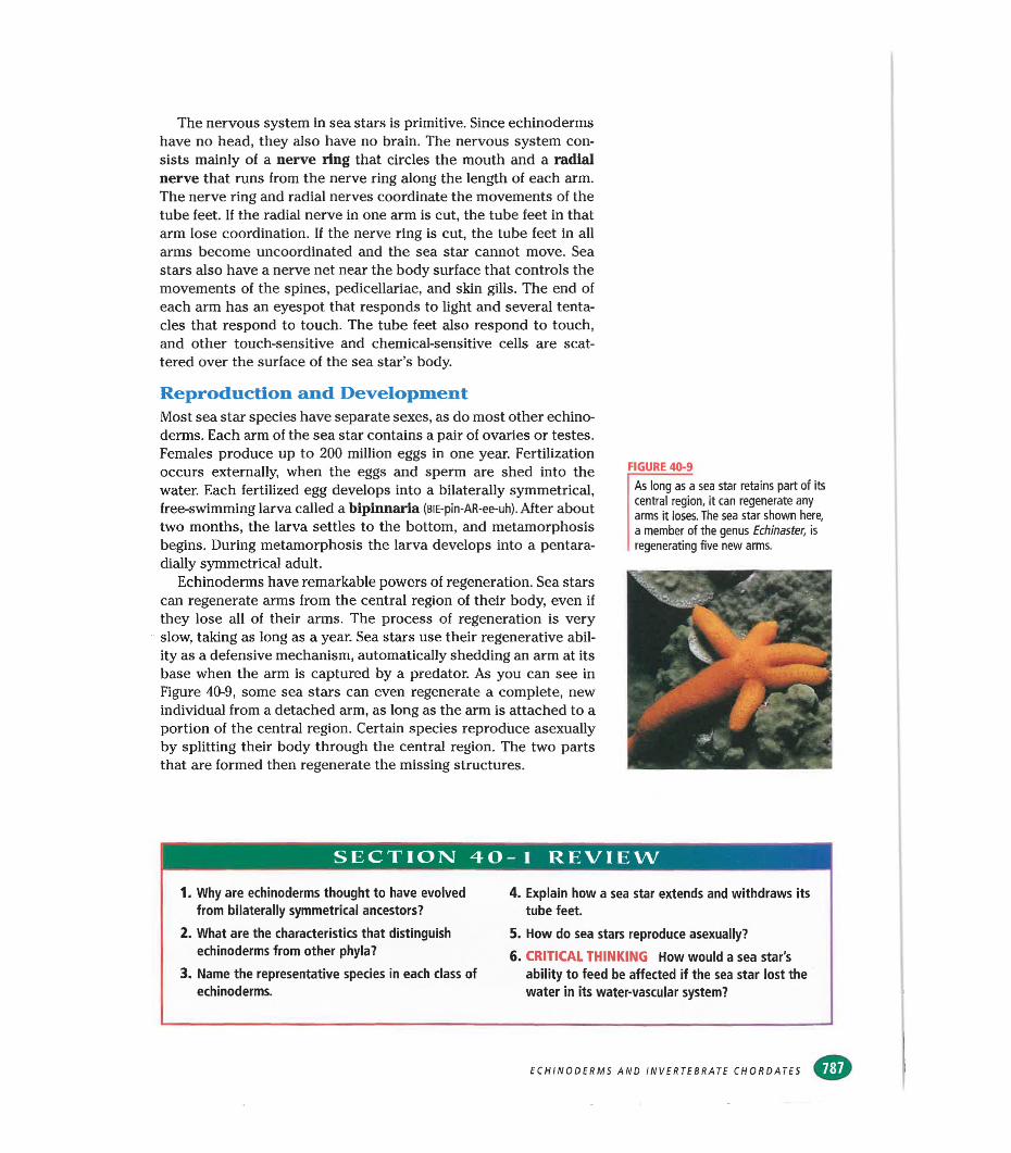
The nervous system in sea stars is primitive. Since echinoderms have no head, they also have no brain. The nervous system consists mainly of a nerve ring that circles the mouth and a radial nerve that runs from the nerve ring along the length of each arm. The nerve ring and radial nerves coordinate the movements of the tube feet. If the radial nerve in one arm is cut, the tube feet in that arm lose coordination. If the nerve ring is cut, the tube feet in all arms become uncoordinated and the sea star cannot move. Sea stars also have a nerve net near the body surface that controls the movements of the spines, pedicellariae, and skin gills. The end of each arm has an eyespot that responds to light and several tentacles that respond to touch. The tube feet also respond to touch, and other touch-sensitive and chemical-sensitive cells are scattered over the surface of the sea star’s body.
Reproduction and DevelopmentMost sea star species have separate sexes, as do most other echinoderms. Each arm of the sea star contains a pair of ovaries or testes. Females produce up to 200 million eggs in one year. Fertilization occurs externally, when the eggs and sperm are shed into the water. Each fertilized egg develops into a bilaterally symmetrical, free-swimming larva called a bipinnaria (BiE-pin-AR-ee-uh). After about two months, the larva settles to the bottom, and metamorphosis begins. During metamorphosis the larva develops into a pentara- dially symmetrical adult.
Echinoderms have remarkable powers of regeneration. Sea stars can regenerate arms from the central region of their body, even if they lose all of their arms. The process of regeneration is very slow, taking as long as a year. Sea stars use their regenerative ability as a defensive mechanism, automatically shedding an arm at its base when the arm is captured by a predator. As you can see in Figure 40-9, some sea stars can even regenerate a complete, new individual from a detached arm, as long as the arm is attached to a portion of the central region. Certain species reproduce asexually by splitting their body through the central region. The two parts that are formed then regenerate the missing structures.
FIGURE 40-9
As long as a sea star retains part of its central region, it can regenerate any arms it loses. The sea star shown here, a member of the genus Echinaster, is regenerating five new arms.
SECTION 40- 1 REVIEW
1. Why are echinoderms thought to have evolved from bilaterally symmetrical ancestors?
2. What are the characteristics that distinguish echinoderms from other phyla?
3. Name the representative species in each class of echinoderms.
4. Explain how a sea star extends and withdraws its tube feet.
5. How do sea stars reproduce asexually?6. CRITICAL THINKING How would a sea star's
ability to feed be affected if the sea star lost the water in its water-vascular system?
ECHINODERMS AND INVERTEBRATE CHORDATES
SECTION!
internefconnect
OBJECTIVES
IiInvertebrateChordates
A
List the major characteristics of chordates.
•Describe the structure
of lancelets.
The phylum Chordata (kohr-DAHT-uh) includes all of the
vertebrates, or animals with backbones. It also includes two groups of animals that lack backbones and are therefore invertebrates. The development of chordates is similar to the development of echinoderms, suggesting that these two phyladescended from a common ancestor.
■Describe the structure
of tunicates. CHARACTERISTICSAll animals with a backbone are vertebrates, and they make up one
SCilNKSL TOPIC: Chordates GO TO: www.scilinks.org KEYWORD: HM788
of the subphyla in the phylum Chordata, whose members are called chordates. Chordates are so named because they have a notochord, a stiff but flexible rod of cells that runs the length of the body near the dorsal surface. The notochord is illustrated in Figure 40-10. The stiffness of the notochord provides a resistance against which the body muscles can exert force when they contract. The flexibility of the notochord allows the body to bend from side to side as well as up and down. Some kinds of chordates retain the notochord throughout their life. In most vertebrates, however, the notochord is present in embryos but becomes greatly reduced when the vertebral column, or backbone, develops. In adult mammals, the notochord persists only as small patches of tissue between the bones of the vertebral column.
You learned in Chapter 34 that in addition to a notochord, all chordates have the following three characteristics during some stage of their life: (1) a dorsal nerve cord, (2) pharyngeal pouches, and (3) a postanal tail. These characteristics are also illustrated in Figure 40-10. Unlike the ventral nerve cords of invertebrates such as
FIGURE 40-10
All chordates have a notochord, a dorsal nerve cord, pharyngeal pouches, and a postanal tail during at least some stage of their life.
Dorsal nerve cord NotochordMuscles
Mouth 'Pharyngeal pouch with slits
Anus Postanaltail
CHAPTER 40
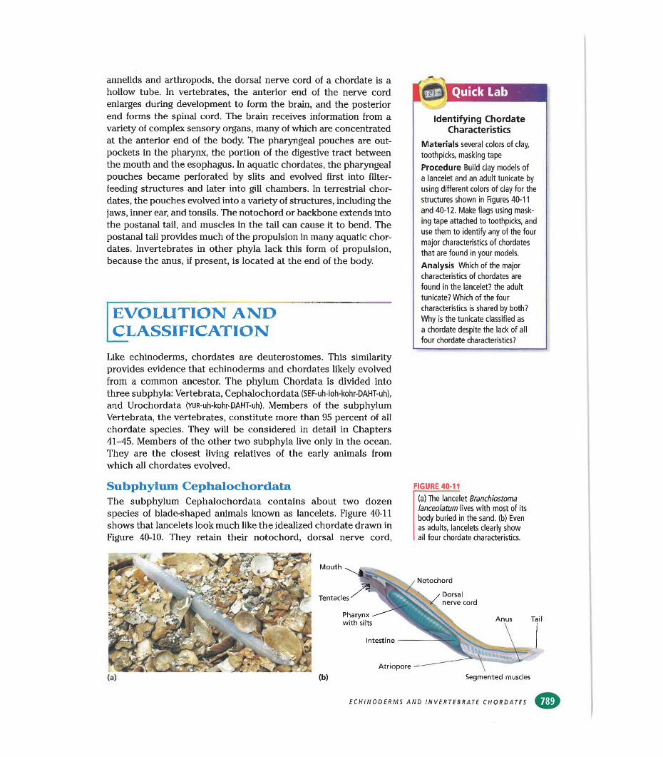
annelids and arthropods, the dorsal nerve cord of a chordate is a hollow tube. In vertebrates, the anterior end of the nerve cord enlarges during development to form the brain, and the posterior end forms the spinal cord. The brain receives information from a variety of complex sensory organs, many of which are concentrated at the anterior end of the body. The pharyngeal pouches are out- pockets in the pharynx, the portion of the digestive tract between the mouth and the esophagus. In aquatic chordates, the pharyngeal pouches became perforated by slits and evolved first into filterfeeding structures and later into gill chambers. In terrestrial chordates, the pouches evolved into a variety of structures, including the jaws, inner ear, and tonsils. The notochord or backbone extends into the postanal tail, and muscles in the tail can cause it to bend. The postanal tail provides much of the propulsion in many aquatic chordates. Invertebrates in other phyla lack this form of propulsion, because the anus, if present, is located at the end of the body.
EVOLUTION AND CLASSIFICATION
Like echinoderms, chordates are deuterostomes. This similarity provides evidence that echinoderms and chordates likely evolved from a common ancestor. The phylum Chordata is divided into three subphyla: Vertebrata, Cephalochordata (SEF-uh-loh-kohr-DAHT-uh),
and Urochordata (YUR-uh-kohr-DAHT-uh). Members of the subphylum Vertebrata, the vertebrates, constitute more than 95 percent of all chordate species. They will be considered in detail in Chapters 41-45. Members of the other two subphyla live only in the ocean. They are the closest living relatives of the early animals from which all chordates evolved.
Quick Lab
Identifying Chordate Characteristics
Materials several colors of clay, toothpicks, masking tape
Procedure Build clay models of a lancelet and an adult tunicate by using different colors of clay for the structures shown in Figures 40-11 and 40-12, Make flags using masking tape attached to toothpicks, and use them to identify any of the four major characteristics of chordates that are found in your models.
Analysis Which of the major characteristics of chordates are found in the lancelet? the adult tunicate? Which of the four characteristics is shared by both? Why is the tunicate classified as a chordate despite the lack of all four chordate characteristics?
Subphylum CephalochordataThe subphylum Cephalochordata contains about two dozen species of blade-shaped animals known as lancelets. Figure 40-11 shows that lancelets look much like the idealized chordate drawn in Figure 40-10. They retain their notochord, dorsal nerve cord,
FIGURE 40-11
(a) The lancelet Branchiostoma lanceolatum lives with most of its body buried in the sand, (b) Even as adults, lancelets clearly show all four chordate characteristics.
Mouth
Notochord
Tentacles Dorsal nerve cord
Pharynx with slits
Intestine
Segmented musclesAtriopore
ECHINODERMS AND INVERTEBRATE CHORDATES
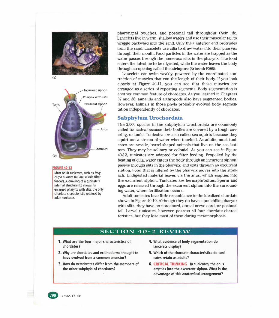
Anus
Incurrent siphon
Pharynx with slits
Excurrent siphon
Stomach
Tunic
FIGURE 40-12
Most adult tunlcates, such as Poly- carpa auranta (a), are sessile filter feeders. A drawing of a tunicate's internal structure (b) shows Its enlarged pharynx with slits, the only chordate characteristic retained by adult tunicates.
pharyngeal pouches, and postanal tail throughout their life. Lancelets live in warm, shallow waters and use their muscular tail to wriggle backward into the sand. Only their anterior end protrudes from the sand. Lancelets use cilia to draw water into their pharynx through their mouth. Food particles in the water are trapped as the water passes through the numerous slits in the pharynx. The food enters the intestine to be digested, while the water leaves the body through an opening called the atriopore (AY-tree-oh-POHR).
Lancelets can swim weakly, powered by the coordinated contraction of muscles that run the length of their body. If you look closely at Figure 40-11, you can see that these muscles are arranged as a series of repeating segments. Body segmentation is another common feature of chordates. As you learned in Chapters 37 and 38, annelids and arthropods also have segmented bodies. However, animals in those phyla probably evolved body segmentation independently of chordates.
Subphylum UrochordataThe 2,000 species in the subphylum Urochordata are commonly called tunicates because their bodies are covered by a tough covering, or tunic. Tunicates are also called sea squirts because they squirt out a stream of water when touched. As adults, most tunicates are sessile, barrel-shaped animals that live on the sea bottom. They may be solitary or colonial. As you can see in Figure 40-12, tunicates are adapted for filter feeding. Propelled by the beating of cilia, water enters the body through an incurrent siphon, passes through slits in the pharynx, and exits through an excurrent siphon. Food that is filtered by the pharynx moves into the stomach. Undigested material leaves via the anus, which empties into the excurrent siphon. Tunicates are hermaphrodites. Sperm and eggs are released through the excurrent siphon into the surrounding water, where fertilization occurs.
Adult tunicates bear little resemblance to the idealized chordate shown in Figure 40-10. Although they do have a pouchlike pharynx with slits, they have no notochord, dorsal nerve cord, or postanal tail. Larval tunicates, however, possess all four chordate characteristics, but they lose most of them during metamorphosis.
SECTION 40-2 REVIEW
1. What are the four major characteristics of chordates?
2. Why are chordates and echinoderms thought to have evolved from a common ancestor?
3. How do vertebrates differ from the members of the other subphyla of chordates?
4. What evidence of body segmentation do lancelets display?
5. Which of the chordate characteristics do tunicates retain as adults?
6. CRITICAL THINKING In tunicates, the anus empties into the excurrent siphon. What is the advantage of this anatomical arrangement?
CHAPTER 40
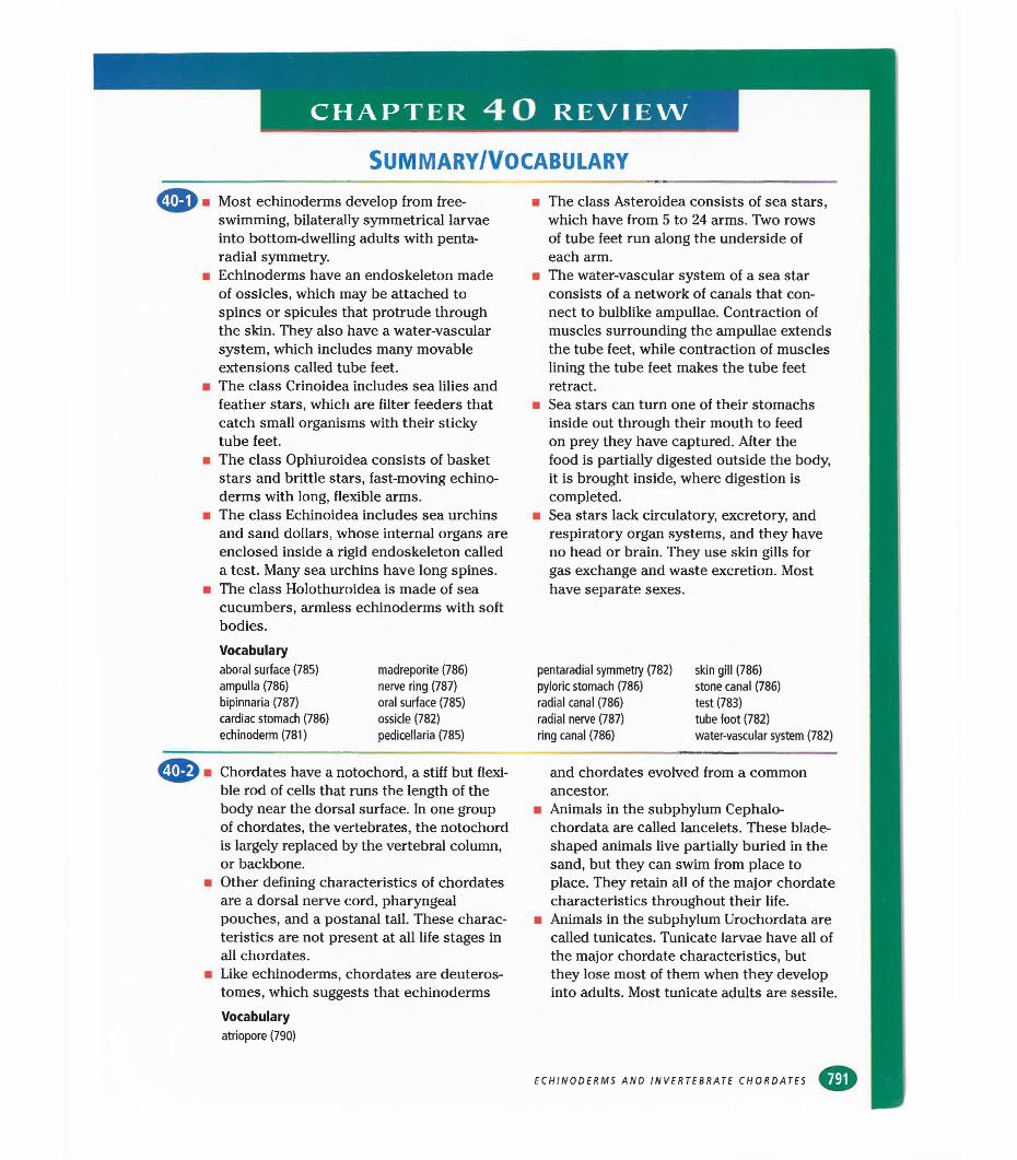
CHAPTER 40 REVIEW
SUMMARY/VOCABULARY
ipl ■ Most echinoderms develop from free- swimming, bilaterally symmetrical larvae into bottom-dwelling adults with penta- radial symmetry.
■ Echinoderms have an endoskeleton made of ossicles, which may be attached to spines or spicules that protrude through the skin. They also have a water-vascular system, which includes many movable extensions called tube feet.The class Crinoidea includes sea lilies and feather stars, which are filter feeders that catch small organisms with their sticky tube feet.
■ The class Ophiuroidea consists of basket stars and brittle stars, fast-moving echinoderms with long, flexible arms.
■ The class Echinoidea includes sea urchins and sand dollars, whose internal organs are enclosed inside a rigid endoskeleton called a test. Many sea urchins have long spines.
■ The class Holothuroidea is made of sea cucumbers, armless echinoderms with soft bodies.Vocabularyaboral surface (785) ampulla (786) bipinnaria (787) cardiac stomach (786) echinoderm (781)
madreporite (786) nerve ring (787) oral surface (785) ossicle (782) pedicellaria (785)
■ The class Asteroidea consists of sea stars, which have from 5 to 24 arms. Two rows of tube feet run along the underside of each arm.
■ The water-vascular system of a sea star consists of a network of canals that connect to bulblike ampullae. Contraction of muscles surrounding the ampullae extends the tube feet, while contraction of muscles lining the tube feet makes the tube feet retract.
■ Sea stars can turn one of their stomachs inside out through their mouth to feed on prey they have captured. After the food is partially digested outside the body, it is brought inside, where digestion is completed.
■ Sea stars lack circulatory, excretory, and respiratory organ systems, and they have no head or brain. They use skin gills for gas exchange and waste excretion. Most have separate sexes.
pentaradial symmetry (782) pyloric stomach (786) radial canal (786) radial nerve (787) ring canal (786)
skin gill (786) stone canal (786) test (783) tube foot (782) water-vascular system (782)
Chordates have a notochord, a stiff but flexible rod of cells that runs the length of the body near the dorsal surface. In one group of chordates, the vertebrates, the notochord is largely replaced by the vertebral column, or backbone.
■ Other defining characteristics of chordates are a dorsal nerve cord, pharyngeal pouches, and a postanal tail. These characteristics are not present at all life stages in all chordates.
■ Like echinoderms, chordates are deuteros- tomes, which suggests that echinodermsVocabularyatriopore (790)
and chordates evolved from a common ancestor.
■ Animals in the subphylum Cephalo- chordata are called lancelets. These bladeshaped animals live partially buried in the sand, but they can swim from place to place. They retain all of the major chordate characteristics throughout their life.
■ Animals in the subphylum Urochordata are called tunicates. Tunicate larvae have all of the major chordate characteristics, but they lose most of them when they develop into adults. Most tunicate adults are sessile.
ECHINODERMS AND INVERTEBRATE CHORDATES
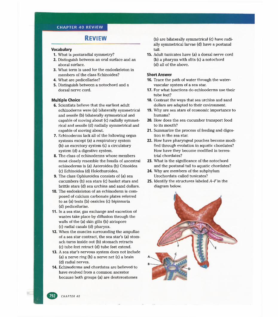
CHAPTER 40 REVIEW
REVIEW
Vocabulary1. What is pentaradial symmetry?2. Distinguish between an oral surface and an
aboral surface.3. What term is used for the endoskeleton in
members of the class Echinoidea?4. What are pedicellariae?5. Distinguish between a notochord and a
dorsal nerve cord.
Multiple Choice6. Scientists believe that the earliest adult
echinoderms were (a) bilaterally symmetrical and sessile (b) bilaterally symmetrical and capable of moving about (c) radially symmetrical and sessile (d) radially symmetrical and capable of moving about.
7. Echinoderms lack all of the following organ systems except (a) a respiratory system(b) an excretory system (c) a circulatory system (d) a digestive system.
8. The class of echinoderms whose members most closely resemble the fossils of ancestral echinoderms is (a) Asteroidea (b) Crinoidea(c) Echinoidea (d) Holothuroidea.
9. The class Ophiuroidea consists of (a) sea cucumbers (b) sea stars (c) basket stars and brittle stars (d) sea urchins and sand dollars.
10. The endoskeleton of an echinoderm is composed of calcium carbonate plates referred to as (a) tests (b) ossicles (c) bipinnaria(d) pedicellariae.
11. In a sea star, gas exchange and excretion of wastes take place by diffusion through the walls of the (a) skin gills (b) atriopore(c) radial canals (d) pharynx.
12. When the muscles surrounding the ampullae of a sea star contract, the sea star’s (a) stomach turns inside out (b) stomach retracts(c) tube feet retract (d) tube feet extend.
13. A sea star’s nervous system does not include (a) a nerve ring (b) a nerve net (c) a brain(d) radial nerves.
14. Echinoderms and chordates are believed to have evolved from a common ancestor because both groups (a) are deuterostomes
(b) are bilaterally symmetrical (c) have radially symmetrical larvae (d) have a postanal tail.
15. Adult tunicates have (a) a dorsal nerve cord (b) a pharynx with slits (c) a notochord(d) all of the above.
Short Answer16. Trace the path of water through the water-
vascular system of a sea star.17. For what functions do echinoderms use their
tube feet?18. Contrast the ways that sea urchins and sand
dollars are adapted to their environment.19. Why are sea stars of economic importance to
humans?20. How does the sea cucumber transport food
to its mouth?21. Summarize the process of feeding and diges
tion in the sea star.22. How have pharyngeal pouches become modi
fied through evolution in aquatic chordates? How have they become modified in terrestrial chordates?
23. What is the significance of the notochord and the postanal tail to aquatic chordates?
24. Why are members of the subphylum Urochordata called tunicates?
25. Identify the structures labeled A-F in the diagram below.
CHAPTER 40 REVIEW
Critical Thinking
1. Sea cucumbers and sea lilies are relatively sessile animals. Their larvae, however, are capable of swimming. What advantage do swimming larvae provide for these echinoderms?
2. Commercial oyster farmers used to remove sea stars from their oyster beds, chop them in half, and throw them back into the water. Was this a good way to protect the oysters from sea stars? If your answer is yes, explain why. If your answer is no, explain why not and describe a better method.
3. Scientists have found many echinoderm fossils from the Cambrian period, but they have found few fossils of other species that must have lived during the Cambrian period. What might explain the large number of fossilized echinoderms?
4. Sea urchin eggs, or roe, are highly prized in Japan for sushi. In trying to supply the Japanese market for this delicacy, divers have nearly wiped out sea urchin populations in some areas of California. What steps could be taken to reestablish sea urchin populations in those areas?
5. Great populations of plankton rise to the surface of the ocean at night and return to the ocean depths during the day. Basket stars are also active at night, when they uncoil their thin, flexible arms. During the day, basket stars curl up and become a compact mass. Why do you think basket stars uncoil their arms at night and coil up during the day?
6. Neoteny is a phenomenon in which larvae become sexually mature while retaining larval characteristics. The term has been used to help explain the evolution of vertebrates from ancestral chordates. Use what you know about tunicates to support this hypothesis.
7. The two curves below show relative changes in the populations of two mollusk species (A and B) in a coastal area over time. At the time indicated by the dotted line, a species of sea star that preys on both A and B was introduced to the area. Which mollusk species is the preferred prey of the sea stars? Why did the population of the other mollusk species also decline in size after the sea stars were introduced? Explain your answers.
Decline of Mollusk Populations
Extension
1. Read “Universe in a Puddle” in Life, April 1998, on page 52. Describe some of the adaptations that tidal pool organisms have that help them survive the crashing and pounding of incoming tides.
2. Write a short paper on a species of echinoderms that is not discussed in the chapter. Focus on the particular adaptations that enable the species you have selected to
survive in its environment.3. Research several species of sea urchins that
have very different types of spines. Why and how might these differences have evolved? Report on why you think sea urchin spines have different forms. Mention in your report the mechanisms that predators use to get past these spines.
ECHINODERMS AND INVERTEBRATE CHORDATES
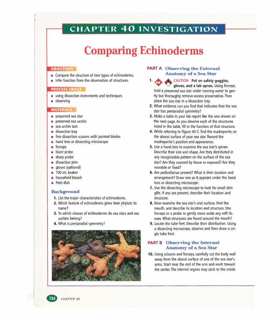
CHAPTER 40 INVESTIGATION
Comparing EchinodermsOBJECT!■ Compare the structure of two types of echinoderms.■ Infer function from the observation of structures.
PROCESS S■ using dissection instruments and techniques■ observing
■IMilWHH
■ preserved sea star■ preserved sea urchin■ sea urchin test■ dissection tray■ fine dissection scissors with pointed blades■ hand lens or dissecting microscope■ forceps■ blunt probe■ sharp probe■ dissection pins■ gloves (optional)■ 100 mL beaker■ household bleach■ Petri dish
Background1. List the major characteristics of echinoderms.2. Which feature of echinoderms gives their phylum its
name?3. To which classes of echinoderms do sea stars and sea
urchins belong?4. What is pentaradial symmetry?
PART A Observing the External Anatomy of a Sea Star
1. ■-%. CAUTION Put on safety goggles,^ v gloves, and a lab apron. Using forceps,
hold a preserved sea star under running water to gently but thoroughly remove excess preservative. Then place the sea star in a dissection tray.
2. What evidence can you find that indicates that the sea star has pentaradial symmetry?
3. Make a table in your lab report like the one shown on the next page. As you observe each of the structures listed in the table, fill in the function of that structure.
4. While referring to Figure 40-7, find the madreporite on the aboral surface of your sea star. Record the madreporite's position and appearance.
5. Use a hand lens to examine the sea star's spines. Describe their size and shape. Are they distributed in any recognizable pattern on the surface of the sea star? Are they covered by tissue or exposed? Are they movable or fixed?
6. Are pedicellariae present? What is their location and arrangement? Draw one as it appears under the hand lens or dissecting microscope.
7. Use the dissecting microscope to look for small skin gills. If any are present, describe their location and structure.
8. Now examine the sea star's oral surface. Find the mouth, and describe its location and structure. Use forceps or a probe to gently move aside any soft tissues. What structures are found around the mouth?
9. Locate the tube feet. Describe their distribution. Using a dissecting microscope, observe and then draw a single tube foot.
PART B Observing the Internal Anatomy of a Sea Star
10. Using scissors and forceps, carefully cut the body wall away from the aboral surface of one of the sea star's arms. Start near the end of the arm and work toward the center. The internal organs may stick to the inside
CHAPTER 40
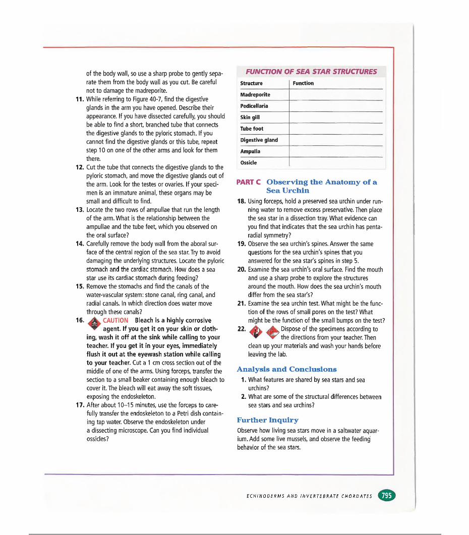
of the body wall, so use a sharp probe to gently separate them from the body wall as you cut. Be careful not to damage the madreporlte.
11. While referring to Figure 40-7, find the digestive glands in the arm you have opened. Describe their appearance. If you have dissected carefully, you should be able to find a short, branched tube that connects the digestive glands to the pyloric stomach. If you cannot find the digestive glands or this tube, repeat step 10 on one of the other arms and look for them there.
12. Cut the tube that connects the digestive glands to the pyloric stomach, and move the digestive glands out of the arm. Look for the testes or ovaries. If your specimen is an immature animal, these organs may be small and difficult to find.
13. Locate the two rows of ampullae that run the length of the arm. What is the relationship between the ampullae and the tube feet, which you observed on the oral surface?
14. Carefully remove the body wall from the aboral surface of the central region of the sea star. Try to avoid damaging the underlying structures. Locate the pyloric stomach and the cardiac stomach. How does a sea star use its cardiac stomach during feeding?
15. Remove the stomachs and find the canals of the water-vascular system: stone canal, ring canal, and radial canals. In which direction does water move through these canals?
16. jgk CAUTION Bleach is a highly corrosive ^ agent. If you get it on your skin or cloth
ing, wash it off at the sink while calling to your teacher. If you get it in your eyes, immediately flush it out at the eyewash station while calling to your teacher. Cut a 1 cm cross section out of the middle of one of the arms. Using forceps, transfer the section to a small beaker containing enough bleach to cover it. The bleach will eat away the soft tissues, exposing the endoskeleton.
17. After about 10-15 minutes, use the forceps to carefully transfer the endoskeleton to a Petri dish containing tap water. Observe the endoskeleton undera dissecting microscope. Can you find individual ossicles?
FUNCTION OF SEA STAR STRUCTURESStructure Function
Madreporite
Pedicellaria
Skin gill
Tube foot
Digestive gland
Ampulla
Ossicle
PART C Observing the Anatomy of a Sea Urchin
18. Using forceps, hold a preserved sea urchin under running water to remove excess preservative. Then place the sea star in a dissection tray. What evidence can you find that indicates that the sea urchin has penta- radial symmetry?
19. Observe the sea urchin's spines. Answer the same questions for the sea urchin's spines that you answered for the sea star's spines in step 5.
20. Examine the sea urchin's oral surface. Find the mouth and use a sharp probe to explore the structures around the mouth. How does the sea urchin's mouth differ from the sea star's?
21. Examine the sea urchin test. What might be the function of the rows of small pores on the test? What might be the function of the small bumps on the test?
22. Dispose of the specimens according to the directions from your teacher. Then
clean up your materials and wash your hands before leaving the lab.
Analysis and Conclusions1. What features are shared by sea stars and sea
urchins?2. What are some of the structural differences between
sea stars and sea urchins?
Further InquiryObserve how living sea stars move in a saltwater aquarium. Add some live mussels, and observe the feedingbehavior of the sea stars.
ECHINODERMS AND INVERTEBRATE CHORDATES