152: physical exam indicated cerclage versus expectant management: a systematic review and...
TRANSCRIPT

Review
Physical Examination–Indicated CerclageA Systematic Review and Meta-analysis
Robert M. Ehsanipoor, MD, Neil S. Seligman, MD, Gabriele Saccone, MD, Linda M. Szymanski, MD, PhD,Christina Wissinger, MS, MLIS, Erika F. Werner, MD, MS, and Vincenzo Berghella, MD
OBJECTIVE: To estimate the effectiveness of physical
examination–indicated cerclage in the setting of second-
trimester cervical dilatation by systematic review and
meta-analysis of published studies.
DATA SOURCES: We searched MEDLINE, EMBASE, Sco-
pus, ClinicalTrials.gov, Web of Science, and the Cochrane
Library for studies published between 1966 and 2014 that
evaluated cervical cerclage for the treatment of cervical
insufficiency.
METHODS OF STUDY SELECTION: The search yielded
6,314 citations. We included cohort studies and random-
ized controlled trials comparing cerclage placement with
expectant management of women with cervical dilata-
tion between 14 and 27 weeks of gestation. Two
investigators independently reviewed each citation for
inclusion or exclusion and discordant decisions were
arbitrated by a third reviewer. Summary estimates were
reported as the mean difference and 95% confidence
interval (CI) for continuous variables or relative risk and
with 95% CI for dichotomous outcomes. Fixed- and
random-effects meta-analysis was used, depending on
heterogeneity.
TABULATION, INTEGRATION, AND RESULTS: Ten
studies met inclusion criteria and were included in the
final analysis. One was a randomized controlled trial, two
were prospective cohort studies, and the remaining
seven were retrospective cohort studies. Of the 757
women, 485 (64%) underwent physical examination–
indicated cerclage placement and 272 (36%) were expec-
tantly managed. Cerclage was associated with increased
neonatal survival (71% compared with 43%; relative risk
1.65, 95% CI 1.19–2.28) and prolongation of pregnancy
(mean difference 33.98 days, 95% CI 17.88–50.08).
CONCLUSION: Physical examination–indicated cerc-
lage is associated with a significant increase in neonatal
survival and prolongation of pregnancy of approximately
1 month when compared with no such cerclage. The
strength of this conclusion is limited by the potential
for bias in the included studies.
(Obstet Gynecol 2015;126:125–35)
DOI: 10.1097/AOG.0000000000000850
Cervical insufficiency, previously referred to ascervical incompetence, has classically been
defined as painless dilation of the cervix in the absenceof contractions or bleeding in the second trimester.1
Painless second-trimester cervical dilation is anuncommon finding in the general population occur-ring in less than 1% of pregnancies.2 Cerclage for theprevention of pregnancy loss in women with priorsecond-trimester loss or second-trimester cervical dila-tation in the index pregnancy was first reported in the1950s.3,4 Cerclage placement in the setting of cervicaldilatation has been variably referred to as “physicalexamination–indicated cerclage,” “rescue cerclage,”and “emergency cerclage.” To date, the benefits ofcerclage for this indication are not entirely clear.
The optimal evaluation and management ofasymptomatic patients presenting with second-trimester cervical dilatation remain controversial.There is only one randomized controlled trial to dateevaluating the use of cerclage in this clinical scenario,and it included only 23 patients, seven of whom werepregnant with twins.5 Several nonrandomized studieshave compared outcomes of women receiving cerc-lage with those expectantly managed in the setting
From the Departments of Gynecology and Obstetrics, Johns HopkinsUniversity, Baltimore, Maryland, University of Rochester, Rochester, NewYork, and Warren Alpert Medical School of Brown University, Providence,Rhode Island; the Department of Neuroscience, Reproductive Sciences andDentistry, School of Medicine, University of Naples Federico II, Naples, Italy;and the Division of Maternal-Fetal Medicine, Department of Obstetrics andGynecology, Sidney Kimmel Medical College of Thomas Jefferson University,Philadelphia, Pennsylvania.
Corresponding author: Robert M. Ehsanipoor, MD, Johns Hopkins UniversitySchool of Medicine, 600 N. Wolfe Street, Phipps 228, Baltimore, MD 21287;e-mail: [email protected].
Financial DisclosureThe authors did not report any potential conflicts of interest.
© 2015 by The American College of Obstetricians and Gynecologists. Publishedby Wolters Kluwer Health, Inc. All rights reserved.ISSN: 0029-7844/15
VOL. 126, NO. 1, JULY 2015 OBSTETRICS & GYNECOLOGY 125
Copyright ª by The American College of Obstetriciansand Gynecologists. Published by Wolters Kluwer Health, Inc.
Unauthorized reproduction of this article is prohibited.
of second-trimester cervical dilatation. Observationalstudies have inherent limitations, but until a well-designed and adequately powered randomized con-trolled study is performed, these are the best dataavailable on which to make management decisions.The objective of this review was to systematicallyreview the literature enabling comparison and combi-nation of the results to arrive at the most appropriateconclusion regarding the effectiveness of physicalexamination–indicated cerclage.
SOURCES
The methodology conformed to Meta-analysis ofObservational Studies in Epidemiology (MOOSE)criteria.6 MEDLINE, EMBASE, Scopus, Clinical-Trials.gov, Web of Science, and the Cochrane Librarywere systematically searched using the keywordsearch terms: “cerclage,” “cervical cerclage,” “physicalexamination-indicated cerclage,” “rescue cerclage,”“emergency cerclage,” “cervical incompetence,” “cer-vical insufficiency,” “uterine cervix cerclage,” “uterinecervix incompetence,” “uterine cervix insufficiency,”“salvage cerclage,” and “cervical salvage cerclage.”The search was conducted by an experienced librar-ian (C.W.). References from relevant research articlesand reviews were also reviewed. No attempt wasmade to search for unpublished studies. Searches werelimited to studies in humans in all languages and topublication dates from 1966 to November 2014.
STUDY SELECTION
For inclusion, studies needed to compare cerclagewith no cerclage in women with a physical examina-tion that revealed cervical dilatation of 0.5 cm orgreater between 14 and 27 weeks of gestation. Studiesevaluating cerclage based on ultrasound findings butwith a closed cervix were ineligible. Both prospectiveand retrospective studies, including abstracts, wereincluded; those without a control group of expectantlymanaged women were excluded.
Titles and abstracts for all of the identified studieswere independently reviewed by two reviewers (R.M.E.,L.S.), and the full article was reviewed in further detailwhen needed to determine if the study met inclusioncriteria. Any disagreements were resolved withdiscussion with a third reviewer (N.S.S.). Data wereindependently extracted by the two reviewers (R.M.E. and L.S.) using previously prepared data extrac-tion forms and any discrepancies were resolved bydiscussion with a third reviewer (N.S.S.). Furtherclarification and additional data were sought fromauthors when required and attempts were made toobtain patient-level data from authors. The protocol
for this review was registered in the PROSPEROInternational Prospective Register of Systematic Re-views (CRD42014015654).
Statistical analyses were conducted with ReviewManager (RevMan) 5.3. Cerclage placement wascompared with 114 no cerclage placement, and theprimary outcomes of interest were neonatal survivaland time from diagnosis to delivery. Additional out-comes included gestational age at delivery, pretermdelivery at less than 24 weeks of gestation, deliverybetween 24 and 28 weeks of gestation, delivery at lessthan 34 weeks of gestation, intraoperative membranerupture, cervical laceration, and birth weight.
Meta-analyses were performed with cohort stud-ies and the randomized controlled trial combinedgiven that there was only one randomized trial. Thestudies were weighted based on the number ofparticipants. The data analysis was completed inde-pendently by authors (N.S.S., G.S., V.B.). The com-pleted analyses were then compared, and anydifference was resolved with review of the entire dataand independent analysis. Statistical heterogeneitybetween studies was assessed using the Cochrane Qstatistic and Higgins I2 statistics. In case of statisticallysignificant heterogeneity (P value of the Cochrane Qstatistic ,.1), the random-effects model of DerSimo-nian and Laird was used to obtain the pooled relativerisk (RR) estimate; otherwise, a fixed-effect modelswas planned. The summary measures were reportedas RRs with 95% confidence intervals (95% CIs).
To further address heterogeneity, it was prede-termined that an analysis of the studies deemed to beat the lowest risk of bias would be performed. Thequality of each study was evaluated by two inves-tigators (R.M.E. and N.S.S.). The Cochrane tool forassessment of bias was used for the randomizedcontrolled trial and for the cohort studies, assessmentfor bias was performed using an approach similar tothat described by the Cochrane Non-RandomizedStudy Group.7 With this approach, differences inbaseline characteristics are compared to evaluate forselection bias because the factors determining whichgroup a woman is allocated to are often unknown.The study design and similarity of the treatment andcontrol groups were evaluated to estimate the risk ofbias for the following six characteristics: obstetric his-tory, gestational age at diagnosis, cervical dilation atdiagnosis, subclinical evidence of infection, antibioticuse, and tocolysis use. If a study reported a statisticallysignificant difference in parity, history of pretermdelivery, or second-trimester loss, the potential forbias regarding obstetric history was considered to behigh. A difference of greater than 1 week for
126 Ehsanipoor et al Physical Examination–Indicated Cerclage OBSTETRICS & GYNECOLOGY
Copyright ª by The American College of Obstetriciansand Gynecologists. Published by Wolters Kluwer Health, Inc.
Unauthorized reproduction of this article is prohibited.
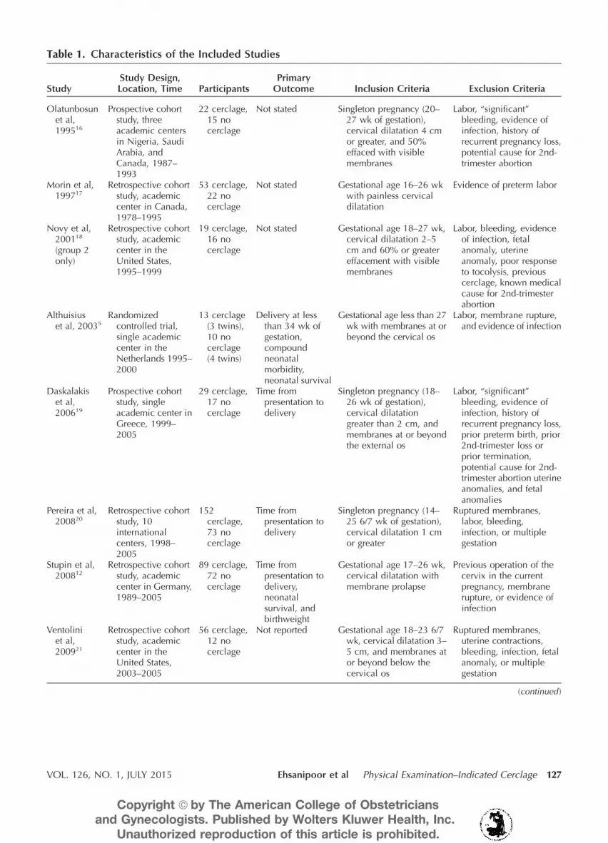
Table 1. Characteristics of the Included Studies
StudyStudy Design,Location, Time Participants
PrimaryOutcome Inclusion Criteria Exclusion Criteria
Olatunbosunet al,199516
Prospective cohortstudy, threeacademic centersin Nigeria, SaudiArabia, andCanada, 1987–1993
22 cerclage,15 nocerclage
Not stated Singleton pregnancy (20–27 wk of gestation),cervical dilatation 4 cmor greater, and 50%effaced with visiblemembranes
Labor, “significant”bleeding, evidence ofinfection, history ofrecurrent pregnancy loss,potential cause for 2nd-trimester abortion
Morin et al,199717
Retrospective cohortstudy, academiccenter in Canada,1978–1995
53 cerclage,22 nocerclage
Not stated Gestational age 16–26 wkwith painless cervicaldilatation
Evidence of preterm labor
Novy et al,200118
(group 2only)
Retrospective cohortstudy, academiccenter in theUnited States,1995–1999
19 cerclage,16 nocerclage
Not stated Gestational age 18–27 wk,cervical dilatation 2–5cm and 60% or greatereffacement with visiblemembranes
Labor, bleeding, evidenceof infection, fetalanomaly, uterineanomaly, poor responseto tocolysis, previouscerclage, known medicalcause for 2nd-trimesterabortion
Althuisiuset al, 20035
Randomizedcontrolled trial,single academiccenter in theNetherlands 1995–2000
13 cerclage(3 twins),10 nocerclage(4 twins)
Delivery at lessthan 34 wk ofgestation,compoundneonatalmorbidity,neonatal survival
Gestational age less than 27wk with membranes at orbeyond the cervical os
Labor, membrane rupture,and evidence of infection
Daskalakiset al,200619
Prospective cohortstudy, singleacademic center inGreece, 1999–2005
29 cerclage,17 nocerclage
Time frompresentation todelivery
Singleton pregnancy (18–26 wk of gestation),cervical dilatationgreater than 2 cm, andmembranes at or beyondthe external os
Labor, “significant”bleeding, evidence ofinfection, history ofrecurrent pregnancy loss,prior preterm birth, prior2nd-trimester loss orprior termination,potential cause for 2nd-trimester abortion uterineanomalies, and fetalanomalies
Pereira et al,200820
Retrospective cohortstudy, 10internationalcenters, 1998–2005
152cerclage,73 nocerclage
Time frompresentation todelivery
Singleton pregnancy (14–25 6/7 wk of gestation),cervical dilatation 1 cmor greater
Ruptured membranes,labor, bleeding,infection, or multiplegestation
Stupin et al,200812
Retrospective cohortstudy, academiccenter in Germany,1989–2005
89 cerclage,72 nocerclage
Time frompresentation todelivery,neonatalsurvival, andbirthweight
Gestational age 17–26 wk,cervical dilatation withmembrane prolapse
Previous operation of thecervix in the currentpregnancy, membranerupture, or evidence ofinfection
Ventoliniet al,200921
Retrospective cohortstudy, academiccenter in theUnited States,2003–2005
56 cerclage,12 nocerclage
Not reported Gestational age 18–23 6/7wk, cervical dilatation 3–5 cm, and membranes ator beyond below thecervical os
Ruptured membranes,uterine contractions,bleeding, infection, fetalanomaly, or multiplegestation
(continued )
VOL. 126, NO. 1, JULY 2015 Ehsanipoor et al Physical Examination–Indicated Cerclage 127
Copyright ª by The American College of Obstetriciansand Gynecologists. Published by Wolters Kluwer Health, Inc.
Unauthorized reproduction of this article is prohibited.
gestational age or 1 cm for dilatation at the time ofdiagnosis, between the cerclage and control groups,was considered to suggest a high risk of bias. If studiesreported on markers of inflammation or a history ofpreterm delivery, a statistically significant differencewas considered to suggest a high risk of bias. Whentocolysis and antibiotic regimens were stated to be thesame for both groups, the potential for treatment biaswas considered low; if it was stated to be different, ora statistically significant difference was shown, thepotential for treatment bias was considered to be high.If details were not reported, it was unable to bedetermined.
RESULTS
The initial search yielded a total of 6,314 titles andabstracts. Of these, 17 met the inclusion criteria, andseven were subsequently excluded for the one of thefollowing reasons: could not feasibly be translatedinto English,8,9 did not adequately report results forthe control group,10 appeared to be a duplicate pub-lication,11,12 only included patients undergoing repeatcerclage,13 only included twins,14 or was limited tomultiple gestations attempting a delayed intervaldelivery.15 Attempts were made to contact all authorsto obtain unpublished data. The remaining 10 studieswere included in the analysis, resulting in a total of757 women eligible for a physical examination–indi-cated cerclage.5,12,16–23 Of these, 485 (64%) womenunderwent cerclage placement and 272 (36%) wereexpectantly managed. Only one of the studies wasa randomized controlled trial (23 pregnancies),5 twowere prospective cohort studies,16,19 and the remain-ing seven were retrospective cohort studies.12,17,18,20–23
Characteristics of each study are described in Table 1and a description of the surgical procedure and asso-ciated is provided in Table 2.
For the randomized controlled trial, randomiza-tion was organized in balanced blocks and assigned bytelephone.5 The study was at low risk for selectionbias or bias with regard to attrition or reporting. Par-ticipants and health care providers were not blinded,and all of the women in the cerclage group routinelyreceived perioperative indomethacin; those in the bedrest group did not. All of the women in both groupsreceived 1 g amoxicillin–clavulanic acid intrave-nously every 6 hours and 500 mg metronidazole intra-venously every 8 hours for 1 week. Blinding was notfeasible in this study, and the use of indomethacin inthe cerclage group does result in a potential for treat-ment bias. The risk of bias for the cohort studies isshown in Table 3. Three of the nine (33.3%) cohortstudies did not demonstrate a high risk of bias in anyof the predefined categories,16,19,21 but for one of thesestudies,21 there was insufficient information to evalu-ate six of the seven elements in the bias table. Pereiraet al20 were the only authors who performed a multi-variate analysis to adjust for potential confounders.
The cerclage and expectantly managed groupswere similar in regard to maternal age, nulliparity,and history of previous preterm birth. Pereira et alreported significantly higher rates of previous second-trimester loss in the cerclage group compared with theexpectantly managed group (45% compared with18%, P#.01). When reported, no differences betweenthe groups regarding parity5,12,16,18,19,22,23 or history ofpreterm birth5,16,18–20,23 were reported in any study.Specific characteristics of the study groups are furtherreported in Table 4. Women in the cerclage groupwere diagnosed approximately 1 week earlier thanthe expectant management group (mean difference21.02 weeks, 95% CI 22.00 to 20.05), but therewas no significant difference in the cervical dilatationbetween the cerclage group and the expectantly
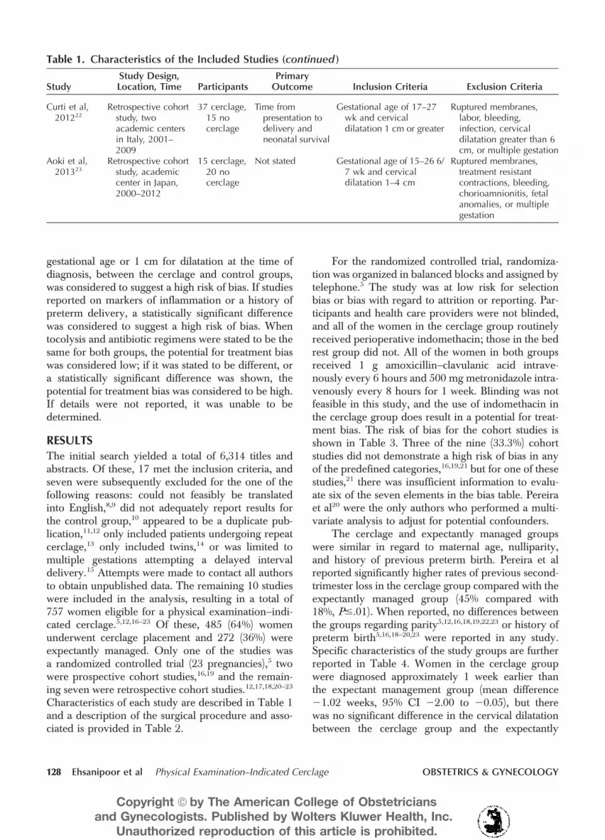
Table 1. Characteristics of the Included Studies (continued )
StudyStudy Design,Location, Time Participants
PrimaryOutcome Inclusion Criteria Exclusion Criteria
Curti et al,201222
Retrospective cohortstudy, twoacademic centersin Italy, 2001–2009
37 cerclage,15 nocerclage
Time frompresentation todelivery andneonatal survival
Gestational age of 17–27wk and cervicaldilatation 1 cm or greater
Ruptured membranes,labor, bleeding,infection, cervicaldilatation greater than 6cm, or multiple gestation
Aoki et al,201323
Retrospective cohortstudy, academiccenter in Japan,2000–2012
15 cerclage,20 nocerclage
Not stated Gestational age of 15–26 6/7 wk and cervicaldilatation 1–4 cm
Ruptured membranes,treatment resistantcontractions, bleeding,chorioamnionitis, fetalanomalies, or multiplegestation
128 Ehsanipoor et al Physical Examination–Indicated Cerclage OBSTETRICS & GYNECOLOGY
Copyright ª by The American College of Obstetriciansand Gynecologists. Published by Wolters Kluwer Health, Inc.
Unauthorized reproduction of this article is prohibited.

Table 2. Description of the Intervention and Associated Treatments
Study Intervention Antibiotics Tocolysis Activity
Olatunbosunet al,199516
Observation period: 4–6 h;procedure: steepTrendelenburg tilt andinflated Foley balloonwith tip cut or retrogradefilling of the bladder usedto reduce membranes;cerclage type: modifiedMcDonald; two 1 silkpursestring suturesfollowed by four 00 silkstay sutures tied over theexternal os; suturematerial: silk
Ampicillin or cefoxitin35 d Ritodrine intravenous orindomethacinsuppository348 h
Cerclage group: strictbedrest348 h thendischarge to home in 5–6d and advised to continuepelvic rest; no cerclagegroup: admitted for bedrest until delivery
Morin et al,199717
Observation period:unspecified; procedure:unspecified; cerclagetype: unspecified; suturematerial: unspecified
Unspecified Unspecified Unspecified
Novy et al,200118
(group 2only)
Observation period: 4–24 h,possible tocolysis;procedure: steepTrendelenburg tilt andmoist swab used toreduce membranes;cerclage type: modifiedShirodkar or McDonald;suture material:Shirodkar: Mersilene tape(5 mm); McDonald:“large nonabsorbablemonofilament suture”
Broad-spectrum antibioticsfrequently used butregimens unspecified
Various agents frequentlyused but unspecified
Bed rest in the hospital andthen discharged to homeand advised to continuebed rest as feasible
Althuisiuset al, 20035
Observation period: nonestated; procedure: steepTrendelenburg tilt andinflated Foley balloonused to reducemembranes; cerclagetype: McDonald; suturematerial: braidedpolyester thread (metric8/United StatesPharmacopeia 6)
All received 1 gamoxicillin–clavulanicacid intravenously every6 h and 500 mgmetronidazoleintravenously every 8 hfor 1 wk
Cerclage group onlyreceived 100 mgindomethacinsuppository 2 h beforethe procedure and 6 hafter the procedure
Inpatient bed rest until 30wk of gestation
Daskalakiset al,200619
Observation period: 8–24 h;procedure: steepTrendelenburg tilt andmoist swab used toreduce membranes;cerclage type: McDonald;suture material: polyestercerclage tape (5 mm)
Cefuroxime andmetronidazoleintravenous348 h; 1.5 gerythromycin orallydaily310 d
100 mg indomethacinsuppository twicea d32 d; 5 mg ritodrineorally every 6 h32 wk
Bed rest in the hospital37d then discharge to homeand advised to continue“strict bedrest” to 32 wkand then mobilizationwith “plenty of rest” untildelivery
Pereira et al,200820
Observation period:unspecified; procedure:variable and unspecified;cerclage type: variableand unspecified; suturematerial: not specified
Regimens not specified Regimens not specified Not standardized orspecified
(continued )
VOL. 126, NO. 1, JULY 2015 Ehsanipoor et al Physical Examination–Indicated Cerclage 129
Copyright ª by The American College of Obstetriciansand Gynecologists. Published by Wolters Kluwer Health, Inc.
Unauthorized reproduction of this article is prohibited.

managed group (mean difference 20.18 cm, 95% CI20.34 to 0.17) (Table 4). Markers of inflammation orinfection were evaluated in three of the studies12,22,23
and in one study, a higher leukocyte count was notedin the expectantly managed group compared with thecerclage group (median white blood cell count 9.76K/microliter compared with 14.73 K/microliter;mean difference 4.97, 95% CI 2.25–7.69).22 Mem-branes were visible or prolapsed in all women in allof the included studies with the exception of Pereiraet al20 in which membranes were not visible in 85 of152 (56%) of the cerclage cases; this was not reportedfor the expectantly managed group. Three studiescompared the degree of membrane prolapse, andthere was no significant difference between the co-horts in any study.12,22,23 Twins were excluded in six
studies,16,19–23 included in two studies,5,18 and inclu-sion could not be determined in two studies.12,17
The use of amniocentesis, antibiotics, and tocolysisat the time of initial evaluation and management wassimilar between the two groups (Table 4). Administra-tion of tocolysis was reported in seven stud-ies5,12,16,18,19,22,23 and was nearly universal with twoexceptions. In the study by Curti et al,22 34 of 52(65%) of the women received tocolysis, and in the trialby Althuisius et al,5 indomethacin was not used in theexpectant management group. Only two studies re-ported on the use of amnioce2ntesis and there wasno difference between the groups (12% compared with8%; RR 1.53, 95% CI 0.68–3.43).20,22 A sensitivity anal-ysis was performed to evaluate for potential selectionbias that could be introduced the use of amniocentesis.
Table 2. Description of the Intervention and Associated Treatments (continued )
Study Intervention Antibiotics Tocolysis Activity
Stupin et al,200812
Observation period:preferably less than 24 h;procedure: variable anddetails not specified;cerclage type: 73McDonald, 14combination of othermethods with fibrinadhesive in cervicalcanal, 2 Saling (seereference); suturematerial: unspecified
Regimens not specified Regimens not specified Absolute bed rest initiallyand then relaxed andlifted if asymptomatic
Ventoliniet al,200921
Observation period: 24 h;procedure: moist iodine-soaked swab, retrogradefilling of the bladder usedto reduce membranes, orboth; cerclage type:Shirodkar; Suturematerial: Mersilene tape(5 mm)
None Cerclage group onlyreceivedindomethacin324 h
Discharged to home withpelvic and bed rest
Curti et al,201222
Observation period: at least24 h; procedure: detailsnot specified; cerclagetype: Shirodkar n536,McDonald n51; suturematerial: Mersilene tape(5 mm)
Ampicillin or erythromycinintravenous37 d
Variable and regimen notspecified
Not specified
Aoki et al,201323
Observation period: fewhours but less than 24 h;procedure:Trendelenburg but notfurther described;cerclage type: McDonaldn512, Shirodkar n52,both n51; suturematerial: Mersilene tape(5 mm)
“Broad spectrum”35 d All participants receivedbut regimen notspecified
Cerclage group 6/15 (40%)were temporarilymanaged as outpatients;bed rest group were allmanaged inpatient;details regarding activitywere not furtherdescribed
130 Ehsanipoor et al Physical Examination–Indicated Cerclage OBSTETRICS & GYNECOLOGY
Copyright ª by The American College of Obstetriciansand Gynecologists. Published by Wolters Kluwer Health, Inc.
Unauthorized reproduction of this article is prohibited.

After excluding the two studies in which amniocentesiswas used,20,22 the primary outcome of neonatal survivalwas still significantly higher in the cerclage group com-pared with the expectantly managed group (69.7%compared with 50.3%; RR 1.48, 95% CI 1.06–2.05).Shirodkar cerclage was the primary technique used intwo studies21,22; the remainder primarily used eithera McDonald cerclage5,12,19,23 or modified McDonaldcerclage,16 and, for three studies, the technique couldnot be determined.17,18,20 Management strategies forthe study groups are further reported in Table 4 andspecific management and interventions for each studyare described in Table 2.
The primary outcome of neonatal survival wasreported in eight studies.5,12,16–20,22 Survival was more
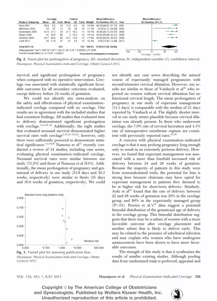
likely in the cerclage group compared with the expec-tantly managed group (71% compared with 43%; RR1.65, 95% CI 1.19–2.28) (Table 5; Fig. 1). Cerclageplacement was associated with significant prolonga-tion of pregnancy (mean difference 33.98 days, 95%CI 17.88–50.08) (Fig. 2) and greater gestational age ofdelivery (mean difference 4.62 weeks, 95% CI 3.89–5.36) (Table 5). Physical examination–indicated cerc-lage was associated with significant reductions in pre-term birth between 24 and 28 weeks of gestation (8%compared with 37%; RR 0.23, 95% CI 0.13–0.41),preterm birth at less than 34 weeks of gestation(50% compared with 82%; RR 0.55, 95% CI 0.38–0.80), and higher birth weight (mean difference1,028 g, 95% CI 714–1,341) (Table 5).
Table 3. Bias Assessment for Nonrandomized Studies
StudyObstetricHistory
GestationalAge Dilatation
Evidence ofInfection Tocolysis Antibiotics
Olatunbosun et al,199516
Low Low Low Unable todetermine
Low Low
Morin et al, 199717 Unable todetermine
Unable todetermine
High Unable todetermine
Unable todetermine
Unable todetermine
Novy et al, 200118
(group 2 only)Low High Low Unable to
determineUnable todetermine
Unable todetermine
Daskalakis et al, 200619 Low Low Low Unable todetermine
Low Low
Pereira et al, 200820 High High Low Unable todetermine
Unable todetermine
Unable todetermine
Stupin et al, 200812 Low High High Low Unable todetermine
Unable todetermine
Ventolini et al, 200921 Unable todetermine
Unable todetermine
Unable todetermine
Unable todetermine
Unable todetermine
Unable todetermine
Curti et al, 201222 Low High High High Low Unable todetermine
Aoki et al, 201323 Low High Unable todetermine
Low Unable todetermine
Unable todetermine
Table 4. Characteristics of the Women Included in the Meta-analysis
Characteristic Studies/Participants Cerclage Expectant Effect Estimate*
Age (y) 6/556 29.8 29.9 0.82 (20.35 to 1.95)Nulliparous 5/331 123/192 (64) 87/139 (63) 1.02 (0.87–1.21)Previous preterm birth 3/306 62/196 (32) 30/110 (27) 1.16 (0.80–1.68)Gestational age at diagnosis (wk) 8/614 21.7 22.8 21.02 (22.00 to 20.05)Cervical dilatation at diagnosis (cm) 6/556 3.3 3.5 20.18 (20.34 to 0.17)Amniocentesis† 2/277 12/189 (12) 7/88 (8) 1.53 (0.68–3.43)Antibiotics† 9/682 271/432 (63) 182/250 (73) 0.86 (0.78–1.15)Tocolysis† 7/389 208/224 (93) 141/165 (85) 1.02 (0.93–1.13)
Data are weighted mean or n/N (%) unless otherwise specified.Bold indicates statistical significance.* Data are mean difference (95% confidence interval) for rows 1, 4, and 5 and relative risk (95% confidence interval) for rows 2, 3, 6, 7,
and 8† At the time of initial diagnosis and management.
VOL. 126, NO. 1, JULY 2015 Ehsanipoor et al Physical Examination–Indicated Cerclage 131
Copyright ª by The American College of Obstetriciansand Gynecologists. Published by Wolters Kluwer Health, Inc.
Unauthorized reproduction of this article is prohibited.

In women undergoing cerclage, the incidence ofintraoperative membrane rupture was 4.1% (10 of246)5,12,19,21–23 and for cervical laceration was 7.9%(seven of 140)12,16,17,19; however, these data were notreported for the control groups. The patients in whichintraoperative membrane rupture occurred wereincluded in the treatment group based on the princi-ple of intention to treat for these 10 patients. Olatun-bosun also reported one case of intraoperativemembrane rupture, but this participant was excludedfrom the study.16 No maternal deaths were reported;however, one study reported an intensive care unitadmission for sepsis in a cerclage recipient,20 but thisoutcome was only specified in one other study.23
Rates of placental abruption, premature preterm rup-ture of membranes, and chorioamnionitis were incon-sistently and variably reported.
Six of the 10 studies were excluded from thesubanalysis of studies with the lowest risk of biasbecause a high risk of bias was noted in at least onecategory evaluated (Table 3).12,17,18,20,22,23 Althoughthe potential for treatment bias in the trial by Althui-sius et al was high, the decision was made to include
it in the subanalysis because indomethacin in thisclinical setting has not been shown to have a signifi-cant effect on outcomes.24 Of the remaining studies,outcome reporting by Ventolini et al21 was insuffi-cient to permit evaluation of bias for six of the sevencategories evaluated so it was excluded. Therefore,the studies by Daskalakis et al, Olatunbosun et al,and Althuisius et al were included in the subanaly-sis.5,16,19 In the subanalysis, cerclage was also associ-ated with higher rates of neonatal survival (78%compared with 33%, RR 2.11, 95% CI 1.41–3.55)and prolongation of pregnancy (mean difference34.00 days, 95% CI 3.11–64.89).
DISCUSSION
Included articles in this meta-analysis were limited innumber and variable in quality and study design.Although differences in our primary outcomes werefound, this meta-analysis also underscores the paucityand low quality of existing studies of physical exam-ination–indicated cerclage. Our findings suggest thatphysical examination–indicated cerclage is associatedwith significantly but modestly higher rates of neonatal
Table 5. Primary and Secondary Outcomes
Studied Outcome Studies/Participants Cerclage Expectant Effect Estimate*
Neonatal survival 8/657 294/413 (71) 106/244 (43) 1.66 (1.19–2.30)Delivery at less than 24 wk of gestation 3/316 51/221 (23) 31/95 (33) 0.47 (0.14–1.53)Delivery at 24—28 wk of gestation† 2/239 14/165 (8) 31/83 (37) 0.23 (0.13–0.41)Delivery at less than 34 wk of gestation 3/316 110/221 (50) 77/94 (82) 0.55 (0.38–0.80)Time to delivery (d) 6/385 56.7 18.8 33.98 (17.88–50.08)Gestational age at delivery (wk) 8/643 30.6 25.2 4.62 (3.89–5.36)Birth weight (g) 5/331 1,714.6 829.4 1,028 (714–1,341)
Data are n/N (%) or weighted mean unless otherwise specified.Bold indicates statistical significance.* Data are relative risk (95% confidence interval) for the top four rows and mean difference (95% confidence interval) for the bottom three
rows.† Referent group is women who delivered either before 24 weeks of gestation or after 28 weeks of gestation.
Fig. 1. Forest plot for neonatal survival. M-H, Mantel-Haenszel test; CI, confidence interval.
Ehsanipoor. Physical Examination–Indicated Cerclage. Obstet Gynecol 2015.
132 Ehsanipoor et al Physical Examination–Indicated Cerclage OBSTETRICS & GYNECOLOGY
Copyright ª by The American College of Obstetriciansand Gynecologists. Published by Wolters Kluwer Health, Inc.
Unauthorized reproduction of this article is prohibited.
survival and significant prolongation of pregnancywhen compared with no operative intervention. Cerc-lage was associated with statistically significant favor-able outcomes for all secondary outcomes evaluated,except delivery before 24 weeks of gestation.
We could not identify any meta-analysis onthe safety and effectiveness of physical examination–indicated cerclage compared with no cerclage. Ourresults are in agreement with the included studies thathad consistent findings. All studies that evaluated timeto delivery demonstrated significant prolongationwith cerclage.5,12,18–23 Additionally, the eight studiesthat evaluated neonatal survival demonstrated highersurvival rates with cerclage5,12,16–20,22; however, onlythree were sufficiently powered to demonstrate statis-tical significance.12,19,20 Namouz et al25 recently con-ducted a review of 34 studies, including case series,evaluating physical examination–indicated cerclage.Neonatal survival rates were similar between ourstudy (72.2%) and those of Namouz et al (81%). Addi-tionally, the mean prolongation of pregnancy and ges-tational of delivery in our study (53.8 days and 30.5weeks, respectively) were similar to theirs (56 daysand 30.6 weeks of gestation, respectively). We could
not identify any case series describing the naturalcourse of expectantly managed pregnancies withsecond-trimester cervical dilatation. However, our re-sults are similar to those of Vaisbuch et al26 who re-ported on women without cervical dilatation but nofunctional cervical length. The mean prolongation ofpregnancy in our study of expectant management(14.1 days) is comparable with the median of 21 daysreported by Vaisbuch et al. The slightly shorter inter-val in our study seems plausible because cervical dila-tation was already present. In those who underwentcerclage, the 7.9% rate of cervical laceration and 4.1%rate of intraoperative membrane rupture are consis-tent with previously reported rates.27,28
A concern with physical examination–indicatedcerclage is that it may prolong pregnancy long enoughonly to result in an extremely preterm delivery. How-ever, we found that expectant management was asso-ciated with a more than fourfold increased risk ofdelivery between 24 and 28 weeks of gestation.Because the majority of patients in this study werefrom nonrandomized trials, the potential for bias isstrong here because clinicians may have opted forexpectant management in patients they deemed tobe at higher risk for short-term delivery. Similarly,Aoki et al23 found that the rate of delivery between22 and 28 weeks of gestation was 20% in the cerclagegroup and 80% in the expectantly managed group(P,.01). Pereira et al.’s20 data suggest a potentialbimodal distribution of the gestational age of deliveryin the cerclage group. This bimodal distribution sug-gests that there may be a subset of women with a morefavorable outcome after cerclage placement andanother subset that is likely to deliver early. Thismay be related to the presence of subclinical infectionand may explain why women who have undergoneamniocentesis have been shown to have more favor-able outcomes.
The strength of this study is that it synthesizes theresults of smaller existing studies. Although poolingdata from randomized trials is preferred, appraisal and
Fig. 2. Forest plot for prolongation of pregnancy. SD, standard deviation; IV, independent variable; CI, confidence interval.
Ehsanipoor. Physical Examination–Indicated Cerclage. Obstet Gynecol 2015.
Fig. 3. Funnel plot for assessing publication bias.
Ehsanipoor. Physical Examination–Indicated Cerclage. ObstetGynecol 2015.
VOL. 126, NO. 1, JULY 2015 Ehsanipoor et al Physical Examination–Indicated Cerclage 133
Copyright ª by The American College of Obstetriciansand Gynecologists. Published by Wolters Kluwer Health, Inc.
Unauthorized reproduction of this article is prohibited.

systematic evaluation of existing observational studiescan nonetheless yield important conclusions. Althoughthe quality of the study methods and reporting of thesestudies varied widely, the results of the subanalysis ofthe highest quality studies were consistent with theprimary findings. Risk of publication bias was assessedby visual inspection of the funnel plot and thesymmetric plot suggested no publication bias (Fig. 3).
Study quality was the most obvious limitation.Suboptimal study design can introduce bias in a meta-analysis of observational studies. The potential forselection bias exists with nonrandom allocation. Nei-ther the cerclage nor the expectant group was at anobviously higher risk for poor outcome. Cervicaldilatation, prolapsed membranes, obstetric history,evidence of infection, and evaluation with amniocente-sis are factors that have been correlated with outcomesof physical examination–indicated cerclage.29–31 In onestudy, participants in the cerclage group were morelikely to have a history of second-trimester loss20 andin another, the expectant group had a higher meanleukocyte count.22 Otherwise, no differences werenoted in cervical dilatation, membrane prolapse, oruse of amniocentesis. The only significant differenceseen was that the mean gestational age for the cerclagegroup was 22.8 weeks of gestation compared with 21.7weeks of gestation. Cerclage placement at earlier gesta-tional ages has been associated with improved out-comes in some, but not all studies.30,31 The possibilityof treatment bias exists, and the use of obstetric or neo-natal interventions such as fetal monitoring, cesareandelivery, and neonatal resuscitation was not reported.Neonatal outcomes were inconsistently reported andlong-term child outcomes were not reported. Despitethese limitations, the consistency of the findings of theincluded studies, subanalysis, and published case seriessuggests reliability.25
In summary, the current literature suggests thatphysical examination–indicated cerclage is associ-ated with markedly improved outcomes. The qualityof these studies is limited, and a randomized con-trolled trial is warranted as are prospective studiesto identify the best candidates for cerclage. Until fur-ther evidence is available, physical examination–indicated cerclage should be considered in appropri-ately selected patients.
REFERENCES1. Rand L, Norwitz ER. Current controversies in cervical cerclage.
Semin Perinatol 2003;27:73–85.
2. Berghella V. Every 30 seconds a baby dies of preterm birth.What are you doing about it? Am J Obstet Gynecol 2010;203:416–7.
3. Shirodkar V. A new method of operative treatment for habitualabortions in the second trimester of pregnancy. Antiseptic1955;52:299–300.
4. McDonald IA. Suture of the cervix for inevitable miscarriage.J Obstet Gynaecol Br Emp 1957;64:346–50.
5. Althuisius SM, Dekker GA, Hummel P, van Geijn HP; CervicalIncompetence Prevention Randomized Cerclage Trial. Cervicalincompetence prevention randomized cerclage trial: emergencycerclage with bed rest versus bed rest alone. Am J Obstet Gy-necol 2003;189:907–10.
6. Stroup DF, Berlin JA, Morton SC, Olkin I, Williamson GD,Rennie D, et al. Meta-analysis of observational studies in epi-demiology: a proposal for reporting. Meta-analysis Of Obser-vational Studies in Epidemiology (MOOSE) group. JAMA2000;283:2008–12.
7. Reeves B, Deeks J, Higgins J, Wells G. Including non-randomized studies. In: Higgins JPT, Green S, eds. Cochranehandbook for systematic reviews of interventions. Version 5.0.1. London (UK): The Cochrane Collaboration; 2008:p. 13.
8. Ochi M, Ishikawa K, Itoh H, Miwa S, Fujmura Y, Kimura T,et al. Aggressive management of prolapsed fetal membranesearlier than 26 weeks’ gestation by emergent McDonaldcerclage combined with amniocentesis and bladder overfill-ing [in Japanese]. Nihon Sanka Fujinka Gakkai Zasshi 1994;46:301–7.
9. Pohl M, Petricevic L, Haiden N, Witt A, Husslein P, Kiss H.Originals—cerclage for prolonging pregnancies with prolapsedfetal membranes. Geburtsh Frauenheilk 2002;62:48–51.
10. Goodlin RC. Surgical treatment of patients with hour glassshaped or ruptured membranes prior to the twenty-fifth weekof gestation. Surg Gynecol Obstet 1987;165:410–2.
11. David M, Siedentopf J, Farkic M, Dudenhausen J. Originals—prolapsed fetal membranes in the second trimester: a retrospec-tive comparison of expectant vs. surgical management. Ge-burtsh Frauenheilk 2001;61:578–85.
12. Stupin JH, David M, Siedentopf J, Dudenhausen JW. Emer-gency cerclage versus bed rest for amniotic sac prolapsebefore 27 gestational weeks. A retrospective, comparativestudy of 161 women. Eur J Obstet Gynecol Reprod Biol2008;139:32–7.
13. Song J, Lee K, Jun H. Repeat cerclage prolongs pregnancy inwomen with prolapsed membranes. Acta Obstet GynecolScand 2011;90:111–3.
14. Roman A, Calluzzo I, Fleischer A, Rochelson B. Physical examindicated cerclage in twin pregnancy. Am J Obstet Gynecol2014;210:S391.
15. Doger E, Cakiroglu Y, Ceylan Y, Kole E, Ozkan S, Caliskan E.Obstetric and neonatal outcomes of delayed interval delivery incerclage and non-cerclage cases: an analysis of 20 multiplepregnancies. J Obstet Gynaecol Res 2014;40:1853–61.
16. Olatunbosun O, al-Nuaim L, Turnell R. Emergency cerclagecompared with bed rest for advanced cervical dilatation in preg-nancy. Int Surg 1995;80:170–4.
17. Morin L, Klam S, Hamilton E. Emergency cerclage for pre-vention of second trimester loss. Am J Obstet Gynecol 1997;176:S147.
18. Novy MJ, Gupta A, Wothe DD, Gupta S, Kennedy KA,Gravett MG. Cervical cerclage in the second trimester of preg-nancy: a historical cohort study. Am J Obstet Gynecol 2001;184:1447–54.
19. Daskalakis G, Papantoniou N, Mesogitis S, Antsaklis A. Man-agement of cervical insufficiency and bulging fetal membranes.Obstet Gynecol 2006;107:221–6.
134 Ehsanipoor et al Physical Examination–Indicated Cerclage OBSTETRICS & GYNECOLOGY
Copyright ª by The American College of Obstetriciansand Gynecologists. Published by Wolters Kluwer Health, Inc.
Unauthorized reproduction of this article is prohibited.

20. Pereira L, Cotter A, Gómez R, Berghella V,Prasertcharoensuk W, Rasanen J, et al. Expectant managementcompared with physical examination-indicated cerclage (EM-PEC)in selected women with a dilated cervix at 14(0/7)–25(6/7) weeks:results from the EM-PEC international cohort study. Am J ObstetGynecol 2007;197:483.e1–8.
21. Ventolini G, Genrich T, Roth J, Neiger R. Pregnancy outcomeafter placement of “rescue” Shirodkar cerclage. J Perinatol2009;29:276–9.
22. Curti A, Simonazzi G, Farina A, Memeci H, Facchinetti F,Rizzo N. Exam-indicated cerclage in patients with fetal mem-branes at or beyond external os: a retrospective evaluation.J Obstet Gynaecol Res 2012;38:1352–7.
23. Aoki S, Ohnuma E, Kurasawa K, Okuda M, Takahashi T,Hirahara F. Emergency cerclage versus expectant managementfor prolapsed fetal membranes: a retrospective, comparativestudy. J Obstet Gynaecol Res 2014;40:381–6.
24. Berghella V, Prasertcharoensuk W, Cotter A, Rasanen J,Mittal S, Chaithongwongwatthana S, et al. Does indomethacinprevent preterm birth in women with cervical dilatation in thesecond trimester? Am J Perinatol 2009;26:13–9.
25. Namouz S, Porat S, Okun N, Windrim R, Farine D. Emergencycerclage: literature review. Obstet Gynecol Surv 2013;68:379–88.
26. Vaisbuch E, Romero R, Mazaki-Tovi S, Erez O, Kusanovic JP,Mittal P, et al. The risk of impending preterm delivery in asymp-tomatic patients with a nonmeasurable cervical length in the sec-ond trimester. Am J Obstet Gynecol 2010;203:446.e1–9.
27. Aarts JM, Brons JT, Bruinse HW. Emergency cerclage:a review. Obstet Gynecol Surv 1995;50:459–69.
28. Seravalli V, Potti S, Berghella V. Risk of intrapartum cervicallacerations in women with cerclage. J Matern Fetal NeonatalMed 2013;26:294–8.
29. Mays JK, Figueroa R, Shah J, Khakoo H, Kaminsky S,Tejani N. Amniocentesis for selection before rescue cerclage.Obstet Gynecol 2000;95:652–5.
30. Abu Hashim H, Al-Inany H, Kilani Z. A review of the contem-porary evidence on rescue cervical cerclage. Int J Gynecol Ob-stet 2014;124:198–203.
31. Gupta M, Emary K, Impey L. Emergency cervical cerclage:predictor of success. J Matern Fetal Neonatal Med 2010;23:670–4.
Harold A. Kaminetzky AwardThe American College of Obstetricians and Gynecologists (the College) and Obstetrics & Gynecology established the Harold A. Kaminetzky Award to recognize the best paper from a non-U.S. researcher each year.
Dr. Harold A. Kaminetzky, former College Secretary and President, as well as Vice President, Practice Activities, had a long career as editor of major medical journals. His last editorship was as Editor of the International Journal of Gynecology and Obstetrics. Dr. Kaminetzky also had a long interest in international activities.
The Harold A. Kaminetzky Award winner will be chosen by the editors and a special committee of former Editorial Board members. The recipient of the award will receive $2,000.
rev 1/2015
VOL. 126, NO. 1, JULY 2015 Ehsanipoor et al Physical Examination–Indicated Cerclage 135
Copyright ª by The American College of Obstetriciansand Gynecologists. Published by Wolters Kluwer Health, Inc.
Unauthorized reproduction of this article is prohibited.