147334.pdf - university of glasgow
TRANSCRIPT
de Vicente, F. and Hammond, G. (2017) Ultrasonographic identification of
the dorsal atlantoaxial ligament in dogs. Veterinary Surgery, 46(8), pp.
1126-1130.
There may be differences between this version and the published version.
You are advised to consult the publisher’s version if you wish to cite from
it.
de Vicente, F. and Hammond, G. (2017) Ultrasonographic identification of
the dorsal atlantoaxial ligament in dogs. Veterinary Surgery, 46(8), pp.
1126-1130. (doi:10.1111/vsu.12702)
This article may be used for non-commercial purposes in accordance with
Wiley Terms and Conditions for Self-Archiving.
http://eprints.gla.ac.uk/147334/
Deposited on: 13 September 2017
TITLE: ULTRASONOGRAPHIC IDENTIFICATION OF THE DORSAL 1
ATLANTOAXIAL LIGAMENT IN DOGS 2
3
AUTHORS: 4
de Vicente, F1, DVM, PhD, FHEA, DipECVS, MRCVS 5
Hammond, G2, MA, VetMB, MVM, CertVDI, DipECVDI, FHEA, MRCVS 6
7
INSTITUTIONS: 8
1Department of Small Animal Clinical Science, Institute of Veterinary Science, University of 9
Liverpool, The Leahurst Campus, Neston, UK 10
2Small Animal Clinical Sciences, School of Veterinary Medicine, College of Medical, 11
Veterinary and Life Sciences, University of Glasgow, UK 12
13
CORRESPONDING AUTHOR: 14
Felipe de Vicente 15
Department of Small Animal Clinical Science, Institute of Veterinary Science, University of 16
Liverpool, The Leahurst Campus, Neston, UK 17
Phone: 0044 (0)7724723659 18
Email: [email protected] 19
20
GRANT OR FINANCIAL SUPPORT: None 21

Ultrasonographic identification of the dorsal atlantoaxial ligament in dogs 22
ABSTRACT 23
OBJECTIVE: The purpose of this study was to evaluate if ultrasonography is a feasible tool 24
to identify the dorsal atlantoaxial ligament in dogs. 25
STUDY DESIGN: Canine cadaveric study. 26
SAMPLE POPULATION: Canine cervical spines (n=35) 27
METHODS: Thirty-five canine cadavers with an estimated body weight of 6-35kg were 28
retrieved. Three cervical spines were dissected to demonstrate the dorsal aspect of the 29
atlantoaxial joint and assess the length and thickness of the dorsal atlantoaxial ligament. 30
Thirty cadavers were used for the ultrasonographic evaluation of the dorsal atlantoaxial 31
ligament and a subjective score (0-3) was assigned to each dog depending on the visibility of 32
the dorsal atlantoaxial ligament in both the transverse and the sagittal planes. 33
RESULTS: The dorsal atlantoaxial ligament was detectable on ultrasound in all cadavers: 34
27/30 and 28/30 were graded as moderately visible (grade 2) or clearly visible (grade 3) in 35
the sagittal and transverse view respectively. Only 1/30 cadaver specimen of a large breed 36
dog was graded as 1 (indistinct) in both the sagittal and transverse planes. None of the 37
cadavers were graded as 0 (not visible) in any view. 38
CONCLUSIONS: Ultrasonographic identification of the dorsal atlantoaxial ligament is a 39
feasible technique in normal canine cadavers. Future studies on patients clinically affected 40
from atlantoaxial instability/subluxation need to be done to evaluate the role of this 41
diagnostic tool in a safer diagnosis of this condition. 42
CLINICAL RELEVANCE: Identification of the dorsal atlantoaxial ligament through 43
ultrasonography could potentially diagnose patients with atlantoaxial instability/subluxation 44
using a non-invasive and safe diagnostic imaging technique. 45
46
INTRODUCTION 47
Atlantoaxial instability is a common condition of the cervical spine which can result in 48
subluxation of the axis in relation to the atlas and subsequent spinal cord compression1,2,3,. 49
Atlantoaxial instability/subluxation is a potentially life-threatening condition4. Patients with 50
this condition develop clinical signs at a young age, with 52-70% of patients being less than 1 51
year old5. Clinical signs vary from neck pain and mild ataxia in 24.9% of cases to tetraplegia 52
in 6.5% of cases1. 53
Atlantoaxial subluxation has been reported in 38 breeds of dogs, being most common 54
in Yorkshire terriers (28%), Toy Poodles (18%) and Chihuahuas (15% of cases)6. Congenital 55
atlantoaxial subluxation is most commonly seen in small and toy breed dogs, but can also be 56
seen in medium and large breed dogs7,8,9,10,11,12. These animals with atlantoaxial congenital 57
abnormalities will have atlantoaxial subluxation either spontaneously or with minimal 58
trauma13. Traumatic atlantoaxial subluxation can occur in any breed of dog2 with one of the 59
possible causes being traumatic rupture of the atlantoaxial ligaments during a forceful 60
overflexion of the neck2. 61
Diagnosis of atlantoaxial subluxation is based on clinical evaluation of the patient, 62
and is confirmed by various imaging techniques (plain radiographs, CT or MRI). One of the 63
simplest methods is identifying an increased space between the dorsal lamina of the atlas and 64
the spinous process of the axis2,5 on plain radiographs, which indirectly demonstrates the 65
dorsal atlantoaxial ligament. Flexed cervical radiographs can be done in those cases where 66
the increase space cannot be detected on neutral position5, but this should be done with 67
extreme care as it could lead to further compression of the spinal cord2 and deterioration in 68
the clinical status of the patient. Although myelography can been used to diagnose 69
atlantoaxial subluxation, this technique is no longer recommended due to the risks associated 70
to myelograms and the accessibility to other diagnostic techniques1. Advanced diagnostic 71
techniques such as MRI and CT can provide additional information regarding spinal cord 72
injury14 and osseous abnormalities15 in the atlantoaxial joint, being also useful for surgical 73
planning1. However these techniques are expensive and have disadvantages such as requiring 74
a long anaesthetic period (MRI) or exposing the patient to an increased radiation dose (CT). 75
In contrast to these techniques, ultrasonography is a fast, safe and relatively inexpensive 76
technique. Ultrasonography has been used to assess the craniocervical junction in animals 77
with Chiari-like malformation and syngomyelia5, 16, 17 and intracranial arachnoid cysts18. 78
Ultrasonography has also been used to evaluate the musculoskeletal anatomy of the dorsal 79
cervical spine19. However, information regarding the use of ultrasonography to assess 80
atlantoaxial ligaments is lacking. 81
The dorsal atlantoaxial ligament courses from the dorsal lamina of the atlas to the 82
most cranioventral aspect of the spinous process of the axis, and this thick dorsal atlantoaxial 83
ligament contributes to the stability of the atlantoaxial joint20. As this is the exact location 84
where the increase in intervertebral space is seen radiographically in those patients with 85
atlantoaxial subluxation, it has been suggested that the increase in space is allowed by the 86
stretching or the rupture of the dorsal atlantoaxial ligament2, 20, 21. 87
The objective of this study was to evaluate if ultrasonography was a feasible tool to 88
identify and evaluate the integrity of the dorsal atlantoaxial ligament. Based on the relative 89
small size of the ligament and its anatomical location we hypothesised that the dorsal 90
atlantoaxial ligament would not be detectable on ultrasonography. 91
92
93
94
95
96
MATERIALS AND METHODS 97
Ethical approval for the study was obtained from the authors’ institution. Thirty five adult 98
canine cadavers of dogs euthanized for reasons unrelated to this study were used. The 99
cadavers had been initially frozen for several days and were allowed to thaw at room 100
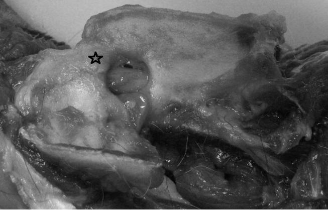
temperature prior to our study. Initially 3 cadavers were dissected to demonstrate the dorsal 101
aspect of the cervical spine and assess the length and thickness of the dorsal atlantoaxial 102
ligament (Figure 1), and to subjectively determine if it would be of sufficient size and in an 103
adequate location to be detected by ultrasonography. A further two cadavers were used to 104
correlate the ultrasonographic findings with the anatomy of each specimen. Ultrasound 105
guided injection of ink into the suspected dorsal atlantoaxial ligament was followed by 106
dissection of these 2 cadavers. Thirty adult canine cadavers were then collected and used for 107
the ultrasonographic evaluation of the dorsal atlantoaxial ligament. The cadavers were 108
positioned in lateral recumbency and the dorsal cervical area was clipped at the level of the 109
atlantoaxial joint. The skin was cleaned with spirit and gel was applied. 110
Ultrasonography was carried out using an 8 MHz microconvex transducer, with the 111
cadavers positioned in right lateral recumbency with a neutral or slightly flexed cervical 112
position. The transducer was placed on the dorsal aspect of the neck caudal to the occipital 113
crest, and the cranial aspect of the spinous process of C2 was identified. The transducer was 114
placed in a transverse plane to identify the spinous process of C2 and then moved cranially to 115
identify the dorsal atlantoaxial ligament as a thin hyperechoic structure with linear striations 116
running in a cranioventral direction from the cranial tip of the spinous process of C2 to the 117
dorsocaudal aspect of the dorsal lamina of C1. Once the ligament had been identified it was 118
also scanned in a straight sagittal plane (Figures 2 and 3). All the ultrasonographic studies 119
were performed by a board-certified specialist in diagnostic imaging. A subjective score (0-3) 120
was assigned to each dog depending on the detectability of the dorsal atlantoaxial ligament 121
and its differentiation from surrounding structures in both the transverse and the sagittal 122
plane. This grading score was similar to the classification used in previous cadaveric 123
studies22. Those cadavers in which the dorsal atlantoaxial ligament was clearly identified 124
based on the expected location, fibre orientation and fibre echogenicity were assigned a grade 125
3, if it was moderately visible they were assigned a grade 2, if the margins of the ligament 126
were indistinct but an hypoechogenic band could be identified in the expected location of the 127
ligament they were assigned a grade 1 and they were assigned a grade 0 when the fibres of 128
the dorsal atlantoaxial ligament were not discernible at ultrasonographic evaluation. 129
130
131
132
133
134
135
136
137
138
139
140
141
142
143
144
145
146
RESULTS 147
Anatomical dissection of the two cadavers in which coloured ink was injected (ultrasound 148
guided) in the ligament showed the presence of the injected ink in close association with the 149
dorsal atlantoaxial ligament (with some leakage from the ligament suspected), confirming the 150
accuracy of the ultrasonographic findings. 151
All the cadavers were mature adult dogs, but the exact age was unknown. The 152
majority of dogs were crossbreed dogs, while there were 3 German shepherd, 1 Labrador, 1 153
Rottweiler, 1 Rough Collie and 1 Husky. The estimated weight of the dogs ranged between 6 154
and 35kg. Dogs were classified as small (estimated body weight <10kg), medium (estimated 155
body weight 10-25 kg) or large (body weight>25kg). In total 19 cadavers were large breed 156
dogs, 10 cadavers were medium size dogs and 1 cadaver was a small breed dog. Table 1 157
summarizes the results. 158
Of the 19 large size cadavers 10 were graded as 3 either in the sagittal, in the 159
transverse or in both planes. Eight of the other large breed cadavers were graded as 2 either 160
in the sagittal, in the transverse or in both planes, while 1 cadaver was graded as 1 in both the 161
sagittal and the transverse plane. None of the large size cadavers were graded as 0 in any of 162
the planes. 163
Of the 10 medium size cadavers 5 were graded as 3 either in the sagittal, in the 164
transverse or in both planes and 5 were graded as 2 in either in the sagittal, in the transverse 165
or in both planes. None of the medium size cadavers were graded as 0 or 1 in any of the 166
planes. 167
Finally, the only small size cadaver was graded as 3 in both the sagittal and the 168
transverse plane. 169
170
171

DISCUSSION 172
Results from our study show that ultrasonography is a useful diagnostic tool to identify the 173
dorsal atlantoaxial ligament in normal canine cadavers, and the ligament was detectable in all 174
cadavers. 175
In our study none of the cadavers were graded as 0 (not visible) in any view and 27/30 176
and 28/30 were graded as moderately visible (grade 2) or clearly visible (grade 3) in the 177
sagittal and transverse view respectively. Only 1 cadaver specimen of a large breed dog 178
(Husky) was graded as 1 (indistinct) in both the sagittal and transverse plane, while in the 179
other 3 dogs (all large size) in which the identification of the ligament was graded as 1 in one 180
view it was graded as 2 in the other view. Therefore, 29/30 specimens were graded at least 2 181
in one ultrasonographic view and 16/30 had at least one view graded as 3, with 7/30 being 182
graded 3 in both views, 10/30 being graded 2 in both sagittal and transverse views and 9/30 183
being graded 2 in one view and 3 in the other view. Interestingly, none of the medium size 184
dogs was graded as 1 in any of the 2 ultrasonographic views, and the only small breed 185
cadaver was graded as 3 in both views. These results suggest that visualization of the 186
ligament might be clearer in smaller breeds. The reason for this finding is not clear from this 187
study, but the authors hypothesise that this could be due to a proportionally smaller muscle 188
mass surrounding the ligament in smaller breeds compared to large breeds, particularly those 189
larger dogs with a bull terrier type conformation. Many of the larger-breed cadavers in this 190
study did have a fairly broad skull conformation, so there may be a degree of bias due to this. 191
An alternative hypothesis would be a proportionally larger dorsal atlantoaxial ligament in 192
smaller breeds allowing proportionally clearer visualisation. As atlantoaxial subluxation is a 193
condition commonly diagnosed in small breed dogs, in which there is typically a congenital 194
origin, further studies will need to determine the feasibility of this technique in small breed 195
dogs. Most of the breeds in this study were medium or large breed dogs, and therefore this 196
study shows that ultrasonography could be a useful diagnostic tool for these breed of dogs, in 197
which the congenital form of atlantoaxial instability/subluxation is less common and are more 198
likely to have the traumatic presentation of atlantoaxial subluxation. 199
In order to ultrasonographically identify and assess the echogenicity of a ligament it is 200
necessary to maintain the ultrasound beam perpendicular to the ligament23, 24. In our study 201
the acoustic window was considered small due to the large size of the spinous process of the 202
axis, and therefore it is possible that the amount of ligament identified did not represent the 203
full extent of the dorsal atlantoaxial ligament. On the other hand, the ultrasonographic 204
evaluation was done with the neck in neutral position or slightly flexed, and it is likely that 205
identification of the dorsal atlantoaxial ligament would have been better if the neck was in a 206
flexed position, as there would be less overlapping of the spinous process of the axis over the 207
atlas. However, the authors decided to do the ultrasonographic evaluation of the dorsal 208
atlantoaxial ligament in neutral or slightly flexed positions as these would be the more 209
clinically relevant positions, avoiding the risk of flexing the neck in clinically affected 210
patients. An 8-10Mhz linear array transducer was briefly used to try to identify the ligament 211
at the beginning of the study, but the clarity of the resulting images of the ligament was poor, 212
with the microconvex array performing noticeably better, and hence this type of transducer 213
was used. The authors suspect that this is due to the conformation of the spinous process of 214
the axis resulting in the ligament being partially masked by the acoustic shadow from the 215
process, and the footprint of the linear array preventing easy angulation to improve image 216
quality due to the surrounding musculature. The fan-shaped image generated by the 217
microconvex array, coupled with the smaller footprint, allowed a clear image of the ligament 218
to be obtained. As the transducer was positioned in a straight sagittal position, further studies 219
will be required to assess if positioning the transducer in a parasagittal position to obtain 220
oblique views of the dorsal atlantoaxial ligament improves its assessment in dogs. 221
Five specimens were used to assess the feasibility of this technique. Three specimens 222
were used for anatomical dissection to subjectively assess the dimensions and exact location 223
of the dorsal atlantoaxial ligament. Two further specimens had coloured ink injected into the 224
dorsal atlantoaxial ligament under ultrasound guidance followed by anatomical dissection. 225
Although the total number of cadavers used for these assessments was relatively small, the 226
authors considered that the findings in the cadavers were very consistent regarding the 227
subjective dimensions and position of the dorsal atlantoaxial ligament. The location of the 228
coloured ink in the ligament was very accurate in the 2 cadavers used for this purpose, 229
confirming that the ultrasonographic identification of the dorsal atlantoaxial ligament was 230
correct. Even though only 2 cadavers were used for this purpose, the authors considered this 231
number sufficient due the accuracy found in the ultrasound guided ink injection when 232
dissecting both cadavers and the characteristic ultrasonographic images found at the expected 233
anatomical location of the dorsal atlantoaxial ligament. 234
The authors acknowledge several limitations to this study. First, we did not perform 235
anatomic dissection of the dorsal atlantoaxial ligament of all the cadaveric specimens because 236
the purpose of the study was only to determine whether ultrasound imaging can depict the 237
dorsal atlantoaxial ligament. We therefore acknowledge that although the breeds of the 238
cadavers were not breeds predisposed to atlantoaxial instability/subluxation it is possible that 239
some of the cadavers did actually have abnormalities in the dorsal atlantoaxial ligament. 240
Secondly, the study was performed on previously frozen cadavers and to the authors 241
knowledge there are no studies assessing the influence of the process of freezing/thawing 242
cadavers on the ultrasonographic image of a ligament. Therefore it is possible that this could 243
have affected the ability of the operator to identify the dorsal atlantoaxial ligament. Finally, 244
no attempts were made to perform measurements in the ultrasonographic images and 245
correlate them to anatomic dissections as the purpose of the study was to assess if 246
identification of the dorsal atlantoaxial ligament was possible. The authors performed the 247
study trying to mimic the clinical scenario and only allowing a mild flexion of the neck of the 248
cadavers. In order to measure the full extent of the ligament the dorsal atlantoaxial ligament 249
would have need to be taut by fully flexing the head, which would invalidate the potential 250
benefits of this technique compared with traditional plain radiographs in the diagnosis of 251
atlantoaxial subluxation/instability. 252
In conclusion, this study demonstrates that ultrasonographic identification of the 253
dorsal atlantoaxial ligament is a feasible technique in normal canine cadavers weighing more 254
than 6 kg. Therefore, identification of the dorsal atlantoaxial ligament through 255
ultrasonography could potentially diagnose patients with atlantoaxial instability/subluxation 256
using a non-invasive and safe diagnostic imaging technique. This technique has the 257
advantage over conventional radiographs that it can be performed with the neck at a neutral 258
or slightly flexed position. Further studies are needed to assess the feasibility to identify the 259
dorsal atlantoaxial ligament in live and numerous small breed dogs and correlate the findings 260
with patients clinically affected by atlantoaxial instability/subluxation. 261
262
263
264
265
266
267
268
269
270
271

ACKNOWLEDGEMENTS 272
None 273
274
275
276
277
278
279
280
281
282
283
284
285
286
287
288
289
290
291
292
293
294
295
296

DISCLOSURE 297
The authors declare no conflicts of interest related to this paper. 298
299
300
301
302
303
304
305
306
307
308
309
310
311
312
313
314
315
316
317
318
319
320
321
REFERENCES 322
1. Slanina MC. Atlantoaxial instability. Vet Clin North Am Small Anim Pract 2016; 46 323
(2): 265-75. 324
2. Platt, S, da Costa R. Cervical spine. In: Tobias KM, Johnston SA, editors. Veterinary 325
surgery small animal. Elsevier Saunders Missouri, 2012. 326
3. Oliver J, Lewis R. Lesions of the atlas and axis in dogs. J Am Anim Hosp Assoc 1973; 327
(9): 304-313. 328
4. Westworth DR, Sturge BK. Congenital spinal malformations in small animals. Vet 329
Clin North Am Small Anim Pract 2010; 40 (5): 951-81. 330
5. Cerda-Gonzalez S, Dewey CW. Congenital diseases of the craniocervical junction in 331
the dog. Vet Clin North Am Small Anim Pract 2010; 40 (1): 121-41. 332
6. Plessas I, Volk H. Signalment, clinical signs and treatment of atlantoaxial subluxation 333
in dogs: a systematic review of 336 published cases from 1967 to 2013. J Vet Intern Med 334
2014; 28 (3): 948. 335
7. Rochat MC, Shores A. Fixation of an Atlantoaxial Subluxation by Use of Cannulated 336
Screws. VCOT 1999; 1: 48-51. 337
8. Wheeler SJ. Atlantoaxial subluxation with absence of the dens in a rottweiler. Journal 338
of Small Animal Practice 1992; 33 (2): 90-93. 339
9. Huibregtse BA, Smith CW, Fagin BD. Atlantoaxial luxation in a Doberman pinscher. 340
Canine Practice 1992; 17: 7-10. 341
10. Hurov L. Congenital atlantoaxial malformation and acute subluxation in a mature 342
Basset Hound: surgical treatment by wire stabilization. JAAHA 1979; 15: 177-180. 343
11. Stigen Ø, Aleksandersen M, Sørby M, Jørgensen HJ. Acute non-ambulatory 344
tetraparesis with absence of the dens in two large breed dogs: case reports with a radiographic 345
study of relatives. Acta Vet Scand 2013; 55(1): 31. 346
12. Aikawa T, Shibata M, Fujita H. Modified ventral stabilization using positively 347
threaded profile pins and polymethylmethacrylate for atlantoaxial instability in 49 dogs. Vet 348
Surg 2013; 42 (6): 683-92. 349
13. Stalin C, Gutierrez-Quintana R, Faller K, Guevar J, Yeamans C, Penderis J. A review 350
of canine atlantoaxial joint subluxation. Vet Comp Orthop Traumatol 2015; 28 (1): 1-8. 351
14. Kent M, Eagleson JS, Neravanda D, Schatzberg SJ, Gruenenfelder FI, Platt SR. 352
Intraaxial spinal cord haemorrhage secondary to atlantoaxial subluxation in a dog. J Am Anim 353
Hosp Assoc 2010; 46(2): 132-7. 354
15. Parry AT, Upjohn MM, Schlegl K, Kneissl S, Lamb CR. Computed tomography 355
variations in morphology of the canine atlas in dogs with and without atlantoaxial 356
subluxation. Vet Radiol Ultrasound 2010; 51 (6): 596-600. 357
16. Couturier J, Rault D, Cauzinille L. Chiari-like malformation and syringomyelia in 358
normal cavalier King Charles spaniels: a multiple diagnostic imaging approach. J Small 359
Anim Pract 2008; 49(9): 438–43. 360
17. Schmidt MJ, Wigger A, Jawinski S, Golla T, Kramer M. Ultrasonographic appearance 361
of the craniocervical junction in normal brachycephalic dogs and dogs with caudal occipital 362
(Chiari-like) malformation. Vet Radiol Ultrasound 2008; 49(5): 472–6. 363
18. Saito M, Olby NJ, Spaulding K. Identification of arachnoid cysts in the quadrigeminal 364
cistern using ultrasonography. Vet Radiol Ultrasound 2001; 42(5): 435-9. 365
19. Sarto CG, Hage MC, Guimarães LD, Giglio RF, Borges APB, Vulcano LC. The role 366
of B-mode ultrasonography in the musculoskeletal anatomical evaluation of the cervical 367
region of the dog spine. Pesq. Vet. Bras 2014; 34(1): 91-97. 368
20. de Lahunta A, Glass E, Small animal spinal cord disease. In: de Lahunta A, Glass 369
E editors. Veterinary neuroanatomy and clinical neurology. 3rd ed. Elsevier Saunders, 370
Missouri, 2009. 371
21. Sharp NJ, Wheeler S. Atlantoaxial subluxation. In: Sharp NJ, Wheeler SJ, editors. 372
Small animal spinal disorders. 2nd ed. Elsevier Mosby, UK, 2005. 373
22. Teixeira PAG, Omoumi P, Trudell DJ, Ward SR, Lecocq S, Blum A, Resnick DL. 374
Ultrasound assessment of the lateral collateral ligamentous complex of the elbow: imaging 375
aspects in cadavers and normal voluteers. Eur Radiol 2011; 21: 1492-1498. 376
23. Lamb CR, Duvernois A. Ultrasonographic anatomy of the normal canine calcaneal 377
tendon. Vet Radiol Ultrasound 2005; 46 (4): 326-30. 378
24. Lamb CR, Wong K. Ultrasonographic anatomy of the canine elbow. Vet Radiol 379
Ultrasound 2005; 46 (4): 319-25. 380
381
382
383
384
385
386
387
388
389
390
391
392
393
394
395
396

FIGURE AND TABLE LEGENDS 397
398
Figure 1: Anatomic dissection of the dorsal aspect of the atlantoaxial joint showing the 399
anatomic location of the dorsal atlantoaxial ligament (star) 400
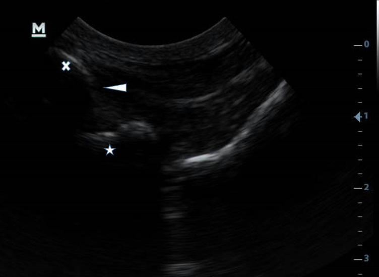
Figure 2: Ultrasonographic sagittal image of the dorsal aspect of the atlantoaxial joint 401
showing the cranial aspect of the spinous process of the axis (x), the dorsal atlantoaxial 402
ligament (arrowhead) and the dorsal arch of the atlas (star) 403
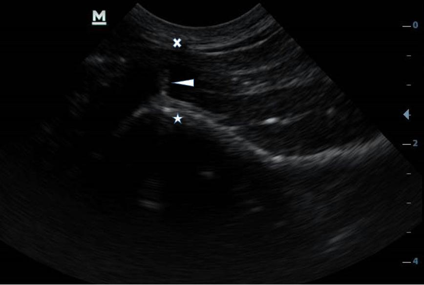
Figure 3: Ultrasonographic transverse image of the dorsal aspect of the atlantoaxial joint 404
showing the spinous process of the axis (x), the dorsal atlantoaxial ligament (arrowhead) and 405
the dorsal arch of the atlas (star) 406
Table 1: Assigned grades in the sagittal and transverse ultrasonographic views according to 407
the size of the cadavers. 408
409
410
411
412
413
414
415
416
417
418
419
420
421
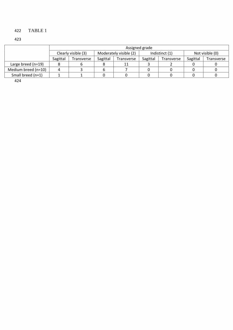
TABLE 1 422
423
Assigned grade
Clearly visible (3) Moderately visible (2) Indistinct (1) Not visible (0)
Sagittal Transverse Sagittal Transverse Sagittal Transverse Sagittal Transverse
Large breed (n=19) 8 6 8 11 3 2 0 0
Medium breed (n=10) 4 3 6 7 0 0 0 0
Small breed (n=1) 1 1 0 0 0 0 0 0
424