doctoral thesis - diva portal525687/fulltext01.pdf · doctoral thesis royal institute of technology...
TRANSCRIPT

Development of Protein-Functionalized Magnetic Iron Oxide
Nanoparticles: Potential Application in Water Treatment
Chuka Okoli
Doctoral Thesis
Royal Institute of Technology
School of Biotechnology
Stockholm 2012

© Chuka Christian Okoli
Stockholm 2012
Royal Institute of Technology
School of Biotechnology
Department of Environmental Microbiology
AlbaNova University Center
SE-106 91 Stockholm
Sweden
and
Royal Institute of Technology
School of Chemical Science & Engineering
Division of Chemical Technology
SE-100 44 Stockholm
Sweden
Printed by University service US-AB
Stockholm, Sweden
ISBN 978-91-7501-311-4
TRITA-BIO Report 2012:8
ISSN 1654-2312
All rights reserved. No part of this thesis may be reproduced, without the written permission
from the author.

To Queen, Lily and Munachi

Dedication
This work is dedicated to my beloved Mom, Late Mrs. Mabel N. Okoli for her resilience in
insisting to educate and encourage me amidst the absolute critical situation. I pray that you
will continue to rest in the bosom of Almighty God; knowing that I have accomplished that
which you always wanted.
Chuka Okoli
i
Abstract
The treatment of water to make it safe for human consumption is a problem of immense
concern, both in developing and developed countries. However, the production of clean water
with chemicals as coagulants has several drawbacks associated with cost, health risks and
complexity in sludge management. The application of nanotechnology in water treatment is a
fast growing discipline proposed as an efficient alternative that will combat these hurdles. The
aim of this thesis is to develop new water treatment strategies in a more eco-friendly manner
based on a bottom-up approach using: (i) a natural coagulant protein from Moringa oleifera
purified with nanoscale magnetic iron oxide nanoparticles for in situ treatment; and (ii) a
protein-functionalized nanoparticle (MOCP-MNPs) system by means of binding the coagulant
protein onto the nanoparticles in order to develop a potential reusable water treatment process.
Magnetic iron oxide nanoparticles with different surface chemistry have been prepared from
co-precipitation in aqueous solution and (water-in-oil and oil-in-water) microemulsion
methods.
The prepared nanoparticles were studied in terms of size, morphology, magnetic
behavior, structure, surface area including surface chemical structure and charges using
different techniques such as TEM, VSM/SQUID, XRD, BET, FT-IR and zeta potential. The
prepared nanoparticles exhibited a size ranging from 2-30 nm with superparamagnetic
properties. The Moringa oleifera coagulant protein (MOCP) with known molecular mass (6.5
kDa) was purified from the crude Moringa oleifera (MO) seed extracts using nanoparticles
prepared from both methods. The obtained MOCP exhibits comparable coagulation activity
with alum in terms of water turbidity removal, implying alternative replacement to chemical
coagulants. This technique can be easily applied where natural materials are available locally.
Studies on the interaction between MOCP and surface modified nanoparticles were
essential to understand the binding mechanism for the development of a protein-
functionalized nanoparticle. Based on in silico investigation, the overall molecular docking
studies reveal the interactions between protein-ligand complexes by electrostatic, van der
Waals and hydrogen-bonding; which imply, that there are at least two binding sites is i.e. one
located at the core binding site (TEOS and APTES ligand) while the other located at the side
chain residues (TSC and Si60-OH).
This work underscores advancement in the development and use of MOCP-MNPs for
potential water treatment. About 70% turbidity removal was achieved gravimetrically using
MOCP-MNPs (60 min) in high and low turbid waters, whereas alum requires 180 min to
reduce the turbidity especially in low turbid waters. The turbidity removal efficiency was
enhanced by the use of MOCP-MNPs under the influence of an external magnetic field. More
than 95% turbidity removal was achieved within 12 min in high and low turbid waters when
MOCP-MNPs were used. The combination of natural coagulant protein and magnetic
nanoparticles as well as the use of applied magnetic field enhanced the performance
coagulating/flocculating properties in the water samples.
These results suggest a successful development of MOCP-MNPs as demonstrated in the
regeneration study. The data shown in this work represent novel potential water treatment
strategies that could be cost-effective, simple, robust and environmentally friendly whilst
utilizing biocompatible materials.
Keywords: magnetic nanoparticles; co-precipitation; microemulsion; Moringa oleifera;
protein purification; water treatment; magnetophoresis; coagulation activity; turbidity
removal.

Development of Protein-Functionalized Magnetic Iron Oxide Nanoparticles: Potential Application in Water Treatment
ii
Chuka Okoli
iii
List of Publications
This thesis is based on the following papers:
I. Chuka Okoli, Andrea Fornara, Jian Qin, Muhammet Toprak, Gunnel Dalhammar,
Mamoun Muhammed and Gunaratna Rajarao-Kuttuva. Characterization of
Superparamagnetic Iron Oxide Nanoparticles and Its Application in Protein
Purification. J. Nanosci. Nanotech. 2011, 11, 10201-10206.
II. Chuka Okoli, Magali Boutonnet, Laurence Mariey, Sven Järås and Gunaratna
Rajarao-Kuttuva. Application of Magnetic Iron Oxide Nanoparticles Prepared
From Microemulsions For Protein Purification. J. Chem. Techno. Biotech. 2011,
86, 1386-1393.
III. Chuka Okoli, Margarita Sanchez-Dominguez, Magali Boutonnet, Sven Järås,
Concepción Civera, Conxita Solans and Gunaratna Rajarao-Kuttuva. Comparison
and Functionalization Study of Microemulsion-Prepared Magnetic Iron Oxide
Nanoparticles. Langmuir. 2012, (in press).
IV. Chuka Okoli, Magali Boutonnet, Sven Järås and Gunaratna Rajarao-Kuttuva.
Protein-Functionalized Magnetic Iron Oxide Nanoparticles: Time Efficient
Potential-Water Treatment. 2012, (submitted).
V. Chuka Okoli, Selvaraj Sengottaiyan, Arul N. Murugan, Pavankumar A.
Ramachandran, Hans Ågren and Gunaratna Rajarao-Kuttuva. In Silico Modeling
and Experimental Evidence of Coagulant Protein Interaction with Precursors for
Nanoparticle Functionalization. 2012, (submitted).
VI. Pavankumar R. Ramachandran, Kayathri Rajarathinam, Arul N. Murugan, Zhang
Qiong, Chuka Okoli, Hans Ågren and Gunaratna Rajarao-Kuttuva. Dimerization
of flocculent protein from Moringa oleifera: experimental evidence and in silico
interpretation. 2012, (manuscript).
VII. Ramnath Lakshmanan, Chuka Okoli, Magali Boutonnet, Sven Järås and
Gunaratna Rajarao-Kuttuva. Effect of Magnetic Iron Oxide Nanoparticles for
Surface Water Treatment: Trace Minerals and Microbes. 2012, (manuscript).
Other work not included:
1. Chuka Okoli, Ramnath Lakshmanan, Duke Antwi, Magali Boutonnet, Sven Järås,
and Gunaratna Rajarao-Kuttuva. Investigating the Use of Magnetic Nanoparticles for
Efficient Reduction of Phosphates and Nitrates in Wastewater Treatment Process,
(manuscript).
All papers are reproduced with kind permission from the respective copyright holders.

Development of Protein-Functionalized Magnetic Iron Oxide Nanoparticles: Potential Application in Water Treatment
iv
Contributions of the author
Paper I. Principal author, involved in planning, performed the experiments, performed part
of sample characterization, evaluation of the results and writing the article.
Paper II. Principal author, involved in planning, performed the experiments, performed part
of sample characterization, evaluation of the results and writing the article.
Paper III. Principal author, involved in planning, performed the experiments, performed part
of sample characterization, evaluation of the results and writing the article.
Paper IV. Principal author, involved in planning, performed the experiments, performed part
of sample characterization, evaluation of the results and writing the article.
Paper V. Principal author, involved in planning, and performed the experimental part,
involved in evaluation of the results and writing part of the article.
Paper VI. Participated in developing ideas and evaluation of results
Paper VII. Participated in planning, synthesis of magnetic nanoparticles, performed part of
experiments, evaluation of results and writing part of the article.
Patent Applications
1. Chuka Okoli, Magali Boutonnet, Laurence Mariey, Sven Järås and Gunaratna
Rajarao-Kuttuva. Application of Magnetic Iron Oxide Nanoparticles Prepared From
Microemulsions For Protein Purification. U.S. Patent and trade office, 61407455,
October 28, 2010
2. Chuka Okoli, Magali Boutonnet, Sven Järås and Gunaratna Rajarao-Kuttuva. Time
Efficient Drinking Water Treatment Using Protein-Functionalized Magnetic Iron
Oxide Nanoparticles. U.S. Patent and trade office, 61599960, February 17, 2012.

Chuka Okoli
v
Conference presentations
1. Membrane Technology Conference for Water and Wastewater Treatment; (attended),
September 1-3, 2009. Beijing, P.R.China.
2. Chuka Okoli, Andrea Fornara, Jian Qin, Muhammet S. Toprak, Gunnel Dalhammar,
Mamoun Muhammed and Gunaratna Rajarao-Kuttuva. “Superparamagnetic
nanoscale-based particles for coagulant protein purification” (poster), 2010
International Chemical Congress of Pacific Basin Societies; December 15-20, 2010.
Honolulu, Hawaii, USA.
3. Chuka Okoli, Ramnath Lakshmanan, Magali Boutonnet, Sven Järås and Gunaratna
Rajarao-Kuttuva “Microemulsion magnetic nanoparticles: Synthesis, characterization
and their application” (poster), 94th Canadian Chemistry Conference and Exhibition;
June 5-9, 2011. Montréal, Québec, Canada.
4. Chuka Okoli, Ramnath Lakshmanan, Magali Boutonnet, Sven Järås and Gunaratna
Rajarao-Kuttuva “Magnetic nanoparticles for coagulant protein purification and
water treatment” (oral), Clean water through Bio- and Nano-technology; May 7-9,
2012. Lund, Sweden.

Development of Protein-Functionalized Magnetic Iron Oxide Nanoparticles: Potential Application in Water Treatment
vi

Chuka Okoli
vii
Abbreviations and Symbols
AAS atomic absorption spectroscopy
AOT Bis(2-ethylhexyl) sulfosuccinate
APTES 3-Aminopropyltriethoxysilane
BET Brunauer-Emmet-Teller
CMC carboxyl methyl cellulose
CTAB cetyltrimethylammonium bromide
Hc coercivity
DBPs disinfectant by-products
Dc critical size thresholds
DOC dissolved organic carbon
Ds superparamagnetism
FDA Food and drug administration
FTIR Fourier transform infrared spectroscopy
GAC granulated active carbon
H applied magnetic field
HLB hydrophilic-lipophilic balance
HRTEM high-resolution transmission electron microscopy
IEX ion exchange
M magnetization
ME-MIONs microemulsion-prepared magnetic iron oxide nanoparticles
MNPs magnetic nanoparticles
MO Moringa oleifera
MO2.1 Moringa oleifera recombinant protein
MOCP Moringa oleifera coagulant protein
MR magnetic remanence
MS saturation magnetization
NF nanofiltration
NMs nanomaterials
NOM natural organic matter
NTU nephelometric turbidity units
o/w oil-in-water
OD optical density
PAC polyaluminium chloride
PEI poly ethylene imine
PMO purified Moringa oleifera
Pzc point of zero charge
SAED selected area electron diffraction
SDS-PAGE sodium dodecyl sulphate-polyacrylamide gel electrophoresis
SENs single-enzyme nanoparticles
SPION superparamagnetic iron oxide nanoparticle
SQUID superconducting quantum interference device magnetometer
TEM transmission electron microscopy

Development of Protein-Functionalized Magnetic Iron Oxide Nanoparticles: Potential Application in Water Treatment
viii
TEOS tetraethoxysilane
THMs trihalomethanes
TLC thermoelectric temperature controller
TSC trisodium citrate
UV ultraviolet
VSM vibrating sample magnetometer
w/o water-in-oil
WHO world health organization
XRD X-ray diffraction
ζ zeta potential (Zp)
χ magnetic susceptibility
Chuka Okoli
ix
Table of Contents
1. Introduction ..................................................................................................................................... 1
1.1 Conventional water treatment processes ..................................................................................... 2
1.2 Nanotechnology in water treatment ............................................................................................. 5
1.3 Natural coagulants in water treatment ......................................................................................... 7
1.3.1 Brief History of the Moringa oleifera (MO) treef ................................................................ 7
1.3.2 Water treatment with MO protein ........................................................................................ 8
1.3.2.1 Microbial elimination .................................................................................................... 9
1.3.3 Purification of the MO protein ........................................................................................... 10
1.3.4 Structural prediction ........................................................................................................... 11
1.4 Magnetic nanoparticles .............................................................................................................. 11
1.4.1 Iron oxide nanoparticles ..................................................................................................... 12
1.4.1.1 Magnetic behavior ....................................................................................................... 13
1.4.1.2 Superparamagnetic phenomenon ................................................................................. 14
1.4.2 Synthesis methods .............................................................................................................. 15
1.4.2.1 The co-precipitation method in aqueous solution ........................................................ 16
1.4.2.2 The microemulsion method ......................................................................................... 16
1.4.2.3 The thermal decomposition method ............................................................................ 18
1.4.2.4 The hydrothermal method ........................................................................................... 18
1.4.3 Magnetic separation and mixing ........................................................................................ 19
1.5 Objectives of the present work .................................................................................................. 20
2. Experimental .................................................................................................................................. 23
PART ONE ....................................................................................................................................... 23
Synthesis and characterization .......................................................................................................... 23
2.1 Extraction and analysis of coagulant protein (Papers I & II) .................................................... 23
2.1.1 Preparation of synthetic water ............................................................................................ 24
2.1.2 Coagulation activity test ..................................................................................................... 24
2.2 Synthesis of magnetic iron oxide nanoparticles (Papers I─III) ................................................. 24
2.2.1 Co-precipitation method in aqueous solution ..................................................................... 25
2.2.1.1 Coating with TSC ........................................................................................................ 25
2.2.1.2 Coating with TEOS ..................................................................................................... 26
2.2.1.3 Coating with APTES ................................................................................................... 26
2.2.2 Microemulsion methods ..................................................................................................... 27
2.2.2.1 Synthesis of w/o ME-MION ....................................................................................... 27
2.2.2.1 Synthesis of o/w ME-MION ....................................................................................... 29
2.3 Characterization (Papers I─III) ................................................................................................. 29
2.3.1 Nanoparticles prepared by co-precipitation (Paper I) ......................................................... 30
2.3.2 Nanoparticles prepared by Microemulsion (Papers II-III) ................................................. 31
PART TWO ....................................................................................................................................... 31
2.4 Application of nanotechnology in water treatment (Papers III, IV, V & VII) .......................... 31
2.4.1 Purified MOCP for water treatment (Approach I) .............................................................. 31
2.4.1.1 Purification of the MO coagulant protein with MNPs and activity test ...................... 32
2.4.1.2 Analysis and characterization ...................................................................................... 32
2.4.1.3 Turbidity removal in surface waters ............................................................................ 33
2.4.1.4 Interaction of MOCP and MNPs (Papers III & V) ...................................................... 33

Development of Protein-Functionalized Magnetic Iron Oxide Nanoparticles: Potential Application in Water Treatment
x
2.4.1.4.1 Computational and experimental studies .............................................................. 34
2.4.1.4.2 Fluorescence emission study (Paper III) .............................................................. 35
2.4.2 MOCP-MNPs for water treatment (Approach II) ............................................................... 35
2.4.2.1 Development of MOCP-MNPs system ....................................................................... 36
2.4.2.2 Turbidity removal in surface waters ............................................................................ 37
2.4.2.3 Separation by gravity and magnetic field .................................................................... 37
2.4.2.4 Reusability of MOCP-MNPs system ........................................................................... 37
3. Results and discussion ................................................................................................................... 39
PART ONE ....................................................................................................................................... 39
Synthesis and characterization .......................................................................................................... 39
3.1 Investigation of the active protein (Paper I) .............................................................................. 39
3.2 Characterization of MNPs (Papers I─III) .................................................................................. 40
3.2.1 Size and morphology studies ............................................................................................. 40
3.2.2 Structural studies ............................................................................................................... 41
3.2.3 Magnetic studies ................................................................................................................ 42
3.2.4 Surface charge studies ........................................................................................................ 43
PART TWO ....................................................................................................................................... 44
3.3 Application of nanotechnology in water treatment ................................................................... 44
3.3.1 Purification of the MO protein with MNPs and characterization (Papers I─III) ............... 44
3.3.1.1 Turbidity removal in surface waters (Approach I) ...................................................... 45
3.3.2 Interaction of MOCP and MNPs (Papers III & V) ............................................................. 46
3.3.2.1 Computational and experimental studies ..................................................................... 46
3.3.2.2 Fluorescence emission study ....................................................................................... 50
3.3.3 Turbidity removal with the developed MOCP-MNPs system (Approach II) ..................... 51
(Paper IV & V) .............................................................................................................................. 51
3.3.3.1 Separation by gravity and magnetic field .................................................................... 53
3.3.3.2 Reusability of MOCP-MNPs system ........................................................................... 54
4. Conclusions .................................................................................................................................... 57
Further studies ..................................................................................................................................... 58
Acknowledgement ............................................................................................................................... 59
References ............................................................................................................................................ 61
Appendix .............................................................................................................................................. 71

Chuka Okoli
1
1. Introduction
Access to clean and safe drinking water is a human right; however, the availability of potable
water is a major concern in both developed and developing countries.1 Water treatment offers
the benefit of potable water in terms of quality (reduced level of contaminants) and quantity
(availability). The world is facing formidable challenges in meeting the rising demands for
safe drinking water supply due to population growth, increasing pollution of water bodies
from several industrial and agricultural activities, drought and competing demands from a
variety of users.1-5
As a result of this water scarcity, children and elderly people are more
vulnerable to water-borne diseases. According to the World Health Organization (WHO),
more than 1 billion people especially in the developing countries still do not have access to an
adequate supply of drinking water.5 As a consequence, the quality of health and welfare
vulnerable groups (children, the elderly and the poor) are dependent on the availability of a
safe and affordable water supply.5, 6
It has been predicted that in the year 2025, about 3.5
billion people, 48% of the world’s population, will have an inadequate water supply.7
The provision of potable water from most raw water sources involves the use of coagulants
introduced during the coagulation/flocculation step to remove turbidity in the form of
dissolved or suspended materials. Chemical coagulants such as aluminum salts (AlCl3),
organic polyaluminum chloride (PAC)8 and ferric chloride (FeCl3) are frequently used to
enhance coagulation and flocculation.9 Nevertheless, given that the choice of chemicals in
water treatment is decisive, treatment processes with synthetic coagulants or disinfectants
may add substances such as disinfectant by-products (DBPs) to the treated water, which can
increase the risk of disease.10
In spite of their high efficiency, the presence of residual DBPs11
or aluminum residues12
in treated water is a huge concern due to their neurotoxicity and
carcinogenic properties such as those found in Parkinson’s, Alzheimer’s and cancer diseases.9,
10, 12-14 Moreover, the existence of large volumes of sludge, the increase in water pH as well as
the high cost of importing these chemicals is still a challenge.15
Considering the importance of potable water globally, and keeping in mind concerns about
the feasibility of recent practices to meet the rising water demands, there is a pressing need to
develop novel technologies and materials that are associated with natural coagulants which
will combat these challenges whilst reducing or replacing the use of chemicals in water

Development of Protein-Functionalized Magnetic Iron Oxide Nanoparticles: Potential Application in Water Treatment
2
treatment. While new technologies are being developed today, it is considered paramount to
develop biodegradable, cost effective, user-friendly, robust and more efficient systems than
the existing techniques for the removal of water contaminants either in water treatment
processes or in situ.16
The emergence of nanotechnology has been identified as a promising
technology that could play a major role in providing potable water.5
The use of natural coagulants such as the Moringa oleifera coagulant protein (MOCP)17-20
associated with magnetic nanomaterials16, 21, 22
is one way in which nanotechnology can play a
vital role in providing potable water. Functionalized nanomaterials, such as magnetic iron
oxide nanoparticles, are known to display novel and significant physicochemical as well as
biological properties due to their unique size, structure and large surface-to-volume ratio.21, 23,
24 Protein-functionalized magnetic nanoparticles offer several advantages such as fast
separation, enhanced efficiency, and/or significant reduction in sludge volume and material
recycling. In particular, the incorporation of natural coagulant proteins on magnetic
nanoparticles constitutes an excellent alternative to chemical remediation in water treatment
processes.
1.1 Conventional water treatment processes
Globally, in many water treatment plants, the combination of coagulation/flocculation,
sedimentation, filtration and disinfection is a widely applied water treatment technology
(Figure 1.1) for the production of potable water. This practice has been in use since the early
20th century.16
However, coagulation/flocculation is an important primary step in the water
treatment processes and as such, is critical for other successive steps including the amount of
chemical usage and the overall production cost.
Figure 1.1 Scheme of general water treatment processes.
Coagulation/ flocculation is a process that involves the removal of suspended and dissolved
particles by the addition of salts such as aluminum sulphate, ferric chloride or organic
polyaluminum chloride followed by rapid mixing. These chemicals, which are positively
Incoming water
Coagulation/
Flocculation Sedimentation Filtration Disinfection Outlet
Sludge

Chuka Okoli
3
charged, act as coagulants. Particles in water are negatively charged and for this reason repel
each other when they come in close contact. This will force them to remain in suspension
rather than clump together and settle out of the water. The addition of positively charged
coagulants destabilizes the negative charges of the suspended and dissolved particles in the
water.25
When this reaction occurs, the particles bind together (coagulation); this process is
also referred to as flocculation. The large particles (flocs) formed are denser and quickly settle
to the bottom of the vessel.15
This settling process is called sedimentation. The heavy
particles are then removed, and the water moves to the next step.
Nonetheless, coagulation/flocculation and sedimentation are crucial for successful removal of
large amounts of organic compounds, including dissolved organic materials such as Dissolved
Organic Carbon (DOC) or Natural Organic Matter (NOM). Large amounts of these dissolved
organic materials are responsible for undesirable water odor, taste, color and the presence of
microorganisms.26
Previous studies show that coagulation can remove suspended and
dissolved particles by adsorption and charge neutralization, adsorption and inter-particle
bridging, and precipitation.27-29
The most common mechanism in coagulation/flocculation
processes is the inter-particle bridging and charge neutralization.30
Due to the inter-related
properties of coagulation/flocculation processes, the optimization of raw water treatment with
coagulant/coagulant aids can sometimes be very difficult to achieve; thus some factors may
influence the performance of the coagulants in water treatment processes.
These factors include the source water characteristics, raw water pH, choice of
coagulant/coagulant aids and their order of addition, temperature, alkalinity, coagulant dosage
and the degree and mixing condition, including the flocculation period. Keeping track of the
pH and alkalinity is very critical for good performance.31
Alkalinity of water is referred to as
its capacity to neutralize acids. Water alkalinity may be due to the presence of one or more
ions like hydroxides, carbonates and bicarbonates. For instance, raw water with low alkalinity
will utilize most of the available alkalinity by the addition of a coagulant such as ferric salts,
thereby lowering the water pH to a level that hampers effective treatment. Conversely, water
with high alkalinity may require additional coagulants in order to lower the pH to the
acceptable range for coagulation. Therefore, it is more cost-effective to treat water with
acceptable alkalinity. The coagulation/flocculation process can also be affected by
temperature. The coagulant stability, increasing water viscosity as well as hindering the
kinetics of hydrolysis reaction and particle flocculation are all attributes of low water
temperature. Organic coagulants seem to be more effective in cold water since they are pre-

Development of Protein-Functionalized Magnetic Iron Oxide Nanoparticles: Potential Application in Water Treatment
4
hydrolyzed.15, 32
Given that the aforementioned coagulants are efficient and widely used in
the first step of water treatment, there are still huge concerns about their adverse health effects
associated with residual chemicals and, increase in water pH, including increased sludge
volume and complexity.
Sludge conditioning and dewatering: inappropriate disposal of sludge from water treatment
plants with little or no treatment is a common practice in many places. However, the direct
disposal of untreated sludge is considered a risk to recipients due to the chemical composition
of the sludge.33
Sludge from water comprises more than 90% of water; thus, it is a big
challenge to dewater.34
Methods such as centrifugation, chemical precipitation and heat
treatment are commonly used sludge conditioning systems. However, these methods are
somewhat complicated and require experienced operators; moreover, the energy needed for
the heat treatment and the high cost of chemicals is a big concern for both developing and
developed countries. Nonetheless, the environmental impact of commonly used chemical
conditioners may be averted if locally available natural materials can be employed.
Filtration is the second step in a conventional water treatment process. This step removes
particulate matter from water by means of forcing the water to percolate and pass via porous
media. The filtration system often consists of sand, gravel and charcoal as filters, as well as
microfiltration or ultrafiltration membranes with varying sizes of pores. The particles,
including microorganisms and ions, that are removed from water during filtration depend on
the pore size of the filter used.35
Membrane filtration or granulated active carbon (GAC)36
is
often used, but the drawback is the high cost of maintenance. For surface water, sand filtration
is usually preferred in the treatment process. Basically, two types of sand filtration exist, slow
sand filtration and rapid sand filtration. Rapid sand filtration is a physical process that
involves the removal of suspended solids. Compared to slow sand filtration, this process is
common; it has fairly high flow rates and requires less space. Slow sand filtration is a
biological treatment and the most preferred method, because it uses bacteria to treat the water.
The surface of the sand filtration is designed in such a way that the bacteria establish a
community on the top layer (biofilm) of sand, whilst cleaning the water by digesting the
contaminants as it passes through the sand filters.37
The process requires considerable
operational space and must be situated in the open air, which makes it vulnerable to other
contaminants. It also requires long hours including periodic cleaning.

Chuka Okoli
5
Disinfection is the final step in water treatment processes before sending the water to the end
users. Frequently used disinfectants in water treatment processes include chlorine,
chloramines, and ozone. Chlorine or and its derivatives are relatively cheap and effective; in
spite of their high efficiency, the presence of residual Disinfectant By-Product (DBPs) such as
trihalomethanes (THMs) in the treated water is a major concern for public health due to their
neurotoxicity and carcinogenic properties such as those found in Parkinson’s, Alzheimer’s
and cancer diseases.10, 11
Another effective disinfectant alternative to chlorine compounds is
the use of UV light and ozone treatment; however, this method consumes high energy and
also increases the emission of carbon. For these reasons, it is not considered cost-effective and
eco-friendly.11
Taking into account various limitations and concerns in water treatment
processes, the need for the production of potable water have not yet been met.
The outbreaks of water-borne diseases are increasing exponentially. For instance, recent
outbreaks were reported in places like Östersund, Sweden in 2011 and Manila, the Philippines
in 2012. The presence of different organic contaminants and highly toxic metallic cations
such as arsenic, lead, cadmium, or inorganic anions like nitrates, perchlorate are of major
concern to public health as well as the ecosystem. Moreover, the use of chemicals in water
treatment has resulted in the generation of large amounts of effluents that are not
biodegradable including disinfectant by-products. The potential application of
nanotechnology for novel material development in order to combat these challenges is
deemed necessary for the production of potable water in terms of quality and quantity.
1.2 Nanotechnology in water treatment
Nanotechnology is the engineering and art of manipulating matter at the nanoscale, basically
at the dimensions between 1─100 nm, where unique phenomena enable novel applications in
various disciplines including Biology. Nanotechnology offers the potential of novel nanoscale
materials for the treatment of all kinds of waters including surface water and wastewater.5
Due
to their unique properties towards various water contaminants and flexible applications, many
nanomaterials (NMs) are currently being developed. The key potential impact areas for
nanotechnology in water treatment applications can be divided into three groups, namely
treatment and remediation, sensing and detection, and pollution control.16
The first
mentioned is likely to contribute to long-term water quality, availability (quantity), and
viability of water resources, such as through the use of nanoreactive and functionalized
membranes, and advanced filtration materials that enable greater water reuse, recycling, and

Development of Protein-Functionalized Magnetic Iron Oxide Nanoparticles: Potential Application in Water Treatment
6
desalination. Within the category of sensing and detection, NMs can promote the
development of new and enhanced sensors to detect biological and chemical contaminants
even at very low concentration levels in the assayed water sample. An example of such
material is single-enzyme nanoparticles (SENs).38, 39
Recent research on the synthesis of
different nanomaterials includes nanosorbents, nano-structured/reactive membranes,
nanocatalysts and bioactive nanoparticles.5, 40
The nanosorbents or membrane process
techniques are seen as the main driving force in the advancement made in water purification
and desalination technologies, since conventional water treatment methods are not effective in
removing organic pollutants. Trial experiments with some of these materials are ongoing in
Africa.16
Nanomaterials such as nanoparticles, carbon nanotubes and dendrimers (Figure 1.2)
contribute to the development of more robust, efficient and cost-effective water filtration
processes. Two types of membranes that could serve as effective water treatment techniques
are (i) nanostructured filters, where either carbon nanotubes or nanocapillary arrays provide
the basis for nanofiltration (NF); and (ii) nanoreactive membranes, where functionalized
nanoparticles enhance the filtration process.41-43
The synthesis of dendritic polymers such as
cyclodextrins, poly(propylene imine) and poly(amidoamine) dendrimers offers opportunities
to refine as well as to develop operative filtration processes for purification of water
contaminated by different organic solutes and inorganic pollutants.5
Figure 1.2 Nanotechnology-based materials for water treatment: (a) carbon nanotube (1-3
nm), (b) dendritic polymer as nanoreactive NMs; rectangular shape (blue) represents different
functional groups, and (c) Nanofiltration membranes with pore sizes of 10, 20 and 50 nm.
The use of bioactive nanoparticles for water disinfection is another key area of
nanotechnology in water sanitization.
(a) (c) (b)

Chuka Okoli
7
Nanobiocides may present a reasonable alternative that will reduce the amount of sludge
produced during water treatment whilst underscoring new chlorine-free biocides that are
environmentally friendly. Among the most promising antimicrobial NMs are metallic and
metal oxide nanoparticles, especially functionalized iron oxide, polyethyleneimine (PEI),
chitosan, silver nanoparticles (AgNPs) and titanium dioxide catalysts for photocatalytic
disinfections.16, 44
Moreover, current research is being focused on the use of natural coagulant
materials as alternative sources in order to meet the above-mentioned challenges.
1.3 Natural coagulants in water treatment
Recent research has focused on the development and use of natural coagulants which can be
extracted or produced from plants, animals or microorganisms due to their presumed safety to
humans and the environment.9 The application of natural materials of plant origin for
clarifying turbid raw water from rivers is an ancient and domestic household practice in
tropical developing countries where these natural materials act as primary coagulants due to
their availability throughout the year.45, 46
Different kinds of natural coagulants obtained from
apricots (beach kernels), groundnut seed, nirmali seed, pumice seed, maize and the Moringa
oleifera (MO) coagulant protein have been described in various reports.9, 25, 26, 47
However,
among the investigated natural materials, water soluble extracts from the seeds of MO have
attracted particular attention since they possess dual functionality in water treatment by acting
as coagulants as well as antimicrobial agents.9, 48
Jahn, S.A.A49, 50
and Madsen et al.,51
were
among the first researchers to study the use of MO as natural coagulants/disinfectants in water
treatment. Since then, numerous studies have been conducted to optimize its use in water
treatment.20, 29, 52
The protein extracted from MO seeds competes quite favorably with
chemical coagulants.20, 53 It is widely accepted from various studies that extracts from MO
seeds possess effective coagulation/flocculation as well as antimicrobial properties, even
though the properties and nature of the active component seem to differ slightly, depending on
the extraction method used48
or the geographical region where the plants were grown.
1.3.1 Brief History of the Moringa oleifera (MO) treef
Moringa oleifera is a multipurpose tree belonging to the family of Moringaceae, a single
family of shrubs with 13 known species.54
It is a tropical plant found throughout Asia, sub-
Saharan Africa and Latin America. MO is widely recognized as a tree with almost every part
of the plant utilized for beneficial purposes. Due to the diverse applications of MO, it is
sometimes referred to as the miracle tree.32
The MO tree (Figure 1.3) is drought-tolerant and

Development of Protein-Functionalized Magnetic Iron Oxide Nanoparticles: Potential Application in Water Treatment
8
is generally used in the developing world as a vegetable, medicinal plant, nutrition
supplement, cattle fodder, fertilizer and a source of oil.54, 55
Moringa oleifera seeds are also
very rich in iron and calcium and they contains 40% by weight of oil which can be used for
cooking, lamp fuel and production of soap.54
It has been reported that the press cake
remaining after oil extraction still contains active coagulants.18
The medicinal and therapeutic
potential of MO is being utilized in the cure of different diseases and ailments.56
Among
many other properties, seeds from MO contain a coagulant protein that can be used for water
clarification.17, 20, 49, 57, 58
The coagulant is obtained at extremely low or zero net cost.59
MO
seed protein is known to be one of the most effective natural coagulants and the study on
treatment of different kinds of waters has been growing recently.32, 58, 60
Figure 1.3 Moringa oleifera tree with pods and seed kernels. The active coagulant is
extracted from the seed kernel.
The active components of MO are water soluble cationic proteins with a molecular weight of
6.5 to 13 kDa and pI values around 10. Ndabigengesere et al.,61
in their study, described the
active coagulating agent of MO as a dimeric cationic protein having a molecular weight of 13
kDa and an isoelectric point between 10 and 11. The MO coagulant protein was identified as a
heterogeneous mixture consisting of sixty amino acid residues.57, 61, 62
Moreover, extracts
from MO seeds possess significant properties in the reduction of sludge volume and bacteria
in contaminated waters63, 64
without affecting the water pH, conductivity and alkalinity,
thereby making the MO protein more attractive than aluminum salts in water treatment.53
Apart from being non-toxic, the MO protein is entirely biodegradable.9, 48
1.3.2 Water treatment with MO protein
The active component can be extracted from seeds by the use of water or salt solutions,
usually NaCl.57, 65
The use of MO seed protein in water treatment can be applied both at
industrial scale where it can be used as a coagulant aid15, 66
or at the household level.67
Several
reports suggest that the MO seed is more efficient when applied in high turbid waters; hence,

Chuka Okoli
9
its use in large scale water treatment during the spring season when water turbidity is at its
highest level will benefit the water treatment plants that are forced to shut down due to lack of
funding.67
The coagulation/flocculation mechanism of MO coagulant protein (MOCP) was
described by Ndabigengesere et al.,57
as a mechanism involving adsorption and neutralization
of charges, implying that the positively charged amino acids of this protein bind to the
suspended or dissolved particles that are mainly negatively charged, and this leads to the
formation of negatively and positively charged areas on the particle surface. Inter-particle
neutralization of differently charged sectors and formation of flocs take place due to particle
collision (Figure 1.4).30
Figure 1.4 Mechanism of coagulation/flocculation with Moringa oleifera coagulation
protein (MOCP) showing adsorption and neutralization of the colloidal charges with net-like
structure.
1.3.2.1 Microbial elimination
Traditional water disinfection processes usually make use of chlorinated (chlorine and
chloramine) chemical additives in eliminating the microbial contaminants. While their
benefits are well established, concerns have also been raised about their safety issues.11
Conversely, MO seed extracts are capable of bacterial aggregation and removal. Their
antimicrobial activity may lead to growth inhibition and killing of bacteria, including
antibiotic-resistant human microorganisms.53
On the other hand, Broin et al.,68
showed that a
recombinant protein (MO2.1) of MOCP is capable of flocculating both Gram-Positive and
Gram-negative bacterial cells. Microorganisms can be removed in this case by settling in the

Development of Protein-Functionalized Magnetic Iron Oxide Nanoparticles: Potential Application in Water Treatment
10
same way as the removal of colloids in well coagulated and flocculated water; alternatively,
the protein may also act directly upon the microorganism, resulting in growth inhibition.68
The microbial growth inhibition might be mediated by the interaction of positively charged
amino acids with the negatively charged surface of the microorganism’s membrane.76
1.3.3 Purification of the MO protein
Apart from several advantages of the MO protein over chemical coagulants, the main
drawback in using the crude extracts of MO seeds in water treatment is the release of organic
matter and nutrients to the water. Previous studies show that crude seed extracts increase the
organic, nitrate and phosphate contents in treated water, while the purified form (MOCP) does
not.18, 69
The presence of these organic loads is a source of odor, color and taste in water;
moreover they also facilitate the growth of microorganisms upon storage, thus limiting the use
of crude MO seed extract as a coagulant for water treatment in domestic and industrial
levels.70
In order to overcome these limitations, the MO coagulant protein needs to be
purified.
Consequently, the quest for low cost and simple purification procedures is critical for efficient
maximization of natural coagulant in use. Previous reports imply that the MO purification
process involves extensive methods that require several steps, which makes the purification
system time consuming, complicated and expensive. Furthermore, it has become difficult to
purify the protein on a large scale for water treatment applications.48, 69
Recently, Habauka et
al.,48
employed a purification method that involves more than six steps, including dialysis. A
single-step elution procedure was adopted by Ghebremichael et al.18, 59
They developed a
simple method for the purification of coagulant protein using ion-exchange matrix (IEX).
While these methods are efficient, there are still some unresolved issues, such as the low
binding capacity of the IEX matrix, the high cost of material as in the case of commercial
beads, as well as long process times. These challenges can be a limiting factor which might
potentially detract from the advantages of the aforementioned methods, bearing in mind the
accessibility problem for people in the developing countries.
The application of magnetic nanoparticles could add more benefits over the existing
technologies.71
This fast and easy technique can achieve separations (via magnetophoresis)
that are difficult to achieve practically with conventional methods.72-74
Magnetic separation
eliminates some steps during the purification, thereby promoting gentle, quick, and scalable
alternatives to the conventional methods. The desired targets are captured on magnetic

Chuka Okoli
11
nanoparticles coated with a specific surface and then separated from the samples with an
external magnet.75
1.3.4 Structural prediction
Many studies have been carried out with MOCP, but to date, the structure of this protein has
not been solved by X-ray crystallography or NMR spectroscopy. However, Suarez et al76
performed computer simulation modeling to reveal the structure of the MO2.1 peptide. In their
study, they predicted the structure of this protein from the known α-helices modeled structure
of the napin peptide in a consensus model of MO2.1 peptide secondary structure, which depicts
high probability of α-helices in the three regions that form helical structures in the
corresponding parts of napin.76
Studies show that MOCP consists of three α-helices.67
The
structure-function study of the peptides used, reveals the relationship between the water
clarification and the disinfectant activities of the MOCP. Studies show that sedimentation
requires positively charged glutamine and arginine residues of the protein to form aggregate
of particles. The bactericidal activities follow a sequence prone to form a helix-loop-helix
structure, which may consist of a hydrophobic patch containing two prolines surrounded by
positively charged arginines pointing outward from the folded polypeptide chain.
1.4 Magnetic nanoparticles
Magnetic nanoparticles are of great interest for a broad range of applications, including
catalysis,74, 77
biotechnology,78-81
biomedicine,82, 83
data storage,84, 85
magnetic resonance
imaging86-88
and environmental remediation.16, 89, 90
The use of nanoparticles offers several
advantages due to their unique size and physical properties. However, in order to design
magnetic nanoparticles for a specific application, it is important to understand their atomic
structure, surface and magnetic structure or spin dynamics.74
Several magnetic nanoparticles
have been designed for a specific application, ranging from pure metals (Fe, Co, Ni and Mn)
to metal oxides (Fe3O4, γ-Fe2O3), metal alloys (FePt, CoPt) and ferrites (MFe2O4, where M =
Co, Cu, Ni, Mn and Mg).91
Among the investigated magnetic nanoparticles, iron oxide
nanoparticles have been employed in virtually all the discussed fields due to their intrinsic
properties. In fact, magnetic iron oxide nanoparticles are the only magnetic materials
approved for clinical use by the US Food and Drug Administration (FDA), owing to the fact
that they are non-toxic, biocompatible and relatively simple to prepare.92, 93

Development of Protein-Functionalized Magnetic Iron Oxide Nanoparticles: Potential Application in Water Treatment
12
1.4.1 Iron oxide nanoparticles
In nature, iron oxides exist in various forms, the most common being magnetite (Fe3O4),
maghemite (γ-Fe2O3) and hematite (α-Fe2O3).94
Fe3O4 exhibits the strongest magnetism of all
three mentioned iron oxides because it is in a more stable form. It is also known as black
oxide and contains both divalent and trivalent Fe ions. On the other hand, γ-Fe2O3 consists of
magnetite and hematite which reflect the similarity between the magnetite and maghemite
structures. Maghemite (γ-Fe2O3) is reddish brown in color and it occurs in soil as a
weathering product of magnetite. Hematite (α-Fe2O3) is the oldest known of the iron oxides
and is sometimes referred to as ferric oxide. At ambient condition, these iron oxides are
extremely stable and often may occur as the end products of transformation of other iron
oxides.94
Some of their physical and magnetic properties are presented in Table 1.1.
The crystal structure of the aforementioned iron oxides can be described in terms of a close-
packed plane of oxygen anions having iron in octahedral or tetrahedral sites. In magnetite and
maghemite (Figure 1.5b), the oxygen ions occupy cubic closed-pack interstitial positions.
Fe3O4 has inverse spinel structure with Fe ions distributed randomly between tetrahedral and
octahedral positions. The electrons can jump between Fe2+
and Fe3+
ions in the octahedral
positions at room temperature, rendering magnetite an important class of half-metallic
materials. When Fe3O4 crystals are oxidized, their crystal lattice transforms from the inverse
spinel magnetite to the cubic Fe3+
oxide lattice of maghemite.95
Conversely, γ-Fe2O3 has a
spinel structure similar to that of Fe3O4 but with vacancies in the cation sublattice. All the iron
atoms are in the trivalent state and the cation vacancies compensate for the oxidation of Fe2+
.
The eight cations occupy tetrahedral sites while the remaining cations are randomly
distributed over the octahedral sites and the vacancies are confirmed to the octahedral
positions. Their unit cell edge length is a = 0.8347 nm as compared to a = 0.8396 nm in the
magnetite, but both have eight formula units in their individual unit cells. Maghemite has a
composition close to ferric oxide and exhibits strong magnetism and remanence. It can be
considered as an Fe(II)-deficient magnetite. In the crystal structure of hematite, the Fe3+
occupies octahedral positions while oxygen ions are in a close-packed hexagonal arrangement
(Figure 1.5a).

Chuka Okoli
13
Figure 1.5 Crystal structures of (a) hematite and (b) magnetite and maghemite. In hematite,
oxygen ions are in hexagonal close-packed arrangements with Fe3+
occupying octahedral
positions. The oxygen ions in magnetite and maghemite are in a cubic close-packed
arrangement.94
Table 1.1: Physical and magnetic properties of iron oxide nanoparticles96
Property
Magnetite
Maghemite
Hematite
Molecular formular Fe3O4 γ-Fe2O3 α-Fe2O3
Lattice parameter (nm) a = 0.8396 a = 0.8347 a = 0.5034
Crystallographic system Cubic Cubic or tetrahedral Hexagonal
Structural type Inverse spinel Defect spinel Corundum
Type of magnetism Ferromagnetic or Ferromagnetic or Weakly ferromagnetic
superparamagnetic superparamagnetic
1.4.1.1 Magnetic behavior
Magnetic materials are usually classified based on their magnetic susceptibility (χ), which is
defined as the ratio between the induced magnetization (M) and the applied magnetic field
(H). The iron atom has a very strong magnetic moment as a result of four unpaired electrons
in its 3d orbital; conversely, when crystals are formed from iron atoms, several magnetic
states can arise.94
Magnetic iron oxide nanoparticles may be broadly divided into three main
classes: paramagnetic, ferromagnetic and superparamagnetic (Figure 1.6).97
A material is said
to be in paramagnetic state when the magnetic dipoles are oriented in random directions at
normal temperature due to unpaired electrons. Some of these single-electron dipoles align to
the direction of applied magnetic field, which causes them to display a low positive
susceptibility (weak attraction) in a magnetic field; however, when the magnetic field is
removed, they do not remain magnetized, because the ambient thermal energy is sufficient to
spin the dipoles in random directions. Ferromagnetic materials, on the other hand, contain

Development of Protein-Functionalized Magnetic Iron Oxide Nanoparticles: Potential Application in Water Treatment
14
unpaired electrons, but unlike the unpaired electrons in paramagnetic materials, these are
arranged into domains consisting of many ions and atoms, whilst each domain is a single
magnetic dipole with dimensions less than 100 nm. The magnetic dipoles of ferromagnets are
equally organized in random directions; however, when a magnetic field is applied, they spin
in the direction of the applied field, and remain aligned even in the absence of field because
the ambient thermal energy is insufficient to flip them back. Since the ferromagnetic materials
depend on their domain structure to remain magnetized even in the absence of an applied
field, their size decreases to less than the domain size (of the order of tens of nanometers)
when their properties undergo a significant change. Magnetic nanoparticles of this size are
said to be superparamagnetic because, even though their dipoles align parallel to an applied
magnetic field, the ambient thermal energy is sufficient to spontaneously disorganize the
direction of their magnetization in the absence of applied magnetic field.74, 97
This unique
property makes iron oxide nanoparticles better candidates for a wide range of applications.
Figure 1.6 Magnetization hysteresis loops of different magnetic materials with induced
magnetization (M) and applied magnetic field (H). Superparamagnetic materials have a high
saturation magnetization (MS) and zero remanence (MR) and coercivity (Hc). In ferromagnets,
a residual magnetic moment remains at zero field. The magnetic susceptibility of
superparamagnetic nanoparticles is much larger than that of paramagnets.
1.4.1.2 Superparamagnetic phenomenon
Superparamagnetism is a form of magnetism, which occurs in small ferromagnetic
nanoparticles (Figure 1.7). It exhibits a behavior similar to paramagnets but differs slightly in
magnetic susceptibility. The ambient thermal energy in the superparamagnetic materials is
high enough to reorient the nanoparticles even at temperatures below the Curie or the Néel
temperature, i.e. the temperature at which ferromagnetic nanoparticles exhibit paramagnetic

Chuka Okoli
15
properties upon heating. In small nanoparticles (1-15 nm), magnetization can randomly spin
direction under the influence of temperature.94
Néel relaxation time is the typical time
between these two spins. If the time used to measure the magnetization of nanoparticles is
much longer than the Néel relaxation time without an external magnetic field, their
magnetization appears to be on average zero; thus, they are said to be in the
superparamagnetic state.74, 98
In this state, similar to paramagnets, an external magnetic field
is able to magnetize the nanoparticles. However, the magnetic susceptibility of
superparamagnetic nanoparticles is much larger than that of paramagnets since the magnetic
moment of the entire nanoparticle tends to align in the direction of the magnetic field,99
unlike
in paramagnets where each individual atom is independently influenced by an external
magnetic field.
Figure 1.7 Scheme showing magnetic behavior of ferromagnetic and superparamagnetic
nanoparticles (a) with and without external magnetic field. Under applied magnetic field, both
ferromagnetic and superparamagnetic nanoparticles align with the applied field but in the
absence of external magnetic field, only ferromagnetic nanoparticles retain a net
magnetization. (b) Size effect and magnetic domain in nanoparticles. Ds and Dc are
superparamagnetism and critical size thresholds respectively.99
1.4.2 Synthesis methods
In recent decades, great efforts have been devoted to the synthesis of superparamagnetic iron
oxide nanoparticles (SPION) due to their potential applications in several diverse fields. Since
the SPION behavior strongly depends on size, surface chemistry and state of aggregation of
the particles, preparation methods to produce nanoparticles with unique properties are
required. Particularly during the past few years, many publications have described efficient
Development of Protein-Functionalized Magnetic Iron Oxide Nanoparticles: Potential Application in Water Treatment
16
synthesis routes that offer the opportunity to easily modify process parameters that will lead
to nanoparticles with tailored structure and highly stable monodispersed suspensions with
good magnetic properties. Several common methods including chemical co-precipitation,
microemulsion synthesis, thermal decomposition, and hydrothermal synthesis have been
studied.
1.4.2.1 The co-precipitation method in aqueous solution
This method is a facile and convenient way to prepare magnetic nanoparticles. SPIONs can be
synthesized by co-precipitation of Fe3+
and Fe2+
aqueous salt solutions by addition of bases
under inert atmosphere at room temperature or at elevated temperature. Control over the size,
shape and composition of nanoparticles depends on the type of salt used i.e. chlorides,
nitrates, sulphates, etc., Fe3+
and Fe2+
ratio, ionic strength, and the pH of the medium.100-102
However, if the synthesis conditions are fixed, the quality of the nanoparticles can be
reproducible. SPIONs (either Fe3O4 or γ-Fe2O3) and ferrites are usually prepared in aqueous
medium with this approach. Magnetite nanoparticles are not very stable under ambient
conditions, and are easily oxidized to maghemite or dissolved in an acidic medium.74
The
synthesis of magnetite nanoparticles can be considered to proceed according to the overall
reaction given in Eq. 1.1.
2FeCl3 + FeCl2 + 4H2O + 8NH3 → Fe3O4 + 8NH4Cl (1.1)
According to the above reaction, a complete precipitation of Fe3O4 is expected in the pH
range 7.5-14 while maintaining a molar ratio of Fe3+
/ Fe2+
= 2:1 under a non-oxidizing
environment. The co-precipitation method allows the preparation of large quantities of
nanoparticles in a single batch; however, the size distribution is usually not very narrow.74, 91
1.4.2.2 The microemulsion method
Microemulsions are isotropic, thermodynamically stable single-phase colloidal dispersions
which consist of spherical aqueous nanodroplets called micelles surrounded by surfactant
molecules. Microemulsion systems comprise three components: water, oil and an amphiphilic
molecule, called a surfactant. The surfactant molecule, which could be cationic
(cetyltrimethylammonium bromide, CTAB), anionic, (bis(2-ethylhexyl) sulfosuccinate,
AOT), and nonionic, Synperonic® 6/10), lowers the interfacial tension between the water and
oil, resulting in the formation of a transparent solution. Depending on the ratio of oil and
water and on the hydrophilic-lipophilic balance (HLB) of the surfactant (Figure 1.8),
Chuka Okoli
17
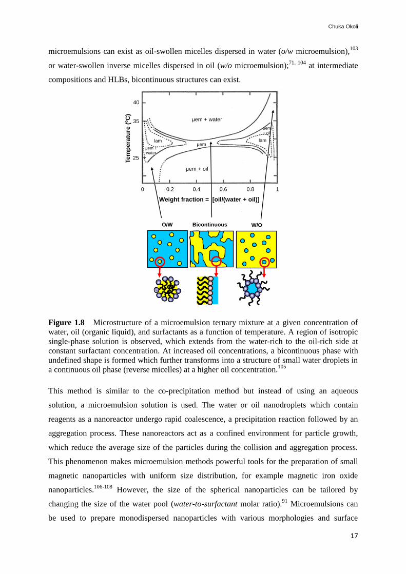
microemulsions can exist as oil-swollen micelles dispersed in water (o/w microemulsion),103
or water-swollen inverse micelles dispersed in oil (w/o microemulsion);71, 104
at intermediate
compositions and HLBs, bicontinuous structures can exist.
Figure 1.8 Microstructure of a microemulsion ternary mixture at a given concentration of
water, oil (organic liquid), and surfactants as a function of temperature. A region of isotropic
single-phase solution is observed, which extends from the water-rich to the oil-rich side at
constant surfactant concentration. At increased oil concentrations, a bicontinuous phase with
undefined shape is formed which further transforms into a structure of small water droplets in
a continuous oil phase (reverse micelles) at a higher oil concentration.105
This method is similar to the co-precipitation method but instead of using an aqueous
solution, a microemulsion solution is used. The water or oil nanodroplets which contain
reagents as a nanoreactor undergo rapid coalescence, a precipitation reaction followed by an
aggregation process. These nanoreactors act as a confined environment for particle growth,
which reduce the average size of the particles during the collision and aggregation process.
This phenomenon makes microemulsion methods powerful tools for the preparation of small
magnetic nanoparticles with uniform size distribution, for example magnetic iron oxide
nanoparticles.106-108
However, the size of the spherical nanoparticles can be tailored by
changing the size of the water pool (water-to-surfactant molar ratio).91
Microemulsions can
be used to prepare monodispersed nanoparticles with various morphologies and surface
25
35
40
Te
mp
era
ture
(ºC
)
W/O O/W Bicontinuous
0 0.2 0.4 0.6 0.8 1
Weight fraction = [oil/(water + oil)]
µem + water
µem + oil
µem lam lam
µem + oil
µem + water

Development of Protein-Functionalized Magnetic Iron Oxide Nanoparticles: Potential Application in Water Treatment
18
specificity in order to enhance their efficiency. However, in some cases (w/o), this method
may require a large amount of solvent to prepare an appreciable amount of particles.74
1.4.2.3 The thermal decomposition method
Another method of preparing quality semiconductor nanocrystals and magnetic nanoparticles
with a high level of monodispersity and tunable size is by the use of thermal decomposition.
This is achieved when organometallic precursors such as metal acetylacetonates, metal
cupferronates [FexCupx] where (Cup = N-nitosophenylhydoxyl amine) or carbonyls (iron
pentacarbonyl)109
are heated in high-boiling organic solvents and a surfactant such as oleic
acid or hexadecylamine.110, 111
Metal oxide magnetic nanoparticles including iron oxide
nanoparticles can also be prepared by the thermal decomposition method. To date, two
different approaches have been used for this purpose in order to avoid the use of highly toxic
compounds such as metal carbonyls. Hyeon et al.,112
in their procedure, used cheap and
environmentally friendly FeCl3 and sodium oleate. The first step consists of iron oleate, or
stereate precursors that decompose in a second step by slowly heating the mixture to 320 oC
to form the desired nanoparticles.113
The size and morphology of the magnetic nanoparticles
can be affected by factors like the ratio of the starting reagents, for instance organometallic
compounds, solvents and surfactants. The reaction time, reaction temperature and aging time
are also critical. Even though thermal decomposition seems well suited for the preparation of
controlled size and high crystalline nanoparticles,113
one of the drawbacks of this method is
the uncertainty of whether the prepared nanoparticles can be suspended in aqueous media,
which limits the extent of their applications in biological and environmental fields.
Furthermore, this method usually leads to complicated processes or requires relatively high
temperature and inert atmosphere, thereby making synthesis time-consuming and expensive.91
1.4.2.4 The hydrothermal method
Under hydrothermal synthesis, a wide range of nanoparticles can be formed. These reactions
are performed in an aqueous medium in autoclaves or reactors under high pressure and
temperature. The hydrothermal method has been reported as being environmentally friendly
and versatile since it does not involve the use of any organic solvent;114
thus, it has been
widely studied for the preparation of metal oxide nanoparticles.115
Reduction of metal ions
under hydrothermal conditions is being utilized in the preparation of iron oxide nanoparticles,
using metal salts, polyethylene glycol and sodium acetate.116
The slow reaction kinetics at any
given temperature is one of the limitations of this method; in addition, the engineering of

Chuka Okoli
19
nanoparticle surfaces cannot be accomplished in situ and therefore additional post-processing
steps are required.91
Among the above-described methods, co-precipitation and microemulsion methods have
shown success, and are widely utilized in the synthesis of superparamagnetic iron oxide
nanoparticles for biomedical and environmental applications. Normally, they are preferred
due to their facile and convenient preparation approach, low production cost, as well as
reproducibility. Moreover, the working window for these two methods, especially co-
precipitation, is quite large; thus, it promotes the surface chemistry modification of the
nanoparticle during the synthesis (in situ) or after synthesis. Another advantage of co-
precipitation and microemulsion methods is that the reaction temperature and time preparation
are much lower than in the other methods (Table 1.2), hydrothermal and thermal
decomposition.
Table 1.2: Comparative summary of magnetic iron oxide synthesis methods
Synthesis
method Synthesis Solvent Reaction
temp. (oC)
Reaction
period
Size and
shape control Yield
Co-precipitation very simple, ambient
conditions
water 20-90 minutes difficult high/scalable
Microemulsion very simple, ambient
conditions
water-
organic
20-40 minutes-
hours
easy medium
Thermal
decomposition
complicated, inert
atmosphere
organic 100-320 hours-days easy high/scalable
Hydrothermal simple, high pressure water-
organic
220 hours-days easy medium
1.4.3 Magnetic separation and mixing
Magnetic nanoparticles, especially SPIONs, are easily separated from ambient matrices under
mild conditions with the use of simple and cost-effective devices. For this reason, magnetic
iron oxide nanoparticles are used in biological or environmental applications. However,
magnetic separation may be as simple as applying external magnetic fields to the outside
container; such a technique is used in both small- and large-scale separators which allow
magnetic nanoparticles to be retained, whilst supernatant volumes ranging from 10 µl to a few
milliliters are removed manually. However, the automated versions that allow multiple

Development of Protein-Functionalized Magnetic Iron Oxide Nanoparticles: Potential Application in Water Treatment
20
samples to be processed in large volumes, up to several liters, are available from companies
such as Dexter Magnetic Technologies, IL, USA.97
When magnetic iron oxide nanoparticles
are introduced in a sample, they tend to sediment under the influence of gravity; therefore,
mixing is required to keep them in suspension in order to improve performance. Since the
solution volumes used in most of the assays are usually very small, slow mixing or slow-tilt
rotation may be sufficient to keep the nanoparticles well-dispersed in solution. Depending on
the application and magnetic strength of the nanoparticles, permanent or electro magnets may
be applied. For biological or environmental assays with magnetic nanoparticles, a permanent
magnet system is most preferred for magnetophoresis (i.e., particle migration that occurs
when a magnetic field is exerted on a nanoparticle), since most of the laboratory-based
research studies require a simple and portable device, instead of the complicated, on-chip
integrated magnetic device.97, 117, 118
1.5 Objectives of the present work
The aim of this thesis is to develop a simple and efficient water treatment strategy that will
benefit society by making use of available and environmentally-friendly materials. In this
perspective, the proposed technology is based on a bottom-up approach i.e. the synthesis,
characterization and application of magnetic nanoparticles. As a novel water treatment
technique, magnetic nanoparticles (MNPs) for binding and separation of a coagulant protein
were developed. The first part (Part One) of this study aimed at extracting the active
coagulant protein from MO seeds; also, the synthesis of MNPs with suitable surfaces for
protein binding was carried out using two different methods. The prepared MNPs were
characterized with different techniques in order to evaluate their physico-chemical properties
as well as the biological properties of the protein, and the efficiency of the prepared magnetic
nanoparticles in the purification of the MO protein was investigated.
The second part (Part Two) highlights the development of feasible water treatment strategy
(Figure 1.9) for both developing and developed countries using two different approaches.
Approach I is based on the use of MOCP in an in situ water treatment. The crude MO protein
is adsorbed onto MNPs, followed by desorption to obtain the purified protein (MOCP). The
purified MOCP was then used for the treatment of turbid waters. These experiments were
carried out in a batch system. Approach II is based on the use of protein-functionalized
nanoparticles (MOCP-MNPs) for water treatment processes. In this approach, the MOCP
adsorbed onto the MNPs was used. The MOCP-MNPs were separated from the water by

Chuka Okoli
21
applying an external magnetic field. This method is particularly appealing because it promotes
material recycling; i.e. the same material can be used several times. In addition, this approach
has the potential of reducing the overall cost of treatment process including time.
Figure 1.9 Scheme showing the development of potential water treatment strategies.
Approach I is based on the use of purified MOCP for water treatment. The MOCP is desorbed
from the MNPs, followed by separation of MNPs with applied magnetic fields. Approach II is
based on the use of MOCP-MNPs for water treatment. The MOCP-MNPs can be reused after
cleaning.
Approach I Approach II
Crude MO proteins MNPs MOCP-MNPs

Development of Protein-Functionalized Magnetic Iron Oxide Nanoparticles: Potential Application in Water Treatment
22

Chuka Okoli
23
2. Experimental
This section highlights the experimental procedures that are not described in detail, in the
appended publications, whilst reflecting the data already reported in papers I-VII. Their
results are discussed in section 3.
PART ONE
Synthesis and characterization
2.1 Extraction and analysis of coagulant protein (Papers I & II)
The MO seeds were purchased from Kenya and stored at room temperature. The husks were
removed manually and the seed kernels were ground to a fine powder using mortar and pestle.
The seed powder was defatted by mixing with ethanol (95%) using a magnetic stirrer for 30
min. The supernatant was separated by centrifugation (3000 rpm, 10 min) and the residual
powder dried at room temperature overnight. From the dried powder, the crude coagulant
protein was extracted (5% w/w) using Milli-Q water.59
The mixture was stirred for 30 min
using a magnetic stirrer and then by centrifugation at 3000 rpm for 45 min. The resultant
supernatant was termed as the crude extract. The extraction process of the active component
from crude MO seed is shown in Figure 2.1. The protein content of the crude extract was
estimated using the Bradford method.119
Figure 2.1 Extraction of the crude coagulant protein from MO seeds. The by-products
obtained during the processing are useful at domestic level.

Development of Protein-Functionalized Magnetic Iron Oxide Nanoparticles: Potential Application in Water Treatment
24
2.1.1 Preparation of synthetic water
Synthetic turbid water (150-170 NTU) was prepared from kaolin (clay suspension) in order to
examine the performance of the crude extract. The clay suspension was prepared by mixing
1L of tap water and 10g of Kaolin clay with a magnetic stirrer for 30 min to obtain an even
dispersion of the kaolin particles.59
The solution was allowed to settle for 24 h to achieve a
complete hydration of the kaolin. The clear solution containing the clay suspension was
decanted into a fresh bottle and kept at room temperature prior to use.
2.1.2 Coagulation activity test
A small-volume (1 mL) coagulation activity test was employed using kaolin clay suspension59
in order to show the coagulation effect of the crude MO seed extract. This method can also be
used to study the performance of other coagulants such as alum or purified coagulant protein.
Three sample volumes (2, 5 and 10 µL) were added separately to a clay suspension to a final
volume of 1 mL in a semi-micro plastic cuvette (10x4x45mm). The samples were mixed
instantly and the initial absorbance (time 0) was measured with a Thermo Spectronic UV-
visible spectrophotometer at 500 nm (OD500). The samples were allowed to settle for 1 h and a
final absorbance was measured. A decrease in absorbance relative to control defines
coagulation activity. Eq. 2.1 was used to estimate the percentage coagulation activity of the
samples.
(2.1)
where IAbs is the initial absorbance and FAbs is the finial absorbance.
2.2 Synthesis of magnetic iron oxide nanoparticles (Papers I─III)
In this study, two different methods have been employed in the preparation of magnetic iron
oxide nanoparticles. The synthesis consists of co-precipitation in aqueous solution and
microemulsion methods. For clarity, nanoparticles prepared by the co-precipitation method
will be referred to as SPIONs while ME-MIONs will be used for microemulsion-prepared
magnetic iron oxide nanoparticles.

Chuka Okoli
25
2.2.1 Co-precipitation method in aqueous solution
A facile way to prepare SPIONs is the use of the chemical co-precipitation method. A typical
synthetic procedure according to Massart et al.,120
with some modifications is briefly
described.
The iron source was prepared by dissolving 1 M FeCl3 and 0.5 M FeCl2 in Milli-Q water. A
volume of 35 mL of the iron source was added to 250 mL of 0.7 M ammonia solution at 70 oC
under vigorous stirring (1200 rpm). The reaction mixture was allowed to proceed for 45 min
under N2 atmosphere in a closed system. The black precipitated nanoparticles were collected
with an external magnet and then washed several times with Milli-Q water. The obtained
magnetic nanoparticles were re-suspended in water and stored at 4 oC for further use and
characterization. Part of the synthesized nanoparticles was coated with inorganic precursors
for protein binding and separation.
2.2.1.1 Coating with TSC
Citrate moieties have been extensively used as stabilizing agents for magnetic iron oxide
nanoparticles.121, 122
Their molecular functions by chemical or biological means have been
explored for different applications due to the presence of carboxyl functional groups on the
nanoparticles surface (Figure 2.2). By virtue of the complexation (sorption of oppositely
charged surfaces) occurring between the iron ions on the nanoparticles and the citrate groups,
highly stable magnetic fluid with well-dispersed nanoparticles can be obtained.123
Coating of the SPION with trisodium citrate (TSC) salt was carried out by re-dispersing 20
mL (18 mg/ml) of the magnetic nanoparticles suspension in Milli-Q water. The resultant
solution was subjected to vigorous stirring (620 rpm, 30 min) at 90 oC upon the addition of
trisodium citrate solution (0.2 g). The reaction mixture was cooled to room temperature and
the obtained TSC-coated SPION was re-suspended in water after several washing steps with
water. The TSC-coated SPION core-shell was evaluated by Fourier transform infrared
spectroscopy (FT-IR).112
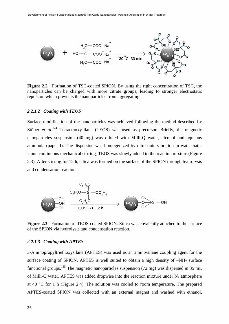
Development of Protein-Functionalized Magnetic Iron Oxide Nanoparticles: Potential Application in Water Treatment
26
Figure 2.2 Formation of TSC-coated SPION. By using the right concentration of TSC, the
nanoparticles can be charged with more citrate groups, leading to stronger electrostatic
repulsion which prevents the nanoparticles from aggregating.
2.2.1.2 Coating with TEOS
Surface modification of the nanoparticles was achieved following the method described by
Stöber et al.124
Tetraethoxysilane (TEOS) was used as precursor. Briefly, the magnetic
nanoparticles suspension (40 mg) was diluted with Milli-Q water, alcohol and aqueous
ammonia (paper I). The dispersion was homogenized by ultrasonic vibration in water bath.
Upon continuous mechanical stirring, TEOS was slowly added to the reaction mixture (Figure
2.3). After stirring for 12 h, silica was formed on the surface of the SPION through hydrolysis
and condensation reaction.
Figure 2.3 Formation of TEOS-coated SPION. Silica was covalently attached to the surface
of the SPION via hydrolysis and condensation reaction.
2.2.1.3 Coating with APTES
3-Aminopropyltriethoxysilane (APTES) was used as an amino-silane coupling agent for the
surface coating of SPION. APTES is well suited to obtain a high density of –NH2 surface
functional groups.125
The magnetic nanoparticles suspension (72 mg) was dispersed in 35 mL
of Milli-Q water. APTES was added dropwise into the reaction mixture under N2 atmosphere
at 40 °C for 1 h (Figure 2.4). The solution was cooled to room temperature. The prepared
APTES-coated SPION was collected with an external magnet and washed with ethanol,
Fe3O
4
OH OH OH TEOS, RT, 12 h
Si
C2H
5O
C2H
5O
C2H
5O
OC2H
5
Fe3O
4
O O O
Si OH
Fe3O
4 +
Fe3O
4
COO─
C
H2C
HO
Na+
H2C COO
─
COO─
Na+
Na+
30 o
C, 30 min

Chuka Okoli
27
followed by Milli-Q water. Finally, APTES-coated SPION was re-suspended in 20% ethanol
and kept at 4 oC prior to use.
Figure 2.4 Formation of APTES-coated SPION. 3-Aminopropyltriethoxysilane (APTES)
was used as the amino-silane coupling agent.
2.2.2 Microemulsion methods
The preparation of microemulsion-based magnetic iron oxide nanoparticles (ME-MIONs) was
carried out from two different microemulsion systems: water-in-oil (w/o) and oil-in-water
(o/w).
2.2.2.1 Synthesis of w/o ME-MION
For the synthesis of w/o ME-MIONs, two approaches were used (Figure 2.5). Microemulsion
system 1 consists of a surfactant, cetyltrimethyl ammonium bromide (30 wt%, CTAB), a co-
surfactant (11 wt%, 1-butanol), an oil phase (27 wt%, hexanol) and an aqueous phase
containing either of the iron precursors: 31.6 wt%, Milli-Q water plus FeCl3 and FeCl2, 2:1
molar ratio or NH3 solution. The system was termed ME 1-MION.
Microemulsion 2 consists of a surfactant, cetyltrimethyl ammonium bromide (13.2 wt%,
CTAB), a co-surfactant (10.1 wt%, 1-butanol), an oil phase (49.8 wt%, n-octane) and an
aqueous phase containing the iron precursors (26.9 wt%, Milli-Q water plus FeCl3 and FeCl2,
2:1 molar ratio) and a precipitating agent (15 wt% NH3).71
The preparation of ME 1-MION was achieved by mixing two microemulsions (Figure 2.5a).
The two microemulsions respectively consist of an aqueous solution of iron precursor and a
precipitating agent (aq.NH3). Precipitation of magnetic nanoparticles was carried out by
adding the former microemulsion into the latter at room temperature upon vigorous stirring
for 1 h. The pH of the mixture was adjusted to 11 with ammonia. Formation of magnetic
nanoparticles was observed by the resulting black coloration. The obtained magnetic
nanoparticles were separated with an external magnet and then washed by several cycles of
precipitation and re-suspension in a mixture of chloroform and methanol (1:1), and finally
Fe3O
4
OH OH OH APTES, 40
o
C, 1 h
Si
C2H
5O
C2H
5O
C2H
5O
(CH2)3 NH
2
Fe3O
4
O O O
Si (CH2)3 NH
2

Development of Protein-Functionalized Magnetic Iron Oxide Nanoparticles: Potential Application in Water Treatment
28
water. The nanoparticles were either dried at 70 ◦C or suspended in Milli-Q water and kept at
4 ◦C until further use.
Figure 2.5 Schemes for the preparation of inorganic magnetic nanoparticles by the w/o
microemulsion reaction approach by (a) mixing two microemulsions of identical composition
except for reactant I (metal precursors) and reactant II (precipitating agent), (b) using a
microemulsion containing Fe salts (FeCl3:FeCl2), whilst the precipitating agent (aq.NH3) is
added directly.
For the synthesis of ME 2-MION, a single-step mode of preparation was used. In this
approach, only one type of microemulsion solution is needed in the formation of magnetic
nanoparticles (Figure 2.5b). Addition of the precursor solution to the mixture of CTAB/1-
butanol/n-octane leads to the formation of a microemulsion. Formation of magnetic
nanoparticles was achieved by adding the appropriate amount of precipitating agent (aq. NH3)
dropwise to the aforementioned microemulsion containing the precursor upon vigorous
stirring until pH 11 was achieved. The reaction mixture was allowed to stir for 1 h at 25 oC. A
dark brown coloration indicates the formation of magnetic nanoparticles in the reaction
mixture. The obtained magnetic nanoparticles were separated with an external magnet and
then washed by several cycles of precipitation and re-suspension in a mixture of chloroform

Chuka Okoli
29
and methanol (1:1), and finally water. The nanoparticles were either dried at 70 ◦C or
suspended in Milli-Q water and kept at 4 ◦C until further use.
2.2.2.1 Synthesis of o/w ME-MION
The procedure of o/w microemulsion synthesis was developed by Sanchez-Dominquez et
al.103
The microemulsion system consists of a nonionic surfactant (21.5 wt%, Synperonic®
10/6), an oil phase containing the iron precursor (14 wt%, hexane plus Iron (III) 2-
ethylhexanoate), and an aqueous phase (64.5 wt%, Milli-Q water). The microemulsion
containing the corresponding organometallic precursor was prepared by mixing the three
components in the aforementioned proportions and stirring at 30oC until a homogenous,
transparent brownish isotropic phase was obtained. The precipitating agent (30 wt% NH3) was
added directly while stirring the mixture in order to increase the pH and precipitate the
magnetic iron oxide nanoparticles. The reaction mixture was adjusted to pH 11 and kept under
continuous stirring at ~30oC. The obtained magnetic nanoparticles (Figure 2.6) were separated
and washed by several cycles of precipitation and re-suspension in ethanol-water (1:1) and
finally ethanol. The magnetic nanoparticles were either dried at 70 o
C or suspended in Milli-Q
water and kept at 4oC until further use.
Figure 2.6 Scheme for a single-step preparation of inorganic magnetic nanoparticles by the
o/w microemulsion reaction approach. The magnetic nanoparticles were precipitated upon the
addition of an aqueous NH3 solution into the microemulsion containing Fe salts. Figure
adapted from103
.
2.3 Characterization (Papers I─III)
Several techniques have been employed to investigate the size, morphology, magnetic
property and surface chemistry of the nanoparticle. The two types of ME-MION have been
compared with different surface-coated SPIONs prepared by the water-based co-precipitation

Development of Protein-Functionalized Magnetic Iron Oxide Nanoparticles: Potential Application in Water Treatment
30
method. The following subsections discuss the characterization techniques used for each
preparation method.
2.3.1 Nanoparticles prepared by co-precipitation (Paper I)
The average diameters of nanoparticles were estimated from TEM micrographs. Transmission
electron microscopy (TEM) was carried out using a JEM-2100F microscope under an
accelerating voltage of 200 kV. The specimen for TEM imaging was prepared by the
dispersion of magnetic nanoparticles in alcohol. A drop of well dispersed sample was placed
on a carbon-coated 200 mesh copper grid and the sample dried under ambient condition
before loading the grid in the sample holder of the microscope for TEM analysis.
Crystallographic analysis of the sample was performed on dried powder by powder X-ray
diffraction (Xpert PRO, model PW3040/60) to determine the crystal structure. The 2θ data
were collected using a continuous scan mode in the range of 20o-70
o. The average crystalline
sizes of the nanoparticle were calculated using the Scherrer equation:
d = 0.9 λ / β cos θ (2.2)
where d is the particle size (nm), λ: X-ray wavelength (Å), θ: diffraction angle (o) and β: the
full width at half maximum of the diffraction peak at θ (rad).
Atomic absorption spectroscopy (AAS) analysis was used in the quantitative determination of
the nanoparticle concentration using a Spectra AA-200 spectrometer.
Fourier transform infrared spectroscopy (FT-IR) was carried out on a Nicolet Avatar 360 FT-
IR ESP to confirm the presence of carboxyl group in the TSC-coated SPION. The absorption
bands are characteristic of the functional groups present in a sample. Different peaks
(functional groups) were evaluated and compared with uncoated and coated SPION.
In order to determine the magnetic behavior of the nanoparticles, room temperature magnetic
hysteresis loops measurement was carried out using a vibrating sample magnetometer (VSM,
Model 155 EG&G Princeton Applied Research). The magnetic moments of the particles were
determined in field over a range of ± 8kOe.
Zeta potential (Zp) measurement using Zetasizer Nano series equipment was employed to
study the surface charge of the nanoparticles. The pH of surface-coated SPIONs were
adjusted to different pH values ranging from 3 to 12 with 1 M NaOH and 1 M HCl solutions.
The charges were determined and the point of zero charge was estimated from the zeta
potential versus the pH curve.

Chuka Okoli
31
2.3.2 Nanoparticles prepared by Microemulsion (Papers II-III)
X-ray diffraction data of the nanoparticles prepared by microemulsion methods were acquired
between 20o and 70
o on a Siemens D5000 diffractometer, using Cu Kα radiation (Paper I).
The average crystalline sizes of the nanoparticle were calculated using the Scherrer equation
(Eq. 2.2).
The size and morphology of the nanoparticles were examined by High-Resolution
Transmission Electron Microscopy (HRTEM), which was carried out using a Field Emission
Transmission Electron Microscope (JEM-2200FS) at 200 kV, with 0.19 nm resolution in
TEM mode. For this purpose, dry powder samples were dispersed in isopropanol and
deposited on formvar carbon copper grids. Selected area electron diffraction patterns (SAED)
of the nanoparticles were also recorded from the samples.
Room temperature magnetic hysteresis loops of the nanoparticles were measured using a
Superconducting Quantum Interference Device (SQUID) Magnetometer, Quantum Design
MPMS XL. The magnetic moments of the particles were determined in field over a range of ±
60kOe.
Surface area and porosimetry of the nanoparticles were calculated by the Brunauer-Emmet-
Teller (BET) method using Micromeritics ASAP 2010. Specific surface area as well as the
specific pore volume was detected after degassing the sample overnight at 110 oC prior to the
measurement.
PART TWO
2.4 Application of nanotechnology in water treatment (Papers III, IV, V & VII)
2.4.1 Purified MOCP for water treatment (Approach I)
The purpose of this study is to demonstrate the potential of MOCP in turbid water
clarification. For this reason, a novel purification approach based on the use of magnetic
nanoparticles (MNPs) prepared from two different systems (co-precipitation and
microemulsion methods) was developed and investigated. The purification of MOCP for
water treatment as well as its analysis and characterization is discussed in the following
subsections.

Development of Protein-Functionalized Magnetic Iron Oxide Nanoparticles: Potential Application in Water Treatment
32
2.4.1.1 Purification of the MO coagulant protein with MNPs and activity test
Purification of the MO protein was carried out with the aforementioned magnetic
nanoparticles (2-30 nm size) in a batch system (Figure 2.7). The MNPs were equilibrated
using 10 mM ammonium acetate buffer, pH 6.7. Crude extract (2.6 mg/mL) was added to the
equilibrated MNPs and the sample volume was adjusted with 10 mM ammonium acetate
buffer. The sample was mixed and incubated at room temperature for 1 h. Finally, the
adsorbed protein was separated from the mixture using an external magnetic field (6000 G).
The coagulant protein was eluted with different concentrations of NaCl (0.6, 0.8 or 1.0 M) in
10 mM ammonium acetate solution.71, 126
The sample was either stored at -20 oC or freeze-
dried. The IEX and CMC beads were used as controls.126
Coagulation activity of the MOCP
was measured following the procedure already described in section 2.1.2.
Figure 2.7 Batch purification procedure of the MO protein using MNPs. The MO protein
binds to MNPs; unbound proteins (pink and blue) are removed by washing. The MOCP is
desorbed (eluted) from the MNPs. Rapid separation is achieved with the help of an external
magnetic field to separate the MOCP and MNPs. The desorbed MOCP (green) is either used
in suspension or as a freeze-dried powder for in situ water treatment.
2.4.1.2 Analysis and characterization
Protein contents of the MO seed extract and purified protein were determined by the Bradford
assay.119
The protein profile, molecular weight of the purified protein and that of the crude
extract were determined and compared in 10% sodium dodecyl sulphate (SDS)-
polyacrylamide gel electrophoresis (PAGE)126
mini gels stained with Coomassie brilliant blue
to visualize the protein.

Chuka Okoli
33
Mass spectrometric analysis of the purified protein was carried out based on MS/MS peptide
sequence analysis of in-gel trypsin digested protein using a Q-TOFTM
II mass spectrometer.
2.4.1.3 Turbidity removal in surface waters
A simple and quick method to verify the coagulation activity of MOCP in terms of turbidity
removal was carried out. The water samples for this study were collected from three different
lakes in Sweden. Raw surface waters with initial turbidity ranging from 7–17 NTU were
collected from Lake Mälaren in autumn 2009; Arboga (Västmanland) and Vättern-Lake
(Götaland) waters were collected during spring 2010. The choice of seasonal variation in the
collection of raw water samples is essential to evaluate the performance of the coagulant
agent with different turbidity/contaminants. Once collected, the samples were stored at 4 oC to
prevent any bacterial growth. The kaolin clay with an initial turbidity of 150–170 NTU was
used as one of the control sample for comparison purposes. Aluminum sulphate (5%, alum)
was prepared and used as a control. All experiments were carried out in a batch system.
In order to estimate the performance (turbidity removal) of the MOCP or alum, coagulation
activity assays were carried out as described in section 2.1.2. In a typical experimental
procedure, MOCP (0.1-0.5 mg/L) was added to raw surface water or clay suspension. The
sample was mixed rapidly at 50 rpm for 5 min to allow an optimal contact between the sample
waters and MOCP. A small volume of the sample was collected and the initial absorbance
measured at 500 nm (OD500) with a Thermo Spectronic UV-visible spectrophotometer. The
sample was allowed to settle (gravity) for 1 h and the absorbance was measured again (final
absorbance). However, the measurements may be carried out periodically (0-240 min) during
their settling time in order to monitor the optimal time needed to achieve good activity. A
decrease in absorbance relative to the controls defines turbidity removal from the source
water. Alum was used for comparison (positive control) while surface water and a clay
suspension without the protein were used as negative controls. The interaction studies of
MOCP and MNPs with different surface chemistry have been carried out to understand the
binding behavior of MOCP on the surface-coated MNPs. The following section illustrates the
in silico modeling investigation.
2.4.1.4 Interaction of MOCP and MNPs (Papers III & V)
Several studies have been performed in order to elucidate the binding of MOCP onto MNPs
with different modified surfaces. For this purpose, computational modeling study of the
interaction between the protein molecule and the surface-coated SPIONs was performed and

Development of Protein-Functionalized Magnetic Iron Oxide Nanoparticles: Potential Application in Water Treatment
34
the data were compared with experimental results. Furthermore, circular dichroism (CD)
spectroscopy of MO2.1 fluorescence emission of the protein and ME-MIONs were
investigated.
2.4.1.4.1 Computational and experimental studies
The MOCP (MO2.1) secondary structure was examined using circular dichroism (CD)
spectroscopy. The far-UV CD was measured on a Jasco-710 dichrograph previously calibrated
with d-10-camphorsulphonic acid. Measurements were performed in 0.5 cm path-length
cuvette at 22 ºC. The ellipticity was recorded with a 0.5 nm bandwidth and a 2s response.
Sample concentrations were ~1mg/mL, in ammonium acetate buffer pH 6.7, 0.6 M NaCl.
Five accumulations were acquired for the spectrum. The mean residue ellipticity (MRE) was
calculated using:
cl10
MRW obsmr
deg. cm
2.dmol
-1 (2.3)
where MRW is the molecular weight per residue (MRW=MW/(n-1)), θobs: observed ellipticity
in degrees and c: concentration in g/ml.
Theoretical modeling tools have been used to understand the structure of a
coagulating/flocculating protein from MO and its ability to bind to various ligand precursors
(Paper V). Gaussian 03 Software127
was used to generate 3D structures for these ligand
molecules, namely trisodium citrate (TSC), 3-aminopropyl trimethoxysilane (APTES), and
tetraethoxysilane (TEOS). The electrostatic potential of this protein was also calculated.
Different theoretical tools were employed in the following sequence. (i) Homology modeling
was used to obtain the protein structure from the known peptide sequence of MO2.1 (P24303)
through superimposition with the mabinlin sequence (Paper VI);
QGPGRQPDFQRCGQQLRNISPPQRCPSLRQAVQLTHQQQGQVGPQQVRQMYR
VASNIPST.61
(ii) Molecular docking studies were performed to investigate the binding
ability and mode of binding of different organic molecules and hydroxyl groups
functionalized with silicon clusters with the aforementioned protein. (iii) Since the docking
studies mimic the zero temperature situations, molecular dynamics simulations for the docked
protein-ligand structures were carried out in aqueous solution to study finite temperature
effects. Protein-ligand distance profile data was compared with the experimental results.
Basically a computation of the instantaneous distance (defined as RN-ligand where N refers to

Chuka Okoli
35
the residue number and ligand refers to the residues, namely TSC, APTES and TEOS)
between each of the 60 residues of protein and the ligand center of mass was calculated. In
addition, computation of the average RN-ligand was also conducted. This calculation has been
done for 1000 configurations obtained from the molecular dynamics128, 129
2.4.1.4.2 Fluorescence emission study (Paper III)
The binding of MOCP with w/o ME-MION and o/w ME-MION was studied. Emission
fluorescence on a modular PTI Spectrometer with an excitation wavelength of 255 nm was
employed. Emission was recorded at λ= 285 to 475 nm. The excitation and emission slits
were adjusted to 2 nm. Temperature was controlled (22 oC) with a thermoelectric temperature
controller, TLC 50. Cells with a path length of 1 cm were used and all the experiments were
performed at least twice. The average spectra were subtracted from appropriate blanks before
analysis.
2.4.2 MOCP-MNPs for water treatment (Approach II)
Here, a novel approach for water treatment with MOCP-MNPs was developed (Figure 2.8).
The use of MOCP-MNPs enables the separation and recovery of this system from water using
an external magnetic field, and consequently the opportunity to regenerate the system for
further water treatment.

Development of Protein-Functionalized Magnetic Iron Oxide Nanoparticles: Potential Application in Water Treatment
36
Figure 2.8 Scheme of the proposed MOCP-MNPs for water treatment. Treatment:- MOCP-
MNPs is added to water followed by a slow mixing. The impurities in the water bind to the
MOCP-MNPs. Separation can be achieved either by gravity or external magnet. A rapid
separation is achieved with the help of an external magnetic field within 1-2 min. Clean and
potable water is obtained following the separation step. Regeneration:- MOCP-MNPs is
cleaned, separated and reused for another batch of raw water.
2.4.2.1 Development of MOCP-MNPs system
The development of MOCP-MNPs followed a batch system preparatory approach. The MNPs
(2-30 nm size) prepared either by co-precipitation or microemulsions, were equilibrated using
10 mM ammonium acetate buffer, pH 6.7. Crude MO seed extract (2.6 mg/mL) or MOCP
(0.6 mg/mL) was added to the equilibrated MNPs. The samples were kept for 1-2 h in a rotary
mixer (20 rpm) at room temperature. The supernatant was removed by magnetic separation
and the resultant MOCP-MNPs was washed with ammonium acetate buffer to remove the
non-adsorbed protein and other impurities. The MOCP-MNPs was suspended in ammonium
acetate buffer and kept at 4 oC or freeze-dried prior to use. The developed MOCP-MNPs were
termed MOCP+SPION or MOCP+ME-MION systems.
The functionality of the developed MOCP-MNPs was tested in clay suspension according to
the procedure previously described in section 2.1.2.

Chuka Okoli
37
2.4.2.2 Turbidity removal in surface waters
The efficiency of the developed MOCP-MNPs in terms of turbidity removal against surface
water was analyzed following a similar experimental procedure described in section 2.4.1.1.
The clay suspension and alum (5%) were used as controls.
Briefly, MOCP+SPION or MOCP+ME-MION (0.01-0.4 mg/L) were added to the water
samples and the initial absorbance measured. The samples were allowed to settle for 1 h and
the final absorbance was measured. However, the measurements may be carried out
periodically (0-240 min) during their settling time in order to monitor the optimal time needed
to achieve good activity. A decrease in absorbance relative to controls defines turbidity
removal from the source water. The percentage coagulation activity (turbidity removal) was
calculated according to Eq. 2.1.
2.4.2.3 Separation by gravity and magnetic field
A quick method to achieve efficient turbidity removal as well as rapid separation with
MOCP+ME-MION was tested. The coagulation and magnetization properties of MOCP+ME-
MION under the influence of an external magnetic field were compared with the settling of
impurities by gravity. For settling of impurities by gravity, alum was used as a control.
Synthetic clay suspension and natural surface water samples (SW1; 7 NTU and SW2; 17
NTU) from lake Mälaren were used. The MOCP+ME-MION and alum were added separately
in two different flasks containing the turbid water samples and mixed instantly; the initial
absorbance of the mixture was measured at 500 nm. Then the resultant mixture was allowed
to interact for 10 min at room temperature. After the interaction time, MOCP+ME-MIONs
were separated from water with a magnet (1-2 min) to obtain clean water; the water sample
containing alum was decanted (Figure 2.8). Final absorbance of the clean water was measured
and the turbidity removal efficiency calculated.
2.4.2.4 Reusability of MOCP-MNPs system
In this study, the possibility to reuse MOCP-MNPs was investigated. The number of times the
developed system can be reused in water treatment was verified.
In this investigation, the impurities adsorbed on MOCP-MNPs were cleaned with a mild
detergent or a cleaning solution (0.1 wt% Tween 20 in water and 20% ethanol) after each
turbidity removal assay with water samples. A solution of 0.1 wt% Tween 20 or 20% ethanol
was added to the used MOCP-MNPs and the mixture was left for 10 min at room temperature.

Development of Protein-Functionalized Magnetic Iron Oxide Nanoparticles: Potential Application in Water Treatment
38
The MOCP-MNPs were removed by magnetic separation followed by two washes with 10
mM ammonium acetate buffer. The control sample was the initial coagulation activity of
MOCP-MNPs in turbid water sample before cleaning. The coagulation activity test was
performed as described earlier (section 2.1.2).
Chuka Okoli
39
3. Results and discussion
Some of the data presented in this section have not been published; however, they reflect the
results already appended in papers I-VII.
PART ONE
Synthesis and characterization
3.1 Investigation of the active protein (Paper I)
In order to estimate the activity of the crude MO seed extract, a control sample (clay
suspension) was used. The difference between the control sample and the MO protein is
considered as the real coagulation activity. The crude MO seed protein showed good
coagulation activity on clay suspensions. The baseline in Figure 3.1 indicates settling of the
clay suspension by gravity; this can be subtracted from the percentage activity of the active
components of the protein to obtain the real activity. The coagulant protein exhibits good
activity (~ 80%) in all the studied concentrations.32, 130
Given that the MO crude extract
contained the coagulant protein, it was appealing to bind or separate the active protein onto
the right surface of the magnetic nanoparticles in order to develop a simple and feasible water
treatment procedure. The subsequent sections discuss the results obtained from the design and
application of MNPs for water treatment.
Figure 3.1 Coagulation activity of crude MO seed extract (2.5 mg/ml, stock) on a synthetic
clay suspension with 1 h as a setting time. The baseline indicates settling of the clay
suspension by gravity; this can be subtracted from the percentage activity (turbidity reduction)
of the active components of the protein to obtain the real activity.
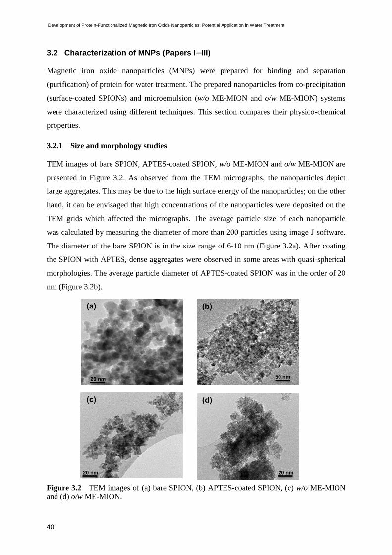
Development of Protein-Functionalized Magnetic Iron Oxide Nanoparticles: Potential Application in Water Treatment
40
3.2 Characterization of MNPs (Papers I─III)
Magnetic iron oxide nanoparticles (MNPs) were prepared for binding and separation
(purification) of protein for water treatment. The prepared nanoparticles from co-precipitation
(surface-coated SPIONs) and microemulsion (w/o ME-MION and o/w ME-MION) systems
were characterized using different techniques. This section compares their physico-chemical
properties.
3.2.1 Size and morphology studies
TEM images of bare SPION, APTES-coated SPION, w/o ME-MION and o/w ME-MION are
presented in Figure 3.2. As observed from the TEM micrographs, the nanoparticles depict
large aggregates. This may be due to the high surface energy of the nanoparticles; on the other
hand, it can be envisaged that high concentrations of the nanoparticles were deposited on the
TEM grids which affected the micrographs. The average particle size of each nanoparticle
was calculated by measuring the diameter of more than 200 particles using image J software.
The diameter of the bare SPION is in the size range of 6-10 nm (Figure 3.2a). After coating
the SPION with APTES, dense aggregates were observed in some areas with quasi-spherical
morphologies. The average particle diameter of APTES-coated SPION was in the order of 20
nm (Figure 3.2b).
Figure 3.2 TEM images of (a) bare SPION, (b) APTES-coated SPION, (c) w/o ME-MION
and (d) o/w ME-MION.
(a) (b)
50 nm 20 nm
20 nm
(c) (d)
20 nm

Chuka Okoli
41
However, from the TEM micrographs of ME-MION, the w/o ME-MION nanoparticles were
irregular in shape, consisting of elongated, rod-like morphologies with widths ranging from 5-
10 nm (Figure 3.2c). The o/w ME-MION nanoparticles were globular and in the order of 3 nm
with dense aggregates (Figure 3.2d). The results obtained from the TEM size estimation
(Figures 3.2, c and d) were in agreement with the crystalline size calculation of w/o and o/w
ME-MIONs (Paper III) using the Scherrer equation (Eq. 2.2, section 2.3.1) with the XRD data
(Figure 3.3). The present study underscores the fact that nanoparticles with similar
morphology and size can be obtained irrespective of the preparation method. Nevertheless,
there is a considerable overlap and large aggregation in ME-MIONs due to the large surface
energy of the nanoparticles. Aggregation of these nanoparticles may depend on experimental
parameters like temperature, pH, reaction environment and surface condition. If not properly
controlled, these parameters can influence the structure of the obtained iron oxide
nanoparticles.71, 103
3.2.2 Structural studies
The diffraction patterns of nanoparticles prepared from co-precipitation and microemulsion
systems depict the presence of a magnetic phase of either Fe3O4 or γ-Fe2O3 (Figure 3.3);
which is quite typical at 35o for (311) and 63
o for (440).
Figure 3.3 XRD patterns of (a) bare SPION and (b) w/o- and o/w- ME-MION.
The XRD patterns of bare SPION and w/o ME-MION nanoparticles indicate that the particles
are highly crystalline, which confirms the inverse spinel structure of magnetite and
(b)
o/w
20 25 30 35 40 45 50 55 60 65 70
Inte
ns
ity (
arb
. u
nit
)
2θ (o)
(311)
(220) (400)
(511)
(440)
(a)
w/o
(311)
(440)
(511)
(220)
(b)
o/w

Development of Protein-Functionalized Magnetic Iron Oxide Nanoparticles: Potential Application in Water Treatment
42
maghemite (even though it is difficult to distinguish between them from XRD).122, 126
The o/w
ME-MION XRD pattern showed wide peaks characteristic of small nanoparticles suggesting
that the original size of the nanoparticles is very small and fits well to a typical spinel phase of
either magnetite (Fe3O4) or maghemite (γ-Fe2O3) or a combination of both.107, 131
3.2.3 Magnetic studies
The results from magnetization (M) versus applied field (H) measurements at room
temperature (300 K) for bare SPION, w/o- and o/w- ME-MIONs are presented in Figure 3.4.
It can be seen that the magnetization curves of bare SPION and w/o ME-MION () clearly
depict zero remanence and coercivity, confirming a superparamagnetic behavior and larger
particles size as compared to o/w ME-MION (). The recorded saturation magnetization (Ms)
value for bare SPION is 70 emu/g while w/o ME-MION presented MS of 30 emu/g.
Conversely, the magnetic moment of o/w ME-MION exhibited a weak/or incomplete
saturation magnetization (MS) of 10 emu/g, indicating a decrease in mean particle size. The
obtained data imply that the nanoparticle preparation method determines their magnetic
behavior; moreover, the specific magnetization of iron oxide nanoparticles is known to
depend on their size.132
Figure 3.4 Magnetization as a function of magnetic field at 300 K for (a) VSM of SPION.
Magnetic moments of the samples were determined in field over a range ± 8kOe. The
recorded MS value of bare SPION is 70 emu/g; (b) SQUID magnetometer of w/o ME-MION
() and o/w ME-MION (). The recorded MS value for w/o ME-MION is 30 emu/g and 10
emu/g for o/w ME-MION. Magnetic moments of the samples were determined in field over a
range of ± 60kOe.
-8000 -6000 -4000 -2000 0 2000 4000 6000 8000-80
-60
-40
-20
0
20
40
60
80
(M
agn
etizatio
n (
em
u/g
)
Magnetic field (Oe)
-30
-20
-10
0
10
20
30
-60 -50 -40 -30 -20 -10 0 10 20 30 40 50 60
H / kOe
M /
em
u g
-1
(b) (a)

Chuka Okoli
43
3.2.4 Surface charge studies
The ζ-potential results of SPION, TSC-coated SPION, TEOS-coated SPION and APTES-
coated SPION are shown in Figure 3.5. The magnetic nanoparticle dispersion was titrated
from pH values of 2 to 12, using HCl (0.1 M) and NaOH (0.1 M) solutions. Bare SPION
(magnetite or maghemite) showed a point of zero-charge (pzc) at a pH of ~7, above which the
surface of SPION is negatively charged and below positively charged. It is envisaged that the
ME-MIONs present similar behavior, implying that magnetite or maghemite exhibits a point
of pzc at pH around 7.5.133
Figure 3.5 Zeta potential (ζ) as a function of pH for bare SPION, TSC-coated SPION,
TEOS-coated SPION and APTES-coated SPION.
The pzc for TSC-coated SPION is observed at pH 5.3, which clearly illustrates that the
surface of SPION was successfully modified with TSC. In the case of APTES-coated SPION,
the amino groups from the surface of SPION displayed a shape almost similar to a sinusoidal
curve with a positive maximum value at ζ=48.1 mV (pH=2) and a negative maximum value at
ζ= ─ 42 mV (pH=6.8). The APTES-coated displayed pzc at pH around 5.85; therefore, it can
be concluded that the dispersion of APTES-coated SPION suspension is stable from pH 2 to
9.134
Conversely, the TEOS-coated SPION exhibited a net negative charge for a wide range
of pH values (pH 2 to 9).
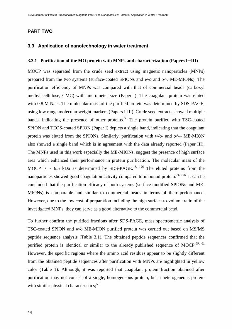
Development of Protein-Functionalized Magnetic Iron Oxide Nanoparticles: Potential Application in Water Treatment
44
PART TWO
3.3 Application of nanotechnology in water treatment
3.3.1 Purification of the MO protein with MNPs and characterization (Papers I─III)
MOCP was separated from the crude seed extract using magnetic nanoparticles (MNPs)
prepared from the two systems (surface-coated SPIONs and w/o and o/w ME-MIONs). The
purification efficiency of MNPs was compared with that of commercial beads (carboxyl
methyl cellulose, CMC) with micrometer size (Paper I). The coagulant protein was eluted
with 0.8 M Nacl. The molecular mass of the purified protein was determined by SDS-PAGE,
using low range molecular weight markers (Papers I-III). Crude seed extracts showed multiple
bands, indicating the presence of other proteins.59
The protein purified with TSC-coated
SPION and TEOS-coated SPION (Paper I) depicts a single band, indicating that the coagulant
protein was eluted from the SPIONs. Similarly, purification with w/o- and o/w- ME-MION
also showed a single band which is in agreement with the data already reported (Paper III).
The MNPs used in this work especially the ME-MIONs, suggest the presence of high surface
area which enhanced their performance in protein purification. The molecular mass of the
MOCP is ~ 6.5 kDa as determined by SDS-PAGE.18, 126
The eluted proteins from the
nanoparticles showed good coagulation activity compared to unbound protein.71, 126
It can be
concluded that the purification efficacy of both systems (surface modified SPIONs and ME-
MIONs) is comparable and similar to commercial beads in terms of their performance.
However, due to the low cost of preparation including the high surface-to-volume ratio of the
investigated MNPs, they can serve as a good alternative to the commercial bead.
To further confirm the purified fractions after SDS-PAGE, mass spectrometric analysis of
TSC-coated SPION and w/o ME-MION purified protein was carried out based on MS/MS
peptide sequence analysis (Table 3.1). The obtained peptide sequences confirmed that the
purified protein is identical or similar to the already published sequence of MOCP.59, 61
However, the specific regions where the amino acid residues appear to be slightly different
from the obtained peptide sequences after purification with MNPs are highlighted in yellow
color (Table 1). Although, it was reported that coagulant protein fraction obtained after
purification may not consist of a single, homogeneous protein, but a heterogeneous protein
with similar physical characteristics;59

Chuka Okoli
45
Table 3.1: Mass spectrometric analysis─ MS/MS determination of the sequence of peptides
from the trypsin digested protein
Purification method
Peptide Sequence
TSC-coated SPION QAVQLTHQQQGQVGPQQVR
w/o ME-MION QAVQLTHQQQGQVGPQQVR
IEX Chromatographya QAVQLETQQQGQVGPQQVR
IEX Chromatographyb QAVQLTHQQQGQVGPQQVR
*a,b
Similar or identical to sequence reported by Ghebremichael
et al. (2005) and Gassenschmidst et al. (1995) respectively.
3.3.1.1 Turbidity removal in surface waters (Approach I)
The performance of the MOCP in terms of turbidity removal efficiency in natural and
synthetic waters is shown in Figure 3.6. The coagulation efficiency of the active protein
eluted from TSC-coated SPION, w/o ME-MION and o/w ME-MION was compared with
alum.
Figure 3.6 Coagulation performance of purified MOCP and alum after a 1 h settling period
in clay suspension (170 NTU) and surface water (17 NTU) from Lake Mälaren. The MOCP 1,
MOCP 2 and MOCP 3 represent protein purified with TSC-coated SPION, w/o ME-MION
and o/w ME-MION respectively. Control is the water sample.
As Figure 3.6 clearly depicts, the coagulation activity of MOCP is significantly higher and
comparable with alum in turbidity removal assay. Their performance follows a similar pattern
in both synthetic clay suspension and natural water, implying that, in a 1 h settling period, ~
50 % turbidity removal can be achieved in low turbid waters (17 NTU) with MOCP, whereas
more than 75 % turbidity removal can be obtained in high turbid water (170 NTU). It can be

Development of Protein-Functionalized Magnetic Iron Oxide Nanoparticles: Potential Application in Water Treatment
46
observed that parameters such as initial water turbidity, coagulant agent dosage and
interaction time have significant influence on the removal of suspended particles from the
water samples.32, 59
This observation may be explained in terms of the increase in suspended
particles available for adsorption and inter-particle bridge formation in clay suspension. The
net effect is increase in particle collision and agglomeration (floc formation).31
This behavior
reveals the variation observed in coagulation performance of clay suspension and surface
water. Muyibi et al.,31
reported that the use of high concentrations (large doses) of the
coagulant agents can lead to overdosing, which may result in the restabilization of the already
destabilized particles due to insufficient number of particles to form more inter-particle
bridges.
3.3.2 Interaction of MOCP and MNPs (Papers III & V)
This section gives an in-depth knowledge of MOCP interactions with MNPs of different
surface chemistry; the present study was critical for the development of robust water
treatment process. Results from the MOCP-ligand interactions are shown and discussed in the
following sections. In silico and in vitro data are reported in section 3.3.2.1 for surface-coated
SPIONs (Paper V) while in section 3.3.2.2, a fluorescence emission study of ME-MIONs and
with MOCP is reported (Paper III).
3.3.2.1 Computational and experimental studies
Based on the homology modeling, the tertiary structure for the MOCP (MO2.1) monomer is
proposed to consist of three α-helices and two loops (Figure 3.7). The hypothetical structure
of the monomer and the stability of dimers were established via homology modeling, protein-
protein docking studies and binding free energy calculations. Paper VI provides in-depth
knowledge on this aspect. Furthermore, the secondary structure of MO2.1 has been obtained
from the circular dichroism (CD) spectroscopy (Figure 3.7c). As seen in Figure 3.7c, MO2.1
depicts typical secondary structure of α-helix protein with two minima (202 and 248 nm
respectively).
However, given that the binding sites of the MOCP protein are poorly understood, it was
essential to understand the molecular interactions of MOCP-ligand chemistry for a wide range
of applications (Paper V).
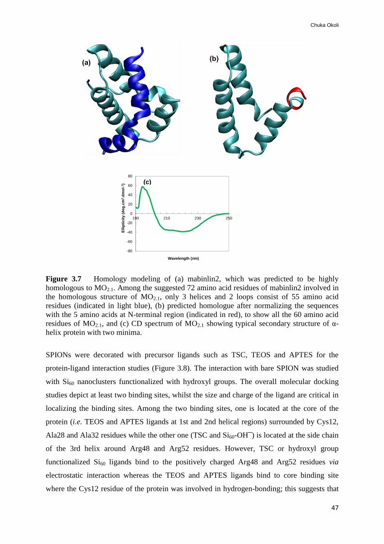
Chuka Okoli
47
Figure 3.7 Homology modeling of (a) mabinlin2, which was predicted to be highly
homologous to MO2.1. Among the suggested 72 amino acid residues of mabinlin2 involved in
the homologous structure of MO2.1, only 3 helices and 2 loops consist of 55 amino acid
residues (indicated in light blue), (b) predicted homologue after normalizing the sequences
with the 5 amino acids at N-terminal region (indicated in red), to show all the 60 amino acid
residues of MO2.1, and (c) CD spectrum of MO2.1 showing typical secondary structure of α-
helix protein with two minima.
SPIONs were decorated with precursor ligands such as TSC, TEOS and APTES for the
protein-ligand interaction studies (Figure 3.8). The interaction with bare SPION was studied
with Si60 nanoclusters functionalized with hydroxyl groups. The overall molecular docking
studies depict at least two binding sites, whilst the size and charge of the ligand are critical in
localizing the binding sites. Among the two binding sites, one is located at the core of the
protein (i.e. TEOS and APTES ligands at 1st and 2nd helical regions) surrounded by Cys12,
Ala28 and Ala32 residues while the other one (TSC and Si60-OH─) is located at the side chain
of the 3rd helix around Arg48 and Arg52 residues. However, TSC or hydroxyl group
functionalized Si60 ligands bind to the positively charged Arg48 and Arg52 residues via
electrostatic interaction whereas the TEOS and APTES ligands bind to core binding site
where the Cys12 residue of the protein was involved in hydrogen-bonding; this suggests that
-80
-60
-40
-20
0
20
40
60
80
190 210 230 250
Ell
ipti
cit
y (
de
g.c
m2.d
mo
l-1)
Wavelength (nm)
(c)

Development of Protein-Functionalized Magnetic Iron Oxide Nanoparticles: Potential Application in Water Treatment
48
the Cys12 residue was protonated in the aqueous solution at pH 7. The Alanine (Ala) residues,
on the other hand, initiated van der Waals interaction.
Figure 3.8 Docking study of MOCP (MO2.1) interaction with ligand molecules (a) TSC
ligand, (b) TEOS ligand, (c) APTES ligand and (d) Si60 nanoclusters. Important protein
residues involved in binding are highlighted; arginine (Arg, green), cysteine (Cys, yellow),
alanine (Ala, white) and Si60 nanoclusters with OH group (red and white).
The protein residues-ligands distance profile analysis as well as the coagulation activity of
protein-ligand (MOCP-MNPs) are shown in Figure 3.9. The average distance value for each
residue and its distribution with respect to the average value of the studied protein-ligand
interaction (protein-TSC, protein-TEOS and protein-APTES) are shown in Figures 3.9 (a)-(c).
The width is directly related to the amplitude of the residue motion (with respect to the ligand).
A larger r-value for the width means a large amplitude motion for the residue while a smaller r-
value for the width means a localized motion for the particular residue. As can be seen in
Figure 3.9a, the residues Arg48 and Arg52 are in closer contact with the TSC ligand.
Interestingly, a smaller width computed for these two residues (among all 60 residues) was
observed. This clearly shows that the binding of the ligand reduces the dynamics of these two
residues. When compared to TOES and APTES ligands, a relatively smaller value for widths of

Chuka Okoli
49
other residues in the 3rd helix is observed; thus, the effect of ligand binding is in some sense
seen over the whole helix, whilst larger width for the residues that form 1st and 2nd helices
implies increased conformational flexibility for these two helices when compared to 3rd helix.
The phenomenon was critical in the high performance of TSC-modified SPION (Figure 3.9d)
in terms of coagulation activity.
Figure 3.9 Motion distance calculations of the protein residues with different ligands (a)
TSC, (b) TEOS, (c) APTES and (d) experimental results showing coagulation activity of
MOCP attached to different surface-modified SPIONs. Blue arrow depicts the position where
protein residues bind to the precursor ligand.
Conversely, for the other two cases (TEOS and APTES ligands), the 1st and 2nd helices
have restricted dynamics over the third helix since these two helices are involved in binding
with the ligands. Particularly, Cys12 of helix 1 and Ala28 and Ala32 of the protein (helix 2)
appear at smaller r-value in the figure, implying that they are involved in the binding. As
mentioned earlier, in these two cases, a smaller width in the case of Cys12, Ala28 and Ala32
residues was also observed. The features seen in the cases of protein-TEOS and protein-APTES
are almost similar except in the region corresponding to the residues 39-50. A relatively larger
width for the residues was seen in the case of protein-APTES which suggests an increased
conformational flexibility for the 3rd helix; this behavior also explains why the APTES-

Development of Protein-Functionalized Magnetic Iron Oxide Nanoparticles: Potential Application in Water Treatment
50
modified SPIONs presented better performance over TEOS-modified SPION (Figure 3.9d).
However, the protein-ligand distance profile analysis confirms the difference in functional
activity of the surface-modified SPIONs, evidencing a correlation between the computational
model and experimental data.
3.3.2.2 Fluorescence emission study
Emission fluorescence spectra of MOCP, MOCP+w/o ME-MION, and MOCP+o/w ME-
MION are presented in Figure 3.10. A significant interaction between the magnetic
nanoparticles and the protein can be described by changes in fluorescence emission spectra.
The MOCP has a broad peak with apparently two maxima at 310 and 326 nm. A shift to λmax
of 295 nm is observed with MOCP+w/o ME-MION, implying a modification of protein
fluorophore environment. Similar behavior is observed in the absorption spectra of MOCP-
o/w ME-MION. However, the binding of nanoparticles in the ligand binding site of the
protein makes the environment of the aromatic side chains in the binding site less polar.135
The decrease in the fluorescence intensity can be attributed to the adsorption of MOCP onto
the nanoparticles surface. In fact, the small particle size and large surface-to-volume ratio
(Paper III) could be critical for this behavior; hence the density of MOCP+ME-MIONs
increased following the absorption of protein on the particles which initiated large protein
precipitation with the nanoparticles. Adsorbed MOCP still retains its functionality after
binding to ME-MIONs, thus implying the suitability of this phenomenon in the development
of protein-functional nanoparticles for water treatment.
Figure 3.10 Fluorescence emission spectra of MOCP (red), MOCP+w/o- (blue) and o/w-
(green) ME-MION at 255 nm excitation wavelength.
0
0,2
0,4
0,6
0,8
1
1,2
280 300 320 340 360 380 400
No
rmalized
flu
ore
scen
ce
Wavelength (nm)
MOCP
w/o ME-MION
o/w ME-MION

Chuka Okoli
51
3.3.3 Turbidity removal with the developed MOCP-MNPs system (Approach II)
(Paper IV & V)
Limited research has been carried out on developing a method that will promote the reuse of
MOCP in water or wastewater treatment processes. According to our knowledge and to date,
all the investigated applications with MOCP in turbid water clarification have been based only
on one time use,17, 67, 136
as it is difficult practically to recover and reuse the protein. The
coagulation activity of the developed MOCP-MNPs was investigated in the clarification of
surface waters (Figure 3.11). The optimal time required to achieve an efficient coagulation
activity (turbidity removal) with different coagulant agents (MOCP+w/o ME-MION,
MOCP+o/w ME-MION, MOCP+TSC-coated SPION, alum and MOCP) was around 60 min.
The coagulation activity of alum or MOCP depends on the composition or initial turbidity of
the water source; thus, an optimal time for effective turbidity removal may vary between 90
and 120 min.

Development of Protein-Functionalized Magnetic Iron Oxide Nanoparticles: Potential Application in Water Treatment
52
0
10
20
30
40
50
60
70
80
90
100
0 20 40 60 80 100 120 140 160 180 200 220 240
Co
ag
ula
tio
n a
cti
vit
y (
%)
Time (min)
(b)
(a)
Figure 3.11 Coagulation kinetic studies comparing the performance of MOCP-MNPs,
MOCP and alum in surface water from Lake Mälaren with (a) an initial turbidity of 17 NTU
and (b) an initial turbidity of 7 NTU. The control represents a surface water sample.
However, as shown in Figure 3.12, neither of the coagulant agents demonstrated a significant
increase in coagulation performance at concentrations above 0.2 mg/L, which could represent
the optimal concentration of MOCP-MNPs for surface water turbidity removal. The MOCP-
MNPs (MOCP+w/o ME-MION and MOCP+TSC-coated SPION) exhibited higher
coagulation performance of about 20% compared to alum and MOCP.
The MOCP+w/o ME-MION, MOCP+o/w ME-MION and MOCP+TSC-coated SPION depict
similar patterns in performance, implying a significant decrease in turbidity when tested in
natural water. It has been reported that the performance of most chemical coagulant agents
typically depends on the initial turbidity of the water sample.32
The experimental results show
0
10
20
30
40
50
60
70
80
90
100
0 20 40 60 80 100 120 140 160 180 200 220 240 260
Co
ag
ula
tio
n a
cti
vit
y (
%)
Time (min)
MOCP+w/o ME-MIONMOCP+o/w ME-MIONMOCP+TSC-coated SPIONAlumMOCPControl
(a)

Chuka Okoli
53
that with MOCP+w/o ME-MION, MOCP+o/w ME-MION and MOCP+TSC-coated SPION,
coagulation activity of ~ 80 % can be achieved.
Figure 3.12 Coagulation performance vs. applied dosage of MOCP-MNPs, alum and MOCP
(mg/L) in terms of turbidity removal in surface water at 1 h interaction time.
However, the difference observed between MOCP-MNPs and other coagulant agents could be
ascribed to the high density mass of the magnetized flocs induced by electrostatic interaction,
which resulted in rapid flocculation in the water sample. In contrast, alum and MOCP were
found to be less effective, especially in water samples with initial turbidity of 7 NTU (Figure
3.11b). Nonetheless, alum and MOCP were effective in surface water with 17 NTU. This
study suggests that the MOCP-MNPs may represent a feasible complementary agent in water
treatment process.
3.3.3.1 Separation by gravity and magnetic field
As shown in Figure 3.13, the magnetization effect of MOCP+ME-MION in the clarification
of turbid waters (coagulation activity) was enhanced under the influence of an external
magnetic field when compared with the turbidity removal by means of gravitational settling
i.e., as in the case of alum. It was reported that (Paper IV), with MOCP+ME-MION, almost
95% turbidity removal can be achieved within 12 min due to the existence of
magnetophoresis of the nanoparticles.137, 138
This implies that when the MOCP+ME-MION
comes in contact with polluted water, it tends to react with the colloidal or suspended particles
thereby transiting from dispersed to aggregated state. According to magnetization curve
theories, when nanoparticles are placed within a steady external magnetic field, the internal
magnetic moment spins in the same direction as the external magnetic field. As a result, the
0
10
20
30
40
50
60
70
80
90
100
0 0,1 0,2 0,3 0,4 0,5
% a
cti
vit
y
mg/L
Alum
MOCP
MOCP+TSC-coated SPION
MOCP+w/o ME-MION

Development of Protein-Functionalized Magnetic Iron Oxide Nanoparticles: Potential Application in Water Treatment
54
shifting of magnetic domain wall occurs, which boosts the magnetic properties of the
nanoparticles;74, 91
this phenomenon leads to the aggregation of the water impurities.
Figure 3.13 Separation performance by magnetic field and/or gravity in natural and
synthetic turbid water samples. MOCP+ME-MION and alum were added to water samples
followed by 10 min interaction time and 2 min separation. SW1 and SW2 represent surface
water (7 and 17 NTU respectively) from Lake Mälaren. ME-MION is bare nanoparticle
(control).
However, these dynamics will induce the rapid aggregation of the water contaminants by
MOCP+ME-MION. In contrast to the coagulation/flocculation of impurities by gravity, i.e. in
the case of water containing alum, the combination of MOCP+ME-MION and applied
magnetic field enhanced the effectiveness of the system in forming flocs. Moreover, it was
apparent that even though the bare ME-MION may possess some coagulation potential, a
lower performance was also observed when compared with MOCP+ME-MIONs. This clearly
reveals that the binding of the protein onto the nanoparticle was critical in enhancing their
performance.
3.3.3.2 Reusability of MOCP-MNPs system
From the coagulation performance of the MOCP-MNPs, it is clearly evident that the system
exhibits reusability potential after removing the adsorbed impurities with mild detergent or
cleaning solutions (0.1% Tween 20 and 20% EtOH). As shown in Figure 3.14, the
0
10
20
30
40
50
60
70
80
90
100
Clay SW1 SW2 Clay SW1 SW2 Clay SW1 SW2
Co
ag
ula
tio
n a
cti
vit
y (
%)
Water samples
MOCP+ME-MION
Alum
ME-MION
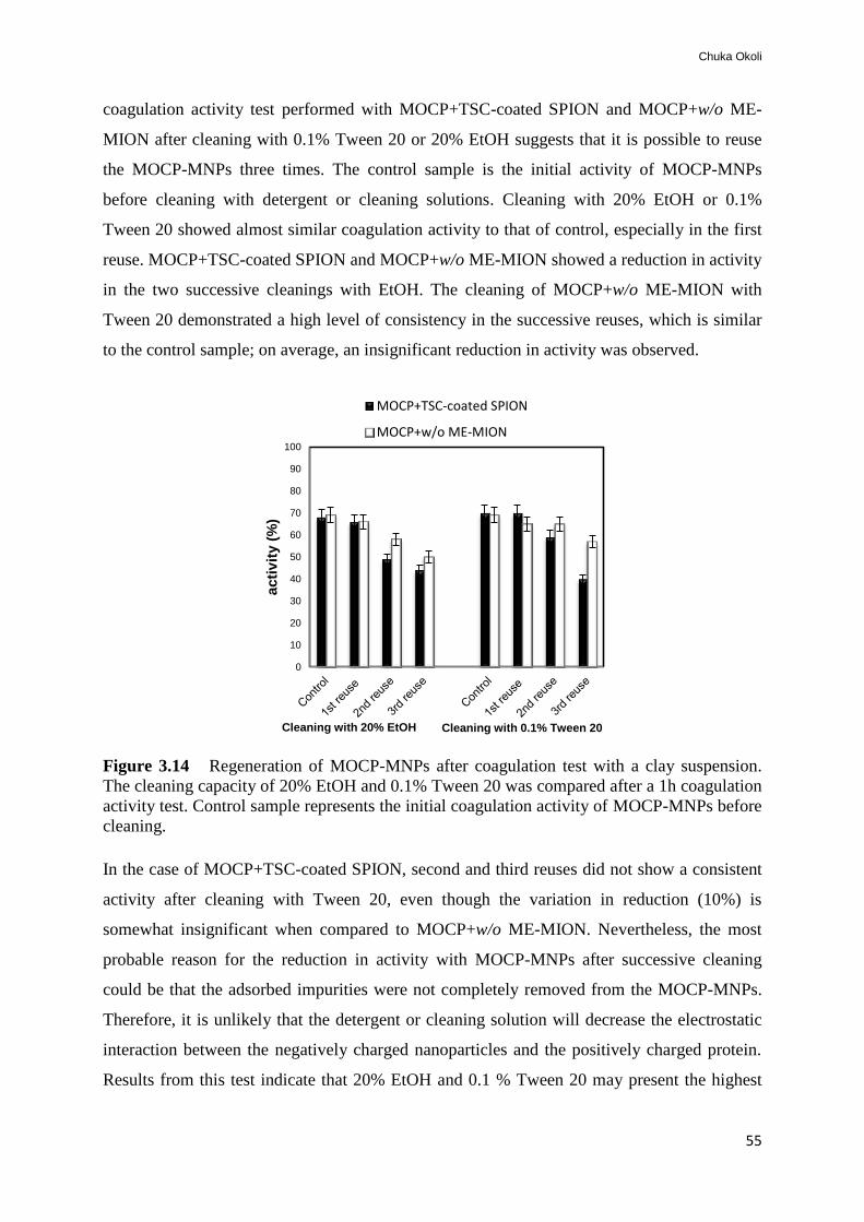
Chuka Okoli
55
coagulation activity test performed with MOCP+TSC-coated SPION and MOCP+w/o ME-
MION after cleaning with 0.1% Tween 20 or 20% EtOH suggests that it is possible to reuse
the MOCP-MNPs three times. The control sample is the initial activity of MOCP-MNPs
before cleaning with detergent or cleaning solutions. Cleaning with 20% EtOH or 0.1%
Tween 20 showed almost similar coagulation activity to that of control, especially in the first
reuse. MOCP+TSC-coated SPION and MOCP+w/o ME-MION showed a reduction in activity
in the two successive cleanings with EtOH. The cleaning of MOCP+w/o ME-MION with
Tween 20 demonstrated a high level of consistency in the successive reuses, which is similar
to the control sample; on average, an insignificant reduction in activity was observed.
Figure 3.14 Regeneration of MOCP-MNPs after coagulation test with a clay suspension.
The cleaning capacity of 20% EtOH and 0.1% Tween 20 was compared after a 1h coagulation
activity test. Control sample represents the initial coagulation activity of MOCP-MNPs before
cleaning.
In the case of MOCP+TSC-coated SPION, second and third reuses did not show a consistent
activity after cleaning with Tween 20, even though the variation in reduction (10%) is
somewhat insignificant when compared to MOCP+w/o ME-MION. Nevertheless, the most
probable reason for the reduction in activity with MOCP-MNPs after successive cleaning
could be that the adsorbed impurities were not completely removed from the MOCP-MNPs.
Therefore, it is unlikely that the detergent or cleaning solution will decrease the electrostatic
interaction between the negatively charged nanoparticles and the positively charged protein.
Results from this test indicate that 20% EtOH and 0.1 % Tween 20 may present the highest
0
10
20
30
40
50
60
70
80
90
100
acti
vit
y (
%)
Cleaning with 20% EtOH Cleaning with 0.1% Tween 20
MOCP+TSC-coated SPION
MOCP+w/o ME-MION

Development of Protein-Functionalized Magnetic Iron Oxide Nanoparticles: Potential Application in Water Treatment
56
capacity to regenerate the MOCP+ME-MION after several reuses. More investigation is being
conducted to fully understand the mechanism of cleaning in order to improve the efficiency of
the system while reducing the overall cost of the treatment process.

Chuka Okoli
57
4. Conclusions
The work presented in this thesis demonstrates advancement in the development of eco-
friendly water treatment strategy using magnetic iron oxide nanoparticles for MOCP
separation (i.e. in situ treatment) as well as the preparation of MOCP-MNPs systems for water
treatment. Magnetic nanoparticles with different surface chemistry (sizes, 2-30 nm) were
successfully prepared from co-precipitation and microemulsion (w/o and o/w) methods. The
characterization of the prepared nanoparticles was critical for protein-nanoparticles interaction
study. The protein-nanoparticle binding mechanism was verified by in silico study, which
validates the experimental (in vitro) data.
A simple, fast and efficient method for the purification of MOCP was developed and used in
an in situ water treatment strategy. This approach is based on the separation of the coagulant
protein with magnetic nanoparticles; the performance of the obtained MOCP (turbidity
removal) was compared with alum for turbid water clarification. Moreover, the purification
method used here is especially appealing since the used materials are obtained at a low cost.
Similarly, the second treatment approach highlights the development and application of
MOCP-MNPs for efficient and rapid water treatment. The strategy used is based on the
magnetophoretic effects of the nanoparticles under the influence of external magnetic field.
The strategy presented in this work (approach II) shows superior performance over alum or
MOCP in the removal of water turbidity, irrespective of the water composition and turbidity
level.
The MOCP-MNPs could effectively remove more than 95% turbidity in surface waters under
the influence of an external magnetic field within 12 min, whereas conventional water
treatment processes require several hours. The combination of natural coagulant protein plus
magnetic nanoparticles and magnetic field enhanced the effectiveness of the system in
coagulating/flocculating impurities in water samples. The present investigation also suggests
the possibility of regenerating the MOCP-MNPs; moreover, the developed strategy is unlikely
to introduce residual ions during water treatment. Similarly, this strategy does not influence
the pH or conductivity of the treated waters (Paper VII). These characteristics make the
MOCP-MNPs appealing in water treatment. To the best of our knowledge and according to
the published data, this is the first report on the separation or binding of MOCP using
magnetic nanoparticles. The data shown in this work represent water treatment approaches
(with safe materials) that are simple to use, robust and environmentally friendly.

Development of Protein-Functionalized Magnetic Iron Oxide Nanoparticles: Potential Application in Water Treatment
58
Further studies
This work endorses the possibilities of using alternative natural coagulant materials (MOCP)
or their association with magnetic nanoparticles (MOCP-MNPs) in the treatment of different
surface waters. In order to appreciate the real integrated effect and to apply these findings to
water treatment plants, pilot and full-scale studies are required. Furthermore, confirmatory
studies are needed to validate that the treatment approaches do not alter the pH or
conductivity of the treated waters, although preliminary results with bare nanoparticles or
MOCP-MNPs (Paper VII) suggest that neither the pH nor the conductivity of the treated
water was changed. It is necessary to investigate the cytotoxicity of the MOCP+MNPs even
though it has been established by the U.S. FDA that iron oxide nanoparticles are the only safe
nanoparticles approved for clinical use.
The development of recombinant MOCP for large-scale protein production is a work in
progress. This procedure will promote large-scale production of the protein whilst, reducing
cost. Current investigation with NMR is ongoing in order to determine the MOCP protein
structure. To date, the highest number of amino acid residues in one of the identified peptide
sequences obtained from MS/MS analysis was 19 (section 3.3.1) which differs from 60
residues reported by Gassenschmidt et al., (1995). However, in-depth study is deemed
necessary to identify the full sequence of this protein, and in particular to identify which
region of the protein is responsible for coagulation and antimicrobial action. Moreover, the
existence of the heterogeneous nature of the protein suggests the need to disclose the full
sequence of the MOCP. Presently, studies are being carried out in our group with in silico
modeling tools to understand and possibly resolve the protein structure.
The present investigation on this work suggests that the MOCP and/or its combination with
MNPs possess other potential applications beside water treatment such as dye removal. A
study on the removal of other water pollutants like industrial effluents is essential in order to
assess the wider application of the developed approaches.

Chuka Okoli
59
Acknowledgement
I wish to express my sincere gratitude to my supervisor, Assoc. Prof. Gunaratna Rajarao-
Kuttuva; your constant support, encouragement and positive criticisms have always been a
source inspiration. Thank you very much for introducing me to the fascinating field of
biology. In fact, I cannot thank you enough.
I am very obliged to my co-supervisors, Assoc. Prof. Magali Boutonnet and Prof. Sven Järås
at the School of Chemical Science & Engineering. You both have encouraged and motivated
me during the years of my study especially in supporting most of my conference and research
trips. I thank you Magali, for introducing me to the synthesis of nanoparticles by
microemulsion methods.
Many thanks to Prof. Per-Åke Nygren, Prof. Vincent Bulone, Prof. Gunnel Dalhammar, Prof.
Lars J Pettersson, and Dr. Arul Murugan for their brilliant suggestions and assistance. I
would like to thank Dr. Margarita Sanchez-Dominguez (CIMAV-Unidad Monterrey, Mexico)
for her fruitful collaboration including her excellent contributions and assistance in material
characterization. I enjoy the company, including the brilliant ideas of my colleagues; Dr. A.R.
Pavankumara, Ida, Ramnath, Delaram and Tobias. To all my colleagues, in the school of
Biotechnology (Plan 1) and at the Division of Chemical Technology; thanks for the quality
time spent with you guys. To Marita Johnsson; thanks for me helping out with administrative
issues. Many thanks to kaj for the I.T support.
My deep gratitude goes to my family; my Dad, Chikodi, Kenneth, Uju, Chinedu, Ifeanyi,
Chekwus, Onyeka, and Ifeoma. My profound appreciation goes to Mr. & Mrs. Ononiwu,
Chike Ilechukwu and Barrister Osonwa; I thank you all for your support and understanding. I
thank my in-laws for their support and best wishes.
Many thanks to Efraim and Ulrika Moström for their prayers and support; it’s a huge blessing
to be around you guys. To Philip and KC Onu, Mr. & Mrs. Rogo, Inno Okeke and his family,
Fred and Chinenye Ijeboi, including all my pals that graduated from KTH; thank you folks for
all sorts of activities, which made it possible to relax outside research.
My sincere appreciation goes to my aunt, Lolo Enuma Ginigeme for her unfailing love and
motivation especially, her constant prayers and support towards my wife and kids while I was
abroad; Aunt, we are proud of you. To Dera, Uche, Kosi and Nnamdi; it’s fun to be around
you guys and I am certain that you all will make Mom proud. My profound appreciation goes
to Mr. & Mrs. Okey Nweke for their kindness and support. My deepest gratitude goes to Rev.
& Mrs. Julius Edah, and all the members of UAPC Dallas, TX for their prayers and support.
Many thanks to my very good friend, Dr. Mfon Inyang and his family.
I am grateful to the Swedish Research Institute; FORMAS for financing this project.
Finally, I wish to express my profound appreciation to the love of my life; my wife, Queen,
who has constantly supported and encouraged me during these years. Without her
understanding and prayers, it would not have been possible for me to complete my research at
this time. I sincerely appreciate your hard work in taking good care of our wonderful kids,
Lily and Munachi.

Development of Protein-Functionalized Magnetic Iron Oxide Nanoparticles: Potential Application in Water Treatment
60

Chuka Okoli
61
References
1. Moe, C.L. & Rheingans, R.D. Global challenges in water, sanitation and health. J
Water Health 4 Suppl 1, 41-57 (2006).
2. Vorosmarty, C.J., Green, P., Salisbury, J. & Lammers, R.B. Global water resources:
Vulnerability from climate change and population growth. Science 289, 284-288
(2000).
3. Lee, E.J. & Schwab, K.J. Deficiencies in drinking water distribution systems in
developing countries. J Water Health 3, 109-127 (2005).
4. Coetser, S.E., Heath, R.G.M. & Ndombe, N. Diffuse pollution associated with the
mining sectors in South Africa: a first-order assessment. Water Sci Technol 55, 9-16
(2007).
5. Theron, J., Walker, J.A. & Cloete, T.E. Nanotechnology and water treatment:
Applications and emerging opportunities. Crit Rev Microbiol 34, 43-69 (2008).
6. Theron, J. & Cloete, T.E. Emerging waterborne infections: Contributing factors,
agents, and detection tools. Crit Rev Microbiol 28, 1-26 (2002).
7. Brown, K. Environmental data. Water scarcity: forecasting the future with spotty data.
Science 297, 926-927 (2002).
8. Bolto, B. & Gregory, J. Organic polyelectrolytes in water treatment. Water Res 41,
2301-2324 (2007).
9. Sciban, M., Klasnja, M., Antov, M. & Skrbic, B. Removal of water turbidity by
natural coagulants obtained from chestnut and acorn. Biores Technol 100, 6639-6643
(2009).
10. Zouboulis, A., Traskas, G. & Samaras, P. Comparison of efficiency between poly-
aluminium chloride and aluminium sulphate coagulants during full-scale experiments
in a drinking water treatment plant. Separ Sci Technol 43, 1507-1519 (2008).
11. Sadiq, R. & Rodriguez, M.J. Disinfection by-products (DBPs) in drinking water and
predictive models for their occurrence: a review. Sci Total Environ 321, 21-46 (2004).
12. Rondeau, V., Jacqmin-Gadda, H., Commenges, D. & Dartigues, J.F. Re: Aluminum in
drinking water and cognitive decline in elderly subjects: The Paquid cohort. Am J
Epidemiol 154, 288-290 (2001).
13. Othman, M.N., Abdullah, M.P. & Abd Aziz, Y.F. Removal of Aluminium from
Drinking Water. Sains Malays 39, 51-55 (2010).
14. Gauthier, E. et al. Aluminum forms in drinking water and risk of Alzheimer's disease.
Environ Res 84, 232-246 (2000).
15. Ghebremichael, K., Abaliwano, J. & Amy, G. Combined natural organic and synthetic
inorganic coagulants for surface water treatment. J Water Supply Res T 58, 267-276
(2009).

Development of Protein-Functionalized Magnetic Iron Oxide Nanoparticles: Potential Application in Water Treatment
62
16. Cloete, T.E. Nanotechnology in water treatment applications. (Caister Academic
Press, Norfolk, UK; 2010).
17. Ndabigengesere, A. & Narasiah, K.S. Use of Moringa oleifera seeds as a primary
coagulant in wastewater treatment. Environ Technol 19, 789-800 (1998).
18. Ghebremichael, K.A., Gunaratna, K.R. & Dalhammar, G. Single-step ion exchange
purification of the coagulant protein from Moringa oleifera seed. Appl Microbiol Biot
70, 526-532 (2006).
19. Bhuptawat, H., Folkard, G.K. & Chaudhari, S. Innovative physico-chemical treatment
of wastewater incorporating Moringa oleifera seed coagulant. J Hazard Mater 142,
477-482 (2007).
20. Yongabi, K.A., Lewis, D.M. & Harris, P.L. Indigenous plant based
coagulants/disinfectants and sand filter media for surface water treatment in Bamenda,
Cameroon. Afr J Biotechnol 10, 8625-8629 (2011).
21. Mikhaylova, M. et al. Superparamagnetism of magnetite nanoparticles: Dependence
on surface modification. Langmuir 20, 2472-2477 (2004).
22. Li, Q.L. et al. Antimicrobial nanomaterials for water disinfection and microbial
control: Potential applications and implications. Water Res 42, 4591-4602 (2008).
23. Liao, M.H. & Chen, D.H. Characteristics of magnetic nanoparticles-bound YADH in
water/AOT/isooctane microemulsions. J Mol Catal B-Enzym 18, 81-87 (2002).
24. Grecu, V.V. et al. Magnetic characterization of some nanometric iron oxides.
Hyperfine Interact 183, 205-214 (2008).
25. Diaz, A. et al. A preliminary evaluation of turbidity removal by natural coagulants
indigenous to Venezuela. Process Biochem 35, 391-395 (1999).
26. Raghuwanshi, P.K., Mandloi, M., Sharma, A.J., Malviya, H.S. & Chaudhari, S.
Improving filtrate quality using agrobased materials as coagulant aid. Water Qual Res
J Can 37, 745-756 (2002).
27. Amirtharajah, A. Coagulation - Rejuvenation for a classical process. Water Eng
Manag 137, 25-& (1990).
28. Gregory, J. & Duan, J.M. Hydrolyzing metal salts as coagulants. Pure Appl Chem 73,
2017-2026 (2001).
29. Yin, C.Y. Emerging usage of plant-based coagulants for water and wastewater
treatment. Process Biochem 45, 1437-1444 (2010).
30. Okuda, T., Baes, A.U., Nishijima, W. & Okada, M. Coagulation mechanism of salt
solution-extracted active component in Moringa oleifera seeds. Water Res 35, 830-834
(2001).
31. Muyibi, S.A. & Evison, L.M. Optimizing physical parameters affecting coagulation of
turbid water with Moringa-oleifera seeds. Water Res 29, 2689-2695 (1995).

Chuka Okoli
63
32. Sanchez-Martin, J., Ghebremichael, K. & Beltran-Heredia, J. Comparison of single-
step and two-step purified coagulants from Moringa oleifera seed for turbidity and
DOC removal. Bioresource Technol 101, 6259-6261 (2010).
33. Kaggwa, R.C., Mulalelo, C.I., Denny, P. & Okurut, T.O. The impact of alum
discharges on a natural tropical wetland in Uganda. Water Res 35, 795-807 (2001).
34. Ghebremichael, K. in Land and Water Resources Engineering, PhD thesis (Royal
Institue of Technology; KTH, Stockholm, Sweden. ISSN 1650-8602; 2004).
35. Morales, C.F.L., Strathmann, M. & Flemming, H.C. Influence of biofilms on the
movement of colloids in porous media. Implications for colloid facilitated transport in
subsurface environments. Water Res 41, 2059-2068 (2007).
36. Eschauzier, C., Beerendonk, E., Scholte-Veenendaal, P. & De Voogt, P. Impact of
treatment processes on the removal of perfluoroalkyl acids from the drinking water
production chain. Environ Sci Technol 46, 1708-1715 (2012).
37. Kubiak, K.A. & Oszako, T. Slow Sand Filter as a method of protection forest plants
against phytopathogens in forest nurseries. Sylwan 155, 228-235 (2011).
38. Kim, J. & Grate, J.W. Single-enzyme nanoparticles armored by a nanometer-scale
organic/inorganic network. Nano Lett 3, 1219-1222 (2003).
39. Kim, J. et al. Single enzyme nanoparticles in nanoporous silica: A hierarchical
approach to enzyme stabilization and immobilization. Enzyme Microb Tech 39, 474-
480 (2006).
40. Bhattacharyya, D. et al. Novel poly-glutamic acid functionalized microfiltration
membranes for sorption of heavy metals at high capacity. J Membrane Sci 141, 121-
135 (1998).
41. Koyuncu, I. & Yazgan, M. Application of nanofiltration and reverse osmosis
membranes to the salty and polluted surface water. J Environ Sci Heal A 36, 1321-
1333 (2001).
42. Koyuncu, I. & Topacik, D. Effect of organic ion on the separation of salts by
nanofiltration membranes. J Membrane Sci 195, 247-263 (2002).
43. Brant, J.A., Koyuncu, I., Lecoanet, H., Veerapaneni, S. & Wiesner, M. Occurrence
and composition of particulates in filter process streams. J Am Water Works Ass 103,
46-+ (2011).
44. Chen, C.Z. & Cooper, S.L. Interactions between dendrimer biocides and bacterial
membranes. Biomaterials 23, 3359-3368 (2002).
45. Madrona, G.S. et al. Study of the Effect of saline solution on the extraction of the
Moringa oleifera seed's active component for water treatment. Water Air Soil Poll 211,
409-415 (2010).

Development of Protein-Functionalized Magnetic Iron Oxide Nanoparticles: Potential Application in Water Treatment
64
46. Madrona, G.S., Bergamasco, R., Seolin, V.J. & Klen, M.R.F. The potential of
different saline solution on the extraction of the Moringa oleifera seed's active
component for water treatment. Int J Chem React Eng 9 (2011).
47. Sanghi, R., Bani, B.A. & Singh, V. Cassia angustifolia seed gum as an effective
natural coagulant for decolourisation of dye solutions. Green Chem 4, 252-254 (2002).
48. Kwaambwa, H.M., Hellsing, M. & Rennie, A.R. Adsorption of a water treatment
protein from Moringa oleifera seeds to a silicon oxide surface studied by neutron
reflection. Langmuir 26, 3902-3910 (2010).
49. Jahn, S.A.A. & Dirar, H. Studies on natural-water coagulants in the Sudan, with
special reference to Moringa-Oleifera seeds. Water San 5, 90-97 (1979).
50. Jahn, S.A.A. Using Moringa seeds as coagulants in developing-countries. J Am Water
Works Ass 80, 43-50 (1988).
51. Madsen, M., Schlundt, J. & Omer, E.F. Effect of water coagulation by seeds of
Moringa oleifera on bacterial concentrations. The Journal of tropical medicine and
hygiene 90, 101-109 (1987).
52. Folkard, G. & Sutherland, J. Development of a naturally derived coagulant for water
and wastewater treatment. Wat Sci Technol 2, 89-94 (2002).
53. Suarez, M. et al. Expression of a plant-derived peptide harboring water-cleaning and
antimicrobial activities. Biotechnol Bioeng 81, 13-20 (2003).
54. Katayon, S. et al. Effects of storage conditions of Moringa oleifera seeds on its
performance in coagulation. Biores Technol 97, 1455-1460 (2006).
55. Katayon, S., Ng, S.C., Johari, M.M.N.M. & Ghani, L.A.A. Preservation of coagulation
efficiency of Moringa oleitera, a natural coagulant. Biotechnol Bioproc E 11, 489-495
(2006).
56. Akhtar, M., Moosa Hasany, S., Bhanger, M.I. & Iqbal, S. Sorption potential of
Moringa oleifera pods for the removal of organic pollutants from aqueous solutions. J
Hazard Mater 141, 546-556 (2007).
57. Ndabigengesere, A., Narasiah, K.S. & Talbot, B.G. Active agents and mechanism of
coagulation of turbid waters using Moringa-Oleifera. Water Res 29, 703-710 (1995).
58. Sciban, M., Klasnja, M. & Skrbic, B. Water turbidity removal by natural coagulants.
Prog Environ Sci Technol, Vol Ii, Pts a and B, 1085-1088 (2009).
59. Ghebremichael, K.A., Gunaratna, K.R., Henriksson, H., Brumer, H. & Dalhammar, G.
A simple purification and activity assay of the coagulant protein from Moringa
oleifera seed. Water Res 39, 2338-2344 (2005).
60. Beltran-Heredia, J. & Sanchez-Martin, J. Improvement of water treatment pilot plant
with Moringa oleifera extract as flocculant agent. Environ Technol 30, 525-534
(2009).

Chuka Okoli
65
61. Gassenschmidt, U., Jany, K.D., Tauscher, B. & Niebergall, H. Isolation and
characterization of a flocculating protein from Moringa-Oleifera Lam. Bba-Gen
Subjects 1243, 477-481 (1995).
62. Kwaambwa, H.M. & Maikokera, R. A fluorescence spectroscopic study of a
coagulating protein extracted from Moringa oleifera seeds. Colloid Surf B 60, 213-
220 (2007).
63. Grabow, W.O.K., Slabbert, J.L., Morgan, W.S.G. & Jahn, S.A.A. Toxicity and
mutagenicity evaluation of water coagulated with Moringa-Oleifera seed preparations
using Fish, protozoan, bacterial, coliphage, enzyme and ames salmonella assays.
Water San 11, 9-14 (1985).
64. Muyibi, S.A. & Evison, L.M. Moringa-Oleifera seeds for softening hardwater. Water
Res 29, 1099-1104 (1995).
65. Okuda, T., Baes, A.U., Nishijima, W. & Okada, M. Improvement of extraction method
of coagulation active components from Moringa oleifera seed. Water Res 33, 3373-
3378 (1999).
66. Vieira, A.M.S. et al. Use of Moringa oleifera seed as a natural adsorbent for
wastewater treatment. Water Air Soil Poll 206, 273-281 (2010).
67. Nkurunziza, T., Nduwayezu, J.B., Banadda, E.N. & Nhapi, I. The effect of turbidity
levels and Moringa oleifera concentration on the effectiveness of coagulation in water
treatment. Water Sci Technol 59, 1551-1558 (2009).
68. Broin, M. et al. Flocculent activity of a recombinant protein from Moringa oleifera
Lam. seeds. Appl Microbiol Biot 60, 114-119 (2002).
69. Okuda, T., Baes, A.U., Nishijima, W. & Okada, M. Isolation and characterization of
coagulant extracted from Moringa oleifera seed by salt solution. Water Res 35, 405-
410 (2001).
70. Ndabigengesere, A. & Narasiah, K.S. Quality of water treated by coagulation using
Moringa oleifera seeds. Water Res 32, 781-791 (1998).
71. Okoli, C., Boutonnet, M., Mariey, L., Jaras, S. & Rajarao, G. Application of magnetic
iron oxide nanoparticles prepared from microemulsions for protein purification. J
Chem Technol Biot 86, 1386-1393 (2011).
72. Franzreb, M., Siemann-Herzberg, M., Hobley, T.J. & Thomas, O.R.T. Protein
purification using magnetic adsorbent particles. Appl Microbiol Biot 70, 505-516
(2006).
73. Ma, D.L. et al. Multifunctional nano-architecture for biomedical applications. Chem
Mater 18, 1920-1927 (2006).
74. Lu, A.H., Salabas, E.L. & Schuth, F. Magnetic nanoparticles: Synthesis, protection,
functionalization, and application. Angew Chem Int Edit 46, 1222-1244 (2007).

Development of Protein-Functionalized Magnetic Iron Oxide Nanoparticles: Potential Application in Water Treatment
66
75. Bruno, J.G. & Kiel, J.L. Use of magnetic beads in selection and detection of biotoxin
aptamers by electrochemiluminescence and enzymatic methods. Biotechniques 32,
178-+ (2002).
76. Suarez, M. et al. Structure-function characterization and optimization of a plant-
derived antibacterial peptide. Antimicrob Agents Chemo 49, 3847-3857 (2005).
77. Lu, A.H. et al. Nanoengineering of a magnetically separable hydrogenation catalyst.
Angew Chem Int Edit 43, 4303-4306 (2004).
78. Beveridge, J.S. et al. Purification and Magnetic Interrogation of Hybrid Au-Fe(3)O(4)
and FePt-Fe(3)O(4) Nanoparticles. Angew Chem Int Edit 50, 9875-9879 (2011).
79. Park, K., Harrah, T., Goldberg, E.B., Guertin, R.P. & Sonkusale, S. Multiplexed
sensing based on Brownian relaxation of magnetic nanoparticles using a compact AC
susceptometer. Nanotechnology 22 (2011).
80. Xi, P.X., Cheng, K., Sun, X.L., Zeng, Z.Z. & Sun, S.H. Fluorescent magnetic
nanoparticles based on a ruthenium complex and Fe(3)O(4). J Mater Chem 21, 11464-
11467 (2011).
81. Zhang, H., Harpster, M.H., Park, H.J. & Johnson, P.A. Surface-enhanced Raman
scattering detection of DNA derived from the west nile virus genome using magnetic
capture of Raman-active gold nanoparticles. Anal Chem 83, 254-260 (2011).
82. Chorny, M., Fishbein, I., Forbes, S. & Alferiev, I. Magnetic nanoparticles for targeted
vascular delivery. Iubmb Life 63, 613-620 (2011).
83. Kouassi, G.K. Magnetic and gold-coated magnetic iron oxide nanoparticles as
detection tools: Preparation, characterization, and biosensing applications. Curr
Nanosci 7, 510-523 (2011).
84. Hyeon, T. Chemical synthesis of magnetic nanoparticles. Chem Commun, 927-934
(2003).
85. Franco, A., Machado, F.L.A. & Zapf, V.S. Magnetic properties of nanoparticles of
cobalt ferrite at high magnetic field. J Appl Phys 110 (2011).
86. Abakumov, M.A. et al. Magnetic resonance imaging of endothelial cells with
vectorized iron oxide nanoparticles. B Exp Biol Med+ 151, 726-730 (2011).
87. Herranz, F. et al. The Application of nanoparticles in gene therapy and magnetic
resonance imaging. Microsc Res Techniq 74, 577-591 (2011).
88. Kinsella, J.M. et al. Enhanced magnetic resonance contrast of Fe(3)O(4) nanoparticles
trapped in a porous silicon nanoparticle host. Adv Mater 23, H248-+ (2011).
89. Elliott, D.W. & Zhang, W.X. Field assessment of nanoscale biometallic particles for
groundwater treatment. Environ Sci Technol 35, 4922-4926 (2001).

Chuka Okoli
67
90. Takafuji, M., Ide, S., Ihara, H. & Xu, Z.H. Preparation of poly(1-vinylimidazole)-
grafted magnetic nanoparticles and their application for removal of metal ions. Chem
Mater 16, 1977-1983 (2004).
91. Faraji, M., Yamini, Y. & Rezaee, M. Magnetic nanoparticles: Synthesis, stabilization,
functionalization, characterization, and applications. J Iran Chem Soc 7, 1-37 (2010).
92. Bourrinet, P. et al. Preclinical safety and pharmacokinetic profile of ferumoxtran-10,
an ultrasmall superparamagnetic iron oxide magnetic resonance contrast agent. Invest
Radiol 41, 313-324 (2006).
93. Saltan, N., Kutlu, H.M., Hur, D., Iscan, A. & Say, R. Interaction of cancer cells with
magnetic nanoparticles modified by methacrylamido-folic acid. Int J Nanomed 6, 477-
484 (2011).
94. Teja, A.S. & Koh, P.Y. Synthesis, properties, and applications of magnetic iron oxide
nanoparticles. Prog Cryst Growth Ch 55, 22-45 (2009).
95. Thorek, D.L., Chen, A.K., Czupryna, J. & Tsourkas, A. Superparamagnetic iron oxide
nanoparticle probes for molecular imaging. Annals Biomed Eng 34, 23-38 (2006).
96. Cornell, R.M. & Schwertmann, U. The iron oxides : structure, properties, reactions,
occurrences, and uses, Edn. 2nd, completely rev. and extended. (Wiley-VCH,
Weinheim; 2003).
97. Kumar, C.S.S.R. Magnetic nanomaterials. (Wiley-VCH, Weinheim, Germany; 2009).
98. Mathew, D.S. & Juang, R.S. An overview of the structure and magnetism of spinel
ferrite nanoparticles and their synthesis in microemulsions. Chem Eng J 129, 51-65
(2007).
99. Dave, S.R. & Gao, X.H. Monodisperse magnetic nanoparticles for biodetection,
imaging, and drug delivery: a versatile and evolving technology. Wires Nanomed
Nanobi 1, 583-609 (2009).
100. Kim, D.K., Zhang, Y., Voit, W., Rao, K.V. & Muhammed, M. Synthesis and
characterization of surfactant-coated superparamagnetic monodispersed iron oxide
nanoparticles. J Magn Magn Mater 225, 30-36 (2001).
101. Tartaj, P., Morales, M.D., Veintemillas-Verdaguer, S., Gonzalez-Carreno, T. & Serna,
C.J. The preparation of magnetic nanoparticles for applications in biomedicine. J Phys
D Appl Phys 36, R182-R197 (2003).
102. Liu, Z.L. et al. Synthesis and characterization of ultrafine well-dispersed magnetic
nanoparticles. J Magn Magn Mater 283, 258-262 (2004).
103. Sanchez-Dominguez, M., Boutonnet, M. & Solans, C. A novel approach to metal and
metal oxide nanoparticle synthesis: the oil-in-water microemulsion reaction method. J
Nanopart Res 11, 1823-1829 (2009).

Development of Protein-Functionalized Magnetic Iron Oxide Nanoparticles: Potential Application in Water Treatment
68
104. Boutonnet, M., Logdberg, S. & Svensson, E.E. Recent developments in the
application of nanoparticles prepared from w/o microemulsions in heterogeneous
catalysis. Curr Opin Colloid In 13, 270-286 (2008).
105. Olsson, U. & Wennerstrom, H. Globular and bicontinuous phases of nonionic
surfactant films. Adv Colloid Interfac 49, 113-146 (1994).
106. LopezPerez, J.A., LopezQuintela, M.A., Mira, J. & Rivas, J. Preparation of magnetic
fluids with particles obtained in microemulsions. Ieee T Magn 33, 4359-4362 (1997).
107. Perez, J.A.L., Quintela, M.A.L., Mira, J., Rivas, J. & Charles, S.W. Advances in the
preparation of magnetic nanoparticles by the microemulsion method. J Phys Chem B
101, 8045-8047 (1997).
108. Vidal-Vidal, J., Rivas, J. & Lopez-Quintela, M.A. Synthesis of monodisperse
maghemite nanoparticles by the microemulsion method. Colloid Surface A 288, 44-51
(2006).
109. Redl, F.X. et al. Magnetic, electronic, and structural characterization of
nonstoichiometric iron oxides at the nanoscale. J Am Chem Soc 126, 14583-14599
(2004).
110. Huber, D.L. Synthesis, properties, and applications of iron nanoparticles. Small 1,
482-501 (2005).
111. Li, Y., Afzaal, M. & O'Brien, P. The synthesis of amine-capped magnetic (Fe, Mn,
Co, Ni) oxide nanocrystals and their surface modification for aqueous dispersibility. J
Mater Chem 16, 2175-2180 (2006).
112. Hyeon, T., Lee, S.S., Park, J., Chung, Y. & Na, H.B. Synthesis of highly crystalline
and monodisperse maghemite nanocrystallites without a size-selection process. J Am
Chem Soc 123, 12798-12801 (2001).
113. Park, J. et al. Ultra-large-scale syntheses of monodisperse nanocrystals. Nat Mater 3,
891-895 (2004).
114. Sue, K., Kimura, K. & Arai, K. Hydrothermal synthesis of ZnO nanocrystals using
microreactor. Mater Lett 58, 3229-3231 (2004).
115. Lian, S.Y. et al. Synthesis of magnetite nanorods and porous hematite nanrods. Solid
State Commun 129, 485-490 (2004).
116. Deng, H. et al. Monodisperse magnetic single-crystal ferrite microspheres. Angew
Chem Int Edit 44, 2782-2785 (2005).
117. Han, K.H. & Frazier, A.B. Paramagnetic capture mode magnetophoretic
microseparator for high efficiency blood cell separations. Lab Chip 6, 265-273 (2006).
118. Kang, J.H. & Park, J.K. Magnetophoretic continuous purification of single-walled
carbon nanotubes from catalytic impurities in a microfluidic device. Small 3, 1784-
1791 (2007).
Chuka Okoli
69
119. Bradford, M.M. A rapid and sensitive method for the quantitation of microgram
quantities of protein utilizing the principle of protein-dye binding. Anal Biochem 72,
248-254 (1976).
120. Massart, R. & Cabuil, V. Effect of some parameters on the formation of colloidal
magnetite in alkaline-medium - yield and particle-size control. J Chim Phys Pcb 84,
967-973 (1987).
121. Bee, A., Massart, R. & Neveu, S. Synthesis of very fine maghemite particles. J Magn
Magn Mater 149, 6-9 (1995).
122. Deng, Y.H., Wang, C.C., Hu, J.H., Yang, W.L. & Fu, S.K. Investigation of formation
of silica-coated magnetite nanoparticles via sol-gel approach. Colloid Surf A 262, 87-
93 (2005).
123. Yang, D., Hu, J.H. & Fu, S.K. Controlled synthesis of magnetite-silica
nanocomposites via a seeded sol-gel approach. J Phys Chem C 113, 7646-7651
(2009).
124. W. Stöber, A.F., and E. Bohn. Controlled growth of monodisperse silica spheres in the
micron size range. Colloid Interface Sci 26, 62-68 (1968).
125. Shen, X.C., Fang, X.Z., Zhou, Y.H. & Liang, H. Synthesis and characterization of 3-
aminopropyltriethoxysilane-modified superparamagnetic magnetite nanoparticles.
Chem Lett 33, 1468-1469 (2004).
126. Okoli, C. et al. Characterization of superparamagnetic iron oxide nanoparticles and its
application in protein purification. J Nanosci Nanotech 11, 10201-10206 (2011).
127. Frisch, et.al., Vol. Gaussian 09, A1 (Gaussian Inc. , Wallingford, CT, U.S.A; 2009).
128. Kollman, P.A. et al. Calculating structures and free energies of complex molecules:
Combining molecular mechanics and continuum models. Accounts Chem Res 33, 889-
897 (2000).
129. Brandsdal, B.O. et al. Free energy calculations and ligand binding. Adv Protein Chem
66, 123-+ (2003).
130. Asrafuzzaman, M., Fakhruddin, A. & Hossain , M. Reduction of turbidity of water
using locally available natural coagulants. ISRN Microbio (2011).
131. Peng, S., Wang, C., Xie, J. & Sun, S.H. Synthesis and stabilization of monodisperse
Fe nanoparticles. J Am Chem Soc 128, 10676-10677 (2006).
132. Chin, A.B. & Yaacob, I.I. Synthesis and characterization of iron oxides nanoparticles.
Key Eng Mater 306-308, 1115-1119 (2006).
133. Mehta, R.V., Upadhyay, R.V., Charles, S.W. & Ramchand, C.N. Direct binding of
protein to magnetic particles. Biotechnol Techniq 11, 493-496 (1997).

Development of Protein-Functionalized Magnetic Iron Oxide Nanoparticles: Potential Application in Water Treatment
70
134. Durdureanu-Angheluta, A. et al. Progress in the synthesis and characterization of
magnetite nanoparticles with amino groups on the surface. J Magn Magn Mater 324,
1679-1689 (2012).
135. Albrecht, C. Joseph R. Lakowicz: Principles of fluorescence spectroscopy, 3rd
Edition. Anal Bioanal Chem 390, 1223-1224 (2008).
136. Jerri, H.A., Adolfsen, K.J., McCullough, L.R., Velegol, D. & Velegol, S.B.
Antimicrobial sand via adsorption of cationic Moringa oleifera protein. Langmuir 28,
2262-2268 (2012).
137. Zborowski, M., Fuh, C.B., Green, R., Sun, L.P. & Chalmers, J.J. Analytical
magnetapheresis of ferritin-labeled lymphocytes. Anal Chem 67, 3702-3712 (1995).
138. McCloskey, K.E., Chalmers, J.J. & Zborowski, M. Magnetic cell separation:
Characterization of magnetophoretic mobility. Anal Chem 75, 6868-6874 (2003).

Chuka Okoli
71
Appendix
Papers I to VII