doctor of philosophy - astonpublications.aston.ac.uk/19248/1/studentthesis-2012.pdf · doctor of...
TRANSCRIPT

DOCTOR OF PHILOSOPHY
Requirement for and optimisation ofpremium intraocular lenses
Gurpreet Bhogal
2012
Aston University

Some pages of this thesis may have been removed for copyright restrictions.
If you have discovered material in AURA which is unlawful e.g. breaches copyright, (either
yours or that of a third party) or any other law, including but not limited to those relating to
patent, trademark, confidentiality, data protection, obscenity, defamation, libel, then please
read our Takedown Policy and contact the service immediately

1
REQUIREMENT FOR AND OPTIMISATION OF PREMIUM INTRAOCULAR LENSES
GURPREET KAUR BHOGAL
Doctor of Philosophy
ASTON UNIVERSITY
September 2012
© Gurpreet Kaur Bhogal, 2012 asserts her moral right to be identified as
the author of this thesis
This copy of the thesis has been supplied on condition that anyone who consults it is
understood to recognise that its copyright rests with its author and that no quotation
from the thesis and no information derived from it may be published without proper
acknowledgement

2
ASTON UNIVERSITY
REQUIREMENT FOR AND OPTIMISATION OF PREMIUM INTRAOC ULAR
LENSES
GURPREET KAUR BHOGAL
DOCTOR OF PHILOSOPHY
SEPTEMBER 2012
Summary
Premium intraocular lenses (IOLs) aim to surgically correct astigmatism and presbyopia following cataract extraction, optimising vision and eliminating the need for cataract surgery in later years. It is usual to fully correct astigmatism and to provide visual correction for distance and near when prescribing spectacles and contact lenses, however for correction with the lens implanted during cataract surgery, patients are required to purchase the premium IOLs and pay surgery fees outside the National Health Service in the UK. The benefit of using toric IOLs was thus demonstrated, both in standard visual tests and real-world situations. Orientation of toric IOLs during implantation is critical and the benefit of using conjunctival blood vessels for alignment was shown. The issue of centration of IOLs relative to the pupil was also investigated, showing changes with the amount of dilation and repeat dilation evaluation, which must be considered during surgery to optimize the visual performance of premium IOLs.
Presbyopia is a global issue, of growing importance as life expectancy increases, with no real long-term cure. Despite enhanced lifestyles, changes in diet and improved medical care, presbyopia still presents in modern life as a significant visual impairment. The onset of presbyopia was found to vary with risk factors including alcohol consumption, smoking, UV exposure and even weight as well as age. A new technique to make measurement of accommodation more objective and robust was explored, although needs for further design modifications were identified. Due to dysphotopsia and lack of intermediate vision through most multifocal IOL designs, the development of a trifocal IOL was shown to minimize these aspects.
The current thesis, therefore, emphasises the challenges of premium IOL surgery and need for refinement for optimum visual outcome in addition to outlining how premium IOLs may provide long-term and successful correction of astigmatism and presbyopia.
Keywords: presbyopia, accommodation, intraocular lens, pupil centration, astigmatism

3
Dedicated to my late grandfather
Jagir Singh Bhogal

4
Acknowledgements
Foremost, I would like to express my gratitude to my supervisors Professor James
Wolffsohn and Dr Leon Davies for inspiring me to pursue a career in research and
mostly for providing me with the opportunity to begin this career in ophthalmic
research. Their expertise and advice have been invaluable throughout the course
of this doctorate and I have been highly privileged to have worked with such
supportive and knowledgeable supervisors.
I am grateful to Aston University and the Ophthalmic Research Group Anterior Eye
team for supporting this research. Many thanks to Pickford Opticians for kindly
providing a provision for conducting my research in addition to the substantial
contributions to data collection.
I would also like to acknowledge and thank my colleagues Dr Phillip Buckhurst, Dr
Amy Sheppard, Dr Stephanie Mroczkowska and Professor Sunil Shah for their
individual contributions to data collection and Dr Tom Drew and Alec Kingsnorth
for their advice and development of the electronic accommodometer. Thanks also
to Dr Hannah Bartlett and Dr Mark Dunne for their statistical expertise.
Finally, I give special thanks to all my family and friends for all their kindness and
support. Thank you to my fiancé, Gurjeet Bhamra, who has been a pillar of
strength throughout my studies. I owe sincere thanks to my mother, father and
brother Charanpreet for their support, unconditional love and continuous
encouragement in all my life goals. To my father especially I would like to say
thank you for encouraging me to follow a career in optometry and thank you to my
grandparents for their heartfelt blessings. I am forever grateful to you all.

5
CONTENTS
Thesis Summary................................................................................................ 2
Dedication…………………………………………………………………………….. 3
Acknowledgements……..………………………………………………………….... 4
List of Tables...................................................................................................... 10
List of Figures..................................................................................................... 11
CHAPTER 1: Introduction ……………………………………………………....
15
1.1. Crystalline Lens………………………………………………………............
17
1.2. Accommodation…………………………………………………………….....
25
1.3. Presbyopia……………………………………………………………………..
29
1.4. Cataracts…………………………………………………………………….....
32
1.5. Cataract Surgery……………………………………………………………....
38
1.6. Intraocular Lenses…………………………………………………………….
41
1.6.1. Aspheric IOLs…………………………………………………......
43
1.7. Presbyopia & IOLs……………………………………………………............
44
1.8. Premium IOLs……………………………………………………………........
45
1.8.1. Multifocal IOLs…………………………………………………….. 45
1.8.2. Accommodating IOLs…………………………………………….. 53
1.8.2.1. Single-Optic IOLs……………………………………………….... 55
1.8.2.2. Dual-Optic IOLs…………………………………………………… 57

6
1.8.2.3. Curvature Change IOLs………………………………………….. 59
1.8.3. Toric IOLs………………………………………………………..... 61
1.8.4. Blue-Light Filtering IOLs………………………………………..... 65
1.8.5. Light-Adjustable Lens (LALs)……………………………………. 69
1.8.6. Phakic IOLs………………………………………………………... 71
1.9. Conclusion…………………………………………………………………...... 73
CHAPTER 2: Effect of Uncorrected Astigmatism- Implications for Toric Intraocular Lenses ……………………………………....
75
2.1. Introduction………………………………………………………………….....
76
2.2. Methods……………………………………………………………………......
79
2.3. Statistical Analysis…………………………………………………………….
86
2.4. Results………………………………………………………………………....
87
2.5. Discussion…………………………………………………………………......
97
2.6. Conclusion…………………………………………………………………......
101
CHAPTER 3: The Effect of Dilation on Toric Intraocular Lens Aligning and Centring ……………………………………………………....
102
3.1. Introduction………………………………………………………………….....
103
3.2. Methods……………………………………………………………………......
108
3.3. Results……………………………………………………………………….....
115
3.4. Discussion…………………………………………………………………......
116
3.5. Conclusion…………………………………………………………………......
122

7
CHAPTER 4: Stability of Pupil Dilation Following Cataract Surgery ……............................................................................
126
4.1. Introduction………………………………………………………………….....
127
4.2. Methods……………………………………………………………………......
135
4.3. Statistical Analysis......………………………………………………….........
138
4.4. Results..................…………………………………………………………….
139
4.5. Discussion…………………………………………………………………......
143
4.6 Conclusion................................................................................................
146
CHAPTER 5: Amplitude of Accommodation & Lifestyle – Implicati ons for Multifocal and Accommodating IOLs …………………...
147
5.1. Introduction………………………………………………………...................
148
5.2. Methods............…………………………………………………………….....
154
5.3. Statistical Analysis....................................................................................
157
5.4. Results...…………………………………………………………………….....
158
5.4.1. Onset of Presbyopia…………………………………………….... 158
5.4.2. Age and Gender………………………………………………...... 159
5.4.3. Height, Weight & Body Mass Index (BMI)……………………… 163
5.4.4. Ethnicity………………………………………………………........ 163
5.4.5. Iris Colour………………………………………………………….. 164
5.4.6. Ultraviolet Light Exposure & Climate…………………………… 165
5.4.7. Ultraviolet Light Protection………………………………………. 167
5.4.8. Diet & Nutrition……………………………………………………. 168
5.4.9. Alcohol Consumption…………………………………………….. 170
5.4.10. Smoking………………………………………………………….... 171
5.4.11. Vitamin Supplement Intake…………………………………........ 174
5.4.12. Diabetes……………………………………………………………. 174

8
5.4.13. Hair Dye……………………………………………………………. 175
5.4.14. VDU & Mobile Phone Use……………………………………….. 176
5.4.15. Near Work…………………………………………………………. 177
5.4.16. Near Working Distance…………………………………………... 178
5.4.17. Modelling of Continuous Variables……………………………... 178
5.5. Discussion……………………………………………………………….......... 181
5.6. Conclusion................................................................................................ 198
CHAPTER 6: Validity and Repeatability of a Digital Accommodometer …...............................................................
199
6.1. Introduction………………………………………………………………….....
200
6.2. Methods……………………………………………………………………......
207
6.3. Statistical Analysis………………......……………………………………......
214
6.4. Results....................………………………………………………………......
215
6.5. Discussion…………………………………………………………………......
224
6.6.
Conclusion................................................................................................ 231
CHAPTER 7: Visual Outcomes & Subjective Experience Following Bilateral Implantation of a New Trifocal Intraocula r Lens .…………….....................................................................
234
7.1. Introduction.......………………………………………………………............
235
7.2. Methods............…………………………………………………………….....
243
7.2.1. Intraocular Lens Characteristics……………………………....... 243
7.2.2. Experimental Design................................................................ 246
7.3. Statistical Analysis…………………………………………………………….
251
7.4. Results...................................................................................................... 252

9
7.5. Discussion................................................................................................
258
7.6. Conclusion................................................................................................
262
CHAPTER 8: Summary & Conclusions ………………………………………. 263
8.1. Introduction………………………………………………………………….....
264
8.2. Summary..…………………………………………………………………......
266
8.3. Conclusion………………......…………………………………………...........
276
REFERENCES…………………………………………………….......................
277
APPENDIX……………………………………………………………………….....
324
A1. Lifestyle Questionnaire…………………………………………………….....
325
A2. The Near Visual Activity Questionnaire (NAVQ) …………………………. 334
A3. The Near Visual Activity Questionnaire (NAVQ) Linear Adjustment…….
335
A4. Amplitude of Accommodation & Presbyopia Onset Studies……………...
336
A5. Power Analysis.........................................................................................
339
A6. References used in Appendix………………………………………………..
343
A7. Supporting Publications………………………………………………….......
345

10
LIST OF TABLES
Table 1.1
Categories of aetiological classification of cataracts……………….................................................................
32
Table 2.1
Trial lens combinations used to simulate uncorrected astigmatism……...................................................................................
84
Table 3.1
Standard deviations of repeat analysis for the ocular measurements……………………………………………………......
116
Table 5.1
Average amplitude of accommodation (dioptres, D) measured using RAF rule for pre-presbyopes and presbyopes……………….............................................................
171
Table 6.1 Decrease in accommodation for pre-presbyopes and presbyopes……………………………………………………………
215
Table 7.1 Monocular and binocular logMAR distance visual acuities 3 months following FineVision IOL implantation…………………….
253
Table A2 Near Visual Activity Questionnaire (NAVQ)..................................
332
Table A3 Near Visual Activity Questionnaire (NAVQ) Linear Adjustment…………………………………………………………….
333
Table A4 Amplitude of Accommodation & Presbyopia Onset Studies….....
334

11
LIST OF FIGURES
Figure 1. 1 Crystalline lens…………………………………………………….
18
Figure 1.2 Internal view of crystalline lens…………………........................
21
Figure 1. 3 Cortical Cataract…………………………………………………...
34
Figure 1. 4 Nuclear Cataract……………………………………………..........
35
Figure 1. 5
Posterior Subcapsular Cataract………………………………..... 36
Figure 1. 6
Diagram of IOL.......……………………………………………….. 41
Figure 1. 7
Fully diffractive multifocal optic …………………………………. 48
Figure 1.8
Refractive multifocal optic......................................................... 49
Figure 1. 9
Single-Optic accommodating IOL mechanism……………….... 56
Figure 1. 10
Synchrony dual-optic accommodating IOL…………………….. 58
Figure 1. 11
NuLens accommodating IOL…………………………………….
60
Figure 1.1 2
Toric IOL................................................................................... 62
Figure 1.1 3
Blue-Filtering IOL...................................................................... 67
Figure 1.1 4
Mechanism of adjusting power of LAL………………………….. 70
Figure 1. 15
Phakic IOL................................................................................ 72
Figure 2.1 Foci with corneal astigmatism...................................................
76
Figure 2. 2 Cquant straylight meter…………………………………………...
81
Figure 2. 3
Driving Simulator ……......………………………………….........
82
Figure 2. 4 Web page used for subjective rating of clarity........................................................................................
82
Figure 2. 5 Distance visual acuity with uncorrected astigmatism power and axis …................................................................................
88

12
Figure 2. 6 Near visual acuity with uncorrected astigmatism power and
axis ..........................................................................................
90
Figure 2. 7 Reading speed with uncorrected astigmatism power and axis..................……………………………………………….........
91
Figure 2. 8 Light scatter with uncorrected astigmatism power and axis.………………………………………………..........................
92
Figure 2. 9 Driving task performance with uncorrected astigmatism power and axis ...……………………………………………….....
94
Figure 2. 10
Subjective rating of clarity with uncorrected astigmatism power and axis ………………...................................................
96
Figure 3.1 C.S.O Elite video slit lamp……………………………………….. 110
Figure 3.2 Two conjunctival vessels chosen, connected with line for measurements…………………………………………………......
112
Figure 3.3.A
Vertical iris features………………………………………………. 113
Figure 3.3.B Horizontal iris features…………………………………………....
113
Figure 3.4 Image Analysis………………………………………………….....
114
Figure 3.5
Variations in width and height of pupil, limbus and sclera with dilation………………………………………………………...........
118
Figure 3.6 Orientation changes of conjunctival blood vessels and iris features through the process of dilation………………………...
120
Figure 3.7 Change in centration with dilation…………………………….....
121
Figure 4.1 Measurement of pupil, limbus and IOL edge............................
137
Figure 4. 2 Dilated IOL, pupil and limbal centration relative to each other with time post cataract surgery…………………………………..
140
Figure 4. 3 Dilated pupil width and height with time post cataract surgery……...............................................................................
142
Figure 5.1 Comparison of binocular amplitude of accommodation with Royal Air Force (RAF) rule and minus lens technique…..........
160

13
Figure 5.2 Comparison of average binocular amplitude of accommodation between pre-presbyopic and presbyopic males……………......................................................................
162
Figure 5.3 Comparisons of smokers and non-smokers.............................
172
Figure 6.1 Electronic accommodometer……………………………………..
209
Figure 6.2 Royal Air Force (RAF) rule…………………………………….....
210
Figure 6.3 Snellen optotype used for measurement of amplitude of accommodation…………………………………………………....
210
Figure 6.4 Use of the electronic accommodometer……………………......
212
Figure 6.5 LCD screen displaying target optotype (letter E), followed by results display screen……………………………………………..
212
Figure 6.6 Reduction in amplitude of accommodation (in dioptres) with RAF rule, Accommodometer and Lens to Clear technique…...
217
Figure 6.7 Comparison of average minimum focal length with RAF rule and accommodometer…………………………………………….
219
Figure 6.8 Bland-altman comparison of push-up and pull-down measurement techniques…………………………………….......
221
Figure 6.9 Comparison of repeatability with RAF rule and accommodometer………………………………………………....
223
Figure 6.10 Instructions as seen on digital accommodometer prior to taking measurements of accommodation……………………....
226
Figure 6.11 Prototype Smartphone Accommodometer……………………...
232
Figure 7.1 FineVision trifocal diffractive IOL………………………………...
245
Figure 7.2
Multiple foci of trifocal IOL……………………………………….. 245
Figure 7. 3 Binocular mean defocus curves for the FineVision trifocal IOL in photopic and mesopic conditions…………………………......
253
Figure 7. 4 Binocular mean defocus curves for the FineVision trifocal IOL in photopic and mesopic conditions…………………………......
255

14
Figure 7. 5 Size of monocular and binocular photopic scotomas,
measured using halometry under mesopic conditions………... 257

15
CHAPTER 1
Introduction

16
Cataract, or clouding of the crystalline lens in the eye, is currently the leading form
of visual impairment in the world and surgery to remove cataracts is now the most
common surgical procedure in the developed world, undertaken by
ophthalmologists. Cataract Surgical Rate (CSR), defined as the number of cataract
extractions carried out per million population per year is estimated at 4,000-6,000
within developed countries (Vision 2020; Sparrow, 2007). The demand for cataract
extraction and intraocular lens (IOL) implantation has grown due to improvements
in the healthcare provision, which has increased life expectancy (Foster, 2000). In
addition, visual expectation and task demands are increasing within the older
population, particularly with the demands of mobile communication. Since the
advent of intraocular lenses (IOLs) in the 1950’s, designs have advanced to not
only optimize the spherical power of the eye for distance vision, but also aim to
achieve spectacle independence through correction of astigmatism and by
increasing the range of clear focus in the presbyopic eye. These ‘premium IOLs’
are not normally covered by public health systems and may benefit patients more
than lenses that are conventionally implanted during cataract surgery. Hence a
clear evaluation of the benefits they offer and when they should be considered
needs to be understood.

17
1.1. Crystalline Lens
The crystalline lens (Figure 1.1) is an avascular, biconvex structure located in the
posterior chamber of the eye between the posterior surface of the iris and anterior
vitreous chamber, composed of 65% water and 35% protein (Pipe and Rapley,
1987).
It is a flexible structure and can change shape by forces of contraction by the
ciliary body and zonular fibres that are attached to the lens. This creates a change
in dioptric power of the lens known as accommodation, allowing near objects to be
focused on the retina. The lens provides approximately 20 dioptres of refractive
power in its non-accommodated state and contributes to a third of the overall
refractive power of the eye.

Figure 1.1: Crystalline lens

19
The crystalline lens is initially formed from the inverted epidermal layer and is
known for constant cellular mitosis; as more cells are produced older cells are
pushed towards the centre of the lens leaving newer cells in the periphery
(Davson, 1990). The cells eventually lose all their organelles giving the vital
property of transparency.
The thickness of the lens in the un-accommodated state is approximately 3.5-
5mm, which increases by 0.02mm throughout each year of life (Dubbelman et al.,
2001; Remington, 2005). The diameter of the adult lens measures approximately
9mm (Hogan et al., 1971; Remington, 2005) which increases through life from
6mm, with its posterior surface being much steeper in comparison to its anterior
surface.
The lens is attached to the ciliary body by elastic fibres known as the zonules of
Zinn. The structure consists of many components (Figure 1.1 &1.2) but is often
divided into three main entities; the lens capsule, lens fibres and epithelium. The
lens capsule is an ellipse shaped basement membrane that surrounds the lens
cortex and nucleus. It is the thickest basement membrane within the human body
and comprises of two main purposes; firstly it encases the lens contents and IOLs
when implanted and secondly translates the force of contraction to the lens
components during accommodation. Additionally, the capsule provides a barrier to
large molecules from entering into the lens which would obscure its transparency.

20
The main component of the capsule is type IV collagen arranged in a meshwork
with sulphated glycosaminoglycans giving its property of elasticity. The young lens
capsule is very strong and shows high elasticity which is eventually lost with aging
(Krag et al., 1997). The thickness of the capsule is not uniform; it is thickest around
the anterior pole and thinnest at the equator. The lens capsule comprises of the
anterior and posterior capsules which merge at what is known as the ‘equatorial’
plane (David et al., 2007) and is often described as being axisymmetric (David et
al., 2007; Krag et al., 1994), showing nonlinear behaviour (David et al., 2007; Krag
et al., 1994).

21
Figure 1.2: Internal view of crystalline lens
22
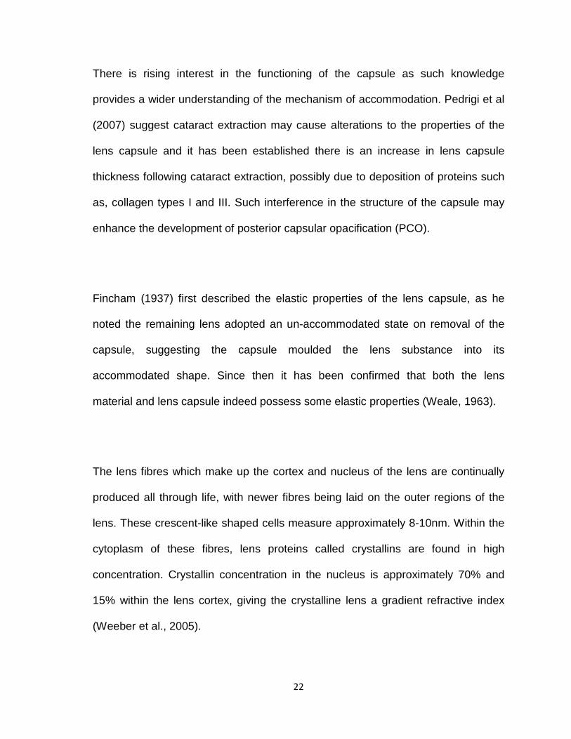
There is rising interest in the functioning of the capsule as such knowledge
provides a wider understanding of the mechanism of accommodation. Pedrigi et al
(2007) suggest cataract extraction may cause alterations to the properties of the
lens capsule and it has been established there is an increase in lens capsule
thickness following cataract extraction, possibly due to deposition of proteins such
as, collagen types I and III. Such interference in the structure of the capsule may
enhance the development of posterior capsular opacification (PCO).
Fincham (1937) first described the elastic properties of the lens capsule, as he
noted the remaining lens adopted an un-accommodated state on removal of the
capsule, suggesting the capsule moulded the lens substance into its
accommodated shape. Since then it has been confirmed that both the lens
material and lens capsule indeed possess some elastic properties (Weale, 1963).
The lens fibres which make up the cortex and nucleus of the lens are continually
produced all through life, with newer fibres being laid on the outer regions of the
lens. These crescent-like shaped cells measure approximately 8-10nm. Within the
cytoplasm of these fibres, lens proteins called crystallins are found in high
concentration. Crystallin concentration in the nucleus is approximately 70% and
15% within the lens cortex, giving the crystalline lens a gradient refractive index
(Weeber et al., 2005).

23
The epithelium of the lens, located immediately adjacent to the anterior lens
capsule, is composed of a single layer of cuboidal cells providing metabolic
transport for the entire structure and regulating its osmolalrity, in addition to
providing new lens fibres through division and differentiation. These cells span
15µm in width and 6µm in height and become much longer towards the lens
equator (Pipe and Rapley, 1987).
Within the pre-equatorial region of the lens, known as the germinal zone, cell
mitosis regularly occurs, the new cells then move into a transitional zone where
they differentiate into lens fibre cells. The processes of the new cells pass through
the anterior and posterior epithelium, forming layers on top of older cells by
pushing older fibres towards the nucleus of the lens. These processes are the lens
fibres. The fibres eventually meet with other fibres within their layer and form a
suture. During embryological stages they link as three branches forming a Y-
shaped suture which further develop into a radial pattern as seen in an adult lens.
The crystalline lens receives nutrients from the aqueous humour and vitreous via
diffusion through the lens capsule with waste products being removed in a similar
way, generally there is a low metabolic requirement.

24
Measurements of capsular thicknesses between ages 1 to 94 years show
increases in thickness up to 70 years (Krag et al., 2003) although this differs
between the anterior and posterior regions. The posterior capsule shows no
changes with age and may even become thinner with age (Barraquer et al., 2006),
whereas the anterior capsule is considerably thicker (Bron et al., 1997) and is the
thickest of basement membranes in the entire human body (Strenk et al., 2005).
Understanding the mechanism of functions of the lens and its components and the
extent to which they contribute to the phenomenon of accommodation will allow
further advances in creating more efficient IOLs with the capability to provide
effective accommodation or reviving accommodative ability.

25
1.2. Accommodation
Accommodation is the ability to alter the dioptric power of the eye by changes in
anatomical structures in order to produce a retinal image of objects at various
distances. Many theories have been postulated to explain this phenomenon but its
exact mechanism has not yet been determined. The first comprehensive and most
widely accepted theory of accommodative mechanism is that of Helmholtz (1855,
cited in Strenk et al., 2005), whereby accommodation results from ciliary muscle
contraction, causing relaxation of resting zonular tension surrounding the lens
equator. The outward tension on the lens capsule is hence released leading to an
increase in anterior and posterior lens surface curvature with a decrease in lens
diameter, resulting in an increase of the dioptric power of the crystalline lens.
Cessation of accommodation is facilitated by relaxation of the ciliary muscle,
zonular tension is restored on the lens equator, pulling the capsule into a flatter
form, decreasing the curvature of lens surfaces and increasing the lens diameter
(Helmholtz 1855 cited in Strenk et al., 2005).
The Helmholtzian theory suggests the crystalline lens is an elastic entity taking on
a natural accommodative state with removal of tension forces. The theory was
based upon observations of forward movements of the anterior lens surface and
increases in curvature with accommodative effort, it was also believed that the
posterior surface curvature increased but no movement was observed. Axial
thickness was recorded to have increased by 0.5mm, however, as the lens volume

26
did not alter it was concluded that equatorial diameter may decrease on
accommodation.
In order to refine the Helmholtzian theory, experimental work by Fincham (1937)
recognized the backward movement of the posterior lens surface, in addition to
ciliary body movement and decreases in lens diameter. It was soon concluded that
the lens capsule is under tension whilst unaccommodated which is released on
accommodative effort, providing evidence for Helmholtz previous description.
Despite evidence from Fincham, Helmholtz theory did not gain entire acceptance
and has faced many opposing theories. One opposition, prior to Fincham’s
findings, includes that of Tscherning (1895, cited in Strenk et al., 2005) who
believed the zonular fibres do not relax and contraction forces of the ciliary muscle
further increased zonular tension moulding the lens into a conoidal shape.
However, Fincham (1937) later provided evidence that the zonular tension in fact
decreases on accommodation, hence proving the theory of Tscherning incorrect. A
second theory proposed by Tscherning (1909, cited in Strenk et al., 2005) explains
ciliary contraction to exert tension on the choroid which compresses the vitreous
against the periphery of the posterior lens surface whilst the anterior lens surface
remains stationary with tension from zonular fibres.

27
A more recent theory by Schachar et al (1992), in favour of Tscherning, proposes
stretching of the lens causes the central lens surface to become steeper and
central thickness to increase while peripheral areas of the lens surface become
flatter inducing an increase in power. Schachar and Anderson (1995) provide
details that movement of the anterior ciliary muscle towards the sclera, on
contraction of the ciliary muscle, produces increased zonular tension at the lens
equator while tension is released from anterior and posterior zonular fibres. It is
believed that the outward force produced shifts the lens equator towards the sclera
which with relaxation of zonules would reduce the curvature of peripheral lens
surfaces coupled with increases in central curvature. Schachar’s theory however
has not gained wide acceptance. There has been controversy over Schachar’s
proposals as various studies have failed to support his theory of accommodation,
also studies of scleral expansion surgery have not reported any valuable
restoration of accommodation (Glasser and Kaufman, 1999; Mathews, 1999).
Obtaining in-vivo evidence for accommodation theories is challenging as imaging
of the ciliary body and fibres are obstructed by the iris as well as image distortion
with corneal power (Strenk et al., 2005). Published observations by Fincham
(1937, cited in Strenk et al., 2005), however, on a case of aniridia noted a
decrease in diameters of the lens equator and ciliary body with increases in lens
thickness. Fincham’s finding has been further supported by more recent work of
Wilson (1997), where retro-illumination infrared video imagery has also captured a
decrease in lens equator diameter.

28
The general understanding of accommodation at present combines the findings of
Helmholtz and Fincham. On accommodation, contraction of the ciliary muscle
causes movement towards the lens equator releasing zonular tension. The lens
capsule then shapes inner softer material into its accommodated form. Such an
action increases lens surface curvatures and axial thickness with a corresponding
decrease in diameter. Ceasing accommodation involves relaxation of the ciliary
body which is pulled backwards. Zonular fibre tension is then restored pulling the
lens back into a flatter form.

29
1.3. Presbyopia
The progressive loss of accommodation with age, termed presbyopia, is a process
believed to occur as a result of age-related anatomical changes of the eye.
Although, not yet entirely understood, it is assumed to be resultant of a variety of
mechanical changes occurring within the accommodation system. Finding a
solution to presbyopia is now becoming of growing interest in ocular research as
visual demands of an aging population increase; for this the processes leading to
presbyopia must be understood.
As with accommodation various explanations for the development of presbyopia
exist. In periods of early research Helmholtz (1855, cited in Strenk et al., 2005) had
suggested it to be due to lens sclerosis whilst Donders (1864, cited in Strenk et al.,
2005) approached the explanation by describing the lack of shortening of the
ciliary muscle with age. Further proposals by Tscherning-Pluugk (1909, cited in
Strenk et al., 2005) mention a reduction in viscosity of the vitreous humour (Strenk
et al., 2005). Proposed theories for presbyopia may be categorized as lenticular
(Duane-Fincham theory) or extralenticular theories (Hess-Gullstrand theory).

30
It is generally assumed that presbyopia is a result of mechanical changes of the
crystalline lens. For some time it has been assumed that increased stiffness of the
crystalline lens is a cause of presbyopia development (Gilmartin, 1995) and has
been supported by many investigators suggesting increased stiffness reduces the
ability of the lens to change shape (Glasser et al., 1998; Pierscionek, 1995;
Atchison, 1995).
A more recent theory has been introduced proposing that lens growth leads to
accommodative loss. With age the ciliary muscle is displaced anteriorly and
inwards (Strenk et al., 1999; 2005). The pupil margin is placed against the anterior
surface of the lens which produces an upward force pushing against the iris and
ciliary muscle. A second tangential force acts on the ciliary muscle produced by
the sclera leading to an anterior and inward shift in the position of the ciliary
muscle and iris root. Such displacement may cause a decrease in pupil diameter
which may explain the development of senile miosis; this allows the pupil margin to
move towards the anterior surface the lens where it is thickest. As lens growth
continues with age, the ciliary muscle moves further anteriorly and upwards
decreasing the circumlental space available and reducing the zonular tension in
the process, thus creating greater curvature on disaccommodation with less
effective ability to respond on accommodation. These events have been described
as the Modified Geometric Theory which supports the variety of changes that have
previously been indentified in lenticular aging (Strenk et al., 2005).

31
Presbyopia is therefore most likely attributed to a combination of lenticular and
extralenticular effects, making the condition ‘multifactorial’ (Weale, 1989; Burd et
al., 2002). However, the changes in mechanical properties of the lens structure do
not present until after presbyopia has manifested. It may therefore be proposed
that the continuous increase in lenticular mass generates these changes and is
thus the sole factor for developing presbyopia. Presbyopia, although known to
generally occur in the fourth decade of life, differs in rate of progression amongst
individuals and may present earlier or later than when commonly expected.
Various aspects of lifestyle may influence this rate of progression such as; diet,
climate, latitude, environmental temperature and race. Understanding the aetiology
of presbyopia will aid advancements in provisions which aim to correct presbyopia.
In addition, knowledge of factors which increase or inhibit its progression will assist
in the global research aim of alleviating this inevitable effect of age.

32
1.4. Cataracts
A cataract, from the Latin cataracta, is defined as opacification of the crystalline
lens. It leads to the loss of transparency of the lens, causing vision to become hazy
and if left untreated can eventually lead to blindness. Patients present with a
reduction in visual acuity and occasional complaints of glare and ‘clouded’ vision.
Classification of cataracts may be anatomical or aetiological. Aetiological
classification is divided into many different categories as listed in Table 1.1.
Aetiological Classification of Cataracts
Age-Related Traumatic: injury or surgery
Congenital: hereditary or complications on birth
Systemic disease: diabetes mellitus
Secondary to ocular pathology: uveitis, glaucoma, retinitis pigmentosa
Drug-Induced: chloroquinine, steroids, amiodarone
Table 1.1: Categories of aetiological classification of cataracts

33
Age-related (senile) cataract remains the most common form encountered,
however, the formation of a cataract can be multifactorial (Hammond, 2001) and
cannot be attributed to a single aetiology. The anatomical classification seems the
more suitable choice for clinicians, which consists of three types or categories;
cortical, nuclear and subcapsular cataract, each of which impose a varied affect on
visual function.
Cortical cataracts (Figure 1.3.) are opacities located in the lens cortex usually
appearing as spokes radiating from the lens periphery. Such opacities rarely cause
visual symptoms until they have extended further centrally interfering with the
visual axis.

34
Figure 1.3: Cortical Cataract

35
Nuclear cataract (Figure 1.4) typically begins with brunescence, a brown
discolouration, of the lens nucleus which increases central refractive index leading
to its associated myopic shift.
Figure 1.4: Nuclear Cataract

36
Subcapsular cataract may occur on the anterior or posterior regions of the
crystalline lens. Anterior subcapsular cataract occurs with fibrous metaplasia of the
anterior epithelium whilst posterior subcapsular cataract (Figure 1.5) is due to
migration of lens epithelial cells. Individuals suffer particularly debilitating glare
from bright lights with the latter and often require removal far earlier than other
forms of cataracts.
Figure 1.5: Posterior Subcapsular Cataract

37
If cataracts remain untreated they may progress into a mature cataract in which
the crystalline lens becomes completely opaque. Over time leakage of fluid and
shrinkage of the cataract leads to a hypermature cataract. Liquefaction of the
cataract cortex into a milky fluid may result in morgagnian cataract; the lens
nucleus in this case may sink inferiorly causing potential capsule ruptures.
Leakage of fliud through ruptures may cause severe inflammation within the eye
and may lead to phacomorphic glaucoma.
Cataract increases light scatter within the eye degrading contrast sensitivity (Elliott,
1993; Miyajima et al., 1992) and as a result degrades the retinal image that is
formed. There is currently no medicinal cure for the occurrence of cataract; the
only successful remedy is surgical extraction and replacement of the natural
crystalline lens with an intraocular lens implant (IOL).
Cataract extraction is indicated where there is significant deterioration of vision. It
is generally agreed upon that referral for cataract surgery is warranted when visual
quality is significantly affected. Referral for extraction, however, should not be
based solely upon visual acuity measurements, degree of glare and ability to carry
out daily tasks must also be taken into consideration. Individuals with reasonable
acuity on high contrast test charts may demonstrate a reduction in visual
functioning on contrast sensitivity or brightness acuity tests. No NICE (National
Institute for Health and Clinical Excellence) guidelines currently exist on cataracts

38
warranting referral for surgery hence symptoms must be appropriately investigated
to determine how a patient’s lifestyle is affected by the reduction in vision.
1.5. Cataract Surgery
Cataract surgery dates back to early civilizations with the Egyptians, Chinese and
Indus Valley civilizations all describing primitive methods of cataract extraction or
displacement from the visual axis. A procedure known as couching was the
earliest form of cataract treatment being dated as early as 600 BCE, this involved
inserting a sharp needle into the eye and displacing the opaque material into the
vitreous cavity, resulting in aphakia and blurred vision (Fan, 2005). Couching
continued up to the 19th century and is still performed in some developing
countries, however, severe post-operative complications are commonly associated
with this procedure such as endophthalmitis and retinal detachment (Bamashmus,
2010).
Following the traditional couching method, intracapsular cataract extraction (ICCE)
and extracapsular cataract extraction (ECCE) developed which coexisted in the
early 1900s. ICCE, in which a large incision of approximately 14-16mm in the
cornea facilitated the removal of the entire lens and capsule, this procedure
however was associated with high rates of complications and is now rarely
practiced. ICCE procedures require implantation of an anterior chamber IOL as it
lacks the capsular bag to support a posterior chamber IOL, although the majority of

39
cases remained aphakic, due to other factors such as anterior chamber depth..
Extracapsular cataract extraction (ECCE) involved removal of the cataractous
material manually through a large incision of 10-12mm leaving the capsular bag
within the eye and required stitches. These early procedures have now been
superseded by the more preferred phacoemulsification procedure.
Phacoemulsification works on a similar principle to ECCE where cataractous
content is removed leaving the lens capsule behind, however removal is performed
using an ultrasonic instrument to break up the cloudy material, allowing smaller
incision sizes of approximately 3.2mm and fewer surgical complications. Due to
the high costs of phacoemulsification ECCE is still commonly performed in
developing countries and may occasionally be required in developed countries if
phacoemulsification presents difficulty or to facilitate the removal of highly dense
cataracts. As part of cataract surgery, intraocular lenses are usually implanted into
the patient’s eye to correct for the refractive error that would present with aphakia,
relieving patients of significantly poor vision following surgery.
Prior to intraocular lenses, extraction of cataracts left patients aphakic requiring
very high positive powered spectacles. Intraocular lenses to replace the optical
power of the crystalline lens were not introduced until after the World War II. Sir
Harold Ridley working in St. Thomas’s Hospital in London examining aircraft pilots,
with penetrating injuries from their shattered perspex canopes, noticed the relative
biomimetic properties of the synthetic material (Apple and Sims, 1996). His early
attempts at intraocular lens design and implantation required large corneal

40
incisions and many failed due to optical and physiological complications, but
formed the basis for the development of modern IOLs. Since then and with recent
developments in intraocular lens implants there has been a growing interest
amongst researchers in methods of restoring optimal vision following cataract
surgery.
Before implantation, determination of IOL power is required, which is subject to
various ocular measurements including; corneal curvature, axial length, anterior
chamber depth and post-operative positioning of the IOL. Originally A-scan
ultrasound and keratometry were performed; more recently pre-operative
measurements are established using the Zeiss IOL Master, which utilises the
sophisticated technique of partial coherence interferometry (PCI) to accurately
measure axial length, anterior chamber depth and automated keratometry.
Occasionally A-scan ultrasound is performed with more dense opacities due to
measurement difficulties with PCI.
In recent years, with increased cataract extractions underway due to an ageing
population, higher demands of spectacle independence from the older population
have resulted and optimizing vision after cataract surgery is now paramount.
Advances such as smaller wound incisions, continuous curvilinear capsulorhexis
(CCC), improved biometric techniques and use of topical anesthesia have led to
the highly successful post-operative outcomes of cataract surgery. As surgical
techniques have advanced profoundly interests now turn to advancing intraocular
lens designs to provide optimum vision following cataract extraction.

41
1.6. Intraocular Lenses
Intraocular lenses consist of an optic where the refractive power of the implant is
concentrated, projections from this termed haptics which provide stability when
implanted in the eye (Figure 1.6).
Figure 1.6: Diagram of IOL

42
The majority of IOL implants in routine cataract surgery are monofocal and
spherical, providing clear vision at only one focal length. Emmetropia is usually the
aim for distance vision with monofocal IOLs, leaving the patient with no true
accommodation, although some depth of focus is present due to the pupil
aperture, optical aberrations and the patient’s tolerance to blur (Wolffsohn et al.
2010). The desire to optimize uncorrected distance vision post-surgery has
resulted in the development of aspheric, toric and light-adjustable lenses (LALs).
To extend the range of eye focus, multifocal designs have been developed,
together with attempts to restore more natural eye focus with ‘accommodating’ IOL
designs. Complementing these optical advances, the transmission properties of
IOLs have been altered to try to protect the retina, inserters and
phacoemulsification techniques have allowed smaller corneal incisions and the
edges have been moulded to reduce posterior capsular opacification (PCO). In the
following sections these various designs of intraocular lenses will be described.

43
1.6.1. Aspheric IOLs
The positive aberration of the cornea in youth is mainly cancelled out by the
negative spherical aberration of the crystalline lens. In the aging eye there is
increasing positive aberration which contributes to a decrease in visual quality due
to imbalance of the aberration between the two structures (He et al., 2003).
Removal of the natural lens and introduction of a spherical IOL leaves positive
aberration of the cornea creating high-order aberrations (Barbero et al., 2003).
The development of aspheric IOLs has aimed to balance the spherical aberration
of the cornea by introducing negative spherical aberration (Holladay et al., 2002). It
was estimated that aspheric lenses would decrease high-order aberrations to a
level below that of the cornea in 45-86% of implantations (Wang and Koch., 2005)
thereby improving contrast sensitivity and visual acuity of the eye (Fahle, 2009).
Aspheric lenses may be designed to be aberration-free or simply reduce levels of
existing aberration; termed aberration-correcting IOLs (Buckhurst et al., 2010). The
performance of aspheric IOLs in comparison to spherical IOLs have shown to be
superior or at least equal for distance visual acuity (Mester et al., 2003; Bellucci et
al., 2005) and mesopic contrast sensitivity (Mester et al., 2003; Packer et al.,
2002), although the depth of clear focus has been found to be reduced in some
(Marcos et al., 2005; Rocha et al., 2007; Nanavaty et al., 2009), but not all studies
(Shentu et al., 2008). However, it should be noted that the optical benefits of these
IOLs are heavily reliant on their centration within the capsular bag and pupil size

44
(Montés-Micó et al., 2009), for example decentration exceeding 0.5mm would
provide no beneficial asphericity (Atchison, 1991).
1.7. Presbyopia & IOLs
Although the power of implanted conventional monofocal IOLs is usually calculated
from ocular biometry to correct distance refractive error, the range of clear focus
varies greatly between individuals. This range of clear focus is due to residual
myopia, myopic astigmatism, monovision, corneal multifocality through aberrations
and pupil miosis (Nakazawa and Ohtsuki, 1983; 1984; Nanavaty et al., 2006). A
combination of these factors in eyes implanted with monofocal IOLs can produce a
pseudo-accommodative range of 0.7 - 5.1D (Menapace et al., 2007).
Monovision, where one implant is optimized for distance vision and the other
focused at a closer distance, is often targeted by surgeons to optimize the range of
clear vision. The technique relies on the suppression of the blurred image from one
eye by the brain, but is not tolerated by an estimated 10-20% of patients
(Greenbaum, 2002; Handa et al., 2004). Monovision also results in stereopsis and
contrast sensitivity loss, with the reduction growing with the power difference
between the two eyes (Durrie; 2006). However, a recent study of IOL monovision
with on average 1.2D anisometropia between the eyes has suggested that contrast

45
and stereopsis can be maintained, although only one quarter of patients were
spectacle independent (Finkelman et al., 2009).
To meet demands of clearer near vision and spectacle independence premium
intraocular lenses have been devised to optimise vision for both distance and near.
Toric IOLs aim to correct higher degrees of astigmatism for distance vision, while
multifocal and accommodating designs aim to provide clarity for near tasks in
addition to distance vision. The remainder of the current chapter will discuss the
current premium IOL options available for implantation during cataract surgery and
refractive lens exchange.
1.8. Premium IOLs
1.8.1. Multifocal IOLs
Simultaneous vision is used in IOLs to provide multifocal clear distances. Until
recently, the optical designs have been concentric refractive or diffractive designs,
or a combination of both. Publications on their performance have been limited by
the use of non-linear Snellen acuity measurements at distance and reporting the
number of people who can read Jaegar text of a certain size at near (e.g. Steinert
et al., 1999; Pineda-Fernandez et al., 2004; Javitt and Steinert, 2000) when it is
well established that Jaegar print sizes differ between charts (Mehr and Fried,
1976).

46
Enlargement of pupil size such as in dim conditions, exposes more annular zones
of the IOL, changing the distribution of light energy between the distance and near
focus of refractive IOLs, depending on the optical design. Hence refractive IOLs
are dependent on pupil size and this is important to consider prior to implantation.
The brain selects the in focused image and suppresses other. Such interference
can lead to development of various photopic phenomena.
Although multifocal IOLs split the light entering the pupil between distance and
nearer distances, most studies show comparable distance vision between
multifocal and monofocal IOLs (Steinert et al., 1999; Vaquero-Ruano et al., 1998;
Orme et al., 2002) as well as improvements in near acuity and depth of focus in
multifocal IOLs (Javitt and Steinert, 2000).
Refractive multifocal designs (Figure 1.8) comprise of concentric areas of differing
refractive power usually on the anterior optic surface, created by differences in
curvature for distance and near power correction. Refraction is described as the
change in direction of light rays travelling from one medium to another of differing
density due to a change in speed. Using this principle a refractive IOL is able to
change the way in which light focuses on the retina. The changes in power within
regions of a refractive IOL enable foci from a range of distances to fall on the retina
simultaneously. Distance correction tends to be central with peripheral regions
designated to near correction. Such lenses are distanced-biased for small pupils,

47
allowing nearly all light energy to be used for distance viewing as peripheral near
focus zones are obstructed by the pupil. Enlargement of pupil size such as in dim
conditions, exposes more annular zones of the IOL, where some light energy is
transferred to near focus regions and less to that of distance. When viewing in the
distance regions providing distance focus form an image on the retina whilst other
regions form a blurry image. These are superimposed; the brain selects the in
focused image and suppresses other. Such interference can lead to development
of various photopic phenomena. Most refractive IOLs are dependent on pupil size
and thus this is important to consider prior to implantation.
Diffractive IOLs (Figure 1.7) use the Huygens-Fresnel principle of concentric
eschelets on the surface of the IOL to create a diffraction pattern by acting as a
grating. Unlike refractive optics, all rings work together to produce constructive and
destructive interference for distance and near foci, and hence the optics are largely
pupil independent. Not all the available incident light can be used by a diffractive
IOL, approximately 40% of light is used for both distance and near viewing, hence
contrast is lost as well as halos created by the concentric prism elements.
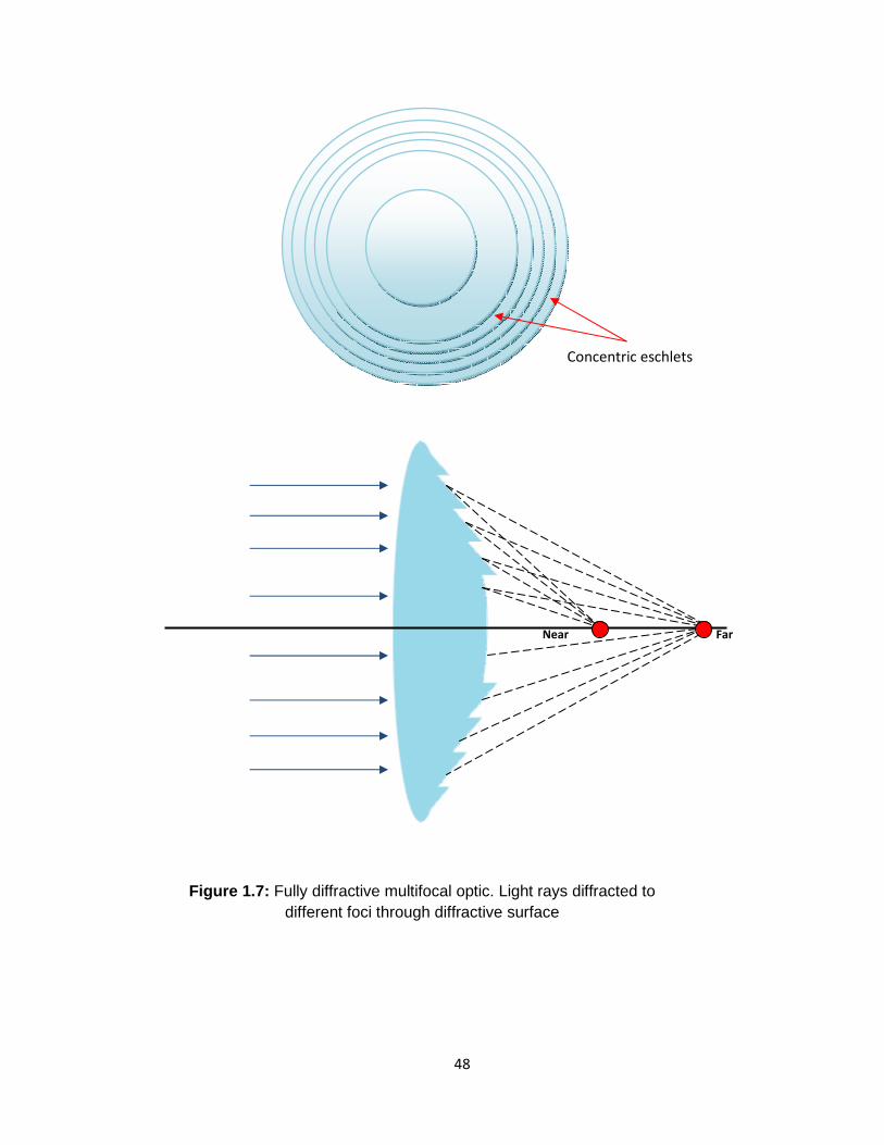
48
Figure 1.7: Fully diffractive multifocal optic. Light rays diffracted to different foci through diffractive surface
Near Far
Concentric eschlets

49
Figure 1.8: Refractive multifocal optic. Juxtaposition of zones of differing power produces different foci.
DISTANCE
NEAR
Near Far
Concentric rings of
differing refractive
power

50
The third multifocal design known as apodized diffraction or ‘partially diffractive’ is
a combination of refractive and diffractive techniques to obtain multifocality
(Kohnen and Derhartunian, 2007). To achieve this, the step height at each
diffraction step is gradually decreased resulting in reduced reflection at edges.
The aim of such a design is to improve image quality, to reduce contrast sensitivity
loss and to be less pupil dependant. Patients report high levels of spectacle
independence following implantation of multifocal IOLs (e.g. Alfonso et al., 2008)
which indicates the benefit of their use. Distance visual acuity is often better with a
distance dominant IOL, while near visual acuity shows more improvement with a
near dominant lens (Steinert, 2000; Alfonso et al., 2008; Kershner, 2003; Jacobi et
al., 2002; Choi et al., 2008; Simpson, 1992; Berdeaux et al., 2008). Bilateral
multifocal IOL implantation generally gives better visual function (Steinert, 2000;
Pineda-Fernandez et al., 2004), however unilateral implantation of a multifocal IOL
in those with unilateral cataracts may still prove beneficial due to possibilities of
aniseikonia and anisometropia with spectacle correction (Jacobi et al., 1999).
Mixing and matching different multifocal IOLs in the two eyes of an individual
patient is now becoming a common technique to overcome the limitations of a
single design (Gunenc and Celik, 2008; Goes, 2008; Hutz et al., 2010).

51
The most recent development in premium lenses aiming to overcome presbyopia
following cataract surgery is the trifocal IOL. The aim of this design is to provide
better intermediate vision in addition to good distance and near vision by
incorporating three foci into the optic of the IOL. Being of relatively new concept
very few studies have reported the visual performance of these lenses. Although
distance and near vision is maintained with the added benefit of intermediate
vision there is skepticism of contrast sensitivity and dysphotopsia as this IOL is in
effect of diffractive design. To date, only two studies by Voskresenkaya et al
(2010) and Gatinel et al (2011) have investigated trifocal performance, the latter
only being of optical bench tests. Both studies show promising results however
further investigations are required within patients to fully explore the benefits of
such a design.
Despite improved near visual acuity and comparable distance visual acuity to
spherical IOLs, implantation of multifocal IOLs are associated with photopic
phenomena such as haloes, disability glare and reduced contrast sensitivity
particularly in mesopic conditions (Steinert, 2000; Richter-Mueksch et al., 2002;
Awwad et al., 2008). Glare and halo phenomena tends to occur more in refractive
multifocal IOL designs than diffractive, though contrast sensitivity shows greater
impairment with diffractive designs (Pieh et al., 1998; Hayashi et al., 2009).
However, such visual phenomenon is shown to reduce over time as adaptation
occurs (Vaquero-Ruano et al., 1998). The contrast sensitivity can be improved and

52
the range of clear focus extended into the intermediate range by adding asphericity
to the multifocal IOL (Alfonso et al., 2008).
It is important to establish the visual demands of the patient in order to provide the
most suitable near additional power. Lower adds provide better intermediate, but
poorer near vision than higher additions, along with less unwanted visual effects
such as reduced contrast sensitivity loss (Hayashi et al., 2009). The IOL power is
traditionally stated for the spectacle plane so this must be converted to the optical
plane to determine the optimum working distance with the IOL implanted. Residual
astigmatism after IOL implantation reduced the effectiveness of multifocal IOLs;
therefore corneal astigmatism must be quantified prior to surgery and reduced by
corneal relaxing incisions or a toric multifocal IOL if greater than about 1 dioptre
(Hayashi et al., 2010).

53
1.8.2. Accommodating IOLs
Current ‘accommodating’ IOLs rely on the Helmholtz theory of accommodation,
where inwards and forward contraction of the ciliary muscle loosens the zonules
coupling the muscle with the crystalline lens. The elastic lens capsule can then
take up a more convex shape, increasing its optical power. Studies using
ultrasound biomicroscopy (Bacskulin et al., 1996; Stachs et al., 2002) and
magnetic resonance imaging (MRI) (Strenk et al., 1999; 2006) demonstrating that
the human ciliary muscle maintains its contractility throughout life, allows a
mechanism to control an IOL through natural eye focusing structural changes.
Accommodating IOLs were initially conceptualised in the 1980s by J. S. Cumming
who observed remarkable near vision acuity in patients implanted with plate haptic
silicone IOLs. Though pseudoaccommodation, which is described as the
subjective range of clear focus enhanced by ocular aberrations, pupil size and an
individual’s tolerance to blur, could not be solely responsible, measurements with
A-scan ultrasonography showed movement of the IOL (Doane, 2004).

54
Accommodating IOLs are categorised in accordance to their mechanism of action
(Sheppard et al., 2010). Assessment of the performance of accommodating IOLs
must separate true accommodation from pseudoaccommodation. Many studies
have attempted to image the lens mechanism using pharmacological (2%
pilocarpine) rather than physiological methods to stimulate the ciliary muscle, but
the resulting change shows the maximum potential of the implant rather than the
natural or achievable objective accommodation (Koeppl et al., 2005). The following
will discuss the various types of accommodating IOL designs.

55
1.8.2.1. Single-Optic IOLs
Single optic IOLs (Figure 1.9) were designed to move forward with contraction of
the ciliary muscle, either due to the forward movement of the capsule and
contraction of the lens equator pushing against a hinge mechanism, or due to the
increased vitreous pressure from the ciliary muscle bulk displacement into the
vitreous chamber (Glasser, 2008). The space in the anterior chamber limits the
potential objective accommodation of single optic-shift IOLs to approximately 1.5D
(for a maximum 1mm movement; McLeod et al., 2003). Although dynamic
objective accommodation has been shown (Wolffsohn et al., 2006a), the average
objective accommodation is only as much as 0.75D (Wolffsohn et al., 2006b;
Menapace et al., 2007) and decreases with increased time after surgery. One
study investigating the performance of the 1CU accommodating lens
(HumanOptics AG, Erlangen, Germany) reported a decrease in objective
accommodation of -0.19 ± 0.44D with a corresponding decrease in subjective
measurement of -0.25± 0.59D two years following implantation (Wolffsohn et al.,
2006a,b). More recently it has been shown that the mechanism of action mainly to
be due to the flexing of the lens changing the high order aberrations, although not
in a systematic manner between individuals (Wolffsohn et al., 2010). PCO rates
are also high due to the IOLs not forming a restrictive kink in the capsule with lens
fibrosis following cataract surgery (Sheppard et al., 2010).

Figure 1.9: Single-Optic accommodating IOL mechanism

57
1.8.2.2. Dual-Optic IOLs
Dual-optic IOLs consist of a biconvex anterior optic with a power of approximately
32D and a posterior negatively powered haptic which is altered in power to correct
for the patients ocular biometry. The optics are separated by connecting spring
haptics, which are designed to push the optics further apart on contraction of the
ciliary muscle to create an inwards equatorial tension from the crystalline lens
capsule. The optics therefore take the form of a Galilean telescope and can deliver
up to 4.0D of power change within the confines of the anterior chamber (McLeod et
al., 2007). Currently, there is only one published report on the clinical performance
of such an IOL. The Synchrony dual-optic accommodating IOL (Visiogen, Irvine,
CA) (Figure 1.10) was reported to give 3.2 ± 0.9D of accommodation (Ossma et
al., 2007), although the defocus curve presented has no error bars and is
symmetrical around zero dioptres which casts doubt over the methodology used.
However, the low PCO rate reported supports the view that holding the lens
capsule open after cataract surgery may limit epithelial cell migration to the
posterior pole.

58
Figure 1.10: Synchrony dual-optic accommodating IOL

59
1.8.2.3. Curvature Change IOLs
The natural change in optical power with the relaxation of the lens zonules occurs
principally due to the crystalline lens curvature increasing rather than the lens
moving forward in the eye (Davies et al., 2010). Hence, ideally accommodating
IOLs would work by a similar mechanism, resulting in much larger changes in
optical power being possible. No commercially available IOLs using this
mechanism are currently available although some are described in the literature.
The most natural restoration of accommodation would be from lens refilling once
the cataract has been removed. The capsulorhexsis would have to be small,
peripheral and able to be effectively sealed. Although much research has been
conducted into achieving this procedure, sealing the capsular bag and preventing
the proliferation of epithelial cells to the posterior pole of that capsule, where the
traditional treatment with YAG laser would destroy the accommodating lens, have
resulted in a lack of progress to clinical trials (Nishi et al., 2009).
The FluidVision IOL (PowerVision Inc, Belmont, CA) has fluid channels connecting
a hollow haptic to the optic that can retain fluid. On contraction of the ciliary
muscle, fluid from the haptics is pushed into the optic increasing its volume and
equatorial diameter and hence causing an increase in power, with a potential of 8D
change in lens curvature (Pepose, 2009). A recent paper described the NuLens
(NuLens Ltd.) (Figure 1.11) which uses the capsular bag as a diaphragm, whose
forward movement from the contraction of the ciliary muscle bulges a transparent
silicone gel through an aperture causing a curvature change. Despite suggesting a

60
large change in optical power of the IOL is possible from the 0.1-0.2mm movement
of the gel lens sag, the current design reduces the optical power on
accommodation (Alio et al., 2009).
Figure 1.11: NuLens accommodating IOL

61
1.8.3. Toric IOLs
Approximately 20% of patients requiring cataract surgery present with over 1.50
dioptres of corneal astigmatism (Hoffer, 1980; Ferrer-Blasco et al., 2009). The
effect of which has not been determined on daily life. While any residual
astigmatism can be corrected with spectacles, refined biometric techniques enable
the selected IOL power to leave little residual spherical refractive error. With more
patients desiring to be spectacle independent for distance viewing, more efforts to
minimize residual astigmatism during cataract surgery must be made to meet such
demands (Buckhurst et al., 2010).
Skilled surgeons can use corneal (CRIs) or limbal relaxing incisions (LRIs) to
reduce post-operative astigmatism. These involve partial thickness incisions along
the axes of the astigmatism with the depth and arc length relating to the degree of
pre-operative corneal astigmatism. However, wound healing variability limits the
accuracy and magnitude of the effect (Amesbury and Miller, 2009). The toric IOL
(Figure 1.12) was so forth devised in the mid 1990’s to eliminate the need for
incisional surgery and increase spectacle independence for astigmats (Medicute,
2008).

Figure 1.12: Toric IOL

63
Implantation of a toric IOL requires careful determination of corneal cylindrical
power. Manual or automated methods can be used, with newer biometry devices
measuring axial length and corneal curvature (Buckhurst et al., 2009). Ideally
corneal topography should be confirmed by a second device (Budak et al., 1999)
and the operator should be well trained (Cronje et al., 1999). Prior to surgery,
reference marks are placed at the limbus for the alignment of the IOL. The patient
must be upright for application of these exterior markings due to deviation of the
eyes in a supine position (Horn, 2007) which may lead to misalignment of the IOL.
Newer techniques involve digital imaging of the eye to allow alignment of the toric
IOL axis to predetermined iris features or bulbar conjunctival features (Wolffsohn
and Buckhurst, 2010) and in the future the chosen axis will be presented through
the surgical microscope, tracked to the orientation of the orbit. Studies have
presented visual improvements with toric IOLs, though post-operative rotation of
toric IOLs is still a concern (Gills et al., 2002; Sun et al., 2000; Buckhurst et al.,
2010). Deviations from the correct axis will reduce the effective power of the
cylinder, with a rotation of 30º or more providing no cylindrical correction (Shimzu
et al., 1994).

64
Rotation after IOL implantation occurs mainly due to compression of the haptics
caused by contraction of the capsule through fibrosis. Friction between the haptics
and the capsule are important for reducing IOL rotation, as is the haptic design and
careful removal of the viscoelastic from around the IOL after lens implantation
(Buckhurst et al., 2010). Stabilization does occur within a few days to weeks,
probably due to joining of the anterior and posterior capsules holding the IOL in a
fixed position (Shimzu et al., 1994; Buckhurst et al., 2010). Rotation is reported as
less with open loop haptics than plate haptics, although plate haptics show better
long-term stability (Parssinen et al., 1998). Newly introduced closed-loop haptics
may be more stable during capsular compression though this requires further
research (Buckhurst et al., 2010).
Repositioning a rotated lens is possible, although it is more complicated and
difficult to achieve the longer after the original surgery it is attempted due to
fibrosis with the lens capsule. Lens extraction and repositioning is considered
optimum approximately 1 week after surgery as earlier may allow the lens to re-
rotate and later than two weeks following implantation will cause difficulty in trying
to reposition due to fusion of the capsule (Novis, 2000). It has associated risks
such as cystoid macular oedema, capsular tears and endophthalmitis and thus is
favourable to avoid (Sun et al., 2000).

65
1.8.4. Blue-Light Filtering IOLs
The visual spectrum spans from 400 to 700nm. It is widely known and accepted
that exposure to UV radiation and blue light is harmful to retinal structures
(Mainster et al., 1983). In order to protect the eye the cornea absorbs wavelength
below 295nm, the remaining wavelengths 300-400nm are then blocked by the
crystalline lens. Despite this some blue light still reaches the retina and crystalline
lens removal through cataract surgery will leave the retina further exposed if the
IOL transmits harmful wavelengths.
It has long been known that blue light is associated with an increased risk of
development of macular degeneration (Mainster et al., 1983). The blue light
causes the production of reactive oxygen species (ROS), being extremely reactive,
these cause damage mainly to the retinal pigment epithelium (RPE) (Ham et al.,
1980; Boulton et al., 2001). Lipofuscin within the RPE absorbs short wavelengths
due to its component A2E (light absorbing chromophore), that has a peak
absorbance of 335-435nm (Sparrow et al., 2000). Absorption results in production
of ROS leading to RPE apoptosis and eventually cellular death. It is suggested that
the levels of A2E increase with age. Natural cellular defenses against ROS
production include; superoxide dismutase, catalase, phospholipase and pigments
such as xanthophylls (Patel, 2007). Macular pigment can only be obtained from
diet and possesses antioxidant properties; it absorbs blue light providing protection
for the retina. Also, with age, oxidative changes occur in the crystalline lens

66
causing ‘brunescence’ or yellowing of the lens, due to an accumulation of
chromophores (Brockmann et al., 2008). Thus its absorbance widens to 400-
500nm providing further protection against shorter wavelengths of light. The young
lens also contains a short wavelength filtering substance 3-Hydroxykynurenine-
glucoside, however the ageing yellowing lens provide three times more protection
(Benz et al., 2007).
It may therefore be argued that on cataract extraction, replacement with a clear
IOL removes natural protection and increases the risk of macular degeneration
and retinal phototoxicity as transmission of shorter wavelengths of light (UV and
blue) will be increased (Brockmann et al., 2008). Originally IOLs were constructed
from PMMA with no form of UV filtration allowing all UV through to the retina. In
1978 it was identified that UV radiation (100-400µm) was harmful to the retina
(Mainster, 1978). Some IOLs have shown to transmit more than 10% of
wavelengths 350-400nm which is not adequate protection against UVA (Laube et
al., 2004). By the 1980’s chromophores were incorporated into most IOLs to block
UV light. However these IOLs still transmit an undesirable amount of blue light
(Henderson et al., 2010) thus the RPE is still vulnerable to damage. This has
driven the development of blue light filtering IOLs (Figure 1.13). Chromophores are
added to the IOL which block blue light; the IOL takes on a yellow appearance and
hence are also known as ‘yellow’ IOLs.

67
Figure 1.13: Blue-filtering IOLs

68
Despite the proposed benefits of yellow IOLs there has been much debate over
light filtering and its significance to ocular health (Henderson et al., 2010). There
has been speculation over the effects on contrast sensitivity, colour perception,
glare sensitivity, scotopic vision and circadian cycle. In general no clinical effects
have been measured (Hayashi and Hayashi, 2006; Brockmann et al., 2008),
although some investigators have found a reduction in scotopic sensitivity which
could increase the risk of falls in the elderly (Schwiegerling, 2006). More recently,
concern with blocking blue light from reaching the retina has focused on sleep
regulation through the circadian rhythm. A substance called melanopsin within the
retinal ganglion cells is stimulated by blue light which aids the control of melatonin,
via the pineal gland which is associated with sleep regulation. In dark conditions
less blue light is available to stimulate melanopsin therefore the pineal gland
secretes melatonin which causes sleeping. With bright light the secretion is
reduced causing an increase in attention. Thus reduced transmission of blue light
due to yellow IOLs may cause deregulated sleeping patterns (Mainster, 2006). An
estimated 27-38% decrease in melatonin suppression is reported by Mainster
(2006). However, this is in comparison to a UV filtering IOL and not an opacified
cataractous lens where all light transmission is significantly reduced (Henderson et
al., 2010). Hence Edwards and Gibson (2010) in their recent review conclude “The
real value of blue-blocking lenses in preventing AMD or its progression has yet to
be shown and, while there would not appear to be any proven significant
limitations associated with these lenses, the current trend of using evidence based

69
medicine to determine treatment modalities would seem to be missing for these
lenses which attract a price premium”.
1.8.5. Light Adjustable Lens (LALs)
As IOLs are implanted in the eye, patients cannot test the effects as easily as they
can with spectacles and contact lenses. Even after accurate biometry, wound
healing can lead to unexpected residual refractive error. To overcome this and
allow post-implantation adjustment of the IOL power and multifocality, a Light
Adjustable Lens implant (LAL) has been introduced (Calhoun Vision, Inc,
Pasandena, California). The lens implant consists of light-sensitive macromers in
an X-linked silicone matrix that are sensitive to ultraviolet light (365nm). Exposure
of UV light controlled and monitored by a digital computer system, induces
polymerisation of the macromers to create an interpenetrating polymer in the lens,
causing thickening in that area (von Mohrenfels, et al., 2010). The non radiated
macromers diffuse into areas free of UV radiation exposure and this leads to
changes to the shape and or refractive index of the LAL (Figure 1.14). Myopic
changes are achieved by applying UV emission on the periphery of the LAL,
effectively thickening that zone. A hyperopic change is thus achieved by directing
the beam towards the centre of the LAL. This adjustment is current performed two
weeks post-operatively usually followed by two lock-in exposures. Each session
allows a change of up to 2D. To correct presbyopia, monovision can be tested in-
vivo to see how the patient adapts and the lens profile can also be made multifocal

(Hengerer et al., 2009).
adjusted or removed.
Figure 1.1
70
2009). If the patient cannot tolerate their correction, it can be
Figure 1.14: Mechanism of adjusting power of LAL
If the patient cannot tolerate their correction, it can be

71
1.8.6. Phakic IOLs
IOLs that can be implanted in the anterior chamber have been developed to
correct refractive error without affecting natural accommodation in pre-presbyopes
(Figure 1.15). These lenses are generally used in patients whose refractive error is
too high to correct by laser refractive surgery and where the corneal thickness was
not adequate to allow the ablation depth required to correct their ametropia.
Endothelial loss leading to a loss in corneal transparency and cataract still remain
the two main concerns with such implantation, despite the effectivity, predictability
and general safety having improved (Espandar et al., 2009).

72
Figure 1.15: Phakic IOL

73
1.9. Conclusion
While many advances have been made in IOL designs, the benefits of premium
IOLs have not been well established, nor when they should be considered as an
optical correction. Implanting advanced optical designs also raises more complex
surgical issues relating to IOL rotation and centration which deserve attention to
optimise the visual results. This thesis therefore examines the benefits of
astigmatic correction with a toric lens over the mean spherical equivalent and how
pupil dilation influences the alignment of these lenses. Centration of IOLs during
surgery and over the following 6 months will also be examined.
There is a clear requirement for correction of presbyopia. Reports of global
prevalence of functional presbyopia in 2005 estimated 1.04 billion cases, which is
expected to escalate to 1.4 billion in 2020 and 1.6 billion in 2050 (Holden et al.,
2008). While many non-surgical options for presbyopia exist, such as spectacles
and contact lenses, there is some interest in presbyopic IOLs, which are usually
fitted when cataracts have developed, to implant them when presbyopia first
occurs. This would allow spectacle independence in addition to removing the
probable need for cataract surgery later in life when other age related health
problems may make surgery more complex and risky. Hence, better methods to
measure residual accommodation will be explored along with what factors, other
than age, affect when an individual becomes presbyopic and therefore should
consider elective IOL surgery with implantation of a premium IOL. Finally, the

74
benefits of a new type of IOL, a trifocal diffractive design, is evaluated to determine
the range of clear vision offered as well as the visual compromises experienced.

75
CHAPTER 2
Effect of Uncorrected
Astigmatism- Implications for
Toric Intraocular Lenses

2.1. Introduction
Corneal astigmatism is ametropia due to irregular surface curvature of the cornea
where two perpendicular meridians give differ
result is two foci with a blurred area between them
giving rise to blurred vision (
Figure 2.1:
76
Corneal astigmatism is ametropia due to irregular surface curvature of the cornea
ere two perpendicular meridians give different focal powers (Figure 2.1
result is two foci with a blurred area between them known as the interval of Stürm,
blurred vision (Zadnik, 1997).
Figure 2.1: Foci with corneal astigmatism
Corneal astigmatism is ametropia due to irregular surface curvature of the cornea,
Figure 2.1). The
known as the interval of Stürm,

77
It is a common condition, occurring in about 85% of the general population, with
20-30% of the older population (>60 years), when cataracts are most common,
having significant severity (>1 dioptre) (Vitale et al., 2008; Ferrer-Blasco et al.,
2009). During routine cataract surgery, enhanced biometric techniques now allow
the approximation of IOL power to fully correct spherical errors leaving the patient,
in most cases, spectacle free for distance vision. Correction of astigmatism,
however, presents more difficulty; traditionally corneal relaxing incisions (CRIs) or
astigmatic keratotomy and limbal relaxing incisions (LRIs) are used in order to
reduce post-operative astigmatism (Lindstrom 1990; Thornton 1990). CRI’s appear
more ‘effective’ but LRIs tend to be preferred amongst surgeons as are considered
safer (Gills et al., 2002). Reducing higher levels of astigmatism requires numerous
incisions leading to complications such as corneal distortion. LRIs although
extensively used are therefore limited to lower astigmatic errors. Predictability of
incision surgery is also variable and can result in over- or under-correction, skills of
the surgeon and variability amongst healing responses are also factors affecting
surgical outcome (Lindstrom et al., 1994; Thornton, 1994; Abbey et al., 2009). The
toric IOL, first devised by Shimzu et al in 1994 (Mendicute, 2008) aims to eliminate
the need for incisional surgery and increase spectacle independence. Various
studies have reported successful reduction in astigmatism with toric implants and
good satisfaction rates (Ruíz-Mesa et al., 2009; Dardzhikova et al., 2009; Ahmed
et al., 2010). A study by Sun et al (2000) of 130 toric implantations documented
84% achieving uncorrected visual acuities of 20/40 or better. Implantation of toric

78
IOLs may therefore enhance a patient’s perception of clarity of vision regardless if
reflected in visual acuities.
While it is standard practice to correct astigmatism when prescribing glasses and
about one third of prescribed contact lenses correct astigmatism (Morgan & Efron,
2009), many public health services consider intraocular lenses that correct
astigmatism as specialist devices. Therefore, older astigmatic patients desiring
optimum vision following their cataract operation must pay for both the intraocular
lens (IOL) and the cost of private surgery. Suboptimal vision is associated with
reduced quality of life and an increase in falls in the elderly (Black & Wood, 2005;
Lotery et al., 2007), however the impact of uncorrected astigmatism has not
previously been assessed.
Since the advent of intraocular lens correction following cataract surgery in the
1950’s, surgical techniques and intraocular lenses have developed rapidly to keep
pace with the increasing demand created by expanding life expectancy and a
corresponding more active lifestyle in patients experiencing cataracts. Therefore
this study examines the challenges of uncorrected astigmatism in everyday life in
people of the age when cataracts typically form to determine the need for toric
intraocular lenses to be implanted as the routine standard of care.

79
2.2. Methods
Twenty-one presbyopes, aged 50-69 years old (58.9 ± 2.8 years, ten females) with
no ocular pathology, not on medication likely to influence the stability of refractive
error, having less than 0.75D of manifest astigmatism and binocular acuity better
than 0.0 logMAR were recruited. Due to the nature of the glare tests involved it
was important to exclude patients with epilepsy. The study conformed to the
declaration of Helsinki and was approved by the institutional ethics committee.
Subjects gave their informed consent to take part.
Each of the subjects were made familiar with the assessments of visual function.
Visual acuity and contrast sensitivity was assessed binocularly using a Thomson
computerised logMAR progression Test Chart (Thomson Software Solutions,
Hatfield, UK). Subjects were asked to read the smallest visible letters and were
encouraged to guess when uncertain. Each letter was scored as 0.02logMAR and
the acuity measured with 95%, 50% and 10% contrast letters, randomised
between measures. Near acuity and reading speed at 0.2 logMAR larger than this
acuity was assessed at a 40cm working distance with a +2.50D near addition using
standardised reading performance texts of 830 ± 2 characters length (Hahn et al.,
2006). The chart was presented on an LCD computer screen and the words were
changed between each repetition. Light scatter in the right eye was assessed
using the C-Quant straylight meter (Oculus Optikgerate GmbH, Wetzlar,
Germany), recording the average of three repeated measured (Figure 2.2). Driving

80
was simulated using a split attention task displayed on a 14” computer monitor.
Subjects responded to the car in front travelling at a matched 60mph breaking
(and increasing in visual angle) or a pedestrian initially seen at 3.6° eccentricity on
the off or far side beginning to cross the path of the subject when they reached
6.5° eccentricity (Figure 2.3). Reaction times were averaged for 3 repeats of each
condition over a 1.5 minute simulated drive. Finally mobile phone screen and
internet computer screen clarity (Figure 2.4) positioned at the subject’s standard
working distance for that task were each subjectively rated as a percentage, whilst
viewing binocularly.

81
Figure 2.2: Cquant straylight meter

82
Figure 2.3: Driving Simulator
Figure 2.4: Web page used for subjective rating of clarity

83
Having practiced each of the tasks until they felt comfortable and had a consistent
performance, subjects repeated the tasks seven times wearing a trial frame with
spectacle lenses fitted to correct their refractive error together with a cylindrical
addition of different powers and axes in randomised order compensated with a
sphere so the mean spherical equivalent was always zero (Table 2.1). They had
approximately 5 minutes to adapt to each set of lenses before testing, although it
appears the brain does not adapt to binocular astigmatism (Yehezkel et al., 2005).

84
Effect of
uncorrected
astigmatism:
Full sphero-
cylindrical
refractive error
Effect of
uncorrected
astigmatism:
Full sphero-
cylindrical
refractive error
POWER
+0.00 / -0.00 x 90
AXIS
+1.50 / -3.00 x 90
+0.50 / -1.00 x 90 +1.50 / -3.00 x 180
+1.00 / -2.00 x 90 +1.50 / -3.00 x 45
+1.50 / -3.00 x 90
+2.00 / -4.00 x 90
Table 2.1: Trial lens combinations used to simulate uncorrected astigmatism

85
The lens combinations simulated the typical situation of patients with an astigmatic
corneal refractive error being implanted with a spherical intraocular lens of a power
to compensate the average refractive error for distance, i.e. based on the average
corneal curvature. To compensate for the effects of the trial lens surfaces, the
minimal astigmatism comparison was simulated with the same number of trial
lenses as the other conditions. The effects of uncorrected astigmatism power was
assessed with the negative cylinder orientated vertically (90°) as this is the
commonest cylindrical axis up to approximately 60 years of age (Ferrer-Blasco et
al., 2009). A three dioptre cylinder was chosen as the power at which to assess the
effect of the cylinder axis as this encompasses 95% of astigmatic errors in this
population (Ferrer-Blasco et al., 2009).

86
2.3. Statistical analysis
Descriptive statistics of mean and standard deviation were plotted for each
assessment. Kolmogorov-Smirnov tests were performed to check normality of
data. Visual acuity, contrast sensitivity, reading speed, light scatter and reaction
time data were compared using repeated measure analysis of variance with paired
t-tests for post hoc testing. The linearity of changes was assessed with Pearson’s
correlations. Subjective ratings of clarity were compared using Fisher non-
parametric related sample comparisons with Wilcoxon signed rank tests for post
hoc testing.

87
2.4. Results
Distance visual acuity decreased significantly with increasing uncorrected
astigmatic power (F = 174.50, p < 0.001) and was reduced at lower contrasts as
expected (F = 170.77, p < 0.001), with no interaction between these effects (F =
1.47, p = 0.26). Each dioptre of uncorrected astigmatism caused a significantly
lower acuity than the previous power at each contrast level (p < 0.01; Figure 2.5).
Distance visual acuity was significantly affected by uncorrected astigmatic axis (F
= 5.19, p = 0.02) and was reduced at lower contrasts as expected (F = 129.75, p <
0.001), with no interaction between these effects (F = 0.36, p = 0.83). Uncorrected
astigmatism at 90° orientation resulted in a significantly better acuity than with the
axis at 45° or 180° at each contrast level (p < 0.05; Figure 2.5). On average
uncorrected astigmatism caused a reduction of 1.5 lines per dioptre (+0.15 ± 0.03
logMAR/DC, r = -0.76) in visual acuity at high contrast and a similar effect at 50%
and 10% contrast (+0.14 ± 0.03 logMAR/DC, r = -0.91; +0.14 ± 0.05 logMAR/DC, r
= -0.80, respectively)

88
MSE Cylinder - Axis (o)
90 180 45-0.2
0.0
0.2
0.4
0.6
0.8
Visual Acuity
MSE Cylinder - Power (D)
0D -1D -2D -3D -4D
Vis
ual A
cuity
(lo
gMA
R)
-0.2
0.0
0.2
0.4
0.6
0.895% Contrast50% Contrast10% Contrast
Figure 2.5: Distance visual acuity with uncorrected astigmatism power and axis. N=21. Error bars = 1 S.D.

89
Near visual acuity decreased significantly with increasing uncorrected astigmatism
power (F = 221.62, p < 0.001). Each dioptre of uncorrected astigmatism caused a
significantly lower acuity than the previous level (p < 0.001; Figure 2.6). Near
visual acuity was significantly affected by uncorrected astigmatic axis (F = 26.00, p
< 0.001). Uncorrected astigmatism at 90° orientation resulted in a significantly
better acuity than with the axis at 45° or 180° at each contrast level (p < 0.001;
Figure 2.5). Reading speed decreased significantly with increasing uncorrected
astigmatism power (F = 11.97, p < 0.001), but only with -3.0DC or greater
(p<0.001; Figure 2.7). Reading speed was significantly affected by uncorrected
astigmatic axis (F = 4.45, p = 0.03). Uncorrected astigmatism at 180° orientation
resulted in a significantly worse reading speed than with the axis at 45° (p = 0.03;
Figure 2.6).

90
MSE Cylinder - Axis (o)
90 180 450.0
0.2
0.4
0.6
0.8
Near Visual Acuity
MSE Cylinder - Power (D)
0D -1D -2D -3D -4D
Nea
r V
isua
l Acu
ity (
logM
AR
)
0.0
0.2
0.4
0.6
0.8
Figure 2.6: Near visual acuity with uncorrected astigmatism power and axis. N=21. Error bars = 1 S.D.

91
MSE Cylinder - Axis (o)
90 180 450
200
400
600
800
1000
1200
Reading Speed
MSE Cylinder - Power (D)
0D -1D -2D -3D -4D
Rea
ding
Spe
ed (
Cha
ract
ers/
min
ute)
0
200
400
600
800
1000
1200
Figure 2.7: Reading speed with uncorrected astigmatism power and axis. N=21. Error bars = 1 S.D.

92
Although there was no significant increase in light scatter with increasing
uncorrected astigmatism power (F = 1.11, p = 0.559) or changes in axis (F = 0.13,
p = 0.878; Figure 2.8), the reliability and variability of measurements decreased
with increasing uncorrected astigmatic power (F = 2.93, p = 0.026; F = 2.44, p =
0.05).
MSE Cylinder - Axis (o)
90 180 450.0
0.5
1.0
1.5
2.0
Light Scatter
MSE Cylinder - Power (D)
0D -1D -2D -3D -4D
Ligh
t Sca
tter
(Log
)
0.0
0.5
1.0
1.5
2.0
Figure 2.8: Light scatter with uncorrected astigmatism power and axis. N=21. Error bars = 1 S.D.

93
Responding to a car in front braking (F = 0.813, p = 0.521) or a nearside (F =
1.266, p = 0.290) or offside (F = 0.200, p = 0.102) pedestrian was not significantly
affected by uncorrected astigmatic power (Figure 5). Responding to a car in front
breaking (F = 0.111, p = 0.895) or a nearside (F = 1.148, p = 0.327) or offside (F =
1.441, p = 0.249) pedestrian was unaffected by uncorrected astigmatic axis (Figure
2.9).

94
MSE Cylinder - Axis (o)
90 180 450
500
1000
1500
2000
Driving Simulation
MSE Cylinder - Power (D)
0D -1D -2D -3D -4D
Reaction Time (ms)
0
500
1000
1500
2000
Leading CarNearside PedestrianOffside Pedestrian
Figure 2.9: Driving task performance with uncorrected astigmatism power and axis. N=21. Error bars= 1 S.D.

95
Subjective rating of clarity decreased significantly with increasing uncorrected
astigmatic power when viewing a mobile phone (Chi Squared = 81.29, p < 0.001)
or a computer screen (Chi-Squared = 79.91, p < 0.001). Each dioptre of
uncorrected astigmatism caused a significantly lower rating of clarity (p < 0.01;
Figure 6). Subjective clarity was significantly affected by uncorrected astigmatic
axis when viewing a mobile phone (Chi-Squared F = 19.01, p < 0.001) or a
computer screen (Chi-Squared = 21.53, p < 0.001). Uncorrected astigmatism at
90° orientation resulted in a significantly better rating than with the axis at 180°,
and the 180° orientation was rated significantly better than the 45° orientation for
both mobile phone and computer viewing (p < 0.01; Figure 2.10).

96
MSE Cylinder - Axis (o)
90 180 450
20
40
60
80
100
Subjective Rating of Clarity
MSE Cylinder - Power (D)
0D -1D -2D -3D -4D
Sub
ject
ive
Rat
ing
of V
isua
l Cla
rity
(%
)
0
20
40
60
80
100 Mobile PhoneComputer Screen
Figure 2.10: Subjective rating of clarity with uncorrected astigmatism power and axis. N=21. Error bars= 1 S.D.
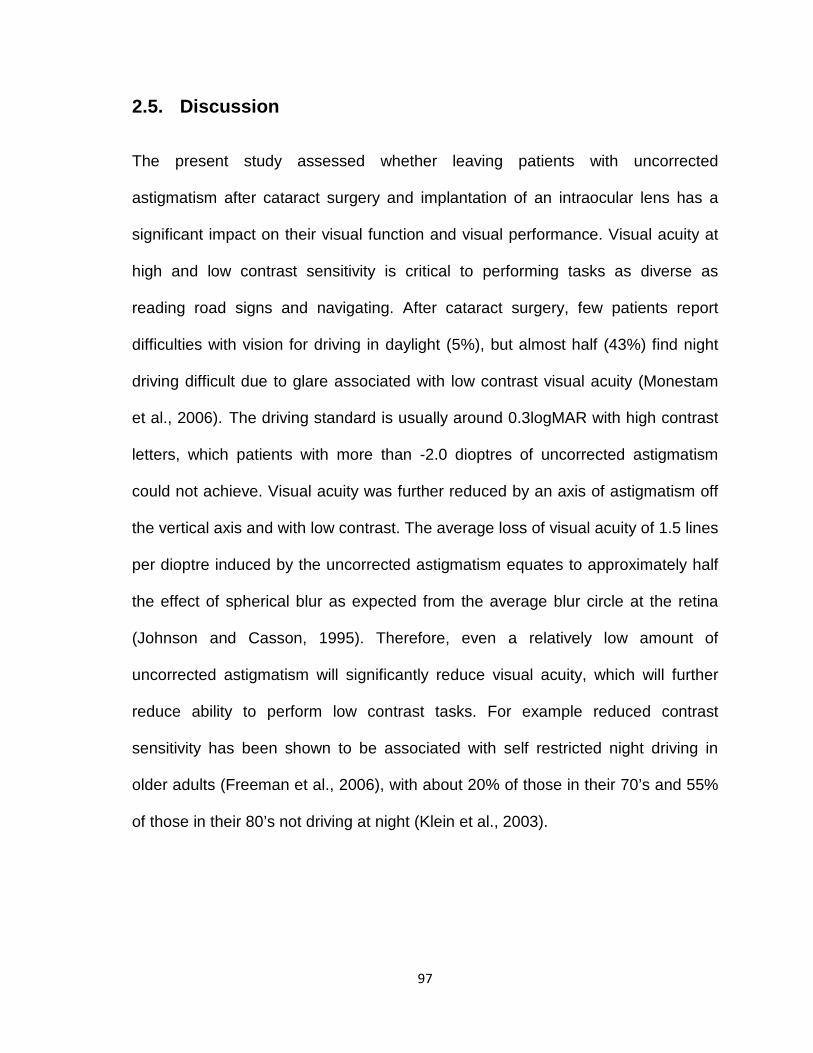
97
2.5. Discussion
The present study assessed whether leaving patients with uncorrected
astigmatism after cataract surgery and implantation of an intraocular lens has a
significant impact on their visual function and visual performance. Visual acuity at
high and low contrast sensitivity is critical to performing tasks as diverse as
reading road signs and navigating. After cataract surgery, few patients report
difficulties with vision for driving in daylight (5%), but almost half (43%) find night
driving difficult due to glare associated with low contrast visual acuity (Monestam
et al., 2006). The driving standard is usually around 0.3logMAR with high contrast
letters, which patients with more than -2.0 dioptres of uncorrected astigmatism
could not achieve. Visual acuity was further reduced by an axis of astigmatism off
the vertical axis and with low contrast. The average loss of visual acuity of 1.5 lines
per dioptre induced by the uncorrected astigmatism equates to approximately half
the effect of spherical blur as expected from the average blur circle at the retina
(Johnson and Casson, 1995). Therefore, even a relatively low amount of
uncorrected astigmatism will significantly reduce visual acuity, which will further
reduce ability to perform low contrast tasks. For example reduced contrast
sensitivity has been shown to be associated with self restricted night driving in
older adults (Freeman et al., 2006), with about 20% of those in their 70’s and 55%
of those in their 80’s not driving at night (Klein et al., 2003).

98
Reading tasks are considered the most critical to quality of life by older individuals
(Wolffsohn and Cochrane, 1999). Although less systematic than distance visual
acuity (r = -0.42), reading acuity decreased a similar amount with uncorrected
astigmatism power (+0.13 ± 0.07 logMAR/DC). Newspaper print (approximately
0.4 logMAR at 40cm) was only just resolvable with 2.00DC of uncorrected cylinder
and made worse by the steepest axis not being in the vertical, as occurs with
increasing age (Ferrer-Blasco et al., 2009). Allowing for an acuity reserve of 6-18
times the threshold letter size to achieve highly fluent reading speed (Whittaker
and Lovie-Kitchin, 1993), even 1.00DC of uncorrected astigmatism will affect
simple everyday reading tasks. Despite reading speed being assessed with words
0.2logMAR larger than the threshold reading acuity with each lens combination,
the speed was significantly reduced with higher levels of uncorrected astigmatism
which will make reading tasks more difficult to perform, less pleasurable, and often
leading to a reduction in independence and quality of life (Wolffsohn and
Cochrane, 2000).
Difficulties with night driving and glare are reported by both elderly drivers with
visual impairment and those with healthy eyes (McGregor and Chaparro, 2005).
Trouble with driving at night is a commonly reported symptom in the elderly,
occurring in 28.2% of drivers over the age of 50 years in the Australian Blue
Mountains population study (Ives, et al., 2000). Although there was no significant
increase in light scatter with increasing uncorrected astigmatism power or changes
in axis, this was principally due to the systematic reduction in quality of the

99
measurements. It would therefore appear that uncorrected astigmatism negatively
affects glare even though this accepted test of light scatter was unable to quantify
the effect. Interestingly, while visual acuity declines with decreasing luminance and
or blur, steering performance does not unless the visual field is restricted (Owens
and Tyrell, 1999; Brooks et al., 2005). Therefore the driving simulator findings were
not unexpected. However, performance with unexpected events and poorer driving
conditions such as rain and on-coming traffic headlights may still be compromised
by uncorrected astigmatism.
Patients’ rating of clarity was significantly reduced by increasing uncorrected
astigmatic power. The patients were allowed to use their standard viewing
conditions, which were kept constant between comparisons. They were also
masked to the level of uncorrected astigmatism, so this was a real effect.
Therefore, as well as the effect on visual function, patients were aware of their
reduced vision, so uncorrected astigmatism is likely to negatively impact quality of
life. Although patients could choose to improve their vision by the use of
spectacles or contact lenses, they are unlikely to do this if left with just astigmatic
refractive error with current improvements in biometry (Buckhurst et al., 2009) or
implantation of intraocular lenses that correct presbyopia (Madrid-Costa et al.,
2010; Sheppard et al., 2010).

100
The main limitation to this study relates to adaptation effects known to occur after
changes in the optics in from of the eye, which may have reduced the impairment
measured with time. It was unrealistic to expect the patients to wear the
corrections examined in the study for about a month before examining each one.
Patients could have been recruited with different levels of astigmatism, but that
would not have allowed comparison of different levels of uncorrected astigmatism
without a huge cohort to account for individual variability. Assessment of glare with
increasing astigmatic error failed to quantify any systematic effect that may have
been present due to reduction in the quality of measurements as the power of
astigmatism increased. Glare testing could be repeated using halometry devices
(see chapter 7), to quantify the extent of glare perception. Driving simulation also
did not show any compromise with astigmatic error perhaps due to peripheral
vision being less sensitive to blur, as this task consisted of reacting to pedestrians
crossing and braking with changes in speed. The high contrast nature of the test
may have also influenced results, repeating with real driving simulations in poor
weather or night conditions may better depict the typical effects of astigmatism
during driving.

101
2.6. Conclusion
In conclusion, uncorrected astigmatism significantly compromises a patient’s
vision. In the long-term this is likely to lead to restricted independence, reduced
quality of life and falls (Black and Wood, 2005; Lotery et al., 2007). With modern
intraocular lenses implanted after cataract surgery, astigmatism can easily be
corrected (Buckhurst et al., 2010) and modern designs show increased
orientational stability. However, there is still no standard procedure for aligning
toric IOLs during surgery and new imaging techniques require further refinement
which will be addressed in chapter 3. The findings of this chapter show the
additional cost of these ‘premium’ lenses is likely to be far less than the
consequences of leaving them with uncorrected astigmatism. It is estimated by the
Royal Society for the Prevention of Accidents (ROSPA) that one in three aged 65
years and over may experience a fall at least once a year and one in two in those
over 80 years (NICE guideline for Assessment & Prevention of Falls, 2004). In
1999, fall related injuries incurred costs of £908.9 million with 63% of which was
accounted by patients over 75 years. Toric intraocular lenses typically cost £500
per eye with private surgery fees reaching £2500 per eye. However if such
implants were made available on the National Health Service with the option for
patients to ‘top up’ fees to receive toric IOLs it would prove beneficial by aiding the
prevention falls as well as improving driving safety. Hence, this study suggests
correction of corneal astigmatism during cataract surgery and intraocular lens
implantation should be the standard of care.

102
CHAPTER 3
The Effect of Dilation on Toric
Intraocular Lens Aligning and
Centring

103
3.1. Introduction
Astigmatism over 1.50 dioptres occurs in approximately 20% of patients presenting
for cataract surgery (Ferrer-Blasco et al., 2009). Although this astigmatism can be
reduced by corneal or limbal relaxing incisions, toric intraocular lens implants
(IOLs) give a more predictable result and can correct higher levels of astigmatism
(Sun et al., 2000; Mendicute et al., 2009). Chapter 2 has already shown that
uncorrected astigmatism can have a significant impact on the performance of daily
tasks even if the mean spherical equivalent refraction is corrected, resulting in
reduced quality of life. Successful toric IOL implantation requires precise and
accurate marking of the orientation at which the intraocular lens is to be implanted,
as misalignment by as little as 1° can result in an estimated 3.3% loss of cylindrical
correction (Lane, 2006). It is reported a deviation of 30° with a toric IOL results in
no correction of cylindrical power (Shimzu et al., 1994) and anything greater will
add to the cylindrical power requiring correction (Novis, 2000).
The axis of astigmatic orientation or horizontal axis is generally marked on the
peripheral cornea or over the sclera prior to surgery to account for the cyclorotation
of the eye when moving to a supine position for the surgical procedure (Smith et
al., 1994; 1995). Ink markings often spread due to the blink action and tear film
movement, and fade with time. Even laceration markings have a finite size and rely
on a surgeon's skill to be correctly located with reference to a biomicroscope slit
illumination orientation or protractor increments. Markings are usually made with

104
patients sitting at a slit-lamp biomicroscope, thus stable head positioning is critical
in order to avoid misalignments due to head tilts (Wolffsohn et al., 2011). There
have been suggestions of using Nd:YAG laser to mark the cornea to overcome
some of these issues (Bordanaba et al., 2009), but head tilt again may prove a
source of error. Imaging systems use conjunctival blood vessels or iris features as
markers for orientation, but the robustness of this approach with variation in
dilation during the imaging and surgery has not been investigated (Wolffsohn et al.,
2010).
During refractive surgery and cataract extraction pharmacological dilation of the
pupil is required to create an adequate opening for removal of the crystalline lens
and insertion of the IOL. It is generally assumed that the pupil is circular, well
centred within the iris and that dilation, caused by smooth muscle in the iris, occurs
symmetrically. However, the pupil is generally located slightly nasal and superior to
the geometric centre of the cornea (Yang et al., 2002). It has been documented
that with pupil dilation there is a corresponding change in pupil centration, which is
of clinical importance in ophthalmic surgery where pupil centration relative to the
limbus can affect surgical outcomes (Walsh 1988; Wilson and Campbell 1992;
Yang et al., 2002). The eye suffers a range of regular and irregular optical
aberrations which can vary between eyes (Walsh et al., 1988). Both pupil dilation
and decentration have been noted to increase high order aberrations within the
visual system (Ivanhoff 1956; Yang et al., 2002), in particular spherical aberration
and coma (Wilson et al., 1992), thus degrading retinal image quality. These

105
aspects must be taken into consideration for various refractive procedures such as
toric implantation, as poor centration of an IOL will give rise to further optical
aberrations resulting in reduced image quality.
Various studies have been carried out in order to investigate the changes in pupil
centration with changes in diameter. Walsh (1988) analysed projected
photographs of light and dark adapted and pharmacologically dilated eyes of 39
subjects and found superior (0.02 ± 0.14mm) and temporal (0.03 ± 0.15mm)
decentration of the pupil with pharmacologically- induced mydriasis. No
systematic pattern was found with decentration in relation to the level of mydriasis,
however, although the decentration measured was of small magnitude this can still
impact image quality hence expressing its significance in surgery. The centration
changes were also found to be similar between eyes for the majority. Wyatt (1995)
found nasal and superior decentration of the pupil with constriction in a study of 23
subjects, through analysis of projected and digitised slit lamp photographs. The
photographs were first captured in steady illumination followed by photographs of
eyes in darkness after approximately 10 to 20 seconds, hence observing natural
dilation. Wilson et al (1992) however found greater shifts in centration in
comparison to the above studies.

106
More recent experimentation conducted by Yang et al (2002) involved monitoring
pupil decentration in mesopic, photopic and pharmacologically dilated conditions
using an infrared camera. High resolution images of 70 subjects were analysed
and found a significant supero-temporal (0.162 ± 0.083mm) shift in the pupil centre
following mydriatics. A similar shift was found when measuring dilation from
photopic to scotopic conditions (0.183 ± 0.093 mm). As with the investigations
carried out by Walsh (1988) and Wyatt (1995), Yang et al (2002) also found nasal
decentration on pupil constriction, with left eyes showing greater displacement
than right eyes. Vertical decentration was shown to be very slight in the superior
direction (0.04mm) with drug-induced dilation. Although the changes in pupil
location have generally been found to be small, greater, unpredictable shifts can
still occur particularly with drug-induced mydriasis (Walsh 1988; Yang et al., 2002;
Wilson et al., 1992) which proves a challenge for surgery.
Age and refractive error may be considered as variables which may affect the pupil
location however Yang et al (2002) reports no such relationship exists. There is,
however, a well known linear decrease in pupil size with age thus young patients
may show greater shifts in pupil centre (Winn et al 1994; Yang et al 2002).

107
Therefore this study examined the effect of pharmaceutical dilation on conjunctival
vessels and iris features and their potential as biological markers for aligning toric
IOLs. In addition, the changes in pupil centration relative to the limbus that occur
with various degrees of dilation were assessed.

108
3.2. Methods
Thirty six subjects were recruited with a mean age of 21.7 ± 2.7 years, 20 males
and 16 females. All participants were healthy individuals with no ocular pathology.
Exclusion criteria included no ocular abnormalities or pathology, no previous
history of ocular surgery and no medication likely to affect rate or process of pupil
dilation. Patients were required to maintain adequate posture as the procedure
involved sitting still at a slit lamp for a duration of time.
After obtaining informed consent, intraocular pressures and anterior angle
measurements were measured prior to pharmaceutical dilation to ensure suitability
of individuals for the investigation. Right eyes of each subject were then
pharmacologically dilated using two drops of Tropicamide 0.5% to ensure maximal
dilation. Ethical approval was granted by the Aston University Ethics Committee
and the study conformed to the tenets of the declaration of Helsinki.
Subjects were seated at a slit lamp where images of right eyes were taken every 2
minutes from immediately following instillation of the drops using an anterior eye
imaging system attached to a CSO Elite slit lamp (Figure 3.1) with high pixel light
sensitivity (Costruzione Strumenti Oftalmici, Scandicci, Italy). Images were
captured for 30 minutes until maximal dilation was achieved (Levine, 1976;
Krumholz et al., 2006). Patients were instructed to keep as stable as possible

109
during photographs to ensure clear images were captured for analysis, as
movement would cause blurring and loss of detail in images.
The images were captured onto a computer linked to the imaging device, the
camera exposure and image brightness were both controlled by Epsilon Lyrae
software. Pairs of images were taken at each 2 minute time interval to allow for
blurred images to be replaced. The pupils were illuminated by an external diffuse
light source and a thin slit beam was positioned temporal to the limbus to minimise
any influence on pupil diameter. At the end of the process intraocular pressure and
angle measurements were repeated.

110
Figure 3.1: CSO Elite Slit Lamp

111
Analysis of the photographic images was then carried out using bespoke
LabView™ 2010 imaging software (Wolffsohn and Buckhurst, 2010). For each
subject, pairs of the most prominent conjunctival vessels were chosen horizontally
separated on either side of the limbus, and using LabView™ (National Instruments
Corporation, Austin, United States) the orientation of these vessels were
determined by drawing a line from one vessel to the other (Figure 3.2).
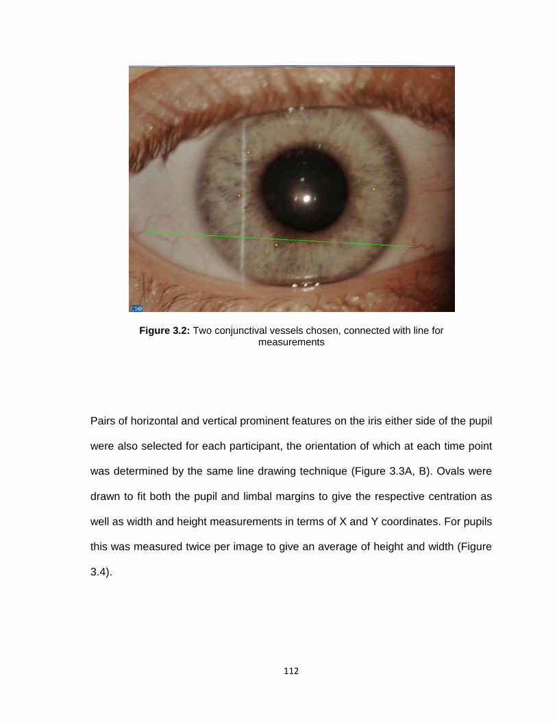
112
Pairs of horizontal and vertical prominent features on the iris either side of the pupil
were also selected for each participant, the orientation of which at each time point
was determined by the same line drawing technique (Figure 3.3A, B). Ovals were
drawn to fit both the pupil and limbal margins to give the respective centration as
well as width and height measurements in terms of X and Y coordinates. For pupils
this was measured twice per image to give an average of height and width (Figure
3.4).
Figure 3.2: Two conjunctival vessels chosen, connected with line for measurements

113
Figure 3.3:
Horizontal (A) and vertical (B) iris features connected for measurements of separation
A
B

114
Figure 3.4: Image analysis showing orientation lines joining horizontal conjunctival blood vessels and horizontal and vertical iris features as
well as ovals fitted to the pupil and limbal edge to allow centration and width and height to be determined during dilation.
Measurement between
conjunctival blood vessels
Measurement
between vertical
iris features
Measurement between
horizontal iris features
Measurement of pupil
centration, height and width
Measurement of limbus
centration

115
3.3. Statistical Analysis
For repeatability purposes one randomly selected image per subject was re-
analysed. As the values obtained were in pixel units a conversion factor
determined from image analysing a millimetre unit ruler was applied (1 pixel =
0.01475mm) in order to convert the measurements to millimetres. Statistical
analysis was then carried out to determine the significance of the results obtained.

116
3.4. Results
The average standard deviation of the 15 images for each subject captured every
2 minutes between dilation drug instillation and 30 minutes of limbal width (0.17 ±
0.07 mm) and height (0.21 ± 0.08 mm) and the ratio between them (0.02 ± 0.01)
were similar to that of the measurement variability of repeated analysis of the
same image twice (Table 3.1).
Blood Vessel Orientation
(⁰⁰⁰⁰)
Horiz. Iris
Features (⁰⁰⁰⁰)
Vertical Iris
Features (⁰⁰⁰⁰)
Pupil Width (mm)
Pupil Height (mm)
Limbus Width (mm)
Limbus Height (mm)
Limbal Width/Height
Ratio
SD
0.63
0.39
0.87
0.12
0.12
0.23
0.22
0.03
Hence the eye orientation and position from the slit lamp camera seems to be well
controlled by the conventional slit lamp head/chin rest and the slit-lamp focusing,
and no compensation of the images captured between visits based on apparent
limbal dimensions was therefore applied. At the typical slit lamp to eye distance of
30cm, a 0.20mm change in limbal width (of 12.00mm) would occur from a 5mm
change in slit lamp to eye distance and a 0.02mm change in limbal width to height
ratio from a 11.4⁰ orientation change of the pupil to the camera plane.
Table3.1: Standard deviations of repeat analysis for the ocular measurements. N = 36.

117
The limbus was generally vertically oval varying little with repeat imaging at each
visit (width to height ratio 1.065 ± 0.003). The pupil was essentially symmetrical
(width to height ratio 1.018 ± 0.018 at baseline), and remained so with dilation (SD
± 0.013) as it increased in size in a sigmoid manner, with maximum dilation
reached by approximately 25 minutes (y = -3.594 / (1 + e(-(x-13.18)/3.569)) r2=0.998;
Figure 3.5).

118
Time after Dilation Drops Instilled (mins)
0 5 10 15 20 25 30
Siz
e (m
m)
0
2
4
6
8
10
12
Scleral WidthScleral HeightPupil WidthPupil HeightScleral Width/HeightPupil Width/Height
Figure 3.5: Variations in width and height of pupil, limbus and sclera with dilation. N = 36. Error bars =1 S.D.

119
Pupil dilation had no significant effect on the average change in orientation of the
conjunctival blood vessels, horizontal or vertical iris features (F = 2.95, p = 0.069,
Figure 3.6), however, the variability between subjects was much greater using iris
features than conjunctival blood vessels (F = 31.233, p < 0.001; Figure 3.6). The
change in pupil size was more strongly correlated to the change in orientation with
the horizontal (r = 0.13 ± 0.60) and vertical (0.18 ± 0.49) iris features than the
conjunctival blood vessels (r = 0.02 ± 0.43; F = 3.149, p = 0.049).

120
Time after Dilation Drops Instilled (mins)
0 5 10 15 20 25 30
Cha
nge
in O
rient
atio
n (d
egre
es)
-10
-5
0
5
10Conjunctival Blood VesselsHorizontal Iris FeaturesVertical Iris Features
Figure 3.6: Orientation changes of conjunctival blood vessels and iris features through the process of dilation. N = 36. Error bars = 1 S.D.

121
The pupil was on average was centred superior (0.07 ± 0.09mm) and nasal (0.26 ±
0.14mm) compared to the centre of the limbus, moving superiorly with dilation with
the change mainly occurring in the first 10 minutes post-dilation and some
recovery from 20 minutes. Overall, there is an inferior displacement of the pupil
centre (Figure 3.7).
Nasal0.0 0.1 0.2 0.3 0.4 0.5
Super ior
0.0
0.1
0.2
0.3
0.4
0.5
Temporal0.00.10.20.30.40.5
In ferior
0.0
0.1
0.2
0.3
0.4
0.5
0
30
60
90
120
150
180
210
240
270
300
330
Figure 3.7: Change in centration with dilation
0 mins
30 mins
Decentration (mm)

122
3.5. Discussion
The pupil, as expected, increased in diameter with pharmacological dilation
reaching maximum dilation at approximately 25 minutes following drug instillation,
the increase was generally symmetrical through the process with a sigmoidal
increase in diameter. On analysis of pupil centration it was found to be located
superior-nasal on average, moving inferior upon dilation, which coincides with
previous studies of pupil centration (Walsh, 1988; Wyatt, 1995; Yang et al., 2002).
It is possible that the changes in pupil centration may be smaller with physiological
than pharmacological dilation (Yang et al, 2002), but the intraocular lens is
implanted under pharmacological dilation so these pupil centration changes need
to be considered. Previous research has shown these changes are not age
dependant and hence the use of young subjects should not have influenced this
conclusion. Non-linearity of the pupil was calculated as 0.018 which corresponds
to that found by Wyatt (1995) reporting 0.017 in dark conditions and 0.016 in light
conditions.
Analysis of limbus height and width showed good repeatability over time, similar to
that found on repeat analysis of the same images a second time. Therefore, this
suggests there is adequate control of eye orientation using the head and chin rest
of slit lamp with minimal effect from head tilt. The limbus remained generally oval
in the horizontal plane which varied little upon repeat measurements. As surgeons
generally use the pupil rather than the limbus to judge centration, such findings

123
confirm the change in pupil centration when the eye is restored to physiological
stimulation must be taken into consideration for surgery requiring dilation,
particularly LASIK refractive surgery as ablation of the cornea must be confined to
the optical zone. Ablation beyond this area can lead to glare, haloes and ghost
images, hence resulting in an unsuccessful outcome (Yang et al., 2002) and
dissatisfaction from the patient.
Previous research has shown image analysis of lens rotation to be more robust
than subjective rating, and the use of conjunctival blood vessels or iris features to
allow for head rotation further improves reliability (Wolffsohn and Buckhurst, 2010).
Although dilation of the pupil had no effect on the average change in orientation of
blood vessels and iris features, the variability of iris features with dilation was much
greater in comparison to conjunctival blood vessels. The conjunctival blood
vessels therefore prove to be better markers for orientation and hence provide
surgeons with anatomical markers that can be used for various procedures.
Studies carried out to investigate the changes in pupil centration with diameter,
generally report a superior-temporal shift in pupil centre with pharmaceutically
induced mydriasis and inferior-temporal or temporal with light induced mydriasis of
magnitude less than 0.2mm, which is only related to pupil size for the latter (Walsh,
1988; Wyatt, 1995; Yang et al., 2002). However, the effect of different degrees of
dilation on pupil centration has not previously been assessed. This study showed a

124
similar inferior centration shift finding with pharmaceutical dilation to that previously
found with light induced mydriasis, although there was a counter movement
superiorly from 20 minutes onwards. While the study was performed on younger
adults to allow a wide dilation change to be imaged, age does not appear to affect
pupil centration (Yang et al., 2002), although non-linearities in pupil shape increase
with age (Wyatt, 1995).
The limitation in this study was evaluation of only young subjects which may be
found to differ if compared to older individuals. Also, the outcomes of the
investigation were limited to the quality of images obtained; despite capturing two
images for each stage of dilation some images presented difficulty in analysis.
Hence more robust instrumentation for imaging the anterior eye and IOL should be
designed and evaluated to support researchers and surgeons alike.

125
3.6. Conclusion
In conclusion, it is well documented that shifts in pupil centration occur with
pharmacological dilation, which need to be considered for any type of refractive
surgery to in order to avoid an unsuccessful outcome. The current study supports
existing knowledge of this change in pupil location through dilation and has
established conjunctival blood vessels as better markers for correcting the
alignment of toric IOLs and compensating for head rotation compared to using iris
features.
Hence, the pupil position may significantly influence the surgical outcome of
premium IOLs due to the complexity of their optics through misalignment and
increases in aberrations, which will be discussed further in the following chapter.
The effect of modern IOL surgery on the pupil and long term changes in pupil
centration relative to the limbus and IOL have not previously been considered as a
potential factor in the visual performance of premium IOLs. The next chapter
therefore aims to research the significance of this aspect of premium IOL
optimisation.

126
CHAPTER 4
Stability of Pupil Dilation
Following Cataract Surgery

127
4.1. Introduction
Various refractive surgical procedures, such as implantation of intraocular lenses,
require pharmacological dilation of the pupil. It is well established, however, that
the location of the pupil centre shifts with dilation (Walsh 1988; Wilson et al., 1992;
Yang et al., 2002,) and as discussed in the previous chapter such decentration
may lead to undesirable outcomes in refractive surgery.
The pupil is the entrance point for light approaching the visual system which
regulates retinal illumination by changes in its diameter. Pupil diameter is
controlled by smooth muscles in the iris; the sphincter pupillae located within the
stroma of the iris and dilator pupillae radiating from the iris root into the stroma
(Remington, 2005). Pupil size is affected by various factors such as; level of
illumination, age, accommodation and emotions (Winn et al., 1994).
Generally, the eye exhibits a range of optical aberrations (Walsh et al., 1988) with
increases in pupil diameter and decentration from the visual axis having been
shown to increase high-order aberrations (Ivanhoff 1956; Yang et al., 2002;
Campbell et al., 1966; Artal 1990; Liang et al., 1997; Kasper et al., 2006; Castejon-
Mochon et al., 2002; Wang et al., 2003) such as spherical aberration and coma
(Wilson et al., 1992). High-order aberrations (HOAs) cause various visual
symptoms for example, spherical aberration may form starburst and glare, whilst
horizontal coma can lead to monocular diplopia (Chalita et al., 2003, 2004). Optical

128
quality is reduced with spherical aberration and often manifests as a reduction in
contrast sensitivity (McLellan et al., 2001; Nio et al., 2000). Sakai et al (2007)
demonstrated the correlation between pupil size with HOAs and residual
astigmatism suggesting that the pupil adjusts accordingly to optimize retinal image
quality in varying luminance levels.
Pupil decentration can lead to ablation outside of the optical zone in
keratorefractive procedures resulting in halos, glare and some reports of reduced
contrast sensitivity (Pande and Hillman, 1993). It is therefore plausible that
decentration of a dilated pupil may lead to misalignment of an intraocular lens
(IOL) affecting the visual outcome and degrading the retinal image, particularly
with advanced optical designs such as the optics on multifocal and toric intraocular
lenses.
Previous studies have indicated greater shifts in pupil centration with
pharmacologically, rather than physiologically, induced dilation (Yang et al., 2002).
The general finding of temporal movement of the pupil centre with slight vertical
shifts have been documented (Walsh et al., 1988; Yang et al., 2002; Wyatt 1995).
Pupil decentration may be of particular concern in younger patients requiring
refractive surgery, such as for congenital cataracts or when electing for refractive
lens exchange, as larger pupils have shown greater degradation of M.T.F (Modular
Transfer Function) by Walsh and Charman (1988).

129
Misalignment of a toric intraocular lens results in no correction of the cylinder and if
great enough may even add to the cylindrical power (Shimzu et al., 1994). Toric
IOLs are susceptible to rotational instability (Novis, 2000) and if coupled with initial
misalignment during surgery, may result in a very poor surgical outcome with
substantial patient dissatisfaction.
Multifocal IOLs manipulate aberrations to enhance the depth of focus of the eye,
but can give rise to photic phenomena (Leyland and Zinicola, 2003; Hayayshi et
al., 2009; Steinert, 2000). As IOL position and tilt may adversely affect the
effectiveness of the IOL it could be assumed that there may be an association
between the centration of multifocal intraocular lenses and dysphotopsia.
Age-related lenticular changes lead to increased high-order aberrations (HOAs) in
later life, particularly spherical aberration (Nio et al., 2002; McLellan et al., 2001).
In the young eye the positive corneal aberration is reduced by the negative
aberration of the crystalline lens. Following cataract surgery there is an increase in
such aberration as often the natural lens is replaced by a spherical monofocal lens
which exhibit higher degrees of optical aberrations (De Castro et al., 2007). For
these reasons aspheric designs have been developed to improve the visual
outcome by incorporating negative spherical aberration within the lens. The effect
of these aspheric lenses, however, may be lost by IOL displacement as shown by
many studies; for example Mckelvie et al (2011) showed pupil size had the most

130
affect on HOA in their investigation followed by IOL decentration, stating 0.5mm of
decentration can vastly affect the performance of an aspheric IOL particularly
those which incorporate higher levels of negative asphericity. The authors
demonstrated that 0.5mm decentration of an IOL, with -0.27µm of negative
spherical aberration, would increase HOA by 48% which would increase to 80%
with 1.5mm of decentration. Performance of aspheric multifocal IOLs are also
jeopardized by decentration exceeding 0.5mm (Atchison 1991; Altman et al.,
2005).
Coma also increases with decentration and tilt, as shown by optical bench tests by
Epigg et al (2009) and Pieh et al (2009), which impairs visual quality. Despite
these claims, some studies have not found any significant change in HOAs with
decentration and tilt of an aspheric IOL such as that of Choi et al (2010) which
perhaps may have been due to their small sample size of only 32 participants,
however, another study by Baumeister et al., (2009) also did not find any
correlation between image quality and decentration of aspheric IOLs.

131
Holladay et al (2002) stated if an aspheric IOL was centred within 0.4mm and tilted
less than 7° its optical performance would surpass that of a monofocal while
Lopez-Gil et al (2007) suggest for optimum performance an IOL should be centred
in line with the visual axis. It is therefore clear that negative spherical aberration
can be altered by IOL tilt and decentration the effect of which is greater with higher
levels of asphericity inducing higher amounts of HOAs.
The result of decentration or tilting of IOLs that provide negative spherical
aberration may be observed as impairment of contrast sensitivity (Baumeister et
al., 2005). It has been shown by Pepose et al (2005) an increase of 1µm in HOAs
can reduce photopic contrast sensitivity by 2.5 logarithm of min angle of resolution
and a reduction in mesopic contrast sensitivity of 12 logarithm min angle of
resolution. In addition, Epigg et al (2009) investigated six IOLs and reported poor
MTF measures with decentration with a 3mm pupil for the five aspheric IOLs,
whereas measures with the spherical IOL were relatively unaffected. For a larger
pupil of 4.5mm decentration gave rise to horizontal coma, astigmatism and
defocus, with the performance of the Tecnis Z9000 IOL being most affected by
decentration. Aspheric lenses, particularly, aberration-correcting IOLs are thus
more vulnerable to tilt and decentration despite their aim of providing better image
quality. Conversely, aberration-free IOLs are less affected by decentration, the use
of which is suggested where centration could be suboptimal (Eppig et al., 2009).
Many premium IOLs incorporate asphericity into their design and hence may be

132
vulnerable to impairment of contrast sensitivity, which in the case of multifocal
IOLs may exacerbate the already reported reduced contrast sensitivity.
Misplacement of IOLs may also affect refractive error; computer modelling by
Korynta et al (1999) indicated a shift towards myopia and oblique astigmatism with
IOL decentration, but the resultant effect was dependent on the amount of
dislocation. Earlier investigations by Atchison (1989) had already established that
decentration considerably affected refractive errors, which were calculated to be
proportional to the square of decentration. In addition, it was found coma-like
aberration and astigmatism affected the retinal image by IOL displacement and
that larger pupils (≥4mm) suffered more from spherical aberration, whilst smaller
pupils were affected principally by astigmatism (Atchison, 1989).
It is suggested that in addition to surgical technique, IOL haptic design may also
influence stability of an IOL (Epigg et al., 2009). Haptics made from PMMA have
been reported to give better stability (Ohmi et al., 1995; Gallagher et al., 1999),
although Baumeister et al (2005) found no such difference between haptic
materials over a 12 month period. Post-operative intraocular lens rotation tends to
occur during the earlier postoperative stages (Rushwurm et al., 2000) as a result of
friction between lens haptics and capsular bag, instability of the anterior chamber
due to intraocular pressure or ocular trauma, IOL design, the level of fibrosis or
compression of haptics as shrinkage of the capsular bag occurs (Buckhurst et al.,

133
2010). Post-operative rotation combined with initial displacement of the IOL due to
pupil decentration can lead to various visual symptoms and thus a poor surgical
outcome. In extreme cases dislocated IOLs may have to be explanted, putting
unnecessary constraints on financial resources and possibly increasing risks of
endophthalmitis. Repositioning a rotated IOL, such as a toric lens is possible,
however may further raise complications such as; CMO, capsular tears and
endophthalmitis and thus is favourable to avoid (Sun et al., 2000).
Regardless of the direction of pupil decentration, the resultant aberration will
degrade the quality of the retinal image and affect spatial vision (Walsh and
Charman 1988; Thibos 1987). Even small pupil displacements may induce
significant levels of aberrations (Rynders et al., 1995). There is now more interest
developing in IOL tilt and decentration as development of IOLs begins to widen
attention to broader aspects of IOL design and focus more on correcting HOAs in
addition to refractive error (Mester et al., 2003).
There is little literature on the effects of cataract surgery on the pupil; however
Gibbens et al (1989) did carry out an investigation to examine the effects of post-
operative pupil dilation with intracapsular cataract extraction (ICCE) and
extracapsular cataract extraction (ECCE) surgical procedures. A significantly
smaller post-operative pupil size was noted on dilation with both surgical
procedures, but the location of the pupil was not assessed. The effect of more

134
recent cataract extraction procedures on the stability and post-operative location of
the pupil has not yet been explored within the literature therefore; the purpose of
the present study was to investigate the changes in size and centration stability of
the pupil six months following cataract surgery and intraocular lens (IOL)
implantation.

135
4.2. Methods
Two hundred and four patients were implanted monocularly with the fifth
generation Akreos AO aspheric IOL (Bausch and Lomb, Rochester, New York) in
one eye at six hospital sites across Europe. Inclusion criteria included age-related
cataract amenable to treatment with standard phacoemulsification and IOL
implantation, and pupils which could be dilated to at least 5mm. Subjects were
aged 67.6 ± 7.9 years (range 51 to 89 years) and 65% were female.
The acrylic, hydrophilic lens has a 6 mm optic with a 360° posterior square edge
barrier attached to 11 mm closed loop haptics. The optic has aspheric surfaces
aiming to induce no IOL aberrations. A 5.5 mm continuous curvilinear capsulotomy
was used through which phacoemulsification was performed. Once the capsular
bag was filled with a viscoelastic substance, the lens was inserted using an Akreos
single use insertion device through a 2.8 mm incision and the viscoelastic device
aspirated from in front and behind the lens.
Patients were dilated using phenylephrine 2.5% and tropicamide 1.0% at the
operative visit and the following four post-operative appointments. These
appointments were conducted 0, 1-2, 7-14, 30-60 and 120-180 days after IOL
implantation. The intraocular lens was imaged at 10x magnification in
retroilluminantion using a CSO SL-990 digital slit-lamp biomicroscope

136
(Construzione Strumenti Oftalmici, Florence, Italy). Informed consent was obtained
from all participants prior to lens implantation and the study was approved by
ethical committees at each of the sites.
The centre of three ovals overlaid to circumscribe the IOL optic edge, the pupil
margin and the limbus, were compared to determine the IOL centration (Figure
4.1). Pupil diameter and height were taken from the pupil oval dimensions. This
technique has previously been evaluated and showed excellent repeatability
(Wolffsohn and Buckhurst, 2010).

137
Figure 4.1: Measurement of pupil, limbus and IOL edge
Limbus measurement Iris Measurement
IOL optic edge measurement

138
4.3. Statistical Analysis
The IOL and pupil centration with respect to the limbus at each visit was
subtracted from the values immediately after surgery to assess decentration.
Repeated measure analysis of variance was used to assess locational stability
between visits.

139
4.4. Results
There was a significant inferior shift in pupil centration with time after cataract
surgery (F=2.953, p=0.02), but no significant change in horizontal centration
(F=1.010, p=0.4). Intraocular lens centration was stable with respect to the limbus
(width F=0.483, p=0.75; height F=0.282, p=0.89), but was decentred relative to the
pupil vertically (F=7.672, p<0.001), but not horizontally (F=1.120, p=0.35;
Figure4.2) thus indicating a change in centration.

140
0.00 0.05 0.10 0.15 0.20 0.25 0.300.00
0.05
0.10
0.15
0.20
0.25
0.30
0.000.050.100.150.200.250.30
0.00
0.05
0.10
0.15
0.20
0.25
0.30
Pupil vs LimbusIOL vs PupilIOL vs Limbus
Figure 4.2: IOL, dilated pupil and limbal centration relative to each other with time post-cataract surgery. Pupil centration becomes increasingly inferior relative to the limbus and
IOL. N = 204. (Red arrows indicate direction of movement on subsequent visits)
Decentration (mm)
Decentration (mm)
nasal temporal
superior
inferior

141
Pupil width (F=32.476, p<0.001) and height (F=35.167, p<0.001) were significantly
larger with dilation immediately after surgery than at subsequent dilations (Figure
4.3). The ratio between pupil width and height was close to 1.0, but also altered
with time after surgery (F=36.009, p<0.001), becoming more vertically oval.

142
Time Post-Operative (days)
0 20 40 60 80 100 120 140 160
Pup
il S
ize
(mm
)
5.0
5.5
6.0
6.5
7.0
7.5
8.0
8.5
9.0
WidthHeight
Figure 4.3: Dilated pupil width and height with time post cataract surgery.
N = 204. Error bars = 1 S.D.
Pupil Size following Cataract Surgery

143
4.5. Discussion
The results have shown a significant inferior shift in pupil centration following
cataract surgery. As there was no relative change with time between the IOL and
limbus, this suggests that the IOL is stable within the eye, but the pupil dilator
pupillae or iris tissue has been damaged in some way as the shift was only in one
meridian. The significant decrease in post-operative pupil diameter with dilation
found with time may indicate additional sphincter muscular damage.
An earlier investigation by Gibbens et al (1989) similarly assessed the pupil
following ECCE as well as ICCE, as it was stated at the time clinicians believed the
aphakic pupil did not dilate as well as pre-operatively. In scotopic conditions the
pupil diameter was on average 0.3mm less than pre-operatively whereas in
photopic conditions it constricted less and was larger by 0.3mm, suggesting
possible damage to the iris which reduced its mobility. The diameter of the aphakic
pupils implanted with an IOL were also reduced by 0.9mm when dilated with
Tropicamide 0.5% and Phenylephrine 10% similar to the horizontal pupil dilation
reduction found in this study. Gibbens and colleagues (1989) found the pupil size
reduction with repeat dilation after surgery did not occur in aphakic eyes
suggesting the damage was due to IOL implantation, rather than the cataract
removal, although surgical techniques and IOL materials and designs since have
changed.

144
Understandably, with Gibbens et al (1989) study, ICCE without IOL implantation
reduced the pupil diameter most, as this involves the removal of the entire lens
cavity and capsule and hence the most disturbance to the eye. No ICCE with IOL
implantation group were examined so it is not possible to tell whether the IOL
would have contributed further damage in this scenario.
Decentration and tilt of IOLs may also occur following cataract surgery for various
reasons such as; capsule contraction, asymmetric bag and sulcus fixation or
capsular tear (Jung et al., 2000), surgical technique, residual cortical material, IOL
properties, asymmetric haptic fixation or haptic location (Cabellero et al., 1991;
Hansen et al., 1988; Akkin et al., 1994; Kimura et al., 1996). The incidence of such
occurrences has reduced since the introduction of phacoemulsification and
development of more stable IOLs. However, combined with pupil decentration
there may be substantial impairment of visual quality.
The apparent inferior shift in the pupil centre following cataract surgery may
account for various visual phenomena with multifocal IOLs. Many studies have
shown a general increase in high-order aberrations following cataract surgery
(Hayashi et al., 2000; Barbero et al., 2003; Guirao et al., 2004), perhaps
contributed to by poor centration of IOLs, reducing contrast sensitivity and causing
undesirable refractive errors. Better surgical technique of aligning IOLs is therefore
required in order to prevent dislocation cases (Mutlu et al., 2005). Visual
145
phenomena do tend to lessen in subjective significance with time (Vaquero-Ruano
et al., 1998), but this may be due to adaptation to the aberrations and their effects
rather than them resolving with time. Restricted pupil diameters following surgical
intervention may also affect the treatment of the peripheral retina, should it be
required, and the effectiveness of premium IOLs such as; multifocal designs if
annular zones are not adequately exposed.
The main limitation of the present investigation was lack of pre-operative pupil
diameters, measurements were only taken on the date of surgery and on post-
operative visits. Knowledge of the average pupil diameters before surgery would
provide confirmation of reductions of pupil mobility and hence iris damage.
However it should be noted the differences that were presented at subsequent
visits may be attributed to increased corneal permeability at the time of surgery.

146
4.6. Conclusion
In conclusion, this research confirms that when a pupil is pharmacologically dilated
for cataract surgery it cannot be used as a satisfactory guide for centration (Melki
et al., 2011). The limbus is a better guide although the pre-dilation pupil
decentration relative to the limbus also needs to be taken into account when
positioning an IOL. Such knowledge becomes even more critical when premium
IOLs are to be implanted because of their more complex optical designs, which
can cause a significant reduction in visual quality if they are not perfectly centred.
In addition, research is warranted into the development of new surgical IOL
implantation techniques and IOL materials and designs that do not impair the
dilation response.
Premium IOLs are becoming of growing interest in refractive surgery and not just
for optimising distance vision following cataract surgery. Patients are continuously
seeking for possible solutions from practitioners to presbyopia. In order to advise
patients of their suitability for refractive lens exchange and what factors influence
when it might be needed, a better understanding of the risk factors for presbyopia
is required and this is explored in chapter 5.

147
CHAPTER 5
Amplitude of Accommodation &
Lifestyle –
Implications for Multifocal and
Accommodating IOLs

148
5.1. Introduction
Presbyopia is recognized as the most common ocular change occurring in middle
age (Weale, 2003). The decline in amplitude of accommodation with age is well
documented within literature, however, the rate of decline and onset of presbyopia
for individuals varies and may be influenced by various health and environmental
factors such as nutrition, climate, altitude, ethnicity and physical stature (Kragha,
1985; Kragha and Hofstetter, 1986).
Both presbyopia and cataracts occur with an ageing lens and tend to develop
much earlier in populations in warmer climates (Miranda, 1979; 1980). It could be
assumed that the factors increasing the development of senile cataracts may also
be factors accelerating presbyopia, such that presbyopia may be the initial stage in
the development of senile cataracts. Higher environmental temperature has been
noted as possibly accelerating the onset of presbyopia by some authors (Kragha
and Hofstetter, 1986). An inverse relationship between environmental temperature
and onset of presbyopia has been demonstrated through investigations by Weale
(1981a, b) and Miranda (1979) with correlation coefficients of -0.89 and -0.85
respectively. However Kragha and Hofstetter (1986) explain this relationship to be
of self-selection bias and conversely reported no significant differences in bifocal
additions prescribed across North and Central America, with a range of average
temperatures between -7°C and 26°C. An earlier inve stigation by Kragha (1985)
on bifocal additions prescribed to patients in Nigeria showed the influence of

149
temperature on accommodation to only just approach significance (p= 0.051,
correlation coefficient -0.44), although the additions prescribed to females were
significantly higher compared to males, explained to be as a result of the smaller
stature of females and hence shorter working distances used rather than actual
difference in accommodative ability. More recent studies, however, show females
exhibit presbyopia earlier due to possible hormonal differences (Nirmalan et al.,
2006; Pointer 1995; Mukesh et al., 2006). Women are also considered to have a
higher incidence of developing cataracts as shown by the Beaver Dam Study
(Klein et al., 1998) and Barbados Eye Study (Leske et al., 2000) although reasons
for this are unclear. Other comparisons of near additions include that of Hofstetter
(1968), who showed additions prescribed to patients in Fiji and Ghana were on
average 0.50 dioptres higher than prescribed to Europeans of a similar age profile.
Rambo (1953) reported variance in the age of presbyopia onset with geographical
location; it was documented on average as 40 years in Iran, India, Iraq, Arabia and
Cuba whereas in Italy it was typically 42-43 years, but occurred much later in
Sweden and Norway at 48 years of age, indicating a much earlier onset in regions
closer to the equator. Similarly, Ong (1981) found presbyopia to occur earlier in
southeastern Asian refugees of around 42 years. Geographical data collected by
Miranda (1980) also shows a negative correlation in the Western and Eastern
hemispheres, whereby presbyopia onset occurred earlier in regions of warmer
climate. Differences may also occur within regions, particularly with coastal areas
where presbyopia seems to occur earlier than in mountainous areas of that same

150
region (Miranda, 1979). Variations in the findings of these studies of climate and
accommodation may perhaps be due to the lack of definition of presbyopia,
making comparisons between them difficult (Kragha, 1985). A study in Tanzania,
where presbyopia was defined to be the inability to read N8 text at 40 centimetres
with distance correction and improvement using an additional lens, found 61.7% in
the population of 40 year olds and older to be presbyopic (Burke et al., 2006). A
similar study in Nigeria, but without a clear description of presbyopia, reported a
prevalence of only 33% (Nwosu, 1998).
Ultraviolet (UV) radiation is a well-known factor contributing to the aging of the
human body (Stevens and Bergmanson, 1989). For the eye, radiation between
310-400nm affects the crystalline lens (Miranda, 1979) increasing the risk of
cataract development with prolonged exposure (Stevens and Bergmanson, 1989;
Bergmanson and Söderberg, 1995; Roberts, 2011). It is therefore reasonable to
assume that UV light can affect an individual’s amplitude of accommodation and its
rate of decline. Health is another aspect which can greatly affect amplitude of
accommodation. Reports of conditions such as diabetes and Human
Immunodeficiency Virus (HIV) have been associated with reduced amplitudes of
accommodation (Newsome, 1989; Westcott et al., 2001). The effect seems to be
more pronounced in younger patients aged between 25-29 years; it is perhaps less
noticeable in older age groups due to a significant decrease in amplitude already
having taken place (Westcott et al., 2001). The pathological processes in HIV
infection may accelerate the changes associated with the aging of the crystalline

151
lens through possible autonomic dysfunction (Westcott et al., 2001). Comparisons
of young insulin-dependent diabetic patients and non-diabetic patients have also
shown reductions in pull-down measures of accommodation with diabetes (Moss
et al., 1987) which may be associated with hypoxia (Berens et al., 1932). Duane
(1925) had also noted diabetes as a possible factor reducing the amplitude of
accommodation.
Most studies discussed above, considering the environmental influences on
accommodation, have based their investigations on one main factor. Jain et al
(1982) however, investigated a variety of factors possibly contributing to onset of
presbyopia in an Indian population, including; temperature, UV radiation, diet and
exposure to toxic substances. Approximately one-third (36%) of the population
studied were found to enter presbyopia at 38 years of age or younger. Patients
within rural areas showed earlier onset of presbyopia, possibly due to greater UV
exposure which was not quantified in the study. A low dietary intake of amino acids
was also associated with reduced levels of accommodation and earlier onset of
presbyopia. Furthermore, the study noted the effect of hair dye on earlier onset of
presbyopia in 4% of patients, which was supported by previous work by Jain et al
(1979) where lenticular changes occurred in 89% and presbyopia in 7% of 200
patients using hair dye.

152
Specific vitamins or minerals have not been associated with presbyopia, however
levels of antioxidants have been linked with cataracts particularly vitamin C, low
levels of which have been noted in a population with a high prevalence of cataracts
(Dherani et al., 2008). Levels of carotenoids (lutein, zeaxanthin, carotenes and
lycopene) are also thought to have protective effects on the crystalline lens
(Dherani et al., 2008). Caffeine intake can increase levels of accommodation within
30 minutes of consumption, the effect of which slowly reduces after 30 minutes
(Ajayi and George, 2007), although long-term effects have not been investigated.
Lifestyle habits such as long-term excessive alcohol intake and smoking may also
bear significance on amplitude of accommodation and presbyopia. Subjective
monocular measurements have shown reduced accommodation in alcoholics
when compared to controls particularly in younger individuals, the effect being
sustained even following a recovery period (Campbell et al., 2001). The onset of
presbyopia with prolonged higher alcohol consumption may therefore occur much
earlier than anticipated. Smoking may be associated with lenticular changes, as
this has been described as a possible risk factor in developing nuclear opacities
(Mukesh et al., 2006; Christen 1992; Hiller et al., 1997; Hankinson et al., 1992).
The processes by which smoking effects the crystalline lens requires more
research, however it is proposed that plasma concentrations which maintain
transparency are interfered with (Christen et al., 1992). Other explanations include
excessive oxidative stress leading to cellular DNA damage (Kleiman and Spector,
1993).

153
As part of the World Health Organisation Vision 2020 initiative presbyopia is not
recognised as a refractive error, however increases in the elderly population will
inevitably result in higher prevalence of presbyopia, attention to which should be
considered, as this is an unnecessary source of visual impairment. In this study, a
combination of environmental and nutritional factors were investigated, using a
detailed lifestyle questionnaire and more advanced methodology, in comparison to
previous studies discussed, to assess the influence on amplitude of
accommodation and onset of presbyopia in a population attending optometric
practice in the United Kingdom. Intraocular lenses for presbyopia have advanced,
but are usually implanted as part of cataract surgery. Cataracts typically occur from
50 years onwards when presbyopia has already manifested, with patients missing
out on the potential benefits of premium IOLs for many years and at the peak of
their working ability. Patients are already benefiting from refractive lens exchange,
replacing their hardened lens causing presbyopia, however, it is important to know
when presbyopia occurs and what factors influence this as lifestyle has
significantly changed since the work of Jain et al (1979) and Miranda (1979), with
changes in diet and visual demands. A better knowledge of when individual’s may
become presbyopic will help with counselling on premium IOL options they might
wish to consider alongside other forms of refractive correction.

154
5.2. Methods
Four hundred and ninety eight consecutive patients attending routine optometric
practice in the West Midlands (United Kingdom), who met the inclusion/exclusion
criteria and gave consent, participated in the study. They were aged between 18
years to 90 years, with an average age of 42± 23 years. Ethical approval, adhering
to Declaration of Helsinki, was obtained from the Aston University ethics
committee. Participants required good physical and mental health with no ocular
pathology, as observed by slit-lamp biomicroscopy and ophthalmoscopy and no
previous ocular surgery. All participants were English-speaking to ensure
instructions and questionnaires were fully understood.
Following informed consent, refractive error was determined subjectively based on
the principal of maximum plus power for best distance visual acuity to ensure the
patient was fully corrected for distance vision. Pupil sizes were recorded in
photopic conditions (300 lux), right eye and binocular amplitude of accommodation
were then measured using the RAF near point rule (H.S Clement Clarke
International, Harlow, Essex, United Kingdom) as the push-up technique is the
most widely accepted method for measuring subjective amplitude of
accommodation. For pre-presbyopic patients measurements were obtained whilst
fully corrected for distance. However, for presbyopes a +2.00 dioptre lens addition
was used required to allow for measurements to be taken within a more sensitive
range on the RAF rule. Push-up and pull-down techniques were carried out three

155
times for both monocular and binocular amplitudes of accommodation using the
Snellen chart optotype on the instrument. For push-up measurements, subjects
were asked to view their lowest line of acuity at 40cm as the target was slowly
moved towards them. Observers were asked to report when the target became
blurry and could no longer be resolved. For pull-down measurements, the optotype
was positioned initially in front of the point of blur on push-up testing; the target
was then slowly pulled away from the observer until it could again be resolved.
The reference point at which measurements were made was the spectacle plane,
located 12mm from the cornea. Additional lighting was used to illuminate optotypes
to ensure high contrast at all distances (range 80 to 100cd/m2). The same
examiner carried out all measurements of accommodation for all subjects to
reduce inter-examiner variability.
Minus lens measurements of accommodation were also obtained by presenting
minus lenses in 0.25 dioptre steps from blur to initial clearance of a 6 metre target.
The target viewed was determined as the individual’s lowest line of distance visual
acuity on a computerised logMAR chart. These two methods were used to allow
comparison with previous studies on the development of presbyopia (table A4).

156
In addition, adult participants were asked to complete a lifestyle questionnaire
which is not validated but has previously been used as part of age-related macular
degeneration studies. Questions included dietary intake, alcohol intake, medical
conditions and medication, UV exposure, iris colour and supplement intake (see
appendix for questionnaire, A1).

157
5.3. Statistical Analysis
For statistical comparisons the amplitude of accommodation with the RAF rule was
calculated as the average of push-up and push down values, in order to provide
the best estimation of accommodation (Zadnik, 1997). As the RAF rule was
determined as more sensitive to accommodative change with age (see chapter 6),
these values were used for the correlation with lifestyle in preference to the minus
lens technique. As both eyes should be affected by any of the lifestyle issues
evaluated, binocular RAF rule amplitude of accommodation was considered. All
data was assessed for normal distribution using the Kolmogorov-Smirnov test.
Comparisons between categories were performed using analysis of covariance
(ANCOVA) with age as a covariate, as it is known to be the strongest factor
associated with amplitude of accommodation. Non-parametric data was analysed
using Kruskal-Wallis and Mann-Whitney U tests. Linear regression modelling was
used to determine which variables may influence accommodation.

158
5.4. Results
5.4.1. Onset of Presbyopia
All presbyopic participants were asked when they first required a reading
prescription which would indicate the onset of presbyopia, as at this time a patient
seeks help from a practitioner due to inability to carry out near tasks satisfactorily.
The average age of onset of presbyopia, reported by the population that
participated in the study was calculated as 48.9 ± 7.3 years. The average age of
presbyopia onset for females was 48.2 ± 7.2 years and for males it was 49.7± 7.4
years, these results however showed no statistical difference (p=0.11).
Average photopic pupil size for male presbyopes was calculated as 3.19 ±
0.05mm, for females it was 3.17±0.05mm, although this showed no statistical
difference (F=0.07, p=0.79).

159
5.4.2. Age & Gender
Binocular amplitude of accommodation with the RAF rule, calculated as the mean
of binocular push-up and pull-down values and minus lens technique amplitudes
were compared. Both techniques show decrease in amplitude with age (Figure
5.1).

160
Binocular Amplitude of Accommodation with Age
Age (years)
0 20 40 60 80 100
Am
plitu
de o
f A
ccom
mod
atio
n (D
)
0
5
10
15
20
25
RAF Rule
Figure 5.1: Comparison of binocular amplitude of accommodation with RAF rule
(n=536)

161
The relationship between age and binocular accommodation with RAF rule was
investigated by performing analysis of Pearson product-moment correlation
coefficient. A strong negative correlation was present between age and
accommodation, where an increase in age results in a decrease in
accommodation, for males; r= -0.86, n=215, (p<0.001) and for females; r= -0.85,
n=278 (p<0.001).
On age-controlled comparisons of accommodation between gender, pre-
presbyopic females (<45 years) showed higher average binocular amplitude of
accommodation, with the RAF rule, of 10.29 ± 3.29 D compared to an average of
9.54 ± 2.77 D for pre-presbyopic males, although this was just outside statistical
difference (p=0.06). Presbyopic males (≥ 45 years) exhibited a mean amplitude of
accommodation of 3.46 ± 0.76 D compared to females with mean amplitude of
3.39 ± 1.00 D; again this was not found to be of statistical significance (p= 0.5,
Figure 5.2).

162
Average Binocular Amplitude of Accommodation & Gender
Figure 5.2: Comparison of average binocular amplitude of accommodation between pre-presbyopic and presbyopic males (n=215) and females (n=278). Error bars = 1 S.D.

163
5.4.3. Height, Weight & Body Mass Index (BMI)
For height (cm) no association with accommodation (r= 0.06, n= 493, p= 0.202)
was found. Weight (kg) showed a very small positive association, with a Pearsons
correlation coefficient of 0.11, found to be of significance (p=0.014, n= 493).
However, Body Mass Index (kg/m2) showed a significant stronger negative
association with accommodation, (r= -0.44, n= 493, p<0.001).
5.4.4. Ethnicity
To investigate the influence of ethnicity on accommodation, comparisons
weremade only between the two largest ethnic groups found from the
questionnaire; British (n=103) and South Asian (n=90). A one-way between-
groups analysis of covariance (ANCOVA) was conducted to compare the two
major ethnic groups from results of the questionnaire and their binocular
accommodation values obtained with the RAF rule; the independent variable being
noted as ethnicity, the dependent variable set was accommodation and age
selected as a covariate.
No significant differences were found amongst the ethnic groups (F= 3.31, p= 0.07,
partial eta squared= 0.02).

164
5.4.5. Iris Colour
Comparisons of iris colour groups; black/dark brown, blue, brown, green, grey,
hazel and light brown, were made as these were the most common categories
obtained from the questionnaire. One-way between-groups analysis of covariance
(ANCOVA) was conducted to compare iris colours and binocular accommodation
values obtained with the RAF rule, the independent variable being noted as iris
colour, the dependent variable was set as accommodation and age selected as a
covariate.
Differences between iris colour whilst controlling for age were found to be
significant (F= 5.96, p<0.001, partial eta squared= 0.09). 9% of the variance in
accommodation, according to the analysis, can be explained by iris colour. Further
post hoc comparisons with the Tukey HSD test shows black/dark brown (7.42±
0.28D, n=59), brown (5.76± 0.21D, n=85) and light brown (6.21± 0.43D, n=19)
irides significantly differ from blue (5.54± 0.19D, n=108) and grey (5.29± 0.41D,
n=22) irides. The darker irides gave higher values of mean accommodation in
comparison to lighter irides. The three groups of brown iris colours (Black/dark
brown, brown, light brown) also showed statistical differences between each other,
with black/dark brown giving the greatest adjusted mean amplitude of
accommodation. Green (5.28± 0.34D, n=31) and Hazel (5.72± 0.31D, n=37)
showed no statistical differences with all iris colour groups.

165
5.4.6. Ultraviolet Light Exposure & Climate
To investigate the possible effects of ultraviolet (UV) light and climate on amplitude
of accommodation, subjects were asked a number of questions; the hours spent
outside per week in Spring/Summer months and Autumn/Winter months, annual
exposure to strong sunlight abroad and the total number of weeks in hot climates
throughout the individual’s entire lifetime. Generally the hours spent outdoors in
daylight were more in the Spring/Summer (24.57± 20.14 hours) period than in
Autumn/Winter period (15.07± 14.14 hours, p<0.001). To investigate if a
relationship exists between the number of hours outdoors per week in warmer and
colder seasons and accommodation, partial correlation was conducted between
binocular amplitude of accommodation (RAF rule) and the two sets of seasons.
Again controlling for age was a requirement for this analysis.
For hours of UV exposure per week in the Autumn/Winter months, a significant but
relatively small negative association was found (r= -0.12, n= 491, p= 0.01),
suggesting that the less hours an individual spends outside in the colder seasons,
the higher the amplitude of accommodation. Surprisingly, the correlation between
binocular accommodation and weekly exposure to UV light in the Spring/Summer
months showed only a borderline association (r= -0.08, n=491, p=0.06).

166
Participants were also asked for how many weeks of their annual holidays they
were exposed to strong sunlight abroad. Partial correlation showed no significance
between annual exposure to strong sunlight and amplitude of accommodation (r =
0.01, n= 491, p=0.77). Using the total number of weeks spent abroad in hot climate
in the participant’s entire lifetime obtained from the questionnaire to explore the
possibility of an association between hot climate and amplitude of accommodation,
partial correlation gave no significant association between time in hot climates
throughout a lifetime and amplitude of accommodation (r= 0.01, n=491, p=0.82).

167
5.4.7. Ultraviolet Light Protection
Within the questionnaire, participants were asked about their UV protection and
how often sunglasses were worn during times of bright sunlight. Answers were
split into categories of; always, most of the time, sometimes, occasionally, very
rarely and never.
A Kruskal-Wallis test was conducted and revealed a significant difference amongst
the six groups (p<0.001, χ2 = 39.76). Never wearing sunglasses generated a
lower median than all other groups except for always wearing sunglasses. Mann-
Whitney tests were conducted between all groups and showed a significant
difference in amplitude of accommodation between never (Md= 4.06, n=89)
wearing sunglasses and four other groups; very rarely (p<0.001, Md= 8.12, n= 57),
occasionally (p=0.03, Md= 7.58, n= 44), sometimes (p<0.001, Md= 7.94, n= 64)
and most of the time (p=0.001, Md= 8.50, n= 71). Never wearing sunglasses and
always wearing sunglasses in bright conditions, however, showed no statistical
difference (p=0.87, Md= 3.50, n= 83).

168
5.4.8. Diet & Nutrition
One way between groups analysis of covariance (ANCOVA) was conducted to
assess the binocular amplitude of accommodation measured with the RAF rule for
meat eaters (n=428), vegetarians (n=41) and partial vegetarians (n=24). No
vegans were found to have participated in the study. The covariate was selected
as age. However, the different categories in diet did not show any significance in
the level of binocular accommodation measured (F=2.12, p= 0.12).
For more detailed information on dietary intake patients were asked how many
separate servings of fruit and vegetables they consumed per week and how many
eggs and oily fish were eaten per week. One cup of fruit or vegetables was defined
as a serving of food. Fruit and vegetable intake per week was combined to give an
overall estimate of weekly intake by summing up the separate serving scores for
fruit and vegetables. Partial correlation was used to explore the relationship
between the combined intake per week and binocular accommodation (RAF Rule)
whilst controlling for age. No significant association was found between fruit and
vegetable intake and binocular accommodation (r =0.007, n=491, p=0.88).

169
The recommended daily intake of fruit and vegetables is known to be 5 servings of
both fruit and vegetables per day, making a weekly intake of 35 servings. To
investigate if this recommendation may of benefit to accommodation levels, one
way between groups analysis of covariance (ANCOVA) was conducted for weekly
intake of less than 35 servings of fruit and vegetables (n=441) and 35 or more
(n=52). No significant difference in accommodation was found between individuals
adhering to the recommended intake and those not achieving the recommended
intake (F=0.19, p=0.66, partial eta squared<0.001).
The number of eggs consumed per week and accommodation was also assessed
using partial correlation, again controlling for age. Again, no association was found
(r=0.03, n=491, p=0.51), hence suggesting the number of eggs consumed does
not affect binocular accommodation. Partial correlation was again used to assess if
a relationship exists between the amount of oily fish consumed per week and
binocular accommodation (RAF rule). Oily fish intake per week also did not show
any significant relationship on binocular amplitude of accommodation (r=0.03,
n=491, p=0.51).

170
5.4.9. Alcohol Consumption
To assess the effects of alcohol consumption adult participants were asked if they
consumed alcohol and if so how many units were consumed per week. One way
between groups analysis of covariance was used to compare the binocular
accommodation (RAF rule) between participants that drink alcohol (n=263) and
participants that do not drink alcohol (n=230), with the covariate set as age. The
results of the ANCOVA show some significance in the differences of amplitude of
accommodation between the alcohol drinkers and non-alcohol drinkers (F=9.91,
p=0.002, partial eta squared= 0.02). The mean accommodation was higher for
those that do not drink alcohol (6.81 ±0.13 D) in comparison to those that do (6.26
± 0.12 D).
In order to investigate how much alcohol is required to affect accommodation,
partial correlation analysis on binocular accommodation and the units of alcohol
consumed per week, whilst controlling for the effect of age. No correlation was
found as the value was just outside significance (r= -0.11, n= 263, p=0.07),
suggesting that for participants in the current study, the quantity of alcohol
consumption did not affect levels of accommodation.

171
5.4.10. Smoking
All adult participants were asked if they smoked cigarettes, subjects were then split
into two groups of smokers and non-smokers. Binocular accommodation values
measured with the RAF rule were compared for both presbyopic and pre-
presbyopic smokers and non-smokers. Comparisons of average accommodation
with the RAF rule within pre-presbyopes (<45 years) show significantly lower
average accommodation for smokers (n=23) compared to non-smokers (n= 236,
p< 0.001). The reverse is shown in presbyopic subjects (≥ 45 years) where
smokers (n=38) exhibit a significant and slightly higher mean amplitude of
accommodation with the RAF rule compared to non-smokers (n=196, p<0.001,
Table 5.1, Figure 5.3).
Pre-Presbyopes (<45 yrs) Presbyopes ( ≥ 45 yrs)
Non-smokers 10.25 ± 2.98D 3.32 ± 0.68D
Smokers 7.21 ± 3.51D 3.75 ± 0.79D
Table 5.1: Average amplitude of accommodation (dioptres, D) measured using RAF rule for pre-presbyopes and presbyopes

172
Smokers Vs Non-Smokers
Age (yrs)
0 20 40 60 80 100
Am
plitu
de o
f Acc
omm
odat
ion
(D)
0
5
10
15
20
Non-SmokersSmokers
S
NS
S NS
Figure 5.3: Comparisons of smokers and non-smokers

173
One way between groups analysis of covariance (ANCOVA) with age set as the
covariate also confirmed an overall difference between smokers and non-smokers
(F=13.36, p<0.001, partial eta squared 0.3). Smokers showed reduced mean
amplitude of accommodation (5.93± 0.27D, n=61) in comparison to non-smokers
(6.98D± 0.10D, n=432).
To determine if there is any variation amongst the quantity of cigarettes smoked,
one way analysis of covariance (ANCOVA) was also carried out within the smoker
group. Comparisons of those smoking less than 100 cigarettes per week and 100
or more cigarettes per week, whilst controlling for age, showed a significant
difference (F= 6.72, p= 0.01, partial eta squared= 0.1). Smokers that smoke 100 or
more cigarettes per week seem to possess greater amplitude of accommodation
once age has been controlled for, with adjusted means of 10.50 ± 0.35D and
12.14D ± 0.49D for smoking less than 100 cigarettes (n=39) and 100 or more
(n=22), respectively.

174
5.4.11. Vitamin Supplement Intake
As part of the questionnaire, patients were asked if they used vitamin
supplements, such as multivitamins, as part of their diet in the last ten years.
Comparisons of the binocular accommodation (with RAF rule) between individuals
taking vitamin supplements once a day for ten years (n= 125) and those not taking
supplements (n= 368) were made using one way between groups analysis of
covariance (ANCOVA), with age as a covariate. Intake of vitamin supplements
showed no significant affect on amplitude of accommodation (F= 1.92, p= 0.17,
partial eta squared= 0.004).
5.4.12. Diabetes
One way between groups analysis of covariance was used to explore the
differences in binocular accommodation with the RAF rule between diabetic
patients (n= 89) and non-diabetic patients (n= 404), age was selected as a
covariate. No significant difference was found between the two groups, (F= 2.56,
p= 0.11, partial eta squared= 0.005), but there were only 89 diabetics in the
population examined.

175
5.4.13. Hair Dye
Comparisons between groups that have used hair dye in the last ten years (n=
138) and those that have not (n=355) were carried out using a one way between
groups ANCOVA, with age as a covariate. A significant difference was found
between the two groups (F= 5.51, p=0.02), with the group not using hair dye
showing a greater mean of binocular accommodation with the RAF rule (6.99
±0.11 D) then the group using hair dye (6.49 ± 0.18D). Suggesting hair dye use
may have some involvement in accommodation levels.

176
5.4.14. VDU & Mobile Phone Use
To investigate how more recent visually demanding technology may affect
accommodation, subjects were asked if they used a laptop or PC and mobile
phones. To assess if the amplitude of accommodation differs between users of
VDUs and non-users a one way between groups analysis of covariance
(ANCOVA) was performed, with age set a covariate. Interestingly, the analysis
showed a significant difference amongst the two groups (F= 5.77, p= 0.02),
suggesting that using a VDU may have some influence on accommodation levels.
However it must be noted although age was controlled in this analysis, 74% of the
users were pre-presbyopes (<40 years of age). The mean RAF amplitude of
accommodation, controlled for age, was slightly greater amongst those that do not
use VDUs (6.96± 0.21D, n= 173) compared to those using VDUs (6.27± 0.13D, n=
320). Partial correlation was then used to investigate if the number of hours daily
using a VDU also influenced amplitude of accommodation, again the effect of age
was controlled for. No significant correlation was obtained between number of
hours daily on VDUs (r = 0.01, n= 320, p=0.31) and RAF binocular
accommodation, suggesting the amount of time spent working on computers does
not affect accommodation, but using or not using does.

177
For mobile phone use, one way between groups analysis of covariance was also
used to explore the differences in accommodation between those using mobile
phones and those that do not. Again age was selected as the covariate. The
difference between users and non-users of mobile phones significantly differed in
binocular accommodation values with RAF rule (F= 10.32, p=0.001, partial eta
squared=0.021). Again the majority of mobile phone users were pre-presbyopes
(62%, <40 years). With age adjusted, non-users of mobile phones show greater
mean binocular amplitude of accommodation (7.18± 0.22D, n= 110) than mobile
users (6.33± 0.11D, n= 383).
5.4.15. Near Work
Patients were asked how many hours per day they spend on near tasks such as
reading. To assess if the amount of time carrying out near tasks and levels of
accommodation are associated, partial correlation analysis was used. No
significant correlation was found between the number of hours spent on near tasks
per day and accommodation (r= -0.05, n= 493, p= 0.31) with the RAF rule.

178
5.4.16. Near Working Distance
Parametric comparisons were made to determine if the preferred near working
distance of an individual influences the onset of presbyopia or rather the
requirement for reading spectacles.
A significant positive correlation between near working distance and presbyopia
onset exists (r =0.17, n= 236, p=0.01), therefore as the preferred working distance
increases, the later an individual will require reading spectacles. However, the
correlation is small suggesting that near working distance is a small factor
influencing presbyopia in addition to others.
5.4.17. Modelling of Continuous Variables
To form a model of predicting presbyopia onset and level of accommodation,
continuous variables including age, gender, weight, height, BMI, alcohol units
consumed were investigated. The effect of smoking, diabetes, UV exposure, diet,
use of hair dye, mobile phones and VDU usage were also investigated controlling
for the variables found to affect the model.

179
For stepwise linear regression modelling, age, height, weight, BMI, weekly fruit and
vegetable intake, alcohol units, hours spent outdoors in autumn/winter and
spring/summer, weeks spent abroad in strong sunlight per year, total time spent in
hot climates through lifetime, hours spent on near tasks daily and reading distance
were all entered as variables. The model shows only age, weight, units of alcohol
units consumed per week, hours spent outdoors in autumn/winter and reading
distance to have some affect on accommodation, excluding the other variables that
were entered. The model obtained was given as follows;
Accommodation= 16.876 + (Age – 0.135) + (weight -0. 023) + (reading distance
-0.053) + (alcohol units – 0.023) + (hours autumn/w inter – 0.013)
The final R2 value was 74.6%, hence the majority of the variation in binocular
accommodation with the RAF rule can be explained by the model shown above,
with the additional variables excluding age adding 2.3% to the variation in
accommodation.

180
Categorical regression in which the remaining non-continuous variables were
entered showed smoking, diet type, alcohol consumption, sunglass wear, vitamin
intake and diabetes to account for a small variation in accommodation. The R2
value calculated as 13.7% indicating a small influence on accommodation yielded
by the categorical model. Use of VDU shows the greatest measure of relative
importance (0.42) and is thus fundamental to the regression model. The relative
importance for smoking (0.06), diet type (0.12), alcohol (0.14), sunglass wear
(0.10), vitamin intake (0.007), diabetes (0.08) and mobile phone use (0.06) were all
much lower in comparison. Tolerance values for smoking, diet type, alcohol,
sunglass wear, vitamin intake and diabetes all have found to be near 1, hence
cannot be predicted well from one another and show less bearing on the
regression model. VDU and mobile phone use show tolerance values of 0.61 and
0.63 respectively, although relative importance of mobile phone use was only 0.06.

181
5.5. Discussion
With both techniques of measurement a definite decrease in amplitude of
accommodation was evident with age. The average age of patient reported onset
of presbyopia (nearly 49 years) which was similar for males and females was
considerably higher than previously reported by other researchers (please refer to
appendix, table A4). The difference may be explained by improvements in
healthcare and considerable changes in diet and general lifestyle and seem to
concur with when the objective data asymptotes.
The onset of presbyopia between males and females did not differ significantly,
despite females showing slightly earlier development, similar to Hickenbotham et
al (2012, in press). There is a general agreement that females tend to develop
presbyopia earlier than males (Nirmalan et al., 2006; Millidot et al., 1989; Koretz et
al., 1989; Kragha 1986; Miranda 1979; Carnevali and Southaphanh 2005; Morny
1995). Although hormonal differences have been suggested as possible reasons,
recent work by Hickenbotham et al (2012, in press) have demonstrated the
differences are attributed to preferred near working distances and differences in
hobbies requiring near vision rather than physiological variations and focusing
ability. In addition, length of arms, occupation, and lighting tasks may also
influence the requirement for reading spectacles. Therefore, the measurement of
amplitude of accommodation alone does not provide sufficient information on
presbyopia as one must consider the visual requirements of the patient

182
(Hickenbotham et al., 2012, in press). Consideration of depth of focus, high order
aberrations and pupil size in future research is also recommended to determine
any differences between males and females. Near working distance amongst
women has already been determined (Millidot and Millidot 1989) and seems the
most likely factor in the earlier development of presbyopia as the physical stature
of women is much smaller than men. Women over 40 years also show a tendency
towards hyperopic refractive errors (Kempen et al., 2004) which may exacerbate
near vision symptoms leading to an earlier prescription for reading spectacles.
No previous work has considered height, weight or body mass index (BMI) as
possible contributors to levels of accommodation and onset of presbyopia, even
though they may relate to arm length and eye size. Within the current results no
association was found between height and accommodation. Weight and BMI,
however, showed a small relationship with accommodation. Weight was positively
associated with accommodation indicating as weight increases amplitude of
accommodation may also increase, though this was only a small relationship it was
of enough significance to be included in regression modeling. Conversely, BMI
showed a negative relationship, suggesting as BMI increases the amplitude of
accommodation will decrease, the magnitude of this relationship was far greater
than that of weight but unusually was not included in regression modeling. Height
and weight alone, clearly do not show a vast bearing on accommodation, however
when combined to give an overall score such as body mass index, a relationship
with accommodation may be observed.

183
Ethnicity has long been recognized as a considerable contributing factor to the age
of onset of presbyopia and bears a significant effect on accommodation levels.
Caucasians have been reported to possess greater amplitudes of accommodation
in comparison to other ethnic groups (Covell, 1950, Rambo and Sangal, 1960),
although it is not always to differentiate this from environmental conditions at some
point in the subjects’ history. Covell (1950) reported a 5 to 10 year early onset of
presbyopia amongst black migrants with a very rapid decrease in accommodation
when compared to white Panamanians, this was distinguished by examining the
near additions prescribed to both ethnic groups. Hofstetter (1968, 1949) had also
reported a higher addition for near work was prescribed to black patients in
comparison to Caucasians in the order of ~0.50 dioptres. In addition, Olurin (1973)
stated an early onset of presbyopia in native Nigerians, where 48% aged 35 to 40
years old require near additions. Further research by Ong (1984), as previously
mentioned, discovered a particularly earlier onset of presbyopia, below the age of
35 years, in Southeast Asian refugees. These studies on presbyopia and
accommodation have reported differences between ethnic groups originating from
different countries. No retrospective studies have investigated differences between
ethnic groups within in the same country or region. In the present investigation all
participants were UK residents.

184
Similar to Carnevali and Southaphanh (2005), investigating presbyopia in Hispanic
and non- Hispanic groups and Hunter et al (1997), no statistically significant
differences were found amongst ethnic groups within the current study. The lack of
significance between results is not unexpected, as all ethnic groups were residents
of the United Kingdom and have hence been subjected to the same climate and
general lifestyle. Such a finding suggests rather than ethnic origin influencing
accommodation levels, climate and geographical location may be more valuable
factors for consideration.
Age-related lenticular changes, such as cataracts have been associated with
darker irides in numerous studies (Leske et al., 2002; McCarty et al., 1999;
Cumming et al., 2000, Delcourt et al., 2000). The general notion being darker
irides absorb more heat energy and transfer this to the crystalline lens inducing
thermal damage and aging (Langley et al., 1960), which in turn increases the risk
of opacities and hence may even play a role in reducing amplitude of
accommodation. It is also suggested that iris melanin may produce free radicals
through UV exposure which damage the lens (Mason et al., 1960). However, as
the present study has found greater binocular amplitudes with brown irides
compared to blue irides, it could be postulated that darker irides absorb more UV
radiation protecting the crystalline lens from UV damage. A similar finding was
cited in a study investigating iris pigment and UV radiation in rats, where cataracts
developed more in non-pigmented rats than the pigmented species (Löfgren et al.,
2012). The authors also suggested darker irides may absorb more sunlight hence

185
provide better protection, in addition melanin may provide protection against
damage (Hill et al., 1997) due to proclaimed antioxidant properties (Löfgren et al.,
2012). Previous findings of association with opacities and dark irides may be
influenced by the geographical locations in which research was carried out, the
investigations tended to be in regions of hot climate with high levels of sunlight
where darker irides are most prevalent. Furthermore, these regions consist of
lower socio-economical status and poorer nutrition which also may have affect the
rate of aging.
Ultraviolet light (UV) is defined as three sets of wavelengths; UVA of wavelengths
315-400nm, UVB of wavelengths 280-315nm and UVC with the shortest
wavelengths of 100-280nm. The shorter the wavelength the more energy is carried
and hence increase the severity of any damage caused. In the young eye, the
cornea absorbs any UVC penetrating the atmosphere and the lens absorbs any
UVB traversing the cornea, though aging processes this absorption may increase
to include UVA (Bergmanson and Söderberg, 1995). Chronic UV exposure can
significantly deteriorate ocular health, external effects include development of
pterygia and piguecula on the conjunctiva and photokeratitis, with reports of
pleomorphism and polymegathism as possible results of UV damage. Internally,
damage through macular degeneration and cataracts may develop. It is therefore
assumed that UV light may induce aging of the crystalline lens (Stevens and
Bergmanson, 1989).

186
The lens is also very sensitive to temperature and lifelong exposure can accelerate
ageing by affecting metabolic processes of lens epithelium. As discussed
previously there is strong evidence suggesting a link between presbyopia and hot
climates as well as with increased exposure to UV light, whereby presbyopia
develops much earlier in regions of warmer climates with high levels of sunlight
(Stevens and Bergmanson, 1989). Also Miranda (1979) has suggested an earlier
onset of presbyopia with lower latitudes.
UV light of 290-400nm causes considerable damage to the crystalline lens and
prolonged exposure to 290-400nm throughout life has been associated with
accumulation of chromophores and increase in the insoluble proteins within lens
fibres (Lerman, 1980). Various studies have described processes by which UV
light may cause damage to induce aging of the lens, which could explain the
earlier onset of presbyopia closer to the equator. Light exposure may induce an
inflammatory response within the eye, which leads to the release of reactive
oxygen species that damage tissues, photoxidation may also lead to production of
reactive oxygen species by pigments within the eye (Roberts, 2011). As the lens is
made up of dead proteins, which accumulate through life, any damage to the
proteins also accumulates as no processes exist to remove damaged cells
(Andley, 2008; Young, 1992). UVB induced damage has also been linked to
increased calcium levels; this is believed to activate a substance called calpain
which damages crystallin proteins (Hightower and McCreedy, 1997). Damage to
the lens by UV radiation can occur in as little as 24-48 hours, with experimentation

187
showing swelling of epithelial cells, granules within cytoplasm and formation of
peripheral wall cells within 48-72 hours of exposure (Pitts et al., 1977). Changes
within the anterior epithelium have also been noted particularly within pupil area,
although the damage appeared to reverse after ten days (Duke-Elder 1926, 1954).
Tryptophan, found in ϒ- crystalline, when irradiated with UV light produces
chromatic photoproducts that bind to the lens proteins and change their colour and
make them insoluble (Zigman et al., 1972). Furthermore, mice experiments by
Zigman et al., (1974) and Zigman and Vaughan (1974) have shown epithelial cells
lose their differentiation ability to form fibre cells 35 weeks following chronic
exposure, cataract development soon manifested after 50 weeks.
Environmental temperature, as shown by Miranda (1979), presents a strong
negative correlation with onset of presbyopia. Schwartz (1965) showed the
temperature in rabbit lenses to be 8°F higher with an environmental temperature of
75°F compared to 50°C. As metabolic rates increase to almost double with
increased tissue temperature, it is likely that aging will occur sooner. In the current
study, no significance was found in accommodation results and how much time an
individual spent abroad in hot and sunny climates annually or throughout their
lifetime. However, the number of hours spent outdoors in winter and autumn
showed a positive correlation but time spent outdoors in summer and spring was
just outside statistical significance. Individuals wearing sunglasses in bright
conditions also showed higher accommodation values than if sunglasses were

188
never worn, suggesting protection around the eyes in bright conditions will reduce
the level of UV damage to the crystalline lens.
Time spent outdoor in autumn and winter may have shown more significance on
accommodation results as it is unlikely that any UV protection is worn around the
eyes during the colder months. Also, with the low-lying sun during these seasons
one may be exposed to more UV light than anticipated. The finding of reduced
accommodation amongst constant wearers of sunglasses in bright light could be
explained by the disadvantages of sunglasses. Wearing sunglasses may inactivate
the mechanism of squinting, which attempts to provide some protection against
excessive light exposure, additionally, pupil dilation may be stimulated by dark
glasses allowing more access to light coming from around the frame of sunglasses
(Corneo 2011; Deaver et al., 1996; Sliney 2001). Perhaps for these reasons, the
group wearing sunglasses ‘most of the time’ presented the highest levels of
accommodation compared to all other categories, as this would not completely
inhibit defense mechanisms. However, the importance of eye protection against
UV radiation must be expressed to patients and the general public despite these
claimed disadvantages of sunglasses. Sunglasses should be of wraparound style
to provide the best protection. Persons wearing fulltime refractive correction
should be advised on UV coated lenses and contact lens wearers should be
encouraged to opt for lenses providing UV filters. UV protection in children must
also be stressed as the development of presbyopia is the result of life-long

189
metabolic processes of the crystalline lens and the retina is not fully protected from
UV until adulthood, hence earlier protection may help prevent signs of early aging.
The role of diet in various age-related processes and disease has become of
growing interest among researchers. Increased fruit and vegetable intake is
recommended with age as normal rates of antioxidant production are reduced with
advancing age (Jacques et al., 2001; Lyle et al., 1999). Through aging the
crystalline lens endures photoxidative damage, oxidation of lens proteins may
cause changes within the lens, eventually resulting in senile cataracts (Jacques et
al., 1997). Studies have shown reductions in age-related conditions such as
cataracts and macular degeneration with increased intake of antioxidants such as
vitamin C, E, A and carotenoids, this may be due to their protective effects against
oxidation (Jacques et al., 1997; Jacques et al., 1994; Taylor et al., 1993). Results
from questioning participants on fruit and vegetable intake, however, did not
demonstrate any benefits to accommodation; multivitamin supplementation also
did not show any significant effect on accommodation levels. Similarly, Taylor et al
(2002) observed no association between antioxidant intake and cataract formation,
though did report an association between low vitamin C levels with lens opacities
in women below 60 years of age, the chances of which could be reduced through
long-term vitamin C supplementation. Jacques et al (1997) recommend observing
supplement intake over a ten year period as shorter time scales may fail to
demonstrate any effects. Questioning on supplementation was indeed refined to
intake over 10 years but results failed to indicate any effects with amplitude of

190
accommodation. It is believed that social status may influence the quality of
nutrition and diet and hence rate of presbyopia onset, as reports within developing
countries have determined earlier onset. Hunter et al (1997) have shown social
economic status does not impact presbyopia and again suggests further research
into causative factors of early presbyopia.
Consumption of alcohol has been shown to result in lower amplitude of
accommodation compared to no alcohol consumption. The number of units
consumed did not show any significant change in binocular accommodation levels,
though it should be emphasized that there may have been a bias in information
from patients as to how many units were consumed. It should be noted, however,
the results from minus lens measures (not presented in the results) showed a
negative correlation with weekly alcohol units, where increases in number of units
consumed correlated with lower amplitude of accommodation. The effects of
alcohol on aging of the human body and normal functioning of processes in the
body are well known. Although the effects of alcohol consumption on
accommodation and presbyopia have not widely been researched, Campbell et al
(2001) have noted lower accommodation, measured with the RAF rule, with
excessive consumption particularly in young subjects. These findings were
sustained a week later following a period of no alcohol consumption and therefore
cannot be explained by blood alcohol levels as this period of time is sufficient for
eradication of alcohol within the bloodstream. An increase in pupil size is also

191
noted in alcoholics which may reduce depth of focus and hence reduce
accommodation.
Alcohol may have an effect on lens homeostasis and has already been implicated
in cataractogenesis with moderate to heavy consumption (Manson et al., 1994).
Fatma et al., have also shown cytotoxic effects of ethanol on lens epithelial cells
whilst previous research has shown alterations in membrane permeability which
may affect permeability of calcium (Harding, 1995, Zeng et al., 1998). Additional
studies have shown toxic effects of alcohol leading to loss of LEDGF protein in
lens epithelial cells (Fatma et al., 2004), losses of this protein are correlated with
cell death due to stress. Reductions in LEDGF in cells may impair homeostasis
and hence functioning of cells. Furthermore, LEDGF is a transcriptional regulator
of the genes ADH and ALDH, both of which participate in detoxification of cells
from ethanol toxicity. Less LEDGF may hence reduce this action making the
crystalline lens more susceptible to damage through alcohol consumption (Fatma
et al., 2004).
Cigarette smoking has already been categorised as being cataractogenic (Cekic,
1998), hence smoking may affect amplitude of accommodation and onset of
presbyopia. Smokers were found to exhibit significantly lower accommodation
when compared to age-matched non-smokers, particularly in pre-presbyopes. In
presbyopes, however, the opposite occurred where smokers had significantly
higher amplitudes, although this was a small difference of only ~0.4 D. It has been

192
stated that 2.6% of cataracts may be attributed to smoking (van Heyningen &
Harding, 1988), the risk of which may be increased in diabetic smokers (Klein et
al., 1985). Numerous toxins within tobacco smoke are particularly harmful to ocular
structures causing ischemic or oxidative stress (Mosad et al., 2010). Oxidative
stress is known to induce opacities within the lens (Balasubramanian et al., 1993)
but it is reasonable to assume this may initially affect accommodative ability and
lead to early presbyopia. Various substances such as cadium, cyanide,
thiocyanide, free radicals and aldehydes are all increased within smokers (Reznick
et al., 1992). Aldehydes may cause damage by attacking proteins and enzymes
within the crystalline lens (Harding, 1993). Cadium levels are of particular concern,
as increased amounts have been noted in cataractous lenses which increase
further with the number of cigarettes smoked (Cekic 1998; Mosad et al., 2010).
Reports have suggested that cadium may denature lens proteins (Ramakrishnan
et al., 1995) or affect copper metabolism, which is required for enzyme
functionality (Cook & McGahan, 1986). Increased levels of copper and lead have
also been noted in lenses of smoker which may be due to the activity of cadium
within the lens (Cekic, 1998). Cadium has a half-life of approximately 30 years,
hence is able to cause disruption within the human body for some time, possibly
inducing premature aging of the lens.
Smoking is also linked to lower levels of antioxidants, such as vitamin C, E and
carotenoids (Christen et al., 1992; Hankinson et al., 1992). These antioxidants
‘quench’ free radicals that may otherwise cause oxidation (Klein et al., 1985), such

193
substances may therefore aid the prevention of premature aging of the lens but
seem to be reduced in smokers which may provide some understanding of the
reduced accommodation found in the smoker group of the present study. Although
cadium levels increase with increased numbers of cigarettes, no significance was
found on amplitude of accommodation with the amount of cigarettes smoked. In
addition, presbyope smokers showed slightly higher accommodation which could
possibly be attributed to disruption or inhibition of metabolic processes in the lens
epithelium that eventually cause aging.
Diabetes Mellitus has been associated with early onset of various age-related
conditions (Cahill, 1979) and hence may exhibit lower amplitudes of
accommodation. With age the crystalline lens of diabetic patients, when compared
that of healthy subjects, is thicker and more convex in shape (Brown et al., 1982;
Saw et al., 2007; Huggert 1953; Brown and Hungerford 1982). Cataracts also tend
to occur more frequently and at an earlier age with diabetes sufferers (Bron et al.,
1993). It is estimated that duration of diabetes has 70% more of an effect than age
on the lens per year (Sparrow et al., 1990; Goldmann, 1964), making it a
significant factor to consider in onset of presbyopia. The increase in lens thickness
is believed to be due to increases in the size of the cortex and nucleus (Sparrow et
al., 1990), particularly the cortex, with effect being more apparent in type I
diabetics. Weimer et al., (2008a) has also shown variations in the structure of the
lens with diabetes compared to controls, again with greater changes in type I
diabetes. The thickness of the cortex and nucleus were found significantly thicker

194
in diabetes type I, duration of diabetes was a significant factor showing a positive
relationship with thickness. Type II diabetes, however, showed less effect on the
structure of the lens. Weimer et al (2008a) proposed two theories to explain the
increase in thickness – the theory of enhanced growth and theory of lens swelling.
The increase in thickness of the lens could be attributed to an increase in lens fibre
production through using insulin (Reddan et al., 1982, 1983). Alternatively, the lens
swelling theory may provide better explanation, in which increases in thickness
could be resultant of overhydration of the crystalline lens by increased influx of
water, this is supported by findings of lower refractive index in type I diabetes
(Weimer et al., 2008b). Both Pierro et al (1996) and Weimer et al (2008a) have
found no association with lens structure and metabolic control of the condition,
examining metabolic control for longer periods however may show different effects.
Diabetic retinopathy and photocoagulation have also shown to pose no effect on
the lens (Sparrow et al., 1992).
The results of the current study found no significant difference between diabetic
patients and healthy individuals. Such a finding may perhaps be warranted by
better control of diabetes now due to better healthcare compared to previous
studies. In addition, all diabetics within the study were late-onset which, as proven
by the studies discussed, show variations in lens structure to a lesser extent.
Moss et al (1987) showed reduced amplitudes in younger diabetic patients, the
results were obtained using only pull-down values. Comparisons by the authors
against data presented by Duane (1925) showed a significantly lower mean
195
accommodation, though this may have been influenced by differences in
methodology. However, within the study by Moss et al (1987), a difference
between non-diabetics and diabetics was still found and other factors including
duration of diabetes, diabetic retinopathy and poor metabolic control were also
implicated as affecting accommodation. In addition, hypoxia may contribute to
lower levels of accommodation by disruption of circulation (Moss et al., 1993).
Oxidative stress may be increased in diabetes (Bron et al., 1993), this may lead to
earlier age-related changes as oxidation of lens membrane can increase its
permeability (Duncan et al., 1991). Other findings include more light scattering in
diabetic lenses (Weiss et al., 1982) which is more marked with retinopathy and
photocoagulation. A combination of all the structural variations described in
diabetic lenses may thus all contribute to perceived lower levels of amplitude of
accommodation.
Toxins such as hair dye have been reported by Jain et al (1982) as a possible
agent in earlier onset of presbyopia. Users of hair dye within the present study
appeared to have significantly lower binocular accommodation than individuals that
had never used hair dye. Jain et al (1982) observed 4% of patients with early onset
of presbyopia were longstanding users of hair dye; similarly an earlier study by the
same authors recorded early presbyopia onset in 7% of hair dye users with 89%
showing some form of lenticular changes. The exact effects of hair dye on the
crystalline lens have not been extensively researched; however, it is believed
paraphenylene diamine, found in hair dye, may pose toxic effects on the metabolic

196
reactions within crystalline lens leading to an earlier onset of presbyopia (Jain et
al., 1979, 1982). Further research into chemicals within hair dye affecting the
crystalline lens and accommodation may thus be a worthwhile area of research.
With recent advances in technology, the use of mobile phones and VDUs are
rising, resulting in different demands on the visual system. This study
demonstrated that users of computers and mobile phones both show significantly
reduced levels of binocular accommodation in comparison to non-users of both
devices. Similar findings have been found in other studies (Tyrell and Leibowitz,
1990; Gur et al., 1994). This may link to various reports of asthenopic symptoms
and fatigue through prolonged and regular use of VDUs (Bergqvist 1989; Smith et
al., 1984). The exact mechanism as to how VDU use may impact the visual system
is unclear, a suggestion by Jaschinski-Kruza (1988) of overload on the
convergence system which in turn affects accommodation and causes fatigue may
provide some explanation, as in their study a further working distance of 100cm
was preferred to 50cm by VDU users. These collective findings suggest that
prolonged and regular use of VDUs may contribute to the earlier onsets on
presbyopia, although, interestingly in this study it was found that the number of
hours using a VDU daily did not significantly affect the levels of accommodation.
Mobile phone use has not been previously investigated, making this the first study
to assess its bearing on accommodation. As mobile communication (SMS
messaging, emails) is effectively another form of near task, excessive
convergence fatiguing the visual system may too explain the difference in
accommodation levels of users and non-users.

197
No association was observed between the number of hours an individual carries
out near tasks daily and the binocular amplitude of accommodation. This is not
surprising as the accommodative system has been shown to be very resistant to
fatigue (Wolffsohn et al., 2011). As expected, a significant positive correlation was
found for working distance and onset of presbyopia, therefore as an individual’s
preferred working distance increases the age of presbyopia onset increases. The
onset of presbyopia is often defined as the point at which one cannot sustain focus
satisfactory for near tasks, prescription of reading spectacles bring the focus point
closer to the cornea allowing a comfortable working distance. Those with closer
preferred working distances hence will require reading spectacles far earlier to
maintain comfortable vision, whereas those working at greater distances can
persevere for longer before finding difficulty with near tasks.

198
5.6. Conclusion
It is clear that in addition to age, presbyopia is influenced by many environmental
factors; this study has shown significant variations in accommodation with alcohol
consumption, smoking, UV exposure and even weight. Understanding the factors
which influence the onset of presbyopia will enable practitioners in the future to
advise patients how their lifestyles may influence their individual rate of
progression and onset of presbyopia. Presbyopia is a widespread and inevitable
visual deficit and although cannot be cured, many advanced IOLs are available
which aim to provide spectacle independence in presbyopes. Better education of
patients as to the causes of presbyopia and how it may be effectively managed
including the option of clear lens extraction is hence warranted. Using
questionnaires, however, to gather information from patients presents as a
significant limitation within the current study, as for example there may have been
biased answers for questions regarding diet and alcohol consumption. Bias in
information would therefore influence the results of the investigation.
One of the major limitations of this and all subjective accommodation studies is the
variability in the push-up/down techniques due to the changing visual angle of the
target, pupil size changes and luminance variations. Hence the next chapter
described and evaluates new technology developed in conjunction with engineers
to attempt to overcome some of these limitations.

199
CHAPTER 6
Validity and Repeatability of a
Digital Accommodometer

200
6.1. Introduction
Accommodation is the ability of the crystalline lens to change the dioptric power of
the eye to clearly focus on objects closer than infinity (Glasser, 2008). With age
this ability reduces resulting in near objects becoming blurry and indistinguishable;
this progressive reduction is a result of age rather than actual ageing of the eye,
where clarity at near can no longer be sustained to satisfy an individual’s needs
(Gilmartin, 1995). Presbyopia is usually first noticed around the age of 40 years
and continues gradually until no accommodation remains (Hamasaki et al,. 1956).
Amplitude of accommodation may be described as the dioptric distance between
the near and far points of accommodation.
Accommodation can be measured with subjective or objective techniques. Such
measurements may be used to investigate asthenopic symptoms associated with
near tasks or to evaluate the amplitude of accommodation gained following
presbyopia-correcting surgical procedures such as following implantation of
accommodating intraocular lenses. Objective measures of the optical change in
the power of the eye can be made using autorefractors or aberrometers (Mallen et
al., 2001; Wolffsohn et al., 2004; Win-Hall and Glasser 2008; Wold et al., 2003).
They need to be open-field to not induce proximal accommodation (Rosenfield and
Ciuffreda 1991; Wolffsohn et al., 2002) and to allow presentation of a target that
optimally stimulates accommodation (such as a high contrast Maltese Cross),
often within a Badal optical system to negate changes in target resolution and

201
lighting with changes in working distance (Atchison et al., 1994). Objective
measures are good at assessing real accommodation without the confounds of
pupil size, optical aberrations and perceptual factors which result in a depth of
focus range before blur is noticed by an individual. However, it is also important to
understand how well an individual performs in the real world where both real and
pseudo-accommodation interact with subjective accommodation measures. It is
particularly useful to measure subjective accommodation when assessing
presbyopia-correcting surgery and implantation of multifocal or accommodating
IOLs as objective measures do not provide evidence of accommodation to support
the functional near vision achieved (Wold et al., 2003).
In clinical practice subjective amplitude of accommodation is most commonly
determined using push-up and push-down techniques such as using the Royal Air
Force (RAF) rule (HS Clement Clarke International, Harlow, Essex, U.K.). The
procedure involves advancing an optotype of fixed size until the patient reports
sustained blurring of the target and then moving the optotype away from the
observer until clear. When a subject’s amplitude is higher than can be measured
on the scale of the RAF rule, as in younger patients, additional negative lenses are
required to extend the test range and to make the test more sensitive as dioptre
changes reduce in physical distance as accommodation increases. For older
subjects positive lenses are required due to low amplitude of accommodation
being off the end of the scale.

202
The RAF rule, however, does possess limitations for example artificially higher
values of amplitude of accommodation have been reported with this using the
push-up technique compared to objectively measured accommodation (Rosenfield
et al., 1996; Ostrin and Glasser 2004). Factors contributing to this include the eye’s
depth of focus include pupil size and ocular aberrations (Ostrin and Glasser 2004;
Atchison et al., 1994) as well as the subject’s tolerance to blur (Ostrin and Glasser
2004; Rosenfield and Cohen 1996). The use of a constant target size may also
lead to unreliable results, as with reducing distances, the angular subtense of the
optotype increases leading to poor blur detection and over-estimation of the
amplitude of accommodation particularly with higher amplitudes (Ostrin and
Glasser 2004; Atchison et al., 1994; Rosenfield and Cohen, 1995). To account for
this patients can be instructed to change fixation to a smaller target as the current
target blurs, however if the target is too large, accommodation may be over-
estimated. Also if the target is too small to resolve accommodation may not be
stimulated. It is suggested that targets should subtend 5 minutes of arc at 20cm for
measurements of subjects under 30 years of age (Berens et al., 1950) whilst
others suggest a target size of 6/6-6/9 (London, 1991; Grosvenor, 1989; Carlson,
1990). Constant lighting may also be an issue with the RAF rule as this will affect
the effective contrast of the target and hence accommodative demand, in addition
to pupil size which influences the depth of focus of the eye. The RAF rule is
generally operated by the examiner, hence there may be a small delay in reacting
to when the patient reports blurring of the target, which may vary between
examiners; to overcome this, patients may be asked to take control by moving the
203
target themselves. The speed at which the target approaches the observer is often
difficult to keep consistent between examiners or patients and may also affect the
stimulation of accommodation.
Donders (1864) was the first to investigate accommodation and mapped out
values of binocular amplitude of accommodation for various ages which have since
been used as the standards for comparison of other similar investigations.
However, flaws within the experimental technique may have resulted in these
findings being inaccurate. Donders’ experiment employed the push-up technique
using thin vertical wires as targets and measured accommodation using the nodal
point of the eye, located 7mm behind the cornea. Subjects were assumed to be
emmetropic or near emmetropia. As Donders used the push-up technique to
define his values of accommodation amplitudes there may be some over-
estimation in the results hence these values required re-assessment. Duane
(1909, 1912) later revised Donders findings in an attempt to provide more accurate
measures of accommodation. The push-up test was again used with test targets of
single, thin vertical black lines with the near point taken as the first point of
reported blur from the spectacle plane, located approximately 13mm in front of the
cornea. Emmetropia was ensured in all subjects through use of cycloplegia and
distance correction. Comparisons of amplitude of accommodation curves obtained
by Duane (1909) and Donders (1864) show overall data findings by Duane report
lower amplitudes of accommodation. Duane also noted higher binocular amplitude
of accommodation than monocular, the difference between which reduced with

204
increasing age. The differences between the findings of the two authors are likely
to be resultant of the methodology employed, highlighting the reduced accuracy of
Donders technique (Hofstetter, 1944). Although Duane offered more reasonable
values for amplitude of accommodation the push-up test was again used, in
addition to targets which may not appropriately stimulate accommodation.
A more refined technique would need to include a method to control or take into
account depth of focus which may prove challenging due to functioning of the near
triad response on such testing procedures. Stigmatoscopy, as used by Hamasaki
et al (1956), may be a solution to minimise depth of focus. Hamasaki et al (1956)
compared push-up and stigmatoscopy accommodation and found push-up
amplitudes generally coincided with that of Donders and Duane. In addition, the
measures of stigmatoscopy gave much lower estimates of amplitude of
accommodation in comparison to push-up values, with a mean difference of 1.75D.
Push-up measurements declined up to 50-52 years and remained fairly constant
up to 60 years. Conversely, beyond 52 years stigmatoscopy demonstrated
absolute presbyopia. Depth of focus hence presents as a significant limitation in
subjective push-up measurement of accommodation.

205
There are very few reports investigating the repeatability of subjective
measurements of accommodation. Brozek et al (1948) and Rosenfield and Cohen
(1996) investigated the push-up, pull-down and minus lens techniques at near
reporting that although the techniques showed significant difference between one
another. Rosenfield and Cohen (1996) reported similar repeatability with all three
measurement techniques as no statistical differences were found upon repeated
readings (p=0.95), with standard deviations of 0.73D, 0.71D and 0.73D reported
for the push-up, pull-down and minus lens methods, respectively. Antona et al
(2009) investigated intra-examiner repeatability with the same three techniques
and suggested where multiple techniques are being used for validity and
repeatability purposes the same examiner should perform all measurements, but
found the minus lens method to be most repeatable technique with the least mean
difference of -0.08D and 95% agreement interval of ±2.52D. Generally, with the
RAF rule three measurements are still taken with the instrument although there is
no standardised procedure for its use, as numerous measures should always be
made with subjective methods (Duane, 1909).

206
Taking all these factors into consideration a new and more accurate subjective
method of measuring amplitude of accommodation would be beneficial, for
example in assessing accommodation following presbyopia-correcting surgery,
assessing near vision gained with premium IOLs and investigating asthenopic
symptoms associated with near tasks. A new hand-held electronic device has thus
been constructed to measure the amplitude of accommodation which is back
illuminated to a constant luminance and detects its distance from the patient,
allowing it to maintain a target of constant visual angle. The current study aims to
validate the accuracy and repeatability of this digital accommodometer against the
traditional RAF rule and the minus lens to clear technique in a wide age range of
patients attending optometric practice.

207
6.2. Methods
One-hundred and twenty four consecutive subjects between ages of 19 and 85
years (average 52.8 ±20.2 years) attending optometric practice were included in
this study, following informed consent. The study was approved by the University
Ethics Committee and followed the tenets of the Declaration of Helsinki. Exclusion
criteria included any form of ocular pathology, ocular abnormality such as binocular
vision problems and any form of ocular surgery. All subjects participating in the
investigation were required to speak English to ensure the procedure was fully
understood, as this can affect blur detection and hence amplitude measurements
(Rosenfield et al., 1996, Ostrin and Glasser 2004). A corrected distance visual
acuity of at least 0.0 logMAR (6/6) was required in all subjects, with near vision
acuity of N5 or better at 40 centimetres. Ocular health was checked with a slit-lamp
biomicroscope and ophthalmoscope. The study consisted of a single visit where
patients were refracted using the maximum plus prescription without reducing
optimum visual acuity technique and fully corrected before accommodation
measurements were taken with the digital accommodometer and RAF rule in
random order. Refractive error, age and gender of patients were also recorded.

208
The digital accommodometer (Figure 6.1) used was a hand-held device developed
at Aston University displaying a target letter ‘E’ on a small 4.5cm x 6cm Liquid
Crystal Display (LCD) screen of resolution 800 x 600 pixels, luminance 150cd/m2
and contrast set at 90%. It has two ultrasonic sensors which detect the working
distance to the patient and automatically adjusts the size of the target to maintain
the same visual subtence as is determined as the smallest size of letter the patient
can just read with the device initially held at their arm’s length.
The RAF rule (Figures 6.2, 6.3) consists of a 50cm four-sided rod, with markings of
distance in centimeters from the subject, dioptric values of these distances, a
guide for age and level of convergence to aid the determination of poor amplitude
of accommodation or convergence. A small moveable target attached to the rod
consists of a rotating cube which displays four optotypes. For the purposes of the
current study the small Snellen chart optotype was used which ranges from N12 to
N4 acuity.

Figure 6.1:
Display Screen
209
Figure 6.1: Electronic Accommodometer
Display Screen
Ultrasound
Sensors
Ultrasound
Sensors

210
Figure 6.2: RAF (Royal Air Force) rule; 50cm rod with four optotypes.
Figure 6.3: Snellen optoype used for measurement of amplitude of accommodation

211
Amplitudes of accommodation were measured with the digital accommodometer
(Figures 6.4, 6.5) binocularly both from far to near and near to far to represent
push-up and pull-down amplitudes respectively. First, the accommodometer was
held at arm’s length and the size of the E target adjusted until it was just visible to
set the target visual angle for the tests. To perform far to near measurements,
subjects were asked to hold the accommodometer at arm’s length, focus on the
letter ‘E’ and slowly bring the device closer until the target began to blur, the
accommodometer then needed to be held static at that distance for 5 seconds,
after which the final working distance (the average of 5 readings, one each second
while the device is held at the minimal clear distance) was presented on the screen
and were recorded by the examiner. For near to far measurements the
accommodometer was held by subjects as close as possible to the nose and
pulled back until the target appeared clear. Again the same results were recorded
as above. Both procedures were repeated three times to assess intra-session
repeatability with a time interval of 2 minutes between each measurement.

Figure 6.5: LCD screen displaying target optotype (letter E), followed by results display
Figure 6.4
212
LCD screen displaying target optotype (letter E), followed by results display screen
Figure 6.4: Use of the electronic accommodometer
LCD screen displaying target optotype (letter E), followed by results display

213
For push-up measurements with the RAF rule, the rod was placed on the subject’s
cheeks and they were asked to view their lowest line of acuity on the small chart
optotype. The target was moved at a slow constant speed from the end of the rod
towards the patient until blur was first reported. The patient was then asked if the
target could be made clear again if so, the target was brought closer until blur was
reported. The distance at which the patient reported sustained blur was recorded.
The target was then moved away from the subject and the pull-down amplitude
recorded as the distance at which the target first appeared clear. Both techniques
were performed three times and the spectacle plane was used as the reference
point.
A third technique was also performed to measure subjective accommodation using
optical rather than distance induced blur (Langenbucher et al., 2003a,b; Gupta et
al., 2008). Subjects, wearing their distance correction were asked to view a
logMAR chart 6 meters away and to view their lowest line of acuity. High minus
trial lenses appropriate for the patient’s age were then introduced to blur their line
of best visual acuity and powers were reduced in 0.50D steps until this line again
became clear. By using this lens-to-clear technique a subject’s ability to
accommodate over lenses could be recorded, hence giving a value for amplitude
of accommodation. The highest minus lens through which the letters on the line of
best acuity was correctly identified was recorded; letters on the line of best acuity
were randomized on each lens presentation to avoid patients memorizing the

214
characters. All three techniques were carried out by the same examiner in random
sequence.
6.3. Statistical Analysis
Data was tested for normality using the Kolmogorov-Smirnov test and Analysis of
Variance applied to parametric data. Bland-Altman analysis comparing the
difference between the instruments compared to the mean was conducted plotting
the results, the mean and the 95% confidence interval (Bland and Altman 1986).
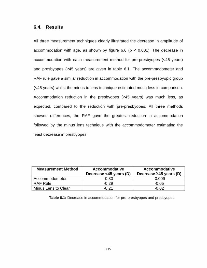
215
6.4. Results
All three measurement techniques clearly illustrated the decrease in amplitude of
accommodation with age, as shown by figure 6.6 (p < 0.001). The decrease in
accommodation with each measurement method for pre-presbyopes (<45 years)
and presbyopes (≥45 years) are given in table 6.1. The accommodometer and
RAF rule gave a similar reduction in accommodation with the pre-presbyopic group
(<45 years) whilst the minus to lens technique estimated much less in comparison.
Accommodation reduction in the presbyopes (≥45 years) was much less, as
expected, compared to the reduction with pre-presbyopes. All three methods
showed differences, the RAF gave the greatest reduction in accommodation
followed by the minus lens technique with the accommodometer estimating the
least decrease in presbyopes.
Measurement Method Accommodative Decrease <45 years (D)
Accommodative Decrease ≥45 years (D)
Accommodometer -0.30 -0.009 RAF Rule -0.29 -0.05 Minus Lens to Clear -0.21 -0.02
Table 6.1: Decrease in accommodation for pre-presbyopes and presbyopes

216
Analysis of variance (ANOVA) showed a significant difference between the three
measurement techniques (F= 341.498, p<0.0001). Comparisons of the
accommodometer measures and RAF rule measures showed no significant
difference (p = 0.75) and this held for pre-presbyopes (<45 years, p = 0.81) and
presbyopes (≥45 years p = 0.84). However there was a significant lower estimation
of amplitude measurements with the lens to clear method in comparison with the
accommodometer and the RAF rule (p < 0.0001).

217
Amplitude of Accommodation with Age
Age (yrs)
10 20 30 40 50 60 70 80 90
Acc
omm
odat
ion
(D)
0
5
10
15
20
Minus Lens to ClearAccommodometerRAF Rule
Figure 6.6: Reduction in amplitude of accommodation (in dioptres) with RAF rule, Accommodometer and Lens to Clear technique. (n=124)

218
Bland-Altman comparison of the RAF rule and accommodometer (Figure 6.7)
showed that on average the two techniques (push-up and push-down) did indeed
provide similar amplitudes of accommodation, shown as minimum focal length
(p=0.75), however the difference between the push-up and pull-down techniques
was greater with the accommodometer. The mean difference for the
accommodometer was calculated as -43mm with 95% agreement interval of ±
162mm. The RAF rule gave a mean difference of -34mm with 95% agreement
interval of ± 54.5mm.

219
Comparison between Average Push-Up/Pull-Down Focal Length Measurements
with Accommodometer & RAF Rule
Average Minimum Focal Length (mm)
0 100 200 300 400 500 600 700Diff
eren
ce b
etw
een
Pus
h -u
p an
d P
ull-
dow
n F
ocal
Len
gths
(m
m)
-300
-200
-100
0
100
200
300
119 mm
-205 mm
20mm
-89 mm
AccommodometerRAFMean95% Confidence Interval
Figure 6.7: A Bland-Altman Comparison of average minimum focal length with RAF rule and accommodometer.

220
Figure 6.8 shows separate comparisons of push-up and pull-down focal lengths
measured with both the digital accommodometer and the RAF rule. Mean
difference for the push-up values were found to be 9mm whilst pull-down showed a
greater mean difference of 18mm. The 95% confidence limits were also greater
with the pull-down values at ±195mm compared to ±177mm with push-up values.
No significant difference was found between these averaged results (p=0.06).

221
Comparison Between Push-Up & Pull-Down Minimum Focal Length Measurements
Mean Minimum Focal Length (mm)
0 100 200 300 400 500 600
Diff
eren
ce B
etw
een
Acc
omm
odom
eter
& R
AF
Rul
e (m
m)
-400
-300
-200
-100
0
100
200
300
400
187mm
-167mm
214mm
-177mm
Push-UpPull-DownMean95% Confidence Intervals
Figure 6.8: A Bland-Altman comparison of push-up and pull-down measurement techniques

222
On further analysis of push-up values with the digital accommodometer and RAF
rule mean differences were found as 244± 130mm and 234± 100mm respectively,
with 95% confidence limits of ±256mm with the accommodometer and ±234mm
with the RAF rule, these differences however were insignificant (p=0.22). For pull-
down measurements the mean differences were higher; calculated as 287±134mm
for the accommodometer and 268±119mm for the RAF rule with 95% confidence
limits of identical values of ±286mm and ±268mm and were calculated as just
within significance (p=0.05). The mean difference and confidence intervals were
higher with pull-down measurements overall in comparison to that of push-up
values (p<0.001).
Bland- Altman plots for repeatability of the RAF rule and accommodometer (Figure
6.9) show acceptable repeatability for both measurement techniques which again
were similar in magnitude and variability (-1.4 ± 27.5mm for accommodometer; -
4.1 ± 29.0mm for RAF rule; p = 0.09). The plot shows there is no change in
repeatability as focal length changes.

223
Repeatability of Accommodometer & RAF Rule
Average Focal Length (mm)
0 100 200 300 400 500 600 700
Diff
eren
ce b
etw
een
1st &
3rd
Mea
sure
men
ts (
mm
)
-100
0
100
200
A: 26mm
R:25mm
R: -33mm
AccommodometerRAF RuleMean95% Confidence Intervals
A: -29mm
Figure 6.9: Comparison of repeatability with RAF rule and accommodometer.

224
6.5. Discussion
On analysis of the results, the accommodometer has shown the expected
decrease in amplitude of accommodation and an increase in minimum focal length
with increasing age up to 45 years of age; following this little or no accommodation
remains and the minimum focal length measured for individuals is achieved by
their depth of focus. There was no overall systematic difference between the digital
accommodometer and RAF rule measurements, however the lens to clear
technique was significantly different compared to both devices. The lens to clear
technique is a distance task in which blur is induced by lenses. Such blurring with
lenses may vary when measured subjectively as there is no standardisation in
conducting this procedure (Wold et al., 2003). At distance the pupil size is
relatively fixed, unlike with near accommodation tasks where the near triad of
responses take place to aid focusing of close objects. However, due to the
minification effects of minus lenses, pupil miosis and hence the near triad of
response are indeed stimulated therefore this aspect is unlikely to provide a full
explanation as to why this technique differed to the accommodometer and RAF
rule. Differences in target size with the minus lens technique and RAF rule may
lend an explanation to differences in the results yielded. With the minus lens to
clear method the target size is fixed whereas with the RAF this varies as the target
brought closer to the observer leading to increased angular subtense with the RAF
rule (Gupta et al., 2008). Increased stimulation of proximal accommodation (depth
of focus) with the RAF rule in particular may have also contributed to the
differences in measurements (Gupta et al., 2008). Furthermore, with the minus

225
lens technique the target was changed on each lens presentation whereas the
same target was presented with both the RAF rule and accommodometer, which
may have influenced the results. Familiarity of the target may have lead to
measures of greater amplitude of accommodation with the accommodometer and
RAF rule as subjects will find the target easier to recognise even through blurring,
whereas with the minus lens method subjects may have lacked confidence in
recognising the letters correctly. However, with the push-up and pull-down
techniques which require continuous presentation and movement of a target, it is
difficult to keep changing optotypes although this could be done at set or random
time intervals with the accommodometer screen.
The digital accommodometer, did present some variability in results. The
accommodometer is required to be held at a 90 degree angle to the patient’s
forehead (as shown in Figure 6.10).

Instructions as seen on digital accommodometer prior to taking measurements of accommodation
226
Figure 6.10:
Instructions as seen on digital accommodometer prior to taking measurements of accommodation
227
The accommodometer can then detect the distance between the device and
patient’s forehead. However tilting of the device during the 5 second processing
period may have led to other facial features such as the nose being detected
instead, leading to an incorrectly estimated focal length and hence measurement
of amplitude of accommodation. In addition to detecting other facial features the
device may have detected lenses from spectacles (Ide et al., 2012) as patients
were fully distance corrected for measurement purposes. The shape profile of
these lenses may have distorted the signal received by the ultrasonic sensors. The
forehead also does not necessarily correlate to the position of the eyes, for
example some individuals may present with deeper inset eyes. Such aspects may
lead to shorter distance detection and over-estimation of accommodation.
Furthermore, the accommodometer testing procedure was patient controlled
whereas the RAF rule was examiner controlled. Patient-controlled push-up tests
have previously shown to yield higher results than if examiner-controlled (Fitch,
1971). To overcome this, the accommodometer testing could also have been
advanced and moved away by the examiner.

228
Although the accommodometer is a digital device, it is still in effect a subjective
method of measuring accommodation and hence shares some similar limitations
as other subjective measurement techniques. For example the understanding of
‘blur’ varies between each individual (Wold et al., 2003) with some reporting
blurring when the target first begins to distort and others waiting until completely
indistinguishable. To avoid this confusion all subjects were informed to stop when
the target first became blurry and could not be made clear with effort for push-up
tests and first became clear for pull-down testing.
Push-up testing may cause over-estimation in any subjective method (Antona et
al., 2009); it has been known for some time that, although commonly used in
practice, the push-up test reads higher amplitudes (Fitch, 1971; Hokoda et al.,
1982; Wold et al., 2003) reported to be between 1.50-2.50 dioptres (Rosenfield
and Gilmartin; 1990, Atchison et al., 1994; Rosenfield and Cohen, 1995). It is
stated that such errors do not pose any clinically significant complications for
young subjects, however, for those approaching early presbyopia it may lead to an
insufficient correction being prescribed for near tasks (Antona et al., 2009). In
addition it may lead to inaccurate estimations of how much accommodation is
gained following surgical procedures aiming to restore accommodation. Pull-down
measurements tend to estimate lower dioptric values in comparison to push-up
methods as the end-points differ. The pull-down technique begins from blurred
vision, where the patient is unaware of the character they are viewing up until it is
clearly identifiable, hence it may take longer until the subject is certain. With the

229
push-up technique the target is clearly distinguishable and can still be identified
whilst blurry leading to a higher reading (Antona et al., 2009). Our results support
this as minimum focal length was significantly greater with the pull-down technique
with both the accommodometer and RAF rule hence providing lower dioptric
values of accommodation. Moreover, with fixed target sizes, it is expected that the
push-up technique will overestimate the amplitude of accommodation, as the
angular subtense of the target will increase and induce proximal accommodation,
the effect being more pronounced in younger subjects with higher levels of
accommodation. For this very reason the accommodometer was designed to
provide an automatically adjustable target which would subtend the same angle
throughout the measurement procedure.
The contrast of the target can also affect the measurement of the amplitude of
accommodation (Johnson, 1976; Tucker and Charman, 1986; Heath, 1956). The
contrast of the accommodometer is 90% but this may vary with reflection of
lighting, within the test area, off the LCD screen. Additional lighting was not
required with the accommodometer as it provided sufficient luminance by back
illumination. However results suggest that neither illumination nor constant target
size has caused any consistent changes in the measurement of accommodation
as observed by comparison of the accommodometer compared to the RAF rule
data.

230
It has been suggested that the lens to clear technique might be a more appropriate
measure of accommodative ability (Wold et al., 2003). However the push-up and
pull-down techniques may provide more useful measurements of accommodation
as the ‘near point of clear vision can be identified’ unlike with minus lens
techniques (Atchison et al., 1994); also the minus lens technique is considered
more variable if measured subjectively (Wold et al., 2003). Furthermore push-up
techniques are considered as more natural methods of measurements.
A recent paper by Ide et al (2012) has also investigated the use of a device similar
to the Aston accommodometer in having ultrasonic sensors to detect the working
distance objectively, but this measurement did not affect the target size, the target
was not back illuminated and the range of measurement was only 20-50cm.
Surprisingly, despite this a difference was found in measurements compared to an
subjective near point measurement device.

231
6.6. Conclusion
In conclusion, despite some limitations with the sensors of the digital
accommodometer and failure to demonstrate advantages in measurement with
constant target subtense and illumination, the repeatability of both the RAF rule
and accommodometer were acceptable and equivalent which suggests the new
measurement technique is good, although re-working of the design is required. As
a result a Smartphone version is currently being developed which should be a
more compact device and overcome the variability issue as the distance between
the sensors and observer is less, allowing better alignment with the subject’s
forehead (Figure 6.11).

232
Figure 6.11: Prototype Smartphone Accommodometer

233
The digital accommodometer although variable, has shown good demonstration of
the increase in minimum focal length and profile of amplitude of accommodation
with age. It has the potential to be a useful alternative to the RAF rule being more
portable and maintaining the illumination and visual subtense of the target. The
development of a Smartphone version of this device may offer better assessment
in the near focus obtained with accommodating and multifocal IOLs.
In addition to demand for better near vision there is a growing demand for
improved intermediate vision with multifocal and accommodating IOLs. Diffractive
multifocal designs incorporating optics which allow three foci have been
developed. The forthcoming chapter presents the evaluation of the visual
performance of such a design using modern assessment techniques.

234
CHAPTER 7
Visual Outcomes & Subjective
Experience Following Bilateral
implantation of a New Trifocal
Intraocular Lens
235
7.1. Introduction
In recent years, there have been many advances in cataract surgery procedures.
Traditionally, cataract extraction involves implantation of a monofocal intraocular
lens (IOL) providing adequate distance vision but the requirement of spectacles for
near vision. Due to an increasingly ageing population and changes in lifestyle, the
demand for spectacle independence following cataract surgery has led to the
development of accommodating and multifocal IOLs.
Multifocal IOLs were first introduced in the 1990s and are now becoming more
popular with new improved designs continually being developed. The two main
designs of multifocal technology include refractive and diffractive optics, both of
which produce simultaneous images of distant and near objects. As described in
Chapter one, refractive multifocal IOLs consist of rings of different refractive
powers for near and distance correction whilst diffractive IOLs use the Huygens-
Fresnel principal where the IOL acts as a diffraction grating. Of the two designs it
has been reported diffractive multifocal IOLs show better visual performance
(Maxwell et al., 2009; Gatinel et al., 2011).

236
Multifocal IOLs, although they attempt to provide spectacle independence, do not
provide adequate vision for intermediate distances for many patients (Bucci 2006;
Alfonso et al., 2007; Petermeier et al., 2007; Goes et al., 2008) with reports of
reduction in visual acuity within the intermediate range (Voskresenskaya et al.,
2010). Since the introduction of computers and boost in mobile technology in the
last decade there has been a significant increase in VDU usage by the elderly
population and hence demand for better intermediate vision. In 2006 it was
reported the percentage of 50-65 year olds using computers doubled
(Voskresenskaya et al., 2010). Such a limitation would require use of spectacles
solely for this working distance, hence it is suggested that multifocal IOLs are
better referred to as ‘bifocal’ IOLs in order to avoid high expectation of intermediate
vision (Voskresenskaya et al., 2010).
Examples of investigations noting decreases in intermediate visual acuity include
that of Hütz et al (2008) with the ReSTOR multifocal IOL where an average
reduction down to 20/40 at distances of 40 to 80cm was reported. Blaylock et al
(2006) also reported a decrease in intermediate visual acuity from 40 to70cm of
20/30 to 20/44 with multifocals, however, only 25% of patients reported
significantly severe problems whilst 75% occasionally or never found any difficulty.

237
Hayashi et al (2009) reported a significant decrease in intermediate vision with
IOLs with a +4.00D addition for near in comparison to +3.00D multifocal lenses,
although the near acuity with +3.00D lenses was less than that of the +4.00D.
Diffractive multifocal IOLs in particular show worse intermediate vision (Alfonso et
al., 2007; Blaylock et al., 2006; Pepose et al., 2007; Hütz et al., 2008).
Visual acuity for intermediate distances may vary between individuals due to the
photopic and mesopic pupil sizes. Alfonso et al (2007) noted those requiring
intermediate correction had pupil diameters of 4-4.5mm on average in photopic
condition and 6-6.5mm in mesopic conditions. They found vision with the ReSTOR
multifocal deteriorated from 40 to 70cm but only 4% of subjects required
spectacles for intermediate work.
Other reported limitations of multifocal intraocular lenses include haloes, glare and
reduced contrast sensitivity particularly in mesopic conditions (Steinert, 2000;
Richter-Mueksch et al., 2002; Awwad et al., 2008). Photic phenomena are reported
to be 3.5 times more prevalent in multifocals than monofocal lenses (Leyland &
Pringle 2006). In a study conducted by Woodward et al (2009) 7% of eyes required
explantation to resolve the visual complications induced by the multifocal lens
used.

238
As incident light is divided and shared amongst two foci in multifocal IOLs,
overlapping of the focused and out of focus images reduces contrast sensitivity
resulting in poor image quality. Poor contrast sensitivity tends to be more
pronounced at lower spatial frequencies (Arens et al., 1999; Vaquero-Ruano et al.,
1998; Steinert 2000) although there are some claims of worse contrast sensitivity
at higher spatial frequencies also (Alfonso et al., 2009; Montés-Micö et al., 2004).
Montes-Mico et al (2001) highlighted this compromise of contrast sensitivity at low
spatial frequencies and suggested it is due to light scatter whilst that at higher
spatial frequencies, the effect is mainly attributed to defocus and optical
aberrations. Despite the reported reductions, photopic contrast sensitivity, although
reduced in comparison to monofocal IOLs, is still found to be within the normal
limits (Hayashi et al., 2009; Montés-Micó et al., 2004). Studies have also indicated
an improvement of contrast sensitivity 3-6 months following implantation (Montes-
Mico et al., 2003).
Glare is another well known phenomenon to occur with multifocal IOLs, particularly
with refractive designs. It was reported 21.3% of patients implanted with ReSTOR
IOL complained of glare in comparison to just 7.5% of those implanted with
monofocal IOLs (Chiam et al., 2006). In a survey carried out by Mamalis et al
(2003) it was reported one of the main reasons for explanting a multifocal IOL was
disability glare in addition to incorrect IOL power and IOL dislocation, as well as
being the most common complaint in a study conducted by Blaylock et al (2006).
Disability glare has been described as light which spreads over at least 1°

239
(Hofmann et al., 2009), but is more likely to vary with individual’s tolerance and
environmental conditions. The size of haloes tends to be greater in patients over
70 years as light scatter from ocular structures such as; the cornea, increases with
age which adds to the effect of multifocals (Dick et al., 1999). Hofmann et al (2009)
investigated retinal straylight with monofocal and multifocal IOLs and reported a
20% increase with multifocal IOLs; this study also pointed out more patient-
reported discomfort in dim light conditions which potentially would affect night
driving. Although this measure of straylight was statistically insignificant it was
concluded that the straylight produced with multifocals adds to visual discomfort
more rather than visual disability as shown by the significantly higher complaint
scores from patients implanted with the AcrySOF ReSTOR IOL. Vries et al (2008)
also investigated retinal straylight measurements in an apodized diffractive IOL
and reported slightly higher levels with the multifocal lens group than monofocal
group. Higher levels of intraocular straylight lead to higher sensitivity to glare (Van
Den Berg, 1995).
There is controversy over patient satisfaction with multifocal IOLs as for example;
a questionnaire completed one year following implantation of the Array multifocal
highlighted three particular symptoms of glare, halos and blurry distance vision
which were reported as significantly worse than with monofocal lenses (Steinert,
2000). These symptoms were described as ‘severe’ in 15% of cases for halos,
11% for glare and 4% for blurred distance vision. However, studies have shown
such occurrence of photic phenomena does reduce with increasing time post-

240
operatively (Vaquero-Ruano et al., 1998) in a process of neuroadaptation
(Voskresenskaya et al., 2010; Souza et al., 2006).
Jacobi et al (1999) suggested asymmetric bilateral implantation of multifocal IOLs
in order to enhance contrast sensitivity (mix and match technique) and
intermediate vision where one diffractive IOL is implanted in one eye and a
refractive IOL in the other. However compromises in distance and near vision
result with loss of stereopsis (Voskresenskaya et al., 2010). Other suggestions
include aspheric designs with a lower additional power for near vision (Hayashi et
al., 2009) however this may not provide sufficient near vision.
A clear requirement thus exists for improved intermediate vision with multifocal
technology. Swanson (1994) developed a theoretical calculation for a diffractive
lens to form three foci, where each focal point receives 28.8% of the incident light,
and a residual 14% of light distributed to the other foci. Such a design, known as a
trifocal lens, allows vision at intermediate distances without degrading vision in the
distance or at near (Gatinel et al., 2011). There are very few studies investigating
this new concept of trifocal lenses. The first published findings were by
Voskresenkaya et al (2010) where the visual outcomes of the MIOL-Record 3
(Reper-NN, Nizhegorodskaja Provinces, Russia) trifocal lens were investigated.
The MIOL-Record consists of a stepped design diffractive optic, this lens
significantly improved distance and near vision comparable to diffractive multifocal

241
IOLs (AcrySof ReSTOR, AcrySof Natural ReSTOR, Acri.Lisa 366D, ReZoom,
Tecnis MF), as well as providing adequate intermediate vision of 0.66 ±0.22 at
50cm.
As there are three simultaneous foci with a trifocal lens, it may be argued that
contrast sensitivity could be compromised further however Voskresenkaya et al
(2010) found photopic contrast sensitivity to be comparable with that of monofocal
IOLs. Mesopic contrast sensitivity however was indeed reduced in comparison to
monofocal IOLs although this did not seem to affect patient satisfaction as 94%
reported no or mild difficulty with vision at night. Glare and halos were also of
acceptable levels as 75-94.5% of subjects did not find significant problems with
such photic phenomena and also noticed a decrease with time following
implantation leading to a significant decrease in complaints 3-6months post-
operatively.
Gatinel et al (2011) described an aspheric fully diffractive IOL aiming to provide
better intermediate vision and less photic phenomena which has been
implemented into the Physiol FineVision IOL; optical bench testing has shown it to
successfully provide these aims, but further clinical studies are required to validate
its performance. The purpose of the present study was therefore to investigate
the visual outcomes, measurement of glare, photopic and mesopic contrast

242
sensitivity and the patient satisfaction with the Physiol FineVision trifocal diffractive
intraocular lens.

243
7.2. Methods
7.2.1. Intraocular Lens Characteristics
Physiol FineVision is a four point haptic aspheric diffractive lens, made of
hydrophilic acrylic with 25% water content, of bi-convex design (Figure 7.1). Three
foci are achieved by a combination of two bifocal diffraction patterns of +1.75
dioptre addition for intermediate vision and +3.50 dioptre addition for near vision
(Figure 7.2). The first diffraction pattern with the addition of +3.50 dioptres is the
first diffraction order with a second diffraction order occurring at a vergence of 7
dioptres. The second pattern, with an addition of +1.75 dioptres as the first order
has a second order which is twice the first diffractive order, resulting in +3.50
dioptres. Since the first and second orders of this second pattern equate to the
intermediate and near powers of the IOL they contribute and enhance intermediate
and near vision. Resulting light loss from this design is reduced to approximately
14% from a typical 18% with bifocal multifocal designs (Gatinel et al., 2011). In
photopic conditions 42% of incident light is used for distance foci, 29% for near
and 15% for intermediate, with mesopic conditions 58% is distributed to distance,
20% for near and 8% for intermediate vision.

244
The IOL is apodized, where the step height reduces from the centre to the
periphery of the lens optic. The design is pupil dependent; with pupil enlargement
light enters through the peripheral areas of the IOL which is assigned to distance
vision, reducing the amount of light reaching near and intermediate foci. The optic
is entirely convoluted where a smoothing function has been implemented into the
design to reduce the perception of haloes. The optic diameter is of 6.15mm with
an overall diameter of 10.75mm. The IOL filters ultraviolet and blue light. There is
also a four-point loop haptic design for increased stability. The powers available in
this particular design range from +10 dioptres to +30 dioptres in 0.50 dioptre steps.

245
Figure 7.1: FineVision trifocal diffractive IOL
Order 0:
Distance
Vision = 40%
Order 1: Near
Vision = 40%
+3.50D
Order 2: Very
Near Vision = 4%
+7D
Figure 7.2: Multiple foci of trifocal IOL

246
7.2.2. Experimental Design
Thirty eyes of fifteen patients undergoing routine bilateral cataract extraction at the
Midland Eye Institute, UK were implanted with the PhysIOL FINe Vision intraocular
lens following informed consent. The study was approved by the Institutional
Review Board and conformed to the tenets of the declaration of Helsinki.
Participants were aged between 52-86 years, with a mean age of 69.8 ± 10.0
years, with bilateral senile cataracts. Exclusion criteria consisted of no retinal
pathology or history of ocular infections, clear intraocular media, no previous
ocular surgery or trauma, potential for BCVA of only 6/12 or worse. Good general
physical and mental health was also required for participation in the study. The
study consisted of an initial pre-operative visit of biometry measurements using the
Zeiss IOLMaster and explanations regarding surgery and the proposed IOL
implantation. Routine phacoemulsification surgical technique and IOL implantation
was completed at the Midland Eye Institute, UK. Following surgery ocular health
was again assessed to ensure no post-surgical complications had arisen. Second
eye surgery took place 6 weeks following the initial operation.

247
Visual performance was assessed at a three months post-operative visit to assess
the visual. Tests included;
� Monocular and Binocular UCDVA (logMAR)
� Subjective Refraction (maximum plus for best distance VA)
� Monocular and Binocular BCDVA
� Monocular defocus curves from +1.50D to -4.00D in 0.50D steps in photopic
(85cd/m2) conditions
� Binocular defocus curves from +1.50D to -4.00D in 0.50D steps, in photopic
and mesopic (5 cd/m2) conditions
� Contrast sensitivity measurement using the CSV-1000 test (VectorVision, Ohio,
USA)
� Halometry glare test
� Completion of the NAVQ questionnaire

248
Defocus curve measurements consisted of recording visual acuity, using
computerised logMAR progression Test Chart (Thomson Test Chart XPert,
Thomson Software Solutions, Hertfordshire, UK ), through random presentations
of trial lenses from +1.50 dioptres to -4.00 dioptres over the refractive correction;
this was repeated moncularly in photopic conditions and binocularly in both
photopic and mesopic conditions. Pupil diameters were also measured in photopic
and mesopic conditions. Letters were randomized as each trial lens was presented
to avoid subjects memorizing the chart (Gupta et al., 2007).
Photopic contrast sensitivity was recorded using the CSV-1000 contrast sensitivity
chart at a distance of 2.5 meters. The chart comprises of 4 rows of 8 sine-wave
gratings consisting of spatial frequencies 3, 6, 12 and 18 cycles/degree. The
subject was instructed to indicate the direction of each sine gratings in each of the
five rows until no longer resolved. The last correctly indentified grating in each row
was recorded.

249
The Halometry glare test (Buckhurst et al,. 2011) used to measure the glare area
for each patient, involved a central LED light source (colour temperature 3200K)
placed centrally on a flat LCD screen on which letters were presented around the
light source at eccentricity of 0, 45, 90, 135, 180, 225, 270, 315 degrees. Letters
were presented at a height that subtended 0.21° at a working distance of 3 meters,
equivalent to the minimum vision requirements for driving standards in Europe.
The letters were presented on a black background at a contrast of 500 Weber
contrast units (Cw) and presented along each of the eight meridians in 0.05° steps.
Testing was conducted in a dark room monocularly and binocularly, letters were
presented in a random order and randomized between presentations along each
meridian from the centre. The closest point to the glare source, at which the patient
correctly identified a letter, was recorded.
To assess subjective satisfaction with near vision function, patients completed a
validated 10-item questionnaire; the Near Activity Visual Questionnaire (NAVQ),
(Buckhurst et al., 2012). The NAVQ is designed for the evaluation of presbyopic
corrections, and requires patients to indicate their level of difficulty performing
common near and intermediate vision tasks without the use of reading spectacles
(where 0 = no difficulty, and 3 = extreme difficulty), and to rate overall satisfaction
with their near vision (where 0 = completely satisfied, and 4 = completely
unsatisfied). The summated score from the main body of 10 questions is adjusted
to a Rasch score (from 0 to 100 Logits) using a conversion table, such that 0

250
indicates no difficulty at all with any near tasks, and 100 indicates extreme difficulty
with all near activities.

251
7.3. Statistical Analysis
Monocular and binocular visual acuity measures were totalled and averaged for
comparisons and defocus curve plots. Contrast sensitivity scores were also
averaged with comparisons made between photopic and mesopic contrast
sensitivity. All answers from the questionnaire were totalled and converted to a
Rasch score to give the final questionnaire result. T-tests for measures of
statistical significance were also performed.

252
7.4. Results
All patients underwent uneventful cataract surgery on both eyes, IOLs were well
centred in all eyes, with no occurrence of pupil distortion or iris trauma. Details of
the means and standard deviations of monocular and binocular distance visual
acuities and distance vision efficacy are shown in Table 7.1. The mean monocular
refractive correction was 0.27 ± 0.36 D sphere (range -0.25 to +1.00 D) and -0.48
± 0.45 D cylinder (range 0 to -1.50 D). Figure 7.3 shows the binocular mean
defocus curves under photopic and mesopic conditions. In both lighting conditions,
optimum visual acuity results were obtained at 0.00 D defocus which is equivalent
to distance vision viewing, with a second “peak” at -2.50 D equivalent to near
viewing at 40 cm. No distinct peak in the intermediate zone was present for either
of the lighting levels, with no sharp drop in acuity in the intermediate zone for the
photopic condition, although the range of clear vision (0.3 logMAR or better)
extended from +1.00 to -2.50 D of defocus. The mean visual acuities were
generally better in the photopic conditions, however the differences between
lighting conditions were not significant, except at -1.50 D defocus (p=0.008),
corresponding to an intermediate viewing distance.

253
Acuity
Number of eyes/ patients (%)
Mean ± SD 0.3 logMAR or better 0.1 logMAR or better
Monocular:
UDVA 0.19 ± 0.09 24 eyes (80) 6 eyes (20)
CDVA 0.08 ± 0.08 30 eyes (100) 21 eyes (70)
Binocular:
CDVA 0.06 ± 0.08 15 patients (100) 13 patients (87)
Defocus (D)
Vis
ual a
cuity
(log
MA
R)
0.0
0.2
0.4
0.6
0.8
PhotopicMesopic
0.50 0.001.001.50 -0.50 -1.00 -1.50 -2.00 -2.50 -3.00 -3.50 -4.00
Table 7.1: Monocular and binocular logMAR distance visual acuities 3 months following FineVision IOL implantation. UDVA = uncorrected distance visual acuity; CDVA = best-
corrected distance visual acuity.
Figure 7.3. Binocular mean defocus curves for the FineVision trifocal IOL in photopic and mesopic conditions. Error bars = ± 1 SD. The dotted reference line at 0.3 logMAR equates
to the European driving standard.

254
Figure 7.4 shows the monocular and binocular distance contrast sensitivity
(log10CS) under photopic conditions. Binocular contrast sensitivity values were
significantly better than monocular values at all spatial frequencies tested (p<0.05).
No significant differences in contrast sensitivity values between right and left eyes
were found at any spatial frequency (p> 0.05).

255
**
*
*
Spatial frequency (cpd)
LogC
S
0.0
0.5
1.0
1.5
2.0
Right eyesLeft eyesBinocular
3 6 12 18
**
*
*
Figure 7.4. Monocular and binocular contrast sensitivity functions with the FineVision
trifocal IOL, under photopic conditions. * = statistically significant difference between
monocular and binocular values.

256
Figure 7.5 illustrates the halometry results, with the magnitude of the mean
monocular and binocular photopic scotomas, measured under mesopic conditions
shown. The mean photopic scotomas are generally uniform in shape, extended
binocularly between 0.69 ± 0.24 degrees and 1.03 ± 0.20 degrees for all 8
meridians.
NAVQ scores for subjective satisfaction with near vision were high, with a mean
Rasch score of 15.9 ± 10.7 Logits (0 = completely satisfied, 100 = completely
unsatisfied; range 0 to 33.3). The final NAVQ item, rating overall satisfaction with
near vision (0= completely satisfied, 4= completely unsatisfied) resulted in a mean
score of 0.7 (range 0 to 2).

257
0
0.2
0.4
0.6
0.8
1
1.2
1.4
90
45
0
315
270
225
180
135
Binocular
Right eyes
Left eyes
Figure 7.5: Size of monocular and binocular photopic scotomas, measured using
halometry under mesopic conditions. Y axis = extent of scotoma from glare source
(degrees), radial axis = visual field meridian (degrees).

258
7.5. Discussion
Multifocal IOLs are becoming more widely used as patients undergoing cataract
surgery have increasing functional expectations and a desire for post-operative
spectacle independence (Hawker et al., 2005; Pager, 2004; Mũnoz et al., 2011).
Current diffractive multifocal IOLs typically provide good vision at distance and
near (Zhang et al., 2011; Munoz et al., 2011; Alió et al., 2008) but have the
disadvantages of bifocal design, potentially leading to intermediate vision
difficulties (Gatinal et al., 2011; Voskresenskaya et al., 2010). To the best of our
knowledge, this is the first study to report clinical outcomes of a cohort implanted
binocularly with a diffractive trifocal IOL design. The mean monocular UDVA (0.19
± 0.09) and CDVA (0.08 ± 0.08) results are similar to the values reported by
Voskresenskaya et al (2010) with predominantly monocular implantation of the
MIOL-Record. Furthermore, the visual acuity outcomes were comparable to those
achieved with several bifocal-design diffractive IOLs (Zhang et al., 2011; Alió et al.,
2011, Alió et al., 2008).
Binocular defocus curve testing indicated an extended range of clear vision.
Although mean VA was 0.3 logMAR or better from +1.00 to -2.50 D defocus in both
photopic and mesopic conditions, no obvious peak in VA was apparent in the
intermediate zone. Such a finding may be expected, as a relatively small
proportion of light is available for intermediate vision compared to distance and
near. As pupil size increases, a greater proportion of light is directed to the
259
distance focus due to the apodized optic, such that for a 5mm pupil, only
approximately 5% of light is available for intermediate vision. The reduced light
available for intermediate vision with larger pupil sizes is likely to be the cause of
the significantly poorer visual acuity in mesopic compared to photopic conditions at
-1.50 D defocus. No significant differences in VA between mesopic and photopic
conditions were found at any of the other defocus levels tested.
With the FineVision IOL binocular contrast sensitivity values were significantly
higher than monocular values at all spatial frequencies. The well-known effect of
binocular summation explains the difference between monocular and binocular
results, and is in agreement with previous reports of diffractive IOL outcomes,
where several authors have advised binocular implantation to optimise contrast
sensitivity (Jacobi et al., 1999; Fernández-Vega et al., 2007; Schmidinger et al.,
2005). Multifocal IOLs have previously been reported to cause up to a 50%
reduction in contrast sensitivity (Pieh et al., 1998), however, in this study
monocular contrast sensitivity values were within the normal range for older adults
as described by Pomerance and Evans (1994), although slightly below their mean
values. Reasons for this may include the older cohort used in the present study
(mean age 69.8± 10.0 years in the present study, compared to 63.9 ± 12.2 years
for Pomerance and Evans) and normal age-related retinal and neural changes
(Elliot 1987, Spear 1993).

260
Photic phenomena frequently associated with multifocal IOLs are approximately
3.5 times more common with multifocal compared to monofocal IOLs (Leyland and
Zinicola, 2003) and may impact on quality of life (Javitt and Steinert, 2000). In the
present study, no patients reported photic phenomena, suggesting that the design
of the FineVision IOL, with increasing far vision dominance as pupil size increases,
may be effective in minimizing halos and glare perception. However, a larger scale
study would be required to gain a full insight into the frequency of adverse photic
phenomena with the FineVision IOL. The mean size of the photopic scotomas
(monocular extent from glare source ranged from 0.6 ± 0.3 to 1.1 ± 0.2 degrees)
measured in the present study compares favourably with previous measures using
the same technique and with another study investigating glare, on patients
implanted with a multifocal and an accommodating IOL design (Berrow, et al,.
2012). The novel halometry technique used within the current study has not been
used widely therefore comparisons with multifocal IOLs are limited. However
previous straylight measurements using the CQuant straylight meter show
straylight levels of 1.2 log units within a diffractive multifocal group (de Vries et al.,
2008) therefore showing much higher levels of halos and glare compared to the
FineVision trifocal lens.
Subjective satisfaction with unaided near vision, as measured with the NAVQ
questionnaire, was high in the present study (mean 15.9 ± 10.7 Logits). The NAVQ
test (Buckhurst et al., 2012) is designed to allow a more standardized comparison
of presbyopia correction strategies, by questioning patients on their ability to
perform common near tasks such as; reading post and seeing the display on a

261
computer without an additional near vision correction. The mean value obtained
with the FineVision trifocal IOL shows a higher level of patient satisfaction with
near vision than reported by Buckhurst et al. (2012) for other multifocal and
accommodating IOLs. The improved score with the FineVision compared to other
presbyopia-correcting IOLs may be due in part to improved intermediate vision
provided by the 1.75 D intermediate add as the NAVQ includes questions relating
to intermediate-distance visual function, such as using a computer and performing
hobbies such as gardening or playing cards.
A particular limitation in this investigation was the small sample size. Although the
sample size efficiently demonstrated the advantages of trifocal design, a larger
cohort would be required in order to validate these findings in a wider range of
patients. Patients are usually happy following cataract surgery as limitations of the
IOL optics are of less concern than the reduction in vision and light scatter caused
by the cataract. However, the use of a validated near vision questionnaire at least
allows comparison with other forms of presbyopic correction already evaluated.

262
7.6. Conclusion
In conclusion, the FineVision trifocal IOL provides a good standard of distance,
intermediate and near visual function, as demonstrated by defocus curve testing.
The increasing far vision dominance of the IOL as pupil size increases may be
effective at reducing photic phenomena frequently associated with multifocal IOLs.
Near vision satisfaction amongst this cohort of bilaterally-implanted patients was
high, which along with the clinical measures, suggests that the FineVision IOL is
an effective method of providing good distance, near and intermediate visual
ability. However a greater cohort is required to appreciate the benefits of this new
IOL design.

263
CHAPTER 8
Summary & Conclusions

264
8.1. Introduction
Since the introduction of intraocular lenses in the 1950s there have been vast
developments in their design. Research initially concentrated on improving IOL
materials to allow long-term stability within the eye and to remain transparent once
implanted. In more recent years, focus has now changed to enhancing the optics
of intraocular lenses to provide optimum vision following surgery. Such lenses,
termed premium IOLs, aim to reduce astigmatism and provide correction for
presbyopia. The health system of the U.K. currently implant basic spherical IOLs
as the standard of care following cataract extraction as the cost of implantation of
premium IOLs exceed that of what is currently available. Individuals seeking
spectacle independence following cataract surgery or refractive lens exchange are
often unaware of the options that premium lenses are able to offer. For patients
that desire such lenses, payment of both the actual lens and private surgery fees
would be required as no ‘top-up’ option is available through the National Health
Service in the U.K.
As outlined in the introduction, although many advances have been made in IOL
designs, the benefits of premium IOLs have not been well established, nor when
they should be considered as an optical correction. Implanting advanced optical
designs also raises more complex surgical issues relating to IOL rotation and
centration which deserve attention to optimise the visual results. The present
thesis therefore examined the benefits of astigmatic correction with a toric lens

265
over the mean spherical equivalent and how pupil dilation influences the alignment
of these lenses. Centration of IOLs during surgery and over the following 6 months
was also examined. Better methods to measure residual accommodation were
explored along with what factors, other than age, affect when an individual
becomes presbyopic and therefore should consider elective IOL surgery with
implantation of a premium IOL. Finally, the benefits of a new type of IOL, a trifocal
diffractive design, was evaluated to determine the range of clear vision offered as
well as the visual compromises experienced.

266
8.2. Summary
Chapter 2 indicated the negative effect of uncorrected astigmatism on a selection
of daily activities in the elderly population. Performance of tasks reduced as
simulated astigmatism increased in power from as low as 1 dioptre with deviation
of the axis from the vertical further affecting visual ability. Contrast sensitivity was
highly compromised by increasing levels of astigmatism which may present as
difficulty in night driving and navigation. As a large proportion of patients already
restrict their journeys in dark conditions post-cataract surgery, residual astigmatism
will simply add to this effect. Individuals with spherical errors alone usually conform
to driving standards following cataract surgery but uncorrected astigmats often do
not without additional refractive correction which may restrict driving further due to
lack of confidence through reduced vision.
In addition, the study showed significantly reduced reading acuity and reading
speed with increases in astigmatic power. Reading is of paramount importance in
the elderly population often due to restrictions in mobility. Furthermore, it has been
iterated that visual demands particularly for near work are continuously rising with
developments in technology and communication, the use of which is increasing
within elderly populations. Despite the lack of a mobile lens in presbyopia already
impacting on near vision, this will be exacerbated by effects of astigmatism. The
subjective ratings of clarity within the study highlighted the awareness of
individuals of that poor vision.

267
Collectively, such restrictions in the ability to perform the daily tasks described will
lead to a significant reduction in quality of life as well as increased risk of falls and
possible risks to driving safety. Chapter 2 therefore outlined the potential benefits
that premium lenses such as toric IOLs may provide to patients through
improvements in quality of life. The study also supports toric implantation as
standard care for cataracts by public health services as it is suggested that the
cost of implantation would outweigh the consequences of residual uncorrected
astigmatism.
Implanting toric IOLs, however, raises speculation of rotational stability. For a
successful visual outcome, toric IOLs require high precision on alignment from
surgeons. As there is currently no standardised procedure for alignment of such
premium intraocular lenses, Chapter 3 therefore examined the effect of dilation on
newer, more robust imaging techniques for toric IOL alignment which rely on either
iris features of conjunctival blood vessels on either side of the iris to compensate
for head rotation when examining IOL orientation. The development of more
complicated optics for correction of presbyopia following cataract surgery requires
more precise and diligent techniques of implantation. Pupil dilation is imperative in
cataract and refractive lens surgery although it has been associated with changes
in pupil centration which could impact alignment. Current positioning techniques
however are crude and may lead to misalignment along with central dislocation
due to deviation in pupil centre on dilation.

268
Assessment of iris features and conjunctival vessels with various degrees of
dilation showed the latter as a more stable landmark for use in IOL alignment.
Head tilt has been considered as a possible source of error in pre-operative
marking for toric implants, however, the current investigation showed little
disturbance to measures by head movement suggesting that a slit lamp head rest
provides sufficient stability. Pupil diameter increased in a sigmoidal pattern moving
more inferiorly through dilation. On average the pupil, prior to dilation, is located
slightly supero-nasal, the limbus measures showed negligible changes in its
position on repeated measures suggesting the pupil to be an unreliable structure
for alignment procedures. Surgeons may thus find use of the limbus as a reference
structure in such proceedings more beneficial in addition to conjunctival vessels as
markers for alignment. As previously discussed only young subjects were recruited
for the study therefore repeating the investigation with an older cohort may provide
further knowledge as to how dilation affects mature pupils. Such information may
prove beneficial as premium IOLs are generally fitted in older age groups.
Future studies could expand this research by investigating the differences between
pupil location in different coloured irides to establish if the effect of dilation in pupil
decentration is more pronounced in dark or light irides. Such information may then
be accounted by surgeons in order to achieve the best possible surgical outcome
with premium IOLs. Scheimpflug imaging may be another way to determine
movement changes in IOL centration with respect to the pupil along with additional

269
information on IOL tilt, with time. Moreover, future research could explore if minor
decentration contributes to visual phenomena with multifocal IOLs.
Chapter 3 therefore clearly established the significant changes of pupil centration
and surface features during pre-operative dilation and how this may affect the
implantation of premium IOLs. However, the possible effect on the pupil following
cataract surgery has not been considered in the performance of premium IOLs,
Chapter 4 thus extended this by examining the post-operative effect on pupils six
months after cataract extraction and IOL implantation. Early investigations by
Gibbens et al (1989) had reported on pupil changes following suggestions of
reduced pupil diameter with ICCE and ECCE procedures. Since then cataract
surgery has advanced considerably and no previous research has investigated
such post-operative effects with newer phacoemulsification techniques. As post-
operative complications have reduced in magnitude since the evolution of cataract
extraction, it would be assumed that little impairment to pupil function would occur.
On the contrary, chapter 4 describes post-operative inferior movement of the pupil
centre but no horizontal displacement. Pupils became more oval with time and the
maximum diameter following dilation showed significant reduction. These changes
tended to become more apparent much later after surgery rather than immediately
following surgery. Interestingly, IOL centration remained stable with respect to the
limbus but showed vertical displacement when compared to the pupil. Such
findings suggest that the pupil must alter its position whilst in fact the IOL remains
stable. In relation to premium IOLs this information emphasizes the need for a

270
more stable structure to assist surgeons with the centration of premium IOLs.
Complexity in their optical designs may present considerable visual disturbances if
inaccurately centred. It is therefore again confirmed that the limbus offers a more
suitable guide for IOL alignment. The investigation further suggests post-operative
changes in pupil structure may interfere with the performance of premium IOLs
regardless of initial positioning, indicating the requirement for further research into
post-operative alignment of premium IOLs.
Comparisons and measurements of diameters prior to surgery were not made,
which may have provided some information to the variation found within chapter 4
and provides scope for further research. Permeability of the cornea could also be
examined in order to investigate if this contributes to the differences in pupil
diameter following surgery. In addition, advanced high resolution imaging of the iris
may aid further research into where damage may occur to the iris structure to
result in only vertical shifts of the pupil centre.
Spectacle independence and correction of presbyopia remain the penultimate aim
of premium IOLs to which there is a rising interest for refractive lens exchange
amongst patients approaching presbyopia. Age has already been deemed as the
greatest factor contributing to presbyopia. However, earlier work from various
researchers introduced the concept of environmental factors influencing amplitude
of accommodation and hence the age of onset of presbyopia. Climate, increased

271
sunlight and environmental temperature presented as common findings affecting
accommodation and leading to early presbyopia, giving the general agreement that
populations inhabiting near the equator exhibited lower amplitudes of
accommodation and developed presbyopia much earlier than clinically expected.
Chapter 5 explored the amplitude of accommodation and onset of presbyopia in a
population within the United Kingdom. Questioning on lifestyle and measurements
of accommodation has shown many factors may contribute to the rate of
progression of presbyopia in addition to age, in particular; alcohol consumption,
smoking, UV exposure, weight and even use of VDUs and mobile communication.
Knowledge of factors which may indicate the rate of progression to presbyopia
allows estimates of an individual’s age of presbyopia onset to be made. Such
knowledge will aid better communication when discussing the changes that would
occur with patients considering a premium IOL clear lens extraction.

272
Within this thesis, the average presbyopia onset was calculated as approximately
48 years. Cataracts typically form later than this, but patients should be provided
with all the relevant information to them which should include advice on premium
IOL implantation, removing the need for further cataract surgery in later years.
Chapter 5 has thus shown despite improvements in health, diet and medicine,
aspects of modern lifestyle may still increase the rate of presbyopia hence
warranting the need of presbyopia correcting solutions which may be addressed by
premium IOLs, as no significant factors have shown to reverse or reduce the effect
of presbyopia.
Further assessment of lifestyle and environmental factors on a more global scale
may be carried out as little information exists on this area of research. To avoid
bias of information given by the patients to questionnaires, data for dietary
information could be collected prospectively using food diaries and blood samples.
Information gathered through such procedures would provide better determination
of vitamin intake as the food and supplements consumed does not necessarily
correlate to nutrition actually absorbed into the bloodstream. Further research
proposes to revise the work of Donders (1864) and Duane (1909, 1912) using
more objective measurements of accommodation rather than subjective. As
previously highlighted the primary limitation of this chapter was the use of
subjective measurement of amplitude of accommodation. Despite combining push-
up and pull-down results, the values obtained may still present higher readings due
to depth of focus. Chapter 6 outlined the inadequacies of the RAF rule in providing
273
reliable measurements of accommodation and how new technology may be able to
overcome the limitations of the standard push-up technique with the RAF rule.
It is important to investigate how much near vision range is gained through
presbyopia correcting procedures, such as implantation of premium lenses, in
order to assess their level of benefit. To accurately measure this, a better
subjective technique is required than those that currently exist. The preference for
subjective techniques, for the assessment of premium lenses, in combination with
objective measures is attributed to their presentation of functional vision.
Functional vision is in association with the patient’s perceived vision, ultimately
showing greater importance in addition to what is clinically gained. Evaluating and
formulating a new method of assessing subjective amplitude of accommodation
would allow better assessment of accommodation following various procedures
aimed at restoration of accommodation. Improved assessment of visual symptoms
on near task performance would also be accomplished by such technology. The
development and validation of an electronic accommodometer aimed to fulfil these
suggestions by introducing an automatically adjustable target, which subtended
the same visual angle and illumination upon push-up and pull-down measurement.
In comparison to measurements of the RAF rule the accommodometer
demonstrated higher amplitudes of accommodation but these were found to be
statistically insignificant. Repeatability of both instruments was acceptable and
similar. The RAF rule and accommodometer, however, showed differences in
comparison to the minus lens technique which may be attributed to differences in

274
target distance. With minus lenses minification of targets is induced and
accommodative demand is increased, conversely, with push-up tests targets
become larger and accommodative demand is decreased.
Further research in this field includes the development of a new modified
accommodometer incorporated into a Smartphone; which would provide a more
compact form of measuring accommodation and would reduce the time delay
leading to erroneous detection and over-estimated output.
Premium IOLs may effectively improve functional vision at near with exceptional
distance vision and thus fulfil the requirement of correcting refractive errors and
presbyopia. Despite good levels of distance and near vision with multifocal IOLs,
poorer intermediate visual acuity hinders aims of spectacle independence. In
addition photic phenomena are of concern with these premium IOLs. In attempts to
provide improved intermediate vision with diffractive multifocal designs an IOL
producing three points of focus has been conceptualized, as conventional
multifocal IOLs have been shown to have a significant drop in visual acuity for
intermediate distance viewing. Restrictions of visual function at this distance not
only limits work on VDUs but other tasks and hobbies such as reading music. To
achieve multiple foci the trifocal design shares incident light amongst three foci
with the aim of not compromising the distance and near images formed.

275
Assessment of the PhysIOL FineVision intraocular lens in Chapter 7, showed
acceptable visual acuity which conformed to European driving standards. Visual
acuity, as expected, was generally better in photopic conditions. Defocus testing
gave a peak for distance vision and strong second peak depicting near vision at
40cm, however, no sharp peaks were attained for intermediate vision and
performance at this distance was better than is generally seen with effectively
bifocal IOL designs.
Being diffractive in nature the trifocal lens could be expected to display
compromise in contrast sensitivity; binocular contrast sensitivity was significantly
improved compared to monocular values and were within normal range. Halometry
findings recognized the presence of photic phenomena within the trifocal design,
however the finding were similar to other non-multifocal optical designs, no reports
of glare or haloes were received from patients and levels of satisfaction were high.
This suggests the combination of apodized design and distant dominance provides
some reduction in glare and halo perception.
Proposals for expanding research would be direct comparisons of visual
performance between groups implanted with a trifocal IOL and another with a
conventional multifocal providing only two foci.

276
8.3. Conclusion
In conclusion this thesis has demonstrated the benefits of as well as the
challenges of implanting premium IOLs which aim to correct astigmatism and
presbyopia. The price of premium products reduces the more that are sold, as the
research and development costs can be spread more widely. If they offer
significant patient benefits, deals should be considered to make them the standard
lens of choice. While correction of refractive errors can be worked, it is ideal and
desirable to overcome uncorrected astigmatism or presbyopia, as this can impact
on quality of life and is also linked to costly incidents such as changes in driving
performance and falls. Patients are also currently poorly informed as to the options
available to them and this needs to be communicated to them at the appropriate
time so they can be directly involved in their health management. Industry is
continuing to invest in new IOLs to provide a better quality of all round vision and
this thesis contributes to the exciting future for optimizing vision in the elderly, a
critical sense in maintaining our quality of life.

277
REFERENCES

278
Abbey A, Ide T, Kymionis G D, Yoo S H (2009) Femtosecond laser-assisted
astigmatic keratotomy in naturally occurring high astigmatism. British Journal of
Ophthalmology. 93:12, 1566-1569.
Abrahamson IA (1984) Eye changes after forty. Am Fam Physician. 29, 171-181.
Accommodating IOLs: What are They? Your-eye-sight.org (2009) [online image]
Available at: http://www.your-eye-sight.org/accommodating-iols.html [Accessed: 20
Nov 2012].
Ahmed IIK, Rocha G, Slomovic AR, Climenhaga H, Gohill J, Grégoire A, Ma J
(2010) Visual function and patient experience after bilateral implantation of toric
intraocular lenses. J Cataract Refract Surg. 36, 609-616.
Ajayi OB, George OG (2007) Acute effect of caffeine on amplitude of
accommodation and near point of convergence. West J Pharmacol Drug Res.
22/23, 27-30.
Akkin C, Ozler SA, Mentes J (1994) Tilt and decentration of bag-fixed intraocular
lenses: A comparative study between capsulorhexis and envelope techniques. Doc
Ophthalmol. 87, 199-209.
Alfonso JE, Fernandez-Vega L, Valcarcel B, Montes-Mico R (2008) Visual
performance after AcrySof ReSTOR aspheric intraocular lens implantation. J
Optom. 1:1, 30-35.
Alfonso JF, Fernandez-Vega L, Begoña Baamonde, Montés-Micó R (2007)
Prospective visual evaluation of apodized diffractive intraocular lenses. J Cataract
Refract Surg. 33, 1235-1243.
Alfonso JF, Fernandez-Vega L, Begoña Baamonde, Montés-Micó R (2007)
Correlation of pupil size with visual acuity and contrast sensitivity after implantation
of an apodized diffractive intraocular lens. J Cataract Refract Surg. 33, 430-438.

279
Alio JL, Ben-Nun J, Rodriguez-Prats JL, et al. (2009) Visual and accommodative
outcomes 1 year after implantation of an accommodating intraocular lens based on
a new concept. J Cataract Refract Surg. 35, 1671–1678.
Alió JL, Elkady B, Ortiz D, Bernabeu G (2008) Clinical outcomes and intraocular
optical quality of a diffractive multifocal intraocular lens with asymmetrical light
distribution. J Cataract Refract Surg. 34, 942-948.
Alió JL, Plaza-Puche AB, Piñero DP, Amparo F, Jiménez R, Rodríguez-Prats JL,
Javaloy J, and Pongo V (2011) Optical analysis, reading performance, and quality-
of-life evaluation after implantation of a diffractive multifocal intraocular lens. J
Cataract Refract Surg. 37, 27-37.
Altman GE, Nichamin, LD, Lane SS, Pepose JS (2005) Optical performance of 3
intraocular lens designs in the presence of decentration. J Cataract Refract Surg.
31, 574-585.
Amesbury EC, Miller KM (2009) Correction of astigmatism at the time of cataract
surgery. Curr Opin Ophthalmol. 20, 19–24.
Andley UP (2008) The Lens Epithelium: Focus on the expression and function of
the α-crystalline chaperones. Int J Biochem Cell Biol. 40, 317-323.
Apple DJ, Sims J (1996) Harold Ridley and the invention of the intraocular lens.
Surv Ophthalmol. 40, 279-292.
Arens B, Freudenthaler N, Quentin CD (1999) Binocular function after bilateral
implantation of monofocal and refractive multifocal intraocular lenses. J Cataract
Refract Surg. 25, 399-404.
Artal P (1990) Calculations of two-dimensional foveal retinal images in real eyes. J
Opt Soc Am (A). 7, 1374-1381.
Atchison DA (1989) Optical design of intraocular lenses. III. On-axis performance
in the presence of lens displacement. Optom & Vis Sci. 66:10, 671-681.

280
Atchison DA (1991) Design of aspheric intraocular lenses. Ophthal Physiol Opt.
11, 137–146.
Atchison DA (1995) Accommodation & Presbyopia. Ophthal Physiol Opt. 15:4,
255-272.
Atchison DA, Capper EJ, McCabe KL (1994) Critical subjective measurement of
amplitude of accommodation. Optom & Vis Sci. 71:11, 699-706.
Atona B, Barra F, Barrio A, Gonzalez E, Sanchez I (2009) Repeatability
Intraexaminer and Agreement in Amplitude of Accommodation Measurements.
Graefes Arch Clin Exp Ophthalmol. 247, 121-127.
Awwad ST, Warmerdam D, Bowman W, et al. (2008) Contrast sensitivity and
higher order aberrations in eyes implanted with AcrySof IQ SN60WF and AcrySof
SN60AT intraocular lenses. J Refract Surg. 24, 619-625.
Bacskulin A, Gast R, Bergmann U, et al. (1996) Ultrasound biomicroscopy imaging
of accommodative configuration changes in the presbyopic ciliary body.
Ophthalmologe. 93, 199–203.
Balasubramanian D, Shalini VK, Luthra M, et al. (1993) Molecular epidemiology of
the smoke cataract connection. Oxidant Stress Mechanism. Invest Ophthalmol Vis
Sci. abstract 2925.
Bamashmus M (2010) Traditional Arabic Technique of Couching for Cataract
Treatment in Yemen. Eur J Ophthalmol. 20:2, 340-4.
Barbero S, Marcos S, Jimenez-Alfaro I (2003) Optical aberrations of intraocular
lenses measured in vivo and in vitro. J Opt Soc Am A Opt Image Sci Vis. 20, 1841-
1851.
Barraquer RI, Michael R, Abreu R, et al. (2006) Human lens capsule thickness as
a function of age and location along the saggital lens perimeter. Invest Ophthalmol
Vis Sci. 47, 2053-2060.

281
Baumeister M, Buhren J, Kohnen (2009) Tilt and decentration of spherical and
aspheric intraocular lenses: effect on higher-order aberrations. J Cataract Refract
Surg. 35, 1006-1012.
Baumeister M, Neidhardt B, Strobel J, Kohnen T (2005) Tilt and decentration of
three-piece foldable high-refractive silicone and hydrophobic acrylic intraocular
lenses with 6mm optics in an intraindividual comparison. Am J Ophthalmol. 140,
1051-1058.
Bellucci R, Scialdone A, Buratto L et al. (2005) Visual acuity and contrast
sensitivity comparison between Tecnis and AcrySof SA60AT intraocular lenses. J
Cataract Refract Surg. 31, 712–17, errata 1857.
Benz PH, Ors JA (2007) A new approach to blue filtering intraocular lens materials.
Benz Res & Develop. 46, 55-58.
Berdeaux G, Viala M, Roborel de Climens, et al. (2008) Patient-reported benefit of
ReSTOR multi-focal intraocular lenses after cataract surgery: results of principal
component analysis on clinical trial data. Health and Quality of Life Outcomes.
6:10, 1-9.
Berens C, Fonda GA (1950) A Spanish-English accommodation and near-test card
using photoreduced type. Am J Ophthalmol. 33, 1788-1792.
Berens C, Stark EK (1932) Studies in ocular fatigue: IV. Fatigue of accommodation
experimental and clinical observations. Am J Ophthalmol. 15, 527-542.
Bergmanson JPG, Söderberg PG (1995) The significance of ultraviolet radiation
for eye diseases. Ophthal Physiol Opt. 15, 83-91.
Bergqvist U (1989) Possible health effects of working with VDUs. Br J Ind Med. 46,
217-221.
Berrow EJ, Shah S, Wolffsohn JS, Sheppard AL, Bhatt U, Bhogal G, Buckhurst P
(2012) Binocular visual outcome of combining a segmented multifocal with an

282
accommodating intraocular lens. Presented at the Association for Research in
Vision and Ophthalmology. 7th May 2012, Fort Lauderdale, FL.
Black A, Wood J (2005) Vision and falls. Clin Exp Optom. 88, 212-222.
Bland J, Altman DG (1986) Statistical methods for assessing agreement between
two methods of clinical measurement. Lancet 1-8476: 307-310.
Blaylock JF, Si Z, Vickers C (2006) Visual and Refractive status at different focal
distances after implantation of the ReSTOR multifocal intraocular lens. J Cataract
Refract Surg. 32, 1464-1473.
Bordonaba MC, Alvarez-Rementeria Fernandez L (2009) Marking the axis for toric
IOL implantation. Cataract & Refract Surg Today Eur. 27-28.
Boulton M, Rozanowska M, Rozanowski B (2001) Retinal photodamage. J
Photochem Photobiol B. 64, 144–61.
Brockmann C, Schulz M, Laube T (2008) Transmittance characteristics of
ultraviolet and blue-light-filtering intraocular lenses. J Cataract Refract Surg. 34,
1161-1166.
Bron AJ, Sparrow J, Brown NAP, Harding JJ, Blakytny R (1993) The lens in
diabetes. Eye. 7, 260-275.
Bron AJ, Tripathi R, Tripathi B (1997) Wolff’s Anatomy of the Eye and Orbit. 8th ed.
Chapman & Hall Medical. London. 414-415.
Brooks JO, Tyrrell RA, Frank TA (2005) The effects of severe visual challenges on
steering performance in visually healthy young drivers. Optom Vis Sci. 82, 689-
697.
Brown NAP, Hungerford J (1982) The influence of the size of the lens in ocular
disease. Trans Ophthalmol Soc UK. 102, 359-363.

283
Brozek J, Simonson E, Bushard WJ, Peterson JH (1948) Effects of practice and
the consistency of repeated measurements of accommodation and vergence. Am
J Ophthalmol. 31, 191-198.
Bucci AF (2006) Asymmetric implantation of multifocal IOLs may improve visual
comfort in patients with cataracts and presbyopia. Eurotimes. 11, 9-16.
Buckhurst PJ, Wolffsohn JS, Shah S, Naroo S, Davies LN (2011) Evaluation of
Dysphotopsia with Multifocal Intraocular Lenses. Presented at the Association for
Research in Vision and Ophthalmology. May 2011, Fort Lauderdale, FL.
Buckhurst PJ, Naroo SA, Shah S (2010a) Advanced intraocular lens design.
European Ophthalmic Review. 4, 2-6
Buckhurst PJ, Wolffsohn JS, Davies LN, Naroo SA (2010b) Surgical correction of
astigmatism during cataract surgery. Clin Exp Optom. 93:6, 409-418
Buckhurst PJ, Wolffsohn JS, Gupta N, Naroo SA, Davies LN, Shah S (2012)
Development of a questionnaire to assess the relative subjective benefits of
presbyopia correction. J Cataract Refract Surg. 38, 74-79.
Buckhurst PJ, Wolffsohn JS, Naroo SA (2010c) Rotational and centration stability
of the aspheric Akreos AO intraocular lens. J Cataract Refract Surg. 36:9, 1523-
1528.
Buckhurst PJ, Wolffsohn JS, Shah S, Naroo SA, Davies LN, Berrow EJ (2009) A
new optical low coherence reflectometry device for ocular biometry in cataract
patients. Br J Ophthalmol. 93, 949-53.
Buckhurst PJ, Wolffsohn JSW, Naroo SA, Davies LN (2010d) Rotational and
centration stability of an aspheric intraocular lens with a simulated toric design. J
Cataract Surg. 36, 1523-1528.

284
Budak K, Khater T, Friedman N, Holladay JT, Koch DD (1999) Evaluation of
relationships among refractive and topographic parameters. J Cataract Refract
Surg. 25, 814-819.
Burd HJ, Judge SJ, Cross JA (2002) Numerical modeling of the accommodating
lens. Vis Res. 42, 2235-2251.
Burke AG, Patel I, Munoz B, Kayongoya A, Mchiwa W, Schwarzwalder AW, West
SK (2006) Population-based study of presbyopia in rural Tanzania.
Ophthalmology. 113, 723-727.
Caballero A, Losada M, Lopez JM, Gallego L, Sulla O, Lopez C (1991)
Decentration of intraocular lenses implanted after intercapsular cataract extraction
(envelope technique). J Cataract Refract Surg. 17, 330-334.
Cahill GF Jr (1979) Current concepts of diabetic complications with emphasis on
hereditary factors: A brief review. Prog Clin Biol Res. 32, 113-125.
Calhounvision.com (n.d.) [online image] Available at:
http://www.calhounvision.com/userfiles/images/CV_MECHANISM-OF-
POWER_REVb_R4_Final.jpg [Accessed: 19 Nov 2012].
Campbell FW, Gubisch RW (1966) Optical quality of the human eye. J Physiol
(Lond). 186, 558-578.
Campbell H, Doughty MJ, Heron G, Ackerley RG (2001) Influence of chronic
alcohol abuse and ensuing forced abstinence on static subjective accommodation
function in humans. Ophthal Pysiol Opt. 21:3, 197-205.
Carlson NB, Kurtz D, Heath DA and Hines C (1990) Clinical Procedures for Ocular
Examination, Appleton & Lange, Norwalk CT, USA, pp11-12Strenk SA, Semmlow
JL, Strenk LM, et al (1999) Age-related changes in human ciliary muscleand lens:
a magnetic resonance imaging study. Invest Ophthalmol Vis Sci. 40, 1162–1169.

285
Carnevali T, Southnphanh (2005) A retrospective study on presbyopia onset and
progression in a Hispanic population. Optometry. 76:1, 37-46.
Castejon-Mochon JF, Lopez-Gil N, Benito A, Artal P (2002) Ocular wave-front
aberration statistics in a normal young population. Vision Res. 42, 1611-1617
Cataract Causes and Types Reclaimyourvision.com (1936) [online image]
Available at: http://www.reclaimyourvision.com/what-causes-cataracts.aspx
[Accessed: 11 Nov 2012].
Cekic O (1998) Effect of cigarette smoking on copper, lead and cadium
accumulation in human lens. Br J Ophthalmol. 82, 186-188.
Chalita MR, Chavala S, Xu M, Krueger RR (2004) Wavefront analysis in post-
LASIK eyes and its correlation with visual symptoms, refraction, and topography.
Ophthalmology. 111, 447-453.
Chalita MR, Xu M, Krueger RR (2003) Correlation of aberrations with visual
symptoms using wavefront analysis in eyes after laser in situ keratomileusis. J
Refract Surg. 19, S682-686.
Charman WN, Walsh G (1989) Variations in the local refractive correction of the
eye across its entrance pupil. Optom & Vis Sci. 66, 34-40.
Chiam PJT, Chan JH, Aggarwal RK, Kasaby S (2006) ReSTOR intraocular lens
implantation in cataract surgery: quality of vision. J Cataract Refract Surg. 32,
1459-1463.
Chicago LASIK, Downtown Chicago Vision Correction, The Synchrony Lens
Doctorsforvisualfreedom.com (2010) [online image] Available at:
http://www.doctorsforvisualfreedom.com/synchrony-lens.html [Accessed: 20 Nov
2012].

286
Choi J, Schwiegerling J (2008) Optical performance measurement and night
driving simulation of ReSTOR, ReZoom, and Tecnis multifocal intraocular lenses in
a model eye. J Refract Surg. 24, 218-222.
Choi SK, Kim JH, Lee D, Park SH, Maeda N, Jung K (2010) IOL Tilt and
Decentration (letter to editor). Ophthalmology. 117:9, 1862-1862.e4.
Christen WG, Manson JE, Seddon JM, Glynn RJ, Buring JE, Rosner B,
Hennekens CH (1992) A Prospective Study of Cigarette smoking and Risk of
Cataract in Men. JAMA. 268:8, 989-993.
Cook CA, Koretz JF, Pfahnl A, Hyun J, Kaufman PL (1994) Aging of the human
crystalline lens and anterior segment. Vision Res. 34:22, 2945-2954.
Cook CS, McGahan, MC (1986) Copper Concentration in Cornea, Iris, Normal and
Cataractous Lenses and Intraocular fluids of Vertebrates. Curr Eye Res. 5, 69-77.
Covell LL (1950) Presbyopia. Comparative observations of white and negro
populations. Am J Ophthalmol. 33:8, 1275-1276.
Cronje S, Harris WF (1999) A comparison of keratometric variation in manual and
automated keratometry. South African Optometrist. 58, 31-38.
Cumming RG, Mitchell P, Lim R (2000) Iris color and cataract: the blue mountains
eye study. Am J Ophthalmol. 130, 237-238.
Dardzhikova A, Shah CR, Gimbel HV (2009) Early experience with the Acrysof toric IOL for the correction of astigmatism in cataract surgery. Can J Ophthalmol. 44:3, 269-273.
David G, Humphrey JD (2007) Finite element model of stresses in the anterior lens
capsule of the eye. Computer Methods in Biomechanics and Biomedical
Engineering. 10:3, 237-243.
Davson H (1990) Davson’s physiology of the eye. Pregamon Press, New York.

287
De Castro A, Rosales P, Marcos S (2007) Tilt and decentration of intraocular
lenses in vivo from Purkinje and Scheimpflug imaging: Validation Study. J Cataract
Refract Surg. 33, 418-429.
Dcareaeyecare.com (n.d.) [online image] Available at:
http://www.dcareaeyecare.com/articles/5w_astigmatism_modelx300.jpg
[Accessed: 2 Dec 2012].
Deaver DM, Davis J, Sliney DH (1996) Vertical visual fields-of-view in outdoor
daylight. Lasers Light Ophthalmol. 7, 121-125.
Delcourt C, Carrière I, Ponton-Sanchez A, Lacroux A, Covacho MJ, Papoz L,
POLA study group (2000) Light exposure and the risk of cortical, nuclear and
posterior subcapsular cataracts. Arch Ophthalmol. 118, 385-392.
De Vries NE, Franssen L, Wbers CAB, Tahzib NG, Cheng YYY, Hendrikse F, Tjia
KF, Van Den Berg T, Nuijts R (2008) Intraocular straylight after implantation of the
multifocal AcrySof ReSTOR SA60D3 diffractive intraocular lens. J Cataract Refract
Surg. 34, 957-962.
Doane JF (2004) Accommodating intraocular lenses. Curr Opin Ophthalmol. 15:1,
16-21.
Donders FC (1864) On the anomalies of accommodation and refraction of the eye:
with a preliminary essay on physiological dioptrics, translated by William Daniel
Moore. The New Sydenham society, London.
Duane A (1925) Subnormal Accommodation. Arch Ophthalmol. 54, 566-587.
Duane A (1909) The accommodation and Donders’ Curve and the need to revising
our ideas regarding them. J Am Med Assoc. 52, 1992-1996.
Duane A (1912) Normal values of the amplitude of accommodation at all ages. J
Am Med Assoc. 59, 1010-1013.

288
Duane A (1922) Studies in monocular and binocular accommodation with their
clinical applications. Am J Ophthalmol. 5, 865-877.
Duane A (1925) Are current theories of accommodation correct? Am J Ophthalmol.
8, 196-202.
Dubbelman M, van der Heijde GL, Weeber HA (2001) The thickness of the aging
human lens obtained from corrected Scheimpflug images. Optom Vis Sci. 78:6,
411-416
Dubbelman M, van der Heijde GL, Weeber HA (2005) Change in shape of the
ageing human crystalline lens with accommodation. Vision Res. 45, 117-132
Duke-Elder WS (1926) The Pathological Action of Light Upon the Eye. Action on
the Outer Eye: photophthalmia. Lancet. 210 (5363), 1137-1141.
Duke-Elder WS (1954) Radiational Injuries, in Textbook of Ophthalmology, vol 6.
St. Louis. The CV Mosby Company, 6443-6579.
Duncan G, Marcantonio JM, Tomlinson J (1991) Lens Calcium and Cataract. In:
Obrecht G, Stark LW, eds. Presbyopia Research. New York: Plenum Press, 33-39.
Durrie DS (2006) The effect of different monovision contact lens powers on the
visual function of emmetropic presbyopic patients (an American Ophthalmological
Society thesis). Trans Am Ophthalmol Soc. 104, 366–401.
Edwards KH, Gibson GA (2010) Intraocular lens short wavelength filtering. Clin
Exp Optom. 93:6, 390-399.
Elliot DB (1987) Contrast sensitivity decline with ageing: a neural or optical
phenomenon? Ophthal Physiol Opt. 7, 415-419.
Elliott DB (1993) Evaluating visual function in cataract. Optom & Vis Sci. 70(11),
896-902.

289
Empire Eye and Laser Center in Bakersfield, California: Aspheric IOLs
Empireeyeandlaser.com (n.d.) [online image] Available at:
http://empireeyeandlaser.com/monofocallens.html [Accessed: 29 Sep 2012].
Eppig T, Scholz K, Löffler A, et al (2009) Effect of decentration and tilt on the
image quality of aspheric lens designs in a model eye. J Cataract Refract Surg. 35,
1091-1100.
Eyedesignbook.com (n.d.) [online image] Available at:
http://www.eyedesignbook.com/ch3/fig3-59cBG.jpg [Accessed: 23 Oct 2012].
Eyedesignbook.com (n.d.) [online image] Available at:
http://www.eyedesignbook.com/ch3/fig3-59aBG.jpg [Accessed: 23 Oct 2012].
Eye Health Problems and Eye Health Care Features from Optician Online - VRICS
CET: C15989 Cataract - part 5 Opticianonline.net (2011) Eye Health Problems and
Eye Health Care Features from Optician Online - VRICS CET: C15989 Cataract -
part 5. [online image] Available at:
http://www.opticianonline.net/Articles/2011/03/02/27225/VRICS+CET+C15989+Ca
taract+-+part+5.htm [Accessed: 11 Nov 2012].
Fahle M (2009) Perceptual learning and sensomotor flexibility: cortical plasticity
under attentional control? Philos Trans R Soc Land B Biol Sci. 364, 313–19.
Fan KW (2005) Couching for cataract and Sino-Indian medical exchange from the
sixth to the twelfth century AD. Clin Exp Ophthal. 33, 188-190.
Fatma N, Kubo E, Chylack LT Jr, Shinohara T, Akagi Y, Singh DP (2004) LEDGF
regulation of alcohol and aldehyde dehydrogenases in lens epithelial cells:
Stimulation of retinoic acid production and protection from ethanol toxicity. Am J
Physiol Cell Physiol. 287, C508-C516.
Fernández-Vega L, Alfonso JF, Begoña Baamonde M, and Montés-Micó R (2007)
Symmetric bilateral implantation of a distance-dominant diffractive bifocal
intraocular lens. J Cataract Refract Surg. 33, 1913-1917.

290
Ferrer-Blasco T, Montes-Mico R, Peixoto-de-Matos SC, Gonzalez-Meijome JM,
Cervino A (2009) Prevalence of corneal astigmatism before cataract surgery. J
Cataract Refract Surg. 35, 70-75.
Fincham EF (1937) The Mechanism of Accommodation. Br J Ophthalmol. Suppl.
8, 5-80.
Finkelman YM, Ng JQ, Barrett GD (2009) Patient satisfaction and visual function
after pseudophakic monovision. J Cataract Refract Surg. 35, 998–1002.
Fisher RF (1969) Elastic constants of the human lens capsule. J Physiol. 201:1, 1-
19.
Fisher RF (1971) Elastic constants of the human lens. J Physiol. 212:1, 147-180.
Fisher RF (1977) The force of contraction of human ciliary muscle during
accommodation. Journal of Physiology. 270- 251.
Fisher RF, Pettet BE (1973) Presbyopia and the water content of the human
crystalline lens with age. J Physiol. 234:2, 443-447.
Fitch RC (1971) Procedural effects on the manifest human amplitude of
accommodation. Am J Optom Arch Am Acad Optom. 48, 918-926.
Foster A (2000) Journal of Community Eye Health. Community Eye Health.13:34,
17-19.
Freeman EE, Munoz B, Turano KA, West SK (2006) Dynamic measures of visual
function and their relationship to self-report of visual functioning. Invest Ophthalmol
Vis Sci . 47, 4762-4766.
Gallagher SP, Pavilack MA (1999) Risk factors for anterior capsule contraction
syndrome with polypropylene or poly(methylacrylate) haptics. J Cataract Refract
Surg. 25, 1356-1361.

291
Gatinel D, Pagnoulle C, Houbrechts, Gobin L (2011) Design and qualification of a
diffractive trifocal optical profile for intraocular lenses. J Cataract Refract Surg. 37,
2060-2067.
Gibbens MV, Goel R, smith SE (1989) Effect of cataract extraction on the pupil
response to mydriatics. Br J Ophthalmol. 73, 563-565.
Gills JP, Van Der Karr M, Cherchio M (2002) Combined toric intraocular lens
implantation and relaxing incisions to reduce high preexisting astigmatism. J
Cataract Refract Surg 28. 1585- 1588.
Gilmartin B (1995) The aetiology of presbyopia: a summary of the role of lenticular
and extralenticular structures. Ophthal Physiol Opt. 15:5, 431-437.
Glasser A (2008) Restoration of accommodation: surgical options for correction of
presbyopia. Clin Exp Optom. 91:3, 279–295.
Glasser A, Campbell CW (1998) Presbyopia and the optical changes in the human
crystalline lens with age. Vision Res. 38:2, 209-229.
Glasser A, Campbell MC (1999) Biometric, optical and physical changes in the
isolated human crystalline lens with age in relation to presbyopia. Vision Research.
39:11, 1991-2015.
Glasser A, Campbell MC (1999) On the potential causes of presbyopia (reply).
Vision Res. 39, 1267-1272.
Glasser A, Kaufman PL (1999) The mechanism of accommodation in primates.
Ophthalmology. 106:5, 863-872.
Goes FJ (2008) Visual results following implantation of a refractive multifocal IOL
in one eye and a diffractive multifocal IOL in the contralateral eye. J Refract Surg.
24, 300–305.
Goldmann H (1964) Senile changes of the lens and the vitreous. The Arthur J.
Bedell Lecture. Am J Ophthalmol. 57, 1-13.
292
Greenbaum S (2002) Monovision Pseudophakia. J Cataract Refract Surg. 28,
1439–1443.
Grosvenor TP (1989) Primary Care Optometry, 2nd ed. New York, USA.
Professional Press, 148-149.
Guirao A, Tejedor J, Artal P (2004) Corneal aberrations before and after small-
incision cataract surgery. Invest Ophthalmol Vis Sci. 45, 4312-4319.
Gullstrand A (1911) How I Found the Intracapsular Mechanism of accommodation.
Nobel Lecture, 11 December.
Gunenc U, Celik L (2008) Long-term experience with mixing and matching
refractive array and diffractive CeeOn multifocal intraocular lenses. J Refract Surg.
24, 233-242
Gupta N, Naroo SA, Wolffsohn JS (2007) Is randomisation necessary for
measuring defocus curves in pre-presbyopes? Contact Lens & Anterior Eye. 30,
119-124.
Gupta N, Wolffsohn JSW, Naroo SA (2008) Optimizing Measurement of Subjective
Amplitude of Accommodation with Defocus Curves. J Cataract Refract Surg. 34,
1329-1338.
Gur S, Ron S, Heicklen-Klein A (1994) Objective evaluation of visual fatigue in
VDU workers. Occup Med. 44, 201-204.
Hahn GA, Penka D, Gehrlich C, Messias A, Weismann M, Hyvärinen L, Leinonen
M, Freely M, Rubin G, Dauxerre C, Vital-Durand F, Featherston S, Dietz K,
Ham WT Jr, Ruffolo JJ Jr, Mueller HA, Guerry III D (1980) The nature of retinal
radiation damage: dependence on wavelength, power level and exposure time.
Vision Res. 20, 1105–11.

293
Hamasaki D, Ong J, Marg E (1956) The Amplitude of Accommodation in
Presbyopia. American Journal of Optometry & Archives of American Academy of
Optometry. 33:1, 3-14.
Hammond C (2001) The Epidemiology of Cataract. Optometry Today. 24-
29.Handa T, Mukano K, Uoazato H, Niida T, Shoji N, Minei R, Nitta M, Shimzu K
(2004) Ocular dominance and patient satisfaction after monovision induced by
intraocular lens implantation. J Cataract Refract Surg. 30, 769–74.
Hankinson SE, Willett WC, Colditz GA, JM Seddon, Rosner B, Speizer FE,
Stampfer MJ (1992) A prospective study of cigarette smoking and risk of cataract
in women. JAMA. 268, 994-998.
Hansen SO, Tetz MR, Solomon KD, Borup MD, Brems RN, O'Morchoe DJ,
Bouhaddou O, Apple DJ (1988) Decentration of a flexible loop posterior chamber
intraocular lenses in a series of 222 postmortem eyes. Ophthalmology. 95, 344-
349.
Harding JJ (1993) Cigarette smoking and risk of cataract. JAMA. 269:6, 747-747.
Harding JJ (1995) Epidemiology, Pathophysiology and World Blindness. Curr Opin
Ophthalmol. 6, 27-30.
Hawker MJ, Madge SN, Baddeley PA, and Perry SR (2005) Refractive
expectations of patients having cataract surgery. J Cataract Refract Surg. 31,
1970-1975.
Hayashi K, Hayashi H (2006) Visual function in patients with yellow tinted
intraocular lenses compared with vision in patients with non-tinted intraocular
lenses. Br J Ophthalmol. 90, 1019-1023.
Hayashi K, Hayashi H, Oshika T, Hayashi F (2000) Fourier analysis of irregular
astigmatism after implantation of 3 types of intraocular lenses. J Cataract Refract
Surg. 26, 1510-1516.

294
Hayashi K, Manabe S, Yoshida M, Hayashi H (2010) Effect of astigmatism on
visual acuity in eyes with a diffractive multifocal intraocular lens. J Cataract Refract
Surg. 36, 1323–1329.
Hayashi K, Manabe S-I, Hayashi H (2009) Visual acuity from far to near and
contrast sensitivity in eyes with a diffractive multifocal intraocular lens with a low
addition power. J Cataract Refract Surg. 35, 2070-2076.
He JC, Gwiazda J, Thorn F, Held R (2003) Wave-front aberrations in the anterior
corneal surface and the whole eye. J Opt Soc Am A Opt Image Sci Vis. 20, 1155–
1163.
Heath GG (1956) The Influence of Visual Acuity on Accommodative Responses of
the Eye. Am J Optom Arch Am Acad Optom. 33, 513-524.
Helmholtz HV (1855) Uber die Akkommodation des Auges. A V Graefes Arch Klin
Ophthalmol. 1, 1-74.
Henderson BA, Grimes KJ (2010) Blue-blocking IOLs: a complete review of the
literature. Survey of Ophthalmology. 55, 284-289.
Hengerer FH, Mellein AC, Buchner SE, Dick HB (2009) The light-adjustable lens –
principles and clinical application. Ophthalmoge. 106, 260–264.
Hermans EA, Dubbelman GL, van der Heijde, Heethaar RM (2008) Change in the
accommodative force on the lens of the human eye with age. Vis Res. 48, 119-
126.
Heys KR, Cram SL, Truscott RJ (2004) Massive increase in the stiffness of the
human lens with age: the basis for presbyopia? Mol Vis. 10, 956-963.
Hickenbotham A, Roorda A, Steinmaus, Glasser A (2012) Meta-Analysis of
Gender Differences in Presbyopia. IOVS [In Press]

295
Hightower K, McCreedy J (1997) The Role of Calcium in UVB-Induced Damage in
Irradiated Ocular Lenses. Photochemistry & Photobiology. 65:1, 155-160.
Hiller R, Sperduto RD, Podgor MJ, Wilson PF, Ferris FL, Colton T, D’Agostino RB,
Roseman MJ, Stockman ME, Milton RC (1997) Cigarette smoking and the risk of
development of lens opacities: the Framingham Studies. Arch Ophthalmol. 115,
1113-1118.
Hoffer KJ (1980) Biometry of 7500 cataractous eyes. Am J Ophthalmol. 90, 360-
368.
Hoffmann PC, Hütz W W (2010) Analysis of biometry and prevalence data for
corneal astigmatism in 23,239 eyes. J Cataract Refract Surg, 36, 1479–1485.
Hofman T, Zuberbuhler B, Cervino A, Montés-Micó R, Haefliger E (2009) Retinal
straylight and complaint scores 18 months after implantation of the AcrySof
monofocal and ReSTOR diffractive intraocular lenses. J Refract Surg. 25, 485-492.
Hofstetter HW (1944) A comparison of Duane’s and Donder’s tables of the
amplitude of accommodation. Am J Optom. 21, 345-363.
Hofstetter HW (1949) The course of presbyopia in several South African ethnic
groups. Am J Arch Am Acad Optom. 40:1, 3-13.
Hofstetter HW (1968) Further data on presbyopia in different ethnic groups. Am J
Optom Arch Am Acad Optom. 45, 522-527.
Hogan MJ, Alvarado JA, Weddell JE (1971) Histology of the Human Eye.
Philadelphia. WB Saunders. 644- 645.
Hokoda SC, Cuiffreda KJ (1982) Measurement of accommodative amplitude in
amblyopia. Ophthalmic Physiol Opt. 2, 205-212.

296
Holden BA, Fricke TR, Ho SM, Wong R, Schlenther G, Cronje S, Burnett A, Papas
E, Naidoo KS, Frick KD (2008) Global impairment due to uncorrected presbyopia.
Arch Ophthalmol. 126:12, 1731-1739.
Holladay JT, Piers PA, Koranyi G, van der Mooren M, Norrby S (2002) A new
intraocular lens design to reduce spherical aberration of pseudophakic eyes. J
Refract Surg. 18, 683-691.
Horn JD (2007) Status of toric intraocular lenses. Curr Opin Ophthalmol. 18, 58-61.
Huggert A (1953) The appearance of the band of dysjunction of the lens in
diabetes mellitus. Am J Ophthalmol. 31, 227-234.
Hunter H Jr, Shipp M (1997) A study of racial differences in age at onset and
progression of presbyopia. J Am Optom Assoc. 68, 171-177.
Hutz WW, Bahner K, Rohrig B Hengerer F (2010) The combination of diffractive
and refractive multifocal intraocular lenses to provide full visual function after
cataract surgery. Eur J Ophthalmol. 20, 370–375.
Hütz WW, Eckhardt H, Röhrig, Grolmus R (2008) Intermediate vision and reading
speed with Array, Tecnis and ReSTOR intraocular lenses. J Refract Surg. 24, 251-
256.
Ide T, Negishi K, Yamaguchi T, Hara S, Toda I, Tsubota K (2012) New compact
accommodometer to measure accommodation amplitude as a biomarker. Asia Pac
J Ophthalmol. 1, 24-27.
Images1.hellotrade.com (n.d.) [online image] Available at:
http://images1.hellotrade.com/data2/LO/TE/HTVENDOR-4204537/new-images-
stories-bilder-footage-ls312-20u-20ls313-20gross-250x250.jpg [Accessed: 19 Nov
2012].
Ivanhoff A (1956) About the Spherical Aberration of the Eye. J Opt Soc Am. 46,
901-903.

297
Ivers RQ, Mitchell P, Cumming RG (2000) Visual function tests, eye disease and
symptoms of visual disability: a population-based assessment. Clin Exp
Ophthalmol. 28, 41-47.
Jacobi FK, Kammann J, Jacobi KW, Grosskopf U, and Walden K (1999) Bilateral
implantation of asymmetrical diffractive multifocal intraocular lenses. Arch Ophth.
117, 17-23.
Jacobi PC, Dietlein TS, Luke C, Jacobi FK (2002) Multifocal intraocular lens
implantation in prepresbyopic patients with unilateral cataract. Ophthalmology.
111:5, 680-686.
Jacques PF, Chylack LT Jr, Hankinson SE, Khu PM, Rogers G, Friend J, Tung W,
Wolfe JK, Padhye N, Willett WC, Taylor A (2001) Long-term nutrient intake and
early age-related nuclear lens opacities. Arch Ophthalmol. 119, 1009-1019.
Jacques PF, Chylack LT, Taylor A (1994) Relationships between natural
antioxidants and cataract formation. In: Frei B, ed. Natural Antioxidants in Human
Health and Disease. San Diego. Academic Press, 515-533.
Jacques PF, Taylor A, Hankinson SE, Willett WC, Mahnken B, Lee Y, Vaid K,
Lahav M (1997) Long-term vitamin C supplement use and prevalence of early age-
related lens opacities. Am J Clin Nutr. 66, 911-916.
Jain IS, Jain GC, Kaul RL, Dhir SP (1979) Cataractogenous effect of hair dyes. A
clinical and experimental study. Ann Ophthalmol. 11, 1681-1686.
Jain IS, Ram J, Gupta A (1982) Early onset of presbyopia. Am J Optom & Physiol
Optics. 59:12, 1002-1004.
Jaschinski-Kruza W (1987) Is the Resting State of Our Eyes a favourable Viewing
Distance for VDU work? In: Knave B, Wideback PG, eds. Work with Display Units
86. Amsterdam: Elsevier. 526- 538.

298
Javitt JC and Steinert RF (2000) Cataract extraction with multifocal intraocular lens
implantation; a multinational clinical trial evaluating clinical, functional, and quality-
of-life outcomes. Ophthalmology. 107, 2040-2048.
Johnson (1976) Effects of luminance and stimulus distance on accommodation
and visual resolution. J Opt Soc Am. 66:2, 138-142.
Johnson CA, Casson EJ (1995) Effects of luminance, contrast sensitivity, and blur
on visual acuity. Optom Vis Sci. 72, 864-869.
Jung CK, Chung SK, Baek NH (2000) Decentration and Tilt: Silicone multifocal
versus acrylic soft intraocular lenses. J Cataract Surg. 26, 582-585.
Kasper T, Buhren J, Kohnen T (2006) Intraindividual comparison of high-order
aberrations after implantation of aspherical and spherical intraocular lenses as a
function of pupil diameter. J Cataract Refract Surg. 32, 78-84.
Kempen JH, Mitchell P, Lee KE, Tielsch JM, Broman AT, Taylor HR, Ikram MK,
Congdon NG, O’Colmain BJ (2004) Eye Diseases Prevalence Research Group.
The prevalence of refractive errors among adults in the United States, Western
Europe and Australia. Arch Ophthalmol. 122, 495-505.
Kershner RM (2003) Retinal image contrast and functional visual performance with
aspheric, silicone, and acrylic intraocular lenses. J Cataract Refract Surg. 29,
1684-1694.
Kimura W, Kimura T, Sawada T, Taguchi H, Hirokane C, Ohte A, Hitani H (1996)
Postoperative decentration of three-piece silicone intraocular lenses. J Cataract
Refract Surg. 22, 1277-1280.
Kleiman NJ, Spector A (1993) DNA single strand breaks in human lens epithelial
cells from patients with cataract. Curr Eye Res. 12, 423-431.
Klein BEK, Klein R, Lee KR (1998) Incidence of Age-Related Cataract: the Beaver
Dam Eye Study. Arch Ophthalmol. 116, 219-225.
299
Klein BEK, Klein R, Moss SE (1985) Prevalence of cataracts in a population-based
study of persons with diabetes mellitus. Ophthalmology. 92, 191-196.
Klein BEK, Moss SE, Klein R, Lee KE, Cruickshanks KJ (2003) Associations of
visual function with physical outcomes and limitations 5 years later in an older
population – The Beaver Dam eye study. Ophthalmology. 110:644-650.
Koeppl C, Findl O, Kriechbaum K, Drexler W (2005) Comparison of pilocarpine-
induced and stimulus-driven accommodation in phakic eyes. Exp Eye Res. 80,
795–800.
Kohnen T, Derhartunian V (2007) Apodized diffractive optic – new concept in
multifocal lens technology. Ophthalmologe. 104, 899-904.
Koretz JF, Cook CA, Kaufman PL (2001) Ageing of the human lens: changes in
lens shape at zero-diopter accommodation, J Opt Soc Am A Opt Image Sci Vis,
18:2, 265-272.
Koretz JF, Handelman GH (1982) Model of the accommodative mechanism in the
human eye. Vis Res. 22:8, 917-927.
Koretz JF, Handelman GH (1983) A model of accommodation in the young human
eye: the effects of lens elastic anisotropy on the mechanism. Vis Res. 23:12, 1679-
1686.
Koretz JF, Handelman GH (1988) How the human eye focuses. Sci Am. 259:1, 92-
99.
Koretz JF, Kaufman PL, Neider MW, Goeckner PA (1989) Accommodation and
presbyopia in the human eye- Aging of the anterior segment. Vis Res. 29, 1685-
1682.
Korynta J, Bok J, Cendelin J (1994) Changes in Refraction Induced by Change in
Intraocular Lens Position. J Refract Corneal Surg. 10, 556-564.

300
Korynta J, Bok J, Cendelin J, Michalova (1999) Computer modeling of visual
impairment caused by intraocular lens misalignment. J Cataract Refract Surg. 25,
100-105.
Krag S, Andreassen TT (2003) Mechanical properties of the human lens capsule.
Prog Retin Eye Res. 22:6, 749-767.
Krag S, Andreassen TT (2003) Mechanical properties of the human posterior lens
capsule. Invest Ophthalmol Vis Sci. 44, 691-696.
Krag S, Olsen T, Andreassen TT (1997) Biomechanical characteristics of the
human anterior lens capsule in relation to age. Invest Ophthalmol Vis Sci. 38:2,
357-363.
Krag S, Oslen T, Andreassen TT (1996) Elastic properties of the lens capsule in
relation to accommodation and presbyopia [ARVO Abstract] Invest Ophthalmol Vis
Sci. 37, 3, S163, Abstract nr 774.
Krag S, Thim K, Corydon L, Kyster B (1994) Biomechanical aspects of the anterior
capsulotomy. J Cataract Refract Surg. 20, 410-416.
Kragha IK (1986) Amplitude of accommodation: Population and methodological
differences. Ophthalmic Physiol Opt. 6, 75-80.
Kragha IKOK (1985) Bifocal adds in Nigeria. Am J Optom & Physiol Optics. 62:11,
781-785.
Kragha IKOK, Hofstetter HW (1986) Bifocal adds and environmental temperature.
Am J Optom & Physiol Optics. 63:5, 372-375.
Krumholz DM, Portello JK, Rosenfield M, Rosenbaum JD (2006) A combination
solution for routine pupillary dilation. Optometry. 77, 350-353.

301
Lahm D, Lee LK, Bettelheim FA (1985) Age dependence of freezable and non-
freezable water content of normal human lenses. Invest Ophthalmol Vis Sci. 26:8,
1162-1165
Lane S (2006) The AcrySof Toric IOL’s FDA Trial Results. Cataract & Refract Surg
Today. 5, 66-68
Langenbucher A, Huber S, Nguyen NX, Seitz B, Gusek-Schneider GC, Kuchele M
(2003a) Measurement of accommodation after implantation of an accommodating
posterior chamber intraocular lens. J Cataract Refract Surg. 29, 677-685.
Langenbucher A, Huber S, Nguyen NX, Seitz B, Kuchele M (2003b) Theoretical
and measured pseudophakic accommodation after implantation of a new
accommodative posterior chamber intraocular lens. Arch Ophthalmol. 121, 1722-
1727.
Langley RK, Mortimer CB, McCulloch C (1960) The experimental production of
cataracts by exposure to heat and light. Arch Ophthalmol. 63, 473-488.
Laube T, Apel H, Koch H-R (2004) Ultraviolet radiation absorption of intraocular
lenses. Ophthalmology. 111, 880-885.
Lerman S (1980) Human Ultraviolet Radiation Cataracts. Ophthal Res. 12, 303-
314.
Leske CM, Wu S-Y, Nemesure B, Hennis A (2002) Barbados Eye Studies Group.
Ophthalmology. 109, 1303-1836.
Leske MC, Wu SY, Nemesure B, Li X, Hennis A, Connell AM (2000) Incidence and
progression of lens opacities in the Barbados Eye Studies. Ophthalmology. 107,
1267-1273.
Levine L (1976) Effective degree of mydriasis with phenylephrine and tropicamide.
Am J Optom & Physiol Optics. 53:12, 774-785.

302
Leyland M and Zinicola E (2003) Multifocal versus monofocal intraocular lenses in
cataract surgery; a systematice review. Ophthalmology. 110, 1789-1798.
Leyland M, Pringle E (2006) Multifocal versus monofocal intraocular lenses after
cataract extraction. Cochrane Database Syst Rev; issue 3, art no. CD003169. DOI:
10.1002/14651858. CD003169. Pub2. Available at:
http://www.cochrane.org/reviews/en/ab003169.html.
Liang J, Williams DR (1997) Aberrations and retinal image quality of the normal
human eye. J Opt Soc Am (A). 14, 2873-2883.
Lindstrom RL (1990) The surgical correction of astigmatism: a clinician’s
perspective. Refract Corneal Surg. 6, 441-454.
Lindstrom RL, Agapitos PJ, Koch D (1994) Cataract Surgery and Astigmatic
Keratotomy. International Ophthalmology Clinics. 34:2, 145-164. [Original Article:
PDF Only]
Löfgren S, Micheal R, Söderberg (2012) Impact of iris pigment and pupil size in
ultraviolet radiation cataract in rat. Acta Ophthalmol. 90, 44-48.
London R (1991) Amplitude of Accommodation. In: JB Eskeridge, JF Amos and JD
Bartlett, eds. Clinical Procedures in Optometry. Philadelphia. Lippincott, 69-71.
Lopez-Gil N, Montes-Mico R (2007) New intraocular lens for achromatzing the
human eye. J Cataract Surg. 33, 1296-1302.
Lotery A, Xu X, Zlatava G, Loftus J (2007) Burden of illness, visual impairment and
health resource utilisation of patients with neovascular age-related macular
degeneration: results from the UK cohort of a five-country cross-sectional study. Br
J Ophthalmol. 91, 1303-1307.
Lyle BJ, Mares-Perlman JA, Klein BE, Klein R, Gregor JL (1999) Antioxidant Intake
and Risk of Incident Age-Related Nuclear Cataracts in the Beaver Dam Eye Study.
Am J Epidemiol. 149, 801-809.

303
Madrid-Costa D, Cervino A, Ferrer-Blasco T, Garcia-Lazaro S, Montes-Mico R
(2010) Visual and optical performance with hybrid multifocal intraocular lenses.
Clin Exp Optom. 93:6, 426-440.
Mainster MA (1978) Spectral transmittance of intraocular lenses and retinal
damage from intense light sources. Am J Ophthalmol. 85, 167–70.
Mainster MA (2006) Violet and blue blocking intraocular lenses: photoprotection
versus photoreception. Br J Ophthalmol. 90, 784-792.
Mainster MA, Ham WT Jr, Delori FC (1983) Potential retinal hazards. Instrument
and environmental light sources. Ophthalmology. 90, 927–32.
Mallen EA, Wolffsohn JS, Gilmartin B, Tsujimura (2001) Clinical Evaluation of the
Shin-Nippon SRW-5000 Autorefractor in Adults. Ophthal Physiol Opt. 21:2, 101-
107.
Mamalis N, Davis B, Nilson CD, Hickman MS, LeBoyer RM (2004) Complications
of foldable intraocular lenses requiring explantation or secondary intervention-
2003 survey update. J Cataract Refract Surg. 30, 2209-2218.
Manns F, Patel J-M, Denham D, et al. (2007) Optomechanical response of human
and monkey lenses in a lens stretcher, Invest Ophthalmol & Vis Sci. 48, 3260-
3268.
Manson JE, Christen WG, Seddon JM, Glynn RJ, Hennekens CH (1994) A
prospective study of alcohol consumption and risk of cataract. Am J Prev Med. 10,
156-161.
Marcos S, Barbero S, Jimenez-Alfaro I (2005) Optical quality and depth-of-field of
eyes implanted with spherical and aspheric intraocular lenses. J Refract Surg. 21,
223–35.
Mason HS, Ingram DJE, Allen B (1960) The free radical property of Melanins. Arch
Biochem Biophys. 86, 225-230.
304
Mathews S (1999) Scleral expansion surgery does not restore accommodation in
human presbyopia. Ophthalmology. 106, 873-877.
Maxwell WA, Lane SS, Zhou F (2009) Performance of presbyopia-correcting
intraocular lenses in distance optical bench tests. J Cataract Refract Surg. 35, 166-
171.
McCarty CA, Mukesh BN, Fu CL, Taylor HR (1999) The epidemiology of cataract
in Australia. Am J Ophthalmol. 128, 446-465.
McGreggor LN, Chaparro A (2005) Visual difficulties reported by low-vision and
nonimpaired older adult drivers. Human Factors. 47:469-478.
McKelvie J, McArdle B, McGhee (2011) The Influence of Tilt, Decentration, and
Pupil Size on the Higher-Order Aberration Profile of Aspheric Intraocular Lenses.
Ophthalmology. 118, 1724-1731
McKelvie J, McArdle B, McGhee (2011) Wavefront Aberrometry: Comparing and
Profiling Higher-Order Aberrations Produced by Intraocular Lenses in vitro using a
Physical Model Eye System and Hartman-Shack Aberrometry. J Cataract Refract
Surg. 35, 547-555.
McLellan JS, Marcos S, Burns SA (2001) Age-Related Changes in Monochromatic
Wave Aberrations of the Human Eye. Invest Ophthalmol Vis Sci. 42, 1390-1395.
McLeod SD, Portney V, Ting A (2003) A dual optic accommodating foldable
intraocular lens. Br J Ophthalmol. 87, 1083–1085.
Mcleod SD, Vargas LG, Portney V, et al (2007) Synchrony dual-optic
accommodating intraocular lens - Part 1: Optical and biomechanical principles and
design considerations. J Cataract Refract Surg. 33:1, 37-46.
Mehr EB, Freid AN (1976) The measurement and recording of vision at near test
distances. Am J Optom Physiol Opt. 53, 314–17.

305
Melki SA, Harissi-Dagher M (2011) Coaxially Sighted Intraocular Lens Light Reflex
for centration of the multifocal single piece intraocular lens. Can J Ophthalmol.
46:4, 319-321.
Menapace R, Findl O, Kriechbaum K, Leydolt-Koeppl Ch (2007) Accommodating
intraocular lenses: a critical review of present and future concepts. Graefes Arch
Clin Exp Ophthalmol. 245, 473–89.
Mendicute J, Irigoyen C, Aramberri J, Ondarra A, Montés-Micó R (2008) Foldable
toric intraocular lens for astigmatism correction in cataract patients. J Cataract
Refract Surg. 34, 601-607.
Mendicute J, Irigoyen C, Ruiz M, Illarramendi I, Ferrer-Blasco T, Montés-Micó R
(2009) Toric intraocular lens versus opposite clear corneal incisions to correct
astigmatism in eyes having cataract surgery. J Cataract Refract Surg. 35, 451–
458.
Mester U, Dillinger P, Anterist N (2003) Impact of a modified optic design on visual
function: clinical comparative study. J Cataract Refract Surg. 29, 652–660.
Millidot M, Millidot S (1989) Presbyopia correction and the accommodation in
reserve. Ophthalmic Physiol Opt. 9, 126-132.
Miranda (1979) The Geographic Onset in the Onset of Presbyopia. Trans Am
Ophthalmol Soc. 77, 603-621.
Miranda (1980) Environmental Temperature and Senile Cataract. Trans Am
Ophthalmol Soc. 78, 254-264.
Miyajima H, Katsumi O, Wang GJ (1992) Contrast visual acuities in cataract
patients.I. Comparison with normal subjects. Acta Ophthalmol (Copenh). 70 (1),
44-52.

306
Monestam E, Lundqvist B (2006) Long-time results and associations between
subjective visual difficulties with car driving and objective visual function 5 years
after cataract surgery. J Cat Refract Surg. 32, 50-55.
Montés-Micó R, Alio JL (2003) Distance and Near Contrast sensitivity after
multifocal intraocular lens implantation. J Cataract Refract Surg. 29, 703-711.
Montés-Micó R, Charman WN (2001) Choice of Spatial frequency for contrast
sensitivity evaluation after corneal refractive surgery. J Refract Surg. 17, 646-651.
Montés-Micó R, Espana E, Bueno I, Charman WN, Menezo JL (2004) Visual
performance with multifocal intraocular lenses: Mesopic contrast sensitivity under
distance and near conditions. Ophthalomology. 111, 85-96.
Montés-Micó R, Ferrer-Blasco T, Cerviño A (2009) Analysis of the possible
benefits of aspheric intraocular lenses: review of the literature. J Cataract Refract
Surg. 35, 172–81.
Morgan PB, Efron N (2009) Prescribing soft contact lenses for astigmatism.
Contact Lens Ant Eye. 32, 97-98.
Morny FK (1995) Correlation between Presbyopia, Age and Number of Births of
Mothers in the Kumasi Area of Ghana. Ophthalmic Physiol Opt. 15:5, 463-466.
Mosad SM, Ghanem AA, El-Fallal HM, El-Kannishy AM, El Baiomy AA, Al-Diasty
Am, Arafa LF (2010) Lens Cadium, Lead and Serum Vitamins C, E and Beta
Carotene in Cataractous Smoking Patients. Curr Eye Res. 35:1, 23-30.
Moss SE, Klein R, Klein BEK (1987) Accommodative Ability in Younger-Onset
Diabetes. Arch Ophthalmol. 105, 508-512.
Mukesh BN, Le A, Dimitrov PN, Ahmed S, Taylor HR, McCarty CA (2006)
Development of Cataract & Associated Risk Factors. Arch Ophthalmol. 124, 79-85.

307
Muñoz G, Albarrán-Diego C, Ferrer-Blasco T, Sakla HF, and García-Lázaro S
(2011) Visual function after bilateral implantation of a new zonal refractive aspheric
multifocal intraocular lens. J Cataract Refract Surg. 37, 2043-2052.
Mutlu FM, Erdurman C, Sobaci G, Bayraktar MZ (2005) Comparisons of tilt and
decentration of 1-piece and 3-piece hydrophobic acrylic intraocular lenses. J
Cataract Refract Surg. 31, 343-347.
Mivision.com.au (n.d.) [online image] Available at:
http://www.mivision.com.au/media/pics/site/imagecache/419BA2CFB776F0FD347
6BB1FE67B7688.jpg [Accessed: 20 Nov 2012].
Nakazawa M, Ohtsuki K (1983) Apparent accommodation in pseudophakic eyes
after implantation of posterior chamber intraocular lenses. Am J Ophthalmol. 96,
435–438.
Nakazawa M, Ohtsuki K (1984) Apparent accommodation in pseudophakic eyes
after implantation of posterior chamber intraocular lenses – optical analysis. Invest
Ophthalmol Vis Sci. 25, 1458–1460.
Nanavaty MA, Spalton DJ, Boyce J, Saha S, Marshall J (2009) Wavefront
aberrations, depth of focus, and contrast sensitivity with aspheric and spherical
intraocular lenses: fellow-eye study. J Cataract Refract Surg. 35, 663–671.
Nanavaty MA, Vasavada AR, Patel AS, Raj SM, Desai TH (2006) Analysis of
patients with good uncorrected distance and near vision after monofocal
intraocular lens implantation. J Cataract Refract Surg. 32, 1091–1097.
National Institute of Clinical Excellence (2004) Clinical practice guideline for the
assessment and prevention of falls in older people. Clinical Practice Guidelines. 1-
95.
Newsome DA (1989) Noninfectious ocular complications of AIDS. Int Ophthalmol
Clin. 29, 95-97.

308
Nio YK, Jansonius NM, Fidler V, Geraghty E, Norrby S, Kooijman AC (2000) Age-
related changes of defocus-specific contrast sensitivity in healthy subjects.
Ophthalmic Physiol Opt. 20, 323-334.
Nio YK, Jansonius NM, Fidler V, Geraghty E, Norrby S, Koojiman AC (2002)
spherical and irregular aberration are important for the optimal performance of the
human eye. Ophthalmic Physiol Opt. 22, 103-112.
Nirmalan PK, Krishnaiah S, Shamanna BR, Rao GN, Thomas R (2006) A
population-based assessment of presbyopia in the state of Andhra Pradesh, South
India: The Andhra Pradesh Eye Disease Study. Invest Ophthalmol Vis Sci. 47,
2324-2328.
Nishi Y, Mireskandari K, Khaw P, Findl O (2009) Lens refilling to restore
accommodation. J Cataract Refract Surg. 35, 374–382.
Novis C (2000) Astigmatism and Toric Intraocular Lenses. Curr Opin Ophthalmol.
11, 47-50.
Nwosu (1998) Ocular Problems of Young Adults in Rural Nigeria. Int Ophthalmol.
22, 259-263
Ohmi S, Uenoyama K (1995) Decentration associated with asymmetric capsular
shrinkage and intraocular lens design in a rabbit model. J Cataract Refract Surg.
21, 293-296.
Olurin O (1973) Refractive errors in Nigerians: A hospital clinical study. Ann
Ophthalmol. 5, 971-976.
Ong J (1981) Southeastern Asian Refugees’ Presbyopia. Percept Mot Skills. 53:2,
667-670.

309
Ophthalmology Times Europe - Torsional ultrasound
Oteurope.com (n.d.) [online image] Available at:
http://www.oteurope.com/ophthalmologytimeseurope/content/printContentPopup.js
p?id=331090 [Accessed: 20 Nov 2012].
Orme ME, Paine AC, Teale CW, Kennedy LM (2002) Cost-effectiveness of the
AMO Array multifocal intraocular lens in cataract surgery. J Refract Surg. 18, 162-
168
Ossma IL, Galvis A, Vargas LG, Trager MJ, Vaqefi MR, McLeod SD (2007)
Synchrony dual-optic accommodating intraocular lens. Part 2: pilot clinical
evaluation. J Cataract Refract Surg. 33, 47-52.
Ostrin LA, Glasser LA (2004) Accommodation measurements in a prepresbyopic
and presbyopic population. J Cataract Refract Surg. 30, 1435-1444.
Owens DA, Tyrrell RA (1999) Effects of luminance, blur, and age on nighttime
visual guidance: A test of the selective degradation hypothesis. J Exp Psychology-
Applied. 5:115-128.
Packer M, Fine H, Hoffman RS, Piers PA (2002) Prospective randomized trial of
an anterior surface modified prolate intraocular lens. J Refract Surg. 18, 692–696.
Pager CK (2004) Expectations and outcomes in cataract surgery; a prospective
test of 2 models of satisfaction. Arch Ophth. 122, 1788-1792.
Pande M, Hillman JS (1993) Optical zone centration in keratorefractive surgery:
Entrance Pupil Center, Visual Axis, Coaxially Sighted Corneal Reflex or Geometric
Corneal Centre? Ophthalmology. 100, 1230-1237.
Pärssinen O, Räty J, Vainikainen J, Timonen J, Lyyra AL (1998) Compression
forces of haptics of freely rotating posterior chamber intraocular lenses. J Cataract
Refract Surg. 24, 415-425.

310
Patel JI (2007) Is cataract surgery a risk factor for progression of macular
degeneration? Curr Opin Ophthalmol. 18, 9-12.
Pedrigi RM, David G, Dzieayc J, Humphrey JD (2007) Regional mechanical
properties and stress analysis of the human anterior lens capsule. Vis Res. 47,
1781-1789.
Pepose JS, Applegate RA (2005) Making sense out of wavefront sensing. Am J
Ophthalmol. 139, 335-343.
Pepose JS, Qazi MA, Davies J, Doane JF, Loden JC, Sivalingham V, Mahmoud
AM (2007) Visual performance of patients with bilateral vs combination Crystalens,
ReZoom and ReSTOR intraocular lens implants. Am J Ophthalmol. 144, 347-357.
Pepose JS, Qazi MA, Edwards KH, Sanderson JP, Sarver EJ (2009) Comparison
of contrast sensitivity, depth of field and ocular wavefront aberrationsin eyes with
an IOL with zero versus positive spherical aberration. Graefes Arch Clin Exp
Ophthal. 247:7, 965-973.
Petermeier K, Szurman P (2007) Subjective and objective outcome following
implantation of the apodized diffractive AcrySof ReSTOR. Ophthalmology. 104:5,
399- 404, 406- 408.
Pieh S, Fiala W, Malz A, Stork W (2009) In vitro strehl ratios with spherical,
aberration-free, average and customized spherical aberration-correcting
intraocular lenses. Invest Ophthalmol Vis Sci. 50, 1264-1270.
Pieh S, Weghaupt H, and Skorpik C (1998) Contrast sensitivity and glare disability
with diffractive and refractive multifical intraocular lenses. J Cataract Refract Surg.
24, 659-662.
Pierro L, Brancato R, Zaganelli E, Guarisco L, Calori G (1996) Correlation of lens
thickness with blood glucose control in diabetes mellitus. Acta Ophthalmol Scand.
74, 539-541.

311
Pierscionek BK (1993) In vitro alteration of human lens curvatures by radial
stretching. Exp Eyes Res. 5:5, 629-635.
Pierscionek BK (1995) Age-related response of human lenses to stretching forces.
Exp Eyes Res 60:3, 325-332.
Pineda-Fernandez A, Jaramillo J, Celis V, et al. (2004) Refractive outcomes after
bilateral multifocal intraocular lens implantation. J Cataract Refract Surg. 30, 685-
688.
Pipe DM, Rapley LJ (1987) Ocular Anatomy and Histology. London: The
Association of Dispensing Opticians. 109-112.
Pointer JS (1995) The Presbyopia Add, II. Age related trend and a gender
difference. Ophthalmic Physiol Opt. 15, 241-248.
Pomerance GN and Evans DW (1994) Test-retest reliability of the CSV-1000
contrast test and its relationship to glaucoma therapy. Invest Ophthalmol Vis Sci.
35, 3357-3361.
Poyer JF, Kaufman PL , Flügel C (1993) Age does not affect contractile responses
of the isolated rhesus monkey ciliary muscle to muscarinic agonists. Curr Eye Res.
12, 413-422.
Ramakrishnan S, Sulochana T, Abdul Rahim A (1995) Smoking of beedies and
cataract: cadium and vitamin C in the lens and blood. Br J Ophthalmol. 79, 202-
206.
Rambo CV (1953) Further notes on the varying ages at which different peoples
develop presbyopia. Am J Ophthalmol. 36, 709-710.
Reddan JR, Dziedzic DC (1982) Insulin-Like Growth Fcators, IGF-1, IGF-2 and
Somatomedin C trigger cell proliferation in mammalian epithelial cells cultured in a
serum-free medium. Exp Cell Res. 142, 292-300.

312
Reddan JR, Wilson-Dziedzic D (1983) Insulin growth factor and epidermal growth
trigger mitosis in lenses cultured in a serum-free medium. Invest Ophthalmo Vis
Sci. 24, 409-416.
Remington LA (2005) Clinical Anatomy of the Visual System, Elsevier,
Butterworth-Heinemann, 87, St Louis, Missouri.
Review of Ophthalmology® Moving Forward with the Crystalens
Revophth.com (2012) [online image] Available at:
http://www.revophth.com/content/d/features/i/1335/c/25605/ [Accessed: 20 Nov
2012].
Reznick AZ, Cross CE, Hu M (1992) Modification of plasma proteins by cigarette
smoke as measured by protein carbonyl formation. Biochem J. 286, 607-611.
Richter-Mueksch S, Weghaupt H, Skorpik C, Velikay-Parel M, Radner W (2002)
Reading performance with a refractive multifocal and a diffractive bifocal
intraocular lens. J Cataract Refract Surg. 28, 1957-1963.
Roberts (2011) Ultravoilet radiation as a risk factor for cataract and macular
degeneration. Eye & Contact Lens. 37, 246-249.
Rocha KM, Soriano ES, Chamon W, Chalita MR, Nose W (2007) Spherical
aberration and depth of focus in eyes implanted with aspheric and spherical
intraocular lenses: a prospective randomized study. Ophthalmology. 114, 2050-
2054.
Rosenfield & Cohen (1996) Repeatability of clinical measurement of the amplitude
of accommodation. Ophthal Physiol Opt. 16:3, 247-249.
Rosenfield M, Ciuffreda KJ (1991) Effect of surround propinquity on the open-loop
accommodative response. Invest Ophthalmol & Vis Sci. 32:1, 142-147.
Rosenfield M, Cohen AS (1995) Push-up amplitude of accommodation and target
size. Ophthal Physiol Opt. 15, 231-232.

313
Ruíz-Mesa R, Carrasco-Sanchez D, Diaz-Álvarez SB, Ruíz-Mateos MA, Ferrer-
Blasco T, Montés-Micó R (2009) Refractive lens exchange with foldable toric
intraocular lens. Am L Ophthalmol. 147, 990- 996, e1.
Rushwurm I, Scholz U, Zehetmayer M, Hanselmayer G, Vass C, Skorpik C (2000)
Astigmatism correction with a foldable toric intraocular lens in cataracts patients. J
Cataract Surg. 26, 1022-1027.
Rynders M, Lidkea B, Chisholm W, Thibos LN (1995) Statistical distribution of
foveal transverse chromatic aberration, pupil centration and angle ψ in a
population of young adult eyes. J Opt Soc Am A. 12:10, 2348-2357.
Sakai H, Hirata Y, Usui S (2007) Relationship between residual aberration and
light-adapted pupil size. Optom Vis Sci. 84, 517-521.
Saw SM, Wong TY, Ting S, et al. (2007) The relationship between anterior
chamber depth and the presence of diabetes in the Tanjong Pagar Survey. Am J
Ophthalmol. 144, 325-326.
Schachar RA (1992) Cause and treatment of presbyopia with a method for
increasing the amplitude of accommodation. Ann Ophthalmol. 24, 445-452.
Schachar RA (1994) Zonular Function: A new hypothesis with clinical implications.
Annals of Ophthalmology. 26, 36-38.
Schachar RA (1995) The Mechanism of Ciliary Muscle Function. Annals of
Ophthalmology. 27, 126-132.
Schachar RA (1999) Is Helmholtz Theory of accommodation correct? Annals of
Ophthalmology. 31, 10-17.
Schacher RA, Tello C, Cudmore DP (1996) In vivo increase of the human lens
equatorial diameter during accommodation. Am J Physiol. 271:3: Pt 2, R670-R676.

314
Schmidinger G, Simader C, Dejaco-Ruhswurm I, Skorpik C, and Pieh S (2005)
Contrast sensitivity function in eyes with diffractive bifocal intraocular lenses. J
Cataract Refract Surg. 31:11, 2076-2083.
Schwartz B (1965) Environmental temperature and the ocular temperature
gradient. Arch Ophthalmol. 74, 237-243.
Schwiegerling J (2006) Blue-light-absorbing lenses and their effect on scotopic
vision. J Cataract Refract Surg. 32, 141-144.
Sergienko NM, Kondratenko YN, Tutchenko NN (2008) Depth of focus in
pseudophakic eyes. Graefes Arch Clin Exp Ophthalmol. 246, 1623-1627.
Shentu X, Tang X, Yao K (2008) Spherical aberration, visual performance and
pseudoaccommodation of eyes implanted with different aspheric intraocular lens.
Clin Exp Ophthalmol. 36, 620–624.
Sheppard A, Bashir A, Wolffsohn JS, Davies LN (2010) Accommodating
intraocular lenses: a review of design concepts, usage and assessment methods.
Clin Exp Optom. 93:6, 441-452.
Shimzu K, Misawa A, Suzuki Y (1994) Toric intraocular lenses: correcting astigmatism while controlling axis shift. J Cataract Refract Surg. 20, 523-526.
Shimzu K, Misawa A, Suzuki Y (1994) Toric intraocular lenses: correcting
astigmatism while controlling axis shift. J Cataract Refract Surg. 20, 523-526.
Shirfar M (2008) Phakic intraocular lenses. Curr Opin Ophthalmol. 19, 349–356.
Siebinga I, Vrensen GF, De Mul FF, Greve J (1991) Age-related changes in local
water and protein content of human eye lenses measured by Raman
microspectroscopy. Exp Eyes Res. 53:2, 223-239.
Simpson MJ (1992) Diffractive multifocal intraocular lens image quality. Applied
Optics. 31:19, 3621-3626.

315
Slataper FJ (1950) Age norms of refraction and vision. Archives of Ophthalmology.
43, 466-481.
Sliney DH (2001) Photoprotection of the eye- UV radiation and sunglasses. J
Photochem Photobiol B. 64, 166-175.
Smith AB, Tanaka S, Halperin W (1984) Correlates of ocular and somatic
symptoms among video display terminal users. Hum Factors. 26, 143-156.
Smith EM, Talamo JH (1995) Cyclotorsion in the seated and supine patient. J
Cataract Refract Surg. 21, 402-403.
Smith EM, Talamo JH, Assil KK, Petaxhnick DE (1994) Comparison of astigmatic
axis in the seated and supine positions. J Cataract Refract Surg. 10, 615-620.
Sparrow JM (2007) Cataract surgical rates: is there an overprovision in certain
areas? Br J Ophthalmol. 91, 852–853.
Sparrow JM, Bron AJ, Brown NA, Neil HA (1990) Biometry of the crystalline lens in
early-onset diabetes. Br J Ophthalmol. 74, 654-660.
Sparrow JM, Bron AJ, Phelps Brown NA, Neil HA (1992) Biometry of the crystalline
lens in late-onset diabetes: The importance of diabetic type. Br J Ophthalmol. 76,
428-433.
Sparrow JR, Nakanishi K, Parish CA (2000) The lipofuscin fluorophore A2E
mediates blue light-induced damage to retinal pigmented epithelial cells. Invest
Ophthalmol Vis Sci. 41, 1981–1989.
Spear PD (1993) Neural bases of visual deficits during aging. Vis Res. 33, 2589-
2609.
Stachs O, Martin H, Kirchhoff A, Stave J, Terwee T, Guthoff R (2002) Monitoring
accommodative ciliary muscle function using three-dimensional ultrasound.
Graefes Arch Clin Exp Ophthalmol. 240, 906-912.

316
Steinert RF (2000) Visual outcomes with multifocal intraocular lenses. Curr Opin
Ophthalmol. 11, 12-21
Steinert RF, Aker BL, Trentacost DJ, Smith PJ, Tarantino N (1999) A prospective
comparative study of the AMO ARRAY zonal-progressive multifocal silicone
intraocular lens and a monofocal intraocular lens. Ophthalmology. 106, 1243-1255.
Stevens MA, Bergmanson JPG (1989) Does sunlight cause premature aging of the
crystalline lens. J Am Optom Assoc. 60:9, 660-663.
Strenk SA, Semmlow JL, Strenk LM, Munoz P, Gronlund-Jacob J, DeMarco JK
(1999) Age-related changes in human ciliary muscle and lens: a magnetic
resonance imaging study. Invest Ophthalmol Vis Sci. 40:6, 1162-1169.
Strenk SA, Strenk LM, Guo S (2006) Magnetic resonance imaging of aging,
accommodating, phakic, and pseudophakic ciliary muscle diameters. J Cataract
Refract Surg. 32, 1792–1798.
Strenk SA, Strenk LM, Koretz JF (2005) The mechanism of presbyopia. Prog Ret
Eye Res. 24, 379-393.
Sun F, Stark L, Wong NJ, Nguyen A, Lakshminarayanan V, Mueller E (1988)
Changes in accommodation with age: static and dynamic. Am J Optom Physiol
Opt. 65, 492-498.
Sun X-Y, Vicary D, Montgomery P, Griffiths M (2000) Toric intraocular lenses for
correcting astigmatism in 130 eyes. Ophthalmology. 107:9, 1776–1781; discussion
by RM Kershner, 1781–1782.
Sun XY, Vicary D, Montgomery P, Griffiths M (2000) Toric intraocular lenses for correcting astigmatism in 130 eyes. Ophthalmology .107, 1776-1781.
Swanson GJ (1994) Diffractive trifocal intraocular lens design. Patent (US) No.
5344447

317
Taylor A, Jacques PF, Chylack Jr LT, Hankinson SE, Khu PM, Rogers G, Friend J,
Tung W, Wolfe JK, Padhye N, Willett WC (2002) Long-term intake of vitamins and
carotenoids and odds of early age-related cortical and posterior subcapsular lens
opacities. Am J Clin Nutr. 75, 540-549.
Taylor A, Jacques PF, Dorey DK (1993) Oxidation and aging: Impact on Vision.
Toxicol Ind Health. 9, 349-371.
The Best of Ophthalmology EyeWorld News Magazine
Eyeworld.org (2012) [online image] Available at: http://www.eyeworld.org/article-
early-cataract-detection-tool-developed [Accessed: 11 Nov 2012].
Thibos LN (1987) Calculation of the influence of lateral chromatic aberration on
image quality across the visual field. J Opt Soc Am A. 4, 1673-1680.
Thornton S (1994) Astigmatic Keratotomy with Corneal Relaxing Incisions.
International Ophthalmology Clinics. 34:4, 79-86. [Original Article: PDF Only]
Thornton SP (1990) Astigmatic keratotomy: A review of basic concepts with case
reports. J Cataract Refract Surg. 16, 430-435.
Trauzettel-Klosinski S (2006) New standardised texts for assessing reading
performance in four European languages. Br J Ophthalmol. 90, 480-484.
Tscherning M (1895) Recherches sur les changement optiques des l’ooeil pendent
‘accommodation. Arch de Physiol Norm et Pathol. 7, 158-180.
Tscherning M (1909) H. v. Helmholtz et la theorie de l’accommodation, Paris.
Tucker J, Charman WN (1975) The depth-of-focus of the human eye for snellen
letters. Am J Optom Physiol Opt. 52, 3-21.
Tucker J, Charman WN (1986) Depth of focus and accommodation for sinusoidal
gratings as a function of luminance. Am J Optom. 63, 58-70.

318
Tyrell RA, Leibowitz HW (1990) The relation of vergence effort to reports of visual
fatigue following prolonged near work. Hum Factors. 32, 341-357.
van Alphen GW, Graebel WP (1991) Elasticity of tissues involved in
accommodation. Vis Res. 31, 1417-1438.
Van Den Berg TJTP (1995) Analysis of intraocular straylight, especially in relation
to age. Optom Vis Sci . 72, 52-59.
Van Heyningen R, Harding JJ (1988) A case-control study of cataract in
Oxfordshire: some risk factors. Br J Ophthalmol. 72, 804-808.
Vaquero-Ruano M, Encinas JL, Millan I, Hijos M, Cajigal C (1998) AMO Array
multifocal versus monofocal intraocular lenses: long-term follow-up. J Cataract
Refract Surg. 24, 118-123
Vision 2020. The cataract challenge. Commun Eye Health. 13, 17–19.
20-20visionperfection.co.uk (n.d.) [online image] Available at: http://www.20-
20visionperfection.co.uk/files/5012/7859/9297/accomodating_iols_image.jpg
[Accessed: 20 Nov 2012].
Vitale S, Ellwein L, Cotch MF, Ferris FL, Sperduto R (2008) Prevalence of
refractive error in the United States, 1999-2004. Arch Ophthalmol.126:1111-1119.
Volume 1, Chapter 73B. Clinical Evaluation of Cataracts Oculist.net (1862) [online
image] Available at:
http://www.oculist.net/downaton502/prof/ebook/duanes/pages/v1/v1c073b.html
[Accessed: 11 Nov 2012].
Voskresenskaya A, Pozdeyeva N, Pashtaev N, Batcov Y, Treushnicov V,
Cherednik V (2010) Initial results of trifocal diffractive IOL implantation. Graefes
Arch Clin Exp Ophthalmol. 248, 1299-1306.

319
Wagstaff DF (1966) The objective measurement of the amplitude of
accommodation- parts VI and VII. Optician. 151, 322-326, 431-436.
Walsh G (1988) The effect of mydriasis on the pupillary centration of the human
eye. Ophthalmic Physiol Opt. 8:178-182.
Walsh G, Charman WN (1988) The effect of pupil centration and diameter on
ocular performance. Vis Res. 28, 659-665.
Wang L, Koch DD (2005) Effect of decentration of wavefront-corrected intraocular
lenses on the higher-order aberrations of the eye. Arch Ophthalmol. 123, 1226-
1230
Wang Y, Zhao K, Jin Y, Niu Y, Zuo T (2003) Changes of higher order aberration
with various pupil sizes in the myopic eye. J Refract Surg. 19, S270-S274.
Weale (1989) Presbyopia toward the end of the 20th century. Surv Ophthalmol.
34:1, 15-30.
Weale RA (2003) Epidemiology of refractive errors and presbyopia. Surv
Ophthalmol, 48, 515-543.
Weale RA (1963) New light on old eyes. Nature. 198, 944-946.
Weale RA (1981a) Human ocular aging and ambient temperature. Br J
Ophthalmol. 65, 869-870.
Weale RA (1981b) The Aging Eye: Geographic and Climatic influence on the aging
process. 2nd Symposium International de la Presbytie. Paris: Essilor Press, 177-
179
Weeber HA, Eckert G, Pechhold W, van der Heijde RJL (2007) Stiffness gradient
in the crystalline lens. Graefe’s Arch Clin Exp Ophthalmol. 245, 1357-1366.
320
Weeber HA, Eckert G, Soergel F, Meyer CH, Pechhold W, van der Heijde
RG(2005) Dynamic mechanical properties of human lenses. Exp Eye Res. 80,
425-434.
Weimer NG, Dubbleman M, Hermans EA, Ringens PJ, Polak BCP (2008a)
Changes in the internal structure of the human crystalline lens with diabetes
mellitus type 1 and type 2. Ophthalmology. 115, 2017-2023.
Weimer NG, Dubbleman M, Kostense PJ, Ringens PJ, Polak BC (2008b) The
influence of diabetes mellitus type 1 and 2 on the thickness, shape and equivalent
refractive index of the human crystalline lens. Ophthalmology. 115, 1679-1686.
Weiss JN, Nishio I, Clark JI, Tanaka T, Benedek GB, Giblin FJ, Reddy VN (1982)
Early detection of cataractogenesis by laser light scattering. Invest Ophthalmol Vis
Sci (ARVO Suppl), 41.
Westcott MC, Ward M, Mitchell SM (2001) Failure of accommodation in patients
with HIV infection. Eye. 15, 474-478.
Wilson MA, Campbell MCW, Simonet P (1992) Change of pupil centration with
change of illumination and pupil size. Optom Vis Sci. 69, 129-136.
Wilson RS (1997) Does the lens diameter increase or decrease during
accommodation? Human accommodation studies: a new technique using infrared
retro-illumination video photography and pixel unit measurements, Trans Am
Ophthalmol Soc. 95, 261-267 discussion 267-70.
Winn B, Whitaker D, Elliott DB, Phillips NJ (1994) Factors affecting light-adapted
pupil size in normal human subjects. Invest Ophthalmol Vis Sci. 35, 1132-1137.
Winn-Hall DM, Glasser A (2008) Objective accommodation measurements in
prepresbyopic eyes using an Autorefractor and an Aberrometer. J Cataract Refract
Surg. 34, 774-784.

321
Wold JE, Hu A, Chen S, Glasser A (2003) Subjective and objective measurement
of human accommodative amplitude. J Cataract Refract Surg. 29, 1878-1888.
Wolffsohn JS, Buckhurst PJ (2010) Objective Analysis of Toric Intraocular Lens
Rotation and Centration. J Cataract Refract Surg. 36, 778-782.
Wolffsohn JS, Cochrane AL (1999) The changing face of the visually impaired. The
Kooyong low vision clinic's past, present and future. Optom Vis Sci. 76, 747-754.
Wolffsohn JS, Cochrane AL (2000) Design of the low vision quality of life
questionnaire (LVQOL) and measuring the outcome of low vision rehabilitation. Am
J Ophthalmol. 130, 793-802.
Wolffsohn JS, Davies LN (2010) Intraocular lenses in the 21st century. Clin Exp
Optom. 93:6, 377-378.
Wolffsohn JS, Davies LN, Gupta N, Naroo SA, Gibson GA, Mihashi T, Shah S
(2010b) Investigating the mechanism of action of the Tetraflex KH3500
‘accommodative’ intraocular lens. J Refract Surg. 26:11, 858-862
Wolffsohn JS, Hunt OA, Gilmartin B (2002) Continuous measurement of
accommodation in human factor applications. Ophthal Physiol Opt. 22, 380-384.
Wolffsohn JS, Hunt OA, Naroo SA, Gilmartin B, Shah S, Cunliffe IA, Benson MT,
Mantry S (2006a) Objective accommodative amplitude and dynamics with the 1CU
accommodative intraocular lens. Invest Ophthalmol Vis Sci. 47, 1230–1235.
Wolffsohn JS, Naroo SA, Motwani NK, Shah S, Hunt OA, Mantry S, Sira M,
Cunliffe IA, Benson MT (2006b) Subjective and objective performance of the
Lenstec KH-3500 ‘accommodative’ intraocular lens. Br J Ophthalmol. 90, 693–
696.
Wolffsohn JS, O’Donnell C, Charman WN, Gilmartin B (2004) Simultaneous
continuous recording of accommodation and pupil size using the modified Shin-
Nippon SRW-5000 Autorefractor. Ophthal Physiol Opt. 24, 142-147.
322
Wolffsohn JS, Sheppard AL, Vakani S, Davies LN (2011) Accommodative
amplitude required for sustained near work. Ophthalmic Physiol Opt. 31, 480-486.
Woodward MA, Randleman B, Stulting D (2009) Dissatisfaction after multifocal
intraocular lens implantation. J Cataract Surg. 35, 992-997.
Wyatt HJ (1995) The Form of the Human Pupil. Vis Res. 35, 2021-2036
Yang Y, Thompson K, Burns SA (2002) Pupil location under mesopic, photopic,
pharmacologically dilated conditions. Invest Ophthalmol Vis Sci. 43, 2508-2512.
Yehezkel O, Belkin M, Sagi D, Polat U (2005) Adaptation to monocular astigmatic
distortion is disrupted by conflicting binocular input. Invest Ophthalmol Vis Sci.
46:S5632 [Abstract].
Young RW (1992) Sunlight and age-related eye disease. J Natl Med Assoc. 84,
353-358.
Zadnik K (1997) The Ocular Examination: Measurements and Findings. 1st ed.
Philadelphia: WB Saunders Company. 88-98.
Zeng J, Borchman D, Paterson CA (1998) Acute effect of ethanol on lens cation
homeostasis. Alcohol. 16, 189-193.
Zhang F, Sugar A, Jacobsen G, and Collins MJ (2011) Visual function and
spectacle independence after cataract surgery: Bilateral diffractive multifocal
intraocular lenses versus monovision pseudophakia. J Cataract Refract Surg. 37,
853-858.
Zigman S, Schultz J, Yulo T (1973) Possible roles of near UV light in the
cataractous process. Exp Eye Res. 15:2, 201-208.
Zigman S, Schultz JB, Yulo T, Grover D (1972) Effects of near-UV irradiation on
lens and aqueous humor proteins. Isr J Med Sci. 8:8, 1590-1595.

323
Zigman S, Vaughan T (1979) Near ultraviolet light effects on the lenses and retinas
of mice. Invest Ophthalmol. 13:6, 462-465.
Zigman S, Yulo T, Schultz J (1974) Cataract induction in mice exposed to near UV
light. Ophthal Res. 6, 259-270.

324
APPENDIX

325
A1.
LIFESTYLE QUESTIONNAIRE
1. Age: ________
2. Gender: � Male � Female
3. What is your occupation?
_________________________________________________________________
4. How would you describe your ethnic group? The categories below were those used
in the 2001 census and are recommended by the Commission for Racial Equality.
A White
� British
� Irish
� Other White background (please state) ______________________
B Mixed
� White and Black Caribbean
� White and Black African
� White and Asian
� Other Mixed background (please state) ______________________
C Asian or Asian British

326
� Indian
� Pakistani
� Bangladeshi
� Other Asian background (please state) ______________________
D Black or Black British
� Caribbean
� African
� Other Black background (please state) ______________________
E Chinese or other ethnic group
� Chinese
� Any Other (please state) ________________________________
5. Height: ________
6. a) Weight: ________
b) Has this been your approximate weight the past t en years?
� Yes � No If no, how has it changed? ________

327
7. Iris colour: � Black/dark brown � Brown � Light brown
� Hazel � Green � Grey
� Blue
� Other (please state, e.g. light brown/hazel) _____________
8. Do you smoke? � Yes � No
If yes, please go to question 9. If no, please go to question 10.
9. a) Approximately how many cigarettes do you smok e in a week? ___
b) What brand(s) of cigarette do you smoke mos t regularly?
________________
c) How many years have you smoked for? ________
10. Have you ever been a regular smoker in the past ? � Yes � No
If yes, please go to question 11. If no, please go to question 12.
11. a) Approximately how many cigarettes did you sm oke in a week? __
b) What brand(s) of cigarette did you smoke most r egularly? ___________
c) How many years did you smoke for? ________

328
d) How long has it been since you stopped smoking? ________
12. a) Which of the following best describes your c urrent dietary background?
� Meat eater � Vegetarian � Partly vegetarian � Vegan
Please specify as appropriate, e.g. ‘meat eater but no beef’ or ‘vegetarian but eat
eggs’ or ‘partly vegetarian (eat fish)’.
__________________________________________________________
(i) On average, how many servings of vegetable do y ou eat per week?
________
(ii) On average, how many servings of fruit do you eat per week?
________
(iii) On average, how many eggs (including yolks) d o you eat per week?
________
(iv) a) On average, how many servings of oily fish do you eat per week?
________
b) Has this been your dietary background for the pa st ten years?
� Yes � No If no, how has it changed? ________
13. a) Do you drink alcohol? � Yes � No
If yes, approximately how many units do you consume in a week? _____
1 alcopop bottle = 1.4 units.
1 bottle of average strength beer/lager/cider = 1.7 units.
1 can of average strength beer/lager/cider = 2.2 units.
1 pint (568 ml) of average strength beer/lager/cider = 2.8 units.

329
1 strong cocktail = 4 units.
25 ml spirit/shot = 1 unit (gin, rum, sambuca, tequila, vodka, whisky).
35 ml spirit/shot = 1.3 units.
1 bottle of average strength wine (12% vol) = 9 units.
1 small glass (125 ml) of wine = 1.5 units.
1 standard glass (175 ml) of wine = 2.1 units.
1 large glass (250 ml) of wine = 3 units.
b) Has this been your approximate alcohol consumpti on for the past ten
years?
� Yes � No If no, how has it changed? ________
14. a) Approximately how many daylight hours per week do you spend
outdoors (i.e. outside of buildings/vehicles and t herefore exposed to light) in:
Autumn/Winter months? ________ Spring/Summer months? ________
b) Has this been your approximate light exposure fo r the past ten years?
� Yes � No If no, how has it changed? ________
15. Do you spend time abroad exposed to strong sunl ight _____ weeks/year
16. a) Do you use sun beds or tanning booths regula rly? � Yes � No
If yes, how often and for how long on average?

330
_______________________________________________
_______________________________________________
b) Has this been your habit for the past ten years?
� Yes � No If no, how has it changed? ________
17. Is your skin particularly sensitive to sunlight ? � Yes � No
18. a) In bright conditions, how often do you wear sunglasses?
� Always � Most of the time � Sometimes
� Occasionally � Very rarely � Never
b) Has this been your habit for the past ten years?
� Yes � No If no, how has it changed? ________
19. a) Have you ever visited or lived in a country of hot climate?
� Yes � No
b) if yes how much time have you spent abroad ?
_________________________________________________________
20. Do you or have you used hair dye?
� Yes � No
If yes, when and for how long?
_______________________________________________
_______________________________________________

331
21. Have you had any kind of medical condition such as diabetes? � Yes � No
If yes, what and when was it diagnosed / treated?
_______________________________________________
_______________________________________________
22. Are you taking any medication?
� Yes � No
If yes, what and what for and how long have you been taking the medication?
_______________________________________________
_______________________________________________
23. Have you taken any regular vitamins or suppleme nts over the last 10 years?
� Yes � No
If yes, what and for how long? (Please include as much detail as possible, e.g. Superboots
Multivitamin A-Z, 1 tablet 3x/week and Seven Oceans 650mg fish oils, 1 cap/day)
__________________________________________________________
__________________________________________________________
__________________________________________________________
24. Do you wear glasses? � No � Yes – which type do you wear? ___________
a) If you wear glasses for reading at what age did you start wearing them?__________

332
25. How much time do you spend wearing your glasses ? ___________________
____________________________________________________________________
26. Do you wear contact lenses? � No � Yes – how much of the time and for how
long? ______
27. a) Do you have any kind of eye condition, besid es needing spectacles?
� Yes� No
If yes, what and when was it diagnosed/treated?
________________________________________________
b) Are you taking any kind of medication for this eye condition?
________________________________________________
28. Do you use a PC or laptop?
� Yes � No
If yes, how many hours daily do you spend on it? _______________hrs
How far away is your screen _______________cm
29. Do you use a mobile phone?
� Yes � No
If yes, how far away do you hold your mobile? __________________cm

333
30. How much time do you spend on reading or any ot her near tasks?
____________________________________________________________
At what distance do you typically read?
__________________cm
If you have any further comments regarding your lif estyle, please write them here:
__________________________________________________________
__________________________________________________________
___________________________________________________________

334
A2. THE NEAR ACTIVITY VISUAL QUESTIONNAIRE (NAVQ)
OVERALL Completely Satisfied
Very Satisfied
Moderately Satisfied
A little satisfied
Completely Unsatisfied
How satisfied are you with your near vision?
0 1 2 3 4
How much difficulty do you have:
N/A or stopped for non-visual
reasons
No Difficulty
A little difficulty
Moderate difficulty
Extreme Difficulty
1. Reading small print, such as: newspaper articles, items on a menu, telephone directories?
x 0 1 2 3
2. Reading labels/ instructions/ ingredients/ prices such as on: medicine bottles, food packaging?
x 0 1 2 3
3. Reading your post/ mail, such as: electric bill, greeting cards, bank statements, letters from friends & family?
x 0 1 2 3
4. Writing and reading your own writing, such as: greeting cards, notes, letters, filling in forms, checks, signing your name?
x 0 1 2 3
5. Seeing the display & keyboard on a computer or calculator?
x 0 1 2 3
6. Seeing the display & keyboard on a mobile or fixed telephone?
x 0 1 2 3
7. Seeing objects close to you and engaging in your hobbies, such as: playing card games, gardening, seeing photographs?
x 0 1 2 3
8. Seeing objects close to you in poor or dim light?
x 0 1 2 3
9. Maintaining focus for prolonged near work?
x 0 1 2 3
10. Conducting near work without spectacles?
x 0 1 2 3

335
A3. The Near Visual Activity Questionnaire (NAVQ) L inear Adjustment The summated NAVQ score can be converted to a linear scale using the following table. Any ‘N/A’ responses are scored according to the median overall score for the subject
Non-Adjusted Score
Rasch Score Non-Adjusted Score
Rasch Score
0 0 16 56.89
1 10.72 17 58.89
2 18.13 18 60.84
3 23.07 19 62.77
4 26.96 20 64.68
5 30.30 21 66.81
6 33.3 22 68.58
7 36.08 23 70.62
8 38.70 24 72.77
9 41.22 25 75.09
10 43.65 26 77.68
11 46.02 27 80.72
12 48.32 28 84.59
13 50.56 29 90.55
14 52.73 30 100
15 54.84

336
A4. Amplitude of Accommodation & Presbyopia Onset S tudies
Study/Author Age Range (yrs)
&
Sample Size
Techniques Used Countries
Examined
Average Age of
Presbyopia Onset
(yrs)
Jain et al., 1982
30-65 years
800 subjects
Minus lens technique at 33.3cm from clear to
blur. Presbyopia determined as amplitude of
accommodation below 3.75D
India
40
Miranda 1979
703
questionnaires
Questionnaire sent to 1,500 ophthalmologists
throughout world
Norway
Finland
Alaska
Canada
Sweden
Denmark
N. USA
Germany
Ireland
Holland
Belgium
Mid USA
Puerto Rico
India
Malaysia
Thailand
46.1
44.5
44.1
44.7
43.8
42.5
44
43.5
46.5
45.3
44.8
43.3
39.8
40
38.9
40.5*

337
Ayrshire 1964
30-75 years
1307 eyes
Push-up test using small print text
England
47
Turner 1958
18-62 years
5000 subjects
Push-up test using small print text
Homatropine used on subjects <20years
England
43
Duane 1912
10-60 years
4200 subjects
Push-up test using thin lines as target.
Homatropine used for subjects <48years
New York
45
Allen 1961 - - Cleveland 50
Kajiura 1965
1651 subjects
Push-test, defined presbyopia as amplitude of
3-4D
Japan
45
Fukuda 1965 - - Japan 43
Hamasaki 1956
42-60 years
106 subjects
Stigmatoscopy and push-up tests
California
43
Fitch 1971
13-67 years
110 subjects
Apparatus consisting of head rest and
movable rod for advancing target. Target of
1mm used. No definition of presbyopia.
Texas
45
Coates 1955
10-80 years
4038 eyes
Exact methodology not stated but target size
and illumination standardised
South Africa
40

338
Raphael 1961
35- 80 years
10,000
refractions
Amplitude measured using Lindsay optometer
Israel
41
Rambo & Sangal
1960
-
Push-up test with single line as target.
Presbyopia defined as amplitude of
accommodation of 3D
India
37
Miranda 1977
(cited in Miranda
1979)
1000 subjects
Age-range not
stated
Minus lens technique used. Presbyopia
defined as amplitude of accommodation
≤3.75D
Puerto Rico
39
Burke et al.,
2006
40-65+ years
1562 subjects
Presbyopia defined as inability to read N8 at
40cm with distance correction and
improvement with additional lens
Tanzania
61.7% at 40 yrs
Nirmalan et al.,
2006
30-102yrs
5587 subjects
Presbyopia defined as addition of at least
+1.00D to give at least N8 near acuity
India
36.6 yrs
in 30-39 yr age
group
*small selection of data from study presented

339
A5. Power Calculations
A5.1. Sample Size for Chapter 2
No previous studies investigating the effects of astigmatism on daily tasks exist
therefore sample sizes for glare testing and subjective rating could not be
calculated. However, sample sizes for contrast sensitivity from previous studies
(Dougherty et al., 2008) with a sample size of 68, can be calculated. Statistical
power of 80%, significance level of 0.05 with an effect size of 20cpd gives a
minimum sample size of 14.
Previous studies of reading speed and near acuity (Chung et al., 2007) quote a
sample size of 19. Minimum sample size with 80% statistical power, significance
level of 0.05 and effect size of 0.1 for near acuity and 10wpm for reading speed
are calculated as 2 and 4 respectively.

340
A5.2. Sample Size for Chapter 3
Previous sample sizes of pupil centration studies vary from 8-70 subjects (Walsh
1988; Wilson et al., 1992; Wyatt 1994; Yang et al., 2002). Using data from Yang et
al., (2002) with statistical power size of 80% and significance level of 0.05 and
meaningful difference of 0.1mm, the minimum sample size required is calculated
as 10.
A5.3. Sample Size for Chapter 4
No recent studies have assessed changes in pupil size following cataract surgery
therefore sample sizes using statistical power cannot be performed. A sample size
of 25 patients was used in an early study by Gibbens et al (1989) but no standard
deviations were reported. Kohnen et al (2003) however measure pupil sizes with a
pupilometer amongst a sample of 50.
Minimum sample size for digital measurements of pupil dilation, with statistical
power of 80% an effect size of 0.2mm, is calculated as 102.
A5.4. Sample Size for Chapter 5
Sample sizes for ethnicity range from 332 to 692 (Carnevali et al., 2005; Edwards
et al., 1993; Hunter et al., 1997) however as no standard deviations were reported
in the above studies minimum sample sizes could not be calculated; also

341
presbyopia onset was determined from additions prescribed and not measurement
of amplitude of accommodation within these studies. Minimum sample sizes also
could not be calculated for iris pigment, UV exposure, smoking and mobile phone
usage as no pervious data exists in relation to accommodation. For VDU usage,
Gur et al (1994) report sample sizes of 16 users and 13 controls however again no
standard deviation values are reported thus minimum samples cannot be
calculated.
For the following variables, statistical power of 80% and significance level of 0.05
was used to calculate sample sizes.
Minimum sample size for measuring accommodation amongst alcohol consumers
and controls with effect size of 0.50 dioptres, using average data, was calculated
as 152.
Minimum sample size calculated for measurement and comparison of amplitude of
accommodation in diabetics and normals was calculated as 241.
A5.5. Sample Size for Chapter 6
To measure the appropriate sample size required for validation of a new
accommodation measuring device, statistical power selected was 80%,
significance level of 0.05 and an effect size of 1 dioptre. Sample sizes vary from 28
to 57 (Ostrin and Glasser 2004; Rutstein et al., 1993), the minimum sample size
was calculated using the averages of previous studies to give 37 patients.
342
A5.6. Sample Size for Chapter 7
Initial results of trifocal diffractive IOL implantation; Voskresenskaya et al., 2010
reports a sample size of 36 patients. The sample sizes for the following measures
were calculated using statistical power of 80% and significance of 0.05.
Minimum sample size for measurements of BCVA with a trifocal implant, with
effect size of 0.1 is calculated as 42.
For CIVA at 50cm with an effect size of 0.1 a minimum sample size of 8 is
required.
6-months postoperative assessments for trifocal implants minimum sample size is
calculated as 11 with an effect size of 0.5.

343
A6. Additional References used in Appendix
Allen DG (1961) Accommodation after the age of forty years. Am J Ophthal. 52,702-705. Ayrshire Study Circle (1964) An investigation into accommodation. Br J Physiol Opt. 21, 31-35. Coates WR (1955) Amplitudes of accommodation in South Africa. Br J Physiol Opt. 12, 76-86. Dougherty PJ, Waring G, Chavet A, Fischer J, Fant B, Bains HS (2008)
Topographically guided laser in situ keratomileusis for myopia using a customized
aspherical treatment zone. J Cataract Refract Surg. 34:11, 1862-1871.
Duane A (1912) Normal values of the amplitude of accommodation at all ages. J
Am Med Assoc. 59, 1010-1013.
Fitch RC (1971) Procedural effects on the manifest human amplitude of accommodation. Am J Optom. 48, 918-926. Fukuda M, Hamada Y, Marvo T (1962) Accommodation-age relation of Japanese. Acta Ophthalmol Jpn. 66, 181-188. Kajiura M (1965) Studies on presbyopia. Jpn J Ophthalmol. 9,85-92.
Rambo VC, Sangal SP (1960) A study of the accommodation of the people of India. Am J Ophthalmol. 49,993-1004. Raphael J (1961) Accommodational variations in Israel 1949-1960. BrJ Physiol Opt. 18, 181-185. Turner MJ (1958) Observations on the normal subjective amplitude of accommodation. Br J Physiol Opt. 15, 70-100.

344
Chung STL, Jarvis SH, Cheung SH (2007) The effect of dioptric blur on reading
performance. Vis Res. 47, 1584-1594.
Kohnen T, Terzi E, Bühren J, Kohnen EM (2003) Comparison of a digital and a
handheld infrared pupillometer for determining scotopic pupil diameter. J Cataract
Refract Surg. 29:1, 112-117.
Edwards MH, Law LF, Lee CM, Leung KM, Lui WO (1993) Clinical norms for
amplitude of accommodation in Chinese. Ophthal Physiol Opt. 13, 199-204.

345
A7. Supporting Publications
Wolffsohn JS, Bhogal GK, Shah S (2011) Effect of Uncorrected Astigmatism on
Vision. J Cataract & Refract Surg. 37:3, 454-460.
Bhogal,GK Wolffsohn,JS (2011) Intraocular lenses: a review of technological
developments and the optometrist’s role. Optometry in Practice. 12: 1-10