dna vector constructs that prime hepatitis b surface antigen-specific cytotoxic t lymphocyte and...
TRANSCRIPT

ELSEVIER Journal of Immunological Methods 193 (1996) 29-40
JOURNAL OF lMMUNOLOGlCAL METHODS
DNA vector constructs that prime hepatitis B surface antigen-specific cytotoxic T lymphocyte and antibody responses
in mice after intramuscular injection
Waltraud Bijhm, Andreas Kuhriiber, Thomas Paier, Thomas Mertens, J&-g Reimann * , Reinhold Schirmbeck
Received 29 June 1995; revised I9 December 1995: accepted 22 January 1996
Abstract
We tested the efficiency of induction of immune responses to the small hepatitis B surface antigen (HBsAg) in mice by intramuscular DNA immunization using different vector constructs that allow high levels of HBsAg expression in mouse
cells. The HBsAg-specific responses of class I-restricted cytotoxic T lymphocytes (CTL) and of B cells (serum antibody titers) were measured. Following the intramuscular inoculation of ‘naked’ DNA, five different vector constructs of 4-8 kb, that contained or did not contain an intron and/or the neo gene. in which HBsAg expression was driven by promoter sequences derived from the immediate early region of HCMV, the SV40 enhancer/promoter region. or a retroviral 3’ LTR efficiently primed responses of class I-restricted CD8+ CTL precursors. In contrast, the constructs in which HBsAg expression was driven by HCMV-derived promoter sequences stimulated significantly higher levels of HBsAg-specific serum antibody titers after intramuscular DNA injection than the SV40 or MPSV vector constructs. Large (15 kb) episomal vector constructs did not stimulate CTL or antibody responses. The data demonstrate that: (i) intramuscular DNA immunization represents an efficient technique for priming CTL and antibody responses to HBsAg; (ii) many vectors can be constructed that express an immunogenic product after intramuscular inoculation of ‘naked’ DNA: (iii) the efficiency of the tested vector constructs to prime after DNA immunization. either a CTL response, or an antibody response, differs.
Keywords: DNA vector construct: Hepatitis B surface antigen: Virology: Cytotoxic T lymphocyte response; Antibody response
1. Introduction
Abbreviations: CTL, cytotoxic T lymphocyte: APC, antigen-
presenting cell: DC, dendritic cell: MHC. major histocompatibility
complex: HBV. hepatitis B virus: HBsAg, hepatitis B surface antigen: HCMV. human cytomegalovirus; SV40. simian virus 40; MPSV. myeloproliferative syndrome virus: LTR, long terminal
repeat: i.m.. intramuscular; i.p., intraperitoneal: s.c.. subcutaneous;
MLTC, mixed lymphocyte tumor cell culture.
* Corresponding author. Fax: + 49 73 I 50246 12.
New approaches to vaccination now being ex- plored are based on the injection of plasmid DNA (reviewed in Donnellv et al.. 1994). The vector d construct used. the mode of DNA delivery. and the spectrum of specific immune reactivities elicited are of interest in the design of DNA vaccines. Intramus- cular inoculation of plasmid DNA carrying microbial
0022.1759/96/$15.00 0 1996 Elsevier Science B.V. All rights reserved
PII SOO22- 1759(96)00035-X
genes cloned under appropriate promoter control has been shown to prime specific immune responses (Tang et al., 1992; D avis et al., 1993a; Davis et al., 1993b, c; Plautz et al., 1993; Raz et al., 1994) and to confer protection against influenza virus (Mont- gomery et al., 1993; Ulmer et al., 1993: Ulmer et al., 1994; Donnelly et al., 1995), rabies virus (Xiang et al., 1994; Xiang and Ertl, 199_5), lymphocytic chori- omeningitis virus-induced pathology (Martins et al., 1995; Yokoyama et al., 1995), malaria (Sedegah et al., 1994), or leishmaniasis (Xu and Liew. 1994, 1995). Intramuscular DNA vaccination is particularly attractive because of its efficacy. The inoculated plasmid DNA seems to persist episomally without replication in the nuclei of myocytes (Jiao et al., 1992; Wang et al., 1992; Wolff et al., 1992a. b). Expression of antigens after intramuscular plasmid DNA injection has been shown in striated muscle cells. We have compared the relative efficacy of induction of MHC class I-restricted cytotoxic T lym- phocyte (CTL) responses and serum antibody re- sponses by different, intramuscularly injected vector constructs encoding the same antigen.
HBsAg was the first recombinant subunit vaccine licensed for widespread use in humans (Ellis, 1990). We studied the immune response of H-2J BALB/c mice to hepatitis B virus (HBV) small surface anti- gen (HBsAg) after DNA immunization. Within in- fected or transfected cells. about 100 subunits of the 226 residue small HBsAg self-assemble into stable 22 nm subviral lipoprotein particles that are secreted (Heermann and Gerlich. 19911. Mice represent an immunologically well-characterised model to study the genetic, cellular and molecular basis of T and B cell responsiveness to HBsAg (Milich. 1987. 1991: Moriyama et al., 1990; Ando et al., 1993, 19941. We have reported that the injection of HBsAg particles without adjuvants into mice efficiently primes class I-restricted CD8+ CTL responses and antibody re- sponses (Schirmbeck et al.. 1994a. b). CTL from HBsAg-immunized H-2d mice recognize the immun- odominant S,8_39 epitope in the context of Ld (Moriyama et al., 1990; Ando et al., 1993. 1994; Schirmbeck et al., 1994a). Both, humoral B cell responses (Davis et al., 1993b) and T cell responses (Schirmbeck et al., 1995a) are efficiently induced in mice of different H-2 haplotypes by intramuscular inoculation of vector DNA carrying the small. mid-
dle. or large surface antigen of HBV. In the vectors used in these studies, transcription of the HBsAg-en- coding gene was regulated by promoter sequences from the immediate early region of the human cy- tomegalovirus (HCMV). We constructed HBsAg-en- coding vectors for DNA immunization of different sizes, containing or not containing an intron, selec- tion markers. and/or the HBV X gene, and express- ing the antigen under different promoter controls. After intramuscular injection of these plasmid DNAs, we measured serum antibody titers and class I-re- stricted CTL reactivity primed by this mode of im- munization. The size of the vector constructs varied from 4 kb to almost 16 kb. We expected DNA constructs of smaller size to be more efficiently taken up by cells in vivo than DNA constructs of large size. An intron in the antigen-encoding gene is supposed to stabilize its message and would there- fore be expected to increase its expression. Coex- pression of selection markers from their own pro- moter sequences might interfere with antigen expres- sion from the same construct. If this does not occur, vectors that express different antigens under different promoter controls could be used to construct multi- valent DNA vaccines. The HBV-derived transactiva- tor X has been shown to interact with viral promoter sequences. Its coexpression in DNA constructs may therefore enhance antigen expression. The relevant antigen-presenting cell (APC) that primes class I-re- stricted CTL and/or class II-restricted helper T cells specific for HBsAg after DNA immunization is un- known. However, we have shown that dendritic cells (DC) are potent APC for priming murine CTL pre- cursors following immunization with HBsAg parti- cles without adjuvants (Bohm et al.. 1995). Many promoter sequences display cell type-specific vari- ability in gene expression. We have therefore tested whether expression of HBsAg from different pro- moter sequences has any influence on the production of immunogenic HBsAg after DNA immunization.
2. Materials and methods
2. I. Mice
BALB/cJ mice (H-2dI were bred and kept under standard pathogen-free conditions in the animal

W. B&m et al./Journal qf hnmunological Methods 193 (19961 29-40 31
colony of Ulm University (Ulm, Germany). Breeding pairs of these mice were obtained from Bomholtgard (Ry, Denmark). Male and female mice were used at 12- 16 weeks of age.
2.2. Construction of the HBsAg-encoding expression electors used for nucleic acid immunization
The HBsAg-encoding fragment of HBV, subtype ayw, was obtained from the plasmid pTKTHBV2, a generous gift of M. Meyer (Munich, Germany). A summary of the seven constructs is given in Table 1, and the maps of the constructs are shown in Fig. 1.
2.3. pRKS (Fig. IA)
The HBsAg-encoding XhoI/NspI fragment was cloned into plasmid pRK (Schirmbeck et al., 1995a). In this construct the HBsAg-encoding gene is ex- pressed under HCMV immediate early promoter con- trol.
2.4. pMPSV-HBs (Fig. IB)
The HBsAg-encoding XhoI/BglII fragment from HBV (1.87 kb) encoding the HBsAg sequence up to the poly(A) signal was cloned into SalI/BamHI-di- gested pMPSVHE (Artelt et al., 1988). In the result- ing plasmid pMPSV-HBs vector HBsAg expression
Table 1
DNA vector constructs used for immunization
is under the control of the myeloproliferative sar-
coma virus (MPSV) promoter (3’-LTR).
2.5. pBHE-HBs (Fig. ICI
The 1.87 kb XhoI/BglII fragment from HBV was cloned into the SalI/BamHI sites of pBHE (Artelt et al., 1988). In plasmid pBHE-HBs HBsAg expression is driven by the simian virus 40 (SV40)
promoter.
2.6. pCMV-HBs (Fig. ID)
The construction of this vector has been described (Davis et al., 1993b, Davis et al., 1993~).
2.7. pAKC-HBs (Fig. IE)
To generate the expression vector pAKC-HBs the 924 bp StuI fragment from HBV was cloned into the SmaI site of pSBC-1 (Dirks et al., 1993). The result- ing construct pSBC-HBs was digested with ClaI, the ends occupied with Klenow fragment and afterwards cut with XhoI, deleting the SV40 promoter and the SV40 t-intron. The remaining vector fragment was ligated with the XbaI-digested. Klenow fragment treated and then XhoI-digested promoter/intron-car- rying fragment from BCMGneo (Karasuyama and Melchers, 1988). In the resulting expression vector
# Vector-DNA, ’ Promoter b Size (kb) ’ Intron ’ Selection Xf
marker e
A pRKS HCMV 8.2 + neo _
B pMPSV-HBs MPSV 4.9 + - +
C pBHE-HBa sv40 4.6 + - +
D pCMV-HBs HCMV 5.6 _ - + E pAKC-HBs HCMV 5.3 + - -
F pBMG-HBs MT 15.6 + neo +
G pBCMG-HBs HCMV 15.6 + hygro +
’ Designation of the plasmid.
h The HBsAg-encoding gene was cloned downstream from promoter sequences derived from the immediate early region genes of the
humanan cytomegalovirus (HCMV), the papova simian virus 40 (SV40). the 3” LTR of the murine meloproliferative sarcoma (MPS) retrovirus, or the metallothionin (MT) gene. ‘ Size of the plasmid in kilo base bairs (kb).
’ The construct contains ( + ) or does not contain ( - ) an intron.
’ The construct does not contain a selection marker ( - ). or contains an expressible neo or hygro gene. ’ The plasmid encodes ( + ) or does not encode ( - ) the HBV-derived X transactivator gene.

Xh
ol
C
D
G
Xh
ol
Xb
al
F
Xh
ul
Sal
I X
bal
~rn
p~
I
ori
15
597
bps
_
Fig
. I.
Map
s of
th
e pl
asm
ids
used
fo
r nu
clei
c ac
id
imm
uniz
atio
n.
The
si
re
(in
ha\e
pa
irs.
bps)
. th
e pr
omot
er
sequ
ence
s,
the
HB
sAg
(S)
codi
ng
sequ
ence
, th
e in
tron
s (in
t).
the
poly
ad
enyl
atio
n si
gnal
!, (P
A).
th
e co
ding
se
quen
ce
of
the
HB
V
X
gene
pr
oduc
t (X
).
and
the
codi
ng
xque
nce
of
the
IWO
gen
t (I
WO
) ar
e hh
own.
W. Biihrn et al./Journal of Immunological Methods 193 (19961 29-40 33
pAKC-HBs the HBsAg expression is controlled by the HCMV immediate early promoter.
2.8. pBMG-HBs and pBCMG-HBs (Fig. IF and 1G)
The bovine papilloma virus-based vectors BMG- Neo and BCMGhygro (a generous gift from Y. Karasuyama and F. Melchers. Base]. Switzerland) were treated with the restriction enzymes XAoI and BumHI, deleting the poly(A) signal of rabbit @globin (Karasuyama and Melchers, 1988). The XhoI/BgiII fragment of HBV was cloned into the BMGNeo vector to generate the plasmid pBMG-HBs as de- scribed previously (Schirmbeck et al., 1994a).
Plasmid DNA used for immunization was purified by anion exchange chromatography using the Qiagen maxi prep kit (Qiagen, Hilden, Germany).
2.9. Cell lirles
The H-2d mastocytoma cell line P815 (TIB-64) and B lymphoma cell line A20 (TIB-208) were obtained from the American Tissue Culture Collec- tion (ATCC. Rockville, MD). The human TAP-defi- cient cell line T2, and its transfected subclone T2.Ld were a generous gift from P. Cresswell (New Haven, CT). The pBMG-HBs vector DNA was transfected into murine cell lines as described (Schirmbeck et al., 1994a). Stable expression of HBsAg was demon- strated in cloned and subcloned PSlS/S and A20/S cells.
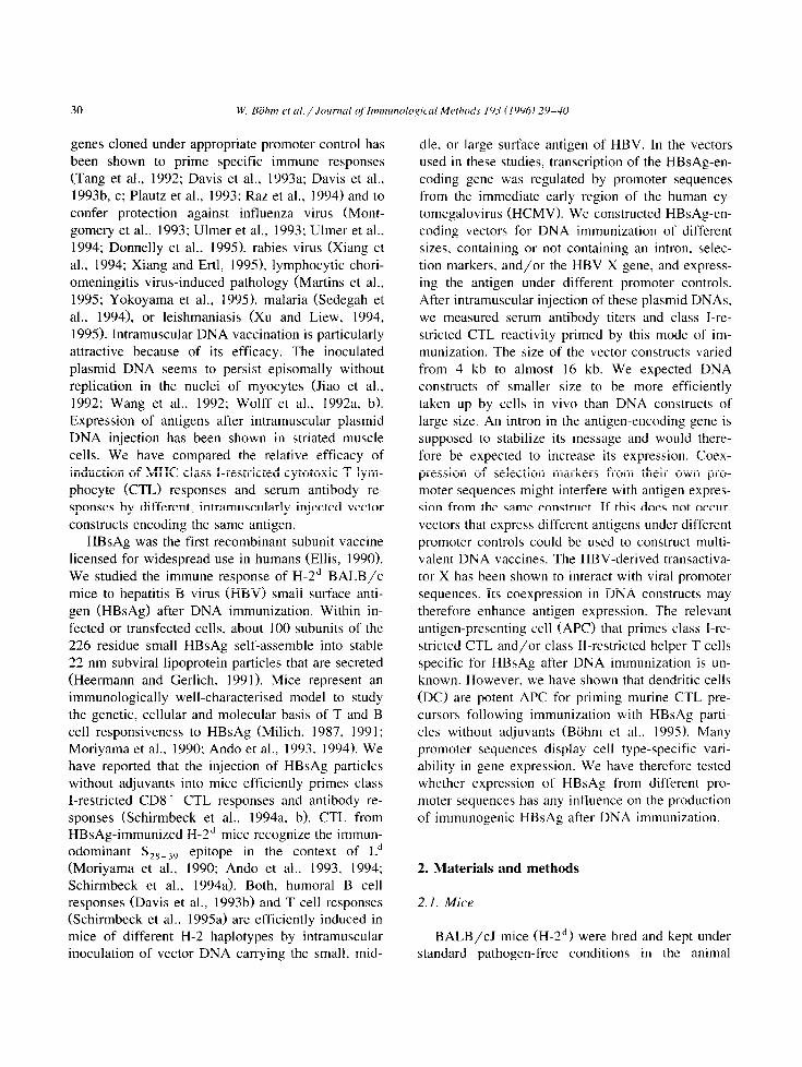
2.10. Transient expressiotl of HBsAg from the L’ector contructs
HBsAg was expressed from all vectors in COS cells after transient transfection (Fig. 2). COS cells (1.5 X 10’) were transfected with plasmid DNA (30 pg) by the CaPO, method. 2 days later, cells were labeled with 400 &i [35S]methionine (cat. no. SJ 10 15; Amersham, Braunschweig, Germany) in me- thionine-free RPMI 1640 medium supplemented with 10% FCS. Labeled cells were washed twice in PBS and extracted with lysis buffer (120 mM NaCl, 1% aprotinin (Trasylol. cat. no. 48764; Bayer, Lev- erkusen, Germany), 50 PM leupeptine, 10% im- munoglobulin-free FCS, 0.5% Nonident P-40 (NP- 40), 10% glycerol, 50 mM Tris/HCl (pH 8.0)) for
Fig. 2. Transient expression of HBsAg in COS cells rransfected
with the expression plasmids listed in Table 1.
30 min at 4°C. Extracts cleared by centrifugation (30 min, 20000 X g, 4°C) were subsequently incubated for 2 h at 4°C with protein A-Sepharose (cat. no. 17-0780-o 1; Pharmacia, Freiburg, Germany). The HBsAg protein was precipitated with a polyclonal rabbit anti-HBs antiserum and protein A-Sepharose at 4”C, and washed extensively in 0.5 M LiCl, 1% NP-40 and 0.1 M Tris/HCl (pH 9.0). After two additional washes of the complexes in 1 X PBS, and one wash in 0.1 X PBS, the HBsAg-containing im- mune complexes were recovered from protein A- Sepharose in 400 ~1 elution buffer (1.5% SDS, 5% P-ME and 7 mM Tris/HCl, pH 6.8). Following an incubation for 30 min at 37°C SDS-denatured elu- ates were lyophilized and dissolved in 30 ~1 aqueous solution of 7% P-ME, 10% glycerol and bromophe- no1 blue. After boiling for 2 min, samples were analyzed by SDS-PAGE.
2.11. Nucleic acid immunization
Into each tibialis anterior muscle, we injected 0.5, 5 or 50 pg plasmid DNA suspended in 50 ~1 PBS. Mice received bilateral intramuscular injections once, i.e., each mouse received 1, 10 or 100 kg plasmid DNA into normal, mature muscle. Control mice were injected intramuscularly with 2 X 50 yl PBS con- taining 100 yg vector DNA without insert.
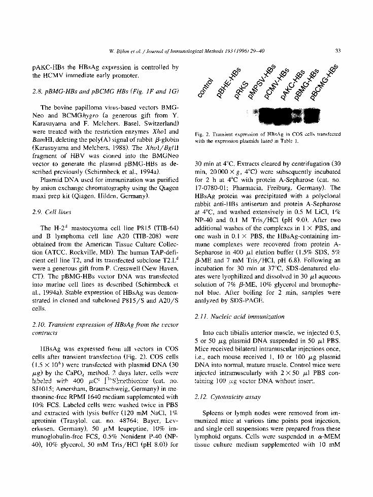
2.12. CytotoxiciQ assay
Spleens or lymph nodes were removed from im- munized mice at various time points post injection, and single cell suspensions were prepared from these lymphoid organs. Cells were suspended in (u-MEM tissue culture medium supplemented with 10 mM
Hepes buffer, 5 X 10e5 M 2-ME, antibiotics and 10% (v/v) FCS (Pan Systems, Aidenbach, Ger- many). 3 X lo7 responder cells were cocultured in a mixed lymphocyte tumor cell culture (MLTC) with 1.5 X IO6 syngeneic A20/S or P815/S transfectants (Schirmbeck et al., 1994a) (irradiated with 20000 rad) in 10 ml medium in upright 25 cm2 tissue culture flasks in a humidified atmosphere/7% CO, at 37°C. After a 5 day coculture. serial dilutions of effector cells were cultured with 2 X lo3 “G-labeled targets in 200 ~1 round-bottom wells. Specific cy- tolytic activity of cells was tested in 4 h 5’Cr-release assays against peptide-pulsed or transfected targets and compared with non-pulsed and non-transfected controls. After a 4 h incubation at 37°C. 100 ~1 of supernatant were collected for y-radiation counting.
90
60
g 70
.$ 60
$ 50
* 0 40
:: 30
20
The percentage specific release was calculated as [(experimental release ~ spontaneous release)/(total release - spontaneous release)] X 100. Total counts were measured by resuspending target cells. Sponta- neously released counts were always less than 15% of the total counts. Data shown are the mean of triplicate cultures. The SEM of triplicate data was always less than 15% of the mean.
2.13. Determinution of serum antibody levels
Serum samples were repeatedly obtained from individual immunized or control mice by tail bleed- ings l-12 weeks post injection. Antibodies against HBsAg were detected in mouse sera using the com- mercially available test IMxAUSAB (Abbott. Wies-
70
60
50
40
30
20
+ P815
- P815/S
++ T2.Ld
* T2.Ld/peptide
Fig. 3. The class I-restricted T cell response of H-2’ mice to HBsAg following DNA immunization. H-2d BALB/c mice were immunized by inoculation of 3 x 50 Fg ‘naked’ plasmid DNA encoding HBsAg into normal muscle. 2 weeks post injection, lymphoid cells were
prepared from the spleen and restimulated in vitro with syngeneic A20/S transfectants. After a j-day MLTC in vitro, effector cells were harvested and tested in a 4 h cytotoxicity assay against HBsAg peptide-pulsed T2.Ld targets or HBsAg-expressing PSIS/S transfectants.
Representative data from individual mice are shown. The plotted lysis values at the indicated effecter/target ratios represent means of
triplicates.

W. Biihm et al./ Journal of Immunological Methods 193 llY961 29-40 35
baden, Germany). In this readout, microparticles coated with HBsAg were mixed with mouse serum samples to allow specific binding of anti-HBsAg antibodies. Microparticles coated with antigen-anti- body complexes were transferred and irreversibly bound to a glass fibre matrix. Biotinylated recombi- nant HBsAg was added, and binding to the complex was traced by an alkaline phosphatase conjugated anti-biotin antibody followed by the addition of the
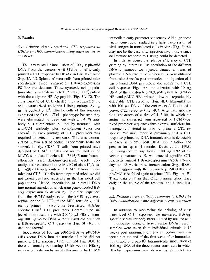
A Dose/response
substrate 4-methyl-umbelliferyl-phosphate. Antibody levels were quantified using six standard sera (O- 1000 mIU/ml). The tested sera were diluted so that the measured OD values were between standard serum one and six. Values presented in this paper are calculated by multiplying the serum dilution with the measured antibody level (mIU/ml). Serum titers shown are the geometric means of eight individual mice.
Kinetics
E 100 90
80
z 70
.g 80
i;
:: 30
20
10
0
Fig. 4. Dose/response and kinetics of the cytotoxic T cell response in mice primed to HBsAg by nucleic acid immunization. Mice were immunized by intramuscular injections of 1 p*g (A ). 10 p*g (8) or 100 @g (C) plasmid DNA from the vector constructs listed in Table I. Spleen cells were obtained from immunized mice three weeks post injection for assaying specific cytolytic reactivity. Mice were injected
i.m. with 100 pg plasmid DNA from the vector constructs listed in Table 1. Spleen cells were taken from immunized mice at 6 days (D) or
12 weeks (E) post immunization and assayed for specific cytolytic reactivity. The plotted specific lysis values represent mean values from
three individual mice tested at an effecter/target ratio of 20.
3. Results
3.1. Priming class I-restricted CTL resporlses to HBsAg by DNA immunization using d$erent ~)ector ccmstructs
The intramuscular inoculation of 100 pg plasmid DNA from the vectors A-E (Table 1) efficiently primed a CTL response to HBsAg in BALB/c mice (Fig. 3A-E). Splenic effector cells from primed mice specifically lysed syngeneic. HBsAg-expressing P8 15/S transfectants. These cytotoxic cell popula- tions also lysed LJ-transfected T2 cells (T2.L”) pulsed with the antigenic HBsAg peptide (Fig. 3A-E). The class I-restricted CTL elicited thus recognized the well-characterized antigenic HBsAg epitope S,,_,, in the context of L”. Effector cells of this response expressed the CD8+ CD4 phenotype because they were eliminated by treatment with anti-CD8 anti- body plus complement, but not by treatment with anti-CD4 antibody plus complement (data not shown). In vivo priming of CTL precursors was required to detect this response. This was demon- strated in two sets of control experiments (data not shown). Firstly. CD8+ T cells from primed mice depleted of CD4+ T cells and restimulated in the MLTC with class I+/class II- P8 15/S transfectants efficiently lysed HBsAg-expressing targets. Sec- ondly, after coculture in the MLTC of class I +/class II’ A2O/S transfectants with CD4+ T from primed mice and CD8+ T cells from unprimed mice, we did not detect cytolytic reactivity in the harvested cell populations. Hence, inoculation of plasmid DNA into normal muscle, in which transgene-encoded HB- sAg expression is driven by promoter sequences from the HCMV early region, the SV40 regulatory region, or the 3’ LTR of the MPS retrovirus, effi- ciently primes in vivo class I-restricted, HBsAg- specific CD8+ CTL precursors. Control mice, in- jected intramuscularly with 2 X 50 ~1 PBS contain- ing 100 pg vector DNA without insert did not elicit a HBsAg-specific CTL response (Fig. 3H-K; and data not shown).
Inoculation of 100 pg pBMG-HBs or pBCMG- HBs vector DNA into the muscle of mice did not prime a CTL response (Fig. 3F and Fig. 3G). In these episomally replicating 15 kb vectors HBsAg expression is driven by metallothionine or by HCMV
immediate early promoter sequences. Although these vector constructs resulted in efficient expression of viral antigen in transfected cells in vitro (Fig. 2) this may not be the case after injection into muscle since no immune response to HBsAg could be detected.
In order to assess the relative efficiency of CTL priming by intramuscular inoculation of the different DNA constructs, we injected titrated amounts of plasmid DNA into mice. Spleen cells were obtained from mice 3 weeks post immunization. Injection of 1 pug plasmid DNA per mouse did not prime a CTL cell response (Fig. 4A). Immunization with 10 pg DNA of the constructs pRKS, pMPSV-HBs, pCMV- HBs and pAKC-HBs primed a low but reproducibly detectable CTL response (Fig. 4B). Immunization with 100 pg DNA of the constructs A-E elicited a potent CTL response (Fig. 4C). After i.m. inocula- tion, constructs of a size of 4-8 kb. in which the antigen is expressed from retroviral or HCMV-de- rived promoter sequences thus express sufficient im- munogenic material in vivo to prime a CTL re- sponse. We have reported previously that a CTL response primed by DNA immunization is detectable as early as 6 days post DNA immunization, and persists for up to 4 months (Davis et al., 1995). Following the i.m. injection of 100 pg DNA of the vector constructs A-E, we detected specific CTL reactivity against HBsAg-expressing targets from 6 days to 12 weeks post immunization (Fig. 4D.E). Immunizations with the plasmids pBMG-HBs and pBCMG-HBs failed again to prime CTL (Fig. 4A-E). These data confirm that CTL priming takes place early in the course of the response and is long-last-
ing.
3.2. Priming serum antibndv resporlses to HBsAg by DNA immu~~i:ation usiizg differetlt l,ector constructs
In addition to monitoring the priming of class I-restricted CTL responses, we measured HBsAg- specific serum antibody titers elicited by nucleic acid immunization using different vector DNAs. Serum samples were taken from individual animals I- 12 weeks post immunization. No antibodies were de- tectable at the end of the first week after immuniza- tion (Table 2, group B). Intramuscular inoculation of 100 pug DNA of the three vector constructs in which HBsAg expression was driven by promoter se-
W. Biihm et al. /Journal of Immunological Methods 193 (1996129-30 37
C 3
D 12
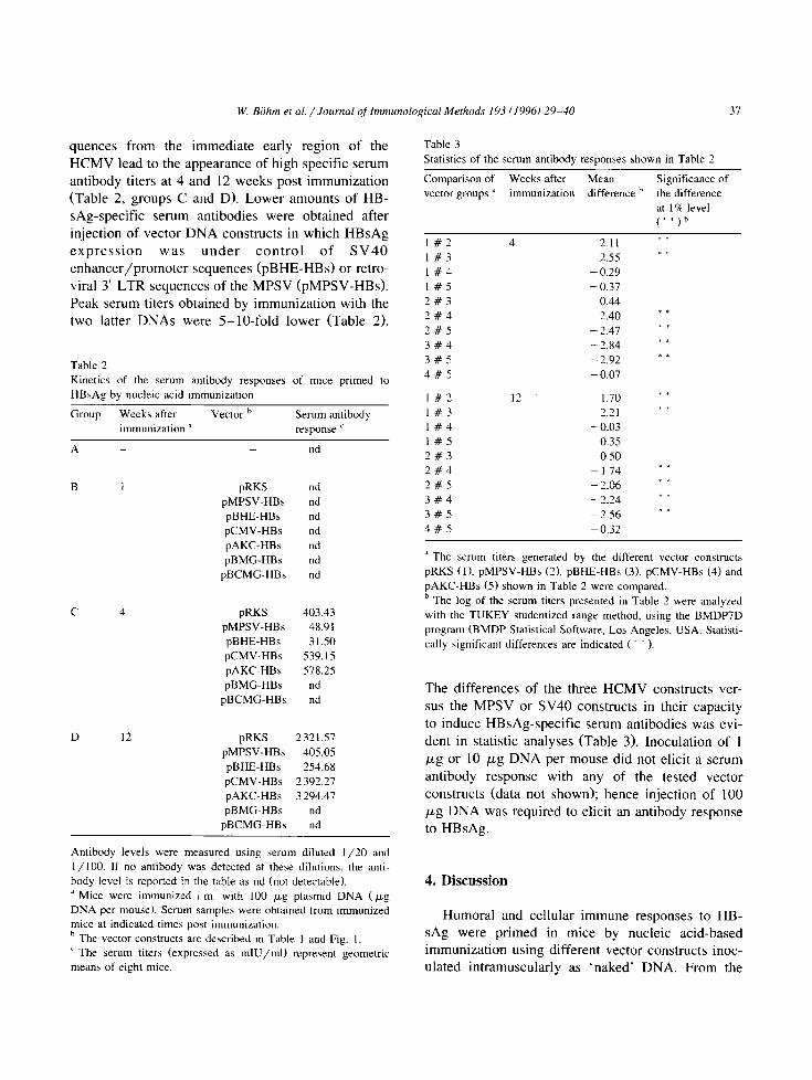
quences from the immediate early region of the HCMV lead to the appearance of high specific serum antibody titers at 4 and 12 weeks post immunization (Table 2, groups C and D). Lower amounts of HB- sAg-specific serum antibodies were obtained after injection of vector DNA constructs in which HBsAg expression was under control of SV40 enhancer/promoter sequences (pBHE-HBs) or retro- viral 3’ LTR sequences of the MPSV (pMPSV-HBs). Peak serum titers obtained by immunization with the two latter DNAs were 5-lo-fold lower (Table 2).
Table 2
Kinetics of the serum antibody responses of mice primed to
HBsAg by nucleic acid immunization
Group Weeks after vector b Serum antibody immunization ’ response c
A _ _ nd
B I pRKS nd
pMPSV-HBs nd
pBHE-HBs nd
pCMV-HBs nd
pAKC-HBs nd
pBMG-HBs nd
pBCMG-HBs nd
pRKS 403.43
pMPSV-HBs 48.9 1 pBHE-HBs 31.50 pCMV-HBs 539.15
pAKC-HBs 578.25
pBMG-HBs nd
pBCMG-HBs nd
pRKS 2321.57
pMPSV-HBs 405.05
pBHE-HBs 254.68
pCMV-HBs 2 392.27
pAKC-HBs 3 294.47
pBMG-HBs nd
pBCMG-HBs nd
Antibody levels were measured using serum diluted l/20 and
I /lOO. If no antibody was detected at these dilutions, the anti-
body level is reported in the table as nd (not detectable).
’ Mice were immunized i-m. with 100 pg plasmid DNA (~g DNA per mouse). Serum samplea were obtained from immunized mice at indicated times post immunization.
’ The vector constructs are described in Table 1 and Fig. 1.
’ The serum titers (expressed as mIU/ml) represent geometric mean5 of eight mice.
Table 3
Statistics of the serum antibody responses shown in Table 3
Comparison of Weeks after Mean Significance of
vector groups ’ immunization difference b the difference
at 19 level
(**)h
I#2 4 2.11 * * I#3 2.55 = I 1#4 - 0.29
1#5 -0.37 2#3 0.44
3#4 - 2.40 * *
2#5 - 2.47 * *
3#4 - 2.84 I *
3#5 -2.92 * *
4#5 - 0.07
I #2 I? 1.70 * * I#3 2.21
**
I#4 - 0.03
1#5 - 0.35 2#3 0.50 2#4 -1.74 *-
2#5 - 2.06 I _
3#4 - 2.24 I I
3#5 -2.56 t = 4#5 -0.32
’ The serum titers generated by the different vector constructs
pRKS (I), pMPSV-HBs (2), pBHE_HBs (3), pCMV-HBs (4) and
pAKC-HBs (5) shown in Table 2 were compared.
b The log of the serum titers presented in Table 2 were analyzed
with the TUKEY studentized range method, using the BMDP7D
program (BMDP Statistical Software, Los Angeles, USA. Statisti-
cally significant differences are indicated ( * * ).
The differences of the three HCMV constructs ver- sus the MPSV or SV40 constructs in their capacity to induce HBsAg-specific serum antibodies was evi- dent in statistic analyses (Table 3). Inoculation of 1 PcLg or 10 Fg DNA per mouse did not elicit a serum antibody response with any of the tested vector constructs (data not shown); hence injection of 100 pg DNA was required to elicit an antibody response to HBsAg.
4. Discussion
Humoral and cellular immune responses to HB- sAg were primed in mice by nucleic acid-based immunization using different vector constructs inoc- ulated intramuscularly as ‘naked’ DNA. From the

spectrum of HBsAg-specific immune responses. we chose to study class I-restricted CD8+ CTL re- sponses and antibody responses. The former is of interest because of its involvement in viral dearance and HBV-associated immunopathology. The latter is a CD4+ T cell-dependent response (Milich, 1987. 1991) and hence indicates priming of the alternative T cell subset. With five of the seven vector con- structs tested we could efficiently prime class I-re- stricted, HBsAg-specific CD8’~ CTL precursors in vivo by DNA immunization. Expression of HBsAg from three different promoter sequences permitted the immunogenic, class I-restricted presentation of an HBsAg epitope to CTL precursors. The presence or absence on the expression vector of an intron. of the nen gene, or of the HBV X gene, did not play a critical role in the intramuscular expression of an immunogenic HBsAg product because the plasmids pRKS, pCMV-HBs and pAKC-HBs, though differ- ent from each other according to those parameters, were equally efficient in priming CTL and serum antibody titers in mice after intramuscular inocula- tion. We excluded the possibility that nucleic acid immunization primes in vivo exclusively CD4’ but not CD8+ T cell precursors, and that primed CD4+ T cells support the initiation of a primary in vitro response of unprimed CD8+ CTL precursors in the MLTC. How and in which tissue CTL precursors are primed after intramuscular DNA injection is un- known. The transgene-encoded HBsAg may be ex- pressed in myocyte or in professional APC (dendritic cells, macrophages) in muscle tissue. The HBsAg
epitope may be presented by MHC class I molecules on the surface of myocytes. or HBsAg particles secreted by myocytes may be picked up by other APC types that process and present the antigen (Schirmbeck et al., 1995b). The T cell response may therefore be initiated. either in muscle tissue. or in secondary lymphoid tissue.
The constructs that failed to prime CTL after intramuscular DNA injection were pBMG-HBs and pBCMG-HBs. Even when we injected 100 pg pBMG-HBs or pBCMG-HBs plasmid DNA into re- generating muscle of cardiotoxin-pretreated mice, we did not detect an immune response (data not shown) although gene transfer is more efficient in regenerat- ing than in mature muscle (Davis et al., 1993a. b, 1995). These large ( 15 kb) vector constructs contain
a bovine papilloma virus-derived sequence that per- mits their episomal replication to high copy numbers in cells (Karasuyama and Melchers, 1988; Kard- suyama et al., 1990). These vectors support high levels of expression of many different viral antigens in stable murine transfectant lines of different histo- types (Schirmbeck et al., 1992. 1993, 1994a; Kuhrijber et al., 1994a, b; Schirmbeck and Reimann, 1994). It is unclear if the large size of these con- structs prevent efficient transfection of antigen-pre- senting cells in vivo. Alternatively, particular se- quences within these vectors might prevent efficient expression of an immunogenic gene product in vivo.
Only vector constructs that expressed HBsAg un- der the control of promoter sequences from the immediate early region of HCMV (i.e., the pRKS, pCMV-HBs and pAKC-HBs plasmids) efficiently primed both. class I-restricted CTL responses and serum antibody responses. The intramuscular injec- tion of the pMPSV-HBs and pBHE-HBs plasmid was efficient in priming class l-restricted CTL but inefficient in priming a serum antibody response. Quantitative or qualitative differences in HBsAg ex- pression in muscle from the five constructs could explain the described data. An injection of lo- 100 ng native HBsAg particles into mice primes a class I-restricted CTL response (Schirmbeck et al., 1994al but no serum antibody response (Schirmbeck et al., 1994b). In contrast, the injection of a lo- lOO-fold higher dose of l-10 pg HBsAg particles primes a T and a B cell response. If the amount of HBsAg produced in situ is limiting, and higher in the CMV promoter-driven constructs than in the alternative constructs, this may explain preferential priming of CTL. Different types of immunogenic presentation by HBsAg expressed from different constructs should also be considered. We have reported that the immu- nization of mice with native HBsAg particles ad- sorbed to aluminum hydroxide primes B cells but not CTL, that the immunization with native HBsAg par- ticles without adjuvants efficiently primes CTL and B cells, and that the immunization with denatured HBsAg monomers primes CTL but not B cells (Schirmbeck et al.. 1994~). Hence, different prepara- tions of a viral antigen can display strikingly differ- ent immunogenicity for class I-restricted CTL or B cells. In situ expression of HBsAg in muscle tissue or some other type of APC from different expression

W. BShm et al. / Jowxal of Immunological Methods 193 (1996) 29-40 39
plasmids may yield different preparations of HBsAg
with different immunogenicities for CTL precursors or B cells.
Acknowledgements
The excellent technical assistance of Evelyn Kury and Doris Munz is appreciated. We gratefully appre- ciate the interesting discussions and the generous gift of plasmid pCMV-HBs from Dr. R.G. Whalen (Paris, France), and the instructions on intramuscular DNA injection by Dr. H.L. Davis (Ottawa, Canada). Dr. P. Cresswell (New Haven, USA) kindly provided trans- fected cell lines. We thank Professor K. Feilen (Uni- versity Ulm) for statistically evaluating the antibody data. The work was supported by grants from the ‘Deutsche Forschungsgemeinschaft’ (Re549/4-2) to J.R. and R.S.
References
Ando, K.. Moriyama, T., Guidotti. L.G., Wirth, S., Schreiber.
R.D., Schlicht, H.-J., Huang, S.-N. and Chisari, F.V. (1993)
Mechanisms of class I restricted immunopathology. A trans-
genie mouse model of fulminant hepatitis. J. Exp. Med. 178,
1541.
Ando, K.. Guidotti, L.G., Cemy, A., Ishikawa. T. and Chisari,
F.V. (1994) Access to antigen restricts cytotoxic T lymphocyte
function in vivo. J. Immunol. 152, 3245.
Artelt. P., Morelle, C.. Ausmeier, M., Fitzek, M. and Hauser. H.
(1988) Vectors for efficient expression in mammalian fibrob-
lastoid, myeloid and lymphoid cells via transfection or infec-
tion. Gene 68, 213.
BGhm. W., Schirmbeck. R., Elbe. A., Melber, K.. Diminsky, D.,
Kraal, G.. Van Rooijen, N., Barenholz, Y. and Reimann, .I.
(1995) Exogenous hepatitis B surface antigen particles pro-
cessed by dendritic cells or macrophages prime murine MHC
class I-restricted cytotoxic T lymphocytes in viva. J. Immunol. 155.3313.
Davis. H.L., Demeneix, B.A., Quantin, B., Coulombe, J. and
Whalen. R.G. (1993a) Plasmid DNA is superior to viral
vectors for direct gene transfer into adult mouse skeletal
muscle. Hum. Gene Ther. 4, 733.
Davis, H.L., Michel. M.L. and Whalen, R.G. (1993b) DNA-based
immunization induces continuous secretion of hepatitis B sur-
face antigen and high levels of circulating antibody. Hum. Mol. Genet. 2, 1847.
Davis. H.L.. Whalen. R.G. and Demeneix, B.A. (1993~1 Direct
gene transfer into skeletal muscle in viva: factors affecting
efficiency of transfer and stability of expression. Hum. Gene
Ther. 4, 151.
Davis. H.L.. Schirmbeck, R.. Reimann. J. and Whalen, R.G.
(1995) DNA-mediated immunization in mice induces a potent
MHC class J-restricted cytotoxic T lymphocyte response to
Hepatitis B virus surface antigen, Hum. Gene Ther. 6, 1447.
Dirks, W., Wirth. M. and Hauser, H. (1993) Dicistronic transcrip-
tion units for gene expression in mammalian cells. Gene 128.
247.
Donnelly, J.J., Ulmer, J.B. and Liu, M.A. (19941 Immunization
with DNA. J. Immunol. Methods 176. 145.
Donnelly. J.J.. Friedman, A., Martinez. D., Montgomery, D.L.,
Shiver. J.W., Motzel. S.L., Ulmer, J.B. and Liu. M.A. (1995)
Preclinical efficacy of a prototype DNA vaccine: enhanced
protection against antigenic drift in influenza virus. Nature-
Medicine 1, 583.
Ellis, R.W. (1990) New and improved vaccines against hepatitis
B. I. Recombinant-derived vaccines against hepatitis B. In:
G.C. Woodrow and M.M. Levine (Eds.1, New Generation
Vaccines. Marcel Dekker, New York, p. 439.
Heermann, K.-H. and Gerlich. W.H. (1991) In: A. McLachlan
(Ed.), Surface Proteins of Hepatitis B Virus, CRC Press, Boca
Raton, FL, p. 109.
Jiao, S.. Williams, P.. Berg. R.K.. Hodgeman, B.A., Liu, L.M.,
Repetto. G. and Wolff, J.A. (19921 Direct gene transfer into
nonhuman primate myofibers in viva. Hum. Gene Ther. 3, 2 1,
Karasuyama. H. and Melchers, F. (1988) Establishment of mouse
cell lines which constituticely secrete large quantities of inter-
leukin 2, 3. 4 and 5. using modified cDNA expression vectors.
Eur. J. Immunol. 18, 97.
Karasuyama. H.. Kudo. A. and Melchers, F. (1990) The protein
encoded by the VpreB and lambda 5 pre-B cell-specific genes
can associate with each other and with /.t heavy chain. J. Exp.
Med. 172, 969.
Kuhrober, A.. Schirmbeck, R. and Reimann. J. (1994al A selfreac-
tive class I-restricted T cell response of H-2b mice to determi-
nants of the Vp8.2 domain of the T cell receptor for antigen. Immunology 83, 532.
Kuhrober. A., Schirmbeck, R. and Reimann, J. (1994b) Vaccina-
tion with T cell receptor peptides primes anti-receptor cyto-
toxic T lymphocytes (CTL) and anergizes T cells specifically
recognized by these CTL. Eur. J. Immunol. 24. 1172.
Martins, L.P., Lau, L.L., Asano, M.S. and Ahmed. R. (1995)
DNA vaccination against persistent viral infection. J. Virol.
69, 2574.
Milich, D.R. (1987) Genetic and molecular basis for T- and B-cell recognition of hepatitis B viral antigens. Immunol. Rev 99, 7 1.
Milich, D.R. (1991) Use of recombinant HBV proteins in im-
munological studies. In: A. McLachlan (Ed.), Molecular Biol-
ogy of the Hepatitis B Virus. CRC Press. Boca Raton, FL. p, 283.
Montgomery, D.L., Shiver, J.W., Leander, K.R.. Perry, H.C.,
Friedman, A., Martinez, D., Ulmer, J.B., Donnelly. J.J. and
Liu. M.A. (1993) Heterologous and homologous protection against influenza A by DNA vaccination: optimization of DNA vectors. DNA Cell Biol. 12. 777.
Moriyama, T.. Guilhot, S.. Klopchin, K., Moss. B., Pinkert, C.A..
Palmiter, R.D.. Brinster, R.L., Kanagawa, 0. and Chisari. F.V.
(19901 Immunobiology and pathogen&s of hepatocellular in-
jury in hepatitis B virus transgenic mice. Science 248, 361,
Plautz, G.E., Yang, Z.Y., Wu, B.Y.. Gao. X., Huang. L. and
Nabel, G.J. (19931 Immunotherapy of malignancy by in viva
gene transfer into tumors [see comments]. Proc. Nat]. Acad.
Sci. USA 90. 4645.
Raz. E., Carson. D.A., Parker. S.E., Parr, T.B.. Abai. A.M..
Aichinger. G., Gromkowski. S.H., Singh, M.. Lew. D..
Yankauckas, M.A. and et al. (19941 Intradermal gene immu-
nization: the possible role of DNA uptake in the induction of
cellular immunity to viruses. Proc. Nat]. Acad. Sci. USA 91.
9519.
Schirmbeck, R. and Reimann. J. (I 9941 Peptide transporter-inde-
pendent. stress protein-mediated endosomal processing of en-
dogenoua protein antigens for major histocompatibility com-
plex class I presentation. Eur. J. Immunol. 24, 1478.
Schirmbeck, R.. Zerrahn, J.. Kuhrober, A.. Kury, E., Deppert. W.
and Reimann. J. (19921 Immunization with soluble simian
virus 40 large T antigen induces a specific response of CD3+
CD4- CDS+ cytotoxic T lymphocytes in mice. Eur. J. Im-
munol. 22. 759.
Schirmbeck. R.. Zerrahn, J., Kuhrober. A., Deppert. W. and
Reimann. J. (19931 Immunization of mice with the N-terminal
t l-271) fragment of simian virus 10 large T-antigen (without
adjuvantsl specifically primes cytotoxic T lymphocytes. Eur. .I.
Immunol. 23. 1578.
Schirmbeck. R., Melber, K., Kuhriiber, A.. Janowicz. Z.A. and
Reimann, J. (1994al Immunization with soluble hepatitis B
virus surface (Sl protein particles elicits murine H-2 class
I-restricted CD8 + cytotoxic T lymphocyte responses in viva.
J. Immunol. 152, 11 10.
Schirmbeck, R.. Melber, K., Mertens. T. and Reimann. J. ( 1994bl
Antibody and cytotoxic T-cell responses to soluble hepatitis B
virus (HBVl S antigen in mice: implication for the pathogene-
sis of HBV-induced hepatitis. J. Virol. 68. 1418.
Schirmbeck. R.. Melber. K.. Mertens. T. and Reimann. J. (1994~1
Selective stimulation of murine cytotoxic T cell and antibody
responses by particulate or monomeric hepatitis B virus sur-
face (Sl antigen. Eur. J. Immunol. 24, 1088.
Schirmbeck, R.. Bohm, W.. Ando. K., Chisari. F.V. and Reimann.
J. (1995al Nucleic acid vaccination primes hepatitis B surface
antigen-specific cytotoxic T lymphocytes in nonresponder mice. J. Virol. 69, 5929.
Schirmbeck, R.. Melber. K. and Reimann, J. (1995bl Hepatitis B
virus surface particles are processed in a novel endosomal
pathway for MHC class I-restricted epitope presentation. Eur.
J. Immunol. 25, 1063.
Sedegah, M.. Hedstrom, R.. Hobart. P. and Hoffman, S.L. (19911
Protection against malaria by immunization with plasmid DNA
encoding circumsporozoite protein. Proc. Nat]. Acad. Sci.
USA 9 I. 9866.
Tang. D.C.. DeVit. M. and Johnston. S.A. (19921 Genetic immu-
nization is a simple method for eliciting an immune response.
Nature 356, 153.
Ulmer. J.B., Donnelly, J.J.. Parker. S.E.. Rhodes, G.H.. Felgner,
P.L.. Dwarki. V.J., Gromkowshi, S.H., Deck. R.R.. Dewitt.
C.M.. Friedman, A. and et al t 19931 Heterologous protection
against influenza by injection of DNA encoding a viral protein
[see comments]. Science 259. 1745.
Ulmer. J.B.. Deck, R.R.. Dewitt. C.M., Friedman, A.. Donnelly.
J.J. and Liu, M.A. (19941 Protective immunity by intramuscu-
lar injection of low doses of influenza virus DNA vaccines.
Vaccine 12. 1541.
Wang. J.. Jiao. S.. Wolff, J.A. and Knechtle. S.J. (19921 Gene
transfer and expression in rat cardiac transplants. Transplanta-
tion 53. 703.
Wolff, J.A.. Dowty. M.E.. Jiao, S.. Repetto. G.. Berg. R.K..
Ludtke. J.J.. Williams, P. and Slautterback, D.B. (lY92al
Expression of naked plasmids by cultured myotubea and entry
of plasmids into T tubules and caveolae of mammalian skele-
tal muscle. J. Cell Sci. 103. 1249.
Wolff, J.A.. Ludtke. J.J.. Acsadi. G., Williams, P. and Jani. A.
(1992bl Long-term persistence of plaamid DNA and foreign
gene expression in mouse muscle. Hum, Mol. Genet. 1. 363.
Xiang. Z.Q. and Ertl. H.C.J. (1995) Manipulation of the immune
response to a plasmid-encoded viral antigen by coinoculation
with plasmids expressing cytokinea. Immunity 2, 129.
Xiang, Z.Q.. Spitalnik, S.. Tran, M., Wunner. W.H.. Cheng. J. and
Ertl, H.C.J. (19941 Vaccination with a plasmid vector carrying
the rabies virus glycoprotein gene induces protective immunity
against rabies virus. Virology 199, 132.
Xu. D. and Liew, F.Y. (19941 Genetic vaccination against leish-
maniasis. Vaccine 12, 1534.
Xu. D. and Lie,. F.Y. t 19951 Protection against leishmaniaais by
injection of DNA encoding a major surface glycoprotein,
gp63. of L. ~nujor. Immunology 83. 173.
Yokoyama, M., Zhang. J. and Whitton, J.L. (19951 DNA immu-
nization confers protection against lethal lymphocytic chori-
omeningitia virus infection, .I. Virol. 69. 2681.