dna replication reconstructed full article.pdf
TRANSCRIPT

7/21/2019 DNA replication reconstructed full article.pdf
http://slidepdf.com/reader/full/dna-replication-reconstructed-full-articlepdf 1/18
ARTICLE doi:10.1038/nature14285
Regulated eukaryotic DNA replication
origin firing with purified proteinsJoseph T. P. Yeeles1, Tom D. Deegan1, Agnieszka Janska1, Anne Early1 & John F. X. Diffley1
Eukaryotic cells initiate DNA replication from multiple origins, which must be tightly regulated to promote precisegenome duplication in every cell cycle. To accomplish this, initiation is partitioned into two temporally discrete steps: a double hexameric minichromosome maintenance (MCM) complex is first loaded at replication origins during G1 phase,and then converted to the active CMG (Cdc45–MCM–GINS) helicase during S phase. Here we describe the reconstitutionofbuddingyeastDNAreplicationinitiationwith16purifiedreplicationfactors,madefrom42polypeptides.Origin-dependentinitiationrecapitulates regulationseen invivo . Cyclin-dependentkinase (CDK)inhibits MCM loading by phosphorylating the origin recognition complex (ORC) and promotes CMG formation by phosphorylating Sld2 and Sld3. Dbf4-dependent
kinase (DDK) promotes replication by phosphorylating MCM, and can act either before or after CDK. These experimentsdefine theminimum complement of proteins, protein kinasesubstrates and co-factors required for regulated eukaryoticDNA replication.
Theinitiation of eukaryotic DNAreplication origin firing is understoodin outline1,2, but the process has not been reconstituted with purifiedproteins.MCMcanbeloadedontoDNAwithpurifiedORC,Cdc6andCdt1–MCM3,4 and loaded MCMs can be activated to replicate in yeastextracts5–7. Mass spectrometry of complexesassembled during replica-tion in these extracts identified previously characterized ‘firing factors’including Sld2, Sld3, Sld7, Dpb11, Cdc45, GINS and DNA polymerasee (pole), butdid notidentifyany novel factors6, suggesting this list may be complete.
DNA replication is regulated during the cell cycle by two proteinkinases,CDKandDDK8. CDKplays tworoles in regulating replication:it inhibits MCMloading andit is essential forhelicase activation1,2.Con-sequently, MCM loading can only occur during G1 phase when CDKactivity is low, andoriginscan only fire afterG1 phasewhen CDKlevelsrise. CDK phosphorylation of ORC, Cdc6 and MCM all contribute topreventing MCM loading outside G1 phase in budding yeast9. Geneticanalysishas indicatedthatSld2 andSld3 arethe twokeyCDKsubstratesrequired for helicase activation. Phosphorylation of these proteins gen-erates binding sites for tandem BRCT repeats in Dpb11 (refs 10–12).DDK is required for origin firing 8 and genetic analysis has indicatedthat Mcm4 and 6 are the key DDK substrates13–15. Until origin firing isreconstituted with purified proteins, however, we will notknow whetherwe have the complete inventory of essential firing factors or whether
CDK and DDK have any additional important substrates.
CDK- and DDK-dependent firing factor recruitmentTo reconstitute MCM loading and activation, we expressed and puri-fied the thirteen replication factorsshownin Fig. 1a(left) andExtendedData Fig. 1a,b. Cdc6, GINS,Mcm10 andcyclin A/Cdk2 (A-Cdk2) wereexpressedinEscherichia coli, whiletheremainingproteinswere expressedin Saccharomyces cerevisiae. In addition to A-Cdk2, budding yeast SphaseCDK (S-CDK)expressed inSaccharomycescerevisiae(Fig. 1a,left)was used in some experiments. Details of expression and purificationstrategies can be found in the Methods.
We adopted the strategy outlined in Fig. 1b to assemble firing fac-tors onto the loaded MCM in a staged manner. We first loaded MCMonto DNAattachedto magnetic beads3 (‘MCM load’). The loadedMCM
wasphosphorylatedwith DDK6, beadswere isolatedand Sld3/7and Cdc45were added (‘DDK step’). Beads were again isolated and theremaining firing factors were added with A-Cdk2(‘CDKstep’). After washing the
1Cancer Research UK London Research Institute, Clare Hall Laboratories, South Mimms EN6 3LD, UK.
Mcm7
Sld3
Sld7
Cdc45
Dpb11
Sld2
Dpb2
Psf1
Mcm10
Protein
omitted O R C D D Kc
N o n e
a
d
Mcm7
Sld3
Sld7
Cdc45
Dpb11
Dpb2
Psf1
Mcm10
A-Cdk2 – +Cdt1–Mcm2–7,
ORC, Cdc6
DDK
Sld3/7, Cdc45
CDK, Sld2, Dpb11,
Pol ε, GINS, Mcm10
b
3× 0.3 M K-Glu washes
30 min
20 min
5 min
10 min
Boil beads in SDS load
D D K
O R C C d c 6
C d t 1 – M c m 2 – 7
S l d 3 / 7
C d c 4 5
S - C D K
D p b 1 1
S l d 2 P o l ε
G I N S
M c m 1 0
160
100
50
20
30
70
40
kDa
1 2 3
1 2
1 2 3 4 5 6 7 8 9 10 11 12
DDK
step
CDK
step
MCM
load
P o l α C t f 4 R P A T o p o
I I
160
100
50
20
30
70
15
kDa
101 2 3 4
Figure 1 | DDK- and CDK-dependent firing-factor recruitment withpurified proteins. a , Purified MCM loading and firing factors (left), andadditionalfactors requiredfor DNA replication (right) analysed by SDS–PAGEwith Coomassie staining. b, Reaction scheme for firing-factor recruitment.c, d, Immunoblots of recruitment reactions conducted as illustrated in b onARS305 linear DNA.
2 6 M A R C H 2 0 1 5 | V O L 5 1 9 | N A T U R E | 4 3 1
Macmillan Publishers Limited. All rights reserved©2015

7/21/2019 DNA replication reconstructed full article.pdf
http://slidepdf.com/reader/full/dna-replication-reconstructed-full-articlepdf 2/18
beads with a low-salt buffer, we analysed bound proteins by immuno-blotting.As showninFig. 1c,Sld3/7,Cdc45,Dpb11,Sld2, pole (theDpb2B subunit), GINS (the Psf1 subunit) and Mcm10 were all recruited inan ORC- and DDK-dependent manner. When A-Cdk2 was omittedfrom thefinal step, Sld3/7and Cdc45 werestillrecruitedbut theremain-ing factors were not (Fig. 1d). Therefore, therecruitment of Sld3/7andCdc45 required DDK but not CDK, while the recruitment of theremaining firing factors required both DDK and CDK.
Recruitment of Cdc45 and GINS into a stable complexConsistent with the dependencies shown in Fig. 1c, d, Figure 2a showsthat the recruitment of Cdc45 did not require any of the factors acting in the CDK step (Dpb11, Sld2, pol e, GINS, Mcm10). However, therecruitment of GINS (Psf1) required all of the other factors acting inthis CDK step except for Mcm10 (that is, Dpb11, Sld2, pol e). CMG issalt-stable16,17, so we tested our complex under more stringent extrac-tion conditions. Figure 2b (lane 1) and Extended Data Fig. 2a, b show that, in addition to MCM,a fraction ofCdc45 and GINSis stableto saltextraction. In contrast to Cdc45 andGINS,Sld3 is not stabilized in thiscomplex (Extended Data Fig. 2b). Figure 2b, lanes 2–5 show that salt-stable Cdc45recruitment requiresDpb11, Sld2, poleand GINS.Mcm10,however, is not required for this salt-stable complex (Fig. 2b, lane 6).
To investigate whether the complex we assembled might be func-tional, we tested its ability to support DNA replication in extracts. Wehave previously shown that MCM loaded with purified proteins andphosphorylatedwith purified DDKcan replicatein extracts from S phasecells made from a strain (KO3) that overexpresses Dpb11,Cdc45, Sld2,Sld3andSld76.Ithasbeenshownthatoverexpressionoffiringfactorsisrequired for replication in a related system7. We therefore constructeda new strain that does not overexpress any firing factors (yJY18) andmade S phase extracts from this strainin which GINS wasalsodepleted(Extended Data Fig. 2c). We reasoned that addition of the complex of MCM withfiring factors to this extract might support DNA replication
(Fig. 2c). Figure2d, lanes 1 and9 show that replication occurred underthese conditions. Indeed, the replication products seen with the puri-fied factors were equivalent to those synthesized in extracts from KO3(Extended Data Fig. 2e). Furthermore, as shown in Fig. 2d, lanes 2–8,replication was not observed if either Sld3/7, Cdc45, A-Cdk2, Dpb11,Sld2, poleor GINS were omitted, indicating that allof the recombinantproteins tested arefunctionaland requiredfor replication. This also sug-gested that the complex of MCM and firing factors may be functional.
Reconstitution of origin firing with purified proteinsWe next expressed and purified four additional proteins predicted tobe important for DNA replication: DNApolymerasea–primase (pola),Ctf4, replication protein A (RPA), and topoisomerase II (Topo II)18,19
(Fig. 1a (right) andExtendedData Fig. 1a). Followingthe CDKstep weisolated the beads and added a new buffer containing these four pro-teins along with additional Mcm10, ribo- and deoxyribonucleosidetriphosphates (NTPs and dNTPs) and [a-32P]dCTP, as outlined inFig. 3a.As shown in Fig. 3b (lane 1),this resulted in DNAsynthesis thatgeneratedlabelledproducts on alkalineagarosegels. SynthesisrequiredORC, DDK, A-Cdk2 and pol a (Fig. 3b) as well as dNTPs (ExtendedData Fig. 3a), suggesting it was genuine initiation.
In this and subsequent experiments, two classes of labelledproducts
wereobserved: a population of short productsof approximately150nu-cleotidesin length, and a more heterogeneous smear of larger products.The template used in these experiments was a 3.2 kilobase pairs plas-mid, which had been randomly biotinylated and attached to streptavi-din beads. Previous work hasshown that attachment of DNAto beadsinterferes withthe completion of DNAreplication5–7. Since the positionof the ARS1 origin relative to the site of attachment to the beads willberandom, leading strand synthesiswouldbe predictedto generate a smearof products averaging half the plasmid size (,1,600 nucleotides), butrangingin size from very small to almost full plasmid length, very sim-ilar to theproducts observed.Synthesis of thesmallerproductclass was
a
N o n e
D p b 1 1
S l d 2
S l d 3 / 7
C d c 4 5Protein
omitted N o n e
P o l ε G I N S
A - C d k 2
b
Mcm7
Cdc45
Psf1
Protein
omitted D p b 1 1
S l d 2
N o n e
G I N S M c m 1 0
P o l ε
d
Mcm7
Cdc45
Psf1
Protein
omitted D p b 1 1
S l d 2
N o n e
G I N S M c m 1 0
P o l ε
0.3 M
K-Glu
wash
0.3 M
KCl
wash
c
MCM load
2× 200 mM K-Glu washes
1 2 3 4 5 61 2 3 4 5 6
1 2 3 4 5 6 7 8 9
DDK step
CDK step
S phase extract
4.4
2.32.0
0.6
kb
Figure 2 | The purified firing factors are functional. a , b, Immunoblots of recruitment reactions performed as in Fig. 1b using ARS305 linear DNA witheither, a , 0.3 M K-glutamate (K-Glu) or b, 0.3 M KCl washes. c, Scheme forextract-based replication reactions. Unless otherwise stated, in this and allsubsequent experiments, components of the ‘CDK’ and ‘DDK’ steps are as inFig. 1. Firing factors were recruited as illustrated in Fig. 1b. Beadswere isolated,washed twice and added to an S phase extract not overexpressing firing factors (yJY18) and where the Psf2 subunit of the GINS complex wasimmunodepleted (Extended Data Fig. 2c). d, Reaction performed as in c.Nascent DNA was labelled by including [a32P]dCTP in the extract step
and products were separated through a 0.6% alkaline agarose gel.
a
b
N o n e
A - C d k 2
P o l α
O R C D D KProtein
omitted
4.4
2.32.0
0.6
0.13
kb
Mcm10,
Pol α, Ctf4,
RPA, Topo II[α-32P]dCTP
30 min
Template
DDK – + – +
WT A-B2-
End
labelling
4.4
2.32.0
0.6
0.13
kb
c Template WT A-B2-
1 2 3 4 5
1 2 3 4
MCM
load
CDK
step
DDK
step
Quench and
alkaline
agarose gel
Figure 3 | The initiation of DNA synthesis with purified proteins.a , Reaction pathway for DNA replication with purified proteins. Firing factorswere bound to MCM, the complex isolated, and added to a new buffercontaining proteins required for DNA synthesis(Fig. 1a (right)). b, Replicationreaction conducted as shown in a on ARS1 circular DNA. In this and allsubsequent replication assays products were separated through 0.7% alkalineagarose gels. c, Replication on ARS1 linear templates. Top, photocleavedDNA templates analysed by native agarose gel electrophoresis and ethidiumbromide staining. Bottom, replication following the pathway shown in a . TheMCM loading conditions were modified to confer origin specificity on the
replication reaction (see Methods for details).
RESEARCH ARTICLE
4 3 2 | N A T U R E | V O L 5 1 9 | 2 6 M A R C H 2 0 1 5
Macmillan Publishers Limited. All rights reserved©2015

7/21/2019 DNA replication reconstructed full article.pdf
http://slidepdf.com/reader/full/dna-replication-reconstructed-full-articlepdf 3/18
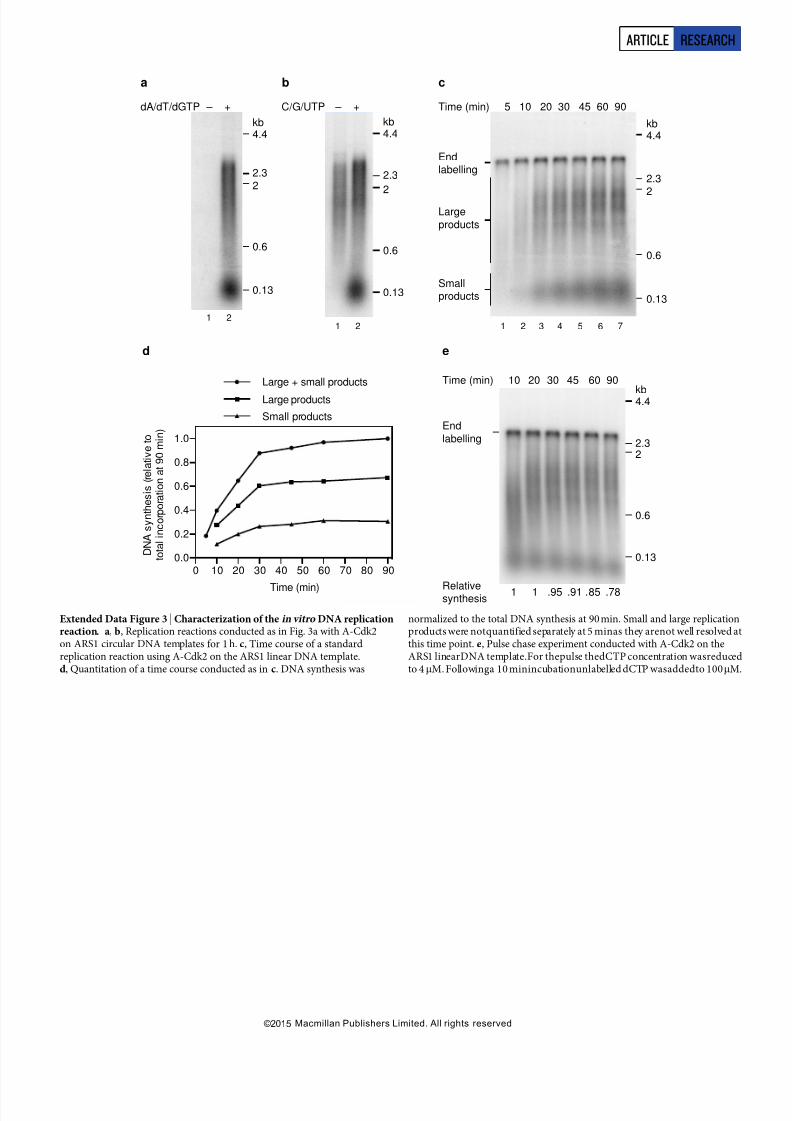
more strongly affected by omission of C/G/UTP than the larger class(Extended Data Fig. 3b), suggesting that the smaller class may corre-spond to lagging strand synthesis,which requires repeated re-priming,butfurtherwork is needed to establish this. Theresidual larger productsformed without C/G/UTP may be primed by RNA made from ATPalone (ARS1 is AT rich), which we cannot omit because it is requiredfor MCM unwinding, or may be primed by inefficient dNTP use by primase.ExtendedData Fig.3c, d shows full-lengthproductsappear by
20 min and continue to accumulate with time; the smaller productsaccumulatewiththe same kineticsas the larger products,but seem to belessabundant,which mayreflect incomplete laggingstrand synthesis. Apulse-chase experiment (Extended Data Fig. 3e) shows that the smallerproductsarenot a precursor ofthelargerones,orvice versa.Approximately 10%of inputplasmid is routinely replicatedduring replicationreactions.
A template containing a functional origin yielded approximately 3.2-fold more DNA synthesis than an equivalent template containing a mutant, non-functional origin (A-B2-) (Fig. 3c (bottom), comparelanes 2 and 4), indicating that replication exhibited origin preferenceunder these conditions. The experiments in Fig. 3c andExtendedDataFig. 3c–e were performed on linear DNA attached at one end to beads,andconsequently,in additionto DDK-dependent replicationproducts,there is also a DDK-independent end-dependent labelling product.
Requirements for DNA synthesis and RPA recruitmentFigure 4a shows that DNA synthesis in this system is dependent uponall of the firing factors, including Mcm10, which was not required forsalt-stable association of Cdc45 and GINS (Fig. 2b). Omission of polaalso preventedDNA synthesis (Fig. 4b, lane2), while Ctf4omission hadlittle effect on DNAsynthesis, althoughthe products werereproducibly slightly smaller (Fig. 4b, lane 3). In the absence of RPA, the small pro-duct class was completely lost, while the larger smear was reduced inboth intensity and size (Fig.4b, lane 4). Finally, in the absence of TopoII, accumulation of products longer than 600 nucleotides was greatly reduced (Fig. 4b, lane 5).
Topo II was required for accumulation of larger products from thecircular template (Fig. 4b, c), but not from the linear template (Fig. 4c,
compare lanes 2 and5). Moreover,topoisomerase I from vaccinia virussupported DNA replication on the circular template (Extended DataFig. 4a). Together, these results indicate that removal of supercoils isrequired for the replication of circular molecules in this system. Thisexperimentalso showsthat linearand circular templatesgenerate similarlabelled products (asidefor theend-labelled product on thelinear tem-plate) and replicate with similar efficiencies (compare lanes 1 and 4).
We nextexaminedthe recruitmentof RPA duringthe DNAsynthesisreaction (Fig. 4d). RPA showed some DDK- and Mcm10-dependentrecruitment (compare lanes 1–3). RPA recruitment was enhanced by omission of pol a (compare lanes 1 and 4), suggesting some uncoup-ling of template unwinding from DNA synthesis. This was supportedby the fact that RPA recruitment was also enhanced in complete reac-tions lacking dNTPs and C/G/UTP (Extended Data Fig. 4b). The ele-
vated RPA recruitmentobserved in the absence of pola required GINS,
Mcm10 and Topo II, but not Ctf4 (Fig. 4e). The requirementfor TopoII is consistent with the idea that extensive unwinding was occurring.This is supported by the fact that enhanced RPA recruitment in theabsence of Topo II could be restored by either Topo I or use of a lineartemplate (Extended Data Fig. 4c). Although Cdc45 and GINS stably associate with loaded MCM in the absence of Mcm10 (Fig. 2b), RPArecruitment in the absence of DNA synthesis was substantiallyreducedwithout Mcm10 (Fig. 4e, lane 3 and Extended Data Fig. 4c), indicating that unwinding requires Mcm10.
Regulation of DNA replication by CDKTo examine the regulation of origin firing by protein kinases in moredetail we used budding yeast S-CDK (Fig. 1 (left), lane 7) in place of A-Cdk2as it can be selectively inhibited by the proteinSic1. CDK pre-
vents MCM loading outside of G1 phase. Consistent with this, Fig. 5a(lane 2) shows thatphosphorylationof MCMloadingfactorswith S-CDKbefore MCMloadingpreventedDNAreplication, butadditionof S-CDKafter MCMloadingdid not(lane1). To determinewhichproteinswereinhibited by S-CDK, we incubated each individually with S-CDK andATP, then inhibited S-CDK with Sic1. Figure 5b shows that treatmentof ORC with S-CDKprevented DNA replicationwhiletreatmentof Cdc6and Cdt1–MCM with S-CDK had no effect. This is likely because, asshownin Fig. 5c,S-CDK treatmentof ORC, butnotCdc6 orCdt1–MCM,blockedMCM loadingand consequently, Cdc45 andGINSrecruitment.Pre-treatment of S-CDK with Sic1 before incubation with ORC pre-
vented the inhibition of replication (Extended Data Fig. 5a, lane 3).Moreover, the inhibitionof replication by S-CDK was ATP-dependent(Extended Data Fig. 5b). Together, these results indicate that ORC’s
ability to load MCM and promote replication is inhibited by CDK-dependent phosphorylation in this system.Previous genetic analysis indicated that Sld2 and Sld3 are the two
critical CDK substrates required for origin firing 10–12. To examine thisbiochemically we phosphorylated Sld3/7 and Sld2 each individually with S-CDK, then inhibited S-CDK with Sic1 before adding the addi-tional proteins required for the DDK step (for Sld3/7) and CDK step
4.4
2.32.0
0.6
0.13
kb
N o n e
D p b 1 1
S l d 2
S l d 3 / 7
C d c 4 5Protein
omitted M c m 1 0
N o n e
P o l ε
G I N S
2.3
2.0
0.6
N o n e
R P A T o p o
I I
P o l α
C t f 4
Protein
omitted
kb
4.4
2.3
2.0
0.6
0.13
kb
N o n e
N o n e T o p o I I
T o p o
I I
D D KProtein
omitted D D K
Template Circular Linear
Endlabelling
ba c
Mcm7
Rpa1
Psf1
N o n e
P o l α
D D K M c m 1 0Protein
omitted C t f 4 R P A
d
Rpa1
Mcm7
N o n e
C t f 4
G I N S M c m 1 0Protein
omitted T o p o I I
e
1 2 3 4 5 6 7 8 9 1 2 3 4 5
1 2 3 4 5 6
1 2 3 4 5 6
1 2 3 4 5
Figure 4 | Requirements for origin firing. Reactions were performed asillustrated in Fig. 3a on circular templates unless stated. a , Firing factorsrequiredto initiate DNA synthesis. b, Protein dependencies of DNA replication
for components functioning downstream of firing factor recruitment.
c, Topoisomerase dependence on circular and linear ARS1 templates. d, RPArecruitment in a complete replication reaction. e, Dependence of RPArecruitment in the absence of pol a.
ARTICLE RESEARCH
2 6 M A R C H 2 0 1 5 | V O L 5 1 9 | N A T U R E | 4 3 3
Macmillan Publishers Limited. All rights reserved©2015

7/21/2019 DNA replication reconstructed full article.pdf
http://slidepdf.com/reader/full/dna-replication-reconstructed-full-articlepdf 4/18
(for Sld2) (Fig. 5d). Phosphorylation of eitherSld2 or Sld3 individually did not support DNA replication (Fig. 5e, lanes 2 and 3) or salt-stableassociationof Cdc45 andGINS (Fig. 5f,lanes 3–6). However, when bothSld2 and Sld3 were phosphorylated separately and added in the samereaction, DNA replication (Fig. 5e, lane 4) and salt-stable associationof Cdc45 and GINS (Fig. 5f, lanes 7 and 8) occurred efficiently. If Sic1was omitted from the Sld2 CDK phosphorylation step, replication andsalt-stable Cdc45 and GINS association occurred even when Sld3 wasnot pre-phosphorylated (Fig. 5e, lane 1; Fig. 5f,lanes1 and2), presum-
ably because active CDK was added to the complete reaction, where it
could also phosphorylate Sld3. Thus, comparison of lanes 1 and 2 inFig. 5eand lanes 2 and 4 in Fig. 5fshowsSic1waseffective in inhibiting S-CDK. Extended Data Fig. 5c shows that ATP is essential for activa-tion of both Sld2 andSld3/7. Taken together, these results indicate thatphosphorylation of Sld2and Sld3/7 is necessary and sufficientfor initi-ating DNA replication.
Regulation by DDK and the order of kinase action
Highsalt washafter MCM loading and DDKphosphorylationremovesboth ORC andDDK(Fig. 6a(left); Orc6and Dbf4). This high-salt washedMCM complex was competent for salt-stable Cdc45 and GINS asso-ciation (Fig. 6a (right)) as well as DNA replication (Fig. 6b) withoutfurther DDK addition. This indicates that MCMis thekey substrate of DDK. This experiment also shows that ORC plays no essential role ininitiating DNA replication after MCM loading, similar to conclusionsfrom Xenopus egg extracts20. Previous work using yeast extracts indi-cated that replication didnot happen if theCDK phosphorylation stepwasexecuted before the DDK step7. However, using theapproachout-lined in Fig. 6c with purified proteins, stable association of Cdc45 andGINS (Fig. 6d) and DNA replication (Fig. 6e) were equally efficientregardless of the order of kinase treatment.
DiscussionOur experiments define the minimum set of proteins and small mole-cule co-factors required to initiate origin-dependent DNA replicationin a eukaryotic system(Extended Data Fig. 6). We show that the MCMcomplex assembled with purified proteins is a precursor of DNA repli-cation initiation.Moreover, because this complex is competentfor rep-lication after high-salt wash, the loading factors ORC, Cdc6 and Cdt1playno essential rolein initiation after MCMloading. Sld3/7and Cdc45arerecruited to MCMin a DDK-dependent manner,and the remaining firing factors (Sld2, Dpb11, GINS, pol e, Mcm10) are recruited in aDDK- and CDK-dependent manner. This set of firing factors is suffi-cient to promote RPArecruitment.RPA recruitmentis likelydue to thegeneration of substantial single strandedDNA because it requirestopo-isomerase on a circular template, and it is enhanced when DNA syn-
thesis is preventedby omittingeither polaor dNTPs fromthe reaction.Mcm10 is required for RPA recruitment, but not salt-stable associationof Cdc45 and GINS, consistent with results in yeast extracts7 andin vivo21,22. Thus, a CMG-like complex can be assembled withoutMcm10, but this complex is inactive. All DNA synthesis requires pola, consistent with its role in generating primers required forDNA syn-thesis. Because pole is required forCMG formation, we cannotat pres-ent determine whether it alsocontributes to subsequentDNA synthesisin this system, for example, through generation of a CMG–pole com-plex 23. Thepresence of short andlong products is suggestive of leading and laggingstrand synthesis.However,our replication system currently lacks DNA polymerase d, which is responsible for lagging strand syn-thesis in vivo24, RFC and PCNA, which localize to lagging strands dur-ing replication in vivo25, aswellas the many factorsrequired forOkazakifragment maturation. It also lacks other replisome-associated factorssuch as Mrc1 and Tof1–Csm3(ref. 17). Consequently, DNA synthesisin this system does not yet fully recapitulate normal coupled leading andlagging strand replication, andthis is clearly an important goal forthe future.
Ourexperimentsalsodefine theminimum setof CDKand DDKsub-stratesrequired for regulated replication.CDK phosphorylation of ORCdirectly preventsit fromloading MCMand promoting replication,con-sistentwithpreviouswork 1,26,27. Although it is clear that CDKphosphor-ylation of Cdc6andCdt1–MCMcontributesto preventingre-replicationin vivo in budding yeast, our experiments show that CDK phosphor-ylation does not directly inhibit the ability of these proteins to loadMCM. Instead, it is likely that CDK inhibits Cdc6 function by pro-moting its degradation in cells, and CDK inhibits Cdt1–MCM func-tion by promoting nuclear exclusion2. Our work also confirms that
Sld3/7and Sld2 arethe onlytwo CDKsubstrates required forreplication
d
MCM load
Sld3/7
±
S-CDK
7 min ±
Sic1
3 min
3 min
Mcm7
Cdc45
Psf1
– + – + – + – +DDK
Sld3/7
Sld2
S-CDK
Sic1
S-CDK
Sic1
+
–
–
–
+
+
+
+
–
–
+
+
+
+
–
–
4.4
2.3
2.0
0.6
kb– + – + – +S-CDK
Protein ORC Cdc6Cdt1–
Mcm2–7
a b
1 2 3 4 5 6
1 2 3 4 5 6 7 8
e
4.4
2.32.0
0.6
kb
0.13
Sld3/7
Sld2
S-CDK
Sic1
S-CDK
Sic1
+
–
–
–
–
–
+
+
+
+
–
–
+
+
+
+
1 2 3 4
f
Mcm7
Cdc45
Psf1
1 2 3 4 5 6
S-CDK – + – + – +
Protein ORC Cdc6Cdt1–
Mcm2–7
c
4.4
2.3
2.0
0.6
kb
0.13
MCM
load
S-CDK
S-CDK
MCM
load
1 2
Sld2
±
S-CDK
7 min ±
Sic1
DDK step
CDK step
Figure 5 | Regulation of MCM loading and origin firing by S-CDK.a , Replication reactions where MCM loading factors were incubated withS-CDK before or after MCM loading on ARS1 circular DNA. Replication,b, and protein recruitment, c, on ARS1 circular DNA where ORC, Cdc6 andCdt1–Mcm2–7 were individually incubated with S-CDK followed by additionof Sic1. The remaining MCM loading factors were added and reactionsperformed as shown in Fig. 3a (replication) and Fig. 1b (recruitment).d, Scheme to phosphorylate Sld3/7 and Sld2 in isolation from MCM and firing factors. Prior to both the DDK step and CDK step Sld3/7 and Sld2 wereincubated with S-CDK before addition of Sic1. The remaining firing factorswere added together with isolated DNA beads and each reaction step wasperformedas for standard reactions.e, Replication reaction staged as illustratedin d. f , Firingfactorrecruitmentconducted as in d, followedby 0.3 M KCl washes.
RESEARCH ARTICLE
4 3 4 | N A T U R E | V O L 5 1 9 | 2 6 M A R C H 2 0 1 5
Macmillan Publishers Limited. All rights reserved©2015

7/21/2019 DNA replication reconstructed full article.pdf
http://slidepdf.com/reader/full/dna-replication-reconstructed-full-articlepdf 5/18
initiation. In contrast to conclusionsfrom experimentsin yeast extracts7,ourresultswith purifiedproteinsshow CDKdoesnot haveany inhibitory role afterMCMloading, and CDK and DDK can act in either order. Inthe experiments using extracts7, we suggest rapid dephosphorylationof Sld2 andSld3 occurred after Sic1(ref. 6) and DDK addition, requir-ingone or both of these proteins to be re-phosphorylatedby CDKafterSld3/7 and Cdc45 were loaded. Our experiments show that the essen-tial DDK substratefor initiation is MCM. Further work will be requiredto ascertain which MCM subunits and sites are critical.
The initiation step of DNA replication has taken many years toreconstitute because of the large number of factors involved and thecomplicated regulation by protein kinases. Having achieved this, weare now in a position to understand how the MCM double hexamer isactivated in molecular and atomic detail. Moreover, this work sets thestage forthe completereconstitutionof chromosomereplication,whichhasthepotential to provide insights intomanyDNA replication-couplednuclear processes.
Online Content Methods, along with any additional Extended Data display itemsandSource Data, areavailablein theonline versionof thepaper; references uniqueto these sections appear only in the online paper.
Received 14 December 2014; accepted 6 February 2015.
Published online 4 March 2015.
1. Tanaka, S.& Araki, H. Helicaseactivationand establishment of replicationforks atchromosomal origins of replication. Cold Spring Harb. Perspect. Biol. 5, a010371(2013).
2. Siddiqui, K.,On, K. F. & Diffley, J. F. X. Regulating DNAreplication in Eukarya. Cold Spring Harb. Perspect. Biol. 5, a012930 (2013).
3. Remus, D. et al. Concerted loading of Mcm2–7 double hexamers around DNAduring DNA replication origin licensing. Cell 139, 719–730 (2009).
4. Evrin,C. et al. A double-hexamericMCM2-7 complex is loaded onto origin DNAduring licensing of eukaryotic DNA replication. Proc. Natl Acad. Sci. USA 106,20240–20245 (2009).
5. Gros, J., Devbhandari, S. & Remus, D. Origin plasticity during budding yeast DNAreplication in vitro. EMBO J. 33, 621–636 (2014).
6 . On, K.F . et al. Prereplicative complexes assembled in vitro support origin-dependent and independent DNA replication. EMBO J. 33, 605–620 (2014).
7. Heller, R.C. et al. Eukaryotic origin-dependent DNA replication in vitro revealssequential action of DDK and S-CDK kinases. Cell 146, 80–91 (2011).
8. Labib, K. How do Cdc7 and cyclin-dependent kinases trigger the initiation ofchromosome replication in eukaryotic cells? Genes Dev. 24, 1208–1219 (2010).
9. Nguyen,V. Q.,Co, C.& Li,J. J.Cyclin-dependent kinasesprevent DNAre-replicationthrough multiple mechanisms. Nature 411, 1068–1073 (2001).
10. Zegerman, P. & Diffley,J. F. X. Phosphorylation of Sld2and Sld3 by cyclin-dependent kinases promotes DNA replication in budding yeast. Nature 445,281–285 (2007).
11. Tanaka, S. et al. CDK-dependent phosphorylation of Sld2 and Sld3 initiates DNAreplication in budding yeast. Nature 445, 328–332 (2007).
12. Masumoto, H., Muramatsu, S., Kamimura, Y. & Araki, H. S-Cdk-dependentphosphorylation of Sld2 essential for chromosomal DNA replication in buddingyeast. Nature 415, 651–655 (2002).
13. Randell,J. C. etal.Mec1is oneof multiplekinases thatprimethe Mcm2–7helicasefor phosphorylation by Cdc7. Mol. Cell 40, 353–363 (2010).
14. Sheu, Y. J. & Stillman, B. Cdc7-Dbf4 phosphorylates MCM proteins via a dockingsite-mediated mechanism to promoteS phase progression. Mol.Cell 24, 101–113
(2006).15. Sheu,Y. J.& Stillman,B. TheDbf4–Cdc7kinase promotesS phase byalleviatingan
inhibitory activity in Mcm4. Nature 463, 113–117 (2010).16. Moyer, S. E., Lewis, P. W. & Botchan, M. R. Isolation of the Cdc45/Mcm2–7/GINS
(CMG) complex,a candidate forthe eukaryotic DNAreplication forkhelicase.Proc.Natl Acad. Sci. USA 103, 10236–10241 (2006).
17. Gambus, A. et al. GINS maintains association of Cdc45 with MCM in replisomeprogression complexes at eukaryotic DNA replication forks. Nature Cell Biol. 8,358–366 (2006).
18. Bell, S. P. & Dutta, A. DNA replication in eukaryotic cells. Annu. Rev. Biochem. 71,333–374 (2002).
19. Gambus, A. et al. A key role for Ctf4 in coupling the MCM2–7 helicase to DNApolymerase a within the eukaryotic replisome. EMBO J. 28, 2992–3004 (2009).
20. Rowles, A., Tada, S. & Blow, J. J. Changes in association of the Xenopus originrecognition complex with chromatin on licensing of replication origins. J. Cell Sci.112, 2011–2018 (1999).
21. Watase,G., Takisawa, H. & Kanemaki, M.T. Mcm10 plays a rolein functioning of theeukaryotic replicative DNA helicase, Cdc45-Mcm-GINS. Curr. Biol. 22, 343–349(2012).
22. van Deursen, F., Sengupta, S., De Piccoli, G., Sanchez-Diaz, A. & Labib, K. Mcm10associates withthe loaded DNA helicaseat replicationorigins and definesa novelstep in its activation. EMBO J. 31, 2195–2206 (2012).
23. Georgescu, R. E. et al. Mechanism of asymmetric polymerase assembly at theeukaryotic replication fork. Nature Struct. Mol. Biol. 21, 664–670 (2014).
24. Clausen,A. R. et al. Tracking replication enzymology in vivo by genome-widemapping of ribonucleotide incorporation. Nature Struct. Mol. Biol. http://dx.doi.org/10.1038/nsmb.2957 (2015).
25. Yu, C. etal. Strand-specificanalysis shows protein binding at replication forks andPCNA unloading from lagging strands when forks stall. Mol. Cell 56, 551–563(2014).
26. Chen, S. & Bell, S. P. CDK prevents Mcm2–7 helicase loading by inhibiting Cdt1interaction with Orc6. Genes Dev. 25, 363–372 (2011).
27. Frigola, J., Remus,D., Mehanna, A. & Diffley,J. F. X. ATPase-dependent qualitycontrol of DNA replication origin licensing. Nature 495, 339–343 (2013).
Supplementary Information is available in the online version of the paper.
Acknowledgements Weare grateful to B. Pfander and M. Douglas for the Mcm10expression plasmid and advice on purification, C. Kurat for construction of plasmidsused to generate ARS1 linear templates, K. On and D. Boos for Sic1 and A-Cdk2 andK. Labib forPsf1 antibodies andthe E. coli GINSexpressionstrain. We thank A. Alidoustand N. Patel for growing yeast cultures. This work was supported by Cancer ResearchUK, a FEBS Return-to-Europe fellowship to J.T.P.Y., a Boehringer Ingelheim Fonds PhDfellowship to A.J. and an ERC grant (249883 – EUKDNAREP) to J.F.X.D.
Author Contributions J.F.X.D. and J.T.P.Y. designed the experiments and wrotethe manuscript. J.T.P.Y. performed the experiments. T.D.D., A.J. and A.E.generated protein expression constructs and strains, and established proteinpurification protocols.
Author Information Reprints and permissions information is available atwww.nature.com/reprints. The authors declare no competing financial interests.Readers are welcome to comment on the online version of the paper.Correspondence and requests for materials should be addressed to J.F.X.D. ([email protected]).
b
Mcm7
Psf1
– + – +S-CDK
Wash L HWash L H
Mcm7
Orc6
Dbf4
4.4
2.32.0
0.6
kb
0.13
Wash L H
Mcm7
Orc6
Dbf4
No
DDK
CDK
DDK
CDK
CDK
DDK
Mcm7
Cdc45
Psf1
d
DDK
CDK
CDK
DDK
1 2 1 2 3 4
1 2 3
1 2
a
4.4
2.3
0.6
kb
0.13
NoDDK
CDK
e
1 2 3
2.0
MCM load
Firing factors
+
DDK
Firing factors
+
CDK
20 min 10 min
Add in
CDK
Add in
DDK
10 min 20 min
MCM load
DDK
CDK
CDK
DDK
c
Analysis of protein recruitment
or DNA replication
Figure 6 | DDK phosphorylation of MCM promotes origin firing either before or after S-CDK. a , Reactions performed essentially as in Fig. 1b oncircular DNA but with an additional mid-reaction wash immediately following DDK phosphorylation, in either a low-salt (L) (0.3 M K-Glu), or high-salt(H) (0.6 M NaCl) buffer. Bound proteins immediately following the mid-reaction wash (left), and after firing factor recruitment followed by 0.3M KClwashes (right). b, Replication reactions with either high- or low-saltmid-reaction washes. c, Reaction schemes to test the order of DDK and CDKaction. Firing factor recruitment was performed in a single step with DDK andCDK added at different times as indicated. Protein recruitment, d, and
replication, e, performed with S-CDK as illustrated in c.
ARTICLE RESEARCH
2 6 M A R C H 2 0 1 5 | V O L 5 1 9 | N A T U R E | 4 3 5
Macmillan Publishers Limited. All rights reserved©2015

7/21/2019 DNA replication reconstructed full article.pdf
http://slidepdf.com/reader/full/dna-replication-reconstructed-full-articlepdf 6/18
METHODSYeastexpression strain construction. All strains wereconstructedby transform-ingstrain yJF127 withlinearizedexpressionvectors using standard genetic techniques(SeeExtended DataTables 2 and3 fordetailsof vectors andstrains). ForexpressionofSld3,Dpb11 andSld2, affinitytags were addedby transformationwith PCR pro-ducts following integration of the expression vector. The PCR products for Sld3tagging weregeneratedwitholigonucleotidesSupsld3 TCPtag fwdand SupTCP tag rev from the template pBS1539/TAPTCP 3 (yTD6), and oligonucleotides Sld3SUPC-term Flag tag fwd and Sld2SUP C-term Flag rev using pBP83 (a derivative of
pYM1828 modified to insert a C-terminal 33Flag tag associated with the NatNT2marker) as template (yTD11). PCR products for Sld2 and Dpb11 were generatedfrompBP83 using oligonucleotides Sld2SUP C-term Flagrev and Sld2SUP C-termFlag revfor Sld2 andJTY10and Cdc45 flag tagrev forDpb11. Affinity taggedpro-tein expression was verified by immunoblot.Proteins and protein expression. Vaccinia virus topoisomerase I was purchasedfrom Sigma. ORC, Cdc6, Cdt1–Mcm2–7, DDK, cyclin A/Cdk2 and Sic1 were ex-pressedand purifiedas previously described6,27,29. Allproteinspurified in thisstudy containedaffinitytags, which wereused in thefirst stepof purification. Forthe ami-noacidsequences ofthe affinitytagsand theirlocations seeExtendedData Table1.GINS andMcm10wereexpressedin E. coli. All other proteins were overexpressedin S. cerevisiaefrombidirectionalGal1-10promotersin a pep4D, bar1D strain back-ground(yJF1) (seeExtended DataTable 2 fordetailsof expressionstrains).Expres-sion strains forDpb11,Sld2,Cdc45,Ctf4,RPAand Polewere grownat 30uCinYP1 2% raffinose to a density of , 2–43 107 cells per ml. Cells were arrested in G1with 100ng ml21 alpha factor and incubation was continued for 3 h. Protein ex-pression was induced by addition of galactose to 2% and growth continuedfor 3 hat30 uC. TopoII expression was conducted essentially as describedabovebut alphafactorwas addedtogetherwithgalactose andgrowthwas continuedfor6 h.For Pola expression, cells were grownto a densityof ,23 107 cellsper mland expressionwas induced for 2 h by addition of galactose (2%). S-CDK expressing cells weregrown to2 3 107 cells perml andproteinexpression induced by addition of galac-tose (2%) together with nocodazole (5 mg ml21) with growth continued for a fur-ther 3 h. In all cases, cells were collected, washed once with 25 mM HEPES-KOHpH7.6, 1 M sorbitol andoncewiththe appropriate initialproteinpurification buf-fer (see individual purification protocols for buffers used) lacking protease inhi-bitors. Cells were resuspendedin 0.3–0.4 volumes of the initial purification buffer1proteaseinhibitorsand the suspensionsfrozendrop-wisein liquidnitrogen. Fro-zen cells were crushed in a freezer mill (SPEX CertiPrep 6850 Freezer/Mill) with6 cycles of 2 min at a crushing rate of 15. The resulting powders were stored at280 uC until required.
Sld2 purification. Frozen cell powder was thawed and resuspendedin 3 volumes25 mM HEPES-KOH pH 7.6, 10% glycerol, 0.02% v/v Nonidet P40 substitute(NP-40-S) (Roche#11754599001),1 mMEDTA,1 mMDTT,500 mMKCl (bufferH 1 500 mM KCl) 1 protease inhibitors (0.3 mM PMSF, 7.5 mM benzamidine,0.5 mMAEBSF,1 mMleupeptin,1 mMpepstatin A and 1mg ml21 aprotinin (Sigma)).Insoluble material was cleared by centrifugation (235,000 g , 4 uC, 1 h) and solidammonium sulphate wasaddedto thesupernatantto 32%followed by gentlestir-ring (10 min, 4uC). Insoluble material was removed by centrifugation (27,000 g ,4 uC, 20 min) andthe ammonium sulphate concentration increasedto 48%followedby stirring for10 min.Precipitatedproteinwas collectedby centrifugation (27,000 g ,4 uC, 20 min) and resuspended in 1/3 of the original volume buffer H 1 500mMKCl1protease inhibitors. Flag-taggedSld2 was boundto anti-Flag M2 affinity gel(Sigma) in batch for30 minat 4uC. Resin was collected in 20 ml chromatography columns (Bio-Rad)and washedextensively in buffer H1500mMKCl1proteaseinhibitors. The resin was resuspended in 10 column volumes (CV) buffer H with-out EDTA1 500mM KCl1 10 mM magnesium acetate1 1 mM ATP and incu-
bated for 10minat 4 uC. The flow-through was discarded and the column washedwith 20–40 CV bufferH 1 500mM KCl. Sld2–Flagwas elutedin 1 CV bufferH 1
500mM KCl 1 0.5 mgml21 33Flag peptide, followed by 2 CV buffer H 1
500mM KCl 1 0.25 mg ml21 33Flag peptide.The eluates were pooled, dialysed against buffer H1 280 mM KCl and applied
toa1-mlHiTrapSPFFcolumn(GEHealthcare)equilibratedinbufferH 1250mMKCl. Sld2–Flagwas elutedwithan 18 CVgradientfrom250 mMKCl to1 M KClinbuffer H. Peak fractions were pooled and dialysed against buffer H with 40% v/v glycerol 1 350 mM KCl. Protein concentration was assessed using the Bradfordassay (Bio-Rad) and this method was used for all proteins in this study.Sld3/7 purification. To purify TCP-tagged Sld3/7, cell powder from yTD6 wasthawed in buffer H 1 500 mM KCl 1 protease inhibitors and insoluble materialwas cleared by centrifugation (235,000 g , 4 uC, 1 h). Sld3/7 was depleted from thesoluble extract by incubation for 40 min at 4 uC with IgG Sepharose 6 Fast Flow (GEHealthcare). Theresin wascollectedin a 20 ml column andwashedextensively in buffer H with 0.5 mM EDTA1 500 mM KCl. Sld3/7 was cleaved from the col-
umnbyincubationfor2hat4 uC withtobaccoetch virus (TEV) protease(50 mg ml21)
in buffer H with 0.5mM EDTA 1 500 mM KCl. His-tagged TEV protease wasremoved by passing the eluate over a Ni-NTA Agarose column (Qiagen). The re-sulting flow through was concentrated and separated on a Superdex 200 column(GE Healthcare) equilibrated in buffer H 1 500 mM KCl.
The Flag-tagged Sld3/7 usedfor Cdc45depletion (ExtendedData Fig.7) was pu-rified fromyTD11 cellsessentially as described above,exceptthatSld3/7 was depletedfrom thesolubleextract byincubation with anti-FlagM2 affinitygel for1 h at 4 uC.BoundSld3/7 waseluted asdescribedforSld2 andthe eluatewas concentratedandseparated on a Superdex 200 column equilibrated in buffer H 1 500 mM KCl.
Cdc45 purification. 80g cell powderwas thawedin 200 ml bufferH without NP-40-S1500 mM potassiumacetate1protease inhibitorsand celldebris was removedby centrifugation (235,000 g , 4 uC,1 h). 8 mlanti-FlagM2 affinitygelwas addedandtheextractincubated for 4 h at 4 uC.Resinwas collectedin 20 mlcolumns(2 ml percolumn) washed extensively in buffer H without NP-40-S 1 500 mM potassiumacetate, then buffer H without NP-40-S 1 300 mM potassium acetate. Cdc45 waseluted by incubation of the resin in 1 CV buffer H without NP-40-S 1 300mMpotassium acetate1 0.5mgml21 33Flag peptide. Theeluatewas dialysed against150mM potassiumacetate,0.5 mMDTT, 10%glycerol, 20 mMK-phosphate pH7.4,(buffer P 1 20 mM K-phosphate) and then applied to 1.5 ml hydroxyapatite col-umn(Bio-Rad) equilibratedin thesame buffer. Thecolumn waswashed with buf-fer P 1 80 mM K-phosphate and Cdc45 was eluted with buffer P 1 250mMK-phosphate. The eluate was extensively dialysed against buffer H without NP-40-S 1 300 mM potassium acetate before storage. To test the functionality of theinternally Flag-tagged Cdc45, we depleted endogenous Cdc45 from an S phaseextract(ExtendedData Fig. 7a). DNAreplication wasnot detected after depletion
but wasrestoredfollowing additionof purifiedCdc45(Extended DataFig. 7b).Thedistributions of replication productsfollowing Cdc45 depletionand add back wereindistinguishable from an undepleted sample (compare lanes 1 and 4) suggesting that the recombinant protein is functional.
S-CDK purification. 40 g cell powder was thawed in 80 ml 40 mM HEPES-KOHpH 7.6, 10% glycerol, 0.02% v/v NP-40-S, 300 mM potassium acetate (buffer CD1 300 mM potassium acetate) 1 protease inhibitors. Cell debris was cleared by centrifugation (235,000 g , 4 uC, 1 h),calcium chloride wasaddedto 2 mM togetherwith calmodulin affinity resin (Agilent) and the extract was incubated at 4 uC for1 h. Resin was collected in a 20 ml column, washed extensively with buffer CD1
300 mM potassium acetate1 2 mM CaCl2 and S-CDK was eluted by incubationfor 16h with 100mg ml21 TEV protease in buffer CD 1 2 mM CaCl2. TEV pro-teasewas removed by passingthe eluate overTalon resin(Clontech)and collecting theflow through,whichwas then applied to a Superose 6 column(GE Healthcare)equilibrated in buffer CD1 300 potassiumacetate.Peak fractionswere pooled andconcentrated.
Dpb11 purification.Cell powderwas resuspendedin 2 volumes bufferH1500mMKCl and the debris removed by centrifugation (235,000 g , 4 uC, 1 h). Dpb11–Flag was bound to anti-Flag M2 affinity gel for 90 min at 4uC. The resin was collected,washed extensively with buffer H1 500 mM KCl and the proteineluted in bufferH1500mMKClusing33Flagpeptide as forSld2. Theeluate wasdialysedagainstbuffer H 1 150 mM KCl and applied to a 1ml MonoS column (GE Healthcare).Dpb11–Flag was eluted with a 30 CV gradient from 150 mM KCl to 1 M KCl inbuffer H with the peak fractions then dialysed against buffer H 1 300 mM pot-assium acetate.
DNApolymerase e purification. Cell powderwasslowly thawedin bufferH with-out NP-40-S and EDTA 1 400 mM potassium acetate (buffer E 1 400 mM pot-assium acetate) 1 1 Complete, EDTA free protease inhibitor tablet (Roche) per25 ml buffer andcell debris was removedby centrifugation (235,000 g , 4 uC,1h).Tothe supernatant CaCl2 was added to 2 mM together with calmodulin affinity resinand the solution was rotated at 4 uC for 60 min. Resin was collected in a 20 ml col-
umn, washed extensively in buffer E1 400 mM potassiumacetate1 2 mMCaCl2
andPolewaselutedin 2 mlfractions with bufferE1 400mM potassiumacetate12mMEDTA1 2 mMEGTA.The eluatewas pooledandapplieddirectly toa 5 mlHi-trapheparincolumn(GE Healthcare) equilibratedin buffer E1400mM potas-siumacetate.Following extensivewashingwith buffer E1 450mM potassiumace-tate proteins were eluted with a 12 CV gradient from 450 mM – 1 M potassiumacetate in buffer E. Heparin fractions containing Pol e were pooled, concentratedandseparated on a Superdex 200column (GEHealthcare)equilibrated in bufferE1 500 mM potassium acetate.
Ctf4 purification. Ctf4purificationwas basedon a published method30 withmod-ifications. Cell powderwasthawedin 2 volumes25 mMTris-HClpH 7.2, 10% glyc-erol,1 mMDTT,200mM NaCl(bufferC 1 200mM NaCl)1 protease inhibitorsandthe insoluble materialremovedby centrifugation (235,000 g , 4 uC,1h).Calciumchloride was added to 2 mM together with calmodulin affinity resin. The extractwas incubated at 4 uC for 90 min before the resin was collected and washed exten-sively withbufferC 1200mMNaCl1 2mMCaCl2. Proteinswere eluted in buffer
C1200mM NaCl12mMEDTA12 mMEGTA. TheNaClconcentrationof the
RESEARCH ARTICLE
Macmillan Publishers Limited. All rights reserved©2015

7/21/2019 DNA replication reconstructed full article.pdf
http://slidepdf.com/reader/full/dna-replication-reconstructed-full-articlepdf 7/18
eluate was reduced to 150 mM by dilution in buffer C before application to a 1 mlMonoQ equilibrated in buffer C1 150mM NaCl1 1 mM EDTA. Ctf4 waselutedwitha30CVgradientfromthe150mMto1MNaClinbufferC.Peakfractionswerepooled, concentrated and separated on a Superdex 200 column (GE Healthcare)equilibrated in buffer C 1 150 mM NaCl 1 1 mM EDTA. Ctf4 containing frac-tions were dialysed against buffer C 1 75 mM NaCl1 1 mM EDTA.
DNA polymerase a–primase purification. Cell powder was thawed in buffer C1 0.02% NP-40-S1 400 mM NaCl (buffer D 1 400 mM NaCl)1 protease inhi-bitors and insoluble material was cleared by centrifugation (235,000 g , 4 uC, 1h).
TheNaCl concentrationwas reducedto 300 mMby dilutionin buffer lackingNaCl,and calcium chloride was added to 2 mM together with calmodulin affinity resin.The extract was incubated at 4 uC for 90 min before the resin was collected in a20 ml column, washed extensively with buffer D1 300 mM NaCl1 2 mM CaCl2
andproteins eluted withbufferD1300mMNaCl12mMEDTA12 mMEGTA.Pooled fractions weredilutedin buffer D to a conductivityequivalentto buffer D1
120mM NaCl and were loaded onto a 1 ml MonoQ column. Bound proteins wereremovedwitha 30CV gradientfrom 120mM to1 MNaClin bufferD andfractionscontainingpol awerepooled,concentratedto 400ml andapplied toa Superdex 200column equilibrated in buffer D1 150mM NaCl. Thepeakfractions were pooled,concentrated to,0.7mgml21 and snap frozen in liquid nitrogen.
RPA purification. 40 g cell powder was thawed in buffer C 1 500 mM NaCl 1proteaseinhibitorsand insoluble material clearedby centrifugation(235,000 g , 4 uC,1 h). Lysate conductivitywas reducedto the equivalent of buffer C1 200mM NaClby twofold dilution in buffer C 1 protease inhibitors, followed by 1 h dialysisagainst buffer C 1 200 mM NaCl. CaCl2 was added to 2 mM together with 1 ml
calmodulin affinityresinand the extract incubatedat 4 uCfor90minwithrotation.Resin was collected in a 20 ml column, washed with 35 CVs buffer C1 200mMNaCl 1 2 mM CaCl2 and bound proteins were eluted in 1 ml fractions of bufferC1 2 mMEDTA1 2 mM EGTA. Peak fractions were pooled, diluted twofold inbuffer C1 1 mMEDTA,dialysedfor 1 h againstbuffer C1 50mMNaCl1 1 mMEDTAand loadedonto a 1 ml Hi-trapheparincolumn equilibratedin thesame buf-fer. After extensive washing proteins were eluted with a 30 CV gradient from50mM–1MNaClinbufferC1 1 mM EDTA. Peakfractions werepooled,dilutedthreefold in buffer C 1 1 mM EDTA, dialysed for 1 h against buffer C1 150mMNaCl1 1 mMEDTAand loadedonto a 1 ml MonoQequilibratedin thesamebuf-fer.RPAwaseluted witha 25CV gradientfrom 150 mM– 1 M NaClin bufferC 1
1 mM EDTA and peak fractions were pooled and dialysed overnight in buffer Cwith 38% glycerol 1 1 mM EDTA 1 50 mM NaCl.
Topoisomerase II purification.Powderwas thawedin bufferD 1 300mM NaCl1protease inhibitors and insolublematerialremovedby centrifugation (235,000 g ,
4u
C,1 h).Thesolubleextract wassupplementedwith2 mMCaCl2 and calmodulinaffinity resin was added and the solution incubated at 4 uC for 1 h. Resin was col-lected in a 20 ml column, washed extensively with buffer D 1 300 mM NaCl andthe CBP-tagged Topo II eluted with buffer D 1 300 mM NaCl1 2 mM EDTA12 mM EGTA. The eluate was concentrated and applied directly to a Superdex 200column equilibrated in buffer D 1 150 mM NaCl. Topo II containing fractionswere pooled and the salt concentration adjusted to , 100 mM by dilution beforeloadingontoa 1 ml MonoQequilibrated inbuffer D1 100mMNaCl.TopoIIwaseluted with a 25 CV gradient from 100 to 800 mM NaCl in buffer D and the peak fractions werepooledand dialysedagainst bufferD 1150mM NaClbeforestorage.
GINS purification. Plasmid pFJD5 (gift from K. Labib), which is a derivative of pFJD1219 in which the Psf3 subunit is expressed with an N-terminal His-tag, wasused to transform Bl21 (DE3)Rosetta pLysS.Cellsweregrownin LB at 37 uCtoan
A600nm of 0.5 and protein expression was induced by addition of IPTG to 1 mM.Growth was continued for 2 h before cells were harvested by centrifugation. Cellpellets were resuspended in buffer D1 400mM NaCl1 10 mMimidazole1pro-
tease inhibitors, cells were lysedby sonication andthe debrisremovedby centrifu-gation (257,000 g , 4 uC, 30 min). His-tagged proteins were depleted by incubationwith Ni-NTA resin (Qiagen) for 1h at 4 uC and the resin was collected in a 20 mlcolumn. Following extensive washing with buffer D 1 400 mM NaCl 1 10mMimidazoleand thenbuffer D1100mMNaCl1 15 mM imidazole,GINS waselutedwith buffer D1 100 mM NaCl1 200 mM imidazole. Fractions were pooled anddialysed against buffer D1 100 mM NaCl before being applied to a 1 ml MonoQcolumnequilibratedin thesamebuffer.Proteins were elutedwith a 25CV gradientfrom 100mM to500 mMNaClin buffer D andtheGINScontainingfractions werepooled, concentrated and separated through a Superdex 200 column equilibratedinbuffer D1 150mM NaCl. Thepeakwas rebound to Ni-NTAAgaroseresinandthe resin was washed with 50 mM HEPES-KOH pH7.6, 10% glycerol, 0.05% NP-40-S, 10 mM magnesium acetate before GINS was eluted in the same buffer sup-plemented with 50 mM imidazole. Finally the eluate was dialysed against bufferH 1 200 mM potassium acetate before storage.
Mcm10 purification. Plasmid pET28a-Mcm10, which expresses Mcm10with an
N-terminal His/T7 tag, was used to transform BL21 (DE3). Cells were grown at
37 uCtoan A600nm of0.6 beforethe temperaturewas reduced to 25 uC andproteinexpression induced by additionof IPTG to 1 mM.Growth wascontinued for3 h at25 uC beforecellswere harvested, washedin 50mM Tris-HCl pH7.6, 10%sucroseandresuspendedin 25 mMTris-HCl pH 7.6, 10%glycerol,0.01%NP-40-S, 500mMNaCl (buffer M 1 500 mM NaCl)1 protease inhibitors. Cells were lysed by son-ication andthe debrisremovedby centrifugation (257,000 g , 4 uC,30min).Theclearlysatewasappliedunder gravityflowto a 1.5ml Ni-NTAAgarose column. Thecol-umn was washed with 15 CV buffer M1 500mM NaCl followed by 10 CV bufferM 1 200 mM NaCl 1 20 mM imidazole and His–Mcm10 was eluted with buffer
M1 200mMNaCl1 200mM imidazole. Theeluate wasappliedto a 1 ml MonoSequilibrated in buffer M 1 200 mM NaCl and proteins were eluted with a 20 CVgradientfrom200 mMto 1 M NaCl inbufferM. PeakMcm10 fractions werepooled,theNaCl concentration adjusted to 200mM by dilution inbuffer M andreappliedto a 1 ml MonoS. The column was run as described for the first run and Mcm10containingfractionswere pooledand dialysedagainst buffer H with0.01% NP-40-S 1 200 mM K-glutamate.
S phase extracts. S phase extracts were prepared as described previously 6. Psf2–Flag (GINS complex) was depleted from the yJY18 extract by two 45 min incuba-tions at 4 uC with a 1:10 extract volume of anti-Flag M2 magnetic beads (Sigma).To deplete Cdc45 from the yJY16 extract, Flag-tagged Sld3/7 was pre-bound toanti-Flag M2 magnetic beads (5 pmol per 1ml of resin). Cdc45 was then depletedfrom the extract by two 30 minincubationsusing2.5 ml beads per20 ml of extract.
DNA templates.1 kb biotinylatedlinear ARS305 DNA templates were generatedas described3,31. For linear ARS1 templates a 2.8 kb region surrounding ARS1 wasamplifiedusingprimersARS1_XmaI-F and ARS1_XhoI-R and cloned intopBlue-scriptKS (1) usingXhoI andXmaIto generateplasmidpCFK1.An A-B2- derivative,pCFK2, was generated by sequential rounds of site-directed mutagenesis using oligonucleotides MutA_F, MutA_R, MutB2_F,MutB2_R.2.8 kb biotinylated wildtype andA-B2-ARS1 linearDNA templates were generatedby PCRusingthe oli-gonucleotides ARS1-PC-Bio-F and ARS1-Bio-R with pCFK1 and pCFK2 as tem-plates, respectively. 3.2 kb randomly biotinylated circular DNA templates weregeneratedas described6. BiotinylatedDNAwas coupledto streptavidinM-280mag-netic DNAbeads (Invitrogen) essentiallyas described3,31. ForlinearDNA templates250 ng DNA was coupled to 5 ml bead slurry. As biotinylation efficiency was esti-matedonlytobe,10–20%for thecircularDNA templates, 1 mgofDNAwascou-pled per 5 ml bead slurry.
Standardproteinrecruitment and replicationreactions. Unlessstatedin thefig-ure legends, protein recruitment assays were conducted using the ARS305 lineartemplate,whereasDNA replication reactionsused ARS1circular DNAas template.All incubations were conducted with agitation (1,250 r.p.m.) in a Thermomixer
(Eppendorf). The concentrations of proteins in recruitment and replication reac-tions were determined empirically using the concentrations of individual firing factors in S phase extracts forguidance. Sld3/7purifiedfrom yTD6 wasused in allprotein recruitment andreplication reactions.Salt contributions fromproteinsaddedto reactions were less than 30 mM in all steps. MCM loading reactions (‘MCM’load)(7.5-10ml)were performedin a buffercontaining25 mMHEPES-KOH pH 7.6,100 mM K-glutamate, 10 mM magnesium acetate, 0.02% NP-40-S, 5% glycerol,2 mMDTT,5 mMATP (loadingbuffer),45 nMORC,45 nMCdc6,100nM Cdt1–Mcm2–7andeither6.25ng perml linearor approximately 4 ngperml circular DNAimmobilisedon M-280streptavidinmagnetic beads(Invitrogen).After 20 min in-cubation at 25 uC DDK was addeddirectly to the reaction to a final concentrationof 130nM andincubationwas continuedat 25 uC for30 min. Bufferand unboundproteinswere removed usinga magnetic rack andwerereplaced bya newbufferof twofold the MCMloading reaction volume (15–20 ml) containing40 mMHEPES-KOH pH 7.6, 300 mM K-glutamate, 10 mM magnesium acetate, 0.02% NP-40-S,
8% glycerol, 400mg ml21
BSA(NEB),2 mMDTT, 5 mM ATP(firing-factor recruit-mentbuffer1300mM K-glutamate)and26 nMSld3/7and 50nM Cdc45. Follow-ing incubation at 25 uC for 5 min the supernatant was removed and replaced withthesame volume (15–20ml) of firing-factorrecruitmentbuffer1250mM K-glutamateand 40nM Dpb11, 62nM Sld2, 30nM Pol e, 210 nM GINS, 2.5 nM Mcm10 and50 nMA-Cdk2 (cyclin A/Cdk2) (Figs 1–4and Extended DataFigs2–4) or S-CDK(Clb5/Cdc28/Cks1)at a concentrationof 30nM for Fig.6, 20nM for Fig.5e, f andExtended DataFig.5c and25 nMfor Fig. 5a–c andExtendedData Fig. 5a,b. Reac-tions were incubated at 25 uC for 10 min (‘CDK’ step). For protein recruitmentassaysthat were terminated after the‘CDK’step,10 ngper ml poly(dI-dC) (Sigma)was added to both the buffer containing Sld3/7 and Cdc45, and the ‘CDK’ step.After the‘CDK’stepsupernatants were againremoved andreplacedwiththe same
volume of buffer containing 40 mM HEPES-KOH pH 7.6, 150 mM K-glutamate,10 mM magnesium acetate, 5% glycerol, 2 mM DTT, 2 mM ATP, 200mM CTP,GTP,UTP,40mM dATP,dTTP,dCTP,dGTP, 2.5nM Mcm10, 50nM RPA, 25nMpola, 30nM Ctf4,25 nMTopo IIand,forreplicationreactions,40 nM[a-32P]dCTP
(Perkin Elmer). Reactions were incubated at 30 uC for 30min.
ARTICLE RESEARCH
Macmillan Publishers Limited. All rights reserved©2015
7/21/2019 DNA replication reconstructed full article.pdf
http://slidepdf.com/reader/full/dna-replication-reconstructed-full-articlepdf 8/18
For protein recruitment assays, unless stated in the figure legends, the final re-action buffer (either after both the ‘DDK’ and ‘CDK’ steps or the final replicationstep)was removed andbeads were washedthreetimes in150 ml room temperaturebuffercontaining40 mMHEPES-KOH pH 7.6, 300mM K-glutamate,10 mMmag-nesiumacetate,5% glycerol, 0.02%NP-40-S (washbuffer1300mM K-glutamate).Beadswere resuspended in SDS-loading buffer, heated to 95 uC for 3 min and pro-teins wereseparated through 4–12% Bis-Trispolyacrylamide gels(Bio-Rad) beforeanalysis by immunoblotting.
Replication reactions were terminatedby removing the supernatant and imme-
diately washing the beads twicewith 150ml 40mM HEPES-KOHpH 7.6, 600mMKCl, 5% glycerol, 0.02% NP-40-S, 5 mM EDTA before resuspending the beads in5 mM EDTA and then adding NaOH and sucrose to 50mM and 1% w/v, respect-ively. Samples were incubated at room temperature for 20–30 min before the pro-ducts were separated through 0.7% alkaline agarose gels in 30mM NaOH, 2 mMEDTA for16 h at 25V. Gels were fixedwith5% cold trichloroaceticacid, driedontochromatography paper (Whatman) and autoradiographed with AmershamHyperfilm-MP (GE Healthcare).When gels were quantifiedimages were scannedusing a Typhoon phosphorimager (GE Healthcare) and were quantified using ImageQuant software (GE Healthcare).Modified proteinrecruitment and replication reactions. For experiments wheremid-reactionwashes were included (Fig. 6a,b), two150 ml washes were conductedin wash buffer containing the salts indicated in the figure legends, followed by one150ml wash inwash buffer1 300 mM K-glutamate. Reactionswith a single firing-factor recruitment step(Fig.6d, e) wereconducted in firing-factor recruitment buf-fer1 250 mM K-glutamate for 30min at 25 uC, with 30nM S-CDK, 65nM DDK
and all other proteins at the same concentrations as in standard reactions.InFig. 5a,lane 2,MCM loadingfactors (22.5 nMOrc, 45 nMCdc6,100 nMCdt1–
Mcm2–7) were pre-treatedin loadingbufferwith25 nMS-CDK for10 min at 25uCbefore DNA addition for 20 min. When S-CDK was added after DNA (lane 1),MCMloadingfactors wereincubated withDNA for20 minand S-CDKthen addedfora further 10 min.Pre-treatment of individual MCMloadingfactors withS-CDK(Fig.5b, c andExtendedDataFig.5a, b) wasperformedunderthe same conditionsasthose usedforMCMloading except that ORCwaspresent at22.5nM andS-CDKandSic1were at 25nM and125 nM, respectively.Proteins were incubated at 25uCwith S-CDK for10 min, S-CDKwas inhibitedwithSic1(5 min) beforethe remain-ingproteins required forMCM loading were added andincubation continuedfor20 min. To ensure S-CDK and Sic1 were removed from the above reactions, sam-ples were subjected to two 150 ml washes in wash buffer1 600 mM NaCl and one150ml wash inwashbuffer1 300mM K-glutamate after eitherthe MCM loading step(Fig.5a), or afterDDK phosphorylation (Fig.5b, c andExtendedDataFig.5a, b).
When Sld3/7and Sld2 were pre-incubated with S-CDK (Fig.5e, f andExtendedData Fig.5c) the incubationswere performedin the standard firing-factor recruit-ment buffers with 20 nM S-CDK for 7 min followed by addition of Sic1 (100 nM)for3 min.The remainingfiringfactorswere thenaddedto thesame concentrationsas used in standard reactions.
Fororigindependentreplicationreactions (Fig. 3c)ORC waspre-boundto DNAfor 10 min at 30uC in reactions (10 ml) containing 25 mM HEPES-KOH pH 7.6,50 mM KCl, 10 mM magnesium acetate, 5% glycerol, 2 mM ATP, 1 mM DTT,100mg ml21 BSA,6.25ng/ml ARS1 linearDNAbeadsand2.5 nMORC.The super-natant was removed, the beads washed twice with wash buffer 1 300mM pot-assium acetate and a new buffer (10 ml) containing 25 mM HEPES-KOH pH 7.6,100 mM potassium acetate, 10 mM magnesium acetate, 0.02% NP-40-S, 5% gly-cerol, 2 mMDTT,5 mMATP,45 nMCdc6, 100nM Cdt1–Mcm2–7 wasaddedandthe mix incubated at 30uC for30 min. The buffer was removed and replaced withloading buffer 1 130nM DDK (10ml) and the samples incubated at 25 uC for30 min. Alldownstreamsteps wereperformedas described for standard reactions.
Extract-based replication. For reactions on bead-bound DNA (Fig. 2d and Ex-tended Data Fig. 2e), MCM and firing factors were assembled as described forstandard proteinrecruitment and replication assays on immobilised ARS1circulartemplate(10 ml MCMloading reactions).Beads werewashedtwicewith 150mlwashbuffer1200mM K-glutamate andresuspended in extract replication buffer (20ml)containing 65 mM HEPES-KOH pH 7.6, 150 mM K-glutamate, 10.5mM magnes-iumacetate, 5% glycerol, 0.5mM EDTA, 0.5mM EGTA, 40 mM creatinephosphate
and100ngperml creatine phosphokinase, 3.5mM DTT, 2.5mM ATP, 200mMCTP,GTP, UTP, 100mM dGTP, dATP, dTTP, 25 mM dCTP and 50nM [a-32P]dCTP.Samples were incubated at 25 uC for 30 min and were processed as described forstandard replication reactions.
The soluble replication reaction in Extended Data Fig. 7b wasperformed as de-scribed previously 6 with unmodified ARS1 circular DNA as template. Productswere separated through a 1% native agarose gel.
Oligonucleotides. JTY10, AAACCTATGAGACGACAGACAAGAAATCAGACAAAGGAATTAGATTCTCTGGAAGTGCTGTTTCAGGGCCCGCGTACGC
TGCAGGTCGAC; JTY32, CGATGATGAGACAATATCTAATAAAAGAGG;JTY33, TCTTTATAGTCCTCGTCTGTGACTTCATC; JTY40, GATGATGATGGGGACTATAAAGACGATGATGAGAC; JTY41, TTTATAATCCTCGTCTGTGACTTCATCGGC; JTY92, AGCACAATAAGGCGCGCCTATAAAACAATGTTCAGGCAGTCAAAAAG; JTY100, CTTTTCCCTTCTCGAGTTAATGATTACCATTATTG; Dpb11 fwd, AAGCTCACCGGTGATGAAGCCCTTTCAAGGAATA; Dpb11 reverse, CTGAGGCGGCCGCTCAAGAATCTAATTCCTTTGT; Sld3SUP C-term Flag fwd, CTTCTAAGAGAAGAGTTAGAAGAAGATTGTTCGCTCCAGAATCTACTCTGGAAGTGCTGTTTCAGGGCCCGCGTACGCTGCAGGTCGAC; Sld2SUP C-term Flag fwd, GTTGCCAAAGAAGAACAGATTCTCCAACGGTAGATGGGGTAGAAGACTGGAAGTGCTGTTTCAGGGCCCGCGTACGCTGCAGGTCGAC; Sld2SUP C-term Flag rev, GAGAAAAGAAAAAAATTGATCTATCGATTTCAATTCAATTCAATTTAATCGATGAATTCGAGCTCG; Sup sld3 TCP tag fwd, CTAAGAGAAGAGTTAGAAGAAGATTGTTCGCTCCAGAATCTACTGAAAACTTGTACTTCCAAGG; Sup TCPtag rev, GGAAAGAGAAAAGAAAAAAATTGATCTATCGATTTCAATTCAATTCAATTACGACTCACTATAGGG; Cdc45 flag tag rev, CGACTCACTATAGGGCGAATTGGAGCTCCACCGCGGTGGCGGCCGCTTAATCGATGAATTCGAGCTCG; Cdc45 fwd, CTGGACACCGGTGATGTATTATGGAATCAGCCAG; Cdc45 rev, AGCACGCGGCCGCTTATAACAATCCACTCAAGGT;ARS1_XmaI-F, CGATCCCGGGGGTAGTTATAAGAAAGAGACCGAGTTAG;ARS1_XhoI-R, CGATCTCGAGAAGAGTATTGGCGATGACGAAAC; ARS1-PC-Bio-F, Bio-GGTAGTTATAAGAAAGAGACCGAGTTAG; ARS1-Bio-R, AAGAGTATTGGCGATGACGAAAC; MutA_F, GCATAAAAGATCTAAACATACCTCGAGGAAAATAACAAGATGTAAAG; MutA_R,CTTTACATCTTGTTATTTTCCTCGAGGTATGTTTAGATCTTTTATGC;
MutB2_F, GTTATTACTGAGTAGTATTTCCTCGAGGATTGTTTGTGCACTTGCCTG; MutB2_R, CAGGCAAGTGCACAAACAATCCTCGAGGAAATACTACTCAGTAATAAC.
Antibodies. Anti-Mcm7 (yN-19,sc-6688,SantaCruz),anti-Orc6 (SB49).Psf2–Flag was visualized with anti-Flag M2-peroxidase (Sigma), Mcm10was detected using
the T7-tag antibody (69522, Novagen), Dbf4 was visualized with an anti-CBP tag antibody (07-482 MerckMillipore)and Rpa1was detected withanti-scRPA (AS07214, Agrisera). Psf1 antibodies were a gift from K. Labib. Polyclonal antibodiesagainst Sld3, Sld7, Sld2, Cdc45 and Dpb11 were described previously 6. Polyclonalantibodiesagainstthe Dpb2subunit of polewereraisedagainstfull-length protein,and their specificity was confirmed using purified pol e.
28. Janke,C. et al. A versatile toolbox for PCR-based tagging of yeast genes: newfluorescent proteins, more markers and promoter substitution cassettes. Yeast 21, 947–962 (2004).
29. Boos, D. et al. Regulation of DNA replication through Sld3-Dpb11 interaction isconserved from yeast to humans. Curr. Biol. 21, 1152–1157 (2011).
30. Simon,A. C. et al. A Ctf4 trimer couples the CMG helicase to DNA polymerasealpha in the eukaryotic replisome. Nature 510, 293–297 (2014).
31. Coster, G., Frigola, J., Beuron, F., Morris, E. P. & Diffley, J. F. X. Origin licensingrequires ATP binding and hydrolysis by the MCM replicative helicase. Mol. Cell55, 666–677 (2014).
32. Brown, N. R. et al. The crystal structure of cyclin A. Structure 3, 1235–1247(1995).
33. Itou, H., Muramatsu, S., Shirakihara, Y. & Araki, H. Crystal structureof thehomology domain of the eukaryotic DNA replication proteins Sld3/Treslin.Structure 22, 1341–1347 (2014).
34. Sharp, P. M. & Li, W. H. The codon adaptation index-a measure of directionalsynonymous codon usage bias,and its potential applications.Nucleic Acids Res.15, 1281–1295 (1987).
RESEARCH ARTICLE
Macmillan Publishers Limited. All rights reserved©2015

7/21/2019 DNA replication reconstructed full article.pdf
http://slidepdf.com/reader/full/dna-replication-reconstructed-full-articlepdf 9/18
160
100
50
20
30
70
40
kDa
160
100
50
20
30
70
15
kDa
10
25
15
10
3040
50
70
100
kDa
Extended Data Figure 1 | Coomassie-stained SDS–PAGE analysis of multi-subunit complexes required for DNA replication. a , Annotation of thepolyacrylamide gels in Fig. 1a in which individual protein subunits have beenlabelled. b, Analysis of A-Cdk2. The protein complex consists of humanCdk2 and the bovine cyclin A-3 fragment32.
ARTICLE RESEARCH
Macmillan Publishers Limited. All rights reserved©2015

7/21/2019 DNA replication reconstructed full article.pdf
http://slidepdf.com/reader/full/dna-replication-reconstructed-full-articlepdf 10/18
Extended Data Figure 2 | Analysis of firing factor recruitment.a , Immunoblots of protein recruitment conducted as in Fig. 1b but with0.3 M KCl washes. b, Stability of recruited firing factors following washes of varying strength (lanes 1 and 5, 0.3 M K-glu; lanes 2 and 6, 0.3 M KCl; lanes 3and 7, 0.45M KCl; lanes 4 and 8, 0.6 M KCl). c, Psf2–Flag was depletedfrom a yJY18 S phase extract by two rounds of incubation with anti-Flag M2magnetic beads. Levels of Psf2–Flag were determined by immunoblotting with the Flag-M2 antibody. Soluble and bead bound protein fractions are
illustrated. d, Extract-based replication reaction schemes. In the leftpathway (i), loaded MCM is treated with DDK and added to a KO3 extract(Sld3, Sld7, Cdc45, Dpb11, Sld2 overexpression). In the right pathway (ii),firing factors are recruited to MCM as illustrated in Fig. 1b and the complex isadded to a yJY18 extract (nofiringfactor overexpression) in which Psf2 (GINScomplex) has been depleted. e, Replication reactions as described in d using A-Cdk2 for firing factor recruitment. Where indicated, Sic1 was added to theextract 20 min before replication.
RESEARCH ARTICLE
Macmillan Publishers Limited. All rights reserved©2015
7/21/2019 DNA replication reconstructed full article.pdf
http://slidepdf.com/reader/full/dna-replication-reconstructed-full-articlepdf 11/18
ba
dA/dT/dGTP – +
4.4
2.32
0.6
0.13
kb
4.4
2.3
2
0.6
0.13
kb
Endlabelling
Time (min) 5 10 20 30 45 60 90
c
4.4
2.3
2
0.6
0.13
kb
C/G/UTP – +
Time (min) 10 20 30 45 60 90
4.4
2.32
0.6
0.13
kb
End
labelling
Relativesynthesis 1 1 .95 .91 .85 .78
1 2 3 4 5 6 7
1 21 2
ed
Large
products
Smallproducts
0 10 20 30 40 50 60 70 80 900.0
0.2
0.4
0.6
0.8
1.0
Time (min)
D N A s y n t h e s i s ( r e l a t i v e t o
t o t a l i n c o r p o r a t i o n a t 9 0 m i n )
Large + small products
Large products
Small products
Extended Data Figure 3 | Characterization of the in vitro DNA replicationreaction. a , b, Replication reactions conducted as in Fig. 3a with A-Cdk2on ARS1 circular DNA templates for 1 h. c, Time course of a standardreplication reaction using A-Cdk2 on the ARS1 linear DNA template.d, Quantitation of a time course conducted as in c. DNA synthesis was
normalized to the total DNA synthesis at 90 min. Small and large replicationproducts were notquantified separately at 5 minas they arenot well resolved atthis time point. e, Pulse chase experiment conducted with A-Cdk2 on theARS1 linearDNA template.For thepulse thedCTP concentration wasreducedto 4mM. Followinga 10 minincubationunlabelled dCTP wasaddedto 100mM.
ARTICLE RESEARCH
Macmillan Publishers Limited. All rights reserved©2015

7/21/2019 DNA replication reconstructed full article.pdf
http://slidepdf.com/reader/full/dna-replication-reconstructed-full-articlepdf 12/18
Extended Data Figure 4 | Characterization of RPA recruitment. Unlessstated reactions were conducted on ARS1 circular templates. a , Vaccinia virustopoisomerase I supports DNA replication with purified proteins. Replicationreactions with either Topo II (25 nM) or vaccinia virus topoisomerase I (0.125units per ml). Two different Topo II fractions (Fr1 and Fr2) were used forcomparison. b, Nucleotide dependence of RPA recruitment in a complete
replication reaction with Topo II. c, RPA recruitment reactions wereconducted on ARS1 circular template in the presence of vaccinia virustopoisomerase I (0.125 units per ml),or on a linear template in the absence of atopoisomerase. dNTPs, C/G/UTP, pol a and Ctf4 were omitted from the finalstep of the reaction.
RESEARCH ARTICLE
Macmillan Publishers Limited. All rights reserved©2015

7/21/2019 DNA replication reconstructed full article.pdf
http://slidepdf.com/reader/full/dna-replication-reconstructed-full-articlepdf 13/18
Extended Data Figure 5 | Regulation of MCM loading and origin firing by S-CDK is ATP dependent. a , Replication reaction where ORC was pre-incubated with S-CDK before MCM loading. When Sic1 was added beforeORC the mix was incubated for 5 min and ORC was then added for 10 min.b, Pre-incubation of ORC with S-CDK in the presence or absence of ATP.
After incubation with Sic1, ATP was added to the reaction lacking ATP.c, Sld3/7 and Sld2 were pre-incubated with S-CDK as illustrated in Fig. 5d inthe presence or absence of ATP. Following incubation with Sic1, samplesthat did not contain ATP for the pre-incubation step were supplementedwith ATP.
ARTICLE RESEARCH
Macmillan Publishers Limited. All rights reserved©2015

7/21/2019 DNA replication reconstructed full article.pdf
http://slidepdf.com/reader/full/dna-replication-reconstructed-full-articlepdf 14/18

7/21/2019 DNA replication reconstructed full article.pdf
http://slidepdf.com/reader/full/dna-replication-reconstructed-full-articlepdf 15/18
Extended Data Figure 7 | Internally Flag-tagged Cdc45 supports normalDNA replication in S phase extracts. a , The previously reported interactionbetween Sld3 and Cdc45 (ref. 33) was exploited to co-immunoprecipitateCdc45 from yJY16 extracts (Dpb11 and Sld2 overexpression) by incubation
with Flag–Sld3/7 that was pre-coupled to anti-Flag M2 magnetic beads.b, In vitro extract-based replication reaction on soluble ARS1 circular template
using yJY16 extracts where endogenous Cdc45 was depleted as indicated.The extract was supplemented with purified Sld3/7 as the complex is notoverexpressed in yJY16. The experiment was conducted for 30 min asdescribed previously 6 and products were separated through a 1% native
agarose gel.Internally Flag-tagged Cdc45(52 nM) was added back as indicated.The locations of the different replication products are illustrated.
ARTICLE RESEARCH
Macmillan Publishers Limited. All rights reserved©2015

7/21/2019 DNA replication reconstructed full article.pdf
http://slidepdf.com/reader/full/dna-replication-reconstructed-full-articlepdf 16/18
Extended Data Table 1 | Affinity tag strategies for protein purification
Protein Yeast
strain
Affinity tag strategy Affinity tag sequence
Cdc45 yJY13 Internal 2xFLAG tag E197 – DYKDDDG – D198, E199Y, E200K, E202D *
Sld3/7 yTD6 C-terminal TCP tag on Sld3 ENLYFQGEKRRWKKNFIAVSAANRFKKISSSGALDYDIPTTASKTAALA
QHDEAVDNKFNKEQQNAFYEILHLPNLNEEQRNAFIQSLKDDPSQSANLLAEAKKLNDAQAPKVDNKFNKEQQNAFYEILHLPNLNEEQRNAFIQ
SLKDDPSQSANLLAEAKKLNGAQAPKVDANSAGKST
Sld3/7 yTD11 C-terminal 3xFLAG tag on Sld3 LEVLFQGPRTLQVDDYKDDDDKDYKDDDDKDYKDDDDK
Sld2 yTD8 C-terminal 3xFLAG tag LEVLFQGPRTLQVDDYKDDDDKDYKDDDDKDYKDDDDK
Dpb11 yJY26 C-terminal 3xFLAG tag LEVLFQGPRTLQVDDYKDDDDKDYKDDDDKDYKDDDDK
Pol yAJ2 C-terminal CBP tag on Dpb4 ENLYFQGEKRRWKKNFIAVSAANRFKKISSSGAL
S-CDK yAE37 N-terminal CBP tag on Clb5 MKRRWKKNFIAVSAANRFKKISSSGALENLYFQGE
Pol - primase yJY23 N-terminal CBP tag on Pri1 MKRRWKKNFIAVSAANRFKKISSSGALENLYFQGE
Ctf4 yAE40 N-terminal CBP tag MKRRWKKNFIAVSAANRFKKISSSGALENLYFQGE
Topo II yAE46 C-terminal CBP tag ENLYFQGEKRRWKKNFIAVSAANRFKKISSSGAL
GINS N/A N-terminal 6His tag on Psf3 MGSSHHHHHHSSGLVPRGSHMAS
Mcm10 N/A N-terminal 6His tag MGSSHHHHHHSSGLVPRGSHMASMTGGQQMGRGSEF
RPA yAE31 N-terminal CBP tag on Rfa1 MKRRWKKNFIAVSAANRFKKISSSGALENLYFQGE
AllN-terminaltags arelocatedimmediately upstreamof originalstartcodon.All C-terminaltagsare locatedimmediatelybeforethe originalstop.*Amino acidresiduenumberscorrespondto thewild-type Cdc45
sequence.
RESEARCH ARTICLE
Macmillan Publishers Limited. All rights reserved©2015

7/21/2019 DNA replication reconstructed full article.pdf
http://slidepdf.com/reader/full/dna-replication-reconstructed-full-articlepdf 17/18
Extended Data Table 2 | Saccharomyces cerevisiae strains
Strain Genotype Reference
yTD6 MAT a ade2-1 ura3-1 his3-11,15 trp1-1 leu2-3,112 can1-100,bar1::Hygpep4::KanMX,his3::HIS3pRS303/SLD3-TCP, GAL4,leu2::LEU2pRS305/SLD7
This study
yTD8 MAT a ade2-1 ura3-1 his3-11,15 trp1-1 leu2-3,112 can1-100bar1::Hygpep4::KanMXhis3::HIS3pRS303/Sld2-3xflag (Nat-NT2), GAL4
This study
yTD11 MAT a ade2-1 ura3-1 his3-11,15 trp1-1 leu2-3,112 can1-100bar1::Hygpep4::KanMXhis3::HIS3pRS303/SLD3-3xflag (Nat-NT2), GAL4leu2::LEU2pRS305/SLD7
This study
yJY7 MAT ade2-1 ura3-1 his3-11,15 trp1-1 leu2-3,112 can1-100, cdc7-4pep4::KanMX
This study
yJY13 MAT a ade2-1 ura3-1 his3-11,15 trp1-1 leu2-3,112 can1-100bar1::Hygpep4::KanMX
his3::HIS3pRS303/Cdc45 iflag2 , GAL4
This study
yJY16 MAT ade2-1 ura3-1 his3-11,15 trp1-1 leu2-3,112 can1-100, cdc7-4pep4::KanMXura3::URA3pRS306/Dpb11trp1::TRP1pRS304/Sld2
This study
yJY18 MAT ade2-1 ura3-1 his3-11,15 trp1-1 leu2-3,112 can1-100, cdc7-4pep4::KanMXPsf2-3xFlag (Nat-NT2)
This study
yJY23 MAT a ade2-1 ura3-1 his3-11,15 trp1-1 leu2-3,112 can1-100bar1::Hygpep4::KanMXtrp1::TRP1pRS304/Pol1/Pol12ura3::URA3pRS306/CBP-Tev-Pri1/Pri2
This study
yJY26 MAT a ade2-1 ura3-1 his3-11,15 trp1-1 leu2-3,112 can1-100bar1::Hygpep4::KanMXhis3::HIS3pRS303/Dpb11-3xflag (Nat-NT2), GAL4
This study
yAJ2 MAT a ade2-1 ura3-1 his3-11,15 trp1-1 leu2-3,112 can1-100bar1::Hygpep4::KanMXura3::URA3pRS306/Dpb2, Dpb3trp1::TRP1pRS304/Pol2, Dpb4-Tev-CBP
This study
yAE31 MAT a ade2-1 ura3-1 his3-11,15 trp1-1 leu2-3,112 can1-100bar1::Hyg
pep4::KanMX his3::HIS3pRS303/CBP-Tev-RFA1, GAL4ura3::URA3pRS306/RFA2, RFA3
This study
yAE37 MAT a ade2-1 ura3-1 his3-11,15 trp1-1 leu2-3,112 can1-100bar1::Hygpep4::KanMXura3::URA3pRS306/CKS1, CDC28his3::HIS3pRS303/CBP-Tev-Clb5, GAL4
This study
yAE40 MAT a ade2-1 ura3-1 his3-11,15 trp1-1 leu2-3,112 can1-100bar1::Hygpep4::KanMXhis3::HIS3pRS303/CBP-Tev-Ctf4, GAL4
This study
yAE46 MAT a ade2-1 ura3-1 his3-11,15 trp1-1 leu2-3,112 can1-100bar1::Hyg
pep4::KanMXtrp1::TRP1pRS304/TOP2-Tev-CBP
This study
ARTICLE RESEARCH
Macmillan Publishers Limited. All rights reserved©2015

7/21/2019 DNA replication reconstructed full article.pdf
http://slidepdf.com/reader/full/dna-replication-reconstructed-full-articlepdf 18/18
Extended Data Table 3 | Plasmids used to generate yeast expression strains
Plasmid Original vector* Insert Plasmid construction
pTD2 pJF2 Dpb11 PCR product from S. cerevisiae W303 genomic DNA using primers Dpb11
fwd and Dpb11 reverse cloned 5 - SgrAI , 3 - NotI
pTD5 pJF2 Sld2 Synthetic construct cloned 5 - SgrAI , 3 - NotI
pTD6a pJF2 Sld3 Synthetic construct cloned 5 - SgrAI , 3 - NotI
pTD6b pJF4 Sld7 Synthetic construct cloned 5 - SgrAI , 3 - NotI
pRS303-Cdc45 pJF2 Cdc45 PCR product cloned from S. cerevisiae W303 genomic DNA using primers
cdc45 fwd and cdc45 rev 5 - SgrAI , 3 - NotI
pRS303-Cdc45iFlag1
pJF2 Cdc45 iFlag1
Site directed mutagenesis using oligonucleotides JTY32 and JTY33 andpRS303-Cdc45 as template
pRS303-Cdc45iFlag2
pJF2 Cdc45 iFlag2
Site directed mutagenesis using oligonucleotides JTY40 and JTY41 and
pRS303-Cdc45 iFlag1
as template
pRS304 (Pol2 + Dpb4-CBP) pJF18 Pol2
Dpb4-CBPPol2 - synthetic construct cloned 5 - AscI , 3 - XhoI
Dpb4-CBP - synthetic construct cloned 5 - SgrAI , 3 - NotI
pRS306 (Dpb2 + Dpb3) pJF19 Dpb2
Dpb3
Dpb2 - synthetic construct cloned 5 - SgrAI , 3 - NotI
Dpb3 - synthetic construct cloned 5 - AscI , 3 - XhoI
pRS304/Gal-CBP-Top2 pCG001 Top2 Synthetic construct cloned 5 - AscI , 3 - XhoI
pRS303/Gal-CBP-Ctf4 pJF2 Ctf4 Synthetic construct cloned 5 - SgrAI , 3 - NotI
pRS303/Gal4 + CBP-Tev-Clb5 pJF2 Clb5 Synthetic construct cloned 5 - SgrAI , 3 - NotI
pRS306/Cks1-Gal-Cdc28 pJF5 Cks1
Cdc28Cks1 - synthetic construct cloned 5 - SgrAI , 3 - NotI
Cdc28 - synthetic construct cloned 5 - AscI , 3 - XhoI
pRS303/Gal4 + CBP-Tev-Rfa1 pJF2 Rfa1 Synthetic construct cloned 5 - SgrAI , 3 - NotI
pRS306/Rfa2-Gal-Rfa3 pJF5 Rfa2Rfa3
Rfa2 - synthetic construct cloned 5 - AscI , 3 - XhoI
Rfa3 - synthetic construct cloned 5 - SgrAI , 3 - NotI
pRS304/Pol1-Gal1-10-Pol12 pJF3 Pol1Pol12
Pol1 - synthetic construct cloned 5 - SgrAI , 3 - NotIPol12 - synthetic construct cloned 5 - AscI , 3 - XhoI
pRS306/CBP-Tev-Pri1-Gal1-10-genomic Pri2
pJF5 Pri1Pri2
Pri 1 - synthetic construct cloned 5 - AscI , 3 - XhoIPri2 - PCR product from S. cerevisiae W303 genomic DNA using primers
JTY92 and JTY100 cloned 5 - AscI , 3 - XhoI
Synthetic constructs werecodonoptimisedfor expressioninSaccharomyces cerevisiae34. Sequencesfromthe 59 and39 endofthePGK1 genewere alsoadded.Geneswere synthesizedby GeneArtGene Synthesis
(Life technologies). *For details of the original expression vectors see refs 27, 31.
RESEARCH ARTICLE