dna repair mechanisms and their biological roles in the ... · dna repair mechanisms and their...
TRANSCRIPT
DNA Repair Mechanisms and Their Biological Roles in the MalariaParasite Plasmodium falciparum
Andrew H. Lee,a Lorraine S. Symington,a David A. Fidocka,b
Department of Microbiology and Immunology, Columbia University, New York, New York, USAa; Division of Infectious Diseases, Department of Medicine, ColumbiaUniversity, New York, New York, USAb
SUMMARY . . . . . . . . . . . . . . . . . . . . . . . . . . . . . . . . . . . . . . . . . . . . . . . . . . . . . . . . . . . . . . . . . . . . . . . . . . . . . . . . . . . . . . . . . . . . . . . . . . . . . . . . . . . . . . . . . . . . . . . . . . . . . . . . . . . . . . . . . . . . . . . . . .469INTRODUCTION . . . . . . . . . . . . . . . . . . . . . . . . . . . . . . . . . . . . . . . . . . . . . . . . . . . . . . . . . . . . . . . . . . . . . . . . . . . . . . . . . . . . . . . . . . . . . . . . . . . . . . . . . . . . . . . . . . . . . . . . . . . . . . . . . . . . . . . . . . . .469THE PLASMODIUM LIFE CYCLE . . . . . . . . . . . . . . . . . . . . . . . . . . . . . . . . . . . . . . . . . . . . . . . . . . . . . . . . . . . . . . . . . . . . . . . . . . . . . . . . . . . . . . . . . . . . . . . . . . . . . . . . . . . . . . . . . . . . . . . . . . . . .470OVERVIEW OF DNA DOUBLE-STRAND BREAK REPAIR . . . . . . . . . . . . . . . . . . . . . . . . . . . . . . . . . . . . . . . . . . . . . . . . . . . . . . . . . . . . . . . . . . . . . . . . . . . . . . . . . . . . . . . . . . . . . . . . . . . .470
Causes of Double-Strand Breaks. . . . . . . . . . . . . . . . . . . . . . . . . . . . . . . . . . . . . . . . . . . . . . . . . . . . . . . . . . . . . . . . . . . . . . . . . . . . . . . . . . . . . . . . . . . . . . . . . . . . . . . . . . . . . . . . . . . . . . . . . .470Generating double-strand breaks in Plasmodium. . . . . . . . . . . . . . . . . . . . . . . . . . . . . . . . . . . . . . . . . . . . . . . . . . . . . . . . . . . . . . . . . . . . . . . . . . . . . . . . . . . . . . . . . . . . . . . . . . . . . .471
DNA DOUBLE-STRAND BREAK REPAIR. . . . . . . . . . . . . . . . . . . . . . . . . . . . . . . . . . . . . . . . . . . . . . . . . . . . . . . . . . . . . . . . . . . . . . . . . . . . . . . . . . . . . . . . . . . . . . . . . . . . . . . . . . . . . . . . . . . . .471Mitotic Recombination: Mechanisms and Evidence in Plasmodium . . . . . . . . . . . . . . . . . . . . . . . . . . . . . . . . . . . . . . . . . . . . . . . . . . . . . . . . . . . . . . . . . . . . . . . . . . . . . . . . . . . . .472
Initial double-strand break sensing and resection . . . . . . . . . . . . . . . . . . . . . . . . . . . . . . . . . . . . . . . . . . . . . . . . . . . . . . . . . . . . . . . . . . . . . . . . . . . . . . . . . . . . . . . . . . . . . . . . . . . . .472Extensive resection . . . . . . . . . . . . . . . . . . . . . . . . . . . . . . . . . . . . . . . . . . . . . . . . . . . . . . . . . . . . . . . . . . . . . . . . . . . . . . . . . . . . . . . . . . . . . . . . . . . . . . . . . . . . . . . . . . . . . . . . . . . . . . . . . . . .474Rad51 nucleoprotein filament formation and strand invasion. . . . . . . . . . . . . . . . . . . . . . . . . . . . . . . . . . . . . . . . . . . . . . . . . . . . . . . . . . . . . . . . . . . . . . . . . . . . . . . . . . . . . . . . .474Noncrossovers (NCOs) and crossovers (COs) . . . . . . . . . . . . . . . . . . . . . . . . . . . . . . . . . . . . . . . . . . . . . . . . . . . . . . . . . . . . . . . . . . . . . . . . . . . . . . . . . . . . . . . . . . . . . . . . . . . . . . . . . .474
Meiotic Recombination: Mechanisms and Evidence in P. falciparum . . . . . . . . . . . . . . . . . . . . . . . . . . . . . . . . . . . . . . . . . . . . . . . . . . . . . . . . . . . . . . . . . . . . . . . . . . . . . . . . . . . . .475End Joining . . . . . . . . . . . . . . . . . . . . . . . . . . . . . . . . . . . . . . . . . . . . . . . . . . . . . . . . . . . . . . . . . . . . . . . . . . . . . . . . . . . . . . . . . . . . . . . . . . . . . . . . . . . . . . . . . . . . . . . . . . . . . . . . . . . . . . . . . . . . . .475
Basic mechanisms of end joining . . . . . . . . . . . . . . . . . . . . . . . . . . . . . . . . . . . . . . . . . . . . . . . . . . . . . . . . . . . . . . . . . . . . . . . . . . . . . . . . . . . . . . . . . . . . . . . . . . . . . . . . . . . . . . . . . . . . . .475Absence of nonhomologous end joining in Plasmodium . . . . . . . . . . . . . . . . . . . . . . . . . . . . . . . . . . . . . . . . . . . . . . . . . . . . . . . . . . . . . . . . . . . . . . . . . . . . . . . . . . . . . . . . . . . . .475Possible alternative end-joining mechanisms in Plasmodium . . . . . . . . . . . . . . . . . . . . . . . . . . . . . . . . . . . . . . . . . . . . . . . . . . . . . . . . . . . . . . . . . . . . . . . . . . . . . . . . . . . . . . . . .476Potential implications of the absence of end joining in Plasmodium . . . . . . . . . . . . . . . . . . . . . . . . . . . . . . . . . . . . . . . . . . . . . . . . . . . . . . . . . . . . . . . . . . . . . . . . . . . . . . . . . .476
HOMOLOGOUS RECOMBINATION IN P. FALCIPARUM . . . . . . . . . . . . . . . . . . . . . . . . . . . . . . . . . . . . . . . . . . . . . . . . . . . . . . . . . . . . . . . . . . . . . . . . . . . . . . . . . . . . . . . . . . . . . . . . . . . . .477Subtelomeric Regions and Antigenic Diversification . . . . . . . . . . . . . . . . . . . . . . . . . . . . . . . . . . . . . . . . . . . . . . . . . . . . . . . . . . . . . . . . . . . . . . . . . . . . . . . . . . . . . . . . . . . . . . . . . . . . .477Copy Number Variation . . . . . . . . . . . . . . . . . . . . . . . . . . . . . . . . . . . . . . . . . . . . . . . . . . . . . . . . . . . . . . . . . . . . . . . . . . . . . . . . . . . . . . . . . . . . . . . . . . . . . . . . . . . . . . . . . . . . . . . . . . . . . . . . . .477
Copy number variation and drug resistance. . . . . . . . . . . . . . . . . . . . . . . . . . . . . . . . . . . . . . . . . . . . . . . . . . . . . . . . . . . . . . . . . . . . . . . . . . . . . . . . . . . . . . . . . . . . . . . . . . . . . . . . . . .477Possible mechanisms of copy number variation. . . . . . . . . . . . . . . . . . . . . . . . . . . . . . . . . . . . . . . . . . . . . . . . . . . . . . . . . . . . . . . . . . . . . . . . . . . . . . . . . . . . . . . . . . . . . . . . . . . . . . .478
Genome Editing . . . . . . . . . . . . . . . . . . . . . . . . . . . . . . . . . . . . . . . . . . . . . . . . . . . . . . . . . . . . . . . . . . . . . . . . . . . . . . . . . . . . . . . . . . . . . . . . . . . . . . . . . . . . . . . . . . . . . . . . . . . . . . . . . . . . . . . . . .479Established technologies . . . . . . . . . . . . . . . . . . . . . . . . . . . . . . . . . . . . . . . . . . . . . . . . . . . . . . . . . . . . . . . . . . . . . . . . . . . . . . . . . . . . . . . . . . . . . . . . . . . . . . . . . . . . . . . . . . . . . . . . . . . . . .479Engineered endonucleases . . . . . . . . . . . . . . . . . . . . . . . . . . . . . . . . . . . . . . . . . . . . . . . . . . . . . . . . . . . . . . . . . . . . . . . . . . . . . . . . . . . . . . . . . . . . . . . . . . . . . . . . . . . . . . . . . . . . . . . . . . . .479
CONCLUSIONS AND PERSPECTIVES . . . . . . . . . . . . . . . . . . . . . . . . . . . . . . . . . . . . . . . . . . . . . . . . . . . . . . . . . . . . . . . . . . . . . . . . . . . . . . . . . . . . . . . . . . . . . . . . . . . . . . . . . . . . . . . . . . . . . . .481ACKNOWLEDGMENTS. . . . . . . . . . . . . . . . . . . . . . . . . . . . . . . . . . . . . . . . . . . . . . . . . . . . . . . . . . . . . . . . . . . . . . . . . . . . . . . . . . . . . . . . . . . . . . . . . . . . . . . . . . . . . . . . . . . . . . . . . . . . . . . . . . . . . .481REFERENCES . . . . . . . . . . . . . . . . . . . . . . . . . . . . . . . . . . . . . . . . . . . . . . . . . . . . . . . . . . . . . . . . . . . . . . . . . . . . . . . . . . . . . . . . . . . . . . . . . . . . . . . . . . . . . . . . . . . . . . . . . . . . . . . . . . . . . . . . . . . . . . . .481AUTHOR BIOS . . . . . . . . . . . . . . . . . . . . . . . . . . . . . . . . . . . . . . . . . . . . . . . . . . . . . . . . . . . . . . . . . . . . . . . . . . . . . . . . . . . . . . . . . . . . . . . . . . . . . . . . . . . . . . . . . . . . . . . . . . . . . . . . . . . . . . . . . . . . . .486
SUMMARY
Research into the complex genetic underpinnings of the malariaparasite Plasmodium falciparum is entering a new era with thearrival of site-specific genome engineering. Previously restrictedonly to model systems but now expanded to most laboratory or-ganisms, and even to humans for experimental gene therapy stud-ies, this technology allows researchers to rapidly generate previ-ously unattainable genetic modifications. This technologicaladvance is dependent on DNA double-strand break repair(DSBR), specifically homologous recombination in the case ofPlasmodium. Our understanding of DSBR in malaria parasites,however, is based largely on assumptions and knowledge takenfrom other model systems, which do not always hold true in Plas-modium. Here we describe the causes of double-strand breaks, themechanisms of DSBR, and the differences between model systemsand P. falciparum. These mechanisms drive basic parasite func-tions, such as meiosis, antigen diversification, and copy numbervariation, and allow the parasite to continually evolve in the con-texts of host immune pressure and drug selection. Finally, we dis-cuss the new technologies that leverage DSBR mechanisms to ac-celerate genetic investigations into this global infectious pathogen.
INTRODUCTION
DNA double-strand breaks (DSBs) are one of the most deleteriousforms of damage that a cell can encounter. A single DSB can
potentially lead to loss of heterozygosity (in diploids), chromosometranslocations, chromosome loss, cell cycle stalling, overall genomeinstability, and, ultimately, cell death (1, 2). The repair of DNA dam-age is closely tied with DNA replication to ensure accurate copying ofthe genome (3). The DNA repair machinery requires a significantamount of energy to function, a sign of its importance to cell viability.DNA double-strand break repair (DSBR) has been studied exten-sively over the past 30 years, primarily in the budding yeast Saccha-romyces cerevisiae (4, 5) as well as in humans (2, 6), in particular
Address correspondence to David A. Fidock, [email protected].
Supplemental material for this article may be found at http://dx.doi.org/10.1128/MMBR.00059-13.
Copyright © 2014, American Society for Microbiology. All Rights Reserved.
doi:10.1128/MMBR.00059-13
September 2014 Volume 78 Number 3 Microbiology and Molecular Biology Reviews p. 469 – 486 mmbr.asm.org 469
on May 26, 2020 by guest
http://mm
br.asm.org/
Dow
nloaded from
because maintaining genome stability is important for preventingcancer.
Despite the destructive potential of DSBs, their role in the cell canalso be beneficial. The fidelity of DSBR must be lax enough to allowfor sufficient genetic variation. DSBs are created in certain cell typesto initiate programmed genome rearrangements, such as meioticcrossing over (5, 7), immune response diversification (8), the mating-type switch of budding yeast (5), and antigenic variation (e.g., inTrypanosoma brucei [9]). Genetic manipulation relies on the gener-ation of DSBs by use of site-specific endonucleases. Broken DNAends are highly recombinogenic (10), and the generation of DSBs is amajor rate-limiting factor in recombination (11, 12). Current tech-nologies and strategies rely on the opportunistic use of the DSBRpathways for accurate, efficient, and predictable gene editing (13, 14).Here we first provide a brief overview of DSBR, including key find-ings established in yeast, and describe the current research on Plas-modium orthologs, functions, and unique attributes. We then delveinto the use of DSBR in basic parasite processes and the leveraging ofDSBR to genetically modify Plasmodium falciparum.
THE PLASMODIUM LIFE CYCLE
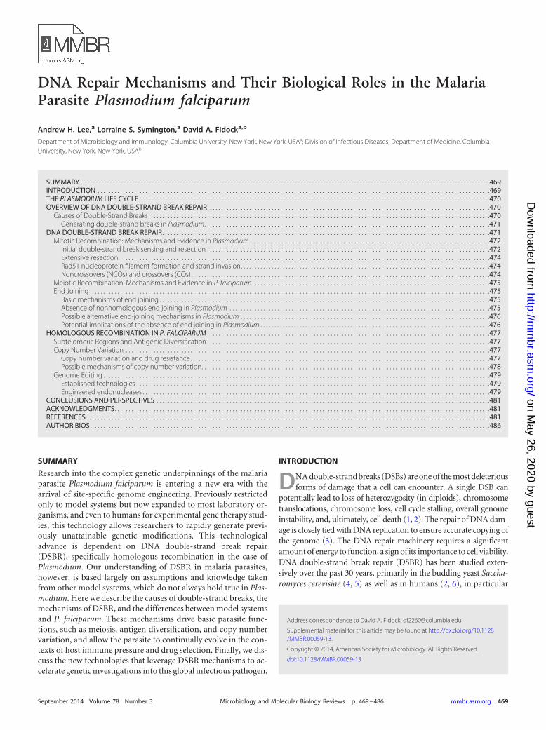
An infected female Anopheles mosquito taking a blood meal willsecrete its saliva along with Plasmodium sporozoites into the dermisof its new host (15) (Fig. 1). Within an hour, these sporozoites mi-
grate to the host’s liver and invade hepatocytes. The sporozoites thenreplicate, forming up to tens of thousands of merozoites, which burstfrom the hepatocytes to enter the peripheral circulation. Merozoitesquickly invade erythrocytes, starting the asexual cycle (16) (Fig. 2).Over the next 48 h, an intraerythrocytic P. falciparum parasite pro-duces as many as 24 daughter merozoites, which burst from the hostcell to reinitiate a new round of asexual replication. Intraerythrocyticparasites can also adopt an alternative, sexual-stage developmentalpathway, in which they form male or female gametocytes. Over a2-week period, these stages mature and become infectious for mos-quitoes (17). Upon transmission, gametocytes convert into gametesthat can then mate to form a zygote (15). Following meiosis, zygotesconvert into motile ookinetes that exit the blood meal confines andtraverse the midgut epithelium, after which they lodge under thebasal lamina and form oocysts. During the process of sporogony in-side an oocyst, the parasite undergoes multiple rounds of replicationto generate thousands of haploid sporozoites, which migrate to themosquito salivary glands to await the next blood meal.
OVERVIEW OF DNA DOUBLE-STRAND BREAK REPAIR
Causes of Double-Strand Breaks
DSBs can be generated experimentally; others are inherent to cel-lular processes (5). DSBs are used in many model organisms, and
FIG 1 The Plasmodium life cycle. A malaria infection begins with the transmission of a Plasmodium parasite via a female Anopheles mosquito host (left) to ahuman host (right). After the initial liver stage, the parasite begins its asexual intraerythrocytic cycle. Sexual forms, which develop from the intraerythrocyticparasites, can be transmitted to another mosquito. In the mosquito, parasites undergo meiotic and mitotic replication to form sporozoites, which can infectanother human host. RBCs, red blood cells.
Lee et al.
470 mmbr.asm.org Microbiology and Molecular Biology Reviews
on May 26, 2020 by guest
http://mm
br.asm.org/
Dow
nloaded from
experimental sources include ionizing radiation (IR) (e.g., �- andX-rays) (18), UV irradiation, chemical mutagens (e.g., hy-droxyurea, camptothecin [CPT], and methyl methanesulfonate),and DNA nucleases (e.g., zinc finger nucleases, Tal effector-likenucleases, and the clustered regularly interspaced short palin-dromic repeats-Cas [CRISPR/Cas] system). Inherent sources canstem from cellular processes, such as the generation of reactiveoxygen species by aerobic metabolism (19), transcription (20),and replication fork collapse (21).
Generating double-strand breaks in Plasmodium. In compar-ison to DNA repair research done in model organisms such asyeast, the use of DSB sources to interrogate Plasmodium biologyhas been limited. For example, in Plasmodium, irradiation hasbeen used primarily in efforts to create attenuated sporozoite vac-cines (22–24), such that parasites are compromised in their intra-cellular replicative ability. Some chemical mutagens have beenused to study nucleotide or base excision repair (NER or BER,respectively) (25–27). CPT, which generates DSBs by trapping thetopoisomerase I (TopI) reaction intermediate during relaxation ofsupercoiled DNA and blocks the progression of the replisome, hasbeen shown to inhibit P. falciparum TOP1 (PlasmoDB gene iden-tifier PF3D7_0510500) function in vitro (28) and is active againstparasite cultures, with a half-maximal inhibitory concentration(IC50) of �1 �M in a 72-h drug assay (our unpublished results).
Several studies using the first-line antimalarial drug artesunateas a chemical mutagen have emerged in the past decade. The ef-fects of artesunate on mammalian cell lines show that it can pro-mote oxidative DNA damage (29) and also induce a DSBR re-sponse (29, 30). However, the far more potent activity ofartesunate against Plasmodium asexual blood-stage parasites (inthe low nanomolar range in vitro) and the recent identification ofa variant kelch protein (PF3D7_1343700) as a candidate molecu-lar marker of artemisinin resistance (31) suggest a primary modeof antimalarial action distinct from DNA damage.
DNA nucleases, specifically engineered endonucleases, are be-coming more commonplace as tools for genetic modification inmany organisms, due to their ability to bind and generate DSBs inmost investigator-defined DNA sequences (14). These nucleasesand their use in Plasmodium are described in further detail below.
DNA damage arising from transcription, replication fork col-lapse, or by-products of normal cellular processes, such as aerobicrespiration or hemoglobin degradation, has been studied even lessin Plasmodium. Replisome collisions with transcription bubblesor single-stranded DNA (ssDNA) generated by oxidative damagecan lead to DSBs (20, 32). As an intraerythrocytic parasite grows, itdegrades copious amounts of hemoglobin in the digestive vacuole,releasing heme. Heme is oxidized from ferrous (Fe2�) to ferric(Fe3�) iron, producing hydroxyl radicals, a potent DNA-damag-ing agent (33). Therefore, it is possible that in Plasmodium, cellu-lar processes such as hemoglobin degradation and the release offree radicals, coupled with the many rounds of DNA replication,may result in the production of DSBs that the parasite must repairto maintain viability.
DNA DOUBLE-STRAND BREAK REPAIR
In most eukaryotes, the DSBR response can be split into two mainbranches: the “error-free” homologous recombination (HR)pathway and the potentially “error-prone” end-joining (EJ) path-ways. During HR, a broken DNA duplex utilizes a homologoustemplate (a sister chromatid, a homologous chromosome in dip-loids, a donor plasmid, or an ectopic donor if the DSB formswithin a repeated sequence) for highly accurate repair. The EJpathways do not use a homologous template and instead ligatebroken DNA ends together, resulting in a higher possibility ofinsertions or deletions (indels). Below, we outline DSBR and de-scribe P. falciparum orthologs and experimental evidence wherepossible. We use S. cerevisiae nomenclature in reference to or-thologs, omitting Homo sapiens nomenclature for clarity unless
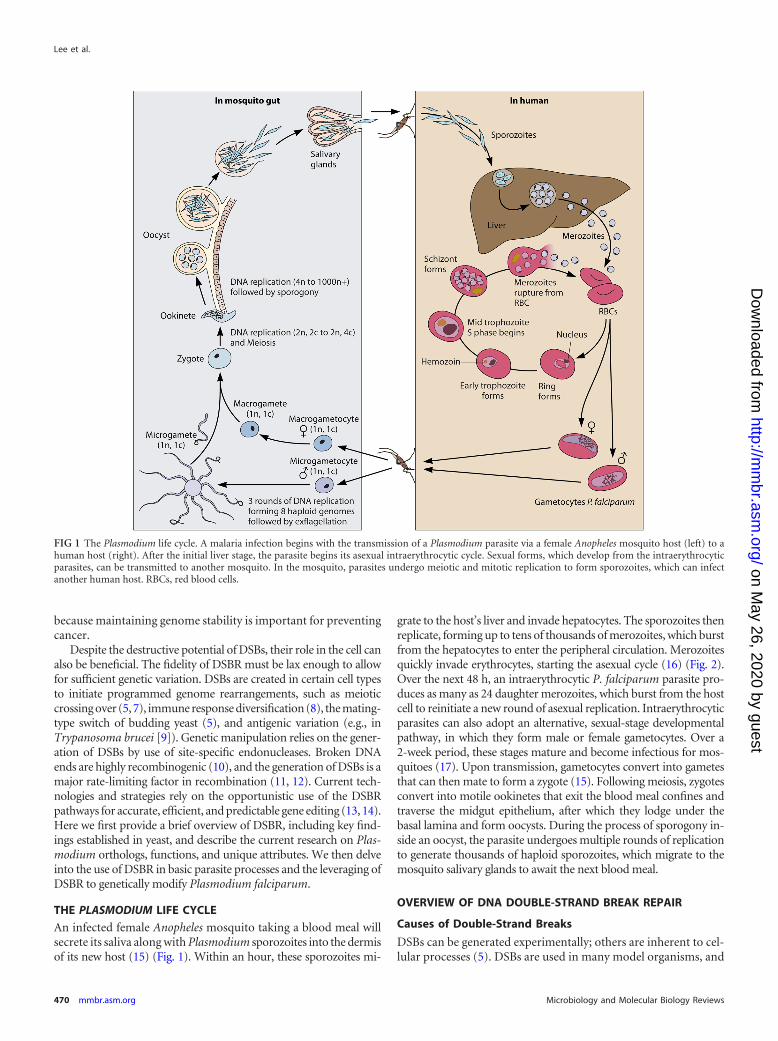
FIG 2 The Plasmodium asexual intraerythrocytic cycle. A haploid (1n) merozoite invades a red blood cell (RBC) and develops as the “ring” form from 0 h toabout 24 h postinvasion, corresponding to the G1 phase of the cell cycle. As the parasite transitions from rings to trophozoites, its metabolic activity increases inpreparation for DNA replication. Prior to S phase and DNA replication, the parasite is still haploid, allowing for possible alternative EJ pathways. DNA replicationproduces multiple copies of the genome in an intact nucleus that does not undergo membrane degradation, providing homologous templates for HR. Plasmo-dium DNA replication is asynchronous and can produce a range of sister chromatids, up to about 24n. Nearing the end of the 48-h cycle, each genome is packagedinto separate daughter merozoites, which then egress and invade another RBC.
DNA Double-Strand Break Repair in P. falciparum
September 2014 Volume 78 Number 3 mmbr.asm.org 471
on May 26, 2020 by guest
http://mm
br.asm.org/
Dow
nloaded from

otherwise stated. P. falciparum, S. cerevisiae, and H. sapiens or-thologs can be found in Table 1. For more in-depth reviews ofDSBR, we refer the reader to several excellent publications (1, 2, 4,5, 34).
Mitotic Recombination: Mechanisms and Evidence inPlasmodium
Initial double-strand break sensing and resection. A DSB is firstsensed by the Mre11-Rad50-Xrs2 (MRX) complex (Fig. 3A) (35).
TABLE 1 Bioinformatic comparison of genes involved in homologous recombination, nonhomologous end joining, and microhomology-mediatedend joining among S. cerevisiae, H. sapiens, and P. falciparum
Repair mechanism
Gene product or PlasmoDB ID
S. cerevisiae H. sapiens P. falciparumd
Homologous recombination Mre11 MRE11 PF3D7_0107800Rad50 RAD50 PF3D7_0605800Xrs2 NBS1Sae2 CtIPExo1 EXO1 PF3D7_0725000Sgs1 BLM PF3D7_0918600
WRN PF3D7_1429900a
Top3 TopoIII� PF3D7_1347100Rmi1 RMI1Dna2 DNA2 PF3D7_1010200Rfa1 RPA1 PF3D7_0409600, PF3D7_0904800Rfa2 RPA2Rfa3 RPA3Rad51 RAD51 PF3D7_1107400Dmc1 DMC1 PF3D7_0816800Rad52 RAD52
BRCA2 PF3D7_1328200b
Rad54 RAD54 PF3D7_0803400DNA polymerase � DNA polymerase � PF3D7_1017000PCNA PCNA PF3D7_1361900 (PCNA 1)
PF3D7_1226600 (PCNA 2)Srs2 RTEL1 PF3D7_0514100c
Rad1 XPF PF3D7_1368800Rad10 ERCC1 PF3D7_0203300Mus81 MUS81 PF3D7_1449400Mms4 EME1Yen1 GEN1 PF3D7_0206000Spo11 SPO11 PF3D7_1217100
Nonhomologous end joining Ku70 Ku70Ku80 Ku80Dnl4 DNA ligase IV
DNA-PKcsArtemis
Lif1 XRCC4Nej1 Cernunnos/XLF
Microhomology-mediated end joining Mre11 MRE11 PF3D7_0107800Rad50 RAD50 PF3D7_0605800Xrs2 NBS1Sae2 CtIPTel1 ATMRad1 XPF PF3D7_1368800Rad10 ERCC1 PF3D7_0203300Rad27 FEN1 PF3D7_0408500Cdc9 DNA ligase I PF3D7_1304100Rev3 DNA Pol � catalytic subunit PF3D7_1037000Pol4 DNA Pol Rad30 DNA Pol
a Lacks 5=-to-3= exonuclease indicative of WRN.b Low homology.c Possible UvrD helicase.d P. falciparum orthologs were determined from previous publications or were confirmed by BLAST and identification of unique domains with Pfam, version 27.0.
Lee et al.
472 mmbr.asm.org Microbiology and Molecular Biology Reviews
on May 26, 2020 by guest
http://mm
br.asm.org/
Dow
nloaded from

FIG 3 Canonical models of homologous recombination, synthesis-dependent strand annealing, and break-induced replication. A double-strand break (DSB)in a chromosome (A) is first sensed and tethered together by the MRX-Sae2 complex (B). (C) Resection by Exo1 (gray “Pac-man”) or the STR-Dna2 complex(yellow and red triangles, respectively) exposes 3= ssDNA tails, which are then bound by RPA (green dots). RPA is replaced by Rad51 (gray helices) (D), whichforms a nucleoprotein filament that invades homologous templates (E). (F) Synthesis-dependent strand annealing (SDSA) uses the invading 3= tail as a primerfor DNA synthesis. Once sufficient homology is synthesized, the invading strand can bridge the DSB to restore chromosome integrity. (L) If the distalchromosome arm is lost, then break-induced replication can occur, with DNA synthesis continuing conservatively until it reaches the end of the chromosomearm. If both broken DNA ends are captured by a homologous template (G), then a double Holliday junction (dHJ) can be formed (H). This structure can beresolved in a number of ways. (I) Merging of both Holliday junctions by the STR complex (yellow triangles) dissolves the dHJ in a hemicatenane structure, whichis unlinked by the Top3 topoisomerase. Cleavage of both Holliday junctions by structure-specific resolvases (purple triangles) can unlink the dHJ to produceeither noncrossover (J) or crossover (K) products.
DNA Double-Strand Break Repair in P. falciparum
September 2014 Volume 78 Number 3 mmbr.asm.org 473
on May 26, 2020 by guest
http://mm
br.asm.org/
Dow
nloaded from

The MRX complex binds to double-stranded DNA (dsDNA) endsformed by a DSB, positions both ends in close proximity, and, incollaboration with Sae2, initiates 5=-to-3= resection, creating short3=-terminated ssDNA tails (Fig. 3B). Initial resection is a rate-limiting step in HR, as it provides the substrates for further, ex-tensive resection (36, 37). Mre11 and Rad50 show high homologywith their Plasmodium orthologs, but Xrs2 and Sae2 do not, likelydue to low sequence conservation among all species.
Extensive resection. Initial resection is followed by 5=-to-3= ex-tensive resection by functionally redundant factors (exonuclease 1[Exo1] and the Sgs1-Top3-Rmi1-Dna2 [STR-Dna2] complex)(36, 37), which commits DSBR to HR and generates long 3=ssDNA tails (Fig. 3C). Exo1 degrades DNA from the 5= to the 3=end and is a member of the 5=-structure-specific Rad2/XPG familyof nucleases, which are involved in most DNA repair pathways(e.g., mismatch repair [MMR] and NER). Sgs1 is a RecQ helicasethat unwinds linear dsDNA, creating a Y-shaped structure that isthen cleaved by the flap endonuclease Dna2 (Fig. 3C) (38). Sgs1has two human homologs, BLM and WRN (mutated in Bloom’sand Werner’s syndromes, respectively). BLM has been shown tobe the primary resection protein in mammalian cells (39, 40),although Xenopus WRN-DNA2 can also resect linear DNA sub-strates in vitro (41). Both BLM and WRN have P. falciparum ho-mologs (PF3D7_0918600 and PF3D7_1429900, respectively),though the P. falciparum WRN homolog lacks the 5=-to-3= exonu-clease domain that is characteristic of WRN proteins in other eu-karyotes. It is therefore possible that PF3D7_1429900 is in fact adifferent RecQ helicase.
The extent of resection varies between species. Yeast can ex-hibit ssDNA tract lengths of up to 2 to 4 kb during mitotic HR andup to 850 bases during meiotic HR (34). Detailed analyses of re-section have not been performed in Plasmodium. ExtensivessDNA resection tracts are likely to signal cell cycle arrest andprevent unwanted recombination by exposing more unique se-quence to search for a homologous template (34). In an A/T-richorganism with extensive regions of low complexity, such as P.falciparum, long resection tracts may be beneficial for ensuringthat a resected sequence is unique enough to undergo HR with thecorrect template.
Rad51 nucleoprotein filament formation and strand inva-sion. 3= ssDNA tails generated by resection are first coated by theheterotrimeric replication protein A (RPA) complex (Fig. 3C),protecting them from degradation and secondary structure for-mation (42). P. falciparum encodes all three subunits of RPA andan additional truncated version of the RPA1 subunit (RPA1S),which antagonizes the long form (RPA1L) during in vitro recom-bination (43). Yeast Rad52 (or the human breast cancer type IIsusceptibility protein [BRCA2]) then displaces RPA and simulta-neously delivers ATP-bound Rad51, the central HR recombinase,onto ssDNA, allowing it to form a nucleoprotein filament (Fig.3D). Rad51 catalyzes invasion and pairing between the ssDNA towhich it is bound and the complementary sequence to form adisplacement loop (D loop) (Fig. 3E).
Rad52 homologs have not been found in Plasmodium (44), andthe P. falciparum RPA1 homologs lack similarity to the N-termi-nal domains of other eukaryotic RPA1 orthologs (45) that arenecessary for Rad52 interactions (46). Caenorhabditis elegans,Drosophila melanogaster, and Arabidopsis thaliana all lack Rad52,which may also be the case for P. falciparum. Mammals useBRCA2 in addition to Rad52, and BRCA2 sequence similarity
even among mammals is relatively low. Nevertheless, BRCA2homologs have been briefly mentioned for P. falciparum(PF3D7_1328200) and Plasmodium yoelii (PY17X_1348100),where homology is found in only 6 BRC repeats. These repeats areknown to stimulate Rad51-ssDNA binding and to prevent non-specific Rad51-dsDNA binding (47). Trypanosoma cruzi, T. bru-cei, and Leishmania major also have BRCA2 homologs with vari-ous numbers of BRC repeats (48). Other BRCA2 regions (theoligonucleotide-binding [OB] fold that binds ssDNA and theTower domain, which binds dsDNA), however, have not beenidentified in these homologs. These regions also cannot be iden-tified in the D. melanogaster BRCA2 homolog (47) and may be toodivergent to detect bioinformatically, as they can have low se-quence conservation (49). Nevertheless, D. melanogaster BRCA2remains proficient in homologous recombination (50), as is ex-pected to be the case with Plasmodium parasites. In stark contrastto Rad52 and BRCA2, P. falciparum Rad51 shows high homologyto Rad51 proteins of many species. Predictably, it is upregulated inresponse to the DNA-damaging agent MMS, performs strand ex-change on DNA substrates in vitro, and hydrolyzes ATP (44, 51).
Efficient Rad51-mediated D-loop formation is enhanced bythe Rad55-Rad57 heterodimer and the Rad54 motor protein.Rad55-Rad57 stabilizes the Rad51 nucleoprotein filament by pre-venting Rad51 displacement from ssDNA (52). Rad54 is adsDNA-dependent ATPase that translocates along dsDNA, en-hances Rad51-dependent strand exchange, and stabilizes Rad51filament formation on both ssDNA and dsDNA (53–55). Rad54,however, only dissociates Rad51 from dsDNA. The synergisticeffects of Rad54 on Rad51-mediated strand exchange are mir-rored in in vitro assays using purified P. falciparum Rad51(PfRad51) and PfRad54 (PF3D7_0803400) (43).
D-loop formation is followed by polymerase �-dependentDNA synthesis primed from the 3= OH group of the invading 3=ssDNA tail (Fig. 3E). Synthesis extends for �250 bp in yeast (56),expanding the D loop and incorporating any single nucleotidepolymorphisms (SNPs). So far, extension of the invading arm hasbeen found to incorporate SNPs 150 bases distal to the DSB with ahigh frequency (13), and up to 900 bp distal at a lower but sub-stantial level, during gene editing in P. falciparum (57). More de-tailed frequencies and tract lengths have yet to be delineated thor-oughly.
Noncrossovers (NCOs) and crossovers (COs). Two basic op-tions exist after D-loop formation: synthesis-dependent strandannealing (SDSA) (Fig. 3F) and formation of double Hollidayjunctions (dHJs) (Fig. 3H). During SDSA, the invading strand isextended to traverse the DSB, the D loop is collapsed, and theextended strand bridges the DSB. The homologous template is leftunchanged. Possible SNPs copied from the homologous templatewill generate heteroduplex DNA and be corrected by the MMRmachinery. MMR tends to favor the donor as the “correct” tem-plate, but the mechanism of this proclivity has yet to be deter-mined (56).
If the D loop captures the second 3= ssDNA tail (Fig. 3G), thenthe D-loop intermediate can form a dHJ (Fig. 3H). dHJs are thenprocessed by two distinct, canonical pathways. dHJs can be “dis-solved,” whereby they are merged together by the Sgs1-Top3-Rmi1 (STR) complex into a hemicatenane structure, which is un-linked by the topoisomerase Top3 to generate an NCO product(Fig. 3I). Alternatively, dHJs can be resolved by Mus81-Mms4,Yen1, or Slx1-Slx4 (58). Depending on the cleavage pattern of the
Lee et al.
474 mmbr.asm.org Microbiology and Molecular Biology Reviews
on May 26, 2020 by guest
http://mm
br.asm.org/
Dow
nloaded from

dHJs, resolution will form NCO or CO products (Fig. 3J and K,respectively). Currently, more intricate pathways branching fromthe canonical model of dHJ resolution are being elucidated (58,59). Some of the nucleases, such as Mus81, Yen1, Rad1, andRad10, have orthologs in P. falciparum and are listed in Table 1.Orthologs could not be found for Mms4, Slx1, and Slx4.
By definition, HR by any of these mechanisms is highly accu-rate, but errors can arise. For example, mitotic crossovers in dip-loids can lead to loss of heterozygosity. Nonallelic HR (NAHR)between repeated sequences can generate copy number variants(CNVs) (discussed below) or translocations. Furthermore, if achromosome arm is lost during repair, the invading strand inter-mediate can copy the homologous template until the site of itstelomere, using a process termed break-induced replication (BIR)(Fig. 3L). BIR can cause extensive loss of heterozygosity in diploids(60). In Plasmodium, BIR is one mechanism that is believed togenerate novel var sequences (see below).
Meiotic Recombination: Mechanisms and Evidence in P.falciparum
Meiotic recombination is important for the efficient productionof sporozoites in Plasmodium (61), spores in budding yeast (62,63), and gametes in mammals (7, 64). The basic mechanistic out-comes of meiotic recombination are largely similar to those ofmitotic recombination, but they involve a larger set of meiosis-specific proteins for the programmed generation of genetic diver-sity (7, 63). Here we briefly discuss meiosis in Plasmodium andfocus on the meiotic factors involved in recombination.
The mosquito stage of the Plasmodium life cycle begins withtransmission of male and female gametocytes via a blood meal(Fig. 1). Almost immediately, the male microgametocyte under-goes three rapid rounds of DNA replication, producing eight hap-loid genomes (65). Exflagellation produces eight single microg-ametes (1n) (66). A microgamete can subsequently fertilize afemale macrogamete (1n), producing a zygote (2n). A subsequentround of DNA replication (2n, 4c) is then followed by meiosis (67,68) to form a tetraploid ookinete (2n, 4c). During meiosis, cross-ing over via HR is known to be important for the formation ofyeast spores (62) and begins with the production of programmedDSBs by the meiosis-specific topoisomerase-like protein Spo11(69). Note that Plasmodium orthologs of Spo11 exist, suggesting aconserved mechanism (70). In yeast, meiotic HR utilizes theRad51 homolog Dmc1 to catalyze strand exchange, with Rad51playing a supporting role (71). In Plasmodium berghei, a Dmc1knockout produces fewer and smaller oocysts and substantiallysmaller numbers of sporozoites (61), implicating marked defectsin meiotic recombination. While a similar knockout has not beenstudied in P. falciparum, Dmc1 is expressed in gametocytes priorto meiotic recombination, implicating a similar role (72).
As with mitotic HR, successful strand invasion (Fig. 3E) pro-vides the choice between forming NCOs and COs by various path-ways (Fig. 3F and H). Spo11-induced DSBs are predominatelyrepaired as NCOs (likely by SDSA) and COs at a 10:1 ratio inmammals (7). This relative excess of NCOs has also been seen in P.falciparum (73, 74). However, these reports may still underrepre-sent the number of NCOs, due to a lack of resolution or to NCOproducts that are indistinguishable from either parental chromo-some.
In contrast to the case for mitotic HR, resolution of meiotic dHJintermediates in S. cerevisiae requires the MutL homologs Mlh1
and Mlh3 along with Exo1 (7, 62). In the case of P. falciparum,whole-genome sequence analysis of artemisinin-resistant Cambo-dian parasite populations showed a high frequency of an Mlh1(PF3D7_1117800) mutation (75), possibly implicating a defect inmeiotic crossing over. In a yeast exo1� strain, the homologousMlh1 mutation showed a moderate mutator phenotype (76), con-sistent with a loss of function. To date, no Mlh3 ortholog has beenidentified in P. falciparum. During meiotic recombination, eachchromosome averages one crossover (73, 74, 77) and has a geneticmap unit distance of �10 kb (77) to �15 kb (73) per centimorgan.Meiosis is followed by many rounds of DNA replication insideoocysts to form thousands of genomes that segregate into individ-ual haploid sporozoites (66, 78).
Investigations into the process of meiotic recombination inPlasmodium have been limited to analyzing P. berghei DNA con-tent during sexual reproduction (65, 67). Several other studies,nonetheless, have explored outcomes of meiotic recombination.P. falciparum genetic cross studies have analyzed the segregationof chromosomal markers (SNPs and microsatellites) spread outover the genome between geographically distinct parasite clones:3D7 � HB3 (79), HB3 � Dd2 (73, 74, 80–82), and 7G8 � GB4(77, 83, 84). These were instrumental in mapping the dihydro-folate reductase gene (dhfr) (85) and P. falciparum chloroquineresistance transporter gene (pfcrt) (86) that are key drug resistanceloci and identifying key determinants of host cell tropism (77,83, 84).
End Joining
Basic mechanisms of end joining. Parallel to HR, the EJ pathwaysare comprised of the classical and alternative EJ pathways, bywhich broken DNA ends are religated without a homologous tem-plate for repair. The classical nonhomologous end-joining(NHEJ) pathway (Fig. 4A) has commonly been referred to as the“error-prone” pathway, though it is likely rather error-free butaccommodating of promoting genetic variability (87).
In yeast, NHEJ can be performed with just the Ku70/80 (Ku)heterodimer and the DNA ligase IV-Lif1-Nej1 complex (88). TheKu heterodimer binds broken DNA ends, which can protect themfrom end resection and commitment to HR (89) and act as aplatform for NHEJ factor recruitment (88). Ku promotes EJ of avariety of substrates by sterically fitting in the grooves of the DNAdouble helix, as opposed to forming specific base interactions(90). In vertebrates, the DNA-dependent protein kinase catalyticsubunit (DNA-PKcs) and the endonuclease Artemis are essentialNHEJ factors. DNA-PKcs and Artemis bind Ku-bound ends, andphosphorylation of Artemis by DNA-PKcs activates its endonu-clease activity (91). Either nucleolytic degradation or polymerasenucleotide addition generates DNA ends compatible for EJ.
Absence of nonhomologous end joining in Plasmodium. Todate, bioinformatic analyses have failed to identify any Plasmo-dium homologs of NHEJ proteins (Table 1) (92). In contrast, an-other apicomplexan parasite, Toxoplasma gondii, carries a func-tional Ku-dependent NHEJ pathway (93), suggesting that despiterelative evolutionary proximity to T. gondii, Plasmodium has lostits NHEJ machinery. In support of this hypothesis, a recent studyshowed the absence of any NHEJ products recovered from in vivoendonuclease-generated DSBs in Plasmodium (13). Furthermore,other eukaryotic pathogens, such as Giardia lamblia (94), Enceph-alitozoon cuniculi (95), and Trichomonas vaginalis (96), also lackNHEJ components.
DNA Double-Strand Break Repair in P. falciparum
September 2014 Volume 78 Number 3 mmbr.asm.org 475
on May 26, 2020 by guest
http://mm
br.asm.org/
Dow
nloaded from
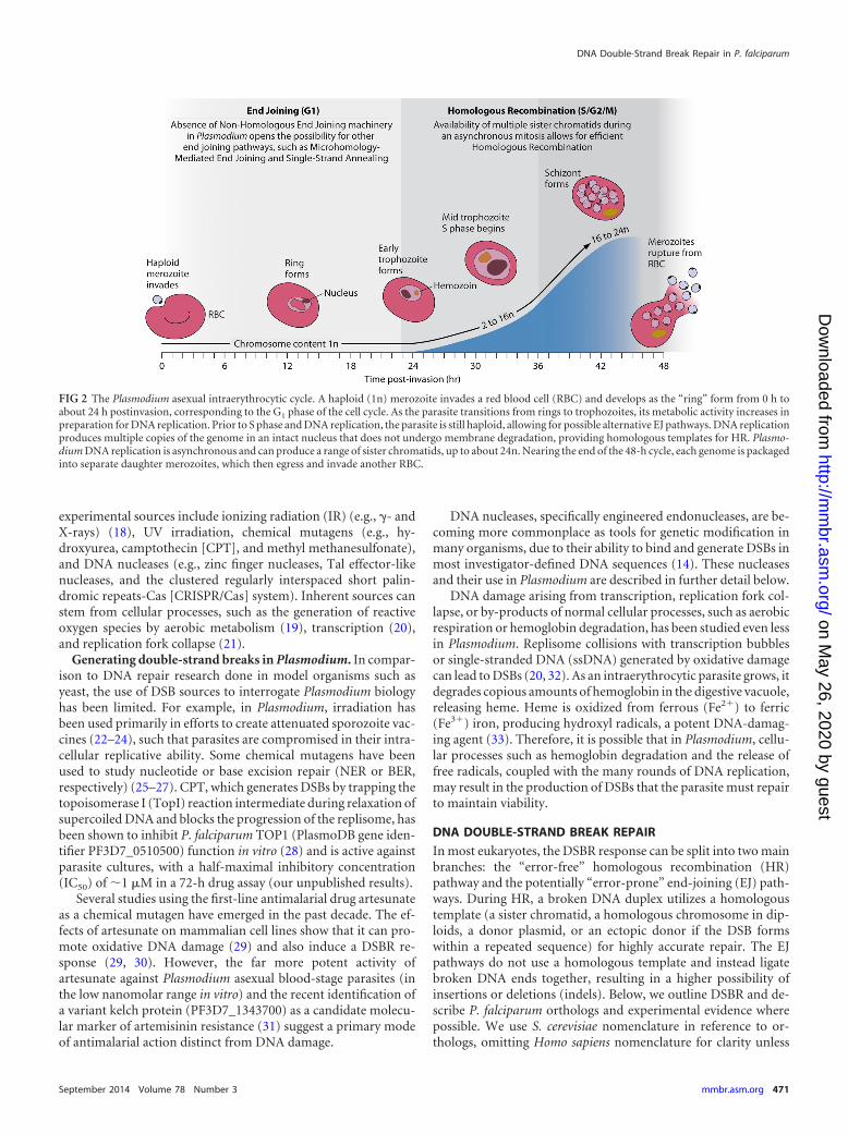
Possible alternative end-joining mechanisms in Plasmodium.If NHEJ was indeed lost during Plasmodium evolution, then alter-native EJ pathways may still be active. In yeast, ku� mutants re-vealed pathways that occur at lower frequencies than that of NHEJ(97). These Ku-independent processes include microhomology-mediated end joining (MMEJ) (Fig. 4B) and single-strand anneal-ing (SSA) (98) (Fig. 4C). Both pathways require resection to ex-pose homologies internal to the DSB ends. MMEJ refers to joiningbetween microhomologies (up to 25 bp), whereas SSA occurs be-tween more extensive homologies. Yeast Rad52 promotes anneal-ing of the complementary ssDNA during SSA (99), by displacingRPA coating ssDNA (100), but plays little or no role in MMEJ. Asdiscussed above, BRCA2, but not Rad52, is homologous to P. fal-ciparum PF3D7_1328200. The Ustilago maydis BRCA2 homolog,Brh2, is reported to promote strand annealing similar to the reac-tion catalyzed by Rad52 (101), suggesting that the PlasmodiumBRCA2 homolog may fulfill a similar role. Nonetheless, currentevidence suggests that SSA is not a major mechanism of DNArepair in Plasmodium (13). For another eukaryotic pathogen, T.brucei, chromosomal and in vitro plasmid EJ assays produce onlyMMEJ-generated repair products (102–104). NHEJ is not used,and T. brucei Ku functions only in telomere maintenance (105).
A recent report (106) suggests that P. falciparum may be able torepair a DSB by an alternative EJ mechanism whereby a few basesare added to the broken ends, thereby providing microhomolo-gies. The frequency of these events is very low, and larger deletions(e.g., possible SSA products) beyond the locus analyzed were notexamined. Nevertheless, Plasmodium species carry all necessarycomponents of the MMEJ machinery, many of which overlapthose for HR (Table 1). However, given the general lack of NHEJ,
MMEJ, and SSA products observed in Plasmodium, it seems un-likely that EJ processes occur to any significant extent in malariaparasites.
Potential implications of the absence of end joining in Plas-modium. The absence, or at least highly infrequent use, of EJ path-ways in P. falciparum may be an important factor in the produc-tion of a live, radiation-attenuated sporozoite vaccine. Haploidsporozoites in the Sanaria PfSPZ vaccine are metabolically activeyet nonreplicating (23, 24, 107). PfSPZ is generated with sporozo-ites dissected from infected mosquitoes exposed to 15 krad of�-irradiation. Sufficient irradiation introduces DNA damage(108) without compromising hepatocyte invasion, gene expres-sion, and initial trophozoite development, but it prevents nucleardivision (109). Estimations from yeast data suggest that the 15-krad dose is sufficient to generate small but sufficient numbers ofDSBs (110). Therefore, the crucial replication defect of irradiatedsporozoites may result from the parasite’s lack of an efficient EJpathway during this strictly haploid stage and may manifest itselfonly when DNA replication occurs during liver-stage prolifera-tion.
Studies of irradiated blood-stage P. falciparum support thisnotion. Experimental analyses showed that cellular distress is dose(111) and cell cycle (112) dependent. Studies with parasites sub-jected to various IR doses throughout the asexual blood stage (Fig.2) showed that ring-stage parasites and multinuclear schizontswith a 1c chromosome content cannot reconstitute an in vitroculture after IR exposure as efficiently as the case with trophozo-ites, which have a chromosome content of 2c (112, 113). Giventhat the trophozoite stage has numerous sister chromatids as tem-plates for repair in a syncytium, the IR-induced DNA damage is
FIG 4 EJ pathways. (A) Nonhomologous end joining (NHEJ) involves the joining of broken DNA ends with little to no homology. A simplified model of NHEJdepicts the restoration of the original DNA sequence and introduction of small insertions (blue strands) or small deletions. (B) Microhomology-mediated endjoining (MMEJ) relies on minimal resection by proteins shared with HR to expose short homologies of up to 25 bp, which can anneal, deleting any interveningsequence. (C) Single-strand annealing (SSA) involves longer stretches of resection. Rad52 (not shown) in yeast displaces RPA and anneals longer homologies,generating larger deletions.
Lee et al.
476 mmbr.asm.org Microbiology and Molecular Biology Reviews
on May 26, 2020 by guest
http://mm
br.asm.org/
Dow
nloaded from
likely readily repaired by HR. Further studies are required to dis-cern whether the segregation of chromosomes after DNA replica-tion (78) inhibits efficient HR. Altogether, without a robust EJpathway, the data show that it is possible that Plasmodium is moresensitive to DNA damage than other model organisms, such asyeast and humans.
HOMOLOGOUS RECOMBINATION IN P. FALCIPARUM
Barring a few exceptions, DSBR in Plasmodium is rarely studiedsolely at a mechanistic level. Most published studies examine theproducts of such processes in the context of drug resistance, anti-genic diversification, population structure, or genetic manipula-tion. Together, these studies each contribute to a larger picture ofthe consequences of Plasmodium DSBR, but few provide directexperimental evidence of its underlying molecular intricacies.Nevertheless, the compiled picture shows a unique organism inwhich DSBR dictates a broad spectrum of phenotypes.
Subtelomeric Regions and Antigenic Diversification
Unlike the case in T. brucei, antigenic variation in the Plasmodiumparasite does not occur by HR. Whereas T. brucei undergoesRad51-dependent gene conversion to replace the active antigen-encoding gene (the variant surface glycoprotein gene [VSG]) withone of the many inactive pseudogenes (114) in order to evade hostdefenses, P. falciparum antigenic variation is mediated at the epi-genetic level (115). P. falciparum erythrocyte membrane protein 1(PfEMP1), which is presented on the host’s infected erythrocyteand exposed to the immune system, is encoded by the clonallyvariant var gene family. var does not require a DSB to switchbetween its family members and instead epigenetically silences allbut one var cassette among the �60 total per parasite (115).
Though recombination plays no role in antigenic variation, itdoes mediate var gene antigen sequence diversification and vargene family composition during mitotic (116–118) as well as mei-otic (77, 119) cell cycles. var genes are located primarily in subte-lomeric regions of chromosomes (120, 121), although some arealso found clustered in central chromosomal regions (92, 121,122). The subtelomeric regions are also home to the rifin andstevor multicopy gene families, which also interact with host fac-tors. Spatial positioning studies show that chromosome ends areclustered together at the nuclear periphery (121–123). In yeast,spatial proximity of a DSB to a donor template greatly enhancesHR (124). Therefore, in addition to providing a mechanism forselective var expression, clustering of subtelomeric ends may pro-vide the close proximity between heterologous chromosomes forrecombination to generate novel var sequences (122).
The rate of recombination in these subtelomeric regions has beenshown to be much higher than in the core chromosome: for in vitro3D7 cultures, the mitotic recombination rate is about 4.7 � 10�6
events per base pair per generation, with over 80% of events occur-ring in the subtelomere (117). Parasite populations from differentglobal regions reaffirm this notion (119). It is possible, nevertheless,that both mitotic and meiotic recombination contribute to recombi-nation in subtelomeric regions, as both can occur during the mos-quito stages. This may provide parasites with the ability to persistlonger in a single blood-stage infection by forming novel var se-quences and, thus, distinct PfEMP1 antigens (116).
Mechanistically, the diversification of var sequences and vargene family composition can be driven by a number of recombi-nation pathways. Reports pinpointing the precise mechanisms
driving these events have not been published, although insightscan be gleaned by analyzing the recombination products. For ex-ample, a var allele can copy a portion of a donor allele to producea novel, chimeric allele. In mitotically dividing P. falciparum cells,studies suggest that this process is mediated by an SDSA-mediatedgene conversion event (116, 117) (Fig. 3F) and by BIR (Fig. 3L)(117). Intragenic COs between two rif genes can create two novelsequences (116). NAHR (discussed below) (Fig. 5A) may also de-lete or amplify large swaths of sequence in subtelomeric regions,thereby altering var gene family composition for a given parasite(117, 118).
For the sexual stages, a recent study has shown that some hotspots for ectopic var recombination are energetically likely toform DNA secondary structures, which may act as substrates togenerate DSBs (125). Analyses of parasite populations (119)and genetic crosses (74, 77) showed that the subtelomeric re-gions have a high rate of recombination during the sexualstages. Further studies are needed to ascertain whether theseevents occur during meiosis or the numerous subsequent mi-totic divisions in the mosquito vector.
Copy Number Variation
One of the best-characterized phenotypes of gain-of-functionevents is the acquisition of drug resistance by copy number vari-ation (CNV). CNVs, either amplifications or deletions, change thenumber of select genes within a genomic region in order to altertheir total expression levels. Original analyses primarily usedpulsed-field gel electrophoresis (PFGE) to separate chromosomes,but now CNV detection relies more heavily on quantitative real-time PCR (qRT-PCR) (126), high-density tiling microarrays(127), or whole-genome sequencing (117).
Copy number variation and drug resistance. The classic exam-ple of the relationship between CNVs and drug resistance is theincreased tolerance to mefloquine conferred by the amplificationof a genomic region (amplicon) on chromosome 5 containing theP. falciparum multidrug resistance gene, pfmdr1, which encodesthe P-glycoprotein homolog (Pgh-1). The W2 parasite, initiallyderived from a Southeast Asian isolate, was pressured in vitro witha stepwise increase of mefloquine over 96 weeks, generating themefloquine-resistant parasite W2-mef (128). Characterization ofW2-mef and its derived clones revealed that the key to resistancewas the amplification of pfmdr1. This results in increased Pgh-1expression, which is thought to more effectively transport meflo-quine into the digestive vacuole, away from its primary site ofaction (129, 130). Amplifications in other regions in the genomecan also lead to increased drug tolerance to a variety of antimalar-ials. These are listed in Table S1 in the supplemental material.
Additionally, genomic deletions have been described for manyparasites. Some prominent examples come from the deletion ofunnecessary genes in culture-adapted parasites (e.g., Dd2). Con-tinuous in vitro culture is absent of host-derived selective pressure;therefore, functions such as cytoadherence to endothelial cell sur-face receptors in the microvasculature (131, 132) or gametocyto-genesis (133), required for transmission to mosquitoes, can read-ily be lost.
CNVs are generally believed to be detrimental to cell fitnessdue to the imbalanced dosage of gene products, thereby perturb-ing cellular homeostasis. For example, changes in gene dosagehave been linked to cancer and neurological disorders, such asautism (134). In P. falciparum, the fitness costs to parasite growth
DNA Double-Strand Break Repair in P. falciparum
September 2014 Volume 78 Number 3 mmbr.asm.org 477
on May 26, 2020 by guest
http://mm
br.asm.org/
Dow
nloaded from
have been observed with amplifications of gch1 (106) and pfcrt(our unpublished results). However, in the context of selective ordiverse environments, CNVs can prove advantageous in promot-ing adaptability, as in the case of in vitro culture growth and drugresistance. Other CNVs, such as an amplification of adjacent plas-mepsin2 and hap (histo-aspartic protease) genes found in a Thaifield isolate, have been speculated to increase nutrient acquisitionand may be beneficial for the parasite (132). Furthermore, in-creasing the number of copies of genes that confer drug resistancecan allow for one copy to maintain the wild-type sequence andfunctionality, while the additional copy or copies may mutate toconfer drug resistance (126). Therefore, the investigation of par-asite CNVs may prove to be increasingly important, particularly inthe context of varying geographical drug landscapes.
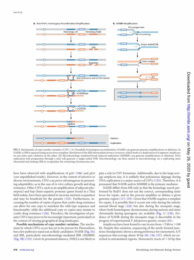
Possible mechanisms of copy number variation. The mecha-nism by which CNVs occur has yet to be proven for Plasmodium,but a few pathways stand out as likely candidates: NAHR (Fig. 5A)and BIR, particularly microhomology-mediated BIR (MMBIR)(Fig. 5B) (135). Given its presumed absence, NHEJ is not likely to
play a role in CNV formation. Additionally, due to the large aver-age amplicon size, it is unlikely that polymerase slippage duringDNA replication is a major source of CNVs (135). Therefore, it ispresumed that NAHR and/or MMBIR is the primary mediator.
NAHR differs from HR only in that the homology search per-formed by Rad51 does not use the correct, corresponding sisterlocus for repair, and in the process amplifies or deletes a givengenomic region (117, 135). Given that NAHR requires a templatefor repair, it is possible that it occurs not only during the mitoticasexual blood stage (126) but also during the mosquito stage,where both homologous chromosomes during meiosis and sisterchromatids during sporogony are available (Fig. 1) (136). Evi-dence of NAHR during the mosquito stage is discernible in theprogeny of experimental P. falciparum genetic crosses (81).
Amplicons have been found to vary in size from �5 kb to 100kb. Despite this variation, sequencing of the newly formed junc-tions (breakpoints) shows a strong preference for monomeric A/Tsequences that average about 30 bp (81, 136, 137), which are en-riched in untranslated regions. Monomeric tracts of 10 bp that
FIG 5 Mechanisms of copy number variation (CNV). (A) Nonallelic homologous recombination (NAHR) can generate genome amplifications or deletions. InNAHR, a DSB is repaired using an incorrect template. Resolution of the dHJ intermediate forms a crossover, which leads to a duplication of a segment (amplicon)on one sister and a deletion on the other. (B) Microhomology-mediated break-induced replication (MMBIR) can generate amplifications or deletions. DNAreplication fork progression through a nick will generate a single-ended DSB. Microhomology can then anneal to microhomology on a replicating sisterchromatid and undergo BIR to recapitulate the remaining chromosome arm.
Lee et al.
478 mmbr.asm.org Microbiology and Molecular Biology Reviews
on May 26, 2020 by guest
http://mm
br.asm.org/
Dow
nloaded from
speckle the genome are estimated to be, on average, about 600 bpapart, or 5% of the genome (136). It may be that one consequenceof the genome’s A/T richness is to increase the likelihood of copynumber variants.
These short, monomeric tracts are possible substrates forMMBIR (Fig. 5B). In this process, a short, monomeric tract re-vealed by end resection anneals to another microhomology regionon another chromosome, independently of Rad51. This tract thenserves as a primer for polymerase extension until the telomere,thereby copying that chromosome arm. Recently, the initial mi-totic amplification of chromosomal regions around the pfdhodhgene in response to the drug DSM1 was speculated to be MMBIRbased (126). Amplicons, which varied in size for each clone,ranged from 34 to 95 kb and were arranged head to tail. Break-points were short, monomeric tracts. As DSM1 pressure in-creased, the amplified region containing pfdhodh was further am-plified. But, importantly, all amplifications were exact copies ofthe initial amplicon, implicating a faster, homology-based expan-sion, such as NAHR, as the causal pathway. Therefore, it is possi-ble that MMBIR (Fig. 5B) occurs at a low frequency in the parasite,generating amplicons of various sizes. The close proximity of theseamplicons presumably provides substrates more ideal for NAHRthan for MMBIR under increasing selective pressure, thereby cre-ating exact copies of the initial amplicon (Fig. 5A).
Together, the evidence collected from a number of studies de-picts a parasite that generates copy number variants at a low butsignificant frequency. Many may be detrimental to growth and failto establish themselves at the population level. Given the sequenceidentity of breakpoints and types of amplicons produced, the par-asite may employ several of these pathways as tools to increase itsgenome diversity.
Genome Editing
Genetics in P. falciparum is currently becoming increasingly ac-cessible with the nearly simultaneous arrival of new transfec-tion-based technologies, which will undoubtedly deepen ourunderstanding of this parasite and lead to a more thoroughdefinition of parasite determinants of drug resistance, fitness,pathogenesis, and overall cellular organization and developmen-tal biology. The common factor among all the old and new tech-nologies is the requirement for a DSB to initiate HR. It was recog-nized early in yeast and mammalian systems that the introductionof a single DSB significantly increases the rate of recombination(10, 11). Genetic research in malaria was not too far behind inrecognizing the potential of DSBs, though several hurdles haveprevented their capitalization, until recently.
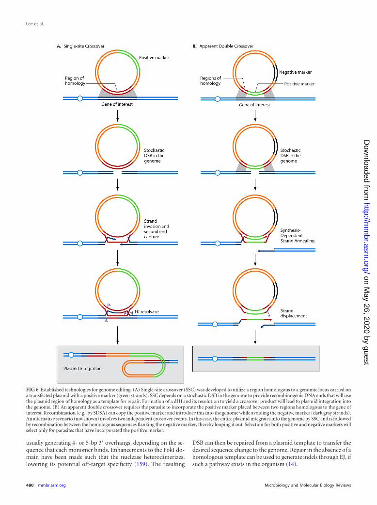
Established technologies. Early transfection and mutant para-site generation began around the mid-1990s for both P. falciparumand P. berghei. Stable transfection of P. falciparum originated byintroducing an episomal copy of the chloramphenicol acetyltrans-ferase selectable marker (138). This was quickly followed by plas-mid integration of a selectable marker, the dihydrofolate reduc-tase-thymidylate synthase (dhfr-ts), into the genome, by a methodreferred to as “single-site crossover” (139, 140) (Fig. 6A). Simul-taneously, transfections in P. berghei had also produced resistantdhfr-ts parasites with both episomal and integrated plasmids (141,142). Both species were transfected by electroporation, using ringstages for P. falciparum and merozoites in the case of P. berghei.
Although the number of selectable markers increased (143–145) and alternate transfection methods were developed (146),
genetic manipulation techniques and efficiency did not changedrastically for P. falciparum, aside from the introduction of the“double crossover” technique (Fig. 6B). This technique uses neg-ative in addition to positive selection to remove unwanted recom-bination events (147, 148). These methods still yield low efficien-cies, at best 1 in 105 parasites per transfection (149, 150), andrequire 2 to 3 months of continuous culture, at minimum, toproduce desired recombination events at a high enough rate toclone the appropriate parasites. Mechanistically, double cross-overs may occur either by SDSA (Fig. 6B) or by integration ofthe plasmid via single-site crossover followed by crossover-based excision of the negative marker by use of homologywithin the plasmid.
In contrast to the fate of P. falciparum genetic manipulation,methods in P. berghei benefitted substantially from the success oflinear DNA transfection. Linear DNA (displaying broken DNAends) in P. berghei was readily incorporated into the genome via“ends-in” and “ends-out” methods, which mirror the standardmethods utilized in yeast genetics (151, 152). Use of linear DNAand optimization of transfection technology have reduced thetime required to generate mutant parasites to less than a week,with average efficiencies nearing 1 in 100 to 1 in 1,000 parasites pertransfection (153). These methods were also shown to work in P.knowlesi (154).
In P. falciparum, transfection of linear DNA by use of nano-somes has shown modest luciferase expression lasting a few days,but no genomic integration was reported (155). To date, linearizedDNA for gene editing has not been successful in P. falciparum, evenwhen the plasmid is linearized in cells by use of zinc finger nucleases(ZFNs) (our unpublished results). To date, bioinformatic studieshave not identified any differences in DNA repair pathways betweenP. falciparum and P. berghei that would explain the markedly dif-ferent efficiencies in linear DNA-based gene replacement.
Engineered endonucleases. The recent development of threepowerful gene-editing technologies has spurred enthusiasm forthe future of Plasmodium genetics, as ZFNs, Tal effector-like nu-cleases (TALENs), and the CRISPR/Cas system have been shownto be extremely useful in other research and translational settings.For example, TALENs have been designed for all protein-encod-ing genes in the human genome (156), and ZFN-mediated inacti-vation of CCR5 in CD4 T cells can lower the HIV burden in treatedpatients (157).
ZFNs and TALENs both act as heterodimers where eachmonomer contains a DNA-binding domain and the nuclease do-main of the FokI endonuclease. The DNA-binding domain, alsoknown as the zinc finger protein (ZFP) region, consists of an as-sembly of three to six C2H2 zinc fingers, which each bind, onaverage, three bases (14). TALENs share the same architecture asZFNs but differ in that the DNA-binding domain consists of atandem array of 34-amino-acid repeat modules where each repeatis identical except for two amino acids, which bind a single base(NI binds adenine, HD binds cytosine, NG binds thymine, andNN binds guanine or adenine) (158). The benefits of TALENsinclude their simple, modular design, which enables high-throughput construction of a large number of variants to recog-nize different targets. However, difficulties may arise due to theinherent repetitive nature of the DNA-binding domain sequenceand the overall size of the nuclease. Fused to both the ZFN andTALEN DNA-binding domains is the FokI nuclease domain,which, upon DNA binding, homodimerizes and creates a DSB,
DNA Double-Strand Break Repair in P. falciparum
September 2014 Volume 78 Number 3 mmbr.asm.org 479
on May 26, 2020 by guest
http://mm
br.asm.org/
Dow
nloaded from
usually generating 4- or 5-bp 3= overhangs, depending on the se-quence that each monomer binds. Enhancements to the FokI do-main have been made such that the nuclease heterodimerizes,lowering its potential off-target specificity (159). The resulting
DSB can then be repaired from a plasmid template to transfer thedesired sequence change to the genome. Repair in the absence of ahomologous template can be used to generate indels through EJ, ifsuch a pathway exists in the organism (14).
FIG 6 Established technologies for genome editing. (A) Single-site crossover (SSC) was developed to utilize a region homologous to a genomic locus carried ona transfected plasmid with a positive marker (green strands). SSC depends on a stochastic DSB in the genome to provide recombinogenic DNA ends that will usethe plasmid region of homology as a template for repair. Formation of a dHJ and its resolution to yield a crossover product will lead to plasmid integration intothe genome. (B) An apparent double crossover requires the parasite to incorporate the positive marker placed between two regions homologous to the gene ofinterest. Recombination (e.g., by SDSA) can copy the positive marker and introduce this into the genome while avoiding the negative marker (dark gray strands).An alternative scenario (not shown) involves two independent crossover events. In this case, the entire plasmid integrates into the genome by SSC and is followedby recombination between the homologous sequences flanking the negative marker, thereby looping it out. Selection for both positive and negative markers willselect only for parasites that have incorporated the positive marker.
Lee et al.
480 mmbr.asm.org Microbiology and Molecular Biology Reviews
on May 26, 2020 by guest
http://mm
br.asm.org/
Dow
nloaded from

The use of ZFNs in the Plasmodium field is currently gainingtraction. Thus far, published genes targeted in P. falciparum in-clude pfcrt (13), a genome-integrated enhanced green fluorescentprotein gene (egfp) (13), and the phosphatidylinositol-4-OH ki-nase [PI(4)K] gene (57). We and other laboratories have also hadsuccess with several other genes (unpublished data). ZFNs havealso been recently used to target the P. vivax dhfr gene (171). Dueto the high A/T content of the P. falciparum genome, ZFN designis more difficult than that for other species, but nevertheless, it isfeasible. Various factors affect the success of gene editing. ZFNcleavage activity in the parasite generally correlates with activity inyeast proxy assays (159; our unpublished observations). Proxim-ity of SNPs to be incorporated into the genome to the site of ZFNcleavage improves the chances of editing (160). The length of thehomologous region on the plasmid positively correlates with therate of recombination (161, 162). Lengths of homologous regionsin Plasmodium typically range around 1 kb (13) but can be longer(57), depending on plasmid size constraints.
To date, TALENs have been designed in silico to target Plasmepsin Vandhaveshownfunctionality inayeastreportercleavageassay(163),butno Plasmodium TALEN studies have been published to date.
The first uses of the CRISPR/Cas system in Plasmodium researchhave only recently been published (172, 173). This system modifies aprokaryotic viral defense system to cleave a specific genomic se-quence harboring a unique motif, using an RNA-guided Cas9 endo-nuclease (164–166). Cas9 can bind a fusion RNA sequence where onesegment is necessary for secondary structure formation and Cas9binding and the other is complementary to a given target DNA se-quence, which Cas9 will cleave, forming a DSB (164). This systemcircumvents the relatively more arduous engineering requirementsinherent to ZFNs or TALENs. However, studies in other model sys-tems show that the limitations of the CRISPR/Cas system lie in thetarget genomic site criteria. Target genomic sites must carry a 3-bpNGG protospacer-adjacent motif (PAM) (167) adjacent to a 20-bpgenomic recognition sequence.
Recent reports in both P. falciparum (172) and P. yoelii (173) havesuccessfully shown the ability of the CRISPR/Cas system to introduceSNPs into a gene of interest, tag proteins (e.g., with gfp), and knockout coding sequence with and without a marker (e.g., human dhfr).The outcomes of each experiment showed a high editing efficiencywith no detectable amount of undesirable, off-target events. Theseinitial studies offer a glimpse of the promising future of Plasmodiumgenetics and research. Harnessing DSBs enables researchers to utilizethe recombination machinery to generate novel parasites. Using thesenucleases, combined with a high-throughput method of design andcloning, may prove to be extremely valuable both for parasitologyand for other eukaryotic pathogens, as has already been demon-strated for higher eukaryotes (168–170).
CONCLUSIONS AND PERSPECTIVES
The current understanding of DSBR in P. falciparum is in its earlystages, as only a few reports directly studying HR have been pub-lished. Despite this, meaningful inferences regarding DSBR can bemade from studies of antigenic diversification of var genes, linkagedisequilibrium in genetic crosses, and drug resistance-associatedgene amplifications. Whole-genome sequencing and microarrayanalyses also provide insights into processes such as BIR and CNV.Yet large gaps in our understanding of DSBR still exist. The ab-sence of clear evidence of NHEJ and alternative EJ pathways alsoremains perplexing. Nevertheless, the recent gain in popularity of
genome-editing technologies is putting greater focus on DSBRmechanisms in P. falciparum. Understanding the nuances ofDSBR will better enable the use of these technologies to gain in-sights into virulence, pathogenesis, drug resistance, and drugmodes of action for one of the most pernicious pathogens encoun-tered throughout human history.
ACKNOWLEDGMENTS
Partial funding for this work was provided by the NIH (grants R01AI50234 and AI109023 to D.A.F. and grants GM041784 and GM094386to L.S.S.).
REFERENCES1. Kolodner RD, Putnam CD, Myung K. 2002. Maintenance of genome
stability in Saccharomyces cerevisiae. Science 297:552–557. http://dx.doi.org/10.1126/science.1075277.
2. Moynahan ME, Jasin M. 2010. Mitotic homologous recombinationmaintains genomic stability and suppresses tumorigenesis. Nat. Rev.Mol. Cell Biol. 11:196 –207. http://dx.doi.org/10.1038/nrm2851.
3. Lambert S, Carr AM. 2013. Replication stress and genome rearrange-ments: lessons from yeast models. Curr. Opin. Genet. Dev. 23:132–139.http://dx.doi.org/10.1016/j.gde.2012.11.009.
4. Paques F, Haber JE. 1999. Multiple pathways of recombination inducedby double-strand breaks in Saccharomyces cerevisiae. Microbiol. Mol.Biol. Rev. 63:349 – 404.
5. Krogh BO, Symington LS. 2004. Recombination proteins in yeast.Annu. Rev. Genet. 38:233–271. http://dx.doi.org/10.1146/annurev.genet.38.072902.091500.
6. Jackson SP, Bartek J. 2009. The DNA-damage response in human biol-ogy and disease. Nature 461:1071–1078. http://dx.doi.org/10.1038/nature08467.
7. Baudat F, Imai Y, de Massy B. 2013. Meiotic recombination in mam-mals: localization and regulation. Nat. Rev. Genet. 14:794 – 806. http://dx.doi.org/10.1038/nrg3573.
8. Keim C, Kazadi D, Rothschild G, Basu U. 2013. Regulation of AID, theB-cell genome mutator. Genes Dev. 27:1–17. http://dx.doi.org/10.1101/gad.200014.112.
9. Alsford S, Horn D, Glover L. 2009. DNA breaks as triggers for antigenicvariation in African trypanosomes. Genome Biol. 10:223. http://dx.doi.org/10.1186/gb-2009-10-6-223.
10. Orr-Weaver TL, Szostak JW, Rothstein RJ. 1981. Yeast transformation:a model system for the study of recombination. Proc. Natl. Acad. Sci.U. S. A. 78:6354 – 6358. http://dx.doi.org/10.1073/pnas.78.10.6354.
11. Rouet P, Smih F, Jasin M. 1994. Introduction of double-strand breaksinto the genome of mouse cells by expression of a rare-cutting endonu-clease. Mol. Cell. Biol. 14:8096 – 8106.
12. Rouet P, Smih F, Jasin M. 1994. Expression of a site-specific endonu-clease stimulates homologous recombination in mammalian cells. Proc.Natl. Acad. Sci. U. S. A. 91:6064 – 6068. http://dx.doi.org/10.1073/pnas.91.13.6064.
13. Straimer J, Lee MC, Lee AH, Zeitler B, Williams AE, Pearl JR, ZhangL, Rebar EJ, Gregory PD, Llinas M, Urnov FD, Fidock DA. 2012.Site-specific genome editing in Plasmodium falciparum using engineeredzinc-finger nucleases. Nat. Methods 9:993–998. http://dx.doi.org/10.1038/nmeth.2143.
14. Urnov FD, Rebar EJ, Holmes MC, Zhang HS, Gregory PD. 2010.Genome editing with engineered zinc finger nucleases. Nat. Rev. Genet.11:636 – 646. http://dx.doi.org/10.1038/nrg2842.
15. Bannister L, Mitchell G. 2003. The ins, outs and roundabouts of ma-laria. Trends Parasitol. 19:209 –213. http://dx.doi.org/10.1016/S1471-4922(03)00086-2.
16. Bannister LH, Hopkins JM, Fowler RE, Krishna S, Mitchell GH. 2000.A brief illustrated guide to the ultrastructure of Plasmodium falciparumasexual blood stages. Parasitol. Today 16:427– 433. http://dx.doi.org/10.1016/S0169-4758(00)01755-5.
17. Alano P. 2007. Plasmodium falciparum gametocytes: still many secrets ofa hidden life. Mol. Microbiol. 66:291–302. http://dx.doi.org/10.1111/j.1365-2958.2007.05904.x.
18. Jeggo PA, Geuting V, Lobrich M. 2011. The role of homologous recom-bination in radiation-induced double-strand break repair. Radiother.Oncol. 101:7–12. http://dx.doi.org/10.1016/j.radonc.2011.06.019.
DNA Double-Strand Break Repair in P. falciparum
September 2014 Volume 78 Number 3 mmbr.asm.org 481
on May 26, 2020 by guest
http://mm
br.asm.org/
Dow
nloaded from
19. Hoeijmakers JH. 2009. DNA damage, aging, and cancer. N. Engl. J. Med.361:1475–1485. http://dx.doi.org/10.1056/NEJMra0804615.
20. Kim N, Jinks-Robertson S. 2012. Transcription as a source of genome instabil-ity. Nat. Rev. Genet. 13:204–214. http://dx.doi.org/10.1038/nrg3152.
21. Allen C, Ashley AK, Hromas R, Nickoloff JA. 2011. More forks on theroad to replication stress recovery. J. Mol. Cell Biol. 3:4 –12. http://dx.doi.org/10.1093/jmcb/mjq049.
22. Nussenzweig RS, Vanderberg J, Most H, Orton C. 1967. Protectiveimmunity produced by the injection of X-irradiated sporozoites ofPlasmodium berghei. Nature 216:160 –162. http://dx.doi.org/10.1038/216160a0.
23. Epstein JE, Tewari K, Lyke KE, Sim BK, Billingsley PF, Laurens MB,Gunasekera A, Chakravarty S, James ER, Sedegah M, Richman A, Velmu-rugan S, Reyes S, Li M, Tucker K, Ahumada A, Ruben AJ, Li T, StaffordR, Eappen AG, Tamminga C, Bennett JW, Ockenhouse CF, Murphy JR,Komisar J, Thomas N, Loyevsky M, Birkett A, Plowe CV, Loucq C,Edelman R, Richie TL, Seder RA, Hoffman SL. 2011. Live attenuatedmalaria vaccine designed to protect through hepatic CD8(�) T cell immu-nity. Science 334:475–480. http://dx.doi.org/10.1126/science.1211548.
24. Seder RA, Chang LJ, Enama ME, Zephir KL, Sarwar UN, Gordon IJ,Holman LA, James ER, Billingsley PF, Gunasekera A, Richman A,Chakravarty S, Manoj A, Velmurugan S, Li M, Ruben AJ, Li T, EappenAG, Stafford RE, Plummer SH, Hendel CS, Novik L, Costner PJ,Mendoza FH, Saunders JG, Nason MC, Richardson JH, Murphy J,Davidson SA, Richie TL, Sedegah M, Sutamihardja A, Fahle GA, LykeKE, Laurens MB, Roederer M, Tewari K, Epstein JE, Sim BK, Ledger-wood JE, Graham BS, Hoffman SL, VRC 312 Study Team. 2013.Protection against malaria by intravenous immunization with a nonrep-licating sporozoite vaccine. Science 341:1359 –1365. http://dx.doi.org/10.1126/science.1241800.
25. Haltiwanger BM, Matsumoto Y, Nicolas E, Dianov GL, Bohr VA,Taraschi TF. 2000. DNA base excision repair in human malaria parasitesis predominantly by a long-patch pathway. Biochemistry 39:763–772.http://dx.doi.org/10.1021/bi9923151.
26. Haltiwanger BM, Karpinich NO, Taraschi TF. 2000. Characterizationof class II apurinic/apyrimidinic endonuclease activities in the humanmalaria parasite, Plasmodium falciparum. Biochem. J. 345:85– 89. http://dx.doi.org/10.1042/0264-6021:3450085.
27. Nicolas E, Beggs JM, Haltiwanger BM, Taraschi TF. 1998. A new classof DNA glycosylase/apurinic/apyrimidinic lyases that act on specific ad-enines in single-stranded DNA. J. Biol. Chem. 273:17216 –17220. http://dx.doi.org/10.1074/jbc.273.27.17216.
28. Tosh K, Cheesman S, Horrocks P, Kilbey B. 1999. Plasmodium falcip-arum: stage-related expression of topoisomerase I. Exp. Parasitol. 91:126 –132. http://dx.doi.org/10.1006/expr.1998.4362.
29. Berdelle N, Nikolova T, Quiros S, Efferth T, Kaina B. 2011. Artesunateinduces oxidative DNA damage, sustained DNA double-strand breaks,and the ATM/ATR damage response in cancer cells. Mol. Cancer Ther.10:2224 –2233. http://dx.doi.org/10.1158/1535-7163.MCT-11-0534.
30. Li PC, Lam E, Roos WP, Zdzienicka MZ, Kaina B, Efferth T. 2008.Artesunate derived from traditional Chinese medicine induces DNAdamage and repair. Cancer Res. 68:4347– 4351. http://dx.doi.org/10.1158/0008-5472.CAN-07-2970.
31. Ariey F, Witkowski B, Amaratunga C, Beghain J, Langlois AC, KhimN, Kim S, Duru V, Bouchier C, Ma L, Lim P, Leang R, Duong S, SrengS, Suon S, Chuor CM, Bout DM, Menard S, Rogers WO, Genton B,Fandeur T, Miotto O, Ringwald P, Le Bras J, Berry A, Barale JC,Fairhurst RM, Benoit-Vical F, Mercereau-Puijalon O, Menard D.2014. A molecular marker of artemisinin-resistant Plasmodium falcipa-rum malaria. Nature 505:50 –55. http://dx.doi.org/10.1038/nature12876.
32. Cooke MS, Evans MD, Dizdaroglu M, Lunec J. 2003. Oxidative DNAdamage: mechanisms, mutation, and disease. FASEB J. 17:1195–1214.http://dx.doi.org/10.1096/fj.02-0752rev.
33. Atamna H, Ginsburg H. 1993. Origin of reactive oxygen species inerythrocytes infected with Plasmodium falciparum. Mol. Biochem. Para-sitol. 61:231–241. http://dx.doi.org/10.1016/0166-6851(93)90069-A.
34. Symington LS, Gautier J. 2011. Double-strand break end resection andrepair pathway choice. Annu. Rev. Genet. 45:247–271. http://dx.doi.org/10.1146/annurev-genet-110410-132435.
35. Mimitou EP, Symington LS. 2011. DNA end resection— unraveling thetail. DNA Repair (Amst.) 10:344 –348. http://dx.doi.org/10.1016/j.dnarep.2010.12.004.
36. Mimitou EP, Symington LS. 2008. Sae2, Exo1 and Sgs1 collaborate in
DNA double-strand break processing. Nature 455:770 –774. http://dx.doi.org/10.1038/nature07312.
37. Zhu Z, Chung WH, Shim EY, Lee SE, Ira G. 2008. Sgs1 helicase and twonucleases Dna2 and Exo1 resect DNA double-strand break ends. Cell134:981–994. http://dx.doi.org/10.1016/j.cell.2008.08.037.
38. Cejka P, Cannavo E, Polaczek P, Masuda-Sasa T, Pokharel S, Camp-bell JL, Kowalczykowski SC. 2010. DNA end resection by Dna2-Sgs1-RPA and its stimulation by Top3-Rmi1 and Mre11-Rad50-Xrs2. Nature467:112–116. http://dx.doi.org/10.1038/nature09355.
39. Gravel S, Chapman JR, Magill C, Jackson SP. 2008. DNA helicases Sgs1and BLM promote DNA double-strand break resection. Genes Dev. 22:2767–2772. http://dx.doi.org/10.1101/gad.503108.
40. Nimonkar AV, Genschel J, Kinoshita E, Polaczek P, Campbell JL,Wyman C, Modrich P, Kowalczykowski SC. 2011. BLM-DNA2-RPA-MRN and EXO1-BLM-RPA-MRN constitute two DNA end resectionmachineries for human DNA break repair. Genes Dev. 25:350 –362. http://dx.doi.org/10.1101/gad.2003811.
41. Liao S, Toczylowski T, Yan H. 2011. Mechanistic analysis of XenopusEXO1’s function in 5=-strand resection at DNA double-strand breaks.Nucleic Acids Res. 39:5967–5977. http://dx.doi.org/10.1093/nar/gkr216.
42. Chen H, Lisby M, Symington LS. 2013. RPA coordinates DNA endresection and prevents formation of DNA hairpins. Mol. Cell 50:589 –600. http://dx.doi.org/10.1016/j.molcel.2013.04.032.
43. Gopalakrishnan AM, Kumar N. 2013. Opposing roles for two molecularforms of replication protein A in Rad51-Rad54-mediated DNA recom-bination in Plasmodium falciparum. mBio 4:e00252–13. http://dx.doi.org/10.1128/mBio.00252-13.
44. Bhattacharyya MK, Bhattacharyya nee Deb S, Jayabalasingham B,Kumar N. 2005. Characterization of kinetics of DNA strand-exchangeand ATP hydrolysis activities of recombinant PfRad51, a Plasmodiumfalciparum recombinase. Mol. Biochem. Parasitol. 139:33–39. http://dx.doi.org/10.1016/j.molbiopara.2004.09.007.
45. Voss TS, Mini T, Jenoe P, Beck H-P. 2002. Plasmodium falciparumpossesses a cell cycle-regulated short type replication protein A largesubunit encoded by an unusual transcript. J. Biol. Chem. 277:17493–17501. http://dx.doi.org/10.1074/jbc.M200100200.
46. Hays SL, Firmenich AA, Massey P, Banerjee R, Berg P. 1998. Studiesof the interaction between Rad52 protein and the yeast single-strandedDNA binding protein RPA. Mol. Cell. Biol. 18:4400 – 4406.
47. Lo T, Pellegrini L, Venkitaraman AR, Blundell TL. 2003. Sequencefingerprints in BRCA2 and RAD51: implications for DNA repair andcancer. DNA Repair (Amst.) 2:1015–1028. http://dx.doi.org/10.1016/S1568-7864(03)00097-1.
48. Passos-Silva DG, Rajao MA, Nascimento de Aguiar PH, Vieira-da-Rocha JP, Machado CR, Furtado C. 2010. Overview of DNA repair inTrypanosoma cruzi, Trypanosoma brucei, and Leishmania major. J. Nu-cleic Acids 2010:840768. http://dx.doi.org/10.4061/2010/840768.
49. Flynn RL, Zou L. 2010. Oligonucleotide/oligosaccharide-binding fold proteins:a growing family of genome guardians. Crit. Rev. Biochem. Mol. Biol. 45:266–275. http://dx.doi.org/10.3109/10409238.2010.488216.
50. Brough R, Wei D, Leulier S, Lord CJ, Rong YS, Ashworth A. 2008. Functionalanalysis of Drosophila melanogaster BRCA2 in DNA repair. DNA Repair(Amst.) 7:10–19. http://dx.doi.org/10.1016/j.dnarep.2007.07.013.
51. Bhattacharyya MK, Kumar N. 2003. Identification and molecular char-acterisation of DNA damaging agent induced expression of Plasmodiumfalciparum recombination protein PfRad51. Int. J. Parasitol. 33:1385–1392. http://dx.doi.org/10.1016/S0020-7519(03)00212-1.
52. Liu J, Renault L, Veaute X, Fabre F, Stahlberg H, Heyer W-D. 2011.Rad51 paralogues Rad55-Rad57 balance the antirecombinase Srs2 inRad51 filament formation. Nature 479:245–248. http://dx.doi.org/10.1038/nature10522.
53. Amitani I, Baskin RJ, Kowalczykowski SC. 2006. Visualization ofRad54, a chromatin remodeling protein, translocating on single DNAmolecules. Mol. Cell 23:143–148. http://dx.doi.org/10.1016/j.molcel.2006.05.009.
54. Kwon Y, Chi P, Roh DH, Klein H, Sung P. 2007. Synergistic action ofthe Saccharomyces cerevisiae homologous recombination factors Rad54and Rad51 in chromatin remodeling. DNA Repair (Amst.) 6:1496 –1506.http://dx.doi.org/10.1016/j.dnarep.2007.04.012.
55. Li X, Zhang XP, Solinger JA, Kiianitsa K, Yu X, Egelman EH, HeyerWD. 2007. Rad51 and Rad54 ATPase activities are both required tomodulate Rad51-dsDNA filament dynamics. Nucleic Acids Res. 35:4124 – 4140. http://dx.doi.org/10.1093/nar/gkm412.
Lee et al.
482 mmbr.asm.org Microbiology and Molecular Biology Reviews
on May 26, 2020 by guest
http://mm
br.asm.org/
Dow
nloaded from
56. Mitchel K, Zhang H, Welz-Voegele C, Jinks-Robertson S. 2010. Mo-lecular structures of crossover and noncrossover intermediates duringgap repair in yeast: implications for recombination. Mol. Cell 38:211–222. http://dx.doi.org/10.1016/j.molcel.2010.02.028.
57. McNamara CW, Lee MC, Lim CS, Lim SH, Roland J, Nagle A, SimonO, Yeung BK, Chatterjee AK, McCormack SL, Manary MJ, ZeemanAM, Dechering KJ, Kumar TR, Henrich PP, Gagaring K, Ibanez M,Kato N, Kuhen KL, Fischli C, Rottmann M, Plouffe DM, Bursulaya B,Meister S, Rameh L, Trappe J, Haasen D, Timmerman M, SauerweinRW, Suwanarusk R, Russell B, Renia L, Nosten F, Tully DC, KockenCH, Glynne RJ, Bodenreider C, Fidock DA, Diagana TT, Winzeler EA.2013. Targeting Plasmodium PI(4)K to eliminate malaria. Nature 504:248 –253. http://dx.doi.org/10.1038/nature12782.
58. Munoz-Galvan S, Tous C, Blanco MG, Schwartz EK, Ehmsen KT,West SC, Heyer WD, Aguilera A. 2012. Distinct roles of Mus81, Yen1,Slx1-Slx4, and Rad1 nucleases in the repair of replication-born double-strand breaks by sister chromatid exchange. Mol. Cell. Biol. 32:1592–1603. http://dx.doi.org/10.1128/MCB.00111-12.
59. Mazon G, Symington LS. 2013. Mph1 and Mus81-Mms4 prevent aber-rant processing of mitotic recombination intermediates. Mol. Cell 52:63–74. http://dx.doi.org/10.1016/j.molcel.2013.09.007.
60. Donnianni RA, Symington LS. 2013. Break-induced replication occursby conservative DNA synthesis. Proc. Natl. Acad. Sci. U. S. A. 110:13475–13480. http://dx.doi.org/10.1073/pnas.1309800110.
61. Mlambo G, Coppens I, Kumar N. 2012. Aberrant sporogonic develop-ment of Dmc1 (a meiotic recombinase) deficient Plasmodium bergheiparasites. PLoS One 7:e52480. http://dx.doi.org/10.1371/journal.pone.0052480.
62. Wang TF, Kleckner N, Hunter N. 1999. Functional specificity of MutLhomologs in yeast: evidence for three Mlh1-based heterocomplexes withdistinct roles during meiosis in recombination and mismatch correction.Proc. Natl. Acad. Sci. U. S. A. 96:13914 –13919. http://dx.doi.org/10.1073/pnas.96.24.13914.
63. Hunter N. 2007. Meiotic recombination, p 381– 442. In Aguilera A,Rothstein R (ed), Molecular genetics of recombination, vol 17. Springer,Berlin, Germany.
64. Edelmann W, Cohen PE, Kane M, Lau K, Morrow B, Bennett S, UmarA, Kunkel T, Cattoretti G, Chaganti R, Pollard JW, Kolodner RD,Kucherlapati R. 1996. Meiotic pachytene arrest in MLH1-deficient mice.Cell 85:1125–1134. http://dx.doi.org/10.1016/S0092-8674(00)81312-4.
65. Janse CJ, van der Klooster PF, van der Kaay HJ, van der Ploeg M,Overdulve JP. 1986. DNA synthesis in Plasmodium berghei during asex-ual and sexual development. Mol. Biochem. Parasitol. 20:173–182. http://dx.doi.org/10.1016/0166-6851(86)90029-0.
66. Baton LA, Ranford-Cartwright LC. 2005. Spreading the seeds of mil-lion-murdering death: metamorphoses of malaria in the mosquito.Trends Parasitol. 21:573–580. http://dx.doi.org/10.1016/j.pt.2005.09.012.
67. Janse CJ, Van der Klooster PF, Van der Kaay HJ, Van der Ploeg M,Overdulve JP. 1986. Rapid repeated DNA replication during microga-metogenesis and DNA synthesis in young zygotes of Plasmodium berghei.Trans. R. Soc. Trop. Med. Hyg. 80:154 –157. http://dx.doi.org/10.1016/0035-9203(86)90219-1.
68. Sinden RE, Hartley RH. 1985. Identification of the meiotic division ofmalarial parasites. J. Protozool. 32:742–744. http://dx.doi.org/10.1111/j.1550-7408.1985.tb03113.x.
69. Keeney S. 2001. Mechanism and control of meiotic recombination ini-tiation. Curr. Top. Dev. Biol. 52:1–53. http://dx.doi.org/10.1016/S0070-2153(01)52008-6.
70. Malik SB, Ramesh MA, Hulstrand AM, Logsdon JM, Jr. 2007. Protisthomologs of the meiotic Spo11 gene and topoisomerase VI reveal anevolutionary history of gene duplication and lineage-specific loss. Mol.Biol. Evol. 24:2827–2841. http://dx.doi.org/10.1093/molbev/msm217.
71. Cloud V, Chan YL, Grubb J, Budke B, Bishop DK. 2012. Rad51 is anaccessory factor for Dmc1-mediated joint molecule formation duringmeiosis. Science 337:1222–1225. http://dx.doi.org/10.1126/science.1219379.
72. Le Roch KG, Zhou Y, Blair PL, Grainger M, Moch JK, Haynes JD, DeLa Vega P, Holder AA, Batalov S, Carucci DJ, Winzeler EA. 2003.Discovery of gene function by expression profiling of the malaria parasitelife cycle. Science 301:1503–1508. http://dx.doi.org/10.1126/science.1087025.
73. Su X, Ferdig MT, Huang Y, Huynh CQ, Liu A, You J, Wootton JC,
Wellems TE. 1999. A genetic map and recombination parameters of thehuman malaria parasite Plasmodium falciparum. Science 286:1351–1353.http://dx.doi.org/10.1126/science.286.5443.1351.
74. Samarakoon U, Regier A, Tan A, Desany BA, Collins B, Tan JC,Emrich SJ, Ferdig MT. 2011. High-throughput 454 resequencing forallele discovery and recombination mapping in Plasmodium falciparum.BMC Genomics 12:116. http://dx.doi.org/10.1186/1471-2164-12-116.
75. Miotto O, Almagro-Garcia J, Manske M, Macinnis B, Campino S,Rockett KA, Amaratunga C, Lim P, Suon S, Sreng S, Anderson JM,Duong S, Nguon C, Chuor CM, Saunders D, Se Y, Lon C, FukudaMM, Amenga-Etego L, Hodgson AV, Asoala V, Imwong M, Takala-Harrison S, Nosten F, Su XZ, Ringwald P, Ariey F, Dolecek C, HienTT, Boni MF, Thai CQ, Amambua-Ngwa A, Conway DJ, Djimde AA,Doumbo OK, Zongo I, Ouedraogo JB, Alcock D, Drury E, Auburn S,Koch O, Sanders M, Hubbart C, Maslen G, Ruano-Rubio V, Jyothi D,Miles A, O’Brien J, Gamble C, Oyola SO, Rayner JC, et al. 2013.Multiple populations of artemisinin-resistant Plasmodium falciparum inCambodia. Nat. Genet. 45:648 – 655. http://dx.doi.org/10.1038/ng.2624.
76. Amin NS, Nguyen MN, Oh S, Kolodner RD. 2001. exo1-dependentmutator mutations: model system for studying functional interactions inmismatch repair. Mol. Cell. Biol. 21:5142–5155. http://dx.doi.org/10.1128/MCB.21.15.5142-5155.2001.
77. Jiang H, Li N, Gopalan V, Zilversmit MM, Varma S, Nagarajan V, LiJ, Mu J, Hayton K, Henschen B, Yi M, Stephens R, McVean G,Awadalla P, Wellems TE, Su XZ. 2011. High recombination rates andhotspots in a Plasmodium falciparum genetic cross. Genome Biol. 12:R33. http://dx.doi.org/10.1186/gb-2011-12-4-r33.
78. Gerald N, Mahajan B, Kumar S. 2011. Mitosis in the human malariaparasite Plasmodium falciparum. Eukaryot. Cell 10:474 – 482. http://dx.doi.org/10.1128/EC.00314-10.
79. Walliker D, Quakyi IA, Wellems TE, McCutchan TF, Szarfman A,London WT, Corcoran LM, Burkot TR, Carter R. 1987. Genetic anal-ysis of the human malaria parasite Plasmodium falciparum. Science 236:1661–1666. http://dx.doi.org/10.1126/science.3299700.
80. Wellems TE, Panton LJ, Gluzman IY, do Rosario VE, Gwadz RW,Walker-Jonah A, Krogstad DJ. 1990. Chloroquine resistance not linkedto mdr-like genes in a Plasmodium falciparum cross. Nature 345:253–255. http://dx.doi.org/10.1038/345253a0.
81. Samarakoon U, Gonzales JM, Patel JJ, Tan A, Checkley L, Ferdig MT.2011. The landscape of inherited and de novo copy number variants in aPlasmodium falciparum genetic cross. BMC Genomics 12:457. http://dx.doi.org/10.1186/1471-2164-12-457.
82. Hinterberg K, Mattei D, Wellems TE, Scherf A. 1994. Interchromo-somal exchange of a large subtelomeric segment in a Plasmodium falcip-arum cross. EMBO J. 13:4174 – 4180.
83. Hayton K, Gaur D, Liu A, Takahashi J, Henschen B, Singh S, LambertL, Furuya T, Bouttenot R, Doll M, Nawaz F, Mu J, Jiang L, Miller LH,Wellems TE. 2008. Erythrocyte binding protein PfRH5 polymorphismsdetermine species-specific pathways of Plasmodium falciparum invasion.Cell Host Microbe 4:40 –51. http://dx.doi.org/10.1016/j.chom.2008.06.001.
84. Sa JM, Twu O, Hayton K, Reyes S, Fay MP, Ringwald P, Wellems TE.2009. Geographic patterns of Plasmodium falciparum drug resistance dis-tinguished by differential responses to amodiaquine and chloroquine.Proc. Natl. Acad. Sci. U. S. A. 106:18883–18889. http://dx.doi.org/10.1073/pnas.0911317106.
85. Peterson DS, Milhous WK, Wellems TE. 1990. Molecular basis ofdifferential resistance to cycloguanil and pyrimethamine in Plasmodiumfalciparum malaria. Proc. Natl. Acad. Sci. U. S. A. 87:3018 –3022. http://dx.doi.org/10.1073/pnas.87.8.3018.
86. Fidock DA, Nomura T, Talley AK, Cooper RA, Dzekunov SM, FerdigMT, Ursos LM, Sidhu AB, Naude B, Deitsch KW, Su XZ, Wootton JC,Roepe PD, Wellems TE. 2000. Mutations in the P. falciparum digestivevacuole transmembrane protein PfCRT and evidence for their role inchloroquine resistance. Mol. Cell 6:861– 871. http://dx.doi.org/10.1016/S1097-2765(05)00077-8.
87. Betermier M, Bertrand P, Lopez BS. 2014. Is non-homologous end-joining really an inherently error-prone process? PLoS Genet. 10:e1004086. http://dx.doi.org/10.1371/journal.pgen.1004086.
88. Daley JM, Palmbos PL, Wu D, Wilson TE. 2005. Nonhomologous endjoining in yeast. Annu. Rev. Genet. 39:431– 451. http://dx.doi.org/10.1146/annurev.genet.39.073003.113340.
89. Mimitou EP, Symington LS. 2010. Ku prevents Exo1 and Sgs1-
DNA Double-Strand Break Repair in P. falciparum
September 2014 Volume 78 Number 3 mmbr.asm.org 483
on May 26, 2020 by guest
http://mm
br.asm.org/
Dow
nloaded from
dependent resection of DNA ends in the absence of a functional MRXcomplex or Sae2. EMBO J. 29:3358 –3369. http://dx.doi.org/10.1038/emboj.2010.193.
90. Walker JR, Corpina RA, Goldberg J. 2001. Structure of the Ku het-erodimer bound to DNA and its implications for double-strand breakrepair. Nature 412:607– 614. http://dx.doi.org/10.1038/35088000.
91. Ma Y, Pannicke U, Schwarz K, Lieber MR. 2002. Hairpin opening andoverhang processing by an Artemis/DNA-dependent protein kinasecomplex in nonhomologous end joining and V(D)J recombination. Cell108:781–794. http://dx.doi.org/10.1016/S0092-8674(02)00671-2.
92. Gardner MJ, Hall N, Fung E, White O, Berriman M, Hyman RW,Carlton JM, Pain A, Nelson KE, Bowman S, Paulsen IT, James K,Eisen JA, Rutherford K, Salzberg SL, Craig A, Kyes S, Chan MS, NeneV, Shallom SJ, Suh B, Peterson J, Angiuoli S, Pertea M, Allen J,Selengut J, Haft D, Mather MW, Vaidya AB, Martin DM, FairlambAH, Fraunholz MJ, Roos DS, Ralph SA, McFadden GI, CummingsLM, Subramanian GM, Mungall C, Venter JC, Carucci DJ, HoffmanSL, Newbold C, Davis RW, Fraser CM, Barrell B. 2002. Genomesequence of the human malaria parasite Plasmodium falciparum. Nature419:498 –511. http://dx.doi.org/10.1038/nature01097.
93. Fox BA, Ristuccia JG, Gigley JP, Bzik DJ. 2009. Efficient genereplacements in Toxoplasma gondii strains deficient for nonhomologousend joining. Eukaryot. Cell 8:520 –529. http://dx.doi.org/10.1128/EC.00357-08.
94. Manning G, Reiner DS, Lauwaet T, Dacre M, Smith A, Zhai Y, SvardS, Gillin FD. 2011. The minimal kinome of Giardia lamblia illuminatesearly kinase evolution and unique parasite biology. Genome Biol. 12:R66. http://dx.doi.org/10.1186/gb-2011-12-7-r66.
95. Gill EE, Fast NM. 2007. Stripped-down DNA repair in a highlyreduced parasite. BMC Mol. Biol. 8:24. http://dx.doi.org/10.1186/1471-2199-8-24.
96. Carlton JM, Hirt RP, Silva JC, Delcher AL, Schatz M, Zhao Q,Wortman JR, Bidwell SL, Alsmark UC, Besteiro S, Sicheritz-Ponten T,Noel CJ, Dacks JB, Foster PG, Simillion C, Van de Peer Y, Miranda-Saavedra D, Barton GJ, Westrop GD, Muller S, Dessi D, Fiori PL, RenQ, Paulsen I, Zhang H, Bastida-Corcuera FD, Simoes-Barbosa A,Brown MT, Hayes RD, Mukherjee M, Okumura CY, Schneider R,Smith AJ, Vanacova S, Villalvazo M, Haas BJ, Pertea M, FeldblyumTV, Utterback TR, Shu CL, Osoegawa K, de Jong PJ, Hrdy I, Hor-vathova L, Zubacova Z, Dolezal P, Malik SB, Logsdon JM, Jr, HenzeK, Gupta A, Wang CC, Dunne RL, Upcroft JA, Upcroft P, et al. 2007.Draft genome sequence of the sexually transmitted pathogenTrichomonas vaginalis. Science 315:207–212. http://dx.doi.org/10.1126/science.1132894.
97. McVey M, Lee SE. 2008. MMEJ repair of double-strand breaks (direc-tor’s cut): deleted sequences and alternative endings. Trends Genet. 24:529 –538. http://dx.doi.org/10.1016/j.tig.2008.08.007.
98. Agnandji ST, Lell B, Fernandes JF, Abossolo BP, Methogo BG, KabwendeAL, Adegnika AA, Mordmuller B, Issifou S, Kremsner PG, Sacarlal J, AideP, Lanaspa M, Aponte JJ, Machevo S, Acacio S, Bulo H, Sigauque B,Macete E, Alonso P, Abdulla S, Salim N, Minja R, Mpina M, Ahmed S, AliAM, Mtoro AT, Hamad AS, Mutani P, Tanner M, Tinto H, D’AlessandroU, Sorgho H, Valea I, Bihoun B, Guiraud I, Kabore B, Sombie O,Guiguemde RT, Ouedraogo JB, Hamel MJ, Kariuki S, Oneko M, OderoC, Otieno K, Awino N, McMorrow M, Muturi-Kioi V, Laserson KF,Slutsker L, Otieno W, Otieno L, et al. 2012. A phase 3 trial of RTS,S/AS01malaria vaccine in African infants. N. Engl. J. Med. 367:2284–2295. http://dx.doi.org/10.1056/NEJMoa1208394.
99. Ivanov EL, Sugawara N, Fishman-Lobell J, Haber JE. 1996. Geneticrequirements for the single-strand annealing pathway of double-strandbreak repair in Saccharomyces cerevisiae. Genetics 142:693–704.
100. Smith J, Rothstein R. 1999. An allele of RFA1 suppresses RAD52-dependent double-strand break repair in Saccharomyces cerevisiae. Ge-netics 151:447– 458.
101. Mazloum N, Holloman WK. 2009. Second-end capture in DNA double-strand break repair promoted by Brh2 protein of Ustilago maydis. Mol.Cell 33:160 –170. http://dx.doi.org/10.1016/j.molcel.2008.12.023.
102. Glover L, McCulloch R, Horn D. 2008. Sequence homology and mi-crohomology dominate chromosomal double-strand break repair in Af-rican trypanosomes. Nucleic Acids Res. 36:2608 –2618. http://dx.doi.org/10.1093/nar/gkn104.
103. Glover L, Jun J, Horn D. 2011. Microhomology-mediated deletion and
gene conversion in African trypanosomes. Nucleic Acids Res. 39:1372–1380. http://dx.doi.org/10.1093/nar/gkq981.
104. Burton P, McBride DJ, Wilkes JM, Barry JD, McCulloch R. 2007. Kuheterodimer-independent end joining in Trypanosoma brucei cell ex-tracts relies upon sequence microhomology. Eukaryot. Cell 6:1773–1781.http://dx.doi.org/10.1128/EC.00212-07.
105. Conway C, McCulloch R, Ginger ML, Robinson NP, Browitt A,Barry JD. 2002. Ku is important for telomere maintenance, but notfor differential expression of telomeric VSG genes, in African try-panosomes. J. Biol. Chem. 277:21269 –21277. http://dx.doi.org/10.1074/jbc.M200550200.
106. Heinberg A, Siu E, Stern C, Lawrence EA, Ferdig MT, Deitsch KW,Kirkman LA. 2013. Direct evidence for the adaptive role of copy numbervariation on antifolate susceptibility in Plasmodium falciparum. Mol. Mi-crobiol. 88:702–712. http://dx.doi.org/10.1111/mmi.12162.
107. Hoffman SL, Billingsley PF, James E, Richman A, Loyevsky M, Li T,Chakravarty S, Gunasekera A, Chattopadhyay R, Li M, Stafford R,Ahumada A, Epstein JE, Sedegah M, Reyes S, Richie TL, Lyke KE,Edelman R, Laurens MB, Plowe CV, Sim BK. 2010. Development of ametabolically active, non-replicating sporozoite vaccine to prevent Plas-modium falciparum malaria. Hum. Vaccin. 6:97–106. http://dx.doi.org/10.4161/hv.6.1.10396.
108. Kappe SH, Mikolajczak SA. 2011. Immunology. Another shot at a ma-laria vaccine. Science 334:460 – 461. http://dx.doi.org/10.1126/science.1213934.
109. Silvie O, Semblat JP, Franetich JF, Hannoun L, Eling W, Mazier D.2002. Effects of irradiation on Plasmodium falciparum sporozoite hepaticdevelopment: implications for the design of pre-erythrocytic malariavaccines. Parasite Immunol. 24:221–223. http://dx.doi.org/10.1046/j.1365-3024.2002.00450.x.
110. Westmoreland J, Ma W, Yan Y, Van Hulle K, Malkova A, Resnick MA. 2009.RAD50isrequiredforefficientinitiationofresectionandrecombinationalrepairat random, gamma-induced double-strand break ends. PLoS Genet.5:e1000656. http://dx.doi.org/10.1371/journal.pgen.1000656.
111. Oakley MS, Gerald N, Anantharaman V, Gao Y, Majam V, Mahajan B,Pham PT, Lotspeich-Cole L, Myers TG, McCutchan TF, Morris SL,Aravind L, Kumar S. 2013. Radiation-induced cellular and molecularalterations in asexual intraerythrocytic Plasmodium falciparum. J. Infect.Dis. 207:164 –174. http://dx.doi.org/10.1093/infdis/jis645.
112. Waki S, Yonome I, Suzuki M. 1983. Plasmodium falciparum: attenua-tion by irradiation. Exp. Parasitol. 56:339 –345. http://dx.doi.org/10.1016/0014-4894(83)90079-6.
113. Waki S, Yonome I, Suzuki M. 1985. X-ray sensitivity and DNA synthesisin synchronous culture of Plasmodium falciparum. Z. Parasitenkd. 71:213–218. http://dx.doi.org/10.1007/BF00926271.
114. Vink C, Rudenko G, Seifert HS. 25 December 2011. Microbial antigenicvariation mediated by homologous DNA recombination. FEMS Micro-biol. Rev.. http://dx.doi.org/10.1111/j.1574-6976.2011.00321.x.
115. Guizetti J, Scherf A. 2013. Silence, activate, poise and switch! Mecha-nisms of antigenic variation in Plasmodium falciparum. Cell. Microbiol.15:718 –726. http://dx.doi.org/10.1111/cmi.12115.
116. Frank M, Kirkman L, Costantini D, Sanyal S, Lavazec C, TempletonTJ, Deitsch KW. 2008. Frequent recombination events generate diver-sity within the multi-copy variant antigen gene families of Plasmodiumfalciparum. Int. J. Parasitol. 38:1099 –1109. http://dx.doi.org/10.1016/j.ijpara.2008.01.010.
117. Bopp SE, Manary MJ, Bright AT, Johnston GL, Dharia NV, LunaFL, McCormack S, Plouffe D, McNamara CW, Walker JR, FidockDA, Denchi EL, Winzeler EA. 2013. Mitotic evolution of Plasmodiumfalciparum shows a stable core genome but recombination in antigenfamilies. PLoS Genet. 9:e1003293. http://dx.doi.org/10.1371/journal.pgen.1003293.
118. Deitsch KW, del Pinal A, Wellems TE. 1999. Intra-cluster recombina-tion and var transcription switches in the antigenic variation of Plasmo-dium falciparum. Mol. Biochem. Parasitol. 101:107–116. http://dx.doi.org/10.1016/S0166-6851(99)00062-6.
119. Mu J, Awadalla P, Duan J, McGee KM, Joy DA, McVean GA, Su XZ.2005. Recombination hotspots and population structure in Plasmodiumfalciparum. PLoS Biol. 3:e335. http://dx.doi.org/10.1371/journal.pbio.0030335.
120. Hernandez-Rivas R, Mattei D, Sterkers Y, Peterson DS, Wellems TE,Scherf A. 1997. Expressed var genes are found in Plasmodium falciparumsubtelomeric regions. Mol. Cell. Biol. 17:604 – 611.
Lee et al.
484 mmbr.asm.org Microbiology and Molecular Biology Reviews
on May 26, 2020 by guest
http://mm
br.asm.org/
Dow
nloaded from
121. Ay F, Bunnik EM, Varoquaux N, Bol SM, Prudhomme J, Vert JP,Noble WS, Le Roch KG. 26 March 2014. Three-dimensional modelingof the P. falciparum genome during the erythrocytic cycle reveals a strongconnection between genome architecture and gene expression. GenomeRes. http://dx.doi.org/10.1101/gr.169417.113.
122. Freitas-Junior LH, Bottius E, Pirrit LA, Deitsch KW, Scheidig C,Guinet F, Nehrbass U, Wellems TE, Scherf A. 2000. Frequent ectopicrecombination of virulence factor genes in telomeric chromosome clus-ters of P. falciparum. Nature 407:1018 –1022. http://dx.doi.org/10.1038/35039531.
123. Ralph SA, Scheidig-Benatar C, Scherf A. 2005. Antigenic variation inPlasmodium falciparum is associated with movement of var loci betweensubnuclear locations. Proc. Natl. Acad. Sci. U. S. A. 102:5414 –5419. http://dx.doi.org/10.1073/pnas.0408883102.
124. Agmon N, Liefshitz B, Zimmer C, Fabre E, Kupiec M. 2013. Effect ofnuclear architecture on the efficiency of double-strand break repair. Nat.Cell Biol. 15:694 – 699. http://dx.doi.org/10.1038/ncb2745.
125. Sander AF, Lavstsen T, Rask TS, Lisby M, Salanti A, Fordyce SL,Jespersen JS, Carter R, Deitsch KW, Theander TG, Pedersen AG,Arnot DE. 2014. DNA secondary structures are associated with recom-bination in major Plasmodium falciparum variable surface antigen genefamilies. Nucleic Acids Res. 42:2270 –2281. http://dx.doi.org/10.1093/nar/gkt1174.
126. Guler JL, Freeman DL, Ahyong V, Patrapuvich R, White J, Gujjar R,Phillips MA, DeRisi J, Rathod PK. 2013. Asexual populations of thehuman malaria parasite, Plasmodium falciparum, use a two-step genomicstrategy to acquire accurate, beneficial DNA amplifications. PLoS Pat-hog. 9:e1003375. http://dx.doi.org/10.1371/journal.ppat.1003375.
127. Dharia NV, Sidhu AB, Cassera MB, Westenberger SJ, Bopp SE, East-man RT, Plouffe D, Batalov S, Park DJ, Volkman SK, Wirth DF, ZhouY, Fidock DA, Winzeler EA. 2009. Use of high-density tiling microar-rays to identify mutations globally and elucidate mechanisms of drugresistance in Plasmodium falciparum. Genome Biol. 10:R21. http://dx.doi.org/10.1186/gb-2009-10-2-r21.
128. Oduola AM, Milhous WK, Weatherly NF, Bowdre JH, Desjardins RE.1988. Plasmodium falciparum: induction of resistance to mefloquine incloned strains by continuous drug exposure in vitro. Exp. Parasitol. 67:354 –360. http://dx.doi.org/10.1016/0014-4894(88)90082-3.
129. Wilson CM, Serrano AE, Wasley A, Bogenschutz MP, Shankar AH,Wirth DF. 1989. Amplification of a gene related to mammalian mdrgenes in drug-resistant Plasmodium falciparum. Science 244:1184 –1186.http://dx.doi.org/10.1126/science.2658061.
130. Cowman AF, Galatis D, Thompson JK. 1994. Selection for mefloquine resis-tance in Plasmodium falciparum is linked to amplification of the pfmdr1gene and cross-resistance to halofantrine and quinine. Proc. Natl. Acad.Sci. U. S. A. 91:1143–1147. http://dx.doi.org/10.1073/pnas.91.3.1143.
131. Kidgell C, Volkman SK, Daily J, Borevitz JO, Plouffe D, Zhou Y,Johnson JR, Le Roch K, Sarr O, Ndir O, Mboup S, Batalov S, WirthDF, Winzeler EA. 2006. A systematic map of genetic variation in Plas-modium falciparum. PLoS Pathog. 2:e57. http://dx.doi.org/10.1371/journal.ppat.0020057.
132. Ribacke U, Mok BW, Wirta V, Normark J, Lundeberg J, Kironde F,Egwang TG, Nilsson P, Wahlgren M. 2007. Genome wide gene ampli-fications and deletions in Plasmodium falciparum. Mol. Biochem. Para-sitol. 155:33– 44. http://dx.doi.org/10.1016/j.molbiopara.2007.05.005.
133. Alano P, Roca L, Smith D, Read D, Carter R, Day K. 1995. Plasmodiumfalciparum: parasites defective in early stages of gametocytogenesis. Exp.Parasitol. 81:227–235. http://dx.doi.org/10.1006/expr.1995.1112.
134. Tang YC, Amon A. 2013. Gene copy-number alterations: a cost-benefitanalysis. Cell 152:394 – 405. http://dx.doi.org/10.1016/j.cell.2012.11.043.
135. Hastings PJ, Lupski JR, Rosenberg SM, Ira G. 2009. Mechanisms ofchange in gene copy number. Nat. Rev. Genet. 10:551–564. http://dx.doi.org/10.1038/nrg2593.
136. Nair S, Nash D, Sudimack D, Jaidee A, Barends M, Uhlemann AC,Krishna S, Nosten F, Anderson TJ. 2007. Recurrent gene amplificationand soft selective sweeps during evolution of multidrug resistance inmalaria parasites. Mol. Biol. Evol. 24:562–573. http://dx.doi.org/10.1093/molbev/msl185.
137. Triglia T, Foote SJ, Kemp DJ, Cowman AF. 1991. Amplification of themultidrug resistance gene pfmdr1 in Plasmodium falciparum has arisenas multiple independent events. Mol. Cell. Biol. 11:5244 –5250.
138. Wu Y, Sifri CD, Lei HH, Su XZ, Wellems TE. 1995. Transfection of
Plasmodium falciparum within human red blood cells. Proc. Natl. Acad.Sci. U. S. A. 92:973–977. http://dx.doi.org/10.1073/pnas.92.4.973.
139. Wu Y, Kirkman LA, Wellems TE. 1996. Transformation of Plasmodiumfalciparum malaria parasites by homologous integration of plasmids thatconfer resistance to pyrimethamine. Proc. Natl. Acad. Sci. U. S. A. 93:1130 –1134. http://dx.doi.org/10.1073/pnas.93.3.1130.
140. Crabb BS, Cowman AF. 1996. Characterization of promoters and stabletransfection by homologous and nonhomologous recombination inPlasmodium falciparum. Proc. Natl. Acad. Sci. U. S. A. 93:7289 –7294.http://dx.doi.org/10.1073/pnas.93.14.7289.
141. van Dijk MR, Waters AP, Janse CJ. 1995. Stable transfection of malariaparasite blood stages. Science 268:1358 –1362. http://dx.doi.org/10.1126/science.7761856.
142. van Dijk MR, Janse CJ, Waters AP. 1996. Expression of a Plasmodiumgene introduced into subtelomeric regions of Plasmodium berghei chro-mosomes. Science 271:662– 665. http://dx.doi.org/10.1126/science.271.5249.662.
143. Fidock DA, Wellems TE. 1997. Transformation with human dihydro-folate reductase renders malaria parasites insensitive to WR99210 butdoes not affect the intrinsic activity of proguanil. Proc. Natl. Acad. Sci.U. S. A. 94:10931–10936. http://dx.doi.org/10.1073/pnas.94.20.10931.
144. Mamoun CB, Gluzman IY, Goyard S, Beverley SM, Goldberg DE.1999. A set of independent selectable markers for transfection of thehuman malaria parasite Plasmodium falciparum. Proc. Natl. Acad. Sci.U. S. A. 96:8716 – 8720. http://dx.doi.org/10.1073/pnas.96.15.8716.
145. Ganesan SM, Morrisey JM, Ke H, Painter HJ, Laroiya K, Phillips MA,Rathod PK, Mather MW, Vaidya AB. 2011. Yeast dihydroorotate de-hydrogenase as a new selectable marker for Plasmodium falciparumtransfection. Mol. Biochem. Parasitol. 177:29 –34. http://dx.doi.org/10.1016/j.molbiopara.2011.01.004.
146. Deitsch K, Driskill C, Wellems T. 2001. Transformation of malariaparasites by the spontaneous uptake and expression of DNA from humanerythrocytes. Nucleic Acids Res. 29:850 – 853. http://dx.doi.org/10.1093/nar/29.3.850.
147. Duraisingh MT, Triglia T, Cowman AF. 2002. Negative selection ofPlasmodium falciparum reveals targeted gene deletion by double cross-over recombination. Int. J. Parasitol. 32:81– 89. http://dx.doi.org/10.1016/S0020-7519(01)00345-9.
148. Maier AG, Braks JA, Waters AP, Cowman AF. 2006. Negative selectionusing yeast cytosine deaminase/uracil phosphoribosyl transferase inPlasmodium falciparum for targeted gene deletion by double crossoverrecombination. Mol. Biochem. Parasitol. 150:118 –121. http://dx.doi.org/10.1016/j.molbiopara.2006.06.014.
149. Hasenkamp S, Russell KT, Horrocks P. 2012. Comparison of the ab-solute and relative efficiencies of electroporation-based transfection pro-tocols for Plasmodium falciparum. Malar. J. 11:210. http://dx.doi.org/10.1186/1475-2875-11-210.
150. O’Donnell RA, Freitas-Junior LH, Preiser PR, Williamson DH, Du-raisingh M, McElwain TF, Scherf A, Cowman AF, Crabb BS. 2002. Agenetic screen for improved plasmid segregation reveals a role for Rep20in the interaction of Plasmodium falciparum chromosomes. EMBO J.21:1231–1239. http://dx.doi.org/10.1093/emboj/21.5.1231.
151. Nunes A, Thathy V, Bruderer T, Sultan AA, Nussenzweig RS, MenardR. 1999. Subtle mutagenesis by ends-in recombination in malaria para-sites. Mol. Cell. Biol. 19:2895–2902.
152. Hastings PJ, McGill C, Shafer B, Strathern JN. 1993. Ends-in vs.ends-out recombination in yeast. Genetics 135:973–980.
153. Janse CJ, Franke-Fayard B, Mair GR, Ramesar J, Thiel C, EngelmannS, Matuschewski K, van Gemert GJ, Sauerwein RW, Waters AP. 2006.High efficiency transfection of Plasmodium berghei facilitates novel selec-tion procedures. Mol. Biochem. Parasitol. 145:60 –70. http://dx.doi.org/10.1016/j.molbiopara.2005.09.007.
154. Moon RW, Hall J, Rangkuti F, Ho YS, Almond N, Mitchell GH, PainA, Holder AA, Blackman MJ. 2013. Adaptation of the genetically trac-table malaria pathogen Plasmodium knowlesi to continuous culture inhuman erythrocytes. Proc. Natl. Acad. Sci. U. S. A. 110:531–536. http://dx.doi.org/10.1073/pnas.1216457110.
155. Gopalakrishnan AM, Kundu AK, Mandal TK, Kumar N. 2013. Novelnanosomes for gene delivery to Plasmodium falciparum-infected redblood cells. Sci. Rep. 3:1534. http://dx.doi.org/10.1038/srep01534.
156. Kim Y, Kweon J, Kim A, Chon JK, Yoo JY, Kim HJ, Kim S, Lee C,Jeong E, Chung E, Kim D, Lee MS, Go EM, Song HJ, Kim H, Cho N,Bang D, Kim S, Kim JS. 2013. A library of TAL effector nucleases
DNA Double-Strand Break Repair in P. falciparum
September 2014 Volume 78 Number 3 mmbr.asm.org 485
on May 26, 2020 by guest
http://mm
br.asm.org/
Dow
nloaded from

spanning the human genome. Nat. Biotechnol. 31:251–258. http://dx.doi.org/10.1038/nbt.2517.
157. Tebas P, Stein D, Tang WW, Frank I, Wang SQ, Lee G, Spratt SK,Surosky RT, Giedlin MA, Nichol G, Holmes MC, Gregory PD, AndoDG, Kalos M, Collman RG, Binder-Scholl G, Plesa G, Hwang WT,Levine BL, June CH. 2014. Gene editing of CCR5 in autologous CD4 Tcells of persons infected with HIV. N. Engl. J. Med. 370:901–910. http://dx.doi.org/10.1056/NEJMoa1300662.
158. Moscou MJ, Bogdanove AJ. 2009.AsimpleciphergovernsDNArecognitionbyTAL effectors. Science 326:1501. http://dx.doi.org/10.1126/science.1178817.
159. Doyon Y, Vo TD, Mendel MC, Greenberg SG, Wang J, Xia DF, MillerJC, Urnov FD, Gregory PD, Holmes MC. 2011. Enhancing zinc-finger-nuclease activity with improved obligate heterodimeric architectures.Nat. Methods 8:74 –79. http://dx.doi.org/10.1038/nmeth.1539.
160. Elliott B, Richardson C, Winderbaum J, Nickoloff JA, Jasin M. 1998.Gene conversion tracts from double-strand break repair in mammaliancells. Mol. Cell. Biol. 18:93–101.
161. Hasty P, Rivera-Perez J, Bradley A. 1991. The length of homologyrequired for gene targeting in embryonic stem cells. Mol. Cell. Biol. 11:5586 –5591.
162. Jinks-Robertson S, Michelitch M, Ramcharan S. 1993. Substrate lengthrequirements for efficient mitotic recombination in Saccharomycescerevisiae. Mol. Cell. Biol. 13:3937–3950.
163. Cermak T, Doyle EL, Christian M, Wang L, Zhang Y, Schmidt C, Baller JA,Somia NV, Bogdanove AJ, Voytas DF. 2011. Efficient design and assembly ofcustom TALEN and other TAL effector-based constructs for DNA targeting.Nucleic Acids Res. 39:e82. http://dx.doi.org/10.1093/nar/gkr218.
164. Jinek M, Chylinski K, Fonfara I, Hauer M, Doudna JA, Charpentier E.2012. A programmable dual-RNA-guided DNA endonuclease in adap-tive bacterial immunity. Science 337:816 – 821. http://dx.doi.org/10.1126/science.1225829.
165. Mali P, Yang L, Esvelt KM, Aach J, Guell M, DiCarlo JE, Norville JE,
Church GM. 2013. RNA-guided human genome engineering via Cas9.Science 339:823– 826. http://dx.doi.org/10.1126/science.1232033.
166. Cong L, Ran FA, Cox D, Lin S, Barretto R, Habib N, Hsu PD, Wu X,Jiang W, Marraffini LA, Zhang F. 2013. Multiplex genome engineeringusing CRISPR/Cas systems. Science 339:819 – 823. http://dx.doi.org/10.1126/science.1231143.
167. Sternberg SH, Redding S, Jinek M, Greene EC, Doudna JA. 2014. DNAinterrogation by the CRISPR RNA-guided endonuclease Cas9. Nature507:62– 67. http://dx.doi.org/10.1038/nature13011.
168. Doyon JB, Zeitler B, Cheng J, Cheng AT, Cherone JM, Santiago Y, LeeAH, Vo TD, Doyon Y, Miller JC, Paschon DE, Zhang L, Rebar EJ,Gregory PD, Urnov FD, Drubin DG. 2011. Rapid and efficient clathrin-mediated endocytosis revealed in genome-edited mammalian cells. Nat.Cell Biol. 13:331–337. http://dx.doi.org/10.1038/ncb2175.
169. Bozas A, Beumer KJ, Trautman JK, Carroll D. 2009. Genetic analysis ofzinc-finger nuclease-induced gene targeting in Drosophila. Genetics 182:641– 651. http://dx.doi.org/10.1534/genetics.109.101329.
170. Wang T, Wei JJ, Sabatini DM, Lander ES. 2014. Genetic screens inhuman cells using the CRISPR-Cas9 system. Science 343:80 – 84. http://dx.doi.org/10.1126/science.1246981.
171. Barros RM, Straimer J, Sa J, Salzman R, Melendez-Muniz V, Mu J,Fidock DA, Wellems TE. Editing the Plasmodium vivax genome usingzinc-finger nucleases. J. Infect. Dis., in press.
172. Ghorbal M, Gorman M, Macpherson CR, Martins RM, Scherf A,Lopez-Rubio JJ. 2014. Genome editing in the human malaria parasitePlasmodium falciparum using the CRISPR-Cas9 system. Nat. Biotechnol.http://dx.doi.org/10.1038/nbt.2925.
173. Zhang C, Xiao B, Jiang Y, Zhao Y, Li Z, Gao H, Ling Y, Wei J, Li S,Lu M, Su XZ, Cui H, Yuan J. 2014. Efficient editing of malaria parasitegenome using the CRISPR/Cas9 system. mBio 5:e01414-14. http://dx.doi.org/10.1128/mBio.01414-14.
Andrew H. Lee is a graduate student in the De-partment of Microbiology and Immunology atthe Columbia University Medical Center. Hereceived his B.A. in Molecular and Cell Biologywith Honors from the University of California,Berkeley, in 2009. He conducted undergraduateresearch in the laboratory of Jay Keasling beforeperforming research at Sangamo BioSciences,Inc., Richmond, CA, a Bay Area biopharmaceu-tical company specializing in zinc finger nu-clease gene therapy. In 2010, he began his Ph.D.work in the laboratory of David A. Fidock. His work focuses mainly on theDNA repair pathways in the malaria parasite Plasmodium falciparum.
Lorraine S. Symington is a Professor of Micro-biology & Immunology at the Columbia Uni-versity Medical Center. She received her B.S. inBiology from the University of Sussex and aPh.D. in Genetics from the University of Glas-gow. After postdoctoral training with RichardKolodner at Harvard Medical School and TomPetes at the University of Chicago, she joinedthe faculty of Columbia University in 1988. Herlaboratory uses a multifaceted plan of attack,incorporating genetic, biochemical, and molec-ular approaches to understand mechanisms of homology-directed DSB re-pair, using the yeast Saccharomyces cerevisiae as an experimental system. Theresearch in her laboratory is focused on three critical aspects of DSB repair:the mechanism and regulation of DNA end processing, mechanisms ofbreak-induced replication, and identification of nucleases involved in mat-uration of homologous recombination intermediates.
David A. Fidock is a Professor of Microbiology& Immunology and of Medical Sciences (inMedicine) at the Columbia University MedicalCenter. He received his Bachelor of Mathemat-ical Sciences with Honors from Adelaide Uni-versity in Australia in 1986 and his Ph.D. in Mi-crobiology from the Pasteur Institute in Paris in1994. Following postdoctoral research at UC Ir-vine with Anthony James and at the NIH withThomas Wellems, he started his independentgroup at the Albert Einstein College of Medi-cine in New York in 2000. He moved to Columbia University in 2007. Hisresearch program focuses on the genetic and molecular basis of antimalarialdrug resistance in P. falciparum, drug discovery, genetically attenuated vac-cines, and parasite lipid metabolism.
Lee et al.
486 mmbr.asm.org Microbiology and Molecular Biology Reviews
on May 26, 2020 by guest
http://mm
br.asm.org/
Dow
nloaded from