dna repair farmasi 2014
TRANSCRIPT
DNA Mutation and Repair
Endah Wulandari
Biokimia Farmasi (SMT Genap) 2014

DNA Damage
Machanism Cumulative error frequency
Base pairing ~10-1 - 10-2
DNA polymerase actions (including base ~10-5 - 10-6
selection, 3'->5' proofreading)
Accessory proteins (e.g. SSBP) ~10-7
Post-replication mismatch correction ~10-10
Mechanisms for maintaining genetic stability associated with DNA replication in E. Coli

(a) Mismatches: Occurs during DNA synthesis (i.e. replication, repair, or recombination)
Spontaneous alterations:

(b) Tautomeric shifts
Nucleotides spontaneously under go a transient rearrangement of bonding, e.g. a shift from NH2 (amino form) to NH (imino form) or C=O (keto) to C-OH (enol). Therefore, if any base in a template strand exists in its rare tautomeric form during DNA replication, misincorporation in the daughter strand can result.

Base Pairing of Imino A-C
(c) Deamination
Three of the four bases normally present in DNA (cytosine, adenine, and guanine) contain amino group (NH2). The loss of the amino group (deamination) can occur spontaneously and result in the conversion of the affected bases to uracil, hypoxanthine, and xanthine, respectively.

(d) Loss of basesDepurination and depyrimidination: The loss of purines or pyrimidines from DNA usually occurs at acidic pH; however, it can also happen in physiological pH (~10,000 purine per day in mammalian cell; ~500 pyrimidine/day). This will results in breaking the 3' phosphodiester bond called b-elimination.

Induced Mutations(a) Physical agents that damage DNA:
--- Ionizing radiation: OH, O2-, H2O2,
damage base and sugar residues.
--- UV radiation: Cyclobutane pyrimidine dimers, Thymidine dimers (T-T) dimer

Chemical Agents
(b) Chemical agents that damage DNA:--- Alkylating agents: Alkylating agents are electrophilic compounds with affinity for nucleophilic centers in organic macromolecules. These include a wide variety of chemicals, many of which are proven or suspected carcinogens (such as nitrous acid, hydroxylamine, and ethylmethane sulfonate, EMS), Adding alkyl group to hydrogen-bonding oxygen of G or T, resulting in G-T mispairing
G-C ---> G*T --->A-T
T-A --->T*-G ---> CG

Light Agent
• Just a few types of damage is repaired via simple reversal of the chemical change -– UV light induced dimers– Methylation of bases– Ethylation of bases– Large chemical groups added to the DNA
• Most other damage require other systems…

06_24_radiation.jpgRandom photons of ultraviolet (UV) light induce aberrant bonding between neighbouring pyrimidines (thymine & cytosine) bases on the same strand of DNA. The will prevent the replication machine from duplicating the DNA. The cell will die!
This type of defect can be readily reversed by a process called photoreactivation. Visible light energy is used to reverse the defect (in bacteria, yeasts, protists, some plants, and some animals but NOT in humans)

Base-analogue AgentsA base analogue is a substance other than a standard nucleic acid base that can be incorporated into a DNA molecule by the normal process of polymerization. Such a substance must be able to pair with the base on the complementary strand being copies, or the 3'->5' editing function will remove it. For example, 5-bromouracil is an analogue of thymine and might cause an A-T to G-C transition mutation.

Base Analogue
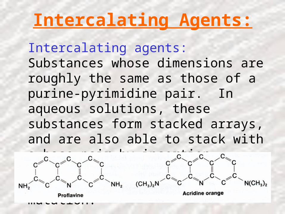
Intercalating Agents:
Intercalating agents: Substances whose dimensions are roughly the same as those of a purine-pyrimidine pair. In aqueous solutions, these substances form stacked arrays, and are also able to stack with a base-pair by insertion between two base-pairs. This may result in frameshift mutation.
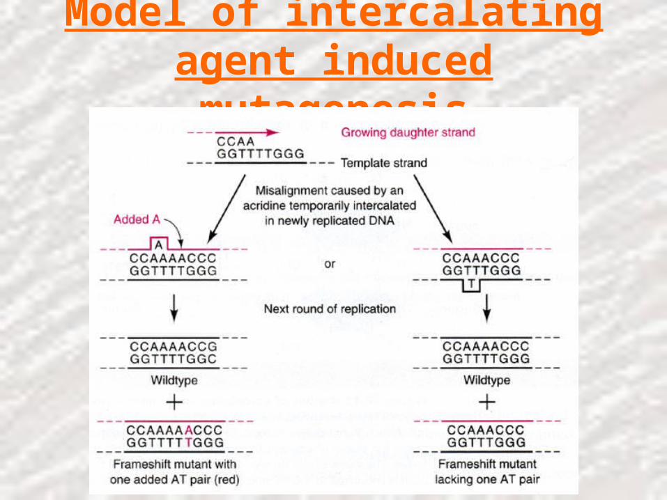
Model of intercalating agent induced mutagenesis
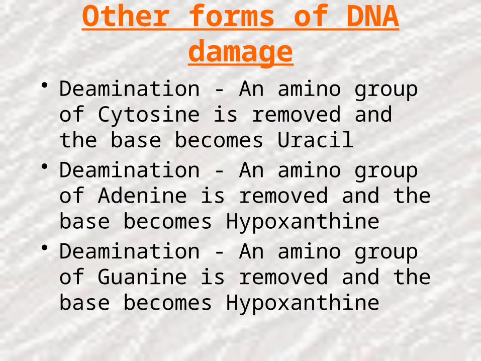
Other forms of DNA damage
• Deamination - An amino group of Cytosine is removed and the base becomes Uracil
• Deamination - An amino group of Adenine is removed and the base becomes Hypoxanthine
• Deamination - An amino group of Guanine is removed and the base becomes Hypoxanthine
And…
• Depurination - the base is simply ripped out of the DNA molecule leaving a gap (like a missing tooth)…
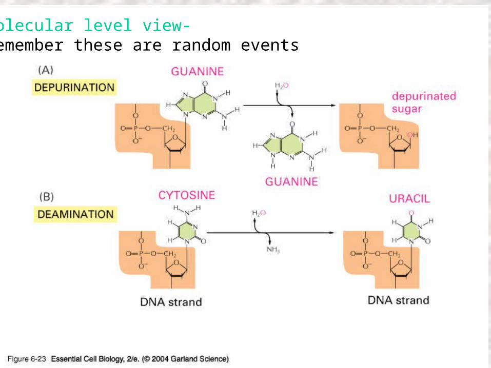
06_23_Depurination.jpgMolecular level view-Remember these are random events
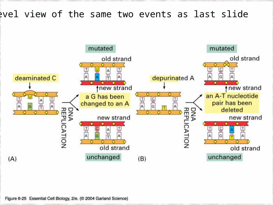
06_25_mutations.jpgDNA level view of the same two events as last slide
Metabolite Mutagens
Chemicals that are metabolized to electrophilic reagents: Aflatoxins, benzo[a]pyrene
A mutagen is a physical or chemical agent that causes mutations to occurs.
Mutagenesis is the process of producing a mutation.
Mutant refers to an organism or a gene that is different from the normal or wild type.
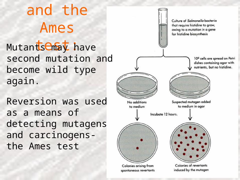
Reversion and the Ames test:
Mutants may have second mutation and become wild type again.
Reversion was used as a means of detecting mutagens and carcinogens- the Ames test

DNA Repair Mechanisms
(1) Repair by direct reversal: The simplest mechanism. e.g. UV induced T-T dimer is recognized by photolyase and is cleaved into intact thymine (light dependent). This is called photoactivation
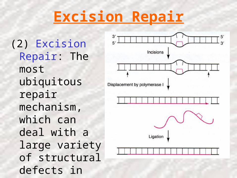
Excision Repair
(2) Excision Repair: The most ubiquitous repair mechanism, which can deal with a large variety of structural defects in DNA.

Recombinational Repair
(3) Recombinational repair (Postreplicational repair): Occurs before excision repair has happened or when excision repair can not fix the problem

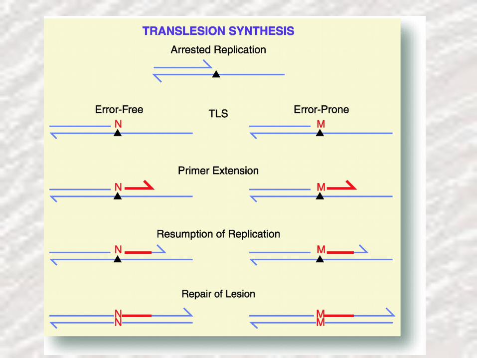
The SOS response
(4) The SOS response: The SOS response system is only active in response to some signal such as a blocked of replication fork. In E. Coli, recA and lexA govern the expression of a number of other genes involved in DNA repair. This is an error-prone DNA repair mechanism and result in higher than normal mutagenesis.

SOS DNA Repair1. DNA damage
2. RecA converted to RecA*
3. RecA* facilitated LexA self-cleavage
4. Increased synthesis of SOS proteins
5. Error prone repair induced
6. DNA damage repaired
7. RecA* returned to RecA
8. LexA no longer self-cleaved
9. LexA repressed SOS genes
10. LexA repress lexA gene expression

Type of Mutations(I)
Transition: One purine replaced by a different purine;or one pyrimidine replaced by a diferent pyrimidineA G T C
Transversion: A purine replaced by a pyrimidine or vice versa
I. Point mutation:A. Base substitution
A T
Change in DNA
C G
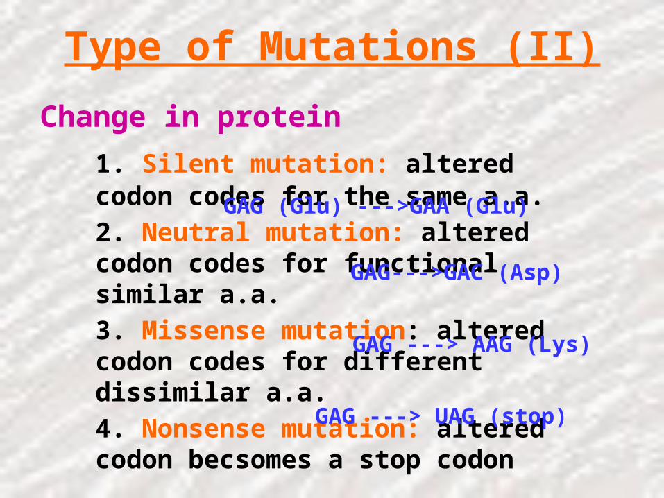
Type of Mutations (II)
Change in protein
1. Silent mutation: altered codon codes for the same a.a.
2. Neutral mutation: altered codon codes for functional similar a.a.
3. Missense mutation: altered codon codes for different dissimilar a.a.
4. Nonsense mutation: altered codon becsomes a stop codon
GAG (Glu) --->GAA (Glu)
GAG--->GAC (Asp)
GAG ---> AAG (Lys)
GAG ---> UAG (stop)

Type of Mutations (III)
B. Frameshift mutation: addition or deletion of one base-pair result in a shift of reading frame and alter amino acid sequence
1. Wild type: ATG ACC AGG TC
2. Base addition: ATG ACA CAG GTC
3. Base deletion: ATG ACA GGT C
ArgMet Thr
Met Thr Gln Val
Met Thr Gly

Type of Mutations (IV)
II. Insertion
III. Deletion
IV. Translocation
V. Inversion


Sickle Cell Disease
• This is a very good illustration of the devastating effects of even tiny changes to the DNA
• Red Blood Cells• Hemoglobin -
– Has a large protein component– 2 beta globin chains– A single base change -substitution causes the disease

06_19_sickle_cell.jpg

Cellular protection from DNA damage
• Natural errors: polymerase base selection, proofreading, mismatch repair
• Endogenous/exogenous DNA damage: base excision repair, nucleotide excision repair, (recombination, polymerase bypass)
• Recombination and polymerase bypass do not remove damage but remove its block to replication. Polymerase bypass is itself often mutagenic.
Which is which?
• The cell has a big problem to overcome…• How does it tell which strand carried the
correct information?
• We think we know…

06_21_Errors corrected.jpgThe cell has to pick the right strand to fix or else…


Correction mechanisms
• Direct reversal of damage - Photoreactivation (bacteria, yeast, some vertebrates - not humans) Two thymines connected together by UV light.
• Excision Repair - removal of defective DNA. There are three distinct types– 1) base-excision– 2) nucleotide-excision– 3) mismatch repair

base-excision
• Presence of the Uracil in DNA is a great example of this type
• Special enzymes replace just the defective base– 1 snip out the defective base– 2 cut the DNA strand– 3 Add fresh nucleotide– 4 Ligate gap

nucleotide-excision
• Same as previous except that– It recognizes more varieties of damage– Remove larger segments of DNA (10 -100s of
bases)

mismatch repair
• Special enzymes scan the DNA for bulky alterations in the DNA double helix
• These are normally caused by mismatched bases
• AG• AC• CT• These are excised and the DNA repaired

Base excision repair (BER)
• Major pathway for repair of modified bases, uracil misincorporation, oxidative damage
• Various DNA glycosylases recognize lesion and remove base at glycosidic bond, thereby producing an “abasic” or AP (apurinic/ apyrimidinic) site by base “flipping out”
• One of several AP endonucleases incises phosphodiesterase backbone adjacent to AP site
• AP nucleotide removed by exonuclease/dRPase and patch refilled by DNA synthesis and ligation

Mechanism of BER


N
N
NH2
O
O
H2C
O
ON
HN
O
O
O
H2C
O
O
deoxycytosine deoxyuracil
1’
2’3’
4’
5’
12
34
5
6
CH3
thymine
glycosidic bond

Types of lesions repaired by BER• Oxidative lesions; 8-oxo-G, highly mutagenic,
mispairs with A, producing GC --> TA transversions example MutY, MutM=Fpg from E. coli
• Deoxyuracil: from misincorporation of dU or deamination of dC-->dU, example Ung, uracil N-glycosylase
• Various alkylation products e. g. 3-meA• These lesions are not distorting and do not block
DNA polymerases• Spontaneous depurination (esp. G) yield abasic
sites that are repaired by second half of BER pathway

“Flipping out” mechanism

Mismatch repair (MMR)• Despite extraordinary fidelity of DNA synthesis, errors do
persist• Such errors can be detected and repaired by the post-
replication mismatch repair system• Prokaryotes and eukaryotes use a similar mechanism with
common structural features• Defects in MMR elevate spontaneous mutation rates 10-
1000x• Defects in MMR underlie human predisposition to colon
and other cancers (“HNPCC”)• MMR also processes mispairs that result from heteroduplex
DNA formed during genetic recombination: act to exclude “homeologous” recombination

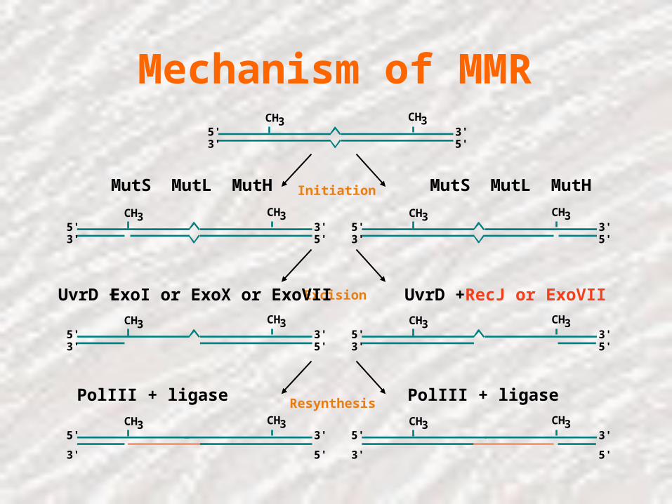
Mechanism of MMRCH3 CH3
5'3' 5'
3'
Initiation
CH3 CH35'3' 5'
3'CH3 CH3
5'3' 5'
3'
MutS MutL MutH MutS MutL MutH
Excision
CH3 CH35'3' 5'
3'CH3 CH3
5'3' 5'
3'
UvrD + RecJ or ExoVIIUvrD + ExoI or ExoX or ExoVII
ResynthesisCH3 CH3
5'
3' 5'
3'CH3 CH3
5'
3' 5'
3'
PolIII + ligase PolIII + ligase
Mechanism of MMRCH3 CH3
5'3' 5'
3'
Initiation
CH3 CH35'3' 5'
3'CH3 CH3
5'3' 5'
3'
MutS MutL MutH MutS MutL MutH
Excision
CH3 CH35'3' 5'
3'CH3 CH3
5'3' 5'
3'
UvrD + RecJ or ExoVIIUvrD + ExoI or ExoX or ExoVII
ResynthesisCH3 CH3
5'
3' 5'
3'CH3 CH3
5'
3' 5'
3'
PolIII + ligase PolIII + ligase

Basis of MMR recognition
• MutS dimer (in yeast, Msh2/Msh3 or Msh2/Msh6 heterodimer)
• By DNA binding expts in vitro and DNA heteroduplex repair expts in vivo: MMR can recognize all base substitutions except C:C and short frameshift loops <4 bp
• Transition mispairs G:T and A:C and one base loops are particularly well-recognized (these are also the most common polymerase errors)

Structure of MutS bound to DNA
60° kink in DNA
Widens minor groove, narrows major groove
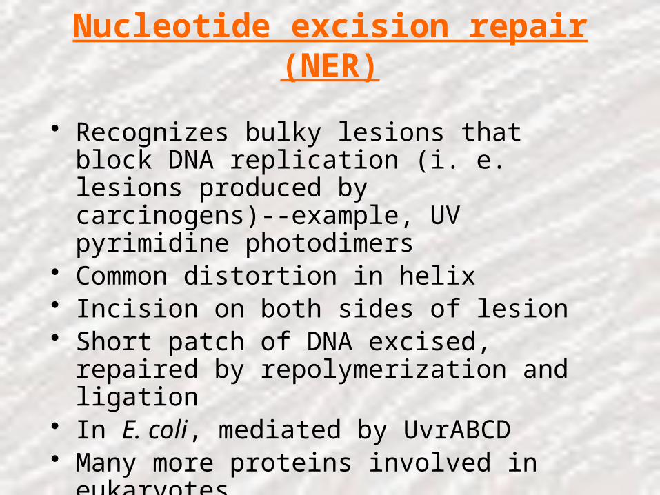
Nucleotide excision repair (NER)
• Recognizes bulky lesions that block DNA replication (i. e. lesions produced by carcinogens)--example, UV pyrimidine photodimers
• Common distortion in helix• Incision on both sides of lesion• Short patch of DNA excised, repaired by
repolymerization and ligation• In E. coli, mediated by UvrABCD• Many more proteins involved in eukaryotes• Can be coupled to transcription (TCR,
“transcription coupled repair”)• Defects in NER underlie Xeroderma pigmentosum

Xeroderma pigmentosum
• Autosomal recessive mutations in several complementation groups
• Extreme sensitivity to sunlight
• Predisposition to skin cancer (mean age of skin cancer = 8 yrs vs. 60 for normal population)

Recognition and binding
UvrA acts as classical “molecular matchmaker”
Incision
Nicks delivered 3’ and 5’ to lesion by UvrBC
Excision and repair
Short fragment released by helicase action
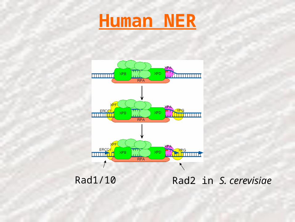
Human NER
Rad1/10 Rad2 in S. cerevisiae
Further references• Friedberg. DNA repair and mutagenesis. ASM Press, Washington, D. C. • *Marti TM, Kunz, C, Fleck O. 2002 DNA mismatch repair and mutation
avoidance pathways. J. Cell. Physiol. 191: 28-41• *Harfe BD, Jinks-Robertson S. 2000 DNA mismatch repair and genetic
instability. Annu. Rev. Genet. 34: 359-399.• *Krokan, HE, Standal, R, Slupphaug, G. 1997 DNA glycosylases in the base
excision repair of DNA Biochem. J. 325: 1-16. • *De Laat, WL, Jaspers, NGJ, Hoeijmakers, JHJ. 1999 Molecular mechanism
of nucleotide excision repair. Genes Dev. 13: 768-785• Petit, C, Sancar, A. 1999 Nucleotide excision repair: from E. coli to man.
Biochimie 81: 15-25• *Goodman, MF, Tippin, B. 2000. Sloppier copier DNA polymerases involved
in genome repair. Curr. Opin. Genet. Dev. 10:162-168.• *Friedberg, EC, Wagner, R, Radman, M. Specialized DNA polymerases,
cellular survival and the genesis of mutations. Science 296: 1627-1630. • Goodman, MF 2002. Error-prone repair DNA polymerases in prokaryotes and
eukaryotes. Annu. Rev. Biochem. 71: 17-50

06_26_three steps.jpgBasic mechanism is the same for all three types
1) Remove damaged region
2) Resynthesis DNA3) Ligate
Thank You