dna methyltransferase enzymes (mtases) may not enhance
TRANSCRIPT
DNA methyltransferase enzymes (MTases) may not enhance genomic protection against exogenous DNA invasions
by
Sanoji Wijenayake
A thesis submitted to the Faculty of Graduate and Postdoctoral Affairs in partial fulfillment of the requirements for the degree of
Masters of Science
in
Biological Science
Carleton University
Ottawa, Ontario
2012 Sanoji Wijenayake
1+1Library and Archives Canada
Published Heritage Branch
Bibliotheque et Archives Canada
Direction du Patrimoine de I'edition
395 Wellington Street Ottawa ON K1A0N4 Canada
395, rue Wellington Ottawa ON K1A 0N4 Canada
Your file Votre reference
ISBN: 978-0-494-93533-0
Our file Notre reference ISBN: 978-0-494-93533-0
NOTICE:
The author has granted a nonexclusive license allowing Library and Archives Canada to reproduce, publish, archive, preserve, conserve, communicate to the public by telecommunication or on the Internet, loan, distrbute and sell theses worldwide, for commercial or noncommercial purposes, in microform, paper, electronic and/or any other formats.
AVIS:
L'auteur a accorde une licence non exclusive permettant a la Bibliotheque et Archives Canada de reproduire, publier, archiver, sauvegarder, conserver, transmettre au public par telecommunication ou par I'lnternet, preter, distribuer et vendre des theses partout dans le monde, a des fins commerciales ou autres, sur support microforme, papier, electronique et/ou autres formats.
The author retains copyright ownership and moral rights in this thesis. Neither the thesis nor substantial extracts from it may be printed or otherwise reproduced without the author's permission.
L'auteur conserve la propriete du droit d'auteur et des droits moraux qui protege cette these. Ni la these ni des extraits substantiels de celle-ci ne doivent etre imprimes ou autrement reproduits sans son autorisation.
In compliance with the Canadian Privacy Act some supporting forms may have been removed from this thesis.
While these forms may be included in the document page count, their removal does not represent any loss of content from the thesis.
Conformement a la loi canadienne sur la protection de la vie privee, quelques formulaires secondaires ont ete enleves de cette these.
Bien que ces formulaires aient inclus dans la pagination, il n'y aura aucun contenu manquant.
Canada
Abstract
DNA methylation is thought to function as a host-driven response against extracellular invaders
in all three domains of life. Here I devise and test an alternative hypothesis, the transposon-
mediated immunity hypothesis, that gene regulation via DNA methylation originated in
ancestral eubacteria as a transposon-mediated countermeasure to endonuclease cleavage. I
predict that transposon-coded proteins may have influenced eubacterial DNMTases into
hypermethylating eubacterial restriction endonuclease sites, decreasing endonuclease enzyme’s
accessibility to palindromic sequences and rendering rm system II ineffective at removing
transposable elements from the genome.

Acknowledgements
I would like to thank Dr. Root Gorelick for giving me the opportunity to finish my master’s
under his supervision. Thank you for always being patient and understanding whenever I
proposed a wacky research idea. I know at times we have had very different viewpoints on
evolution and on the empirical aspect of science, but you always trusted my judgment (even
when you did not agree with it) and allowed me to make independent decisions about my thesis.
This helped transform me into the researcher I am today.
I would like to thank Danielle Fraser, my good friend and colleague in the Gorelick lab, for the
countless hours of discussions and insights on the phylogenetic comparative method. Dani, you
taught me how to write algorithms for R and most importantly taught me how to trouble shoot
and not be intimidated by R. You are one of the most driven scientific researchers I have ever
met and you aspire me to become a better researcher on a daily basis. I would also like to thank
Zoe Panchen and Lindsay Derraugh for the advice and support.
I would like to sincerely, thank Dr. Tom Sherratt and each person in the Sherratt lab. Chris, you
are simply the BEST, thank you for always willing to help me with just about everything, I know
that you are just a text, email, phone call, or six hour plane ride away. Bells, Tom H, Kevin, Rich
and Jeimette, you have made the last two years the best graduate experience, and I know once I
leave here, I will not only have six colleagues, but six really good friends.
Most importantly, I would like to thank my family, my dad, mum, Aiyya and Akki. Ammi and
Thathi, you two are the reason why I am finishing my master’s today and going on to complete a
PhD in Biology. I could not have wished for two better people to call my mum and dad. Thank
you for always loving and supporting me through everything. You two are and have always been

my biggest fans and I know regardless I succeed or fail in whatever I do, you will be proud of
me. Akki, I am soo glad that I have a sister like you. I can count on for everything in life and I do
not know how I will get through the day without you, I love you to death. Aiyya thank you for
the sibling competition. Trying to compete with you is like running with a cheetah, it is very hard
to keep up.
Thank you to my besties, Supun, Spojmai and Uzma. You three are my shadows. I know that I
can count on you to laugh, cry, fight or even cover up a murder if need be. Thank you to all my
other amazing friends and all my cousins (especially Nad) for your constant support, well wishes
and love. I am very lucky to have all of you in my life!
Last but not least, thank you to my partner in crime, Tharanga. I love you more than words can
say. Thank you for being my comer stone! I cannot wait to spend the rest of my life with you.
iv
Contributions:
Dr. Chris Hassel and I worked on the algorithm for R to assign species-specific, sequence probes
that correspond to the endonuclease restriction and methylase sequences for the 68-eubacterial
species.
Dani Fraser and I worked closely on the phylogenetic comparative method in R to correlate the
total number of endonuclease restriction to the methylase sites.
v

Table of Contents
Abstract................................................................................................................................... i
Acknowledgements..............................................................................................................iii
Contributions..........................................................................................................................v
Table of Contents...............................................................................................................w i
List of Tables...................................................................................................................... vii
List of Figures....................................................................................................................viii
List of Appendices............................................................................................................... ix
1.1 Abstract...................................................................................................................... 1
1.2 Introduction...............................................................................................................2
1.3 Materials and Methods........................................................................................... 11
Sequence comparison.................................................................................11
Phylogenetic construction......................................................................... 13
Phylogenetic signal....................................................................................13
1.4 Results......................................................................................................................14
1.5 Discussion.................................................................................................................17
Bibliography....................................................................................................................... 21
Appendix I. Complete data set........................................................................................... 28
Appendix II. R code used for statistical analysis..............................................................52
Pagel’s lambda phylogenetic signal.........................................................11
Quantitative sequence search.................................................................. 13
Appendix III. Molecular phylogeny based on 16S rRNA 62

List of Tables
Table Title Page
1 Total number of type II endonuclease restriction sites, number of methylase 15sites, and number of overlapping sites in 16s rRNA genomes of 40microbial species in 23 families.
2 28 microbial species with zero endonuclease and methylase sites in the 16S 16rRNA gene loci.
5 1 Restriction sequences of 344 type II endonuclease enzymes and 212 type II 28methyltransferases, belonging to 261 microbial species.
52 NCBI GenBank accession numbers for 16s rRNA, type II restriction 39endonuclease restriction enzymes and respective palindromic restrictionsites, methylase enzymes and respective methylase sites for all 68 species.
53 Number of endonuclease restriction sequences, number of methylase 45sequences, and number of overlapping sequences of 96 type II restriction endonuclease enzymes and 96 corresponding methylase enzymes in 40microbial species.
vii
List of Figures:
Figure Title Page
1 Three main types of covalently methylated nitrogenous base products. 3
2 Alternative forms of DNA methylation. 3
3 Methylation of palindromic recognition sites prevents systematic 11cleavage of transposons by EcoRI.
4 Molecular phylogeny of 38 eubacterial and 2 archaeabacterial species 62based on complete and partial 16S rRNA.
viii
List of Appendices:
Appendix Title Page
I Complete data set 28
51. Restriction sequences o f344 type II endonuclease enzymes and 466 28 methylase sequences o f 212 type II methyltransferases, belonging to 261 microbial species.
52. NCBI GenBank accession numbers for 16s rRNA, type II restriction 39 endonuclease restriction enzymes and respective palindrome restrictionsites, methylase enzymes and respective methylase sites for all 68 species.
53. Number o f endonuclease restriction sequences, number o f methylase 45 sequences, and number o f overlapping sequences o f 96 type IIrestriction endonuclease enzymes and 96 corresponding methylase enzymes in 68 microbial species.
II R code used for statistical analysis 52
Pagel’s lambda phylogenetic signal 52
Quantitative sequence search 55
III Molecular phylogeny based on 16S rRNA 62
ix
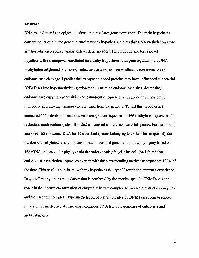
Abstract
DNA methylation is an epigenetic signal that regulates gene expression. The main hypothesis
concerning its origin, the genomic autoimmunity hypothesis, claims that DNA methylation arose
as a host-driven response against extracellular invaders. Here I devise and test a novel
hypothesis, the transposon-mediated immunity hypothesis, that gene regulation via DNA
methylation originated in ancestral eubacteria as a transposon-mediated countermeasure to
endonuclease cleavage. I predict that transposon-coded proteins may have influenced eubacterial
DNMTases into hypermethylating eubacterial restriction endonuclease sites, decreasing
endonuclease enzyme’s accessibility to palindromic sequences and rendering rm system II
ineffective at removing transposable elements from the genome. To test this hypothesis, I
compared 666 palindromic endonuclease recognition sequences to 466 methylase sequences of
restriction modification system II in 262 eubacterial and archaeabacterial species. Furthermore, I
analyzed 16S ribosomal RNA for 40 microbial species belonging to 23 families to quantify the
number of methylated restriction sites in each microbial genome. I built a phytogeny based on
16S rRNA and tested for phylogenetic dependence using Pagel’s lambda (X). I found that
endonuclease restriction sequences overlap with the corresponding methylase sequences 100% of
the time. This result is consistent with my hypothesis that type II restriction enzymes experience
“cognate” methylation (methylation that is conferred by the species-specific DNMTases) and
result in the incomplete formation of enzyme-substrate complex between the restriction enzymes
and their recognition sites. Hypermethylation of restriction sites by DNMTases seem to render
rm system II ineffective at removing exogenous DNA from the genomes of eubacteria and
archaeabacteria.
1
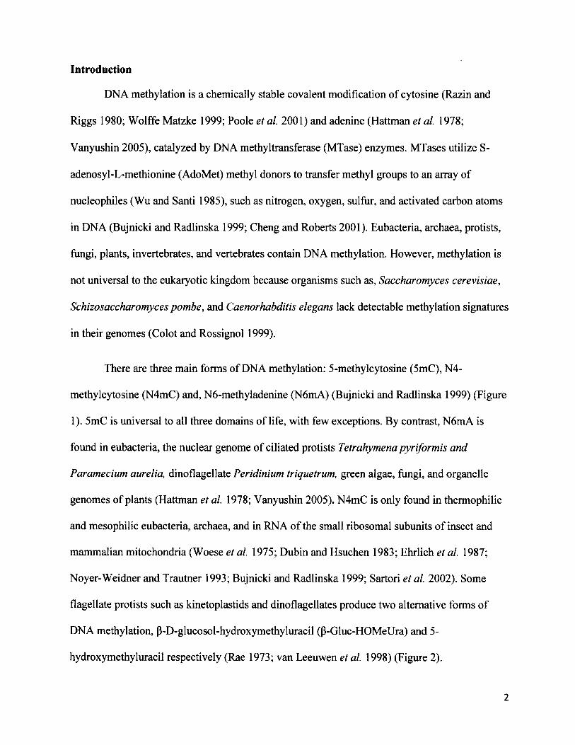
Introduction
DNA methylation is a chemically stable covalent modification of cytosine (Razin and
Riggs 1980; Wolffe Matzke 1999; Poole et al. 2001) and adenine (Hattman et al. 1978;
Vanyushin 2005), catalyzed by DNA methyltransferase (MTase) enzymes. MTases utilize S-
adenosyl-L-methionine (AdoMet) methyl donors to transfer methyl groups to an array of
nucleophiles (Wu and Santi 1985), such as nitrogen, oxygen, sulfur, and activated carbon atoms
in DNA (Bujnicki and Radlinska 1999; Cheng and Roberts 2001). Eubacteria, archaea, protists,
fungi, plants, invertebrates, and vertebrates contain DNA methylation. However, methylation is
not universal to the eukaryotic kingdom because organisms such as, Saccharomyces cerevisiae,
Schizosaccharomyces pombe, and Caenorhabditis elegans lack detectable methylation signatures
in their genomes (Colot and Rossignol 1999).
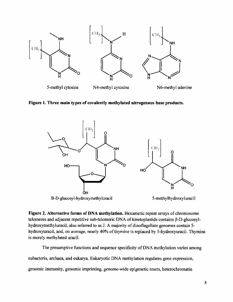
There are three main forms of DNA methylation: 5-methylcytosine (5mC), N4-
methylcytosine (N4mC) and, N6-methyladenine (N6mA) (Bujnicki and Radlinska 1999) (Figure
1). 5mC is universal to all three domains of life, with few exceptions. By contrast, N6mA is
found in eubacteria, the nuclear genome of ciliated protists Tetrahymena pyriformis and
Paramecium aurelia, dinoflagellate Peridinium triquetrum, green algae, fungi, and organelle
genomes of plants (Hattman et al. 1978; Vanyushin 2005). N4mC is only found in thermophilic
and mesophilic eubacteria, archaea, and in RNA of the small ribosomal subunits of insect and
mammalian mitochondria (Woese et al. 1975; Dubin and Hsuchen 1983; Ehrlich et al. 1987;
Noyer-Weidner and Trautner 1993; Bujnicki and Radlinska 1999; Sartori et al. 2002). Some
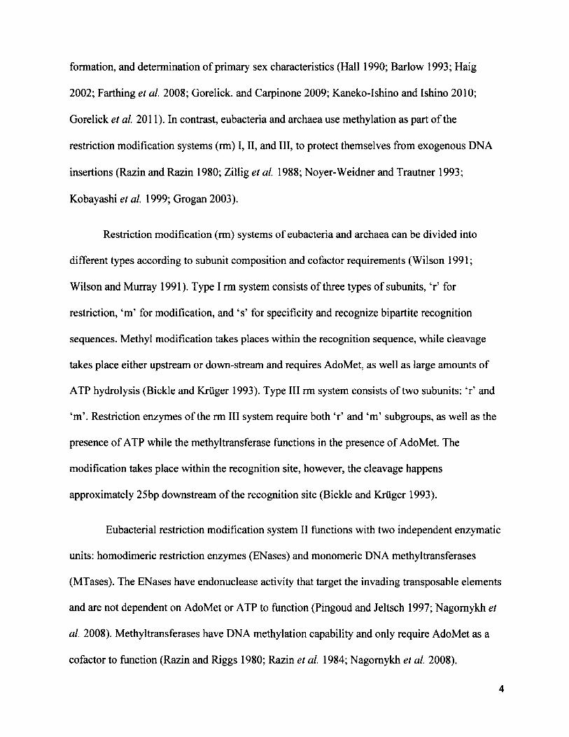
flagellate protists such as kinetoplastids and dinoflagellates produce two alternative forms of
DNA methylation, p-D-glucosol-hydroxymethyluracil (P-Gluc-HOMeUra) and 5-
hydroxymethyluracil respectively (Rae 1973; van Leeuwen et al. 1998) (Figure 2).
2
Cl I,
Cll,
NH
5-methyl cytosine N4-methyl cytosine N6-methyl adenine
Figure 1. Three main types of covalently methylated nitrogenous base products.
NHOH
HO
OH
NHHO
5-methylhydroxyluracilB-D-glucosyl-hydroxymethyluracil
Figure 2. Alternative forms of DNA methylation. Hexameric repeat arrays of chromosome telomeres and adjacent repetitive sub-telomeric DNA of kinetoplastids contains P-D-glucosyl- hydroxymethyluracil, also referred to as J. A majority of dinoflagellate genomes contain 5- hydroxyuracil, and, on average, nearly 40% of thymine is replaced by 5-hydroxyuracil. Thymine is merely methylated uracil.
The presumptive functions and sequence specificity of DNA methylation varies among
eubacteria, archaea, and eukarya. Eukaryotic DNA methylation regulates gene expression,
genomic immunity, genomic imprinting, genome-wide epigenetic resets, heterochromatin
3
formation, and determination of primary sex characteristics (Hall 1990; Barlow 1993; Haig
2002; Farthing et al. 2008; Gorelick. and Carpinone 2009; Kaneko-Ishino and Ishino 2010;
Gorelick et al. 2011). In contrast, eubacteria and archaea use methylation as part of the
restriction modification systems (rm) I, II, and III, to protect themselves from exogenous DNA
insertions (Razin and Razin 1980; Zillig et al. 1988; Noyer-Weidner and Trautner 1993;
Kobayashi et al. 1999; Grogan 2003).
Restriction modification (rm) systems of eubacteria and archaea can be divided into
different types according to subunit composition and cofactor requirements (Wilson 1991;
Wilson and Murray 1991). Type I rm system consists of three types of subunits, ‘r’ for
restriction, ‘m’ for modification, and ‘s’ for specificity and recognize bipartite recognition
sequences. Methyl modification takes places within the recognition sequence, while cleavage
takes place either upstream or down-stream and requires AdoMet, as well as large amounts of
ATP hydrolysis (Bickle and Kruger 1993). Type III rm system consists of two subunits: ‘r’ and
‘m’. Restriction enzymes of the rm III system require both ‘r’ and ‘m’ subgroups, as well as the
presence of ATP while the methyltransferase functions in the presence of AdoMet. The
modification takes place within the recognition site, however, the cleavage happens
approximately 25bp downstream of the recognition site (Bickle and Kruger 1993).
Eubacterial restriction modification system II functions with two independent enzymatic
units: homodimeric restriction enzymes (ENases) and monomeric DNA methyltransferases
(MTases). The ENases have endonuclease activity that target the invading transposable elements
and are not dependent on AdoMet or ATP to function (Pingoud and Jeltsch 1997; Nagomykh et
al. 2008). Methyltransferases have DNA methylation capability and only require AdoMet as a
cofactor to function (Razin and Riggs 1980; Razin et al. 1984; Nagomykh et al. 2008).
4
Endonucleases are highly sequence-specific and species-specific enzymes that target and cleave
exogenous and endogenous nucleotides (Wilson 1991). For example, EcoRI restriction
endonuclease cleaves DNA only at recognition site G/AATTC and produce 5’sticky ends in
Escherichia coli, whereas BamHI only cleaves DNA at recognition sequence site G/GATCC in
Bacillus amyloliquefaciens (Dale and Park 2010). EcoRI restriction endonuclease cannot
function in the absence of palindromic recognition sequences nor in species other than
Escherichia coli, specificity also found in BamHI and hundreds of other restriction enzymes.
I exclusively analyzed the type II rm system because it can be studied in the absence of
endonuclease (restriction) activity, unlike with type I and III rm system, in which a single
enzymatic unit performs the dual functions of methylation and restriction cleavage. Type II
restriction enzymes typically recognize palindromic recognition sites that are 4-8bp in length,
and the modification and cleavage occur within the recognition sites. In addition, type II
restriction modification is the most prominent and frequently occurring genomic line of defense
in a majority of eubacterial species and has the capability to produce N6-methyladenine, N4-
methylcytosine and 5-methylcytosine. The role, ubiquity, diversity, and independent evolution of
rm system II suggests that it plays an important role in the population structure and (co)evolution
of bacteria with invading exogenous DNA elements (Levin 1993).
Exogenous DNA elements, such as viruses, repetitive elements, transposons, and
retrotransposons, are capable of rapid mobilization via transposition (McClintock 1950; Kimura
1968; Bestor 1990; Levin 1993; Bheemanaik et al. 2006; Suzuki et al. 2007; Harony and Ankri
2008; Kaneko-Ishino and Ishino 2010). Transposons are present in almost all eubacteria,
archaeabacteria, and eukarya, and occupy a significant portion of the genome. For example,
about half of the human genome is derived from transposable elements (Boyes and Bird, 1991),
5
with more than a dozen hominoid functional genes originating from ancestral transposons
(Lander et al. 2001; Allis et al. 2007). Transposons have a high degree of genetic and functional
diversity and remarkable ability to adapt and thrive within host lineages. Some eubacterial
species require exogenous DNA for the survival and spread while in other eubacterial species
exogenous DNA enhance the ability of eubacterial populations to colonize and adapt to changing
environments (McClintock 1950). Previous studies have shown that exogenous DNA provides
(1) aid in the production of allelopathic agents such as bacteriocins (Tani and Nasu 2010), (2)
protection against pathogens (Levin 1993), oxidation and fermentation of toxic carbon
compounds (Liu 2006), and (3) resistance to the adverse effects of ultraviolet light, heavy metals
(He and Hader 2002), and antibiotics (Courvalin 1994; Walker 1996; Tani and Nasu 2010).
Some transposable elements lack a strong target site preference, and their distribution
appears more or less random. However, transposition is rarely fully random and majority of
transposons are highly selective for the sites where they insert. A primary example for targeted
transposition is the bacterial Tn7 transposon. Tn7 is highly specialized to insert into a single
sequence motif in the E. coli genome (Lichtenstein and Brenner 1982). Kirchner et al. (1995)
and Devine and Boeke (1996) showed that Ty LTR retrotransposons of the yeast S. cerevisiae
genome exhibit strong integration bias into Tyl and Ty3 elements that are upstream of RNA
polymerase III transcribed genes. The single-celled eukaryote, Dictyostelium discoideum has a
highly compact genome, and transposons in D. discoideum employ two strategies for site-
directed insertion. One strategy is to integrate into previously inserted transposon clusters
(Loomis et al. 1995). The other strategy is targeted insertion into non-protein coding regions of
the genome (Craigie et al. 1987; Winckler et al. 2005). However, the question of whether
6
transposons preferentially insert in methylated regions of the host genome has yet to be
answered.
The genomic autoimmunity hypothesis is the primary hypothesis that addresses the role
of DNA methyltransferases in the restriction modification system II of eubacteria and
archaeabacteria. The genomic autoimmunity hypothesis postulates that the “cognate”
methyltransferases have evolved to hypermethylate endonuclease recognition sites in order to
protect the host genome against its own restriction cleavage, a preventative method against
genomic autoimmunity (Razin et al. 1984; Nagomykh et al. 2008; Dale and Park 2010). In other
words, eubacteria and archaea use DNA methyltransferase enzymes to hypermethylate host loci
to prevent endonuclease restriction cleavage of host’s genomic material. However the genomic
autoimmunity hypothesis cannot explain the occurrence of the “rare cutter” restriction enzymes
whose lengthy recognition sequences (approximately eight to ten base pairs) are unlikely to be
present in many eubacterial viruses and/or transposable elements (Naito et al. 1995) and often
lead to the restriction of host DNA.
Wilson and Murray (1991) showed that the genes encoding the restriction modification
unit of EcoRI are capable of leaving the eubacterial genome via plasmids and thus participate
frequently in horizontal gene transfer. In a majority of cases, the methyltransferases and
endonuclease restriction enzymes do not leave the bacterial host cell in unison, but rather
participate independently in horizontal gene transfer (Jeltsch and Pingoud 1996). Therefore, at
times the host genome is left with endonuclease restriction enzymes without the corresponding
methyltransferases, which results in the death of the bacterial host cell. Thus, Dawkins (1989)
and Naito et al. (1995) describe the behavior of the restriction modification system II as
parasitic, symbiont, and “selfish,” mobile genetic elements.
7

Furthermore, Type II DNA methyltransferases do not distinguish between endogenous
and exogenous DNA (Lauster et al. 1989). Rather, DNMTases search along a given DNA strand
and covalently modify cytosine and adenine nitrogenous bases. The DNA target-recognition
domains (TRD’s) of the amino acid chain of type II MTases are responsible for identifying the
methylase recognition sites (Lauster et al. 1989; Kumar 2004). However, Lauster et al. (1989)
postulated that eubacterial type II MTases do not have an enzymatic domain that regulates the
identification of self to non-self. According to Kruger and Reuter (1999) and Kumar (2004) the
target recognition domain (TRD) is not conserved between different groups of eubacterial
MTases, and is completely devoid of self-recognition motifs. A significant characteristic of
eubacterial DNA methylation is that eubacterial MTases will methylate all available target sites
without discrimination as long as the sites are found within the methylase recognition sequences
(Kumar 2004).
Is there a fundamental difference between exogenous and endogenous DNA in eubacteria
and archaeabacteria? If there is no, how can the host genome protect itself against extracellular
invaders? Even though, type II methyltransferases cannot distinguish between exogenous and
endogenous DNA, transposons and retrotransposons have unique structural characteristics that
differ from the host’s genomic material. All DNA transposons belonging to eubacteria and
archaeabacteria contain two sets of flanking repeat sequences referred to as IS (insertion
sequence) elements and/or IS-like elements. The IS elements are either direct or inverted repeats
ranging from 20-40bp in length (Starlinger 1980; Calos and Miller 1980). All DNA transposons
also carry the transposase gene that is essential for transposition. Transposase is a transposon-
coded DNA binding protein that is essential for the formation of the transposome, a
nucleoprotein synaptic complex. Transposomes contain the DNA of the transposable element,
8
the transposon-encoded transposase enzyme, targeted host DNA, and host-encoded proteins
factors (Surret et al. 1987; Agrawal et al. 1998; Hiom et al. 1998; Izsvaik et al. 2002; Kapitonov
and Jurka 2005; Walisko et al. 2006).
Calos and Miller (1980) postulated that the insertion sequences and/or transposase gene
can be used as molecular markers to locate transposon-derived genes in eubacteria and eukarya
because the insertion sequences and/or the transposase gene remain as part of the host genome
post-transposition. Rice and Marshall (1992) discovered the presence of conserved 121bp-long
inverted repeats belonging to Tn552 and Tn4002 transposons in the transposon-derived, beta-
lactamase gene in Enterococcus faecalis strains CH19 and CX19. Tamura et al. (2000) used the
conserved, inverted terminal repeats of piggyBac transposon in the germline of the silkworm,
Bombyx mori to confirm if piggyBac transposition is transgenerational. Furthermore, Youngson
et al. (2005) showed that the sushi-ichi transposase gene is highly conserved and remains intact
in nine Ty3/gypsy retrotransposon-derived mammalian-genes. Kapitonov and Jurka (2004)
identified a widely expressed HARBI1 gene encoding a 350 amino acid protein that is entirely
derived from Harbinger transposase belonging to the Harbinger superfamily of DNA
transposons. The HARBI1 gene is believed to have transposed 450-500 million years ago and is
conserved in humans, rats, mice, cows, pigs, chickens, frogs, and bony fish. Therefore, even
though host-encoded, type II methyltransferases cannot distinguish between exogenous and
endogenous DNA, transposons and retrotransposons have unique structural characteristics such
as flanking insertion sequences and transposase genes. Insertion sequences and/or transposase
genes are conserved in transposon-derived genes in eubacteria and eukarya and thus can be used
to set apart transposon-derived elements from host genomic material.
9
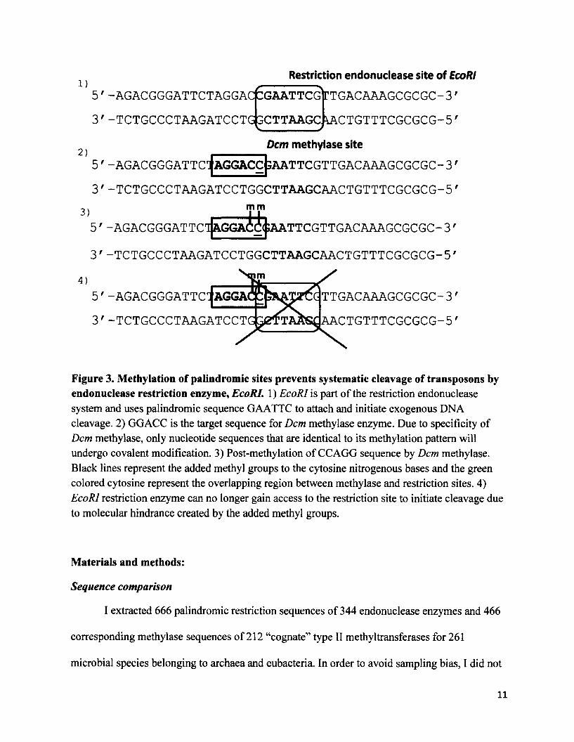
Based on the notion that transposable elements are structurally unique pre and post
transposition, I present a modified version of the genomic autoimmunity hypothesis termed the
“transposon-mediated immunity hypothesis”, which emphasizes the possible role of transposable
elements in regulating the function of type II DNA methyltransferases. I hypothesize that gene
regulation via DNA methylation originated in ancestral eubacteria as a transposon-
mediated countermeasure to endonuclease cleavage. Transposon-coded proteins influenced
eubacterial DNMTases into hypermethylating eubacterial restriction endonuclease sites. This
decreases restriction enzyme’s accessibility to palindromic sequences and may prevent rm
system II from removing transposable elements from the genome (Figure 3). Therefore, I predict
that “cognate” methyltransferase sites (methylation conferred by species-specific MTases)
present in a majority of microbial genomes are similar in nucleotide identity and frequency of
occurrence to restriction endonuclease sites.
10
1)5 ' -AGACGGGATTCTAGGAC GGAATTCG
3 ' -TCTGCCCTAAGATCCTG GCTTAAGC.
Restriction endonuclease site of EcoRI
rTG A C A A A G CG CG C -3'
\A C T G T T T C G C G C G -5 '
Dcm methylase site
5 ' -AGACGGGATTCIAGGACC ;A ATTCG TTG ACA A A G CG CG C-31
3)
3 ' -TCTG CCCTA A G ATCCTG G CTTA AG CA A CTG TTTCG CG CG -51mm
5 ' -AGACGGGATTCIAGGACC( ;AATTCG TTGACAAAG CGCG C-31
3 ' -TCTG CCCTA A G A TCCTG G CTTA A G CAA CTG TTTCG CG CG -5'
5 ' -AGACGGGATTC^AGGAc! TTG A CA A A G CG CG C-31
3 ' -TCTGCCCTAAGATCCTG A A CTG TTTCG CG CG -5 ‘
Figure 3. Methylation of palindromic sites prevents systematic cleavage of transposons by endonuclease restriction enzyme, EcoRI. 1) EcoRI is part of the restriction endonuclease system and uses palindromic sequence GAATTC to attach and initiate exogenous DNA cleavage. 2) GGACC is the target sequence for Dcm methylase enzyme. Due to specificity of Dcm methylase, only nucleotide sequences that are identical to its methylation pattern will undergo covalent modification. 3) Post-methylation of CCAGG sequence by Dcm methylase. Black lines represent the added methyl groups to the cytosine nitrogenous bases and the green colored cytosine represent the overlapping region between methylase and restriction sites. 4) EcoRI restriction enzyme can no longer gain access to the restriction site to initiate cleavage due to molecular hindrance created by the added methyl groups.
Materials and methods:
Sequence comparison
I extracted 666 palindromic restriction sequences of 344 endonuclease enzymes and 466
corresponding methylase sequences of 212 “cognate” type II methyltransferases for 261
microbial species belonging to archaea and eubacteria. In order to avoid sampling bias, I did not
11
pick the 261 species, but rather chose the 344 most prominent endonuclease restriction enzymes
that are exclusive to the restriction modification system II. These 344 enzymes are not present in
species that use type I or type III restriction modification systems. I used the online restriction
enzyme database REBASE at http://rebase.neb.com/rebase/rebase.html. NCBI GenBank at
http://www.ncbi.nlm.nih.gov/genbank/. volume 5 of Gene Amplification and Analysis (Roberts
1987), and Blakesley et al. (1981) to collect the sequence information. Then I used ClustalX2
(Larkin et al. 2007) to align each endonuclease sequence with the corresponding methylase
sequence to calculate the percent nucleotide identity.
Next, I quantified the total number of methylated restriction sites for each of the 68
species (66 eubacterial and 2 archaeabacterial) in 27 families. The sample size reduced from 261
to 68 species due to lack of sequence data available on GenBank, i.e. a majority of the
eubacterial species that I used in the analysis were not sequenced at these loci.
I extracted fully and partially sequenced 16S ribosomal RNA sequences in FASTA
format and obtained the corresponding accession numbers from NCBI GenBank. I used the 16S
rRNA gene because it is one of the few gene loci that are conserved amongst all eubacterial
species. I attempted to use 12S rRNA and RNA polymerase P as secondary and tertiary
molecular markers to validate the results. However, the amount of nucleotide data available from
GenBank for 12S rRNA and RNA polymerase p was insufficient.
I used two types of species-specific reference probes: probes exclusively targeting the
methylase sequences and probes exclusively targeting the endonuclease restriction sequences. I
quantified the hybridization between the probes and the target sequences, as well as the
12

overlapping regions between methylase and endonuclease sites using seqinr (Charif and Lobry
2007) and ape (Paradis et al. 2004) packages in R.
Phylogenetic construction
I used the 16S rRNA sequences to create a phylogeny for the 38 eubacterial and two
archaeabacterial species in 23 families that showed positive hybridization between the probes
and the target sequences. I used MEGA version 5.05 (Tamura et al. 2011) for the phylogenetic
and molecular evolutionary analyses and used ClustalX2 for the pairwise and multiple sequence
alignments. I used the alignment scores and three published eubacterial and archaeabacterial
supertrees (Ludwig and Schleifer 1994; Daubin et al. 2001; Case et al. 2007) to construct a
consensus tree based on maximum likelihood in MEGA 5.05.1 calculated bootstrap confidence
intervals for each node of the phylogeny with 500 bootstrap replicates. I constructed a phylogeny
based on maximum likelihood, as opposed to maximum parsimony, because maximum
likelihood is computationally more accurate than maximum parsimony.
Phylogenetic signal
I computed Pagel’s lambda (X.) correlation using Phytools and Geiger packages in R to
test for phylogenetic dependence between the total number of endonuclease and methylase sites
in 68 microbial species.
Pagel’s lambda (X) is the most common way to estimate phylogenetic signal. The test
assumes a constant-variance, random effects model of evolution and is based on maximum
likelihood (Pagel 1999). Pagel’s X varies between 0 (phylogenetic independence) and 1
(phylogenetic dependence). I calculated Pagel’s lambda for the total number of endonuclease and
methylase sites corresponding to the original, 16S rRNA tree of the 40 taxa (Figure 4, Appendix
13

II). Then I transformed the original 16S rRNA tree with 40 taxa into a null tree with uniform
branch lengths, portraying zero phylogenetic relatedness between the 40 species, and again
calculated Pagel’s X. If the lambda value for the 16S rRNA tree is less or equal to the lambda
value for the null tree, then I considered the data set to be independent and lacking phylogenetic
covariance. Annotated R codes for all the above analyses are in Appendix I.
Results
ClustalX2 alignment of the 666 palindromic endonuclease restriction sequences with the
466 species-specific, methylase sequences produced 100% nucleotide identity.
The total number of methylated restriction sites in the 16S rRNA gene locus of 40
microbial taxa yielded 100% overlap between endonuclease restriction sites and the “cognate”
methylase sites (R2=1.00) (Table 1). The number of endonuclease restriction sequences for each
species was identical to the number of methylase sequences for that species. In other words,
every species contained an equal number of restriction sites and methylase sites at the 16S rRNA
locus.
However, I saw something unexpected in 28 out of the 68 microbial species, in that they
contained neither endonuclease restriction nor methylase sites (Table 2).
14
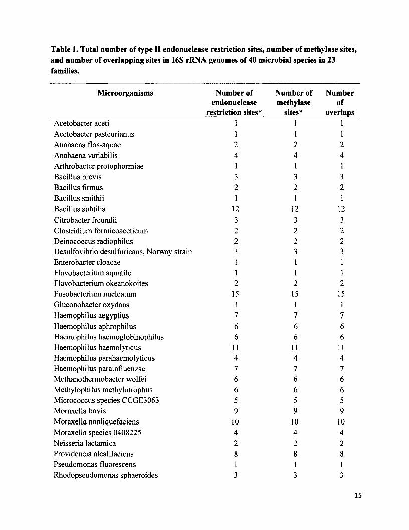
Table 1. Total number of type II endonuclease restriction sites, number of methylase sites, and number of overlapping sites in 16S rRNA genomes of 40 microbial species in 23 families.
Microorganisms Number of endonuclease
restriction sites*
Number of methylase
sites*
Numberof
overlapsAcetobacter aceti 1 1 1Acetobacter pasteurianus 1 1 1Anabaena flos-aquae 2 2 2Anabaena variabilis 4 4 4Arthrobacter protophormiae 1 1 1Bacillus brevis 3 3 3Bacillus firmus 2 2 2Bacillus smithii 1 1 1Bacillus subtilis 12 12 12Citrobacter freundii 3 3 3Clostridium formicoaceticum 2 2 2Deinococcus radiophilus 2 2 2Desulfovibrio desulfuricans, Norway strain 3 3 3Enterobacter cloacae 1 1 1Flavobacterium aquatile 1 1 1Flavobacterium okeanokoites 2 2 2Fusobacterium nucleatum 15 15 15Gluconobacter oxydans 1 1 1Haemophilus aegyptius 7 7 7Haemophilus aphrophilus 6 6 6Haemophilus haemoglobinophilus 6 6 6Haemophilus haemolyticus 11 11 11Haemophilus parahaemolyticus 4 4 4Haemophilus parainfluenzae 7 7 7Methanothermobacter wolfei 6 6 6Methylophilus methylotrophus 6 6 6Micrococcus species CCGE3063 5 5 5Moraxella bovis 9 9 9Moraxella nonliquefaciens 10 10 10Moraxella species 0408225 4 4 4Neisseria lactamica 2 2 2Providencia alcalifaciens 8 8 8Pseudomonas fluorescens 1 1 1Rhodopseudomonas sphaeroides 3 3 3
15

Streptococcus cremoris 7 7 7Streptomyces phaeochromogenes 1 1 1Thermoplasma acidophilum 9 9 9Thermus aquaticus 3 3 3Thermus flliformis 3 3 3Xanthomonas campestris___________________________1______________ 1__________ ]_
* The number of restriction and methylase sites for type II endonuclease and methylase enzymes represents the total for a species. Refer to the supplementary material (S2) for a complete list of results categorized by each enzyme.
Table 2.28 microbial species with zero endonuclease and methylase sites in the 16S rRNA gene locus________________________________________Acinetobacter calcoaceticus Neisseria mucosaAgrobacterium gelatinovorum Plesiomonas shigelloides 319-73Agrobacterium tumefaciens C58 Proteus vulgarisArthrobacter ON 14 Providencia stuartii 164Bacillus amyloliquefaciens Pseudomonas aeruginosaBacillus caldolyticus Saccharopolyspora hirsute ATCC 2787Bacillus coagulans Serratia marcescensBacteroides caccae ATCC43185T Sphaerotilus natansCaryophanon latum L Staphylococcus wameriErwinia toletana Streotomyces caespitosusHaemophilus influenza Streptomyces albusMicrococcus luteus Streptomyces fimbriatusMycoplasma fermentans Streptomyces griseusNeisseria denitrificans Xanthomonas oryzae
The molecular phylogeny of the 40 microbial species in 23 families generated a
consensus tree (Figure 4, Appendix II). My estimated phylogeny matches previously published,
molecular phylogenies based supertrees (Ludwig and Schleifer 1994; Daubin et al. 2001; Case et
al. 2007).
16
Phylogenetic signal analysis using Pagel’s lambda (A.) correlation produced a lambda
value of 1.0 x 10’5 for the original, 16S rRNA tree of the 40 microbial species. The transformed,
null, tree with uniform branch lengths produced a lambda value of 0.5.
Discussion
I tested whether hypermethylation of protein-coding genes in eubacteria and archaea
might be a host-driven mechanism against exogenous DNA invasions. Alternatively, do
exogenous DNA mediate eubacterial DNA methyltransferase enzymes into hypermethylating the
eubacterial palindromic recognition sites to avoid restriction cleavage? Genomic autoimmunity
hypothesis, postulated that the host genomic machinery regulates the enzymatic dynamics of
endonuclease and methylase enzymes of rm system II to minimize the spread of exogenous DNA
elements. However, I hypothesized that gene regulation via DNA methylation may have
originated in ancestral eubacteria as a transposon-mediated countermeasure to endonuclease
cleavage. I predicted that transposon-coded proteins may have influenced eubacterial DNMTases
into hypermethylating eubacterial restriction endonuclease sites. This decreases endonuclease
enzyme’s accessibility to palindromic sequences and may render rm system II ineffective at
removing transposable elements from the genome. The fundamental message of the transposon-
mediated immunity hypothesis is that endonuclease restriction enzymes and methyltransferases
of rm system II function as independent enzymatic units and that type II DNA methyltransferases
may not necessarily improve the effectiveness of rm system II against transposon invasions.
I showed that there is a significant correlation between the palindromic restriction
endonuclease sequences and the “cognate” methylase sequences in eubacteria and archaea. This
17
corroborates the findings of Noyer-Weidner and Trautner (1993) and Geier and Modrich (1979)
by showing that the addition of methyl moieties to restriction recognition sequences seems to
prevent endonucleases from binding to and forming a stable enzyme-substrate complex with
palindromic recognition sites.
The sequence alignment of the 666 palindromic, endonuclease restriction sequences with
the 466, “cognate” methylase sequences produced 100% nucleotide identity. However, I did not
calculate the percent identity of 200 endonuclease restriction sequences due to the lack of
published sequence data available for the corresponding methylase sequences. Nevertheless, I
predict that the remaining 200 restriction sequences will yield similar results.
I used 16S rRNA to build the phylogeny for the 66 eubacterial and 2 archaeal species
because 16S rRNA is ubiquitous and one of the few housekeeping genes, along with RNA
polymerase P subunit gene (rpoB), that is highly conserved among eubacteria and archaea. Since
1990, the use of 16S rRNA as a genomic marker has revolutionized microbiology in
understanding phylogenetic diversity (Amann et al. 1990; Garcia-Martinez et al. 1999; Case et
al. 2007).
The reason why the endonuclease sequence probes and the methylase sequence probes
failed to hybridize with the 16S rRNA sequences for 28 out of 68 microbial species (Table 2) is
probably because, according to Case et al. (2007), multiple copies of 16S rRNA gene are often
present in a given bacterium and intragenomic copies can differ in sequence identity. These
heterogeneity hot spots were found in a majority of gene fragments that are commonly used in
molecular phylogenetic analysis and the hot spots can influence hybridization of primers and
probes, 16S rRNA gene tree topology, and phylogenetic resolution at the species level (Case et
18
al. 2007). Lack of hybridization between endonuclease and methylase probes and the target
sequences in the 28 microbial taxa does not mean that these species completely lack
endonuclease and methylase sites. I did not probe the complete genomes of the 68 microbial
species to quantify the total number of endonuclease and methylase sites. For each species, I
probed the 16S rRNA locus, which represents a small portion of each microbial genome. Lack of
hybridization simply means that the 16S rRNA genomic regions in which I probed for
hybridization, in fact, lack the targeted endonuclease and methylase sites. In addition, the 16S
rRNA loci that I extracted from NCBI GenBank for the 28 eubacterial species with zero
hybridization were all partial sequences that ranged from 241bps to 721 bps. This is
approximately one quarter the size of the remaining 16S rRNA loci that I used in the analysis,
which are well over 1561bps.
Phylogenetic comparative methods are commonly used in interspecific comparative
studies (Martins and Hansen 1997; Price 1997; Freckleton et al. 2002; Blomberg et al. 2003)
because species that are closely related tend to share similar trait values compared to species that
are distantly related. Statistical non-independence that arises due to phylogenetic distance can be
resolved using phylogenetic comparative methods, such as independent contrasts and
phylogenetic generalized least squares regression (Blomberg et al. 2003; Felsenstein 1985;
Grafen 1989). However, it is not necessary to control for phylogenetic bias in all comparative
studies (Bjorklund 1994; Fitter 1995; Losos 1999). I did not use the phylogenetic comparative
method in this study because Pagel’s lambda (2) indicated that my data on total number of
endonuclease and methylase sites lacked a phylogenetic signal.
Currently the most widely accepted hypothesis regarding the origin of eubacterial and
archaeabacterial rm system II is the genomic autoimmunity hypothesis that describes rm II as the
19

main line of genomic defense against exogenous DNA. The genomic autoimmunity hypothesis
emphasizes that DNA methyltransferase enzymes protect the host genome from exogenous DNA
insertions and the host genomic machinery is solely responsible for regulating the function and
maintenance of restriction modification and methyltransferase enzymes.
In this thesis, I presented a modified version of the genomic autoimmunity hypothesis,
namely the transposon-mediated immunity hypothesis, which postulates that the presence of
DNA methylation as part of the restriction modification system II of eubacteria and archaea is
not a host-driven mechanism against transposition, but may be a transposon-mediated
countermeasure to gain access to the host genome via bypassing endonuclease cleavage. I found
that palindromic endonuclease restriction sequences and “cognate” methylase sequences share
100% nucleotide identity in 210 eubacterial and 2 archaeabacterial species. Furthermore, the
total number of restriction and methyltransferase sequences in the 16S rRNA of 40 microbial
species showed a perfect correlation (R2=1.00). This result is consistent with the transposon-
mediated immunity hypothesis in that hypermethylation of endonuclease restriction sites by
“cognate” DNMTases seem to render restriction modification system II ineffective at removing
transposable elements from the genomes of eubacteria and archaeabacteria.
20
Bibliography
Agrawal, A., Q.M. Eastman, and D.G. Schatz, 1998. Transposition mediated by RAG1 andRAG2 and its implications for the evolution of the immune system. Nature 394: 744-751.
Allis, D. C., T. Jenuwein, D. Reinberg, and M. Caparros, 2007. Epigenetics. Cold Spring Harbor Laboratory Press, Cold Spring Harbor.
Amann, R.I., B.J. Binder, R.J. Olson, S.W. Chisholm, R. Devereux, and D.A. Stahl, 1990. Combination of 16S rRNA-targeted oligonucleotide probes with flow cytometry for analyzing mixed microbial populations. Applied and Environmental Microbiology 56: Applied and Environmental Microbiology 56: 1919-1925.
Barlow, D. P., 1993. Methylation and imprinting: from host defense to gene regulation? Science 260: 309-310.
Bestor, T. H., 1990. DNA methylation: evolution of a bacterial immune function into a regulator of gene expression and genome structure in higher eukaryotes. Philosophical Transactions of the Royal Society of London. Series B, Biological Sciences 326: 179- 187.
Bheemanaik, S., Y. V. R. Reddy, and D. N. Rao, 2006. Structure, function and mechanism of exocyclic DNA methyltransferases. Biochemical Journal 399: 177-190.
Bickle, T. A., and D. H. Kruger, 1993. Biology of DNA restriction. Microbiological Reviews 57: 434-450.
Bjorklund, M., 1994. The independent contrasts method in comparative biology. Cladistics 10: 425-433.
Blakesley, R. W., 1981. Restriction endonucleases: specificities, diversities and computer analysis. Gene Amplification and Analysis 1: 1-43.
Blomberg, S. P., T. Garland, and A. R. Ives, 2003. Testing for phylogenetic signal in comparative data; behavioral traits are more labile. Evolution 57: 717-745.
Boyes, J., and A. Bird, 1991. DNA methylation inhibits transcription indirectly via a methyl- CpG binding protein. Cell 64: 1123-1134.
Bujnicki, J. M., and M. Radlinska, 1999. Molecular evolution of DNA-(cytosine-N4)methyltransferases: evidence for their polyphyletic origin. Nucleic Acids Research 27: 4501-4509.
Calos, M.P., J.H. Miller, 1980. Transposable elements. Cell 3: 579-595.
21
Case, R. J., Y. Boucher, I. Dahllof, C. Holmstrom, W. F. Doolittle et al., 2007. Use of 16S rRNA and rpoB genes as molecular markers for microbial ecology studies. Applied and Environmental Microbiology 73: 278-288.
Charif, D., and J. R. Lobry, 2007. A contributed package to the {R} project for statistical computing devoted to biological sequences retrieval and analysis in Structural Approaches to Sequence Evolution: Molecules, Networks, Populations, edited by U. Bastolla, M. Porto, H.E. Roman, and M. Vendruscolo. Springer, New York, pp. 207-232.
Cheng, X., and R. J. Roberts, 2001. AdoMet-dependent methylation, DNA methyltransferases and base flipping. Nucleic Acids Research 29: 3784-3795.
Colot, V., and J. L. Rossignol, 1999. Eukaryotic DNA methylation as an evolutionary device. BioEssays 21: 402-411.
Courvalin, P., 1994. Transfer of antibiotic resistance genes between gram-positive and gram- negative bacteria. Antimicrobial Agents and Chemotherapy 38: 1447-1451.
Craigie, R., and K. Mizuuchi, 1987. Transposition of Mu DNA: joining of Mu to target DNA can be uncoupled from cleavage at the ends of Mu. Cell 51: 493-501.
Dale, J. W., and S. F. Park, 2010. Molecular Genetics o f Bacteria. Wiley-Blackwell, West Sussex.
Daubin, V., M. Gouy, and G. Perriere, 2001. Bacterial molecular phylogeny using supertree approach. Genome Informatics 12: 155-164.
Dawkins, R., 1989. The Selfish Gene. Oxford University Press, Oxford.
Devine, S.E., and J.D. Boeke, 1996. Integration of the yeast retrotransposon Tyl is targeted to regions upstream of genes transcribed by RNA polymerase III. Genes & Development 10: 620-633.
Dubin, D. T., and C. C. Hsuchen, 1983. The 3'-terminal region of mosquito mitochondrial small ribosomal-subunit RNA-sequence and location of methylated residues. Plasmid 9: 307- 320.
Ehrlich, M., G. G. Wilson, K. C. Kuo, and C. W. Gehrke, 1987. N4-methylcytosine as a minor base in bacterial DNA. Journal of Bacteriology 169: 939-943.
Farthing, C. R., G. Ficz, R. K. Ng, C. F. Chan, S. Andrews et al., 2008. Global mapping of DNA methylation in mouse promoters reveals epigenetic reprogramming of pluripotency genes. PLoS Genetics 4: e l000116.
Felsenstein, J., 1985. Phylogenies and the comparative method. American Naturalist 125: 1-15.
22

Fitter, A.H., 1995. Interpreting quantitative and qualitative characteristics in comparative analyses. Journal of Eology 83: 730.
Freckleton, R.P., P.FI. Harvey, and M. Pagel, 2002. Phylogenetic analysis and comparative data: a test and review of evidence. American Naturalist 160: 712-726.
Garcia-Martinez, J., S. G. Acinas, A. I. Anton., and F. Rodriguez-Valera, 1999. Use of the 16S- 23S ribosomal genes spacer region in studies of prokaryotic diversity. Journal of Microbiological Methods 36: 55-64.
Geier, G.E., and P. Modrich, 1979. Recognition sequences of the Dam methylase of Escherichia coli K12 and mode of cleavage of Dpn endonuclease. Journal of Biological Chemistry 254: 1408-1413.
Gorelick, R., M. Laubichler, and R. Massicotte, 2011. Asxuality and epigenetic variation, inEpigenetics: Linking Genotype and Phenotype in Development and Evolution., edited by B. Hallgrimsson and B.K. Hall. University of California Press, San Francisco, pp. 87- 102.
Gorelick., R., and J. Carpinone., 2009. Origin and maintenance of sex: the evolutionary joys of self sex. Biological Journal of the Linnean Society 98: 707-728.
Grafen, A., 1989. The phylogenetic regression. Philosophical Transactions of the Royal Society of London. Series B, Biological Sciences 326: 119-157.
Grogan, D. W., 2003. Cytosine methylation by the Sual restriction-modification system:Implications for genetic fidelity in a hyperthermophilic archaeon. Journal of Bacteriology 185: 4657-4661.
Hackett, C.S., A. M. Geurts, and P.B. Hackett, 2007. Predicting preferential DNA vectorinsertion sites: implications for functional genomics and gene therapy. Genome Biology 8: S1-S12.
Haig, D., 2002. Genomic Imprinting and Kinship. Rutgers University Press, New Brunswick.
Hall, J. G., 1990. Genomic imprinting: review and relevance to human diseases. American Journal of Human Genetics 46: 857-873.
Harony, H., and S. Ankri, 2008. What do unicellular organisms teach us about DNA methylation? Trends in Parasitology 24: 205-209.
Hattman, S., C. Kenny, L. Berger, and K. Pratt, 1978. Comparative study of DNA methylation in three unicellular eucaryotes. Journal of Bacteriology 135: 1156-1157.
He, Y.-Y., and D.-P. Hader, 2002. Reactive oxygen species and UV-B: effect on cyanobacteria. Photochemical & Photobiological Sciences 1: 729-736.
23

Hiom, K., M. Malek, and M. Gallert, 1998. DNA transposition by the RAG1 and RAG2 proteins: a possible source of oncogenic translocations. Cell 94: 463-470.
Izsvaik, Z., D. Khare, J. Behlke, U. Heinemann, R.H. Plasterk., and Z. Ivies, 2002. Involvement of a bifunctional, paired-like DNA-binding domain and a transpositional enhancer in Sleeping Beauty transposition. Journal of Biological Chemistry 277: 34581-34588.
Jeltsch, A., and A. Pingoud, 1996. Horizontal gene transfer contributes to the wide distribution and evolution of type II restriction modification systems. Journal of Molecular Evolution 42: 91-96.
Kaneko-Ishino, T., and F. Ishino, 2010. Retrotransposon silencing by DNA methylation contributed to the evolution of placentation and genomic imprinting in mammals. Development Growth & Differentiation 52: 533-543.
Kapitonov, V.V., and J. Jurka, 2005. RAG1 core and V(D)J recombination signal sequences were derived from Transib transposons. PLoS Biology 3: el 81.
Kapitonov, V.V., and J. Jurka, 2004. Harbinger transposons and an ancient HARBI1 gene derived from a transposase. DNA and Cell Biology 23: 311-324.
Kimura, M., 1968. Evolutionary rate at the molecular level. Nature 217: 624-626.
Kirchner, J., C.M. Connolly, and S.B. Sandmeyer, 1995. Requirement of RNA polymerase III transcription factors for in vitro position-specific integration of a retrovirus-like element. Science 267: 1488-1491.
Kobayashi, I., A. Nobusato, N. Kobayashi-Takahashi, and I. Uchiyama, 1999. Shaping thegenome restriction modification systems as mobile genetic elements. Current Opinion in Genetics & Development 9: 649-656.
Kruger, D.V., and M. Reuter, 1999. Host controlled modification and restriction, inEncyclopedia o f Virology, edited by A. Granoff and R.G. Webster. Elsevier, Oxford, pp. 758-763.
Kumar, S., 2004. DNA methyltransferases, structural themes, in Encyclopedia o f Biological Chemistry, edited by W.J. Lennarz and D.M. Lane. Elsevier, New York, pp. 652-659.
Lander, E. S., L. M. Linton, B. Birren, C. Nusbaum, M. C. Zody et al., 2001. Initial sequencing and analysis of the human genome. Nature 409: 860-921.
Larkin, M.A., G. Blackshields, N.P. Brown, R. Chenna, P.A. McGettigan, H. Me William, F. Valentin, I.M. Wallace, A. Wilm, R. Lopez, J.D. Thompson, T.J. Gibson, and D.G. Higgins, 2007. Clustal W and Clustal X version 2.0. Bioinformatics, 23: 2947-2948.
24
Lauster, R., T. A. Trautner, and M. Noyer-Weidner, 1989. Cytosine specific type II DNAmethyltransferases: a conserved enzyme core with variable target recognizing domains. Journal of Molecular Biology 206: 305-312.
Levin, B. R., 1993. The accessory genetic elements of bacteria: existence conditions and (co)evolution. Current Opinion in Genetics & Development 3: 849-854.
Lichtenstein, C., and S. Brenner, 1982. Unique insertion site of Tn7 in the E. coli chromosome. Nature 297: 601-603.
Loomis, W. F., D. Welker, J. Hughes, D. Maghakian, and A. Kupsa, 1995. Integrated maps of the chromosomes in Dictyostelium discoideum. Genetics 141: 147-157.
Losos, J.B., 1999. Uncertainty in the reconstruction of ancestral character states and limitations on the use of phylogenetic comparative methods. Animal Behaviour 58: 1319-1324.
Ludwig, W., and K. H. Schleifer, 1994. Bacterial phylogeny based on 16S and 23 S rRNA sequence analysis. FEMS Microbiology Reviews 15: 155-173.
Martins, E. P., and T. F. Hansen, 1997. Phylogenies and the comparative method: A generalapproach to incorporating phylogenetic information into the analysis of interspecific data. American Naturalist 149: 646-667.
McClintock, B., 1950. The origin and behavior of mutable loci in maize. Proceedings of the National Academy of Sciences USA 36: 344-355.
Naito, T., K. Kusano, and I. Kobayashi, 1995. Selfish behavior of restriction modification systems. Science 267: 897-899.
Nagomykh, M. O., E. S. Bogdanova, A. S. Protsenko, A. S. Solonin, M. V. Zakharova et al.,2008. Regulation of gene expression in a type II restriction modification system. Russian Journal of Genetics 44: 523-532.
Noyer-Weidner, M., and T. A. Trautner, 1993. Methylation of DNA in prokaryotes. Experientia Supplementum 64: 39-108.
Pagel, M., 1999. Inferring the historical patterns of biological evolution, Nature 401: 877-884.
Paradis, E., J. Claude, and K. Strimmer, 2004. APE: analyses of phylogenetics and evolution in R language. Bioinformatics 20: 289-290.
Pingoud, A., and A. Jeltsch, 1997. Recognition and cleavage of DNA by type-II restriction endonucleases. European Journal of Biochemistry 246: 1-22.
Poole, A., D. Penny, and B. M. Sjoberg, 2001. Confounded cytosine! Tinkering and the evolution of DNA. Nature Reviews Molecular Cell Biology 2: 147-151.
25
Price, T., 1997. Correlated evolution and independent contrasts. Philosophical Transactions of the Royal Society of London. Series B, Biological Sciences 352: 519-529.
Rae, P. M. M., 1973. 5-Hydroxymethyluracil in the DNA of a dinoflagellate. Proceedings of the National Academy of Sciences USA 70: 1141-1145.
Razin, A., H. Cedar, and A. Riggs, 1984. DNA Methylation; Biochemistry and Biological Significance. Springer, Basel.
Razin, A., and S. Razin, 1980. Methylated bases in mycoplasmal DNA. Nucleic Acids Research 8: 1383-1390.
Razin, A., and A. Riggs, 1980. DNA methylation and gene function. Science 210: 604-610.
Rice, L.B., and S.H. Marshall, 1992. Evidence of incorporation of the chromosomal beta- lactamase gene of Enterococcus faecalis CHI9 into a transposon derived from staphylococci. Antimicrobial Agents and Chemotherapy 36: 1843-1846.
Roberts, R. J., 1987. Restriction and modification enzymes and their recognition sequences in Restriction Endonucleases and Methylases, edited by J. G. Chirikjian. Elsevier, Washington, D.C., pp. 2-34.
Sartori, A. A., S. Fitz-Gibbon, H. J. Yang, J. H. Miller, and J. Jiricny, 2002. A novel uracil-DNA glycosylase with broad substrate specificity and an unusual active site. EMBO Journal 21:3182-3191.
Starlinger, P., 1980. IS elements and transposons. Plasmid 3: 241-259.
Surette, M.G., S.J. Buch, and G. Chaconas, 1987. Transpososomes: stable protein-DNAcomplexes involved in the in vitro transposition of bacteriophage Mu DNA. Cell 49: 253-262.
Suzuki, S., R. Ono, T. Narita, A. Pask, J, G. Shaw et al., 2007. Retrotransposon silencing by DNA methylation can drive mammalian genomic imprinting. PLoS Genetics 3: e55.
Tamura, K., D. Peterson, N. Peterson, G. Stecher, M. Nei, and S. Kumar, 2011. MEGA5: molecular evolutionary genetics analysis using maximum likelihood, evolutionary distance, and maximum parsimony methods. Molecular Biology and Evolution 10: 2731- 2239.
Tani, K., and M. Nasu, 2010. Roles of extracellular DNA in bacterial ecosystem. Extracellular Nucleic Acids. 25: 25-37.
26

Tamura, T., C. Thibert, C.Royer, T.Kanda, A, Eappen et al., 2000. Germline transformation of the silkworm Bombyx mori L. using a piggyBac transposon-derived vector. Nature Biotechnology 18: 81-84.
van Leeuwen, F., R. Kieft, M. Cross and P. Borst, 1998. Biosynthesis and function of the modified DNA base beta-D-glucosyl-hydroxymethyluracil in Trypanosoma brucei. Molecular and Cellular Biology 18: 5643-5651.
Vanyushin, F., 2005. Methylation of adenine residues in DNA of eukaryotes. Molecular Biology 39: 557-566.
Walker, C. B., 1996. The acquisition of antibiotic resistance in the periodontal microflora. Periodontology 2000 10: 79-88.
Walisko, O., Z. Izsvak, K. Szabo, C.D. Kaufman, S. Herold, and Z. Ivies, 2006. Sleeping Beauty transposase modulates cell-cycle progression through interaction with Miz-1.Proceedings of the National Academy of Sciences USA 103: 4062-4067.
Wilson, G. G., 1991. Organization of restriction-modification systems. Nucleic Acids Research 19: 2539-2566.
Wilson, G. G., and N. E. Murray, 1991. Restriction and modification systems. Annual Review of Genetics 25: 585-627.
Woese, C. R., G. E. Fox, L. Zablen, T. Uchida, L. Bonen et al., 1975. Conservation of primary structure in 16S ribosomal RNA. Nature 254: 83-86.
Wolffe, A. P., and M. A. Matzke, 1999. Epigenetics: Regulation through repression. Science 286: 481-486.
Wu, J. C., and D. V. Santi, 1985. On the mechanism and inhibition of DNA cytosine methyltransferases. Progress in Clinical and Biological Research 198: 119-129.
Youngson, N.A., S. Kocialkowaski, N. Peel, and A.C. Ferguson-Smith, 2005. A small family of sushi-class retrotransposon-derived genes in mammals and their relationship to genomic imprinting. Journal of Molecular Evolution 61: 481-490.
Zillig, W., P. Palm, W. D. Reiter, F. Gropp, G. Puhler et al., 1988. Comparative evaluation of gene expression in archaebacteria European Journal of Biochemistry 173: 473-482.
27
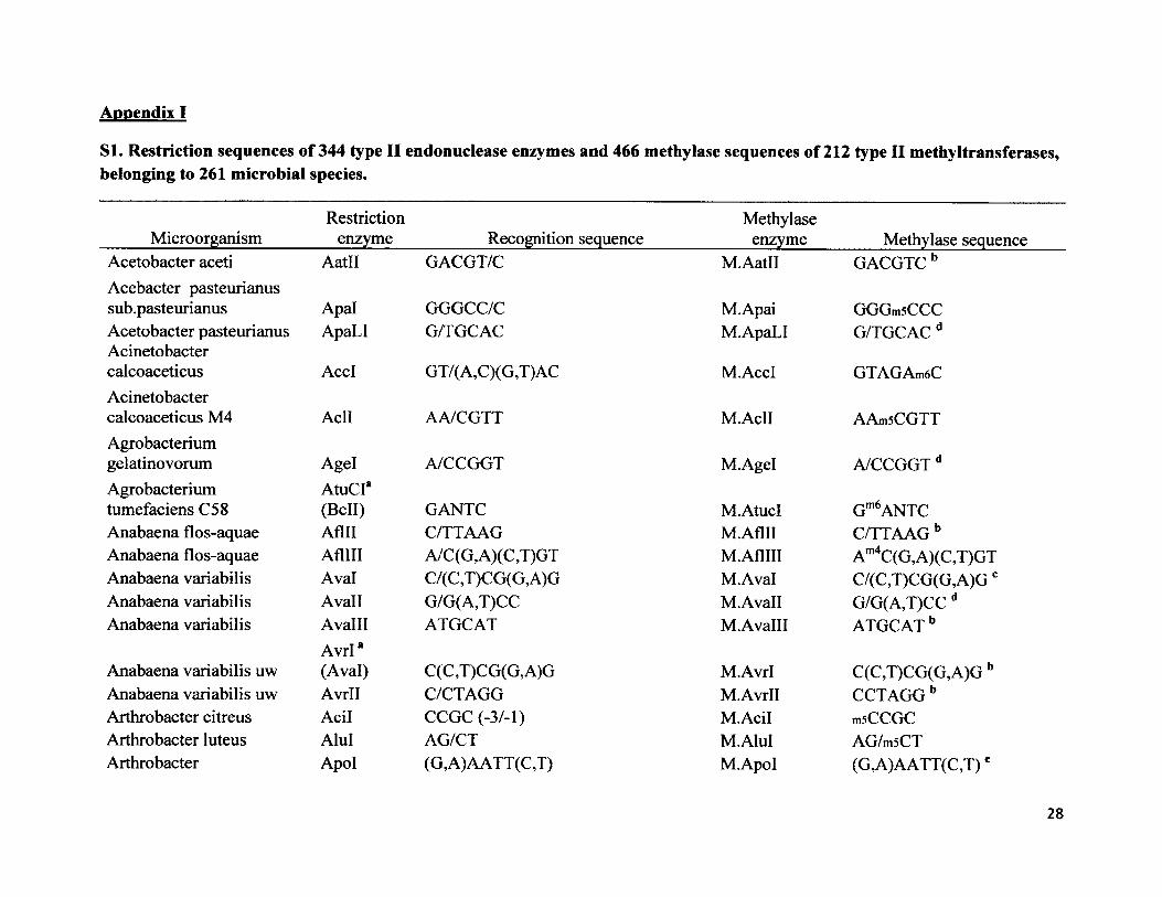
Appendix I
SI. Restriction sequences of 344 type II endonuclease enzymes and 466 methylase sequences of 212 type II methyltransferases, belonging to 261 microbial species.
Restriction Methylase Microorganism________ enzyme___________ Recognition sequence____________ enzyme_________Methylase sequenceAcetobacter aceti AatO GACGT/C M.Aatll GACGTCbAcebacter pasteurianussub.pasteurianus Apal GGGCC/C M.Apai GGGmsCCCAcetobacter pasteurianus ApaLI G/TGCAC M.ApaLI G/TGCAC dAcinetobactercalcoaceticus AccI GT/( A,C )(G,T)AC M.AccI GTAGAm6CAcinetobactercalcoaceticus M4 Acll AA/CGTT M.AclI AAmsCGTTAgrobacteriumgelatinovorum Agel A/CCGGT M.Agel A/CCGGT dAgrobacterium A tuCr
Gm6ANTCtumefaciens C58 (Bell) GANTC M.AtucIAnabaena flos-aquae Aflll C/TTAAG M.Aflll C/TTAAG bAnabaena flos-aquae AflHI A/C(G,A)(C,T)GT M.AflHI Am4C(G,A)(C,T)GTAnabaena variabilis Aval C/(C,T)CG(G,A)G M.Aval C/(C,T)CG(G,A)GcAnabaena variabilis Avail G/G(A,T)CC M.Avall G/G(A,T)CC dAnabaena variabilis Avalll
Avrl*ATGCAT M.AvalH ATGCAT b
Anabaena variabilis uw (Aval) C(C ,T)CG(G, A)G M.Avrl C(C,T)CG(G,A)G bAnabaena variabilis uw Avrll C/CTAGG M.Avrll CCTAGGbArthrobacter citreus Acil CCGC (-3/-1) M.Acil msCCGCArthrobacter luteus Alul AG/CT M.AluI AG/m5CTArthrobacter Apol (G,A)AATT(C,T) M.Apol (G,A)AATT(C,T)e
28
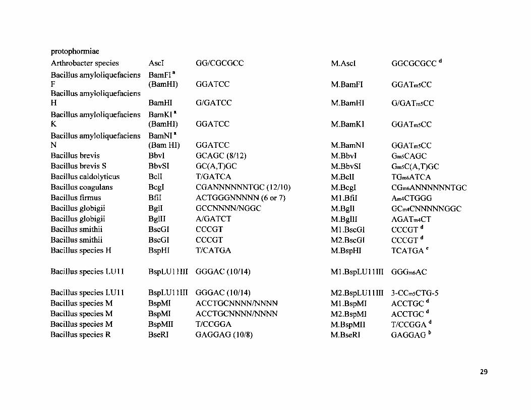
protophormiae Arthrobacter species AscI GG/CGCGCC M.AscI GGCGCGCCdBacillus amyloliquefaciens F
BamFI*(BamHI) GGATCC M.BamFI GGATmsCC
Bacillus amyloliquefaciens H BamHI G/GATCC M.BamHI G/GATmsCCBacillus amyloliquefaciens K
BamKI*(BamHI) GGATCC M.BamKI GGATmsCC
Bacillus amyloliquefaciens N
BamNI* (Bam HI) GGATCC M.BamNI GGATmsCC
Bacillus brevis Bbvl GCAGC (8/12) M.BbvI GmsCAGCBacillus brevis S BbvSI GC(A,T)GC M.BbvSI Gm5C(A,T)GCBacillus caldolyticus Bell T/GATCA M.BclI TGmeATCABacillus coagulans Bcgl CGANNNNNNTGC (12/10) M.Bcgl CGm6 ANNNNNNT GCBacillus firmus Bfil ACTGGGNNNNN (6 or 7) Ml.Bfil Am4CTGGGBacillus globigii Bgll GCCNNNN/NGGC M.Bgll GCm4CNNNNNGGCBacillus globigii Bglll A/GATCT M.Bglll AGATm4CTBacillus smithii BscGI CCCGT Ml.BscGI CCCGT dBacillus smithii BscGI CCCGT M2. BscGI CCCGT dBacillus species H BspHI T/CATGA M.BspHI TCATGAe
Bacillus species LU11 BspLU 11 III GGGAC (10/14) Ml.BspLU 11 III GGGm6AC
Bacillus species LU 11 BspLU 11 III GGGAC (10/14) M2.BspLUl 1III 3-CCm5CTG-5Bacillus species M BspMI ACCTGCNNNN/NNNN Ml.BspMI ACCTGCdBacillus species M BspMI ACCTGCNNNN/NNNN M2.BspMI ACCTGCdBacillus species M BspMII T/CCGGA M.BspMII T/CCGGA dBacillus species R BseRI GAGGAG (10/8) M.BseRI GAGGAG b
29
Bacillus sphaericus Bsp 12861 G (A,G,T)GC( A,C ,T)/CBacillus sphaericus GCsubgroup Bsgl
BspRI*GTGCAC (16/14)
Bacillus sphaericus R (Haelll) GG/CCBacillusstearothermophilus BsaAI (C,T) AC/GT (G, A)Bacillusstearothermophilus BsrBI CCG/CTCBacillusstearothermophilus BsrBI CCG/CTCBacillusstearothermophilus BsrI ACTGGN/Bacillusstearothermophilus BsrI ACTGGN/Bacillus BstNI*stearothermophilus (EcoRII) CC/(A,T)GGBacillusstearothermophilus BstXI CCANNNNN/NTGGBacillusstearothermophilus 1503- BstI*4R (BamHI) G/GATCCBacillusstearothermophilus A664 BsmAI GTCT CN/NNNNBacillusstearothermophilus D70 BsrDI GCAATGNN/Bacillusstearothermophilus D70 BsrDI GCAATGNN/Bacillusstearothermophilus ET BstEII G/GTNACC
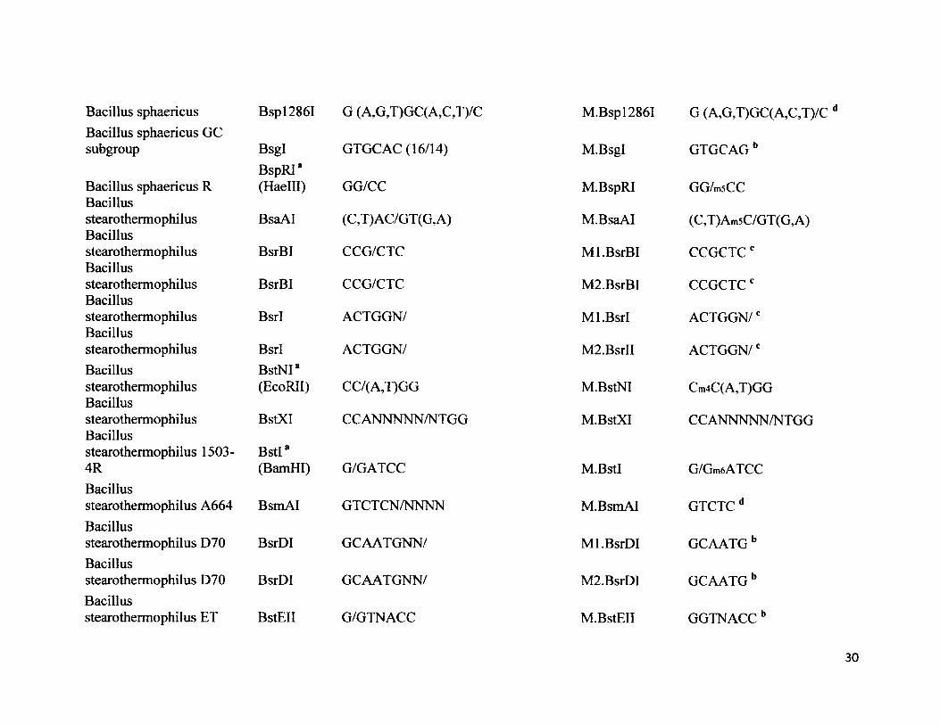
M.Bsp12861
M.Bsgl
M.BspRI
M.BsaAI
Ml.BsrBI
M2.BsrBI
Ml.Bsrl
M2.BsrII
M.BstNI
M.BstXI
M.BstI
M.BsmAI
Ml.BsrDI
M2.BsrDI
M.BstEII
G (A,G,T)GC(A,C,T)/C d
GTGCAGb
GG/m5CC
(C,T)Am5C/GT(G,A)
CCGCTCc
CCGCTCc
ACTGGN/c
ACTGGN/c
Cm4C(A,T)GG
CCANNNNN/NTGG
G/Gm6ATCC
GTCTCd
GCAATGb
GCAATGb
GGTNACCb
30
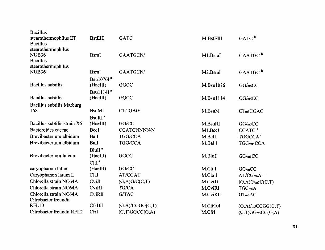
Bacillusstearothermophilus ET BacillusstearothermophilusNUB36BacillusstearothermophilusNUB36
Bacillus subtilis
Bacillus subtilisBacillus subtilis Marburg 168
Bacillus subtilis strain X5 Bacteroides caccae Brevibacterium albidum Brevibacterium albidum
Brevibacterium luteum
caryophanon latum Caryophanon latum L Chlorella strain NC64A Chlorella strain NC64A Chlorella strain NC64A Citrobacter freundii RFL10Citrobacter freundii REL2
BstEIII
BsmI
BsmIBsul076I8(Haelll)B s u ll l4 Ia(Haelll)
BsuMIBsuRI8(Haelll)BccIBallBallBluII8 (Haelll)Cltl8(Haelll)ClalCviJICviRICviRII
Ctrl 01 CM
GATC
GAATGCN/
GAATGCN/
GGCC
GGCC
CTCGAG
GG/CCCCATCNNNN/NTGG/CCATGG/CCA
GGCC
GG/CCAT/CGAT(G,A)G/C(C,T)TG/CAG/TAC
(G,A)/CCGG(C,T)(C,T)GGCC(G,A)
M.BstEIII GATC b
Ml.BsmI
M2.BsmI
M.Bsul076
M .Bsul114
M.BsuM
M.BsuRI Ml.BccI M.Ball M.Bal I
M.BluII
M.Clt I M.Cla 1 M.CviJI M.CviRI M.CviRII
M.CfrlOIM.CfrI
GAATGC b
GAATGC b
GG/msCC
GG/msCC
CTmsCGAG
GG/msCC CCATCb TGGCCAc TGG/m4CCA
GG/m5CC
GG/mCCAT/CGm6AT(G,A)G/m5C(C,T)TGCm6AGTmftAC
(G,A)/m5CCGG(C,T)(C,T)GGmsCC(G,A)
31
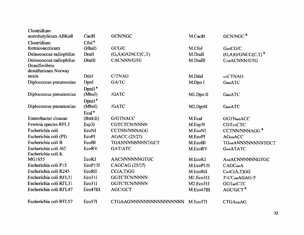
Clostridium acetobutylicum ABKn8Clostridium formicoaceticum Deinococcus radiophilus Deinococcus radiophilus Desulfovibrio desulfuricans Norway strainDiplococcus pneumoniae
Diplococcus pneumoniae
Diplococcus pneumoniae
Enterobacter cloacae Erwinia species RFL3 Escherichia coli Escherichia coli (PI) Escherichia coli B Escherichia coli J62 Escherichia coli K MG1655Escherichia coli PI 5 Escherichia coli R245 Escherichia coli RFL31 Escherichia coli RFL31 Escherichia coli RFL47
Escherichia coli RFL57
Cac8I GCN/NGCCfol8(Hhal) GCG/CDrall (G,A)G/GNCC(C,T)Dralll CACNNN/GTG
M.Cac8I GCN/NGCb
M.CfoI GmsCG/CM.Drall (G,A)G/GNCC(C,T)bM.Dralll Cm6ACNNN/GTG
DdelDpnlDpnII8 (Mbol)DpnII8(Mbol)E cal8(BstEII)Esp3IEcoNIEcoPIEcoBIEcoRV
C/TNAGGA/TC
/GATC
/GATC
G/GTNACCCGTCTCN/NNNNCCTNN/NNNAGGAGACC (25/27)TGANNNNNNNNTGCTGAT/ATC
M.Ddel msCTNAGM.Dpn I GmeATC
M l.DpnII GmeATC
M2.DpnII GmeATC
M.Ecal GGTNmeACCM.Esp3I CGTmsCTCM.EcoNI CCTNN/NNNAGGbM.EcoPI AGmeACCM.EcoBI TGmeANNNNNNNNTGCTM.EcoRV GmeATATC
EcoKI AACNNNNNN GTGC M.EcoKI AmeACNNNNNNGTGCEcoPI 51 CAGCAG (25/27) M.EcoPI 51 CAGCmeAEcoRII CC(A,T)GG M.EcoRII Cm5C(A,T)GGEco31I GGTCTCN/NNNN Ml.Eco31I 3'-CCmeAGAG-5'Eco31I GGTCTCN/NNNN M2.Eco31I GGTmsCTCEco47III AGC/GCT M.Eco47III AGC/GCT b
Eco57I CTGAAGNNNNNNNNNNNNNNNN M.Eco57I CTGAmeAG
32
Escherichia coli RY13 EcoRI G/AATTC M.EcoRI G/Am6ATTCFlavobacterium aquatile Faul CCCGCNNNN/NN Ml.Faul CmsCCGCFlavobacteriumokeanokoites FokI GGATGNNNNNNNNN/NNNN M.FokI GGm6ATGFrankia species Eul lb Fsel GGCCGG/CC M.Fsel GGCCGG/CC dFusobacterium nucleatum 4H Fnu4HI GC/NGC M.Fnu4HI GCNGCdFusobacterium nucleatum 4H Fnu4HI GC/NGC M.Fnu4HI GC/NGC dFusobacterium nucleatum D FnuDII CG/CG M.FnuDII mSCGCGFusobacterium nucleatum D FnuDIII GCG/C M.FnuDIII GCGC bFusobacterium nucleatum D
FnuDI*(Haelll) GG/CC M.FnuDI GG/mSCC
Fusobacterium nucleatum D FnuDII CG/CG M.FnuDII mSCGCGFusobacterium nucleatum D
FnuDIIIa (Hhal) GCG/C M.FnuDIII Gm5CG/C
Gluconobacter suboxydans H-15T Gsul CTGGAG (16/14) M.GsuI CTGGAG bHaemophilus aegyptius Haell (G,A)GCGC(C,T) M.Haell (G,A)GCGC(C,T)Haemophilus aegyptius Haelll GG/CC M.Haelll GGmSCC
Haemophilus aphrophilusHapIIa(Hpall) C/CGG M.HapII CmSCGG
Haemophilus gallinarum Hgal GACGC (5/10) Ml.Hgal 3'-CTGm5CG-5’Haemophilus gallinarum Hgal GACGC (5/10) M2.Hgal GAmsCGCHaemophilushaemoglobinophilus
Hhgl"(Haelll) GGCC M.Hhgl GG/msCC
33
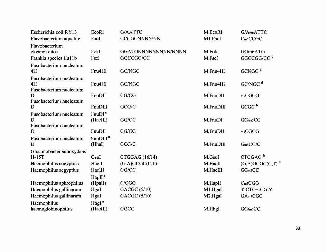
Hhall *Haemophilus haemolyticus (Hinfl) GANTCHaemophilus haemolyticus Hhal GCG/CHaemophilus influenza HincII8serotype c, 1160 (Hindll) GT (C ,T)(G, A)ACHaemophilus influenzaeRd Hindll GT (C,T)(G, A)ACHaemophilus influenzaeRd Hindlll A/AGCTTHaemophilus influenzaeRf Hinfl G/ANTCHaemophilus influenzaeRf Hinflll CGAATHaemophilusparahaemolyticus HphI GGTGA (8/7)Haemophilusparahaemolyticus HphI GGTGA (8/7)Haemophilusparainfluenzae Hpal GTT/AACHaemophilusparainfluenzae Hpall C/CGGHerpetosiphon giganteusHP 1023 HgiAI G(A,T)GC(A,T)CHerpetosiphon giganteusHpa2 HgiDI G(G,A)CG(C,T)CHerpetosiphon giganteus HgiEI *Hpg 24 (Avail) G/G(A,T)CCHerpetosiphon giganteus HgiBI *Hpg 5 (Avail) G/G(A,T)CCHerpetosiphon giganteus HgiCII*Hpg 9 (Avail) G/G(A,T)CC
M.Hhall G/m6ANTCM.Hhal GmsCGC
M.HincII GT(C ,T)(G, A)m6 AC
M. Hindll GT(C,T)(G,A)m6AC
M.HindlH m6 AAGC TT
M.HinfT GmeANTC
M.Hinflll CGAm6AT
Ml.HphI 3'-CCAm5CT-5'
M2.HphI GGTGm6A
M.Hpal GTTAmeAC
M.Hpall CmsCGG
M.HgiAI G(A,T)GC(A,T)C b
M.HgiDI G(G,A)CG(C,T)C d
M.HgiEI G/G(A,T)CC d
M.HgiBI G/G(A,T)CC d
M.HgiCII G/G(A,T)CC d
34
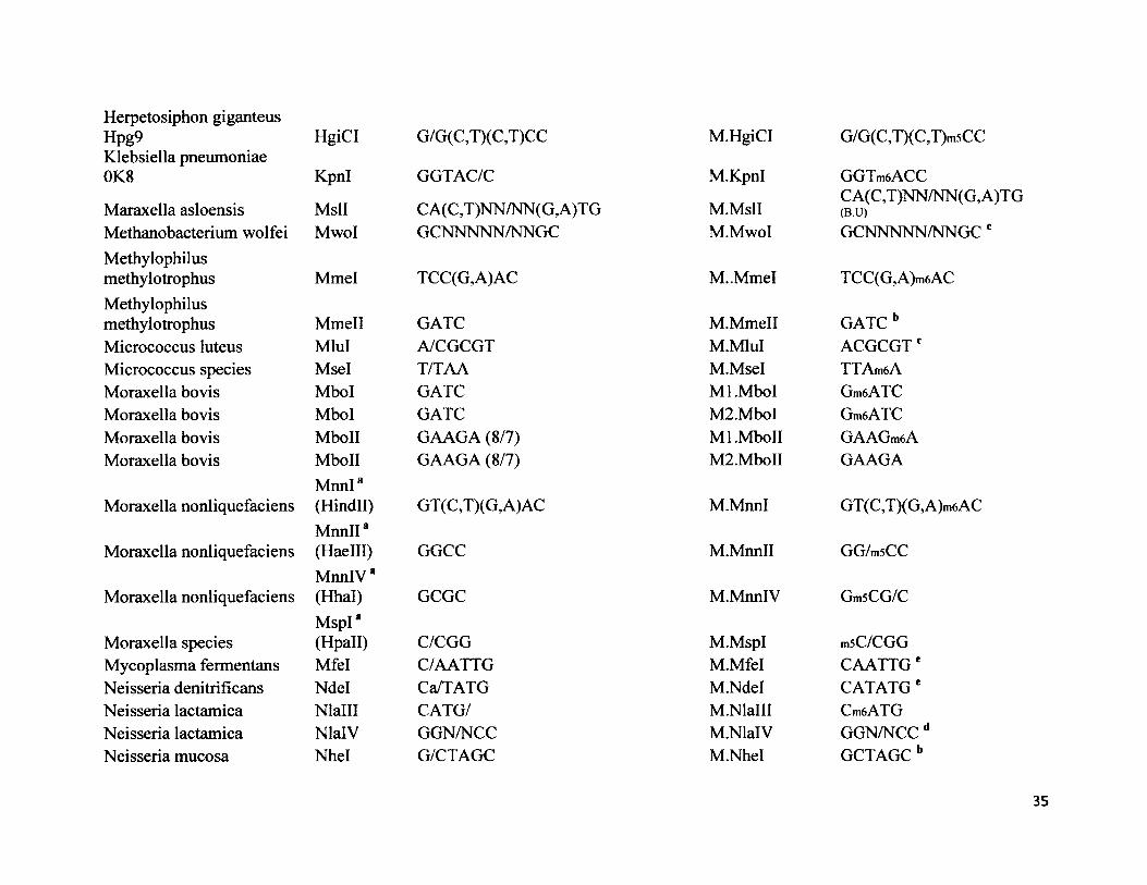
H erpetosiphon g iganteusHpg9 HgiCI G/G(C,T)(C,T)CC M.HgiCI G/G(C,T)(C,T)m5CCKlebsiella pneumoniae 0K8 Kpnl GGTAC/C M.Kpnl GGTmeACC
Maraxella asloensis MslI CA(C,T)NN/NN(G,A)TG M.MslICA(C,T)NN/NN(G,A)TG(B.U)
Methanobacterium wolfei Mwol GCNNNNN/NNGC M.MwoI GCNNNNN/NNGC cMethylophilusmethylotrophus Mmel TCC(G,A)AC M..MmeI TCC(G,A)m6ACMethylophilusmethylotrophus Mmell GATC M.Mmell GATC bMicrococcus luteus Mlul A/CGCGT M.MluI ACGCGTcMicrococcus species Msel T/TAA M.Msel TTAm6AMoraxella bovis Mbol GATC Ml.Mbol Gm6ATCMoraxella bovis Mbol GATC M2.Mbol Gm6ATCMoraxella bovis MboII GAAGA (8/7) Ml.MboII GAAGm6AMoraxella bovis MboII GAAGA (8/7) M2.MboII GAAGA
Moraxella nonliquefaciensMnnI*(Hindll) GT (C,T)(G, A)AC M.MnnI GT(C,T)(G,A)m6AC
Moraxella nonliquefaciensM nnll8(Haelll) GGCC M.Mnnll GG/m5CC
Moraxella nonliquefaciensMnnlV*(Hhal) GCGC M.MnnlV Gm5CG/C
Moraxella speciesMspI*(Hpall) C/CGG M.MspI msC/CGG
Mycoplasma fermentans Mfel C/AATTG M.Mfel CAATTG eNeisseria denitrificans Ndel Ca/TATG M.Ndel CATATGeNeisseria lactamica Nlalll CATG/ M.Nlalll Cm6ATGNeisseria lactamica NlalV GGN/NCC M.NlalV GGN/NCC dNeisseria mucosa Nhel G/CTAGC M.Nhel GCTAGCb
35
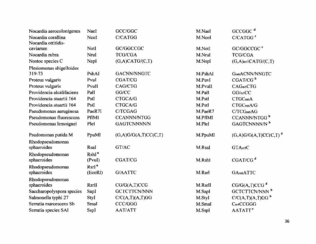
Nocardia aerocolonigenes Nael GCC/GGCNocardia corallina Ncol C/CATGGNocardia ottitidis-caviarum Notl GC/GGCCGCNocardia rubra Nrul TCG/CGANostoc species C NspI (G,A)CATG/(C,T)Plesiomonas shigelloides319-73 PshAI G ACNN/NN GT CProteus vulgaris Pvul CGAT/CGProteus vulgaris PvuII CAG/CTGProvidencia alcalifaciens Pall GG/CCProvidencia stuartii 164 PstI CTGCA/GProvidencia stuartii 164 PstI CTGCA/GPseudomonas aeruginosa PaeR7I C/TCGAGPseudomonas fluorescens PflMI CCANNN/NTGGPseudomonas lemoignei Plel GAGTCNNNN/N
Psudomonas putida M PpuMI (G,A)G/G(A,T)CC(C,T)Rhodopseudomonassphaeroides Rsal GT/ACRhodopseudomonas RshI*sphaeroides (Pvul) CGAT/CGRhodopseudomonas RsrI*sphaeroides (EcoRI) G/AATTCRhodopseudomonassphaeroides RsrII CG/G(A,T)CCGSaccharopolyspora species SapI GCTCTTCN/NNNSalmonella typhi 27 Styl C/C (A,T)( A,T)GGSerratia marcescens Sb Smal CCC/GGGSerratia species SAI SspI AAT/ATT
M.Nael GCCGGCdM.NcoI C/CATGGc
M.Notl GC/GGCCGC cM.NruI TCG/CGAM.NspI (G,A)ms\CATG/(C,T)
M.PshAI Gm6 ACNN/NN GT CM.PuvI CGAT/CG bM.PvuII CAGmsCTGM.Pall GG/m5CCM.PstI CTGCmsAM.PstI CTGCm6A/GM.PaeR7 C/TCGm6AGM.PflMI CCANNN/NTGG bM.Plel GAGTCNNNN/N b
M.PpuMI (G,A)G/G(A,T)CC(C,T)
M.Rsal GTAmsC
M.RshI CGAT/CG d
M.RsrI GAmeATTC
M.RsrII CG/G(A,T)CCG dM.SapI GCTCTTCN/NNN bM.Styl C/C(A,T)(A,T)GG bM.Smal Cm4CCGGGM.SspI AATATTe
36
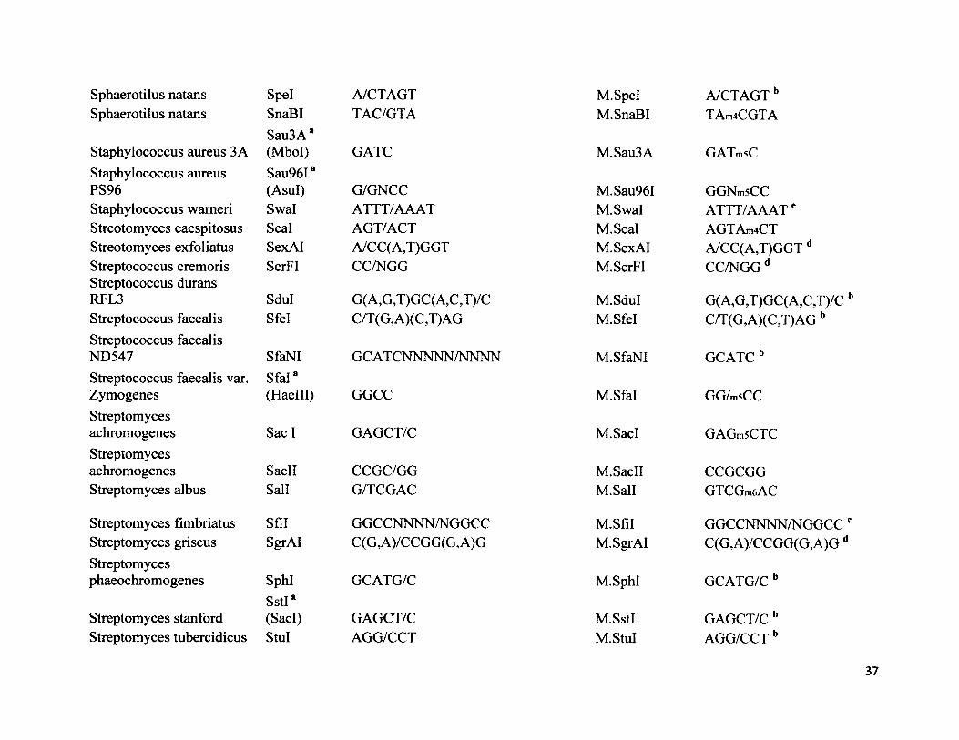
Sphaerotilus natans Spel A/CTAGTSphaerotilus natans SnaBI TAC/GTA
Staphylococcus aureus 3 ASau3A a (Mbol) GATC
Staphylococcus aureus Sau96IaPS96 (Asul) G/GNCCStaphylococcus wameri Swal ATTT/AAATStreotomyces caespitosus Seal AGT/ACTStreotomyces exfoliatus SexAI A/CC(A,T)GGTStreptococcus cremoris ScrFI CC/NGGStreptococcus durans RFL3 Sdul G(A,G,T)GC(A,C,T)/CStreptococcus faecalis Sfel C/T(G,A)(C,T)AGStreptococcus faecalis ND547 SfaNI GC AT CNNNNN/NNNNStreptococcus faecalis var. Sfal8Zymogenes (Haelll) GGCCStreptomycesachromogenes Sac I GAGCT/CStreptomycesachromogenes SacII CCGC/GGStreptomyces albus Sail G/TCGAC
Streptomyces fimbriatus Sfil GGCCNNNN/NGGCCStreptomyces griseus SgrAI C(G,A)/CCGG(G,A)GStreptomycesphaeochromogenes SphI GCATG/C
Streptomyces StanfordSstI*(SacI) GAGCT/C
Streptomyces tubercidicus StuI AGG/CCT
M.Spel A/CTAGT bM. SnaBI TAm4CGTA
M.Sau3A GATmsC
M.Sau96I GGNmsCCM.Swal ATTT/AAATeM.Scal AGTAm4CTM. SexAI A/CC(A,T)GGT dM. ScrFI CC/NGG d
M.Sdul G(A,G,T)GC(A,C,T)/C 1M.Sfel C/T(G,A)(C,T)AG b
M. SfaNI GCATCb
M.Sfal GG/m5CC
M.SacI GAGmsCTC
M.SacII CCGCGGM.Sall GTCGm6AC
M.Sfil GGCCNNNN/NGGCC'M. SgrAI C(G,A)/CCGG(G,A)G d
M.SphI GCATG/C b
M.SstI GAGCT/C bM.StuI AGG/CCT b
37
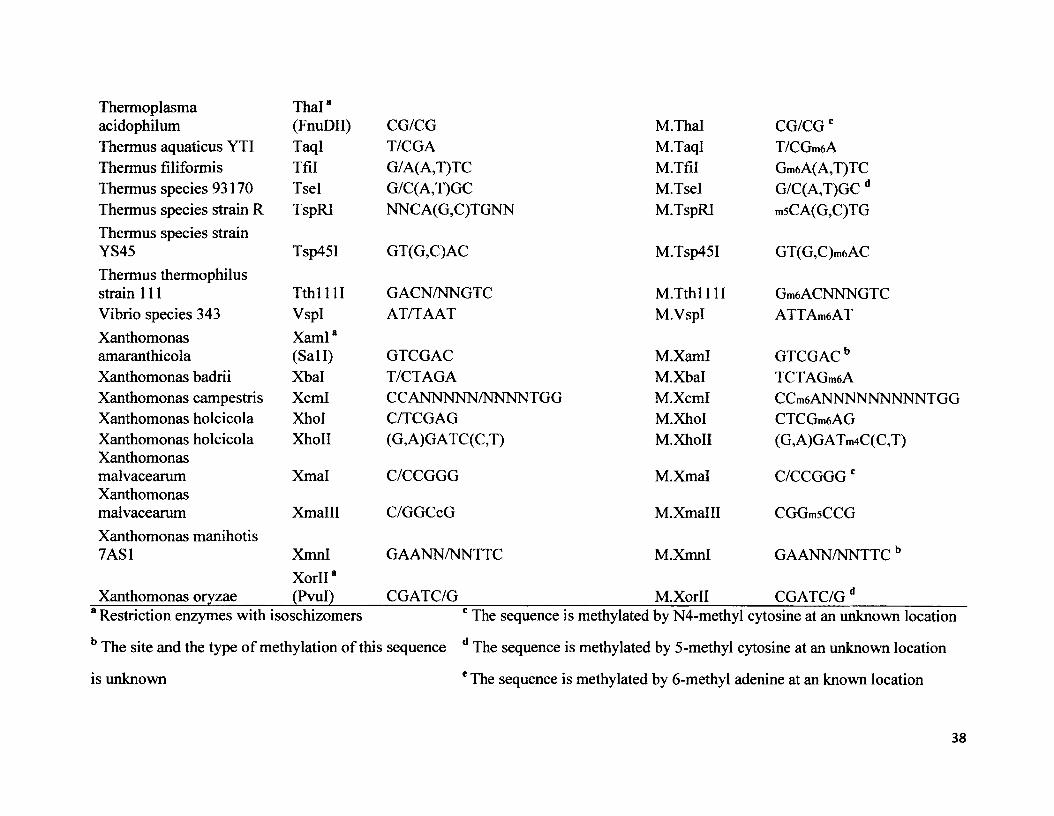
Thermoplasma Thai8acidophilum (FnuDII) CG/CG M.Thal CG/CGcThermus aquaticus YTI TaqI T/CGA M.TaqI T/CGm6AThermus filiformis Tfil G/A(A,T)TC M.Tfil Gm6A(A,T)TCThermus species 93170 Tsel G/C(A,T)GC M.Tsel G/C(A,T)GC dThermus species strain R TspRI NNCA(G,C)TGNN M.TspRI m5CA(G,C)TGThermus species strainYS45 Tsp45I GT(G,C)AC M.Tsp45I GT(G,C)m6ACThermus thermophilusstrain 111 T th ll l l GACN/NNGTC M.Tthl i l l Gm6ACNNNGTCVibrio species 343 VspI AT/TAAT M.VspI ATTAmaATXanthomonas XamI8amaranthicola (Sail) GTCGAC M.XamI GTCGAC bXanthomonas badrii Xbal T/CTAGA M.Xbal TCTAGmeAXanthomonas campestris Xcml CCANNNNN/NNNNTGG M.Xcml CCmeANNNNNNNNNTGGXanthomonas holcicola Xhol C/TCGAG M.XhoI CTCGmeAGXanthomonas holcicola XhoII (G,A)GATC(C,T) M.XhoII (G,A)GATm4C(C,T)Xanthomonasmalvacearum Xmal C/CCGGG M.Xmal C/CCGGG cXanthomonasmalvacearum Xmalll C/GGCcG M.Xmalll CGGmsCCGXanthomonas manihotis7AS1 XmnI
X orll8GAANN/NNTTC M.XmnI GAANN/NNTTC b
Xanthomonas oryzae (Pvul) CGATC/G M.Xorll CGATC/G d8 Restriction enzymes with isoschizomers c The sequence is methylated by N4-methyl cytosine at an unknown location
b The site and the type of methylation of this sequence d The sequence is methylated by 5-methyl cytosine at an unknown location
is unknown e The sequence is methylated by 6-methyl adenine at an known location
38
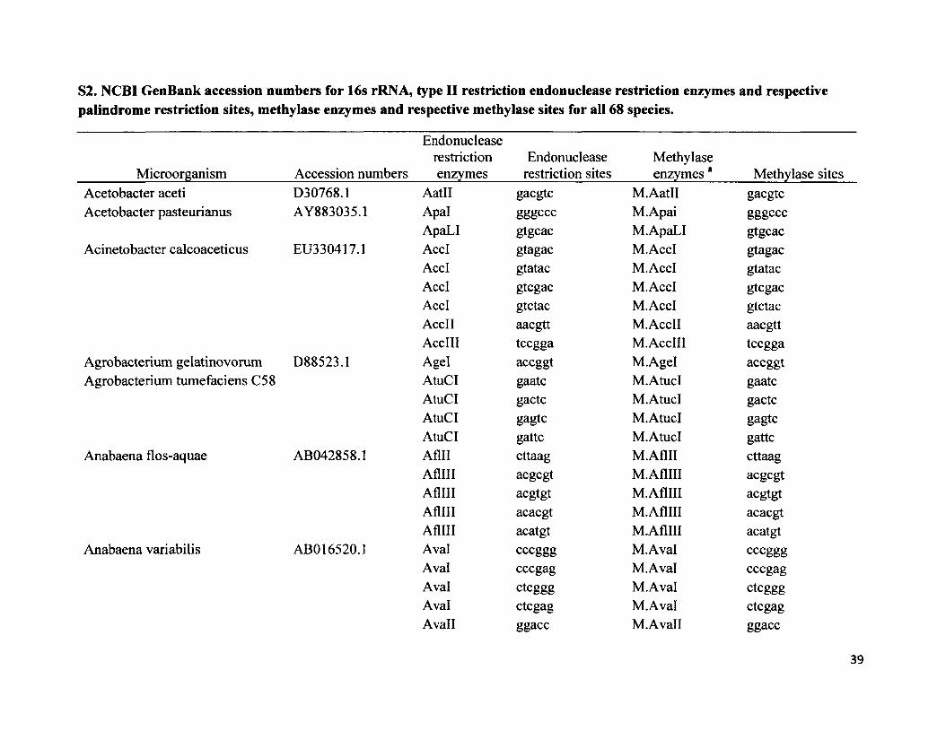
S2. NCBI GenBank accession numbers for 16s rRNA, type II restriction endonuclease restriction enzymes and respective palindrome restriction sites, methylase enzymes and respective methylase sites for all 68 species.
Endonuclease restriction Endonuclease Methylase
Microorganism_________ Accession numbers enzymes_____restriction sites______enzymes *______Methylase sitesAcetobacter aceti D30768.1 AatH gacgtc M.AatH gacgtcAcetobacter pasteurianus AY883035.1 Apal gggC CC M.Apai gggccc
ApaLI gtgcac M. ApaLI gtgcacAcinetobacter calcoaceticus EU330417.1 AccI gtagac M.AccI gtagac
AccI gtatac M.AccI gtatacAccI gtcgac M.AccI gtcgacAccI gtctac M.AccI gtctacAccII aacgtt M.AccII aacgttAccIII tccgga M.AccIII tccgga
Agrobacterium gelatinovorum D88523.1 Agel accggt M.Agel accggtAgrobacterium tumefaciens C58 AtuCI gaatc M.AtucI gaatc
AtuCI gactc M. AtucI gactcAtuCI gagtc M.AtucI gagtcAtuCI gattc M.AtucI gattc
Anabaena flos-aquae AB042858.1 Aflll cttaag M.Aflll cttaagAfllll acgcgt M.AflHI acgcgtAflHI acgtgt M.AflHI acgtgtAfllll acacgt M.AflHI acacgtAfllll acatgt M.AflIII acatgt
Anabaena variabilis ABO 16520.1 Aval cccggg M.Aval cccgggAval cccgag M.Aval cccgagAval ctcggg M.Aval ctcgggAval ctcgag M.Aval ctcgagAvail ggacc M.Avall ggacc
39

Arthrobacter ON 14 AJ810894.1
AvailAvalllAvalVPAval VPAvalVPAvalVPAvalXAvalXAvalXAvalXAscI
Arthrobacter protophormiae AY577525.1 Apol
Bacillus amyloliquefaciens HM 136792.1
ApolApolApolBamFI
Bacillus brevis X60612.1 Bbvl
Bacillus caldolyticus Z26924.1BbvIIBell
Bacillus coagulans D16267.1 BcglBacillus firmus DQ118015.1 BfilBacillus smithii Z26935.1 BscGIBacillus subtilis AB598736.2 BsuRI
Bacteroides caccae ATCC43185T X83951.1
BsuMI Bsul076I B sul1141
BccICaryophanon latum L X70314.1 Clal
ggtcc M.Avall ggtccatgcat M.Avalll atgcatgctaagc M.AvalVP gctaagcgctcagc M.AvalVP gctcagcgctgagc M.AvalVP gctgagcgcttagc M.AvalVP gcttagcaccggc M.AvalX accggcgccggc M.AvalX gccggcaccggt M.AvalX accggtgccggt M.AvalX gccggtggcgcgcc M.AscI ggcgcgccgaattc M.Apol gaattcgaattt M.Apol gaatttaaattc M.Apol aaattcaaattt M.Apol aaatttggatcc M.BamFI ggatccgcagc M.BbvI gcagcgaagac M.BbvII gaagactgatca M.BclI tgatcacgannnnnntgc M.Bcgl cgannnnnntgcactggg Ml.Bfil actgggcccgt Ml.BscGI cccgtggcc M.BsuRI ggccctcgag M.BsuM ctcgagggcc M.Bsul076 ggccggcc M .B sulll4 ggcc
ccatc Ml.BccI ccatcatcgat M.Clal atcgat
40

Citrobacter freundii HQ399663.1
Clostridium formicoaceticum NR 029267.1Deinococcus radiophilus NR_026402.1
Desulfovibrio desulfuricansNorway strain HQ591418.1
Enterobacter cloacae HM585374.1
Erwinia toletana FR870447.1Flavobacterium aquatile AB517711.1Flavobacterium okeanokoites D55729.1Fusobacterium nucleatum AF543300.1
CfrICfrICMCMCfrI 01CfrlOICfflOICfrlOICfolDrallDrallDrallDrallDralll
DdelDdelDdelDdelEcalEcalEcalEcalEsp3IFaulFokIFnu4HIFnu4HIFnu4HI
cggccgcggccatggccgtggccagccggcgccggtaccggcaccggtgcgcgggncccgggncctaggncccaggncctcacnnngtg
ctaagctcagctgagcttagggtaaccggtcaccggtgaccggttacccgtctccccgcggatggcagcgccgcgcggc
M.CfrIM.CMM.CfrIM.CMM.CfrI 01M.CfrI 01M.CfflOIM.Cffl 01M.CfoIM.DrallM.DrallM.DrallM.DrallM.DraHI
M.DdelM.DdelM.DdelM.DdelM.EcalM.EcalM.EcalM.EcalM.Esp3IMl.FaulM.FokIM.Fnu4HIM.Fnu4HIM.Fnu4HI
cggccgcggccatggccgtggccagccggcgccggtaccggcaccggtgcgcgggncccgggncctaggncccaggncctcacnnngtg
ctaagctcagctgagcttagggtaaccggtcaccggtgaccggttacccgtctccccgcggatggcagcgccgcgcggc
41

Gluconobacter oxydans AB308275.1Haemophilus aegyptius M75044.1
Haemophilus aphrophilus M75041Haemophilushaemoglobinophilus M75064.1Haemophilus haemolyticus M75045
Haemophilus influenzae AY360336.1
Haemophilus parahaemolyticus M75073.1Haemophilus parainfluenzae M75081.1
Methanothermobacter wolfei AB104858.1Methylophilus methylotrophus AB 193724.1
Micrococcus luteus EF 154262.1
Fnu4HIFnuDIIFnuDIIIGsulHaellHaellHaellHaellHaelllHapII
HhglHhalHhallHhallHhal IHhallHincIIHincIIHincIIHincIIHphIHpalHpallMwolMmelMmelMmellMlul
gctgc M.Fnu4HI gctgccgcg M.FnuDII cgcggcgc M.FnuDIII gcgcctggag M.GsuI ctggagggcgcc M.Haell ggcgccggcgct M.Haell ggcgctagcgcc M.Haell agcgccagcgct M.Haell agcgctggcc M.Haelll ggccccgg M.HapII ccgg
ggcc M.Hhgl ggccgcgc M.Hhal gcgcgaatc M. Hhall gaatcgactc M.Hhall gactcgagtc M.Hhall gagtcgattc M.Hhall gattcgtcgac M.HincII gtcgacgtcaac M.HincII gtcaacgttgac M.HincII gttgacgttaac M.HincII gttaacggtga Ml.HphI ccactgttaac M.Hpal gttaacccgg M.Hpall ccgggcnnnnnnngc M.MwoI gcnnnnnnngctccgac M..MmeI tccgactccaac M..MmeI tccaacgate M.Mmell gateaegegt M.MluI aegegt
42
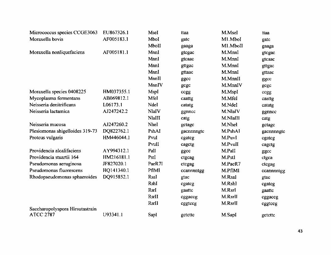
Micrococcus species CCGE3063 Moraxella bovis
Moraxella nonliquefaciens
Moraxella species 0408225 Mycoplasma fermentans Neisseria denitrificans Neisseria lactamica
Neisseria mucosa Plesiomonas shigelloides 319-73 Proteus vulgaris
Providencia alcalifaciens Providencia stuartii 164 Pseudomonas aeruginosa Pseudomonas fluorescens Rhodopseudomonas sphaeroides
Saccharopolyspora Hirsutastrain ATCC 2787
EU867326.1 MselAF005183.1 Mbol
MboIIAF005181.1 MnnI
MnnIMnnIMnnIMnnllMnnlV
HM037355.1 MspIAB069812.1 MfelL06173.1 NdelAJ247242.2 NlalV
NlalllAJ247260.2 NhelDQ822762.1 PshAIHM446044.1 Pvul
PvuIIAY994312.1 PallHM216181.1 PstIJF827020.1 PaeR7IHQ141340.1 PflMIDQ915852.1 Rsal
RshI RsrI RsrII RsrI I
U93341.1 SapI
ttaa M.Msel ttaagate Ml.Mbol gategaaga Ml.MboII gaagagtcgac M.MnnI gtcgacgtcaac M.MnnI gtcaacgttgac M.MnnI gttgacgttaac M.MnnI gttaacggcc M.Mnnll ggccgcgc M.MnnlV gcgcccgg M.MspI ccggcaattg M.Mfel caattgcatatg M.Ndel catatgggnncc M.NlalV ggnncccatg M.Nlalll catggctagc M.Nhel gctagcgacnnnngtc M.PshAI gacnnnngtccgatcg M.PuvI cgatcgcagctg M.PvuII cagctgggcc M.Pall ggccctgcag M.PstI ctgcactcgag M.PaeR7 ctegagccannnntgg M.PflMI ccannnntgggtac M.Rsal gtaccgatcg M.RshI cgatcggaattc M.RsrI gaattccggaccg M.RsrII cggaccgcggtccg M.RsrII cggtccg
gctcttc M.SapI gctcttc
43
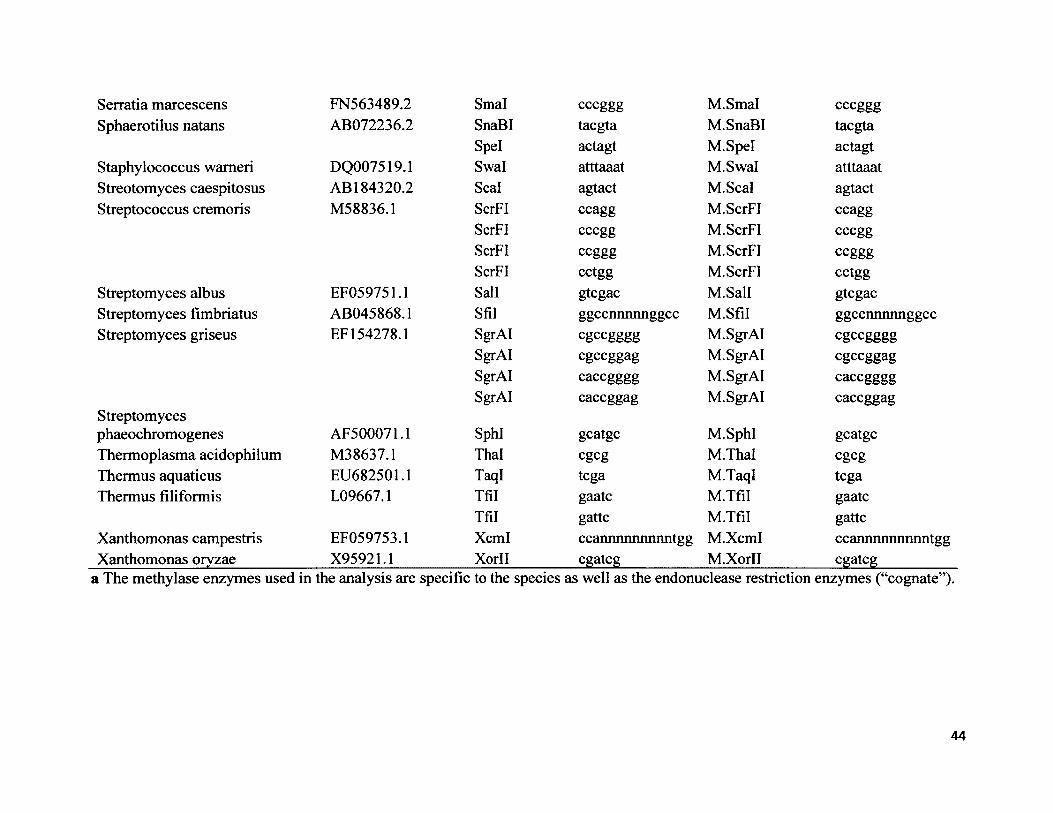
Serratia marcescens FN563489.2 Smal cccggg M.Smal cccgggSphaerotilus natans AB072236.2 SnaBI taegta M.SnaBI taegta
Spel actagt M.Spel actagtStaphylococcus wameri DQ007519.1 Swal atttaaat M.Swal atttaaatStreotomyces caespitosus AB 184320.2 Seal agtact M.Scal agtactStreptococcus cremoris M58836.1 ScrFI ccagg M.ScrFI ccagg
ScrFI cccgg M. ScrFI cccggScrFI ccggg M. ScrFI ccgggScrFI cctgg M. ScrFI cctgg
Streptomyces albus EF059751.1 Sail gtcgac M.Sall gtcgacStreptomyces fimbriatus AB045868.1 Sfil ggccnnnnnggcc M.Sfil ggccnnnnnggccStreptomyces griseus EF 154278.1 SgrAI egeegggg M. SgrAI egeegggg
SgrAI egeeggag M. SgrAI egeeggagSgrAI caccgggg M.SgrAI caccggggSgrAI caccggag M.SgrAI caccggag
Streptomycesphaeochromogenes AF500071.1 SphI geatge M.SphI geatgeThermoplasma acidophilum M38637.1 Thai cgcg M.Thal cgcgThermus aquaticus EU682501.1 TaqI tcga M.TaqI tcgaThermus filiformis L09667.1 Tfil gaatc M.Tfil gaatc
Tfil gattc M.Tfil gattcXanthomonas campestris EF059753.1 Xcml ccannnnnnnnntgg M.Xcml ccannnnnnnnntggXanthomonas oryzae X95921.1 Xorll cgatcg M.Xorll cgatcg
a The methylase enzymes used in the analysis are specific to the species as well as the endonuclease restriction enzymes (“cognate”).
44

S3. Number of endonuclease restriction sequences, number of methylase sequences, and number of overlapping sequences of 96 type II restriction endonuclease enzymes and 96 corresponding methylase enzymes in 68 microbial species.
Microorganisms Endonucleaserestrictionenzymes
Endonuclease restriction sites
Number of endonucleas e restriction
sites
Methylaseenzymes
Methylase sites Number of
methylas e sites
Number of overlaps
Acetobacter aceti Aatll gacgtc 1 M.Aatll Gacgtc 1 1Acetobacterpasteurianus Apal gggC C C 1 M.Apai Gggccc 1 1
ApaLI gtgcac 0 M.ApaLI Gtgcac 0 0Acinetobactercalcoaceticus AccI gtagac 0 M.AccI Gtagac 0 0
AccI gtatac 0 M.AccI Gtatac 0 0AccI gtcgac 0 M.AccI Gtcgac 0 0AccI gtctac 0 M.AccI Gtctac 0 0AccII aacgtt 0 M.AccII Aacgtt 0 0AccIII tccgga 0 M.AccIII Tccgga 0 0
Agrobacteriumgelatinovorum Agel accggt 0 M.Agel Accggt 0 0Agrobacteriumtumefaciens C58 AtuCI gaatc 0 M.AtucI Gaatc 0 0
AtuCI gactc 0 M.AtucI Gactc 0 0AtuCI gagtc 0 M.AtucI Gagtc 0 0AtuCI gattc 0 M.AtucI Gattc 0 0
Anabaena flos-aquae Aflll cttaag 0 M.Aflll Cttaag 0 0AfHII aegegt 1 M.AfHII Aegegt 1 1Afllll aegtgt 0 M.AflHI Aegtgt 0 0AflHI acacgt 1 M.AfHII Acacgt 1 1Afllll acatgt 0 M.AflHI Acatgt 0 0
Anabaena variabilis Aval cccggg 1 M.Aval Cccggg 1 1Aval cccgag 1 M.Aval Cccgag 1 1Aval cteggg 0 M.Aval Cteggg 0 0
45
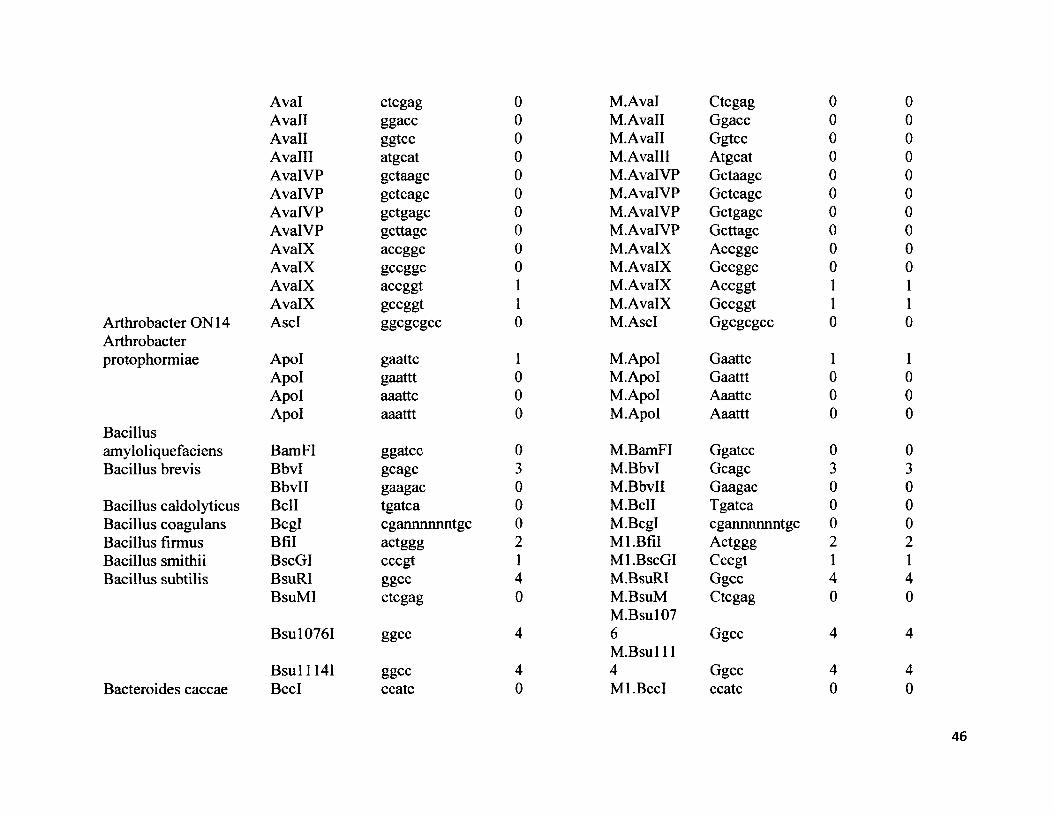
Aval ctcgagAvail ggaccAvail ggtccAvalll atgeatAvalVP gctaagcAvalVP gctcagcAvalVP gctgagcAvalVP gcttagcAvalX accggcAvalX gccggcAvalX accggtAvalX gccggt
Arthrobacter ON 14 AscI ggcgcgccArthrobacterprotophormiae Apol gaattc
Apol gaatttApol aaattcApol aaattt
Bacillusamyloliquefaciens BamFI ggatccBacillus brevis Bbvl gcagc
BbvII gaagacBacillus caldolyticus Bell tgatcaBacillus coagulans Bcgl egannnnnntgeBacillus firmus Bfil actgggBacillus smithii BscGI cccgtBacillus subtilis BsuRI ggcc
BsuMI ctcgag
Bsu 10761 ggcc
B su l1141 ggccBacteroides caccae BccI ccatc
0000000000110
1000
030002140
4
40
M.Aval Ctcgag 0 0M.Avall Ggacc 0 0M.Avall Ggtcc 0 0M.Avalll Atgeat 0 0M.Aval VP Gctaagc 0 0M.Aval VP Gctcagc 0 0M.AvalVP Gctgagc 0 0M.AvalVP Gcttagc 0 0M. AvalX Accggc 0 0M. AvalX Gccggc 0 0M. AvalX Accggt 1 1M.AvalX Gccggt 1 1M.AscI Ggcgcgcc 0 0
M.Apol Gaattc 1 1M.Apol Gaattt 0 0M.Apol Aaattc 0 0M.Apol Aaattt 0 0
M.BamFI Ggatcc 0 0M.BbvI Gcagc 3 3M.BbvII Gaagac 0 0M.BclI Tgatca 0 0M.Bcgl egannnnnntge 0 0Ml.Bfil Actggg 2 2Ml.BscGI Cccgt 1 1M.BsuRI Ggcc 4 4M.BsuMM.Bsul07
Ctcgag 0 0
6M.Bsul 11
Ggcc 4 4
4 Ggcc 4 4Ml.BccI ccatc 0 0

ATCC43185T Caryophanon latumL Clal atcgatCitrobacter freundii CfrI cggccg
CfrI cggccaCfrI tggccgCfrI tggccaCfrlOI gccggcCfrlOI gccggtCfrlOI accggcCfrlOI accggt
Clostridiumformicoaceticum Cfol gcgcDeinococcusradiophilus Drall gggnccc
Drall gggncctDrall aggncccDrall aggncctDralll cacnnngtg
DesulfovibriodesulfuricansNorway strain Ddel ctaag
Ddel ctcagDdel ctgagDdel cttag
Enterobacter cloacae Ecal ggtaaccEcal ggtcaccEcal ggtgaccEcal ggttacc
Erwinia toletana Esp3I cgtctcFlavobacteriumaquatile Faul cccgcFlavobacterium FokI ggatg
010000011
2
10010
012010000
12
M.Clal Atcgat 0 0M.CfrI Cggccg 1 1M.CfrI Cggcca 0 0M.CfrI Tggccg 0 0M.CfrI Tggcca 0 0M.CfrI 01 Gccggc 0 0M.CfrlOI Gccggt 0 0M.CfrI 01 Accggc 1 1M.CfrlOI Accggt 1 1
M.CfoI Gcgc 2 2
M.Drall Gggnccc 1 1M.Drall Gggncct 0 0M.Drall Aggnccc 0 0M.Drall Aggncct 1 1M.Dralll Cacnnngtg 0 0
M.Ddel Ctaag 0 0M.Ddel Ctcag 1 1M.Ddel Ctgag 2 2M.Ddel Cttag 0 0M.Ecal Ggtaacc 1 1M.Ecal Ggtcacc 0 0M.Ecal Ggtgacc 0 0M.Ecal Ggttacc 0 0M.Esp3I Cgtctc 0 0
Ml.Faul Cccgc 1 1M.FokI ggatg 2 2

okeanokoitesFusobacteriumnucleatum Fnu4HI gcagc
Fnu4HI gccgcFnu4HI gcggcFnu4HI gctgcFnuDII cgcgFnuDIII gcgc
Gluconobactersuboxydan H15T Gsul ctggagHaemophilusaegyptius Haell ggcgcc
Haell ggcgctHaell agcgccHaell agcgctHaelll ggcc
Haemophilusaphrophilus HapII ccggHaemophilushaemoglobinophilus Hhgl ggccHaemophilushaemolyticus Hhal gcgc
Hhall gaatcHhall gactcHhall gagtcHhall gattc
Haemophilusinfluenza HincII gtcgac
HincII gtcaacHincII gttgacHincII gttaac
Haemophilusparahaemolyticus HphI ggtga
M.Fnu4HI Gcagc 2 2M.Fnu4HI Gccgc 1 1M.Fnu4HI Gcggc 0 0M.Fnu4HI Gctgc 0 0M.FnuDII Cgcg 8 8M.FnuDIII Gcgc 4 4
M.GsuI Ctggag 1 1
M.Haell Ggcgcc 1 1M.Haell Ggcgct 0 0M.Haell Agcgcc 0 0M.Haell Agcgct 0 0M.Haelll Ggcc 6 6
M.HapII Ccgg 6 6
M.Hhgl Ggcc 6 6
M.Hhal Gcgc 2 2M.Hhall Gaatc 4 4M.Hhall Gactc 2 2M.Hhall Gagtc 1 1M.Hhall Gattc 2 2
M.HincII Gtcgac 0 0M.HincII Gtcaac 0 0M.HincII Gttgac 0 0M.HincII Gttaac 0 0
Ml.HphI Ccact 4 4

Haemophilusparainfluenzae
Methanothermobact er wolfei Methylophilus methylotrophus
Micrococcus luteus Micrococcus species CCGE3063 Moraxella bovis Moraxella bovis Moraxella nonliquefaciens
Moraxella species0408225MycoplasmafermentansNeisseriadenitrificansNeisseria lactamica
Neisseria mucosa Plesiomonas shigelloides 319-73 Proteus vulgaris
Hpal gttaac 1Hpall ccgg 6
Mwol gcnnnnnnngc 6
Mmel tccgacMmel tccaacMmell gateMlul aegegt
Msel ttaaMbol gateMboII gaaga
MnnI gtcgacMnnI gtcaacMnnI gttgacMnnI gttaacMnnll ggccMnnlV gcgc
MspI ccgg
Mfel caattg
Ndel catatgNlalV ggnnccNlalll catgNhel gctagc
PshAI gacnnnngtc 0Pvul cgatcg 0
© ©
VO ©
<0 r-~
<N O
O O
— t"
<N ■'}■
O ©
© <N
©
M.Hpal Gttaac 1 1M.Hpall Ccgg 6 6
M.MwoI gcnnnnnnngc 6 6
M..MmeI Tccgac 0 0M..MmeI Tccaac 0 0M.Mmell Gate 6 6M.MluI Aegegt 0 0
M.Msel Ttaa 5 5Ml.Mbol Gate 7 7Ml.MboII Gaga 2 2
M.MnnI Gtcgac 0 0M.MnnI Gtcaac 0 0M.MnnI Gttgac 0 0M.MnnI Gttaac 1 1M.Mnnll Ggcc 7 7M.MnnlV Gcgc 2 2
M.MspI Ccgg 4 4
M.Mfel Caattg 0 0
M.Ndel Catatg 0 0M.NlalV Ggnncc 0 0M.Nlalll Catg 2 2M.Nhel Gctagc 0 0
M.PshAI Gacnnnngtc 0 0M.PuvI Cgatcg 0 0
49
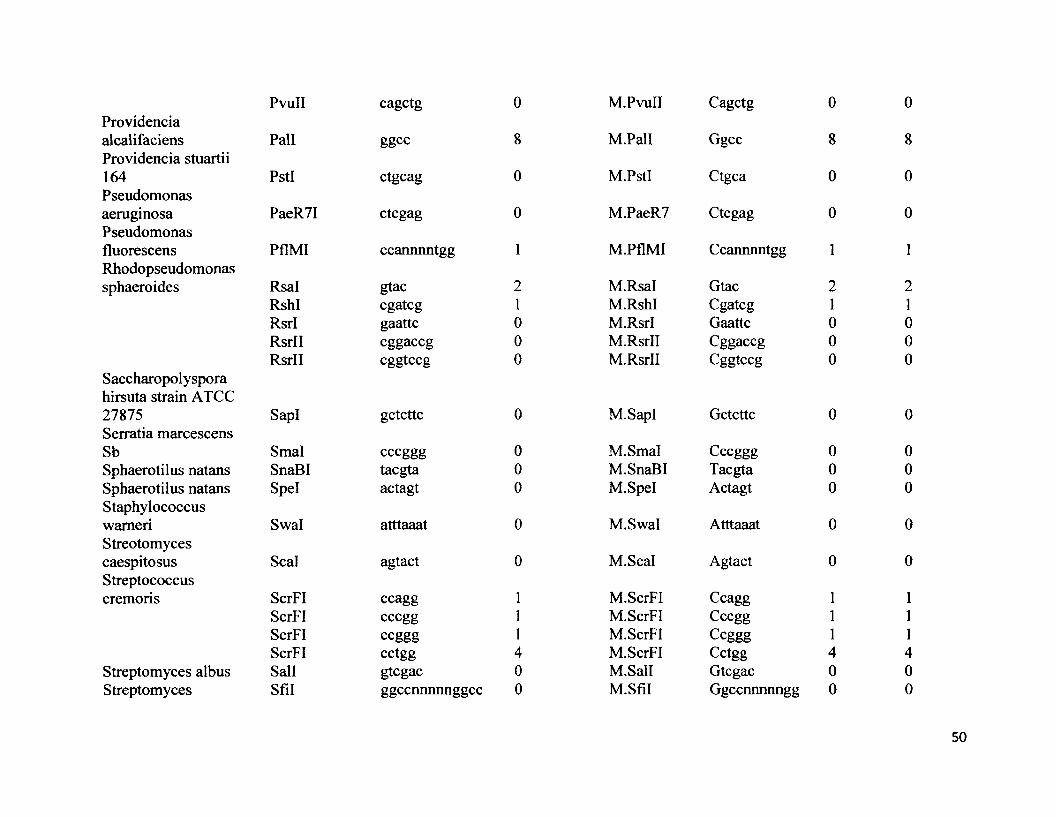
Providencia alcalifaciens Providencia stuartii 164PseudomonasaeruginosaPseudomonasfluorescensRhodopseudomonassphaeroides
Saccharopolyspora hirsuta strain ATCC 27875Serratia marcescens SbSphaerotilus natansSphaerotilus natansStaphylococcuswameriStreotomycescaespitosusStreptococcuscremoris
Streptomyces albus Streptomyces
PvuII cagctg 0
Pall ggcc 8
PstI ctgcag 0
PaeR7I ctcgag 0
PflMI ccannnntgg 1
Rsal gtac 2RshI cgatcg 1RsrI gaattc 0RsrII cggaccg 0RsrII cggtccg 0
SapI gctcttc 0
Smal cccggg 0SnaBI taegta 0Spel actagt 0
Swal atttaaat 0
Seal agtact 0
ScrFI ccagg 1ScrFI cccgg 1ScrFI ccggg 1ScrFI cctgg 4Sail gtcgac 0Sfil ggccnnnnnggcc 0
M.PvuII Cagctg 0
M.Pall Ggcc 8
M.PstI Ctgca 0
M.PaeR7 Ctcgag 0
M.PflMI Ccannnntgg 1
M.Rsal Gtac 2M.RshI Cgatcg 1M.RsrI Gaattc 0M.RsrII Cggaccg 0M.RsrII Cggtccg 0
M.SapI Gctcttc 0
M.Smal Cccggg 0M.SnaBI Taegta 0M.Spel Actagt 0
M.Swal Atttaaat 0
M.Scal Agtact 0
M.ScrFI Ccagg 1M.ScrFI Cccgg 1M.ScrFI Ccggg 1M.ScrFI Cctgg 4M.Sall Gtcgac 0M.Sfil Ggccnnnnngg 0
0
8
0
0
1
21000
0
000
0
0
111400
50
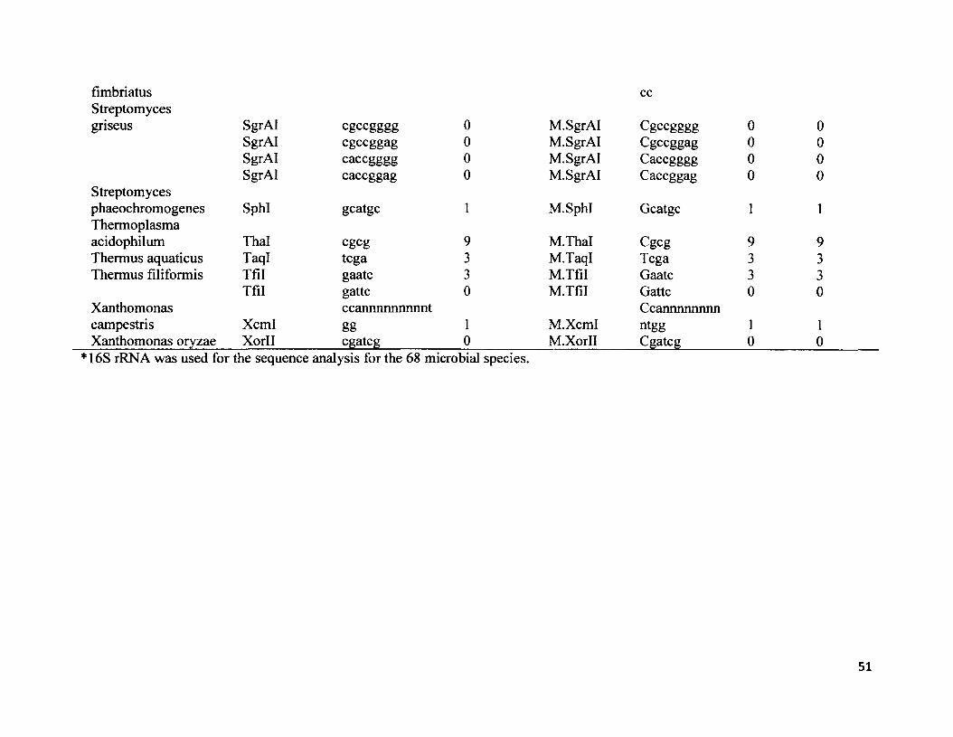
fimbriatus ccStreptomycesgriseus SgrAI egeegggg 0 M.SgrAI Cgccgggg 0 0
SgrAI egeeggag 0 M.SgrAI Cgccggag 0 0SgrAI caccgggg 0 M.SgrAI Caccgggg 0 0SgrAI caccggag 0 M.SgrAI Caccggag 0 0
Streptomycesphaeochromogenes SphI geatge 1 M.SphI Geatge 1 1Thermoplasmaacidophilum Thai cgcg 9 M.Thal Cgcg 9 9Thermus aquaticus TaqI tcga 3 M.TaqI Tcga 3 3Thermus filiformis Tfil gaatc 3 M.Tfil Gaatc 3 3
Tfil gattc 0 M.Tfil Gattc 0 0Xanthomonas ccannnnnnnnnt Ccannnnnnnncampestris Xcml gg 1 M.Xcml ntgg 1 1Xanthomonas oryzae Xorll cgatcg 0 M.Xorll Cgatcg 0 0
* 16S rRNA was used for the sequence analysis for the 68 microbial species.
51
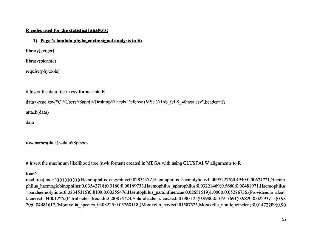
R codes used for the statistical analysis:
1) PagePs lambda phylogenetic signal analysis in R:
library(geiger)
library (picante)
require(phytools)
# Insert the data file in csv format into R
data<-read.csv("C://Users//Sanoji//Desktop//Thesis Defense (MSc.)//l 6S_GLS_40taxa.csv",header=T)
attach(data)
data
ro w.names(data)<-data$ Species
# Insert the maximum likelihood tree (nwk format) created in MEGA with using CLUSTALW alignments to R
tree<-read.tree(text-'(((((((((((((((Haemophilus_aegyptius:0.02834477,Haemophilus_haemolyticus:0.00952275)0.4940:0.00674721,Haemo philus_haemoglobinophilus:0.03542718)0.3160:0.00169733, Haemophilus_aphrophilus:0.03221469)0.5660:0.00481971, Haemophilus _parahaemolyticus:0.03345315)0.8100:0.00255476,Haemophilus_parainfluenzae:0.02651519) 1.0000:0.05286736,(Providenciaalcali faciens:0.04001225,(Citrobacter_ffeundii:0.00874124,Enterobacter_cloacae:0.01983125)0.9980:0.01917691)0.9820:0.02297715)0.98 20:0.04481612,(Moraxella_species_0408225:0.05260118,(Moraxella_bovis:0.01587325,Moraxella_nonliquefaciens:0.01472209)0.90
52
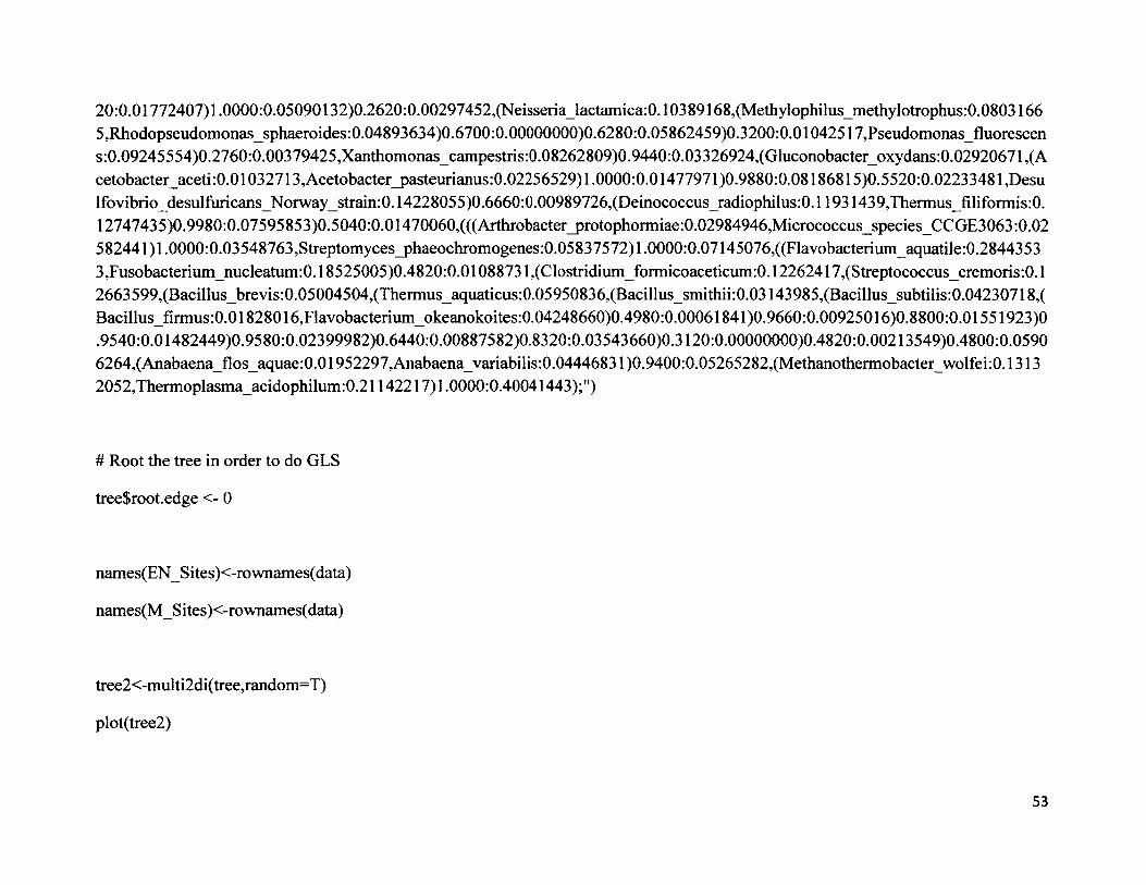
20:0.01772407)1.0000:0.05090132)0.2620:0.00297452, (Neisseria_lactamica:0.10389168, (Methylophilus_methylotrophus:0.0803166 5,Rhodopseudomonas_sphaeroides:0.04893634)0.6700:0.00000000)0.6280:0.05862459)0.3200:0.01042517,Pseudomonas fluorescen s:0.09245554)0.2760:0.00379425,Xanthomonas_campestris:0.08262809)0.9440:0.03326924, (Gluconobacter_oxydans:0.02920671, (A cetobacter_aceti:0.01032713,Acetobacter_pasteurianus:0.02256529)l.0000:0.01477971)0.9880:0.08186815)0.5520:0.02233481,Desu lfovibrio_desulfhricans_Norway_strain:0.14228055)0.6660:0.00989726,(Deinococcus_radiophilus:0.11931439,Thermus_filiformis:0. 12747435)0.9980:0.07595853)0.5040:0.01470060,(((Arthrobacter_protophormiae:0.02984946,Micrococcus_species_CCGE3063:0.02 582441)1.0000:0.03548763,Streptomyces_phaeochromogenes:0.05837572)1.0000:0.07145076,((Flavobacterium_aquatile:0.2844353 3,Fusobacterium_nucleatum:0.18525005)0.4820:0.01088731 ,(Clostridium_formicoaceticum:0.12262417,(Streptococcus_cremoris:0.1 2663599,(Bacillus_brevis:0.05004504,(Thermus_aquaticus:0.05950836,(Bacillus_smithii:0.03143985,(Bacillus_subtilis:0.04230718,( Bacillus_firmus:0.01828016,Flavobacterium_okeanokoites:0.04248660)0.4980:0.00061841 )0.9660:0.00925016)0.8800:0.01551923)0 .9540:0.01482449)0.9580:0.02399982)0.6440:0.00887582)0.8320:0.03543660)0.3120:0.00000000)0.4820:0.00213549)0.4800:0.0590 6264,(Anabaena_flos_aquae:0.01952297,Anabaena_variabilis:0.04446831 )0.9400:0.05265282,(Methanothermobacter_wolfei:0.1313 2052,Thermoplasma_acidophilum:0.21142217)1.0000:0.40041443);")
# Root the tree in order to do GLS
tree$root.edge <- 0
names(EN_Sites)<-rownames(data)
names(M_Sites)<-rownames(data)
tree2<-multi2di(tree,random=T)
plot(tree2)
53

#testing for phylogenetic signal using Blomberg's K:
signal<-phylosignal(EN_Sites,tree2)
signal
signal<-phylosignal(M_Sites,tree2)
signal
#testing for phylogenetic signal using Pagel's Lambda:
phylosig(tree,ENSites,method-’lambda",test=TRUE)
phylosig(tree,M Sites,method-’lambda",test=TRUE)
# This calculates Pagel's lambda for the actual tree
fitContinuous(tree2, data$EN_Sites, model= "lambda")
fitContinuous(tree2, data$M_Sites, model= "lambda")
54

# This creates a new, "null" tree with uninformative branch lengths
phyO <- lambdaTree(tree, 0)
plot(phy0)
# Run the code on the new null tree
fitContinuous(phyO, data$EN_Sites,model= "lambda")
fitContinuous(phyO, data$M_Sites, model= "lambda")
2) Quantitative sequence comparison of endonuclease restriction sequences with the methvltransferase sequences using Seainr and Ape packages in R:
# Install and load the "ape" and "seqRFLP" packages
#install.packages("ape")
library(ape)
#install.packages("seqRFLP")
library( seqRFLP)
# Read in the data from a comma-separated file
accession.numbers<-read.csv("C:\\Users\\Chris\\Documents\\Chris' FilesWCarleton WorkWSanoji R stuffWDatasheet. csv" ,header=TRUE)
# Attach this data file to the workspace
55
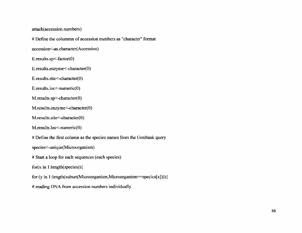
attach(accession.numbers)
# Define the colummn of accession numbers as "character" format
accession<-as.character( Accession)
E.results.sp<-factor(0)
E.results.enzyme<-character(0)
E.results.site<-character(0)
E.results.loc<-numeric(0)
M.results.sp<-character(0)
M.results.enzyme<-character(0)
M.results.site<-character(0)
M.results.loc<-numeric(0)
# Define the first column as the species names from the GenBank query
species<-unique(Microorganism)
# Start a loop for each sequences (each species)
for(x in l:length(species)){
for (y in 1 :length(subset(Microorganism,Microorganism— species[x]))){
# reading DNA from accession numbers individually
56
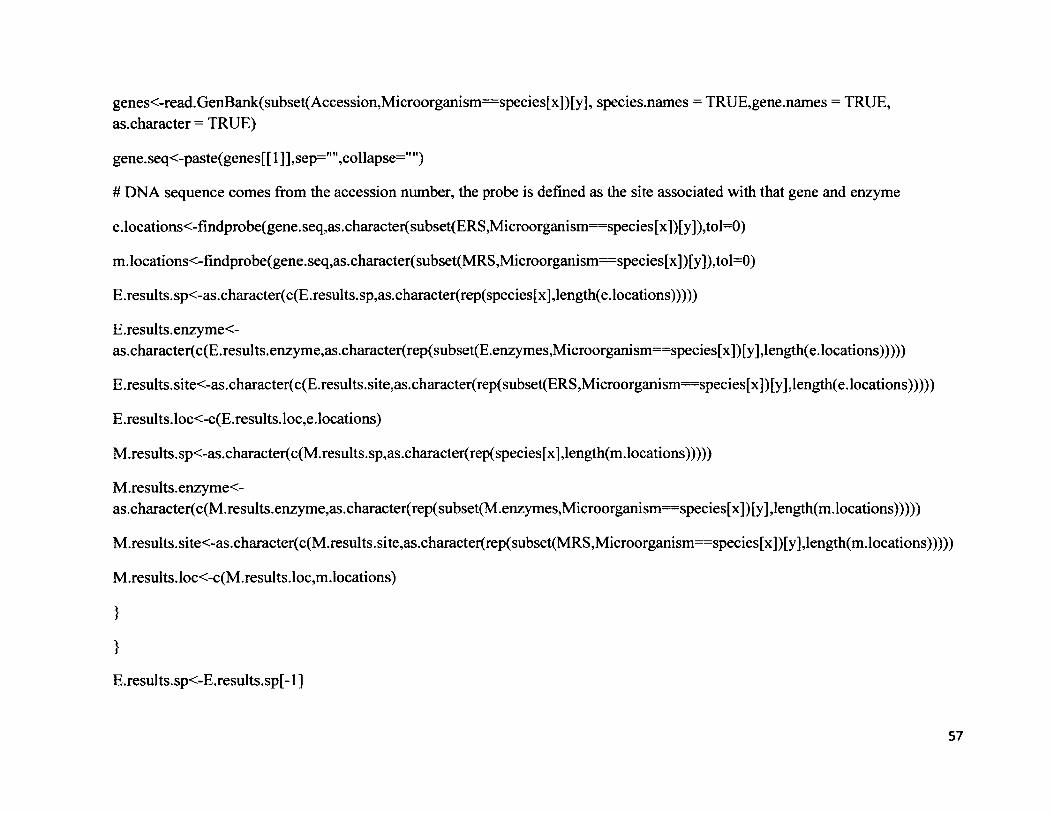
genes<-read.GenBank(subset(Accession,Microorganism==species[x])[y], species.names = TRUE,gene.names = TRUE, as.character = TRUE)
gene. seq<-paste(genes[ [ 1 ] ] ,sep="" ,collapse=" ")
# DNA sequence comes from the accession number, the probe is defined as the site associated with that gene and enzyme
e.locations<-findprobe(gene.seq,as.character(subset(ERS,Microorganism==species[x])[y]),tol=0)
m.locations<-findprobe(gene.seq,as.character(subset(MRS,Microorganism==species[x])[y]),tol=0)
E.results.sp<-as.character(c(E.results.sp,as.character(rep(species[x],length(e.locations)))))
E .results. enzyme<-as.character(c(E.results.enzyme,as.character(rep(subset(E.enzymes,Microorganism==species[x])[y],length(e.locations)))))
E.results.site<-as.character(c(E.results.site,as.character(rep(subset(ERS,Microorganism==species[x])[y],length(e.locations)))))
E.results.loc<-c(E.results.loc,e.locations)
M.results.sp<-as.character(c(M.results.sp,as.character(rep(species[x],length(m.locations)))))
M.results.enzyme<-as.character(c(M.results.enzyme,as.character(rep(subset(M.enzymes,Microorganism=species[x])[y],length(m.locations)))))
M.results.site<-as.character(c(M.results.site,as.character(rep(subset(MRS,Microorganism==species[x])[y],length(m.locations)))))
M.results.loc<-c(M.results.loc,m.locations)
}
}
E.results.sp<-E.results.sp[-1 ]
57
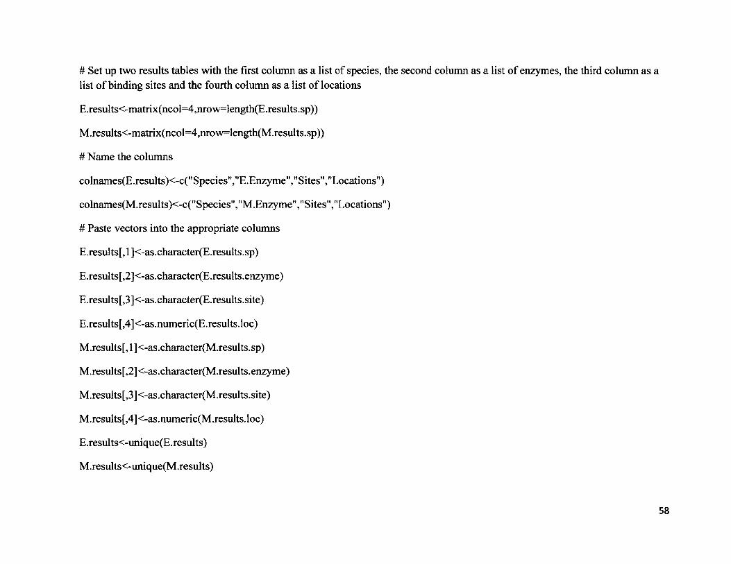
# Set up two results tables with the first column as a list of species, the second column as a list of enzymes, the third column as a list o f binding sites and the fourth column as a list of locations
E.results<-matrix(ncol=4,nrow=length(E.results.sp))
M.results<-matrix(ncol=4,nrow=length(M.results.sp))
# Name the columns
colnames(E.results)<-c("Species","E.Enzyme","Sites","Locations")
colnames(M.results)<-c("Species","M.Enzyme","Sites","Locations")
# Paste vectors into the appropriate columns
E.results[, 1 ]<-as.character(E.results.sp)
E.results[,2]<-as.character(E.results.enzyme)
E.results[,3]<-as.character(E.results.site)
E.results[,4]<-as.numeric(E.results.loc)
M.results[, 1 ]<-as.character(M.results.sp)
M.results[,2]<-as.character(M.results.enzyme)
M.results[,3]<-as.character(M.results.site)
M.results[,4]<-as.numeric(M.results.loc)
E.results<-unique(E.results)
M .results<-unique(M .results)
58
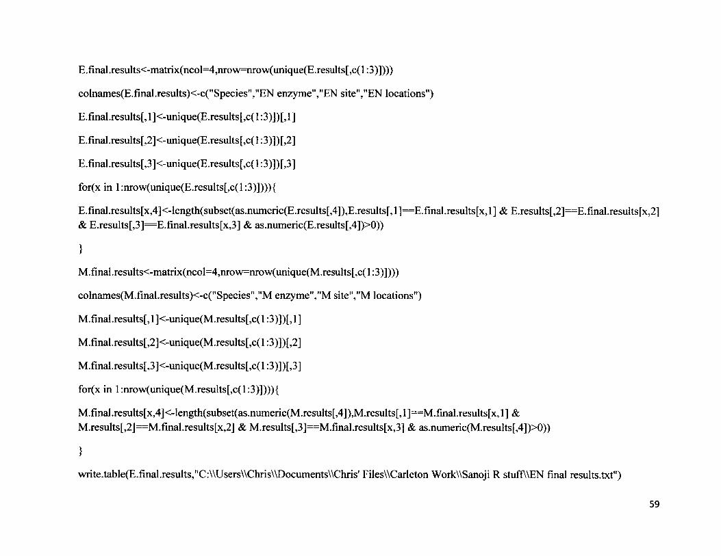
E.final.results<-matrix(ncol=4,nrow=nrow(unique(E.results[,c(l:3)])))
colnames(E.fmal.results)<-c("Species","EN enzyme","EN site","EN locations")
E.final.results[, 1 ]<-unique(E.results[,c( 1:3)])[, 1 ]
E.final.results[,2]<-unique(E.results[,c(l:3)])[,2]
E.final.results[,3]<-unique(E.results[,c(l:3)])[,3]
for(x in l:nrow(unique(E.results[,c(l:3)]))){
E.final.results[x,4]<-length(subset(as.numeric(E.results[,4]),E.results[,l]==E.final.results[x,l] & E.results[,2]=E.final.results[x,2] & E.results[,3]=E.final.results[x,3] & as.numeric(E.results[,4])>0))
}
M.final.results<-matrix(ncol=4,nrow=nrow(unique(M.results[,c(l:3)])))
colnames(M.final.results)<-c("Species","M enzyme","M site","M locations")
M.final.results[, 1 ]<-unique(M.results[,c( 1:3)])[, 1 ]
M.final.results[,2]<-unique(M.results[,c( 1:3)])[,2]
M.final.results[,3]<-unique(M.results[,c(l:3)])[,3]
for(x in l:nrow(unique(M.results[,c(l:3)]))){
M.final.results[x,4]<-length(subset(as.numeric(M.results[,4]),M.results[,l]==M.final.results[x,l] & M.results[,2]“ M.final.results[x,2] & M.results[,3]==M.final.results[x,3] & as.numeric(M.results[,4])>0))
}
write.table(E.final.results,"C:\\Users\\Chris\\Documents\\Chris' FilesWCarleton WorkWSanoji R stuffWEN final results.txt")
59
write.table(M.final.results,"C:\\Users\\Chris\\Documents\\Chris' FilesWCarleton WorkWSanoji R stuffWM final results.txt")
# Calculating the overlap between sequences.
# First, checking what the locations mean:
test.seq<-"ABCDEFGHIJKLMNOP"
probe<-"FGHI"
findprobe(test. seq,probe,tol=0)
# Location=6, so
# A B C D E F G H I J K L M N O P
# 1 2 3 4 5 F G H I
for(x in l:2){#length(species)){
E.sites<-as.numeric(subset(E.results[,4],E.results[,l]” Species[x]))
M.sites<-as.numeric(subset(M.results[,4],M.results[,l]=species[x]))
site.comparison<-matrix(ncol=3,nrow=:length(E.sites)*length(M.sites))
site.comparison[, 1 ]<-rep(E.sites,length(M.sites))
site.comparison.data<-numeric(length=0)
for(y in l:length(M.sites)){
site.comparison.data<-c(site.comparison.data,rep(M.sites[y],length(E.sites)))
}
60
site.comparison[, 1 ]<-site.comparison.data
}
for(y in l:length(site.comparison[,l])){
site.comparison[y,3]<-site.comparison[y, 1 ]-site.comparison[y,2]
}
table(site .comparison [y, 3 ] <nchar(
}
write.table(results,"C:\\Users\\Chris\\Documents\\Chris' FilesWCarleton WorkWSanoji R stuffWoverlap results.txt",header^TRUE)
61
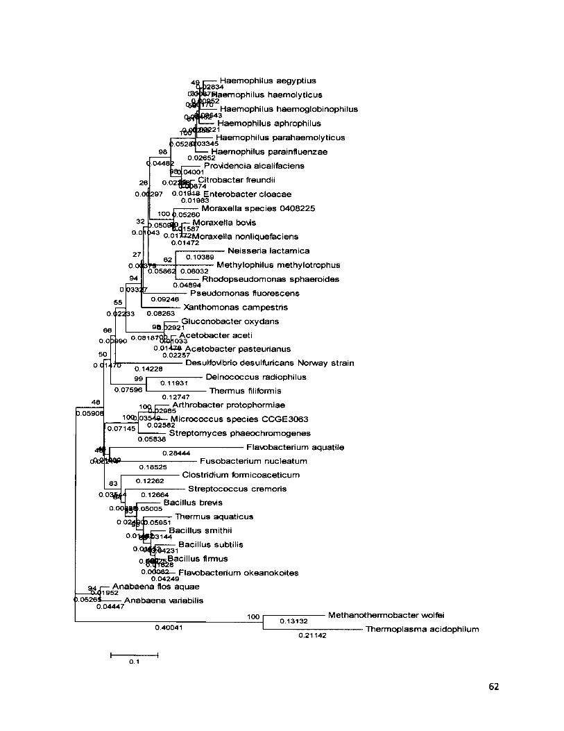
49 r— Haemophilus aegyptius t 4)2834
cBO »7Haemophilus haemolyticus
1 ^Haemophilus haemoglobinophilusHaemophilus aphrophilus
Haemophilus parahaem olyticus I 03345
Haemophilus parainfluenzae0.02652
Providencia alcalifaciens0 O2j t o r Citrobacter fireundii
o.oisua Enterobacter cloacae 0.01983
Moraxella sp ec ies 0408225100^.05260
Moraxella bovis 1587
o.oi*Z2Moraxella nonliquefaciens 0.01472
Neisseria lactam ica
o.c
48
.044804001
0.0< 297
050 1043
0.10389OCX 3
0.05862
0.09246
0.022 33
0.08187
0.14228 99 0.11931
3.05906
0.07596
Methylophilus methylotrophus0.08032
Rhodopseudom onas sphaeroides0.04894
Pseudom onas fluorescensXanthomonas cam pestris
0.08263G luconobacter oxydans
. 32921
*8 Lfio^Cet0baCter aceti0.014Z& A cetobacter pasteurianus
0.02257Desulfb vibrio desulfuricans Norway strain
Deinococcus radiophilus Thermus filifbrmis
0.1274710ft ^ Arthrobacter protophormiae
iocbJo354a— M icrococcus spec ies CCGE3063 0.02582
Streptom yces phaeochrom ogenes0.071450.05838
; 0.28444Flavobacterium aquatile
0.18525Fusobacterium nucleatum
830 . 0 3 $ ■
0.12262
0.00 1.05005
0.12664Bacillus brevis
Clostridium formicoaceticum - S treptococcus cremoris
Thermus aquaticus0 .02^(3 .05951
„ „ r— Bacillus smithii° ° 1 3 ^ P 3 1 4 4
Bacillus subtilis
>05265—
°09#J.%4231Bacillus firmus 8
0.0d062 Flavobacterium okeanokoites 0.04249
A nabaena Dos aquae952
A nabaena variabilis0.04447
1000.40041
013132
0.21142
M ethanotherm obacter wolfei -------------Thermoplasma acidophilum
0.1
62

Figure 4. Molecular phytogeny of 38 eubacterial and 2 archaea species based on complete and partial 16S rRNA. The tree was constructed in MEGA 2.02 with the maximum likelihood method and 500 bootstrap intervals. The branch lengths correspond to a scale of 0.05.
63