dna extraction & quantitation for forensic analysts
TRANSCRIPT

DNA Extraction & Quantitation for Forensic Analysts
This course is provided free of charge and is part of a series designed to teach about DNA and forensic DNA use and analysis. Find this course live, online at: http://dna.gov/training/extraction Updated: October 8, 2008
PPPP RRRR EEEE SSSS IIII DDDD EEEE NNNN TTTT ’’’’ SSSS
IIII N I T I A T I V EN I T I A T I V EN I T I A T I V EN I T I A T I V E
DNA
www.DNA.gov
About this Course
This PDF file has been created from the free, self-paced online course “Crime Scene and
DNA Basics for Forensic Analysts.” To learn more and take this and other courses
online, go to http://www.dna.gov/training/online-training/. Most courses are free but
you must first register at http://register.dna.gov.
If you already are registered for any course on DNA.gov, you may login directly at the
course URL, e.g., http://letraining.dna.gov or you can reach the courses by using the
URL http://www.dna.gov/training and selecting the “Login and view your courses” link.
Questions? If you have any questions about this file or any of the courses or content on
DNA.gov, visit us online at http://www.dna.gov/more/contactus/.
Links in this File
Most courses from DNA.Gov contain animations, videos, downloadable documents
and/or links to other useful Web sites. If you are using a printed, paper version of this
course, you will not have access to those features. If you are viewing the course as a PDF
file online, you may be able to use some of these features if you are connected to the
Internet.
Animations, Audio and Video. Throughout this course, there may be links to animation,
audio or video files. To listen to or view these files, you need to be connected to the
Internet and have the requisite plug-in applications installed on your computer.
Links to other Web Sites. To listen to or view any animation, audio or video files, you
need to be connected to the Internet and have the requisite plug-in applications installed
on your computer.
Legal Policies and Disclaimers
See Legal Policies and Disclaimers for information on Links to Other Web Sites,
Copyright Status and Citation and Disclaimer of Liability and Endorsement.

DNA Analysis Considerations
Introduction
The nature of the wide variety of substrates on which forensic biological samples are deposited can createproblems for the DNA analyst. For example, the substrate may cause collection problems or it may containsubstances that interfere with the Polymerase Chain Reaction (PCR) assay. The main problem is inhibition;the efficiency of amplification of forensic samples is often significantly decreased because of inhibitorspresent in the sample substrate. Inhibition can result in partial DNA profiles and, in severe instances, falsenegative reactions.
Even when sample collection and PCR have worked properly, difficulties may arise in the interpretation ofsamples from multiple donors.
Read more about mixtures in the STR Data Analysis & Interpretation PDF File.
Objectives
Upon successful completion of this unit of instruction, the student shall be able to:
Identify potential obstacles in collection, extraction, and amplification• Describe potential PCR inhibitors• Identify techniques to overcome inhibition•
DNA Extraction and Quantitation for Forensic Analysts
1/35

In some instances, collection of samples poses a significant challenge. Ingeneral, the best collection strategy is to submit the entire item. This canbe easily accomplished for items such as a small rock, clothing, or aknife, but is more difficult in the case of larger, fixed objects. In caseswhere the samples can be collected using a slightly moistened swab,such as from a window, the collection process is fairly straightforward.Some problems can arise when a hard porous surface, such as pavementor concrete, is encountered. In these cases, it can be difficult to collectsmall bloodstains or other biological fluids by swabbing. There areoccasions where scraping the substrate containing the biological sampleis a viable option. However, care must be taken. For example, if thecrime scene is outdoors, wind could cause loss of sample orcontamination of another item.
Inhibitors
It is hard to determine all of the causes of inhibition on the PCR reaction. The PCR process can be affected bycompounds that interfere with the interaction between DNA and Taq polymerase, and thus inhibit thereaction. Many inhibitors are removed during the extraction process through ethanol precipitation or the use ofa Microcon® or Centricon® centrifugal filter unit. However, some inhibitors co-elute with the DNA, whichmay lead to PCR inhibition. A number of inhibitors are contained in the samples themselves, while others canbe introduced by the substrate or the analysis process. The presence of inhibitors can result in loss of data orresults that could be mistaken for degradation. We can classify sources of inhibition into three groups:
Internal, or those found in body fluids.• Substrates, or those arising from the materials on which the blood stain or other source of DNA hasbeen deposited.
•
Other sources, such as reagents and materials used in the analysis.•
Internal
Many body fluids contain substances that can inhibit PCR:01
Table of Biological Substances and InhibitorsBiologicalSubstance
Inhibitor Comment
Blood Heme The amplification of blood samples can be significantlyreduced or blocked by natural components of blood,
DNA Extraction and Quantitation for Forensic Analysts
2/35
Immunoglobulin G such as heme, and immunoglobulin G. Hemin, ahemoglobin derivative, and its breakdown products,bilirubin and bile salts, are also found to be PCRinhibitors.
Vaginal Samples Bacteria
Microorganisms
Bacteria and microorganisms are commonly found invaginal, fecal, and buccal samples. Note: these can alsobe found in other biological samples found at crimescenes.
Buccal SamplesFecal Samples
Hair
Tissue
melanin Melanin, a pigment that affects skin, eye, and hair color,can inhibit PCR.
Bone
Teeth
Ca2+ Ca2+ is commonly found in bone and teeth and is knownto interfere with Mg2+ concentration, which in turn mayaffect the activity of Taq polymerase.
Semen Polyamines Spermine and spermidine (polyamines originally isolatedfrom semen) are found in ribosomes and living tissues,and can inhibit PCR.
Urine Urea Urea, a chemical found in urine, can inhibit PCR.
Substrates
Some of the substrates that contain inhibitors are:
Table of Substrates and InhibitorsSubstrate Inhibitor Comment/ExampleTextile dyes Textile dyes Indigo dye used to color denimFabrics Tannic Acid LeatherEnvironmental
Samples
Humic compounds
Heavy metals
Soil02
Food
Constituents
Organic compounds
Phenolic compounds
Glycogen
Fats
Ca2+
Many food products, such as milk, contain inhibitors,like Ca2+. However, forensic scientists have hadsuccess in developing DNA profiles from saliva lefton food or drink containers.01
Examples of substrates with inhibitive properties
DNA Extraction and Quantitation for Forensic Analysts
3/35
Other Inhibitors
Inhibitors can be introduced in the collection and analysis processes or at the crime scene
Table of Other InhibitorsSource Inhibitor CommentExtractionChemicals
Phenolic compounds from theorganic extraction
Chelex resin
Salts
Guanidine
Proteases
Organic solvents
Phosphate buffers Detergents (suchas Sodium Dodecyl Sulfate (SDS))
Reagents commonly used in the purification ofnucleic acids are inactivators of DNApolymerases. Phenol or Chelex resin left withthe extracted DNA can inhibit the PCR process.
Anticoagulants EDTA and heparin Known blood reference samples are collected intubes containing anticoagulants.
Powder Glove powder Many forensics scientists use powdered gloves.Laboratory plasticware
PCR tubes It has been reported that an inhibitory substancecan be released from polystyrene orpolypropylene upon exposure to ultraviolet light.
Plant and foodproducts
Pollen
Cellulose
Plant polysaccharides
Ca2+
Biological material can be deposited on plantsand food.
Overcoming Inhibition
DNA Extraction and Quantitation for Forensic Analysts
4/35
Not all of the factors affecting inhibition are known, and most of the methods used to overcome inhibition arespecific to the inhibiting compound. In contrast, Bovine serum albumin (BSA) is included in both AppliedBiosystems and Promega STR typing kits as a more general means of overcoming the effect of enzymeinhibitors.
Amplification test gels are a long-established technique to evaluate samples for inhibition. More recently,Quantitative PCR gives analysts the ability to assess inhibition during the quantitation process. However, notall inhibition will be detected. One certain way to assess a sample for inhibition is to add a portion of theextract to a control sample and attempt amplification. Failure of amplification demonstrates the presence of aninhibitor.
More about this in the Quantitation Module of this course.
The following are techniques that can be used to overcome inhibition:
One of the easiest approaches taken to overcome inhibition is to dilute the template DNA sample andreamplify. This dilutes out the inhibitor, allowing successful amplification to occur. In general, the quality ofDNA and purity of the sample is more important to amplification than the quantity of DNA. Usually, a 1:10 orgreater dilution is sufficient to overcome inhibition.
BSA has been shown to reduce inhibition in samples affected by hemin as well as other inhibitingcompounds. BSA is a general stabilizing agent, as is gelatin, which has also been used to overcomeinhibition.03,02,04
Reextraction & Additional Sample Preparation
An analyst can take additional steps to clean up the sample by reextraction using Chelex resin, phenolchloroform, Thiopropyl Sepharose 6B (Sigma)04 extraction beads, or magnetic beads (DNA IQ™). Samplepreparation using a Centricon® or Microcon® device may also assist in removing inhibitors.
Increased Taq Polymerase
The interaction between Taq polymerase and the template DNA is affected by inhibitors. Increasing theconcentration of Taq polymerase in the reaction can overcome the effects of some inhibitors.04
Non-ionic detergents, such as Tween® -20, NP-40 and Triton® X-100, can assist in overcoming inhibition,having the specific benefit of overcoming the inhibitory effects of trace amounts of strong ionic detergents,such as SDS.05
Miscellaneous Chemicals
Ammonium ions and dimethyl sulfoxide (DMSO) have been used to overcome inhibition.
One study cites a method for neutralization of Taq polymerase inhibitors by denaturing and washing withNaOH in Microcon® -100 filtration units. The study speculates that many inhibitors bind the template DNAand possibly intercalate into the double stranded DNA. Denaturizing the DNA could reduce the substance'saffinity for it. One other hypothesis is that the NaOH may directly inactivate the inhibitor.06
The effects of some inhibitors can be overcome with the use of shorter primers.07
DNA Extraction and Quantitation for Forensic Analysts
5/35

Summary
This module has briefly dealt with collection of DNA from scenes, and with one of the main difficultiesencountered with the PCR process, namely the presence of inhibitors. The module describes the nature ofinhibitors and techniques for their removal or neutralization.
Organic Extraction
Introduction
Techniques using organic reagents for DNA extraction are well acceptedin the forensic science community. Organic extraction methods are oftenpreferred for the extraction of biological stains containing small amountsof DNA or degraded DNA.01-05 These methods could be considered lessharsh than other methods, such as the use of Chelex beads, because noboiling step is required.
Upon successful completion of this unit of instruction, the student shallbe able to:
List steps involved in extracting DNA using an organicextraction method
•
Identify the advantages and disadvantages for use of organic extraction•
Organic Extraction Process
Historically, DNA extraction was accomplished by mechanical or chemicaldisruption of cells to release their organelles and contents. This works wellin samples containing many cells, but has required adaptation for use withthe much smaller biological samples collected at crime scenes.
Organic extraction is a conventional method that uses organic chemicals toisolate genomic DNA. The procedure can be described in four steps:
solubilization of the stain components1. denaturation and hydrolysis of proteins2. removal of denatured proteins3. purification of DNA064.
Step One: Solubilization of Stain Components
Water must be replaced so that dried stains can be resolubilized for DNA extraction procedures. The DNA isprotected from unnecessary degradation in this process by adding EDTA, a magnesium chelator, to the lysisbuffer. EDTA prevents nucleases from degrading the DNA. Tris (a component of the buffer) interacts with thelipopolysaccharides present on the outer cell membrane, which helps to make it permeable. This effect isenhanced with the addition of EDTA.06
Step One Reagents
DNA Extraction and Quantitation for Forensic Analysts
6/35
Stain extraction buffers are usually slightly alkaline and generally contain Tris base or Tris-HCl (pH between7.5-8.0), EDTA, and sodium chloride (NaCl).
Step Two: Denaturation and Hydrolysis of Proteins
Cells are lysed using a detergent, Proteinase K, and dithiothreitol (DTT). Extractions must use appropriate saltconcentration and pH to ensure that proteins and other contaminants are separated into the organic phase andthat DNA remains in the aqueous phase.
Detergents, which are included in the stain extraction buffer, have the following functions:
They lyse cell membranes.• They separate histone proteins from DNA.• They denature histone proteins.• They destroy secondary and tertiary structures of proteins, which decreases their solubility in aqueoussolution.
•
Proteinase K is used to hydrolyze histone proteins and is well suited to the extraction process for thefollowing reasons:
It is active over a wide pH range (4-12.5).• It is active in the presence of SDS.• It is not affected by EDTA.•
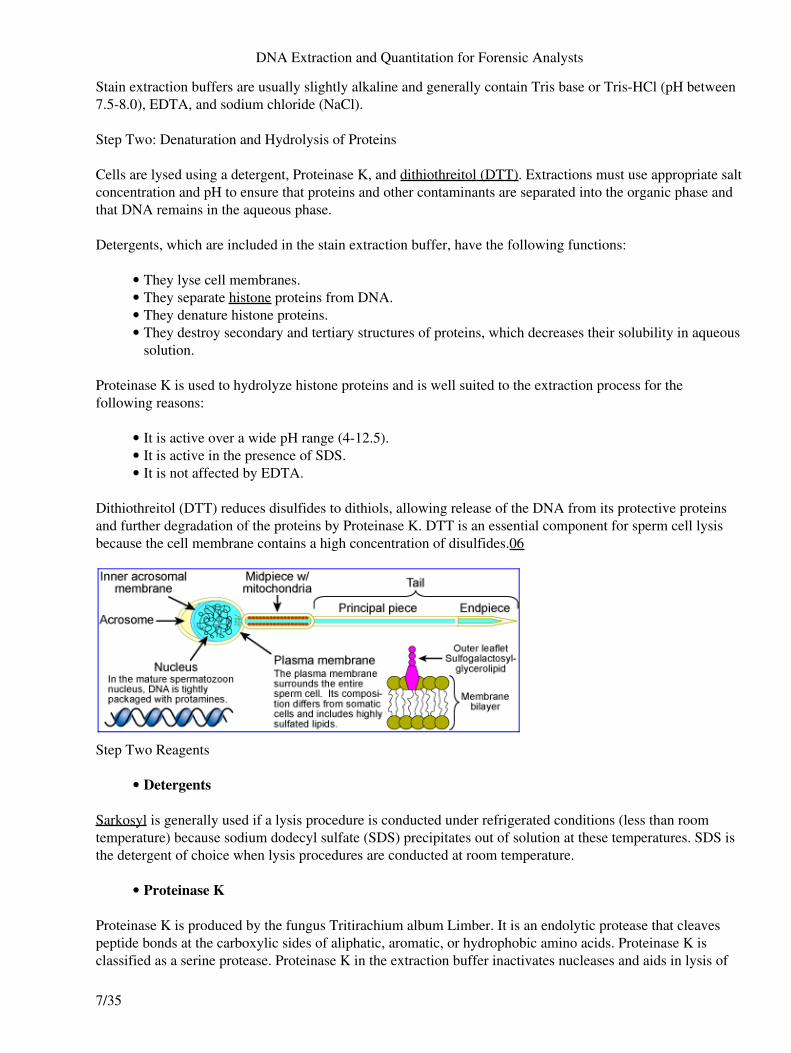
Dithiothreitol (DTT) reduces disulfides to dithiols, allowing release of the DNA from its protective proteinsand further degradation of the proteins by Proteinase K. DTT is an essential component for sperm cell lysisbecause the cell membrane contains a high concentration of disulfides.06
Step Two Reagents
Detergents•
Sarkosyl is generally used if a lysis procedure is conducted under refrigerated conditions (less than roomtemperature) because sodium dodecyl sulfate (SDS) precipitates out of solution at these temperatures. SDS isthe detergent of choice when lysis procedures are conducted at room temperature.
Proteinase K•
Proteinase K is produced by the fungus Tritirachium album Limber. It is an endolytic protease that cleavespeptide bonds at the carboxylic sides of aliphatic, aromatic, or hydrophobic amino acids. Proteinase K isclassified as a serine protease. Proteinase K in the extraction buffer inactivates nucleases and aids in lysis of
DNA Extraction and Quantitation for Forensic Analysts
7/35
epithelial and white blood cells to free nuclear DNA. Nuclease inactivation is a very important step in DNAisolation. Nucleases naturally exist in the cell to break down the nucleic acids after they serve their functionsin protein manufacture, thus allowing the individual building blocks of the DNA and RNA to be recycled bythe cell. Inactivating these nucleases preserves the DNA so that it can be extracted and purified.06
DTT•
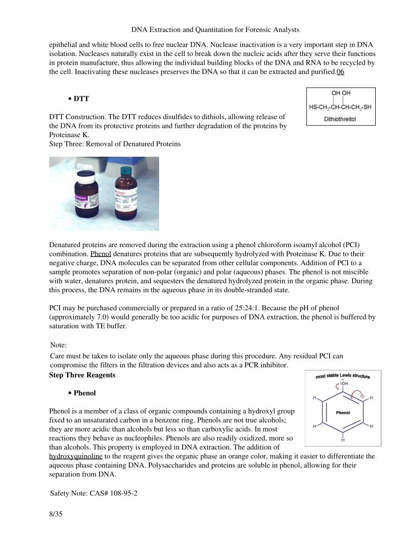
DTT Construction. The DTT reduces disulfides to dithiols, allowing release ofthe DNA from its protective proteins and further degradation of the proteins byProteinase K.Step Three: Removal of Denatured Proteins
Denatured proteins are removed during the extraction using a phenol chloroform isoamyl alcohol (PCI)combination. Phenol denatures proteins that are subsequently hydrolyzed with Proteinase K. Due to theirnegative charge, DNA molecules can be separated from other cellular components. Addition of PCI to asample promotes separation of non-polar (organic) and polar (aqueous) phases. The phenol is not misciblewith water, denatures protein, and sequesters the denatured hydrolyzed protein in the organic phase. Duringthis process, the DNA remains in the aqueous phase in its double-stranded state.
PCI may be purchased commercially or prepared in a ratio of 25:24:1. Because the pH of phenol(approximately 7.0) would generally be too acidic for purposes of DNA extraction, the phenol is buffered bysaturation with TE buffer.
Note:Care must be taken to isolate only the aqueous phase during this procedure. Any residual PCI cancompromise the filters in the filtration devices and also acts as a PCR inhibitor.Step Three Reagents
Phenol•
Phenol is a member of a class of organic compounds containing a hydroxyl groupfixed to an unsaturated carbon in a benzene ring. Phenols are not true alcohols;they are more acidic than alcohols but less so than carboxylic acids. In mostreactions they behave as nucleophiles. Phenols are also readily oxidized, more sothan alcohols. This property is employed in DNA extraction. The addition ofhydroxyquinoline to the reagent gives the organic phase an orange color, making it easier to differentiate theaqueous phase containing DNA. Polysaccharides and proteins are soluble in phenol, allowing for theirseparation from DNA.
Safety Note: CAS# 108-95-2
DNA Extraction and Quantitation for Forensic Analysts
8/35
Although originally used for its antiseptic properties, phenol is highly toxic and should be handled in a fumehood while wearing personal protective equipment. Skin contact and inhalation should be avoided.
Chloroform•
Chloroform (CHCl 3), or trichloro-methane, is a colorless liquid that is slightlywater-soluble and miscible with organic solvents such as phenol. It is more densethan water or buffer (in which DNA is soluble), yet less dense than phenol. As itincreases the phenol phase density, it promotes a sharp interface between theorganic and aqueous layers. Chloroform also solubilizes lipids. During theextraction procedure, cellular debris that is not totally digested can be observed atthe interface.
Safety Note: CAS# 67-66-3Previously widely used as an anesthetic, inhalation of chloroform depresses the central nervous system. It isalso a suspected teratogen and known carcinogen and should never be handled outside of a fume hood.
Isoamyl Alcohol•
Isoamyl alcohol, or 3-methyl-1-butanol, is a primary alcohol. A liquid solvent, it isoften included in genomic extraction protocols to help prevent foaming of thereagents, making it easier to detect the interface between the organic and aqueous phases. It is included in theprotocol in very small concentration, compared to the other reagents.06
Safety Note: CAS# 123-51-3A component of some paint strippers and other solvents, isoamyl alcohol causes irritation upon skin contactor inhalation. Vapors can also cause ocular discomfort and effects. This reagent should be handled with careand in a fume hood.Step Four: DNA Purification
DNA can be recovered from the aqueous phase with an ethanol precipitation or using a centrifugal filter unit(Centricon® or Microcon®).
DNA Extraction and Quantitation for Forensic Analysts
9/35

Centrifugal filter units are used to purify and concentrate DNA. When extracting DNA from small ordegraded forensic samples, the final concentration of DNA may be too low for subsequent amplification.Although 1ng is a target quantity of DNA for amplification, if 1ng is suspended in 100 µl of fluid, it would beimpossible to transfer 100 µl of this solution into an amplification reaction optimized for 50 µl or less.Centrifugal filter units increase the concentration of DNA in solution by retaining the DNA while eliminatinga portion of the fluid from the sample. Another benefit of the unit is the ability to secure DNA whilecontaminants (possibly PCR inhibitors) are washed from the sample.
Centrifugal filter units separate molecules by size through a series of washing and centrifugation steps. TheMillipore Corporation produces two centrifugal filter units under the names Centricon® and Microcon®.
Visit the Millipore Corporation website.
Attributes of Centricon® and Microcon® filter units include:
Both employ Amicon® filters to retain the DNA.• Filter porosity varies.• Proteins flow through, rather than sticking to, the surface during centrifugation.• Filters are anisotrophic, with increasingly smaller interstitial spaces in the direction of filtration,allowing for better retention of smaller molecules.
•
Filters are composed of regenerated cellulose, which can be sterilized.• Filters exhibit sufficient strength when wet.•
Note:It is important not to spin the unit at rates higher than recommended by the manufacturer because the unitmay become compromised, resulting in reduced sample recovery.Textile dye molecules, such as indigo, are known PCR inhibitors and are readily washed through the filter.Salts introduced by buffers are removed from the sample during the process. Salts carried over from theprocess may interfere with capillary electrophoresis.
Read about capillary electrophoresis in the Amplified DNA Product Separation PDF File.
Note:Care must be taken to avoid introducing phenol from the organic extraction into the unit because it will breakdown both the cellulose filter and its supporting silicone rubber o-ring.Step Four Reagents
The only reagents used in this process are buffers used in the DNA purification process.
Techniques
DNA Extraction and Quantitation for Forensic Analysts
10/35
Organic extraction techniques are used on all types of forensic samples. The basic organic extraction method is used on samples that do notcontain spermatozoa, whereas a modified differential extraction is usedon those samples containing spermatozoa.
Microcentrifuge tubes with spin baskets can be used for both basic anddifferential extractions, allowing the substrate (e.g. fabric, cotton swab,etc.) to be removed without difficulty during the extraction process.
This section outlines:
Use of microcentrifuge tubes with spin baskets, which can be incorporated into most extractionmethods
•
Basic organic extraction• Differential organic extraction•
Microcentrifuge Tubes with Spin Baskets
Constructed with an open end and a woven end, spin baskets hold a cutting of substrate and are inserted into acommon microcentrifuge tube. This allows for the retention of liquid carrying the biological material to beseparated from the substrate during centrifugation steps. Any cells that may not be completely freed from thesubstrate are forced into the tube, along with the excess fluid. Spin baskets may be incorporated into mostextraction methods; however, their composition prohibits immersion in phenol/chloroform/isoamyl alcohol(PCI).
Basic Organic Extraction
The basic organic extraction method can be used for most forensic samples, which includes bloodstains, salivastains, tissue and hair. Details of the method are given in the laboratory manual.
A stain extraction buffer containing a buffer, detergent, DTT, and Proteinase K is added to the sample. Thismixture is incubated, and the sample substrate is removed. Then, the mixture is extracted with aphenol/chloroform/isoamyl alcohol combination. The extracted DNA is generally purified and concentratedwith a filtration device.07
Vortexed PCI Aqueous PhaseDifferential Method
Differential extraction methods are used to separate spermatozoa from other cell types. Spermatozoa are moredifficult to lyse than other cells and conditions can be set so that all cells except spermatozoa are lysed. Thesupernatant containing the DNA from these cells is removed from the sperm cells, which can then be lysedseparately.
DNA Extraction and Quantitation for Forensic Analysts
11/35

The differential extraction steps are:
Optional wash step
Some laboratories have incorporated an optional wash step at the beginning of the procedureto remove cellular debris and contaminants. The sample is gently washed in a buffer anddetergent and the supernatant is removed (wash fraction). This can be done under refrigeratedconditions or at room temperature.
♦
•
Non-sperm cell lysis
An extraction buffer containing a buffer, detergent, and Proteinase K is added to the sampleand incubated. This step lyses all cells except spermatozoa. The supernatant containing theDNA from the lysed cells (fraction 1) is removed after pelleting the spermatozoa. The spermpellet is often washed numerous times with a buffer to remove excess DNA from this lysisstep. If this wash is not done, it is not unusual to see a low level of fraction 1 DNA in fraction2. If any of the sperm cells are weak or otherwise compromised, these may lyse in the firststep, leaving a low level of fraction 2 DNA in fraction 1.
♦
•
Sperm cell lysis
The pelleted sperm cells are lysed under more stringent conditions, using a buffer, detergent,DTT, and a higher concentration of Proteinase K (fraction 2), and are subsequently incubated.
♦
Both fractions (including the wash fraction, if appropriate) are extracted separately with thephenol/chloroform/isoamyl alcohol combination and purified.08
♦
•
The success of differential extraction depends on the sperm head resisting the processes that readily lyseepithelial and white blood cells. Separating the sources of DNA from different contributors to a stain, namelya male donor and female victim, lessens the difficulty associated with mixture interpretation during dataanalysis, source attribution, and/or statistical calculations. The value of differential extraction is demonstratedby the requirement in the Quality Assurance Standards (QAS) for inclusion in the laboratory's recordedprocedures (see Standard 9.1.3). Details of the method are provided in the laboratory manual.
The online version of this course contains a multimedia [or downloadable] file. Visit this URL to view thefile: http://beta.extraction.dna.devis.com/m02/03/default_page
A commercial kit for differential extraction is available from Promega. The Differex™ System, includes aproprietary separation solution and the use of a spin basket. The procedure begins with a Proteinase Kdigestion to lyse non-sperm cells. The sample and buffer are placed into a spin basket within the tubecontaining the proprietary solution (which is not miscible with the aqueous buffer). Through centrifugation,the sperm cells are isolated as a pellet, while the non-sperm DNA remain in solution. The difference betweenthis system and routine differential extraction is that the separation solution acts as a selective membrane(without the risk of clogging). After the removal of the solution containing non-sperm DNA, the sperm DNAis isolated using the DNA IQ™ System, described later in this subject.
Go to the Differex™ System website.
DNA Extraction and Quantitation for Forensic Analysts
12/35

Chelex®100 Extraction
Extraction procedures using Chelex® 100 (Bio-Rad Laboratories) are popular in the forensic sciencecommunity because they save time, reduce costs, simplify extractions, reduce safety risks, and minimizepotential for contamination. Chelex® 100, a chelating resin, is used to successfully extract DNA from manyforensic samples, including bloodstains, tissue, hair, and bone. 01,02, 03, 04, 05
Visit Bio-Rad's website and learn more about Chelex® 100.
Objectives
Upon successful completion of this unit of instruction, the student shall be able to:
List steps involved in extracting DNA using the Chelex® method.• Identify the advantages and disadvantages of the Chelex® method for use of organic extraction inorder to be consistent with the organic module.
•
Chelex® 100 Extraction Process
Chelating resins have been used in ion-exchange columns, trace metal removal, metal analysis, and watertesting in environmental and agricultural laboratories. In clinical applications and biomedical research,chelating resins can be used to remove or assay cations in whole blood or urine, to remove contaminants frombuffers and stock solutions, and to prepare samples for nuclear magnetic resonance spectroscopy.
Chelex®, like most chemicals, is supplied in various grades of purity. Analytical grade Chelex®100 resin ishighly purified and most suitable for forensic DNA applications.
DNA Extraction and Quantitation for Forensic Analysts
13/35
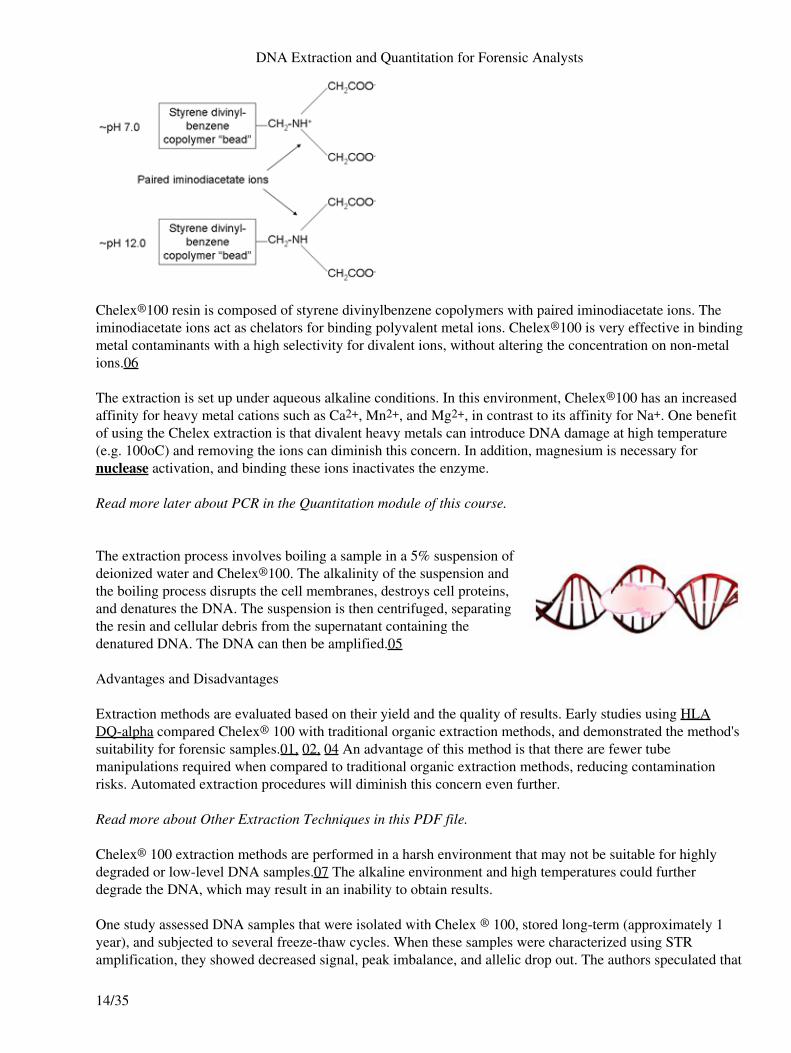
Chelex®100 resin is composed of styrene divinylbenzene copolymers with paired iminodiacetate ions. Theiminodiacetate ions act as chelators for binding polyvalent metal ions. Chelex®100 is very effective in bindingmetal contaminants with a high selectivity for divalent ions, without altering the concentration on non-metalions.06
The extraction is set up under aqueous alkaline conditions. In this environment, Chelex®100 has an increasedaffinity for heavy metal cations such as Ca2+, Mn2+, and Mg2+, in contrast to its affinity for Na+. One benefitof using the Chelex extraction is that divalent heavy metals can introduce DNA damage at high temperature(e.g. 100oC) and removing the ions can diminish this concern. In addition, magnesium is necessary fornuclease activation, and binding these ions inactivates the enzyme.
Read more later about PCR in the Quantitation module of this course.
The extraction process involves boiling a sample in a 5% suspension ofdeionized water and Chelex®100. The alkalinity of the suspension andthe boiling process disrupts the cell membranes, destroys cell proteins,and denatures the DNA. The suspension is then centrifuged, separatingthe resin and cellular debris from the supernatant containing thedenatured DNA. The DNA can then be amplified.05
Advantages and Disadvantages
Extraction methods are evaluated based on their yield and the quality of results. Early studies using HLADQ-alpha compared Chelex® 100 with traditional organic extraction methods, and demonstrated the method'ssuitability for forensic samples.01, 02, 04 An advantage of this method is that there are fewer tubemanipulations required when compared to traditional organic extraction methods, reducing contaminationrisks. Automated extraction procedures will diminish this concern even further.
Read more about Other Extraction Techniques in this PDF file.
Chelex® 100 extraction methods are performed in a harsh environment that may not be suitable for highlydegraded or low-level DNA samples.07 The alkaline environment and high temperatures could furtherdegrade the DNA, which may result in an inability to obtain results.
One study assessed DNA samples that were isolated with Chelex ® 100, stored long-term (approximately 1year), and subjected to several freeze-thaw cycles. When these samples were characterized using STRamplification, they showed decreased signal, peak imbalance, and allelic drop out. The authors speculated that
DNA Extraction and Quantitation for Forensic Analysts
14/35

the unbuffered suspension of the DNA, coupled with multiple freeze-thaw cycles, could accelerate thedegradation process of isolated DNA samples.08
DNA isolated using the Chelex® 100 extraction is denatured. This was problematic when laboratories usedrestriction fragment length polymorphism (RFLP) for characterization since double-stranded DNA is required(RFLP). This is no longer a concern with current STR methods.
The Chelex® 100 procedure does not include a purification step. If the sample contains inhibitors andcontaminants, increasing the sample size increases their concentration, which can inhibit PCR. Somelaboratories have incorporated the use of a filter unit such as a Microcon® or Centricon® prior toamplification so that some PCR inhibitors can be removed and the sample can be concentrated. Because theresin is a PCR inhibitor, it is important to ensure that when removing the supernatant, no resin is removedfrom the tube.09
Read more about DNA Analysis Consideration later in this PDF file.
Chelex®100 Resin Extraction Method
Advantages and DisadvantagesAdvantages DisadvantagesFast Harsh extraction environment (pH between 10-11 and temperature
approximately 100° C)Simple Potential degradation concerns for long-term storage of isolated DNA samplesInexpensive Resin remaining in extracted DNA sample can inhibit PCR processNo hazardous chemicals Less effective extraction of some sample typesFew tube manipulations Isolated DNA is single-strandedBinds heavy metal cations Resin loses its chelating capacity after a few hours in suspensionRemoves some PCRinhibitors
Non-differential
Chelex® 100 extraction methods are straightforward, with little variation between procedures. Some examplesare:
Laboratories may differ with respect to their inclusion of Proteinase K, a serine protease that aids inprotein digestion.
•
For extraction from blood, the sample may be incubated in sterile deionized water that promoteshemolysis of red blood cells. This aids in the removal of heme, a known PCR inhibitor.
•
DNA Extraction and Quantitation for Forensic Analysts
15/35
Some laboratories wash the sample in phosphate buffered saline prior to extraction. The sample maybe washed one or more times and pelleted through centrifugation before resuspending it to carry outthe Chelex® 100 extraction procedure.
•
Differential
Generally, sexual assault evidence is a mixture of epithelial cells and semen. The QAS Standard 9.1.3 (FO)requires the laboratory to have a procedure for the differential extraction of stains that contain semen. Manylaboratories have adopted a Chelex differential extraction procedure for these sample types. 09
Proteinase K, mentioned in the non-differential procedure, lyses epithelial and white blood cells but not spermcells under the extraction conditions. It is particularly useful in organic extraction procedures as it maintainsactivity in the presence of denaturing agents such as sodium dodecyl sulfate (SDS), a component of extractionor digest buffers. Similarly, Proteinase K is not inactivated by metal chelating agents.
Read more about Organic Extraction later in this PDF file.
After extracting the sample in digest buffer and Proteinase K, the sample is centrifuged. The supernatantcontains DNA from the lysed epithelial and white blood cells. The supernatant containing the cell lysate issubjected to extraction using 5% Chelex® 100. This fraction is treated as a single sample and referred to asthe F1 fraction, the epithelial cell fraction, or the female fraction, depending upon laboratory procedures
Sperm DNA is wound around proteins called protamines, which are analogous to histones. Protamines containa high concentration of cysteine residues and disulfide bonds, making the sperm head and its DNA moreresistant to the effects of Proteinase K. The pellet from the original extraction contains the sperm componentsfrom the sample and is subjected to a second incubation in buffer containing Proteinase K and dithiothreitol(DTT). This fraction is treated as a second sample referred to as the F2, sperm fraction, or male fraction. DTTprevents oxidation of thiol (SH) groups and reduces disulfides to dithiols.
Other Extraction Techniques
Introduction
DNA Extraction and Quantitation for Forensic Analysts
16/35

This module focuses on the modifications to common extraction methods as well as commercially availableextraction systems. While several techniques are presented, this module does not cover all available methodsrepresented in forensic DNA laboratories.
Objectives
Upon successful completion of this unit of instruction, the student shall be able to:
List several commercially available extraction kits and accessories• Identify the advantages and disadvantages in the use of various types of proprietary reagents andmaterials
•
Determine the appropriate application of various techniques to different sample types and substrates• Describe the use of commercially obtained materials in automated systems•
Many extraction procedures incorporate the use of a microcentrifuge tube with a spin basket. Costar Spin-X®
centrifuge tube filters are one example. These centrifuge tube inserts contain no membrane, but have abonded fritted bottom on the 500µL insert that fits into the microcentrifuge tube.1,2
This allows for the separation of liquid carrying the biological material from the substrate duringcentrifugation steps. Another modification is the use of centrifugal filter units. These allow for extractedDNA to be purified and concentrated after extraction and prior to amplification. Millipore Microcons® areone example of centrifugal filter units.2 Both of these modifications are routinely used in DNA extractionprocedures.
Microcentrifuge Tubes with Spin Baskets
Constructed with an open end and a woven end, spin baskets hold a cutting of substrate and are inserted into acommon microcentrifuge tube. This allows for the retention of liquid carrying the biological material to beseparated from the substrate during centrifugation steps. Any cells that may not be completely freed from thesubstrate are forced into the tube, along with the excess fluid. Spin baskets may also be used in organicextractions, although their composition prohibits immersion in phenol/chloroform/isoamyl alcohol (PCI).
DNA Extraction and Quantitation for Forensic Analysts
17/35

For example, Promega offers the Differex™ System, which includes a proprietary separation solution and theuse of a spin basket, increasing the efficiency of the traditional method of differential extraction from spermversus non-sperm cells. The procedure begins with Proteinase K digestion to lyse non-sperm cells. Thesample and buffer are placed into a spin basket within the tube containing the proprietary solution. Thissolution is not miscible with the aqueous buffer. Through centrifugation, the sperm cells form pellets, whilethe non-sperm DNA remains in solution. The difference between this system and routine differentialextraction is that the separation solution acts as a selective membrane (without the risk of clogging), which ispurported to reduce the chance of sperm loss. After the removal of the solution containing non-sperm DNA,the sperm DNA is isolated using the DN™ System, described later in this module.
Centrifugal Filter Units
Centrifugal filter units are used to purify and concentrate DNA. When extracting DNA from small ordegraded forensic samples, the final concentration of DNA may be too low for subsequent amplification.Although 1ng is a target quantity of DNA for amplification, if 1ng is suspended in 100µl of fluid, it would beimpossible to transfer 100µl of this solution into an amplification reaction optimized for 50µl or less.Centrifugal filter units increase the concentration of DNA in solution by retaining the DNA while eliminatinga portion of the fluid from the sample. Another benefit of the unit is the ability to secure DNA whilecontaminants (possibly PCR inhibitors) are washed from the sample.
Centrifugal filter units separate molecules by size through a series of washing and centrifugation steps. TheMillipore Corporation produces two centrifugal filter units under the names Centricon® and Microcon®.
Visit The Millipore Corporation website.
Attributes of Centricon® and Microcon® filter units:
Both employ Amicon® filters to retain the DNA.• Filter porosity varies.• Proteins flow through rather than sticking to the surface during centrifugation.• Filters are anisotrophic, with increasingly smaller interstitial spaces in the direction of filtrationallowing for better retention of smaller molecules.
•
DNA Extraction and Quantitation for Forensic Analysts
18/35
Filters are composed of regenerated cellulose, which can be sterilized.• Filters exhibit sufficient strength when wet.•
Note:It is important not to spin the unit at rates higher than recommended by the manufacturer because the unitmay become compromised, resulting in reduced sample recovery.Textile dye molecules, such as indigo, are known PCR inhibitors and are readily washed through the filter.Salts introduced by buffers are removed from the sample during the process. Salts carried over from theprocess may interfere with capillary electrophoresis.
Read more about capillary electrophoresis in the Amplified DNA Product Separation PDF file.
Note:Care must be taken to avoid introducing phenol from the organic extraction into the unit because it will breakdown both the cellulose filter and its supporting silicone rubber o-ring. Chelex® beads carried over fromextraction may clog or damage the filter.Other Techniques
The Chelex® and organic extraction methods have been mainstays in the forensic science community, but arenot without their limitations. The community continues to assess other extraction techniques that areautomatable, safe, and demonstrate high performance. Automated extraction methods are rapidly replacingChelex® and organic extraction techniques in forensic laboratories.
Read more about Organic Extraction earlier in this PDF file.
Spin Column
The Qiagen QIAamp® DNA Micro Kit, a spin column using a silica-based extraction method, is used inforensic DNA laboratories. This kit does not require the use of hazardous chemicals. Nucleic acids areattracted to the silica bead under high chaotropic salt concentrations. The sample and lysis buffer (provided inthe kit) are added to a sterile tube. The lysate is combined with alcohol and placed into the spin column, whichis inserted into a tube. The removal of proteins and divalent cations is accomplished using multiple bufferwashes and centrifugation steps. Removal of cations, such as Mg2+, prevents nucleases from further
DNA Extraction and Quantitation for Forensic Analysts
19/35
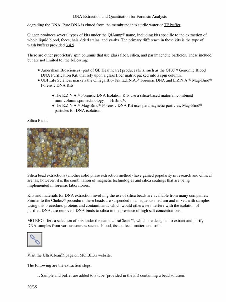
degrading the DNA. Pure DNA is eluted from the membrane into sterile water or TE buffer.
Qiagen produces several types of kits under the QIAamp® name, including kits specific to the extraction ofwhole liquid blood, feces, hair, dried stains, and swabs. The primary difference in these kits is the type ofwash buffers provided.3,4,5
There are other proprietary spin columns that use glass fiber, silica, and paramagnetic particles. These include,but are not limited to, the following:
Amersham Biosciences (part of GE Healthcare) produces kits, such as the GFX™ Genomic BloodDNA Purification Kit, that rely upon a glass fiber matrix packed into a spin column.
•
UBI Life Sciences markets the Omega Bio-Tek E.Z.N.A.® Forensic DNA and E.Z.N.A.® Mag-Bind®
Forensic DNA Kits.
The E.Z.N.A.® Forensic DNA Isolation Kits use a silica-based material, combinedmini-column spin technology — HiBind®.
♦
The E.Z.N.A.® Mag-Bind® Forensic DNA Kit uses paramagnetic particles, Mag-Bind®
particles for DNA isolation.♦
•
Silica Beads
Silica bead extractions (another solid phase extraction method) have gained popularity in research and clinicalarenas; however, it is the combination of magnetic technologies and silica coatings that are beingimplemented in forensic laboratories.
Kits and materials for DNA extraction involving the use of silica beads are available from many companies.Similar to the Chelex® procedure, these beads are suspended in an aqueous medium and mixed with samples.Using this procedure, proteins and contaminants, which would otherwise interfere with the isolation ofpurified DNA, are removed. DNA binds to silica in the presence of high salt concentrations.
MO BIO offers a selection of kits under the name UltraClean ™, which are designed to extract and purifyDNA samples from various sources such as blood, tissue, fecal matter, and soil.
Visit the UltraClean™ page on MO BIO's website.
The following are the extraction steps:
Sample and buffer are added to a tube (provided in the kit) containing a bead solution.1.
DNA Extraction and Quantitation for Forensic Analysts
20/35
A typical extraction buffer containing sodium dodecyl sulfate (SDS) is added, followed by anothersolution designed to precipitate out the humic acid components and other PCR inhibitors.
2.
The tubes are vortexed extensively. Chemical lysis, using SDS, breaks down cell membranes andproteins, while mechanical lysis occurs as the beads physically beat up the cells.
3.
Centrifugation pellets the debris while the DNA remains in the aqueous supernatant.4. A DNA binding salt solution, which promotes binding of the DNA to the silica beads, is added.5. The solution of beads and DNA (in aqueous buffer) is added to a spin device. The DNA remainsbound and is impeded by a membrane in the spin filter. Other molecules and contaminants passthrough.
6.
The DNA can be washed with an ethanol solution for further purification.7. An elution buffer is added to remove the salt, thus allowing the DNA to be washed through themembrane while the silica beads are retained. The resulting filtrate is purified DNA in an aqueousbuffer, suitable for quantitation.
8.
Note:The kits for extracting DNA from soil and feces contain reagents that reportedly break down humic acids.Read more about DNA Analysis Considerations elsewhere in this PDF file.
Magnetic Beads
The use of magnetic beads is another solid phase extraction method.
These methods:
Can be automated• Increase throughput• Demonstrate the ability to remove amplification inhibitors• Use no hazardous chemicals.•
These kits can also be used as adjuncts to traditional methods, when additional clean-up is warranted.
The principle underlying magnetic bead procedures involves attracting DNA to magnetic beads, holding thebeads in place using a magnetized source, such as a rack or tube holder, and washing away other componentsof the sample.
Invitrogen's ChargeSwitch® Technology (CST®) involves the use of magnetic beads whose surface bears acharge that can be switched based on the pH of the surrounding buffer environment. At low pH, the beads arepositively charged, attracting the negatively charged DNA molecules and allowing proteins and contaminantsto be removed by washing.
The ChargeSwitch® Forensic DNA Purification Kit includes:
Lysis buffer• Magnetic beads• Proteinase K• Purification buffer• Wash buffer• Elution buffer•
View an animation on magnetic beads.
DNA Extraction and Quantitation for Forensic Analysts
21/35

The DNA IQ™ System by Promega uses silica-coated magnetic beads. DN™ was designed for automation onrobotic systems. The quantity of beads used in the procedure defines the binding capacity, which isapproximately 100ng of DNA. This relative quantitative capability makes it ideal for databasing and paternityapplications because consistent quantities of DNA are isolated.
The DNA IQ™ kit contains the proprietary resin and several specialized buffers, including lysis, wash, andelution. The method demonstrates efficient DNA extraction from small samples and the removal of inhibitingcompounds. For example, internal validation studies of DN™ were successful in the extraction of DNA fromsmall stains deposited on difficult substrates, such as denim, leather, and soil.
Promega also offers a special buffer for the extraction of DNA from bone and a procedure for differentialextraction.
FTA® Technology
FTA® is an acronym for fast technology for analysis of nucleic acids. It wasoriginally developed by Burgoyne and Fowler at Flinders University inAustralia in the 1980s as a means of protecting nucleic acid samples fromdegradation by nucleases and other processes. The concept was to apply aweak base, chelating agent, anionic surfactant or detergent, and uric acid (ora urate salt) to a cellulose based matrix (filter paper). A sample containingDNA could then be applied to the treated filter paper for preservation andlong-term storage.
Whatman® licenses the FTA® technology from Flinders University. Theyoffer a line of products using this technology, most notably filter paper cards.
Read more about filter paper cards on Whatman's website.
Biological samples, such as blood and saliva, adhere to the paper through the mechanism of entanglement,while the mixture of chemicals lyses cells and denatures proteins. Because nucleases are inactivated, the DNAis essentially stable when the sample is properly dried and stored. Nucleic acid damage from nucleases,oxidation, ultraviolet light (UV) damage, microbes, and fungus is reduced when samples are stored on the
DNA Extraction and Quantitation for Forensic Analysts
22/35

FTA card.09,10
A marketable advantage of the FTA® technology is that samples spotted on treated cards may be stored atroom temperature. The chemicals on the FTA cards enhance the preservation of the DNA and inactivate manydangerous pathogens that may be found in liquid blood samples or dried biological stains. Because the cardsare small in size (approximately 3.5" x 5"), they are easily packaged, shipped, and stored for databasing.
Quantitation
Introduction
Quantitation refers to determining the quantity or concentration of extracted DNA in a biological sample. It isrequired by the quality assurance standards (QAS) as a step in the analysis of evidence samples.
Objectives
Upon successful completion of this unit of instruction, the student shall be able to:
List the reasons quantitation is necessary• List the advantages and disadvantages of the principal DNA quantitation methods• Understand the basic underlying chemistry involved in several quantitation methods including slotblots, luminescent reactions, and quantitative PCR
•
Discuss proprietary kits available for DNA quantitation commonly used in forensic laboratories• Perform calculations and dilutions to target specific amounts of DNA suitable for PCR•
Overview
Current forensic DNA analysis uses polymerase chain reaction (PCR) based short tandem repeat (STR)testing. Many laboratories use commercially available STR amplification kits. Depending on the kit andreaction volume, the optimal concentration of input DNA will be in the range of 0.5ng – 2ng. Adding toomuch or too little DNA to the amplification reaction can result in problems in the analysis.
Read more about PCR in the Crime Scene and DNA Basics for Forensic Analysts PDF file.
In addition to the need for amplification optimization, quantitation of samples is a requirement of the QAS.Standards 9.3 and 9.4 require forensic laboratories to use a quantitation method that is specific to humanDNA.
The quantitation process also serves as a quality control and/or troubleshooting procedure. If the STR resultsare not concordant with those from the quantitation, it may be an indication of a problem, such as inhibition,sample switching, or contamination.
Methods
Commonly used quantitation methods include the following:
Yield gels• Spectrophotometry• Fluorometry• Slot blot hybridization• AluQuant®•
DNA Extraction and Quantitation for Forensic Analysts
23/35

Quantitative PCR (qPCR)•
AluQuant® and Quantitative PCR are the most recent techniques and are becoming more widely used.
Yield Gels
The yield gel technique is a semi-quantitative/qualitative assay that allows for the estimation of theconcentration and quality of DNA in a specimen. The method consists of the electrophoresis of DNA in anagarose gel matrix incorporating a fluorescent intercalating dye such as ethidium bromide (EtBr). Theconcentration of a sample can be determined by comparing the intensity of the fluorescence of the sample tothat of the calibration standards.
One of the benefits of yield gels is the ability to assess the quality of the DNA sample. Larger (undegraded)DNA fragments migrate at a slower rate than those of lower molecular weight. Degraded DNA will consist oflower molecular weight fragments.
Safety Note:Ethidium bromide (EtBr) is a known carcinogen.Read more about capillary electrophoresis in the Amplified DNA Product Separation PDF file.
DNA Extraction and Quantitation for Forensic Analysts
24/35

Absorption spectrophotometry is a widely used technique in analytical chemistry based on the property ofmolecules to absorb light at specific wavelengths. The optical density (O.D.) of a solution containing 50µg/µlof double-stranded DNA or 40 µg/µl of single-stranded DNA is 1.00 at a wavelength of 260nm.04 The qualityor purity of the sample can be determined by comparing the measurements at 260 and at 280 nm (thewavelengths for which DNA and protein absorb).
Fluorometry is another spectrophotometric technique based on the absorption of light by molecules. In thiscase, however, the molecules lose some of the absorbed energy by irradiating light of a longer wavelength.This property is known as fluorescence and can be the basis for sensitive quantitative assays.02
There are several dyes that show significant fluorescence enhancement when bound to double-stranded DNA,providing a quantitative assay for the DNA. PicoGreen® is one of the more common fluorescent dyes that canbe used in this way. The fluorescence is easily measured using a fluorometer.03
Slot blot hybridization was the most commonly used method until recently. The majority of laboratories usedthe commercially available QuantiBlot® kit, which employs the following procedures:
Extracted DNA is denatured and the single-stranded DNA is bound to a positively charged nylonmembrane.
•
After the DNA is bound to the membrane, a probe complementary to the D17Z1 locus (present inhigh quantities in higher primates) is applied and allowed to hybridize to the DNA.
•
The hybridized complex is detected by one of several methods.04• The amount of DNA in the sample is estimated by comparison of the density of the band or bandsobserved to that of the standards.
•
DNA Extraction and Quantitation for Forensic Analysts
25/35

Detection methods include:
Colorimetry, usually employing tetramethylbenzidine (TMB), which yields a blue color whenoxidized by hydrogen peroxide.
•
Chemiluminescence, for example ACES™ (Gibco BRL) and™ (Amersham Biosciences). Thechemiluminescent reactions cause the release of photons that are captured on film or a digital imagingdevice. Chemiluminescence is more sensitive than colorimetry and can detect down to 10 to 20 pg ofDNA.05
•
AluQuant™
Alu sequences are abundant in the human genome numbering approximately 500,000 to 1,000,000 copies pergenome. The AluQuant™ Human DNA Quantitation System probes Alu repeats. It is performed bydenaturing the sample and incubating it with the AluQuant™ Enzyme and AluQ™ Probe solutions that arefurnished with the kit. The method employs a series of reactions that ultimately result in the production ofadenosine triphosphate (ATP), which correlates with the amount of DNA present. The amount of ATP isdetermined by using a luminometer.06
Read more about the AluQuant™ reaction at Promega.com
Quantitative PCR
Quantitative PCR (qPCR), sometimes refered to as real time PCR, is the most accurate, precise, and efficientmethod currently available for human DNA quantitation. It is an amplification process that detects and
DNA Extraction and Quantitation for Forensic Analysts
26/35

measures the accumulation of fluorescent dyes as the reaction progresses.
The initial quantity of DNA in the sample is detected by monitoring the exponential growth phase of thereaction and measuring the cycle number at which the fluorescent intensity of the sample overcomes thebackground noise or threshold. This cycle number is directly proportional to the quantity of DNA in thereaction. Analysis of the quantity of DNA in the sample is performed using software that compares theunknowns with the best fit regression line constructed from the standards.
The use of Quantitative PCR is discussed by reference to two methods:
SYBR® Green detection• Fluorescent resonance energy transfer (FRET)•
Note:Quantitative PCR instruments include: Applied Biosystems (AB) 7000 and AB 7500, Corbett Rotor-Gene™,Stratagene® Mx 4000®, Cepheid® SmartCycler®, and Roche LightCycler®).07SYBR® Green Detection Method
The Quantitative PCR instrument consists of a thermal cycler housed together with a digital fluorometerdetector. The principle behind the method is that as the PCR amplification process progresses, there is anincrease in fluorescence from SYBR® Green dye. As the SYBR® Green dye binds to double-strandedamplicons, it undergoes a conformational change and emits fluorescence at a greater intensity.07 For forensicDNA analysis the most common SYBR® Green method is Alu-based, which targets human specific Alusequences.08
Fluorescent Resonance Energy Transfer (FRET)
Methods of fluorescent resonance energy transfer (FRET) technology involve a single probe where two dyes(the quencher and the reporter) are in close proximity. Due to this close proximity, an energy transfer occursbetween the two dyes, suppressing the fluorescence of the reporter dye.09
Read more about FRET at Wikipedia.com
Methods include:
TaqMan® kit from Applied Biosystems uses a probe that hybridizes to the complementary target onthe DNA strand. Polymerization cleaves the probe, releasing the reporter dye and resulting influorescence.10
•
DNA Extraction and Quantitation for Forensic Analysts
27/35

View an animation about TaqMan kits.
Molecular beacons are probes that form a hairpin loop with attached fluorescent reporter andquencher dyes. The fluorescent dye is suppressed while the probe is in this confirmation, due to theproximity of the dyes. Upon heating the probe (during the denaturation cycle of the PCR process) thehairpin structure is disassociated. This allows the probe (beacon) to bind to the PCR product in thesubsequent annealing cycle. Binding of the beacon inactivates the quencher and releases fluorescenceinto the reaction.11
•
View an animation about molecular beacons.
Data Analysis for Quantitative PCR
Quantitative PCR monitors the increase in fluorescent signal throughout the PCR cycling process. The changein fluorescence is monitored between samples and standards so that a comparison can be made.12
The amplification process can be summarized in three consecutive phases:
Exponential/ Geometric amplification1. Linear amplification2. Plateau region3.
Read more about the PCR process in the Crime Scene and DNA Basics for Forensic Analysts PDF file.
During the exponential phase of the PCR process, the reaction results in a theoretical doubling of ampliconswith each cycle. While the ideal efficiency is not achieved, the doubling is close to 100%, yielding aconsistent relationship of input DNA to product.13
At the beginning of the exponential phase, the baseline is determined by measuring the backgroundfluorescence signal (noise). This baseline establishes the threshold. During the amplification process of asample, the point (in terms of amplification cycles) in which the level of fluorescence exceeds the threshold isreferred to as the Cycle Threshold (CT) . The CT value is lower for a sample with a higher initialconcentration and is higher for a lower-concentration sample.
DNA Extraction and Quantitation for Forensic Analysts
28/35
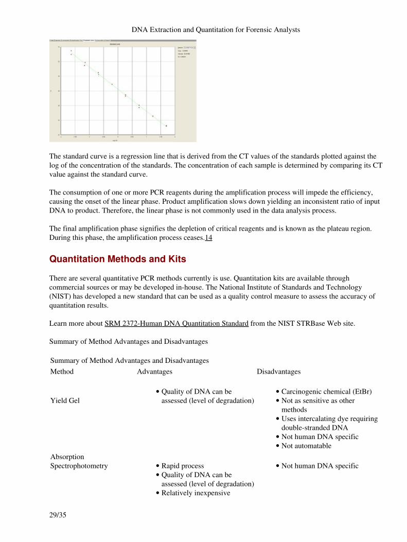
The standard curve is a regression line that is derived from the CT values of the standards plotted against thelog of the concentration of the standards. The concentration of each sample is determined by comparing its CTvalue against the standard curve.
The consumption of one or more PCR reagents during the amplification process will impede the efficiency,causing the onset of the linear phase. Product amplification slows down yielding an inconsistent ratio of inputDNA to product. Therefore, the linear phase is not commonly used in the data analysis process.
The final amplification phase signifies the depletion of critical reagents and is known as the plateau region.During this phase, the amplification process ceases.14
Quantitation Methods and Kits
There are several quantitative PCR methods currently is use. Quantitation kits are available throughcommercial sources or may be developed in-house. The National Institute of Standards and Technology(NIST) has developed a new standard that can be used as a quality control measure to assess the accuracy ofquantitation results.
Learn more about SRM 2372-Human DNA Quantitation Standard from the NIST STRBase Web site.
Summary of Method Advantages and Disadvantages
Summary of Method Advantages and DisadvantagesMethod Advantages Disadvantages
Yield GelQuality of DNA can beassessed (level of degradation)
• Carcinogenic chemical (EtBr)• Not as sensitive as othermethods
•
Uses intercalating dye requiringdouble-stranded DNA
•
Not human DNA specific• Not automatable•
AbsorptionSpectrophotometry Rapid process•
Quality of DNA can beassessed (level of degradation)
•
Relatively inexpensive•
Not human DNA specific•
DNA Extraction and Quantitation for Forensic Analysts
29/35
Automatable• Fluorometry
Semi-selective fordouble-stranded DNA
•
Relatively sensitive for DNAdetection
•
Automatable•
Not human DNA specific• Quality of DNA cannot beassessed (level of degradation)
•
Slot Blot HybridizationHuman DNA specific• Easy to analyze•
Not automatable• More labor intensive than someother methods
•
Semi-quantitative• Quality of DNA cannot beassessed (level of degradation)
•
AluQuant™Sensitive• Automatable• Human DNA specific•
Quality of DNA cannot beassessed
•
SYBR® GreenSensitive• Indications of inhibition14• Less expensive - norequirement for probes
•
No commercial kit available• Binds to double stranded DNA• Must be placed in a dedicatedroom due to amplified product
•
Fluorescent resonanceenergy transfer (FRET) Rapid process•
Higher precision and accuracycompared to other methods
•
Automatable• Human DNA specific• Indications of inhibition• Sensitive•
Must be placed in a dedicatedroom due to amplified product
•
No commercial kit currentlyavailable for all methods(molecular beacons)
•
Author: Debbie FigarelliDebbie Figarelli serves as DNA Technical Leader at the National Forensic Science Technology Center.Debbie assists with the development of DNA training programs and participates in compliance auditsof DNA laboratories.Author: Leigh ClarkLeigh Clark is a DNA Analyst with the Florida Department of Law Enforcement. Previously, Leighwas Academic Program Specialist at the National Forensic Science Technology Center, where sheauthored this module.Author: Leigh Clark
Author: Leigh ClarkLeigh Clark is a DNA Analyst with the Florida Department of Law Enforcement. Previously, Leighwas Academic Program Specialist at the National Forensic Science Technology Center, where sheauthored this module.Author: Leigh Clark
DNA Extraction and Quantitation for Forensic Analysts
30/35
Leigh Clark is a DNA Analyst with the Florida Department of Law Enforcement. Previously, Leighwas Academic Program Specialist at the National Forensic Science Technology Center, where sheauthored this module.Author: Carrie SutherlandCarrie Sutherland joined the NFSTC in June 2005 as the Senior Physical Scientist. As the onsite DNAanalyst, she is responsible for validating DNA instruments, providing laboratory support andinstruction for workshops and DNA academies, participating in compliance audits of DNAlaboratories, contributing to the creation of a laboratory training manual for the President's DNAInitiative - Analyst Training project, and for maintaining proficiency for STR DNA testing.Author: Debbie FigarelliDebbie Figarelli serves as DNA Technical Leader at the National Forensic Science Technology Center.Debbie assists with the development of DNA training programs and participates in compliance auditsof DNA laboratories.Resources by Module
DNA Analysis Considerations• Organic Extraction• Chelex 100 Extraction• Other Extraction Techniques• Quantitation•
DNA Analysis Considerations
Works Cited
Wilson, I. G. 1997. Inhibition and facilitation of nucleic acid amplification. Appl Environ Microbiol63 (10): 3741–51.
1.
Harry, M., B. Gambier, Y. Bourezgui, and E. Garnier-Sillam. 1999. Evaluation of purificationprocedures for DNA extracted from organic rich samples: Interference with humic substances.Analusis 27 (5): 439–42.
2.
Al-Soud, W. A., and P. Radstrom. 2001. Purification and characterization of PCR-inhibitorycomponents in blood cells. J Clin Microbiol 39 (2): 485–93.
3.
Shutler, G. G., Gagnon, P., Verret, G., Kalyn, H., Korkosh, S., Johnston, E., and Halverson, J. 1999.Removal of a PCR inhibitor and resolution of DNA STR types in mixed human-canine stains from afive year old case. J Forensic Sci 44 (3): 623–6.
4.
Innis, Michael A., David H. Gelfand, John J. Sninsky, and Thomas J. White, eds. 1990. PCRprotocols: A guide to methods and applications. San Diego, CA: Academic Press.
5.
Bourke, M. T., C. A. Scherczinger, C. Ladd, and H. C. Lee. 1999. NaOH treatment to neutralizeinhibitors of Taq polymerase. J Forensic Sci 44 (5): 1046–50.
6.
Chung, D. T., J. Drabek, K. L. Opel, J. M. Butler, B. R. McCord. 2004. A study on the effects ofdegradation and template concentration on the amplification efficiency of the STR Miniplex primersets. J Forensic Sci 49 (4): 733–40.
7.
Online Links
What Every Law Enforcement Officer Should Know About DNA Evidencehttp://dna.gov/training/letraining/default_page
•
DNA Extraction and Quantitation for Forensic Analysts
31/35

Organic Extraction
Works Cited
Lorente, M., C. Entrala, J. A. Lorente, J. C. Alvarez, E. Villanueva, B. Budowle. 1998. Dandruff as apotential source of DNA in forensic casework. J Forensic Sci 43 (4): 901–2.
1.
Drobnic, K. 2003. Analysis of DNA evidence recovered from epithelial cells in penile swabs.Croatian Medical Journal 44 (3): 350–54.
2.
Schmerer, W. M., S. Hummel, and B. Herrmann. 1999. Optimized DNA extraction to improvereproducibility of short tandem repeat genotyping with highly degraded DNA as target.Electrophoresis 20 (8): 1712–16.
3.
Primorac, D., S. Andelinovic, M. Definis-Gojanovic, I. Drmic, B. Rezic, M. M. Baden, M. A.Kennedy, M. S. Schanfield, S. B. Skakel, and H. C. Lee. 1996. Identification of war victims frommass graves in Croatia, Bosnia, and Herzegovina by use of standard forensic methods and DNAtesting. J Forensic Sci 41(5): 891–94.
4.
Zehner, R., J. Amendt, and R. Krettek. 2004. STR typing of human DNA from fly larvae fed ondecomposing bodies. J Forensic Sci 49(2): 337–40.
5.
Baechtel, F. S. 1989. The extraction, purification and quantification of DNA. In Proceedings of theInternational Symposium on the Forensic Aspects of DNA Analysis, 25–28. Washington, D.C.:U.S. Government Printing Office.
6.
Comey, C. T. 1994. DNA extraction strategies for amplified fragment length polymorphism analysis.J Forensic Sci 39 (5): 1254–69.
7.
Yoshida, K., K. Sekiguchi, N. Mizuno, K. Kasai, I. Sakai, H. Sato, and S. Seta. 1995. The modifiedmethod of two-step differential extraction of sperm and vaginal epithelial cell DNA from vaginal fluidmixed with semen. Forensic Sci Int 72(1): 25–33.
8.
Read the Sam Baechtel paper.
Online Links
Differex™ System web pagehttp://www.promega.com/catalog/catalogproducts.asp?catalog_name=Promega_Products&category_name=Differex+System&cookie_test=1
•
Millipore Corporation WebsiteUltraClean™ page on MO BIO's websitehttp://www.mobio.com/products/categorylist.php?cat_id=5
•
Chelex 100 Extraction
Works Cited
1. Walsh, P. S., D. A. Metzger, and R. Higuchi. 1991. Chelex 100 as a medium for simple extraction of DNAfor PCR-based typing from forensic material. Biotechniques 10 (4): 506–13.
2. Suenaga, E., and H. Nakamura. 2005. Evaluation of three methods for effective extraction of DNA fromhuman hair. J Chromatography Biology Analyt Technol Biomed Life Sci 820 (1): 137–41.
DNA Extraction and Quantitation for Forensic Analysts
32/35
3. Vandenberg N., R. A. van Oorschot, and R. J. Mitchell. 1997. An evaluation of selected DNA extractionstrategies for short tandem repeat typing. Electrophoresis 18 (9): 1624–6.
4. Tsuchimochi, T., M. Iwasa, Y. Maeno, H. Koyama, H. Inoue, I. Isobe, R. Matoba, M. Yokoi, and M.Nagao. 2002. Chelating resin-based extraction of DNA from dental pulp and sex determination fromincinerated teeth with Y-chromosomal alphoid repeat and short tandem repeats. Am J Forensic Med Pathol 23(3): 268–71.
5. Sweet, D., M. Lorente, A. Valenzuela, J. A. Lorente, and J. C. Alvarez. 1996. Increasing DNA extractionyield from saliva stains with a modified Chelex method. Forensic Sci Int 83 (3): 167–77.
6. Greenspoon, S. A., M. A. Scarpetta, M. L. Drayton, and S. A. Turek. 1998. QIAamp spin columns as amethod of DNA isolation for forensic casework. J Forensic Sci 43 (5): 1024–30.
7. Hoff-Olsen, P., B. Mevag, E. Staalstrom, B. Hovde, T. Egeland, and B. Olaisen B. 1999. Extraction ofDNA from decomposed human tissue: An evaluation of five extraction methods for short tandem repeattyping. Forensic Sci Int 105 (3): 171–83.
8. Comey, C. T. 1994. DNA extraction strategies for amplified fragment length polymorphism analysis. JForensic Sci 39 (5): 1254–69.
9. Yoshida, K., K. Sekiguchi, N. Mizuno, K. Kasai, I. Sakai, H. Sato, and S. Seta. 1995. The modified methodof two-step differential extraction of sperm and vaginal epithelial cell DNA from vaginal fluid mixed withsemen. Forensic Sci Int 72 (1): 25–33.
Online Links
Bio-Radhttp://www.bio-rad.com
•
Other Extraction Techniques
Works Cited
Corning Life Sciences. 2006. Product catalog entry, Costar® Spin-X® centrifuge tube insert withoutmembrane, nonsterile, 1000/case (item #9301).
1.
Millipore Corporation. 2006. Product catalog entry, Microcon® centrifugal filter units.http://www.millipore.com/catalogue.nsf/docs/C3034
2.
Horsman, K. M., S. L. Barker, J. P. Ferrance, K. A. Forrest, K. A. Koen, and J. P. Landers. 2005.Separation of sperm and epithelial cells in a microfabricated device: Potential application to forensicanalysis of sexual assault evidence. Analytical Chemistry 77 (3): 742–49.
3.
Greenspoon, S. A., M. A. Scarpetta, M. L. Drayton, and S. A. Turek. 1998 . QIAamp spin columns asa method of DNA isolation for forensic casework. J Forensic Sci 43 (5): 1024–30.
4.
Sinclair, K., and V. M. McKechnie. 2000. DNA extraction from stamps and envelope flaps usingQIAamp and QIAshredder. J Forensic Sci 45 (1): 229–30.
5.
Vandenberg, N., and R. A. van Oorschot. 2002. Extraction of human nuclear DNA from fecessamples using the QIAamp DNA Stool Mini Kit. J Forensic Sci 47 (5): 993–95.
6.
Greenspoon, S. A., J. D. Ban, K. Sykes, E. J. Ballard, S. S. Edler, M. Baisden, and B. L. Covington.2004. Application of the BioMek 2000 Laboratory Automation Workstation and the DNA IQ Systemto the extraction of forensic casework samples. J Forensic Sci 49 (1): 29–39.
7.
DNA Extraction and Quantitation for Forensic Analysts
33/35
Nagy, M., P. Otremba, C. Kruger, S. Bergner-Greiner, P. Anders, B. Henske, M. Prinz, and L.Roewer. 2005. Optimization and validation of a fully automated silica-coated magnetic beadspurification technology in forensics. Forensic Sci Int 152 (1): 13–22.
8.
Salvadore, J. M., and M. C. De Ungria. 2003. Isolation of DNA from saliva of betel quid chewersusing treated cards. J Forensic Sci 48 (4): 794–97.
9.
Thacker C., C. P. Phillips, and D. Syndercombe-Court. 2000. Use of FTA cards in small volume PCRreactions. Progress in Forensic Genetics 8 (1999): 473–75.
10.
Online Links
Filter Paper Cards on Whatman's websitehttp://www.whatman.com/products/?pageID=7.31
•
UltraClean™ page on MO BIO's websitehttp://www.mobio.com/products/categorylist.php?cat_id=5
•
Quantitation
Works Cited
Baechtel, F. S. 1989. The extraction, purification and quantification of DNA. In Proceedings of theInternational Symposium on the Forensic Aspects of DNA Analysis, 25–28. Washington, D.C.:U.S. Government Printing Office.
1.
Turner BioSystems. YEAR? Fluorescence theory. In An introduction to fluorescence measurements.http://www.turnerbiosystems.com/ doc/appnotes/998_0050/0050_c1.html (accessed August 31,2006).
2.
Turner BioSystems. A TD-700 Laboratory Fluorometer Method for PicoGreenR" Turner Designs.Luminometers and Fluorometers
3.
"QuantiBlotTM. A DNA Quantitation Method" Perkin Elmer, April 1996. 7-154. Walsh, P. S., J. Varlaro, and R. Reynolds. 1992. A rapid chemiluminescent method for quantitation ofhuman DNA. Nucleic Acids Res 20 (19): 5061-65.
5.
Promega Corporation. 2006. Technical Bulletin, AluQuant® Human DNA Quantitation System,instructions for use of products DC1010 and DC1011, part #TB291.www.promega.com/tbs/tb291/tb291.pdf (accessed August 31, 2006)
6.
Edwards, Kirstin, Julie Logan, and Nick Saunders, eds. 2004. Real-time PCR: An essential guide,4–5. Wymondham, Norfolk, U.K.: Horizon Bioscience.
7.
Edwards, Kirstin, Julie Logan, and Nick Saunders, eds. 2004. Real-time PCR: An essential guide,18–22. Wymondham, Norfolk, U.K.: Horizon Bioscience.
8.
Edwards, Kirstin, Julie Logan, and Nick Saunders, eds. 2004. Real-time PCR: An essential guide,45–46. Wymondham, Norfolk, U.K.: Horizon Bioscience.
9.
Timken, Mark D., Katie L. Swango, Cristián Orrego, Mavis Date Chong, and Martin R.Buoncristiani. 2005. Quantitation of DNA for forensic DNA typing by qPCR (quantitative PCR):Singleplex and multiplex modes for nuclear and mitochondrial genomes, and the Y chromosome.Final report for U.S. Department of Justice grant no. 2002-IJ-CX-K008.
10.
Department of Biology, Davidson College. 2001. Real-time PCR method. Genomics course materialshttp://www.bio.davidson.edu/courses/ genomics/method/realtimepcr.html (accessed August 31,2006).
11.
Edwards, Kirstin, Julie Logan, and Nick Saunders, eds. 2004. Real-time PCR: An essential guide,51–52. Wymondham, Norfolk, U.K.: Horizon Bioscience.
12.
Applied Biosystems. 2003. QuantifilerTM Human DNA Quantification Kit and QuantifilerTM YHuman DNA Quantification Kit User's Manual, rev. B.
13.
DNA Extraction and Quantitation for Forensic Analysts
34/35

Butler, John M. 2005. Forensic DNA typing: Biology, technology, and genetics of STR markers. 2nded, 77–78. Burlington, MA: Elsevier Academic Press.
14.
Online Links
Abstract for AluQuant® Human DNA Quantitation Systemhttp://www.promega.com/tbs/tb291/tb291.html
•
FRET at Wikipediahttp://en.wikipedia.org/wiki/Fluorescence_resonance_energy_transfer
•
DNA Extraction and Quantitation for Forensic Analysts
35/35