dna binding-rev3.pdf
TRANSCRIPT
The Journal of Medicinal Chemistry
Asymmetrical di–aromatic guanidinium/2–aminoimidazolinium
derivatives: synthesis and DNA affinity
Padraic S. Nagle, Fernando Rodriguez,
§ Amila Kahvedžić, Susan J. Quinn,
#* and
Isabel Rozas*
School of Chemistry, University of Dublin, Trinity College, Dublin 2, Ireland
Abstract
In this paper we report the synthesis of three families of new amidine–based aromatic
derivatives as potential DNA minor groove binding agents for the treatment of cancer. The
preparation of mono–guanidine, mono–2–aminoimidazoline and asymmetric diphenyl
guanidine/2–aminoimidazoline derivatives (compounds 1a,b,c to 8a,b,c) is presented. The
affinity of these substrates and of a family of mono– and bis–isoureas (previously prepared in
Rozas’ laboratory) for DNA was evaluated by means of DNA thermal denaturation
measurements. In particular, compounds 2c, 5c, 6c, 7c, and 8c were found to bind strongly
both to natural DNA and to Adenine-Thymine oligonucleotides, showing a preference for the
Adenine-Thymine base pairs sequences.
Abbreviations: HB, hydrogen bond; ΔTm, increment in DNA denaturation temperature; AT,
Adenine–Thymine pairs; MES, 2–(N–morpholino)ethanesulfonic acid; P/D, ratio between
base pairs and ligand (drug);
* Authors to whom correspondence should be addressed: e–mail: [email protected],
[email protected]; FAX: +353 1 671 2826. Phone: +353 1 896 3731.
§ Present address: Departamento de Quimica, Universidad de La Rioja, Grupo de Sintesis
Quimica de La Rioja, UA–CSIC, E–26006 Logroño, Spain.
# Present address: School of Chemistry and Biochemistry, University College Dublin, Ireland

2
Introduction
The DNA minor–groove is the interaction site for many replicative and repair enzymes
and transcription control proteins. It also provides an opportunity to design drugs that are
capable of selective binding and hence the potential to interfere with these processes.
Minor-groove binding agents typically have long and planar structures that allow them to
adopt a crescent shape that fits into the minor–groove forming close contacts in the deep,
narrow space formed between the two DNA strands. These DNA/minor–groove binder
complexes are typically stabilized by hydrogen bonds (HBs), van der Waals contacts and/or
ionic interactions.1
Binding is typically driven by hydrophobic interactions and if the
molecule is positively charged this is usually accompanied by the liberation of a large number
of water molecules. The positively charged natural products distamycin and netropsin were
among the first examples of molecules that specifically target the minor-groove. Since their
discovery, many drugs that target the minor–groove have been developed. They are highly
selective and they compromise the fundamental biochemistry of a cell through inhibition of
the actions of DNA–dependent enzymes, for example by direct inhibition of transcription.2,3,4
Sophisticated synthesis has yielded oligoamides capable of highly specific binding;5
further development has also witnessed the emergence of hairpins polyamides to latch around
the groove.6 While many different minor–groove binders have been synthesised and tested as
anticancer agents, their severe toxicity has prevented any therapeutic use. However, some
dicationic aromatic diamidines such as furamidine (Figure 1),7 have shown promising
antiproliferative activity against different tumour cell–lines. The furamidine derivative
furimidazoline is one particularly good example.8 It is worth noting that fluorescence
microscopy indicates the rapid nuclear accumulation of some furamidines in tumour cells.9
These findings together open new possibilities for developing novel aromatic dicationic
derivatives with enhanced interaction and selectivity within the DNA minor–groove and
possibly with more selective toxicity.
3
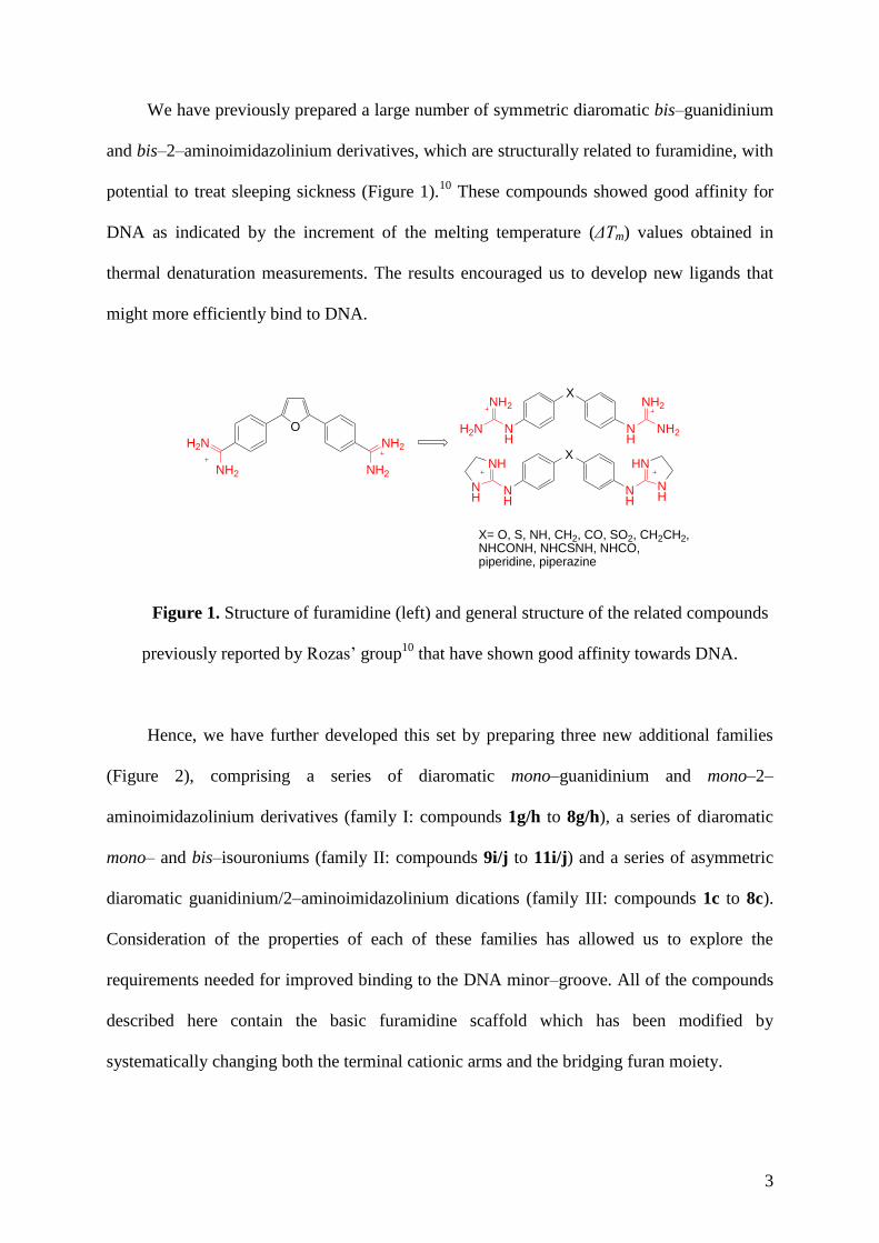
We have previously prepared a large number of symmetric diaromatic bis–guanidinium
and bis–2–aminoimidazolinium derivatives, which are structurally related to furamidine, with
potential to treat sleeping sickness (Figure 1).10
These compounds showed good affinity for
DNA as indicated by the increment of the melting temperature (ΔTm) values obtained in
thermal denaturation measurements. The results encouraged us to develop new ligands that
might more efficiently bind to DNA.
O
H2N
NH2
NH2
NH2
X
NH
NH
NH2 NH2
H2N NH2
X
NH
NH
NH
NH
NH
HN
X= O, S, NH, CH2, CO, SO2, CH2CH2, NHCONH, NHCSNH, NHCO, piperidine, piperazine
Figure 1. Structure of furamidine (left) and general structure of the related compounds
previously reported by Rozas’ group10
that have shown good affinity towards DNA.
Hence, we have further developed this set by preparing three new additional families
(Figure 2), comprising a series of diaromatic mono–guanidinium and mono–2–
aminoimidazolinium derivatives (family I: compounds 1g/h to 8g/h), a series of diaromatic
mono– and bis–isouroniums (family II: compounds 9i/j to 11i/j) and a series of asymmetric
diaromatic guanidinium/2–aminoimidazolinium dications (family III: compounds 1c to 8c).
Consideration of the properties of each of these families has allowed us to explore the
requirements needed for improved binding to the DNA minor–groove. All of the compounds
described here contain the basic furamidine scaffold which has been modified by
systematically changing both the terminal cationic arms and the bridging furan moiety.

4
X
H2N NH
HN
NH
X
R O NH2
NH2(CH2)n
n=2, nil
Family I
X
NH
NH
NH2
H2N NH
HN
R= -NH2, -O-C(NH2)2+
Family II
Family III
Figure 2. Structure of the three families studied
The cationic groups may bind to DNA in two ways, firstly through direct H-bonds
through the NH groups and secondly through water-mediated interactions with the bases.
These two mechanisms of binding mean that different interactions, depending on the nature
of the cation, can exist. In this work the amidinium cations have been exchanged with
guanidinium, 2-aminoimidazolinium and isouronium cations. In these cases, we sought to
enhance HB contacts in the minor groove by increasing the number of HB donors (NH
groups in the guanidinium and 2–aminoimidazolinium cations) or introducing an additional
HB acceptor (an O atom in the isouronium cation). Furthermore, these are more articulated
cations than amidinium. The structure of the supporting molecular scaffold or linker, is very
important if we wish to capitalize on these multiple binding interactions. The optimum linker
will allow for a better orientation of the positive charges within the groove and introduce the
curvature or geometry best suited to fitting. However, it is now known that curvature is not
essential to fit into the minor groove, since Boykin et al. have shown that linear molecules are
also capable of strongly binding to the minor–groove.11
Work by Nguyen et al. showed that
in the linear molecule CPG-40215A, the N,N’-diaminoguanidine bridging group served as a
‘see-saw’ hinge facilitating a variety of interactions with the bases through motions that
position the terminal amidine cations in a range of binding modes.12
5
Moreover, to investigate the optimal linking scaffold we have exchanged the furan
moiety of furamidine, for a number of smaller functional groups (O, S, NH, CO, CH2,
CH2CH2 or NHCONH). We hypothesized that such a substitution should afford a two-fold
advantage by firstly facilitating the interaction with DNA through HBs (as donors or
acceptors) and secondly, as our cations are larger than in the furamidine series, making this
central group smaller should ensure optimization of the distance between the cationic
moieties (guanidinium and 2–aminoimidazolinium) for DNA binding.
To assess the binding to DNA we have performed thermal denaturation experiments
in natural DNA (salmon sperm, 68% Adenine-Thymine base pair –AT- content).
Furthermore, since binding to the minor groove requires the presence of a run of five AT base
pairs binding experiments were also conducted in the presence of double stranded AT
homopolymers [poly(dA•dT)2 and poly(dA)•poly(dT)] to assess their potential selectivity for
the minor groove .
Results and Discussion
Chemistry
On the basis of our previous experience and taking into account the good results
obtained by our group10,13,14
the guanidinylation and 2–aminoimidazolinylation of the present
target molecules was performed following our established methodology. This consists of the
reaction of a deactivated aromatic amine with N,N’–bis(tert–butoxycarbonyl)thiourea (Boc
protected guanidine precursor) or N,N’–di(tert–butoxycarbonyl)imidazoline–2–thione13
(Boc
protected 2–aminoimidazoline precursor) assisted by mercury (II) chloride in the presence of
an excess of triethylamine. Deprotection of the Boc–intermediates of the first stage and
further treatment with Amberlyte resin in water leads to the hydrochloride salts of the target
molecules.10,13,14,15
Preparation of the corresponding mono–isouronium (9i, 10i, 11i, Table 1)
and bis–isouronium derivatives (9j, 10j, 11j, Table 2) has been reported by our group in a

6
previous article16
using a similar synthetic approach to the one just described starting from
the corresponding aromatic alcohols.
Synthesis of asymmetric guanidine/2–aminoimidazoline derivatives (Family III)
All our starting materials are symmetric and, hence, the two amino moieties have the
same reactivity. As we intend to introduce different groups at both ends of the molecule, an
excess of the diamines was required to avoid di–functionalization. Thus, three equivalents of
each of the corresponding diamines were treated with just one equivalent of N,N’–bis(tert–
butoxycarbonyl)thiourea, one equivalent of mercury (II) chloride and an excess of
triethylamine to afford the desired mono–functionalized Boc–protected guanidines (1a–8a) as
main products with yields ranging from 29 to 82% (see Scheme 1). Working under these
conditions, less than 5% of the di–functionalized products was observed.
X
H2N NH2
X
NH2NH
BocHN
NBoc
Excess
X
NNH
BocHN
NBoc BocN
NBoc
X
NH
NH
H2N
NH N
NH
X = CH2 (1), CH2CH2 (2), O (3), S (4), NH (5), piperazine (6), CO (7), NHCONH (8)
Reagents and conditions: (A) N,N'-bis(tert-butoxycarbonyl)thiourea, HgCl2, TEA, DCM or
DMF, rt.; (B) N,N'-di(tert-butoxycarbonyl)imidazoline-2-thione, HgCl2, TEA, DCM or DMF, rt.; (C) (i) TFA, DCM, rt. (ii) Amberlyte resin, H2O, rt.
1a-8a
1b-8b 1c-8c
(A) (B)
(C)
Scheme 1
In the second stage of the synthesis, one equivalent of each of the previous
intermediates was reacted with one equivalent of N,N’–di(tert–butoxycarbonyl)imidazoline–
2–thione, a slight excess of mercury (II) chloride and an excess of triethylamine to afford the

7
asymmetric Boc–protected derivatives (1b–8b) in moderate to good yields. Standard
deprotection of the Boc groups with an excess of trifluoroacetic acid in dichloromethane
followed by treatment with Amberlyte resin in water led to the hydrochloride salts of the
asymmetric guanidine– and 2–aminoimidazoline containing products (1c–8c).
Synthesis of amino–guanidinium and amino–2–aminoimidazolinium derivatives (Family I)
Regarding the synthesis of the mono–guanidine and mono–2–aminoimidazoline
containing derivatives, in the first step we decided to protect one of the amino groups of the
starting amines with Boc (see Scheme 2) to increase the solubility of the intermediates and
allow a faster development of the column chromatography.
Following the same strategy as mentioned above, three equivalents of each of the
starting diamines were treated with one equivalent of Boc anhydride. The mono–Boc–
protected amines (1d–6d) were obtained as the main products in good yields.
The urea derivative 8d was prepared following a different route (see Scheme 2). Thus,
reaction of 4–nitrobenzoisocyanate with mono–Boc–protected 4–aminoaniline yielded the
corresponding Boc protected 4–[3–(4–nitrophenyl)ureido]aniline (12), which upon
hydrogenation produced derivative 8d. However, the carbonyl group proved to be too
deactivating and compound 7d (and subsequently 7e, 7f and 7h) could not be prepared. All
these compounds are new, except for 1d17
and 3d18
that have been already described.
In the second stage, the mono–Boc–protected amines were treated with one equivalent
of N,N’–bis(tert–butoxycarbonyl)thiourea or N,N’–di(tert–butoxycarbonyl)imidazoline–2–
thione, a slight excess of mercury (II) chloride and triethylamine. The guanidine precursors
(1e–6e and 8e) were purified by column chromatography in silica gel, whereas for the 2–
aminoimidazoline ones (1f–6f and 8f), neutral alumina was used instead. Finally,
deprotection of the Boc groups afforded the final mono–amidinium–like products after three

8
stages in moderate to good overall yields. It should be mentioned that the amino–guanidine
derivatives 1g–8g could also be obtained after Boc deprotection of 1a–8a. In fact, this was
the only possible route to prepare compound 7g and proved to be a shorter and more efficient
methodology, producing higher overall yields.
X
BocHN NH2
X
H2N NH
NH2
NH
X
H2N NH
NH
N
X
H2N NH2
Excess
X
BocHN N NBoc
BocN
X
BocHN NH
NHBoc
NBoc
Reagents and conditions: (A) Boc2O, TEA, DCM or DMF, rt; (B) N,N'-di(tert-butoxycarbonyl)imidazoline-2-thione, HgCl2, TEA, DCM or DMF, rt.; (C) N,N'-bis(tert-butoxycarbonyl)thiourea, HgCl2, TEA, DCM or DMF,
rt.; (D) (i) TFA, DCM, rt. (ii) Amberlyte resin, H2O, rt.; (E) 4-nitrobenzoisocyanate, DCM, 0 °C; (F) H2 (3 Bar),
Pd-C, MeOH.
X
BocHN NO2
X = NHCONH (12)
NH2
BocHN
(A)
(B)
(D)
(C)
(E)
(D)
1d-6d, 8d
1e-6e, 8e
1f-6f, 8f
1g-8g (7g from 7a)
1h-6h, 8hX = CH2 (1), CH2CH2 (2), O (3), S (4), NH
(5), piperazine (6), CO (7), NHCONH (8)
(F)
Scheme 2
All the diamines used as starting materials are commercially available either from
Aldrich or Fluka, except for the 1,4–(bis–4–aminophenyl)piperazine, whose synthesis has
been described previously.19
It is also worth mentioning that, the unreacted starting diamines
could be recovered from the column eluting with a more polar solvent system.
9
Biophysical Results: Thermal Denaturation Assays
Thermal Denaturation experiments are easy to perform and provide quick and reliable
information on the binding of small molecules to DNA. These assays, for example in
unspecific salmon sperm DNA can be used as a general screening for the suitability of the
compounds studied as DNA binders. Afterwards, the preference for specific base sequences
can be explored.
Denaturation studies of families I–III in the presence of natural DNA
The interaction of all the molecules with DNA was firstly examined by performing
thermal denaturation experiments using a mixed sequence salmon sperm DNA with a 32 %
GC content. The melting temperature of salmon sperm DNA (Tm(SS)) was measured in the
presence of the different families of molecules.
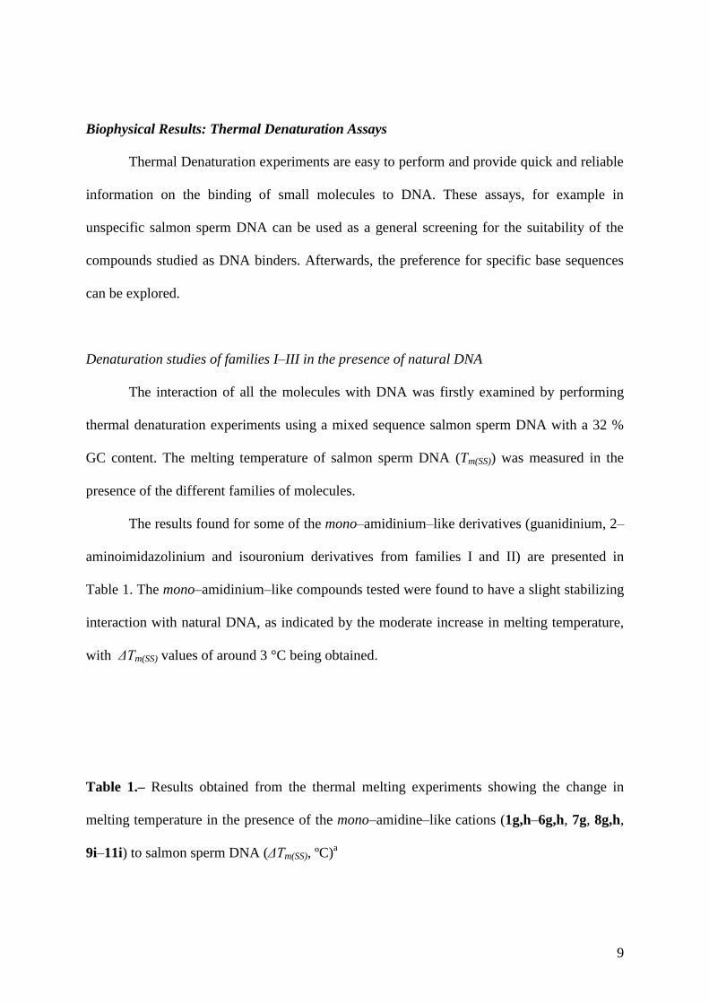
The results found for some of the mono–amidinium–like derivatives (guanidinium, 2–
aminoimidazolinium and isouronium derivatives from families I and II) are presented in
Table 1. The mono–amidinium–like compounds tested were found to have a slight stabilizing
interaction with natural DNA, as indicated by the moderate increase in melting temperature,
with ΔTm(SS) values of around 3 °C being obtained.
Table 1.– Results obtained from the thermal melting experiments showing the change in
melting temperature in the presence of the mono–amidine–like cations (1g,h–6g,h, 7g, 8g,h,
9i–11i) to salmon sperm DNA (ΔTm(SS), ºC)a
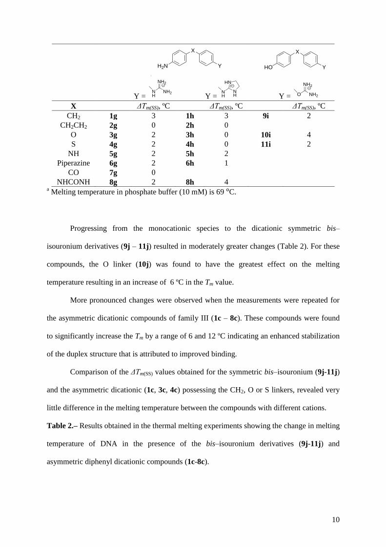
10
X
YH2N
X
YHO
Y = NH2O
NH2
Y = NH2N
H
NH2
Y =NH
HN
NH
X ΔTm(SS), ºC ΔTm(SS), ºC ΔTm(SS), ºC
CH2 1g 3 1h 3 9i 2
CH2CH2 2g 0 2h 0
O 3g 2 3h 0 10i 4
S 4g 2 4h 0 11i 2
NH 5g 2 5h 2
Piperazine 6g 2 6h 1
CO 7g 0
NHCONH 8g 2 8h 4 a Melting temperature in phosphate buffer (10 mM) is 69 °C.
Progressing from the monocationic species to the dicationic symmetric bis–
isouronium derivatives (9j – 11j) resulted in moderately greater changes (Table 2). For these
compounds, the O linker (10j) was found to have the greatest effect on the melting
temperature resulting in an increase of 6 ºC in the Tm value.
More pronounced changes were observed when the measurements were repeated for
the asymmetric dicationic compounds of family III (1c – 8c). These compounds were found
to significantly increase the Tm by a range of 6 and 12 ºC indicating an enhanced stabilization
of the duplex structure that is attributed to improved binding.
Comparison of the ΔTm(SS) values obtained for the symmetric bis–isouronium (9j-11j)
and the asymmetric dicationic (1c, 3c, 4c) possessing the CH2, O or S linkers, revealed very
little difference in the melting temperature between the compounds with different cations.
Table 2.– Results obtained in the thermal melting experiments showing the change in melting
temperature of DNA in the presence of the bis–isouronium derivatives (9j-11j) and
asymmetric diphenyl dicationic compounds (1c-8c).
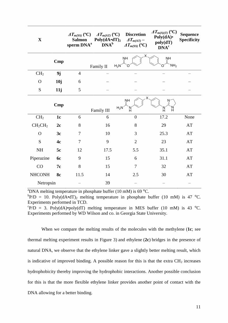
11
X
ΔTm(SS) (ºC)
Salmon
sperm DNAa
ΔTm(AT) (ºC)
Poly(dA•dT)2
DNAb
Discretion
ΔTm(AT) –
ΔTm(SS) (ºC)
ΔTm(A)(T) (ºC)
Poly(dA)•
poly(dT)
DNAc
Sequence
Specificity
Cmp Family II
X
OOH2N
NH NH
NH2
CH2 9j 4 – – – –
O 10j 6 – – – –
S 11j 5 – – – –
Cmp
Family III
X
NH
NH
H2N
NH N
NH
CH2 1c 6 6 0 17.2 None
CH2CH2 2c 8 16 8 29 AT
O 3c 7 10 3 25.3 AT
S 4c 7 9 2 23 AT
NH 5c 12 17.5 5.5 35.1 AT
Piperazine 6c 9 15 6 31.1 AT
CO 7c 8 15 7 32 AT
NHCONH 8c 11.5 14 2.5 30 AT
Netropsin – 39 – – –
aDNA melting temperature in phosphate buffer (10 mM) is 69 °C.
bP/D = 10. Poly(dA•dT)2 melting temperature in phosphate buffer (10 mM) is 47 °C.
Experiments performed in TCD. cP/D = 3. Poly(dA)•poly(dT) melting temperature in MES buffer (10 mM) is 43 °C.
Experiments performed by WD Wilson and co. in Georgia State University.
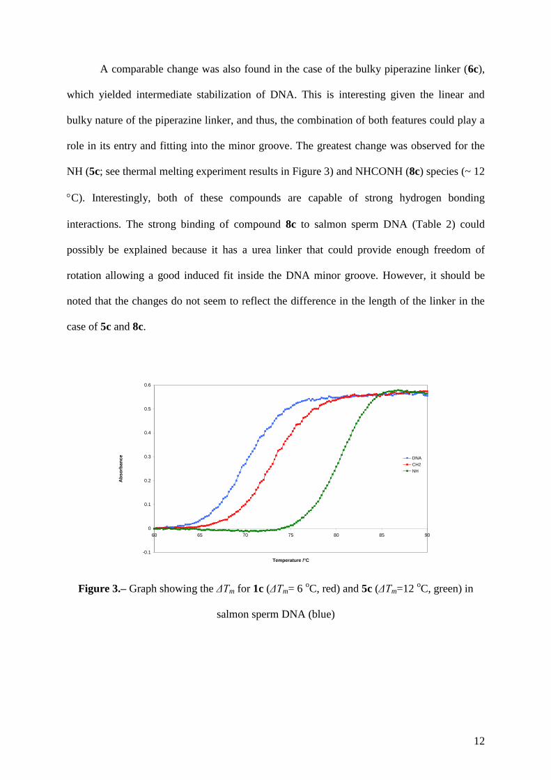
When we compare the melting results of the molecules with the methylene (1c; see
thermal melting experiment results in Figure 3) and ethylene (2c) bridges in the presence of
natural DNA, we observe that the ethylene linker gave a slightly better melting result, which
is indicative of improved binding. A possible reason for this is that the extra CH2 increases
hydrophobicity thereby improving the hydrophobic interactions. Another possible conclusion
for this is that the more flexible ethylene linker provides another point of contact with the
DNA allowing for a better binding.
12
A comparable change was also found in the case of the bulky piperazine linker (6c),
which yielded intermediate stabilization of DNA. This is interesting given the linear and
bulky nature of the piperazine linker, and thus, the combination of both features could play a
role in its entry and fitting into the minor groove. The greatest change was observed for the
NH (5c; see thermal melting experiment results in Figure 3) and NHCONH (8c) species (~ 12
C). Interestingly, both of these compounds are capable of strong hydrogen bonding
interactions. The strong binding of compound 8c to salmon sperm DNA (Table 2) could
possibly be explained because it has a urea linker that could provide enough freedom of
rotation allowing a good induced fit inside the DNA minor groove. However, it should be
noted that the changes do not seem to reflect the difference in the length of the linker in the
case of 5c and 8c.
-0.1
0
0.1
0.2
0.3
0.4
0.5
0.6
60 65 70 75 80 85 90
Temperature /°C
Ab
so
rba
nc
e DNA
CH2
NH
Figure 3.– Graph showing the ΔTm for 1c (ΔTm= 6 oC, red) and 5c (ΔTm=12
oC, green) in
salmon sperm DNA (blue)

13
Denaturation studies of family III in the presence of poly(dA•dT)2 and poly(dA)•poly(dT)
The experiments carried out on the asymmetric series in the presence of the salmon
sperm DNA revealed that, considering the linkers, the order of affinity is: –NH–
–NHCONH– > (–Piperazine– , –CO– , –CH2CH2– , –O– , –S– , –CH2–). In light of the
promising results found for family III, the experiments were repeated in the presence of
poly(dA•dT)2 using similar experimental conditions. Here, the absence of the G•C base pair
reduces the width of the groove allowing for a tighter fit, with minor groove complexes
typically forming with a run of four A•T base pairs. In almost all experiments, a greater
change in melting temperature was found in the presence of the asymmetric dications (2c–8c)
in the AT oligonucleotides. This result was taken as a strong indication of a preference for the
minor groove binding site afforded in homopolymer DNA and indicates sequence selectivity
for the AT domains of DNA (Table 2). The exception was found for the CH2 species (1c)
which was found to stabilize the homopolymer to the same extent as natural DNA.
However, in the case of the ethylene linker (2c) the melting temperature showed a
two–fold increment compared to salmon sperm DNA. This would imply that while 2c binds
to AT rich domains, which are characterized by a narrow minor groove, 1c is unlikely to bind
in the same fashion as the increased availability of binding sites in the AT homopolymer is
not accompanied by increased binding affinity. Thus, it may be concluded that the restricted
degree of rotation conferred by the –CH2– bridge does not favour insertion into the narrower
minor groove present in the AT DNA and the molecule most probably binds more
effectively through an alternative mode. However, the higher degree of rotation due to the
extra CH2 in 2c, allows the molecule to wrap around the minor groove better than 1c.
Next, we considered the influence of the other bridging groups in the selective
binding between the different DNA systems. By comparing molecules 1c, 3c, 4c and 5c
(linker: –CH2–, –O–, –S– and –NH– respectively), it was noted that compound 5c had the
highest melting temperature in both salmon sperm DNA and poly(dA•dT)2. This was
14
followed by molecules 3c and 4c that have the same melting temperature in salmon sperm
DNA but slightly different in poly(dA•dT)2 DNA (by 1 °C). As noted previously, the
smallest change was observed for compound 1c. These results seem to indicate the
importance of hydrogen bonding with a possible HB donation to the O2 of thymine being a
potential dominant factor. The fact that 3c and 4c gave better melting results in comparison to
1c illustrates further the order of binding since the oxygen and sulphur linkers could be HB
acceptors, but the methylene bridge is a known weak HB donor.
However, if we consider the relative sensitivity of the melting temperature (Trel
)
defined to the DNA type (Tm(AT) – Tm(SS); see Table 2), two distinct groupings emerge. The
first group contains the compounds 2c (–CH2CH2–), 5c (–NH–), 6c (–piperazine–) and 7c
(–CO–) and shows a Trel
change of the order of 6 C. The second grouping shows
significantly less sensitivity to the two forms of DNA, 1c (–CH2–), 8c (–NHCONH–), 4c
(–S–) and 3c (–O–), with the ether linker showing the greatest difference of 3 C.
When comparing the results for molecules 1c, 5c and 7c (linkers: –CH2–, –NH– and
–CO– respectively), the primary difference is the HB characteristics of the corresponding
linkers, with the methylene bridge being a weak HB donor, the –NH– a stronger HB donor
and the carbonyl group a HB acceptor. The order of binding to DNA and AT
oligonucleotides, from best to worst, was found to be: strong HB donor (NH) > HB acceptor
(CO) > weak HB donor (CH2). This indicates that, in the synthesis of future molecules,
incorporating more HB donors would be advantageous. The geometry around the bridging
linker must also be considered as it is likely to play an important role. Here, there are two
important factors. Firstly, the steric nature of the geometry (tetrahedral –CH2– vs trigonal
planar –CO– and trigonal pyramidal –NH–) will influence the approach to and binding in the
pocket. Secondly, the rotational freedom conferred by the bridging moiety would be expected
to influence the binding. There is no freedom of rotation in 7c (trigonal planar) whereas there
15
is freedom of rotation around the linker for molecules 1c (tetrahedral linker) and 5c (trigonal
pyramidal linker). It is interesting to note that compound 6c not only shows a medium affinity
towards natural DNA but also to poly(dA•dT)2. This linker is longer than the rest of the
groups considered and the relatively good results obtained could be justified by the large size
and linear characteristics of the piperazine ring. The Tm of netropsin (a well known minor
groove binder) was also measured in this homopolymer for the sake of comparison. Even
though this compound shows a two-fold binding strength compared to the asymmetric
dications, this can be explained by the large number of HB donor and acceptor groups present
in this molecule.
AT sequence selectivity between different 5-base-pair binding sites has previously
been reported for the different binding modes (1:1 versus 2:1) of the classic minor grove
binder distamycin.20
In the study by Chen et al., a preference for the ATATA over AAAAA
was observed. In addition, small molecules can bind in a 2:1 and a 1:1 fashion in the minor-
groove and this seems to strongly depend on the sequence. It has been found by Vesnaver and
co.21
that the narrowest groove is that of five base pairs of AT and Netropsin prefers 1:1
binding in such system. For this reason, we also considered the interesting results already
published by us in collaboration with W.D. Wilson (Georgia State University, USA) on the
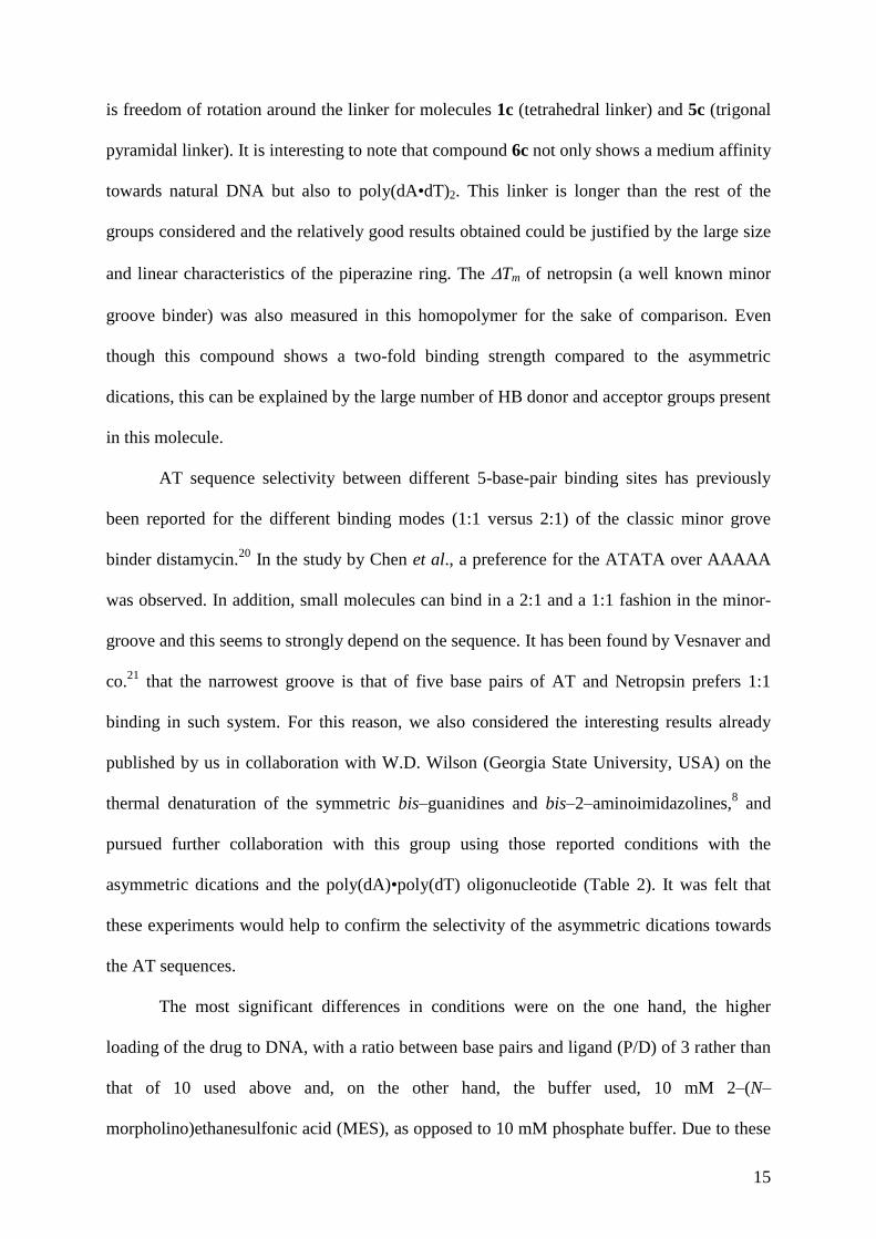
thermal denaturation of the symmetric bis–guanidines and bis–2–aminoimidazolines,8 and
pursued further collaboration with this group using those reported conditions with the
asymmetric dications and the poly(dA)•poly(dT) oligonucleotide (Table 2). It was felt that
these experiments would help to confirm the selectivity of the asymmetric dications towards
the AT sequences.
The most significant differences in conditions were on the one hand, the higher
loading of the drug to DNA, with a ratio between base pairs and ligand (P/D) of 3 rather than
that of 10 used above and, on the other hand, the buffer used, 10 mM 2–(N–
morpholino)ethanesulfonic acid (MES), as opposed to 10 mM phosphate buffer. Due to these
16
differences, the results obtained cannot be quantitatively compared to the poly(dA•dT)2
results. However, the trend observed is similar and a good correlation was found between
both sets of results as shown in Figure 4. Comparing the results previously published with
Wilson’s group on the thermal denaturation of the symmetric bis–guanidines and bis–2–
aminoimidazolines,10
and those found now using the same Wilson’s conditions (Table 2), it is
interesting to observe how the Tm values obtained for the asymmetric compounds are almost
an average of those measured for the bis–guanidines (smaller) and those obtained for the bis–
2–aminoimidazolines (larger). This would imply a similar binding mode in both AT DNAs
and, most probably linked to a 1:1 mode as would be expected given the bis-cationic nature
of the compounds under study.
Tm (dA)(dT)= 1.38 Tm (dAdT) + 10.15
R2 = 0.929
15
20
25
30
35
40
4 9 14 19
poly(dAdT)
poly
(dA
)poly
(dT
)
Figure 4. Correlation found between the ΔTm poly(dA•dT)2 vs. ΔTm poly(dA)•poly(dT)
From these studies, it can be concluded that hydrophobic interactions, hydrogen
bonding, electrostatic interactions and molecular conformation are all important factors in
DNA minor groove binding for the asymmetric dications. More importantly, these show
strong binding to DNA showing AT–sequence specificity.

17
Conclusions
We have prepared three different families of mono– and bis–amidinium–based
derivatives, in order to explore their potential as DNA minor–groove binders. First, with
respect to family I, the mono–guanidinium and mono–2–aminoimidazolinium derivatives
have been prepared following the synthetic pathways previously described by us.10,13,14
Second, regarding family II, the preparation of the asymmetric guanidinium/2–
aminoimidazolinium di–cations has been performed from the corresponding mono–
guanidinium derivatives and subsequent introduction of the 2–aminoimidazoline group.
Third, regarding family III, the preparation of the mono– and bis–isouronium derivatives has
been reported in a previous paper from our group.16
The ability of these compounds to bind to DNA has been evaluated by thermal
denaturation experiments not only with mixed sequence DNA (salmon sperm), which
comprises all four bases, but also with poly(dA•dT)2 and poly(dA)•poly(dT)
oligonucleotides. Despite showing cationic groups at both ends of the di–aromatic moieties,
mono–amidinium–like derivatives (ammonium and guanidinium or 2–aminoimidazolinium)
and bis–isouroniums (two isouronium cations) have shown to poorly/moderately bind to
DNA. Nevertheless, in general, asymmetric bis–amidinium–based derivatives displayed a
good affinity towards DNA and a preference for the AT sequences. In particular, compounds
2c, 5c, 6c, 7c and 8c strongly bind to both unspecific and AT oligonucleotides.
Taking into consideration these promising results, we believe that these asymmetric
compounds deserve more investigation as DNA targeting agents and thus, more biophysical
studies are ongoing and will be reported soon.

18
Experimental
Chemistry
All the commercial chemicals were obtained from Sigma–Aldrich or Fluka and were
used without further purification. Deuterated solvents for NMR use were purchased from
Apollo. Dry solvents were prepared using standard procedures, according to Vogel, with
distillation prior to use. Chromatographic columns were run using Silica gel 60 (230–400
mesh ASTM) or Aluminium Oxide (activated, Neutral Brockman I STD grade 150 mesh).
Solvents for synthesis purposes were used at GPR grade. Analytical TLC was performed
using Merck Kieselgel 60 F254 silica gel plates or Polygram Alox N/UV254 aluminium oxide
plates. Visualisation was by UV light (254 nm). NMR spectra were recorded in a Bruker
DPX–400 Avance spectrometer, operating at 400.13 MHz and 600.1 MHz for 1H–NMR and
100.6 MHz and 150.9 MHz for 13
C–NMR. Shifts are referenced to the internal solvent
signals. NMR data were processed using Bruker Win–NMR 5.0 software. Electrospray mass
spectra were recorded on a Mass Lynx NT V 3.4 on a Waters 600 controller connected to a
996 photodiode array detector with methanol, water or ethanol as carrier solvents. Melting
points were determined using an Electrothermal IA9000 digital melting point apparatus and
are uncorrected. Infrared spectra were recorded on a Mattson Genesis II FTIR spectrometer
equipped with a Gateway 2000 4DX2–66 workstation and on a Perkin Elmer Spectrum One
FT–IR Spectrometer equipped with Universal ATR sampling accessory. Elemental analyses
(C, H, N) of the target compounds, which were performed at the Microanalysis Laboratory
(School of Chemistry and Chemical Biology, University College Dublin), are within ±0.4%
of the calculated values confirming ≥ 95% purity. Fractional moles of water frequently found
in amidinium-like salts could not be prevented despite 24-48 h of drying in vacuum.

19
General method for the synthesis of mono–functionalized Boc–protected guanidines:
Method 1 [Scheme 1, conditions (A)].
Over a solution containing an excess (9.0 mmol) of each of the starting diamines, 3.0 mmol
of N,N’–di(tert–butoxycarbonyl)thiourea and 1.3 mL (9.3 mmol) of TEA in DCM (5 mL) at 0
°C, 3.3 mmol of HgCl2 were added. The resulting mixture was stirred at 0 °C for 1 h and for
the appropriate duration at room temperature. Then, the reaction was diluted with EtOAc and
filtered through a pad of Celite. The filter cake was rinsed with EtOAc. The organic phase
was washed with water (2 x 30 mL), washed with brine (1 x 30 mL), dried over anhydrous
Na2SO4 and concentrated under vacuum to give a residue that was purified by silica gel
column chromatography, eluting with the corresponding hexane:EtOAc mixture to yield the
desired compound as a solid. Less than 5% of the disubstituted substrate was observed.
General procedure for the synthesis of Boc–protected asymmetric guanidine/2–
aminoimidazoline, Boc–protected amino/2–aminoimidazolidine, and Boc–protected
amino/guanidine derivatives: Method 2 [Scheme 1, conditions (B); Scheme 2, conditions
(B) and (C)].
Each of the corresponding amines was treated in DCM at 0 °C with 1.1 equivalents of
mercury (II) chloride, 1.0 equivalent of N,N’–di(tert–butoxycarbonyl)imidazolidine–2–thione
(for the 2–aminoimidazoline precursors) or N,N’–di(tert–butoxycarbonyl)thiourea (for the
guanidine precursors) and 3.1 equivalents of TEA. The resulting mixture was stirred at 0 °C
for 1 hour and for the appropriate duration at room temperature. Then, the reaction mixture
was diluted with EtOAc and filtered through a pad of Celite to get rid of the mercury sulfide
formed. The filter cake was rinsed with EtOAc. The organic phase was washed with water (2
x 30 mL), washed with brine (1 x 30 mL), dried over anhydrous Na2SO4 and concentrated
under vacuum to give a residue that was purified by silica gel (guanidine precursors) or
20
neutral alumina column flash chromatography (2–aminoimidazoline precursors), eluting with
the appropriate hexane:EtOAc mixture.
General procedure for the synthesis of mono–Boc–protected 4,4’–diphenyl diamines:
Method 3. Compounds 1d to 6d [Scheme 2, conditions (A)].
Over a solution containing an excess (30.0 mmol) of each of the corresponding diamines in
DCM (200 mL), 10.0 mmol of TEA and 10.0 mmol of di–tert–butyl dicarbonate (Boc2O)
were added at 0 °C. The resulting mixture was stirred at 0 °C for 1 h and 16 h more at room
temperature. Then, the reaction was concentrated under vacuum to give a residue that was
purified by silica gel column chromatography, eluting with the appropriate hexane:EtOAc
mixture. The mono–Boc–protected compound was obtained as a solid. The excess of
unreacted starting material diamine was recovered from the column. Less than 10%
formation of the di–Boc–protected compound was observed.
General procedure for the synthesis of the hydrochloride salts: Method 4.
Each of the corresponding Boc–protected precursors (0.5 mmol) was treated with 15 mL of a
50% solution of trifluoroacetic acid in DCM for 3 h. After that time, the solvent was
eliminated under vacuum to generate the trifluoroacetate salt. This salt was dissolved in 20
mL of water and treated for 24 h with IRA400 Amberlyte resin in its Cl– form. Then, the
resin was removed by filtration and the aqueous solution washed with DCM (2 x 10 mL).
Evaporation of the water afforded the pure hydrochloride salt. Absence of the trifluoroacetate
salt was checked by 19
F NMR.
21
Dihydrochloride salt of N–{4–[4–(imidazolidin–2–ylideneamino)benzyl]phenyl}
guanidine (1c): Method 4.
White solid (96%); mp 78–80 °C; 1H NMR (D2O) 3.58 (s, 4H, CH2), 3.76 (s, 2H,
PhCH2Ph), 6.98 (d, 2H, J = 8.0 Hz, Ar.), 7.01 (d, 2H, J = 8.0 Hz, Ar.), 7.15 (d, 2H, J = 8.0
Hz, Ar.), 7.16 (d, 2H, J = 8.0 Hz, Ar.); 13
C NMR (D2O) 39.6 (PhCH2Ph), 42.2 (CH2 Im),
123.3, 125.1, 129.6, 129.7, 131.5, 132.6, 139.7, 140.4 (Ar.), 155.5, 157.6 (CN); HRMS
(ESI+) m/z 309.1728 calcd [M + H]
+; found 309.1732. Anal. (C17H22Cl2N6·1.5H2O) C, H, N.
Dihydrochloride salt of N–(4–{2–[4–(imidazolidin–2–ylideneamino)phenyl]ethyl}
phenyl)guanidine (2c): Method 4.
White solid (94%); mp decomposes over 215 °C; 1H NMR (D2O) 2.84 (s, 4H, CH2), 3.67
(s, 4H, CH2 Im), 7.03–7.13 (m, 4H, Ar.), 7.15–7.25 (m, 4H, Ar.); 13
C NMR (D2O) 35.4,
35.5 (2CH2), 42.2 (CH2 Im), 123.3, 125.1, 129.4, 129.5, 131.3, 132.3, 140.3, 140.9 (Ar.),
155.6, 157.8 (CN); HRMS (ESI+) m/z 323.1984 calcd. [M + H]
+; found 323.1988. Anal.
(C18H24Cl2N6·0.8H2O) C, H, N.
Dihydrochloride salt of N–{4–[4–(imidazolidin–2–ylideneamino)phenoxy]phenyl}
guanidine (3c): Method 4.
Yellowish solid (94%); mp 48–50 °C; 1H NMR (D2O) 3.70 (s, 4H, CH2 Im), 6.97–7.06 (m,
4H, Ar.), 7.17–7.26 (m, 4H, Ar.); 13
C NMR (D2O) 42.3 (CH2), 119.5, 119.6, 125.8, 127.4,
129.0, 130.1, 154.8, 155.4 (Ar.), 155.8, 158.1 (CN); HRMS (ESI+) m/z 311.1594 calcd. [M +
H]+; found 311.1595. Anal. (C16H20Cl2N6O·2.8H2O) C, H, N.

22
Dihydrochloride salt of N–{4–[4–(imidazolidin–2–ylideneamino)phenylsulfanyl]
phenyl}guanidine (4c): Method 4.
Yellowish solid (95%); mp 94–96 °C; 1H NMR (D2O) 3.67 (s, 4H, CH2), 7.05–7.12 (m, 4H,
Ar.), 7.19–7.27 (m, 4H, Ar.); 13
C NMR (D2O) 42.2 (CH2), 123.7, 125.5, 131.5, 132.0,
132.4, 132.9, 133.6, 134.1 (Ar.), 155.3, 157.4 (CN); HRMS (ESI+) m/z 327.1293 calcd. [M +
H]+; found 327.1295. Anal. (C16H20Cl2N6S·1.8H2O) C, H, N.
Dihydrochloride salt of N–{4–[4–(imidazolidin–2–ylideneamino)phenylamino]phenyl}
guanidine (5c): Method 4.
Green solid (94%); mp 66–68 °C; 1H NMR (D2O) 3.60 (s, 4H, CH2), 6.95–7.09 (m, 8H,
Ar.); 13
C NMR (D2O) 42.2 (CH2), 117.8, 118.0, 125.2, 126.0, 126.9, 127.3, 141.3, 142.0
(Ar.), 155.9, 158.1 (CN); HRMS (ESI+) m/z 310.1722 calcd. [M + H]
+; found 310.1724.
Anal. (C16H21Cl2N7·1.7H2O) C, H, N.
Tetrahydrochloride salt of N–(4–{4–[4–(imidazolidin–2–ylideneamino)phenyl]
piperazin–1–yl}phenyl)guanidine (6c): Method 4.
Brownish solid (93%); mp 168–170 °C; 1H NMR (D2O) 3.62 (s, 4H, CH2 Im), 3.71 (s, 8H,
CH2 Pip), 7.23–7.31 (m, 4H, Ar.), 7.37 (d, 2H, J = 9.0 Hz, Ar.), 7.41 (d, 2H, J = 8.5 Hz, Ar.);
13C NMR (D2O) 42.3 (CH2 Im), 50.2, 50.9 (CH2 Pip), 120.0, 120.3, 125.0, 126.6, 131.4,
133.0, 141.9, 143.1 (Ar.), 155.6, 157.8 (CN); HRMS (ESI+) m/z 379.2359 calcd. [M + H]
+;
found 379.2362. Anal. (C20H30Cl4N8·1.0H2O) C, H, N.

23
Dihydrochloride salt of N–{4–[4–(imidazolidin–2–ylideneamino)benzoyl]phenyl}
guanidine (7c): Method 4.
White solid (94%); mp 99–101 °C; 1H NMR (D2O) 3.80 (s, 4H, CH2), 7.38 (d, 2H, J = 8.5
Hz, Ar.), 7.42 (d, 2H, J = 8.5 Hz, Ar.), 7.78–7.84 (m, 4H, Ar.); 13
C NMR (D2O) 42.3 (CH2),
121.8, 123.5, 131.5, 131.6, 133.7, 134.2, 138.9, 139.5 (Ar.), 155.3, 157.5 (CN), 197.5 (CO);
HRMS (ESI+) m/z 323.1620 calcd. [M + H]
+; found 323.1619. Anal. (C17H20Cl2N6O
·2.0H2O) C, H, N.
Dihydrochloride salt of 1–(4–guanidinophenyl)–3–[4–(imidazolidin–2–ylideneamino)
phenyl]urea (8c): Method 4.
White solid (94%); mp decomposes over 200 °C; 1H NMR (D2O) 3.57 (s, 4H, CH2), 6.98
(d, 2H, J = 8.5 Hz, Ar.), 7.03 (d, 2H, J = 8.5 Hz, Ar.), 7.16 (d, 2H, J = 8.5 Hz, Ar.), 7.20 (d,
2H, J = 8.5 Hz, Ar.); 13
C NMR (D2O) 42.1 (CH2), 120.0, 120.1, 123.4, 125.4, 128.3, 129.3,
136.1, 136.8 (Ar.), 153.4, 155.4, 157.3 (CO, CN); HRMS (ESI+) m/z 353.1838 calcd. [M +
H]+; found 353.1832. Anal. (C17H22Cl2N8O
.2.0H2O) C, H, N.
DNA Binding Assays
DNA salmon sperm and poly(dA•dT)2 experiments
Thermal melting experiments were conducted with a Varian Cary 300 Bio spectrophotometer
equipped with a 6x6 multicell temperature–controlled block. Temperature was monitored
with a thermistor inserted into a 1–mL quartz curvet containing the same volume of water as
in the sample cells. Absorbance changes at 260 nm were monitored from a range of 20 ºC to
90 ºC with a heating rate of 1 ºC/min and a data collection rate of five points per ºC. The
salmon sperm DNA was purchased from Sigma Aldrich (extinction coefficient 260=6600
cm–1
M–1
base). A quartz cell with a 1–cm path length was filled with a 1–mL solution of
24
DNA polymer or DNA–compound complex. The DNA polymer (150 µM base) and the
compound solution (15 µM) were prepared in a phosphate buffer [0.01 M
Na2HPO4/NaH2PO4], adjusted to pH 7) so that a compound to DNA base ratio of 0.1 was
obtained. The thermal melting temperatures of the duplex or duplex–compound complex
obtained from the first derivative of the melting curves are reported.
Poly(dA)•poly(dT) experiments
Thermal melting experiments were conducted with a Cary 300 Bio UV–Visible
spectrophotometer (Varian, Inc.) in 1–cm quartz cells. The solutions were prepared in MES
buffer containing 10 mM 2–(N–morpholino)ethanesulfonic acid, 0.1 M NaCl, 1 mM EDTA,
pH 6.25. The DNA polymer poly(dA)–poly(dT) was purchased from Amersham Biosciences
(NJ, USA) and used with an extinction coefficient 260 = 6000 cm–1
M–1
base, room
temperature, MES buffer. The samples were mixed with ratio of one compound per two base
pairs and scanned up and down a temperature range of 25 ºC – 95 ºC with a heating/cooling
rate of 0.5 ºC/min. Temperature was recorded with a temperature probe inserted into a curvet
filled with water. The procedure was repeated twice to generate four thermal melting curves
for each sample. One strong transition was observed for every curve. The data were
processed with a software package included with the instrument.
Acknowledgements
We truly thank Dr. W. David Wilson and Dr. Binh Nguyen at Georgia State University (US)
for providing us with DNA thermal denaturation experiments for our compounds. We want to
express our gratitude to Prof. John M. Kelly for his comments and advices. This research was
supported by Science Foundation Ireland (SFI–CHE275). P.N. and A.K. thank SFI for

25
generous funding. F.R. thanks the Consejeria de Educacion Cultura y Deporte de la
Comunidad Autonoma de La Rioja for his Grant.
Supporting Information Available
Preparation, 1H NMR,
13C NMR and MS data of all new Boc–protected derivatives (1a–8a,
1b–8b, 1d–6d, 8d, 1e–6e, 8e, 1f–6f, 8f, 12) and all mono-guanidines and mono-2-
aminoimidazolines (1g–8g, 1h–6h, 8h) prepared; a table containing the combustion analysis
data of the new target compounds; and the 1H and
13C NMR spectra for the final compounds
(1c–8c) is presented. This material is available free of charge via the Internet at
http://pubs.acs.org.
References
1. (a) Neidle S. DNA minor–groove recognition by small molecules. Nat. Prod. Rep. 2001,
18, 291–309. (b) Warren, C.L.; Kartochvil, N.C.S.; Hauschild, K.E.; Foister, S.;
Brezinski, M.L.; Dervan, P.B.; Phillips Jr., G.N.; Ansari, A.Z. Defining the sequence–
recognition profile of DNA–binding molecules. Proc. Natl. Acad. Sci. USA 2006, 103,
867–872.
2. “Chemotherapeutic agents” in Burger’s Medicinal Chemistry and Drug Discovery, 6th
edition, Vol. 5, Ed. Abraham, D.J.; John Wiley and Sons, Inc.; 2003.
3. Bell, A.; Kittler, L.; Lober, G.; Zimmer, C.; DNA binding properties of minor groove
binders and their influence on the topoisomerase II cleavage reaction. J. Mol. Recog.
1997, 10, 245–255.
26
4. Smolina, I.V.; Demidov, V.V.; Frank–Kamenetskii, M.D.; Pausing of DNA polymerases
on duplex DNA templates due to ligand binding in vitro. J. Mol. Biol. 2003, 326, 1113–
1125.
5. Recent reviews: (a) Kumar, V. A. Structural preorganization of peptide nucleic acids:
chiral cationic analogues with five- or six-membered ring structures. Eur. J. Org. Chem.
2002, 2021–2032; (b) Nielsen, P. E. Targeting double stranded DNA with peptide nucleic
acid (PNA). Curr. Med. Chem. 2001, 8, 545–550; (c) Nielsen, P.E. Peptide nucleic acid: a
versatile tool in genetic diagnostics and molecular biology. Curr. Opin. Biotechnol. 2001,
12, 16–20; (d) Uhlmann, E.; Peyman, A.; Breipohl, G.; Will, D. W. PNA: Synthetic
polyamide nucleic acids with unusual binding properties. Angew. Chem., Int. Ed. 1998,
37, 2796– 2823; (e) Nielsen, P. E. Peptide nucleic acid. A molecule with two identities.
Acc. Chem. Res. 1999, 32, 624–630.
6. Pilch, S. D.; Poklar, N.; Gelfand, C. A.; Law, S. M.; Breslauer, K. J.; Barid, E. E.;
Dervan, P. B. Binding of a hairpin polyamide in the minor groove of DNA: sequence-
specific enthalpic discrimination. Proc. Nat. Acad. Sci (USA) 1996, 93, 8306–8311
7. Francesconi, I.; Wilson, W.D.; Tanious, F.A.; Hall, J.E.; Bender, B.C.; Tidwell, R.R.;
McCurdy, D.; Boykin, D.W. 2,4–Diphenyl furan diamidines as novel anti–Pneumocystis
carinii pneumonia agents. J. Med. Chem. 1999, 42, 2260–2265.
8. Neidle, S.; Kelland, L.R.; Trent, J.O.; Simpson, I.J.; Boykin, D.W.; Kumar, A.; Wilson,
W.D. Cytotoxicity of bis(phenylamidinium) furan alkyl derivatives in human tumour cell
lines: Relation to DNA minor groove binding. Bioorg. Med. Chem. 1997, 7, 1403–1408.
9. Lansiaux, A.; Dassonneville, L.; Facompre, M.; Kumar, A.; Stephens, C.E.; Bajic, M.;
Tanious, F.; Wilson, W.D.; Boykin, D.W.; Bailly, C. Distribution of furamidine analogues
in tumor cells: influence of the number of positive charges. J. Med. Chem. 2002, 45,
1994–2002.
27
10. Rodriguez, F.; Rozas, I.; Kaiser, M.; Brun, R.; Nguyen, B.; Wilson, W.D.; Garcia, R.N.;
Dardonville, C. New bis(2–aminoimidazoline) and bis-guanidne DNA minor groove
binders with potent in vivo antitrypanosomal and antiplasmodial activity. J. Med. Chem.
2008, 51, 909–923.
11. (a) Ismail, M. A.; Arafa, R. K.; Brun, R.; Wenzler, T.; Miao, Y.; Wilson, W. D.;
Generaux, C.; Bridges, A.; Hall, J. E.; Boykin, D. W. Synthesis, DNA affinity, and
antiprotozoal activity of linear dications: Terphenyl diamidines and analogues. J. Med.
Chem. 2006, 49, 5324–5332. (b) Bailly, C.; Chaires, J. B. Sequence-specific DNA minor
groove binders. Design and synthesis of netropsin and distamycin analogues, Bioconjug.
Chem. 1998, 9, 513-538.
12. Nguyen, B.; Lee, M. P. H.; Hamelberg, D.; Joubert, A.; Bailly, C.; Brun, R.; Neidle, S.;
Wilson, D. W. Strong binding in the dna minor grove by an aromatic diamidine with a
shape that does not match the curvature of the groove. J. Am. Chem. Soc, 2002, 124,
13680-13681.
13. Dardonville, C.; Goya, P.; Rozas, I.; Alsasua, A.; Martin, I.; Borrego, M.J. New aromatic
iminoimidazolidine derivatives as alpha1–adrenoceptor antagonists: A novel synthetic
approach and pharmacological activity. Bioorg. Med. Chem. 2000, 8, 1567–1577, and
references therein.
14. (a) Rodriguez, F.; Rozas, I.; Ortega, J.E.; Meana, J.J.; Callado L.F. Guanidine and 2–
aminoimidazoline aromatic derivatives as α2–adrenoceptor antagonists, 1: towards new
antidepressants with heteroatomic linkers. J. Med. Chem. 2007, 50, 4516–4527. (b)
Rodriguez, F.; Rozas, I.; Ortega, J.E.; Erdozain, A.M.; Meana, J.J.; Callado L.F.
Guanidine and 2–aminoimidazoline aromatic derivatives as α2–adrenoceptor antagonists,
2: exploring aliphatic linkers. J. Med. Chem. 2008, 51, 3304–3312. (c) Rodriguez, F.;
Rozas, I.; Ortega, J.E.; Erdozain, A.M.; Meana, J.J.; Callado L.F. Guanidine and 2–
28
aminoimidazoline aromatic derivatives as α2–adrenoceptor ligands: Searching for
Structure–Activity Relationships. J. Med. Chem. 2009, 52, 601–609.
15. Between others: (a) Shie, J.–J.; Fang, J.–M.; Wang, S.–Y.; Tsai, K.–C.; Cheng, Y.–S.E.;
Yang, A.–S.; Hsiao, S.–C.; Su, C.–Y.; Wong, C.–H. Synthesis of tamiflu and its
phosphonate congeners possessing potent anti–influenza activity. J. Am. Chem. Soc. 2007,
129, 11892–11893. (b) Sansone, F.; Dudic, M.; Donofrio, G.; Rivetti, C.; Baldini, L.;
Casnati, A.; Cellai, S.; Ungaro, R. DNA condensation and cell transfection properties of
guanidinium calixarenes: dependence on macrocycle lipophilicity, size and conformation.
J. Am. Chem. Soc. 2006, 128, 14528–14536. (c) Atkinson, R.N.; Moore, L.; Tobin, J.;
King, S.B. Asymmetric synthesis of conformationally restricted l–arginine analogues as
active site probes of nitric oxid synthase. J. Org. Chem. 1999, 64, 3467–3475.
16. Goonan, A.; Kahvedzic, A.; Rodriguez, F.; Nagle, P.; McCabe, T.; Rozas, I.; Erdozain,
A.M.; Meana, J.J.; Callado, L.F. Novel synthesis and pharmacological evaluation as α2–
adrenoceptor ligands of O–phenylisouronium salts. Bioorg. Med. Chem. 2008, 16, 8210–
8217.
17. (a) Dumas, J.; Khire, U.; Lowinger, T.B.; Paulsen, H.; Riedl, B.; Scott, W.J.; Smith, R.A.;
Wood, J.E.; Hatoum–Mokdad, H.; Johnson, J.; Lee, W.; Redman, A.; Sibley, R.; Renick,
J. Inhibition of raf kinase using substituted heterocyclic ureas. Patent US 244120, 2007.
(b) Botez, I.; David–Basei, C.; Gourlaoueen, N.; Nicolaie, E.; Balavoine, F.; Valette, G.;
Serradeil–Le Gal, C. Preparation of N–[[(ureido)phenoxy]hetero/aryl]benzamides and
related derivatives as NPY antagonists and their use for treating obesity, and abnormal
food behavior and for controlling food intake. Patent WO 108965, 2006.
18. (a) Vergne, M.J.; Li, H.; Murgasova, R.; Hercules, D.M. Synthesis and mass spectral
characterization of poly(amic methyl ester) oligomers. Macromolecules 2006, 39, 6928–
6935. (b) Cacciola, J.; Fevig, J.M.; Stouten, P.F.W.; Alexander, R.S.; Knabb, R.M.;
29
Wexler, R.R. Synthesis and activity studies of conformationally restricted beta–ketoamide
factor Xa inhibitors. Bioorg. Med. Chem. Lett. 2000, 10, 1253–1256.
19. Dardonville, C.; Brun, R. Bisguanidine, bis(2–aminoimidazoline), and polyamine
derivatives as potent and selective chemotherapeutic agents against Trypanosoma brucei
rhodeniense. Synthesis and in vitro evaluation. J. Med. Chem. 2004, 47, 2296–2307.
20. Chen, F.-M.; Sha, F.; Circular dichroism and kinetic differentiation of DNA binding
modes of Distamycin. Biochemistry 1998, 37, 11143-11151.
21. Lah, J.; Vesnaver, G. Biochemistry, 2000, 39, 9317-9326