division ii: eumycota subdivision: mastigomycotina, class ... · life cycle of pythium...
TRANSCRIPT
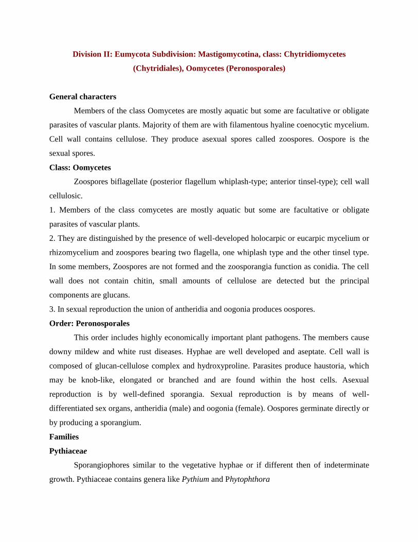
Division II: Eumycota Subdivision: Mastigomycotina, class: Chytridiomycetes
(Chytridiales), Oomycetes (Peronosporales)
General characters
Members of the class Oomycetes are mostly aquatic but some are facultative or obligate
parasites of vascular plants. Majority of them are with filamentous hyaline coenocytic mycelium.
Cell wall contains cellulose. They produce asexual spores called zoospores. Oospore is the
sexual spores.
Class: Oomycetes
Zoospores biflagellate (posterior flagellum whiplash-type; anterior tinsel-type); cell wall
cellulosic.
1. Members of the class comycetes are mostly aquatic but some are facultative or obligate
parasites of vascular plants.
2. They are distinguished by the presence of well-developed holocarpic or eucarpic mycelium or
rhizomycelium and zoospores bearing two flagella, one whiplash type and the other tinsel type.
In some members, Zoospores are not formed and the zoosporangia function as conidia. The cell
wall does not contain chitin, small amounts of cellulose are detected but the principal
components are glucans.
3. In sexual reproduction the union of antheridia and oogonia produces oospores.
Order: Peronosporales
This order includes highly economically important plant pathogens. The members cause
downy mildew and white rust diseases. Hyphae are well developed and aseptate. Cell wall is
composed of glucan-cellulose complex and hydroxyproline. Parasites produce haustoria, which
may be knob-like, elongated or branched and are found within the host cells. Asexual
reproduction is by well-defined sporangia. Sexual reproduction is by means of well-
differentiated sex organs, antheridia (male) and oogonia (female). Oospores germinate directly or
by producing a sporangium.
Families
Pythiaceae
Sporangiophores similar to the vegetative hyphae or if different then of indeterminate
growth. Pythiaceae contains genera like Pythium and Phytophthora

Albuginaceae
Sporangiophores strikingly different from vegetative hyphae, slender or thick, variously
club-shaped, arranged in a layer, and bear sporangia in chain at the tip. These are obligate
parasites. It contains a single genus, Albugo.
Peronosporaceae
Sporangiophores strikingly different from vegetative hyphae, slender or thick, variously
shaped, and with determine growth; sporangia produced singly or in cluster at the tip of
sporangiophores or their branches; obligate parasites.
Classification of Peronosporaceae
A. Sporophores determinate, hyphae-like short, unbranched or obpyriform, not maturing
synchronously, germinating by zoospores; antheridia always paragynous; oogonial wall thick
and confluent with that of the oospores; oospore germinates by germ tube or a sporophore
terminated by a sporangium. - Sclerophthora
AA. Sporophores determinate, macronemous, stout, 10 or more microns broad, branched or
unbranched, oogonial wall thick and rough or ornamented:
B. Sporophores unbranched, apex swollen and with short sterigmata bearing papillate sporangia
germinating by zoospores; oospores aplerotic.- Basidiophora
BB. Sporophores repeatedly branched in the upper portion, dichotomous; spores mature
synchronously; oogonial wall thick; oospore plerotic; sporangia germinate by zoospores or germ
tube; oospores germinate by a germ tube.- Sclerospora
AAA. Sporophores determinate, narrow, not more than 15 microns broad, usually 8- 10 microns;
oogonial wall unornamented except in Bremiella:
B. Spore wall uniformly thick (non-poroid), germination typically by germ tube. - Peronospora
BB. Spore wall poroid, emerging through an apical pore with or without papilla:
C. Branching of sporophore at right angles, tips or branches blunt.- Plasmopara
CC. Branching at acute angles:
D. Tips of branches acute - Pseudoperonospora
DD. Tips much enlarged and bearing 3-4 peripheral sterigmata; oogonial wall and oospore wall
thin and unornamented.. Bremia DDD. Tips of branches blunt and slightly enlarged; oogonial
wall thick and ornamented.- Bremiella.

Club root of cabbage, damping off and life cycles of Plasmodiophora, Pythium and
Phytophthora
Club root of cabbage caused by Plasmodiophora brassicae
Enlarged roots appearing like spindles or clubs due to stimulation of root cells to
abnormal enlargement (hypertrophy) and abnormal division (hyperplasia) is called club root.
Systematic position
Scientific categorization of the organisms in a hierarchal series of groups. Based on
characteristics of the spores, spore bearing structures and mycelium. Many fungi were classified
earlier based on the asexual spore and same were reclassified once they produced sexual spore.
Kingdom: Protista (Eukaryote)
Sub-kingdom: Mycota
Division: Myxomycota
Class: Plasmodiophoromycetes
Order: Plasmodiophorales
Family: Plasmodiophoraceae
Genus: Plasmodiophora
Species: P. brassicae
Symptoms
Enlargement of roots, club-shaped roots due to hyperplasia and hypertrophy, gradual and
inconspicuous stunting, yellowing and wilting of plant.
Pathogen
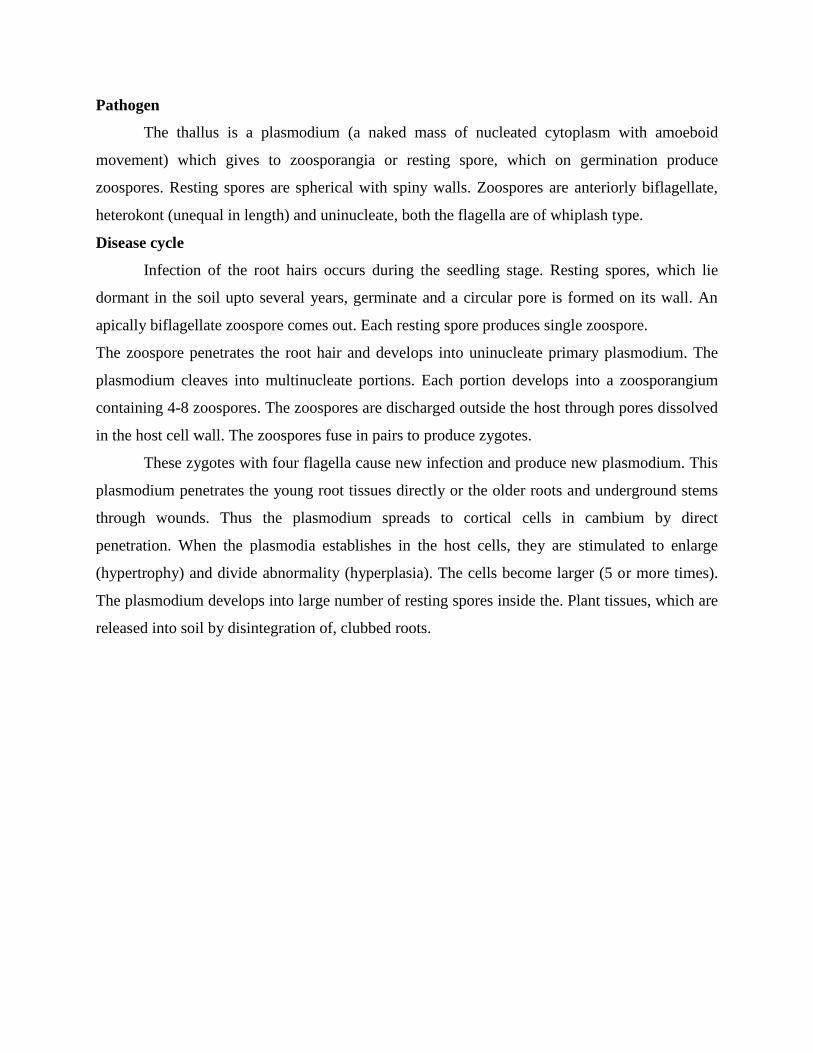
The thallus is a plasmodium (a naked mass of nucleated cytoplasm with amoeboid
movement) which gives to zoosporangia or resting spore, which on germination produce
zoospores. Resting spores are spherical with spiny walls. Zoospores are anteriorly biflagellate,
heterokont (unequal in length) and uninucleate, both the flagella are of whiplash type.
Disease cycle
Infection of the root hairs occurs during the seedling stage. Resting spores, which lie
dormant in the soil upto several years, germinate and a circular pore is formed on its wall. An
apically biflagellate zoospore comes out. Each resting spore produces single zoospore.
The zoospore penetrates the root hair and develops into uninucleate primary plasmodium. The
plasmodium cleaves into multinucleate portions. Each portion develops into a zoosporangium
containing 4-8 zoospores. The zoospores are discharged outside the host through pores dissolved
in the host cell wall. The zoospores fuse in pairs to produce zygotes.
These zygotes with four flagella cause new infection and produce new plasmodium. This
plasmodium penetrates the young root tissues directly or the older roots and underground stems
through wounds. Thus the plasmodium spreads to cortical cells in cambium by direct
penetration. When the plasmodia establishes in the host cells, they are stimulated to enlarge
(hypertrophy) and divide abnormality (hyperplasia). The cells become larger (5 or more times).
The plasmodium develops into large number of resting spores inside the. Plant tissues, which are
released into soil by disintegration of, clubbed roots.
Damping off of vegetables (tomato, brinjal, chillies, etc.) and tobacco – Pythium
aphanidermatum
Damping off is a special name given to denote wilting of young seedlings in nursery. The
rapid death and collapse of very young seedlings in the seedbed is called damping off.
Systematic position
Sub-kingdom: Mycota
Division: Eumycota
Sub-division: Mastigomycotira
Class: Oomycetes
Family: Pythiaceae
Genus: Pythium
Species: P. aphanidermatum

Symptoms
It is generally observed two weeks after sowing. Water-soaked lesions appear on the
collar region of seedlings; browning and shriveling of stem tissues at soil level in the collar
region; toppling down of seedlings in the nursery; ultimate death of sick seedling.
Pathogen
It is a facultative parasite and homothallic (both male and female gametes are produced in
the same mycelium. Mycelium is hyaline, coenocytic (von-septate), branched, inter and intra
cellular giving the appearance of a white fluffy cellular mass, does not have haustoria. Cell wall
of this fungus contains cellulose. Sporangium is lobed or irregular; it forms vesicle.
Sporangiophores are undifferentiated and similar to somatic hyphae.
Zoospores are produced in spherical vesicle and liberated after bursting of vesicle. They are
reniform and biflagellate with flagella attached to lateral
side, one pointing upward is tinsel type and the other
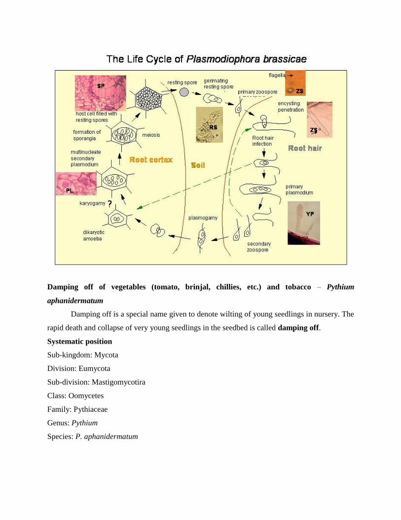
pointing downward is whiplash type. Antheridium (male
gametangium) is paragynous, club shaped, terminal or
intercalary and it is applied to the side of the oogonium; the
hyphal branch bearing antheridium may arise either from
oogonial stalk (monoclinous) or from a separate -hypha (diclinous). Oogonium (female
gametangium) is globose, generally develops at the tip of hyphal branch and consists of central
denser zone called ooplasm or oosphere and peripheral lighter zone called periplasm. Oospores
are the sexual spore, which helps to tide over adverse conditions (resting spore). They are
spherical, thick walled with yellowish brown wall and does not fill oogonial cavity called
aplerotic oospore.
Paragynous arrangement of oogonium and antheridia in Pythium. (Courtesy P.B. Hamm)
(arrows indicate antheridia)
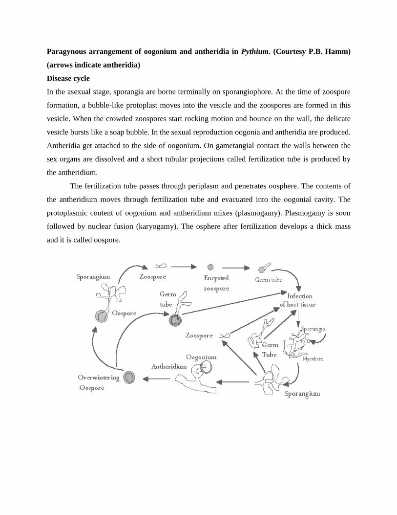
Disease cycle
In the asexual stage, sporangia are borne terminally on sporangiophore. At the time of zoospore
formation, a bubble-like protoplast moves into the vesicle and the zoospores are formed in this
vesicle. When the crowded zoospores start rocking motion and bounce on the wall, the delicate
vesicle bursts like a soap bubble. In the sexual reproduction oogonia and antheridia are produced.
Antheridia get attached to the side of oogonium. On gametangial contact the walls between the
sex organs are dissolved and a short tubular projections called fertilization tube is produced by
the antheridium.
The fertilization tube passes through periplasm and penetrates oosphere. The contents of
the antheridium moves through fertilization tube and evacuated into the oogonial cavity. The
protoplasmic content of oogonium and antheridium mixes (plasmogamy). Plasmogamy is soon
followed by nuclear fusion (karyogamy). The osphere after fertilization develops a thick mass
and it is called oospore.
Life cycle of Pythium aphanidermatum
Disease cycle
Oospore or encysted zoospore germinates and produce germ tubes or saprophytic
mycelium which come in contact with seed or seedling tissues of host plant and enter by direct
penetration. Pectinolytic enzymes of the fungus dissolve the pectins (holding cells together)
resulting in maceration of tissues. The mycelium grows between and through the cells.
Proteolytic and or cellulolytic enzymes causing complete collapse and disintegration of cell walls
break down the protoplasts of invaded cells. As a result, the infected seeds / young seedlings are
killed and turned into a rotten mass.
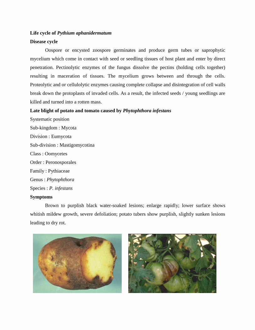
Late blight of potato and tomato caused by Phytophthora infestans
Systematic position
Sub-kingdom : Mycota
Division : Eumycota
Sub-division : Mastigomycotina
Class : Oomycetes
Order : Peronosporales
Family : Pythiaceae
Genus : Phytophthora
Species : P. infestans
Symptoms
Brown to purplish black water-soaked lesions; enlarge rapidly; lower surface shows
whitish mildew growth, severe defoliation; potato tubers show purplish, slightly sunken lesions
leading to dry rot.

Late blight of potato on tuber Late blight of tomato
Late blight of potato on leaf
Pathogen
Mycelium is endophytic, coenocytic, hyaline, branched, and inter-cellular. Haustoria club
shaped. Sporangiophores are hyaline, branched, indeterminate, thickwalled, arise through
stomata on leaves or lenticels on tubers. Sporangia are multinucleate, thin-walled, hyaline, and
oval or per shaped with a definite papilla at the apex. Zoospores are reniform, biflagellate
(anterior tinsel and posterior whiplash). Oospores are thick-walled and smooth.
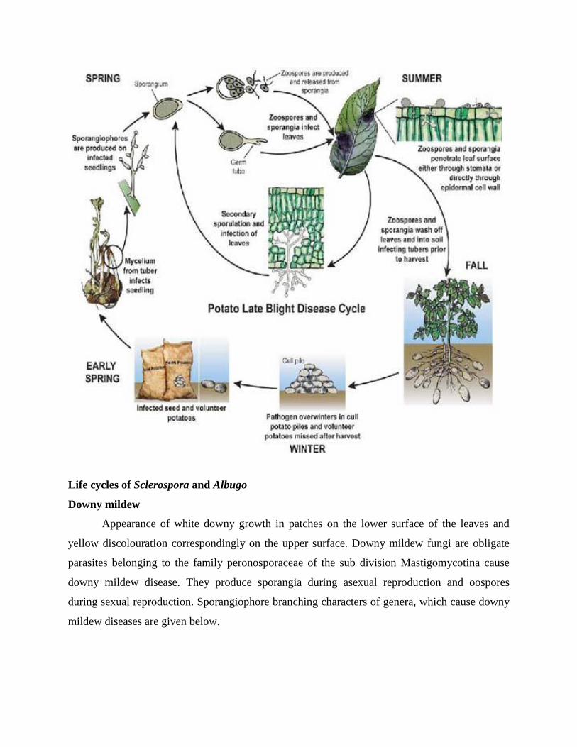
Life cycle
Primary infection is through use of infected tubers. Mycelium spreads into shoots
produced from infected tubers and reaches the aerial parts of the plant. Sporangiophore emerges
through stomata on stem and leaves and produce sporangia, which are spread by rain to wet
potato, leaves or stem and cause disease. Large number of asexual generation in a growing
season kills the foliage rapidly. The zoospores found in the soil germinate, penetrate through
lentils or wounds into the tubers and send intercellular mycelium and haustoria into the cells and
cause infection.
Life cycles of Sclerospora and Albugo
Downy mildew
Appearance of white downy growth in patches on the lower surface of the leaves and
yellow discolouration correspondingly on the upper surface. Downy mildew fungi are obligate
parasites belonging to the family peronosporaceae of the sub division Mastigomycotina cause
downy mildew disease. They produce sporangia during asexual reproduction and oospores
during sexual reproduction. Sporangiophore branching characters of genera, which cause downy
mildew diseases are given below.

i. Sporangiophore is club-shaped with a swollen head, over which the sporangia are borne on
minute sterigmata. e.g., Basidiophora.
ii. Sporangiophore is short, stout with many upright branches near the end, bearing the sporangia
at tips. e.g., Sclerospora.
iii. Sporangiophore is branched at right angles and are irregularly spaced. e.g., Plasmopara.
iv. Sporangiophore is dichotomously branched at acute angles and taper to gracefully curved
pointed tips on which sporangia are borne. e.g., Peronospora and Pseudoperonospora.
v. Sporangiophore is dichotomously branched at acute angles and the tips of the branches are
expanded into cup-shaped apophyses with four sterigmata. e.g., Bremia.
Downy mildew of pearlmillet caused by Sclerospora graminicola
Systematic position
Sub-kingdom : Mycota
Division : Eumycota
Sub-division : Mastigomycotina
Class . Oomycetes
Order : Peronosporales
Family : Peronosporaceae
Genus : Sclerospora
Species : S. graminicola
Symptoms
Pale yellow discolouration of leaves; whitish fungal growth on the lower surface of
leaves; twisting and crinkling of leaves; drying of leaves; infected seedlings when planted die
within 30 days; green ear symptom i.e., transformation of floral parts into green leaf-like
structures.
Symptoms often vary as a result of systemic infection. Leaf symptoms begin as chlorosis
at the base and successively higher leaves show progressively greater chlorosis. Infected
chlorotic leaf areas can support abundant white asexual sporulation on the lower leaf surface.
Severely infected plants are generally stunted and do not produce panicles. Green ear symptoms
result from transformation of floral parts into leafy structures

Pathogen
It is an obligate parasite. Mycelium is hyaline, coenocytic, intercellular and become
systemic. Haustoria are finger- or button - like. Sporangiophores emerge through stomata, short,
stout, non-septate with upright branches, crowded with sporangia bearing stalks (sterigmata)
with pointed ends at the apex. Sporangia are hyaline, broadly elliptical, thin, smooth walled and
papillate. Each sporangium contains 3 to 23 zoospores, which are irregularly reniform and
biflagellate. Oospores are spherical, thick walled and yellowish brown.
Disease cycle
Soil borne oospores germinate by put forth germ tube and infect the root hairs / coleoptile
of the host seedlings. Inside host tissue, fungus becomes systemic and produces hyaline,
coenocytic, highly branched, strictly intercellular mycelium with finger shaper haustoria.
During dewly nights, hyphae emerge through the stomata and form sporangiophores either singly
or in groups. During such period, downy growth is noticed on the diseased area. A single
sporangium is formed at the tip of the sterigma. The sporangia are deciduous and are carried by
wind. The sporangia germinate by releasing zoospores. Zoospores swim for sometimes, come to
rest, encyst and then germinate by germ tube to form new mycelium. Infected plant parts
produces sporangia over a considerable period of time under humid condition and then necrosis
begins.
In the sexual stage, the sex organs (antheridia and oogonia) develop in the intercellular
spaces of the host tissues (leaves and malformed floral organs). It is typically oogamous. The
fertilization tube formed by the antheridium carries the male nucleus into the oosphere where the
two nuclei fuse to form a diploid zygote nucleus. The oosphere develops a warty wall and
becomes the oospore. Oospores have a long period of rest lie in the soil (soil - borne) or on the
seed surface. Oospores are liberated by the disintegration of the host tissue .They germinate and
infect roots of young seedlings, from where the mycelium spreads systemically in the entire
plant.
White rusts or white blisters
White rusts or white blisters are the characteristic pustules fructifications of Albugo in
Albuginaceae on plant surfaces, especially on leaves.e.g.,white rust of Amaranthus caused by
Albugo bliti, white rust of crucifers caused by A. candida and white rust of sweetpotato caused
by A. ipomeae panduranae.
White rust of crucifers - A. candida
Systematic position
Sub-kingdom : Mycota
Division : Eumycota
Subdivision : Mastigomycotina
Class : Oomycetes
Order : Peronosporales
Family : Albuginaceae
Genus : Albugo
Species : A. candida
Symptoms
The fungus attacks cabbage, cauliflower, mustard, radish and turnip. The disease name is
a misnomer. The pustules formed by white rust resembles the aecial stage of true rust belonging
to the subdivision Basidiomycotina and hence the name. All aerial plant parts viz., leaf, stem and
inflorescence are affected. On the lower surface of leaves it causes white or creamy yellow
pustules of various sizes and shape. They are shiny and 1 to 2 mm in dia. Rarely the infection is
seen on the upper leaf surface. Very often several of them coalesce to form patches.
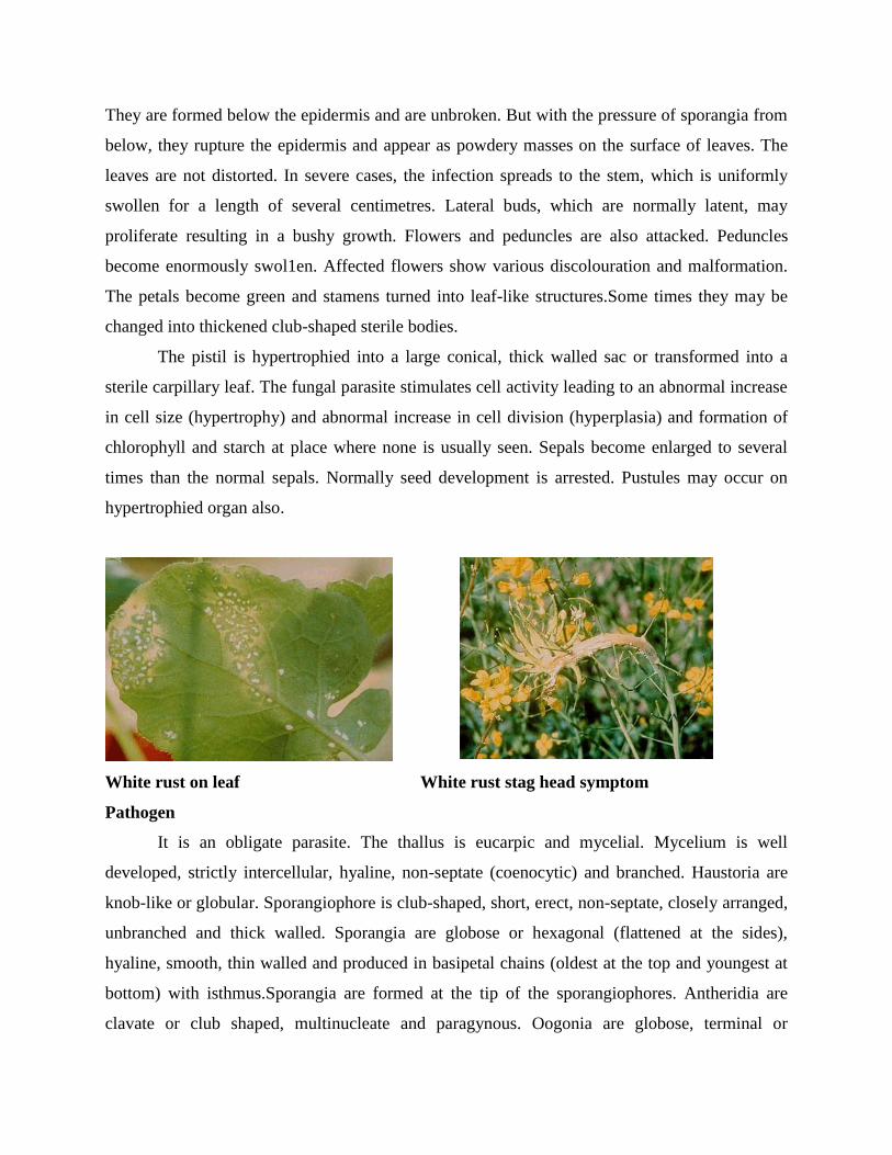
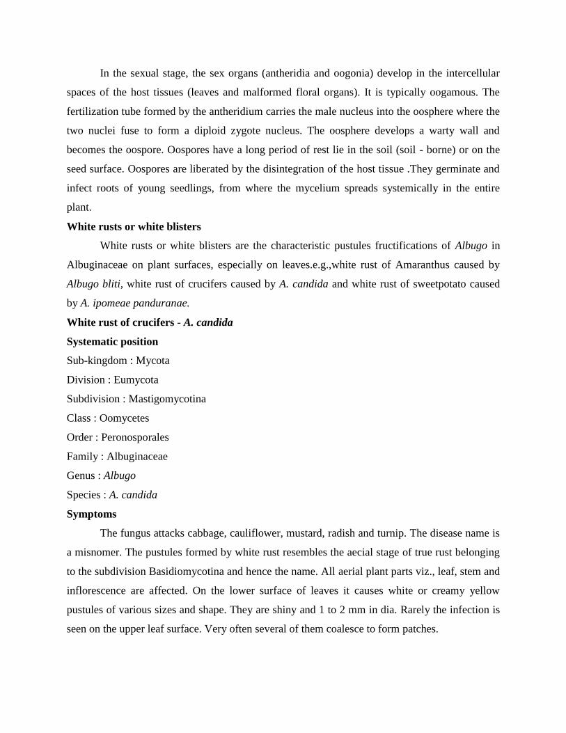
They are formed below the epidermis and are unbroken. But with the pressure of sporangia from
below, they rupture the epidermis and appear as powdery masses on the surface of leaves. The
leaves are not distorted. In severe cases, the infection spreads to the stem, which is uniformly
swollen for a length of several centimetres. Lateral buds, which are normally latent, may
proliferate resulting in a bushy growth. Flowers and peduncles are also attacked. Peduncles
become enormously swol1en. Affected flowers show various discolouration and malformation.
The petals become green and stamens turned into leaf-like structures.Some times they may be
changed into thickened club-shaped sterile bodies.
The pistil is hypertrophied into a large conical, thick walled sac or transformed into a
sterile carpillary leaf. The fungal parasite stimulates cell activity leading to an abnormal increase
in cell size (hypertrophy) and abnormal increase in cell division (hyperplasia) and formation of
chlorophyll and starch at place where none is usually seen. Sepals become enlarged to several
times than the normal sepals. Normally seed development is arrested. Pustules may occur on
hypertrophied organ also.
White rust on leaf White rust stag head symptom
Pathogen
It is an obligate parasite. The thallus is eucarpic and mycelial. Mycelium is well
developed, strictly intercellular, hyaline, non-septate (coenocytic) and branched. Haustoria are
knob-like or globular. Sporangiophore is club-shaped, short, erect, non-septate, closely arranged,
unbranched and thick walled. Sporangia are globose or hexagonal (flattened at the sides),
hyaline, smooth, thin walled and produced in basipetal chains (oldest at the top and youngest at
bottom) with isthmus.Sporangia are formed at the tip of the sporangiophores. Antheridia are
clavate or club shaped, multinucleate and paragynous. Oogonia are globose, terminal or
intercalary. Oospores are reticulate and round. Zoospores are biflagellate and reniform and 4 to 8
per sporangium.
Disease cycle
In the asexual stage, hyphae aggregate at several places under the epidermis.
Sporangiospores are formed as a palisade-like layer. These cut multinucleate sporangia, which
remain, attached to form a chain at the apex. The oldest sporangia lie at the top and youngest at
the base of the chain (called basipetal). The sporangia are separated from each other by a
gelatinuous disc-like structure called disjunctor or isthmus. The disjunctors are dissolved by
water and the sporangia are set free. The numerous sporangia that are produced at the apical end
of sporangiophores push against the epidermis, which bulges out and ultimately breaks.
The areas with broken epidermis and creamy mass of sporangia appear as pustules or
blisters on the leaves. Sporangia germinate by means of germ tube (direct germination) or by
formation of zoospores (indirect germination). Direct germination is not common. Sexual
reproduction occurs when the crop season comes to an end and it is typically oogamous.
The antheridia and oogonia borne terminally on somatic hyphae. Plasmogamy takes place
by gametangial contact, where the male nucleus from antheridium is transferred to oogonium
through the fertilization tube. Karyogamy occurs and a thin membrane develops around the
diploid zygote and a thick warty, tuberculate or roughened epispore. After a resting period, the
oospore germinates and forms a vesicle, which contains 40-60 zoospores. The rupture of the
vesicle wall releases the zoospores. The zoospore germinates by forming a germ tube, which
infects the host plant.