diversity of endophytes in various plants from woods hole, ma · biodiversity endophytic ... of...
TRANSCRIPT

Diversity of Endophytes in Various Plants from
Woods Hole, MA
By
Ivelisse Irizarry
Universidad del Turabo/Rutgers University
Introduction
Various plants from different environments around Woods Hole, MA were assessed for
biodiversity endophytic symbionts. Endophytes are microorganisms (mainly fungi and bacteria) that
inhabit tissues of plants and cause asymptomatic infections (Wilson, 1995). In a recent review, it has
been summarized that endophytes can promote plant growth by a number of different mechanisms.
These organisms are capable of solubilizing phosphate, producing indole acetic acid, vitamins, and
nitrogen metabolism (Ryan et al., 2008).
The main interest in this study was to observe the endophytic biodiversity and if endophytic
communities of plants differ according to the environments where the hosts are located. It is
hypothesized that different endophytic organisms will be found when comparing the plant hosts from
different environments.
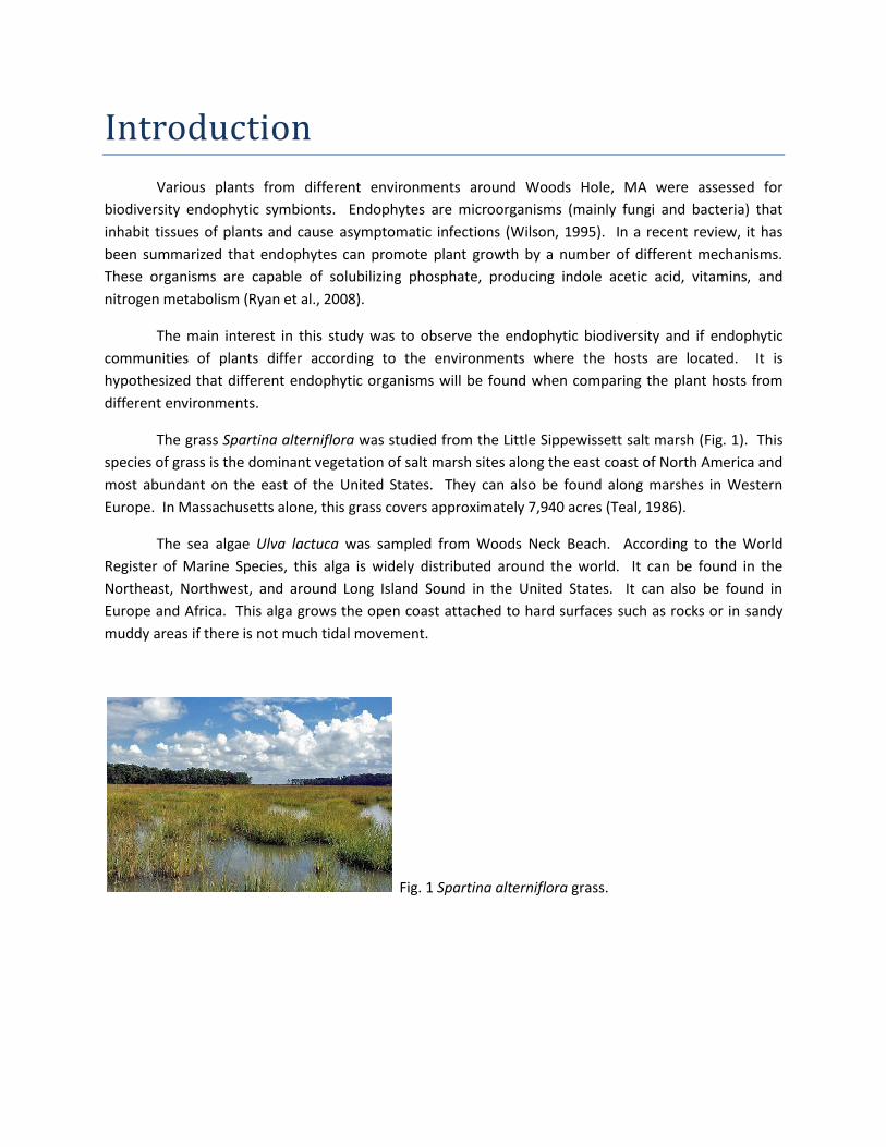
The grass Spartina alterniflora was studied from the Little Sippewissett salt marsh (Fig. 1). This
species of grass is the dominant vegetation of salt marsh sites along the east coast of North America and
most abundant on the east of the United States. They can also be found along marshes in Western
Europe. In Massachusetts alone, this grass covers approximately 7,940 acres (Teal, 1986).
The sea algae Ulva lactuca was sampled from Woods Neck Beach. According to the World
Register of Marine Species, this alga is widely distributed around the world. It can be found in the
Northeast, Northwest, and around Long Island Sound in the United States. It can also be found in
Europe and Africa. This alga grows the open coast attached to hard surfaces such as rocks or in sandy
muddy areas if there is not much tidal movement.
Fig. 1 Spartina alterniflora grass.
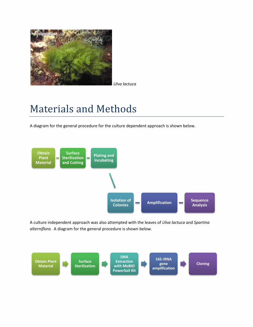
Ulva lactuca
Materials and Methods
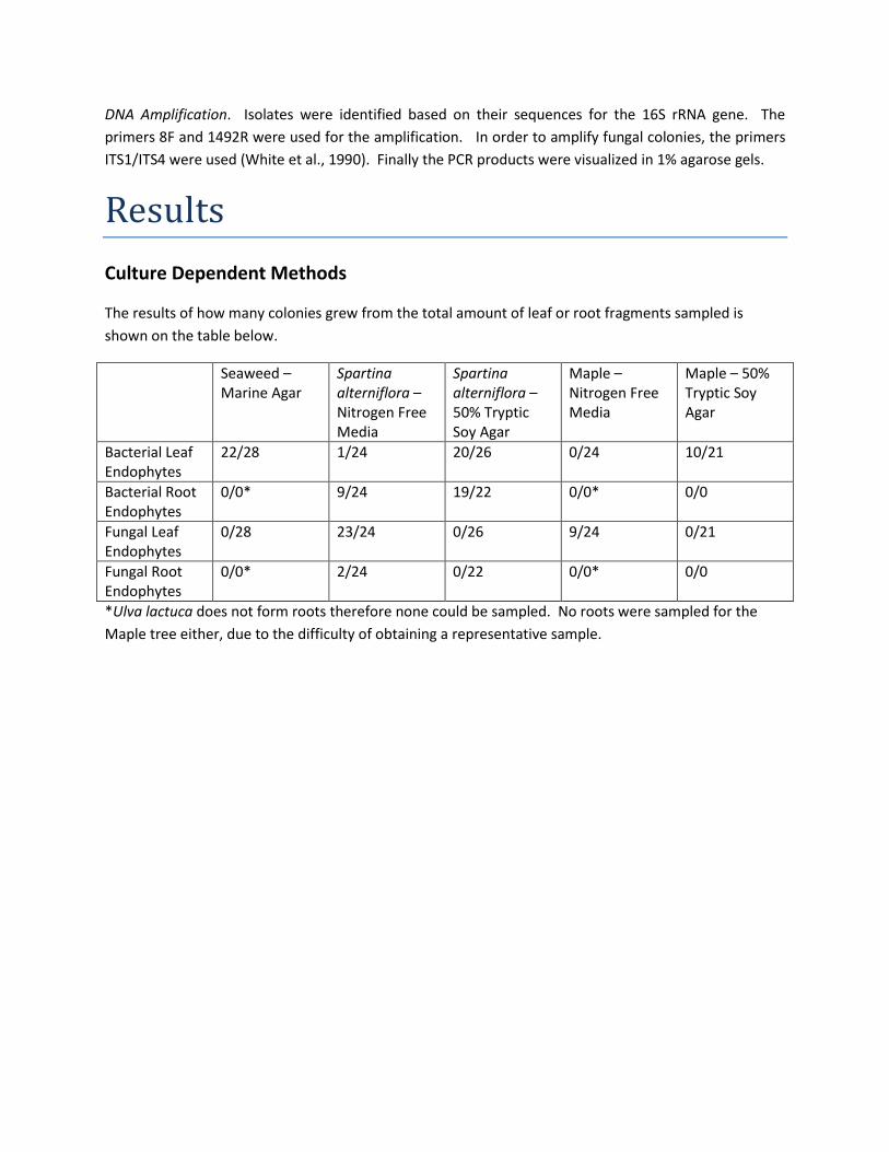
A diagram for the general procedure for the culture dependent approach is shown below.
A culture independent approach was also attempted with the leaves of Ulva lactuca and Spartina
alterniflora. A diagram for the general procedure is shown below.
Obtain Plant
Material
Surface Sterilization and Cutting
Plating and Incubating
Isolation of Colonies
Amplification Sequence Analysis
Obtain Plant Material
Surface Sterilization
DNA Extraction
with MoBIOPowerSoil Kit
16S rRNAgene
amplificationCloning

Sampling. Three different plants were sampled for this project. One leaf from each of three plants from
the same species was studied from each site. The specimens were observed to be healthy and mature
plants. Various grass samples of Spartina alterniflora were obtained from superficial pools where purple
sulfur bacteria are found at Little Sippewisset Salt Marsh. These grass samples were sampled along with
their roots. The plants with the most intact roots were chosen for the study. Another plant that was
studied for the presence of endophytes was the bright green seaweed Ulva lactuca which was sampled
from rocks at the bottom of Woods Neck Beach. A third sampling of leaves was performed on the
Maple tree located next to the Swope Building. Seaweed samples were transported in ziplock bags
containing seawater. The other samples from Spartina alterniflora and Maple were also stored
individually in ziplock bags. In the case that they could not be immediately processed, the leaves were
placed at 4˚C in order to stop decaying of the leaf which would cause saprophytes to be isolated. All
samples were processed within 48 hours of being sampled.
Surface Sterilization and Tissue Processing. Leaves and roots were washed with distilled water to
remove excess debris. Once washed they were surface sterilized to eliminate epiphytes. Leaves were
immersed in 70% ethanol for 1 minute, 10% NaOCl for 3 minutes, and 70% ethanol for 30 seconds.
Finally, the leaves were washed three times with sterile water to wash away the remaining alcohol and
NaOCl. Negative controls were prepared using the water from the last wash in order to determine if the
surface sterilization technique was successful. The plant material was then placed on a sterile Petri dish
where pieces were cut using a scalpel following proper aseptic techniques. Leaves were cut into pieces
that were approximately 3mm x 3mm in size. Roots fragments were approximately 3mm in length.
Endophyte Cultures. Surface sterilized leaf fragments and root fragments of Spartina alterniflora were
inoculated from each site into nitrogen-free media (NFM) and 50% tryptic soy agar (TSA) plates. The
samples that came from Ulva lactuca were inoculated onto Marine Agar (MA). Marine Agar has been
previously used in other successful studies for the isolation of endophytes of a seagrass (Couto-
Rodríguez, 2009). The leaf fragments from Maple were inoculated onto 50% TSA and NFM. The plates
were incubated at 30˚C. Once endophytes colonies were observed growing from the edges of the leaf
fragments these were isolated to a pure culture. Those endophytes which grew on NFM were isolated
again on NFM in order to make sure that they were capable of fixing nitrogen. Negative controls were
prepared by placing 100µL of the water used in the last wash on media. 50% Potato dextrose agar
media (PDA) was prepared in order to isolate and grow fungal colonies.
Community Analysis. A community analysis of the bacterial endophytes present in some samples was
attempted. DNA was extracted directly from leaf pieces that had been previously surface sterilized
following the instructions of the MOBIO PowerSoil® DNA Isolation Kit. Bacterial 16S rRNA genes were
amplified using the 8F and 1492R primers. Once the appropriate sized band was observed on a 1%
agarose gel, it was excised and DNA was recovered using the Millipore Gel DNA Extraction Kit.
DNA Extractions from Isolates. DNA extraction from each isolate was performed. These were carried
out by taking a single colony and placing it in a PCR tube with 75-100µl of sterile water. They were
placed in the thermal cycler where they were boiled for 10-15 minutes.
DNA Amplification. Isolates were identified based on their sequences for the 16S rRNA gene. The
primers 8F and 1492R were used for the amplification. In order to amplify fungal colonies, the primers
ITS1/ITS4 were used (White et al., 1990). Finally the PCR products were visualized in 1% agarose gels.
Results
Culture Dependent Methods
The results of how many colonies grew from the total amount of leaf or root fragments sampled is
shown on the table below.
Seaweed – Marine Agar
Spartina alterniflora – Nitrogen Free Media
Spartina alterniflora – 50% Tryptic Soy Agar
Maple – Nitrogen Free Media
Maple – 50% Tryptic Soy Agar
Bacterial Leaf Endophytes
22/28 1/24 20/26 0/24 10/21
Bacterial Root Endophytes
0/0* 9/24 19/22 0/0* 0/0
Fungal Leaf Endophytes
0/28 23/24 0/26 9/24 0/21
Fungal Root Endophytes
0/0* 2/24 0/22 0/0* 0/0
*Ulva lactuca does not form roots therefore none could be sampled. No roots were sampled for the
Maple tree either, due to the difficulty of obtaining a representative sample.

The percent of colonization of bacterial and fungal endophytes for each of the plants and media used
are represented in the following bar graph.
In total, 53 isolates from the different plants and treatments were attempted to be sequenced
after obtaining a positive PCR reaction. All PCR reactions that were submitted for sequencing were
performed along with a negative control. From the 53 PCR reactions that were submitted, only 26 came
back with good enough sequences in order to do any subsequent analyses. The sequences that were
good enough, were placed aligned using the SINA Web aligner from the SILVA ribosomal rRNA database
(http://www.arb-silva.de/aligner/). Once the sequences were aligned they were uploaded into ARB
software for their analysis (http://www.arb-home.de/). The tree on the following page represents the
sequences that were obtained which were good enough for analysis. The neighbor joining tree that is
shown, was created using ARB but was exported into FigTree software
(http://tree.bio.ed.ac.uk/software/figtree/).
78.6%
4.2%
76.9%
47.6%
37.5%
86.4%95.8%
37.5%
8.3%
0%
10%
20%
30%
40%
50%
60%
70%
80%
90%
100%
Ulva lactuca- MA
Spartinaalterniflora
- NFM
Spartinaalterniflora- 50% TSA
Maple -NFM
Maple -50% TSA
Bacterial Leaf Endophytes
Bacterial Root Endophytes
Fungal Leaf Endophytes
Fungal Root Endophytes


Culture Independent Approach Results
Two clone libraries were attempted for a community analysis of Ulva lactuca and Spartina
alterniflora. However, once the clones were obtained, they were placed on a single 96-well plate for
sequencing. A positive PCR was obtained from amplifying the 16S rRNA gene of a leaf blade of Spartina
alterniflora and of a leaf of Ulva lactuca. These were then ligated into a vector using the TOPO TA
Cloning Kit and they were transformed into E. coli cells.
This method was only partially successful. Sequences were obtained from 95 out of the 96
wells. 28 sequences were obtained from Ulva lactuca while 68 were obtained from Spartina
alterniflora. Of the 28 sequences obtained from Ulva lactuca 8 belonged to chloroplast sequences and
were discarded from the analysis. All 68 sequences from Spartina alterniflora belonged to chloroplasts
and therefore they could not be analyzed further. A phylogenetic tree was constructed and exported to
FigTree. The neighbor joining tree shown below represents those clones that were obtained from Ulva
lactuca.

Discussion
Culture Dependent Results
The percents of colonization varied among bacterial and fungal endophytes between leaves and
roots of the various plants studied. The percent of colonization was greatest for fungal endophytes in
leaves of Spartina alterniflora that were grown on nitrogen free media (95.8%). This however, should
not be interpreted as fungi that fix nitrogen. These fungi are most likely feeding off the remainder of
the nitrogen in the plant tissue where they inhabit. Only 1 colony of bacterial endophytes was obtained
from Spartina alterniflora growing on Nitrogen Free Media. No fungi were isolated from portions of
Ulva lactuca growing on Marine Agar.
From observing the data obtained for the grass Spartina alterniflora it is evident that nitrogen
free media was the most efficient culture media used during this study to isolate fungal endophytes.
The media that was most efficient for obtaining bacterial endophytes was 50% tryptic soy agar. In terms
of those bacteria that grew on 50% tryptic soy agar, it was also evident that more were recovered from
the root portions of the plant compared to the leaf portions (86.4% vs 76.9%). This is as expected since
most bacterial endophytes should be encountered at the root portions of a plant where they are in close
contact to the soil.
Observing the phylogenetic tree constructed from the sequencing data obtained, it can be
preliminarly determined that the most diverse bacterial endophytes came from the grass Spartina
alterniflora. It is important to mention though, that a lot of the samples to be sequenced that were
submitted did not make it back or were of poor quality and could therefore change the results that are
being observed during this study.
Most of the sequences that were recovered from Spartina alterniflora came from the root
portion of the plant. Some of these bacteria were close to Psychrobacter halophilus and Psychrobacter
faecalis. Species of Psychrobacter can be isolated from cold to warm and slightly to highly saline
ecosystems. Psychrobacter belongs to the family Moraxellaceae and belongs to the
Gammaproteobacteria (Bowman, 2006). Other sequences recovered from the roots of Spartina
alterniflora grouped closely with Rahnella, specifically a member of an uncultured Rahnella species.
Previously, Rahnella aquatilis have been found in the rhizosphere of soybean and has therefore been
found to be somewhat in a relationship with plants (Yong Kim et al., 2006). Another sequence obtained
grouped together with Listonella sp. This bacterium is particularly interesting because it has been found
that Listonella anguilarum has been described as a diazotroph living in the rhizosphere of mangroves
and could be contributing nitrogen to Spartina alterniflora. This was also one a colony that was able of
growing on nitrogen free media. This can also help confirm that the sequence obtained belongs to a
diazotrophic Listonella. Another root inhabiting endophyte grouped together with Pseudomonas sp.
This is of no surprise since species of Pseudomonas sp. are constantly easily identified as endophytes in
a variety of hosts. The bacteria that were isolated from the leaves of Spartina alterniflora clustered with

a different group of organisms than those that were isolated from the roots. Three of the sequences
that were recovered from isolates from the leaves grouped together with different species of Bacillus.
Two of the sequences were closely related to Bacillus cereus while another was more distantly related
to Bacillus circulans.
In terms of the endophytes obtained from Maple tree leaves, these were apparently less diverse
than those obtained from the leaves and roots of Spartina alterniflora. However, it is important to
mention that fewer sequences obtained from isolates that came from Maple trees were included for
analysis and this could affect the amount of diversity being observed in the phylogenetic tree. The
sequences that were found though were closely grouped in the Bacillus genus. Two of the sequences
grouped with Bacillus cereus and the other to with Bacillus sp. Other two sequences of bacterial
endophytes that came from the Maple tree were closely related to Micrococcus sp. and Paracoccus yeei.
Both Micrococcus and Paracoccus have been previously identified as endophytes of banana plants
(Thomas and Thyvalappil, 2009).
Finally, it appeared that the bacterial endophytes were least diverse in the sequences that were
analyzed from Ulva lactuca. Most of these sequences grouped around only two groups. Three of the
sequences grouped with Staphylococcus epidermidis. S. epidermidis was also found to be an endophyte
of banana (Thomas and Thyvalappil, 2009). Three other sequences grouped with Alteromonas and
Pseudoalteromonas. These are typical marine bacteria and have been previously described as
endophytes of seagrass beds (Couto-Rodríguez, 2009).
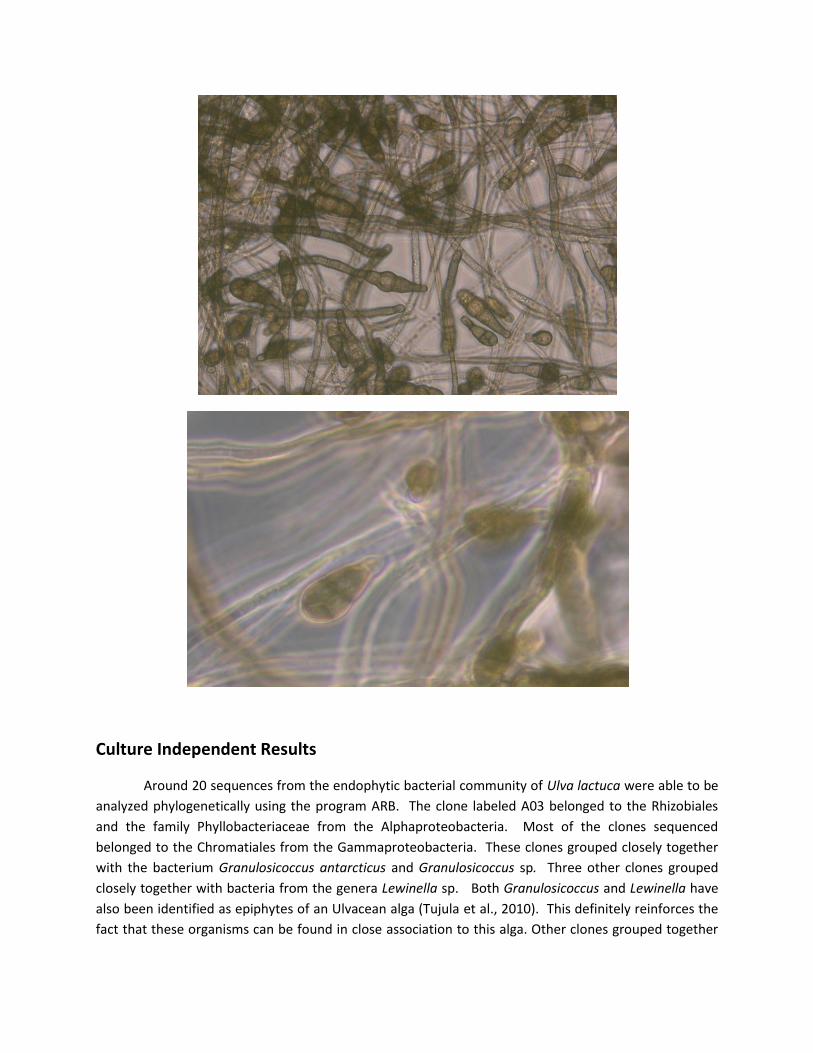
Some PCR reactions from fungal isolates were submitted for sequencing. However, not all
sequences were recovered. Luckily, one of the fungi that grew from the roots of Spartina alterniflora
could be easily identified because of its distinct spore. The shape of the spore suggests strongly that this
fungus belongs to the genus Alternaria. Below, are photographs taken of the hyphae and the spores of
the fungus. Another one of the fungi that was obtained from a Nitrogen Free Media plate was described
as been the oomycete Halophytophthora sp. However, the percent of query and of homology was very
low so it is not a very reliable sequence.
Culture Independent Results
Around 20 sequences from the endophytic bacterial community of Ulva lactuca were able to be
analyzed phylogenetically using the program ARB. The clone labeled A03 belonged to the Rhizobiales
and the family Phyllobacteriaceae from the Alphaproteobacteria. Most of the clones sequenced
belonged to the Chromatiales from the Gammaproteobacteria. These clones grouped closely together
with the bacterium Granulosicoccus antarcticus and Granulosicoccus sp. Three other clones grouped
closely together with bacteria from the genera Lewinella sp. Both Granulosicoccus and Lewinella have
also been identified as epiphytes of an Ulvacean alga (Tujula et al., 2010). This definitely reinforces the
fact that these organisms can be found in close association to this alga. Other clones grouped together
with Rubritalea, which belongs to the Verrucomicrobiales, and has been previously described living in
association with a sea sponge (Scheuermayer et al., 2006). Two clones clustered together in the SR1
candidate division.
Conclusion
In conclusion, the endophytic community that was best described during this project belonged
to the grass Spartina alterniflora which was sampled from the Little Sippewissett purple sulfur pools.
These also had the highest percent of colonization of bacteria. However, it would have been more
descriptive if more sequencing data would have been recovered. The endophytes that were described
during this mini project by phylogenetic association have also been found in previous hosts as
endophytes.
A distinct difference was observed between the sequences of the clones and the sequences of
the organisms that were obtained by culture methods. A combination of both culturing methods and
culture independent methods seems to be the most efficient way for describing the total endophytic
community of a plant instead of just relying on one of the approaches alone. It was also apparent by the
sequences of the isolates obtained that the endophytic communities differ among hosts sampled. Most
of the endophytes recovered from Ulva lactuca represented marine species of organisms. Some
organisms that were recovered from Spartina alterniflora belonged to halophilic, diazotrophs, and
other common terrestrial endophyte species. Finally, no halophiles or marine bacteria were isolated
from the Maple Tree at Swope. Further testing and more in depth analysis of the isolates that were not
able to be sequenced would be necessary to prove this statement. However, what is being seen so far
does suggest that the environment as well as the host tends to dictate what microbial communities are
present in the interior of vegetable tissue.
Literature Cited
Bowman J. 2006. The genus Psychrobacter. In: The Prokaryotes. 6: 920-930.
Couto-Rodríguez M. 2009. Endophytic prokaryotic diversity associated with seagrass beds of Thalassia
testudinum from Cabo Rojo, Lajas, and Vieques, Puerto Rico. Master thesis from Universidad de Puerto
Rico at Mayagϋez.
Ryan R et al. 2008. Bacterial endophytes: recent development and applications. FEMS Microbiology
Letters. 278: 1-9.
Scheuermayer et al. 2006. Rubritalea marina gen. nov., sp. nov., a marine representative of the phylum
‘Verrucomicrobia’, isolated from a sponge (Porifera). Int J Syst Evol Microbiol 56. 2119-2124
Teal J. 1986. The Ecology of Regularly Flooded Salt Marshes of New England: A Community Profile.
Biological Report. 85(7.4). 61pp.

Thomas and Thyvalappil. 2009. Endophytic bacteria associated with growing shoot tips of Banana
(Musa sp.) cv. Grand naine and the affinity of endophytes to the host. Microbial Ecology. 58: 952-964.
White TJ, Bruns T, Lee S, and Taylor JW. 1990. Amplification and direct sequencing of fungal ribosomal
RNA genes for phylogenetics. pp. 315-322 In: PCR Protocols: A Guide to Methods and Applications, eds.
Innis MA, Gelfand DH, Sninsky JJ, and White, TJ. Academic Press, Inc., New York.
Wilson D. 1995. Endophyte – the evolution of a term, and clarification of its use and definition. Oikos.
73(2): 274-27.
World Register of Marine Species
Yong Kim K, Jordan D, Krishnan H. 2006. Rahnella aquatilis, a bacterium isolated from soybean
rhizosphere, can solubilize hydroxyapatite. FEMS Microbiology Letters. 153: 273-277.