divergent evolution of an atypical s-adenosyl-l … evolution of an atypical...
TRANSCRIPT

Divergent evolution of an atypical S-adenosyl-L-methionine–dependent monooxygenase involvedin anthracycline biosynthesisThadée Grocholski, Pedro Dinis, Laila Niiranen, Jarmo Niemi, and Mikko Metsä-Ketelä1
Department of Biochemistry, University of Turku, FIN-20014 Turku, Finland
Edited by Frank M. Raushel, Texas A&M University, College Station, TX, and accepted by the Editorial Board July 7, 2015 (received for review January 27, 2015)
Bacterial secondary metabolic pathways are responsible for thebiosynthesis of thousands of bioactive natural products. Manyenzymes residing in these pathways have evolved to catalyzeunusual chemical transformations, which is facilitated by an evolu-tionary pressure promoting chemical diversity. Such divergentenzyme evolution has been observed in S-adenosyl-L-methionine(SAM)-dependent methyltransferases involved in the biosynthesisof anthracycline anticancer antibiotics; whereas DnrK from the dau-norubicin pathway is a canonical 4-O-methyltransferase, the closelyrelated RdmB (52% sequence identity) from the rhodomycin path-ways is an atypical 10-hydroxylase that requires SAM, a thiol reduc-ing agent, and molecular oxygen for activity. Here, we have usedextensive chimeragenesis to gain insight into the functional differ-entiation of RdmB and show that insertion of a single serine residueto DnrK is sufficient for introduction of the monooxygenation activity.The crystal structure of DnrK-Ser in complex with aclacinomycinT and S-adenosyl-L-homocysteine refined to 1.9-Å resolution revealedthat the inserted serine S297 resides in an α-helical segment adjacentto the substrate, but in a manner where the side chain points awayfrom the active site. Further experimental work indicated that theshift in activity is mediated by rotation of a preceding phenylalanineF296 toward the active site, which blocks a channel to the surface ofthe protein that is present in native DnrK. The channel is also closed inRdmB and may be important for monooxygenation in a solvent-freeenvironment. Finally, we postulate that the hydroxylation ability ofRdmB originates from a previously undetected 10-decarboxylationactivity of DnrK.
enzyme evolution | protein engineering | Streptomyces | polyketide |natural products
Anthracyclines are a medically important class of naturalproducts, which belong to the type II family of aromatic
polyketides (1–3). These compounds, produced by Streptomycesbacteria, are among the most effective anticancer drugs availableand are currently included in 500 clinical trials worldwide (4).Doxorubicin is widely used as a first-choice chemotherapeuticagent for many tumors, including various carcinomas and sar-comas (5), whereas aclacinomycin A has been used in thetreatment of hematological malignancies (6). The biologicalactivity of doxorubicin and aclacinomycin is mediated throughtopoisomerase II (7), while additional mechanisms of thesedrugs include the recently discovered histone eviction activity(4). Despite the success of anthracyclines in cancer chemo-therapy, improved compounds are still urgently needed, be-cause the clinical efficacy of these metabolites has been limitedby severe side effects (8, 9).The great diversity of anthracyclines is generated during tai-
loring steps in their biosynthesis, where glycosylations, hydrox-ylations, decarboxylations, and methylations modify the common7,8,9,10-tetrahydro-5,12-naphthacenoquinone aglycone chromo-phore to obtain the biologically active molecules of the variouspathways (1–3). A detailed understanding of these biosyntheticsteps is essential for the rational design of improved bioactive
metabolites by pathway and protein engineering. In particular,the discovery of thousands of unknown biosynthetic pathways bynext-generation sequencing and the emergence of synthetic bi-ology holds great promise for increasing the chemical space ofnatural products (10). However, identification of the function ofgene products residing in these pathways by bioinformaticanalysis alone is complicated by the atypical chemistry the cor-responding enzymes may catalyze. Unexpected chemical trans-formations appear to be especially common in the biosynthesis ofaromatic polyketides, where biochemical studies have frequentlyrevealed unusual reaction mechanisms (2, 11), which includesinstances where the reactivity of the substrates themselves areused in catalysis (12–14). In addition, homologous enzymes havebeen noted to catalyze diverse chemical transformations (15).Recently, the use of protein engineering for altering the reactionspecificities of biosynthetic enzymes has emerged as anotherdeveloping field for modification of natural products (16, 17).This unusually rapid differentiation of enzyme function may
be related to the evolution of secondary metabolism, where traitsenabling a swift increase in the diversity of natural products arebeneficial (18). To promote chemical diversity rather than for-mation of a single product, specialized metabolism enzymes ap-pear to have evolved as generalists, which is manifested aspromiscuity and slow reaction rates typical to these proteins (19).One intriguing example of such divergent evolution is the enzymepair DnrK and RdmB from the daunorubicin and rhodomycin
Significance
Natural products produced by Streptomyces are widely used inthe treatment of various medical conditions. Over the years,thousands of metabolites with complex chemical structureshave been isolated from cultures of these soil bacteria. Anevolutionary pressure that promotes chemical diversity ap-pears to be critical for generation of this rich source of bi-ologically active compounds. This is reflected in the bio-synthetic enzymes, where functions of similar proteins maygreatly differ. Here, we have clarified the molecular basis ofhow a classical methyltransferase has evolved into an unusualhydroxylase on the biosynthetic pathways of two anthracy-cline anticancer agents. Detailed understanding of enzymesinvolved in antibiotic biosynthesis will facilitate future proteinengineering efforts for generation of improved bioactive na-tural products.
Author contributions: T.G., J.N., and M.M.-K. designed research; T.G., P.D., L.N., and M.M.-K.performed research; T.G., P.D., L.N., J.N., and M.M.-K. analyzed data; and M.M.-K. wrotethe paper.
The authors declare no conflict of interest.
This article is a PNAS Direct Submission. F.M.R. is a guest editor invited by the Editorial Board.
Data deposition: The atomic coordinates and structure factors have been deposited in theProtein Data Bank, www.pdb.org (PDB ID code 4WXH).1To whom correspondence should be addressed. Email: [email protected].
This article contains supporting information online at www.pnas.org/lookup/suppl/doi:10.1073/pnas.1501765112/-/DCSupplemental.
9866–9871 | PNAS | August 11, 2015 | vol. 112 | no. 32 www.pnas.org/cgi/doi/10.1073/pnas.1501765112
pathways, respectively, that are homologous to classical S-adeno-syl-L-methionine (SAM)-dependent methyltransferases.Methyltransferase DnrK is a true methyltransferase that cat-
alyzes the 4-O-methylation of carminomycin (1; Fig. 1) in one ofthe final steps in the biosynthesis of the antitumor drug dauno-rubicin (2; Fig. 1) in Streptomyces peucetius (20, 21). The enzymepossesses rather relaxed substrate specificity in regard to modi-fications in the polyaromatic anthracycline ring system, but it isquite specific with respect to the length of the carbohydrate chainat C-7, accepting only monoglycosides (22). In addition, DnrK haseven been shown to be able to methylate various flavonoids (23).The crystal structure of the ternary complex of DnrK revealed thatthe mechanism of methylation is consistent with canonical SN2-type reactions of SAM-dependent methyltransferases, althoughproximity and orientation effects rather than acid/base catalysisappear to be more critical in the case of DnrK (22).On the other hand, RdmB from the β-rhodomycin pathway in
Streptomyces purpurascens is a very unusual enzyme that is evo-lutionarily closely related to DnrK with a sequence identity of52% (24). Determination of the crystal structure of RdmB con-firmed that the enzyme binds SAM (25) and that the ternarycomplex is indeed very similar to that of DnrK with a root-mean-square deviation of 1.14 Å for 335 equivalent Cα atoms (26).Despite these features, RdmB completely lacks methyltransferaseactivity (24, 27, 28); it is an anthracycline 10-hydroxylase, whichrequires SAM, molecular oxygen, and a thiol reducing agentfor activity (26). Contrary to DnrK, RdmB is able to use bothmonoglycosylated and triglycosylated anthracyclines as substrates.Furthermore, both rhodomycin and daunorubicin gene clus-
ters encode homologous 15-methylesterases RdmC (28) andDnrP (21), respectively. On the rhodomycin pathway, RdmBworks strictly in conjunction with RdmC, which first convertse-rhodomycin (3; Fig. 1) into 15-demethoxy-e-rhodomycin (4; Fig.1), whereas RdmB is responsible for conversion of the RdmCreaction product into β-rhodomycin (5; Fig. 1) (28). This is incontrast to the daunorubicin pathway, where the reaction order ofthe two enzymes DnrP and DnrK is more relaxed, as DnrK is ableto methylate at least five biosynthetic intermediates (21).To gain insight into how a methyltransferase has evolved into a
monooxygenase, we report here the generation of several chi-meric DnrK/RdmB proteins that were designed based on theavailable crystal structures. These initial experiments guided usin further structure/function studies, where we were able to
demonstrate that the insertion of a single amino acid is sufficientfor introduction of the 10-hydroxylase activity into the DnrKscaffold, thus providing an especially illustrative example of di-vergent enzyme evolution. Protein crystallography and furtherbiochemical experiments were then conducted to reveal themolecular basis for the altered enzymatic activity.
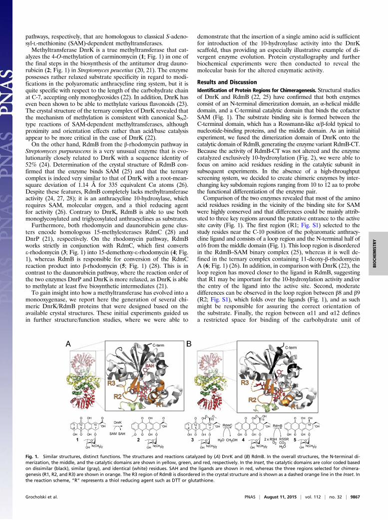
Results and DiscussionIdentification of Protein Regions for Chimeragenesis. Structural studiesof DnrK and RdmB (22, 25) have confirmed that both enzymesconsist of an N-terminal dimerization domain, an α-helical middledomain, and a C-terminal catalytic domain that binds the cofactorSAM (Fig. 1). The substrate binding site is formed between theC-terminal domain, which has a Rossmann-like α/β-fold typical tonucleotide-binding proteins, and the middle domain. As an initialexperiment, we fused the dimerization domain of DnrK onto thecatalytic domain of RdmB, generating the enzyme variant RdmB-CT.Because the activity of RdmB-CT was not altered and the enzymecatalyzed exclusively 10-hydroxylation (Fig. 2), we were able tofocus on amino acid residues residing in the catalytic subunit insubsequent experiments. In the absence of a high-throughputscreening system, we decided to create chimeric enzymes by inter-changing key subdomain regions ranging from 10 to 12 aa to probethe functional differentiation of the enzyme pair.Comparison of the two enzymes revealed that most of the amino
acid residues residing in the vicinity of the binding site for SAMwere highly conserved and that differences could be mainly attrib-uted to three key regions around the putative entrance to the activesite cavity (Fig. 1). The first region (R1; Fig. S1) selected to thestudy resides near the C-10 position of the polyaromatic anthracy-cline ligand and consists of a loop region and the N-terminal half ofα16 from the middle domain (Fig. 1). This loop region is disorderedin the RdmB–SAM binary complex (25), whereas it is well de-fined in the ternary complex containing 11-deoxy-β-rhodomycinA (6; Fig. 1) (26). In addition, in comparison with DnrK (22), theloop region has moved closer to the ligand in RdmB, suggestingthat R1 may be important for the 10-hydroxylation activity and/orthe entry of the ligand into the active site. Second, moderatedifferences can be observed in the loop region between β8 and β9(R2; Fig. S1), which folds over the ligands (Fig. 1), and as suchmight be responsible for assuring the correct orientation ofthe substrate. Finally, the region between α11 and α12 definesa restricted space for binding of the carbohydrate unit of
Fig. 1. Similar structures, distinct functions. The structures and reactions catalyzed by (A) DnrK and (B) RdmB. In the overall structures, the N-terminal di-merization, the middle, and the catalytic domains are shown in yellow, green, and red, respectively. In the Inset, the catalytic domains are color coded basedon dissimilar (black), similar (gray), and identical (white) residues. SAH and the ligands are shown in red, whereas the three regions selected for chimera-genesis (R1, R2, and R3) are shown in orange. The R3 region of RdmB is disordered in the crystal structure and is shown as a dashed orange line in the Inset. Inthe reaction scheme, “R” represents a thiol reducing agent such as DTT or glutathione.
Grocholski et al. PNAS | August 11, 2015 | vol. 112 | no. 32 | 9867
BIOCH
EMISTR
Y

4-methoxy-e-rhodomycin T (7; Fig. 1) in the DnrK structure (Fig.1), which has been noted as the likely explanation for why theenzyme only accepts monoglycosylated substrates (22). The
corresponding region in RdmB is rather different and the carbo-hydrate chain extends from the binding pocket into the bulksolution in the RdmB ternary complex (26), which promptedus to select this region (R3; Fig. S1) as the third segment forchimeragenesis.
Enzymatic Activities of the Native Enzymes. The activity assays wereperformed in a coupled reaction together with the 15-methyl-esterase RdmC using either the monoglycosylated aclacinomycinT (8; Fig. 2) or the triglycosylated aclacinomycin A (9; Fig. 2) assubstrates. The reaction mixtures also included SAM, which isessential as a cosubstrate for DnrK (22) and as a cofactor forRdmB (26), and a reducing agent (DTT) that is required solelyby RdmB (26). The reaction with native DnrK resulted in asingle main product (Fig. 2A), which was identified as the 4-O-methylated and 10-decarboxylated aclacinomycin T (10; Figs. S2and S3, and Table S1), when 8 was used as a substrate, whereasno methylation activity could be observed with the triglycosylated9. In agreement with previous experiments (27), addition of na-tive RdmB to the assays with 8 and 9 led to the accumulation ofthe expected 10-hydroxylated products 11-deoxy-β-rhodomycin T(11; Fig. 2B) and 6 (Fig. 2D), respectively.
Discovery of a Moonlighting 10-Decarboxylation Activity of DnrK.The RdmC and DnrP reaction products with a free 10-carboxylgroup (compounds such as 4 in Fig. 1) have been described asunstable and undergo spontaneous 10-decarboxylation on therhodomycin and daunorubicin pathways, respectively (21, 29).However, indirect evidence has suggested that the decarboxyl-ation might also be enzymatically catalyzed by DnrP (21). Duringour assays with 8, we noted that the RdmC reaction product, 15-demethoxy-aclacinomycin T (15; Fig. 2) is stable if the enzymaticreactions are conducted in the dark, but exposure to light issufficient to promote the 10-decarboxylation reaction to proceedto near completion in 60 min resulting in 10-decarboxymethyl-aclacinomycin T (13; Fig. S3). To our surprise, we next observedthat if 15 is isolated and incubated in the dark together withDnrK, both 4-O-methylation and 10-decarboxylation reactionsoccur, because 10 is observed as the sole product (Fig. S3). Theresult therefore demonstrated directly that it is DnrK instead ofDnrP that harbors the moonlighting 10-decarboxylation activity,which in our view has important evolutionary implications for theemergence of 10-hydroxylation activity (see below).
Enzymatic Activities of the Chimeric Proteins. The single chimericDnrK enzymes, where the amino acid residues in the R1, R2, or R3regions were changed to correspond to RdmB, behaved much likenative DnrK in the activity measurements and led to the accu-mulation of 10 (Fig. 2A) as the main product. However, the re-action mixture of DnrK R1 contained a small amount of theexpected RdmB reaction product, 11 (Fig. 2B), and significantquantities of a compound that was identified as the double DnrK/RdmB reaction product, 4-O-methyl-11-deoxy-β-rhodomycin T(12; Fig. 2C). Neither DnrK nor RdmB alone produced 12, butwhen both native enzymes were incubated together with RdmC,the same metabolite was observed (Fig. 2C). The double and triplechimeric enzymes reinforced our initial results; chimeric proteinsthat contained the R1 region were able to catalyze 10-hydroxyl-ation, whereas the R2 + R3 chimera was only able to perform 4-O-methylation. These multiple exchanges enhanced the RdmB-likeactivity in comparison with the single R1 chimera, with the R3region having a slightly greater effect than the R2 region (Fig. 2B).A similar trend was observed when 9 was used as a substrate
and the 10-hydroxylated reaction product 6 was observed only inchimeras containing the R1 region from RdmB. It is noteworthythat the increased rate of 10-hydroxylation correlated exceed-ingly well with the number of amino acid exchanges from theRdmB template (Fig. 2D), which highlights the importance of
Fig. 2. Relative activities of native and engineered enzymes in a coupled re-action with RdmC. The reactions with 8 lead to the accumulation of threeproducts (A–C), whereas in the case of 9 only one product was observed (D). Thecolumns present the formation of (A) 4-O-methyl-15-decarboxyaclacinomycinT (10), (B) 11-deoxy-β-rhodomycin T (11), (C) 4-O-methyl-11-deoxy-β-rhodomycinT (12), and (D) 11-deoxy-β-rhodomycin A (6) by the enzymes. The overall per-centages may not add up to 100% in all samples because some unreactedsubstrate 15was left with chimeras harboring poor activity (e.g., DnrK R2 + R3).The excess substrate was converted to the corresponding 10-decarboxylatedcompounds by exposure to light (overall conversion of 8 to 13 and 9 to 14; Fig.S2) to facilitate the HPLC analyses.
9868 | www.pnas.org/cgi/doi/10.1073/pnas.1501765112 Grocholski et al.

the R2 and R3 regions in correct alignment of the triglycosidic 9substrate in the active site. Previous studies have shown that theR3 region of DnrK folds around the amino sugar moiety (22),and it was surprising to find that exchange of the R3 region wasnot essential for the gain of the 10-hydroxylation activity. Despitethese changes, no 4-O-methylation activity was detected with thissubstrate in any of the chimeric enzymes, which would indicatethat the aglycone is still not correctly positioned in terms ofgeometry in the active-site cavity. The lack of methylation ac-tivity in native RdmB has been suggested to occur due to wrongalignment of the substrate for a methyl transfer reaction using aSN2 mechanism, where the methyl group has to be in line withthe oxygen of the substrate as well as the sulfur atom of SAM forthe reaction to proceed (26).In contrast to the engineered DnrK enzymes, when the equiv-
alent changes were made to the RdmB template, no methylationactivity could be detected. The chimeric RdmB enzymes eitherbehaved in a manner similar to the native enzyme or simply lost allenzymatic activity.
Mapping the Minimal Region Required for Gain of 10-HydroxylationActivity in DnrK. In an attempt to further pinpoint the origin of the10-hydroxylation activity in the DnrK R1 chimera, we divided theR1 region into two segments corresponding to the loop regionand the following α16 helix, which resulted in two additionalmutants denoted as DnrK R1.1 and DnrK R1.2, respectively.The 10-hydroxylation activity could be attributed solely to theα16 helix (Fig. 2), because the activity of DnrK R1.2 was similarto that of DnrK R1 (i.e., both methylation and hydroxylation of 8and hydroxylation of 9), whereas DnrK R1.1 behaved like nativeDnrK (i.e., methylation of 8 and no activity with 9). Inspection ofthe amino acid sequences of the R1.2 region (Fig. S1) revealedthat the RdmB sequence contains an additional serine insertionin this area in comparison with DnrK. Remarkably, introductionof this single S297 residue to the DnrK scaffold was sufficient topersuade the 10-hydroxylation activity to emerge as seen fromthe activity measurements of the DnrK-Ser variant (Fig. 2).
Structural Basis for the Altered Enzymatic Activity of the DnrK-SerMutant. The DnrK-Ser mutant crystallized in a different spacegroup than native DnrK, and consequently the diffraction datacould be refined to an improved resolution of 1.9 Å (Table S2).Electron density could be traced for most of the polypeptidechain in the two subunits found in the asymmetric unit with the
exception of two amino acids residing in loop regions (D85 andH293) in chain B, where the density in general was less welldefined than in chain A. Based on electron density, one moleculeof 8 and S-adenosyl-L-homocysteine (SAH) was bound to eachsubunit (Fig. S4).The overall structure of DnrK-Ser was exceedingly similar to
native DnrK with an r.m.s.d. of 0.90 Å over 327 residues, and themain differences were mostly restricted to the R1 region. Inshort, introduction of the additional S297 residue in R1.2 ap-peared to trigger a transition of all preceding residues in helixα16 toward the surface of the protein, which altered the structureof the R1.1 loop region. Unexpectedly, the side chain of theinserted serine was found pointing away from the active site, butat the same time the preceding phenylalanine (F296) had rotatedinward adjacent to the ligand near the site of monooxygenation(Fig. 3). Comparison of the structures of DnrK-Ser and RdmBreveals that both enzymes have a phenylalanine, F296 and F300,respectively, next to the anthracycline ligand, which is in contrastto native DnrK, which contains glutamine Q295 at the equivalentposition (Fig. 3). The implication of these changes is that theglutamine in DnrK allows bulk solvent to access the active sitecavity via an open channel, whereas the bulkier hydrophobicphenylalanine residues block this route to the surface of theprotein in DnrK-Ser and RdmB (Fig. 3). The structural analysistherefore suggested that critical to the shift in activity is rotationof F296 and that the amino acid inserted at position 297 wouldnot necessarily have to be serine. Consistent with this hypothesis,reintroduction of the original phenylalanine observed in thisstructural position in native DnrK led to hydroxylation of bothsubstrates by DnrK R1.2 S297F (Fig. 2 B and D).
Mechanistic Divergence of the DnrK and RdmB Reactions. The chi-meragenesis studies of DnrK and RdmB permit us to proposethe following mechanistic proposal for the divergent reactionscatalyzed by the two enzymes (Fig. 4). It is tempting to suggestthat a common initial step is the decarboxylation of the substrate15 to generate a carbanion intermediate. The more open activesite of DnrK allows facile protonation of the carbanion by sol-vent molecules and results in the formation of a neutral 10-decarboxylated and 4-O-methylated anthracycline 10. However,such compounds are no longer suitable as substrates for RdmB(26), which avoids the protonation step by closing the channel tothe surface of the protein and excluding solvent ions from theactive site. The carbanion may be stabilized through distribution
Fig. 3. Cartoon (Top) and surface (Bottom) repre-sentations of the active sites of DnrK in complex with4-methoxy-e-rhodomycin T (7) and SAH, DnrK-Ser withbound 8 and SAH, and the ternary complex of RdmB,11-deoxy-β-rhodomycin (6), and SAM. Insertion ofS297 in DnrK-Ser rotates F296 toward the ligand,which reshapes the DnrK active site toward the RdmBarchitecture. In particular, DnrK-Ser F296 blocks achannel to the surface of the protein that is open innative DnrK, but closed in RdmB. A conserved arginineresidue that is important for both methylation andhydroxylation activities is also shown.
Grocholski et al. PNAS | August 11, 2015 | vol. 112 | no. 32 | 9869
BIOCH
EMISTR
Y
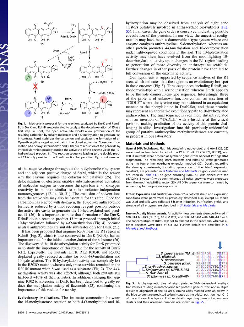
of the negative charge throughout the polyphenolic ring systemand the adjacent positive charge of SAM, which is the reasonwhy the enzyme requires the cofactor for catalysis (26). Thedelocalization of electrons enables substrate-assisted activationof molecular oxygen to overcome the spin-barrier of dioxygenreactivity in manner similar to other cofactor-independentmonooxygenases (12–14, 30, 31). The exclusion of solvent ionsfrom the active site may also be essential for this step. Once thecarbanion has reacted with dioxygen, the 10-peroxy anthracyclineformed is reduced by a thiol reducing reagent possibly outsidethe active-site cavity to generate the 10-hydroxylated end prod-uct 11 (26). It is important to note that formation of the DnrK/RdmB double-reaction product 12 must proceed through initial10-hydroxylation followed by 4-O-methylation (Fig. 4), becauseneutral anthracyclines are suitable substrates only for DnrK (21).It has been proposed that arginine R307 near the R1 region in
RdmB (Fig. 3), which is also conserved in DnrK (R302), has animportant role for the initial decarboxylation of the substrate (26).The discovery of the 10-decarboxylation activity for DnrK promptedus to study the importance of this residue for the activity of DnrKR1.2. Expectedly, the mutants DnrK R1.2 R303K and R303Qdisplayed greatly reduced activities for both 4-O-methylation and10-hydroxylation. The 10-hydroxylation activity was completely lostin the R303Q mutant, whereas only trace activities remained in theR303K mutant when 8 was used as a substrate (Fig. 2). The 4-O-methylation activity was also affected, although both mutants stillharbored ∼10% of their activities. In addition, changing the argi-nine R302 to isoleucine in DnrK has been described to greatly re-duce the methylation activity of flavonoids (23), confirming theimportance of this residue for activity.
Evolutionary Implications. The intimate connection betweenthe 15-methylesterase reaction to both 4-O-methylation and 10-
hydroxylation may be observed from analysis of eight geneclusters putatively involved in anthracycline biosynthesis (Fig.S5). In all cases, the gene order is conserved, indicating possiblecoevolution of the proteins. In our view, the ancestral config-uration may have been a daunorubicin-type system where oneenzyme catalyzes anthracycline 15-demethylation, whereas an-other protein promotes 4-O-methylation and 10-decarboxylationunder light-deprived conditions in the soil. The 10-hydroxylationactivity may then have evolved from the moonlighting 10-decarboxylation activity upon changes in the R1 region leadingto generation of more diversity in anthracycline scaffolds.Further changes in other parts of the protein have then led tofull conversion of the enzymatic activity.Our hypothesis is supported by sequence analysis of the R1
area, which indicates that the region is an evolutionary hot spotin these enzymes (Fig. 5). Three sequences, including RdmB, arerhodomycin-type with a serine insertion, whereas DnrK appearsto be the sole daunorubicin-type sequence. Interestingly, threeof the proteins of unknown function contain an insertion of“TSDLY” where the tyrosine may be positioned in an equivalentmanner to the phenylalanine in DnrK-Ser, and these proteinsmay represent an alternative evolutionary path to 10-hydroxylatedanthracyclines. The final sequence is even more distantly relatedwith an insertion of “TADLH” with a histidine at the criticalposition, making prediction of the activity of this protein chal-lenging in silico. Investigations into this previously unidentifiedgroup of putative anthracycline methyltransferases are currentlyin progress in our laboratory.
Materials and MethodsGeneral DNA Techniques. Plasmids containing native dnrK and rdmB (22, 25)were used as templates for all of the PCRs. DnrK R1.2 S297F, R303Q, andR303K mutants were ordered as synthetic genes from GeneArt (Strings DNAFragments). The remaining DnrK mutants and RdmB-CT were generatedusing the four-primer overhang extension method (32). Details regardingthe cloning experiments, including generation of the RdmC expressionconstruct, are presented in SI Materials and Methods. Oligonucleotides usedare listed in Table S3. The gene encoding RdmB-CT was cloned into thepBAD/His B vector (Invitrogen), whereas all other enzymes were expressedfrom the modified pBHBΔ vector (33). All DNA sequences were confirmed bysequencing before protein expression.
Protein Expression and Purification. Escherichia coli cell strain and expressionconditions were performed as previously described (34), except LB mediawas used and cells were collected 5 h after induction. Purification, yields, andstorage of all enzymes are described in SI Materials and Methods.
Enzyme Activity Measurements. All activity measurements were performed in100 mM Tris·HCl (pH 7.5), 10 mM DTT, and 350 μM SAM with 140 μM 8 or 9.The concentration of RdmC was set to 100 nM in the reactions, whereas allother enzymes were used at 5.8 μM. Further details are described in SIMaterials and Methods.
Fig. 4. Mechanistic proposal for the reactions catalyzed by DnrK and RdmB.Both DnrK and RdmB are postulated to catalyze the decarboxylation of 15 as afirst step. In DnrK, the open active site would allow protonation of theresulting carbanion by solvent molecules and 4-O-methylation to generate 10.In contrast, RdmB stabilizes the carbanion and catalyzes the formation of anO2–anthracycline caged radical pair in the closed active site. Consequent for-mation of a peroxyl intermediate and subsequent reduction of the peroxide byintracellular thiols possibly outside the active site of the enzyme yields the 10-hydroxylated product 11. The reaction sequence leading to the double-prod-uct 12 is only possible if the RdmB reaction happens first. R1, L-rhodosamine.
Fig. 5. A phylogenetic tree of eight putative SAM-dependent methyl-transferases residing in anthracycline biosynthesis gene clusters and multiplesequence alignment of the R1 area. Amino acids marked with an arrow inthe blue column are predicted to be situated at the critical position near C-10of the anthracycline ligands. Further details regarding these unknown geneclusters and their accession numbers are shown in Fig. S5.
9870 | www.pnas.org/cgi/doi/10.1073/pnas.1501765112 Grocholski et al.

Analysis of Metabolites. The various reaction products were analyzed andidentified as described in SI Materials andMethods, Fig. S2, and Tables S1 and S4.All reactions were monitored by HPLC using a SCL-10Avp/SpdM10Avp systemwith a diode array detector (Shimadzu). High-resolution MS (micrOTOF-Q;Bruker Daltonics; linked to an Agilent Technologies 1200 series HPLC system)was used for confirmation of the molecular formulae.
Crystallization, Data Collection, and Structure Determination of DnrK-Ser.Crystallization and structure determination of DnrK-Ser is described in SIMaterials and Methods. Diffraction data were collected at the European
Synchrotron Radiation Facility (ESRF) (Grenoble, France) at beamline ID23-1.Details of the data collection and refinement statistics are listed in Table S2.Figures depicting protein structures were prepared using PyMOL (ThePyMOL Molecular Graphics System, version 1.3; Schrodinger, LLC).
ACKNOWLEDGMENTS. We thank Elina Taipalus and Jonna Pörsti for assis-tance with protein crystallization, and Vilja Siitonen, MSc, for the high-resolutionmass spectrometry measurements. We acknowledge access to synchrotron radi-ation at ESRF (Grenoble, France). This study was supported by Academy of Fin-land Grant 136060 (to M.M.-K.).
1. Metsä-Ketelä M, Niemi J, Mäntsälä P, Schneider G (2008) Anthracycline biosynthesis:Genes, enzymes and mechanisms. Anthracycline Chemistry and Biology I: BiologicalOccurence and Biosynthesis, Synthesis and Chemistry, ed Krohn K (Springer, Berlin),pp 101–140.
2. Hertweck C, Luzhetskyy A, Rebets Y, Bechthold A (2007) Type II polyketide synthases:Gaining a deeper insight into enzymatic teamwork. Nat Prod Rep 24(1):162–190.
3. Hutchinson CR (1997) Biosynthetic studies of daunorubicin and tetracenomycin C.Chem Rev 97(7):2525–2536.
4. Pang B, et al. (2013) Drug-induced histone eviction from open chromatin contributesto the chemotherapeutic effects of doxorubicin. Nat Commun 4:1908.
5. Weiss RB (1992) The anthracyclines: Will we ever find a better doxorubicin? SeminOncol 19(6):670–686.
6. Wei G, et al. (2011) A meta-analysis of CAG (cytarabine, aclarubicin, G-CSF) regimenfor the treatment of 1029 patients with acute myeloid leukemia and myelodysplasticsyndrome. J Hematol Oncol 4:46.
7. Nitiss JL (2009) Targeting DNA topoisomerase II in cancer chemotherapy. Nat RevCancer 9(5):338–350.
8. Singal PK, Iliskovic N (1998) Doxorubicin-induced cardiomyopathy. N Engl J Med339(13):900–905.
9. Kaye S, Merry S (1985) Tumour cell resistance to anthracyclines—a review. CancerChemother Pharmacol 14(2):96–103.
10. Mitchell W (2011) Natural products from synthetic biology. Curr Opin Chem Biol 15(4):505–515.
11. Metsä-Ketelä M, Oja T, Taguchi T, Okamoto S, Ichinose K (2013) Biosynthesis ofpyranonaphthoquinone polyketides reveals diverse strategies for enzymatic carbon-carbon bond formation. Curr Opin Chem Biol 17(4):562–570.
12. Oja T, et al. (2012) Biosynthetic pathway toward carbohydrate-like moieties of al-numycins contains unusual steps for C-C bond formation and cleavage. Proc Natl AcadSci USA 109(16):6024–6029.
13. Grocholski T, et al. (2010) Crystal structure of the cofactor-independent mono-oxygenase SnoaB from Streptomyces nogalater: Implications for the reaction mech-anism. Biochemistry 49(5):934–944.
14. Siitonen V, Blauenburg B, Kallio P, Mäntsälä P, Metsä-Ketelä M (2012) Discovery ofa two-component monooxygenase SnoaW/SnoaL2 involved in nogalamycin bio-synthesis. Chem Biol 19(5):638–646.
15. Patrikainen P, et al. (2012) Tailoring enzymes involved in the biosynthesis of angu-cyclines contain latent context-dependent catalytic activities. Chem Biol 19(5):647–655.
16. Williams GJ, Gantt RW, Thorson JS (2008) The impact of enzyme engineering uponnatural product glycodiversification. Curr Opin Chem Biol 12(5):556–564.
17. Zabala AO, Cacho RA, Tang Y (2012) Protein engineering towards natural productsynthesis and diversification. J Ind Microbiol Biotechnol 39(2):227–241.
18. Firn RD, Jones CG (2000) The evolution of secondary metabolism—a unifying model.Mol Microbiol 37(5):989–994.
19. Bar-Even A, Salah Tawfik D (2013) Engineering specialized metabolic pathways—isthere a room for enzyme improvements? Curr Opin Biotechnol 24(2):310–319.
20. Madduri K, Torti F, Colombo AL, Hutchinson CR (1993) Cloning and sequencing of agene encoding carminomycin 4-O-methyltransferase from Streptomyces peucetiusand its expression in Escherichia coli. J Bacteriol 175(12):3900–3904.
21. Dickens ML, Priestley ND, Strohl WR (1997) In vivo and in vitro bioconversion ofe-rhodomycinone glycoside to doxorubicin: Functions of DauP, DauK, and DoxA.J Bacteriol 179(8):2641–2650.
22. Jansson A, Koskiniemi H, Mäntsälä P, Niemi J, Schneider G (2004) Crystal structure of aternary complex of DnrK, a methyltransferase in daunorubicin biosynthesis, withbound products. J Biol Chem 279(39):41149–41156.
23. Kim NY, et al. (2007) O-Methylation of flavonoids using DnrK based on moleculardocking. J Microbiol Biotechnol 17(12):1991–1995.
24. Niemi J, Mäntsälä P (1995) Nucleotide sequences and expression of genes fromStreptomyces purpurascens that cause the production of new anthracyclines inStreptomyces galilaeus. J Bacteriol 177(10):2942–2945.
25. Jansson A, Niemi J, Lindqvist Y, Mäntsälä P, Schneider G (2003) Crystal structure ofaclacinomycin-10-hydroxylase, a S-adenosyl-L-methionine-dependent methyltransferasehomolog involved in anthracycline biosynthesis in Streptomyces purpurascens. J Mol Biol334(2):269–280.
26. Jansson A, et al. (2005) Aclacinomycin 10-hydroxylase is a novel substrate-assistedhydroxylase requiring S-adenosyl-L-methionine as cofactor. J Biol Chem 280(5):3636–3644.
27. Wang Y, et al. (2000) Modifications of aclacinomycin T by aclacinomycin methyl es-terase (RdmC) and aclacinomycin-10-hydroxylase (RdmB) from Streptomyces purpur-ascens. Biochim Biophys Acta 1480(1-2):191–200.
28. Wang Y, Niemi J, Mäntsälä P (2002) Modification of aklavinone and aclacinomycinsin vitro and in vivo by rhodomycin biosynthesis gene products. FEMS Microbiol Lett208(1):117–122.
29. Jansson A, Niemi J, Mäntsälä P, Schneider G (2003) Crystal structure of aclacinomycinmethylesterase with bound product analogues: Implications for anthracycline rec-ognition and mechanism. J Biol Chem 278(40):39006–39013.
30. Thierbach S, et al. (2014) Substrate-assisted O2 activation in a cofactor-independentdioxygenase. Chem Biol 21(2):217–225.
31. Fetzner S, Steiner RA (2010) Cofactor-independent oxidases and oxygenases. ApplMicrobiol Biotechnol 86(3):791–804.
32. Ho SN, Hunt HD, Horton RM, Pullen JK, Pease LR (1989) Site-directed mutagenesis byoverlap extension using the polymerase chain reaction. Gene 77(1):51–59.
33. Kallio P, Sultana A, Niemi J, Mäntsälä P, Schneider G (2006) Crystal structure of thepolyketide cyclase AknH with bound substrate and product analogue: Implications forcatalytic mechanism and product stereoselectivity. J Mol Biol 357(1):210–220.
34. Koskiniemi H, Grocholski T, Schneider G, Niemi J (2009) Expression, purification andcrystallization of the cofactor-independent monooxygenase SnoaB from the nogala-mycin biosynthetic pathway. Acta Crystallogr Sect F Struct Biol Cryst Commun 65(Pt 3):256–259.
35. Ylihonko K, Hakala J, Niemi J, Lundell J, Mäntsälä P (1994) Isolation and character-ization of aclacinomycin A-non-producing Streptomyces galilaeus (ATCC 31615) mu-tants. Microbiology 140(Pt 6):1359–1365.
36. Niemi J, et al. (1994) Hybrid anthracycline antibiotics: Production of new anthracy-clines by cloned genes from Streptomyces purpurascens in Streptomyces galilaeus.Microbiology 140(Pt 6):1351–1358.
37. Kunnari T, Niemi J, Ylihonko K, Mäntsälä P, Hakala J (1997) Hybrid anthracyclines bya genetically engineered Streptomyces galilaeus mutant. Bioorg Med Chem Lett 7(6):725–726.
38. Gasteiger E, et al. (2005) Protein identification and analysis tools on the ExPASyserver. The Proteomics Protocols Handbook, ed Walker JM (Humana, Totowa, NJ), pp571–607.
39. Kabsch W (2010) Integration, scaling, space-group assignment and post-refinement.Acta Crystallogr D Biol Crystallogr 66(Pt 2):133–144.
40. Collaborative Computational Project, Number 4 (1994) The CCP4 suite: Programs forprotein crystallography. Acta Crystallogr D Biol Crystallogr 50(Pt 5):760–763.
41. Long F, Vagin AA, Young P, Murshudov GN (2008) BALBES: A molecular-replacementpipeline. Acta Crystallogr D Biol Crystallogr 64(Pt 1):125–132.
42. Langer G, Cohen SX, Lamzin VS, Perrakis A (2008) Automated macromolecular modelbuilding for X-ray crystallography using ARP/wARP version 7. Nat Protoc 3(7):1171–1179.
43. Murshudov GN, Vagin AA, Dodson EJ (1997) Refinement of macromolecular struc-tures by the maximum-likelihood method. Acta Crystallogr D Biol Crystallogr 53(Pt 3):240–255.
44. Schüttelkopf AW, van Aalten DMF (2004) PRODRG: A tool for high-throughput crys-tallography of protein-ligand complexes. Acta Crystallogr D Biol Crystallogr 60(Pt 8):1355–1363.
45. Emsley P, Lohkamp B, Scott WG, Cowtan K (2010) Features and development of Coot.Acta Crystallogr D Biol Crystallogr 66(Pt 4):486–501.
46. Afonine PV, et al. (2012) Towards automated crystallographic structure refinementwith phenix.refine. Acta Crystallogr D Biol Crystallogr 68(Pt 4):352–367.
47. Chen VB, et al. (2010) MolProbity: All-atom structure validation for macromolecularcrystallography. Acta Crystallogr D Biol Crystallogr 66(Pt 1):12–21.
48. Sultana A, et al. (2004) Structure of the polyketide cyclase SnoaL reveals a novelmechanism for enzymatic aldol condensation. EMBO J 23(9):1911–1921.
49. Hutchinson CR, Colombo AL (1999) Genetic engineering of doxorubicin production inStreptomyces peucetius: A review. J Ind Microbiol Biotechnol 23(1):647–652.
50. Sullivan MJ, Petty NK, Beatson SA (2011) Easyfig: A genome comparison visualizer.Bioinformatics 27(7):1009–1010.
Grocholski et al. PNAS | August 11, 2015 | vol. 112 | no. 32 | 9871
BIOCH
EMISTR
Y