divergence in skeletal mass and bone morphology in ... eastman et al. 2014.pdf · divergence in...
TRANSCRIPT
Divergence in Skeletal Mass and Bone Morphologyin Antarctic Notothenioid Fishes
Joseph T. Eastman,1* Lawrence M. Witmer,1 Ryan C. Ridgely,1 and Kristen L. Kuhn2
1Department of Biomedical Sciences, Heritage College of Osteopathic Medicine, Ohio University, Athens, Ohio 45701-29792US Department of Agriculture-Agricultural Research Service, Beneficial Insects Introduction Research Unit,Newark, Delaware 19713
ABSTRACT Although notothenioid fishes lack swimbladders, some species live temporarily or permanentlyin the water column. Given its relatively high density,skeletal mass is a key determinant of buoyancy. Noto-thenioids have reduced skeletal ossification, but thereis little quantitative data on the phylogenetic distribu-tion of this trait. We obtained dry skeletal masses for54 specimens representing 20 species from six notothe-nioid families. Although comparative data are sparse,notothenioid skeletons comprise a smaller percentageof body mass, <3.5%, than those of three non-notothenioid perciforms. With relatively high skeletalmass, the non-Antarctic Bovichtus diacanthus is simi-lar in skeletal mass to some non-notothenioids. Elegi-nops maclovinus, the non-Antarctic sister group of theAntarctic clade, has a relatively light skeleton (<2% ofbody mass) similar to many species in the Antarcticclade. Low skeletal mass is therefore a synapomorphyshared by Eleginops plus the Antarctic clade. We pro-vide gross, histological, and micro-CT documentation ofthe structure and location of bone and cartilage inskulls, pectoral girdles, and vertebrae, with emphasison the bovichtid B. diacanthus, the eleginopsidE. maclovinus, and the channichthyid Chaenodracowilsoni. In Eleginops and the Antarctic clade, mostbone is spongy and most species have persisting carti-lage in the skull and appendicular skeleton. We alsomeasured the relative size of the notochordal canal inadult vertebral centra of 38 species representing alleight families. There is considerable interspecific varia-tion in this pedomorphic trait and all species show anontogenetic reduction in the relative size of the canal.However, large persisting canals are present in adultsof the Antarctic clade, especially in the nototheniidsPleuragramma and Aethotaxis and in a number ofbathydraconid and channichthyid genera. J. Morphol.275:841–861, 2014. VC 2014 Wiley Periodicals, Inc.
KEY WORDS: heterochrony; histology; micro-CT scans;Bovichtidae; Eleginopsidae; Channichthyidae
INTRODUCTIONThe Notothenioid Radiation in Antarctica
Notothenioids are a monophyletic group of eightfamilies and 134 species of ray-finned fishes con-fined to the Southern Ocean around Antarctica, itsperipheral islands, and the southern extremities ofSouth America, Australia, and New Zealand (East-
man, 1993; Near et al., 2012; Lautr�edou et al.,2013). The three non-Antarctic families (Fig. 1)have experienced little speciation or ecologicaldiversification. However, in the cold isolatedwaters of the Antarctic continental shelf and slope,the Antarctic clade opportunistically expandedfrom a single lineage into five families and 105endemic species (Eastman, 2005). Notothenioidsare among the numerous examples of the conver-gent loss of the swim bladder among teleosts, anevent that has occurred independently more than30 times (McCune and Carlson, 2004), and that isan adaptive advantage for negatively buoyantfishes living on the substrate. Paradoxically, Ant-arctic notothenioids fill most water column andbenthic niches on the High Antarctic shelf, a con-tingency facilitated by the eradication of previousfaunas during tectonic, oceanographic, and cli-matic changes over the last 25 million years (Nearet al., 2012).
Although there are relatively recent nonadap-tive radiations of exclusively benthic notothenioidssuch as the 22 species of artedidraconid genusPogonophryne (Eakin et al., 2009; Near et al.,2012), vertical partitioning of the water columnaccompanied speciation in early notothenioid
Additional Supporting Information may be found in the online ver-sion of this article.
Contract grant sponsor: National Science Foundation; Contractgrant numbers: ANT 04-36190 (to J.T.E.); IOB 0517257 and IOS-1050154 (to L.M.W. and R.C.R.); Contract grant sponsor: NSF;Contract grant number: OPP 01-32032 (to H.W.D. [NortheasternUniversity]; 2004 ICEFISH cruise on the RVIB Nathaniel B.Palmer).
*Correspondence to: J.T. Eastman, Department of Biomedical Sci-ences, Heritage College of Osteopathic Medicine, Ohio University,Athens, OH 45701-2979. E-mail: [email protected]
Received 14 October 2013; Revised 3 January 2014;Accepted 26 January 2014.
Published online 4 March 2014 inWiley Online Library (wileyonlinelibrary.com).DOI 10.1002/jmor.20258
VC 2014 WILEY PERIODICALS, INC.
JOURNAL OF MORPHOLOGY 275:841–861 (2014)

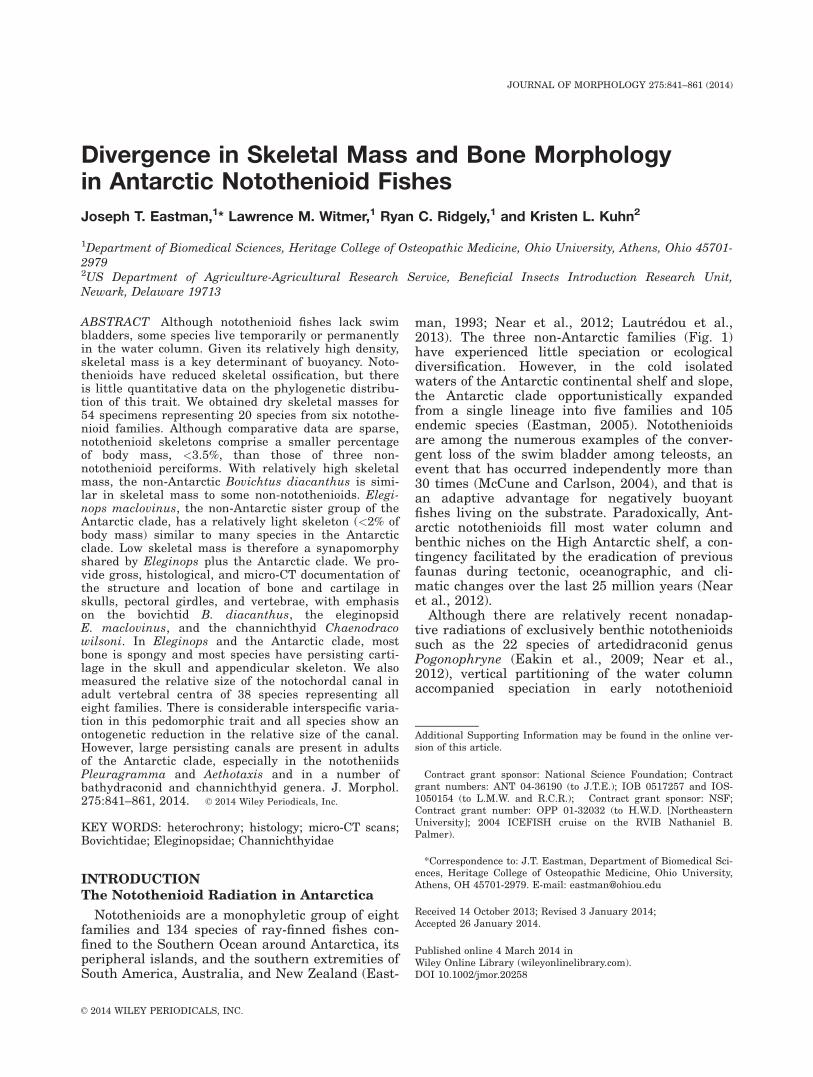
Fig. 1. Cladogram for notothenioids showing diversity in body morphology. Tree topology follows maximum likelihood phylogeny ofNear et al. (2012) based on five nuclear and two mitochondrial genes, although paraphyly of Nototheniidae and Bathydraconidae isnot shown. This is done to simplify discussion of the Antarctic families, given that the classification of notothenioids, and acantho-morphs in general, is entering a period of flux (Dettai et al., 2012; Betancur-R et al., 2013; Near et al., 2013). The three non-Antarctic families are unshaded and the five Antarctic families are color coded: Nototheniidae (blue), Harpagiferidae (light red), Arte-didraconidae (green), Bathydraconidae (orange), and Channichthyidae (purple). Red dot indicates appearance of a relatively lightskeleton, usually <2% of body mass (Table 1), although skeletal mass has not been determined for the Pseudaphritidae. Blue dotsdesignate lineages having a relatively large notochordal canal in the vertebral centra. A drawing for Bovichtus diacanthus is unavail-able; species depicted is the morphologically similar B. angustifrons.
842 J.T. EASTMAN ET AL.
Journal of Morphology

diversification, and subsequently differential useof water column and benthic habitats has been animportant ecological axis of the notothenioidadaptive radiation (Ekau, 1991; Eastman, 1993,2005; Klingenberg and Ekau, 1996; Eastman andBarrera-Oro, 2010). This premise has been rein-forced by a recent study utilizing stable isotopes ofcarbon and nitrogen to infer the use of benthicand pelagic resources by notothenioids. Isotopicdisparity through time indicates multiple and par-allel ecological divergences into overlappingpelagic niches in different families, especially theNototheniidae and Channichthyidae (Rutschmannet al., 2011). Another study found that notothe-nioids also show higher than expected morphologi-cal disparity in the shape of the opercular bone,with the shape reflecting the benthic-pelagic axisof the species studied (Wilson et al., 2013). Thenotion that Antarctic notothenioids, or some cladestherein, constitute marine species flocks on theAntarctic shelf (Eastman and McCune, 2000) hasreceived support from recent molecular studiesusing mitochondrial and nuclear markers to exam-ine the complex relationships and history of speci-ation in various notothenioid groups (Lautr�edouet al., 2012), and from a protocol using iterativeevaluation of the criteria for recognizing flocks(Lecointre et al., 2013).
Only four or five notothenioid species are neu-trally buoyant, but many other species feed in thewater column on seasonally abundant items. Liv-ing and feeding in a variety of habitats is reflectedin the reduced density and divergent buoyanciesof various species (Eastman and DeVries, 1982;Eastman and Sidell, 2002; Near et al., 2012),where buoyancy is expressed as percentage buoy-ancy (%B 5 weight in water/weight in air 3 100).Notothenioids range in mean percentage buoyancyfrom 0%, in neutrally buoyant species, to 6% inheavy benthic species, with 68% (or 61 SD) of 54species falling between 2.0 and 4.6% (Near et al.,2012, Supporting Information Table S5).
Skeletal Tissue Density, Skeletal Mass, andStatic Buoyancy
The density of teleost bone is relatively high at1,700–2,000 kg m23 (Alexander, 2003, p. 301). Fur-thermore, with a density of 1,040–1,092 kg m23,most of the soft tissues composing the body of amarine fish are denser than seawater (1,026 kgm23; Pelster, 1998). The skeletal masses of mam-mals and other terrestrial vertebrates comprise anincreasing proportion of body mass with increasingbody size, reaching a maximum of about 25% inthe largest terrestrial mammals (Schmidt-Nielsen,1975). Conversely, teleost skeletons, exemplified byperciforms, compose 3.4–5.1% of the body mass(Reynolds and Karlotski, 1977), and in samples ofmultiple teleost species the skeleton scales isomet-
rically (Reynolds and Karlotski, 1977; Berrios-Lopez et al., 1996). Although the fish skeleton hasa lower mineral content than in terrestrial verte-brates (Dean and Shahar, 2012; Cohen et al.,2012), it is still inordinately influential in deter-mining buoyancy. This is because percentage buoy-ancy values are a proxy for body density, and thisreflects the proportions and densities of the vari-ous constituent tissues (Alexander, 1968, p. 186).Because notothenioids are inactive and use pri-marily anterior–posterior oscillation with the pec-toral fins (labriform locomotion), they do notgenerate lift like continuously swimming fishes.Hence, a combination of static mechanisms isresponsible for interspecific differences in densityand buoyancy; these include reduction of skeletalmineralization, persistence of cartilage (density 51,060–1,180 kg m23, Alexander, 2003, p. 301), andaccumulation of triglyceride lipids (930 kg m23) inaxial musculature and subcutaneous tissue (DeV-ries and Eastman, 1978; Eastman and DeVries,1981).
Reduction of Bone in Fishes
Reduction in the size and thickness of bones, aswell in as in the mineral content, will minimizeskeletal mass (Pelster, 1997). The type of bone isalso relevant. Bones in fishes may be spongy (lightand porous), compact (heavy and dense), interme-diate between bone and cartilage (chondroid bone),or represented by connective tissue or persistenthyaline cartilage rather than osseous tissue (Meu-nier and Huysseune, 1992). Vertebrate compactbone is fourfold denser than spongy bone (Wain-wright et al., 1976, p. 167). Spongy bone is com-mon in teleosts (de Ricqles et al., 1991, p. 50) andthe numerous cavities in bones of the skull andvertebral centra decrease bone mass and overallbody density and also provide space for triglycer-ide lipid, especially in marine fishes (Lee et al.,1975; Phleger, 1975). The morphology of the verte-brae, which comprise 20–30% of the mass of thefish skeleton, is especially important in contribut-ing to bone mass as they contain as much as 26%more calcium than ribs and opercles (Fraser andHarvey, 1982). Reduction in heavy ions signifi-cantly reduces bone density (Pelster, 1997, p. 225).
Reduced bone development and skeletal ossifica-tion are widespread in fishes (de Beer, 1937; Gos-line, 1971; Meunier and Huysseune, 1992),especially within various teleost lineages includingnotothenioids (Iwami, 1985; Voskoboinikova, 1997,2001). These are frequently the result of hetero-chronic processes that balance the opposingdemands of buoyancy and mechanical stress(Schaeffer, 1961). In Antarctic notothenioids, pedo-morphosis (specifically postdisplacement as definedby Reilly et al., 1997) results in truncation of bonedevelopment (Voskoboinikova et al., 1994),
843NOTOTHENIOID SKELETAL MORPHOLOGY
Journal of Morphology

including delayed closure of the notochordal canalin the vertebral centra (Totton, 1914; DeVries andEastman, 1978), and persistence of cartilage.These traits are apomorphic given the earlyappearance of bones and rapid rate of bone devel-opment in larvae and juveniles of the non-Antarctic bovichtids (Voskoboinikova and Bruce,2001). Recent work using the collagen family ofgenes as markers for cartilage and bone formationalso indicates that the delayed branchial and cra-nial bone development in low-density pelagic andsemipelagic notothenioids is associated with heter-ochronic shifts in skeletal gene expression, specifi-cally persistence of the chondrogenic program anddelay in the osteogenic program during larvaldevelopment (Albertson et al., 2010; Detrich andAmemiya, 2010). This “adaptive osteopenia” hasallowed channichthyids to be advanced asevolutionary-mutant model organisms for investi-gating osteopenia in humans (Albertson et al.,2009; Maher, 2009).
The literature contains a few descriptions ofnotothenioid pectoral girdles and skulls (Starks,1930; Gregory, 1933), and there is a more substan-tial body of work on descriptive osteology as acomponent of systematic studies (Eakin, 1981; Vos-koboinikova, 1982, 1991, 1993; Andersen, 1984;Balushkin, 1984, 2000; Iwami, 1985). As men-tioned previously, there are also numerous contri-butions by Voskoboinikova and collaborators onskeletal development in larvae from most notothe-nioid clades. However, this information does notaddress bone reduction in adult notothenioids, andthere is no quantitative data on skeletal massother than reports that the ashed skeletal massesof 11 species of Nototheniidae exhibit sixfold differ-ence between the lightest neutrally buoyant spe-cies and the heaviest benthic species (DeVries andEastman, 1978; Eastman and DeVries, 1981).These ash values for nototheniids are about two tothreefold less than ash values for phylogeneticallydiverse teleosts (Childress and Nygaard, 1973).Moreover, there has been little study of the histol-ogy of notothenioid bone and cartilage, especiallyin the case of the non-Antarctic notothenioids andthe some of the phylogenetically derived Antarcticfamilies like the Channichthyidae. Therefore, it isnot known whether the skeletal reduction in adultAntarctic notothenioids is a phylogenetically per-sistent condition or whether it appeared later dur-ing diversification into the developing Antarcticmarine ecosystem. Our specific objectives here areto provide data on 1) the dry mass of skeletons fora sample of 54 specimens from 20 species repre-senting six of eight notothenioid families, includ-ing both Antarctic and non-Antarctic species; 2)aspects of gross, microscopic and micro-CT anat-omy, emphasizing neurocrania, pectoral girdles,and vertebral centra, with documentation of thedistribution of bone and cartilage in skeletons of
three phylogenetically diverse taxa; and 3) inter-specific and ontogenetic differences in the relativesize of the notochordal canal in specimens from 38species representing eight families.
MATERIALS AND METHODSSpecimens and Nomenclature
We collected material for this study during fieldwork atshore stations and on cruises to a number of Antarctic and sub-Antarctic localities from 1973 to 2011. As summarized in Tables1 and 2, we examined a total of 38 species representing alleight notothenioid families. Various components of the studyutilized subsets of this total number of species and families.This material is in the personal collection of the senior author.In accordance with protocol L01-14 approved by the Institu-tional Animal Care and Use Committee at Ohio University, weeuthanized all fishes captured through trawling or trapping bysevering the spinal cord, or by immersion in a 200-mg l21 solu-tion of 3-aminobenzoic acid ethyl ester (MS-222, Sigma, St.Louis).
We provide lengths of specimens as standard length (SL) or,when these are undetermined, as total length (TL). Taxonomicnomenclature follows Eastman and Eakin (2000), with subse-quent updates from Catalog of Fishes (Eschmeyer, 2013) andthe list of valid species maintained on Eastman’s web site(http://www.oucom.ohiou.edu/dbms-eastman). Osteologicalnomenclature is based on Jollie (1986) and Rojo (1991), andterms relating to heterochrony are those of Reilly et al. (1997).
Preparation of Skeletons for Determinationof Dry Weight
We prepared 54 dry skeletons from 1 to 11 specimens of eachof 20 notothenioid species (Table 1). We obtained lengths andweights immediately after capture and then froze the speci-mens at 230�C for periods of 3–6 months. The samples forTrematomus newnesi included both large mouth and typicalmorphs, and these were treated as a single species in the anal-ysis. Values for skeletal masses of three species were obtainedduring prior studies of buoyancy (Eastman and Barrera-Oro,2010; Eastman et al. 2011).
Cleaning skeletons involved thawing and then maceratingthe entire fish in soapy (Ajax Lemon dish washing soap andOxyClean) water for 3–5 days at room temperature (�23�C),removal of remaining tissue with running water and com-pressed air, and drying at room temperature to a constantweight for 1–2 months. In the bones of some notothenioids,lipid may account for as much as 15% of the dry weight of theskeleton (Phleger et al., 1999), and this must be removed inorder to obtain an accurate dry skeletal weight. In these cases,we extracted the remaining lipid by soaking in acetone in anultrasonic cleaner with repeated changes until the solvent wasclear. We did not determine the mineral content by ashingthese skeletons because dry notothenioid skeletons are poorlyrepresented in museum collections, and we wished to preservethese skeletons for future study.
Anatomical and Histological Techniques
For material used in anatomical and histological work, weused a combination of preservation techniques including perfus-ing specimens with Bouin’s solution immediately after capture,immersion of entire specimens in 10% formalin fixative, andimmersion of pieces of tissue in either Bouin’s or 10% formalin.We also cleared and stained preserved specimens with alizarinred S (Taylor, 1967) dissolved in 75% ethyl alcohol (Springerand Johnson, 2000).
For examination of skeletal tissue histology, we used 1–4specimens from each of 15 species representing six of the eightnotothenioid families. We removed perfusion-fixed and
844 J.T. EASTMAN ET AL.
Journal of Morphology

TABLE 1. Data for sample of 54 specimens from 20 species of notothenioids used to obtain mean percentage massesof dried skeletons
Species N LocationYear
collected
Range Mean
Standardlength (mm) Body mass (g)
Dry skeletalmass (g)
Skeletal mass/bodymass 3 100 (%)
BovichtidaeBovichtus
diacanthus(BDIA)
2 Tristan da Cunha 2004 116–130 31.30–41.50 1.06–1.43 3.42
EleginopsidaeEleginops
maclovinus(EMA)
3 Falkland Islands 2004 261–334 273.30–631.30 5.32–11.65 1.91
NototheniidaeDissostichus
mawsoni (DMA)1 McMurdo Sound 1973 880 14,061.40 206.60 1.47
Gobionotothengibberifroms (GGI)
2 South Orkney Is. 2009 302–314 407.10–454.90 7.94–8.70 1.93
Nototheniaangustata (NAN)
1 Portobello, N.Z. 1991 306 716.32 21.56 3.01
N. coriiceps (NCO) 2 Bouvet�ya 2004 420–518 1,450.00–2,550.00 45.67–62.59 2.80N. coriiceps 3 King George Is. 2009 220–372 236.40–1,239.40 5.20–30.97 2.45a
N. rossii (NRO) 3 King George Is. 2009 219–363 204.50–978.90 3.56–15.77 1.65a
Lepidonotothensquamifroms (LSQ)
2 South Orkney Is. 2009 322–325 646.00–654.00 10.24–10.35 1.58
L. nudifrons (LNU) 3 King George Is. 2010 148–180 61.87–114.97 1.19–2.32 2.00L. larseni (LLA) 2 South Orkney Is. 2009 158–167 42.00–44.80 0.82–0.95 2.04Trematomus
newnesi (TNEtyp)typical morph
1 McMurdo Sound 2005 142 45.00 0.64 1.42
T. newnesi typicalmorph
5 King George Is. 2009 162–179 71.30–112.0 0.90–1.69 1.38b
T. newnesi (TNElm)large mouth morph
5 King George Is. 2009 174–193 115.10–144.00 1.91–2.36 1.68b
T. bernacchii (TBE) 3 McMurdo Sound 2005 210–227 225.00–275.00 3.81–4.13 1.59Pagothenia
borchgrevinki(PBO)
3 McMurdo Sound 2005 170–204 88.00–147.00 1.01–1.56 1.12
ArtedidraconidaePogonophryne
scotti (PSC)2 South Orkney Is. 2009 147–206 81.40–183.20 2.18–5.16 2.75
BathydraconidaeParachaenichthys
charcoti (PCHAR)2 South Orkney Is. 2009 328–372 232.00–320.00 5.68–7.54 2.40
ChannichthyidaeChampsocephalus
gunnari (CGU)2 South Orkney Is. 2009 426–431 778.00–860.00 8.56–9.27 1.09
Pseudochaenichthysgeorgianus
(PsGEO)
2 South Orkney Is. 2009 435-–458 1,200.00–1,480.00 15.65–17.20 1.23
Chionodracorastrospinosus(CRAST)
2 South Orkney Is. 2009 310-–315 354.00–472.00 6.43–6.71 1.62
Chaenocephalusaceratus (CAC)
2 South Orkney Is. 2009 279–294 152.70–173.20 2.81–3.19 1.84
Cryodracoantarcticus (CANT)
1 South Orkney Is. 2009 414 418.00 8.59 2.06
Non-notothenioidsPercidae
Perca flavescens(PFL)
4 North America 1977 72.60–92.30 2.90–3.47 3.81c
Stizostedion vitreum(SVI)
1 North America 1977 628.60 23.29 3.71c
SerranidaeEpinephelus striatus
(EST)3 North America 1977 1,033.00–1,159.60 44.58–58.16 4.72c
The morphs of Trematomus newnesi are listed separately but are treated as one species in subsequent analyses. Arrangement oftaxa is phylogenetic based on cladogram in Figure 1. Literature values for non-notothenioids are at the end of table. Skeletalmasses of individual notothenioid specimens are available in Supporting Information Table S1.aData from Eastman et al. (2011).bData from Eastman and Barrera-Oro (2010).cData from Reynolds and Karlotski (1977).
845NOTOTHENIOID SKELETAL MORPHOLOGY
Journal of Morphology

immersion-fixed samples of tissue from the skull, pectoral gir-dle, and vertebral centra in an area of the column centered onthe first caudal vertebra. We decalcified tissues for 10–48 h in a
commercial solution containing EDTA in dilute HCl, and post-fixed them in Bouin’s solution. We embedded samples in paraf-fin according to standard procedures. We cut 7-lm-thick
TABLE 2. Relative percentage diameter of the notochordal canal (horizontal diameter of notochordal canal/horizontal diameter ofvertebral centrum 3 100) based on measurements of the first caudal vertebra in each of 130 dry and alizarin stained skeletons
from adults of 38 species representing all eight notothenioid families
Species N
SizeRelative diameter of
notochordal canal (%)
Maximum knownTL or SL (mm)a
This sampleSL (mm) Rangeb Mean
BovichtidaeBovichtus diacanthus 2 250* 116–130 11–10B. variegatus 3 250* 125–172 11–7Cottoperca trigloides 2 500* 217–240 8
PseudaphritidaePseudaphritis urvillii 3 300* 157–182 9–7
EleginopsidaeEleginops maclovinus 4 1,000* 255–357 10–5
NototheniidaePleuragramma antarctica 23 250 105–225 81–50 65Aethotaxis mitopteryx 1 420* 113 48Dissostichus mawsoni 3 2,360* 271–880 17–4D. eleginoides 4 2,250* 276–750 17–4Gobionotothen gibberifroms 2 550* 302–314 9Notothenia angustata 1 410* 306 5N. coriiceps 5 620* 220–518 12–5N. rossii 3 920* 219–363 14–8Lepidonotothen squamifroms 2 500* 322–325 13L. nudifrons 4 190* 148–180 22–17L. larseni 2 240* 158–167 32–28Trematomus newnesi (typical morph) 10 200* 122–179 38–24 30T. newnesi (large mouth morph) 8 139–193 38–22 28T. newnesi (all morphs combined) 18 122–193 38–22 29T. bernacchii 9 350* 93–227 36–15 25Pagothenia borchgrevinki 12 280* 121–204 28–19 22
HarpagiferidaeHarpagifer antarcticus 1 95 92 20
ArtedidraconidaePogonophryne scotti 2 310* 147–206 16–8
BathydraconidaeGymnodraco acuticeps 1 340 189 19Prionodraco evansii 1 150 123 28Vomeridens infuscipinnis 1 220 178 40c
Racovitzia glacialis 1 240 182 35Akarotaxis nudiceps 1 130 112 46Bathydraco macrolepis 1 250 141 32B. marri 1 230 119 43Parachaenichthys charcoti 2 420 328–372 28–22
ChannichthyidaePagetopsis macropterus 1 330* 142 36P. maculatus 1 250* 193 30Champsocephalus gunnari 2 660* 426–431 20–19Pseudochaenichthys georgianus 2 600* 435-–458 17–16Chaenodraco wilsoni 3 430* 174–233 46–31Chionodraco rastrospinosus 2 520* 310-–315 30–28Chaenocephalus aceratus 2 750* 279–294 36–35Cryodraco antarcticus 1 570* 414 24Dacodraco hunteri 1 290* 204 40d
Arrangement of taxa is phylogenetic based on cladogram in Figure 1.aMaximum known sizes from various sources, especially Gon and Heemstra (1990); asterisk * indicates length is total lengthrather than standard length.bArrangement of values is from smallest to largest specimens.cData from Kuhn et al. (2011).dData from radiograph in Eastman (1999).
846 J.T. EASTMAN ET AL.
Journal of Morphology

sections and stained with Gomori’s one step trichrome for 30 s,Pollak’s trichrome for 7 min (Humason, 1979), or Bodian’s Pro-targol for 24 h at 50�C (Clark, 1981).
We used an ocular micrometer fitted to a dissecting micro-scope to determine the relative size of the notochordal canal.We measured the diameter of the notochordal canal and thewidth of vertebral centra. We made these measurements trans-versally across the posterior aspect of one of the first three cau-dal vertebrae, usually the first. We took measurements fromdry vertebrae or from vertebrae that were removed fromalizarin-stained, glycerine-stored specimens.
Imaging TechniquesMicro-CT scanning. We scanned one entire adult speci-
men of Bovichtus diacanthus (SL 5 153 mm) and of Chaeno-draco wilsoni (SL 5 203 mm), and the head of an adultEleginops maclovinus (SL 5 285 mm). The specimens were fixedin formalin and stored in 70% ethanol. All three specimens werescanned at the Ohio University MicroCT Facility (OUlCT),using a GE eXplore Locus in vivo Small Animal MicroCT Scan-ner. Images were acquired at scan resolutions of 45 lm (isotropicvoxel sizes), 80 kVp, 450 lA, 1,200 views around 360� with sevenframes averaged per view, and an integration time of 400 ms.The resulting data volume (in VFF format) were exported fromGE Healthcare’s MicroView 2.1.2 (http://sourceforge.net/projects/microview/) to the DICOM format. These DICOM data werethen imported to Avizo 7.1 (FEI Visualization Sciences Group,Burlington, MA) for analysis and visualization.
Soft radiography. We also used soft radiography, i.e., pro-duction of X-rays at low voltage with filtration through a beryl-lium window (Miller, 1957), for visualizing bone and cartilagein some formalin-fixed, alcohol-stored specimens. We producedradiographs with a Hewlett-Packard Faxitron soft X-raymachine. We operated the machine at 30 kVp and 3.0 mA, withan exposure time of 3–5 min. We used Kodak Industrex MX125film (medium speed, very high contrast, high definition, veryfine grain) in lead-backed cardboard cassettes. Film-to-sourcedistance was 122 cm.
RESULTSSkeletal Mass
In our sample of 20 species (15% of notothenioiddiversity), body masses encompass four orders ofmagnitude with a 454-fold difference between thesmallest and largest specimens. A scatter plot ofrelative skeletal mass (Fig. 2) indicates that noto-thenioid skeletons compose a smaller percentageof body mass than those of three species of non-notothenioid perciforms, at least as judged bythese relatively few perciform measurementsavailable in the literature. The arbitrary valueseparating the two groups is about 3.5%. With rel-atively heavy skeletons, the two specimens of thenon-Antarctic B. diacanthus cluster among thelightest of the non-notothenioids. However, thethree specimens of E. maclovinus, the non-Antarctic sister group of the Antarctic clade, haverelatively light skeletons and cluster among theAntarctic clade. A relatively light skeleton, �2% ofbody mass, is therefore a synapomorphy for Elegi-nops plus the Antarctic clade (Fig. 1).
Skeletal System Anatomy and HistologyGeneralities. The skulls of teleost fishes,
including notothenioids, are composed of a superfi-
cial dermal component and a deeper endoskeletalcontribution that forms most of the mass of theneurocranium around the brain and specialsenses. The endoskeletal series, preformed in hya-line cartilage, includes bones such as the eth-moids, sphenoids, otics, and occipitals. The dermaladdition invests the endoskeletal elements and isexemplified by the frontals, parietals, prevomer,and parasphenoid (Gregory, 1933, p. 88–90;Harder, 1975; Rojo, 1991). The dermal bones areperipheral to the perichondrium of the endoskele-ton and develop via intramembranous ossification.
All notothenioid bone we examined is acellular,as would be expected in an acanthomorph group(Moss, 1961, 1963; Parenti, 1986), and the adapt-ive significance of this trait is unknown (Hortonand Summers, 2009). Actinopterygian fish lack theHaversian organization typically seen in compactbone of other vertebrates (Meunier and Huys-seune, 1992; Summers and Long, 2006). We recog-nize two subtypes of spongy bone: laminar (layersof various thicknesses) and cavitated (i.e., spongy,porous). We refer to small irregular shaped lami-nae of bone as trabeculae. All cartilage weobserved is typical hyaline cartilage, Category 1under the classification of Witten et al. (2010).
Bone and cartilage in the skull. Amongnotothenioids, there is variability in the mass andthe nature of bone among taxa, and in the amountof cartilage in the skeleton. This is clearly seen inmicro-CT scans of species representing families at
Fig. 2. Scatter plot on semilogarithmic coordinates showingrelative skeletal mass. Circles are individual values for 54 noto-thenioids from 20 species and eight non-notothenioid perciforms(see Table 1, bottom) representing three species (data from Reyn-olds and Karlotski, 1977). The arbitrary separation betweennotothenioids and non-notothenioids is about 3.5%. Letter codesfor species are given in Table 1.
847NOTOTHENIOID SKELETAL MORPHOLOGY
Journal of Morphology

the extremes of the phylogeny (Fig. 1). In Figures 4and 5A,C,E, volumes are rendered in a false-colorspectrum in which warmer and cooler colors (redsvs. blues and greens) represent materials of higherand lower density, respectively. The bovichid B.diacanthus (Figs. 4A and 5A,B) has a well-ossifiedskeleton, typical of many acanthomorphs. The redcolor thus indicates the presence and relative den-sity of bone, and it is evident that all major neuro-cranial, as well as jaw, branchial, fin girdle, andvertebral elements are present and well ossified.However, this is not the case in the phylogeneti-cally derived channichthyid C. wilsoni (Figs. 4Band 5E,F). Based on the limited red signal, there isrelatively little bone and considerable cartilage thatis not easily differentiated from other soft tissuesproducing a more widespread lower density bluesignal. Chaenodraco and other channichthyids donot have a uniformly ossified skull with tight, oreven abutting, sutures between bones. Instead,many dermal bones ossify as thin laminae thatensheath persisting cartilage. When a fresh skulldehydrates and the cartilage shrinks greatly in vol-ume, the ensheathing nature of the bones isobvious by comparing Figure 7I versus 7J, and Fig-ure 7K versus 7L. This is also evident in radiogra-phy of the dried skull of another channichthyid,Pseudochaenichthys georgianus (Fig. 6A). Theensheathing bones impart a predominantly graytone to the image, although some bones possess fociof heavier ossification (white) that are sometimes
manifest as spinated or crenulated areas. The eth-moids, supraorbital region of frontals, sphenotics,pterotics, and occipital series are the best examples(Fig. 6A).
Persistent cartilage forms most of the roof, floor,and lateral walls of the channichthyid neurocranium(Figs. 4B and 5E,F). Although not evident in micro-CT scans, the prominence of cartilage is highlightedin histological sections. In C. wilsoni, the bone to theright of the supraorbital canal is only 68–70-lmthick (Fig. 9D), with considerable cartilage below. InChaenocephalus aceratus, cartilage also dominatesthe posteriolateral skull; there are two thin (25 and18 lm) laminae of bone dorsally (not shown) and athicker lamina ventrally (Fig. 9F). The parasphenoid,ventral to the lens of the eye in Figure 5E, is one offew substantially ossified elements in the floor of theneurocranium. Based on the red signal, other well-ossified bones include the jaws, opercular apparatuswith spines, branchiostegal apparatus, cleithral ele-ments, occipital series, pelvic spines, and vertebralcentra (Figs. 4B and 5E).
In addition to channichthyids, some nototheniidsand most bathydraconids also retain partially car-tilaginous neurocrania as adults. Small and largeneutrally buoyant species of the family Notothenii-dae are notable in this regard. In Pleuragrammaantarctica, the dorsal neurocranium of a 163 mmSL specimen is largely cartilaginous, with roofingfrontal bones that are only 18-lm thick (Fig. 9I). A1,420-mm SL specimen of Dissostichus mawsonihas large areas of persistent cartilage in the eth-moid, frontal, otic, and occipital regions of the neu-rocranium (Fig. 6B).
Given the considerable differences in the skele-tal tissue morphology between Bovichtus andchannichthyids, what is the situation in E. maclo-vinus, the sister group of the Antarctic clade (Fig.1)? Micro-CT scanning reveals that Eleginops hasa head skeleton composed predominantly ofspongy bone (Fig. 5C), although the cavitatednature of the bone is best seen in transverse sec-tion (Fig. 5D). The only exception in Figure 5D isthe radiodense bone of the parasphenoid and bran-chiostegal rays. Histology provides additional doc-umentation of the nature of the bone porosity. Forexample, the dorsal skull is a network of thinbony trabeculae surrounded by extensive irregularcavities containing adipocytes and connective tis-sue of the dermis (Fig. 9B). The bone of Eleginopsdoes not give a red signal, as is the case in Bovich-tus and Chaenodraco; the large areas histologi-cally documented as spongy bone are insteadgreen, representing reduced density. This may bean artifact of its preservational history in that thisspecimen was fixed and stored in unbuffered for-malin for 5 months before being transferred toethanol. Some loss of bone mineral from the thintrabeculae (Fig. 9B) may be responsible for agreen rather than a red signal.
Fig. 3. Scatter plot and trend lines showing interspecific differ-ences and ontogenetic changes in relative diameter of notochor-dal canal of the first caudal vertebrae with increasing bodylength in specimens of the nototheniids Pleuragramma antarc-tica (N 5 23), Pagothenia borchgrevinki (12), Trematomus ber-nacchii (9), and T. newnesi (18).
848 J.T. EASTMAN ET AL.
Journal of Morphology

Fig. 4. Volume rendered micro-CT images showing differences in the amount of bone in adult skeletons of (A) non-Antarctic bovichtidBovichtus diacanthus (SL 5 153 mm) and (B) Antarctic channichthyid Chaenodraco wilsoni (SL 5 203 mm). Tissue density is mappedin false color on a spectrum ranging from less dense (cool colors) to more dense (warm colors). Red color is a proxy for the density of bonewhich is considerably more extensive in B. diacanthus. Much of the neurocranium, paired fin girdles and branchial apparatus of C. wil-soni consists of cartilage not shown in the image or not discernible from other low-density (blue-colored) soft tissues.
849NOTOTHENIOID SKELETAL MORPHOLOGY
Journal of Morphology

Fig. 5. Left lateral and transverse micro-CT images of adult (A,B) Bovichtus diacanthus, (C,D) Eleginops maclovinus, and (E,F)Chaenodraco wilsoni. A,B and E,F are same specimens as in Figure 4. C,D is eleginopsid (SL 5 285 mm; head length 5 72 mm), aphylogenetically intermediate species between Bovichtus and Chaenodraco and the sister group to the Antarctic clade. In lateralviews (A,C,E), Bovichtus (A) has dense well-ossified bone as indicated by red tone (see caption for Fig. 4 for explanation of the colormapping). In Eleginops (C), spongy bone with extensive lipid-filled cavities has lower density and bony mass that collectively registeras green (with the possibility of the green attributable to partial demineralization by formalin storage—see text). In Chaenodraco(E), red indicates that jaw and opercular elements are ossified but extensive cartilage persists and is visible in pectoral girdle whichalso shows foci of weak ossification in the radials. Transverse images (B,D,F) show differences in nature and distribution of bone(bright white tones) in equivalent regions of heads posterior to the orbit. Prominent calcified elements visible in transverse sectionsinclude, dorsoventrally, in Bovichtus (B), dense bone of neurocranium, fifth ceratobranchials, gill arch elements, opercular and clei-thral elements, pelvic girdle, and branchiostegal rays; in Eleginops (D), spongy bone of neurocranium and most other bones, gill archelements, opercular and cleithral, and branchiostegal rays and; in Chaenodraco (F), thin laminae of bone ensheathing cartilaginousneurocranium with midventral basioccipital most prominent, dentigerous gill rakers, opercular and cleithral elements (cartilage sur-rounded by thin layer of bone), and branchiostegal rays. Scale bars: (A,B,C,E) 10 mm and (D,F) 5 mm.
850 J.T. EASTMAN ET AL.
Journal of Morphology

Fig. 6. Aspects of skeletal morphology in adult notothenioids. (A) Channichthyid Pseudochaenichthys georgianus and (B–D) noto-theniid Dissostichus mawsoni. (A) Radiograph of dried skull in Figure 7J showing thin ensheathing bone of neurocranium in general(gray), exemplified by parasphenoid, with foci of heavier ossification (white) in other bones, especially the occipital series. (B) Exten-sive cartilage in dorsal skull of formalin-preserved alcohol-stored specimen (SL 5 1,240 mm). (C) Trans-illumination reveals spongybone with array of small cavities (lipid-filled in life) in centrum of fourth caudal vertebra of D. mawsoni (SL 5 880 m). (D) Radio-graph of transverse section of portion of pectoral fin musculature and girdle of D. mawsoni (SL 5 1,000 mm) showing spongy bonewith variously sized cavities surrounding a core of cartilage. Scale bars: (A,D) 10 mm and (C) 2 mm. Abbreviations: BO, basioccipital;C, cartilage; F, crenulated supraorbital ridge of frontal; LE, lateral ethmoid; PAL, palatine; PS, parasphenoid; PTO, pterotic; RS, ros-tral spine of ethmoid; SB, spongy bone; and SPO, sphenotic.
851NOTOTHENIOID SKELETAL MORPHOLOGY
Journal of Morphology

Fig. 7. Ventral view of adult skulls in a phylogenetic series (Fig. 1) representing four of eight notothenioid families: (A) Bovichtusdiacanthus; (B) Cottoperca trigloides; (C) Eleginops maclovinus; (D) Dissostichus eleginoides; (E) Gobionotothen gibberifrons; (F)Notothenia coriiceps; (G) Lepidonotothen squamifrons; (H) Trematomus bernacchii; (I, J) Pseudochaenichthys georgianus, I whenfresh and wet, showing extensive opaque ethmoid cartilage, and J when dried; and (K,L) Cryodraco antarcticus, K when fresh andwet showing cartilage, and L when dried. Smallest skull is B. diacanthus (A) at 29 mm long; largest is D. eleginoides (D) at 117 mmlong. Scale bars: (A) 5 mm and (D) 20 mm. Abbreviation: E, ethmoid cartilage.
852 J.T. EASTMAN ET AL.
Journal of Morphology

The phylogenetic series of skulls in Figure 7,representing four of the eight families, shows thatthe superficial bone is either cavitated, ridged, ortrabeculated or thin smooth-surfaced laminae. InEleginops (Fig. 7C) and some larger and mid-sizednototheniid species (Fig. 7D–G), the bones are pit-ted by cavities containing adipocytes. The same istrue for Notothenia coriiceps, a large nototheniidwith a heavy skeleton (Fig. 7F). However, othersmaller nototheniid species, trematomids forexample, have thin and delicate laminar bone(Fig. 7H). The superficial bones in bovichtids (Fig.7A,B), bathydraconids (not shown), and channich-thyids (Fig. 7I–L) are usually smooth-surfacedlaminae. The characteristic elongated snout regionof channichthyids (Fig. 7I–L) is formed by the thinlaminar ethmoids, underlain by cartilage thatcomposes most of the mass of the snout (Fig.7I,K).
Bone and cartilage in the pectoral girdle.In notothenioids, as in other acanthomorphs (Kou-moundouros et al., 1999), the scapula, coracoid,and radials of the pectoral girdle are endochondralbones. Transverse histological sections of adultpectoral girdles show that skeletal tissue isarranged as a core of cartilage sandwichedbetween peripheral layers of bone of variablethickness. In a phylogenetic series (Fig. 9A,C,E),cartilage becomes progressively a more dominantcomponent between progressively thinner layers ofperipheral bone. For example, at the midpoint ofthe section (Fig. 9A), B. diacanthus has 75- and200-lm-thick layers of compact bone with a core ofpersistent cartilage and adipose tissue. In the ele-ginopsid E. maclovinus, peripheral bone isarranged as laminae 13–35-lm-thick, with a coreof cartilage and adipose tissue (Fig. 9C). However,in the channichthyid C. aceratus (Fig. 9E), most ofthe mass of the structure of the girdle is formedby a thick layer of cartilage bordered by 15-lm-thick layers of superficial bone. The percentagethickness of the girdle occupied by bone in thesethree species is 39, 13, and 3%, respectively.Finally, cartilage also persists in the pectoral gir-dle of most other species, including the neutrallybuoyant nototheniids as exemplified by a 1,000mm SL specimen of D. mawsoni (Fig. 6D).
Bone of the vertebral centra. The structureof the vertebral centra of notothenioids conforms tothe basic teleost pattern (Ford, 1937; Laerm, 1976).There is no cartilage precursor or persistent carti-lage and, instead, the initial mineralization occurswithin the notochordal sheath, with a subsequentaddition of intramembraneous bone from thesomites (Francois, 1966; Laerm, 1982; Koumoun-douros et al., 1999; Bird and Mabee, 2003;Bensimon-Brito et al., 2012). This has been con-firmed for nototheniids in the developing urostylarvertebrae of five species, including P. antarctica(Voskoboinikova et al., 2004). Centra of adult noto-
thenioids, exemplified by E. maclovinus, are cylin-drical and amphicelous, with the hourglass shapeespecially evident in mid sagittal section (Fig.8A,G). All notothenioids have a notochordal canalat the bony constriction point, but there is consider-able interspecific and ontogenetic variation in thepatency of the canal (see below). There is a thin,biconid-shaped layer of laminar bone adjacent tothe notochordal cavities (Fig. 8A,B). Internally, thebone of the centrum is spongy, with cavities thatare extremely variable in size and shape. Cavitiesare lined by periosteum, filled with adipocytes, andhave smooth walls that do not exhibit evidence oferosion by osteoclasts (Fig. 8B,C).
Figure 8D–O is a phylogenetic series showingthe lateral aspect of the first caudal centrum fromeach of 12 species representing seven of the eightnotothenioid families. There is considerable varia-tion in the sculpting of the lateral surfaces of thecentra, especially in the number and thickness ofthe longitudinal ridges and in the degree of poros-ity of the bone (Fig. 8E,I,J,L,N). For example, thebone of the centra shows little porosity in B. diac-anthus (Fig. 8D). In E. maclovinus, the sistergroup of the Antarctic clade, centra consist ofspongy bone (Fig. 8A,B,G). All species in the Ant-arctic clade also have reduced skeletal mass com-pared to Bovichtus (Table 1, Fig. 2), but this is notnecessarily associated with extremely porous bonein the centra. For example, the centra are com-posed of lamellae and cavities of various sizes infour nototheniids (Fig. 8H–K), an artedidraconid(Fig. 8L), a bathydraconid (Fig. 8M), and twochannichthyids (Fig. 8N,O). Another nototheniid,Notothenia angustata, a heavy benthic specieswith largest skeletal mass in this family, has amore massive centrum with a single thick longitu-dinal ridge and little spongy bone (Fig. 8J).
The three neutrally buoyant nototheniids withweakly ossified skeletons have centra with differ-ent morphology and therefore deserve comment.With the exception of Eleginops, spongy bone withsmall cavities is rare in our sample of notothenioidcentra, but bone with small cavities is especiallypervasive in the centra of D. mawsoni (Fig. 6C)and D. eleginoides (Fig. 8H). After removal oflipid, transillumination reveals the delicate natureof the lattice and the extreme porosity of the bonein D. mawsoni (Fig. 6C). With a percentage skele-tal mass of 1.47%, this species has one of the fivelightest skeletons of the 20 species for which wehave data (Table 1). A different means of reductionof the bone mass in is seen in a closely related spe-cies, P. antarctica. The centra exist as a collar ofthin laminar bone, with many small longitudinalridges, around the notochordal canal and partiallypersistent notochord (Fig. 9G,H). Some of the bonymass of the centra is therefore absent and the cen-tra are less amphicelous. We do not have skeletalmass data for Pleuragramma.
Journal of Morphology
853NOTOTHENIOID SKELETAL MORPHOLOGY

Fig. 8. A–C. Anatomy and histology of centrum of first caudal vertebrae of Eleginops maclovinus. (A) Sanded midsagittal sectionshowing minimal amount of laminar bone (arrowheads) bordering the large biconcave notochordal cavities, with remainder of cen-trum consisting of spongy bone. This species has a small notochordal canal (asterisk) in the middle of each centrum. (B,C) Parasagit-tal sections displaying histological detail of centra. (B) Pattern of red-staining bone and lipid-filled cavities. Vacuolated cells fillnotochordal cavities. Width of bone at constriction is 0.6 mm in life and magnification is 327. Stain: Gomori’s trichrome. (C) Nuclearstaining and photography at higher magnification indicate bone is acellular. Dark nuclei adjacent to vacuoles are those of prevacuo-lar cells of notochord. Lipid in cavities of spongy bone is contained in adipocytes. Dark lines in bone matrix are growth checks. Layerof laminar bone is adjacent to notochordal canals and vacuolated cells. Vertical diameter of large cavity is 200 mm in life and magnifi-cation is 3160. Stain: Bodian’s protargol. D–O. Left lateral view of centra of anterior caudal vertebrae in a phylogenetic series (Fig.1) representing seven of eight notothenioid families: (D) Bovichtus diacanthus; (E) Cottoperca trigloides; (F) Pseudaphritis urvillii;(G) E. maclovinus; (H) Dissostichus eleginoides; (I) Gobionotothen gibberifrons; (J) Notothenia angustata; (K) Pagothenia borchgre-vinki; (L) Pogonophryne scotti; (M) Parachaenichthys charcoti; (N) Champsocephalus gunnari; and (O) Chaenocephalus aceratus. Ver-tebra is B. diacanthus (D) at 2.5 mm in horizontal length; largest is D. eleginoides (H) at 9.4 mm. Scale bars: (D) 0.5 mm and (H) 2mm. Abbreviations: A, adipocytes; NCV, notochordal cavities; and V, vacuolated cells of notochord.

Fig. 9. Variable distribution of bone and cartilage in adult notothenioids. A, C, E. Transverse sections of pectoral girdles from (A)Bovichtus diacanthus, (C) Eleginops maclovinus, and (E) Chaenocephalus aceratus showing that in this phylogenetic series there is adecrease in red-staining bone and an increase in blue-staining cartilage. Stain: Gomori’s trichrome. B. Transverse section of skulldorsal to opercular region of E. maclovinus shows spongy bone with large cavities containing adipose tissue and blue-staining denseconnective tissue. Stain: Pollak’s trichrome. D. Transverse section of dorsal skull of channichthyid Chaenodraco wilsoni with thinlaminae of bone around the supraorbital canal and extensive underlying cartilage. Red staining of matrix does not signify calcifiedcartilage but is an artifact of staining possibly caused by tissue disruption due to freezing. Stain: Gomori’s trichrome. F. Transversesection of posterior-lateral skull of channichthyid C. aceratus also showing thin bone and preponderance of cartilage. Stain: Gomori’strichrome. G–I. Bone anatomy and histology of Pleuragramma antarctica. (G) Posterior view of one of the anterior-most caudal verte-brae of 164 mm SL specimen reveals large size (62% of transverse centrum diameter) of notochordal canal. Stain: alizarin red S. (H)Transverse section of caudal peduncle vertebra of 170 mm specimen shows bone of centra as thin collar surrounding vacuolated cellsof a partially persistent notochord. Stain: Pollak’s trichrome. (I) Transverse section of interorbital region of skull of 163 mm specimenwith thin frontal bone underlain by extensive cartilaginous neurocranium with relatively few chondrocytes. Stain: Gomori’s tri-chrome. Abbreviations: A, Adipocytes; C, cartilage; D, dermis of skin; NC, notochordal canal; SO, supraorbital canal; and V, vacuo-lated cells of notochord. Scale bars: (A–F,I) 250 mm, (G) 2 mm, and (H) 0.5 mm.
855NOTOTHENIOID SKELETAL MORPHOLOGY
Journal of Morphology

Interspecific and Ontogenetic Variation inthe Size of the Notochordal Canal
Table 2 summarizes the extensive interspecificvariation that exists in the size of the notochordalcanal in a sample of vertebrae of 130 adults repre-senting 38 species (29% of notothenioid diversity)from all eight families. There is also ontogeneticvariation, specifically a decrease in relative size ofthe notochordal canal—meaning that the noto-chord and canal are larger in smaller specimensand decrease in size with increasing body length.This is graphed for an ontogenetic series in eachof four species (Fig. 3), but the trend is also pres-ent in all species we examined when individualswere of different sizes (Table 2). As the canalsbecome constricted by bone growth during ontog-eny, the centra become more amphicelous.
Examination of Table 2 also indicates that spe-cies in the three non-Antarctic families have rela-tively small canals (�11%), whereas most speciesin the Antarctic clade have larger canals. Using avalue of �48% as a break point, relatively largecanals are present in representatives of three fam-ilies (Fig. 1). The neutrally buoyant nototheniidspecies P. antarctica (Fig. 9G,H) and Aethotaxismitopteryx have especially large canals and aredistinct among notothenioids in the extent of thispedomorphic trait. Consequently, the bone mass ofthe centra is reduced by the persistence of a par-tial notochord, and centra are not completelyamphicelous. The next largest canals (32–46%) areseen in the bathydraconid genera Vomeridens,Racovitzia, Akarotaxis, and Bathydraco, and inthe channichthyid genera Pagetopsis, Chaeno-draco, Chaenocephalus, and Dacodraco. Modestsized canals (25–30%) are present in the notothe-niid genera Trematomus and Pagothenia, and inthe bathydraconid genus Prionodraco. Members ofthe benthic nototheniid genus Notothenia and ofthe benthic families Harpagiferidae and Artedidra-conidae have small canals (�20%, and frequently<10% in larger specimens). Although neutrallybuoyant and possessing extremely porous bone intheir centra, the large heavily muscled, migratorynototheniids D. mawsoni and D. eleginoides havevery small (4%) canals.
DISCUSSIONSkeletal Mass and Life History
Although our data on skeletal mass for 20 spe-cies represent only 15% of notothenioid diversity(Table 1, Fig. 2), the higher level taxonomic cover-age (six of eight families) provides reasonable con-fidence in our results concerning skeletal masswithin the clade. These are 1) that notothenioidskeletons comprise <3.5% of body mass, a lowervalue than those for three non-notothenioid perci-form species; 2) that non-Antarctic B. diacanthus
has a the highest skeletal mass among the noto-thenioids sampled; 3) that E. maclovinus, the non-Antarctic sister group of the Antarctic clade, has alighter skeleton than Bovichtus and, in a scatterplot, clusters among species of the Antarctic clade;and 4) that a relatively light skeleton, frequently�2% of body mass, is a synapomorphy for Elegi-nops plus the Antarctic clade (Fig. 1), rather thana trait confined exclusively to the Antarctic clade.Given the sparse data for skeletal mass in non-notothenioid perciforms (see bottom of Table 1), wehave no basis for discussing either the frequencyor phylogenetic distribution of reduced skeletalmass in other acanthomorphs.
There are several life history factors that mayinfluence skeletal mass in fishes including degreeof activity and mode of locomotion. There are nocontinuously swimming notothenioids—most aresedentary as exemplified by the benthic notothe-niid N. coriiceps, a species with mean percentageskeletal mass and mean percentage buoyancy of2.80 and 4.34%, respectively. A 330-mm TL indi-vidual monitored with an underwater video cam-era at Signy Island in the South Orkneysremained within 3 m of a small cave more than98% of the time, and swam only 1.7% of each day,usually for less than 30 s (North, 1996). For low-speed swimming, most notothenioids use drag-based labriform locomotion, meaning the pectoralfins oscillate in an anterior–posterior fashion, sup-plemented with occasional bursts of undulatorysubcarangiform propulsion (Montgomery and Mac-donald, 1984; Archer and Johnston, 1989).
Although a four or five notothenioid species areneutrally buoyant and pelagic, their life historiesdo not include periods of sustained swimming. Forexample, the neutrally buoyant P. antarctica hasrespiratory and cardiovascular parameters reflect-ing a low activity level (Kunzmann, 1990; Tambur-rini et al., 1997; W€ohrmann et al., 1997), and ithangs in the water column while monitoring zoo-plankton prey with its well-developed visual andmechanosensory systems (Eastman and Lannoo,2011). The presence of a weakly ossified skeleton,including a partially hydrostatic vertebral column,is not problematic but advantageous in reducingdensity. Another neutrally buoyant nototheniid,D. mawsoni, a large predator, is migratory andmay swim as far as 1,000 km for purposes ofreproduction and feeding (Yukhov, 1982; Hanchetet al., 2008). This is accomplished using primarilylabriform locomotion (Eastman, 1993, pp. 202–205)to cover distances of at least 6 km/day (Petrov andTatarnikov, 2010). Although it has a light skeletonwith porous bone and persistent cartilage, there isno doubt that the musculoskeletal system has thecapacity for sustained labriform locomotion—avagrant individual of the morphologically identicalsister species, D. eleginoides, completed a transe-quatorial migration from the Patagonian shelf to
856 J.T. EASTMAN ET AL.
Journal of Morphology

Greenland, a distance of 10,000 km (M�ller et al.,2003).
Diversification in Bone and CartilageMorphology
General trends. Having established thatthere is reduced bone mass in Eleginops plus theAntarctic clade, how is this manifest morphologi-cally? Gosline (1971, p. 11) observed that amongteleosts “the interrelationships between the noto-chord, cartilage, and bone are highly complex, andthe extent of displacement of one skeletal materialby another in the different fishes varies greatly.”He also notes that there is a general and repeatedphylogenetic trend involving ancestral members ofa lineage that are frequently “bonier” than derivedmembers. Gosline’s mention of the notochord alsoimplies an awareness of a role for heterochronicprocesses in determining skeletal tissue composi-tion in adult fishes.
Gosline’s (1971) statements are consistent withwhat we have found in notothenioids. Non-Antarctic B. diacanthus has a relatively heavy,mostly bony skeleton. Extensive spongy boneappears in E. maclovinus, the sister group of theAntarctic clade. In the Antarctic clade, bone isalso spongy, with a fine lipid-filled mesh in thetwo neutrally buoyant species of Dissostichus.Other species in the Antarctic clade have thinlaminar bone, spongy bone, or a mixture thereof.Persistent cartilage is present in most species,especially in the neurocranium and pectoral girdle,and is prominent in some bathydraconids and allchannichthyids where thin laminar boneensheathes the cartilage. In the vertebral centra,a relatively large notochordal canal associatedwith a partially persistent notochord is found invarious degrees throughout the Antarctic cladebut is most prominent in the nototheniid generaPleuragramma and Aethotaxis and in some generaof bathydraconids and channichthyids. On thebasis of their studies of individual families,Balushkin (1984), Iwami (1985, p. 63), and Vosko-boinikova (1993) consider the loss or reduction ofbones and teeth to be a recognizable evolutionarytrend in notothenioids. Our broader taxonomicsampling here establishes the extremes of notothe-nioid skeletal morphology as encompassing a typi-cal bony skeleton (Fig. 4A) as well as one withlittle bone and considerable cartilage (Fig. 4B), asrevealed in the micro-CTs of B. diacanthus andC. wilsoni. This is also supported by our data onpercentage skeletal mass—3.42% for B. diacan-thus and a mean of 1.57% for five channichthyidspecies.
Our findings are also compatible with Voskoboi-nikova’s study of the developmental osteology oflarval and juvenile notothenioids showing thatBovichtus angustifrons has the most rapid rate ofskeletal development among notothenioids, with
bones appearing in larvae of relatively small size(Voskoboinikova and Bruce, 2001). Relative toBovichtus, nonbovichtid notothenioids have (Vos-koboinikova, 2001) 1) a delay in the appearance(postdisplacement) of numerous bony elementssuch that the larvae are larger on the initialappearance of a given element and 2) a more pro-nounced delay in some phylogenetically derivedfamilies, especially some nototheniids, many bath-ydraconids and all channichthyids. For example,the larvae of channichthyids are 2.2–4.7-fold lon-ger (in SL) than those of B. angustifrons when theanlagen of most bones appear.
Implications of extensive cartilage inchannichthyids. Cartilage is the primary skele-tal material in larval actinopterygian fishes(Summers and Long, 2006, p. 55), and studiesindicate that most of the cartilage of the develop-ing teleost head serves “as models for ossified ele-ments” (Walter, 2013, p. 90). There is nothingunusual about the retention of cartilage in theadult teleost skeleton; salmonids are a classicexample (Gregory, 1933, p. 153; de Beer, 1937,p.115–130; Norden, 1961). What is unusual inchannichthyids is the extent of cartilage retainedin the head—it is more of a scaffold than a modelfor the skull bones which are just a veneer. Recentwork has revealed the molecular basis for this het-erochronic shift in early cranial development,especially in hyal, suspensory, and jaw elements,in benthopelagic channichthyids. Relative tobenthic nototheniids and non-notothenioids withmore heavily ossified skeletons, channichthyidshave the most delayed osteogenic developmentamong notothenioids (Voskoboinikova, 1997;Albertson et al., 2010). This heterochronic shift isattributed to altered gene expression, includingdelayed expression of the osteogenic markerscol1a1 and col10a1, and prolonged expression ofthe cartilage differentiation gene col2a1 (Albertsonet al., 2010, p. 2). Albertson et al. (2010) suggestthat these changes in collagen gene expressionpatterns in pelagic notothenioids with lighter skel-etons, including P. antarctica, could be attribut-able to mutations in regulatory genes.
One consequence of the minimal bone in thechannichthyid head (Iwami, 1985, p.63) is thepotential for greater lateral and dorsoventralexpansion of the oral and branchial cavities. Lossof rigidity of the head may facilitate feeding onfish and swarms of krill. Channichthyids are ramfeeders, swimming to and grasping their prey.They exhibit morphology typical of ram feeders,including an elongated head, an enormous gapeand a nonprotractile jaw lacking an ascending pro-cess of the premaxilla (Liem, 1993). The jaw teethare small and conical with a Type 2 mode ofattachment, meaning that a collagenous ligamentbinds the tooth base to the jaw bone, anotherpedomorphic trait (Fink, 1981). The preferred
857NOTOTHENIOID SKELETAL MORPHOLOGY
Journal of Morphology

diets of many of the 16 species include fish and, asadults, some of the larger channichthyids, such asC. aceratus with a maximum TL of 75 cm (Iwamiand Kock, 1990), feed almost exclusively on fish(Kock, 2005; Reid et al., 2007; Kock et al., 2013).They routinely engulf other large-headed notothe-nioids that are 40–50% of their own length, and aC. aceratus has been documented as having swal-lowed a D. mawsoni that was 76% of its lengthand 41% of its weight (Kock et al., 2013). However,pharyngeal gape in fishes is usually smaller thanoral gape, and is likely more important in deter-mining what can be swallowed (Wainwright andRichard, 1995), especially when the prey are largeor have a rigid exoskeleton. The limiting dimen-sion in the predator is thought to be the distancebetween the cleithra of the pectoral girdles (Wain-wright and Richard, 1995). In comparing themicro-CT scans of Bovichtus and Chaenodraco inFigures 4 and 5, it can be seen that the rightcleithrum in the former is wider, with a mediallyprojecting shelf, whereas in the latter it is lessextensive and more planar.
Final Remarks: Adaptation, Heterochronyand Notothenioid Diversification
In both text (Hill et al., 2008) and popular books(Carroll, 2006), notothenioids are highlighted asexemplars of fascinating physiological specializa-tions for life in subzero oxygen-saturated seawater.Some traits are obviously adaptive (antifreeze gly-copeptides), whereas others (loss of hemoglobin andventricular myoglobin) are mutations that wouldprobably have been lethal in any other habitatgiven the extensive compensation necessary tomaintain cardiorespiratory function in the white-blooded channichthyids. However, the appearanceof antifreeze preceded most speciation and ecologi-cal diversification by at least 10 million years, andtherefore it may be viewed as a constitutive adapta-tion, enabling survival in subzero seawater, rathera key adaptation directly linked to ecological diver-sification. There are no known adaptations coupledwith the three major bursts of relatively recentdiversification that resulted in about 50% of modernnotothenioid species (Near et al., 2012).
Contrary to the suggestion by Albertson et al.(2010, p. 8), reduced bone mass is not linked tothe adaptive radiation of the Antarctic clade butwas, as we have shown here, present in their non-Antarctic sister group Eleginopsidae. Given thatnotothenioids lack a swim bladder, reduction ofbody density is difficult without some evolutionarymodification of dense skeletal tissues. This isaccomplished through heterochrony (Gould, 1977),not by novel adult morphology. Pedomorphosis hasplayed an important role in shaping skeletal tissuemorphology since early in the history of jawedfishes. For example, notothenioids possess some of
the same pedomorphic traits, such as persistentcartilage in the skull, seen in dipnoan lungfishes(Bemis, 1984).
Like those of the majority of marine fishes (Leiset al., 2011), notothenioid larvae and juveniles arepelagic (Loeb et al., 1993). In the case of the Antarcticnotothenioid clade, truncation of bone development inlarvae and juveniles by pedomorphosis furnisheddensity-reducing traits that provided potential forniche expansion, adaptive differentiation, and specia-tion. Unoccupied niche space was available about 22million years ago because notothenioids were enteringa developing Antarctic marine ecosystem where theyfaced little competition from midwater fishes lackingantifreeze (Near et al., 2012). The Antarctic clade hadthe relatively low skeletal mass present in their non-Antarctic sister group. Although the weakly ossifiedskeleton and unconstructed notochord of the notothe-niid P. antarctica have been known for a century (Tot-ten, 1914), Balushkin (1984, p. 128) was the first torecognize the evolutionary potential of pelagic larvaein nototheniids. Klingenberg and Ekau (1996) subse-quently demonstrated the importance of larval growthand morphology in an ecomorphological study of diver-sification into water column and benthic niches in 10nototheniid species. They found that morphometricdifferences in the distinguishing features of pelagicand benthic species develop by divergent growth inlarval stages, with growth after metamorphosisaccounting for relatively little of the observed interspe-cific variation in pelagic and benthic traits.
Notothenioids are percomorphs, a group encom-passing more than one-third of all fishes (Nelson,2006), and also exhibiting the most acceleratedrate of diversification of the six vertebrate cladeswith unusually high rates of diversification (Alfaroet al., 2009). Even without the introduction ofnovel morphology, the percomorph developmentalprogram and body plan have proven to have excep-tional potential for adaptation and radiation inmany freshwater and littoral marine habitatsincluding some, like the Antarctic, commonlyviewed as extreme.
ACKNOWLEDGMENTS
The authors are most grateful to Danette Prattfor assembling the figures and to John Sattler forphotography. They also thank Esteban Barrera-Oro for collecting and contributing fish from KingGeorge Island, and David Petzel for collecting andcontributing specimens from McMurdo Sound. Ananonymous reviewer and Steve Reilly providedhelpful comments on the manuscript.
LITERATURE CITED
Albertson RC, Cresko W, Detrich HW, Postlethwait JH. 2009.Evolutionary mutant models for human disease. TrendsGenet 25:74–81.
858 J.T. EASTMAN ET AL.
Journal of Morphology

Albertson RC, Yan YL, Titus TA, Pisano E, Vacchi M, YelickPC, Detrich HW III, Postlethwait JH. 2010. Molecular pedo-morphism underlies craniofacial skeletal evolution in Antarc-tic notothenioid fishes. BMC Evol Biol 10:4; doi:10.1186/1471-2148-10-4.
Alexander RM. 1968. Animal Mechanics. London: Sidgwick &Jackson. 346 pp.
Alexander RM. 2003. Principles of Animal Locomotion. Prince-ton: Princeton University Press. 371 pp.
Alfaro ME, Santini F, Brock C, Alamillo H, Dornburg A,Rabosky DL, Carnevale G, Harmon LJ. 2009. Nine excep-tional radiations plus high turnover explain species diversityin jawed vertebrates. Proc Natl Acad Sci USA 106:13410–13414.
Andersen NC. 1984. Genera and subfamilies of the family Noto-theniidae (Pisces, Perciformes) from the Antarctic and Suban-tarctic. Steenstrupia 10:1–34.
Archer SD, Johnston IA. 1989. Kinematics of labriform andsubcarangiform swimming in the Antarctic fish Notothenianeglecta. J Exp Biol 143:195–210.
Balushkin AV. 1984. Morphological Bases of the Systematicsand Phylogeny of the Nototheniid Fishes. Leningrad: AcadSci USSR, Zool Inst. pp 1–140 [in Russian; English transla-tion available in Russian Translation Series No. 1973, 1990,A.A. Balkema, Rotterdam].
Balushkin AV. 2000. Morphology, classification, and evolution ofnotothenioid fishes of the Southern Ocean (Notothenioidei,Perciformes). J Ichthyol 40(Suppl 1):S74–S109.
Bemis WE. 1984. Paedomorphosis and the evolution of the Dip-noi. Paleobiology 10:293–307.
Bensimon-Brito A, Cardeira J, Cancela ML, Huysseune A,Witten PE. 2012. Distinct patterns of notochord mineraliza-tion in zebrafish coincide with the localization of Osteocalcinisoform 1 during early vertebral centra formation. BMC DevBiol 12:28; doi:10.1186/1471-213X-12-28.
Berrios-Lopez M, Lewis AR, Hensley DA. 1996. Scaling of skel-etal mass to body mass in fishes. J Morphol 227:87–92.
Betancur-R R, Broughton RE, Wiley EO, Carpenter K, L�opezJA, Li C, Holcroft NI, Arcila D, Sanciangco M, Cureton II JC,Zhang F, Buser T, Campbell MA, Ballesteros JA, Roa-VaronA, Willis S, Borden WC, Rowley T, Reneau PC, Hough DJ, LuG, Grande T, Arratia G, Ort�ı G. 2013. The tree of life and anew classification of bony fishes. PLOS Curr. Tree of Life.2013 April 18. doi:10.1371/currents.tol.53ba26640df0ccaee75bb165c8c26288.
Bird NC, Mabee PM. 2003. Developmental morphology of theaxial skeleton of the zebrafish, Danio rerio (Ostariophysi:Cyprinidae). Dev Dyn 228:337–357.
Carroll SB. 2006. The Making of the Fittest: DNA and the Ulti-mate Forensic Record of Evolution. New York: W.W. Norton.301 pp.
Childress JJ, Nygaard MH. 1973. The chemical composition ofmidwater fishes as a function of depth of occurrence offsouthern California. Deep Sea Res 20:1093–1109.
Clark G, editor. 1981. Staining Procedures, 4th ed. Baltimore:Williams & Wilkins. 512 pp.
Cohen L, Dean MN, Shipov A, Atkins A, Monsonego-Ornan E,Shahar R. 2012. Comparison of structural, architectural andmechanical aspects of cellular and acellular bone in two tele-ost fish. J Exp Biol 215:1983–1993.
de Beer G. 1937. The Development of the Vertebrate Skull.Oxford: Clarendon Press. 554 pp.
de Ricqles A, Meunier FJ, Castanet J, Francillon-Vieillot H.1991. Comparative microstructure of bone. In: Hall BK,editor. Bone: A Treatise, Vol. 3. Boca Raton: CRC Press. pp1–77.
Dean MN, Shahar R. 2012. The structure–mechanics relation-ship and the response to load of the acellular bone of neote-leost fish: A review. J Appl Ichthyol 28:320–329.
Detrich HW III, Amemiya CT. 2010. Antarctic notothenioidfishes: Genomic resources and strategies for analyzing anadaptive radiation. Integr Comp Biol 50:1009–1017.
Dettai A, Berkani M, Lautredou A-C, Couloux A, Lecointre G,Ozouf-Costaz C, Gallut C. 2012. Tracking the elusive mono-phyly of nototheniid fishes (Teleostei) with multiple mitochon-drial and nuclear markers. Mar Genomics 8:49–58.
DeVries AL, Eastman JT. 1978. Lipid sacs as a buoyancy adap-tation in an Antarctic fish. Nature 271:352–353.
Eakin RR. 1981. Osteology and relationships of the fishes of theAntarctic family Harpagiferidae (Pisces, Notothenioidei). In:Kornicker LS, editor. Antarctic Research Series, Biology ofthe Antarctic Seas IX, Vol. 31. Washington: American Geo-physical Union. pp 81–147.
Eakin RR, Eastman JT, Near TJ. 2009. A new species and amolecular phylogenetic analysis of the Antarctic fish genusPogonophryne (Notothenioidei: Artedidraconidae). Copeia 4:705–713.
Eastman JT. 1993. Antarctic Fish Biology: Evolution in aUnique Environment. San Diego: Academic Press. 322 pp.
Eastman JT. 1999. Aspects of the biology of the icefish Daco-draco hunteri (Notothenioidei, Channichthyidae) in the RossSea, Antarctica. Polar Biol 21:194–196.
Eastman JT. 2005. The nature of the diversity of Antarcticfishes. Polar Biol 28:93–107.
Eastman JT, Barrera-Oro E. 2010. Buoyancy studies of threemorphs of the Antarctic fish Trematomus newnesi (Notothe-niidae) from the South Shetland Islands. Polar Biol 33:823–831.
Eastman JT, DeVries AL. 1981. Buoyancy adaptations in aswim-bladderless Antarctic fish. J Morphol 167:91–102.
Eastman JT, DeVries AL. 1982. Buoyancy studies of notothe-nioid fishes in McMurdo Sound, Antarctica. Copeia 2:385–393.
Eastman JT, Eakin RR. 2000. An updated species list for noto-thenioid fish (Perciformes; Notothenioidei), with comments onAntarctic species. Arch Fish Mar Res 48:11–20.
Eastman JT, Lannoo MJ. 2011. Divergence of brain and retinalanatomy and histology in pelagic Antarctic notothenioidfishes of the sister taxa Dissostichus and Pleuragramma. JMorphol 272:419–441.
Eastman JT, McCune AR. 2000. Fishes on the Antarctic conti-nental shelf: Evolution of a marine species flock? J Fish Biol57(Suppl A):84–102.
Eastman JT, Sidell BD. 2002. Measurements of buoyancy forsome Antarctic notothenioid fishes from the South ShetlandIslands. Polar Biol 25:753–760.
Eastman JT, Barrera-Oro E, Moreira E. 2011. Adaptive radia-tion at a low taxonomic level: Divergence in buoyancy of theecologically similar Antarctic fish Notothenia coriiceps and N.rossii. Mar Ecol Prog Ser 438:195–206.
Ekau W. 1991. Morphological adaptations and mode of life inHigh Antarctic fish. In: di Prisco G, Maresca B, Tota B, edi-tors. Biology of Antarctic Fish. Berlin: Springer-Verlag. pp23–39.
Eschmeyer WN, editor. 2013. Catalog of Fishes. CaliforniaAcademy of Sciences. Available at: http://research.calacademy.org/research/ichthyology/catalog/fishcatmain.asp, accessed onSeptember 9, 2013.
Fink WL. 1981. Ontogeny and phylogeny of tooth attachmentmode in actinopterygian fishes. J Morphol 167:167–184.
Ford E. 1937. Vertebral variation in teleostean fishes. J MarBiol Assoc UK 22:1–60.
Francois. 1966. Structure et d�eveloppement de la vertebre deSalmo et des t�el�eost�eens. Arch Zool Exp G�en 107:287–328.
Fraser GC, Harvey HH. 1982. Elemental composition of bonefrom white sucker (Catostomus commersoni) in relation tolake acidification. Can J Fish Aquat Sci 39:1289–1296.
Gon O, Heemstra PC, editors. 1990. Fishes of the SouthernOcean. Grahamstown, South Africa: J.L.B. Smith Institute ofIchthyology. 462 pp.
Gosline WA. 1971. Functional Morphology and Classification of Tele-ostean Fishes. Honolulu: University Press of Hawaii. 208 pp.
Gould SJ. 1977. Ontogeny and Phylogeny. Cambridge, MA:Harvard University Press. 501 pp.
859NOTOTHENIOID SKELETAL MORPHOLOGY
Journal of Morphology

Gregory WK. 1933. Fish skulls: A study of the evolution of nat-ural mechanisms. Trans Am Phil Soc 23:75–481.
Hanchet SM, Rickard GJ, Fenaughty JM, Dunn A, WilliamsMJH. 2008. A hypothetical life cycle for Antarctic toothfish(Dissostichus mawsoni) in the Ross Sea region. CCAMLR Sci15:35–53.
Harder W. 1975. Anatomy of Fishes, Part I: Text. Stuttgart: E.Schweizerbart’sche Verlagsbuchhandlung. 612 pp.
Hill RW, Wyse GA, Anderson M. 2008. Animal Physiology, 2nded. Sunderland: Sinauer Associates. 762 pp.
Horton JM, Summers AP. 2009. The material properties of acel-lular bone in a teleost fish. J Exp Biol 212:1413–1420.
Humason GL. 1979. Animal Tissue Techniques, 4th ed. SanFrancisco: W.H. Freeman. 661 pp.
Iwami T. 1985. Osteology and relationships of the family Channich-thyidae. Mem Natl Inst Polar Res, Tokyo, Ser E No. 36:1–69.
Iwami T, Kock K-H. 1990. Channichthyidae.In: Gon O, Heem-stra PC, editors. Fishes of the Southern Ocean. Grahams-town, South Africa: J.L.B. Smith Institute of Ichthyology. pp381–399.
Jollie M. 1986. A primer of bone names for the understandingof the actinopterygian head and pectoral girdle skeleton. CanJ Zool 64:365–379.
Klingenberg CP, Ekau W. 1996. A combined morphometric andphylogenetic analysis of an ecomorphological trend: Pelagiza-tion in Antarctic fishes (Perciformes: Nototheniidae). Biol JLinn Soc 59:143–177.
Kock K-H. 2005. Antarctic icefishes (Channichthyidae): A uniquefamily of fishes. A review, Part I. Polar Biol 28:862–895.
Kock K-H, Gr€oger J, Jones CD. 2013. Interannual variability inthe feeding of ice fish (Notothenioidei, Channichthyidae) inthe southern Scotia Arc and the Antarctic Peninsula region(CCAMLR subareas 48.1 and 48.2). Polar Biol 36:1451–1462.
Koumoundouros G, Divanach P, Kentouri M. 1999. Osteologicaldevelopment of the vertebral column and of the caudal com-plex in Dentex dentex. J Fish Biol 54:424–436.
Kuhn KL, Near TJ, Detrich III HW, Eastman JT. 2011. Biologyof the Antarctic dragonfish Vomeridens infuscipinnis (Noto-thenioidei: Bathydraconidae). Antarct Sci 23:18–26.
Kunzmann A. 1990. Gill morphometrics of two Antarctic fishspecies Pleuragramma antarcticum and Notothenia gibberi-frons. Polar Biol 11:9–18.
Laerm J. 1976. The development, function, and design ofamphicoelous vertebrae in teleost fish. Zool J Linn Soc 58:237–254.
Laerm J. 1982. The origin and homology of the neopterygianvertebral column. J Paleontol 56:192–202.
Lautr�edou A-C, Hisinger DD, Gallut C, Cheng C-HC, BerkaniM, Ozouf-Costaz C, Cruaud C, Lecointre G, Dettai A. 2012.Phylogenetic footprints of an Antarctic radiation: The Trema-tominae (Notothenioidei, Teleostei). Mol Phylogen Evol 65:87–101.
Lautr�edou A-C, Motomura H, Gallut C, Ozouf-Costaz C,Cruaud C, Lecointre G, Dettai A. 2013. New nuclear markersand exploration of the relationships among Serraniformes(Acanthomorpha, Teleostei): The importance of working atmultiple scales. Mol Phylogen Evol 67:140–155.
Lecointre G, Am�eziane N, Boisselier M-C, Bonillo C, Busson F,Causse R, Chenuil A, Couloux A, Coutanceau JP, Cruaud C,d’Acoz CD, De Ridder C, Denys G, Detta€ı A, Duhamel G,El�eaume M, F�eral JP, Gallut C, Havermans C, Held C,Hemery L, Lautr�edou A-C, Martin P, Ozouf-Costaz C, PierratB, Pruvost P, Puillandre N, Samadi S, Saucede T, SchubartC, David B. 2013. Is the species flock concept operational?The Antarctic shelf case. PLoS ONE 8:e68787; doi:10.1371/journal.pone.0068787.
Lee RF, Phleger CF, Horn MH. 1975. Composition of oil in fishbones: Possible function in neutral buoyancy. Comp BiochemPhysiol 50B:13–16.
Leis JM, Siebeck U, Dixson DL. 2011. How Nemo finds home:The neuroecology of dispersal and of population connectivityin larvae of marine fishes. Integr Comp Biol 51:826–843.
Liem KF. 1993. Ecomorphology of the teleostean skull. In:Hanken J, Hall BK, editors. The Skull, Functional and Evolu-tionary Mechanisms, Vol. 3. Chicago: University of ChicagoPress. pp 422–452.
Loeb VJ, Kellermann AK, Koubbi P, North AW, White MG.1993. Antarctic larval fish assemblages: A review. Bull MarSci 53:416–449.
Maher B. 2009. Biology’s next top model? Nature 458:695–698.McCune AR, Carlson RL. 2004. Twenty ways to lose your blad-
der: Common natural mutants in zebrafish and widespreadconvergence of swim bladder loss among teleost fishes. EvolDev 6:246–259.
Meunier FJ, Huysseune A. 1992. The concept of bone tissue inOsteichthyes. Neth J Zool 42:445–458.
Miller RR. 1957. Utilization of X-rays as a tool in systematiczoology. Syst Zool 6:29–40.
Montgomery JC, Macdonald JA. 1984. Performance of motorsystems in Antarctic fishes. J Comp Physiol A 154:241–248.
M�ller PR, Nielsen JG, Fossen I. 2003. Patagonian toothfishfound off Greenland. Nature 421:599.
Moss ML. 1961. Osteogenesis of acellular teleost fish bone. AmJ Anat 108:99–110.
Moss ML. 1963. The biology of acellular teleost bone. Ann NYAcad Sci 109:337–350.
Near TJ, Dornburg A, Kuhn KL, Eastman JT, Pennington JN,Patarnello T, Zane L, Fern�andez DA, Jones CD. 2012.Ancient climate change, antifreeze, and the evolutionarydiversification of Antarctic fishes. Proc Natl Acad Sci USA109:3434–3439.
Near TJ, Dornburg A, Eytan RI, Keck BP, Smith WL, KuhnKL, Moore JA, Price SA, Burbrink FT, Friedman M,Wainwright PC. 2013. Phylogeny and tempo of diversificationin the superradiation of spiny-rayed fishes. Proc Natl AcadSci USA 110:12738–12743.
Nelson JS. 2006. Fishes of the World, 4th ed. Hoboken: Wiley.601 pp.
Norden CR. 1961. Comparative osteology of representativealmonid fishes, with particular reference to the grayling(Thymallus arcticus) and its phylogeny. J Fish Res BoardCan 18:679–791.
North AW. 1996. Locomotory activity and behaviour of the Ant-arctic teleost Notothenia coriiceps. Mar Biol 126:125–132.
Parenti LR. 1986. The phylogenetic significance of bone typesin euteleost fishes. Zool J Linn Soc 87:37–51.
Pelster B. 1997. Buoyancy at depth. In: Randall DJ, Farrell AP,editors. Fish Physiology, Deep-Sea Fishes, Vol. 16. San Diego:Academic Press. pp 195–237.
Pelster B. 1998. Buoyancy.In: Evans DH, editor. The Physiologyof Fishes, 2nd ed. Boca Raton: CRC Press. pp 25–42.
Petrov AF, Tatarnikov VA. 2010. New data on migrations ofAntarctic toothfish Dissostichus mawsoni in the Dumontd’Urville Sea in the 2008/2009 season. J Ichthyol 50:143–144.
Phleger CF. 1975. Bone lipids of Kona coast reef fish: Skullbuoyancy in the hawkfish, Cirrhites pinnulatus. Comp Bio-chem Physiol 52B:101–104.
Phleger CF, Nichols PD, Erb E, Williams R. 1999. Lipids of thenotothenioid fishes Trematomus spp. and Pagothenia borch-grevinki from East Antarctica. Polar Biol 22:241–247.
Reid WDK, Clarke S, Collins MA, Belchier M. 2007. Distribu-tion and ecology of Chaenocephalus aceratus (Channichthyi-dae) around South Georgia and Shag Rocks (SouthernOcean). Polar Biol 30:1523–1533.
Reilly SM, Wiley EO, Meinhardt DJ. 1997. An integrativeapproach to heterochrony: The distinction between interspe-cific and intraspecific phenomena. Biol J Linn Soc 60:119–143.
Reynolds WW, Karlotski WJ. 1977. The allometric relationshipof skeleton weight to body weight in teleost fishes: A prelimi-nary comparison with birds and mammals. Copeia 1:160–163.
Rojo AL. 1991. Dictionary of Evolutionary Fish Osteology. BocaRaton: CRC Press. 273 pp.
Rutschmann S, Matschiner M, Damerau M, Muschick M,Lehmann MF, Hanel R, Salzburger W. 2011. Parallel
860 J.T. EASTMAN ET AL.
Journal of Morphology

ecological diversification in Antarctic notothenioid fishes asevidence for adaptive radiation. Mol Ecol 20:4707–4721.
Schaeffer B. 1961. Differential ossification in the fishes. TransNY Acad Sci Ser II 23:501–505.
Schmidt-Nielsen K. 1975. Scaling in biology: The consequencesof size. J Exp Zool 194:287–308.
Springer VG, Johnson GD. 2000. Use and advantages of etha-nol solution of alizarin red S dye for staining bone in fishes.Copeia 1:300–301.
Starks EC. 1930. The primary shoulder girdle of the bonyfishes. Stanford Univ Publ Biol Sci (Univ Ser) 6:149–239.
Summers AP, Long JH Jr. 2006. Skin and bones, sinew and gristle:The mechanical behavior of fish skeletal tissues. In: ShadwickRE, Lauder GV, editors. Fish Biomechanics, Fish Physiology, Vol.23. San Diego: Elsevier Academic Press. pp 141–177.
Tamburrini M, D’Avino R, Carratore V, Kunzmann A, di PriscoG. 1997. The hemoglobin system of Pleuragramma antarcti-cum: Correlation of hematological and biochemical adapta-tions with life style. Comp Biochem Physiol 118A:1037–1044.
Taylor WR. 1967. An enzyme method of clearing and stainingsmall vertebrates. Proc US Natl Mus 122:1–17.
Totton AK. 1914. The structure and development of the caudalskeleton of the teleostean fish, Pleuragramma antarcticum.Proc Zool Soc Lond 18:251–261.
Voskoboinikova OS. 1982. Visceral skeleton changes in the pro-cess of turning to pelagic mode of life in fishes of the familyNototheniidae. Acad Sci USSR, Proc Zool Inst 114:67–76 [inRussian].
Voskoboinikova OS. 1991. Comparative osteology of dragon-fishes of the subfamily Gymnodraconinae (Bathydraconidae).J Ichthyol 31:24–33.
Voskoboinikova OS. 1993. Evolution of the visceral skeletonand phylogeny of Nototheniidae. J Ichthyol 33:23–47.
Voskoboinikova OS. 1997. Osteological development of theChannichthyidae (Teleostei: Notothenioidei). Cybium 21:369–379.
Voskoboinikova OS. 2001. Evolutionary significance of hetero-chronies in the development of the bony skeleton in fishes ofthe suborder Notothenioidei (Perciformes). J Ichthyol 41:415–424.
Voskoboinikova OS, Bruce B. 2001. Development of bony skele-ton during ontogeny of Bovichtus angustifrons (Bovichtyidae,Notothenioidei) with comparative notes on development ofbony skeleton of congolli Pseudaphritis urvilli (Pseudaphriti-dae). J Ichthyol 41:57–66.
Voskoboinikova OS, Tereshchuk OY, Kellermann A. 1994.Osteological development of the Antarctic silverfish Pleura-gramma antarcticum (Nototheniidae). Cybium 18:251–271.
Voskoboinikova OS, Malashichev EB, Voronina EP. 2004. Onthe development of some bony elements in the ontogenesis offive species of Notothenioidei. J Ichthyol 44:245–251.
Wainwright PC, Richard BA. 1995. Predicting patterns of preyuse from morphology of fishes. Environ Biol Fish 44:97–113.
Wainwright SA, Biggs WD, Currey JD, Gosline JM. 1976.Mechanical Design in Organisms. Princeton: Princeton Uni-versity Press. 423 pp.
Walter BE. 2013. Cranial skeletogenesis and osteology of theredeye tetra Moenkhausia sanctaefilomenae. J Fish Biol 82:69–95.
Wilson LAB, Colombo M, Hanel R, Salzburger W, S�anchez-Vilagra MR. 2013. Ecomorphological disparity in an adaptiveradiation: Percular bone shape and stable isotopes in Antarc-tic icefishes. Ecol Evol 3:3166–3182.
Witten PE, Huysseune A, Hall BK. 2010. A practical approachfor the identification of the many cartilaginous tissues in tele-ost fish. J Appl Ichthyol 26:257–262.
W€ohrmann APA, Hagen W, Kunzmann A. 1997. Adaptations ofthe Antarctic silverfish Pleuragramma antarcticum (Pisces:Nototheniidae) to pelagic life in high-Antarctic waters. MarEcol Prog Ser 151:205–218.
Yukhov VL. 1982. The Antarctic Toothfish (Antarkticheskii kly-kach). Moscow: Nauka. 113 pp [in Russian; four page conclu-sion translated to English by U.S. Department of State].
861NOTOTHENIOID SKELETAL MORPHOLOGY
Journal of Morphology