distinguishing ms from neuromyelitis optica (nmo) 1030.pdf · distinguishing ms from neuromyelitis...
TRANSCRIPT
Dr. Tony Traboulsee,
MS Clinic Director
University of British Columbia
Vancouver, BC Canada
Distinguishing MS from Neuromyelitis
Optica (NMO)
Dr. Jack Simon,
Neuroradiologist,
Portland VA Medical Center
and Oregon Health and Sciences University
Disclosures - Simon
• Research support: Biogen Idec

Disclosures - Traboulsee • Non Pharma:
– Canadian Asian MS research supported by Canadian Institute for Health Research
– NMO research supported by Vancouver Hospital Foundation
• Pharma:
– Research Grants: Bayer, Biogen, Merck Serono, Teva
– Speakers bureau: Bayer, Merck Serono, Teva
– Consultant: Bayer, Merck Serono, Teva, Biogen, Sanofi-Aventis
– DSMB or Steering committee: Merck Serono, Roche

Disclosures - Traboulsee
• Off label treatment warning:
– There are no approved therapies for neuromyelitis optica
– Any discussion of treatments is based on anecdotal personal experience, published case series and/or the opinions of US and international colleagues who also treat NMO.

Objectives
• To be aware of clinical and MRI features that distinguish NMO from MS
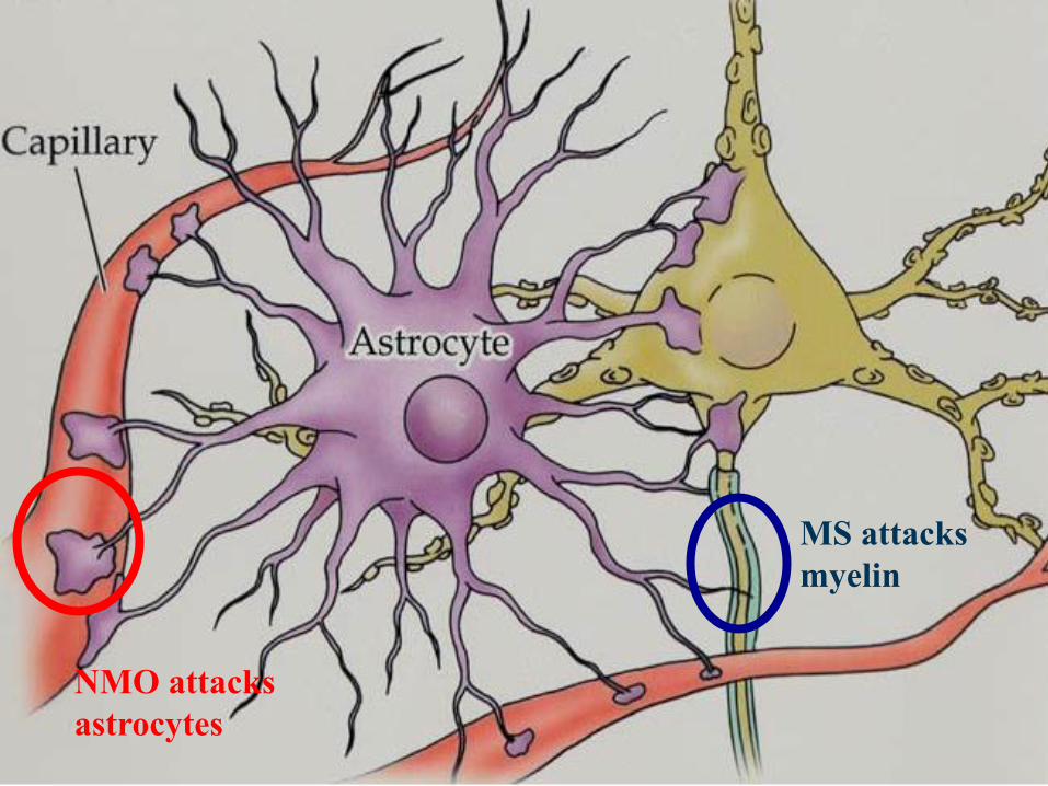
• To understand that NMO is an astrocytopathy and MS is a demyelinating disorder.
• The treatment strategies are different for NMO than for MS.

Case discussion
• AL is a 21 year old Chinese Canadian woman living in Toronto starting her career as a flight attendant.
• During the summer she had three attacks of optic neuritis, severe, with poor recovery after steroids.
• In the fall, she developed a 4th attack of ON and became paraplegic over 72 hours.
• Her brain MRI was normal

8
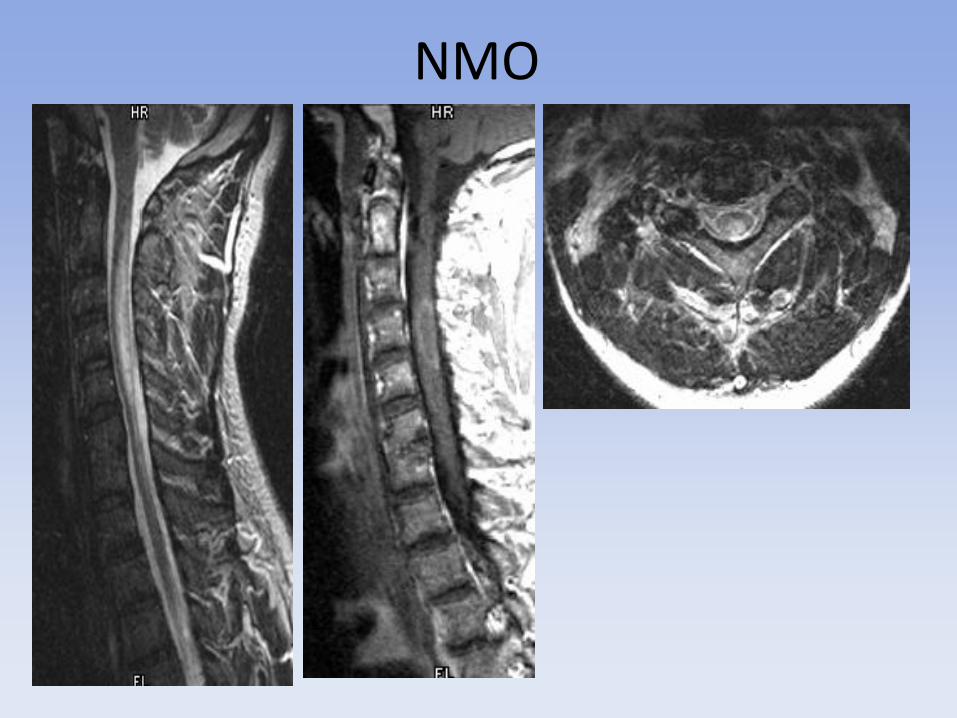
Longitudinally Extensive Spinal Cord Lesion

9

10
NMO: Classic spinal cord findings Longitudinally Extensive Spinal Cord Lesion (LESCL or LETM)
> 3 spinal segments, swollen, central

Axial location of spinal cord lesions may help distinguish NMO from MS
MS Proton Density
Ref: Nakamura J Neurol 2008
NMO T2 weighted
MS: White Matter areas N=20 cases
NMO: Central Gray N=21 cases
Slide adapted from K Fujihara

12
Small NMO lesion on PD
NMO Short lesion
Short cord lesions can occur in both MS and NMO
MS Lesion on Sagittal T2
Classic MS: short lesion

14
Long cord lesions: differential diagnosis

Differential Diagnosis - LESCL
• Autoimmune – NMO – Lupus (SLE) – Sjogren’s syndrome – Antiphospholipid syndrome
• Inflammatory – MS – Acute Disseminated Encephalomyelitis (ADEM) – Neuro-Behcet’s – Neurosarcoidosis
• Infectious – Parainfectious: EB, CMV, H.simplex, Varicella zoster, mycoplasma – Syphilis, TB – Schistosomiasis, Toxocara,Ascaris
The Differential Diagnosis of Longitudinally Extensive Transverse Myelitis Kitley,Leite,George,Palace MS 2012

Differential Diagnosis- LESCL
• Neoplastic – Paraneoplastic-autoantibody, lung, breast
– Intramedullary tumor- astrocytoma,ependymoma
• Metabolic – B12, Copper
• Vascular – Cord infarction, fistula
• Radiotherapy
• Post-vaccination
Kitley,Leite,George,Palace MS 2012

Diffuse subtle long cord lesions seen in conventional MS should not be confused with LESCL of NMO
Progressive MS T2 weighted Progressive MS Proton Density

SLE 44 yo presents with acute transverse myelopathy
Clinical clues for SLE:
Neuropsychiatric symptoms, rash, ulcers ,arthralgia
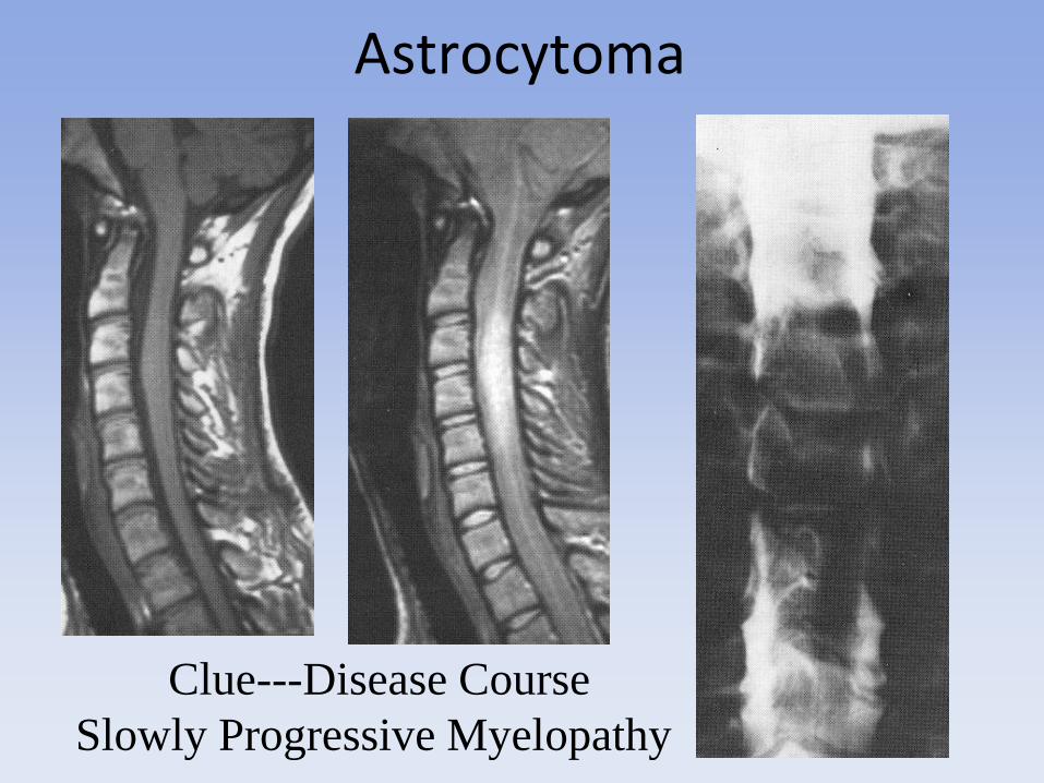
Clue---Disease Course
Slowly Progressive Myelopathy
Astrocytoma

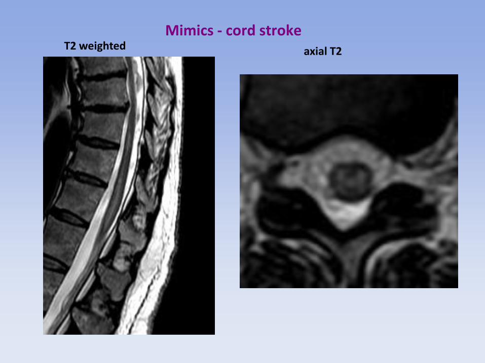
Mimics - cord stroke T2 weighted axial T2
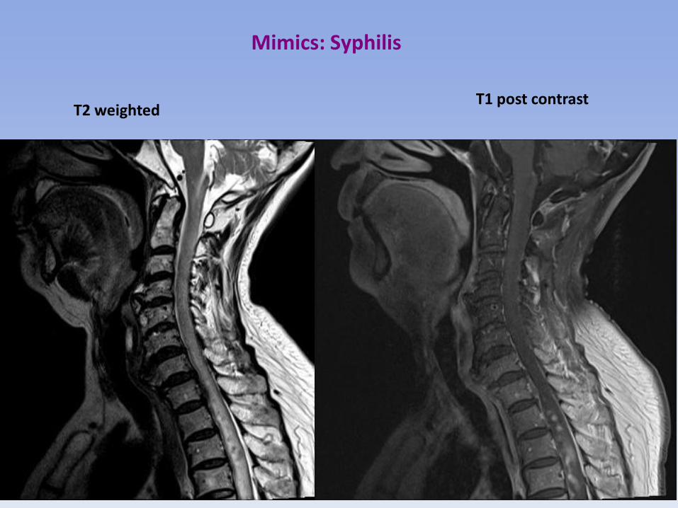
Mimics: Syphilis
T2 weighted T1 post contrast

Mimics - Syphilis T2 weighted
axial T2

Mimics - Syphilis PRE Treatment
T2 weighted POST Treatment T2 weighted

MS – Not a LESCL
NMO

28
Long cord lesions: natural history
Sagittal T2 Acute Lesion
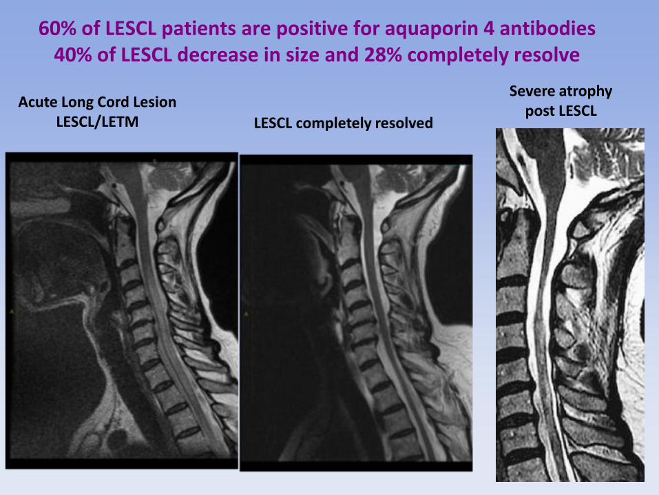
60% of LESCL patients are positive for aquaporin 4 antibodies 40% of LESCL decrease in size and 28% completely resolve
Acute Long Cord Lesion LESCL/LETM
Severe atrophy post LESCL
LESCL completely resolved

LESCL – Split into 3 non-LESCL
Initial 12 Month
Follow-Up
T6
T2
T6
T2
35
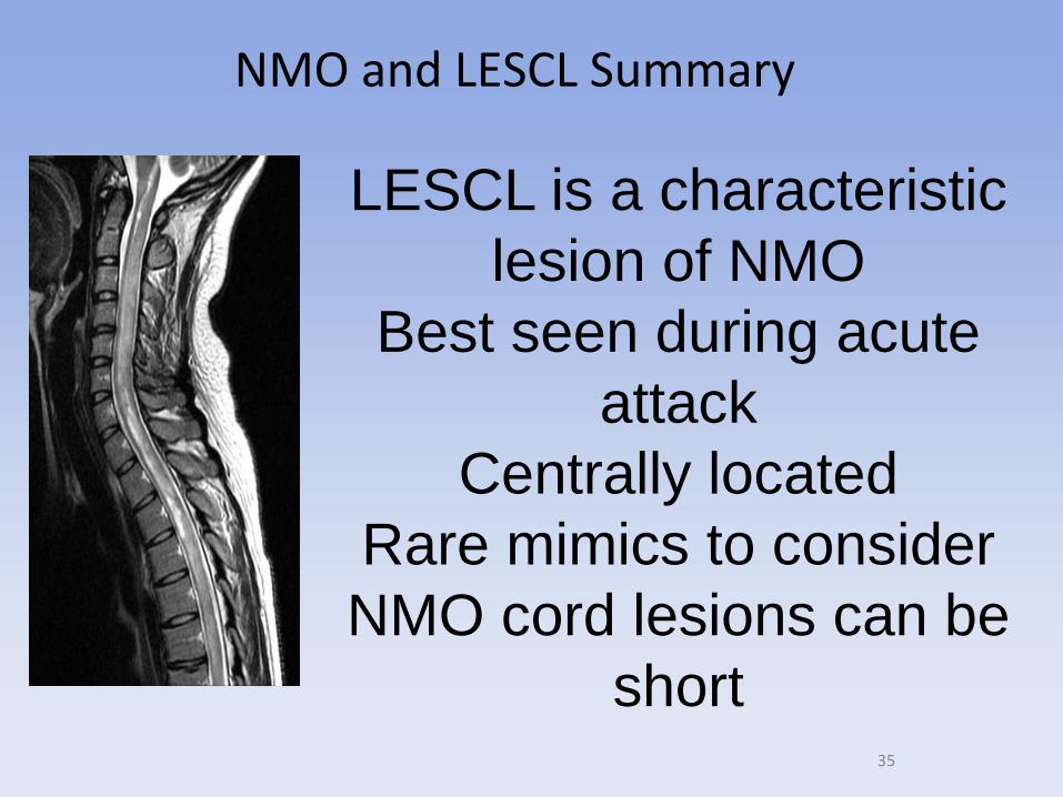
NMO and LESCL Summary
LESCL is a characteristic
lesion of NMO
Best seen during acute
attack
Centrally located
Rare mimics to consider
NMO cord lesions can be
short

36

37
19th Century: Recognition of MS and NMO
Pathologic description of MS
by Robert Carswell
Jean Martin Charcot (1825-93)
comprehensive review of MS
cases.
1884 Devic and Gault reviewed 17
cases 45 year old woman with rapid bilateral optic neuritis
and transverse myelitis. Died within 1 month of
onset. Necrotic lesions in spinal cord and optic
nerve.

Optic neuritis is a common
presentation of both MS and NMO
Optic Neuritis Normal Vision
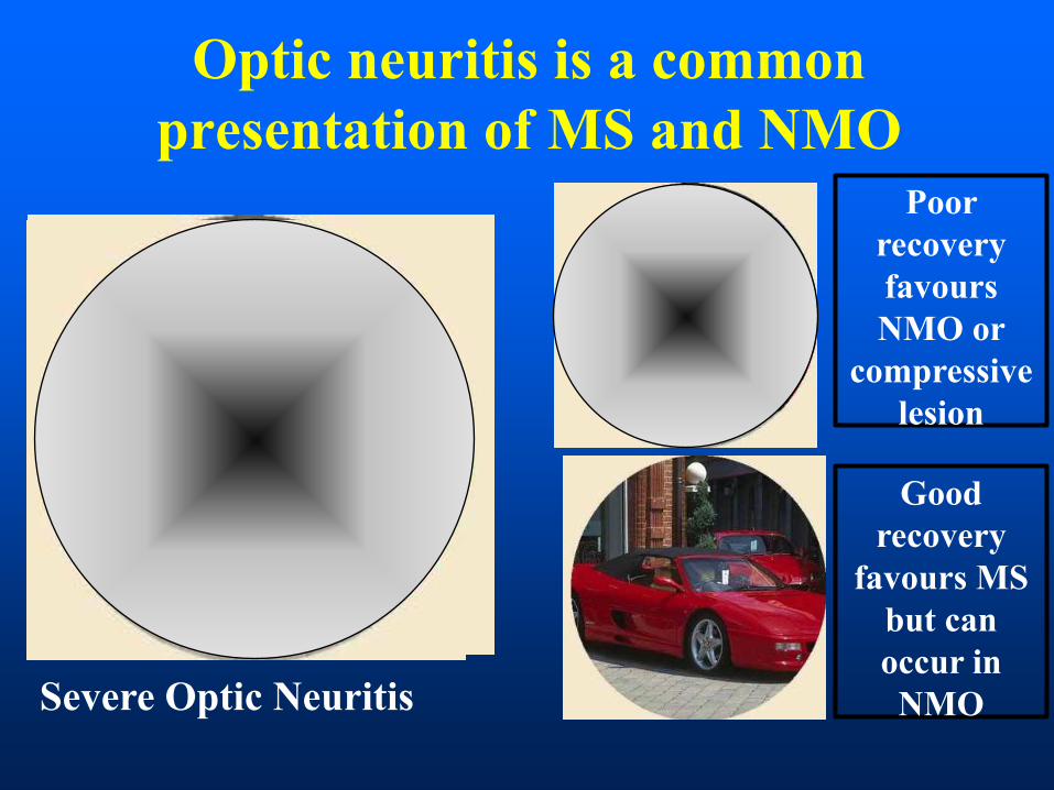
Optic neuritis is a common
presentation of MS and NMO
Severe Optic Neuritis
Poor
recovery
favours
NMO or
compressive
lesion
Good
recovery
favours MS
but can
occur in
NMO

10
42
MS attacks
myelin
NMO attacks
astrocytes
23
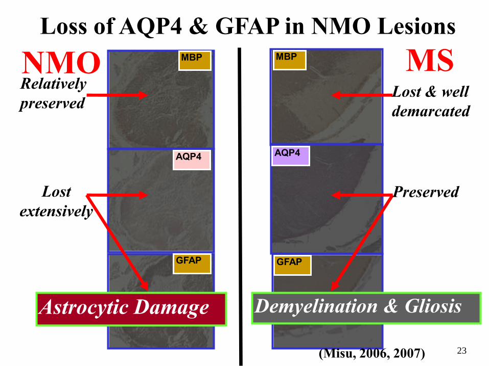
NMO MS
Loss of AQP4 & GFAP in NMO Lesions MBP
AQP4
GFAP
MBP
AQP4
GFAP
Lost & well
demarcated
Preserved Lost
extensively
Relatively
preserved
(Misu, 2006, 2007)
Astrocytic Damage Demyelination & Gliosis

45

NMO Diagnostic Criteria
Wingerchuck 2006 Optic Neuritis and Acute Myelitis and 2/3: •LESCL >3 segments •Brain MRI at onset not diagnostic of MS •NMO-IgG seropositive

NMO Spectrum Disorders (NMOSD)
1. Classic NMO 2. Limited forms of NMO: - Longitudinally Extensive Spinal Cord Lesions (LESCL > 3 spinal segments) - Recurrent or simultaneous bilateral optic neuritis (ON) 3. ON or LESCL associated with another autoimmune disease (example Sjogren’s, Lupus myelitis) 4. ON or transverse myelitis with NMO like brain lesions.
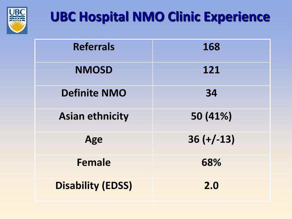
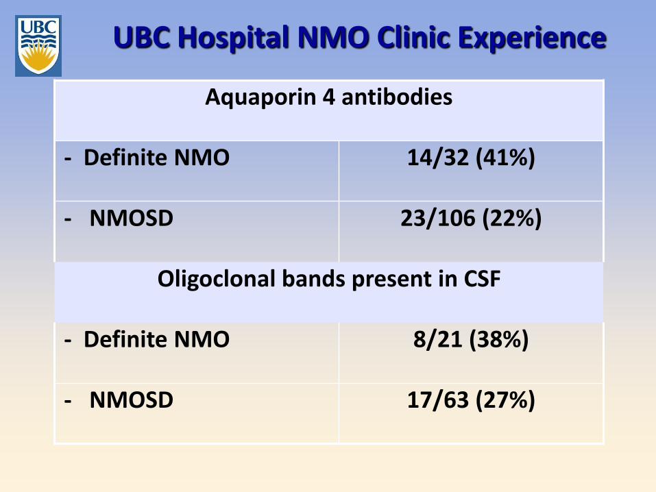
UBC Hospital NMO Clinic Experience
Referrals 168
NMOSD 121
Definite NMO 34
Asian ethnicity 50 (41%)
Age 36 (+/-13)
Female 68%
Disability (EDSS) 2.0
UBC Hospital NMO Clinic Experience
Aquaporin 4 antibodies
- Definite NMO 14/32 (41%)
- NMOSD 23/106 (22%)
Oligoclonal bands present in CSF
- Definite NMO 8/21 (38%)
- NMOSD 17/63 (27%)
50
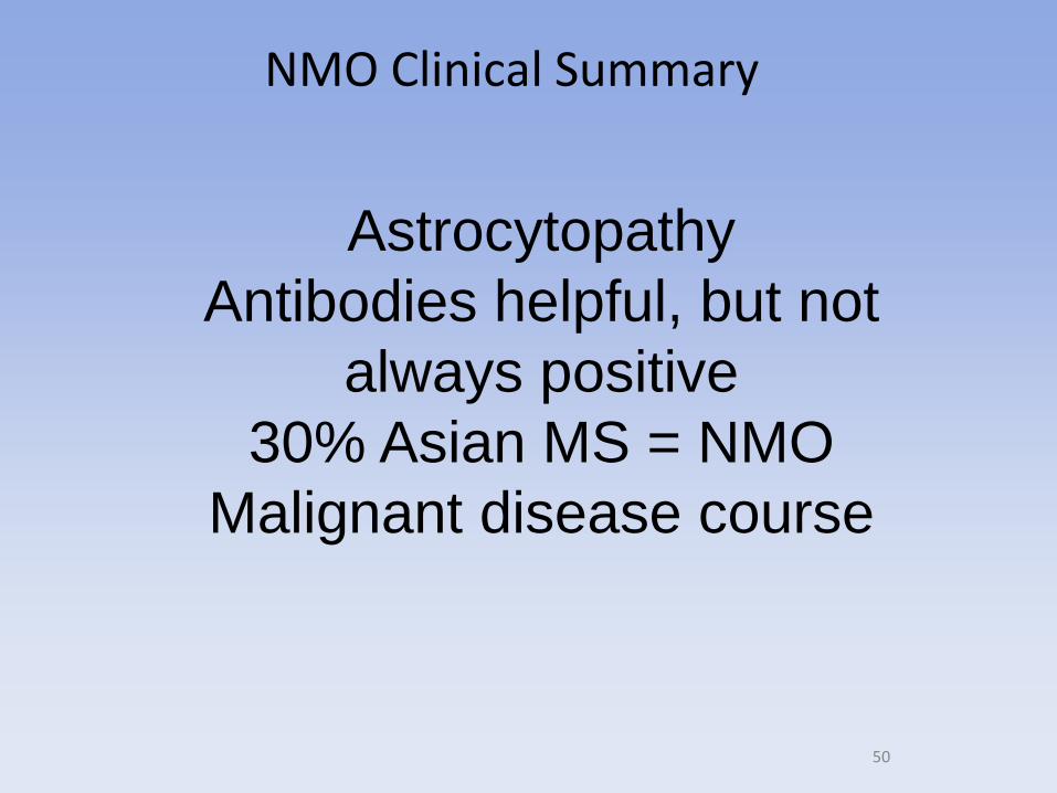
NMO Clinical Summary
Astrocytopathy
Antibodies helpful, but not
always positive
30% Asian MS = NMO
Malignant disease course

53

Neuromyelitis Optica Diagnostic Criteria
Main Criteria (2/2)
1. Optic Neuritis
2. Transverse myelitis
Supportive criteria (2/3)
• Brain MRI not meeting
MS diagnostic criteria • Spinal cord > 3 contiguous
segments
• NMO-IgG seropositive
Wingerchuk et al Neurology 66: 1485-1489, 2006

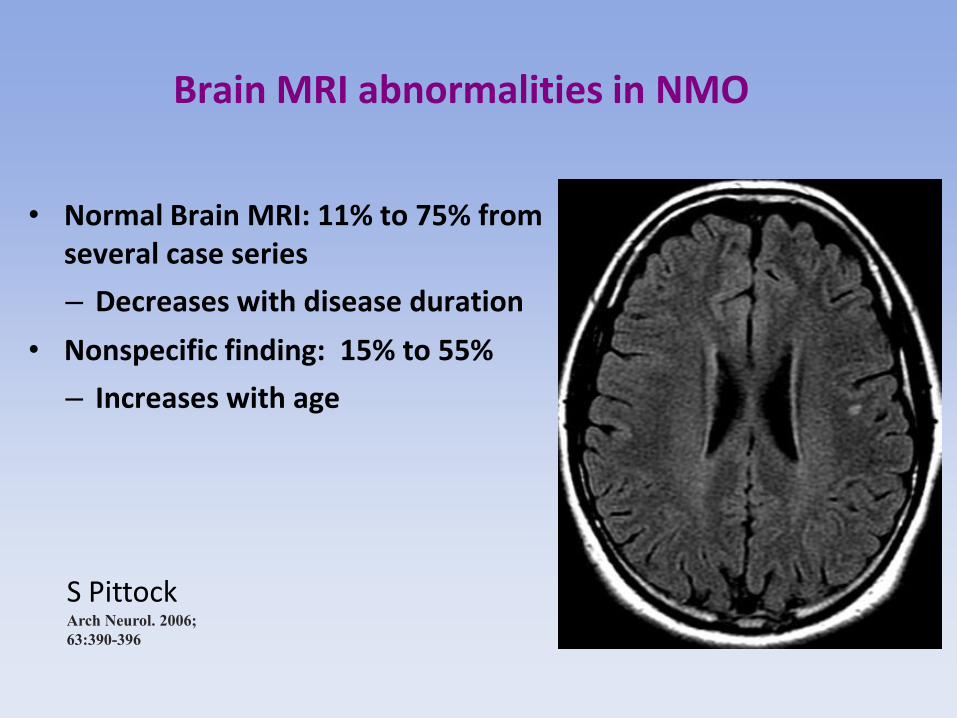
Brain MRI abnormalities in NMO
What proportion of NMO patients have a normal brain MRI?
11%
30%
60%
75%
100%
Brain MRI abnormalities in NMO
• Normal Brain MRI: 11% to 75% from several case series
– Decreases with disease duration
• Nonspecific finding: 15% to 55%
– Increases with age
S Pittock
Arch Neurol. 2006;
63:390-396
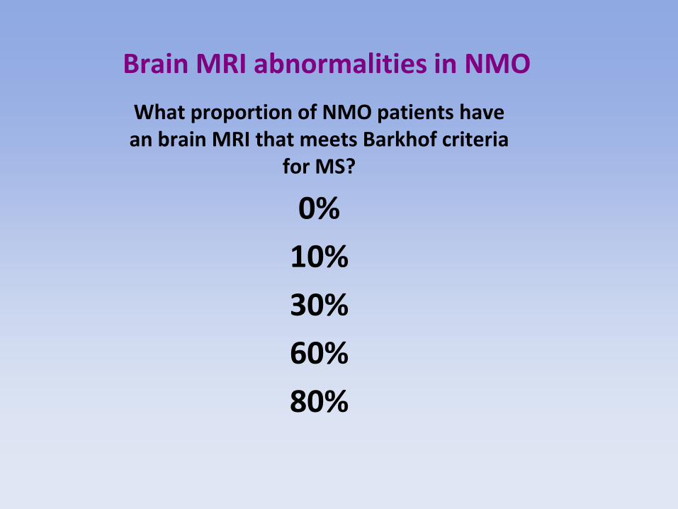
Brain MRI abnormalities in NMO
What proportion of NMO patients have an brain MRI that meets Barkhof criteria
for MS?
0%
10%
30%
60%
80%

52 McDonald WI et al. Ann Neurol. 2001;50:121-127; Images from Dr. A. Traboulsee, UBC MS/MRI Research Group
9 T2
or
1 Gd+
1 Juxtacortical
3 Periventricular
2001 and 2005 MRI Criteria for MS Dissemination in Space
3 out of 4 Barkhof/Tintore criteria (present in 30% of NMO)
1 Infra-
tentorial
OR
1 Spinal
Cord
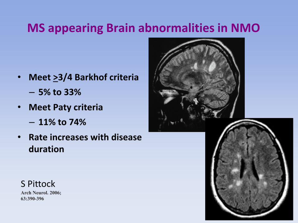
MS appearing Brain abnormalities in NMO
• Meet >3/4 Barkhof criteria
– 5% to 33%
• Meet Paty criteria
– 11% to 74%
• Rate increases with disease duration
S Pittock
Arch Neurol. 2006;
63:390-396
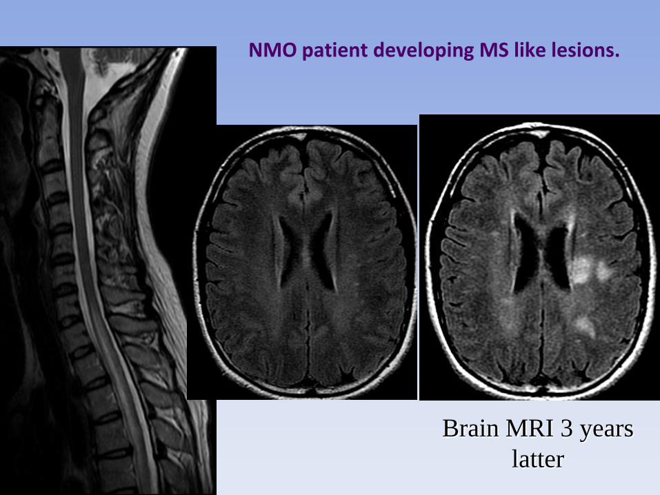
NMO patient developing MS like lesions.
Dr T. Traboulsee, UBC MS/MRI Research Group
Brain MRI 3 years
latter

53 McDonald WI et al. Ann Neurol. 2001;50:121-127; Images from Dr. A. Traboulsee, UBC MS/MRI Research Group
1 Juxtacortical
3 1 Periventricular
2010 MRI Criteria for MS Dissemination in Space
>1 lesions in at least 2 of 4 regions (present in 60% of NMO)
1 Infra-
tentorial
or
1 Spinal
Cord

Distinct Brain MRI abnormalities in NMO
• 8% to 69% prevalence
• Increases with disease duration
– Extensive hemisperic brain lesions
– Brainstem lesions contiguous with LESCL
– Hypothalamic
– Following cortical spinal tracks
– Extensive corpus callosum lesions
– Around 3rd and 4th ventricles
– Patchy/cloudy enhancement

Brain MRI abnormalities in NMO
S Pittock
Arch Neurol. 2006;
63:390-396

Unusual MRI lesions seen in NMO
Callosal Lesion
(Large, Edematous)

15
Brain Lesions in Anti-AQP4-positive Cases
Hypothalamic Lesion Medullary Lesion
(Misu, 2005; Nakashima, 2006; Takahashi, 2007; Shimizu, 2008; Nakamura, 2009)
Callosal Lesion
(Large, Edematous)
Intractable Hiccup
& Nausea
Hypersomnia

Patchy Cloud like enhancement S Ito Ann Neurol 2009;
66:425-428
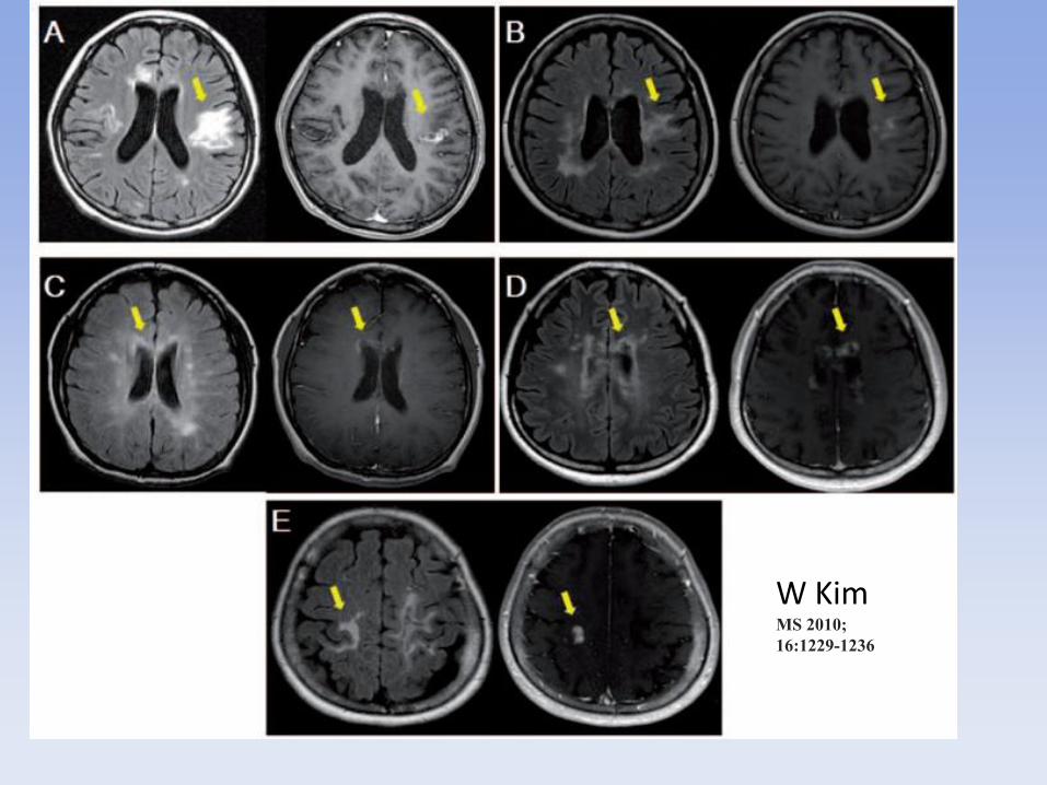
W Kim MS 2010;
16:1229-1236
70 70
71
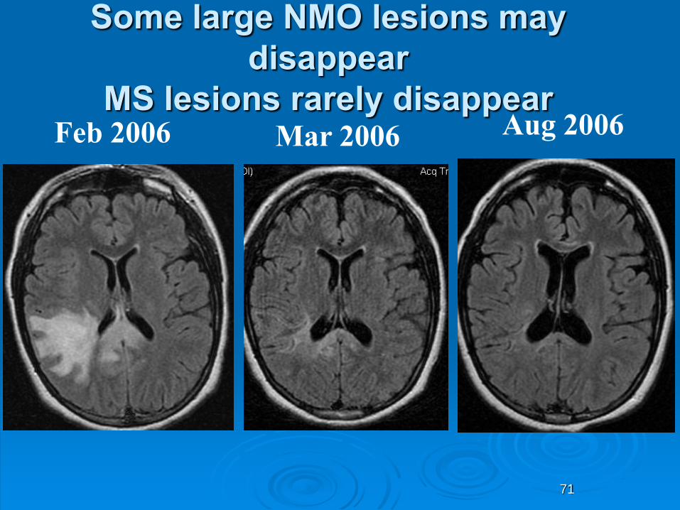
Some large NMO lesions may
disappear
MS lesions rarely disappear Feb 2006 Aug 2006 Mar 2006

73
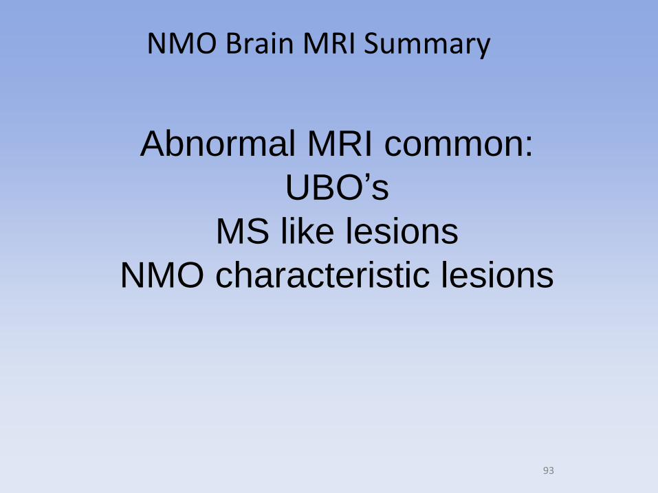
NMO Brain MRI Summary
Abnormal MRI common:
UBO’s
MS like lesions
NMO characteristic lesions
74
Treatment of NMO
Case discussion
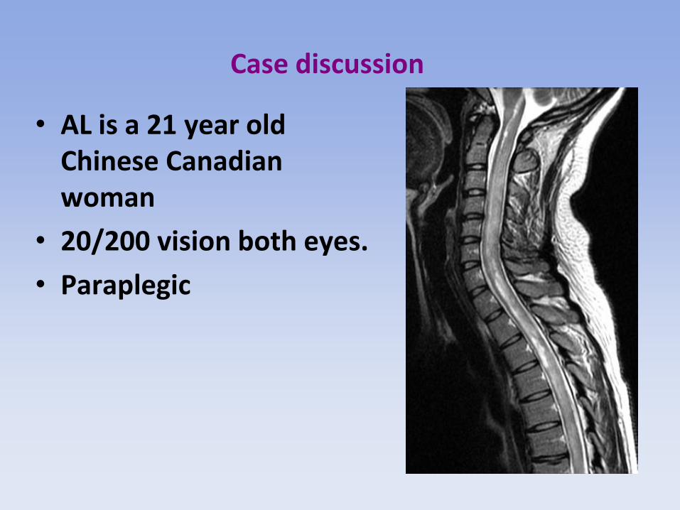
• AL is a 21 year old Chinese Canadian woman
• 20/200 vision both eyes.
• Paraplegic

UBC Hospital NMO Treatment Experience Acute Relapse Management
Time is brain, spinal cord and optic nerve

UBC Hospital NMO Treatment Experience Acute Relapse Management
1. Methylprednisolone 1 gram IV for 3-5 days followed by oral steroid taper to prevent rebound.
Case discussion
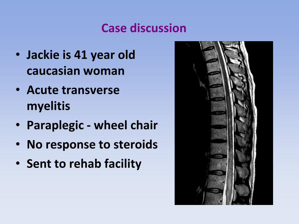
• Jackie is 41 year old caucasian woman
• Acute transverse myelitis
• Paraplegic - wheel chair
• No response to steroids
• Sent to rehab facility
UBC Hospital NMO Treatment Experience Acute Relapse Management
1. Methylprednisolone 1 gram IV for 3-5 days followed by oral steroid taper to prevent rebound. Failure = no clinically meaningful response during
treatment or within 3-5 days of treatment completion.
2. Plasmapheresis course of 5 treatments. Taper schedule if good response. Role for prednisone to prevent rebound.

Improvement in disability (EDSS) baseline, pre and post PLEX (N=52) Responders: MS 37%, NMO 56%
Quadraplegic
Wheelchair
Cane
Walker

Case discussion
• Jackie is 41 year old caucasian woman
• Acute transverse myelitis
• Paraplegic - wheel chair
• No response to steroids
• PLEX started at 3 months and continued for 3 months. EDSS 1.0

UBC Hospital NMO Treatment Experience Acute Relapse Management
1. Methylprednisolone 1 gram IV for 3-5 days followed by oral steroid taper to prevent rebound. Failure = no clinically meaningful response during
treatment or within 3-5 days of treatment completion.
2. Plasmapheresis course of 5 treatments. Taper schedule if good response. Role for prednisone. 3. The Mitoxantrone Rescue/Induction Protocol: 12mg/m2 IV monthly for three months.

Case discussion
• Sue is a 48 year old caucasian woman
• Acute transverse myelitis
• Quadriplegic - ICU
• No response to steroids
• No response to PLEX
UBC Hospital NMO Treatment Experience Acute Relapse Management
1. Methylprednisolone 1 gram IV for 3-5 days followed by oral steroid taper to prevent rebound. Failure = no clinically meaningful response during
treatment or within 3-5 days of treatment completion.
2. Plasmapheresis course of 5 treatments. Taper schedule if good response. Role for prednisone. 3. The Mitoxantrone Rescue/Induction Protocol: 12mg/m2 IV monthly for three months.

Case discussion
• Sue
• Acute transverse myelitis
• Quadriplegic
• Mitoxantrone given at 4 months.
• Walks with a cane (EDSS 6.0)

UBC Mitoxantrone protocol for severe demyelination Improvement at 6 months post Mitoxantrone (18 cases of severe optic neuritis or transverse myelitis)

Prevention of NMO Relapses anecdotal experience and/or case series
•Prednisone •Azathioprine (target lymphocytes 0.5 to 1.0) •Methotrexate •Mycophenolate mofetil (cellcept) •Rituximab •Cyclophosphamide, Cyclosporine •Mitoxantrone (induction only) •Combination (e.g. Azathioprine plus prednisone)
NMO Treatment Summary
• NMO attacks are a neurological emergency.
• The treatment strategies are different for NMO than for MS.
• Prevention is key (chronic immune suppression)
Objectives
• To be aware of clinical and MRI features that distinguish NMO from MS
• To understand that NMO is an astrocytopathy and MS is a demyelinating disorder.
• The treatment strategies are different for NMO than for MS.

91
NMO Clinical Summary
Astrocytopathy
Antibodies helpful, but not
always positive
30% Asian MS = NMO
Malignant disease course
Treat early
Treat aggressively

92
NMO and LESCL Summary
LESCL is a characteristic
lesion of NMO
Best seen during acute
attack
Centrally located
Rare mimics to consider
NMO cord lesions can be
short
93
NMO Brain MRI Summary
Abnormal MRI common:
UBO’s
MS like lesions
NMO characteristic lesions
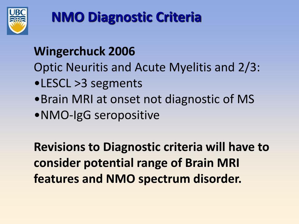
NMO Diagnostic Criteria
Wingerchuck 2006 Optic Neuritis and Acute Myelitis and 2/3: •LESCL >3 segments •Brain MRI at onset not diagnostic of MS •NMO-IgG seropositive
Revisions to Diagnostic criteria will have to consider potential range of Brain MRI features and NMO spectrum disorder.