distinct patterns of notochord mineralization in zebrafish coincide with the localization of
TRANSCRIPT

Distinct patterns of notochord mineralization inzebrafish coincide with the localization ofOsteocalcin isoform 1 during early vertebralcentra formationBensimon-Brito et al.
Bensimon-Brito et al. BMC Developmental Biology 2012, 12:28http://www.biomedcentral.com/1471-213X/12/28

Bensimon-Brito et al. BMC Developmental Biology 2012, 12:28http://www.biomedcentral.com/1471-213X/12/28
RESEARCH ARTICLE Open Access
Distinct patterns of notochord mineralization inzebrafish coincide with the localization ofOsteocalcin isoform 1 during early vertebralcentra formationAnabela Bensimon-Brito1,2,3,5, João Cardeira2, Maria Leonor Cancela2,4*, Ann Huysseune3 and Paul Eckhard Witten3*
Abstract
Background: In chondrichthyans, basal osteichthyans and tetrapods, vertebral bodies have cartilaginous anlagenthat subsequently mineralize (chondrichthyans) or ossify (osteichthyans). Chondrocytes that form the vertebralcentra derive from somites. In teleost fish, vertebral centrum formation starts in the absence of cartilage, throughdirect mineralization of the notochord sheath. In a second step, the notochord is surrounded by somite-derivedintramembranous bone. In several small teleost species, including zebrafish (Danio rerio), even haemal and neuralarches form directly as intramembranous bone and only modified caudalmost arches remain cartilaginous. Thisstudy compares initial patterns of mineralization in different regions of the vertebral column in zebrafish. We ask ifthe absence or presence of cartilaginous arches influences the pattern of notochord sheath mineralization.
Results: To reveal which cells are involved in mineralization of the notochord sheath we identify proliferating cells,we trace mineralization on the histological level and we analyze cell ultrastructure by TEM. Moreover, we localizeproteins and genes that are typically expressed by skeletogenic cells such as Collagen type II, Alkaline phosphatase(ALP) and Osteocalcin (Oc). Mineralization of abdominal and caudal vertebrae starts with a complete ring within thenotochord sheath and prior to the formation of the bony arches. In contrast, notochord mineralization of caudal fincentra starts with a broad ventral mineral deposition, associated with the bases of the modified cartilaginousarches. Similar, arch-related, patterns of mineralization occur in teleosts that maintain cartilaginous archesthroughout the spine.Throughout the entire vertebral column, we were able to co-localize ALP-positive signal with chordacentrummineralization sites, as well as Collagen II and Oc protein accumulation in the mineralizing notochord sheath. In thecaudal fin region, ALP and Oc signals were clearly produced both by the notochord epithelium and cells outsidethe notochord, the cartilaginous arches. Based on immunostaining, real time PCR and oc2:gfp transgenic fish, weidentify Oc in the mineralizing notochord sheath as osteocalcin isoform 1 (Oc1).(Continued on next page)
* Correspondence: [email protected]; [email protected] of Marine Sciences - CCMar, University of Algarve, Faro, Portugal3Evolutionary Developmental Biology, Biology Department, Ghent University,Ghent, BelgiumFull list of author information is available at the end of the article
© 2012 Bensimon-Brito et al.; licensee BioMed Central Ltd. This is an Open Access article distributed under the terms of theCreative Commons Attribution License (http://creativecommons.org/licenses/by/2.0), which permits unrestricted use,distribution, and reproduction in any medium, provided the original work is properly cited.

Bensimon-Brito et al. BMC Developmental Biology 2012, 12:28 Page 2 of 14http://www.biomedcentral.com/1471-213X/12/28
(Continued from previous page)
Conclusions: If notochord mineralization occurs prior to arch formation, mineralization of the notochord sheath isring-shaped. If notochord mineralization occurs after cartilaginous arch formation, mineralization of the notochordsheath starts at the insertion point of the arches, with a basiventral origin. The presence of ALP and Oc1, not onlyin cells outside the notochord, but also in the notochord epithelium, suggests an active role of the notochord inthe mineralization process. The same may apply to Col II-positive chondrocytes of the caudalmost haemal archesthat show ALP activity and Oc1 accumulation, since these chondrocytes do not mineralize their own cartilagematrix. Even without cartilaginous preformed vertebral centra, the cartilaginous arches may have an inductive rolein vertebral centrum formation, possibly contributing to the distinct mineralization patterns of zebrafish vertebralcolumn and caudal fin vertebral fusion.
Keywords: Vertebral column, Vertebral fusion, Notochord, Osteocalcin
BackgroundThe structural units of the vertebral column are the ver-tebrae, composed of neural and haemal arches and thevertebral body itself, the centrum. Vertebral bodies arejoined by intervertebral tissue primarily derived from thenotochord [1-3].The vertebral column results from a strictly controlled
segmentation process that occurs in all vertebrate spe-cies and is associated to two main structures, the noto-chord and the somites [4,5]. The somites are epithelializedspheres of mesoderm that develop on either side ofthe neural tube, give rise to dermis, skeletal muscula-ture (dermomyotome) and vertebrae (sclerotome) [6,7].While the somite contribution to vertebra formation hasbeen extensively studied, particularly in birds [6,8], therole of the notochord has received less attention. In te-leosts, the majority of extant vertebrates, the notochordis composed of a core of large, vacuolated chordocytes,and an epithelial layer of chordoblasts that secrete thenotochord sheath [9]. The notochord sheath is a strati-fied structure, composed of a thin external membrane,with high elastin content, covering a thicker collagenouslayer [10,11].In chondrichthyans and most osteichthyans, including
tetrapods, but not in teleosts, vertebral bodies have acartilaginous anlage that subsequently either mineralizesor is replaced by bone. In teleosts such as zebrafish, ver-tebral centra are formed in the absence of cartilage[12,13]. Indeed, teleost vertebral centra form throughthe mineralization of the notochord sheath (chordacen-trum), which is then surrounded by somite-derivedintramembranous bone (autocentrum) [2,12,14-16].In teleosts, such as Atlantic salmon (Salmo salar) or
zebrafish (Danio rerio), the notochord plays an import-ant role in early life stages, as its mechanical function isonly replaced by the vertebral column in the postem-bryonic life [2,17,18]. In Atlantic salmon, the initialmineralization of the chordacentrum has been describedto be associated with cells of the notochord epithelium(chordoblasts), while bone formation, by sclerotome-
derived cells (autocentrum), is a second step [2]. In con-trast, Inohaya and co-workers suggest that in medaka(Oryzias latipes), only sclerotome-derived cells are invol-ved in chordacentrum and autocentrum mineralization,with no role of chordoblasts [19]. Yet, a recent study[20] shows that, also in medaka, with conditional abla-tion of osterix-positive osteoblasts, notochord sheathmineralization is maintained. Therefore, the main cellu-lar and molecular determinants involved in early noto-chord mineralization are still under debate. Furthermore,the role of extracellular matrix proteins such as osteo-calcin, generally expressed by mature and resting osteo-blasts [21] and by hypertrophic chondrocytes [17,22-24],in that process, remains unclear. In several teleosts, in-cluding zebrafish, two Osteocalcin genes (Oc1 and Oc2)have been identified [25].In amniotes the vertebral column is divided into five
main regions whereas the vertebral column of teleosts isoften only subdivided into two main regions, abdominaland caudal (e.g., [26-28]). However, also in zebrafish,several regions can be recognized within the vertebralcolumn. In particular, regions that contain the most an-terior and the most posterior vertebrae are highly spe-cialized [15,29]. Regional differences are not onlyapparent at the morphological level but also regardingthe tendency of vertebrae to fuse. While zebrafish verte-bral bodies usually display no pathological fusion [30],caudal fin vertebrae undergo several fusions as part ofregular development [15,30-32]. Yet, other teleosts, suchas Atlantic salmon and other farmed species, are knownto suffer frequent pathological vertebral fusions [33,34].Whether regional differences in vertebrae morphologyand mineralization relate to the susceptibility to fuseremains an open question.This study aims to characterize mineralization patterns
in different regions of the vertebral column (abdominal,caudal, caudal fin region) using various methods to re-veal mineral deposition. Subsequently, these patterns arecompared with the histogenesis of the arches, revealedthrough Collagen type II immunostaining, and to the
Bensimon-Brito et al. BMC Developmental Biology 2012, 12:28 Page 3 of 14http://www.biomedcentral.com/1471-213X/12/28
timing of centrum formation. We characterize the proli-feration of notochord cells and we also localize proteinsrelated to mineralization, such as Alkaline phosphataseand Osteocalcin. We here provide the first evidence forthe early presence of Osteocalcin 1 in mineralizing chor-dacentra. Finally, we discuss a possible association betweentiming of centra formation, the mineralization pattern andoccurrence of vertebral fusion.
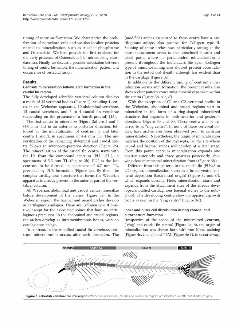
ResultsCentrum mineralization follows arch formation in thecaudal fin regionThe fully developed zebrafish vertebral column displaysa mode of 33 vertebral bodies (Figure 1) including 4 cen-tra in the Weberian apparatus, 10 abdominal vertebrae,15 caudal vertebrae, and 3 to 4 caudal fin vertebrae(depending on the presence of a fourth preural) [15].The first centra to mineralize (Figure 2a) are 3 and 4
(4.0 mm TL) in an anterior-to-posterior direction, fol-lowed by the mineralization of centrum 5, and latercentra 1 and 2, in specimens of 4.4 mm TL. The mi-neralization of the remaining abdominal and caudal cen-tra follows an anterior-to-posterior direction (Figure 2b).The mineralization of the caudal fin centra starts withthe U1 from the compound centrum [PU1++U1], inspecimens of 5.5 mm TL (Figure 2b). PU2 is the lastcentrum to be formed, in specimens of 6.7 mm TL,preceded by PU3 formation (Figure 2c). By then, thecomplex cartilaginous structure that forms the Weberianapparatus is already present in the anterior part of the ver-tebral column.All Weberian, abdominal and caudal centra mineralize
before development of the arches (Figure 3a). In theWeberian region, the haemal and neural arches developas cartilaginous anlagen. These are Collagen type II posi-tive, except for the associated spines that have no carti-laginous precursor. In the abdominal and caudal regions,the arches develop as intramembranous bones, with nocartilaginous anlage.In contrast, in the modified caudal fin vertebrae, cen-
trum mineralization occurs after arch formation. The
Figure 1 Zebrafish vertebral column regions. Weberian, abdominal, cau
(modified) arches associated to these centra have a car-tilaginous anlage, also positive for Collagen type II.Staining of these arches was particularly strong at thebases (attachment areas to the notochord sheath) anddistal parts, where no perichondral mineralization ispresent throughout the individual’s life span. Collagentype II immunostaining also showed protein accumula-tion in the notochord sheath, although less evident thanin the cartilage (Figure 3a’).In addition to the different timing of centrum mine-
ralization versus arch formation, the present results alsoshow a clear pattern concerning mineral expansion withinthe centra (Figure 3b, b’, c, c’).With the exception of C1 and C2, vertebral bodies in
the Weberian, abdominal and caudal regions start tomineralize in the form of a ring-shaped mineralizedstructure that expands in both anterior and posteriordirections (Figure 3b and b’). These centra will be re-ferred to as “ring centra”. In none of these vertebral bo-dies, have arches ever been observed prior to centrummineralization. Nevertheless, the origin of mineralizationmatches the position of the myosepta, i.e. the site whereneural and haemal arches will develop at a later stage.From this point, centrum mineralization expands onequarter anteriorly and three quarters posteriorly, sho-wing clear incremental mineralization fronts (Figure 3b’).Different from this pattern, in the caudal fin (PU4/3 to
U2) region, mineralization starts as a broad ventral mi-neral deposition (basiventral origin) (Figure 3c and c’),which expands dorsally. Here, mineralization starts andexpands from the attachment sites of the already deve-loped modified cartilaginous haemal arches to the noto-chord. The developing centra show no apparent growthfronts as seen in the “ring centra” (Figure 3c’).
Inner and outer cell distribution during chorda- andautocentrum formationIrrespective of the shape of the mineralized centrum,(“ring” and caudal fin centra) (Figure 4a, b), the origin ofmineralization was shown both with von Kossa staining(Figure 4c, c’, d, d’) and TEM (Figure 4e-f), to occur always
dal and caudal fin regions are identified in different shades of grey.

Figure 2 Zebrafish vertebral column development. Lateral view (a) of 4.4 mm TL zebrafish (Danio rerio) vertebral column. In black are themineralized structures, demonstrating the formation of centrum 1 (C1) and the presence of C3 to C5. (b) At 5.5 mm TL caudal fin vertebraeformation with broad base origin can already be seen, represented by U1, and anterior vertebrae also develop, displaying a ring-shaped patternof mineralization. In grey are the cartilaginous structures, including the caudal fin modified haemal arches and the anterior C3-C5 neural arches(black arrowhead). (c) At 6.0 mm TL PU3 appears, followed by the last vertebral body to form in the vertebral column, PU2, formed around 6.7mm TL. At this stage, most anterior vertebral bodies already show bone formation around the notochord sheath. The Weberian apparatus isalready well differentiated. Further abbreviations: C – centrum; con – concha scaphium; Hy1-5 – hypurals 1 to 5; in – intercalarium; os – ossuspensorium; PHy – parhypural; PU1++U1 – compound centrum preural 1 and ural 1; PU2-3 – preurals 2 and 3; rc – roofing cartilage;sc – scaphium; tr – tripus; U2+ – ural 2; (+) sign indicates vertebral elements that are the product of fusion events. Scale bar: 1 mm.
Bensimon-Brito et al. BMC Developmental Biology 2012, 12:28 Page 4 of 14http://www.biomedcentral.com/1471-213X/12/28
within the notochord sheath, thus establishing the chorda-centrum. Once the chordacentrum is fully formed, peri-chordal bone is deposited mostly at the anterior andposterior edges, in this way establishing the vertebralendplates of the centra, thus forming the autocentrum(Figure 5a-d). Autocentrum formation occurs irrespectiveof the initial mineralization pattern within the notochordsheath (“ring” vs. basiventral origin). Yet, in the caudal fincentra, decreased bone formation is observed in the areaswhere cartilaginous arches attach to the notochord(Figure 5c-d).During chordacentrum formation, notochord inner cells
or chordoblasts do not accumulate in the prospective in-tervertebral space (Figure 4a, b), unlike what has beenobserved in other teleost species. However, once the
centrum is mineralized, a higher number of chordoblastscan be seen in the non-mineralized intervertebral space(Figure 5a, c, e). By then, the chordoblasts are oblong, lar-ger in size and are oriented in a dorsal-ventral direction(Figure 5e), as opposed to the squamous morphology dis-played prior to and during chordacentrum development.In addition, chordoblasts proliferate in the intervertebralspace, as shown by PCNA staining. Here, notochordsheath thickening occurs (Figure 5f ). This pattern ofchordoblast distribution is observed throughout thenotochord, regardless of the type of centrum and its mi-neralization pattern (Figure 5a-d).During chordacentrum formation, contrary to the in-
ner notochord cells, the assumed sclerotome-derivednotochord outer cells [35] in the “ring” vertebrae are
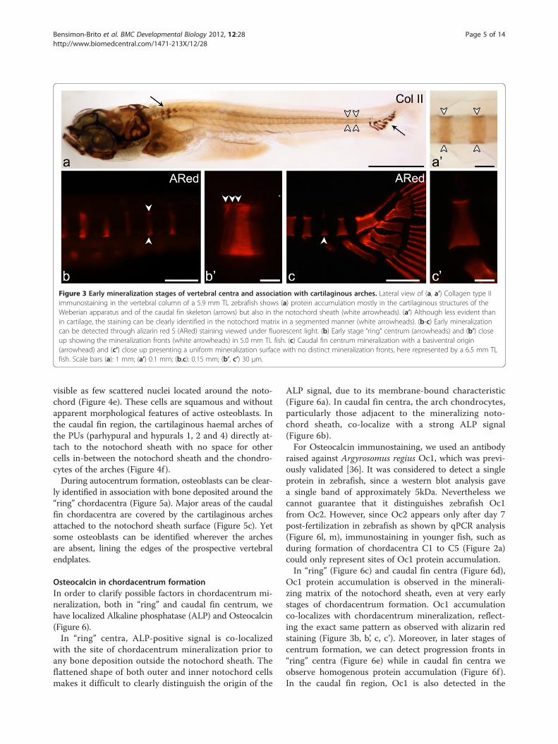
Figure 3 Early mineralization stages of vertebral centra and association with cartilaginous arches. Lateral view of (a, a’) Collagen type IIimmunostaining in the vertebral column of a 5.9 mm TL zebrafish shows (a) protein accumulation mostly in the cartilaginous structures of theWeberian apparatus and of the caudal fin skeleton (arrows) but also in the notochord sheath (white arrowheads). (a’) Although less evident thanin cartilage, the staining can be clearly identified in the notochord matrix in a segmented manner (white arrowheads). (b-c) Early mineralizationcan be detected through alizarin red S (ARed) staining viewed under fluorescent light. (b) Early stage “ring” centrum (arrowheads) and (b’) closeup showing the mineralization fronts (white arrowheads) in 5.0 mm TL fish. (c) Caudal fin centrum mineralization with a basiventral origin(arrowhead) and (c’) close up presenting a uniform mineralization surface with no distinct mineralization fronts, here represented by a 6.5 mm TLfish. Scale bars (a): 1 mm; (a’) 0.1 mm; (b,c): 0.15 mm; (b’, c’) 30 μm.
Bensimon-Brito et al. BMC Developmental Biology 2012, 12:28 Page 5 of 14http://www.biomedcentral.com/1471-213X/12/28
visible as few scattered nuclei located around the noto-chord (Figure 4e). These cells are squamous and withoutapparent morphological features of active osteoblasts. Inthe caudal fin region, the cartilaginous haemal arches ofthe PUs (parhypural and hypurals 1, 2 and 4) directly at-tach to the notochord sheath with no space for othercells in-between the notochord sheath and the chondro-cytes of the arches (Figure 4f ).During autocentrum formation, osteoblasts can be clear-
ly identified in association with bone deposited around the“ring” chordacentra (Figure 5a). Major areas of the caudalfin chordacentra are covered by the cartilaginous archesattached to the notochord sheath surface (Figure 5c). Yetsome osteoblasts can be identified wherever the archesare absent, lining the edges of the prospective vertebralendplates.
Osteocalcin in chordacentrum formationIn order to clarify possible factors in chordacentrum mi-neralization, both in “ring” and caudal fin centrum, wehave localized Alkaline phosphatase (ALP) and Osteocalcin(Figure 6).In “ring” centra, ALP-positive signal is co-localized
with the site of chordacentrum mineralization prior toany bone deposition outside the notochord sheath. Theflattened shape of both outer and inner notochord cellsmakes it difficult to clearly distinguish the origin of the
ALP signal, due to its membrane-bound characteristic(Figure 6a). In caudal fin centra, the arch chondrocytes,particularly those adjacent to the mineralizing noto-chord sheath, co-localize with a strong ALP signal(Figure 6b).For Osteocalcin immunostaining, we used an antibody
raised against Argyrosomus regius Oc1, which was previ-ously validated [36]. It was considered to detect a singleprotein in zebrafish, since a western blot analysis gavea single band of approximately 5kDa. Nevertheless wecannot guarantee that it distinguishes zebrafish Oc1from Oc2. However, since Oc2 appears only after day 7post-fertilization in zebrafish as shown by qPCR analysis(Figure 6l, m), immunostaining in younger fish, such asduring formation of chordacentra C1 to C5 (Figure 2a)could only represent sites of Oc1 protein accumulation.In “ring” (Figure 6c) and caudal fin centra (Figure 6d),
Oc1 protein accumulation is observed in the minerali-zing matrix of the notochord sheath, even at very earlystages of chordacentrum formation. Oc1 accumulationco-localizes with chordacentrum mineralization, reflect-ing the exact same pattern as observed with alizarin redstaining (Figure 3b, b’, c, c’). Moreover, in later stages ofcentrum formation, we can detect progression fronts in“ring” centra (Figure 6e) while in caudal fin centra weobserve homogenous protein accumulation (Figure 6f ).In the caudal fin region, Oc1 is also detected in the

Figure 4 Chordacentrum formation and cell distribution insideand outside the notochord. Lateral view of 6.0 mm TL fish (a-d’).(a, b) Toluidine blue stained sections show the distinctmorphologies of (a) “ring” and (b) caudal fin chordacentrum buthomogenous chordoblast (cb) distribution during chordacentrummineralization. In caudal fin vertebrae, outer (out) cells associatedwith the centra are chondrocytes from the cartilaginous arches(white arrowhead); (c and d) von Kossa staining highlighting thatearly (c) “ring” and (d) caudal fin centrum mineralization occurswithin the notochord sheath, thus forming the chordacentrum (chc;black arrowheads); (c’ and d’) details showing outer and inner cellsin both (c’) “ring” (dorsal side) and (d’) caudal fin centra (ventralside). Lateral view of (e) 5 mm TL fish in TEM micrograph showingthat both chordoblasts and outer cells (out) display a squamousmorphology, associated with sites of “ring” chordacentrummineralization, while in caudal fin centra (f) the cartilaginous matrixof the arches is directly attached to the notochord sheath (whitearrowhead), leaving no space for cells other than chondrocytes (cnc)to interact with the chordacentrum outside the notochord(11 mm TL fish). Further abbreviation: Hy2 – Hypural 2; IV –intervertebral space; Myo – myoseptum. Scale bars (a-d): 50 μm;(c’, d’): 10 μm; (e, f): 20 μm.
Bensimon-Brito et al. BMC Developmental Biology 2012, 12:28 Page 6 of 14http://www.biomedcentral.com/1471-213X/12/28
vertebral arches and caudal fin rays (Figure 6d, f ), whichare examples of perichondral and intramembranousmineralization, respectively.
Analysis of semi-thin sections of the immunostainedsamples confirms accumulation of Osteocalcin in the no-tochord sheath and not around the notochord, duringearly (Figure 6g, g’, h, h’) and late (Figure 6i, j) stages of chor-dacentrum formation. In caudal fin centra (Figure 6h, j),the arch chondrocytes adjacent to the mineralizingnotochord sheath also co-localize with Oc1 protein(Figure 6h’), as observed for ALP. No perichondral ossifi-cation occurred in the arch at sites positive for ALP andOc1, suggesting that these signals are not associated toarch ossification.Observation of the transgenic fish Tg(oc2:gfp; osx:
mcherry) showed that no oc2 expression is presentduring early stages of centrum formation. In fact, oc2is completely absent during chordacentrum formation,appearing first in intramembranous formation of abdom-inal and caudal arches, a process previously described asmediated by mature osteoblasts (Figure 6k, k’, k”). Ana-lysis of relative oc1 and oc2 expression levels duringzebrafish development shows that, while oc1 maternaltranscripts are detected in early developmental stages,when no mature osteoblasts can yet be detected(Figure 6l), oc2 is only expressed after 7dpf (Figure 6m).Chordacentrum formation of the first vertebrae starts at4.0 mm TL, as previously indicated, which, despite thenatural variation in growth, corresponds to fish youngerthan 7 days.
DiscussionZebrafish and medaka vertebral column developmentand morphology have been extensively described (e.g.[1,12,19,29,31,37]). Nevertheless, many questions remainunanswered. In particular, how do distinct mineralizationpatterns relate to regional differences in arch histogenesisand to susceptibility for vertebral fusion? Why do ver-tebrae in the caudal fin region fuse as part of the deve-lopmental process, in contrast to anterior regions wherevertebrae remain individualized [15,30]?Here, we describe two distinct mineralization pat-
terns of vertebral centra associated with specific verte-bral regions. We characterize these patterns based on(1) how mineral is being deposited throughout the cen-trum, (2) timing of development (prior to or after archformation) and (3) arch histogenesis (intramembranousor cartilaginous origin). While describing this processwe localized accumulation of oc1 protein in mineral-izing notochord sheath, suggesting it could play a functionin early mineralization events during chordacentrumformation.
Notochord segmentation, arch contribution to centra andassociation with fusionThe first mineralization of the vertebral bodies, indepen-dent of the vertebral type (Weberian, abdominal, caudal
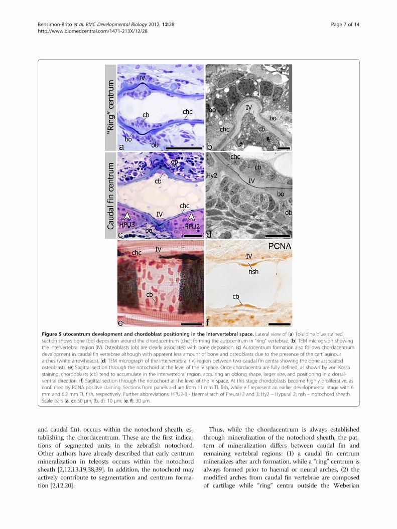
Figure 5 utocentrum development and chordoblast positioning in the intervertebral space. Lateral view of (a) Toluidine blue stainedsection shows bone (bo) deposition around the chordacentrum (chc), forming the autocentrum in “ring” vertebrae. (b) TEM micrograph showingthe intervertebral region (IV). Osteoblasts (ob) are clearly associated with bone deposition. (c) Autocentrum formation also follows chordacentrumdevelopment in caudal fin vertebrae although with apparent less amount of bone and osteoblasts due to the presence of the cartilaginousarches (white arrowheads). (d) TEM micrograph of the intervertebral (IV) region between two caudal fin centra showing the bone associatedosteoblasts. (e) Sagittal section through the notochord at the level of the IV space. Once chordacentra are fully defined, as shown by von Kossastaining, chordoblasts (cb) tend to accumulate in the intervertebral region, acquiring an oblong shape, larger size, and positioning in a dorsal-ventral direction. (f) Sagittal section through the notochord at the level of the IV space. At this stage chordoblasts become highly proliferative, asconfirmed by PCNA positive staining. Sections from panels a-d are from 11 mm TL fish, while e-f represent an earlier developmental stage with 6mm and 6.2 mm TL fish, respectively. Further abbreviations: HPU2-3 - Haemal arch of Preural 2 and 3; Hy2 – Hypural 2; nsh – notochord sheath.Scale bars (a, c): 50 μm; (b, d): 10 μm; (e, f): 30 μm.
Bensimon-Brito et al. BMC Developmental Biology 2012, 12:28 Page 7 of 14http://www.biomedcentral.com/1471-213X/12/28
and caudal fin), occurs within the notochord sheath, es-tablishing the chordacentrum. These are the first indica-tions of segmented units in the zebrafish notochord.Other authors have already described that early centrummineralization in teleosts occurs within the notochordsheath [2,12,13,19,38,39]. In addition, the notochord mayactively contribute to segmentation and centrum forma-tion [2,12,20].
Thus, while the chordacentrum is always establishedthrough mineralization of the notochord sheath, the pat-tern of mineralization differs between caudal fin andremaining vertebral regions: (1) a caudal fin centrummineralizes after arch formation, while a “ring” centrum isalways formed prior to haemal or neural arches, (2) themodified arches from caudal fin vertebrae are composedof cartilage while “ring” centra outside the Weberian
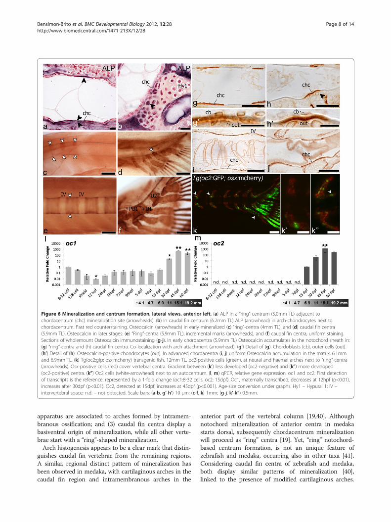
Figure 6 Mineralization and centrum formation, lateral views, anterior left. (a) ALP in a “ring”-centrum (5.0mm TL) adjacent tochordacentrum (chc) mineralization site (arrowheads). (b) In caudal fin centrum (6.2mm TL) ALP (arrowhead) in arch-chondrocytes next tochordacentrum. Fast red counterstaining. Osteocalcin (arrowheads) in early mineralized (c) “ring”-centra (4mm TL), and (d) caudal fin centra(5.9mm TL). Osteocalcin in later stages: (e) “Ring”-centra (5.9mm TL), incremental marks (arrowheads), and (f) caudal fin centra, uniform staining.Sections of wholemount Osteocalcin immunostaining (g-j). In early chordacentra (5.9mm TL) Osteocalcin accumulates in the notochord sheath in:(g) “ring”-centra and (h) caudal fin centra. Co-localization with arch attachment (arrowhead). (g’) Detail of (g). Chordoblasts (cb), outer cells (out).(h’) Detail of (h). Osteocalcin-positive chondrocytes (out). In advanced chordacentra (i, j) uniform Osteocalcin accumulation in the matrix, 6.1mmand 6.9mm TL. (k) Tg(oc2:gfp; osx:mcherry) transgenic fish, 12mm TL. oc2-positive cells (green), at neural and haemal arches next to “ring”-centra(arrowheads). Osx-positive cells (red) cover vertebral centra. Gradient between (k’) less developed (oc2-negative) and (k”) more developed(oc2-positive) centra. (k”) Oc2 cells (white-arrowhead) next to an autocentrum. (l, m) qPCR, relative gene expression. oc1 and oc2. First detectionof transcripts is the reference, represented by a 1-fold change (oc1:8-32 cells, oc2: 15dpf). Oc1, maternally transcribed, decreases at 12hpf (p<0.01),increases after 30dpf (p<0.01). Oc2, detected at 15dpf, increases at 45dpf (p<0.001). Age-size conversion under graphs. Hy1 – Hypural 1; IV –intervertebral space; n.d. – not detected. Scale bars: (a-b, g’-h’) 10 μm; (c-f, k) 1mm; (g-j, k’-k”) 0.5mm.
Bensimon-Brito et al. BMC Developmental Biology 2012, 12:28 Page 8 of 14http://www.biomedcentral.com/1471-213X/12/28
apparatus are associated to arches formed by intramem-branous ossification; and (3) caudal fin centra display abasiventral origin of mineralization, while all other verte-brae start with a “ring”-shaped mineralization.Arch histogenesis appears to be a clear mark that distin-
guishes caudal fin vertebrae from the remaining regions.A similar, regional distinct pattern of mineralization hasbeen observed in medaka, with cartilaginous arches in thecaudal fin region and intramembranous arches in the
anterior part of the vertebral column [19,40]. Althoughnotochord mineralization of anterior centra in medakastarts dorsal, subsequently chordacentrum mineralizationwill proceed as “ring” centra [19]. Yet, “ring” notochord-based centrum formation, is not an unique feature ofzebrafish and medaka, occurring also in other taxa [41].Considering caudal fin centra of zebrafish and medaka,both display similar patterns of mineralization [40],linked to the presence of modified cartilaginous arches.

Figure 7 Progress of mineralization in “ring” and caudal fincentra. Mineralization expansion is represented by the arrows, in (a)a “ring” centrum that expands in linear mineralization fronts in theantero-posterior axis and (b) in caudal fin centrum, directlyassociated with the haemal cartilaginous arch (ca) attached to thenotochord sheath, expanding from a basiventral origin. In black isrepresented the area of initial mineralization, which matches themyoseptum (Myo) in the case of a “ring” centrum.
Bensimon-Brito et al. BMC Developmental Biology 2012, 12:28 Page 9 of 14http://www.biomedcentral.com/1471-213X/12/28
Many basal ray-finned fishes have cartilaginous archesdirectly attached to an unconstricted notochord [18] sug-gesting that cartilaginous arches are an ancestral feature.Therefore, the caudal fin endoskeleton of zebrafish, andalso medaka, appear to display the evolutionarily conser-ved condition, with cartilaginous arch formation.Mineralization in the caudal fin centra shows a clear as-
sociation with attachment sites of the cartilaginous haemalarches to the notochord (basiventral mineralization pat-tern). The association between basiventral mineralizationand presence of cartilaginous haemal arches has also beendescribed in other teleost species, such as the Goldeye(Hiodon alosoides) [42], Atlantic salmon [2] and Whiteseabream (Diplodus sargus) [43]. In these species too, cen-trum mineralization originates exactly where the carti-laginous haemal arches are attached to the notochord.Mineralization then progresses from a basiventral originthrough bilateral wedge-shaped areas that meet dorsally.Moreover, we have identified mineralization related
proteins in the chondrocytes of the cartilaginous arches,in the immediate vicinity of the area of the incipientmineralization of the notochord. This supports the pos-sible role of the cartilaginous arches in early centrum es-tablishment, a hypothesis that will be discussed in moredetail in the next section.The cartilaginous arches appear to be strictly asso-
ciated not only with chordacentrum formation but alsowith the start of autocentrum formation, given that thearches are directly attached to the notochord, affectingbone deposition around the notochord.The type of arches and associated pattern of centrum
mineralization appears to be correlated not only to theformation of the vertebral bodies but also to their abilityto fuse or stay as individual units (Figure 7). In the medakabis (biaxial symmetries) mutant, centra mineralize at siteswhere ectopic hypurals attach to the notochord, leading tothe development of fused centra [40]. Thus, it is temptingto suggest from the zebrafish and medaka data, that thecartilaginous arches have a role in centrum mineralization.Perhaps the predisposition to develop vertebral fusions isrelated to a specific mineralization pattern with basiventralorigin (cartilaginous arch associated).
Distribution of notochord inner and outer cells duringcentrum formationIn Atlantic salmon, notochord sheath mineralization ispreceded by an apparent segmentation of the chor-doblast layer [2]. The same holds for medaka (PEW per-sonal observations). In contrast, differential chordoblastdistribution in zebrafish is only clearly observed afterchordacentrum mineralization, with few scattered squa-mous cells in the mineralized area and large, dorso-ven-trally oriented, highly proliferative cells in the intervertebralregion.
Regarding notochord outer cells, it appears that thesegmental pattern of centra in the caudal fin region isassociated with the anatomical pattern of arches. In con-trast, no association can be found in “ring” centra be-tween outer cell distribution and early chordacentrumformation. The latter is in agreement with the data fromzebrafish fused somite mutants (fss), where paraxialmesoderm lacks a proper segmentation [44]. This mu-tant shows fusion of vertebral arches, while vertebral

Bensimon-Brito et al. BMC Developmental Biology 2012, 12:28 Page 10 of 14http://www.biomedcentral.com/1471-213X/12/28
centra are individualized, suggesting that centrum seg-mentation does not rely on sclerotome patterning only.The resegmentation process in zebrafish has been sug-gested to follow non-lineage restricted compartments,where one somite contributes to more than one verte-bral body, independently of the antero-posterior somiticdomains (leaky-resegmentation) [1].Although mineralization is not preceded by a clear
pattern of inner or outer cell distribution (exceptions arearch-chondrocytes in the caudal fin), these cells may ne-vertheless contribute to centrum mineralization. In Atlanticsalmon, initial centrum mineralization has been related tochordoblasts, followed by sclerotome-derived bone forma-tion [2,10]. Different from Atlantic salmon, in medakasclerotome-derived osteoblasts, with no input of chor-doblasts, have been described to form vertebral centra[19,35]. Yet, recent data show that conditional abla-tion of osteoblasts in medaka maintains notochord mi-neralization and even leads to vertebral fusion [20]. Thissuggested that notochord cells have the ability to inducemineralization of the notochord sheath. This is consistentwith studies in zebrafish [12,45] that show that notochordcell ablation prevents early centrum mineralization.
Osteocalcin and chordacentrum formationOsteocalcin is the most abundant noncollagenous proteinin bone of vertebrates, including all teleosts analyzed so far[46]. Although Osteocalcin was already described in thevertebral bodies of teleosts including zebrafish [47-50], noprevious reports demonstrated Oc in early notochordalmineralization events. Thus, the presence of Oc in stages ofchordacentrum mineralization has to be considered in aperspective different from that of regular bone formation.Within teleosts, Osteocalcin is represented by two dis-
tinct isoforms (Oc1 and Oc2) [25,51], encoded by twodifferent genes, which were proposed to originate from aduplication event [25]. Analysis of relative gene expres-sion levels by qPCR for oc1 and oc2 showed that oc2expression is first detected after 7dpf, while oc1 is mater-nally transcribed, suggesting a different role of the twoisoforms. In fact, oc1 is detected prior to any osteoblastdifferentiation or mineralization event. This leads to thehypothesis that oc1 must be involved in processes priorto bone formation. Detection of Oc1 by immunostainingshows that this protein accumulates exactly where mine-ralization of the chordacentrum occurs. In the “ring”centra, the origin of the secreted protein is difficult toassess, due to the squamous morphology of the cells ofthe notochord epithelium and the inconspicuous pres-ence of cells outside the notochord. In the caudal fin re-gion, ALP positive chondrocytes, directly adjoining themineralizing notochord sheath, are positive for Oc im-munostaining, as well as notochord inner cells. In the Tg(oc2:gfp; osx:mcherry) fish, oc2 expression was located at
the bone of arches of the “ring” centra, that form viaintramembranous bone formation. This time point cor-responds to the phase of autocentrum mineralization, aspreviously described in medaka [52]. Therefore, our re-sults suggest that Oc1, but not Oc2, is connected to chor-dacentrum mineralization. Likewise, in larval and juvenileAtlantic cod [53], oc1 and oc2 are expressed in differentcells associated with mineralizing structures. In Atlanticsalmon, some mineralized areas with reduced expressionof osteocalcin (oc1) were considered to be associated withexpression of a second isoform that the authors did notidentify [49]. Laizé and co-workers [25] described Oc2propeptide as serine-rich, potentially phosphorylated andcontaining acid residues. These characteristics allow thisprotein to bind numerous calcium ions. If Osteocalcinhas an active role in the process of mineralization orif it is a bone-related hormone that passively binds tocalcium [54,55], is a current debate and must be furtherexplored.
ConclusionsIn conclusion, we have analyzed distinct mineralizationpatterns in different regions of the zebrafish vertebralcolumn and related them to the timing and nature ofvertebral arch formation. We have examined the distri-bution of cells within and outside the notochord, andcritically assessed their potential contribution to chor-dacentrum and autocentrum formation. If notochordmineralization occurs prior to arch formation, the mi-neralization of the notochord sheath is ring-shaped. Ifnotochord mineralization occurs after cartilaginous archformation, mineralization of the notochord sheath startsat the insertion point of the arches, with a basiventralorigin. Finally, we have detected Oc1 in the notochordsheath during chordacentrum formation, whereas oc2was only expressed later in association with bone forma-tion. These results emphasize the need to better charac-terize the function of both osteocalcin genes in zebrafishand throughout the vertebrate clade.
MethodsEthics statement on animal experimentsAnimal handling and experiments were legally accre-dited by the Portuguese Direcção Geral de Veterinária(DGV) and all the experimental procedures involvinganimals followed the EU (Directive 86/609/CEE) andNational (Directives 1005/92 from October 23, 466/95from May 17 and 1 1131/97 from November 7) legisla-tion for animal experimentation and welfare.
Sampling and wholemount skeletal analysisTo establish a timeline of events for early vertebrae de-velopment, an ontogenic series of Danio rerio was stu-died. Zebrafish eggs were obtained from natural spawning

Bensimon-Brito et al. BMC Developmental Biology 2012, 12:28 Page 11 of 14http://www.biomedcentral.com/1471-213X/12/28
of wild-type fish and larvae were maintained and reared atstandard conditions [56]. Fish were collected at size inter-vals of 0.1 mm, between 4.0 and 8.0 mm of total length(TL). Similar-sized specimens were always selected amongage-matched fish in order to avoid additional interspecificvariation.Larvae were euthanized with a lethal dose of MS222
(Sigma-Aldrich, St. Louis, MO) and fixed for 24 h in 4%buffered paraformaldehyde at 4ºC.Early mineralization stages of the vertebral bodies
were observed following 10 minutes staining with 0.01%alizarin red S in 70% ethanol solution. Specimens wereobserved under a Zeiss Axio Imager microscope, throughfluorescence detection [57].
Light and transmission electron microscopy (TEM)Zebrafish specimens of 5.0 and 11.0 mm TL with nopre-staining were fixed in a mixture of 1.5% glutaralde-hyde and 1.5% paraformaldehyde in 0.1 M cacodylatebuffer and processed for embedding in Epon, accordingto procedures previously described [58]. Parasagittal 1μm semi-thin sections of the sites of interest werestained with toluidine blue for 1–2 min (0.2% toluidineblue, 2% Na2CO3), rinsed with water, air-dried andmounted with DPX (Fluka, Buchs, Switzerland). Ultra-thin sections were contrasted with uranyl acetate andlead citrate and observed with a Jeol JEM 1010 (Jeol,Tokyo, Japan), operating at 60 kV. Images were digitizedusing a DITABIS system (Pforzheim, Germany).
Identification of sites of mineral deposition and Alkalinephosphatase activity (ALP)To determine sites of mineral deposition and of Alkalinephosphatase (ALP) activity during centrum formation,specimens ranging from 5.0 to 6.5 mm TL were dehy-drated and embedded in glycol methacrylate, as previouslydescribed [59], and sectioned at 5 μm.For detection of minerals, a silver nitrate coupling
method, according to von Kossa [60], was used. Sectionswere counterstained with neutral red. Slides were exa-mined for the appearance of optimal signal, rinsed indistilled water and mounted with DPX (Sigma-Aldrich,St. Louis, MO).Alkaline phosphatase activity was detected using a pre-
viously described method [61] with some modification.Briefly, slides were incubated in staining buffer (100 mMTris/HCl, pH 9.5; 50mM MgCl2; 100 mM NaCl) for 10min. ALP was demonstrated by adding 4-nitro bluetetrazolium chloride (NBT; 0.38 mg ml-1; Roche) and 5-bromo-4-chloro-3-indolyl phosphate (BCIP; 0.175 mgml-1; Roche) to the buffer solution, for a maximum of 30min at 37ºC, in the dark. Negative control slides wereincubated for 10 min at 85ºC in staining buffer prior toaddition of NBT and BCIP. Slides were examined for the
appearance of optimal signal-noise ratio, counterstainedwith Nuclear Fast Red Solution (Sigma-Aldrich, St.Louis, MO) and mounted in DPX (Sigma-Aldrich, St.Louis, MO).
Immunolocalization of PCNA, Osteocalcin and Collagentype IIFor detection of cell proliferation through Proliferatingcell nuclear antigen (PCNA) immunolocalization, 6.2 mmTL fish were paraffin-embedded according to routineprocedures and serially sectioned at 7 μm. A mousemonoclonal primary antibody produced against humanantigen (Sigma-Aldrich, St. Louis, MO, dilution 1:1000)was used [62] together with a goat polyclonal anti-mouse immunoglobulin biotin-coupled secondary anti-body (Dako, dilution 1:500).Immunohistochemical detection of Osteocalcin and
Collagen type II was performed on fish of 4 to 6.5 mmTL as wholemount staining, according to a procedurepreviously described [63], using an anti-meagre (Argyro-somus regius) Osteocalcin rabbit polyclonal primaryantibody, previously validated for zebrafish [36]. ForCollagen type II an anti-chicken mouse antibody (II-II6B3,Hybridoma Bank) previously validated for zebrafish [64],was used. Control fish were treated with secondary anti-body alone.Osteocalcin stained specimens were subsequently
dehydrated, embedded in epon, serially sectioned at 4μm and mounted with DPX (Sigma-Aldrich, St. Louis,MO), to achieve a more detailed analysis. Wholemountfish stained for Collagen type II were observed using aLeica MZ Apo stereomicroscope. Sections were obser-ved using a Zeiss Axio Imager Microscope and photo-graphed using an Axiocam MRC videocamera.
oc2 transgenic lineThe transgenic zebrafish line Tg(oc2:gfp; osx:mcherry),previously described by [65], was provided by StefanSchulte-Merker from Hubrecht Institute-KNAW andUniversity Medical Centre (Uppsalalaan 8, 3584 CTUtrecht, The Netherlands).
RNA extraction and oc1/oc2 quantitative expressionanalysisTotal RNA was extracted from a pool of up to twentyspecimens following the Chomczynski and Sacchi me-thod [66]. Samples were collected at stages 8-32 or 128cells, at shield stage, at 12, 24, 48, 72 and 96 hours post-fertilization (hpf ) and at 5, 7, 15, 30, 45 and 60 dayspost-fertilization (dpf). To prevent genomic DNA con-tamination, following RNA extraction, samples wereDNase treated with RQ1 DNase (Promega, Madison,WI) according to the manufacturer’s protocol, follo-wed by a phenol-chlorophorm purification step. RNA

Bensimon-Brito et al. BMC Developmental Biology 2012, 12:28 Page 12 of 14http://www.biomedcentral.com/1471-213X/12/28
quantity and quality was determined by spectropho-tometry (NanoDrop ND-1000, Thermo Scientific) andelectrophoresis.cDNA was prepared from 1μg of total RNA from each
sample using Moloney-murine leukemia virus (M-MLV)reverse transcriptase (Invitrogen), RNase Out (Invitrogen),and an oligo(dT) adapter (5'-ACGCGTCGACCTCGAGATCGATG(T)13-3') in a total volume of 20μl for 1hat 37ºC. For quantitative real-time PCR a StepOnePlus96 (Applied Biosystems) was used together with 1× Sso-FastTM EvaGreen Supermix (BioRad, Richmond, USA),0.2 μM of forward (Oc1: 50-GAAGCGAACATGAAGAGTCTGACAGTCC-30; Oc2: 50-CCAACTCCGCATCAGACTCCGCATCA-30) and reverse (Oc1- 50-TTTATAGGCGGCGATGATTCC-30; Oc2: 50-AGCAACACTCCGCTTCAGCAGCACAT-30) primers and 200 ng of reverse-transcribed RNA. PCR conditions were 15 min at 95°Cfollowed by 40–50 amplification cycles (each cycle is 30s at 95°C, 15 s at 68°C). Expression values were norma-lized using the Elongation factor 1α (Ef1α) housekeepinggene [67]. Gel electrophoresis and melt curve analysiswere used to confirm specific product formation.All data were statistically analyzed using a non-para-
metric Kruskal-Wallis test, followed by Wilcoxon mul-tiple comparison with the GraphPad Prism software.
TerminologyThe subdivision of the vertebral column in differentregions is based on the nomenclatures of Arratia et al.[16], Bird and Mabee [31] and Nybelin [68] (Figure 1).From anterior to posterior, vertebrae were identified as(1) Weberian vertebrae, (2) abdominal vertebrae (rib-bearing with open haemal arches and without haemalspines), (3) caudal vertebrae (with haemal arches closed),and (4) caudal fin vertebrae (with modified haemal andneural arches and respective spines). The nomenclatureof the Weberian apparatus follows [31]. The caudal finvertebrae nomenclature follows Patterson [69], adap-ted by Arratia and Schultze [70] and Bensimon-Britoet al. [32]. The caudal fin vertebral region contains pre-ural and ural vertebral bodies. Preural vertebral bodies(abbreviated as PU) have haemal and neural arches thatsupport the caudal fin. Ural vertebral bodies (briefly urals,abbreviated as U) support the caudal fin with modifiedhaemal arches that do not enclose the caudal artery. Aplus sign is added in superscript when a vertebral centrumis derived from a fusion event (compound centrum), asdescribed previously [32].
Competing interestsThe funding agencies had no role in study design, data collection andanalysis, decision to publish, or preparation of the manuscript. The authorshave declared no competing interests.
Authors’ contributionsConceived and designed the experiments: ABB, MLC, AH, PEW. Performedthe experiments: ABB and JC. Analyzed the data: ABB, JC, MLC, AH, PEW.Contributed reagents/materials/analysis tools: MLC, AH. PEW. Contributed tothe writing of the paper: ABB, MLC, AH, PEW. All authors read and approvedthe final manuscript.
AcknowledgementsA. Bensimon-Brito acknowledges a FCT PhD fellowship SFRH/BD/40573/2007and is presently the recipient of an ERC Reseal (2007-StG-208631) fellowship,J. Cardeira is the recipient of a CCMAR fellowship CCMAR/BI/0029/2009 andA. Huysseune and P.E. Witten acknowledge a grant from FWO 3G.0040.08. LCacknowledges CCMAR funding. The authors thank Stefan Schulte-Merker forproviding the transgenic zebrafish line Tg(oc2:gfp; osx:mcherry). For assistancein sample collection we thank G. Dionísio and R. Peres dos Santos. Fortechnical assistance we thank Barbara Verstraeten, Myriam Claeys, TommyD’heuvaert and Jasper Dewit.
Author details1PhD Program in Biomedical Sciences, University of Algarve, Faro, Portugal.2Center of Marine Sciences - CCMar, University of Algarve, Faro, Portugal.3Evolutionary Developmental Biology, Biology Department, Ghent University,Ghent, Belgium. 4Dept. of Biomedical Sciences and Medicine, University ofAlgarve, Faro, Portugal. 5Present address: CEDOC - Faculdade de CiênciasMédicas, FCM Universidade Nova de Lisboa, Lisbon, Portugal.
Received: 12 July 2012 Accepted: 3 October 2012Published: 9 October 2012
References1. Morin-Kensicki EM, Melancon E, Eisen JS: Segmental relationship
between somites and vertebral column in zebrafish. Development 2002,129:3851–3386.
2. Grotmol S, Kryvi H, Nordvik K, Totland GK: Notochord segmentation maylay down the pathway for the development of the vertebral bodies inthe Atlantic salmon. Anat Embryol 2003, 207:263–272.
3. Turnpenny PD, Alman B, Cornier AS, Giampietro PF, Offiah A, Tassy O,Pourquié O, Kusumi K, Dunwoodie S: Abnormal vertebralsegmentation and the notch signaling pathway in man. Dev Dyn2007, 236(6):1456–1474.
4. Hall BK: Evolutionary consequences of skeletal differentiation. Am Zool1975, 15:329–350.
5. Kaplan KM, Spivak JM, Bendo JA: Embryology of the spine and associatedcongenital abnormalities. Spine J 2005, 5(5):564–576.
6. Christ B, Huang R, Wilting J: The development of the avian vertebralcolumn. Anat Embryol 2000, 202:179–194.
7. Kahane N, Cinnamon Y, Kalcheim C: The origin and fate of pioneermyotomal cells in the avian embryo. Mech Dev 1998, 74:59–73.
8. Christ B, Huang R, Scaal M: Formation and differentiation of the aviansclerotome. Anat Embryol 2004, 208:333–350.
9. Stemple DL: Structure and function of the notochord: an essential organfor chordate development. Development 2005, 132:2503–2512.
10. Grotmol S, Nordvik K, Kryvi H, Totland GK: A segmental pattern of alkalinephosphatase activity within the notochord coincides with the initialformation of the vertebral bodies. J Anat 2005, 206:427–436.
11. Schmitz RJ: Comparative ultrastructure of the cellular components of theunconstricted notochord in the sturgeon and the lungfish. J Morphol1998, 236:75–104.
12. Fleming A, Keynes R, Tannahill D: A central role for the notochord invertebral patterning. Development 2004, 131:873–880.
13. Huxley TH: Observations on the development of some parts of theskeleton of fishes. Q J Microsc Sci 1859, 7:33–46.
14. François Y: Structure et développement de la vertèbre de Salmo etdes téléostéens. Archives de Zoologie Experimentale et Generale 1966,107:287–328.
15. Bensimon-Brito A, Cancela ML, Huysseune A, Witten PE: Vestiges,rudiments and fusion events: the zebrafish caudal fin endoskeleton inan evo-devo perspective. Evol Dev 2012, 14(1):116–127.
16. Arratia G, Schultze H-P, Casciotta J: Vertebral column and associatedelements in Dipnoans and comparison with other fishes: Developmentand homology. J Morphol 2001, 250:101–172.

Bensimon-Brito et al. BMC Developmental Biology 2012, 12:28 Page 13 of 14http://www.biomedcentral.com/1471-213X/12/28
17. Gavaia PJ, Simes DC, Ortiz-Delgado JB, Viegas CSB, Pinto JP, Kelsh RN,Sarasquete MC, Cancela ML: Osteocalcin and matrix Gla protein inzebrafish (Danio rerio) and Senegal sole (Solea senegalensis):Comparative gene and protein expression during larval developmentthrough adulthood. Gene Expr Patterns 2006, 6(6):637–652.
18. Lauder GV: On the relationship of the myotome to the axial skeleton invertebrate evolution. Paleobiology 1980, 6(1):51–56.
19. Inohaya K, Takano Y, Kudo A: The Teleost intervertebral region actsas a growth center of the centrum: In vivo visualization ofosteoblasts and their progenitors in transgenic fish. Dev Dyn 2007,236:3031–3046.
20. Willems B, Büttner A, Huysseune A, Renn J, Witten PE, Winkler C:Conditional ablation of osteoblasts in medaka. Dev Biol 2012,364:128–137.
21. Huysseune A, Takle H, Soenens M, Taerwe K, Witten PE: Unique and sharedgene expression patterns in Atlantic salmon (Salmo salar) toothdevelopment. Developmental Genes and Evolution 2008, 218:427–437.
22. Pullig O, Weseloh G, Ronneberger D-L, Käkönen S-M, Swoboda B:Chondrocyte differentiation in Human osteoarthritis: Expression ofosteocalcin in normal and osteoarthritic cartilage and bone. Calcif TissueInt 2000, 67:230–240.
23. Aizawa T, Roach HI, Kokubun S, Tanaka Y: Changes in the expression ofFas, osteonectin and osteocalcin with age in the rabbit growth plate.The Journal of Bone and Joint Surgery (Br) 1998, 80-B(5):880–887.
24. Neugebauer BM, Monroe MA, Broess M, Gerstenfeld LC, Hauschka PV:Characterization of structural sequences in the chicken osteocalcin gene:Expression of osteocalcin by maturing osteoblasts and by hypertrophicchondrocytes in vitro. J Bone Miner Res 1995, 10(1):157–163.
25. Laizé V, Viegas CSB, Price PA, Cancela ML: Identification of an osteocalcinisoform in fish with a large acidic prodomain. J Biol Chem 2006,281(22):15037–15043.
26. Fjelldal PG, Hansen T, Breck O, Ornsrud R, Lock EJ, Waagbø R,Wargelius A, Witten PE: Vertebral deformities in farmed Atlantic salmon(Salmo salar L.) – etiology and pathology. J Appl Ichthyol 2012,28(3):433–440.
27. Ward AB, Brainerd EL: Evolution of axial patterning in elongate fishes.Biol J Linn Soc 2007, 90:97–116.
28. Wargelius A, Fjelldal PG, Hansen T: Heat shock during early somitogenesisinduces caudal vertebral column defects in Atlantic salmon (Salmosalar). Dev Genes Evol 2005, 215(7):350–357.
29. Bird NC, Hernandez LP: Building an evolutionary innovation: differentialgrowth in the modified vertebral elements of the zebrafish Weberianapparatus. Zoology 2009, 112(2):97–112.
30. Ferreri F, Nicolais C, Boglione C, Bertolini B: Skeletal characterization ofwild and reared zebrafish: anomalies and meristic characters. J Fish Biol2000, 56(5):1115–1128.
31. Bird NC, Mabee PM: Developmental morphology of the axial skeleton ofthe zebrafish, Danio rerio (Ostariophysi: Cyprinidae). Dev Dyn 2003,228(3):337–357.
32. Bensimon-Brito A, Cancela ML, Huysseune A, Witten PE: The zebrafish(Danio rerio) caudal complex: a model to study vertebral body fusion.J Appl Ichthyol 2010, 26(2):235–238.
33. Witten PE, Obach A, Huysseune A, Baeverfjord G: Vertebrae fusion inAtlantic salmon (Salmo salar): development, aggravation and pathwaysof containment. Aquaculture 2006, 258(1–4):164–172.
34. Fjelldal PG, Nordgarden U, Wargelius A, Taranger GL, Waagbø R, Olsen RE:Effects of vegetable feed ingredients on bone health in Atlantic salmon.J Appl Ichthyol 2010, 26(2):327–333.
35. Ekanayake S, Hall BK: The development of acellularity of the vertebralbone of the Japanese Medaka, Oryzias latipes (Teleostei; Cyprinidontidae).J Morphol 1987, 193:253–261.
36. Simes DC, Williamson MK, Schaff BJ, Gavaia PJ, Ingleton PM, Price PA,Cancela ML: Characterization of osteocalcin (BGP) and matrix gla protein(MGP) fish specific antibodies: validation for immunodetection studies inlower vertebrates. Calcif Tissue Int 2004, 74:170–180.
37. Du SJ, Frenkel V, Kindschi G, Zohar Y: Visualizing normal and defectivebone development in Zebrafish embryos using the fluorescentchromophore calcein. Dev Biol 2001, 238:239–246.
38. Nordvik K, Kryvi H, Totland GK, Grotmol S: The salmon vertebral bodydevelops through mineralization of two preformed tissues that areencompassed by two layers of bone. J Anat 2005, 206:103–114.
39. Kölliker A: Weitere Beobachtungen über die Wirbel der Selachier, insbesondereüber die Wirbel der Lamnoidei, nebst allgemeinen Bemerkungen über dieBildung der Wirbel der Plagiostomen. Frankfurt: Verlag HL. Bröuner; 1863.
40. Hibiya K, Katsumoto T, Kondo T, Kitabayashi I, Kudo A: Brpf1, a subunit ofthe MOZ histone acetyl transferase complex, maintains expression ofanterior and posterior Hox genes for proper patterning of craniofacialand caudal skeletons. Dev Biol 2009, 329:176–190.
41. Laerm J: The origin and homology of the chondrostean vertebralcentrum. Can J Zool 1979, 57(3):475–485.
42. Schultze H-P, Arratia G: Reevaluation of the caudal skeleton of someactinopterygian fishes: II. Hiodon, Elops, and Albula. J Morphol 1988,195(3):257–303.
43. Koumoundouros G, Sfakianakis DG, Maingot E, Divanach P, Kentouri M:Osteological development of the vertebral column and of the fins inDiplodus sargus (Teleostei: Perciformes: Sparidae). Mar Biol 2001,139:853–862.
44. van Eeden FJM, Granato M, Schach U, Brand M, Furutani-Seiki M, Haffter P,Hammerschmidt M, Heisenberg C-P, Jiang Y-J, Kane DA, et al: Mutationsaffecting somite formation and patterning in the zebrafish, Danio rerio.Development 1996, 123:153–164.
45. Fleming A, Keynes RJ, Tannahill D: The role of the notochord in vertebralcolumn formation. J Anat 2001, 199(1–2):177–180.
46. Nishimoto SK, Waite JH, Nishimoto M, Kriwacki RW: Structure, activity, anddistribution of fish osteocalcin. J Biol Chem 2003, 278(14):11843–11848.
47. Simes DC, Williamson MK, Ortiz-Delgado JB, Viegas CSB, Price PA,Cancela ML: Purification of Matrix Gla Protein from a marine teleostfish, Argyrosomus regius: Calcified cartilage and not bone as theprimary site of MGP accumulation in fish. J Bone Miner Res 2003,18(2):244–259.
48. Ytteborg E, Togersen J, Baeverfjord G, Takle H: Morphological andmolecular characterization of developing vertebral fusions using ateleost model. BMC Physiol 2010, 10(13):1–15.
49. Krossøy C, Ornsrud R, Wargelius A: Differential gene expression of bgp andmgp in trabecular and compact bone of Atlantic salmon (Salmo salar L.)verebrae. J Anat 2009, 215:663–672.
50. Pinto JP, Ohresser MCP, Cancela ML: Cloning of the bone Gla proteingene from the teleost fish Sparus aurata. Evidence for overallconservation in gene organization and bone-specific expression fromfish to man. Gene Expr Patterns 2001, 270:77–91.
51. Desbois C, Hogue DA, Karsenty G: The mouse osteocalcin gene clustercontains three genes with two separate spatial and temporal patterns ofexpression. J Biol Chem 1994, 269:1183–1190.
52. Renn J, Winkler C: Characterization of collagen type 10a1 and osteocalcinin early and mature osteoblasts during skeleton formation in medaka.J Appl Ichthyol 2010, 26:196–201.
53. Lie KK, Moren M: Retinoic acid induces two osteocalcin isoforms andinhibits markers of osteoclast activity in Atlantic cod (Gadus morhua)ex vivo cultured craniofacial tissues. Comparative Biochemistry andPhysiology, Part A 2012, 161:174–184.
54. Confavreux CB, Levine RL, Karsenty G: A paradigm of integrativephysiology, the crosstalk between bone and energy metabolisms.Mol Cell Endocrinol 2009, 310:21–29.
55. Ferron M, Hinoi E, Karsenty G, Ducy P: Osteocalcin differentially regulatescell and adipocyte gene expression and affects the development ofmetabolic diseases in wild-type mice. Proceedings of the National Academyof Sciences USA 2009, 105(13):5266–5270.
56. Westerfield M: The zebrafish book. A guide for the laboratory use of zebrafishDanio rerio. 4th edition. Eugene: University of Oregon Press; 2000.
57. Connolly MH, Yelick PC: High-throughput methods for visualizing theteleost skeleton: capturing autofluorescence of alizarin red. J ApplIchthyol 2010, 26:274–277.
58. Huysseune A, Sire JY: Development of cartilage and bone tissues of theanterior part of the mandible in cichlid fish: a light and TEM study. AnatRec 1992, 233:357–375.
59. Witten PE, Hansen A, Hall BK: Features of mono and multinucleatedbone resorbing cells of the zebrafish Danio rerio and their contributionto skeletal development, remodeling and growth. J Morphol 2001,250:197–207.
60. Schenk RK, Olah AJ, Herrmann W: Preparation of calcified tissues for lightmicroscopy. In Methods of Calcified Tissue Preparation. Edited by Dickson GR.Amsterdam: Elsevier; 1984:1–56.

Bensimon-Brito et al. BMC Developmental Biology 2012, 12:28 Page 14 of 14http://www.biomedcentral.com/1471-213X/12/28
61. Miyake T, Cameron AM, Hall BK: Stagespecific expression patterns ofalkaline phosphatase during development of the first arch skeleton ininbred C57BL/6 mouse embryos. J Anat 1997, 190:239–260.
62. Ortego LS, Hawkins WE, Walker WW, Krol RM, Benson WH: Detection ofProliferating Cell Nuclear antigen in tissues of three small fish species.Biotech Histochem 1994, 69(6):317–323.
63. Verstraeten B, Sanders E, Huysseune A: Whole MountImmunohistochemistry and In Situ Hybridization of Larval and AdultZebrafish Dental Tissues. In: Methods Mol Biol. vol. 2012, 887:179–191.
64. Clément A, Wiweger M, Hardt S, Rusch MA, Selleck SB, Chien C-B, Roehl HH,et al: Regulation of Zebrafish Skeletogenesis by ext2/dackel and papst1/pinscher. PLoS Genet 2008, 4:e1000136.
65. Knopf F, Hammond C, Chekuru A, Kurth T, Hans S, Weber CW, Mahatma G,Fisher S, Brand M, Schulte-Merker S, et al: Bone regenerates viadedifferentiation of osteoblasts in the zebrafish fin. Dev Cell 2011,20:713–724.
66. Chomczynski P, Sacchi N: Single-step method of RNA isolation by acidguanidinium thiocyanate-phenol-chloroform extraction. Anal Biochem1987, 162:156–159.
67. Tang R, Dodd A, Lai D, Mcnabb WC, Love DR: Validation of Zebrafish(Danio rerio) reference genes for quantitative real-time RT-PCRnormalization. Acta Biochim Biophys Sin 2007, 39(5):384–390.
68. Nybelin O: Zur morphologie und terminologie des schwanzskelettes derActinopterygier. Arkiv fur Zoologi 1963, 15:485–516.
69. Patterson C: The caudal skeleton in Lower Liassic pholidophorid fishes.Bulletin of the British Museum (Natural History) Geology 1968, 16(5):201–239.
70. Arratia G, Schultze H-P: Reevaluation of the caudal skeleton of certainactinopterygian fishes. III. Salmonidae. Homologization of caudal skeletalstructures. J Morphol 1992, 214:187–249.
doi:10.1186/1471-213X-12-28Cite this article as: Bensimon-Brito et al.: Distinct patterns of notochordmineralization in zebrafish coincide with the localization of Osteocalcinisoform 1 during early vertebral centra formation. BMC DevelopmentalBiology 2012 12:28.
Submit your next manuscript to BioMed Centraland take full advantage of:
• Convenient online submission
• Thorough peer review
• No space constraints or color figure charges
• Immediate publication on acceptance
• Inclusion in PubMed, CAS, Scopus and Google Scholar
• Research which is freely available for redistribution
Submit your manuscript at www.biomedcentral.com/submit