disseminated intravascular coagulation dr. s. parthasarathy md., da., dnb, md (acu), dip. diab. dca,...
TRANSCRIPT
Disseminated intravascular coagulation
Dr. S. Parthasarathy
MD., DA., DNB, MD (Acu), Dip. Diab. DCA, Dip. Software statistics, PhD(physiology)
Mahatma Gandhi Medical College and Research Institute, Puducherry, India

Definition
• Disseminated intravascular coagulation (DIC) is
a clinicopathologic syndrome in which
widespread intravascular coagulation is
induced by procoagulants that are introduced
into or produced in the blood secondary to 1
or more underlying condition(s)

Normal hemostasis

The process -- stage 1
• Injury → platelet adhere (vWF),• subsequent platelet activation and
aggregation• (vWf, serotonin, ADP) ↓• The platelet glycoprotein (Gp) IIb/IIIa (IIb3)
complex
• occlusive platelet thrombus. (plug)

The process – stage 2
• Plasma coagulation proteins (clotting factors) normally circulate in plasma in their inactive forms
• extrinsic and intrinsic pathways – Active forms
• Endothelial tissue factor + platelet factors
• Clotting – fibrin plug ( THROMBUS )

Stage 3
Fibrinolytics
• Platelet plug • Fibrin clot • Fibrinolysis +anticoagulation
Back to
DIC

DIC never occurs alone
• DIC is presently recognized as a deviation from the normal physiologic balance between the processes of thrombus formation and fibrinolysis.
Types as clinical - earlier described
Acute- Large material , overwhelming process Bad
Chronic – slow process – body compensates

Incidence
• 1.5 % of in hospital patients • 25 % of trauma • 30 % of sepsis patients
• Not uncommon
Causes
• Infection • Trauma • Malignancy • Obstetrics • Toxic • Burns • Pancreatitis• obstructive jaundice

The basis
• excessive generation and circulation of thrombin
• impairment of the natural anticoagulant pathway
• suppression of fibrinolysis

Pathogenesis • Malignant cells, sepsis, crush injuries
• Tissue factor activated
• TF + Factor VII
• Activated factor IX and X
• Thrombin

• OK – now thrombin formed
• There are a lot of natural anticoagulants
• Should there be a problem ??

There are 3 most common Anticoagulants in the body:
– Antithrombin, – Active Protein C– Tissue factor pathway inhibitor (TFPI).
• In DIC:– ↓Antithrombin– ↓Active Protein C

Anticoagulation
Normal DIC

Cascade continues

Fibrinolysis
• Fibrinolysis is a process in which plasmin, an active form of plasminogen, cleaves fibrin and fibrinogen to restore vessel patency following hemostasis
• Fibrin to FDPs √

Process proceeds -----

• Malignancy – tissue factor
• Sepsis – inflammatory cytokines
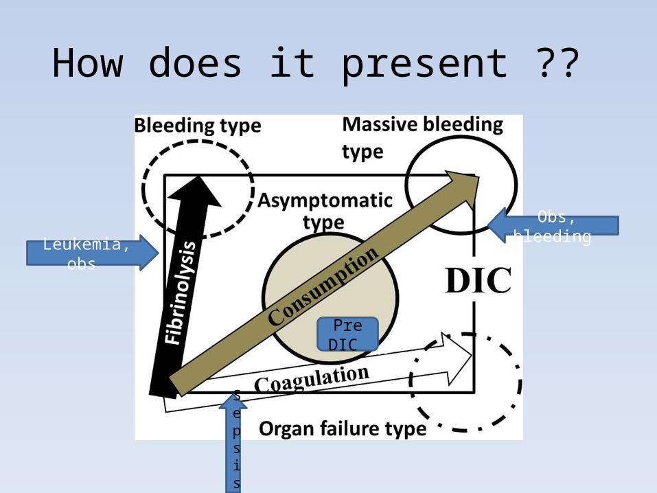
How does it present ??
Leukemia, obs
Sepsis
Obs, bleeding
Pre DIC

What is common ??
• Acute --- bleeding manifestations • Chronic --- thrombotic manifestations
• Setting of a DIC !!
• Sepsis – acute • Malignancy– chronic

Bleeding
• Bleeding is a more common clinical
presentation in patients with acute, fulminant
DIC.
• Petechiae, ecchymoses, epistaxis, gingival/
mucosal bleeding, hematuria, and bleeding
from wounds and puncture sites

Thrombosis- organs affected and symptoms
More chances
• Brain • Heart• Kidney
Less chances
• Adrenals• Spleen• Lungs• Liver

Lab diagnosis
• Thrombocytopenia is the hallmark of DIC, and its presence should prompt consideration of DIC. (platelet count <50 · 109/l)
• A low platelet count on initial testing and, in particular, a progressive drop in the platelet count are sensitive signs of DIC
• Thrombin induced platelet aggregation • Schistocytes on peripheral smear
Lab diagnosis
• FDP consists of both fibrin and fibrin degradation products.
• Elevated levels of FDP reflect accelerated fibrinolysis due to plasmin and are found in 85% to 100% of patients with DIC
• OC pills, infarction, renal diseases ?? Test validity ?? • Soluble fibrin monomer (SF) measurements offer
theoretical advantages in DIC

Lab diagnosis
• D-dimer levels are elevated in almost all cases of DIC,
but it may be falsely high in infection, inflammation,
pregnancy, and other conditions involving thrombosis.
• PT is prolonged in 50% to 75% of DIC cases
• aPTT is prolonged in 50% to 60% of patients
• Fibrinogen level is low 100– both PT and apTT
prolonged – acute phase reactant !!
Exclude heparin contamination

Diagnosis – in order !!
• Platelets ↓• FDPs - ↑• Prothrombin time ↓ • aPTT ↓• Fibrinogen ↓
• In a setting

• D dimer with fragmented red cells in a peripheral film
• In a chronic setting • Normal PT and apTT • May suggest DIC

TEG
• abnormal in septic patients, their diagnostic
sensitivity/specificity for DIC is unclear
• atypical light transmittance profile on the aPTT
has been associated with DIC-
• biphasic wave forms

A lot of such scores like that

DIC is dynamic

Management

1.Treatment of the cause
2.Blood component therapy – stop the cycle
3. Anticoagulation
1.Treatment of the cause
• Sepsis • OBG • Trauma • Malignancy?

Blood – Recommended in bleeding type of
DIC


No role for prophylaxis
Anticoagulation • There is no strong evidence to support the use of heparin
in DIC
• Shows a worse outcome in DIC patients who are treated
with heparin.
• Despite this controversy, it is commonly accepted to use
heparin in cases where thrombosis seems to predominate
(eg, purpura fulminans, solid tumors, hemangiomas, dead
fetus syndrome).
5 units / kg /hour ??

Presently, with the exception of Trousseau syndrome (occurrence of thrombophlebitis
migrans with visceral cancer), anticoagulation is not recommended as a treatment for DIC.

Anticoagulation
• successful use of recombinant factor VIIa in patients
with DIC and life-threatening bleeding.
• However, the efficacy and safety of this treatment in
DIC is unknown
• Antithrombin concentrate – labs better but
patients ??

Anticoagulation
• Consider treating patients with severe sepsis and DIC with recombinant human activated protein C (continuous infusion, 24 mic.g/kg/h for 4 d)
• Risk of bleeds • Aptt prolonged
Others
• Synthetic protease inhibitors, such as Gabexate
mesilate®and nafamostat®, exhibit multiple-
functions, including antagonistic effects on the
kinin/kallikrein system, fibrinolysis, complement
system, and coagulation system.

Tx – no role
• Patients with DIC that is characterized by a primary
hyperfibrinolytic state and who present with severe
bleeding could be treated with lysine analogues, such
as tranexamic acid (e.g. 1 g every 8 h)
• Possible promyelocytic leukemia

Comes for surgery
• Perioperative goals include maintaining the
platelet count in the range of 25,000–
50,000/μL and the fibrinogen level greater
than 50 mg/dL.

Summary
• What is DIC ?? • Causes • Clinical features • Lab diagnosis • Treatment

What does DIC mean
• DIC = “Death is Coming