displacement of mandibular removable partial denture bases
TRANSCRIPT

Displacement of MandibularRemovable Partial Denture
Bases by Tongue MovementsDuring Speech Klaus W. Boening, Dr Med Dent Habih
Purpose: The purpose of this study was to evaluate denture base displacement causedby tongue movements during speech. Materials and Methods: Ten patients with newKennedy Class I mandibular removable partiai dentures participated in this study. Allprostheses were made according to a standardized protocol. Denture base displacementswhen speaking a test sentence were measured with a computer-aided device that wasdeveloped for this purpose. It included 4 Hall sensors, mounted on a paraocclusaf splintand magnets, embedded in the denture hases. Vertical and transverse base displacementswere calculated from Hall voltages. Clasp retention was measured with a dynamometer.Results: Mean displacements were 145 (im seating, 72 pm unseating, 87 |jm buccally,and 74 jjm lingually. Seating and unseating displacements diltered statisticallysigniticantly in absolute values. Mean clasp retention was 2,5 N. No correlation wasfound hetween clasp retention and unseating base displacements. Conclusion: Tonguemovements during speech induced only minimal displacements in the denturesinvestigated. Additionally, tongue activity was found to be denture stabilizing ratherthan denture displacing in this study, IntJ Prosthodont 1999:12:147-152.
Missing posterior teeth are replaced with remov-able partial dentures (RPDi with the main pur-
poses of improving chewing ability, stabilizing thestomatognathic system, and preserving the support-ing tissues. It Í5 generally accepted that in mandibu-lar free-end prostheses the denture base extension isimportant for distributing functional forces as widelyas possible,' Bases should extend onto the retromo-lar pads and into the full functional depth of thebuccal and lingual sulci,^ Additionally, the lingual
'Associate Professor, Department of Prosthetic Dentistry, MedicalFacuity Carl Gustav Carus, Technical University of Dresden.Dresden, Germany.
Reprint lequests: Dr Klaus W. Boening, Department of ProstheticDentistry, Medical Faculty Carl Gustav Carus, Technical Universityof Dresden, Fetscherstrasse 74. 01307 Dresden, Germany. Fax:-f49-3Sl-4S8-43l2. e-maii: baening&'rcs.urz.tu-dresden.de
This paper was presented at the joint meeting of the InternationaiAssociation for Dentai Research/Continentai European DivisionNOF 12-15 September 1996, Berlin, Germany.
bar should be positioned to the functional depth ofthe lingual sulcus to avoid irritation of the lingualmarginal gingiva,^ Thus, tongue movements may in-terfere with a mandibular distal extension RPD,'
In a randomized clinical trial by Kapur,"* patientsreceived either RPDs or implant-supported fixedpartial dentures (FPDi in the mandible. Besides otherquestioned items, approximately 1 of 3 patientsfound their pronunciation poorer and 1 of 5 foundtheir rate of speech slower after RPD insertion. Incontrast, only 10% of the FPD patients encounteredthese speech difficulties. In different studies on treat-ment outcomes, between 12% and 23% of the par-ticipants complained that their mandibular RPDscaused phonetic problems,^-^ Other authors re-ported that only 2% of RPDs interfered withspeech,^ although it is not apparent whether thesecomplaints concerned the mandible or the maxilla.
Clinical studies have predominantly focused ondenture displacements during mastication or oc-clusal loading,''"'^ and research concerning speech
VolumeU, Numtier2,1999 1 4 7 Ttie International Journal of Prosthodontics

Displacement oi" Removable Piirtial Dentuie Bases During Speech
Paraocciusal splinf
Anterior pair of Hali sensors
Posterior pair of Hall sensors
Magnels
Amplifier
A'D boardcomputer
Fig 1 Experimental setup tormeasuring partial denture dis-placements in Ihe mandible.(a) Arrangement of mandibuiarremovable partial denture withembedded magnets and paraoc-ciusal splint with sensor pairs.(b) Sensor pair and the corre-sponding magnet. Tbe axes oftbe sensors and the magneticaxis are perpendicular to thedrawing plane. U,, L/j = Hall volt-ages; g, i> = distances betweensensor and magnet oeniers: d =distance between Hall sensorcenters; r - radius of themagnet.
difficulties with mandibular RPDs is rare in dentalliterature. No clinical data are available on man-dibular denture displacements during speech.
This study evaluated the iTiagnitudes of denturebase displacements by tongue activity duringspeech in bilateral distal extension RPDs. The nullhypothesis was that tongue activity during speechdoes not induce relevant denture displacements, ifcare is taken not to overextend the lingual denturebases. In this study RPDs with cast clasps werechosen because they are considered a suitable andaffordable prosthodontic treatment for the replace-ment of missing mandibular posterior teeth.'''-'^
Materials and Methods
Measuring Device
A computer-aided device was developed to meas-ure denture base displacements intraorally. FourHall sensors (KSY 10, Siemens) were applied as ananterior pair and a posterior pair to a paraocciusalsplint that was fixed to the anterior dentition (Fig 1).Two cyiindric magnets 3-0 mm in diameter (NdFeBmagnets, IBS) were embedded in the denture basesopposite the sensor pairs. The signals were ampli-fied, converted by a 12-bit analog/digital board
Tiie inlernationai ¡ournai of Prostiiodontji 148 • 12, Nimitjer2, 1999
Boening Displacement of Removable Partial Denture Bases During Speech
(Conrad Electronic), and stored in a personal com-puter. A program was written (Quick Basic 4.5,Microsoft) to control calibration procedures, to storedata, and to calculate denture base displacements.
The sensor pairs and their corresponding mag-nets were assembled to measure denture displace-ments as projections on the frontal plane (Eig 1b].With the magnetic axis and the perpendicular sen-sor axes being parallel, tbe distances g and h havean almost linear relationship to i/VHall voltage.Thus, the distances g and h can be calculated fromthe Hall voltages Í7, and U., by means of the fol-lowing equation:
1
- + b+r,„...
The polynomials a and b were determined fromsensor calibrations and r equals the magnet ra-dius. Calibration of the sensor/magnet units wascarried out on tbe table of a measuring micro-scope with micrometer gauges (Carl Zeiss). Frompreset distances between sensor and magnet andthe induced Hall voltages the polynomials in theequation were calculated. Erom d, g, and h (F;gIb) vertical and transverse denture base displace-ments at tbe anterior and the posterior sensor pairswere calculated. Displacements of the bases werecalculated as means from the values ot the ante-rior and the posterior sensor pairs. The accuracyof the system was evaluated in laboratory tests.'^A resolution better than 10 pm and an error ofmeasurement of less than 10% within a range of ±2 mm were determined.
Test Patients and Prosthodontic Treatment
Measurements of denture base displacements werecarried out on seven women and three men rangingin age from 31 to 68 years (mean 54 years). All pa-tients had intact anterior dentitions in the mandiblethat included at least the first premolars and ade-quate denture-bearing tissues. Seven patients hadalready worn RPDs in the mandible tor severalyears. These patients were rated "denture experi-enced" regarding the mandible, Tbe other three pa-tients had previously received mandibular RPDsand were rated "not denture experienced" regard-ing the mandible. Two patients wore maxillaryEPDs, five patients wore maxillary RPDs, and threepatients wore maxillary complete dentures.
The patients received new bilateral distal exten-sion mandibular RPDs, which were made by theauthor according to a standardized protocol. The
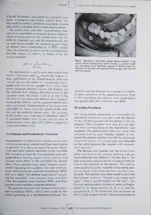
Fig 2 Mandibular removabie partial denture seated in linemouth shows paraocoiusal splint bonded to anterior teeth(a), and sensor pairs opposite magnets m denlure base (b).Sensors are secured wilh autopoiymerizing resin and coveredwith boxing wa^.
patients wore the dentures for a period of 3 monthsto allow adaptation of the supporting tissues. Priorto the recordings the patients were asked aboutany speech difficulties with their new RPDs.
Recording Procedures
Eor measurement preparation an irreversible hy-drocolloid impression was taken with the denturein situ and then poured with the denture in the im-pression. The 2 magnets were placed in the den-ture base corresponding to the mandibular rightquadrant. The paraocclusal splint was made fromautopolymerizing resin (Paladur, Kulzer). It con-tacted the anterior dentition but left the abutmentsuntouched (Fig 1 a). The sensor pairs were mountedon the splint opposite the magnets with autopoly-merizing resin.
The denture was inserted and the paraocciusalsplint was fixed to the anterior dentition with au-topolymerizing resin (Palavit G, Kulzer) (Eig 2). Thedata acquisition started with the recording of the in-traoral zero position of the sensors. The patientswere asked to keep the mandible in its resting posi-tion to ensure a recording free from any force com-ponents. The patients were then asked to articulatethe German sentence, "Harald zeigt hastig nachlinks" ("Harry points hastily to the left"). TheGerman pronunciation includes a variety of linguo-dental (S, Z), linguoalveolar (N, D, T, L), and lin-guopalatal (K, G, R) consonants that are known toeffect a high tongue activity. Labial and labiodental
Volume 12, Number 2, 1999 149 journal of Prosthodontics

Displacement of Removable Partial Denture Bases During Speech Boening
h I I) E 4. a? lunsestlng úlsplacemsnts)
- 1 o E .> 02 (sealing OisplacBi
4-1.0 E 4- O'¿ (lingual aispiacemenis)
/w\
v/- 1 0 E 4- 02 (Buccal displacements)
Fig 3 Recorded demure basedisplacement over time whilespeaking the test sentence (10Q-\im scale).
Table 1 Clasp Relention Based on 5 Measurementsin Each Patient and Maximum Denture BaseDisplacements During Speech
Clasp retention (N)Maximum unseating
displacement (pm]Maximum seating
displacement (jjm]Maximum buccal
displacement (|jm)Maximum lingual
displacement (jjm)
Mean
2.572*
145*
87
74
SD
2.276
78
42
67
Range
0.4-3.36-246
49-269
37-163
11-211
•Statistically signiSD = standard de
consonants (B, P, F, V), as weil as the vowels O andU, were avoided to minimize periorai muscie activ-ity and iience the risk of dislodging tiie spiint andthe sensors. Additionaiiy, tiie buccal mucosa washeid away from tiie splint with a dental mirror toprevent any disturbance during the recordings.
Aii recordings were repeated 3 times. From therecordings of each patient the maximum verticai andtransverse dispiacements were evaiuated. An exam-ple of the recorded dispiacements is shown in Fig 3.
Measurement of Clasp Retention
Cla5p retention was measured in the mandibularright quadrant according to Hosman." A ligaturewas connected to the iinguai bar directiy below theciasp and the arm of a dynamometer (Correx). Theclasp was loaded opposite the path of insertionuntii it dislodged. The measurements were repeated5 times.
Results
The individual measurements varied greatly withrespect to both clasp retention (range 0.4 to 8.2 N)and denture base displacement (6 to 269 (jm)(Tabie 1|, There was a significant difference be-tween vertical dispiacements toward the aiveoiarridge (seating dispiacements) and verticai displace-ments in the opposite direction (unseating dis-placements) (Wilcoxon's test for paired samples, P< 0,05), whereas the horizontai displacements didnot differ and were of a simiiar size as the unseat-ing dispiacements. No correlation was found be-tween ciasp retention and unseating dispiacement(Fig 4), None of the patients complained about5peech difficuities with their new dentures, al-though one patient without denture experience ad-mitted to not wearing his denture at ali because hefelt that he chewed better without it.
Discussion
Paraocclusal spiints are a suitabie tooi for preciseciinicai measurements in mandibuiar removablepartial dentures as proved in different studies."''^"^°Contrary to mastication, forces of the elevator anddepressor muscies during speech can be consideredreiativeiy iow. Forces generated by the tongue andthe periorai muscles are between 0.02 and 0,5N.̂ '̂̂ ^ Tbus, errors in measurements as a resuit ofbending of the mandibie are not to be expected.
Bias is a principai probiem in clinical measure-ments with intraorai appliances, aitering instinctiveand spontaneous function,^^ in tbe present study,measurement bias may have occurred for severalreasons. The patients wore a paraocciusai splint,and specific voweis and consonants in the German
lof Prosthodontics 150 Volume 12, Number 2, 1999

Boening Displatemeni oí Removable Partial Denture Bases During Speech
Fig 4 Scatter plot of clasp re-tention versus unseating denturebase displacement (Spearman'scorrelation coefficient = -0,13, P= 0,28, n = 10) Framed squaresindicate data from patients with-out denture experience.
sentence were chosen to achieve maximum tongueactivity but minimum perioral muscle activity. Fur-thermore, the buccal mtjcosa was retracted with adental mirror to ensure that tongue activity and itsinteraction with the denture would be the onlycause for displacements; this also prevented errorsin measurement as a result of mucosal pressure onthe splint. However, despite the absence of buccalpressure, which is known to counteract denture sta-bility,-'^- unseating as well as buccal and lingualdenture base displacements in this study wererather low and unlikely to be of clinical relevance.
In this study no patients complained about speechdifficulties, and no hint was given from the resultsthat dissimilar patient denture experiences influenc-ed the measurements (Fig 4), This supports Likeman'sconclusion-^ that the tongue can easily adapt to alingual obstruction consistent with denture wearing.Interestingly, the absolute values of vertical denturebase displacements during speech were significantlyhigher in the seating than in the unseating direaion.This indicates that tongue activity during speech ex-erts vertical force components on the denture basesand denture teeth that are higher in magnitude in theseating than in the unseating direction. Thus, underthe conditions of this study tongue activity duringspeech may be considered a denture-stabilizing fac-tor rather than a denture-displacing factor.
The amounts of seating displacements in thisstudy were 20% to 40% of those found at firm oc-clusal loadings," '- The soft tissue over the edentu-lous ridge assumes a different shape in functionand at rest because of changes in the shape of thecell walls under pressure.-''-'' If the cells changeshape easily under relatively light pressure butquickly reach the limit of their ability to changeunder further pressure, then most ofthe movementwill occur under a relatively light force.
Measured clasp retention and its wide variabil-ity were similar to other values reported in the lit-erature."^^^^ By correlating clasp retention andunseating displacements, it was assumed thathigh retentive forces might reduce unseating den-ture base displacements during speech. Althoughthe magnitudes of the measured displacementmay be in the range of play found between abut-ment teeth and clasps,'^ it was concluded thatdenture stability was independent of clasp reten-tion in this study.
Based on the limited conditions of this study,which included only 10 patients and one denturedesign, the resuits proved the null hypothesis thattongue activity during speech does not induce rele-vant denture displacements. Additionally, tongueactivity was found to be denture stabilizing ratherthan denture displacing in this study.
Volume 12, Number 2, 1999 151 The International loumal oi Prosthodontics
Displacement of Removable Partlnl Denlure BL1S05 During Speech
References
Academy ot Prostiiodontits, Principles, concepls, and priicticesin prostiiodontics—1994, J PrüSthel Denl 199S;73;73-94.Davenport |C, Basker RM, Haeth |R, Raiph JP, Removablepartiai dentures, Aylesbjry, England: Woife Medical, 198B:67,156,
Cecconi BT, Linguai-bar design, | Prosliiel Dent 1973;29:635-639,Kapur KK. Veteians Administrat ion Cooperative DentalImpiant Study, Comparisons between fixed partiai denturessupported by biade-vent implants and removable partial den-tures. Part iV: Comparisons of patient satisfaction betweentwo treatment modalities. I Prostbet Dent ]991;66:517-530,Watson CL, Reeve PE, Barnes E, Lane AE, Bates JF, The role ofpersonality in the management of partial dentures, | OralRehabii 1986;13:83-91,Nyhlin |, Gunne ]. Opinions and wearing habits among pa-tients new to removable partiai dentures. An interview study.Swed Dent] I989;I3:89-93.Frank RF, Miigrom P, Leroux BG, Hawiiins NR, Treatmentoutcomes with mandibuiar removable partial dentures: Apopulation based study of patient satisfaction. J Prosthet Dent
8, Carlsson GF, Hedegard D, Koivumaa KK, Studies m partialdenture prosthesis. IV: Final results of a 4-year longitudinaiinvestigation of dentogingivally supported partial dentures.Acta Odonlol Scand 196S;23:443-472,
9. Sheppard IM. Denture base disiodgment during mastication |Prosthet Dent 1963;13:462^68.
10, Cuiver PAJ, Watt i. Denture movements and controi. Br DentI 1973;13S;111-116,
11, Vahidi F. Vertical displacement of distal extension ridges hy dif-ferent impression techniques, ¡ Prosthet Dent 1978;40:374-377,
12, Leupold R|, Flinton RJ, Pfeifer DL. Comparison of verticalmovement occurring during ioading of dista I-extension re-movable partial denlure bases made by tbree impressiontechniques, J Proitliel Dent 1992;63:290-293,
13, Browning |D, Eick JD, McGarrah HE, Abutment tooth move-ment meaîured in vivo by using stereo photogram metry, |Prosthet Dent 1967;57:323-328,
14, Kapur KK, Deupree R, Dent R), Hasse AL, A randomized clin-icai trial of two basic removable partial denture designs. Parti: Comparison of five-year success rates and periodontalhealth, J Prosthet Dent 1994;72:499-512.
15. Kapur KK, Garret NR, Dent R], Haise AL, A randomized clini-cal trial of two basic removable partial denture designs. Part II:Comparison! of masticatory scores, | Prostliet Deril 1997;78:15-21,
16. Boening KW, Kinematii< der Gußklammerveranker tenFreiendprothûse im tJnteri<itier—Eine Klinische Studie,Habilitationsscbrift |thesis|. Medical Faculty, Technical Univof Dresden, 1997,
1 7, Hosman f i j . Influence of ciasp design of distai extension re-movable partiai dentures on tbe pericdontium of the abut-ment teetb, Int) Prosthodont 1990;3:256-265,
18, Bazirgan M, Bates JF. Preliminary study of a method of meas-uring removable partial denture abutment tooth movement invitro and in vivo, J Prosthet Dent 1986;56:204-207,
19, Eick JD, Browning |D, Stewart CD, McCarrah HE, Abutmenttooth movement related to fit of a removable partial denture, JProsthet Dent 1987;57:66-72.
20, Cbristidou L, OsborneJ, Chamberlain JB, The effects of partialdenture design on tbe mobility of abutment teeth. Br Dent |1973;135:9-18,
2 1 , Gould MSE, Picton DCA, An evaluation of a method of meas-uring forces exerted by the tongue on the teelh, Br Dent ]1963;114:175-I8O,
22, Likeman PR, The adaptation oí the tongue to denture wcar-ing,JDent 1979;7:]41-147,
23, Wesserman T, A concept of jaw function with a related clini-cal application, J Prostbet Dent 1963,13:130-140,
24, McCracken WL, A comparison of tooth-borne and tooth-tissue-borne removable partial dentures, ] Prosthet Dent1953;3:378-381,
25, Applegate OC, The partial denture base, J Prosthet Dent1955;5:636-64a,
26, Ahmad I, Sherrif M, Waters NE. The effect of reducing thenumber of clasps on removabie partiai denture retention. IPro5thetDent1992;68:928-933.
27, Firteil DN, Effect of ciasp design upon retention of removablepartial dentures, | Prosthet Dent 1968;20:43-S2,
28, Ghani F, Mahood M. A laboratory examination of the behaviourof cobalt-chromium clasps, | Oral Rehabil 1990;l 7:229-237,
The I n terna ri on a I loumal of Pruitbiidoniics 152 Volume 12, Number 2,1999
