dislocations of shoulder dr.guru prasad
TRANSCRIPT

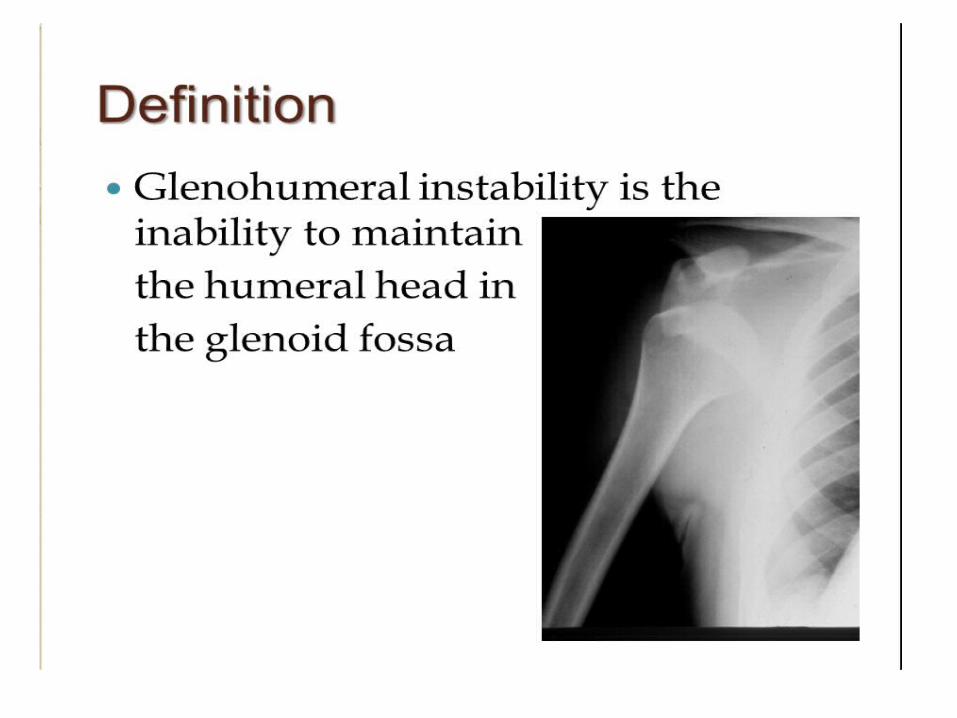
Shoulder dislocation
Dr. Guru prasadDnb orthopaedics

synopsis
• Definition• Anatomy – pathoanatomy• Types – anterior/posterior/inferior• Clinical features – Tests for evaluation • Management -- Reduction techniques• Complications
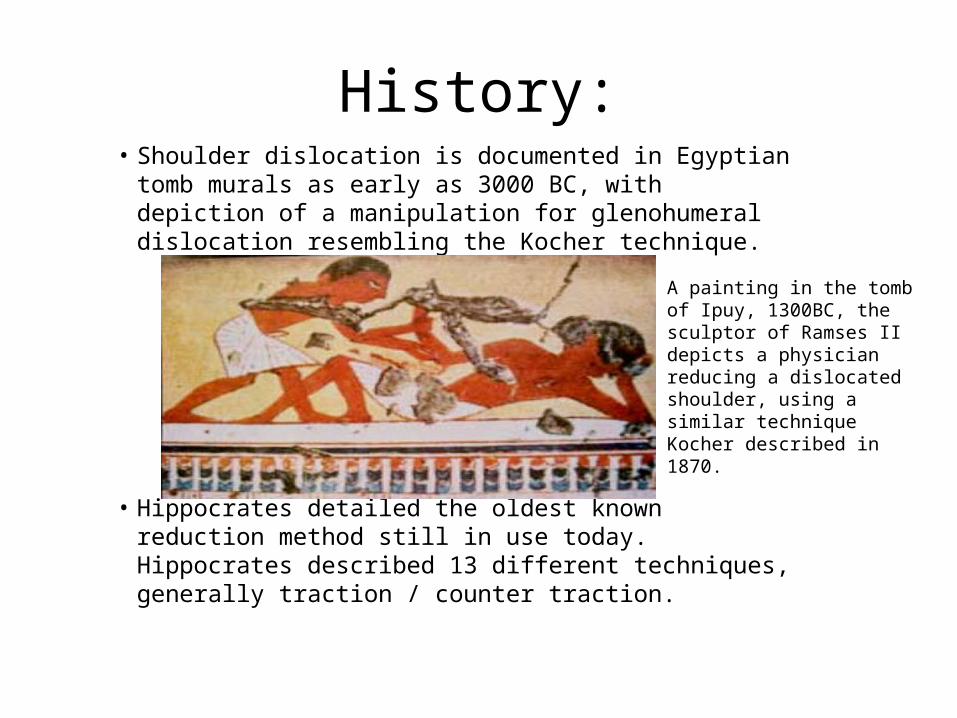
History:• Shoulder dislocation is documented in Egyptian tomb
murals as early as 3000 BC, with depiction of a manipulation for glenohumeral dislocation resembling the Kocher technique.
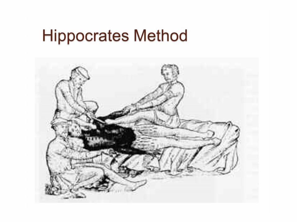
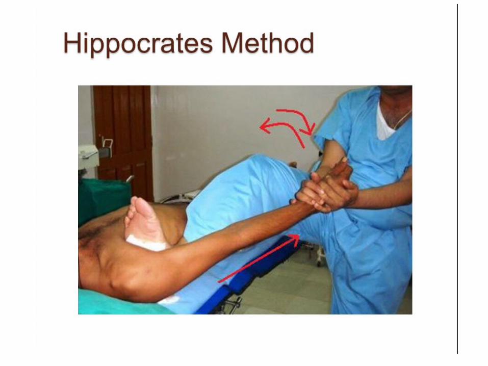
• Hippocrates detailed the oldest known reduction method still in use today. Hippocrates described 13 different techniques, generally traction / counter traction.
A painting in the tomb of Ipuy, 1300BC, the sculptor of Ramses II depicts a physician reducing a dislocated shoulder, using a similar technique Kocher described in 1870.

- SURGICAL ANATOMY - Static stabilizers- anatomic constraints to shoulder motion - Dynamic stabilisers – normal physiological function creates a
stabilizng effect - BONY ANATOMY – has minimal constraints – large range of
motion Scapula – hence glenoid anterverted 30 – 40 Humeral head retroverted ¼ of head articulates with bony glenoid LABRUM – one of the most important stabilizing structures . - Effectively enlarges & deepens the glenoid surface by 1 cm &
50% respectively .- Increases humeral contact to 75 %
• CAPSULE : loose and redundant at most positions - At extremes – tightens , provides stability . • GLENOHUMERAL LIGAMENTS - Superior , middle , inferior • CORACOHUMERAL LIGAMENT – -Passes in the rotator interval ( between subscapularis &
supraspinatus )

DYNAMIC STABILIZERS • ROTATOR CUFF MUSCLES :
- BICEPS TENDON : along the rotator interval • CORACO-ACROMIAL ARCH limits superior translation


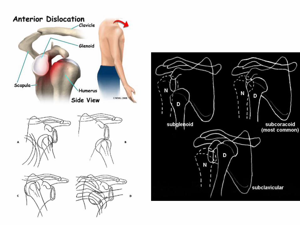

Anterior:• Subcoracoid (anterior):
– Humeral head sits anterior and medial to the glenoid, just inferior to the coracoid.
– ~ 60% of cases.
• Subglenoid (anteroinferior):– humeral head sits inferior
and slightly anterior to the glenoid, that the humeral head has also travelled medially.
– ~ 30% of cases.


• MECHANISM OF INJURY : • DIRECT – less common
INDIRECT – more common • Susecptible position – • Anterior dislocation : abd. , ext. rotation & extension• Post. Dislocation : flexion, add . & int. rotation • In seizures & electrical shock – all the muscles are contracted
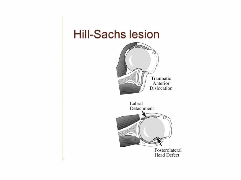
, ext. rotators overpower int. rotators • ASS0CIATED INJURIES : • Humeral head & neck # , tuberosity # , glenoid # , rotator
cuff tears( older age) , neurological ( axillary N.) and vascular injuries
• Any pt. with weakness after shoulder dislocation must be evaluated for rotator cuff tear

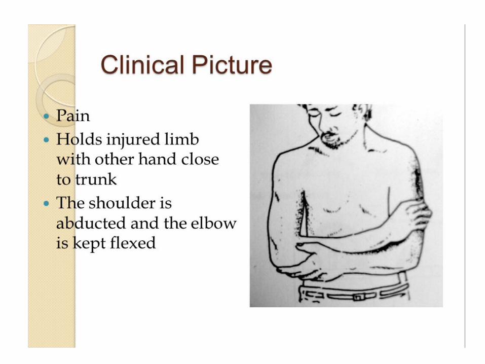
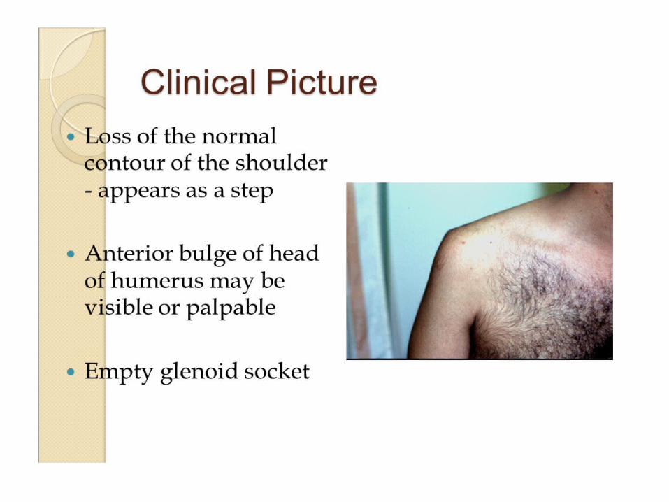


• Presentation of acute dislocation • Pain , typical attitude , • In ant . Dis- limitation of int. rotation & abd.• In post. Disl. – “ of ext. rotation • In inferior disl. – in fully abducted position • PHYSICAL EXAMINATION • Flattening of shoulder • Fullness in delto-pectoral area• Axillary fold at lower level• Able to insinuate finger beneath acromion • dugas test • Hamilton ruler test
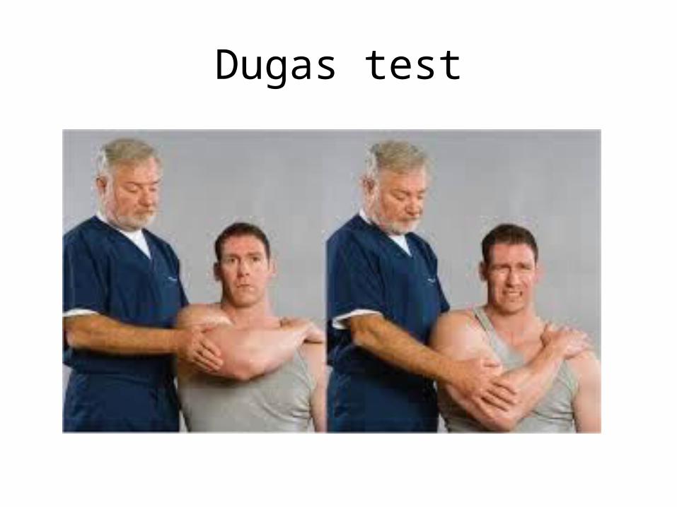
Dugas test

Hamilton ruler test

SULCUS TEST :

• Axillary N. tested for both sensory & motor components

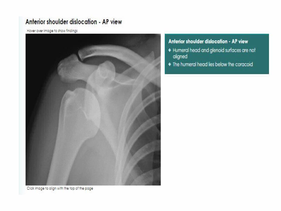
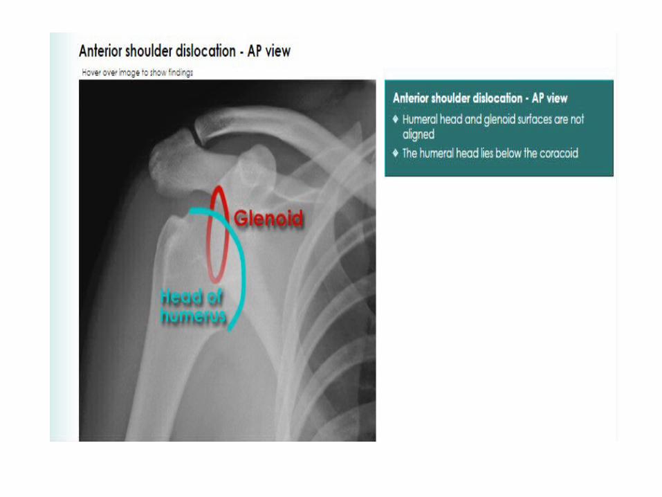
X-Ray
• The anteroposterior x-ray will show the overlapping shadows of the humeral head and glenoid fossa, with the head usually lying below and medial to the socket.


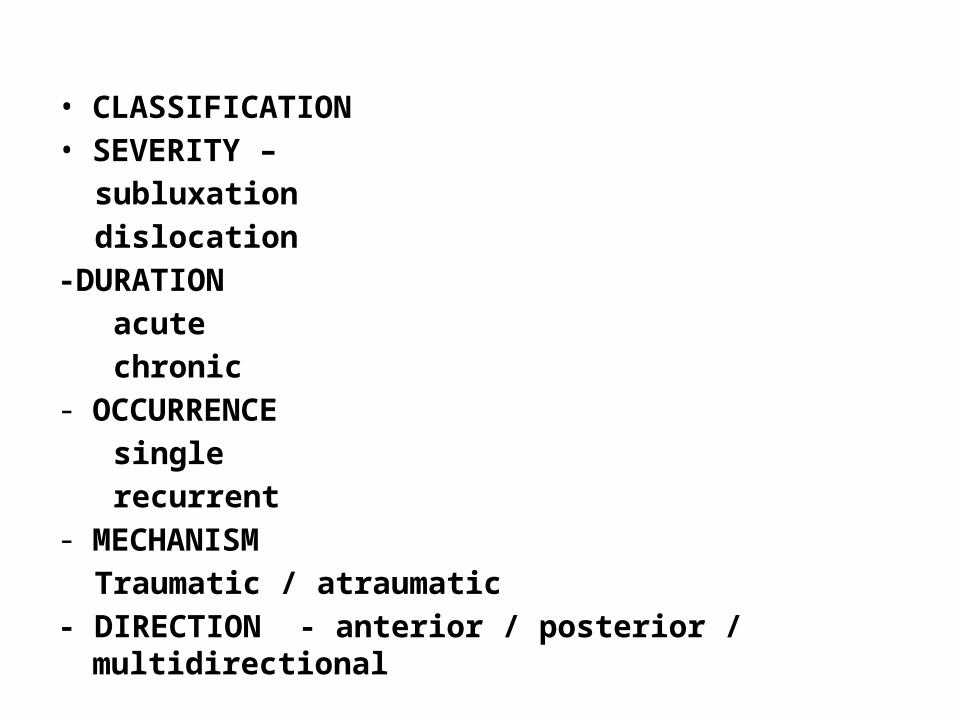
• CLASSIFICATION • SEVERITY – subluxation dislocation -DURATION acute chronic - OCCURRENCE single recurrent - MECHANISM Traumatic / atraumatic - DIRECTION - anterior / posterior / multidirectional


• NONOPERATIVE TRAETMENT • CLOSED REDUCTION FOR ACUTE DISLOCATION- Under i.v. analgesia + sedation - Under intra-articular lignocaine - If initial closed reduction unsucessful , degree of sedation &
analgesia evaluated , if not successful , under G.A for closed / open reduction


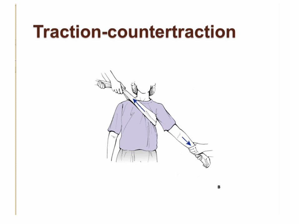
Traction-countertraction• Note how the clinician on
the left has the sheet wrapped around him, allowing him to use his body weight to create traction. Some clinicians employ gentle external rotation to the affected arm while providing traction.


Stimson technique • The patient is placed
prone on the stretcher with the affected shoulder hanging off the edge. Weights (10-15 lbs) are fastened to the wrist to provide gentle, constant traction.


Scapular manipulation • The patient sits upright and
leans the unaffected shoulder against the stretcher. The physician stands behind the patient and palpates the tip of the scapula with his thumbs and directs a force medially. The assistant stands in front of the patient and provides gentle downward traction on the humerus as shown. The patient is encouraged to relax the shoulder as much as possible.

Milch technique • The arm is abducted
and the physician's thumb is used to push the humeral head into its proper position. Gentle traction in line with the humerus is provided with the physician's opposite hand.

Spaso technique • The arm is flexed
forward and gentle traction and external rotation forces are applied.
Management
• The arm is rested in a sling for about three weeks in those under 30 years of age (who are most prone to recurrence) and for only a week in those over 30 (who are most prone to stiffness).
• Then movements are begun, but combined abduction and lateral rotation must be avoided for at least 3 weeks.
• Throughout this period, elbow and finger movements are practised every day.

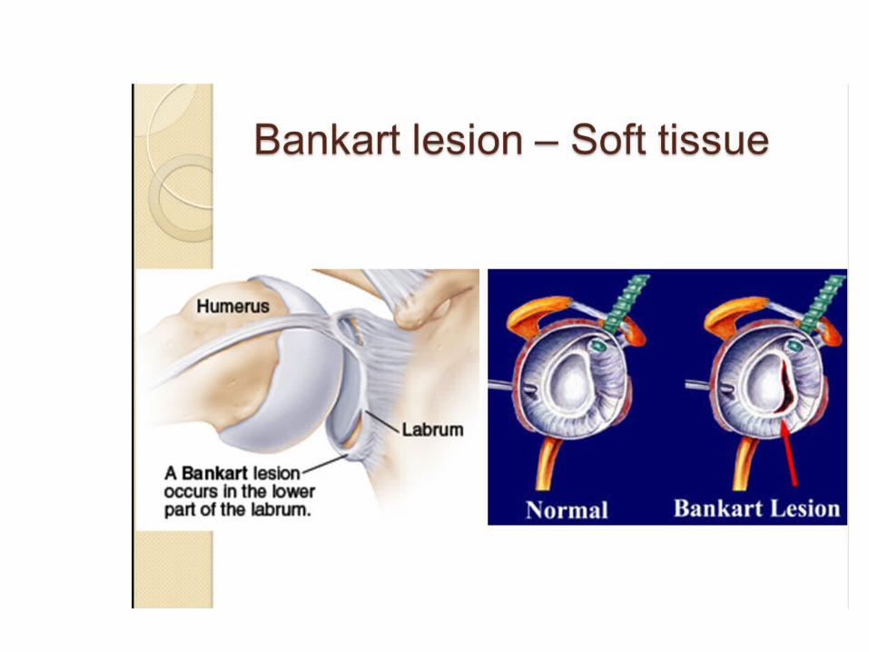
• OPERATIVE TREATMENT • SURGICAL STABILISATION FOR ANT . INSTABILITY in – failed appropriate nonoperative treatment - recurrent dislocation at young age - irreducible dislocation - open dislocation - unstable joint reduction - 1st dislocation in young pt with high demand activity surgical options - arthroscopic surgery - open tech. with soft tissue repair - open tech. with bony augmentation

• Open procedures
• BANKART OPERATION - MC performed surgery - Ant. Labral defect identified , mobilized & reattached to
original anatomic site with suture anchor . - Capsular reconstruction also recommended - Subscapularis tendon is split at junction of upper 2/3rd &
lower 2/3rd
BRISTOW OPERATION
- Suturing of coracoid process with the conjoint tendon to the ant. Portion of scapular neck through a transversely sectioned subscapularis M.
- The transferred short head of biceps & corachobrachialis – strong buttress across the anterior & inferior aspects of joint
- Tendon also holds the lower half of subscapularis M. thus prevents slipping over the humeral head when abducted
• PUTTI-PLAT OPERATION
- Subscapularis and capsule incised vertically - Lateral leaf sutured to the labrum & medial leaf imbricated - Subscapularis is advanced laterally - Gross limitation of ext. rotation - Rarely indicated

• ARTHROSCOPIC PROCEDURES • ADVANTAGES & DISADVANATAGES
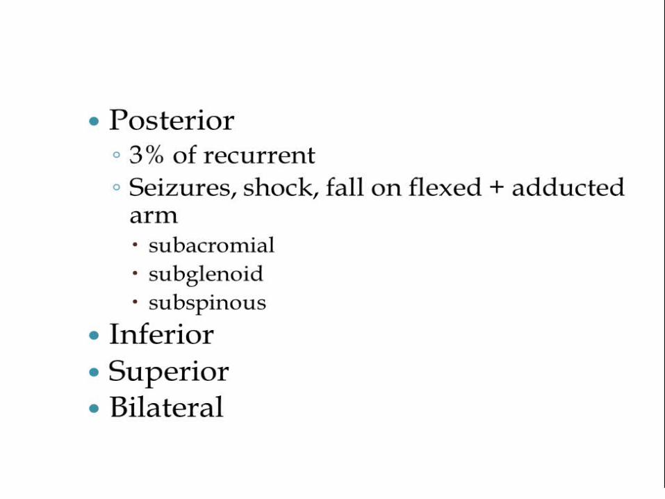
POSTERIOR DISLOCATION OF THESHOULDER
• Posterior dislocation is rare, accounting for less than 2% of all dislocations around the shoulder.

Clinical features
• The diagnosis is frequently missed – partly because reliance is placed on a single anteroposterior x-ray (which may look almost normal) and partly because those attending to the patient fail to think of it.
• There are, in fact, several well-marked clinical features.
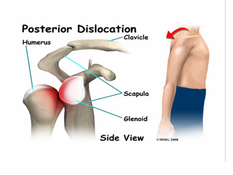

• The arm is held in internal rotation and is locked in that position.
• The front of the shoulder looks flat with a prominent coracoid, but swelling may obscure this deformity; seen from above, however, the posterior displacement is usually apparent.

X-Ray
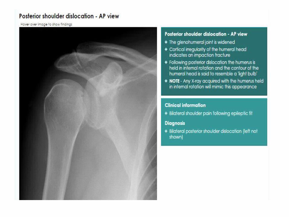
• In the anteroposterior film the humeral head, because it is medially rotated, looks abnormal in shape (like an electric light bulb) and it stands away somewhat from the glenoid fossa (the ‘empty glenoid’ sign).
• An axillary view is essential; it shows posterior subluxation or dislocation and sometimes a deep indentation on the anterior aspect of the humeral head.


Posterior shoulder dislocation reduction
• The underlying approach to the traction-countertraction technique demonstrated in this photograph is similar to that employed in the reduction of anterior dislocations. The notable difference is positioning. Note that the patient is upright and the clinician providing traction is standing in front of the patient.
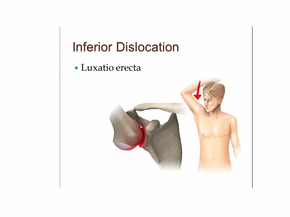
INFERIOR DISLOCATION OF THESHOULDER (LUXATIO ERECTA)
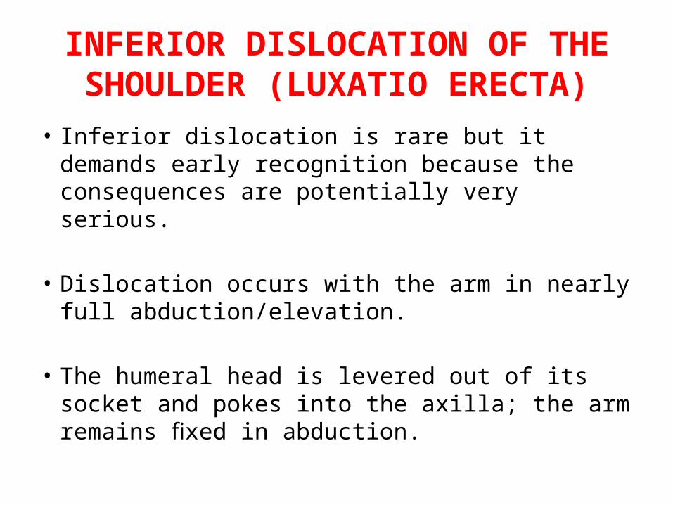
• Inferior dislocation is rare but it demands early recognition because the consequences are potentially very serious.
• Dislocation occurs with the arm in nearly full abduction/elevation.
• The humeral head is levered out of its socket and pokes into the axilla; the arm remains fixed in abduction.
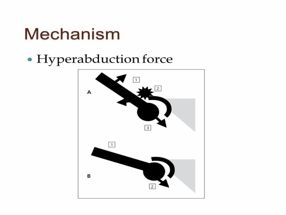

Mechanism of injury and pathology
• The injury is caused by a severe hyper-abduction force.
• With the humerus as the lever and the acromion as the fulcrum, the humeral head is lifted across the inferior rim of the glenoid socket; it remains in the subglenoid position, with the humeral shaft pointing upwards.

• Soft-tissue injury may be severe and includes avulsion of the capsule and sur-rounding tendons, rupture of muscles, fractures of the glenoid or proximal humerus and damage to the brachial plexus and axillary artery.

Clinical features
• The startling picture of a patient with his arm locked in almost full abduction should make diagnosis quite easy.
• The head of the humerus may be felt in or below the axilla.
• Always examine for neurovascular damage.


X-ray
• The humeral shaft is shown in the abducted position with the head sitting below the glenoid.
• It is important to search for associated fractures of the glenoid or proximal humerus.
– NOTE: True inferior dislocation must not be confused with postural downward displacement of the humerus, which results quite commonly from weakness and laxity of the muscles around the shoulder, especially after trauma and shoulder splintage; here the shaft of the humerus lies in the normal anatomical position at the side of the chest.
– The condition is harmless and resolves as muscle tone is regained.
Treatment
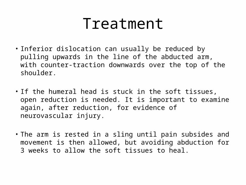
• Inferior dislocation can usually be reduced by pulling upwards in the line of the abducted arm, with counter-traction downwards over the top of the shoulder.
• If the humeral head is stuck in the soft tissues, open reduction is needed. It is important to examine again, after reduction, for evidence of neurovascular injury.
• The arm is rested in a sling until pain subsides and movement is then allowed, but avoiding abduction for 3 weeks to allow the soft tissues to heal.


Complications
• EARLY COMPLICATIONS• Rotator cuff tear: – This commonly accompanies anterior dislocation,
particularly in older people.
– The patient may have difficulty abducting the arm after reduction; palpable contraction of the deltoid muscle excludes an axillary nerve palsy.
– Most do not require surgical attention, but young active individuals with large tears will benefit from early repair.

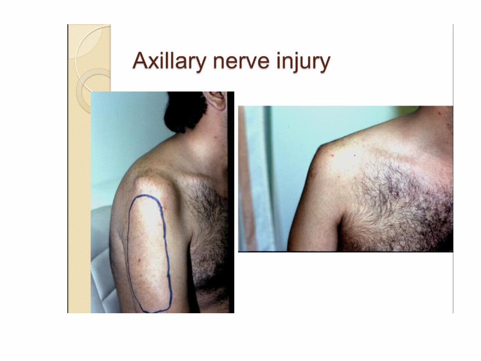
• Nerve injury: – The axillary nerve is most commonly injured; the patient is
unable to contract the deltoid muscle and there may be a small patch of anaesthesia over the muscle.
– The inability to abduct must be distinguished from a rotator cuff tear.
– The nerve lesion is usually a neuropraxia which recovers spontaneously after a few weeks; if it does not, then surgery should be considered as the results of repair are less satisfactory if the delay is more than a few months.

– Occasionally the radial nerve, musculocutaneous nerve, median nerve or ulnar nerve can be injured.
– Rarely there is a complete infra-clavicular brachial plexus palsy.
– This is somewhat alarming, but fortunately it usually recovers with time.

• Vascular injury:– The axillary artery may be damaged, particularly in
old patients with fragile vessels.
– This can occur either at the time of injury or during overzealous reduction.
– The limb should always be examined for signs of ischaemia both before and after reduction.

• Fracture-dislocation – If there is an associated fracture of the proximal humerus,
open reduction and internal fixation may be necessary.
– The greater tuberosity may be sheared off during dislocation.
– It usually falls into place during reduction, and no special treatment is then required.
– If it remains displaced, surgical reattachment is recommended to avoid later subacromial impingement.

• LATE COMPLICATIONS• Shoulder stiffness
– Prolonged immobilization may lead to stiffness of the shoulder, especially in patients over the age of 40.
– There is loss of lateral rotation, which automatically limits abduction.
– Active exercises will usually loosen the joint.
– They are practised vigorously, bearing in mind that full abduction is not possible until lateral rotation has been regained.
– Manipulation under anaesthesia or arthroscopic capsular release is advised only if progress has halted and at least 6 months have elapsed since injury.
• Recurrent dislocations


• Unreduced dislocation – Surprisingly, a dislocation of the shoulder sometimes
remains undiagnosed.
– This is more likely if the patient is either unconscious or very old.
– Closed reduction is worth attempting up to 6 weeks after injury; manipulation later may fracture the bone or tear vessels or nerves.
– Operative reduction is indicated after 6 weeks only in the young, because it is difficult, dangerous and followed by prolonged stiffness.
–

• Thank you all…