diseases of the tubules and interstitium
TRANSCRIPT

1
DISEASES OF THETUBULES ANDINTERSTITIUM
Glen Markowitz, M.D.Gerald Appel, M.D.
Mechanisms ofTubulointerstitial Disease
2 general categories: Ischemic/toxic (non-
inflammatory) Acute tubular necrosis
Inflammatory Tubulointerstitial nephritis
Infection, allergic/drug-induced,systemic disease (i.e. Sarcoid,Sjogren’s), etc

2

3

4
Case 1
A 58 year old W M with a history of ETOHabuse , but normal renal function on ERvisit 2 months ago, is admitted to thehospital in a stuporous conditionhaving been found by his friend in hisroom to be unarousable. The friendstates that they had been drinking 3days ago and when he now called forhis drinking buddy there was an emptybottle of Jack Daniels next to him.
Case 1
PE: BP 100/60 mm Hg, P 110, R12, Temp 101,Cor -, Chest rales at R base, Abd-, Ext swellingand tender R and L legs below the knee.
Lab: BUN 48 mg/dl, Creatinine 6.2 mg dl, CBC –wbc 15, 000, with increased polys, Cxray RLLinfiltrate.
U/A tr prot, 4+ heme, no rbc or wbc.Pt is hydrated with 1 L Saline and BP 135/82.
Given 150 mg Gentamicin and 1g Ampicillin.Over the next 2 days pt makes little urine and
creatinine rises to 8.4 mg/dl.

5
Case 1 Should a kidney biopsy be done? Is the renal failure acute or chronic?
How do you know? How can you proveit?
What is the likely etiology of the renalfailure (hypotension, rhabdomyolysis,gentamicin, leptospirosis )
What lab tests might confirm thediagnosis?

6
Acute renal Failure Prerenal azotemia Post-renal azotemia Acute tubular necrosis Acute interstitial nephritis Acute glomerulonephritis Vascular ARF

7
8
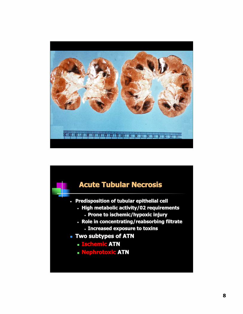
Acute Tubular Necrosis
• Predisposition of tubular epithelial cell• High metabolic activity/02 requirements
• Prone to ischemic/hypoxic injury• Role in concentrating/reabsorbing filtrate
• Increased exposure to toxins
Two subtypes of ATN Ischemic ATN Nephrotoxic ATN

9
Ischemic ATN
• O c c u r s i n s e t t i n g o f d e c r e a s e d r e n a l b l o o d
fl o w / h y p o t e n s i o n , i . e . t r a u m a / s e v e r e b l o o d
l o s s , C H F , s e p t i c s h o c k
• P a t h o l o g y
- G r o s s : P & S
- D e g e n e r a t i v e c h a n g e s
- S u b s e q u e n t r e g e n e r a t i v e c h a n g e s
- M o s t s e v e r e c h a n g e s i n p r o x i m a l t u b a n d
m T A L
Clinical Phases of ATN
Initiation- first 36 hours, dominated by initial event
Maintenance- up to 3 weeks, oliguric, dialysis required
Recovery (“diuretic phase”)- increasing urine output – often substantial, electrolyte abnormalities
Prognosis: > 90% recovery if survive initiating event

10

11
Nephrotoxic ATN
Many toxins implicated Heavy metals: Hg, Pb, gold, arsenic,… Organic solvents: CCl4, ethylene glycol Therapeutics
antibiotics: gentamicin antifungals: amphotericin B chemotherapeutic agents: cisplatin bisphosphonate: zoledronate radiation & radiocontrast pigments: Hgb, Mgb abnormal levels of physiologic substances osmotic agents: mannitol

12
Nephrotoxic ATN
Similar pathology to ischemic ATN Additional, toxin-specific findings:
- Ethylene glycol- Osomotic agents/radiocontrast- Light chains- Hemoglobin/Myoglobin

13

14
Case 2 65 y o retired Ob-Gyn MD refer by NYC
nephrologist for presumed RPGN Past Hx HBP x 40 yrs controlled on
meds, arrhythmia →verapamil,hypothyroidism
Some urinary urgency 1 wk PTA –Urologic check ( U/A neg ) ; Urticarialrash on legs several days PTAdisappeared.
1 wk PTA gave blood and played golf. Not feeling well , thirsty. Check chem7
Case 2 BUN 94, creat 4.4 mg/dl Friend nephrologist – BUN 91 creat. 4.9
mg/dl K+ 6.7 , alb 4.1 WBC 8.4, Hct 36,Plts 441, U/A some rbc , no casts, ANA- ,ASLO 33, UIF normal pattern, Ccr 32cc/min
Meds calan SR, PPI, zoloft, synthroid,cozaar – given Kayexolate →CPMC ? BX
Px BP 170/90, P82, Cor-Chest-Abd- neg,no edema , fine maculo-papular rash onchest and upper arms.
BUN 96 creat 5.1 U/A tr prot, 2+ heme, nocasts

15
Case 2 What is the differential Dx of the
acute renal failure? What labs help or hurt each
diagnosis? Would you biopsy now? Wait and
then consider biopsy if norecovery? Treat and then biopsy ifno recovery? ( What therapy iftreating? )

16
Drug-InducedInterstitial Nephritis
Pathogenesis: cell-mediatedhypersensitivity reaction (T’s)
Pathology interstitial inflammation & edema EOSINOPHLS Tubulitis +/- granulomas

17

18

19
Medication AssociatedAINBeta-Lactam AntibioticsOther Antibiotics- Sulfonamides TM-SMX Rifampin QuinolonesDiureticsNSAIDSOther Drugs – Cimetidine, Dilantin, Sulfinpyrazone, AllopurinalProton Pump Inhibitors
Course Patient 2 D/C all meds – use alts for HBP
and ulcer disease Prednisone 120 mg QOD x 6 wks Plasma creatinine decreased from
5.1 to 1.8 mg/dl Stable RFTs 4 yr later

20
Case 3
A 64 yo BF has had diabetes and mildHBP for 6 yrs. Her BS has recently beenpoorly controlled and and she has hadpolyuria and nocturia. Recently shenoticed dysuria and frequency as well.
She develops fever, chills , and leftflank pain which increases over 24 hrs.She calls her MD who send her to theER immediately.
Case 3
In the ER her BP is 110/72 , P 100, Temp102,R14. She has marked L CVA tenderess.
BUN 35 mg/dl, Creatinine 1.4 mg/dl WBC – 16,500, Hct 39%, platelets nl. U/A shows 3+gluc, 2+ heme, tr alb, 10-15 rbc,
wbc –TNTC, and wbc casts. Urinary Na+ is 42 mEq/L. FENa+ is 1.8. USG shows no hydronephrosis (obstruction )
but a stone in L kidney. She is treated with hydration, amp, gent. Over the next 24 hours her BP incr to 145/82,
temp 100, and urine output remains copious. BUN decrease to 14 mg/dl and creatinine to 0.7
mgl/dl.

21
22

23
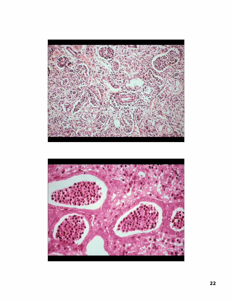
Acute Pyelonephritis
Acute suppurative infection of kidney Clinical: back pain, fever, pyuria, +/- RI
Urine cultures: confirmation / Ab sensitivity Route of infection
ascending > hematogenous ascending starts in bladder as UTI (F>M) hematog: septic emboli, bacteremia (F=M)
Organisms 85% gram negative bacilli (#1 E. coli) fecal flora

24
Acute Pyelonephritis
Increased risk of ascending infection inthree clinical settings Obstruction: BPH, tumors, pregnancy,
neurogenic bladder (DM) Instrumentation Vesicoureteral reflux
50% UTI’s in 1st year of life congenital anomaly: intravesical
portion of ureter lacks normal obliquecourse that prevents reflux

25
Acute Pyelonephritis
Gross: normal size, +/- coalescent abscesses Micro: severe inflammation, PMN’s
Microabscesses PMN casts & tubulitis Distribution:
Ascending: originates near medulla Hematogenous: cortical
Chronic Pyelonephritis
Definition: chronic renal disorder withscarring, inflammation, and deformity ofcalyces/pelvis (ascending*)
Gross: shrunken Irregular, asymmetric broad/flat scars (U*) Papillary blunting and calyceal deformity
Micro: Disproportionate tubulointerstitial scarring Atrophic tubules with colloid casts
(“thyroidization”) Chronic inflammation (not PMN’s)

26
Chronic Pyelonephritis
Clinical insidious onset of RI +/- HTN, mild proteinuria, decreased
urinary concentration, culture neg Rarely follows “usual” acute pyelo More common with persistent
obstruction or VUR +/- awareness of acute episodes Rx: relieve obstruction / correct VUR,
antibiotics as indicated

27
U

28

29
Voiding
cystourethrogram
Vesicoureteralreflux (VUR):
- Congenital
- 50% UTI’s < 1yo

30
Case 4
A 52 yo F has had rheumatoid arthritis for 20yrs and has been taking aspirin, tylenol, andNSAID’s daily but no other medications forher disease. She develops R flank pain, butno fever , chills, or dysuria. Physicalexamination shows marked deformities ofher joints but no edema.
Labs: U/A tr protein, few rbc and many wbc.BUN 32 mg/dl, Pcreatinine 2.4 mg/dl, 24 hrprtoein 0.4 g/d, and negative or normal testsfor complement, anti-DNAantibody, HBV, BS,HCV, etc. Urine culture is “no growth” after 2days.
An Intravenus Urogram is performed.

31
Case 4
Is bacterial pyelonephritis thecause of this patients back pain?
What are other possible causes? What other diseases could cause
this picture?

32
Analgesic Nephropathy
An international disease (Australia, Switzerland,Scandinavia, USA)
Abusers and Users – Headaches and ArthritisFemale:Male 6:1Large amounts over prolonged time periodsRenal abnormalities
sterile pyuria only mild proteinuria and hypertension Decreased concentration ability Decreased net acid excretion Salt wasting Papillary necrosis
Patients can recover function if they stop analgesic use

33
Causes of Papillary Necrosis
Obstructive pyelonephritis Sickle Cell Anemia
medulla leads to sickling sickling leads to medullary ischemia
Analgesic abuse (phenacetin*) increased risk with combinations direct toxicity and ASA-induced PG
deficiency
Diabetes Mellitus
NSAIDs
Multiple patterns of renal disease Acute interstitial nephritis Acute tubular necrosis
Loss of PG vasodilation / precip ATNin the setting of volume depletion
Minimal change disease (rarely MG) Papillary necrosis
Same nephrotoxicity for COX-2inhibitors