directory of services - ap2
TRANSCRIPT

DIRECTORY OF SERVICES
REVISED JUNE 2019

2 DIRECTORY OF SERVICES

DIRECTORY OF SERVICES 1
GENERAL COMPANY OVERVIEW ................................................................................................ 3
COMPANY CONTACT INFORMATION ........................................................................................ 4
ACCREDITATION AND LICENSURE ............................................................................................. 5
CORPORATE AND MEDICAL STAFF ............................................................................................ 6
RESULTS AND REPORTING ............................................................................................................ 6
CASE MANAGEMENT, TEST ADD-ONS AND CANCELLATIONS ......................................... 7
INFORMATION TECHNOLOGY ....................................................................................................... 7
INSURANCE AND BILLING SERVICES ........................................................................................ 8
GENERAL SPECIMEN LABELING AND SUBMISSION ........................................................... 9
SURGICAL PATHOLOGY ................................................................................................................ 10Cervical, Endocervical and Endometrial ....................................................................................... 10Cone and LEEP Conization of Cervix .............................................................................................. 10Endoscopic........................................................................................................................................ 10Skin Excisions ................................................................................................................................... 10Breast Tissue ......................................................................................................................................11Prosthetic Breast Implants ...............................................................................................................11General - Tissue Specimens ........................................................................................................... 12
IMMUNOHISTOLOGY, CYTOCHEMISTRY, MOLECULAR PATHOLOGY ........................... 12Immunohistology ............................................................................................................................. 12Molecular Oncology ......................................................................................................................... 13Chromosomal Studies and Electron Microscopy ......................................................................... 13
HEMATOPATHOLOGY AND FLOW CYTOMETRY .................................................................... 14Flow Cytometry ................................................................................................................................ 14How to Submit a Bone Marrow Biopsy and Aspirate ................................................................... 15How to Submit Tissue for Lymphoma (Lymph Node/Solid Tissue) Workup ............................... 16
INFECTIOUS DISEASE TESTING ................................................................................................. 17UniSwab™ ........................................................................................................................................ 17ThinPrep® and SurePath™ .............................................................................................................. 17Group B Streptococcus Swab ......................................................................................................... 18Aptima® Multi-test and Unisex Swab ............................................................................................. 18 Aptima® Urine ................................................................................................................................... 18
CYTOPATHOLOGY (NON-GYN) .......................................................................................19Induced Sputum and Bronchial Washings for Pneumocystis Pneumonia ................................ 19 Bronchial, Colonic, Esophageal and Gastric Washings ............................................................... 19 Sputum ............................................................................................................................................. 19 Urine ................................................................................................................................................. 20 Nipple Secretions and Smears ....................................................................................................... 21 Cerebral Spinal Fluid ....................................................................................................................... 22
CONTENTS
DIRECTORY OF SERVICES 1
CONTENTS

2 DIRECTORY OF SERVICES
CYTOPATHOLOGY – FINE NEEDLE ASPIRATION ..............................................................................22Thyroid/Parathyroid FNA – Thyroglobulin Assay Option (UniPath Only) ...............................................25
CYTOPATHOLOGY – GYNECOLOGY .....................................................................................................26ThinPrep® Pap Tests .................................................................................................................................... 26SurePath™ Pap Tests .................................................................................................................................. 27Human Papillomavirus (HPV) Testing .......................................................................................................28HPV and Pap Co-Testing .............................................................................................................................28Pap Smears for Diethylstilbestrol (DES) Evaluation ................................................................................29 Conventional Method ............................................................................................................................29 Liquid Based Method .............................................................................................................................30
THE BETHESDA2014 SYSTEM FOR REPORTING CERVICAL/ VAGINAL CYTOLOGIC DIAGNOSES .............................................................................................................31
PAP SMEAR COMMENTS – THE BETHESDA2014 SYSTEM...............................................................32
PEDIATRICS – PLACENTA SERVICE ......................................................................................................35
PEDIATRIC/PERINATAL AUTOPSIES .....................................................................................................36
APPENDICES ................................................................................................................................................40Appendix 1: Immunopathology Library of Antibodies ........................................................................... 41Appendix 2: Immunohistochemistry/Molecular Prognostic Markers.................................................... 45Appendix 3: Special Stains .........................................................................................................................46Appendix 4: Hematopathology Diagnostic Profiles .................................................................................47Appendix 5: Molecular Test Menu..............................................................................................................48Appendix 6: Flow Cytometry Menu ...........................................................................................................49
Contents, Cont.
CONTENTS

DIRECTORY OF SERVICES 3
American Pathology Partners (AP2) is an emerging nationwide network of leading anatomic
pathology laboratories serving physician offices, hospitals, and surgery centers. Our passion is superior patient care. AP2 strives to accomplish this by helping clinicians provide the best treatments possibleto their patients. This means providing treating physicians and their staff accurate, definitive and informative diagnoses, rapid turnaround times, and superior service levels.
Our goal is to provide academic-caliber, subspecialized pathology services by pairing these capabilities with academic-caliber medical consults and interpretation services throughaffiliated but independent, physician-owned medical practices.
Our board-certified, fellowship trained, pathologist partners have extensive trainingand experience in several subspecialtyareas, including surgical pathology, breast pathology, cytopathology, dermatopathology, hematopathology, urologic pathology, gastrointestinal and liver pathology, pediatric and perinatal pathology, immunopathology, and molecular diagnostics.
Passionately delivering exceptional patient care and laboratory services through teamwork, innovation, andan intense focus on our customers.
CORE VALUES➤ SERVICE & CUSTOMER FOCUS
We set the standard of excellence and build long-lasting relationships and exceed our customer’s expectations.
➤ COMMUNICATION We embrace fact-based, consistant, and open dialogue company-wide.
➤ TEAMWORK & RESPECT We treat each other with respect, promote a collaborative environment and seek shared recognition.
➤ PASSION FOR EXCELLENCE We strive to excel and enthusiastically celebrate and recognize accomplishments.
➤ ACCOUNTABILITY We make and meet committment, and take ownership and responsibility.
➤ INNOVATION & LEADERSHIP We continuously improve through creative ideas and advancing technology to produce superior performance.
QUALITYAP2 incorporates extensive quality control and quality assurance during all phases of operation. Interdepartmental process evaluation is conducted regularly and improvements are implemented and identified. AP2 and its subsidiaries have developed departmental committees to interpret statistics, recommend new monitors, discuss resolution of and follow-up on identified problems, and explore ways of improving quality, efficiency, and cost effectiveness.
GENERAL COMPANY OVERVIEWGENERAL COMPANY OVERVIEW

4 DIRECTORY OF SERVICES
AMERICAN PATHOLOGY PARTNERS, INC. CORPORATE HEADQUARTERS103 Continental Place, Suite 400, Brentwood, TN 37027www.ap2.comMain Telephone: 615.916.3200 Fax: 615.916.3218
UNIPATH6116 East Warren AvenueDenver, Colorado 80222-5703
PALM BEACH PATHOLOGY2013 Ponce de Leon Ave.West Palm Beach, Florida 33407
UNIPATH LABORATORYToll Free: 866.864.7284Front Desk: 303.512.0888Fax: 303.512.2288Hours: 8am-5:30pm (MST) Monday-Friday
INFORMATION TECHNOLOGYHelp Desk: 303.512.2263
CLIENT RELATIONS:Department: 303.512.2210Fax: 303.512.2252Hours: 8am-5:30pm (MST) Monday-Friday
CYTOLOGY/HISTOLOGYMain Laboratory: 303.512.0888
HEMATOLOGY/FLOW CYTOMETRYEmail: [email protected]
MOLECULARMain Laboratory: 303.512.0888
ADDITIONAL SERVICESSupplies Department: 303.512.2216Fax: 303.512.2246Add-on Testing: 303.512.2210
NATIONAL BILLING CENTERHotline: 855.307.5899Hours: 7am - 4pm (MST), Monday - Friday
COMPANY CONTACT INFORMATION
COMPANY CONTACT INFORMATION
PALM BEACH PATHOLOGY LABORATORYToll Free: 800.749.6595Front Desk: 561.659.0770Fax: 561.659.0413Hours: 8am - 5:30pm (ET), Monday - Friday
INFORMATION TECHNOLOGYHelp Desk: 303.512.2263
CLIENT RELATIONSDepartment: 561.659.0770Hours: 8am - 5:30pm (ET), Monday - Friday
CYTOLOGY/HISTOLOGY: Main Laboratory: 561.659.0770
HEMATOLOGY/FLOW CYTOMETRYEmail: [email protected]
MOLECULARMain Laboratory: 303.512.0888
ADDITIONAL SERVICESSupplies Department: 561.659.0770Fax: 561.659.0413Add-on Testing: 561.659.0770
NATIONAL BILLING CENTERHotline: 855.307.5899 Hours: 9am - 5pm (ET), Monday-Friday
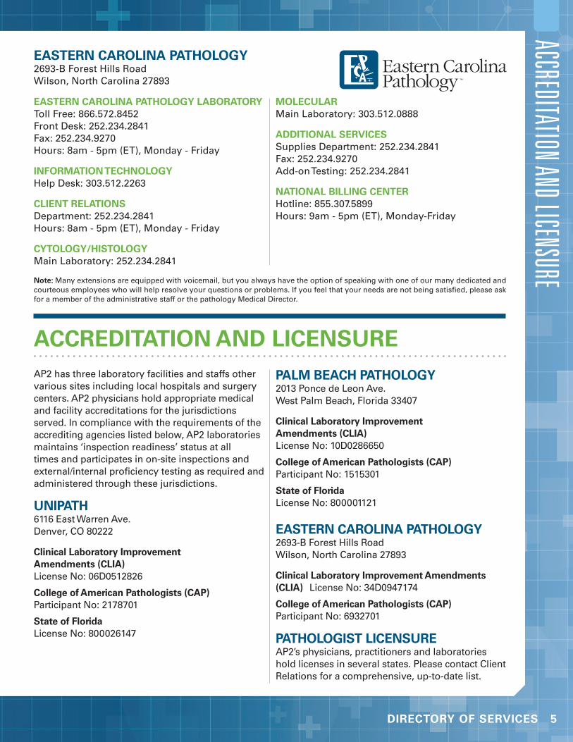
DIRECTORY OF SERVICES 5
EASTERN CAROLINA PATHOLOGY2693-B Forest Hills RoadWilson, North Carolina 27893
EASTERN CAROLINA PATHOLOGY LABORATORYToll Free: 866.572.8452Front Desk: 252.234.2841Fax: 252.234.9270Hours: 8am - 5pm (ET), Monday - Friday
INFORMATION TECHNOLOGYHelp Desk: 303.512.2263
CLIENT RELATIONSDepartment: 252.234.2841Hours: 8am - 5pm (ET), Monday - Friday
CYTOLOGY/HISTOLOGYMain Laboratory: 252.234.2841
MOLECULARMain Laboratory: 303.512.0888
ADDITIONAL SERVICESSupplies Department: 252.234.2841Fax: 252.234.9270Add-on Testing: 252.234.2841
NATIONAL BILLING CENTERHotline: 855.307.5899Hours: 9am - 5pm (ET), Monday-Friday
ACCREDITATION AND LICENSURE
AP2 has three laboratory facilities and staffs other various sites including local hospitals and surgery centers. AP2 physicians hold appropriate medical and facility accreditations for the jurisdictions served. In compliance with the requirements of the accrediting agencies listed below, AP2 laboratories maintains ‘inspection readiness’ status at all times and participates in on-site inspections and external/internal proficiency testing as required and administered through these jurisdictions.
UNIPATH 6116 East Warren Ave. Denver, CO 80222
Clinical Laboratory Improvement Amendments (CLIA) License No: 06D0512826
College of American Pathologists (CAP) Participant No: 2178701
State of Florida License No: 800026147
PALM BEACH PATHOLOGY2013 Ponce de Leon Ave. West Palm Beach, Florida 33407
Clinical Laboratory Improvement Amendments (CLIA) License No: 10D0286650
College of American Pathologists (CAP) Participant No: 1515301
State of Florida License No: 800001121
EASTERN CAROLINA PATHOLOGY2693-B Forest Hills Road Wilson, North Carolina 27893
Clinical Laboratory Improvement Amendments (CLIA) License No: 34D0947174
College of American Pathologists (CAP) Participant No: 6932701
PATHOLOGIST LICENSUREAP2’s physicians, practitioners and laboratories hold licenses in several states. Please contact Client Relations for a comprehensive, up-to-date list.
ACCREDITATION AND LICENSURE
Note: Many extensions are equipped with voicemail, but you always have the option of speaking with one of our many dedicated and courteous employees who will help resolve your questions or problems. If you feel that your needs are not being satisfied, please ask for a member of the administrative staff or the pathology Medical Director.

6 DIRECTORY OF SERVICES
RESULTS AND REPORTING
RESULTS AND REPORTINGAP2 generates final diagnostic reports as quickly as possible while maintaining quality and service. AP2 is in compliance with the Health Insurance Portability and Accountability Act (HIPAA), and all policies, processes and procedures are designed to ensure compliance with HIPAA standards. All AP2 processes and procedures are monitored and audited for HIPAA compliance, and AP2 employees receive training on HIPAA standards annually.
The Client Relations staff is trained to answer many of your questions. If he/she does not know the answers to your questions, they will find out the necessary information and get back to you in a timely manner. AP2 professional staff members, including board-certified, fellowship trained pathologists and certified cytotechnologists, are available to assist with consultations on interpretation of results and reports and technical concerns. Please feel free to communicate any questions or concerns to your client service representative or pathologist at:
UniPath 303.512.0888Palm Beach Pathology 561.659.0770Eastern Carolina Pathology 252.234.2841
TURN AROUND TIMEAP2 and its subsidiaries strive to provide the most rapid turn-around time possible without compromising quality. In general, tissue biopsies and critical cytology specimens are given priority, due to their serious nature. Pathology reports will typically be generated within 48 hours of receipt of the specimen. Routine Pap screening cytology reports will be available within 4-5 working days after receipt of the specimen. STAT processing and reporting are also available for special circumstances.
ISSUING REPORTSDelivery of reports can be made a number of ways:
➤ Courier service➤ United States Mail➤ Auto fax➤ On-site remote printing➤ Electronic file transfers➤ LifePoint Web-Based Reporting
MONTHLY STATISTICAL REPORTSMonthly reports specific to your practice are available upon request. Reports such as diagnostic statistics specific to your practice and lists of patients with diagnostic information are two examples of reports we can provide. Please contact Client Relations for more information.
PATHOLOGY DIRECTED CONSULTATIONSOccasionally, a specific case may require a second opinion from outside AP2’s pathology group. AP2 reserves the right to select the experts used for consultative services. Additional consultative expenses will be the responsibility of the patient or their insurance carrier.
SLIDE RE-CUT PROGRAMExtra slides on cases may be requested by clinicians for study, second opinion or retention in your own files. Slides will be re-cut on request and sent along with the completed pathology report. A nominal fee may be assessed. For more information, please contact Client Relations.
MEDICAL LEGAL TESTINGAP2 laboratories do not perform testing for medical legal purposes.
SPECIMEN PROCESSING SERVICESurgical specimen processing without diagnosis is available. AP2 laboratories can provide specimen gross examination, tissue processing, slide preparation and labeling. This service is exceptionally useful to dermatologists who have specialty training in dermatopathology and wish to microscopically examine and diagnose their own patient’s specimens.
DIRECTORY OF SERVICES 7
CASE MANAGEMENT, TEST ADD-ONS AND CANCELS | INFORMATION TECHNOLOGYCASE MANAGEMENT, TEST ADD-ONS AND CANCELLATIONSFor cases that require a test be canceled or for additional testing to be added, please contact Client Relations as soon as possible at:
UniPath 303.512.0888Palm Beach Pathology 561.659.0770Eastern Carolina Pathology 252.234.2841
Testing will only be performed with clear orders. Verbal orders must be followed up with a signed authorization before testing will be performed.
INFORMATION TECHNOLOGY
DEDICATED IT PERSONNELAP2 operates a large IT department, including IT professionals that provide support in a number of areas: lab informatics, EHR interfacing and client connectivity, network administration, data center operations, database administration, decision support, help desk support, etc. AP2 provides the infrastructure and support that healthcare providers need to access laboratory information.
FLEXIBLE CONNECTIVITY SOLUTIONSAP2 IT Department provides this deep expertise and knowledge base to every client connectivity project. We expeditiously and expertly develop
customized connectivity solutions that meet your needs. We employ every modality available and provide our clients the utmost flexibility when it comes to placing orders and receiving results.
MULTIPLE OPTIONSOrders can be placed manually using our well-designed hardcopy requisition forms or electronically (either via our browser-based online portal or via an order interface from an EHR system). Results can be delivered hardcopy via courier, via fax, auto-print to a remote dedicated in-office printer, via a browser-based online portal, via a results interface into an EHR system, or any combination of the above.

8 DIRECTORY OF SERVICES
INSURANCE AND BILLING SERVICESAP2 participates with many insurance companies and holds many managed care contracts. You do have a choice when it comes to selecting a laboratory for your anatomic pathology and cytology specimens. A list of the current managed care contracts is available upon request. Please contact Client Relations for an updated list and/or any questions that you may have about a specific insurance company.
The billing statement your patients receive represents the pathology fee for professional and technical services associated with the evaluation of a specimen, tissue, blood or bodily fluids submitted to one of our partner laboratories, including UniPath, Palm Beach Pathology or Eastern Carolina Pathology. These laboratory facilities are members of American Pathology Partners (or AP2).
We know that having to deal with medical bills is not particularly enjoyable. Our commitment to every patient is to help them through this process with sensitivity, care and professionalism. Please contact us about any billing related matters:
National Billing Center1.855.307.5899 (toll-free)7:00am – 6:00 pm (CT), Monday – [email protected]
In order to bill appropriately and completely, please submit complete and accurate billing information on the test requisition and include a copy of the patient’s insurance card(s), front and back. The following elements must be included:
➤ Specimen collection date➤ Complete patient name➤ Patient gender➤ Patient date of birth➤ Ordering physician full name and NPI➤ Patient telephone number➤ Subscriber name (if different than patient)➤ Subscriber date of birth (if different than
patient)➤ Complete address of patient or subscriber➤ Relation to subscriber (self, spouse, child,
other)
➤ All ordering ICD-10 diagnosis codes➤ Insurance company name and address (copy
of front and back of insurance card)
FINANCIAL HARDSHIP PATIENTSAP2 recognizes the inability of some patients to pay for necessary anatomic pathology and cytology screening services. This also includes uninsured and underinsured patients who are not covered for specified anatomic pathology and cytology screening services. AP2 provides a financial assistance program for patients who are eligible. Please call the billing phone number for the appropriate laboratory for more information.
THIRD PARTY CARRIERSAP2 will bill a patient’s third party carrier directly. This requires complete and accurate information and copies of the front and back of the patient’s insurance cards. If the patient has a third party insurance carrier contracted with the AP2 laboratory which their specimen was sent, AP2 will accept 100% of the contracted price for a test. AP2 will bill according to the explanation of benefits (EOB) provided by the carrier and will also bill for co-pays, co-insurance, deductibles and non-covered services as instructed by the carrier and required by law.
MEDICAREAP2 is a participating Medicare supplier with the Medicare program and is required by CMS to file all claims for laboratory services rendered. AP2 accepts the Medicare-allowed amounts as full payment for covered services. This assignment does not preclude billing of the patient for services denied by Medicare.
Medicare only pays for those services that it deems to be medically necessary for the diagnosis and treatment of diseases and/or other health related problems. CMS and Medicare have developed a system using ICD-10 diagnosis codes to prevent payment of claims that they determine not to be medically necessary. It is critical that a code used for ordering the anatomic pathology service/test be consistent with the documentation in the patient’s medical records. The ICD-10 code
INSURANCE AND BILLING SERVICES

DIRECTORY OF SERVICES 9
or clinical description used must be specific to the patient’s medical condition and the anatomic pathology service/test requested by the physician for that date of service. If reimbursement is denied due to lack of medical necessity, Medicare rules allow the laboratory to subsequently bill the patient only if the patient
has signed and dated the Advanced Beneficiary Notice (ABN) prior to the testing procedure. To comply with HCFA guidelines for Medicare reimbursement, please be certain to include the appropriate ICD-10 code or clinical description, as well as have the patient sign the ABN.
GENERAL SPECIMEN LABELING AND SUBMISSION
GENERAL SPECIMEN LABELING AND SUBMISSIONPrudent medical-legal practice and laboratory accrediting agencies have strict guidelines for specimen labeling and submission. They also mandate rejection of improperly completed requisitions or incorrectly identified slides or specimens. For example, some specimens cannot be analyzed because of improper collection, preservation or degradation in transit. Other specimens may have prolonged turn-around-times because of lack of necessary patient information. Still other specimens will, by necessity, be rejected because of inaccurate or absent specimen and/or requisition labeling. You will be notified of rejected or problematic specimens upon receipt. To avoid delayed diagnoses and potential specimen rejection, please observe the following requirements:
SURGICAL, CYTOLOGY AND MOLECULAR (UNISWAB AND GBS) SPECIMENS
REQUISITION
1 Patient last name, first name2 Date of birth3 Collection date4 Physician and clinic name and address5 Insurance and/or billing information
including name of insured, subscriber number, group number, name and address of insurance company, and IPA group (if applicable) – copy of insurance card preferred.
6 Reason for testing/ICD-10 code7 Test order
8 Site and type of biopsy if surgical specimen
9 Type of specimen and site of collection if cytology/molecular specimen
10 Brief clinical history11 Requests for any special stains or studies
SPECIMEN CONTAINER
1 Patient last name, first name (last name and first initial are acceptable) on the body of container (not the lid)
2 Second identifier such as date of birth, chart number, SS# or other unique identifier required
3 Specimen type and/or location (i.e. skin lesion, left shoulder etc.)
VIALS/NON-GYN SLIDES
1 A #2 lead pencil is recommended for slides. Markers and ballpoint pens are unacceptable for slides because of wash off during processing. Also, a name written on top of fixative will wash away with the fixative during processing.
2 Fixed smears: Write “Fix” on the fixed slide frosted end. Smear the material onto the glass slide (may use a 2-slide pull technique) and drop immediately into specimen container with fixative.
3 Non-fixed, air-dried smears: Smear the material onto the glass slide (may use a 2-slide pull technique) and allow to air dry, then place into a cardboard slide holder.
10 DIRECTORY OF SERVICES
BIOPSY – ROUTINE CERVICAL, ENDOCERVICAL AND ENDOMETRIAL
MATERIALS REQUIRED
1 Collection container(s) with 10% formalin
2 Requisition form
PROCEDURE
1 Label the body of the collection container(s) (not the lid) with the patient’s name, second identifier and tissue identification.
2 Complete the requisition form including tissue type, patient name, complete address, birth date, date of service and billing data.
3 Provide clinical history, i.e. last menstrual period, prior biopsies or Pap smear information, and any history of hormone use including birth control pills on the requisition form.
4 Immediately place the specimen in the collection container, tightly close container lid and forward to laboratory with requisition.
Note: Use of gauze pads to hold or place endometrial and endocervical samples are discouraged because portions of the specimen are absorbed into the coarse weave and lost. Telfa is acceptable; placing the specimen directly into formalin is preferred.
BIOPSY – CONE AND LEEP CONIZATION OF CERVIXMATERIALS REQUIRED
1 Collection container(s) with 10% formalin
2 Requisition form
PROCEDURE
1 Label the collection container(s) with the patient’s name, second identifier and tissue identification.
2 Complete the requisition form including tissue type, patient name, complete address, birth date, date of service and billing data.
2 Include on the requisition a history of prior Pap smear or biopsy results.
4 Orient cone specimen with surgical suture material is preferred.
5 Immediately place the specimen in the collection container, tightly close container lid and forward to laboratory with requisition.
BIOPSY – ENDOSCOPIC (ESOPHAGUS, GASTRIC, SMALL AND LARGE INTESTINE, LUNGS, ETC.)MATERIALS REQUIRED:
1 Collection container(s) with 10% formalin
2 Telfa
3 Requisition form
PROCEDURE
1 Label the body of the collection container (not the lid) with patient’s name, second identifier and tissue identification.
2 Complete requisition form including tissue type, patient name, complete address, birth date, date of service and billing data.
3 Place specimen directly into formalin, close lid tightly and forward to laboratory.
4 If you choose to use Telfa, gently orient and place tissue on Telfa, mucosal surface up with submucosal surface in contact with the Telfa. Slowly enter the Telfa with attached tissue into the formalin, tightly close lid and forward to laboratory with the requisition.
Note: Use of gauze pads to hold or place endoscopically obtained specimens is discouraged because portions of the specimen are absorbed into the coarse weave and lost. Telfa is acceptable; placing the specimen directly into formalin is preferred.
BIOPSY – SKIN EXCISIONS
MATERIALS REQUIRED:
1 Collection container(s) with 10% formalin
2 Requisition form
SURGICAL PATHOLOGY
SURGICAL PATHOLOGY

DIRECTORY OF SERVICES 11
PROCEDURE
1 Label the collection container(s) (not the lid) with the patient’s name, second identifier and tissue identification.
2 Complete requisition form including tissue type, patient name, complete address, birth date, date of service and billing data.
3 Include on the requisition form any clinical history, a gross description of lesion and history of prior biopsies if available.
4 Indicate if specimen is a shave, punch or excision.
5 Orient specimen as necessary using description or surgical suture material. Immediately place the specimen in the fixative container.
6 Tightly close the container and forward to laboratory with requisition.
BIOPSY – BREAST TISSUEMATERIALS REQUIRED:
1 Collection container(s) filled with 10% neutral-buffered formalin large enough to completely submerge specimen in adequate volume of fixative (10:1)
2 Requisition form completed with the following:
a Patient identifiers and billing information
b Relevant historyc Anatomic site of biopsyd Time tissue was removed from
patiente Time tissue was submerged in
fixative
PROCEDURE
1 Label the collection container(s) with the patient’s name, second identifier and anatomic site of the biopsy/sample.
2 Note on the requisition the time the specimen was removed from the patient.
a The time between removal from the patient to submerging in fixative
(cold ischemic time) should be less than 1 hour.
3 Immediately place the specimen in the formalin-filled container and tightly close the container lid.
a The volume of formalin to tissue should be 10:1 and should completely cover the specimen and allow the tissue to float.
4 Note on the requisition form the time and date the specimen was placed in formalin.
a Inadequate formalin volume and/or failure to provide formalin fixation time may delay processing and the ability to perform special studies.
5 Complete the requisition form with all required information (see above).
6 Forward the specimen and completed paperwork to the laboratory as soon as possible.
BIOPSY – PROSTHETIC BREAST IMPLANTS
MATERIALS REQUIRED1 Collection containers for implants
(submitted without fixative) and containers for fibrous tissue capsules submitted in 10% formalin
2 Completed requisition form
PROCEDURE1 Label the collection containers (not the lid)
with the patient’s name, second identifier and specimen identification.
a The prosthetic breast implants must be submitted in separate containers without fixation.
b The fibrous tissue capsules may be submitted separately in 10% formalin.
2 Complete the requisition form to include tissue type, patient name, date of birth, date of service, and billing insurance data.
3 Indicate on the requisition the presence or absence of prosthetic rupture or leakage.
4 Tightly close container lids and forward to laboratory with requisition.
SURGICAL PATHOLOGY

12 DIRECTORY OF SERVICES
Surgical Pathology, Cont.
GENERAL - TISSUE SPECIMENS FOR SURGICAL PATHOLOGY
Histology and grossing services are available for the majority of tissue types requiring routine microscopic examination (i.e. gallbladders, oral cavity biopsies, lymph nodes, thyroid, bone, appendix). For routine microscopic examination, the tissue is to be submitted in 10% Formalin. If specialized testing is required (i.e. Lymphoma/Leukemia testing, Direct Immunofluorescence, culture) please contact the laboratory’s histology department for specific submission instructions.
For general inquiries about submitting a specimen
or to inquire about a specific tissue type, please contact the laboratory’s histology department for further information and comprehensive instructions for specimen submission.
MATERIALS REQUIRED:
1 Collection container with 10% formalin*
2 Requisition form
*Please contact the histology laboratory before using 10% formalin if specialized testing is required
PROCEDURE
1 See steps 1-3 for Endoscopic Biopsy.
2 Include relevant clinical history.
IMMUNOHISTOLOGY, CYTOCHEMISTRY, MOLECULAR PATHOLOGYContact the laboratory for inquiries about send out and reference laboratory testing not specifically listed.
IMMUNOHISTOLOGY
PRINCIPLEImmunohistology employs highly specific antibodies to detect protein antigens in pathologic material to demonstrate specific types of cellular differentiation. Molecular pathology uses specific probes to identify genetic material, to associate a disease process with an infectious agent or rearrangement of genetic material.
Cytochemistry employs specific chemical reactions to demonstrate sub-cellular organelles or enzymes, which might be associated with certain types of cellular differentiation. These special studies are useful in establishing the correct diagnosis for a wide variety of pathologic conditions. These studies are also useful in determining a patient’s prognosis for a given tumor, such as status of estrogen and
progesterone receptors for breast carcinoma. Finally, these studies are also useful in helping the pathologist to determine if a given histologic pattern represents a malignancy, such as a demonstration of prostatic gland basal cells.
MATERIALS REQUIRED
1 Collection container filled with 10% buffered formalin for specimens not suspected of being lymphoma or leukemia (see next line). The pathologist will determine if these special studies are needed
2 Sterile collection container lined with saline soaked gauze for cases suspected of lymphoma or leukemia
3 Glass slides with frosted end for cytology specimens such as Fine Needle Aspirations or bone marrow aspirates
4 A completed requisition form with proper patient identification, history and source of biopsy
SURGICAL PATHOLOGY

DIRECTORY OF SERVICES 13
IMMUNOHISTOLOGY, CYTOCHEMISTRY, MOLECULAR PATHOLOGYPROCEDURE
1 Label the collection container (not the lid) with the patient’s name, second identifier and tissue identification.
2 Submit routine biopsy material in 10% buffered formalin unless lymphoma or leukemia is suspected. The pathologist will determine if special studies are required after studying the routine preparation.
3 For suspected lymphomas, place the fresh tissue in a clean container, which does not contain any fixative material. The tissue should be placed on gauze dampened with saline.
4 Let smears air dry at room temperature. Place them in a plastic slide box. If you do not have a box, simply wrap the slides up in a paper towel or tissue paper and secure with tape.
5 Complete the requisition form to include patient name, date of birth, date of service and billing data. The pathologist will determine if additional studies are needed after initial examination of the specimen.
6 Contact laboratory Courier Services to arrange for a STAT pick up:
UniPath 303.512.0888Palm Beach Pathology 561.659.0770Eastern Carolina Pathology 252.234.2841
7 Please note that we can accept specimens Monday through Friday only, from 7:00 a.m. to 4:00 p.m. Advanced warnings about specimens being sent fresh for Lymphoma work-ups ensures the specimens will be properly processed upon receipt at the laboratory. If unsure of proper submission, please call prior to biopsy excision.
MOLECULAR ONCOLOGY
PRINCIPLEMolecular Oncology uses molecular methods to characterize genetic material, or to detect infectious agents. These special studies are useful in helping the pathologist determine which translocation occurred or what gene amplified, deleted, etc.
TESTING IS CURRENTLY PERFORMED TO EVALUATE
1 Breast cancer Her2Neu status2 Jak 2 quantitative Polymerase Chain
Reaction (send-out)3 Human papillomavirus high risk detection4 Various genetic rearrangement in
hematologic malignancies
MATERIALS REQUIRED
➤ Tissue Biopsies:1 A tissue specimen that has been
processed and is in a paraffin block or unstained slides cut from a paraffin block.
2 A completed pathology request form with insurance information or copies of insurance information attached.
➤ Blood:1 5 - 10 mL blood specimens should be
collected in EDTA tube (purple top). Heparin samples are NOT acceptable.
CHROMOSOMAL STUDIESCurrently, AP2 does not perform chromosomal studies on tissue or fluid specimens “in house”. If requests for chromosome studies are received, we will forward them to a reference laboratory. Specimens for chromosomal analysis (i.e. Products of Conception) must be submitted fresh in saline or RPMI (without formalin) in an appropriately labeled container and accompanied by a completed requisition form requesting Chromosomal Analysis.
ELECTRON MICROSCOPYCurrently, AP2 does not perform “in-house” Electron Microscopy on tissue or fluid specimens. If Electron Microscopy is requested or required to complete a diagnosis, we will refer to a reference laboratory. Specimens for Electron Microscopy must be submitted in gluteraldehyde. Generally, Electron Microscopy is clinically necessary only for complete evaluation of medical kidney disease. Coordination of preparation and submission of specimen can be accomplished best by contacting the pathologist at the appropriate medical facility prior to collecting the specimen.
14 DIRECTORY OF SERVICES
HEMATOPATHOLOGY AND FLOW CYTOMETRY
FLOW CYTOMETRY OVERVIEWPRINCIPLEFlow cytometry measures physical and chemical properties of the cells. The cells pass the flow cytometric analyzer in a fluid, single-cell stream. The laser beams interrogate each cell and indicate the cell size, internal complexity and the antigens present on cell surface or in the cytoplasm.
METHODOLOGYEach specimen is manually processed. A smear (peripheral blood or bone marrow) or touch prep (tissue) slide is made from the original specimen. A cytospin slide is made from the final cell suspension. The slides are stained and evaluated by the hematopathologist. Every specimen for flow cytometric analysis is manually processed. Panels of surface/intracellular markers are chosen to stain the cells based on morphology and clinical history. After the cells are stained, they are acquired and analyzed with the flow cytometer.
Our immunophenotyping panel configurations are carefully considered based on state-of-the-art scientific references, consensus, and the experiences of our hematopathologists. The combinations of antibodies allow for the identification of normal and abnormal populations of cells within a specimen. Our antibody panels cover acute leukemia, chronic leukemia, T and B cell lymphoma, multiple myeloma, neoplasms and others.
FLOW CYTOMETRY/HEMATOPATHOLOGY GENERAL SPECIMEN REQUIREMENTSREQUISITION
1 Patient last, first name2 Date of birth3 Collection date and time4 Physician and clinic name and address5 Insurance and/or billing information including
name of insured, subscriber number, group number, name and address of insurance company, and IPA group (if applicable) – copy of insurance card preferred.
6 Reason for testing/ICD-10 code
7 Test order8 Type and collection site of specimen9 Brief clinical history (including CBC)10 Requests for any special stains or studies11 DART: If this box is checked, the
hematopathologist will use discretion to determine which tests are appropriate, based on specimen type and clinical history, and order tests appropriately.
BONE MARROW/PERIPHERAL BLOOD SLIDES All bone marrow/peripheral blood slides submitted must be legibly labeled with the patient’s name (last name and first name initial are acceptable) matching the requisition form. Minimally, the last name must be legible and correctly spelled. A second identifier such as date of birth, chart number or other unique identifier is required.
SPECIMEN CONTAINERS1 Patient last name, first name (last name and
first initial are acceptable)2 Second identifier such as date of birth, chart
number or other unique identifier is required3 Specimen type and collection site (i.e.
peripheral blood, bone marrow; right iliac crest, etc.)
PERFORMED
➤ Monday – Friday, STAT on-call available weekends
TURNAROUND TIME
➤ Verbal results usually available within 24 hours of specimen receipt
BONE MARROW1 2-7 mL bone marrow aspirate collected in
NaHep (green top) tube; EDTA (purple top) tube acceptable; ACD (yellow top) tube only if no other options are available
2 Call lab immediately for STAT pickup: UniPath 303.512.0888Palm Beach Pathology 561.659.0770Eastern Carolina Pathology 252.234.2841
HEMATOPATHOLOGY AND FLOW CYTOMETRY

DIRECTORY OF SERVICES 15
3 Specimen needs to be delivered to lab immediately (within 12 hours) and transport at room temperature (20-25ºC).DO NOT REFRIGERATE.
PERIPHERAL BLOOD1 3-5 mL collected in NaHep (green top) tube2 3-5 mL collected in EDTA (purple top) tube for
morphology3 Deliver within 24-hours of collection4 Transport at room temperature (20-25ºC). DO
NOT REFRIGERATE.5 Include CBC results if performed
SOLID TISSUE/FNA4 5 mm solid tissue specimen cut aseptically
into small pieces and placed in RPMI media and stored at refrigerated temperature of 2-8ºC
4 FNA specimen placed in RPMI media and stored at refrigerated temperature of 2-8ºC
4 Transport on “wet” ice. DO NOT SEND ON DRY ICE AND DO NOT FREEZE.
BODY FLUIDS1 Volume required for testing depends on the
cell count of the specimen. Specimen handling varies depending on the specimen type.
2 Cerebral Spinal Fluid (CSF) requires processing within one hour of collection for optimal results. Please call laboratory for details:UniPath 303.512.0888Palm Beach Pathology 561.659.0770Eastern Carolina Pathology 252.234.2841
PROCESS AND HOLDSOccasionally a physician may submit a specimen for possible flow cytometry, depending on the results of permanent section expected at a later time. In these cases, “Process and Hold” is written usually on the requisition. The specimen typically will be held for 2-3 days, during which time the physician must contact the laboratory to request flow analysis. A specimen may also be sent with a diagnosis that does not warrant flow cytometry testing (Hodgkin lymphoma, carcinoma, certain body fluids, etc.) These specimens are held until reviewed by the pathologist. If no further testing is requested, “CANCELED by (pathologist name)” will typically be written on the requisition in red
ink with the date and the technologist initials. The paperwork is filed and the specimen is kept for approximately one week and then discarded.
SPECIMEN REJECTIONA specimen may be rejected if it is frozen or fixed, too old, or if it is hemolyzed or clotted. All specimens MUST be labeled with at least the patient’s name and date of collection. Other identifying information is useful, such as hospital number.
SPECIMEN REQUISITIONPlease use the comprehensive hematopathology requisition. Please indicate specimen type, number and type of tubes, and number of smear or touch prep slides submitted. It is important to indicate the diagnosis under consideration and the indication for flow cytometry testing.
The following information must be completed on each requisition:
1 Patient’s name2 Patient’s date of birth3 Hospital number or accession number (if
available)4 Social security number (if available)5 Date and time of collection6 Specimen source or type (i.e. bone marrow,
left cervical lymph node, etc.)7 Requesting physician name and contact
phone number8 Thorough clinical history9 Indicate whether flow cytometry is needed
All insurance information must be included to ensure proper billing.
HOW TO SUBMIT A BONE MARROW BIOPSY AND ASPIRATEMATERIALS REQUIRED
1 Hematopathology requisition form, with labels for patient name, specimen designation, time specimen was collected, and biohazard bags
2 Formalin (ECP only) or B-Plus fixative (UniPath and PBP)
3 2-5 mL of peripheral blood submitted in EDTA tube (purple top) for morphology (and/or 3-5 mL of peripheral blood in NaHep (green top) for flow cytometry if aspirate not obtainable (dry tap)
HEMATOPATHOLOGY AND FLOW CYTOMETRY

16 DIRECTORY OF SERVICES
HEMATOPATHOLOGY & FLOW CYTOMETRY
4 Copy of CBC results is required5 Two purple top (EDTA) tubes of aspirate (one
for morphology, one for molecular testing)6 Two green top (NaHep) tubes:
a 3-6 mL of aspirate for cytogeneticsb Minimum 1 mL of aspirate for flow
cytometry7 In event of a DRY TAP (no aspirate obtainable):
a Submit 3-5 mL of peripheral blood in green top (NaHep) for flow cytometry.
b Submit a separate 10 mm core biopsy in sterile RPMI.
8 If marrow submitted for STR studies, use yellow top tube (ACD) (transplant patients).
9 If marrow cultures are needed, aspirate should be placed in yellow top isolator tubes (saponin anticoagulant, NOT ACD).
PROCEDURE 1 Label the collection containers (not the lid)
with the patient’s name, second identifier and specimen identification.
2 Complete the hematopathology requisition form including tissue type, patient’s name, complete address, birth date, date of service, and billing data, as well as any other physicians who need copies of the report.
3 Request studies in the test menu or check DART.
4 After preparing 5 touch imprints, the bone marrow core biopsy should be immediately placed in the formalin (ECP only) or B-Plus fixative (UniPath and PBP). Note the time of fixation on the requisition form.
5 Express bone marrow aspirate material into two purple top tubes (EDTA) and two green top tubes (NaHep), cap and gently mix to prevent clotting.
6 The peripheral blood specimen should be placed in a purple top tube (EDTA).
7 Place all the specimen tubes and the requisition form in a biohazard bag.
8 Call laboratory to arrange for a STAT pickup:UniPath 303.512.0888Palm Beach Pathology 561.659.0770Eastern Carolina Pathology 252.234.2841
Note: Bone marrow biopsy specimens are performed for a variety of reasons ranging from evaluation of anemia to the diagnosis of metastatic carcinoma and leukemia. Complete patient history, as well as documentation of any supporting details is vital to the complete and accurate interpretation of the material. It is of particular importance to include information as to growth factor therapy.
HOW TO SUBMIT TISSUE FOR LYMPHOMA (LYMPH NODE/SOLID TISSUE) WORKUPMATERIALS REQUIRED:
1 Sterile screw top container2 RPMI (preferred) or if not available, sterile
isotonic saline3 Formalin or B-Plus fixative (for UniPath
clients)4 5 glass slides5 Completed requisition form6 Biohazard bags7 Ice
PROCEDURE1 Label the collection container (not the lid) with
the patient’s name and identification. 2 Make touch preparations of the lymph node
or tissue, using the 5 glass slides.3 Place a portion of the lymph node or tissue (5
mm of fresh tissue, cut aseptically into small pieces) in the screw top container and cover with RPMI (or wet with saline). DO NOT SUBMIT ALL TISSUE IN FORMALIN OR B-PLUS IF FLOW ANALYSIS IS DESIRED.
4 Place remainder of lymph node or tissue into B-Plus fixative (UniPath and PBP) or formalin (ECP).
5 Fill out requisition and include name of primary care physician or oncologist.
3 Place RPMI container in biohazard bag filled with ice. Place B-Plus or formalin container into a biohazard bag with touch preparations.
6 Specimen should be received within 30-minutes of excision for optimal results. Call laboratory for STAT pickup: UniPath 303.512.0888Palm Beach Pathology 561.659.0770Eastern Carolina Pathology 252.234.2841
7 For a complete list of Hematopathology Diagnostic Profiles, please see Appendix 5.
Hematopathology and Flow Cytometry, Cont.

DIRECTORY OF SERVICES 17
INFECTIOUS DISEASE TESTINGINFECTIOUS DISEASE OVERVIEWInfectious disease testing can be performed on the ThinPrep®, SurePath™, UniSwab™, Aptima® Multi-test and Unisex Swab, GBS Swab, and Aptima® Urine collection devices. These molecular tests aid in the detection, diagnosis and treatment of viral, fungal and bacterial infections. Nucleic acid testing is performed using polymerase chain reaction (PCR), strand displacement amplification (SDA), transcription mediated amplification (TMA) and hybrid capture methodologies providing high levels of sensitivity in testing for the presence of these organisms.
A full list of infectious disease tests offered using these collection devices and methods can be found in Appendix 5 of this manual.
UNISWAB™ COLLECTION
PURPOSE1 For female patients, collection of cervical and
vaginal specimens, including lesions2 For male patients, collection of discharge from
urethra or lesions
PROCEDURE1 Completely fill out Women’s Health or Cyto/
Histo requisition form with proper patient identification, history and collection source.
2 Aseptically remove sterile swab from package.3 Collect specimen by vigorously swabbing site
for 30 seconds.4 Aseptically remove cap from vial.5 Place swab in transport medium and break off
swab against rim of tube.6 Replace cap to vial and close tightly.7 Fill out vial label with patient information.
FOR HSV SAMPLES – PLEASE USE UNISWAB™ FOR SPECIMEN COLLECTION
1 For crusted lesions, pre-moisten the swab with either sterile saline or sterile water.
2 De-roof the crust, and swab the lesion firmly working from outer base of lesion to interior.
3 Place swab into transport media, break off swab against rim of the tube.
4 Replace cap to vial and close tightly.5 Fill out vial label with patient information.
THINPREP® PAP TESTS AND SUREPATH™ PAP TESTSPURPOSE
1 For female patients, collection of cervical and vaginal specimens
PROCEDURE1 Completely fill out the Women’s Health or
Cyto/Histo requisition form with proper patient identification, history and collection source.
2 Label vial with patient information and second identifier.
3 Prior to specimen collection, clean away any visible blood, mucus and/or discharge from the cervix.
4 Obtain a vaginal smear from secretions in the posterior fornix. This material, along with the ectocervical scrape should be placed immediately in the vial and agitated to dislodge the collected cells. Shake vigorously.
5 Collect sample from the endocervix with a cytobrush or broom.
6 For SurePath specimens, place the brush or broom into the vial and agitate it up and down against the bottom of the container to collect as much material as possible. For Thin Prep specimens, swirl brush multiple times into the collection container and then remove collection device.
7 Close vial tightly and place in specimen bag.
GROUP B STREPTOCOCCUS SWABA separate collection device is required for the molecular diagnostic test for Group B Streptococcus (GBS). Per the recommendations by the Centers for Disease Control (www.cdc.gov, 2010), molecular tests for GBS should be performed from specimens enriched by bacterial media, refrigerated and transported in order to obtain maximal sensitivity. Do not use a UniSwabTM, ThinPrep®, SurePath™, or Aptima® Swab for GBS collection.
INFECTIOUS DISEASE TESTING

18 DIRECTORY OF SERVICES
INFECTIOUS DISEASE TESTING
GBS COLLECTIONPURPOSE
1 For female patients, collection of vaginal and rectal specimens
MATERIALS1 GBS collection device and transport media
PROCEDURE1 Completely fill out the Women’s Health or
Cyto/Histo requisition form with proper patient identification, history and collection source.
2 Label GBS vial with patient information and second identifier.
3 Aseptically remove sterile swab from package.4 Collect specimen from vagina and rectum
(collect from vagina first followed by rectum). Both sites are required for adequate results.
5 Aseptically remove cap from vial.6 Place swab in transport medium and replace
cap to vial and close tightly. 7 Refrigerate immediately per recommendation
of CDC.8 Specimen must be delivered to laboratory
within 48 hours.9 Please refer to the Centers for Disease Control
website (www.cdc.gov) for more information.
APTIMA® MULTI-TEST AND UNISEX SWAB
PURPOSE1 For female patients, collection of cervical and
vaginal specimens
PROCEDURE1 Completely fill out Women’s Health or Cyto/
Histo requisition form with proper patient identification, history and collection source.
2 Fill out vial label with patient information.3 Aseptically remove sterile swab from package.4 Collect specimen by rotating swabbing site for
10-30 seconds.5 Aseptically remove cap from vial.
6 Place swab in transport medium and break off swab against rim of tube. Discard top portion of the swab shaft.
7 Replace cap to vial and close tightly.
APTIMA® URINE
PURPOSE1 For female and male patients, collection of
free-catch urine2 For female patients who have had a
hysterectomy
PROCEDURE1 Completely fill out Women’s Health or Cyto/
Histo requisition form with proper patient identification, history and collection source.
2 Label collection cup with patient identification, second identifier, date and time collected.
3 Collect specimen in a sterile, plastic, preservative-free specimen collection cup.
a Patient should collect the first 20-30 mL of voided urine.
4 Place cap securely on urine collection cup.5 Open the Aptima® Urine kit and remove the
tube from the packaging.6 Label the tube with patient identification,
second identifier, date and time collected.7 Hold the tube upright and firmly tap the
bottom of the tube on a flat surface to dislodge and drops from inside the cap.
8 Use the transfer pipette to aspirate urine from the urine cup to the tube.
a Transfer urine to tube within 24 hours of collection.
9 Dispense urine into the tube. Fill tube between the black lines on the fill window located on the label. This volume corresponds to approximately 2 mL of urine. DO NOT under-fill or over-fill the tube. Discard the pipette.
a Specimens that are under filled or overfilled will not be processed.
10 Tighten the cap securely on the tube. a After collection, store urine transport tube
between 2-30ºC (ambient temperature).
Infectious Disease Testing, Cont.

DIRECTORY OF SERVICES 19
CYTOPATHOLOGY (NON-GYN)
CYTOLOGY – INDUCED SPUTUM AND BRONCHIAL WASHINGS FOR PNEUMOCYSTIS PNEUMONIAMATERIALS REQUIRED
1 Leak proof specimen container2 Requisition form
PROCEDURE1 Label specimen containers (not the lid)
with precise identification of specimen site (i.e. right upper lobe of lung). Add patient name and second unique identifier such as date of birth or social security number.
2 Complete requisition form including patient name, date of birth, date of service and billing information. Include requests for “Pneumocystis jiroveci” (formerly carinii) pneumonia (R/O PCP).
3 Enter washings into specimen container.4 If brushings are performed concurrently,
rotate brush gently over slide to apply material and let air dry.
5 Place brush in CytoLyt® container.6 Submit to laboratory with completed
requisition form.Note: Specimens may be submitted “fresh” without fixative. If delay is anticipated, add equal amounts of 50% methyl or ethyl alcohol or CytoLyt®. Refrigerate if specimen delivery is delayed. Only induced sputums or bronchial lavage/wash specimens are acceptable for ruling out PCP. Routine sputums to rule out PCP are rarely sensitive enough to yield reliable results.
CYTOPATHOLOGY – BRONCHIAL, COLONIC, ESOPHAGEAL AND GASTRIC WASHINGSMATERIALS REQUIRED
1 Leak proof specimen container2 Preservative fluid (50% methyl or ethyl
alcohol, or CytoLyt®). DO NOT use SurePath™ Pap test vials.
3 Slides with frosted ends (if brushings obtained concurrently)
4 Requisition form5 Pencil for labeling slides
PROCEDURE1 Label specimen containers (not the lid) with
precise identification of specimen site (i.e. right upper lobe of lung, etc.). Add patient name and second unique identifier such as date of birth or social security number.
2 Label slides with patient’s name using a pencil.
3 Complete the requisition form including patient name, birth date, date of service and billing data. Include requests for special stains or studies (i.e. Pneumocytis jiroveci or fungus).
4 Enter washing into specimen container and add equal volumes of preservative.
CYTOLOGY – SPUTUMMATERIALS REQUIRED
1 Leak proof plastic container with CytoLyt® fixative
2 Requisition form
PROCEDURE1 Label collection container (not the lid) with
patient’s name and second unique identifier such as date of birth or social security number.
2 Complete requisition form including patient name, birth date, date of service and billing data.
3 If the specimen is one of a series of samples taken, indicate position in series (i.e. 1/3 if it is the first of three samples).
4 Have the patient rinse their mouth prior to collection.
5 Give the patient the collection container. 6 Instruct the patient to breathe deeply for 3
minutes.7 Instruct the patient to cough deeply from
the diaphragm, with effort to expectorate material into collection container.
8 At short intervals, repeat the coughing attempts three more times with collection of all coughed up material.
CYTOPATHOLOGY (NON-GYN)

20 DIRECTORY OF SERVICES
CYTOPATHOLOGY (NON-GYN)
9 Add equal volumes of specimen to fixative fluid and shake vigorously (make sure that the container lid is tightly sealed before shaking).
10 Forward specimen to the laboratory with the completed requisition form.
COMMENTS1 Deep cough (from the diaphragm)
specimens are necessary to provide information regarding the lower respiratory tract. The laboratory will determine adequacy of specimen by the presence of alveolar macrophages within the specimen.
2 A series of three sputum samples should be collected on three consecutive days, preferably first thing in the morning with hard, productive coughing is recommended. Morning collections take advantage of the accumulation of secretions during the night. Avoid collecting right after meals to avoid contamination with food particles.
3 If there is difficulty in producing a specimen, collections may be facilitated by the moisture and steam of a preceding, long hot shower. For patients who are unable to produce sputum with repeated attempts, consider aerosol induced coughing and specimen collection.
4 Post bronchoscopy sputums may be productive with diagnostic material even with negative bronchial washings, brushings and biopsies.
5 Specimen consisting of saliva or nasal pharyngeal drainage will be reported as lacking alveolar macrophages, metaplastic cells or bronchial columnar cells. These specimens are inadequate for lesions of the lower respiratory tract and will not be considered as true negative studies.
6 With sputum samples positive for malignant cells, a primary of the head and neck region should be considered as well as malignancies of the lung. Up to 10% of positive sputums may reflect malignancies of the head and neck.
7 Specimens requiring culture must be separately submitted in sterile containers without fixative. The culture specimens must be submitted to a laboratory specializing in microbiologic culturing.
8 Specify the need for asbestos body examination. Studies will include examination of Papanicolaou and Prussian blue stained smears.
9 Specify the need for identification of pneumocystis jiroveci. Specimens will include examinations of Papanicolaou, Diff Quick and GMS as needed. The specimens submitted for pneumocystis should be submitted fresh without fixative.
CYTOLOGY – URINEMATERIALS REQUIRED
1 Leak proof collection container2 Appropriate fixatives (50% ethyl or methyl
alcohol, CytoLyt® or PreservCyt®)3 Requisition form
PROCEDURE1 Label collection container (not the lid) with
the patient’s name and second unique identifier such as date of birth or social security number.
2 Complete the requisition form, including patient name, date of birth, date of service and billing data. Patient’s history of kidney or bladder abnormalities should also be noted.
3 Do not use first morning urine. The patient should drink at least a quart of water an hour prior to collecting the urine specimens.
4 Mix an equal amount of fixative with the urine specimen. Forward the specimen to the laboratory with the completed requisition form.
5 In some cases, separate specimens should be collected and dated for consecutive days.
Cytopathology (Non-Gyn), Cont.

DIRECTORY OF SERVICES 21
COMMENTS1 Urine should be identified as to type of
sample (i.e. voided, catheterized, right or left ureteral or bladder irrigation fluid).
2 For detection of cancer of ureters or kidneys, a serial collection of specimens spaced over three days has proven to increase diagnostic sensitivity and yield.
3 Sensitivity and yield of urine specimens has also been shown to increase with optimal collection and preservation of specimen.
4 Unpreserved urine results in the rapid degeneration of exfoliated cells and may become useless in diagnosis.
CYTOLOGY – URINE W/FISH (UROVYSION®)MATERIALS REQUIRED
1 Leak proof steril collection container2 Appropriate fixatives (PreservCyt® vial)3 Requisition form
PROCEDURE1 Label collection container (not the lid) with
the patient’s name and second unique identifier such as date of birth or social security number.
2 Complete the requisition form, including patient name, date of birth, date of service and billing data. Patient’s history of kidney or bladder abnormalities should also be noted.
3 Collect 50 - 60 mL of urine or bladder wash in the sterile container, preferably not first morning void.
4 Open PreservCyt vial and pour contents into the sterile container with specimen. Total volume should not exceed 90 mL.
5 Tightly close the cap on the container. Forward the specimen to the laboratory with the completed requisition form.
COMMENTS1 Urine should be identified as to type of
sample (i.e. voided, catheterized, right or left ureteral or bladder irrigation fluid).
2 Refrigerate specimen if unable to transport immedately. Transport specimen with ice pack.
CYTOLOGY – NIPPLE SECRETIONS AND SMEARSMATERIALS REQUIRED
1 Slides with frosted ends2 Physiologic saline3 Cotton swab4 Requisition form5 Pencil for labeling slides
PROCEDURE1 Label the slides with the patient’s name and
site from which sample obtained using pencil. Example: “R Breast”.
2 Complete the requisition form, including the patient name, birth date, date of service and billing data. Include any pertinent history on the requisition (especially if the patient is pregnant or lactating).
3 If there is no nipple erosion or ulceration, gently “strip” the area of the breast below the nipple and areola with a motion from beneath the areola towards the nipple surface. Do not massage the entire breast. The stripping motion will propel accumulated secretions within the ampulla of the larger excretory ducts.
4 With appearance of fluid on the nipple surface, touch a slide to the drop of fluid and draw the slide quickly across the nipple.
5 Using a separate slide, repeat this process for the opposite breast.
6 If there is nipple erosion or ulceration, touch a slide to this area three times, with a different part of the slide in contact each time. This is conveniently done, starting with a contact position close to the hand holding the slide and then moving the application area of the slide further with your hand for the next two samplings.
7 Following touch preparation (6) of the ulcerated area, try to express fluid (3) and prepare slides if fluid is obtained.
8 If no fluid can be expressed, a swab may be dipped in saline and gently rolled and rotated on the ulcerated surface and applied to a glass slide.
9 Place the slides in the slide container and forward to the laboratory with the completed requisition.
Note: We recommend completely air-drying the smears to avoid incomplete fixation artifact.
CYTOPATHOLOGY – FINE NEEDLE ASPIRATION
22 DIRECTORY OF SERVICES
CYTOPATHOLOGY – FINE NEEDLE ASPIRATIONING
CYTOLOGY – CEREBROSPINAL FLUIDMATERIALS REQUIRED
1 Leak proof collection container2 RPMI (if hematopoietic disease is suspected)3 Requisition form
PROCEDURE1 Label the specimen container(s) (not the
lid) with the patient’s name, second unique identifier such as date of birth or social security number and specimen site.
2 Complete the requisition form, including the patient name, date of birth, date of service and billing data. Include all pertinent history, especially suspected hematopoietic disease.
3 If hematopoietic disease is suspected, place up to 2mL in separate RPMI container. Submit remaining fluid unfixed in leak proof container. CSF specimens for flow cytometric analysis need to be submitted within an hour of collection for optimal results.
4 Place in refrigerator while waiting for transport.
5 Call laboratory for STAT pickup:
UniPath 303.512.0888Palm Beach Pathology 561.659.0770Eastern Carolina Pathology 252.234.2841
Cytopathology (Non-Gyn), Cont.
CYTOPATHOLOGY – FINE NEEDLE ASPIRATION
CYTOPATHOLOGY – FINE NEEDLE ASPIRATION
PRINCIPLE
Fine needle aspiration (FNA) provides a prompt, cost effective, and relatively safe means of evaluating a mass through cytologic diagnosis. The procedure is generally well tolerated by patients. FNA is often used as an alternative to surgery and may provide a definitive diagnosis that will determine therapy and/or assist in a planned surgical approach with optimal utilization of operating room time. In general, any palpable mass can be evaluated by aspiration techniques. With ultrasound guidance, fluoroscopy and CT, most deep lesions may also be sampled. Lesions that are commonly sampled include thyroid, breast, salivary glands and lymph nodes. Although the technique is relatively simple, it does require some practice and understanding of principles of aspiration.
MATERIALS REQUIRED1 Hand grip syringe holder of preference2 Syringe, screw lock, disposable 10mL or
20mL plastic with tight fitting barrel3 Needles of size preference. Most aspirates
may be obtained with a needle no larger than 21 gauge. Most highly vascular structures (i.e. thyroid) are best sampled with a 23 or 25 gauge needle.
4 Frosted end labeled slides and pencil for labeling slides
5 Specimen container with appropriate fixative as needed for needle washings (CytoLyt®)
6 Alcohol or iodine solutions for sterilization of skin
7 Cotton swabs for sterilization of skin8 Sterile gauze9 Adhesive tape10 Band-Aids11 RPMI for lymph node flow12 Requisition form

DIRECTORY OF SERVICES 23
PROCEDURE1 Using a pencil (ink or markers wash off
during staining) label slides and specimen container(s) with the patient’s name and precise site of aspiration. Add a second unique identifier such as date of birth or social security number on container(s).
2 Place labeled slides in rows of four slides, each row for use with a “pass”. Use the container with the fixative for needle washings.
3 Set up a sterile field including preparation of syringe and syringe holders for initial and repeat passes.
4 Select preferred needles and syringes to be used for the procedure.
5 Place patient in a comfortable sitting or reclining position that allows easy access to aspirate the lesion.
6 Palpate the mass to identify the depth of the target and its relationship to surrounding structures.
7 Clean the skin over the aspiration site with an alcohol swab.
8 Immobilize the mass with the thumb and index finger of one hand or between two fingers of one hand.
9 Take the syringe equipment in the opposite hand and use a one handed withdrawal and release manipulation of the syringe plunger.
10 Place the needle against the skin at a determined puncture site and insert it into the mass area with a single quick motion without negative pressure on the syringe.
11 Once the needle is in the desired area, retract the plunger of the syringe to create negative pressure in the syringe and needle lumen. Minimal negative pressure is needed for highly vascular organs such as the thyroid while more negative pressure is needed for dense fibrous organs like the breast.
12 Move the needle back and forth several times directing it in the same plane. Gently redirect the needle to increase the field of sampling but avoid vigorous redirection of the needle as this tends to produce unnecessary hemorrhage in the lesion.
13 When material appears in the hub of the needle, the aspiration has been completed. Excess blood in the material will dilute the specimen rendering it unsuitable for microscopic diagnosis. One drop of material can usually produce 4-6 smears.
14 Release the pressure in the syringe by releasing the syringe plunger.
15 Gently withdraw the needle from the lesion and apply pressure to the puncture site with sterile gauze.
16 If multiple sites are sampled, label the slides, containers and corresponding sites on the requisitions A, B, C etc. as needed.
SMEAR PREPARATION1 After the needle has been removed from the
mass, detach the needle from the syringe using a surgical clamp or other appropriate instrument, fill the syringe with air, and reattach the needle to the syringe.
2 Place the bevel of the needle against a glass slide and express a small drop of aspirated material onto the slide.
3 If too much material is expressed onto the slide, either re-aspirate a portion of the material by withdrawing the syringe plunger slightly, or spread the material out among additional slides.
4 If the cellular material is semi-solid, place a second slide on top of the material and pull the slides gently and quickly apart as the material spreads from the weight of the slide.
5 If the aspirated material is diluted by fluid or by blood, use the same smear technique as for blood smears.
6 Place a second slide onto the drop of material allowing it to spread. Gently pull the two slides apart.
7 Slide preparation:
a Allow the smears to air-dry for Diff Quick staining. Write “air dried” on the end of the smear slides.
– OR –
b Prepare the smears as a wet fix.
8 Rinse the needle and syringe into the CytoLyt® vial for monolayer preparation.
CYTOPATHOLOGY – FINE NEEDLE ASPIRATION

24 DIRECTORY OF SERVICES
CYTOPATHOLOGY – FINE NEEDLE ASPIRATION
9 If staining and immediate evaluation is available, have the patient remain while adequacy of the aspiration pass is determined and repeat the procedure until the operator is satisfied with the adequacy of the material.
10 Complete the requisition form. Include the patient’s name, birth date, date of service and billing data. Provide the exact site of the lesion (i.e. parotid, thyroid, lymph node, etc.).
COMMENTS
1 When the lesion is composed of solid tissue, the needle tip functions as a cutting instrument. As it is moved back and forth through the tissue, tiny tissue fragments become dislodged and collect inside the needle. When suction is added to this procedure the previously dislodged fragments are sucked into the needle and the tip of the attached syringe.
2 The needle should never be removed while any negative pressure is in the syringe. Such pressure may force aspirated material from the needle into the syringe. This may make preparation of smears difficult and may start air-drying the material.
3 It is important not to dilute the cellular material with fluid or blood.
4 If a cyst is encountered during the aspiration, evacuate all fluid from the cyst and perform a second aspiration on any residual mass. Express the fluid from the cyst into CytoLyt® or 50% methyl or ethyl alcohol.
5 If blood is aspirated into the syringe, stop the procedure and prepare slides. Express residual bloody fluid into CytoLyt® or 50% methyl or ethyl alcohol.
6 If pus is encountered, withdraw as much of the material as possible and perform a repeat aspiration in an adjacent area. (When an infectious process is included in the differential diagnosis, a culture of the aspirated material is often desirable).
7 Necrosis generally occurs in the center of large lesions because of an inadequate blood supply. If the first sample yields only necrotic debris, another sample obtained tangentially to the edge of the mass should be secured.
8 Local anesthesia is rarely required, as the discomfort caused by is application is about the same as the aspiration. Its use is dependent on the lesion location, the discomfort of the patient and the judgment of the operator.
9 Passing through layers of muscle while inserting the needle adds significantly to the discomfort of the patient, while making needle placement more difficult and uncertain. Also, small fragments of muscle may plug the needle, jeopardizing subsequent sampling of the target. The muscle and lesion may sometimes be manipulated so that aspiration technique does not involve the muscle.
Cytopathology – Fine Needle Aspiration, Cont.

DIRECTORY OF SERVICES 25
10 The nipple and areola of the breast are the areas most sensitive to pain from a needle stick. These areas should be avoided whenever possible. Masses in these areas can sometimes be pushed away from the nipple, immobilized and sampled through adjacent skin.
11 When a mass is located close to the chest wall, there is a possibility that the needle may penetrate and cause a pneumothorax. This can be avoided by moving the mass sideways so it rests on a rib. This not only prevents penetration of the chest wall, but also provides good support for immobilization of the target.
12 Complications can vary as to the site of aspiration and cannot all be listed. The aspirator should be aware of these prior to aspiration and communicate them to the patient.
13 When completing the requisition, it is vital to provide the exact site of the lesion (i.e. thyroid, lymph node, parotid, etc.). “Neck mass” provides insufficient information for the cytopathologists to provide a reasoned diagnosis.
14 For thyroid aspirations, it is important to provide further information including the number of nodules present and the results of a scan if it was performed.
15 For lymph node aspirations, history of prior malignancy, antibiotic treatment, etc. are important. Flow cytometry can be performed on FNA’s of lymph nodes if the specimen is properly collected and stored. Prior planning can often alleviate the need for a repeat aspiration or biopsy.
Note: We perform FNAs at many of our covered hospitals. The results are generally available the next business day.
THYROID/PARATHYROID FINE NEEDLE ASPIRATION – THYROGLOBULIN ASSAY OPTION UNIPATH ONLY
BACKGROUND
Thyroglobulin and parathyroid assays are available on needle-rinsed FNA material. Some patients, status-post thyroidectomy for
differentiated (functioning) thyroid carcinomas, are often followed using serum thyroglobulin (Tg) as a marker of tumor recurrence. Patients with elevated serum thyroglobulin levels are often further evaluated by use of ultrasound radioactive iodine scans, or PET/CT scans. Enlarged lymph nodes or nodules in the bed of the thyroid can often be sampled by fine needle aspiration making use of cytology as well as the newly available direct thyroglobulin levels on the aspirate material. The assay is useful in those cases where residual/recurrent disease is suspected, but cytology specimen is negative. To take advantage of this added, sensitive technique of detecting recurrent disease, the following procedure is detailed. Special Note: The thyroglobulin assay is not recommended in cases in which the patient is not status post thyroidectomy.
The procedures for sending samples for parathyroid hormone and thyroglobulin are identical.
PROCEDURE1 Identify areas of suspicion, including
enlarged lymph nodes or nodules in the bed of the resected thyroid. These can be sampled under ultrasound guidance via fine needle aspiration (FNA).
2 Prepare cytologic preparations utilizing only direct smears (2 smears per aspirate). Label slides with pencil.
3 Rinse the remaining needle aspirate material rinsed into normal saline (1 cc). Additional aspirates of the targeted lesion may be pooled in this saline rinse. See Fine Needle Aspiration.
4 Additional aspirates for routine cytology can also be collected in CytoLyt®, but is not required in these particular situations.
5 Collect an additional pass in RPMI if hematopoetic lesion is suspected.
CYTOPATHOLOGY – GYNECOLOGY

26 DIRECTORY OF SERVICES
CYTOPATHOLOGY – FINE NEEDLE ASPIRATION
6 Contact client services for pick-up of the smears and needle rinse specimens. If pick-up is delayed more than 2 hours, the needle rinse material should be frozen.
UniPath 303.512.0888
AT THE LABORATORY1 The laboratory will prepare the cytologic
smears for interpretation. In the event no metastatic carcinoma is identified, the saline rinse material will be forwarded to University of Colorado Health Science Center (UCHSC) for thyroglobulin testing.
2 Results of the thyroglobulin testing will be added to the original cytology interpretation report as an addendum/consolidated report.
PATIENT MANAGEMENT INFORMATION
The presence of thyroglobulin (> 1.0 ng per mL) in the aspirate material of athyrotic patients is presumptive evidence of metastatic disease even in the absence of cytologic findings of metastatic disease. The estimated sensitivity of this procedure is 100% with specificity of 96.2%. Billing for this procedure will be assigned to your patient’s insurance per usual custom. Saline collection devices can be obtained through your laboratory. For more information or to set up the additional reflex testing, please contact your client service representative.
Cytopathology – Fine Needle Aspiration, Cont.
CYTOPATHOLOGY – GYNECOLOGYCYTOLOGY – THINPREP® PAP TESTS™PRINCIPLE
Automation and new methodologies are dramatically changing gynecological cytology, particularly the Pap test. The FDA has approved the ThinPrep® Pap Test™ as a replacement for the conventional Pap smear. Studies indicate that it is more accurate than the conventional Pap smear in detecting premalignant and malignant lesions. The advantages of a monolayer slide include:
1 Reduction of obscuring inflammation and blood
2 Elimination of air drying artifact provided the specimen is placed in the fixative immediately after sampling
3 A more uniform and representative sampling of the collected specimen
4 Residual sample available to perform additional testing as needed
5 Lowering of the ASCUS rate since air-drying and obscuring inflammation are not present
6 Increased sensitivity in detecting High-Grade Dysplasia due to the greater ease in recognizing dysplastic cells on the ThinPrep® Pap Test™ monolayer slide
MATERIALS REQUIRED1 The collection kit includes a cervix brush
with a spatula or broom and a collection vial of PreservCyt®
2 Completed requisition form
PROCEDURE1 Patient preparation:
a Schedule the patient for sample collection mid cycle (obscuring blood from menstrual smears is a major source of less than optimal smears).
b Instruct the patient in advance not to douche for at least 24 hours prior to examination.
2 Label PreservCyt® vial with the patient’s name and second unique identifier such as date of birth or social security number.
3 Complete requisition form including patient name, date of birth, date of service and billing data. Include on the requisition all clinical information (LMP, hormone use, prior Pap smear and biopsy results).
4 Prior to specimen collection, clean away any visible blood, mucus and/or discharge from the cervix.

DIRECTORY OF SERVICES 27
5 If desired, use lukewarm water to warm and lubricate the speculum. When necessary, a small amount of carbomere-free lubricant can be applied sparingly in a thin layer to the exterior of the speculum blades, avoiding the tip.
6 Obtain an adequate sampling from the ectocervix using a plastic spatula. Select contoured end of plastic spatula and rotate 360 degrees around the entire exocervix while maintaining light contact with exocervical surface.
7 Rinse the spatula as quickly as possible into the PreservCyt solution vial by swirling the spatula vigorously in the vial 10 times. Discard the spatula.
8 Obtain an adequate sampling from the endocervix using an endocervical brush device. Insert the brush into the cervix until only the bottommost fibers are exposed. Slowly rotate ¼ or ½ turn in one direction. Do not over-rotate.
9 Rinse the brush as quickly as possible in the PreservCyt solution by rotating the device in the solution 10 times while pushing against the vial wall. Swirl the brush vigorously to further release material. Discard the brush.
10 Close the vial tightly, and place it with a requisition form into a specimen bag and forward to laboratory.
Note: Although formal maturation indexes (M.I.) cannot be performed on the ThinPrep® Pap Test™ specimen, estimated estrogen effect can be performed which may provide clinically useful information. Please see Appendix 7 for more information on maturation indexes and other estrogen effect indicators.
CYTOLOGY – SUREPATH™ PAP TESTSNote: SurePath™ vials should NOT be used for non-gyn specimens.
PRINCIPLE
Automation and new methodologies are dramatically changing gynecological cytology, particularly the Pap test. The FDA has approved SurePath™ Pap Test as a replacement for the conventional Pap Smear and as an alternative to the ThinPrep® Pap Test™. Studies indicate that it is more accurate than the conventional Pap smear in detecting premalignant and malignant lesions. The advantages of this technology include:
1 Reduction of obscuring inflammation and blood
2 Elimination of air drying artifact provided the specimen is placed in the fixative immediately after sampling
3 A more uniform and representative sampling of the collected specimen
4 Residual sample available to perform additional testing as needed
5 Lowering of the ASCUS rate since air-drying and obscuring inflammation are not present
6 Increased sensitivity in detecting High Grade Dysplasias due to the greater ease in recognizing dysplastic cells on the SurePath™ Pap Test slide
MATERIALS REQUIRED1 The collection kit includes a cervix brush
with a spatula or broom and a SurePath™ collection vial
2 Completed requisition form
PROCEDURE1 Patient preparation:
a Schedule the patient for sample collection mid cycle (obscuring blood from menstrual smears is a major source of less than optimal smears).
b Instruct the patient in advance not to douche for at least 24 hours prior to examination.
2 Label PreservCyt® vial with the patient’s name and second unique identifier such as date of birth or social security number.
3 Complete requisition form including patient name, date of birth, date of service and billing data. Include on the requisition all clinical information (LMP, hormone use, prior Pap smear and biopsy results).
4 Prior to specimen collection, clean away any visible blood, mucus and/or discharge from the cervix.
5 Insert brush into the endocervical canal. 6 Rotate the brush 5 times in a CLOCKWISE
direction. 7 Disconnect the entire head of the brush
from the stem and drop into the SurePath™ preservative vial.
8 Close the vial tightly, and place it with a requisition form into a specimen bag and forward to laboratory.
Note: Although formal maturation indexes (M.I.) cannot be performed on the SurePath™ Pap Test specimen, estimated estrogen effect can be performed which may provide clinically useful information. Please see Appendix 7 for more information on maturation indexes and other estrogen effect indicators.
CYTOPATHOLOGY – GYNECOLOGY

28 DIRECTORY OF SERVICES
CYTOPATHOLOGY – GYNECOLOGY
CYTOLOGY – HUMAN PAPILLOMAVIRUS (HPV) TESTING PRINCIPLE
HPV testing is performed directly from the ThinPrep® Pap Test™ PreservCyt® collection vial, SurePath™ collection vial, or Digene specimen transport medium using:
1 Hologic Aptima (ThinPrep® only)2 Roche Cobas HPV (ThinPrep® and SurePath™)3 Digene Hybrid Capture II HPV DNA Assay
(ThinPrep®, SurePath™ and Digene STM).
The National Institutes of Health have concluded that HPV is present in at least 93 percent of cervical cancers. Using a HPV assay will help rapidly categorize women diagnosed with Atypical Squamous Cells of Undetermined Significance (ASCUS) Pap Tests into those who should proceed directly to colposcopy, and those who may be good candidates for more conservative follow-up.
SINGLE SAMPLE SYSTEM PROVIDES1 Convenience: simplifying HPV risk
assessment for ASCUS patients. The HPV Assays are performed from the same sample collected for a Liquid Based Pap test.
2 Efficiency: provide rapid, objective HPV analysis of ASCUS patients, leading to fewer follow up procedures and office visits.
3 Peace of Mind: assuring you of optimal patient care while using the latest cervical cancer detection technologies.
4 HPV Platforms and Testing Options:a Roche: Analyzes HPV DNA
Detection of High Risk (oncogenic) HPV genotypes: 16, 18, 31, 33, 35, 39, 45, 51, 52, 56, 58, 59, 66, 68
Genotyping for 16 and 18 included
HPV primary screening is FDA approved on Roche and available by request.
b Hologic: Analyzes HPV mRNA
Detection of High Risk (oncogenic) HPV genotypes: 16, 18, 31, 33, 35, 39, 45, 51, 52, 56, 58, 59, 66, 68
Reflex genotyping for 16, 18/45 optional
c Digene: Analyzes HPV DNA
Detection of Low Risk HPV genotypes: 6, 11, 42, 43 and 44 Detection High Risk (oncogenic) HPV genotypes: 16, 18, 31, 33, 35, 39, 45, 51, 52, 56, 58, 59, 68
Genotyping not available
d HPV Trifecta: Includes High Risk HPV DNA on Roche, Genotyping for 16/18 on Roche, and Low Risk HPV DNA on Digene. This order option is only available on ThinPrep samples. This order request has entire-office capability only.
PROCEDURE
You can now order a HPV assay every time a patient presents with an ASCUS from a Liquid Based Pap test without having to schedule the patient for a return visit following their initial visit. Following the patient’s diagnostic report, you will receive a HPV Assay request form that can be signed and faxed or mailed back to us. This request must be received within 28 days of original specimen collection.
CYTOLOGY – HPV AND PAP CO-TESTING PRINCIPLEExtensive data shows that HPV nuceleic acid (both DNA and RNA) is a sensitive marker for women at risk of neoplasia and that HPV nuclecic acid testing may be considered as an adjunct to cervical cytology in cancer prevention programs. HPV nucleic acid testing has greater sensitivity than cytology for detecting clinically relevant lesions. The combination of the Pap test and HPV nucleic acid test result in a very sensitive and specific means for assessing a woman’s risk of cervical cancer.
Based on ongoing and completed clinical trials, The American Cancer Society as well as the American College of Obstetrics and Gynecology has endorsed the following recommendations for women over 30:
Cytopathology – Gynecology, Cont.
DIRECTORY OF SERVICES 29
➤ The combined use of a cervical cytology test and a FDA-approved test for high-risk types of HPV (co-testing).
➤ With this option, the patient would receive both a cervical cytology test and a test that checks for certain high-risk types of the human papillomavirus (HPV) known to cause cancer.
➤ Once women test negative on both tests they could be re-screened with the combined tests no more frequently than every 3 years.
PROCEDURE
1 You can order HPV and Pap Test (co-testing) on patients 25 years or older depending on the platform you choose. The test may be requested by completing the requisition form.
2 The specimen can be collected in either the ThinPrep® or the SurePath™ collection vial. (See sections: Cytology – ThinPrep® Pap Tests™ and Cytology – SurePath™ Pap Tests™ for collection procedure). Alternatively, a second collection in Digene Specimen Transport Media may be collected in addition to the cytologic collection. This dual collection will result in reduced possibility of a “quantity not sufficient” error.
INTERPRETATION
1 All HPV and Pap requests will be processed for both cytology and HPV regardless of cytologic diagnosis, unless reflex only testing is indicated on the requisition
2 An integrated report with both findings will be issued along with interpretation and management guidelines.
3 New guidelines for management of abnormal cervical screening tests are outlined in detail in the most recent American Society for Colposcopy and Cervical Pathology guidelines. These incorporate the patient’s age, cytologic diagnosis, prior history, genotyping data (when available) and pregnancy status.
COMMENTS
The three platforms available for HPV screening in conjunction with a Pap test are outlined below.
1 Roche: Analyzes HPV DNA, ThinPrep or SurePath
Detection of High Risk (oncogenic) HPV genotypes: 16, 18, 31, 33, 35, 39, 45, 51, 52, 56, 58, 59, 66, 68
Genotyping for 16 and 18 included
2 Hologic: Analyzes HPV RNA, ThinPrep Only
Detection of High Risk (oncogenic) HPV genotypes: 16, 18, 31, 33, 35, 39, 45, 51, 52, 56, 58, 59, 66, 68
Reflex genotyping for 16, 18/45 optional
3 Digene: Analyzes HPV DNA, ThinPrep, SurePath, Digene STM
Detection of Low Risk HPV genotypes: 6, 11, 42, 43 and 44
Detection High Risk (oncogenic) HPV genotypes: 16, 18, 31, 33, 35, 39, 45, 51, 52, 56, 58, 59, 68
Genotyping not available
CYTOLOGY – PAP SMEARS FOR DIETHYLSTILBESTROL (DES) EVALUATION
PURPOSE
Diethylstilbestrol (DES) is a synthetic estrogen that was developed to supplement a woman’s natural estrogen production. Women who have been exposed to DES either directly or when they were in the womb of a mother exposed to DES (DES daughters) have an increased risk in developing clear cell adenocarcinoma (CCA) of the vagina and cervix.
➤ CONVENTIONAL METHODMATERIALS REQUIRED
1 5 frosted end glass slides2 Speculum3 Gauze packs4 Saline5 Spatula and/or other collection device6 Requisition form
CYTOPATHOLOGY – GYNECOLOGY

30 DIRECTORY OF SERVICES
CYTOPATHOLOGY – GYNECOLOGY
PROCEDURE
1 Label slides in pencil with the patient’s name and site of sampling.
2 Complete the requisition form including the patient’s name, date of birth, date of service and billing data. Include the history of DES exposure and all clinical information (LMP, hormone use, prior Pap smear results and biopsy results).
3 Clear cervix of all mucus. Using saline moistened gauze packs, swab the vagina including fornix.
4 Cover the cervix with a gauze pack.
5 Scrape the two lateral walls with the spatula and prepare two smears labeled right and left lateral walls. Spray fix smears immediately.
6 Scrape the entire anterior and posterior walls and prepare each labeled slide appropriately. Spray fix smears immediately.
7 Collect an endocervical smear on a labeled slide and fix smear immediately.
8 Place slides in slide containers and forward them to the laboratory with the completed requisition form.
Note: Particular attention should be paid to the anterior wall of the vagina, as this is the site of most significant lesions associated with DES exposure.
CYTOLOGY – PAP SMEARS FOR DIETHYLSTILBESTROL (DES) EVALUATION
PURPOSE
Diethylstilbestrol (DES) is a synthetic estrogen that was developed to supplement a woman’s natural estrogen production. Women who have been exposed to DES either directly or when they were in the womb of a mother exposed to DES (DES daughters) have an increased risk in developing clear cell adenocarcinoma (CCA) of the vagina and cervix.
➤ LIQUID BASED METHOD
MATERIALS REQUIRED
1 5 liquid based cytology collection vials
2 Speculum
3 Gauze packs.
4 Saline
5 Spatula and/or other collection device
6 Requisition form
PROCEDURE
1 Label the collection vials with the patient’s name and second unique identifier such as date of birth or social security number and site of sampling.
2 Complete the requisition form including patient name, date of birth, date of service and billing data. Include the history of D.E.S. exposure and all clinical information (LMP, hormone use, prior Pap smear and biopsy results).
3 Clear cervix of all mucus. Using saline moistened gauze packs, swab the vagina including fornix.
4 Cover the cervix with a gauze pack.
5 Scrape the two lateral walls with the spatula or other collection device and transfer into vials labeled right and left lateral walls.
6 Scrape the entire anterior and posterior walls and transfer into appropriately labeled vials.
7 Repeat process for endocervical smear.
8 Forward vials to laboratory with completed requisition form.
Note: Particular attention should be paid to the anterior wall of the vagina, as this is the site of most significant lesions associated with DES exposure.
Cytopathology – Gynecology, Cont.

DIRECTORY OF SERVICES 31
THE BETHESDA2014 SYSTEM FOR REPORTING CERVICAL/VAGINAL CYTOLOGIC DIAGNOSESThe Bethesda System (TBS) for Reporting Cervical/Vaginal Cytologic diagnoses was developed at a National Cancer Institute (NCI) — sponsored workshop in December 1988 to provide uniform diagnostic terminology that would facilitate communication between the laboratory and the clinician. The system was revisited and modified in April 1991, again in September 2001 and most recently in September 2014. The classification used in TBS is not a histogenetic one, but rather a nomenclature designed to facilitate categorization and reporting of cytologic diagnoses. American Pathology Partners adheres to the Bethesda System with minor modifications.
➤ Specimen Type
1 Indicate conventional smear (Pap smear) verses liquid based verses other.
➤ Specimen Adequacy
1 Satisfactory for evaluation (describe presence or absence of endocervical/transformation zone component and any other quality indicators, e.g., partially obscuring blood, inflammation, etc.)
2 Unsatisfactory for evaluation… (specify reason)
a Specimen rejected/not processed (specify reason)
b Specimen processed and examined, but unsatisfactory for evaluation of epithelial abnormality because of (specify reason)
➤ General Categorization (optional)
1 Negative for Intraepithelial Lesion or Malignancy
2 Epithelial Cell Abnormality: See Interpretation/Result (specify ‘squamous’ or ‘glandular’ as appropriate)
3 Other: See Interpretation/Result
➤ Automated Review
1 If case is examined by an automated device, specify device and result
➤ Ancillary Testing
1 Provides a brief description of the test methods and report the result so that it can be easily understood by the clinician.
➤ Interpretation/Result
NEGATIVE FOR INTRAEPITHELIAL LESION OR MALIGNANCYWhen there is no cellular evidence of neoplasia, state this in the “General Categorization” above and/or in the “Interpretation/Result” section of the report, whether or not there are organisms or other non-neoplastic findings).
1 Organisms
a Trichomonas vaginalis
b Fungal organisms morphologically consistent with Candida spp.
c Shift in flora suggestive of bacterial vaginosis
d Bacteria morphologically consistent with Actinomyces spp.
e Cellular changes consistent with Herpes simplex virus
2 Other Non-Neoplastic Findings (optional to report; list not inclusive)
a Reactive cellular changes associated with:
i Inflammation (includes typical repair)
ii Radiation
iii Intrauterine contraceptive device (IUD)
b Glandular cells status post hysterectomy
c Atrophy
3 Other: Endometrial cells (in a woman > 45 years of age)
a (specify if ‘negative for squamous intraepithelial lesion’)
THE BETHESDA2014 SYSTEM FOR REPORTING CERVICAL/VAGINAL CYTOLOGIC DIAGNOSES
32 DIRECTORY OF SERVICES
EPITHELIAL CELL ABNORMALITIES1 Squamous Cell
a Atypical squamous cellsi Of undetermined significance
(ASC-US)ii Cannot exclude HSIL (ASC-H)
b Low grade squamous intraepithelial lesion (LSIL) encompassing HPV/mild dysplasia/CIN1
c High grade squamous intraepithelial lesion (HSIL) encompassing moderate and severe dysplasia, carcinoma in situ (CIS)/CIN2 and CIN3
i With feature suspicious for invasion (if invasion is suspected)
ii Squamous cell carinoma
2 Glandular Cella Atypical – not otherwise specified (NOS)
i Endocervical cells (NOS or specify in comments)
ii Endometrial cells (NOS or specify in comments)
iii Glandular cells (NOS or specify in comments)
b Atypicali Endocervical cells, favor neoplasticii Glandular cells, favor neoplastic
c Endocervical adenocarcinoma in situd Adenocarcinoma
i Endocervicalii Endometrialiii Extrauterineiv Not otherwise specified (NOS)
➤ Other Malignant Neoplasms: (specify)
➤ Educational Notes and Suggestions: (optional)
Suggestions should be concise and consistent with clinical follow-up guidelines published by professional organizations (references to relevant publications may be included).
PAP SMEAR COMMENTS – THE BETHESDA2014 SYSTEMSPECIMEN ADEQUACY STATEMENTSPursuant to the Bethesda System 2014, each Pap smear report is given a statement of adequacy. Two categories of adequacy have been defined:
➤ Satisfactory for evaluation describes the presence or absence of endocervical/ transformation zone component and any other quality indicators, e.g., partially obscuring blood, inflammation, etc.)
➤ Unsatisfactory for evaluation…(specify reason)
Note: The quality category of “Satisfactory but limited by…” has been discontinued, however, the report will continue to contain information that might limit the overall quality of the
specimen (i.e. partially obscuring inflammation, no endocervical component, etc.). There was a general agreement at the recent American Society for Colposcopy and Cervical Pathology (ASCCP) Clinical Management Meeting that negative satisfactory Paps lacking transformation zone component or exhibit partial obscuring factors should have an annual repeat Pap Test. An early repeat (6-12 months) may be considered in women with insufficient screening history or previous abnormalities. Unsatisfactory Paps should be repeated early (generally 2 -4 months) as such Paps are unreliable for detection of epithelial abnormalities.
DEFINITIONS AND CRITERIA FOR SPECIMEN ADEQUACY”Satisfactory for evaluation” indicates that the specimen has all of the following:
➤ Appropriate labeling and identifying information
The Bethesda2014 System for Reporting Cervical/Vaginal Cytologic Diagnoses, Cont.
PAP SMEAR COMMENTS – THE BETHESDA2014 SYSTEM

DIRECTORY OF SERVICES 33
➤ Relevant clinical information
➤ Adequate numbers of well-preserved and well-visualized squamous epithelial cells
“Unsatisfactory for evaluation…” is used for a diagnosis if any of the following apply:
➤ Lack of patient identification on the specimen and/or
➤ A technically unacceptable slide defined as: one that is broken and cannot be repaired, cellular material that is inadequately preserved
➤ A scant squamous epithelial component (well-preserved and well-visualized squamous epithelial cells spread over less than 10% of the slide surface, or for liquid based pap smears, less than 5,000 squamous cells), obscuring blood, inflammation, thick areas, poor fixation, air-drying artifact, contamination, etc. which precludes interpretation of approximately 75% or more of the epithelial cells.
Note: The “Unsatisfactory…” designation indicates that the specimen is unreliable for the detection of cervical epithelial abnormalities. Specimen adequacy is evaluated in all cases. However, any epithelial abnormality is of paramount importance and must be reported regardless of compromised specimen adequacy. If abnormal cells are detected, the specimen is never categorized as ‘Unsatisfactory’ such cases may be considered ‘Satisfactory’, while describing any limiting factors.
PAP TEST INTERPRETATION STATEMENTSPursuant to the Bethesda System 2014, each satisfactory Pap Test report is given an interpretative statement. There are three broad “general” categories:
➤ Negative for Intraepithelial Lesion or Malignancy
➤ Epithelial Cell Abnormality: See Interpretation/Result
➤ Other (i.e., endometrial cells in postmenopausal woman)
NEGATIVE FOR INTRAEPITHELIAL LESION OR MALIGNANCYThis term will replace the category of “Within Normal Limits” and other negative terms. The “Benign Cellular Changes” category is now included in this negative category with descriptors included for organisms and reactive changes (i.e. radiation, inflammation, repair, etc.).
EPITHELIAL CELL ABNORMALITY
SQUAMOUS CELL ABNORMALITYThere are modifications to the category “Atypical Squamous Cells of Undetermined Significance” (ASCUS). The new general category is “Atypical Squamous Cells”. The two subcategories are “of undetermined significance”, and “cannot exclude HSIL”. Most experts recommend colposcopy for the “cannot exclude HSIL” subcategory. For the “Atypical Squamous Cells of Undetermined Significance” subcategory a variety of management options are available including HPV DNA testing, repeat Pap Test or colposcopy. The sub category “Atypical squamous cells, favor reactive” is eliminated. Please see summary in the Clinical Management Guidelines section or visit www.asccp.org.
Note: The terminology for squamous intraepithelial lesions is unchanged (LGSIL, HGSIL).
GLANDULAR CELL ABNORMALITYThe term “Atypical Glandular Cells of Undetermined Significance (AGUS)” is eliminated and replaced by terms such as “Atypical Endocervical Cells” or “Atypical Glandular Cells.” Some cases will be subcategorized as “favor neoplastic” and a few cases may be classified as “Endocervical Adenocarcinoma in situ” if cytologic criteria are met. The category “Atypical Glandular Cells, favor reactive” is eliminated. The “favor reactive” categories were eliminated for both squamous and glandular abnormalities as they proved mixed messages, and some patients did not receive adequate follow-up.
ENDOMETRIAL CELLSThe presence of endometrial glandular cells will be mentioned if the woman is age 45 or older. Given the increasing use of hormonal therapy, the laboratory generally cannot determine whether endometrial cells indicate any abnormal finding, so clinical correlation is suggested. The likelihood of significant endometrial pathology is very low in women younger than age 45; therefore bland endometrial cells will not be reported.
HORMONAL STATUS EVALUATIONThe vaginal/cervical squamous epithelium responds to systemic levels of estrogen by progressively “maturing”. This observation is the basis of the old “Maturation Index
PAP SMEAR COMMENTS – THE BETHESDA2014 SYSTEM

34 DIRECTORY OF SERVICES
PAP SMEAR COMMENTS – THE BETHESDA2014 SYSTEM
(MI)”. The maturation index, however, is not particularly sensitive or specific and has largely been supplanted by serum hormonal assays. Furthermore, the maturation index may only be performed on specimens obtained directly from the lateral vaginal walls (not cervix). Because of the vast majority of Pap Test specimens are from cervical sampling and many are liquid based, a general statement about “estrogen effect” may be given, if indicated, rather than a formal maturation index:
➤ “Estrogen effect consistent with patient age and/or history”
➤ “Low estrogen effect for patient age and/or history”
➤ “High estrogen effect for patient age and/or history”
Formal maturation index readings will continue to be available only on specimens specifically collected from the vaginal walls for the purpose of maturation index calculation.
Please see Appendix 7 for more information on maturation index and other estrogen effect indicators.
INFECTIOUS AGENT COMMENTSUnless cellular atypia or dysplasia is seen, those specimens with clinically significant infectious agents will be classified as “Negative
for Intraepithelial Lesion or Malignancy” with a separate listing of any agents present.
The term “Shift in flora suggestive of bacterial vaginosis” will be used for those cases showing bacteria with features of Gardnerella, Mobiluncus, Peptostreptococcus and Streptococcus.
CLINICAL MANAGEMENT GUIDELINESFor detailed recommendations by the American Society for Colposcopy and Cervical Pathology (ASCCP), please visit www.asccp.org.
A summary of guidelines developed at the 2013 ASCCP-Sponsored Consensus Conference follows:
➤ The management of women with atypical squamous cells (ASC) depends on whether the Pap is subcategorized as ASC-US (undetermined significance) or as ASC-H (cannot exclude high grade squamous intraepithelial lesion). Patients with ASC-US should be managed with a program of repeat cytology tests and testing for high-risk types of human papillomavirus (HPV). Testing for HPV is the preferred approach when liquid-based cytology is used. In most instances, patients with ASC-H, low-grade squamous intraepithelial lesion, HSIL, and atypical glandular cells should be referred for immediate colposcopic evaluation.
Pap Smear Comments - The Bethesda2014 System, Cont.

DIRECTORY OF SERVICES 35
PEDIATRICS – PLACENTA SERVICE
OVERVIEWThe following pages include information regarding placental consultation to be performed by UniPath and by Eastern Carolina Pathology.
UNIPATHThe placenta service at UniPath is performed by the UniPath, PC Pediatric and Perinatal Pathologists. A clinical history worksheet (to be completed by the obstetrician/pediatrician requesting examination) and a placental examination requisition are required for this service. These will provide the pathologist with the information needed to answer important clinical questions. Please also include billing information (billing face sheet) for the patient or mother. All costs associated with transport of specimens will be incurred by the requesting facility. The patient or insurance will be billed for the examination of the placenta and any additional testing (i.e. cytogenetic studies, molecular testing, or other important studies).
It is very important that the paperwork be filled out completely and include contact information for the requesting clinician. Copies of additional medical records or other such information are helpful and should be included if available.
Placentas should be kept fresh in a sealed specimen container and refrigerated until delivery to our institution. Please keep the placenta “fresh” (it is very important not to put the placenta in formalin), to allow for the most complete examination and allow for additional testing if needed. The placenta should have appropriate identification, sealed in a leak proof container, be double bagged in biohazard appropriate wrapping, and appropriately labeled as “biohazard”.
UniPath deliveries should be clearly addressed to:
UniPath Consult Service – Pediatric Pathology
6116 East Warren Avenue Denver, CO 80222
Delivery will only be accepted between the hours of 8:30 a.m. and 4:00 p.m., Monday through Friday. Outside of these hours, the placenta can be
refrigerated and sent the following business day. Delivery can be arranged through courier service.
Once the specimen is received and examination is complete, a final report will be released to the requesting physician and institution of origin. Tissue disposition will occur at our facility unless return is specifically requested. If the placenta is to be returned, cost of transport will be the responsibility of the requesting facility.
Please call UniPath’s Client Relations for any questions regarding this service: 303.512.2210
EASTERN CAROLINA PATHOLOGYEastern Carolina Pathology offers placental consult services for local hospitals. All necessary paperwork is provided and filled out by the requesting hospital and transport of the specimen is facilitated by ECP courier staff.
Placentas should be kept fresh in a sealed specimen container and refrigerated until delivery to our facility. Please keep the placenta “fresh” (it is very important not to put the placenta in formalin), to allow for the most complete examination and allow for additional testing if needed. The placenta should have appropriate identification, sealed in a leak proof container, be double bagged in biohazard appropriate wrapping, and appropriately labeled as “biohazard”.
Once the specimen is received and examination is complete, a final report will be released to the requesting physician and institution of origin. Tissue disposition will occur at our facility unless return is specifically requested. If the placenta is to be returned, cost of transport will be the responsibility of the requesting facility.
Please call ECP’s Client Relations for any questions regarding this service: 252.234.2841
PEDIATRICS – PLACENTA SERVICE

36 DIRECTORY OF SERVICES
PEDIATRIC/PERINATAL AUTOPSIES
PEDIATRIC/PERINATAL AUTOPSIES
UNIPATH OVERVIEW The following pages include information regarding autopsies to be performed by UniPath. Autopsies requested at UniPath will be performed by Unipath, PC Pediatric and Perinatal Pathologists. An autopsy permit for UniPath, PC, a clinical history worksheet (to be completed by the obstetrician/pediatrician requesting examination), and a placental examination requisition are required.
All costs associated with transport of specimens (body and placenta) and autopsy fees will be incurred by the requesting facility. The patient or insurance will be billed for the examination of the placenta and any additional testing (i.e. cytogenetic studies, molecular testing, or other important studies). Billing information (billing face sheet) for the patient or mother (perinatal cases) should be included.
It is very important that the paperwork be filled out completely and signed prior to transport of the body. Autopsy permits are required for all post mortem examinations, including those sent for external examinations only and fetuses of any gestational age. The permit must be signed by the next of kin (mother or father) and witnessed. Phone consent is permitted but requires signature of two witnesses. Please call the lab (303.512.0888), to inform us of a pending autopsy prior to transport of the body. You can fax paperwork (303.512.2288)and enclose the original paperwork with the body. Please do not include original paperwork that your facility may need to keep such as the death certificate. Copies of additional medical records or other such information are helpful and should be included if available.
Please make sure that the container in which the body and “fresh” placenta (it is very important not to put the placenta in formalin) are clearly addressed to:
UniPath Consult Service – Pediatric Pathology
6116 East Warren Avenue Denver, CO 80222
Delivery will only be accepted between the hours
of 8:30 a.m. and 4:00 p.m., Monday through Friday. Outside of these hours, the body can be refrigerated and sent the following business day.
Bodies should be transferred in a timely manner after delivery/death. The body should have appropriate identification, be double bagged in biohazard appropriate wrapping, and sealed in a leak proof container and refrigerated until delivery to our institution.
Once we receive the body and the autopsy is complete, our staff will call the referring facility. Please make arrangements to have the body picked up by your designated courier service and returned back to your facility for either disposition or funeral as requested by the family. Again, all costs associated with transport and autopsy fees will be incurred by the requesting facility.
PEDIATRIC CONSULT SERVICE AUTOPSY CHECKLIST
DELIVERY OF BODY & SPECIMENS
➤ Referring department must call UniPath to inform service of pending autopsy and to determine adequate staffing for receipt of body and performance of examination.
• Arrangements to be made with administrative personnel and pediatric pathologists.
• Bodies and specimens will only be accepted between the hours of 8:30 a.m. and 4:00 p.m.
➤ Bodies should be appropriately packaged and transferred in a timely manner after delivery/death.
• The body should be double bagged in biohazard appropriate wrapping, and sealed in a leak proof container and appropriately labeled as “biohazard”.
• Placentas should be kept fresh in a sealed specimen container and refrigerated until delivery.
• Bodies and placentas need to be appropriately labeled with patient identifiers.
DIRECTORY OF SERVICES 37
➤ All specimens must be appropriately addressed to:
UniPath Consult Service – Pediatric Pathology
6116 East Warren Avenue Denver, CO 80222
Phone: 303.512.0888
Fax: 303.512.2288
➤ All specimens must be appropriately labeled with patient identifiers and accompanied by necessary paperwork to include signed autopsy consent form and clinical history.
• UniPath Consent Form preferred; outside consent forms must indicate that a pediatric pathologist will perform the autopsy at a UniPath facility.
• UniPath post mortem pediatric questionnaire and placental examination requisition preferred; additional clinical history to be included as available.
• Billing information (face sheet) for billing of placenta and extra studies as requested/needed.
• Cases without appropriate paperwork or identification will not be accepted.
• Paperwork may be faxed beforehand to UniPath Autopsy Consult Service at 303.512.2288 to ensure appropriate consent is obtained.
• ***All after hours (4:00 p.m.) and weekend cases must be held for shipment until after 8:30 a.m. on the next business day***
• In this case, body and placenta should be refrigerated and stored fresh (not in formalin) until arrangements are made the following business day.
RELEASE OF REMAINS
➤ UniPath personnel will contact outside referring department when examination is complete.
➤ Bodies will be released by UniPath personnel to indicated courier between 8:30 a.m. and 3:00 p.m. Monday – Friday.
➤ Referring institution to arrange pick-up and return of body to outside department (UniPath will not be responsible for release to mortuaries or families) between 8:30 a.m. and 3:00 p.m. Monday – Friday.
➤ Placental tissue will be held at UniPath and disposed of per UniPath policy for surgical specimens, unless request received to return to institution of origin.
REPORTING RESULTS
➤ Preliminary Autopsy Diagnosis to be released within 2 business days to referring department.
➤ Final Autopsy Diagnosis, including complete report and results of ancillary testing, to follow within 60 business days.
➤ Reports may be released directly to families with signed medical release.
➤ Reports may be released directly to clinician of record or to other clinician if signed medical release is provided.
PEDIATRIC/PERINATAL AUTOPSIES

PEDIATRIC/PERINATAL AUTOPSIES

APPENDIX

40 DIRECTORY OF SERVICES
APPENDIX 1: IMMUNOPATHOLOGY LIBRARY OF ANTIBODIES
APPENDIX 2: IMMUNOHISTOCHEMISTRY/ MOLECULAR PROGNOSTIC MARKERS
APPENDIX 3: SPECIAL STAINS
APPENDIX 4: HEMATOPATHOLOGY DIAGNOSTIC PROFILES
APPENDIX 5: AVAILABLE MOLECULAR TESTS
APPENDIX 6: LABORATORY TEST DIRECTORY
APPENDIX CONTENTS
APPENDIX CONTENTS
Contact the laboratory for inquiries about send out and reference laboratory testing not specifically listed.

DIRECTORY OF SERVICES 41
APPENDIX 1: IMMUNOPATHOLOGY LIBRARY OF ANTIBODIESAP2 and its subsidiaries utilize a large library of antibodies. These antibodies are utilized individually and in panels to assist the pathologist in diagnosing cases and to aid the clinician in further treating their patients.
Arginase -1AFP
ALK 1B72.3Bcl-2Bcl-6
BerEP4Beta-Catenin
BOB.1BRST-2 (GCFP-15)
c-MYCCA 19-9CA 125
CalcitaninCalponin
CD1aCD2CD3CD4CD5CD7CD8CD10CD15CD19CD20CD21CD23CD30CD31CD34CD38CD43CD44
CD45 (LCA)CD56 Lymphoid
NeoplasmCD56 Non – lymph
CD57CD61CD68
CD79aCD99CD117CD138CD163CDX-2
CEA-monoChromogranin A
CK AE1/AE3CK AE1/AE3 Sentinel
NodeCK CocktailCK – HMW
CK – LMW (Cam 5.2/CK8)CK OSCAR
CK 5/6CK 7CK 17CK 19CK 20CMV
Cyclin D1D2-40
DesminDOG-1
E-CadherinEMAER
Factor 13aGATA 3 GFAP
Glycophorin AGlypican-3
Granzyme BH. pyloriHBME-1
hCGHep-Par-1
HHV-8HMB-45
HSV (Herpes I&II)
IgGIgG4
Inhibin-aKappaKi-67
LambdaMIG9 (S100P)Mammaglobin
MelanAMelan A Sentinel Node
MITFMoc-31
MSAMUM-1
Myeloperoxidase (MPO)MyogeninNapsin A
NSEOCT 2
OCT 3/4p16p53p63p120
P501s (PSAP)P504sPD-1
ParvovirusPAX-5PAX-8PLAP
PRProcaollagen
PSAPTHRCC
S-100 polySMA
SMMHCSmoothelin
SOX-10
Strep A
Strep B
Stromelysin-3
Synaptophysin
TdT
Thyroglobulin
TIA-1
TRAcP (TRAP)
Treponema palidum
TLE1
TRAcP(TRAP)
Tryptase
TTF-1
Villin
Vimentin
VZV
WT-1
DUAL COLOR STAINSBcl-2/PAX 5
Breast Marker Cocktail (Triple Breast)
Kappa/Lambda
PIN 4 (Prostate Cocktail)
SMACK
ISH PROBESEBER
HPV
Kappa
Lambda
KIDNEYIF Panel:
FITC Antibodies: IgA, IgG, IgM, C3, Clq, Kappa,
Lambda
C4d
Polyomavirus
APPENDIX 1

42 DIRECTORY OF SERVICES
APPENDIX 1
APPENDIX 1: IMMUNOPATHOLOGY LIBRARY OF ANTIBODIES, Cont.

DIRECTORY OF SERVICES 43
HEMATOPATHOLOGY PANELS
DLBCL (DIFFUSES LARGE B CELL LYMPHOMA)bcl-2bcl-6CD3CD5CD10CD20CD30Ki-67
MUM-1c-MYCPAX-5
EBER (ISH)
CHL (CLASSICAL HODGKIN’S LYMPHOMA)CD3CD15CD20CD30CD45CD163PAX-5
EBER (ISH)
IMF (MYCOSIS FUNGOIDES)CD2CD3CD4CD5CD7CD8CD20CD30
B-CELL (BASIC B-CELL LYMPHOMA)bcl-2bcl-6CD3CD5CD10CD20CD21CD23
Cyclin D1Ki-67PAX-5
LN (LYMPH NODE PANEL)bcl-2CD3CD20CD21Ki-67
MZ (MARGINAL ZONE ADD-ON)CD43Kappa
Lambdabcl-2/PAX-5 Dual Stain
T-CELL LYMPHOMAALK 1bcl-6CD2CD3CD4CD5CD7CD8CD10CD21CD23CD30
CD56-LymphGranzyme B
Ki-67PD-1TdT
TIA-1EBER (ISH)
NLPHL VS. T.HRDLBLBOB.1CD3CD15CD20CD21CD30CD45
Cyclin D1Ki-67OCT 2PAX-5
EBER (ISH)
APPENDIX 1

44 DIRECTORY OF SERVICES
APPENDIX 1
APPENDIX 1: IMMUNOPATHOLOGY LIBRARY OF ANTIBODIES, Cont.
PROOF PROOF PROOF PROOF PROOF
PROOF PROOF PROOF PROOF PROOF
PROOF PROOF PROOF PROOF PROOF
PROOF PROOF PROOF PROOF PROOF
PROOF PROOF PROOF PROOF PROOF
PROOF PROOF PROOF PROOF PROOF
PROOF PROOF PROOF PROOF PROOF
PROOF PROOF PROOF PROOF PROOF
PROOF PROOF PROOF PROOF PROOF
PROOF PROOF PROOF PROOF PROOF
PROOF PROOF PROOF PROOF PROOF
PROOF PROOF PROOF PROOF PROOF
PROOF PROOF PROOF PROOF PROOF
PROOF PROOF PROOF PROOF PROOF
PROOF PROOF PROOF PROOF PROOF
PROOF PROOF PROOF PROOF PROOF
X STAIN CPT CODE X STAIN CPT CODE X STAIN CPT CODEAFB 88312 CD5 88342 MELAN-A 88342ALCIAN BLUE 88313 CD15 88342 MICROSATELITE TESTINGCOLLOIDAL IRON 88313 CD20 88342 MLH-1 88342CONGO RED 88313 CD30 (Ki1) 88342 MSH-2 88342CRYSTAL VIOLET 88313 CD31 88342 MSH-6 88342ELASTIC 88313 CD34 88342 PMS-2 88342FONTANA MASON 88313 CD43 88342 MITF 88342GIEMSA (TISSUE DQ) 88313 CD45RO 88342 NAPSIN A 88342GIEMSA (BM) 88313 CD68 88342 PAX 8 88342GMS/FUNGUS 88312 CDX2 88342 P-16 88342GMS/PCP 88312 CEA 88342 P-40 88342GRAM 88312 CHROMOGRANIN 88342 P-53 88342IRON 88313 CKAE1/AE3 (PAN) 88342 P-57 88342MUCICARMINE 88313 CK5 (CK5/6) 88342 P504/P63 (PIN Cocktail) 88342OIL RED O 88313 CK7 88342 P63 88342PAS/H 88313 CK20 88342 PROGESTERONE 88342PAS/FUNGUS 88312 CK34 betaE12 88342 PSA 88342PAS W/ & W/0 DIAST 88313 CMV 88342 PSAP 88342RETIC 88313 CYCLIN D1 88342 S-100 88342TRICHROME 88313 DESMIN 88342 SOX 10 88342
E-CADHERIN 88342 SYNAPTOPHYSIN 88342EMA 88342 THYROGLOBULIN 88342
IHC ESTROGEN 88342 TTF-1 88342ACTIN (MUSCLE SPEC) 88342 FACTOR 8 88342 VIMENTIN 88342ACTIN (SMOOTH MSCL) 88342 FACTOR 13a 88342BCL-2 88342 GATA 3 88342BCL-6 88342 GCDFP-15 88342Ber-EP4 88342 H PYLORI 88342CALRETININ 88342 HMB-45 88342CD10 88342 HSV I & II 88342CD1a 88342 Ki-67 88342CD138 88342 LCA (CD45) 88342CD3 88342 MAMMAGLOBIN 88342
2013 Ponce de Leon Ave.West Palm Beach, FL 33407561.659.0770561.659.0413 Fax561.693.4753 Billing1.800.749.6595http://www.pbpdx.com
HISTOLOGY SPECIAL STAIN REQUISITIONFOR LAB PURPOSES ONLY
CYTO Accession # __________________________
TISSUE Accession # _________________________
DATE/TIME rec’d ____________________________
PBP CLIA# 10D0286650PBP CAP# 1515301
STAT ROUTINE ICD-10 REQUIRED: ____________________
Name of Referring Physician: ______________________________________________________________________________Patient Last Name:______________________________ First: __________________________ MI: ______ SEX: M FDate of Birth: _________________ SS# ________________________________ Collection Date: ________________________Patient Address: ___________________________________________________ Patient Phone #: _______________________City: _________________________________ State: _______Zip: ___________Patient Work #: ________________________
COPY OF INSURANCE CARD (REQUIRED) FRONT AND BACK Please attach a current patient face sheet.
SPECIMEN # __________________ BLOCK # ________ ORDERING PHYSICIAN _________________________
______Levels X3 ______Serials X3 or 5 ______Recuts ______Additional Tissue Hospital _____________
Please provide ABN when applicable DATE ORDERED:____________________________
DATE COMPLETED: _________________________ TECHNOLOGIST: __________________________Client – Pink Copy Lab – Yellow Copy
APPENDIX 2

DIRECTORY OF SERVICES 45
APPENDIX 2
APPENDIX 2: IMMUNOHISTOCHEMISTRY/MOLECULAR
PROGNOSTIC MARKERS
APPENDIX 2

46 DIRECTORY OF SERVICES
APPENDIX 3: SPECIAL STAINSAP2 and its subsidiaries utilize a variety of special stains to assist the pathologist with either the diagnosis or confirmation of a diagnosis.
SPECIAL STAINS AVAILABLEAcid Fast Bacteria .............................................................................................................Ziehl-Nielson AFBAcid mucosubstance and neutral polysaccharides .........................................................................AB/PASAmyloid .........................................................................................................................................Congo RedAmyloid………………………………………………………………………………………… ......... Crystal VioletBacteria .........................................................................................................................................Gram stainBasement Membrane .......................................................................................................................... JonesBile…… .......................................................................................................................... Hall’s Bilirubin StainCalcium…………………………………………………………………………………… ...............…….VonKossaCarbohydrates ................................................................................................................. Alcian Blue pH 2.5 Connective Tissue ......................................................................................Masson’s Trichrome ConnectiveCopper ........................................................................................................................................... RhodanineElastic .......................................................................................................................Verhoeff’s Elastic (VVG)Fat…… ............................................................................................................................................. Oil Red OFungus ...............................................................................................Grocott’s Methenamine Silver (GMS)Fungus ............................................................................................Periodic Acid-Schiff for Fungus (PAS/F)Glycogen ................................................................................................................. Period Acid Schiff (PAS)Diatase Digestion for Glycogen Removal ………….............………………………………PAS with DiataseIron… ........................................................................................................................................ Gomori’s IronColloidal Iron ............................................................................................................Muller’s Colloidial IronMelanin ............................................................................................................................... Fontana MassonMelanin removal .................................................................................................................. Melanin BleachMast Cells ............................................................................................................................................GiemsaMast Cells……… ............……………………………………………………………………………Toluidine BlueModified Acid Fast Bacteria/Nocardia .................................................................................................. Fite’s Mucin ...........................................................................................................................Mayer’s Mucicarmine Mucin Digestion ....................................................................................................Hyaluronidase DigestionMuscle Striations…………………………………………………………………………………… ..........….PTAHMyelin .............................................................................................................................Luxol Fast Blue/PASMyelin (Brain)………………………………………………………………………………… ..........…….LFB/PASNeurofibrillary and senile plaques (Alzheimer’s disease) ................................................... BielschowskyReticulin Fibers ....................................................................................................................................... ReticSpirochetes .............................................................................................................................Warthin-StarryTissue/Muscle ........................................................................................................................... PentachromeUric Acid ................................................................................................................................. Uric Acid Stain
APPENDIX 3

DIRECTORY OF SERVICES 47
APPENDIX 4: HEMATOPATHOLOGY DIAGNOSTIC PROFILES
➤ ALL – Acute Lymphoid Leukemia
➤ ALPS – Autoimmune Lymphoproliferative Syndrome
➤ AML/Myeloid – Acute Myeloid Leukemia
➤ CLL/Lymphoma – Chronic Lymphocytic Leukemia
➤ MPN – Myeloproliferative Neoplasm
➤ Myeloma
➤ PNH (peripheral blood only) – Paroxysmal Nocturnal Hemoglobinuria
APPENDIX 4

48 DIRECTORY OF SERVICES
APPENDIX 5: MOLECULAR TEST MENU
APPENDIX 5
Molecular Test Menu
UP00
5-06
19-V
2
Phone 866.864.7284 | ap2.com
Add-on Testing: If at least one molecular analyte other than CT, NG, or TV is ordered upfront, add-on testing is available for 45 days on UniSwab, ThinPrep, SurePath, Aptima Multitest Swab, and Aptima Unisex Swab. If no molecular analyte other than CT, NG, or TV is ordered up front, add-on testing is available for 28 days on UniSwab, ThinPrep, and both Aptima Swabs when adequate volume remains. HPV, CT, NG, and TV testing can only be added on to ThinPrep specimens within 28 days after collection.
60008 Aerobic Vaginitis (AV) PanelEnterococcus faecalis Escherichia coliStaphylococcus aureus
60009 BV Essentials PanelAtopobium vaginaeBVAB2Gardnerella vaginalis Megasphaera type 1Lactobacillus Pro�le
60012 Leukorrhea Plus PanelChlamydia trachomatisMycoplasma genitalium Neisseria gonorrhoeae Trichomonas vaginalis
60560 Candida Vaginitis PanelCandida albicansCandida glabrataCandida parapsilosisCandida tropicalis
ThinPrep®
60008 Aerobic Vaginitis (AV) Panel 70142 Atopobium vaginae 60009 BV Essentials Panel 70125 Bacteroides fragilis 70164 BVAB2 70551 Candida albicans 70577 Candida dubliniensis 70559 Candida glabrata 70561 Candida kefyr 70560 Candida krusei 70558 Candida parapsilosis 70557 Candida tropicalis 60560 Candida Vaginitis Panel 70105 Chlamydia trachomatis 70175 Eggerthella-like bacteria 70148 Enterococcus faecalis 70147 Escherichia coli 70132 Gardnerella vaginalis 70126 Herpes Simplex Virus (HSV-1) 71126 Herpes Simplex Virus (HSV-2) 60200 Lactobacillus Pro�le* (3 species) 60012 Leukorrhea Plus Panel 70165 Megasphaera species type 1 70166 Megasphaera species type 2 70801 Mobiluncus curtisii 70800 Mobiluncus mulieris 70129 Mycoplasma genitalium 70130 Mycoplasma hominis 70145 Neisseria gonorrhoeae 70146 Staphylococcus aureus 70111 Trichomonas vaginalis 70320 Ureaplasma urealyticum HPV Test Codes Vary Low Risk HPV High Risk HPV HPV Genotyping
SurePath™
70142 Atopobium vaginae 60009 BV Essentials Panel 70125 Bacteroides fragilis 70164 BVAB2 70551 Candida albicans 70559 Candida glabrata 70561 Candida kefyr 70560 Candida krusei 70558 Candida parapsilosis 70557 Candida tropicalis 60560 Candida Vaginitis Panel 70105 Chlamydia trachomatis 70175 Eggerthella-like bacteria 70132 Gardnerella vaginalis 70126 Herpes Simplex Virus (HSV-1) 71126 Herpes Simplex Virus (HSV-2) 60200 Lactobacillus Pro�le* (3 species) 60012 Leukorrhea Plus Panel 70165 Megasphaera species type 1 70166 Megasphaera species type 2 70801 Mobiluncus curtisii 70800 Mobiluncus mulieris 70129 Mycoplasma genitalium 70130 Mycoplasma hominis 70145 Neisseria gonorrhoeae 70111 Trichomonas vaginalis 70320 Ureaplasma urealyticum HPV Test Codes Vary Low Risk HPV High Risk HPV HPV Genotyping
Aptima® Urine 70400 Chlamydia trachomatis 70404 Mycoplasma genitalium 70401 Neisseria gonorrhoeae 70403 Trichomonas vaginalis 70402 Ureaplasma urealyticum
UniSwab™
60008 Aerobic Vaginitis (AV) Panel 70142 Atopobium vaginae 60009 BV Essentials Panel 70125 Bacteroides fragilis 70164 BVAB2 70551 Candida albicans 70577 Candida dubliniensis 70559 Candida glabrata 70561 Candida kefyr 70560 Candida krusei 70558 Candida parapsilosis 70557 Candida tropicalis 60560 Candida Vaginitis Panel 70105 Chlamydia trachomatis 70175 Eggerthella-like bacteria 70148 Enterococcus faecalis 70147 Escherichia coli 70132 Gardnerella vaginalis 70126 Herpes Simplex Virus (HSV-1) 71126 Herpes Simplex Virus (HSV-2) 61600 HSV 1 & 2, Re�ex VZV 60200 Lactobacillus Pro�le* (3 species) 60012 Leukorrhea Plus Panel 70165 Megasphaera species type 1 70166 Megasphaera species type 2 70801 Mobiluncus curtisii 70800 Mobiluncus mulieris 70129 Mycoplasma genitalium 70130 Mycoplasma hominis 70145 Neisseria gonorrhoeae 70111 Trichomonas vaginalis 70146 Staphylococcus aureus 70320 Ureaplasma urealyticum 70600 Varicella Zoster Virus (VZV)
BBL™ CultureSwab™ (GBS Swab) 70127 Vaginal Group B Strep (GBS) 70137 Vaginal Group B Strep (GBS) Antibiotic Susceptibility Positive result for 70127 required for testing. Only order if patient is penicillin allergic and susceptibilities are required.
Aptima® Multitest SwabAptima® Unisex Swab 60008 Aerobic Vaginitis (AV) Panel 70142 Atopobium vaginae 60009 BV Essentials Panel 70125 Bacteroides fragilis 70164 BVAB2 70551 Candida albicans 70577 Candida dubliniensis 70559 Candida glabrata 70561 Candida kefyr 70560 Candida krusei 70558 Candida parapsilosis 70557 Candida tropicalis 60560 Candida Vaginitis Panel 70105 Chlamydia trachomatis 70175 Eggerthella-like bacteria 70148 Enterococcus faecalis 70147 Escherichia coli 70132 Gardnerella vaginalis 70126 Herpes Simplex Virus (HSV-1) 71126 Herpes Simplex Virus (HSV-2) 60200 Lactobacillus Pro�le* (3 species) 60012 Leukorrhea Plus Panel 70165 Megasphaera species type 1 70166 Megasphaera species type 2 70801 Mobiluncus curtisii 70800 Mobiluncus mulieris 70129 Mycoplasma genitalium 70130 Mycoplasma hominis 70145 Neisseria gonorrhoeae 70146 Staphylococcus aureus 70111 Trichomonas vaginalis 70320 Ureaplasma urealyticum
Panel Components
*At least one BV analyte must be ordered with Lacto Pro�le

DIRECTORY OF SERVICES 49
APPENDIX 6: FLOW CYTOMETRY MENU
TEST NAME: AML/MYELOID
TEST DESCRIPTION: Aids in the diagnosis of hematopoietic neoplasms, specifically acute myloid lymphomas.
METHODOLOGY: Flow Cytometry
SPECIMEN REQUIRED
PATIENT PREPARATIONCOLLECT: Bone marrow or peripheral blood: Green-top (sodium heparin) tube preferred, lavender-top (EDTA) tube, or yellow-top (ACD) tube (acceptable, not preferred)
➤ Solid tissue/FNA: RPMI media, sterile saline (acceptable, not preferred)
SPECIMEN PREPARATION: Transport 2-7 mL heparinized bone marrow OR 3-5 mL whole blood OR 5 mm solid tissue suspended in tissue culture media (e.g., RPMI 1640).
STORAGE/TRANSPORT TEMPERATURE: Bone marrow specimens should be received within 24 hours of collection and peripheral blood/solid tissue/FNA specimens should be received within 24 hours of collection.
➤ Bone marrow or peripheral blood: Room temperature. DO NOT REFRIGERATE.
➤ Tissue or FNA: Transport on “wet ice”. DO NOT USE DRY ICE AND DO NOT FREEZE.
➤ Call lab immediately for STAT pickup:
UniPath 303.512.0888
Palm Beach Pathology 561.659.0770
Eastern Carolina Pathology 252.234.2841
REMARKS: Include relevant clinical history and CBC
STABILITY: Room temperature – 48 hours, Refrigerated – 48 hours, Frozen – unacceptable.
REJECTION CRITERIAFrozen, clotted or hemolyzed specimens; fixed specimens; not enough specimen; improper labeling on specimen collection.
INTERPRETIVE DATA
ASSAY SCHEDULE: Monday – Friday, STAT on-call available weekends
REPORTED: Verbal results available within 24 hours of specimen receipt
CPT CODE(S): 88184 1st marker, 88185 each
additional marker; 88187 or 88188 or 88189 interp.
TEST NAME: ALPSTEST DESCRIPTION: Aids in the diagnosis of Autoimmune Lymphoproliferative Syndrome.
METHODOLOGY: Flow Cytometry
SPECIMEN REQUIRED
PATIENT PREPARATIONCOLLECT: Bone marrow or peripheral blood: Green-top (sodium heparin) tube preferred, lavender-top (EDTA) tube, or yellow-top (ACD) tube (acceptable, not preferred).
➤ Solid tissue/FNA: RPMI media, sterile saline (acceptable, not preferred).
SPECIMEN PREPARATION: Transport 2-7 mL heparinized bone marrow OR 3-5 mL whole blood OR 5 mm solid tissue suspended in tissue culture media (e.g., RPMI 1640).
STORAGE/TRANSPORT TEMPERATURE: Bone marrow specimens should be received within 24 hours of collection and peripheral blood/solid tissue/FNA specimens should be received within 24 hours of collection.
➤ Bone marrow or peripheral blood:
➤ Room temperature. DO NOT REFRIGERATE.
➤ Tissue or FNA: Transport on “wet ice”. DO NOT USE DRY ICE AND DO NOT FREEZE.
➤ Call lab immediately for STAT pickup:
UniPath 303.512.0888
Palm Beach Pathology 561.659.0770
Eastern Carolina Pathology 252.234.2841
REMARKS: Include relevant clinical history and CBC.
APPENDIX 6

50 DIRECTORY OF SERVICES
STABILITY: Room temperature – 48 hours, Refrigerated – 48 hours, Frozen – unacceptable
REJECTION CRITERIAFrozen, clotted or hemolyzed specimens; fixed specimens; not enough specimen; improper labeling on specimen collection.
INTERPRETIVE DATAASSAY SCHEDULE: Monday – Friday, STAT on-call available weekends.
REPORTED: Verbal results available within 24 hours of specimen receipt.
CPT CODE(S): 88184 1st marker, 88185 each additional marker; 88187 or 88188 or 88189 interp.
TEST NAME: CLL/LYMPHOMATEST DESCRIPTION: Aids in the diagnosis of hematopoietic neoplasms, specifically chronic lymphocytic leukemia.
METHODOLOGY: Flow Cytometry
SPECIMEN REQUIRED
PATIENT PREPARATIONCOLLECT: Bone marrow or peripheral blood: Green-top (sodium heparin) tube preferred, lavender-top (EDTA) tube, or yellow-top (ACD) tube (acceptable, not preferred).
SOLID TISSUE/FNA: RPMI media, sterile saline (acceptable, not preferred).
SPECIMEN PREPARATION: Transport 2-7 mL heparinized bone marrow OR 3-5 mL whole blood OR 5 mm solid tissue suspended in tissue culture media (e.g., RPMI 1640).
STORAGE/TRANSPORT TEMPERATURE: Bone marrow specimens should be received within 24 hours of collection and peripheral blood/solid tissue/FNA specimens should be received within 24 hours of collection.
➤ Bone marrow or peripheral blood: Room temperature. DO NOT REFRIGERATE.
➤ Tissue or FNA: Transport on “wet ice”. DO NOT USE DRY ICE AND DO NOT FREEZE.
➤ Call lab immediately for STAT pickup:
UniPath 303.512.0888
Palm Beach Pathology 561.659.0770
Eastern Carolina Pathology 252.234.2841
REMARKS: Include relevant clinical history and CBC.
STABILITY: Room temperature – 48 hours, Refrigerated – 48 hours, Frozen – unacceptable.
REJECTION CRITERIAFrozen, clotted or hemolyzed specimens; fixed specimens; not enough specimen; improper labeling on specimen collection.
INTERPRETIVE DATAASSAY SCHEDULE: Monday – Friday, STAT on-call available weekends.
REPORTED: Verbal results available within 24 hours of specimen receipt.
CPT CODE(S): 88184 1st marker, 88185 each additional marker; 88187 or 88188 or 88189 interp.
TEST NAME: MPN
TEST DESCRIPTION: Aids in the diagnosis of myeloproliferative neoplasms.
METHODOLOGY: Flow Cytometry
SPECIMEN REQUIRED
PATIENT PREPARATIONCOLLECT:➤ Bone marrow or peripheral blood: Green-top
(sodium heparin) tube preferred, lavender-top (EDTA) tube, or yellow-top (ACD) tube (acceptable, not preferred).
➤ Solid tissue/FNA: RPMI media, sterile saline (acceptable, not preferred).
SPECIMEN PREPARATION: Transport 2-7 mL heparinized bone marrow OR 3-5 mL whole blood OR 5 mm solid tissue suspended in tissue culture media (e.g., RPMI 1640).
STORAGE/TRANSPORT TEMPERATURE: Bone marrow specimens should be received within 24 hours of collection and peripheral blood/solid
Appendix 6: Flow Cytometry Menu
APPENDIX 6

DIRECTORY OF SERVICES 51
tissue/FNA specimens should be received within 24 hours of collection.
➤ Bone marrow or peripheral blood: Room temperature. DO NOT REFRIGERATE.
➤ Tissue or FNA: Transport on “wet ice”. DO NOT USE DRY ICE AND DO NOT FREEZE.
➤ Call lab immediately for STAT pickup:
UniPath 303.512.0888
Palm Beach Pathology 561.659.0770
Eastern Carolina Pathology 252.234.2841
REMARKS: Include relevant clinical history and CBC.
STABILITY: Room temperature – 48 hours, Refrigerated – 48 hours, Frozen – unacceptable.
REJECTION CRITERIAFrozen, clotted or hemolyzed specimens; fixed specimens; insufficient specimen; improper labeling; expired collection device.
INTERPRETIVE DATAASSAY SCHEDULE: Monday – Friday, STAT on-call available weekends.
REPORTED: Verbal results available within 24 hours of specimen receipt.
CPT CODE(S): 88184 1st marker, 88185 each additional marker; 88187 or 88188 or 88189 interp.
TEST NAME: MYELOMATEST DESCRIPTION: Aids in the diagnosis of multiple myeloma.
METHODOLOGY: Flow Cytometry
SPECIMEN REQUIRED
PATIENT PREPARATIONCOLLECT:
➤ Bone marrow or peripheral blood: Green-top (sodium heparin) tube preferred, lavender-top (EDTA) tube, or yellow-top (ACD) tube (acceptable, not preferred).
➤ Solid tissue/FNA: RPMI media, sterile saline (acceptable, not preferred).
SPECIMEN PREPARATION: Transport 2-7 mL heparinized bone marrow OR 3-5 mL whole blood OR 5 mm solid tissue suspended in tissue culture media (e.g., RPMI 1640).
STORAGE/TRANSPORT TEMPERATURE: Bone marrow specimens should be received within 24 hours of collection and peripheral blood/solid tissue/FNA specimens should be received within 24 hours of collection.
➤ Bone marrow or peripheral blood: Room temperature. DO NOT REFRIGERATE.
➤ Tissue or FNA: Transport on “wet ice”. DO NOT USE DRY ICE AND DO NOT FREEZE.
➤ Call lab immediately for STAT pickup:
UniPath 303.512.0888
Palm Beach Pathology 561.659.0770
Eastern Carolina Pathology 252.234.2841
REMARKS: Include relevant clinical history and CBC.
STABILITY: Room temperature – 48 hours, Refrigerated – 48 hours, Frozen – unacceptable.
REJECTION CRITERIAFrozen, clotted or hemolyzed specimens; fixed specimens; not enough specimen; improper labeling on specimen collection.
INTERPRETIVE DATAASSAY SCHEDULE: Monday – Friday, STAT on-call available weekends.
REPORTED: Verbal results available within 24 hours of specimen receipt.
CPT CODE(S): 88184 1st marker, 88185 each additional marker; 88187 or 88188 or 88189 interp.
TEST NAME: PNHTEST DESCRIPTION: Aids in the diagnosis of paroxysmal nocturnal hemoglobinuria; a rare genetic blood disorder.
METHODOLOGY: Flow Cytometry
SPECIMEN REQUIRED
PATIENT PREPARATION
APPENDIX 6

52 DIRECTORY OF SERVICES
COLLECT: Peripheral blood: Green-top (sodium heparin) tube preferred, lavender-top (EDTA) tube, or yellow-top (ACD) tube (acceptable, not preferred).
SPECIMEN PREPARATION: 3-5 mL whole blood.
STORAGE/TRANSPORT TEMPERATURE: Peripheral blood specimens should be received within 24 hours of collection.
➤ Peripheral blood: Room temperature. DO NOT REFRIGERATE.
➤ Call lab immediately for STAT pickup:
UniPath 303.512.0888
Palm Beach Pathology 561.659.0770
Eastern Carolina Pathology 252.234.2841
REMARKS: Include relevant clinical history and CBC.
STABILITY: Room temperature – 48 hours, Refrigerated – 48 hours, Frozen – unacceptable.
REJECTION CRITERIAFrozen, clotted or hemolyzed specimens; fixed specimens; not enough specimen; improper labeling; expired collection device.
INTERPRETIVE DATAASSAY SCHEDULE: Monday – Friday, STAT on-call available weekends.
REPORTED: Verbal results available within 24 hours of specimen receipt.
CPT CODE(S): 88184 1st marker, 88185 each additional marker; 88187 or 88188 or 88189 interp.
APPENDIX 6
Appendix 6: Flow Cytometry Menu

DIRECTORY OF SERVICES 53
